US6615062B2 - Referencing optical catheters - Google Patents
Referencing optical cathetersDownload PDFInfo
- Publication number
- US6615062B2 US6615062B2US09/871,759US87175901AUS6615062B2US 6615062 B2US6615062 B2US 6615062B2US 87175901 AUS87175901 AUS 87175901AUS 6615062 B2US6615062 B2US 6615062B2
- Authority
- US
- United States
- Prior art keywords
- catheter
- electromagnetic radiation
- reflected
- reference signal
- specific
- Prior art date
- Legal status (The legal status is an assumption and is not a legal conclusion. Google has not performed a legal analysis and makes no representation as to the accuracy of the status listed.)
- Expired - Lifetime
Links
Images


Classifications
- A—HUMAN NECESSITIES
- A61—MEDICAL OR VETERINARY SCIENCE; HYGIENE
- A61B—DIAGNOSIS; SURGERY; IDENTIFICATION
- A61B5/00—Measuring for diagnostic purposes; Identification of persons
- A61B5/0059—Measuring for diagnostic purposes; Identification of persons using light, e.g. diagnosis by transillumination, diascopy, fluorescence
- A61B5/0082—Measuring for diagnostic purposes; Identification of persons using light, e.g. diagnosis by transillumination, diascopy, fluorescence adapted for particular medical purposes
- A61B5/0084—Measuring for diagnostic purposes; Identification of persons using light, e.g. diagnosis by transillumination, diascopy, fluorescence adapted for particular medical purposes for introduction into the body, e.g. by catheters
- A—HUMAN NECESSITIES
- A61—MEDICAL OR VETERINARY SCIENCE; HYGIENE
- A61B—DIAGNOSIS; SURGERY; IDENTIFICATION
- A61B5/00—Measuring for diagnostic purposes; Identification of persons
- A61B5/145—Measuring characteristics of blood in vivo, e.g. gas concentration or pH-value ; Measuring characteristics of body fluids or tissues, e.g. interstitial fluid or cerebral tissue
- A61B5/14535—Measuring characteristics of blood in vivo, e.g. gas concentration or pH-value ; Measuring characteristics of body fluids or tissues, e.g. interstitial fluid or cerebral tissue for measuring haematocrit
- G—PHYSICS
- G01—MEASURING; TESTING
- G01N—INVESTIGATING OR ANALYSING MATERIALS BY DETERMINING THEIR CHEMICAL OR PHYSICAL PROPERTIES
- G01N21/00—Investigating or analysing materials by the use of optical means, i.e. using sub-millimetre waves, infrared, visible or ultraviolet light
- G01N21/17—Systems in which incident light is modified in accordance with the properties of the material investigated
- G01N21/47—Scattering, i.e. diffuse reflection
- G01N21/4785—Standardising light scatter apparatus; Standards therefor
- A—HUMAN NECESSITIES
- A61—MEDICAL OR VETERINARY SCIENCE; HYGIENE
- A61B—DIAGNOSIS; SURGERY; IDENTIFICATION
- A61B2560/00—Constructional details of operational features of apparatus; Accessories for medical measuring apparatus
- A61B2560/02—Operational features
- A61B2560/0223—Operational features of calibration, e.g. protocols for calibrating sensors
- A61B2560/0228—Operational features of calibration, e.g. protocols for calibrating sensors using calibration standards
- A61B2560/0233—Optical standards
Definitions
- This inventionrelates in vivo optical measurements, including but not limited to spectroscopy, incorporated in catheter-based implementations.
- Obtaining in vivo intravascular readingscan be a difficult task.
- the environmentis very harsh and not conducive to precise, reproducible optical measurements.
- the bloodcan vary in optically significant factors such as hematocrit (which varies scattering) and cholesterol content (which varies absorption), which can cause major variances in optical measurements from patient to patient.
- intravascular measurementscan also be complicated by motion artifacts. This includes not only blood flow, but also cardiac motion in the case of coronary measurements. This motion can induce significant variances, for instance, in the output of a fiber delivery light to the distal end of catheter. These variances must be determined before accurate measurements can be made.
- catheter technologiesrequire complex, microscopic manufacturing processes that make it hard to provide exactly reproducible readings from catheter to catheter.
- the inventionprovides methods and apparatus to reference or normalize optical measurements, by removing or accounting for background factors and artifacts, such as motion artifacts.
- the inventionprovides methods for referencing factors due to equipment that can be addressed prior to use of an optical catheter, as well as factors due to the local measurement conditions in a patient that arise during use. Some of these factors are the same from patient to patient, such as the general nature of blood, and others vary from patient to patient, such as the specific constituents of each patient's blood.
- the inventionfeatures an apparatus for calibrating an optical catheter.
- the apparatusincludes a hollow well with a reflective internal surface; an entrance (and optionally an exit opening) arranged at a proximal end of the hollow well for inserting a catheter; and a sealing structure arranged in the entrance to contact the catheter during use to inhibit external light from entering the hollow well.
- the reflective internal surfacecan include a diffuse or direct reflective material.
- the apparatuscan also include sealing structures arranged in the entrance and exit openings to contact the catheter during use to inhibit external light from entering the hollow well.
- the inventionincludes an apparatus for measuring back-reflection from the distal tip of an optical catheter that includes a beam sampler arranged to transmit a beam of electromagnetic radiation, e.g., visible light, from an electromagnetic radiation source, such as a laser, to the catheter and to receive and divert polarized electromagnetic radiation reflected from a proximal end of the catheter and unpolarized electromagnetic radiation back-reflected from the distal tip of the catheter; an optical redirector, e.g., a mirror or prism, arranged to direct polarized reflected electromagnetic radiation and unpolarized back-reflected electromagnetic radiation from the beam sampler to a polarizer; a polarizer arranged to selectively transmit unpolarized back-reflected electromagnetic radiation and block polarized reflected electromagnetic radiation; and a detector arrange to receive the unpolarized back-reflected electromagnetic radiation.
- a beam samplerarranged to transmit a beam of electromagnetic radiation, e.g., visible light, from an electromagnetic radiation source, such as a laser, to the catheter and to receive and divert polarized electromagnetic radiation
- the inventionfeatures a method of generating a reference signal to normalize optical in vivo intravascular measurements for characteristics of a specific catheter by inserting a catheter into an environment comprising known optical characteristics; transmitting electromagnetic radiation through the catheter into the known environment; receiving and transmitting through the catheter any electromagnetic radiation reflected from the known environment; and processing the reflected electromagnetic radiation transmitted through the catheter to generate a reference signal that is specific for characteristics of the catheter.
- the methodcan be conducted prior to and/or after a catheterization procedure.
- the known environmentcan be a new reflecting apparatus as described herein including a hollow well and a reflective internal surface.
- the known environmentcan also be a liquid having known optical characteristics, such as scatter and absorbance.
- the liquidcan include styrene divinyl/benzene cross-linked copolymer beads suspended in an ultrapure aqueous solution.
- the new methodscan be conducted when or after the catheter is manufactured, and the reference signal can be transcribed into computer-readable data or optically readable symbols.
- the inventionincludes a method for normalizing optical in vivo intravascular measurements for variances in catheter output at the distal tip by transmitting electromagnetic radiation, e.g., light, from a source into the catheter; receiving electromagnetic radiation back-reflected from the distal tip of the catheter and transmitted through the catheter; processing the back-reflected electromagnetic radiation transmitted through the catheter to generate a reference signal specific for the back-reflected electromagnetic radiation; obtaining an actual in vivo intravascular measurement; and normalizing the actual measurement for variances in catheter electromagnetic radiation output at the distal tip by processing the actual measurement with the reference signal.
- electromagnetic radiatione.g., light
- the electromagnetic radiationis polarized light
- the methodfurther includes receiving polarized light reflected from a proximal end of the catheter; receiving unpolarized light back-reflected from the distal tip of the catheter; and removing the polarized reflected light from the unpolarized back-reflected light before processing the back-reflected light.
- the methodcan be conducted during a catheterization procedure, and processing can involve taking the ratio of the actual measurement over the reference signal, or subtracting the reference signal from the actual measurement.
- the inventionalso features a method for normalizing an optical in vivo intravascular measurement in a patient by obtaining a catheter; illuminating a portion of the patient's blood with electromagnetic radiation emitted from the catheter; receiving electromagnetic radiation reflected from the blood; processing the reflected electromagnetic radiation to generate a reference signal that is specific for characteristics of the blood; taking an actual in vivo intravascular measurement in the patient; and normalizing the actual measurement by processing the actual measurement with the reference signal.
- the portion of the patient's bloodcan be in a blood vessel, wherein the method is conducted during a catheterization procedure.
- the portion of the patient's bloodcan also be in a container, wherein the method is conducted before and/or after a catheterization procedure.
- the reference signalcan be specific for, e.g., one or more of blood hematocrit and cholesterol.
- the inventionfeatures a method for normalizing optical in vivo intravascular measurements in a patient by obtaining a catheter; illuminating a portion of vasculature in the patient with electromagnetic radiation emitted from the catheter; receiving electromagnetic radiation emitted from the portion of vasculature; processing the emitted electromagnetic radiation to generate a reference signal that is specific for characteristics of the portion of vasculature; taking an actual in vivo intravascular measurement in the patient; and normalizing the actual measurement by processing the actual measurement with the reference signal.
- the portion of vasculaturecan be normal or diseased vasculature, e.g., vasculature that has a vascular disease other than lipid-rich, vulnerable atherosclerotic plaque, such as fibrous, stable atherosclerotic vasculature.
- vascular diseaseother than lipid-rich, vulnerable atherosclerotic plaque, such as fibrous, stable atherosclerotic vasculature.
- the new apparatuscan be used individually or in conjunction with each other.
- the inventionfeatures methods of obtaining reference measurements of the various components of the materials through which electromagnetic radiation travels from a catheter to the tissue, into the tissue, and back to the catheter to enhance the ability to discriminate measurements from the target.
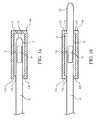
- FIGS. 1A and 1Bare schematic diagrams of catheters attached to a uniformly direct or diffuse reflecting apparatus.
- FIG. 2is a schematic diagram of an optical system for collection of back-reflected light from the distal tip of a delivery catheter.
- FIG. 3is a schematic diagram of a catheter in a blood vessel showing the possible reference measurements that can be taken for each interface of different materials encountered in this environment.
- FIG. 4is a schematic diagram of a guide catheter with an optically transparent transmission window.
- This inventionis a collection of methods and devices that are designed to address optical referencing problems (factors such as artifacts, noise, and background) that arise during in vivo intravascular optical measurements.
- the methodsinclude measurements made both before the catheterization procedure to reference catheter-specific factors, and measurements, e.g., made in real-time, during or before various catheterization procedures to reference patient-specific factors.
- Catheterscan be normalized by taking a reference measurement of the catheter's characteristics before the catheterization procedure begins. This catheter-specific measurement is required to isolate the catheter's variances, because other optical system variances are often addressed during measurement procedures by methods such as optical beam sampling and measurement. These other calibrating methods usually focus on light source variances. Sequential reference measurements can be taken throughout the catheterization process to calibrate for electrical or detector variability, using standard techniques.
- One method of taking such a reference measurement of catheter-specific variancesis to place the catheter into an environment with known and constant optical characteristics.
- the cathetere.g., the tip or other portion of the catheter that includes an optical measurement area
- FIG. 1Ashows one such reflection apparatus 14 a that is attached to the distal end 12 of a catheter 10 during manufacture or just prior to or just after a catheterization procedure.
- the benefit of having this apparatus attached during manufactureis that the entire combination, catheter and apparatus, can be sterilized and packaged at once.
- the benefit of using the apparatus after a catheterization procedureis that the operator no longer has to worry about maintaining sterility of the catheter.
- Reflection apparatus 14 aincludes a hollow cylinder or end-capped well 15 that is internally coated with a reflecting layer 16 of a material such as gold or platinum that provides a high direct reflection.
- a reflecting layer 16of a material such as gold or platinum that provides a high direct reflection.
- materials that provide various degrees of diffuse reflectionsuch as brushed aluminum or Spectralon® (by Lab Spheres, Inc.) can be used as the reflecting layer 16 .
- the materialmust be sufficiently thick such that effectively all of the light emitted by the catheter is reflected back into the catheter or is absorbed by the material.
- Tip apparatus 14 acan be cylindrical or spherical to allow for uniform referencing, regardless of catheter orientation.
- the tip apparatusincludes an entrance 13 a to insert the catheter, but the apparatus should also allow minimal external light penetration.
- the apparatusshould be made of a material that is opaque to the external light, such as an opaque plastic or metal, and have a relatively tight opening to insert the catheter.
- external light penetrationcan be minimized by placing an O-ring, flange, flap valve, or some other sealing structure 13 b, made of a material that will not scratch the catheter, such as plastic, rubber, or silicone, at the entrance 13 a of the tip apparatus 14 .
- the tip apparatusmust have sufficient depth such that no measurable reflected light comes from this sealing structure 13 b back to the catheter tip.
- this reflection apparatus 14 bhas both an entrance 13 a and exit opening 17 a with sealing structures 17 b to accommodate catheters with long distal segments from the measurement point, e.g., catheters having features such as guide wires.
- the optical measurement area 18is not at the distal end 12 of catheter 10 , but is set back from the end. In all embodiments, the important point is to insert the optical measurement area into the apparatus.
- the reference readingcan be taken before or after the catheterization procedure either by the optical system itself, or another specially-designed, dedicated optical device, which when connected to the catheter takes the optical measurements, and then the reflection apparatus can be removed.
- the reference readingcan then be processed to generate a reference signal, e.g., an electrical signal, which is used by the optical system to normalize the actual sample readings measured using the tested catheter.
- a reference signale.g., an electrical signal
- Each catheterwill have its own, unique reference reading/measurement and signal that the optical system takes into account when providing an output signal.
- the systemcan normalize the actual measurements for the catheter's unique characteristics by using standard chemometric methods including signal subtraction or taking the ratio of an actual measurement over the reference measurement.
- the cathetercan be placed into a permanent, non-disposable fixture attached to the optical system.
- This fixturehas characteristics similar to those of the reflection apparatus of FIGS. 1A and 1B described above.
- the environment with known and constant optical characteristicscan be a liquid medium with a defined and constant scatter and absorbance characteristics, or some combination of the two.
- the catheteris merely inserted into the appropriate liquid in a container.
- the primary aspect for the liquid to be measured for such a referencing methodis that it must be pure. There are large, commercially available sources (such as BioRad) for a variety of pure liquids. The reading must be made through a sufficient depth of liquid, determined by the liquid, such that effectively all reflected light is from the liquid and not from the walls of the container holding the liquid.
- the containermust be opaque to the wavelengths utilized for the reference measurement and to external light.
- An example of a liquid that can be used in the new method and that has both defined scattering and absorbance characteristicscomprises styrene divinyl/benzene cross-linked copolymer beads suspended in an ultrapure aqueous solution.
- a reference measurementis made using one of the new methods when the catheter is manufactured, and the reference signal is transcribed into symbols, e.g., optically readable symbols such as a series of numbers or a bar code, and is provided to the optical system operator along with the catheter, for example, written or printed on the catheter or catheter packaging, for entry into the system.
- the reference signalcan also be stored on a computer-readable medium, such as a diskette or magnetic tape or chip, and attached to or sent along with the catheter.
- Such reference datacould include measurements of the direct or diffusely reflected signal received from the catheter, from within the apparatus or liquid described above, with a defined optical power input or the spectral absorbance characteristics of the catheter including UV, visible, near-infrared, and/or infrared wavelengths.
- the patient's bloodis used as a reference before, during, and/or after the catheterization procedure.
- This techniqueensures that variances in the blood characteristics from patient to patient do not interfere with the measurement of the target tissue or material, unless the blood itself contains the target.
- reference measurementsare made to remove any signal corresponding to the optical characteristics that are not intended to be part of the target signal.
- One example of thiswould be to measure the blood's scattering characteristics for those situations in which diffuse absorbance of the blood is measured.
- Methods to incorporate this reference readinginclude obtaining blood from the catheterization site, or elsewhere, and then placing it in an apparatus with characteristic and constant optical properties, including, but not limited to, high direct or diffusely reflecting material.
- a reference measurementcan then be made of the patient's blood and incorporated into the optical system for normalizing across variances in patient-to-patient blood. It is important to measure the patient's blood optical characteristics because factors such as hematocrit can significantly alter optical scatter, and blood cholesterol content can change absorbance or reflectance spectra.
- Ex vivo reference measurement of the patient's bloodprovides more accurate measurements due to the absence of motion and the removal of the possibility of measuring a vascular wall instead of a sufficient depth of blood.
- One method to obtain such a reference readingis to measure the back-reflection of electromagnetic radiation, such as light, from the distal tip of the delivery fiber or fibers.
- electromagnetic radiationsuch as light
- the optical systemis specifically designed to preferentially gather this back-reflected light from the delivery fiber.
- this systemmust separate the electromagnetic radiation (e.g., light) that is reflected from the proximal end of a delivery fiber or catheter, from the radiation (light) that is back-reflected from the distal tip of the optical fiber or catheter.
- FIG. 2shows a system for use in this method.
- System 20includes a light source 21 that emits a delivery beam 28 , which passes through beam sampler 26 on its way to fiber coupler 24 and into catheter 22 .
- Light reflected from the proximal end 23 of catheter 22is returned along path 32 to beam sampler 26 .
- This reflected lightis polarized, just like delivery beam 28 , because it has not entered catheter 22 .
- Light that enters the catheteris transmitted to the distal tip 25 and is reflected back from the inside of the tip of catheter 22 , passes back through the catheter, through fiber coupler 24 , and is directed as a back-reflected beam along path 32 to the beam sampler 26 .
- This back-reflected lightis unpolarized by its travel through the catheter.
- Beam sampler 26receives both the reflected light and the back-reflected light and diverts portions thereof to a polarizer 34 via an optical redirector 30 , such as mirror or prism, to detector 36 , such as a PbS, PbSe, or InGas photodetector.
- Polarizer 34is adjusted to minimize, and thus remove, the polarized reflected light form the proximal end of the optical fiber/catheter from the unpolarized back-reflected light that has been reflected from the distal tip of the optical fiber/catheter.
- the systemcan normalize the measurement for variances in light output at the end of the catheter, given that the total measured loss will be twice that of the losses in one direction along the catheter at any given moment.
- the actual measurementsare normalized using standard chemometric methods including signal subtraction or taking the ratio of the actual measurement over the reference measurement.
- FIG. 3schematically illustrates a catheter 40 with a delivery or output fiber 44 and an input fiber 46 and a transmission window 47 , in blood 43 within a blood vessel 42 .
- delivery fiber 44it passes through the transmission window 47 , the blood 43 , the surface of the blood vessel wall 42 a, the wall 42 b itself, the surface of a lipid pool 45 a, and into the lipid pool 45 b, of a lipid-rich vulnerable atherosclerotic plaque.
- Each of these interfacese.g., device-element/device-element interface, device/sample interface or sample-component/sample-component interface within the sample
- materialscan be measured for referencing.
- eight separate measurementscan be made at the air or delivery fiber/transmission window interface C 1 ( ⁇ ), in the transmission window interior C 2 ( ⁇ ), the transmission/blood interface C 3 ( ⁇ ), the blood C 4 ( ⁇ ), the blood/vessel wall interface C 5 ( ⁇ ), the vessel wall interior C 6 ( ⁇ ), the vessel wall/lipid pool interface C 7 ( ⁇ ), and the lipid pool interior C 8 ( ⁇ ).
- An example of such a measurementis to measure a large vessel, and thus obtain a measurement that is mostly contributed by blood, and then measure points in a smaller vessel, allowing for subtraction of the large vessel/blood measurement to better discriminate the contribution by the vessel wall in the subsequent readings.
- Incorporation of a reference reading of the patient's blood during the catheterization procedureis another example of such a reference measurement.
- This methodwill reduce the requirement for external blood referencing measurements.
- a physiciantakes a reference reading while in a large vessel, such as the aorta, to obtain a depth of blood sufficient to mimic an infinite depth for the optical measurement, which is between about 1 mm to 5 mm, depending primarily upon hematocrit levels and the wavelengths of light utilized.
- This procedurecan be carried out using a self-centering and stabilizing mechanism such as a balloon.
- This reference measurementcan be integrated into the optical system by using any of a variety of known referencing analysis methods described herein, including signal subtraction or taking the ratio of the actual measurement over the reference measurement.
- An apparatus for use in the method described abovecan include a guide catheter 50 (shown schematically in FIG. 4 ), which is used to safely guide a catheter from the entry point of the body to the coronaries, and that is designed with a window 52 that is relatively optically transparent to the wavelengths to be utilized.
- Window 52should be made of a well-defined material, such as different types of polyethylene, polyurethane, or Teflon®, with constant characteristics for incorporation into the referencing system.
- the guide catheter 50includes a distal end 54 and a proximal end 56 .
- the window 52is positioned between the proximal and distal ends so that the window 52 is within the aorta or vena cava when the guide catheter is fully in place.
- Reference measurementscan also be made of normal vasculature that would increase the selective capability for target identification.
- a methodincludes taking a measurement in a similarly sized, but normal, section of vasculature. The determination that a section can be designated “normal” by, e.g., visualization or additional measurements from other technologies such as coronary measurements utilizing intravascular ultrasound. This measurement could then be incorporated in the optical system to normalize the measurement by common analysis methods described herein and known to those skilled in this field to increase discrimination.
- reference measurementscan also be made of diseased vasculature.
- a known atherosclerotic, stenotic lesioncan be identified by fluoroscopy, or a catheter-based technology such as intravascular ultrasound and then imaged or illuminated to get a reference measurement. This would increase discrimination of characteristics such as lipid-pools within different stages of atherosclerotic diseased tissue.
- common analysis methodssuch as partial least squares discriminate analysis will enable greater discrimination of variances in atherosclerosis outside of that disease state such as thin-capped fibroatheroma that contain lipid-pools.
Landscapes
- Health & Medical Sciences (AREA)
- Life Sciences & Earth Sciences (AREA)
- Physics & Mathematics (AREA)
- General Health & Medical Sciences (AREA)
- Pathology (AREA)
- Biomedical Technology (AREA)
- Medical Informatics (AREA)
- Veterinary Medicine (AREA)
- Public Health (AREA)
- Animal Behavior & Ethology (AREA)
- Biophysics (AREA)
- Engineering & Computer Science (AREA)
- Surgery (AREA)
- Heart & Thoracic Surgery (AREA)
- Molecular Biology (AREA)
- Biochemistry (AREA)
- Chemical & Material Sciences (AREA)
- Analytical Chemistry (AREA)
- Immunology (AREA)
- General Physics & Mathematics (AREA)
- Optics & Photonics (AREA)
- Investigating Or Analysing Materials By Optical Means (AREA)
- Measurement Of The Respiration, Hearing Ability, Form, And Blood Characteristics Of Living Organisms (AREA)
- Infusion, Injection, And Reservoir Apparatuses (AREA)
Abstract
Description
Claims (32)
Priority Applications (4)
| Application Number | Priority Date | Filing Date | Title |
|---|---|---|---|
| US09/871,759US6615062B2 (en) | 2001-05-31 | 2001-05-31 | Referencing optical catheters |
| AU2002305512AAU2002305512A1 (en) | 2001-05-31 | 2002-05-10 | Referencing optical catheters |
| EP02734338AEP1401328A4 (en) | 2001-05-31 | 2002-05-10 | Referencing optical catheters |
| PCT/US2002/014790WO2002098478A2 (en) | 2001-05-31 | 2002-05-10 | Referencing optical catheters |
Applications Claiming Priority (1)
| Application Number | Priority Date | Filing Date | Title |
|---|---|---|---|
| US09/871,759US6615062B2 (en) | 2001-05-31 | 2001-05-31 | Referencing optical catheters |
Publications (2)
| Publication Number | Publication Date |
|---|---|
| US20030097048A1 US20030097048A1 (en) | 2003-05-22 |
| US6615062B2true US6615062B2 (en) | 2003-09-02 |
Family
ID=25358063
Family Applications (1)
| Application Number | Title | Priority Date | Filing Date |
|---|---|---|---|
| US09/871,759Expired - LifetimeUS6615062B2 (en) | 2001-05-31 | 2001-05-31 | Referencing optical catheters |
Country Status (4)
| Country | Link |
|---|---|
| US (1) | US6615062B2 (en) |
| EP (1) | EP1401328A4 (en) |
| AU (1) | AU2002305512A1 (en) |
| WO (1) | WO2002098478A2 (en) |
Cited By (84)
| Publication number | Priority date | Publication date | Assignee | Title |
|---|---|---|---|---|
| US20040024298A1 (en)* | 2002-08-05 | 2004-02-05 | Infraredx, Inc. | Spectroscopic unwanted signal filters for discrimination of vulnerable plaque and method therefor |
| US20050275836A1 (en)* | 2004-06-15 | 2005-12-15 | Feldchtein Felix I | Calibration tool for an optical measuring device with an optical fiber probe |
| US7190986B1 (en)* | 2002-10-18 | 2007-03-13 | Nellcor Puritan Bennett Inc. | Non-adhesive oximeter sensor for sensitive skin |
| US20070078500A1 (en)* | 2005-09-30 | 2007-04-05 | Cornova, Inc. | Systems and methods for analysis and treatment of a body lumen |
| US20070270717A1 (en)* | 2005-09-30 | 2007-11-22 | Cornova, Inc. | Multi-faceted optical reflector |
| US20080221455A1 (en)* | 2002-08-05 | 2008-09-11 | Infraredx, Inc. | Near-infrared spectroscopic analysis of blood vessel walls |
| US20090175576A1 (en)* | 2008-01-08 | 2009-07-09 | Cornova, Inc. | Shaped fiber ends and methods of making same |
| US20090187108A1 (en)* | 2006-09-29 | 2009-07-23 | Cornova, Inc. | Systems and methods for analysis and treatment of a body lumen |
| US20090299351A1 (en)* | 2007-11-28 | 2009-12-03 | Spectranetics | Laser Catheter Calibrator |
| US20100023037A1 (en)* | 2003-01-14 | 2010-01-28 | Flowcardia, Inc. | Ultrasound catheter and methods for making and using same |
| US20100049209A1 (en)* | 2003-02-26 | 2010-02-25 | Flowcardia, Inc. | Ultrasound catheter apparatus |
| US20100094109A1 (en)* | 2008-10-15 | 2010-04-15 | Cornova, Inc. | Systems and methods for analysis and treatment of an occluded body lumen |
| US20100174196A1 (en)* | 2007-06-21 | 2010-07-08 | Cornova, Inc. | Systems and methods for guiding the analysis and treatment of a body lumen |
| US20100286531A1 (en)* | 2005-09-30 | 2010-11-11 | Cornova, Inc. | Systems and methods for analysis and treatment of a body lumen |
| US20120162660A1 (en)* | 2007-07-12 | 2012-06-28 | Volcano Corporation | Automatic calibration systems and methods of use |
| US8593641B2 (en) | 2007-07-12 | 2013-11-26 | Volcano Corporation | Apparatus and methods for uniform frequency sample clocking |
| US8679049B2 (en) | 2009-06-12 | 2014-03-25 | Flowcardia, Inc. | Device and method for vascular re-entry |
| US8690819B2 (en) | 2002-08-26 | 2014-04-08 | Flowcardia, Inc. | Ultrasound catheter for disrupting blood vessel obstructions |
| US20140192362A1 (en)* | 2011-09-20 | 2014-07-10 | Olympus Corporation | Optical measurement apparatus and calibration method |
| US8790291B2 (en) | 2004-08-26 | 2014-07-29 | Flowcardia, Inc. | Ultrasound catheter devices and methods |
| US8958867B2 (en) | 2011-08-29 | 2015-02-17 | Infraredx, Inc. | Detection of lipid core plaque cap thickness |
| US8956375B2 (en) | 2002-08-26 | 2015-02-17 | Flowcardia, Inc. | Ultrasound catheter devices and methods |
| US9265520B2 (en) | 2002-08-02 | 2016-02-23 | Flowcardia, Inc. | Therapeutic ultrasound system |
| US9286673B2 (en) | 2012-10-05 | 2016-03-15 | Volcano Corporation | Systems for correcting distortions in a medical image and methods of use thereof |
| US9282984B2 (en) | 2006-04-05 | 2016-03-15 | Flowcardia, Inc. | Therapeutic ultrasound system |
| US9292918B2 (en) | 2012-10-05 | 2016-03-22 | Volcano Corporation | Methods and systems for transforming luminal images |
| US9301687B2 (en) | 2013-03-13 | 2016-04-05 | Volcano Corporation | System and method for OCT depth calibration |
| US9307926B2 (en) | 2012-10-05 | 2016-04-12 | Volcano Corporation | Automatic stent detection |
| US9324141B2 (en) | 2012-10-05 | 2016-04-26 | Volcano Corporation | Removal of A-scan streaking artifact |
| US9360630B2 (en) | 2011-08-31 | 2016-06-07 | Volcano Corporation | Optical-electrical rotary joint and methods of use |
| US9367965B2 (en) | 2012-10-05 | 2016-06-14 | Volcano Corporation | Systems and methods for generating images of tissue |
| US9383263B2 (en) | 2012-12-21 | 2016-07-05 | Volcano Corporation | Systems and methods for narrowing a wavelength emission of light |
| US9381027B2 (en) | 2002-08-26 | 2016-07-05 | Flowcardia, Inc. | Steerable ultrasound catheter |
| US9433433B2 (en) | 2003-09-19 | 2016-09-06 | Flowcardia, Inc. | Connector for securing ultrasound catheter to transducer |
| US9478940B2 (en) | 2012-10-05 | 2016-10-25 | Volcano Corporation | Systems and methods for amplifying light |
| US9486143B2 (en) | 2012-12-21 | 2016-11-08 | Volcano Corporation | Intravascular forward imaging device |
| US9596993B2 (en) | 2007-07-12 | 2017-03-21 | Volcano Corporation | Automatic calibration systems and methods of use |
| US9612105B2 (en) | 2012-12-21 | 2017-04-04 | Volcano Corporation | Polarization sensitive optical coherence tomography system |
| US9622706B2 (en) | 2007-07-12 | 2017-04-18 | Volcano Corporation | Catheter for in vivo imaging |
| US9629643B2 (en) | 2006-11-07 | 2017-04-25 | Flowcardia, Inc. | Ultrasound catheter having improved distal end |
| US9709379B2 (en) | 2012-12-20 | 2017-07-18 | Volcano Corporation | Optical coherence tomography system that is reconfigurable between different imaging modes |
| US9730613B2 (en) | 2012-12-20 | 2017-08-15 | Volcano Corporation | Locating intravascular images |
| US9770172B2 (en) | 2013-03-07 | 2017-09-26 | Volcano Corporation | Multimodal segmentation in intravascular images |
| US9858668B2 (en) | 2012-10-05 | 2018-01-02 | Volcano Corporation | Guidewire artifact removal in images |
| US9867530B2 (en) | 2006-08-14 | 2018-01-16 | Volcano Corporation | Telescopic side port catheter device with imaging system and method for accessing side branch occlusions |
| US10058284B2 (en) | 2012-12-21 | 2018-08-28 | Volcano Corporation | Simultaneous imaging, monitoring, and therapy |
| US10070827B2 (en) | 2012-10-05 | 2018-09-11 | Volcano Corporation | Automatic image playback |
| US10166003B2 (en) | 2012-12-21 | 2019-01-01 | Volcano Corporation | Ultrasound imaging with variable line density |
| US10191220B2 (en) | 2012-12-21 | 2019-01-29 | Volcano Corporation | Power-efficient optical circuit |
| US10219887B2 (en) | 2013-03-14 | 2019-03-05 | Volcano Corporation | Filters with echogenic characteristics |
| US10219780B2 (en) | 2007-07-12 | 2019-03-05 | Volcano Corporation | OCT-IVUS catheter for concurrent luminal imaging |
| US10226597B2 (en) | 2013-03-07 | 2019-03-12 | Volcano Corporation | Guidewire with centering mechanism |
| US10238367B2 (en) | 2012-12-13 | 2019-03-26 | Volcano Corporation | Devices, systems, and methods for targeted cannulation |
| US10285719B2 (en) | 2005-01-20 | 2019-05-14 | Flowcardia, Inc. | Vibrational catheter devices and methods for making same |
| US10292677B2 (en) | 2013-03-14 | 2019-05-21 | Volcano Corporation | Endoluminal filter having enhanced echogenic properties |
| US10332228B2 (en) | 2012-12-21 | 2019-06-25 | Volcano Corporation | System and method for graphical processing of medical data |
| US10357263B2 (en) | 2012-01-18 | 2019-07-23 | C. R. Bard, Inc. | Vascular re-entry device |
| US10413317B2 (en) | 2012-12-21 | 2019-09-17 | Volcano Corporation | System and method for catheter steering and operation |
| US10420530B2 (en) | 2012-12-21 | 2019-09-24 | Volcano Corporation | System and method for multipath processing of image signals |
| US10426590B2 (en) | 2013-03-14 | 2019-10-01 | Volcano Corporation | Filters with echogenic characteristics |
| US10568586B2 (en) | 2012-10-05 | 2020-02-25 | Volcano Corporation | Systems for indicating parameters in an imaging data set and methods of use |
| US10582983B2 (en) | 2017-02-06 | 2020-03-10 | C. R. Bard, Inc. | Ultrasonic endovascular catheter with a controllable sheath |
| US10595820B2 (en) | 2012-12-20 | 2020-03-24 | Philips Image Guided Therapy Corporation | Smooth transition catheters |
| US10638939B2 (en) | 2013-03-12 | 2020-05-05 | Philips Image Guided Therapy Corporation | Systems and methods for diagnosing coronary microvascular disease |
| US10724082B2 (en) | 2012-10-22 | 2020-07-28 | Bio-Rad Laboratories, Inc. | Methods for analyzing DNA |
| US10758207B2 (en) | 2013-03-13 | 2020-09-01 | Philips Image Guided Therapy Corporation | Systems and methods for producing an image from a rotational intravascular ultrasound device |
| US10758256B2 (en) | 2016-12-22 | 2020-09-01 | C. R. Bard, Inc. | Ultrasonic endovascular catheter |
| US10776654B2 (en) | 2015-03-10 | 2020-09-15 | Infraredx, Inc. | Assessment of lipid core plaque integrity |
| US10835267B2 (en) | 2002-08-02 | 2020-11-17 | Flowcardia, Inc. | Ultrasound catheter having protective feature against breakage |
| US10942022B2 (en) | 2012-12-20 | 2021-03-09 | Philips Image Guided Therapy Corporation | Manual calibration of imaging system |
| US10939826B2 (en) | 2012-12-20 | 2021-03-09 | Philips Image Guided Therapy Corporation | Aspirating and removing biological material |
| US10993694B2 (en) | 2012-12-21 | 2021-05-04 | Philips Image Guided Therapy Corporation | Rotational ultrasound imaging catheter with extended catheter body telescope |
| US11026591B2 (en) | 2013-03-13 | 2021-06-08 | Philips Image Guided Therapy Corporation | Intravascular pressure sensor calibration |
| US11040140B2 (en) | 2010-12-31 | 2021-06-22 | Philips Image Guided Therapy Corporation | Deep vein thrombosis therapeutic methods |
| US11141063B2 (en) | 2010-12-23 | 2021-10-12 | Philips Image Guided Therapy Corporation | Integrated system architectures and methods of use |
| US11154313B2 (en) | 2013-03-12 | 2021-10-26 | The Volcano Corporation | Vibrating guidewire torquer and methods of use |
| US11272845B2 (en) | 2012-10-05 | 2022-03-15 | Philips Image Guided Therapy Corporation | System and method for instant and automatic border detection |
| US11284860B2 (en) | 2017-09-15 | 2022-03-29 | Infraredx, Inc. | Imaging catheter |
| US11344750B2 (en) | 2012-08-02 | 2022-05-31 | Flowcardia, Inc. | Ultrasound catheter system |
| US11406498B2 (en) | 2012-12-20 | 2022-08-09 | Philips Image Guided Therapy Corporation | Implant delivery system and implants |
| US11596726B2 (en) | 2016-12-17 | 2023-03-07 | C.R. Bard, Inc. | Ultrasound devices for removing clots from catheters and related methods |
| US11633206B2 (en) | 2016-11-23 | 2023-04-25 | C.R. Bard, Inc. | Catheter with retractable sheath and methods thereof |
| US12201477B2 (en) | 2012-10-05 | 2025-01-21 | Philips Image Guided Therapy Corporation | Methods and systems for establishing parameters for three-dimensional imaging |
| US12343198B2 (en) | 2013-03-14 | 2025-07-01 | Philips Image Guided Therapy Corporation | Delivery catheter having imaging capabilities |
Families Citing this family (80)
| Publication number | Priority date | Publication date | Assignee | Title |
|---|---|---|---|---|
| US7231243B2 (en) | 2000-10-30 | 2007-06-12 | The General Hospital Corporation | Optical methods for tissue analysis |
| US9295391B1 (en) | 2000-11-10 | 2016-03-29 | The General Hospital Corporation | Spectrally encoded miniature endoscopic imaging probe |
| AT503309B1 (en) | 2001-05-01 | 2011-08-15 | Gen Hospital Corp | DEVICE FOR DETERMINING ATHEROSCLEROTIC BEARING BY MEASURING OPTICAL TISSUE PROPERTIES |
| US7355716B2 (en) | 2002-01-24 | 2008-04-08 | The General Hospital Corporation | Apparatus and method for ranging and noise reduction of low coherence interferometry LCI and optical coherence tomography OCT signals by parallel detection of spectral bands |
| US8054468B2 (en) | 2003-01-24 | 2011-11-08 | The General Hospital Corporation | Apparatus and method for ranging and noise reduction of low coherence interferometry LCI and optical coherence tomography OCT signals by parallel detection of spectral bands |
| EP2436307B1 (en) | 2003-03-31 | 2015-10-21 | The General Hospital Corporation | Speckle reduction in optical coherence tomography by path length encoded angular compounding |
| KR101386971B1 (en) | 2003-06-06 | 2014-04-18 | 더 제너럴 하스피탈 코포레이션 | Process and apparatus for a wavelength tunning source |
| EP2280256B1 (en) | 2003-10-27 | 2016-11-16 | The General Hospital Corporation | Method and apparatus for performing optical imaging using frequency-domain interferometry |
| KR101239250B1 (en) | 2004-05-29 | 2013-03-05 | 더 제너럴 하스피탈 코포레이션 | Process, system and software arrangement for a chromatic dispersion compensation using reflective layers in optical coherence tomography (oct) imaging |
| AU2005270037B2 (en) | 2004-07-02 | 2012-02-09 | The General Hospital Corporation | Endoscopic imaging probe comprising dual clad fibre |
| EP1782020B1 (en) | 2004-08-06 | 2012-10-03 | The General Hospital Corporation | Process, system and software arrangement for determining at least one location in a sample using an optical coherence tomography |
| EP2272421A1 (en) | 2004-08-24 | 2011-01-12 | The General Hospital Corporation | Method and apparatus for imaging of vessel segments |
| WO2006024014A2 (en) | 2004-08-24 | 2006-03-02 | The General Hospital Corporation | Process, system and software arrangement for measuring a mechanical strain and elastic properties of a sample |
| US7365859B2 (en) | 2004-09-10 | 2008-04-29 | The General Hospital Corporation | System and method for optical coherence imaging |
| KR101257100B1 (en) | 2004-09-29 | 2013-04-22 | 더 제너럴 하스피탈 코포레이션 | System and Method for Optical Coherence Imaging |
| WO2006058049A1 (en) | 2004-11-24 | 2006-06-01 | The General Hospital Corporation | Common-path interferometer for endoscopic oct |
| WO2006058346A1 (en) | 2004-11-29 | 2006-06-01 | The General Hospital Corporation | Arrangements, devices, endoscopes, catheters and methods for performing optical imaging by simultaneously illuminating and detecting multiple points on a sample |
| ES2337497T3 (en) | 2005-04-28 | 2010-04-26 | The General Hospital Corporation | EVALUATION OF CHARACTERISTICS OF THE IMAGE OF AN ANATOMICAL STRUCTURE IN IMAGES OF TOMOGRAPHY OF OPTICAL COHERENCE. |
| US9060689B2 (en) | 2005-06-01 | 2015-06-23 | The General Hospital Corporation | Apparatus, method and system for performing phase-resolved optical frequency domain imaging |
| BRPI0612654B8 (en) | 2005-06-27 | 2021-05-25 | Glaxosmithkline Biologicals Sa | method for conjugating a saccharide to a protein, and saccharide-protein conjugate |
| EP2267404B1 (en) | 2005-08-09 | 2016-10-05 | The General Hospital Corporation | Apparatus and method for performing polarization-based quadrature demodulation in optical coherence tomography |
| US7843572B2 (en) | 2005-09-29 | 2010-11-30 | The General Hospital Corporation | Method and apparatus for optical imaging via spectral encoding |
| US7889348B2 (en) | 2005-10-14 | 2011-02-15 | The General Hospital Corporation | Arrangements and methods for facilitating photoluminescence imaging |
| EP1971848B1 (en) | 2006-01-10 | 2019-12-04 | The General Hospital Corporation | Systems and methods for generating data based on one or more spectrally-encoded endoscopy techniques |
| US20070238955A1 (en)* | 2006-01-18 | 2007-10-11 | The General Hospital Corporation | Systems and methods for generating data using one or more endoscopic microscopy techniques |
| US8145018B2 (en) | 2006-01-19 | 2012-03-27 | The General Hospital Corporation | Apparatus for obtaining information for a structure using spectrally-encoded endoscopy techniques and methods for producing one or more optical arrangements |
| DK1973466T3 (en) | 2006-01-19 | 2021-02-01 | Massachusetts Gen Hospital | BALLOON IMAGING CATHETER |
| WO2007149602A2 (en) | 2006-02-01 | 2007-12-27 | The General Hospital Corporation | Methods and systems for providing electromagnetic radiation to at least one portion of a sample using conformal laser therapy procedures |
| JP5680829B2 (en) | 2006-02-01 | 2015-03-04 | ザ ジェネラル ホスピタル コーポレイション | A device that irradiates a sample with multiple electromagnetic radiations |
| US9777053B2 (en) | 2006-02-08 | 2017-10-03 | The General Hospital Corporation | Methods, arrangements and systems for obtaining information associated with an anatomical sample using optical microscopy |
| EP2982929A1 (en) | 2006-02-24 | 2016-02-10 | The General Hospital Corporation | Methods and systems for performing angle-resolved fourier-domain optical coherence tomography |
| WO2007133961A2 (en) | 2006-05-10 | 2007-11-22 | The General Hospital Corporation | Processes, arrangements and systems for providing frequency domain imaging of a sample |
| US8838213B2 (en) | 2006-10-19 | 2014-09-16 | The General Hospital Corporation | Apparatus and method for obtaining and providing imaging information associated with at least one portion of a sample, and effecting such portion(s) |
| EP2104968A1 (en) | 2007-01-19 | 2009-09-30 | The General Hospital Corporation | Rotating disk reflection for fast wavelength scanning of dispersed broadband light |
| US9176319B2 (en) | 2007-03-23 | 2015-11-03 | The General Hospital Corporation | Methods, arrangements and apparatus for utilizing a wavelength-swept laser using angular scanning and dispersion procedures |
| US10534129B2 (en) | 2007-03-30 | 2020-01-14 | The General Hospital Corporation | System and method providing intracoronary laser speckle imaging for the detection of vulnerable plaque |
| US8045177B2 (en) | 2007-04-17 | 2011-10-25 | The General Hospital Corporation | Apparatus and methods for measuring vibrations using spectrally-encoded endoscopy |
| WO2008137637A2 (en) | 2007-05-04 | 2008-11-13 | The General Hospital Corporation | Methods, arrangements and systems for obtaining information associated with a sample using brillouin microscopy |
| US9375158B2 (en) | 2007-07-31 | 2016-06-28 | The General Hospital Corporation | Systems and methods for providing beam scan patterns for high speed doppler optical frequency domain imaging |
| WO2009029843A1 (en) | 2007-08-31 | 2009-03-05 | The General Hospital Corporation | System and method for self-interference fluoresence microscopy, and computer-accessible medium associated therewith |
| WO2009049296A2 (en)* | 2007-10-12 | 2009-04-16 | The General Hospital Corporation | Systems and processes for optical imaging of luminal anatomic structures |
| US7933021B2 (en) | 2007-10-30 | 2011-04-26 | The General Hospital Corporation | System and method for cladding mode detection |
| DE102008013858A1 (en)* | 2008-03-12 | 2009-09-24 | Siemens Aktiengesellschaft | Catheter device and associated medical examination and treatment device |
| DE102008013854A1 (en)* | 2008-03-12 | 2009-09-24 | Siemens Aktiengesellschaft | Catheter and associated medical examination and treatment facility |
| US7898656B2 (en) | 2008-04-30 | 2011-03-01 | The General Hospital Corporation | Apparatus and method for cross axis parallel spectroscopy |
| EP2274572A4 (en) | 2008-05-07 | 2013-08-28 | Gen Hospital Corp | SYSTEM, METHOD AND COMPUTER MEDIUM FOR MONITORING THE MOVEMENT OF VESSELS DURING A THREE-DIMENSIONAL MICROSCOPY EXAMINATION OF CORONARY ARTERIES |
| US8861910B2 (en) | 2008-06-20 | 2014-10-14 | The General Hospital Corporation | Fused fiber optic coupler arrangement and method for use thereof |
| WO2010009136A2 (en) | 2008-07-14 | 2010-01-21 | The General Hospital Corporation | Apparatus and methods for color endoscopy |
| DE102008054297A1 (en) | 2008-11-03 | 2010-05-06 | Siemens Aktiengesellschaft | A catheter assembly for insertion into a blood vessel, medical examination and treatment device comprising such a catheter assembly and method for minimally invasive intervention on a blood vessel in the brain |
| JP5731394B2 (en) | 2008-12-10 | 2015-06-10 | ザ ジェネラル ホスピタル コーポレイション | System, apparatus and method for extending imaging depth range of optical coherence tomography through optical subsampling |
| JP2012515576A (en) | 2009-01-20 | 2012-07-12 | ザ ジェネラル ホスピタル コーポレイション | Endoscopic biopsy device, system, and method |
| US8097864B2 (en) | 2009-01-26 | 2012-01-17 | The General Hospital Corporation | System, method and computer-accessible medium for providing wide-field superresolution microscopy |
| WO2010105197A2 (en) | 2009-03-12 | 2010-09-16 | The General Hospital Corporation | Non-contact optical system, computer-accessible medium and method for measuring at least one mechanical property of tissue using coherent speckle techniques(s) |
| DE102009014462B4 (en)* | 2009-03-23 | 2019-01-17 | Siemens Healthcare Gmbh | A blood pump, medical device, comprising a blood pump and methods for assisting the placement of a blood pump |
| DE102009014489B4 (en)* | 2009-03-23 | 2011-03-10 | Siemens Aktiengesellschaft | Catheter and medical device |
| JP5819823B2 (en) | 2009-07-14 | 2015-11-24 | ザ ジェネラル ホスピタル コーポレイション | Device for measuring the flow and pressure inside a blood vessel and method of operating the device |
| DE102010007177B4 (en)* | 2010-02-08 | 2017-06-22 | Siemens Healthcare Gmbh | Display method for an image of the interior of a vessel located in front of a widening device and display device corresponding thereto |
| KR20130004326A (en) | 2010-03-05 | 2013-01-09 | 더 제너럴 하스피탈 코포레이션 | Systems, methods and computer-accessible medium which provide microscopic images of at least one anatomical structure at a particular resolution |
| US9069130B2 (en) | 2010-05-03 | 2015-06-30 | The General Hospital Corporation | Apparatus, method and system for generating optical radiation from biological gain media |
| EP2575598A2 (en) | 2010-05-25 | 2013-04-10 | The General Hospital Corporation | Apparatus, systems, methods and computer-accessible medium for spectral analysis of optical coherence tomography images |
| EP2575597B1 (en) | 2010-05-25 | 2022-05-04 | The General Hospital Corporation | Apparatus for providing optical imaging of structures and compositions |
| JP6066901B2 (en) | 2010-06-03 | 2017-01-25 | ザ ジェネラル ホスピタル コーポレイション | Method for apparatus and device for imaging structures in or in one or more luminal organs |
| WO2012058381A2 (en) | 2010-10-27 | 2012-05-03 | The General Hospital Corporation | Apparatus, systems and methods for measuring blood pressure within at least one vessel |
| US9330092B2 (en) | 2011-07-19 | 2016-05-03 | The General Hospital Corporation | Systems, methods, apparatus and computer-accessible-medium for providing polarization-mode dispersion compensation in optical coherence tomography |
| EP3835718B1 (en) | 2011-08-25 | 2023-07-26 | The General Hospital Corporation | Apparatus for providing micro-optical coherence tomography inside a respiratory system |
| JP2015502562A (en) | 2011-10-18 | 2015-01-22 | ザ ジェネラル ホスピタル コーポレイション | Apparatus and method for generating and / or providing recirculating optical delay |
| WO2013148306A1 (en) | 2012-03-30 | 2013-10-03 | The General Hospital Corporation | Imaging system, method and distal attachment for multidirectional field of view endoscopy |
| JP2015517387A (en) | 2012-05-21 | 2015-06-22 | ザ ジェネラル ホスピタル コーポレイション | Apparatus, device and method for capsule microscopy |
| US9968261B2 (en) | 2013-01-28 | 2018-05-15 | The General Hospital Corporation | Apparatus and method for providing diffuse spectroscopy co-registered with optical frequency domain imaging |
| WO2014120791A1 (en) | 2013-01-29 | 2014-08-07 | The General Hospital Corporation | Apparatus, systems and methods for providing information regarding the aortic valve |
| US11179028B2 (en) | 2013-02-01 | 2021-11-23 | The General Hospital Corporation | Objective lens arrangement for confocal endomicroscopy |
| US10478072B2 (en) | 2013-03-15 | 2019-11-19 | The General Hospital Corporation | Methods and system for characterizing an object |
| EP2997354A4 (en) | 2013-05-13 | 2017-01-18 | The General Hospital Corporation | Detecting self-interefering fluorescence phase and amplitude |
| EP3021735A4 (en) | 2013-07-19 | 2017-04-19 | The General Hospital Corporation | Determining eye motion by imaging retina. with feedback |
| WO2015009932A1 (en) | 2013-07-19 | 2015-01-22 | The General Hospital Corporation | Imaging apparatus and method which utilizes multidirectional field of view endoscopy |
| WO2015013651A2 (en) | 2013-07-26 | 2015-01-29 | The General Hospital Corporation | System, apparatus and method utilizing optical dispersion for fourier-domain optical coherence tomography |
| WO2015105870A1 (en) | 2014-01-08 | 2015-07-16 | The General Hospital Corporation | Method and apparatus for microscopic imaging |
| US10736494B2 (en) | 2014-01-31 | 2020-08-11 | The General Hospital Corporation | System and method for facilitating manual and/or automatic volumetric imaging with real-time tension or force feedback using a tethered imaging device |
| WO2015153982A1 (en) | 2014-04-04 | 2015-10-08 | The General Hospital Corporation | Apparatus and method for controlling propagation and/or transmission of electromagnetic radiation in flexible waveguide(s) |
| US10912462B2 (en) | 2014-07-25 | 2021-02-09 | The General Hospital Corporation | Apparatus, devices and methods for in vivo imaging and diagnosis |
Citations (9)
| Publication number | Priority date | Publication date | Assignee | Title |
|---|---|---|---|---|
| US4050450A (en)* | 1976-03-05 | 1977-09-27 | American Optical Corporation | Reflection standard for fiber optic probe |
| US4322164A (en) | 1976-10-18 | 1982-03-30 | Oximetrix, Inc. | Sterilizable, disposable optical scattering reference medium and container assembly |
| US4650327A (en) | 1985-10-28 | 1987-03-17 | Oximetrix, Inc. | Optical catheter calibrating assembly |
| US4744656A (en)* | 1986-12-08 | 1988-05-17 | Spectramed, Inc. | Disposable calibration boot for optical-type cardiovascular catheter |
| US4904085A (en)* | 1988-05-04 | 1990-02-27 | Simmonds Precision Products, Inc. | Polarimetric fiber optic sensor testing apparatus |
| US5305744A (en)* | 1992-03-18 | 1994-04-26 | Ulrich Pfeiffer | Calibrating reflector device for an optical measuring system |
| US5365925A (en)* | 1993-08-13 | 1994-11-22 | Ohmeda Inc. | Disposable calibration boot for multi-point calibration in fiber optic sensors |
| US5455177A (en) | 1992-02-05 | 1995-10-03 | Boehringer Mannheim Gmbh | Method for analysis of a medical sample |
| US5939610A (en) | 1995-07-08 | 1999-08-17 | Horiba, Ltd. | Ion concentration measuring apparatus with internal calibration fluid reservoir |
Family Cites Families (3)
| Publication number | Priority date | Publication date | Assignee | Title |
|---|---|---|---|---|
| US4469442A (en)* | 1982-01-11 | 1984-09-04 | Japan Crown Cork Co., Ltd. | Detecting irregularities in a coating on a substrate |
| US5104392A (en)* | 1985-03-22 | 1992-04-14 | Massachusetts Institute Of Technology | Laser spectro-optic imaging for diagnosis and treatment of diseased tissue |
| US4823167A (en)* | 1986-12-16 | 1989-04-18 | Baxter International Inc. | Catheter calibration device |
- 2001
- 2001-05-31USUS09/871,759patent/US6615062B2/ennot_activeExpired - Lifetime
- 2002
- 2002-05-10AUAU2002305512Apatent/AU2002305512A1/ennot_activeAbandoned
- 2002-05-10EPEP02734338Apatent/EP1401328A4/ennot_activeWithdrawn
- 2002-05-10WOPCT/US2002/014790patent/WO2002098478A2/ennot_activeApplication Discontinuation
Patent Citations (9)
| Publication number | Priority date | Publication date | Assignee | Title |
|---|---|---|---|---|
| US4050450A (en)* | 1976-03-05 | 1977-09-27 | American Optical Corporation | Reflection standard for fiber optic probe |
| US4322164A (en) | 1976-10-18 | 1982-03-30 | Oximetrix, Inc. | Sterilizable, disposable optical scattering reference medium and container assembly |
| US4650327A (en) | 1985-10-28 | 1987-03-17 | Oximetrix, Inc. | Optical catheter calibrating assembly |
| US4744656A (en)* | 1986-12-08 | 1988-05-17 | Spectramed, Inc. | Disposable calibration boot for optical-type cardiovascular catheter |
| US4904085A (en)* | 1988-05-04 | 1990-02-27 | Simmonds Precision Products, Inc. | Polarimetric fiber optic sensor testing apparatus |
| US5455177A (en) | 1992-02-05 | 1995-10-03 | Boehringer Mannheim Gmbh | Method for analysis of a medical sample |
| US5305744A (en)* | 1992-03-18 | 1994-04-26 | Ulrich Pfeiffer | Calibrating reflector device for an optical measuring system |
| US5365925A (en)* | 1993-08-13 | 1994-11-22 | Ohmeda Inc. | Disposable calibration boot for multi-point calibration in fiber optic sensors |
| US5939610A (en) | 1995-07-08 | 1999-08-17 | Horiba, Ltd. | Ion concentration measuring apparatus with internal calibration fluid reservoir |
Cited By (124)
| Publication number | Priority date | Publication date | Assignee | Title |
|---|---|---|---|---|
| US9265520B2 (en) | 2002-08-02 | 2016-02-23 | Flowcardia, Inc. | Therapeutic ultrasound system |
| US10722262B2 (en) | 2002-08-02 | 2020-07-28 | Flowcardia, Inc. | Therapeutic ultrasound system |
| US10111680B2 (en) | 2002-08-02 | 2018-10-30 | Flowcardia, Inc. | Therapeutic ultrasound system |
| US10835267B2 (en) | 2002-08-02 | 2020-11-17 | Flowcardia, Inc. | Ultrasound catheter having protective feature against breakage |
| US20040024298A1 (en)* | 2002-08-05 | 2004-02-05 | Infraredx, Inc. | Spectroscopic unwanted signal filters for discrimination of vulnerable plaque and method therefor |
| US8060187B2 (en) | 2002-08-05 | 2011-11-15 | Infraredx, Inc. | Near-infrared spectroscopic analysis of blood vessel walls |
| US7689268B2 (en)* | 2002-08-05 | 2010-03-30 | Infraredx, Inc. | Spectroscopic unwanted signal filters for discrimination of vulnerable plaque and method therefor |
| US20080221455A1 (en)* | 2002-08-05 | 2008-09-11 | Infraredx, Inc. | Near-infrared spectroscopic analysis of blood vessel walls |
| US10376272B2 (en) | 2002-08-26 | 2019-08-13 | Flowcardia, Inc. | Ultrasound catheter for disrupting blood vessel obstructions |
| US8690819B2 (en) | 2002-08-26 | 2014-04-08 | Flowcardia, Inc. | Ultrasound catheter for disrupting blood vessel obstructions |
| US9381027B2 (en) | 2002-08-26 | 2016-07-05 | Flowcardia, Inc. | Steerable ultrasound catheter |
| US9421024B2 (en) | 2002-08-26 | 2016-08-23 | Flowcardia, Inc. | Steerable ultrasound catheter |
| US8956375B2 (en) | 2002-08-26 | 2015-02-17 | Flowcardia, Inc. | Ultrasound catheter devices and methods |
| US10285727B2 (en) | 2002-08-26 | 2019-05-14 | Flowcardia, Inc. | Steerable ultrasound catheter |
| US7190986B1 (en)* | 2002-10-18 | 2007-03-13 | Nellcor Puritan Bennett Inc. | Non-adhesive oximeter sensor for sensitive skin |
| US20100023037A1 (en)* | 2003-01-14 | 2010-01-28 | Flowcardia, Inc. | Ultrasound catheter and methods for making and using same |
| US10130380B2 (en) | 2003-02-26 | 2018-11-20 | Flowcardia, Inc. | Ultrasound catheter apparatus |
| US8961423B2 (en) | 2003-02-26 | 2015-02-24 | Flowcardia, Inc. | Ultrasound catheter apparatus |
| US20100049209A1 (en)* | 2003-02-26 | 2010-02-25 | Flowcardia, Inc. | Ultrasound catheter apparatus |
| US11103261B2 (en) | 2003-02-26 | 2021-08-31 | C.R. Bard, Inc. | Ultrasound catheter apparatus |
| US9433433B2 (en) | 2003-09-19 | 2016-09-06 | Flowcardia, Inc. | Connector for securing ultrasound catheter to transducer |
| US10349964B2 (en) | 2003-09-19 | 2019-07-16 | Flowcardia, Inc. | Connector for securing ultrasound catheter to transducer |
| US11426189B2 (en) | 2003-09-19 | 2022-08-30 | Flowcardia, Inc. | Connector for securing ultrasound catheter to transducer |
| US11109884B2 (en) | 2003-11-24 | 2021-09-07 | Flowcardia, Inc. | Steerable ultrasound catheter |
| US20050275836A1 (en)* | 2004-06-15 | 2005-12-15 | Feldchtein Felix I | Calibration tool for an optical measuring device with an optical fiber probe |
| US7227629B2 (en) | 2004-06-15 | 2007-06-05 | Imalux Corporation | Calibration tool for an optical measuring device with an optical fiber probe |
| US10004520B2 (en) | 2004-08-26 | 2018-06-26 | Flowcardia, Inc. | Ultrasound catheter devices and methods |
| US8790291B2 (en) | 2004-08-26 | 2014-07-29 | Flowcardia, Inc. | Ultrasound catheter devices and methods |
| US10682151B2 (en) | 2004-08-26 | 2020-06-16 | Flowcardia, Inc. | Ultrasound catheter devices and methods |
| US10285719B2 (en) | 2005-01-20 | 2019-05-14 | Flowcardia, Inc. | Vibrational catheter devices and methods for making same |
| US11510690B2 (en) | 2005-01-20 | 2022-11-29 | Flowcardia, Inc. | Vibrational catheter devices and methods for making same |
| US20070270717A1 (en)* | 2005-09-30 | 2007-11-22 | Cornova, Inc. | Multi-faceted optical reflector |
| US20070078500A1 (en)* | 2005-09-30 | 2007-04-05 | Cornova, Inc. | Systems and methods for analysis and treatment of a body lumen |
| US20100286531A1 (en)* | 2005-09-30 | 2010-11-11 | Cornova, Inc. | Systems and methods for analysis and treatment of a body lumen |
| US9282984B2 (en) | 2006-04-05 | 2016-03-15 | Flowcardia, Inc. | Therapeutic ultrasound system |
| US9867530B2 (en) | 2006-08-14 | 2018-01-16 | Volcano Corporation | Telescopic side port catheter device with imaging system and method for accessing side branch occlusions |
| US20090187108A1 (en)* | 2006-09-29 | 2009-07-23 | Cornova, Inc. | Systems and methods for analysis and treatment of a body lumen |
| US11229772B2 (en) | 2006-11-07 | 2022-01-25 | Flowcardia, Inc. | Ultrasound catheter having improved distal end |
| US9629643B2 (en) | 2006-11-07 | 2017-04-25 | Flowcardia, Inc. | Ultrasound catheter having improved distal end |
| US10537712B2 (en) | 2006-11-07 | 2020-01-21 | Flowcardia, Inc. | Ultrasound catheter having improved distal end |
| US20100174196A1 (en)* | 2007-06-21 | 2010-07-08 | Cornova, Inc. | Systems and methods for guiding the analysis and treatment of a body lumen |
| US11350906B2 (en) | 2007-07-12 | 2022-06-07 | Philips Image Guided Therapy Corporation | OCT-IVUS catheter for concurrent luminal imaging |
| US20170156599A1 (en)* | 2007-07-12 | 2017-06-08 | Volcano Corporation | Automatic calibration systems and methods of use |
| US8395781B2 (en)* | 2007-07-12 | 2013-03-12 | Volcano Corporation | Automatic calibration systems and methods of use |
| US10219780B2 (en) | 2007-07-12 | 2019-03-05 | Volcano Corporation | OCT-IVUS catheter for concurrent luminal imaging |
| US8593641B2 (en) | 2007-07-12 | 2013-11-26 | Volcano Corporation | Apparatus and methods for uniform frequency sample clocking |
| US9596993B2 (en) | 2007-07-12 | 2017-03-21 | Volcano Corporation | Automatic calibration systems and methods of use |
| US20120162660A1 (en)* | 2007-07-12 | 2012-06-28 | Volcano Corporation | Automatic calibration systems and methods of use |
| US9622706B2 (en) | 2007-07-12 | 2017-04-18 | Volcano Corporation | Catheter for in vivo imaging |
| US10206585B2 (en)* | 2007-07-12 | 2019-02-19 | Volcano Corporation | Automatic calibration systems and methods of use |
| US8100893B2 (en) | 2007-11-28 | 2012-01-24 | The Spectranetics Corporation | Laser catheter calibrator |
| US20090299351A1 (en)* | 2007-11-28 | 2009-12-03 | Spectranetics | Laser Catheter Calibrator |
| US20090227993A1 (en)* | 2008-01-08 | 2009-09-10 | Cornova, Inc. | Shaped fiber ends and methods of making same |
| US20090175576A1 (en)* | 2008-01-08 | 2009-07-09 | Cornova, Inc. | Shaped fiber ends and methods of making same |
| US8260390B2 (en) | 2008-10-15 | 2012-09-04 | Angiolight, Inc. | Systems and methods for analysis and treatment of an occluded body lumen |
| US20100094109A1 (en)* | 2008-10-15 | 2010-04-15 | Cornova, Inc. | Systems and methods for analysis and treatment of an occluded body lumen |
| US9402646B2 (en) | 2009-06-12 | 2016-08-02 | Flowcardia, Inc. | Device and method for vascular re-entry |
| US8679049B2 (en) | 2009-06-12 | 2014-03-25 | Flowcardia, Inc. | Device and method for vascular re-entry |
| US11141063B2 (en) | 2010-12-23 | 2021-10-12 | Philips Image Guided Therapy Corporation | Integrated system architectures and methods of use |
| US11040140B2 (en) | 2010-12-31 | 2021-06-22 | Philips Image Guided Therapy Corporation | Deep vein thrombosis therapeutic methods |
| US9918643B2 (en) | 2011-08-29 | 2018-03-20 | Infraredx, Inc. | Detection of lipid core plaque cap thickness |
| US8958867B2 (en) | 2011-08-29 | 2015-02-17 | Infraredx, Inc. | Detection of lipid core plaque cap thickness |
| US9360630B2 (en) | 2011-08-31 | 2016-06-07 | Volcano Corporation | Optical-electrical rotary joint and methods of use |
| US20140192362A1 (en)* | 2011-09-20 | 2014-07-10 | Olympus Corporation | Optical measurement apparatus and calibration method |
| US11191554B2 (en) | 2012-01-18 | 2021-12-07 | C.R. Bard, Inc. | Vascular re-entry device |
| US10357263B2 (en) | 2012-01-18 | 2019-07-23 | C. R. Bard, Inc. | Vascular re-entry device |
| US11344750B2 (en) | 2012-08-02 | 2022-05-31 | Flowcardia, Inc. | Ultrasound catheter system |
| US9858668B2 (en) | 2012-10-05 | 2018-01-02 | Volcano Corporation | Guidewire artifact removal in images |
| US12201477B2 (en) | 2012-10-05 | 2025-01-21 | Philips Image Guided Therapy Corporation | Methods and systems for establishing parameters for three-dimensional imaging |
| US9478940B2 (en) | 2012-10-05 | 2016-10-25 | Volcano Corporation | Systems and methods for amplifying light |
| US11890117B2 (en) | 2012-10-05 | 2024-02-06 | Philips Image Guided Therapy Corporation | Systems for indicating parameters in an imaging data set and methods of use |
| US9286673B2 (en) | 2012-10-05 | 2016-03-15 | Volcano Corporation | Systems for correcting distortions in a medical image and methods of use thereof |
| US11272845B2 (en) | 2012-10-05 | 2022-03-15 | Philips Image Guided Therapy Corporation | System and method for instant and automatic border detection |
| US9292918B2 (en) | 2012-10-05 | 2016-03-22 | Volcano Corporation | Methods and systems for transforming luminal images |
| US9367965B2 (en) | 2012-10-05 | 2016-06-14 | Volcano Corporation | Systems and methods for generating images of tissue |
| US10070827B2 (en) | 2012-10-05 | 2018-09-11 | Volcano Corporation | Automatic image playback |
| US9307926B2 (en) | 2012-10-05 | 2016-04-12 | Volcano Corporation | Automatic stent detection |
| US9324141B2 (en) | 2012-10-05 | 2016-04-26 | Volcano Corporation | Removal of A-scan streaking artifact |
| US11864870B2 (en) | 2012-10-05 | 2024-01-09 | Philips Image Guided Therapy Corporation | System and method for instant and automatic border detection |
| US11510632B2 (en) | 2012-10-05 | 2022-11-29 | Philips Image Guided Therapy Corporation | Systems for indicating parameters in an imaging data set and methods of use |
| US10568586B2 (en) | 2012-10-05 | 2020-02-25 | Volcano Corporation | Systems for indicating parameters in an imaging data set and methods of use |
| US10724082B2 (en) | 2012-10-22 | 2020-07-28 | Bio-Rad Laboratories, Inc. | Methods for analyzing DNA |
| US10238367B2 (en) | 2012-12-13 | 2019-03-26 | Volcano Corporation | Devices, systems, and methods for targeted cannulation |
| US11141131B2 (en) | 2012-12-20 | 2021-10-12 | Philips Image Guided Therapy Corporation | Smooth transition catheters |
| US9709379B2 (en) | 2012-12-20 | 2017-07-18 | Volcano Corporation | Optical coherence tomography system that is reconfigurable between different imaging modes |
| US10595820B2 (en) | 2012-12-20 | 2020-03-24 | Philips Image Guided Therapy Corporation | Smooth transition catheters |
| US9730613B2 (en) | 2012-12-20 | 2017-08-15 | Volcano Corporation | Locating intravascular images |
| US11406498B2 (en) | 2012-12-20 | 2022-08-09 | Philips Image Guided Therapy Corporation | Implant delivery system and implants |
| US10942022B2 (en) | 2012-12-20 | 2021-03-09 | Philips Image Guided Therapy Corporation | Manual calibration of imaging system |
| US10939826B2 (en) | 2012-12-20 | 2021-03-09 | Philips Image Guided Therapy Corporation | Aspirating and removing biological material |
| US11892289B2 (en) | 2012-12-20 | 2024-02-06 | Philips Image Guided Therapy Corporation | Manual calibration of imaging system |
| US10166003B2 (en) | 2012-12-21 | 2019-01-01 | Volcano Corporation | Ultrasound imaging with variable line density |
| US11253225B2 (en) | 2012-12-21 | 2022-02-22 | Philips Image Guided Therapy Corporation | System and method for multipath processing of image signals |
| US10993694B2 (en) | 2012-12-21 | 2021-05-04 | Philips Image Guided Therapy Corporation | Rotational ultrasound imaging catheter with extended catheter body telescope |
| US9383263B2 (en) | 2012-12-21 | 2016-07-05 | Volcano Corporation | Systems and methods for narrowing a wavelength emission of light |
| US9486143B2 (en) | 2012-12-21 | 2016-11-08 | Volcano Corporation | Intravascular forward imaging device |
| US9612105B2 (en) | 2012-12-21 | 2017-04-04 | Volcano Corporation | Polarization sensitive optical coherence tomography system |
| US11786213B2 (en) | 2012-12-21 | 2023-10-17 | Philips Image Guided Therapy Corporation | System and method for multipath processing of image signals |
| US10058284B2 (en) | 2012-12-21 | 2018-08-28 | Volcano Corporation | Simultaneous imaging, monitoring, and therapy |
| US10191220B2 (en) | 2012-12-21 | 2019-01-29 | Volcano Corporation | Power-efficient optical circuit |
| US10332228B2 (en) | 2012-12-21 | 2019-06-25 | Volcano Corporation | System and method for graphical processing of medical data |
| US10420530B2 (en) | 2012-12-21 | 2019-09-24 | Volcano Corporation | System and method for multipath processing of image signals |
| US10413317B2 (en) | 2012-12-21 | 2019-09-17 | Volcano Corporation | System and method for catheter steering and operation |
| US10226597B2 (en) | 2013-03-07 | 2019-03-12 | Volcano Corporation | Guidewire with centering mechanism |
| US9770172B2 (en) | 2013-03-07 | 2017-09-26 | Volcano Corporation | Multimodal segmentation in intravascular images |
| US12350018B2 (en) | 2013-03-12 | 2025-07-08 | Philips Image Guided Therapy Corporation | Systems and methods for diagnosing coronary microvascular disease |
| US11154313B2 (en) | 2013-03-12 | 2021-10-26 | The Volcano Corporation | Vibrating guidewire torquer and methods of use |
| US10638939B2 (en) | 2013-03-12 | 2020-05-05 | Philips Image Guided Therapy Corporation | Systems and methods for diagnosing coronary microvascular disease |
| US9301687B2 (en) | 2013-03-13 | 2016-04-05 | Volcano Corporation | System and method for OCT depth calibration |
| US11026591B2 (en) | 2013-03-13 | 2021-06-08 | Philips Image Guided Therapy Corporation | Intravascular pressure sensor calibration |
| US10758207B2 (en) | 2013-03-13 | 2020-09-01 | Philips Image Guided Therapy Corporation | Systems and methods for producing an image from a rotational intravascular ultrasound device |
| US10219887B2 (en) | 2013-03-14 | 2019-03-05 | Volcano Corporation | Filters with echogenic characteristics |
| US10426590B2 (en) | 2013-03-14 | 2019-10-01 | Volcano Corporation | Filters with echogenic characteristics |
| US10292677B2 (en) | 2013-03-14 | 2019-05-21 | Volcano Corporation | Endoluminal filter having enhanced echogenic properties |
| US12343198B2 (en) | 2013-03-14 | 2025-07-01 | Philips Image Guided Therapy Corporation | Delivery catheter having imaging capabilities |
| US10776654B2 (en) | 2015-03-10 | 2020-09-15 | Infraredx, Inc. | Assessment of lipid core plaque integrity |
| US11633206B2 (en) | 2016-11-23 | 2023-04-25 | C.R. Bard, Inc. | Catheter with retractable sheath and methods thereof |
| US11596726B2 (en) | 2016-12-17 | 2023-03-07 | C.R. Bard, Inc. | Ultrasound devices for removing clots from catheters and related methods |
| US10758256B2 (en) | 2016-12-22 | 2020-09-01 | C. R. Bard, Inc. | Ultrasonic endovascular catheter |
| US10582983B2 (en) | 2017-02-06 | 2020-03-10 | C. R. Bard, Inc. | Ultrasonic endovascular catheter with a controllable sheath |
| US11638624B2 (en) | 2017-02-06 | 2023-05-02 | C.R. Bard, Inc. | Ultrasonic endovascular catheter with a controllable sheath |
| US12076188B2 (en) | 2017-09-15 | 2024-09-03 | Infraredx, Inc. | Method of using an imaging catheter with a reinforced section |
| US11331074B2 (en) | 2017-09-15 | 2022-05-17 | Infraredx, Inc. | Method of using an imaging catheter with a reinforced section |
| US11284860B2 (en) | 2017-09-15 | 2022-03-29 | Infraredx, Inc. | Imaging catheter |
Also Published As
| Publication number | Publication date |
|---|---|
| AU2002305512A1 (en) | 2002-12-16 |
| WO2002098478A3 (en) | 2003-02-27 |
| US20030097048A1 (en) | 2003-05-22 |
| EP1401328A4 (en) | 2006-04-05 |
| EP1401328A2 (en) | 2004-03-31 |
| WO2002098478A2 (en) | 2002-12-12 |
Similar Documents
| Publication | Publication Date | Title |
|---|---|---|
| US6615062B2 (en) | Referencing optical catheters | |
| JP3679800B2 (en) | Glucose fluorescence test method | |
| US5280788A (en) | Devices and methods for optical diagnosis of tissue | |
| EP1567852B1 (en) | Use of high wavenumber raman spectroscopy for measuring tissue | |
| US6706004B2 (en) | Balloon catheter | |
| US6091984A (en) | Measuring tissue morphology | |
| EP1895891B1 (en) | Optical sensor | |
| US6014204A (en) | Multiple diameter fiber optic device and process of using the same | |
| JP5624541B2 (en) | Catheter for measuring blood flow in living tissue | |
| JPH07505215A (en) | Method and device for measuring glucose concentration | |
| US20060024007A1 (en) | Device for tissue characterization | |
| US20050226548A1 (en) | Method and apparatus for quantification of optical properties of superficial volumes | |
| Meglinskiĭ et al. | Analysis of the spatial distribution of detector sensitivity in a multilayer randomly inhomogeneous medium with strong light scattering and absorption by the Monte Carlo method | |
| IL94822A (en) | Method and apparatus for determining the characteristics of a fluid having a biological analyte. | |
| IL268516B1 (en) | Brain clot characterization using optical signal analysis, and corresponding stent selection | |
| US7486978B2 (en) | Catheter head | |
| JP2007083028A (en) | Non-invasive testing device | |
| JP4675149B2 (en) | Spectroscopic probe for blood vessel diagnosis | |
| WO2015025932A1 (en) | Optical probe and optical measurement method | |
| Warren et al. | Combined ultrasound and fluorescence spectroscopy for physico-chemical imaging of atherosclerosis | |
| JP2009508562A (en) | Medical device | |
| JP4823064B2 (en) | Method and apparatus for backscattering spectroscopy | |
| JPH07120384A (en) | Optical measuring method and device | |
| WO2012127378A1 (en) | An apparatus for optical analysis of an associated tissue sample | |
| Bindig et al. | Fibre‐optic IR‐spectroscopy for biomedical diagnostics |
Legal Events
| Date | Code | Title | Description |
|---|---|---|---|
| AS | Assignment | Owner name:INFRAREDX, INC., MASSACHUSETTS Free format text:ASSIGNMENT OF ASSIGNORS INTEREST;ASSIGNORS:RYAN, S. ERIC;TANG, JING;FURNISH, SIMON;AND OTHERS;REEL/FRAME:013292/0638;SIGNING DATES FROM 20020701 TO 20020830 | |
| AS | Assignment | Owner name:MARQUARD, WILLIAM A., KENTUCKY Free format text:SECURITY AGREEMENT;ASSIGNOR:INFRAREDX, INC.;REEL/FRAME:013314/0427 Effective date:20021220 Owner name:PRIEST, WILLIAM W., NEW YORK Free format text:SECURITY AGREEMENT;ASSIGNOR:INFRAREDX, INC.;REEL/FRAME:013314/0427 Effective date:20021220 Owner name:MARQUARD, WILLIAM A.,KENTUCKY Free format text:SECURITY AGREEMENT;ASSIGNOR:INFRAREDX, INC.;REEL/FRAME:013314/0427 Effective date:20021220 Owner name:PRIEST, WILLIAM W.,NEW YORK Free format text:SECURITY AGREEMENT;ASSIGNOR:INFRAREDX, INC.;REEL/FRAME:013314/0427 Effective date:20021220 | |
| AS | Assignment | Owner name:562 MISSION STREET, LLC, CALIFORNIA Free format text:SECURITY AGREEMENT;ASSIGNOR:INFRAREDX, INC.;REEL/FRAME:013401/0001 Effective date:20030131 Owner name:BUTLER, JAMES C., INDIANA Free format text:SECURITY AGREEMENT;ASSIGNOR:INFRAREDX, INC.;REEL/FRAME:013401/0001 Effective date:20030131 Owner name:DATER, ELIZABETH, NEW YORK Free format text:SECURITY AGREEMENT;ASSIGNOR:INFRAREDX, INC.;REEL/FRAME:013401/0001 Effective date:20030131 Owner name:DB SECURITIES INC CUST FBO ALAN J. ZAKON, MARYLAN Free format text:SECURITY AGREEMENT;ASSIGNOR:INFRAREDX, INC.;REEL/FRAME:013401/0001 Effective date:20030131 Owner name:DENNING, RICHARD B., MASSACHUSETTS Free format text:SECURITY AGREEMENT;ASSIGNOR:INFRAREDX, INC.;REEL/FRAME:013401/0001 Effective date:20030131 Owner name:DEWEY JR., ROBERT M., CONNECTICUT Free format text:SECURITY AGREEMENT;ASSIGNOR:INFRAREDX, INC.;REEL/FRAME:013401/0001 Effective date:20030131 Owner name:FLAHERTY, PETER A., NEW YORK Free format text:SECURITY AGREEMENT;ASSIGNOR:INFRAREDX, INC.;REEL/FRAME:013401/0001 Effective date:20030131 Owner name:GLOUTNEY, PIERRE, CANADA Free format text:SECURITY AGREEMENT;ASSIGNOR:INFRAREDX, INC.;REEL/FRAME:013401/0001 Effective date:20030131 Owner name:HOLLAND, WILLIAM, CANADA Free format text:SECURITY AGREEMENT;ASSIGNOR:INFRAREDX, INC.;REEL/FRAME:013401/0001 Effective date:20030131 Owner name:KETTLE FAMILY LIMITED PARTNERSHIP, VIRGINIA Free format text:SECURITY AGREEMENT;ASSIGNOR:INFRAREDX, INC.;REEL/FRAME:013401/0001 Effective date:20030131 Owner name:MARTINELLI, ALFRED, NEW YORK Free format text:SECURITY AGREEMENT;ASSIGNOR:INFRAREDX, INC.;REEL/FRAME:013401/0001 Effective date:20030131 Owner name:MCGARTY, JOHN J., NEW YORK Free format text:SECURITY AGREEMENT;ASSIGNOR:INFRAREDX, INC.;REEL/FRAME:013401/0001 Effective date:20030131 Owner name:OSTERWEIS REVOCABLE TRUST, THE, U/A DATED 9/13/93, Free format text:SECURITY AGREEMENT;ASSIGNOR:INFRAREDX, INC.;REEL/FRAME:013401/0001 Effective date:20030131 Owner name:PAUL F. MULLER LIVING TRUST DATED SEPTEMBER 25, 19 Free format text:SECURITY AGREEMENT;ASSIGNOR:INFRAREDX, INC.;REEL/FRAME:013401/0001 Effective date:20030131 Owner name:PAULI, DAVID C., CANADA Free format text:SECURITY AGREEMENT;ASSIGNOR:INFRAREDX, INC.;REEL/FRAME:013401/0001 Effective date:20030131 Owner name:ROSSTON FAMILY REVOCABLE TRUST, THE, U/A DATED 9/2 Free format text:SECURITY AGREEMENT;ASSIGNOR:INFRAREDX, INC.;REEL/FRAME:013401/0001 Effective date:20030131 Owner name:SILVERSTEIN, MARK AND HEIDI, CONNECTICUT Free format text:SECURITY AGREEMENT;ASSIGNOR:INFRAREDX, INC.;REEL/FRAME:013401/0001 Effective date:20030131 Owner name:SUBRAMANIAM, SOMU, NEW YORK Free format text:SECURITY AGREEMENT;ASSIGNOR:INFRAREDX, INC.;REEL/FRAME:013401/0001 Effective date:20030131 Owner name:WILHELM, LYNN, CANADA Free format text:SECURITY AGREEMENT;ASSIGNOR:INFRAREDX, INC.;REEL/FRAME:013401/0001 Effective date:20030131 Owner name:WOOD, W. DAVID, CANADA Free format text:SECURITY AGREEMENT;ASSIGNOR:INFRAREDX, INC.;REEL/FRAME:013401/0001 Effective date:20030131 Owner name:562 MISSION STREET, LLC,CALIFORNIA Free format text:SECURITY AGREEMENT;ASSIGNOR:INFRAREDX, INC.;REEL/FRAME:013401/0001 Effective date:20030131 Owner name:BUTLER, JAMES C.,INDIANA Free format text:SECURITY AGREEMENT;ASSIGNOR:INFRAREDX, INC.;REEL/FRAME:013401/0001 Effective date:20030131 Owner name:DATER, ELIZABETH,NEW YORK Free format text:SECURITY AGREEMENT;ASSIGNOR:INFRAREDX, INC.;REEL/FRAME:013401/0001 Effective date:20030131 Owner name:DB SECURITIES INC CUST FBO ALAN J. ZAKON,MARYLAND Free format text:SECURITY AGREEMENT;ASSIGNOR:INFRAREDX, INC.;REEL/FRAME:013401/0001 Effective date:20030131 Owner name:DEWEY JR., ROBERT M.,CONNECTICUT Free format text:SECURITY AGREEMENT;ASSIGNOR:INFRAREDX, INC.;REEL/FRAME:013401/0001 Effective date:20030131 Owner name:FLAHERTY, PETER A.,NEW YORK Free format text:SECURITY AGREEMENT;ASSIGNOR:INFRAREDX, INC.;REEL/FRAME:013401/0001 Effective date:20030131 Owner name:MARTINELLI, ALFRED,NEW YORK Free format text:SECURITY AGREEMENT;ASSIGNOR:INFRAREDX, INC.;REEL/FRAME:013401/0001 Effective date:20030131 Owner name:MCGARTY, JOHN J.,NEW YORK Free format text:SECURITY AGREEMENT;ASSIGNOR:INFRAREDX, INC.;REEL/FRAME:013401/0001 Effective date:20030131 Owner name:HOLLAND, WILLIAM,CANADA Free format text:SECURITY AGREEMENT;ASSIGNOR:INFRAREDX, INC.;REEL/FRAME:013401/0001 Effective date:20030131 Owner name:KETTLE FAMILY LIMITED PARTNERSHIP,VIRGINIA Free format text:SECURITY AGREEMENT;ASSIGNOR:INFRAREDX, INC.;REEL/FRAME:013401/0001 Effective date:20030131 Owner name:SUBRAMANIAM, SOMU,NEW YORK Free format text:SECURITY AGREEMENT;ASSIGNOR:INFRAREDX, INC.;REEL/FRAME:013401/0001 Effective date:20030131 Owner name:DENNING, RICHARD B.,MASSACHUSETTS Free format text:SECURITY AGREEMENT;ASSIGNOR:INFRAREDX, INC.;REEL/FRAME:013401/0001 Effective date:20030131 Owner name:GLOUTNEY, PIERRE,CANADA Free format text:SECURITY AGREEMENT;ASSIGNOR:INFRAREDX, INC.;REEL/FRAME:013401/0001 Effective date:20030131 Owner name:PAULI, DAVID C.,CANADA Free format text:SECURITY AGREEMENT;ASSIGNOR:INFRAREDX, INC.;REEL/FRAME:013401/0001 Effective date:20030131 Owner name:SILVERSTEIN, MARK AND HEIDI,CONNECTICUT Free format text:SECURITY AGREEMENT;ASSIGNOR:INFRAREDX, INC.;REEL/FRAME:013401/0001 Effective date:20030131 Owner name:WILHELM, LYNN,CANADA Free format text:SECURITY AGREEMENT;ASSIGNOR:INFRAREDX, INC.;REEL/FRAME:013401/0001 Effective date:20030131 Owner name:WOOD, W. DAVID,CANADA Free format text:SECURITY AGREEMENT;ASSIGNOR:INFRAREDX, INC.;REEL/FRAME:013401/0001 Effective date:20030131 | |
| STCF | Information on status: patent grant | Free format text:PATENTED CASE | |
| AS | Assignment | Owner name:INFRAREDX, INC., MASSACHUSETTS Free format text:RELEASE BY SECURED PARTY;ASSIGNORS:MARQUARD, WILLIAM A.;PRIEST, WILLIAM W.;562 MISSION STREET, LLC;AND OTHERS;REEL/FRAME:013901/0706;SIGNING DATES FROM 20030713 TO 20030721 Owner name:INFRAREDX, INC.,MASSACHUSETTS Free format text:RELEASE BY SECURED PARTY;ASSIGNORS:MARQUARD, WILLIAM A.;PRIEST, WILLIAM W.;562 MISSION STREET, LLC;AND OTHERS;SIGNING DATES FROM 20030713 TO 20030721;REEL/FRAME:013901/0706 | |
| FEPP | Fee payment procedure | Free format text:PAYOR NUMBER ASSIGNED (ORIGINAL EVENT CODE: ASPN); ENTITY STATUS OF PATENT OWNER: SMALL ENTITY | |
| FPAY | Fee payment | Year of fee payment:4 | |
| AS | Assignment | Owner name:GENERAL HOSPITAL CORPORATION, THE, MASSACHUSETTS Free format text:LICENSE;ASSIGNOR:INFRAREDX, INC.;REEL/FRAME:020385/0886 Effective date:20071121 | |
| FEPP | Fee payment procedure | Free format text:PAYOR NUMBER ASSIGNED (ORIGINAL EVENT CODE: ASPN); ENTITY STATUS OF PATENT OWNER: SMALL ENTITY Free format text:PAYER NUMBER DE-ASSIGNED (ORIGINAL EVENT CODE: RMPN); ENTITY STATUS OF PATENT OWNER: SMALL ENTITY | |
| FPAY | Fee payment | Year of fee payment:8 | |
| AS | Assignment | Owner name:GENERAL ELECTRIC CAPITAL CORPORATION, MARYLAND Free format text:SECURITY INTEREST;ASSIGNOR:INFRAREDX, INC.;REEL/FRAME:034290/0344 Effective date:20141112 | |
| FPAY | Fee payment | Year of fee payment:12 | |
| AS | Assignment | Owner name:NIPRO CORPORATION, JAPAN Free format text:ASSIGNMENT OF ASSIGNORS INTEREST;ASSIGNOR:INFRAREDX, INC.;REEL/FRAME:037013/0099 Effective date:20151021 | |
| AS | Assignment | Owner name:NIPRO CORPORATION, JAPAN Free format text:CORRECTIVE ASSIGNMENT TO CORRECT THE ASSIGNEE'S ADDRESS PREVIOUSLY RECORDED AT REEL: 037013 FRAME: 0099. ASSIGNOR(S) HEREBY CONFIRMS THE ASSIGNMENT;ASSIGNOR:INFRAREDX, INC.;REEL/FRAME:037158/0085 Effective date:20151021 | |
| AS | Assignment | Owner name:INFRAREDX, INC., MASSACHUSETTS Free format text:ASSIGNMENT OF ASSIGNORS INTEREST;ASSIGNOR:NIPRO CORPORATION;REEL/FRAME:039241/0364 Effective date:20160622 |