US6599310B2 - Suture method - Google Patents
Suture methodDownload PDFInfo
- Publication number
- US6599310B2 US6599310B2US09/896,455US89645501AUS6599310B2US 6599310 B2US6599310 B2US 6599310B2US 89645501 AUS89645501 AUS 89645501AUS 6599310 B2US6599310 B2US 6599310B2
- Authority
- US
- United States
- Prior art keywords
- tissue
- suture
- wound
- point
- face
- Prior art date
- Legal status (The legal status is an assumption and is not a legal conclusion. Google has not performed a legal analysis and makes no representation as to the accuracy of the status listed.)
- Expired - Lifetime, expires
Links
Images


Classifications
- A—HUMAN NECESSITIES
- A61—MEDICAL OR VETERINARY SCIENCE; HYGIENE
- A61B—DIAGNOSIS; SURGERY; IDENTIFICATION
- A61B17/00—Surgical instruments, devices or methods
- A61B17/04—Surgical instruments, devices or methods for suturing wounds; Holders or packages for needles or suture materials
- A61B17/06—Needles ; Sutures; Needle-suture combinations; Holders or packages for needles or suture materials
- A61B17/06166—Sutures
- A—HUMAN NECESSITIES
- A61—MEDICAL OR VETERINARY SCIENCE; HYGIENE
- A61B—DIAGNOSIS; SURGERY; IDENTIFICATION
- A61B17/00—Surgical instruments, devices or methods
- A61B17/04—Surgical instruments, devices or methods for suturing wounds; Holders or packages for needles or suture materials
- A—HUMAN NECESSITIES
- A61—MEDICAL OR VETERINARY SCIENCE; HYGIENE
- A61B—DIAGNOSIS; SURGERY; IDENTIFICATION
- A61B17/00—Surgical instruments, devices or methods
- A61B17/04—Surgical instruments, devices or methods for suturing wounds; Holders or packages for needles or suture materials
- A61B17/06—Needles ; Sutures; Needle-suture combinations; Holders or packages for needles or suture materials
- A61B17/062—Needle manipulators
- A—HUMAN NECESSITIES
- A61—MEDICAL OR VETERINARY SCIENCE; HYGIENE
- A61B—DIAGNOSIS; SURGERY; IDENTIFICATION
- A61B17/00—Surgical instruments, devices or methods
- A61B17/11—Surgical instruments, devices or methods for performing anastomosis; Buttons for anastomosis
- A61B17/1146—Surgical instruments, devices or methods for performing anastomosis; Buttons for anastomosis of tendons
- A—HUMAN NECESSITIES
- A61—MEDICAL OR VETERINARY SCIENCE; HYGIENE
- A61B—DIAGNOSIS; SURGERY; IDENTIFICATION
- A61B17/00—Surgical instruments, devices or methods
- A61B17/04—Surgical instruments, devices or methods for suturing wounds; Holders or packages for needles or suture materials
- A61B17/06—Needles ; Sutures; Needle-suture combinations; Holders or packages for needles or suture materials
- A61B2017/06052—Needle-suture combinations in which a suture is extending inside a hollow tubular needle, e.g. over the entire length of the needle
- A—HUMAN NECESSITIES
- A61—MEDICAL OR VETERINARY SCIENCE; HYGIENE
- A61B—DIAGNOSIS; SURGERY; IDENTIFICATION
- A61B17/00—Surgical instruments, devices or methods
- A61B17/04—Surgical instruments, devices or methods for suturing wounds; Holders or packages for needles or suture materials
- A61B17/06—Needles ; Sutures; Needle-suture combinations; Holders or packages for needles or suture materials
- A61B2017/06057—Double-armed sutures, i.e. sutures having a needle attached to each end
- A—HUMAN NECESSITIES
- A61—MEDICAL OR VETERINARY SCIENCE; HYGIENE
- A61B—DIAGNOSIS; SURGERY; IDENTIFICATION
- A61B17/00—Surgical instruments, devices or methods
- A61B17/04—Surgical instruments, devices or methods for suturing wounds; Holders or packages for needles or suture materials
- A61B17/06—Needles ; Sutures; Needle-suture combinations; Holders or packages for needles or suture materials
- A61B17/06166—Sutures
- A61B2017/06176—Sutures with protrusions, e.g. barbs
Definitions
- This inventionrelates generally to a method for joining bodily tissue in surgical applications and wound repair, and more particularly to a surgical suturing method for joining bodily tissue using a suture having a plurality of barbs which permit the suture to be pulled through the tissue in one direction but resisting movement of the suture relative to the tissue in the opposite direction.
- Surgical or accidental woundsare typically closed with a length of filament introduced into the tissue by a sharp metal needle attached to one end of the filament.
- This deviceis known as a “suture”.
- Suturesare used to make “stitches” to close the wound for holding tissues together for healing and regrowth.
- Suturesare used in surgical procedures for wound closure, to close the skin in plastic surgery, to secure damaged or severed tendons, muscles or other internal tissues, and in microsurgery on nerves and blood vessels.
- the suture needleis caused to penetrate and pass through the tissue pulling the suture through the tissue.
- the opposing faces of the tissueare then moved together, the needle is removed, and the ends of the suture are tied in a knot.
- the sutureforms a loop as the knot is tied.
- the knotting procedureallows the tension on the filament to be adjusted to accommodate the particular tissue being sutured and control of approximation, occlusion, attachment or other conditions of the tissue. The ability to control tension is extremely important regardless of the type of surgical
- Suturingis a time-consuming part of most surgical procedures, particularly in microsurgery and endoscopic surgery where there is insufficient space to properly manipulate the suture.
- Loop suturescan leave scars where they penetrate skin.
- the suture materialmust be of a high tensile strength and thus a large diameter thereby increasing scarring.
- the loop suturealso constricts blood flow to the tissue it surrounds, promoting necrosis of the wound margins which compromises healing and increases infection risks.
- the tissueis distorted as it is secured by the suture loop due to excess tension on the knots. Localized tensions from the knots are the culprit for scar formation.
- the bulk of the knotsare also an impediment to wound healing in internal applications.
- fastenerssuch as staples, clips, tacks, clamps and the like.
- the fastenersare usually positioned transversely across a wound for joining or approximating each side of adjacent tissue layers laterally.
- Fastenershave relatively high strength and save time, but are not as accurate as sutures and are bulky and may be painful to remove.
- Fastenersare also generally unsuitable for deeper layers of tissue.
- fastenersdo not provide the advantage of adjustable tension obtained by the knotting of a length of suture material.
- a barbed sutureincludes an elongated body having one or more spaced barbs projecting from the surface of the body along the length of the body. The barbs are configured to allow passage of the suture in one direction through tissue but resist movement of the suture relative to the tissue in the opposite direction.
- a barbed sutureis passed through tissue at each of the opposed sides of a wound. The wound is closed by pushing the sides of the wound together with the barbs maintaining the sutures in place and resisting movement of the tissue away from this position.
- the advantage of using barbed suturesis the ability to put tension in the tissue with less slippage of the suture in the wound. The barbed suture spreads out the holding forces evenly thereby significantly reducing tissue distortion.
- Barbed suturesalso allow better apposition of tissue since the incised or insulted tissues are brought together and secured with almost no movement immediately. Unlike the conventional suturing method wherein tension is applied by pulling on the end of the suture after placement, barbed sutures permit tissue to be approximated and held snug during suturing. This is especially advantageous in closing long incisions. The result is better healing when the tissue levels are harmoniously matched as the cosmetic effect is more pronounced at skin level. Moreover, if there is an accidental breakage of the barbed suture, the wound is minimally disturbed. With conventional sutures, dehiscence would occur.
- the tensile strength of a barbed sutureis less than a loop suture of equivalent size. This is due to the reduced tensile strength resulting from imparting the barb structure onto the body of the suture, which reduces its effective diameter. This limitation is not significant since larger barbed sutures with greater tensile strength can be utilized.
- the conventional methods for introducing barbed sutures into tissuestill do not exhibit the same biomechanical performance of looped sutures.
- the new methodallows a surgeon to suture in an efficient manner to quickly the approximate tissue with appropriate tension.
- the new methodshould preserve blood flow, improve wound healing strength, prevent distortion of the tissue and minimize scarring.
- the methodshould also incorporate the self-retaining benefits of the barbed suture with the holding power of conventional suturing methods.
- a particularly useful methodwould be utilized in surgical applications where space is limited such as microsurgery, endoscopic or arthroscopic surgery.
- Another object of the present inventionis to provide a method of suturing having characteristics similar to conventional suturing methods such that a surgeon can use techniques similar to those used for suturing with a needle and a length of suture material.
- a further object of the present inventionis to provide a method of suturing using a barbed suture.
- Still another object of the present inventionis to provide a method of suturing useful in endoscopic and arthroscopic procedures and microsurgery.
- an object of the present inventionis to provide a method of suturing which minimizes scarring and provides a strong retention force between the joined tissues.
- Yet another object of the present inventionis to provide an efficient procedure for closing wounds, incisions and severed tissues such as tendons, joint capsules, ligaments, bones, vascular structures, valves, and hollow organs.
- a method for joining and holding closed a wound in bodily tissue to allow tissue healing and regrowth together of the two sides of the wound or reconfiguration in vivo using a barbed suture including sharp pointed ends for penetrating the tissuecomprises the steps of inserting the first pointed end of the suture into the tissue at a first side of the wound and pushing the first end of the suture through the tissue until the first end of the suture extends out of the tissue at an exit point in the face of the wound below the surface of the tissue at the first side of the wound.
- the first end of the sutureis pulled out of the tissue for drawing the first portion of the suture through the tissue leaving a length of the first portion of the suture in the tissue between the point of insertion in the first side of the wound and the exit point in the face of the wound at the first side of the wound.
- the first endis then inserted into the face of the tissue below the surface of the tissue at a second side of the wound and pushed through the tissue until the first end of the suture extends out of the tissue at an exit point on the second side of the wound longitudinally spaced in a first direction from the insertion point in the first side of the wound.
- the first end of the sutureis pulled out of the tissue for drawing the first portion of the suture through the tissue while bringing the two sides of the wound together to a closed position along the first portion of the suture in the tissue and leaving a length of the first portion of the suture in the tissue between the point of insertion in the first side of the wound and the exit point in the second side of the wound.
- the second pointed end of the sutureis inserted into the tissue at one side of the wound and pushed through the tissue until the second end of the suture extends out of the tissue at an exit point in the face of the tissue below the surface of the tissue at the one side of the wound.
- the second endis pulled out of the tissue for drawing the second portion of the suture through the tissue and leaving a length of the second portion of the suture in the tissue between the point of insertion in the one side of the wound and the exit point in the face of the wound at the one side of the wound.
- the second end of the sutureis inserted into the face of the tissue below the surface of the tissue at the other side of the wound and pushed through the tissue until the second end of the suture extends out of the tissue at an exit point on the other side of the wound longitudinally spaced in a second direction from the point of insertion of the second end of the suture at the one side of the wound.
- the second endis pulled out of the tissue for drawing the second portion of the suture through the tissue while bringing the sides of the wound together to the closed position along the second portion of the suture in the tissue and leaving a length of the second portion of the suture in the tissue between the point of insertion in the one side of the wound and the exit point in the other side of the wound.
- the methodcomprises the steps of inserting a first pointed end of the suture into the tissue at a point laterally spaced from one side of the wound, pushing the suture through the tissue to extend out of the tissue in a first face of the wound and penetrating an opposite second face of the open wound until the point of the suture emerges from the tissue at an exit point laterally spaced from the other side of the wound and longitudinally spaced in a first direction from the point of insertion of the first end of the suture at the one side of the wound.
- the sutureis gripped at the point end and pulled out of the tissue for drawing a first portion of the suture having barbs for resisting movement in the opposite direction through the tissue leaving a length of the first portion of the suture body in the tissue of the wound.
- the second pointed end of the sutureis inserted into the tissue at the point of insertion of the first end at the one side of the wound.
- the second endis pushed through the tissue to extend out of the tissue at a point in the first face of the wound and penetrating the opposite face of the open wound until the point of the second end emerges from the tissue at an exit point laterally spaced from the other side wound and longitudinally spaced in a second direction from the point of insertion of the second end of the suture at the one side of the wound.
- the second end of the sutureis gripped at the point end and pulled out of the tissue for drawing a second portion of the suture including barbs for resisting movement in the opposite direction through the tissue leaving a length of the second portion of the suture body in the tissue of the open wound.
- a methodfor joining and holding closed a wound in tissue using a barbed suture including curved pointed ends.
- a pointed endis inserted into the tissue below the surface at a first face of the wound and pushed along a curvilinear path until the point of the first end of the suture extends from the tissue at an exit point in the first face of the wound longitudinally spaced in a first direction from the insertion point in the first face of the wound.
- the first end of the sutureis gripped at the point end and pulled out of the tissue for drawing the first portion of the suture including barbs for resisting movement in the opposite direction through the tissue leaving a length of the first portion of the body of the suture in the tissue of the open wound.
- the first end of the sutureis next inserted at a point in the second face of the wound, pushed through the tissue along a curvilinear path until the point end of the suture extends from the tissue at an exit point in the second face of the wound longitudinally spaced in the first direction from the insertion point in the second face of the wound and reinserted in the first face.
- the previous stepsare repeated for advancing longitudinally along the wound in the first direction to one end of the wound.
- the second pointed end of the sutureis then inserted into the tissue at the second face of the wound adjacent the initial point of insertion of the first end of the suture in the first face of the wound and pushed through the tissue along a curvilinear path until the point of the second end extends from the tissue at an exit point in the second face of the wound longitudinally spaced in a second direction from the point of insertion of the second needle in the second face of the wound.
- the second end of the sutureis gripped at the point end and pulled out of the tissue for drawing the second portion of the suture including barbs for resisting movement in the opposite direction through the tissue leaving a length of the second portion of the body in the tissue of the open wound.
- the second endis next inserted at a point in the first face of the wound and pushed through the tissue along a curvilinear path until the point end of the needle extends from the tissue at an exit point in the first face of the wound longitudinally spaced in the second direction from the exit point of the second needle in the second face of the wound and reinserted in the second face.
- the previous stepsare repeated for advancing longitudinally along the wound in the second direction to the other end of the wound as necessary depending on the size of the wound to close the wound.
- Another method for joining and holding closed an open wound in bodily tissue to allow tissue healing and regrowth together of the two sides of the wound using a barbed suture with curved pointed endsincludes the steps of inserting the first pointed end of the suture into the tissue at a first face of the open wound adjacent one end of the wound and pushing the first end of the suture through the tissue along a curvilinear path until the point end extends from the tissue at an exit point in the first face of the wound longitudinally spaced from the one end of the wound in a direction toward the other end of the wound.
- the first end of the sutureis inserted into the second face of the open wound and pushed along a curvilinear path until the point end extends from the tissue at an exit point in the second face of the wound longitudinally spaced from the exit point in the first face of the wound toward the other end of the wound.
- the first end of the sutureis gripped at the point end and pulled out of the tissue for drawing the first portion of the suture including barbs for resisting movement in the opposite direction through the tissue leaving a length of the first portion of the suture body in the tissue of the open wound.
- the end of the sutureis entered in the first face of the wound and the previous steps are repeated for advancing longitudinally along the wound in the direction toward the other end of the wound until the other end of the wound is reached.
- the second pointed end of the sutureis inserted into the tissue at the second face of the open wound adjacent the initial point of insertion of the first end of the suture at the one end of the wound.
- the second end of the sutureis pushed through the tissue along a curvilinear path until the point end extends from the tissue at an exit point in the second face of the wound longitudinally spaced from the one end of the wound in a direction toward the other end of the wound.
- the second end of the sutureis then inserted into the first face of the open wound adjacent the exit point of the first end of the suture and pushed along a curvilinear path until the point end extends from the tissue at an exit point in the first face of the wound longitudinally spaced from the point of insertion in the first face of the wound in a direction toward the other end of the wound.
- the second end of the sutureis then gripped at the point end and pulled out of the tissue for drawing the second portion of the suture including barbs for resisting movement in the opposite direction, through the tissue leaving a length of the second portion of the body in the tissue of the open wound, and reinserted into the second face of the wound.
- the previous stepsare repeated for advancing longitudinally along the wound in the direction toward the other end of the wound until the other end of the wound is reached and the remaining second portion of the suture removed.
- a methodfor joining two ends of severed internal tissue to allow healing and regrowth together of the ends of the internal tissue in vivo using a barbed suture including curved pointed ends.
- the internal tissue repair methodcomprises the steps of inserting the first pointed end of a first suture into one end of the internal tissue and pushing the end through the internal tissue along a curvilinear path until the point of the suture extends from an exit point in a first side of the internal tissue longitudinally spaced from the one end of the internal tissue.
- the end of the first sutureis then inserted into the first side of the internal tissue adjacent the exit point and pushed along a curvilinear path until the point end extends from an exit point in the second side of the internal tissue longitudinally spaced from the insertion point in the first side of the internal tissue.
- the first end of the sutureis gripped at the point end and pulled out of the internal tissue for drawing the first portion of the suture, including barbs for resisting movement of the suture in the opposite direction, through the internal tissue leaving a length of the first portion of the suture in the internal tissue.
- the previous stepsare repeated for advancing the first portion of the suture longitudinally along the internal tissue in the direction away from the one end.
- the previous stepsat the other end of the severed tissue with the second end of the suture.
- the methods of the present inventionare useful in wound closure and repair of internal tissue such as muscle, tendons and ligaments.
- the methodscan be used in open and closed surgery, including arthroscopic and endoscopic surgery.
- FIG. 1is a perspective view of an embodiment of a barbed suture with straight pointed ends for use according to the methods of the present invention
- FIG. 2is a perspective view of a barbed suture with curved pointed ends for use according to the methods of the present invention
- FIGS. 3-6are plan views of an embodiment of a method according to the present invention for joining two sides of an open wound in tissue
- FIGS. 7-10are perspective views of another embodiment of a method according to the present invention for joining two sides of an open wound in tissue
- FIG. 11is a perspective view of a prior art method for joining two sides of an open wound in tissue using a spiraling suture path
- FIGS. 12-15are perspective views of an embodiment of a method according to the present invention for joining two sides of an open wound in tissue using a spiraling suture path;
- FIGS. 16-18are perspective views of still another embodiment of a method according to the present invention for joining two sides of an open wound in tissue
- FIGS. 19 and 20are plan views of a further embodiment of a method according to the present invention for joining two sides of an open wound in tissue;
- FIG. 21is a plan view of the embodiment shown in FIGS. 19 and 20 for use in closing a vascular puncture via cinching of tissues directly above the vessel;
- FIG. 22is a cross-sectional view of the method shown in FIG. 21;
- FIG. 23is a side elevation view of a finger with a portion of the outer layer of tissue cut-away to schematically show a severed tendon;
- FIG. 24is a plan view of the Kessler method for joining two ends of a severed tendon
- FIGS. 25-28are perspective views of an embodiment of a method according to the present invention for joining two ends of a severed tendon.
- woundmeans a surgical incision, cut, laceration, severed tissue or accidental wound in human skin or other bodily tissue, or other condition where suturing, stapling, or the use of another tissue connecting device might be required.
- tissueincludes tissues such as skin, bone, muscle, organs, and other soft tissue such as tendons, ligaments and muscle.
- FIGS. 1 and 2a suture for use according to the present invention and generally designated at 40 .
- the suture 40includes an elongated body 42 having a plurality of barbs 44 disposed along the length of the body 42 .
- First and second ends 46 , 48 of the body 42terminate in points 50 , 52 for penetrating tissue.
- the body 42 of the suture 40is, in one embodiment, circular in cross section. Suitable diameters for the body 42 of the suture 40 range from about 0.001 mm to about 1.0 mm.
- the body 42 of the suture 40could also have a non-circular cross-sectional shape which would increase the surface area of the body 42 and facilitate the formation of multiple barbs 44 .
- the length of the suture 40can vary depending on several factors such as the extent of the wound to be closed, the type of tissue to be joined, the location of the wound, and the like. A suture 40 of proper length is selected for achieving suitable results in a particular application.
- Material for the body 42 of the suture 40is available in a wide variety of monofilament suture material.
- the particular suture material chosendepends on the strength and flexibility requirements.
- the material for the body 42is flexible and substantially nonresilient so that the shape of an inserted suture 40 will be determined by the path of insertion and the surrounding tissue. In some applications, however, it may be desirable for at least a portion of the body 42 to have sufficient dimensional stability to assume a substantially rigid configuration during use and sufficient resiliency to return to a predetermined position after deflection therefrom.
- the portions of the ends 46 , 48 of the suture 40 adjacent the points 50 , 52may be formed of a material sufficiently stiff to enable the points 50 , 52 to penetrate tissue in which the suture 40 is used when a substantially axial force is applied to the body 42 . Variations in surface texture of the body 42 of the suture 40 can impart different interaction characteristics with tissues.
- the body 42can be formed of a bioabsorbable material which allows the suture 40 to be absorbed over time into the tissue as the wound heals.
- Bioabsorbable materialis particularly useful in arthroscopic surgery and methods of suturing.
- Many compositions useful as bioabsorbable materialscan be used to make the body 42 of the suture 40 for use in the methods of the present invention.
- bioabsorbable materialsare thermoplastic polymers. Selection of the particular material is determined by the desired absorption or degradation time period which depends upon the anticipated healing time for the subject of the procedure. Biodegradable polymers and co-polymers range in degradation time from about one month to over twenty-four months.
- Germicidesinclude, but are not limited to, polydioxanone, polylactide, polyglycolide, polycaprolactone, and copolymers thereof. Other copolymers with trimethylene carbonate can also be used. Examples are PDS II (polydioxanone), Maxon (copolymer of 67% glycolide and 33% trimethylene carbonate), and Monocryl (copolymer of 75% glycolide and 25% caprolactone). Germicides can also be incorporated into the body 42 of the suture 40 which are retained by the suture 40 to provide long lasting germicidal properties.
- the body 42 of the suture 40can also be formed from non-absorbable material such as nylon, polyethylene terephthalate (polyester), polypropylene, and expanded polytetrafluoroethylene (ePTFE).
- the suture body 42can also be formed of metal (e.g. steel), metal alloys, plastic, or the like.
- the plurality of barbs 44is axially-spaced along the body 42 of the suture 40 .
- the barbs 44are oriented in one direction facing toward the first end 46 of the suture 40 for a first portion 54 of the length of the suture and in an opposite direction facing the second end 48 of the suture 40 for a second portion 56 of the suture.
- the barbs 44are yieldable toward the body 42 .
- the barbs 44 on each portion 54 , 56 of the sutureare oriented so as to allow movement of the suture 40 through the tissue in one direction along with the corresponding end 46 , 48 of the suture 40 .
- the barbs 44are generally rigid in an opposite direction to prevent the suture 40 from moving in the tissue in the opposite direction.
- the barbs 44can be arranged in any suitable pattern, for example, in a helical pattern as shown in FIGS. 1 and 2.
- the number, configuration, spacing and surface area of the barbs 44can vary depending upon the tissue in which the suture 40 is used, and depending on the composition and geometry of the suture body. The proportions of the barbs 44 may remain relatively constant while the overall length of the barbs 44 and the spacing of the barbs 44 are determined by the tissue being connected. For example, if the suture 40 is intended to be used to connect the edges of a wound in skin or tendon, the barbs 44 can be made relatively short and more rigid to facilitate entry into this rather firm tissue.
- the barbs 44can be made longer and spaced farther apart to increase the holding ability in the soft tissue. Moreover, the ratio of the number of barbs 44 on the first portion 54 of the suture 40 to the number of barbs 44 on the second portion 56 , and the lengths of each portion 54 , 56 , can vary depending on the application and needs.
- the surface area of the barbs 44can also vary.
- fuller-tipped barbs 44can be made of varying sizes designed for specific surgical applications. For joining fat and relatively soft tissues, larger barbs 44 are desired, whereas smaller barbs 44 are more suited for collagen-dense tissues.
- a combination of large and small barbs 44 within the same structurewill be beneficial such as when a suture 40 is used in tissue repair with differing layer structures. Use of the combination of large and small barbs 44 with the same suture 40 wherein barb 44 sizes are customized for each tissue layer will ensure maximum anchoring properties.
- the barbs 44may be formed on the surface of the body 42 according to any suitable method, including cutting, molding, and the like.
- the preferred methodis cutting with acute angular cuts directly into the suture body 42 with cut portions pushed outwardly and separated from the body 42 of the suture 40 .
- the depth of the barbs 44 formed in the suture body 42depends on the diameter of the suture material and the depth of cut.
- a particularly suitable device for cutting a plurality of axially spaced barbs 44 on the exterior of suture filamentsutilizes a cutting bed, a cutting bed vise, a cutting template, and a blade assembly to perform the cutting.
- the cutting deviceWhen operated, the cutting device has the ability to produce a plurality of axially spaced barbs 44 in the same or random configuration and at different angles in relation to each other.
- Various other suitable methods of cutting the barbs 44have been proposed including the use of a laser.
- the barbs 44could also be cut manually. However, manually cutting the barbs 44 is labor intensive, decreases consistency, and is not cost effective.
- the suture 40could also be formed by injection molding, extrusion, stamping and the like.
- the suture 40can be packaged in any number of desired pre-cut lengths and in pre-shaped curves.
- the ends 46 , 48 of the suture 40may be straight (FIG. 1) or curved (FIG. 2 ).
- the ends 46 , 48 of the suture 40may be surgical needles secured at each end of the body 42 of the suture 40 so that the body 42 extends between the shank ends of the two needles.
- the needlesare preferably constructed of stainless steel or other surgical grade metal alloy.
- the needlesmay be secured to the suture body 42 by means of adhesives, crimping, swaging, or the like, or the joint may be formed by heat shrinkable tubing.
- a detachable connectionmay also be employed such that the needles may be removed from the body 42 of the suture 40 by a sharp tug or pull or by cutting.
- the length of the needlesis selected to serve the type of tissue being repaired so that the needles can be completely removed leaving the suture body 42 in the desired position within the tissue.
- a surgical procedure using barbed sutures 40is provided for binding together living tissue for healing and regrowth or reconfiguration in vivo.
- the suture 40is used in tissue to repair a wound, the suture is passed through tissue at each of the sides of the wound.
- the point 50 at one end 46 of the suture 40is inserted into a first side of a wound such that the point 50 pierces the tissue and the barbs 44 on the end portion 54 of the suture 40 corresponding to the one end 46 yield toward the body 42 to facilitate movement of the suture 40 through the tissue in the direction of insertion.
- the other end 48 of the suture 40is also inserted into a side of the wound and advanced through the tissue in like manner.
- ends of the suture 40 in the tissueare made to lie below the surface of the skin by first depressing the skin immediately around the ends and severing the suture body 42 closely against the skin. The skin will rise to cover the ends of the suture 40 .
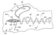
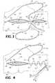
- FIGS. 3-6show a section of tissue including a portion of a patient's skin 58 and subcutaneous tissue defining a wound 60 from the surface of the skin 58 down into the tissue.
- the wound 60 in the tissuecan be of any configuration and from any anatomical part or organ of the body. Accordingly, depending on the configuration of the wound, the wound may comprise several sides and faces.
- the wounds depicted in the FIGS.are straight incisions in the skin 58 to reduce the complexity of the description of the method of the present invention. It is understood that the applicants do not intend to limit the method of the present invention to the closure of only straight incisions.
- the usersuch as a surgeon, selects a suture 40 of sufficient length and having straight ends 46 , 48 .
- the ends 46 , 48may be surgical needles.
- the surgeoninserts the needle 46 at the end of the first portion 54 of the suture 40 into the tissue at a point 62 on a first side 64 of the wound 60 and laterally spaced from the face 66 of the wound 60 at the first side 64 .
- the surgeonadvances the needle 46 along a selected substantially straight path through the tissue to extend out of the tissue at a subcutaneous point (not shown) in the first face 66 of the wound 60 and subcutaneously penetrating a point (not shown) in a face 68 of a second side 70 of the wound 60 .
- the surgeoncontinues to advance the needle 46 through the tissue until the point 50 of the needle emerges from the tissue at a distal end of the selected path at an exit point 72 on the second side 70 of the wound 60 .
- the exit pointis laterally spaced from the face 68 of the second side 70 of the wound and longitudinally spaced in a first direction from the point of insertion 62 at the first side 64 of the wound 60 .
- the surgeongrips the exposed portion of the needle 46 and pulls the needle 46 out of the tissue. This action draws the first portion 54 of the suture 40 having barbs 44 for resisting movement in the opposite direction through the tissue until the barbs 44 on the second portion 56 engage the surface of the skin 58 at the insertion point 62 preventing further advancement of the suture 40 through the tissue.
- a length of the first portion 54 of the suture body 42is thus positioned in the tissue along the selected path.
- the faces 66 , 68 of the wound 60are approximated by pushing the adjacent sides 64 , 70 of the tissue together along the first portion 54 of the body 42 of the suture 40 in the tissue.
- the needle 46is next inserted into the tissue at the exit point 72 and advanced along a substantially straight path through the tissue to extend out of the tissue at a subcutaneous point 74 in the second face 68 of the wound 60 and subcutaneously penetrating a point 76 in the first face 66 of the wound 60 .
- the surgeoncontinues to advance the needle 46 through the tissue until the point end 50 emerges from the tissue at a distal end of the selected path at an exit point 78 on the first side 64 of the wound 60 that is laterally spaced from the first face 66 and longitudinally spaced in the first direction from the point of insertion 72 at the second side 70 of the wound 60 . Again the surgeon grips the exposed portion of the needle 46 and pulls the needle 46 out of the tissue, drawing the first portion 54 of the suture 40 through the tissue.
- the previous stepsare repeated with the first portion 54 of the suture 40 by inserting the needle 46 into the exit point 78 on the first side 64 of the wound 60 for advancing longitudinally in the first direction along the wound 60 in a “zigzag” pattern as shown in FIG. 4 .
- the number of passes of the needle 46is chosen in accordance with the size of the wound 60 and the strength required to hold the wound closed.
- the remaining length of the first portion 54 of the suture 40 protruding from the tissue at a first end 80 of the wound 60is cut and discarded, leaving the remaining first portion 54 of the suture 40 in the tissue.
- the faces 66 , 68 of the wound 60are approximated by pushing the adjacent sides 64 , 70 of the tissue together along the body 42 of the suture 40 in the tissue.
- the step of approximating the sides 64 , 70 of the wound 60can be performed as the suture 40 is advanced or after the end 80 of the wound 60 is reached.
- the surgeonrepeats the steps of this procedure with the second needle 48 on the second portion 56 of the suture (FIG. 5 ).
- the initial insertion point 62 of the second needle 48is at the same initial point of insertion 62 of the first needle 46 at the first side 64 of the wound 60 .
- the surgeonthus advances the second portion 56 of the suture 40 into the tissue along the wound 60 in a direction toward the other end 82 of the wound 60 using the same zigzag pattern approximating the faces 66 , 68 of the wound 60 .
- the remaining length of the second portion 56 of the suture 40 protruding from the skin 58 at the end 82 of the wound 60is then cut and discarded (FIG. 6 ).
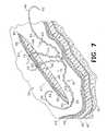
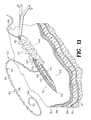
- FIGS. 7-10An embodiment of the method for joining the sides of an open wound in tissue according to the present invention using a subcuticular stitch is shown in FIGS. 7-10.
- the tissue shown in the FIGS.includes an outer epidermis 84 , dermis 86 , fat 88 , fascia 90 and muscle 92 .
- a wound 60can be closed to facilitate healing while minimizing scar tissue.
- the subcuticular stitch method of the present inventionuses a barbed suture 40 including curved ends 46 , 48 .
- the surgeonbegins by inserting the first needle 46 into the tissue below the skin 58 surface at a face 66 on a first side 64 of the wound 60 at an initial insertion point 63 longitudinally spaced from the ends 80 , 82 of the wound 60 .
- the surgeonadvances the needle 46 through the tissue along a curvilinear path until the point 50 of the needle 46 extends from the tissue at a subcutaneous exit point 73 in the first face 66 of the wound 60 longitudinally spaced toward one end 80 of the wound from the entry point 63 of the needle 46 .
- the surgeongrips the needle 46 and pulls the needle 46 out of the tissue, drawing the first portion 54 of the suture 40 through the tissue until the barbs 44 on the second portion 56 engage the tissue at the insertion point 63 preventing further advancement of the suture 40 through the tissue.
- a length of the first portion 54 of the suture body 42is thus positioned in the tissue along the selected curvilinear path as seen in FIG. 7 .
- the surgeonthen inserts the needle 46 into the tissue at a subcutaneous entry point (not shown) in the face 68 at the second side 70 of the wound 60 .
- the surgeonrepeats the above steps of pushing the needle 46 through the tissue along a selected curvilinear path so that the point 50 of the needle 46 emerges from a subcutaneous exit point (not shown) in the second face 68 of the wound 60 longitudinally spaced toward the end 80 of the wound 60 from the entry point.
- the surgeongrips the needle 46 and draws the first portion 54 of the suture 40 into the tissue further along the wound 60 . In this manner, the surgeon advances the first portion 54 of the suture 40 longitudinally along the wound 60 to the one end 80 of the wound in a wave-like or “sinusoidal” pattern.
- the faces 66 , 68 of the wound 60are approximated as the surgeon progresses, or when the end 80 of the wound 60 is reached, by pushing the adjacent sides 64 , 70 of the tissue together along the body 42 of the suture 40 .
- the needle 46 along with remaining length of the first portion 54 of the suture 40is drawn through the surface of the skin 58 at the one end 80 of the wound 60 is cut and discarded (FIG. 8 ).
- the surgeonrepeats the procedure at the other end of the wound (FIG. 9) with the second portion 56 of the suture 40 .
- the surgeonbegins by inserting the second needle 48 into the tissue at a subcutaneous point (not shown) in the second face 68 of the wound 60 .
- the surgeonadvances the second needle 48 along a curvilinear path from the point of initial insertion toward the other end 82 of the wound 60 until the needle 48 emerges from a subcutaneous exit point (not shown) the second face 68 of the wound 60 longitudinally spaced from the initial entry point of the needle 48 .
- FIG. 9shows the needle 48 being drawn a second time from the second face 68 of the wound 60 .
- the surgeonadvances the second portion 56 of the suture in a sinusoidal pattern to the end 82 of the wound 60 (FIG. 10) and approximates the faces 66 , 68 of the wound 60 .
- the length of the second portion 56 of the suture body 42 protruding from the skin 58 at the end of the wound 60is then cut and discarded.
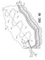
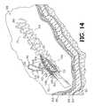
- FIG. 11shows a prior art subcutaneous suturing method for closing a wound 60 using a spiraling, “corkscrew-shaped” stitch pattern.
- the surgeonbegins at one end 80 of the wound by tying a knot 100 in the first loop and advancing the suture in a corkscrew pattern to the other end of the wound 82 where the suture is tied off. Tying the knots at the end and burying them, which is preferred by the surgeon, is technically very challenging, even more so when the incision is almost closed.
- FIGS. 12-15show a similar corkscrew-shaped stitch pattern for closing a wound 60 according to an embodiment of the method of the present invention. This embodiment is similar to the method described above using a subcutaneous sinusoidal stitch pattern.
- the surgeonbegins by inserting one of the needles 46 into the tissue below the skin 58 surface at a face 66 on a first side 64 of the wound 60 at an initial subcutaneous insertion point 63 longitudinally spaced from the ends 80 , 82 of the wound 60 .
- the surgeonadvances the needle 46 upward through the tissue along a curvilinear path until the point 50 of the needle 46 extends from the tissue at a subcutaneous exit point 73 in the first face 66 of the wound 60 longitudinally spaced toward one end 80 of the wound and above the entry point 63 of the needle 46 .
- the surgeonthen inserts the needle 46 into the tissue at a subcutaneous entry point 102 in the face 68 at the second side 70 of the wound 60 .
- the surgeonpushes the needle 46 through the tissue along a selected curvilinear path so that the point 50 of the needle 46 emerges from a subcutaneous exit point 104 in the second face 68 of the wound 60 longitudinally spaced toward the end 80 of the wound 60 and below the entry point 102 .
- the surgeonrepeats these steps (FIG. 13) for advancing the first portion 54 of the suture 40 longitudinally along the wound 60 to the one end 80 of the wound in the spiraling, corkscrew stitch pattern. It is understood that the number and diameter of coils can be varied as desired.
- the surgeongrips the needle 46 for drawing the first portion 54 of the suture 40 through the tissue until the barbs 44 on the second portion 56 engage the tissue at the insertion point 63 preventing further advancement of the suture 40 through the tissue.
- the surgeonapproximates the faces 66 , 68 of the wound 60 as the surgeon progresses or when the end 80 of the wound 60 is reached as described above.
- the remaining length of the first portion 54 of the suture 40is drawn through the surface of the skin 58 at the one end 80 of the wound 60 and cut and discarded.
- the surgeonrepeats the procedure at the other end 82 of the wound 60 with the second portion 56 of the suture 40 .
- several “coils” of the second portion 56 of the suture 40have been entered into the tissue in a direction toward the other end 82 of the wound 60 .
- Subcutaneous entry points 106 and exit points 108 in the faces 66 , 68 of the wound 60are visible.
- the surgeonadvances the second portion 56 of the suture 40 to the end 82 of the wound 60 (FIG. 15) and approximates the faces 66 , 68 of the wound 60 .
- the length of the second portion 56 of the suture body 42 protruding from the skin 58 at the end of the wound 60is then cut and discarded.
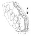
- FIGS. 16-18Another embodiment of a subcutaneous suturing method for joining and holding closed an open wound 60 in tissue according to the present invention is shown in FIGS. 16-18.
- This methodalso uses a barbed suture 40 having curved pointed ends 46 , 48 , such as surgical needles.
- the surgeonbegins by inserting the first needle 46 subcutaneously into the tissue at a face 66 on a first side 64 of the wound 60 at an initial insertion point 63 adjacent one end 80 of the wound 60 and pushes the needle 46 through the tissue along a selected curvilinear path until the needle 46 extends from the tissue at a subcutaneous exit point 73 in the first face 66 of the wound 60 longitudinally spaced from the end 80 of the wound 60 in a direction toward the other end 82 of the wound 60 .
- the surgeongrips the needle 46 and pulls the needle 46 out of the tissue for drawing the first portion 54 of the suture 40 including barbs 44 for resisting movement in the opposite direction through the tissue until the barbs 44 of the second portion 56 engage the first face 66 of the wound 60 at the insertion point 63 preventing further advancement of the suture 40 into the tissue.
- a length of the first portion 54 of the suture body 42is thus positioned in the tissue along the selected curvilinear path.
- the surgeonnext inserts the second surgical needle 48 into the tissue at a subcutaneous entry point (not shown) in the face 68 at the second side 70 of the wound 60 substantially opposite the initial point of insertion 63 of the first needle 46 at the one end 80 of the wound 60 .
- the surgeonadvances the second needle 48 through the tissue along a selected curvilinear path until the needle 48 extends from the tissue at a subcutaneous exit point (not shown) in the second face 68 of the wound 60 .
- the surgeonthen pulls the second needle 48 for drawing the second portion 56 of the suture 40 through the tissue, including barbs 44 for resisting movement in the opposite direction, leaving a length of the second portion 56 of the suture 40 in the tissue at the end 80 of the wound 60 .
- the surgeonrepeats the above steps with the first needle 46 and second needle 48 at the second and first sides 64 , 70 , respectively, of the wound 60 .
- the surgeonadvances the suture 40 longitudinally along the wound 60 from the one end 80 of the wound to the other 82 in a “shoelace” pattern.
- several passes of the suture 40have been entered into the tissue of the wound 60 .
- the faces 66 , 68 of the wound 60are approximated as the surgeon progresses, or when the end 82 of the wound 60 is reached, by pushing the adjacent sides 64 , 70 of the tissue together along the body 42 of the suture 40 .
- the lengths of the first portion 54 and second portion 56 of the suture 40 protruding from the skin 58are cut and discarded (FIG. 18 ).
- FIGS. 7-10can be used to generate a similar stitch pattern if a second suture is used which is entered in the tissue to mirror the path of the first suture.
- FIGS. 19 and 20Another embodiment of the method according to the present invention for joining the sides 64 , 70 of tissue in an open wound 60 is shown in FIGS. 19 and 20.
- the surgeoninserts a first curved or straight end 46 of the suture 40 , such as a needle, into the tissue at a point 62 on a first side 64 of the wound 60 and laterally spaced from the face 66 of the wound 60 at the first side 64 .
- the surgeonadvances the needle 46 through the tissue along a curvilinear path until the needle 46 emerges from the tissue on a second side 70 of the wound at an exit point 72 laterally spaced from the face 68 of the second side 70 of the wound 60 and longitudinally spaced in a first direction from the point of insertion 62 .
- the faces 66 , 68 of the wound 60are approximated by pushing the adjacent sides 64 , 70 of the tissue together along the body 42 of the suture 40 in the tissue.
- the length of the first portion 54 of the body 42 of the suture 40 protruding from the skin 58is cut and discarded (FIG. 19 ).
- the length of the second portion 56 of the suture 40 protruding from the skin 58is cut and discarded, leaving a stitch in the tissue which resembles the Greek letter alpha (FIG. 20 ).
- This stitchhas its greatest benefit in small wound and incision closure.
- the alpha-shaped stitchcan be placed quickly in tissue as compared with conventional loop sutures. Moreover, this stitch pattern has no blood constricting loops, leaves no stitch marks on the surface of the skin, does not have to be removed from the patient if bio-absorbable material is used. Two or more of the alpha-shaped stitches may be used to close a larger wound.
- a particular application of the alpha-stitch according to the method of the present inventionis as a means of restricting bleeding from an arterial opening by constricting the tissue above and around the arterial opening.
- the introduction and removal of catheters into the femoral arteryis typically required when performing cardiac catheterization, percutaneous interventions, and other vascular procedures.
- These puncture woundsare typically self-sealing after several hours of sustained external pressure at and around the insertion site of the puncture wound.
- FIGS. 21 and 22show the alpha-stitch according to a method of the present invention positioned for performing this function.
- the path of the suture portions 54 , 56is curvilinear with the respect to the skin 58 surface and that the deepest points of the arcs pass immediately above the puncture site 112 in the artery 114 .
- the ends 46 , 48 of the suture 40are pulled to put tension in the tissue.
- the tissue embraced by the sutureis pulled both inward from the areas lateral to the artery 114 and downward from areas immediately above the artery 114 .
- This constriction of tissueincreases the density of tissue around the arterial puncture site 112 and imparts forces with vectors directed toward the arteriotomy site to limit bleeding.
- this suture methodavoids the need to traverse the artery wall or lumen, thus eliminating the risk of vessel wall dissection and promoting introgenic thrombogenesis.
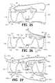
- the method of the present inventionis also useful in binding together partially or completely severed tendons or other internal tissue repairs requiring considerable tensile strength.
- a finger 120is shown with a portion of the outer layer of tissue cut-away to schematically show a severed tendon 122 .
- a Kessler suturing method for joining the two ends 124 , 126 of the tendon 122is shown in FIG. 24 .
- This methodrequires the surgeon to apply an intricate stitch pattern and to complete the tendon connection with one or two technically challenging knots 128 . No portion of the suture knot 128 may protrude from the outside surface of the repaired tendon 122 where it could snag the surrounding tendon sheath and impede healing.
- the knot 128also presents a particular dilemma since it must be tied between the two ends 124 , 126 of the tendon 122 , where it can be a barrier between tendon sections that must appose in order to effectively heal.
- a further limitation of the conventional tendon repair methodis that relatively small amounts of tension can stretch the tendon 122 , allowing it to slide along the smooth monofilament fiber and effectively disrupt, or in the case of greater amounts of tension, separate completely at the wound margin. This outcome substantially limits healing even though the suture material remains intact.
- FIGS. 25-28A method according to the present invention for joining the two ends 124 , 126 of the tendon 122 is shown in FIGS. 25-28.
- the surgeonbegins by inserting the first end 46 of the suture 40 , which may a straight or curved surgical needle, into one end 124 of the tendon 122 and pushing the needle 46 through the tendon 122 along a selected curvilinear path until the point 50 of the needle 46 extends from an exit point 130 in the periphery of the tendon 122 longitudinally spaced from the one end of the tendon 122 .
- the first needle 46is gripped and pulled out of the tendon for drawing the first portion 54 of the suture 40 through the tendon 122 leaving a length of the first portion 54 of the suture in the tendon end 124 between the end of the tendon 122 and the exit point 130 .
- the surgeonreinserts the needle 46 into the periphery of the tendon 122 at an entry point 132 immediately adjacent the exit point 130 and pushes the needle 46 along a selected curvilinear path until the point 50 of the needle 46 exits the other side of the tendon at an exit point 134 that is longitudinally spaced from the entry point 132 . It is understood that the surgeon could use the exit point 130 as the next entry point for the needle 46 if desired.
- these stepsare repeated with the second portion 56 of the suture 40 at the other end 126 of the tendon 122 .
- the pattern of the second portion 56 of the suture 40 in the second end 126 of the tendon 122generally mirrors the first portion 54 of the suture 40 in the first end 124 of the tendon 122 , including exit points 130 a, 134 a, 138 a and entry points 132 a, 136 a.
- the ends 124 , 126 of the tendon 122are brought together while maintaining tension on the free ends of the sutures.
- a second suture 40 ais introduced at the second end 126 of the tendon 122 .
- the first needle 46 a of the second suture 40 ais inserted into the end 126 of the tendon 122 and pushed through the tendon 122 along a selected curvilinear path until the needle 46 a extends from an exit point 140 in the periphery of the tendon 122 substantially opposite the first exit point 130 a of the second portion 56 of the first suture 40 .
- the needle 46 a of the second suture 40 ais pulled out of the tendon 122 for drawing the first portion 54 a of the second suture 40 a through the tendon 122 leaving a length of the suture 40 a in the tendon 122 between the end 126 of the tendon 122 and the exit point 140 .
- the surgeonrepeats the steps described above by reinserting the needle 46 a into the tendon 122 at an entry point 142 (FIG. 28) adjacent the exit point 140 and pushing the needle 46 a along a selected curvilinear path until the needle 46 a emerges from an exit point 144 in the periphery of the tendon 122 substantially opposite the second exit point 134 a of the second portion 56 of the first suture 40 .
- the surgeonadvances longitudinally along the end 126 of the tendon 122 entering at 146 and exiting at 148 .
- the previous stepsare repeated at the other end 124 of the tendon 122 with the second portion 56 a of the second suture 40 a.
- the number of sutures useddepends on the size, caliber, and length of the tendon to be repaired. Big tendons will require more than two sutures whereas one may suffice for very small tendons.
- Tendon repair with two sutures according to the present inventionexhibits equivalent or better holding power as the prior art technique. Moreover, tendons repaired according to the methods of the present invention maintain their original configuration, profile, contour, and form better when being stretched.
- the tissue samplesmeasured 4 cm by 10 cm.
- Each incisionwas centered on the skin sample so that the wound was 4 cm long from end to end.
- Each woundwas closed according to a different suture method using identical barbed sutures made from monofilament PDS (polydioxanone) size 0.
- One woundwas closed according to the method shown in U.S. Pat. Nos. 5,342,376 and 6,241,747, without using the inserting device (“the Ruff method”). Seven sutures were placed along the length of the wound and running generally perpendicularly to the faces of the wound. When placed, the sutures dipped below the incision line thus engaging subcutaneous tissue below the incision and the ends of the sutures engaged some dermis.
- a second woundwas closed using seven needle-tipped sutures placed along the length of the wound in the dermis and running generally perpendicularly to the faces of the wound similar to the method shown in U.S.
- Biomechanical strength testingwas carried out as follows. Each sample was positioned so that the surface of the tissue sample was substantially vertical and the incision was generally horizontal. The bottom edge of the sample was immovably secured. The upper edge of the sample was attached to a Berkley digital fish scale (0-50 lb.) The scale was then raised vertically generating tension across the wound. The scale was raised until the tissues totally separated. The peak force required to separate the incision was recorded as the breaking strength.
- More than one alpha-shaped stitchwas used for longer incisions.
- the dogswere housed for two weeks. Daily clinical and necropsy observations were performed on all surgical sites. With the exception that three of six sites closed by nylon sutures had some sutures chewed out by the dog, all incisions healed normally and no dehiscence occurred. The other three sites closed with nylon sutures had a “railroad-tile” appearance, one site in particular being very pronounced. None of the topical skin sites closed with barbed sutures had such an appearance. This example shows the efficacy of barbed sutures in an in vivo model.
- the methods of the present inventionhave a number of advantages, including improving the biomechanical performance of barbed sutures.
- the curvilinear placement paths of the sutureas contrasted with linear insertion, provide substantially increased strength for holding the edges of a wound together.
- the insertion of a single suture with curvilinear techniquesreplaces the insertion of a plurality of sutures.
- the new methodsprovide an efficient means for a surgeon to close a wound, reducing the time necessary to place the suture and the trauma to the patient. Surgeons can quickly and easily utilize the suturing methods during any type of surgery to quickly join the edges of a wound in tissue without threading and tying numerous individual stitches.
- the new suture methodsare performed in a manner similar to conventional suturing thus realizing the advantages thereof.
- the methodsminimize damage to tissue when inserted and minimize scarring or tissue necrosis across the wound.
- the suturescan be placed in the tissue in a manner to control and adjust the tension on the suture or the compression of the tissue.
Landscapes
- Health & Medical Sciences (AREA)
- Life Sciences & Earth Sciences (AREA)
- Surgery (AREA)
- Heart & Thoracic Surgery (AREA)
- Engineering & Computer Science (AREA)
- Biomedical Technology (AREA)
- Nuclear Medicine, Radiotherapy & Molecular Imaging (AREA)
- Medical Informatics (AREA)
- Molecular Biology (AREA)
- Animal Behavior & Ethology (AREA)
- General Health & Medical Sciences (AREA)
- Public Health (AREA)
- Veterinary Medicine (AREA)
- Surgical Instruments (AREA)
Abstract
Description
| TABLE 1 | |||
| Breaking Strength | |||
| Suture Method | (lbs.) | ||
| Ruff Method | 4.5 | ||
| Buncke Method | 8.5 | ||
| Zigzag Method | 18.3 | ||
| Corkscrew | 16.5 | ||
| Method | |||
| TABLE 2 | ||
| Tissue Level | Barbed Suture Method | Conventional Suture Method |
| Dermis | Alpha, Zigzag | Simple interrupted loop stitches |
| [2-0 nylon, 2-0 silk] | ||
| Subcuticular | Corkscrew | Simple continuous loop stitches |
| [3-0 PDS] | ||
| Subcutaneous | Corkscrew | Simple continuous loop stitches |
| [3-0 PDS] | ||
| Muscular | Corkscrew | Simple continuous loop stitches |
| [3-0 PDS] | ||
Claims (27)
Priority Applications (14)
| Application Number | Priority Date | Filing Date | Title |
|---|---|---|---|
| US09/896,455US6599310B2 (en) | 2001-06-29 | 2001-06-29 | Suture method |
| RU2004102518/14ARU2318458C2 (en) | 2001-06-29 | 2002-06-28 | Method for suturing body tissue |
| AU2002345953AAU2002345953B2 (en) | 2001-06-29 | 2002-06-28 | Suture method |
| PCT/US2002/020449WO2003001979A2 (en) | 2001-06-29 | 2002-06-28 | Suture method |
| US10/065,256US7056331B2 (en) | 2001-06-29 | 2002-09-30 | Suture method |
| US11/307,520US8764796B2 (en) | 2001-06-29 | 2006-02-10 | Suture method |
| US11/307,521US20060111742A1 (en) | 2001-06-29 | 2006-02-10 | Suture method |
| US11/747,707US7857829B2 (en) | 2001-06-29 | 2007-05-11 | Suture method |
| US12/850,230US20100318123A1 (en) | 2001-06-29 | 2010-08-04 | Continuous method anastomosis using self-retaining sutures |
| US12/850,289US20100305401A1 (en) | 2001-06-29 | 2010-08-04 | Endoscopic systems using self-retaining sutures |
| US12/850,336US8777989B2 (en) | 2001-06-29 | 2010-08-04 | Subcutaneous sinusoidal wound closure utilizing one-way suture |
| US12/850,263US8777988B2 (en) | 2001-06-29 | 2010-08-04 | Methods for using self-retaining sutures in endoscopic procedures |
| US12/850,186US8747437B2 (en) | 2001-06-29 | 2010-08-04 | Continuous stitch wound closure utilizing one-way suture |
| US12/850,362US8764776B2 (en) | 2001-06-29 | 2010-08-04 | Anastomosis method using self-retaining sutures |
Applications Claiming Priority (1)
| Application Number | Priority Date | Filing Date | Title |
|---|---|---|---|
| US09/896,455US6599310B2 (en) | 2001-06-29 | 2001-06-29 | Suture method |
Related Child Applications (1)
| Application Number | Title | Priority Date | Filing Date |
|---|---|---|---|
| US10/065,256Continuation-In-PartUS7056331B2 (en) | 2001-06-29 | 2002-09-30 | Suture method |
Publications (2)
| Publication Number | Publication Date |
|---|---|
| US20030014077A1 US20030014077A1 (en) | 2003-01-16 |
| US6599310B2true US6599310B2 (en) | 2003-07-29 |
Family
ID=25406241
Family Applications (1)
| Application Number | Title | Priority Date | Filing Date |
|---|---|---|---|
| US09/896,455Expired - LifetimeUS6599310B2 (en) | 2001-06-29 | 2001-06-29 | Suture method |
Country Status (4)
| Country | Link |
|---|---|
| US (1) | US6599310B2 (en) |
| AU (1) | AU2002345953B2 (en) |
| RU (1) | RU2318458C2 (en) |
| WO (1) | WO2003001979A2 (en) |
Cited By (187)
| Publication number | Priority date | Publication date | Assignee | Title |
|---|---|---|---|---|
| US20030074023A1 (en)* | 2001-06-29 | 2003-04-17 | Andrew Kaplan | Suture method |
| US20030149447A1 (en)* | 2002-02-01 | 2003-08-07 | Morency Steven David | Barbed surgical suture |
| US20030153947A1 (en)* | 2002-02-14 | 2003-08-14 | Tomoaki Koseki | Sternum suture material and its manufacturing method |
| US20040060409A1 (en)* | 2002-09-30 | 2004-04-01 | Leung Jeffrey C. | Barb configurations for barbed sutures |
| US20040088003A1 (en)* | 2002-09-30 | 2004-05-06 | Leung Jeffrey C. | Barbed suture in combination with surgical needle |
| US20040093028A1 (en)* | 1993-05-03 | 2004-05-13 | Ruff Gregory L. | Barbed bodily tissue connector |
| US20040093027A1 (en)* | 2002-03-04 | 2004-05-13 | Walter Fabisiak | Barbed tissue connector for sealing vascular puncture wounds |
| US20040204722A1 (en)* | 2003-04-10 | 2004-10-14 | George Sikora | Fixation device |
| US20040220591A1 (en)* | 2003-04-30 | 2004-11-04 | Bonutti Peter M. | Tissue fastener and methods for using same |
| US20050004601A1 (en)* | 2003-04-29 | 2005-01-06 | Kong James Kam Fu | Coded surgical aids |
| US20050033367A1 (en)* | 2002-08-09 | 2005-02-10 | Leung Jeffrey C. | Suture anchor and method |
| US20050182472A1 (en)* | 2004-02-04 | 2005-08-18 | Wahlstrom Dale A. | Novel lead retention means |
| US20050203576A1 (en)* | 2002-06-07 | 2005-09-15 | Sulamanidze Marlen A | Surgical thread "aptos for cosmetic surgery |
| US20050240224A1 (en)* | 2004-04-07 | 2005-10-27 | Wu Tze Liang W | Surgical thread |
| US20050267532A1 (en)* | 2004-04-07 | 2005-12-01 | Wu Tze Liang W | Surgical thread |
| US20060036271A1 (en)* | 2004-07-29 | 2006-02-16 | X-Sten, Inc. | Spinal ligament modification devices |
| US20060058735A1 (en)* | 2004-09-16 | 2006-03-16 | Lesh Michael D | Systems and devices for soft tissue augmentation |
| US20060058891A1 (en)* | 2004-09-16 | 2006-03-16 | Lesh Michael D | Transformable tissue bulking device |
| DE102005004317B3 (en)* | 2005-01-31 | 2006-06-01 | Ethicon Gmbh | Polypropylene tissue connector comprises a longitudinal core having active substances, helical structures as a rear cut thread spirally coiled around the core and needle type applicator with ends covered by casing |
| US20060192943A1 (en)* | 2005-02-25 | 2006-08-31 | William Roberts | Optimizing focal plane fitting functions for an image field on a substrate |
| US7112214B2 (en) | 2002-06-25 | 2006-09-26 | Incisive Surgical, Inc. | Dynamic bioabsorbable fastener for use in wound closure |
| USD532107S1 (en) | 2003-06-25 | 2006-11-14 | Incisive Surgical, Inc. | Tissue fastening instrument |
| US20070005109A1 (en)* | 2005-06-29 | 2007-01-04 | Popadiuk Nicholas M | Barbed suture |
| US20070010855A1 (en)* | 2005-07-05 | 2007-01-11 | Florez Mendez Maximiliano E | Facial lifting needle and method thereof |
| US20070035822A1 (en)* | 2000-06-17 | 2007-02-15 | Leica Microsystems Cms Gmbh | Arrangement for examining microscopic preparations with a scanning microscope, and illumination device for a scanning microscope |
| US7186262B2 (en)* | 1999-06-25 | 2007-03-06 | Vahid Saadat | Apparatus and methods for treating tissue |
| US20070123890A1 (en)* | 2005-11-04 | 2007-05-31 | X-Sten, Corp. | Tissue retrieval devices and methods |
| US20070213744A1 (en)* | 2003-01-16 | 2007-09-13 | Farris Alex F | Suture material for pneumatic suture instrument |
| US20070225763A1 (en)* | 2006-03-23 | 2007-09-27 | Ethicon Endo-Surgery, Inc. | Marked Suture |
| US20070270928A1 (en)* | 2004-02-04 | 2007-11-22 | Erlebacher Jay A | Lead retention means |
| US20080058869A1 (en)* | 2006-09-06 | 2008-03-06 | Stopek Joshua B | Bioactive substance in a barbed suture |
| EP1917983A2 (en) | 2006-11-02 | 2008-05-07 | Tyco Healthcare Group, LP | Long term bioabsorbable barbed sutures |
| EP1955720A1 (en) | 2007-02-09 | 2008-08-13 | Tyco Healthcare Group, LP | Surface eroding barbed sutures |
| US20080261886A1 (en)* | 2001-07-16 | 2008-10-23 | Novo Nordisk Healthcare A/G | Single-Dose Administration of Factor VIIa |
| US20080275569A1 (en)* | 2004-09-16 | 2008-11-06 | Evera Medical, Inc | Tissue Augmentation Device |
| US20080281338A1 (en)* | 2005-01-31 | 2008-11-13 | Stephen Wohlert | Surgical Suture System |
| US20090043336A1 (en)* | 2007-08-06 | 2009-02-12 | Jie Jenny Yuan | Barbed suture with non-symmetric barbs |
| US20090076543A1 (en)* | 2007-09-17 | 2009-03-19 | Tyco Healthcare Group Lp | Method of Forming Barbs on a Suture |
| US20090112236A1 (en)* | 2007-10-29 | 2009-04-30 | Tyco Healthcare Group Lp | Filament-Reinforced Composite Fiber |
| US7547315B2 (en) | 2002-06-25 | 2009-06-16 | Incisive Surgical, Inc. | Mechanical method and apparatus for tissue fastening |
| US7547326B2 (en) | 2005-04-29 | 2009-06-16 | Jmea Corporation | Disc annulus repair system |
| USD598549S1 (en) | 2008-09-16 | 2009-08-18 | Vertos Medical, Inc. | Surgical trocar |
| US20090248070A1 (en)* | 2008-04-01 | 2009-10-01 | Kosa Timothy D | Anchoring Suture |
| US20090248066A1 (en)* | 2008-03-28 | 2009-10-01 | David Hjalmar Wilkie | Elastic barbed suture and tissue support system |
| US20090248067A1 (en)* | 2008-04-01 | 2009-10-01 | Nicholas Maiorino | Anchoring Device |
| US20090259252A1 (en)* | 2008-04-15 | 2009-10-15 | Kennedy John J | Apparatus For The Joining Of Tissue Having Integral Penetrating End |
| US20090259251A1 (en)* | 2008-04-11 | 2009-10-15 | Cohen Matthew D | Loop suture |
| US20090259306A1 (en)* | 2007-10-15 | 2009-10-15 | Edwards Lifesciences Corporation | Transcatheter heart valve with micro-anchors |
| US7608108B2 (en) | 2005-04-29 | 2009-10-27 | Jmea Corporation | Tissue repair system |
| US7624487B2 (en) | 2003-05-13 | 2009-12-01 | Quill Medical, Inc. | Apparatus and method for forming barbs on a suture |
| US20090299407A1 (en)* | 2008-06-02 | 2009-12-03 | Jie Jenny Yuan | Methods For Using Looped Tissue-Grasping Devices |
| US7632308B2 (en) | 2005-11-23 | 2009-12-15 | Didier Loulmet | Methods, devices, and kits for treating mitral valve prolapse |
| USD606654S1 (en) | 2006-07-31 | 2009-12-22 | Vertos Medical, Inc. | Tissue excision device |
| US7641688B2 (en) | 2004-09-16 | 2010-01-05 | Evera Medical, Inc. | Tissue augmentation device |
| US20100030011A1 (en)* | 2008-07-31 | 2010-02-04 | Ethicon, Inc. | Magnetic implants for treating obstructive sleep apnea and methods therefor |
| US20100024830A1 (en)* | 2008-07-30 | 2010-02-04 | Ethicon, Inc. | Methods and devices for forming an auxiliary airway for treating obstructive sleep apnea |
| US20100030028A1 (en)* | 2006-10-05 | 2010-02-04 | Ramiro Cabrera | Flexible endoscopic stitching devices |
| USD610259S1 (en) | 2008-10-23 | 2010-02-16 | Vertos Medical, Inc. | Tissue modification device |
| USD611146S1 (en) | 2008-10-23 | 2010-03-02 | Vertos Medical, Inc. | Tissue modification device |
| US20100094094A1 (en)* | 2008-10-09 | 2010-04-15 | Tyco Healthcare Group Lp | Tissue Retractor And Method Of Use |
| USD619252S1 (en) | 2008-10-23 | 2010-07-06 | Vertos Medical, Inc. | Tissue modification device |
| USD619253S1 (en) | 2008-10-23 | 2010-07-06 | Vertos Medical, Inc. | Tissue modification device |
| US20100174299A1 (en)* | 2009-01-05 | 2010-07-08 | Tyco Healthcare Group Lp | Method Of Using Barbed Sutures For Gastric Volume Reduction |
| USD620593S1 (en) | 2006-07-31 | 2010-07-27 | Vertos Medical, Inc. | Tissue excision device |
| US20100198257A1 (en)* | 2006-09-06 | 2010-08-05 | Joshua Stopek | Bioactive Substance in a Barbed Suture |
| USD621939S1 (en) | 2008-10-23 | 2010-08-17 | Vertos Medical, Inc. | Tissue modification device |
| US20100211098A1 (en)* | 2008-02-20 | 2010-08-19 | Ahmad Robert Hadba | Compound Barb Medical Device and Method |
| US20100211097A1 (en)* | 2008-02-20 | 2010-08-19 | Ahmad Robert Hadba | Compound Barb Medical Device and Method |
| US20100228270A1 (en)* | 2008-04-11 | 2010-09-09 | Michael Bogart | Deployment System for Surgical Suture |
| US20100268272A1 (en)* | 2008-04-01 | 2010-10-21 | David Kirsch | Anchoring device |
| US20100275750A1 (en)* | 2009-04-29 | 2010-11-04 | Nicholas Maiorino | System and Method for Forming Barbs on a Suture |
| RU2404717C2 (en)* | 2004-05-14 | 2010-11-27 | Квилл Медикал, Инк. | Methods and devices for suturing |
| US7850894B2 (en) | 2006-05-04 | 2010-12-14 | Ethicon, Inc. | Tissue holding devices and methods for making the same |
| US20100323961A1 (en)* | 2007-02-09 | 2010-12-23 | Tyco Healthcare Group Lp | Surface eroding sutures |
| US7878970B2 (en) | 2005-09-28 | 2011-02-01 | Boston Scientific Scimed, Inc. | Apparatus and method for suspending a uterus |
| US7913365B2 (en) | 2001-08-31 | 2011-03-29 | Quill Medical, Inc. | Method of forming barbs on a suture and apparatus for performing same |
| US20110100378A1 (en)* | 2009-10-30 | 2011-05-05 | Ethicon, Inc. | Flexible implants having internal volume shifting capabilities for treating obstructive sleep apnea |
| US7942830B2 (en) | 2006-05-09 | 2011-05-17 | Vertos Medical, Inc. | Ipsilateral approach to minimally invasive ligament decompression procedure |
| US20110125287A1 (en)* | 2009-11-24 | 2011-05-26 | Tyco Healthcare Group LP, New Haven CT and Confluent Surgical, Inc. | Reinforced Tissue Patch |
| US7950559B2 (en) | 2002-06-25 | 2011-05-31 | Incisive Surgical, Inc. | Mechanical method and apparatus for bilateral tissue fastening |
| US7976539B2 (en) | 2004-03-05 | 2011-07-12 | Hansen Medical, Inc. | System and method for denaturing and fixing collagenous tissue |
| US20110216765A1 (en)* | 2002-12-11 | 2011-09-08 | Jeyhan Karaoguz | Media exchange network supporting multiple broadband network and service provider infrastructures |
| US20110264138A1 (en)* | 2008-05-16 | 2011-10-27 | Angiotech Pharmaceuticals, Inc. | Coded heterofunctional sutures and methods |
| US8074857B2 (en) | 2002-06-25 | 2011-12-13 | Incisive Surgical, Inc. | Method and apparatus for tissue fastening with single translating trigger operation |
| US20110319932A1 (en)* | 2008-05-16 | 2011-12-29 | Angiotech Pharmaceuticals, Inc. | Bidirectional self-retaining sutures with laser-marked and/or non-laser marked indicia and methods |
| US20120016384A1 (en)* | 2010-07-19 | 2012-01-19 | Wilke Robert C | Wound closure system |
| US8100939B2 (en) | 2005-07-15 | 2012-01-24 | Incisive Surgical, Inc. | Mechanical method and apparatus for sequential tissue fastening |
| US8118834B1 (en) | 2007-12-20 | 2012-02-21 | Angiotech Pharmaceuticals, Inc. | Composite self-retaining sutures and method |
| US8211126B2 (en) | 2009-09-22 | 2012-07-03 | Jmea Corporation | Tissue repair system |
| US8216273B1 (en) | 2008-02-25 | 2012-07-10 | Ethicon, Inc. | Self-retainers with supporting structures on a suture |
| US8226730B2 (en)* | 2003-08-11 | 2012-07-24 | Cook Medical Technologies Llc | Surgical implant |
| US8273105B2 (en) | 2008-02-20 | 2012-09-25 | Tyco Healthcare Group Lp | Compound barb medical device and method |
| US8303881B2 (en) | 2010-10-28 | 2012-11-06 | Covidien Lp | Suture containing barbs |
| US8307831B2 (en) | 2009-03-16 | 2012-11-13 | Ethicon, Inc. | Implant systems and methods for treating obstructive sleep apnea |
| US8414612B2 (en) | 2010-11-08 | 2013-04-09 | Covidien Lp | Multifilament barbed suture |
| US8430807B2 (en) | 2007-12-28 | 2013-04-30 | Boston Scientific Scimed, Inc. | Devices and methods for treating pelvic floor dysfunctions |
| US8454631B2 (en) | 2006-10-05 | 2013-06-04 | Covidien Lp | Axial stitching devices |
| US8490713B2 (en) | 2009-10-06 | 2013-07-23 | Covidien Lp | Handle assembly for endoscopic suturing device |
| US8561616B2 (en) | 2008-10-24 | 2013-10-22 | Ethicon, Inc. | Methods and devices for the indirect displacement of the hyoid bone for treating obstructive sleep apnea |
| US8561617B2 (en) | 2008-10-30 | 2013-10-22 | Ethicon, Inc. | Implant systems and methods for treating obstructive sleep apnea |
| US8615856B1 (en) | 2008-01-30 | 2013-12-31 | Ethicon, Inc. | Apparatus and method for forming self-retaining sutures |
| US8628545B2 (en) | 2008-06-13 | 2014-01-14 | Covidien Lp | Endoscopic stitching devices |
| US8632488B2 (en) | 2009-12-15 | 2014-01-21 | Ethicon, Inc. | Fluid filled implants for treating medical conditions |
| US8641732B1 (en) | 2008-02-26 | 2014-02-04 | Ethicon, Inc. | Self-retaining suture with variable dimension filament and method |
| US8696671B2 (en) | 2005-07-29 | 2014-04-15 | Vertos Medical Inc. | Percutaneous tissue excision devices |
| US8702718B2 (en) | 2005-04-29 | 2014-04-22 | Jmea Corporation | Implantation system for tissue repair |
| US8715320B2 (en) | 2005-06-29 | 2014-05-06 | Ethicon, Inc. | Braided barbed suture |
| WO2014078754A1 (en)* | 2012-11-19 | 2014-05-22 | Covidien Lp | Tissue fixation device |
| US8734485B2 (en) | 2002-09-30 | 2014-05-27 | Ethicon, Inc. | Sutures with barbs that overlap and cover projections |
| WO2014085363A1 (en)* | 2012-11-30 | 2014-06-05 | Covidien Lp | Looped tissue fixation device |
| US8747436B2 (en) | 2007-06-13 | 2014-06-10 | Ethicon, Inc. | Bi-directional barbed suture |
| US8771313B2 (en) | 2007-12-19 | 2014-07-08 | Ethicon, Inc. | Self-retaining sutures with heat-contact mediated retainers |
| USD708746S1 (en) | 2009-06-10 | 2014-07-08 | Covidien Lp | Handle for surgical device |
| US8777987B2 (en) | 2007-09-27 | 2014-07-15 | Ethicon, Inc. | Self-retaining sutures including tissue retainers having improved strength |
| US8783258B2 (en) | 2008-12-01 | 2014-07-22 | Ethicon, Inc. | Implant systems and methods for treating obstructive sleep apnea |
| US8793863B2 (en) | 2007-04-13 | 2014-08-05 | Ethicon, Inc. | Method and apparatus for forming retainers on a suture |
| US8800567B2 (en) | 2008-12-01 | 2014-08-12 | Ethicon, Inc. | Implant systems and methods for treating obstructive sleep apnea |
| US8813754B2 (en) | 2009-02-17 | 2014-08-26 | Ethicon, Inc. | Magnetic implants and methods for treating an oropharyngeal condition |
| US8821517B2 (en) | 2002-06-25 | 2014-09-02 | Incisive Surgical, Inc. | Mechanical method and apparatus for bilateral tissue fastening |
| US8864776B2 (en) | 2008-04-11 | 2014-10-21 | Covidien Lp | Deployment system for surgical suture |
| US8876865B2 (en) | 2008-04-15 | 2014-11-04 | Ethicon, Inc. | Self-retaining sutures with bi-directional retainers or uni-directional retainers |
| US8875607B2 (en) | 2008-01-30 | 2014-11-04 | Ethicon, Inc. | Apparatus and method for forming self-retaining sutures |
| US8905033B2 (en) | 2011-09-28 | 2014-12-09 | Ethicon, Inc. | Modular tissue securement systems |
| US8916077B1 (en) | 2007-12-19 | 2014-12-23 | Ethicon, Inc. | Self-retaining sutures with retainers formed from molten material |
| US8932328B2 (en) | 2008-11-03 | 2015-01-13 | Ethicon, Inc. | Length of self-retaining suture and method and device for using the same |
| US8968340B2 (en) | 2011-02-23 | 2015-03-03 | Covidien Lp | Single actuating jaw flexible endolumenal stitching device |
| US8968362B2 (en) | 2010-04-08 | 2015-03-03 | Covidien Lp | Coated looped suture |
| US8973582B2 (en) | 2011-11-30 | 2015-03-10 | Ethicon, Inc. | Tongue suspension device and method |
| USRE45426E1 (en) | 1997-05-21 | 2015-03-17 | Ethicon, Inc. | Surgical methods using one-way suture |
| AU2013200710B2 (en)* | 2002-09-30 | 2015-04-09 | Ethicon Llc | Barb configurations for barbed sutures |
| US9044224B2 (en) | 2010-04-12 | 2015-06-02 | Covidien Lp | Barbed medical device and method |
| US9078728B2 (en) | 2007-12-28 | 2015-07-14 | Boston Scientific Scimed, Inc. | Devices and methods for delivering female pelvic floor implants |
| US9078727B2 (en) | 2006-03-16 | 2015-07-14 | Boston Scientific Scimed, Inc. | System and method for treating tissue wall prolapse |
| US9107660B2 (en) | 2012-02-01 | 2015-08-18 | Covidien Lp | Wound closure device |
| US9125647B2 (en) | 2008-02-21 | 2015-09-08 | Ethicon, Inc. | Method and apparatus for elevating retainers on self-retaining sutures |
| US9125716B2 (en) | 2009-04-17 | 2015-09-08 | Boston Scientific Scimed, Inc. | Delivery sleeve for pelvic floor implants |
| RU2564358C2 (en)* | 2010-04-29 | 2015-09-27 | ЭТИКОН, ЭлЭлСи | Methods and device for delivery of tissue treatment compositions to stapled tissues |
| US9144483B2 (en) | 2006-01-13 | 2015-09-29 | Boston Scientific Scimed, Inc. | Placing fixation devices |
| US9144511B2 (en) | 2008-08-14 | 2015-09-29 | Ethicon, Inc. | Methods and devices for treatment of obstructive sleep apnea |
| US9161855B2 (en) | 2011-10-24 | 2015-10-20 | Ethicon, Inc. | Tissue supporting device and method |
| US9168120B2 (en) | 2011-09-09 | 2015-10-27 | Boston Scientific Scimed, Inc. | Medical device and methods of delivering the medical device |
| US9173766B2 (en) | 2012-06-01 | 2015-11-03 | Ethicon, Inc. | Systems and methods to treat upper pharyngeal airway of obstructive sleep apnea patients |
| US9220492B2 (en) | 2012-02-01 | 2015-12-29 | Covidien Lp | Wound closure device |
| US9282958B2 (en) | 2007-12-28 | 2016-03-15 | Boston Scientific Scimed, Inc. | Devices and method for treating pelvic dysfunctions |
| USD752219S1 (en) | 2015-01-02 | 2016-03-22 | Incisive Surgical, Inc. | Tissue fastening instrument |
| US9314244B2 (en) | 2013-12-20 | 2016-04-19 | Medos International Sarl | Directional surgical sutures |
| US9326886B2 (en) | 2009-10-29 | 2016-05-03 | Ethicon, Inc. | Fluid filled implants for treating obstructive sleep apnea |
| US9333070B2 (en) | 2008-02-01 | 2016-05-10 | Evera Medical, Inc. | Breast implant with internal flow dampening |
| US9358002B2 (en) | 2008-04-01 | 2016-06-07 | Covidien Lp | Anchoring device |
| US9387061B2 (en) | 2010-09-02 | 2016-07-12 | Boston Scientific Scimed, Inc. | Pelvic implants and methods of implanting the same |
| US9463020B2 (en) | 2004-09-13 | 2016-10-11 | Wound Care Technologies, Inc. | Wound closure product |
| US9468434B2 (en) | 2014-06-03 | 2016-10-18 | Covidien Lp | Stitching end effector |
| US9486201B2 (en) | 2012-09-27 | 2016-11-08 | Depuy Mitek, Llc | Directionally specific bone anchors and method |
| RU2609057C1 (en)* | 2015-12-30 | 2017-01-30 | Эрик Григорьевич Азизян | Suture material for cosmetic operations |
| US9597079B2 (en) | 2004-11-04 | 2017-03-21 | Wound Care Technologies, Inc. | Wound closure product |
| AU2015201685B2 (en)* | 2008-05-16 | 2017-04-20 | Ethicon Llc | Coded heterofunctional sutures and methods |
| US9636103B2 (en) | 2014-08-28 | 2017-05-02 | Covidien Lp | Surgical suturing instrument |
| US9675341B2 (en) | 2010-11-09 | 2017-06-13 | Ethicon Inc. | Emergency self-retaining sutures and packaging |
| US9700304B2 (en) | 2010-04-19 | 2017-07-11 | Gold Thread Llc | Filament implant system and method |
| US9795378B2 (en) | 2014-10-31 | 2017-10-24 | Ethicon, Inc. | Method for approximating wounds |
| US9814555B2 (en) | 2013-03-12 | 2017-11-14 | Boston Scientific Scimed, Inc. | Medical device for pelvic floor repair and method of delivering the medical device |
| US9844377B2 (en) | 2014-04-25 | 2017-12-19 | Incisive Surgical, Inc. | Method and apparatus for wound closure with sequential tissue positioning and retention |
| US9877862B2 (en) | 2009-10-29 | 2018-01-30 | Ethicon, Inc. | Tongue suspension system with hyoid-extender for treating obstructive sleep apnea |
| US9888997B2 (en) | 2015-07-20 | 2018-02-13 | Arthrex, Inc. | Soft anchor assembly with barbed flexible strand and techniques for use |
| US9955962B2 (en) | 2010-06-11 | 2018-05-01 | Ethicon, Inc. | Suture delivery tools for endoscopic and robot-assisted surgery and methods |
| US9962251B2 (en) | 2013-10-17 | 2018-05-08 | Boston Scientific Scimed, Inc. | Devices and methods for delivering implants |
| US9986992B2 (en) | 2014-10-28 | 2018-06-05 | Stryker Corporation | Suture anchor and associated methods of use |
| US10085747B2 (en) | 2015-09-11 | 2018-10-02 | Incisive Surgical, Inc. | Surgical fastening instrument |
| US10092286B2 (en) | 2015-05-27 | 2018-10-09 | Covidien Lp | Suturing loading unit |
| US10178991B2 (en) | 2015-04-22 | 2019-01-15 | Sofradim Production | Method for forming a barbed suture and the barbed suture thus obtained |
| US10188384B2 (en) | 2011-06-06 | 2019-01-29 | Ethicon, Inc. | Methods and devices for soft palate tissue elevation procedures |
| US10231744B2 (en) | 2009-08-20 | 2019-03-19 | Howmedica Osteonics Corp. | Flexible ACL instrumentation, kit and method |
| US10342650B2 (en) | 2013-09-27 | 2019-07-09 | Covidien Lp | Skirted hernia repair device |
| US10413289B2 (en) | 2008-06-13 | 2019-09-17 | Covidien Lp | Endoscopic stitching devices |
| US10420546B2 (en) | 2010-05-04 | 2019-09-24 | Ethicon, Inc. | Self-retaining systems having laser-cut retainers |
| US10433944B2 (en) | 2015-04-23 | 2019-10-08 | Sofradim Production | Package for a surgical mesh |
| US10448944B2 (en) | 2011-11-23 | 2019-10-22 | Howmedica Osteonics Corp. | Filamentary fixation device |
| US10470760B2 (en) | 2011-12-08 | 2019-11-12 | Ethicon, Inc. | Modified tissue securement fibers |
| US10492780B2 (en) | 2011-03-23 | 2019-12-03 | Ethicon, Inc. | Self-retaining variable loop sutures |
| US10542970B2 (en) | 2016-05-31 | 2020-01-28 | Covidien Lp | Endoscopic stitching device |
| US11007296B2 (en) | 2010-11-03 | 2021-05-18 | Ethicon, Inc. | Drug-eluting self-retaining sutures and methods relating thereto |
| US11197665B2 (en) | 2018-08-06 | 2021-12-14 | Covidien Lp | Needle reload device for use with endostitch device |
| US11331094B2 (en) | 2013-04-22 | 2022-05-17 | Stryker Corporation | Method and apparatus for attaching tissue to bone |
| US11413032B2 (en) | 2020-02-11 | 2022-08-16 | Embody, Inc. | Surgical anchoring device, deployment device, and method of use |
| US11950775B2 (en) | 2021-03-07 | 2024-04-09 | Giftedness And Creativity Company | Stitching method for everted wound closure |
| US12102348B2 (en) | 2016-09-07 | 2024-10-01 | Vertos Medical, Inc. | Percutaneous lateral recess resection methods and instruments |
| US12324572B2 (en) | 2022-06-16 | 2025-06-10 | Vertos Medical, Inc. | Integrated instrument assembly |
Families Citing this family (28)
| Publication number | Priority date | Publication date | Assignee | Title |
|---|---|---|---|---|
| WO2005079388A2 (en)* | 2004-02-13 | 2005-09-01 | The University Of Cincinnati | A coupling device enabled by mechanical continuity of cellular scaffolding across tissue boundaries |
| KR100545105B1 (en)* | 2004-04-07 | 2006-01-24 | 츠 리앙 우플스 우 | Surgical Thread |
| RU2404821C2 (en)* | 2005-05-04 | 2010-11-27 | Синтез ГмбХ | Connecting element |
| US8192462B2 (en) | 2006-05-16 | 2012-06-05 | Marlen Andreevich Sulamanidze | Surgical suture material and method for the application thereof |
| US20090143819A1 (en)* | 2007-10-31 | 2009-06-04 | D Agostino William L | Coatings for modifying monofilament and multi-filaments self-retaining sutures |
| US20090112259A1 (en)* | 2007-10-31 | 2009-04-30 | Angiotech Pharmaceuticals, Inc. | Recombinant expressed bioadsorbable polyhydroxyalkonate monofilament and multi-filaments self-retaining sutures |
| US20090216073A1 (en)* | 2008-02-27 | 2009-08-27 | Ralph Zipper | Devices and Methods for Placing Slings and Other Materials |
| US20100010539A1 (en)* | 2008-07-14 | 2010-01-14 | Ferass Abuzaina | Differentiation Of Surgical Filaments |
| CA2734500A1 (en) | 2008-08-27 | 2010-03-11 | Calcimedica Inc. | Compounds that modulate intracellular calcium |
| WO2011091483A1 (en)* | 2010-01-26 | 2011-08-04 | Dokter Philippe Ehlinger, Besloten Vennootschap Met Beperkte Aansprakelijheid | Surgical thread and surgical device for lifting and suspension of soft tissues. |
| US9017404B2 (en)* | 2012-01-23 | 2015-04-28 | Lloyd P. Champagne | Devices and methods for tendon repair |
| US9084597B2 (en) | 2012-03-09 | 2015-07-21 | Smith & Nephew, Inc. | Suture-based knotless repair |
| US9480515B2 (en) | 2012-07-12 | 2016-11-01 | Exsomed International IP, LLC | Metacarpal bone stabilization device |
| US8906001B2 (en) | 2012-10-10 | 2014-12-09 | Covidien Lp | Electromechanical surgical apparatus including wire routing clock spring |
| US10925606B2 (en)* | 2012-10-15 | 2021-02-23 | Guy Friedman | Tendon repair apparatus |
| WO2015050895A1 (en) | 2013-10-02 | 2015-04-09 | Exsomed Holding Company Llc | Full wrist fusion device |
| US9622523B2 (en) | 2014-01-06 | 2017-04-18 | Exsomed International IP, LLC | Ergonomic work gloves |
| WO2016186847A1 (en) | 2015-05-19 | 2016-11-24 | Exsomed International IP, LLC | Distal radius plate |
| US10245091B2 (en) | 2015-12-30 | 2019-04-02 | Exsomed Holding Company, Llc | Dip fusion spike screw |
| US11147604B2 (en) | 2016-01-12 | 2021-10-19 | ExsoMed Corporation | Bone stabilization device |
| US10194923B2 (en) | 2016-05-10 | 2019-02-05 | Exsomed International IP, LLC | Tool for percutaneous joint cartilage destruction and preparation for joint fusion |
| US11147681B2 (en) | 2017-09-05 | 2021-10-19 | ExsoMed Corporation | Small bone angled compression screw |
| JP2020532407A (en) | 2017-09-05 | 2020-11-12 | エクソームド コーポレーションExsomed Corporation | Threaded intramedullary nail for radial cortex fixation |
| US11191645B2 (en) | 2017-09-05 | 2021-12-07 | ExsoMed Corporation | Small bone tapered compression screw |
| US12114853B2 (en) | 2017-11-14 | 2024-10-15 | Intuitive Surgical Operations, Inc. | Electrically weldable suture material, and apparatus and method for forming welded suture loops and other welded structures |
| EP3709931B1 (en)* | 2017-11-14 | 2024-10-16 | Egan Design LLC | Electrically weldable suture material |
| US12213662B2 (en) | 2017-11-14 | 2025-02-04 | Intuitive Surgical Operations, Inc. | Electrically weldable suture material, and apparatus and method for forming welded suture loops and other welded structures |
| US11129968B2 (en)* | 2019-09-13 | 2021-09-28 | Ethicon, Inc. | Methods of making and implanting barbed microcatheters having fluid egress openings for infusing therapeutic fluids |
Citations (105)
| Publication number | Priority date | Publication date | Assignee | Title |
|---|---|---|---|---|
| US709392A (en) | 1902-05-06 | 1902-09-16 | Haydn Brown | Suture-clamp. |
| US733723A (en) | 1903-02-16 | 1903-07-14 | Clarence D Lukens | Serrefin. |
| US789401A (en) | 1904-07-15 | 1905-05-09 | Ernest V Acheson | Umbilical forceps and metal sealing-band. |
| US816026A (en) | 1905-03-09 | 1906-03-27 | Albert J Meier | Surgical clip. |
| US1142510A (en) | 1914-11-04 | 1915-06-08 | Lillian K Engle | Fastening device. |
| US1321011A (en) | 1919-11-04 | Surgical | ||
| US1728316A (en) | 1927-07-02 | 1929-09-17 | Kirurgiska Instr Fabriks Aktie | Wound clasp |
| US1886721A (en) | 1928-08-10 | 1932-11-08 | Rubber Products Corp | Road marker |
| US2201610A (en) | 1938-05-20 | 1940-05-21 | Jr James C Dawson | Wound clip |
| US2232142A (en) | 1940-09-27 | 1941-02-18 | Schumann Seymour | Wound clip |
| US2254620A (en) | 1939-11-14 | 1941-09-02 | George I Miller | Clip |
| US2421193A (en) | 1943-08-02 | 1947-05-27 | Cleveland Clinic Foundation | Surgical dressing |
| US2472009A (en) | 1945-08-01 | 1949-05-31 | Cleveland Clinic Foundation | Surgical dressing |
| US2684070A (en) | 1953-03-23 | 1954-07-20 | Walter L Kelsey | Surgical clip |
| US2779083A (en) | 1955-02-09 | 1957-01-29 | Edward N Eaton | Lip and mouth adjuster |
| US2817339A (en) | 1953-08-10 | 1957-12-24 | Norman M Sullivan | Rigid fascial suture |
| US2910067A (en) | 1952-10-13 | 1959-10-27 | Technical Oil Tool Corp | Wound clip and extractor therefor |
| US2988028A (en) | 1956-08-13 | 1961-06-13 | John H Alcamo | Surgeon's suturing device |
| US3068869A (en) | 1959-10-01 | 1962-12-18 | Sheiden Charles Hunter | Tissue suture clamp |
| US3068870A (en) | 1960-03-18 | 1962-12-18 | Levin Abraham | Wound clip |
| US3123077A (en) | 1964-03-03 | Surgical suture | ||
| US3166072A (en) | 1962-10-22 | 1965-01-19 | Jr John T Sullivan | Barbed clips |
| US3209754A (en) | 1961-08-10 | 1965-10-05 | Ernest C Wood | Surgical clip |
| US3214810A (en) | 1963-05-23 | 1965-11-02 | Robert V Mathison | Fastener devices |
| US3221746A (en) | 1963-01-25 | 1965-12-07 | Noble John William | Surgical connecting device |
| US3234636A (en) | 1962-03-19 | 1966-02-15 | Ernest C Wood | Clip applicator |
| US3273562A (en) | 1960-02-24 | 1966-09-20 | Rene G Le Vaux | Skin and surgical clips |
| US3352191A (en) | 1965-04-23 | 1967-11-14 | Allan H Crawford | Dowel |
| US3378010A (en) | 1965-07-28 | 1968-04-16 | Coldling | Surgical clip with means for releasing the clamping pressure |
| US3385299A (en) | 1965-10-23 | 1968-05-28 | New Res And Dev Lab Inc | Wound clip |
| US3494006A (en) | 1968-01-12 | 1970-02-10 | George C Brumlik | Self-gripping fastening device |
| US3525340A (en) | 1967-10-31 | 1970-08-25 | Joseph G Gilbert | Surgical dressing with skin clips thereon |
| US3527223A (en) | 1967-09-01 | 1970-09-08 | Melvin Shein | Ear stud and hollow piercer for insertion thereof |
| US3586002A (en) | 1968-01-08 | 1971-06-22 | Ernest C Wood | Surgical skin clip |
| US3608095A (en) | 1970-03-05 | 1971-09-28 | Federal Tool Eng Co | Method of fixing hair pieces to scalps |
| US3608539A (en) | 1968-11-06 | 1971-09-28 | Daniel G Miller | Method for the biopsy of subcutaneous masses |
| US3683926A (en) | 1970-07-09 | 1972-08-15 | Dainippon Pharmaceutical Co | Tube for connecting blood vessels |
| US3716058A (en) | 1970-07-17 | 1973-02-13 | Atlanta Res Inst | Barbed suture |
| US3825010A (en) | 1973-04-23 | 1974-07-23 | Donald B Mc | Surgical apparatus for closing wounds |
| US3981307A (en) | 1974-07-01 | 1976-09-21 | Ethicon, Inc. | Thermal attachment of surgical sutures to needles |
| US4069825A (en)* | 1976-01-28 | 1978-01-24 | Taichiro Akiyama | Surgical thread and cutting apparatus for the same |
| US4073298A (en) | 1976-08-03 | 1978-02-14 | New Research & Development Lab., Inc. | Wound clip |
| US4259959A (en) | 1978-12-20 | 1981-04-07 | Walker Wesley W | Suturing element |
| US4317451A (en) | 1980-02-19 | 1982-03-02 | Ethicon, Inc. | Plastic surgical staple |
| US4428376A (en) | 1980-05-02 | 1984-01-31 | Ethicon Inc. | Plastic surgical staple |
| US4430998A (en) | 1982-06-01 | 1984-02-14 | Thoratec Laboratories Corporation | Wound closing device |
| US4434796A (en) | 1981-04-07 | 1984-03-06 | Vsesojuzny Nauchno-Issledovatelsky I Ispytatelny Institut Meditsinskoi Tekhniki | Surgical staple, a method of and forceps for its removal |
| US4454875A (en) | 1982-04-15 | 1984-06-19 | Techmedica, Inc. | Osteal medical staple |
| US4467805A (en) | 1982-08-25 | 1984-08-28 | Mamoru Fukuda | Skin closure stapling device for surgical procedures |
| US4505274A (en) | 1980-10-17 | 1985-03-19 | Propper Manufacturing Co., Inc. | Suture clip |
| US4510934A (en) | 1983-05-13 | 1985-04-16 | Batra Subhash K | Suture |
| US4531522A (en) | 1983-06-20 | 1985-07-30 | Ethicon, Inc. | Two-piece tissue fastener with locking top and method for applying same |
| US4548202A (en) | 1983-06-20 | 1985-10-22 | Ethicon, Inc. | Mesh tissue fasteners |
| US4610251A (en) | 1985-04-19 | 1986-09-09 | Kumar Sarbjeet S | Surgical staple |
| US4635637A (en) | 1984-03-29 | 1987-01-13 | Schreiber Saul N | Surgical suture |
| US4637380A (en) | 1985-06-24 | 1987-01-20 | Orejola Wilmo C | Surgical wound closures |
| US4653486A (en) | 1984-04-12 | 1987-03-31 | Coker Tom P | Fastener, particularly suited for orthopedic use |
| US4669473A (en) | 1985-09-06 | 1987-06-02 | Acufex Microsurgical, Inc. | Surgical fastener |
| US4676245A (en) | 1983-02-09 | 1987-06-30 | Mamoru Fukuda | Interlocking surgical staple assembly |
| US4719917A (en) | 1987-02-17 | 1988-01-19 | Minnesota Mining And Manufacturing Company | Surgical staple |
| US4776337A (en) | 1985-11-07 | 1988-10-11 | Expandable Grafts Partnership | Expandable intraluminal graft, and method and apparatus for implanting an expandable intraluminal graft |
| US4841960A (en) | 1987-02-10 | 1989-06-27 | Garner Eric T | Method and apparatus for interosseous bone fixation |
| US4873976A (en) | 1984-02-28 | 1989-10-17 | Schreiber Saul N | Surgical fasteners and method |
| US4887601A (en) | 1987-11-06 | 1989-12-19 | Ophthalmic Ventures Limited Partnership | Adjustable surgical staple and method of using the same |
| US4976715A (en) | 1986-05-20 | 1990-12-11 | Concept, Inc. | Repair tack for bodily tissue |
| US4994073A (en) | 1989-02-22 | 1991-02-19 | United States Surgical Corp. | Skin fastener |
| US4997439A (en) | 1989-01-26 | 1991-03-05 | Chen Fusen H | Surgical closure or anastomotic device |
| US5002562A (en) | 1988-06-03 | 1991-03-26 | Oberlander Michael A | Surgical clip |
| US5007921A (en) | 1989-10-26 | 1991-04-16 | Brown Alan W | Surgical staple |
| US5026390A (en) | 1989-10-26 | 1991-06-25 | Brown Alan W | Surgical staple |
| US5047047A (en) | 1988-10-26 | 1991-09-10 | Inbae Yoon | Wound closing device |
| US5053047A (en) | 1989-05-16 | 1991-10-01 | Inbae Yoon | Suture devices particularly useful in endoscopic surgery and methods of suturing |
| US5084063A (en) | 1989-09-27 | 1992-01-28 | United States Surgical Corporation | Surgical needle-suture attachment |
| US5102418A (en) | 1989-09-27 | 1992-04-07 | United States Surgical Corporation | Method for attaching a surgical needle to a suture |
| US5123913A (en) | 1989-11-27 | 1992-06-23 | Wilk Peter J | Suture device |
| US5123911A (en) | 1989-09-27 | 1992-06-23 | United States Surgical Corporation | Method for attaching a surgical needle to a suture |
| US5192302A (en) | 1989-12-04 | 1993-03-09 | Kensey Nash Corporation | Plug devices for sealing punctures and methods of use |
| US5207694A (en) | 1992-06-18 | 1993-05-04 | Surgical Invent Ab | Method for performing a surgical occlusion, and kit and applicator for carrying out the method |
| US5222976A (en) | 1989-05-16 | 1993-06-29 | Inbae Yoon | Suture devices particularly useful in endoscopic surgery |
| US5246441A (en) | 1989-09-08 | 1993-09-21 | Linvatec Corporation | Bioabsorbable tack for joining bodily tissue |
| US5258013A (en) | 1991-01-07 | 1993-11-02 | United States Surgical Corporation | Siliconized surgical needle and method for its manufacture |
| US5269783A (en) | 1991-05-13 | 1993-12-14 | United States Surgical Corporation | Device and method for repairing torn tissue |
| US5292326A (en) | 1990-09-13 | 1994-03-08 | United States Surgical Corporation | Apparatus and method for subcuticular stapling of body tissue |
| US5320629A (en) | 1991-01-07 | 1994-06-14 | Laparomed Corporation | Device and method for applying suture |
| US5342376A (en) | 1993-05-03 | 1994-08-30 | Dermagraphics, Inc. | Inserting device for a barbed tissue connector |
| US5425747A (en) | 1993-10-12 | 1995-06-20 | Brotz; Gregory R. | Suture |
| US5425746A (en) | 1990-12-13 | 1995-06-20 | United States Surgical Corporation | Suture-needle combination with cyanoacrylate tipped sutures |
| US5450860A (en) | 1993-08-31 | 1995-09-19 | W. L. Gore & Associates, Inc. | Device for tissue repair and method for employing same |
| US5500991A (en) | 1994-01-13 | 1996-03-26 | Ethicon, Inc. | Needle and suture swaging and pull-testing method |
| DE4302895C2 (en) | 1993-02-02 | 1996-03-28 | Wiessner Serag Gmbh & Co Kg | Surgical thread |
| US5533982A (en) | 1993-10-05 | 1996-07-09 | United States Surgical Corporation | Heat treated stainless steel needles |
| US5546957A (en) | 1993-09-09 | 1996-08-20 | Norbert Heske | Biopsy needle |
| US5584859A (en) | 1993-10-12 | 1996-12-17 | Brotz; Gregory R. | Suture assembly |
| US5683417A (en)* | 1996-08-14 | 1997-11-04 | Cooper; William I. | Suture and method for endoscopic surgery |
| US5702462A (en) | 1996-01-24 | 1997-12-30 | Oberlander; Michael | Method of meniscal repair |
| US5716376A (en) | 1996-06-28 | 1998-02-10 | United States Surgical Corporation | Absorbable mixture and coatings for surgical articles fabricated therefrom |
| US5722991A (en) | 1995-06-07 | 1998-03-03 | United States Surgical Corporation | Apparatus and method for attaching surgical needle suture components |
| US5931855A (en) | 1997-05-21 | 1999-08-03 | Frank Hoffman | Surgical methods using one-way suture |
| US5984933A (en)* | 1994-12-29 | 1999-11-16 | Yoon; Inbae | Apparatus for suturing tissue |
| US6012216A (en) | 1997-04-30 | 2000-01-11 | Ethicon, Inc. | Stand alone swage apparatus |
| US6056778A (en) | 1997-10-29 | 2000-05-02 | Arthrex, Inc. | Meniscal repair device |
| US6083244A (en)* | 1996-09-13 | 2000-07-04 | Tendon Technology, Ltd. | Apparatus and method for tendon or ligament repair |
| EP1075843A1 (en) | 1999-03-03 | 2001-02-14 | Marlen Andreevich Sulamanidze | Surgical thread for plastic surgery operations |
| US6241747B1 (en) | 1993-05-03 | 2001-06-05 | Quill Medical, Inc. | Barbed Bodily tissue connector |
| US6270517B1 (en)* | 2000-02-04 | 2001-08-07 | Gregory R. Brotz | Suture assembly and method |
Family Cites Families (1)
| Publication number | Priority date | Publication date | Assignee | Title |
|---|---|---|---|---|
| RU2139690C1 (en)* | 1996-03-25 | 1999-10-20 | Плетиков Сергей Михайлович | Method of application of tendon suture |
- 2001
- 2001-06-29USUS09/896,455patent/US6599310B2/ennot_activeExpired - Lifetime
- 2002
- 2002-06-28AUAU2002345953Apatent/AU2002345953B2/ennot_activeExpired
- 2002-06-28RURU2004102518/14Apatent/RU2318458C2/enactive
- 2002-06-28WOPCT/US2002/020449patent/WO2003001979A2/ennot_activeApplication Discontinuation
Patent Citations (108)
| Publication number | Priority date | Publication date | Assignee | Title |
|---|---|---|---|---|
| US1321011A (en) | 1919-11-04 | Surgical | ||
| US3123077A (en) | 1964-03-03 | Surgical suture | ||
| US709392A (en) | 1902-05-06 | 1902-09-16 | Haydn Brown | Suture-clamp. |
| US733723A (en) | 1903-02-16 | 1903-07-14 | Clarence D Lukens | Serrefin. |
| US789401A (en) | 1904-07-15 | 1905-05-09 | Ernest V Acheson | Umbilical forceps and metal sealing-band. |
| US816026A (en) | 1905-03-09 | 1906-03-27 | Albert J Meier | Surgical clip. |
| US1142510A (en) | 1914-11-04 | 1915-06-08 | Lillian K Engle | Fastening device. |
| US1728316A (en) | 1927-07-02 | 1929-09-17 | Kirurgiska Instr Fabriks Aktie | Wound clasp |
| US1886721A (en) | 1928-08-10 | 1932-11-08 | Rubber Products Corp | Road marker |
| US2201610A (en) | 1938-05-20 | 1940-05-21 | Jr James C Dawson | Wound clip |
| US2254620A (en) | 1939-11-14 | 1941-09-02 | George I Miller | Clip |
| US2232142A (en) | 1940-09-27 | 1941-02-18 | Schumann Seymour | Wound clip |
| US2421193A (en) | 1943-08-02 | 1947-05-27 | Cleveland Clinic Foundation | Surgical dressing |
| US2472009A (en) | 1945-08-01 | 1949-05-31 | Cleveland Clinic Foundation | Surgical dressing |
| US2910067A (en) | 1952-10-13 | 1959-10-27 | Technical Oil Tool Corp | Wound clip and extractor therefor |
| US2684070A (en) | 1953-03-23 | 1954-07-20 | Walter L Kelsey | Surgical clip |
| US2817339A (en) | 1953-08-10 | 1957-12-24 | Norman M Sullivan | Rigid fascial suture |
| US2779083A (en) | 1955-02-09 | 1957-01-29 | Edward N Eaton | Lip and mouth adjuster |
| US2988028A (en) | 1956-08-13 | 1961-06-13 | John H Alcamo | Surgeon's suturing device |
| US3068869A (en) | 1959-10-01 | 1962-12-18 | Sheiden Charles Hunter | Tissue suture clamp |
| US3273562A (en) | 1960-02-24 | 1966-09-20 | Rene G Le Vaux | Skin and surgical clips |
| US3068870A (en) | 1960-03-18 | 1962-12-18 | Levin Abraham | Wound clip |
| US3209754A (en) | 1961-08-10 | 1965-10-05 | Ernest C Wood | Surgical clip |
| US3234636A (en) | 1962-03-19 | 1966-02-15 | Ernest C Wood | Clip applicator |
| US3166072A (en) | 1962-10-22 | 1965-01-19 | Jr John T Sullivan | Barbed clips |
| US3221746A (en) | 1963-01-25 | 1965-12-07 | Noble John William | Surgical connecting device |
| US3214810A (en) | 1963-05-23 | 1965-11-02 | Robert V Mathison | Fastener devices |
| US3352191A (en) | 1965-04-23 | 1967-11-14 | Allan H Crawford | Dowel |
| US3378010A (en) | 1965-07-28 | 1968-04-16 | Coldling | Surgical clip with means for releasing the clamping pressure |
| US3385299A (en) | 1965-10-23 | 1968-05-28 | New Res And Dev Lab Inc | Wound clip |
| US3527223A (en) | 1967-09-01 | 1970-09-08 | Melvin Shein | Ear stud and hollow piercer for insertion thereof |
| US3525340A (en) | 1967-10-31 | 1970-08-25 | Joseph G Gilbert | Surgical dressing with skin clips thereon |
| US3586002A (en) | 1968-01-08 | 1971-06-22 | Ernest C Wood | Surgical skin clip |
| US3494006A (en) | 1968-01-12 | 1970-02-10 | George C Brumlik | Self-gripping fastening device |
| US3608539A (en) | 1968-11-06 | 1971-09-28 | Daniel G Miller | Method for the biopsy of subcutaneous masses |
| US3608095A (en) | 1970-03-05 | 1971-09-28 | Federal Tool Eng Co | Method of fixing hair pieces to scalps |
| US3683926A (en) | 1970-07-09 | 1972-08-15 | Dainippon Pharmaceutical Co | Tube for connecting blood vessels |
| US3716058A (en) | 1970-07-17 | 1973-02-13 | Atlanta Res Inst | Barbed suture |
| US3825010A (en) | 1973-04-23 | 1974-07-23 | Donald B Mc | Surgical apparatus for closing wounds |
| US3981307A (en) | 1974-07-01 | 1976-09-21 | Ethicon, Inc. | Thermal attachment of surgical sutures to needles |
| US4069825A (en)* | 1976-01-28 | 1978-01-24 | Taichiro Akiyama | Surgical thread and cutting apparatus for the same |
| US4073298A (en) | 1976-08-03 | 1978-02-14 | New Research & Development Lab., Inc. | Wound clip |
| US4259959A (en) | 1978-12-20 | 1981-04-07 | Walker Wesley W | Suturing element |
| US4317451A (en) | 1980-02-19 | 1982-03-02 | Ethicon, Inc. | Plastic surgical staple |
| US4428376A (en) | 1980-05-02 | 1984-01-31 | Ethicon Inc. | Plastic surgical staple |
| US4505274A (en) | 1980-10-17 | 1985-03-19 | Propper Manufacturing Co., Inc. | Suture clip |
| US4434796A (en) | 1981-04-07 | 1984-03-06 | Vsesojuzny Nauchno-Issledovatelsky I Ispytatelny Institut Meditsinskoi Tekhniki | Surgical staple, a method of and forceps for its removal |
| US4454875A (en) | 1982-04-15 | 1984-06-19 | Techmedica, Inc. | Osteal medical staple |
| US4430998A (en) | 1982-06-01 | 1984-02-14 | Thoratec Laboratories Corporation | Wound closing device |
| US4467805A (en) | 1982-08-25 | 1984-08-28 | Mamoru Fukuda | Skin closure stapling device for surgical procedures |
| US4676245A (en) | 1983-02-09 | 1987-06-30 | Mamoru Fukuda | Interlocking surgical staple assembly |
| US4510934A (en) | 1983-05-13 | 1985-04-16 | Batra Subhash K | Suture |
| US4531522A (en) | 1983-06-20 | 1985-07-30 | Ethicon, Inc. | Two-piece tissue fastener with locking top and method for applying same |
| US4548202A (en) | 1983-06-20 | 1985-10-22 | Ethicon, Inc. | Mesh tissue fasteners |
| US4873976A (en) | 1984-02-28 | 1989-10-17 | Schreiber Saul N | Surgical fasteners and method |
| US4635637A (en) | 1984-03-29 | 1987-01-13 | Schreiber Saul N | Surgical suture |
| US4653486A (en) | 1984-04-12 | 1987-03-31 | Coker Tom P | Fastener, particularly suited for orthopedic use |
| US4610251A (en) | 1985-04-19 | 1986-09-09 | Kumar Sarbjeet S | Surgical staple |
| US4637380A (en) | 1985-06-24 | 1987-01-20 | Orejola Wilmo C | Surgical wound closures |
| US4669473A (en) | 1985-09-06 | 1987-06-02 | Acufex Microsurgical, Inc. | Surgical fastener |
| US4776337A (en) | 1985-11-07 | 1988-10-11 | Expandable Grafts Partnership | Expandable intraluminal graft, and method and apparatus for implanting an expandable intraluminal graft |
| US4776337B1 (en) | 1985-11-07 | 2000-12-05 | Cordis Corp | Expandable intraluminal graft and method and apparatus for implanting an expandable intraluminal graft |
| US4976715A (en) | 1986-05-20 | 1990-12-11 | Concept, Inc. | Repair tack for bodily tissue |
| US4841960A (en) | 1987-02-10 | 1989-06-27 | Garner Eric T | Method and apparatus for interosseous bone fixation |
| US4719917A (en) | 1987-02-17 | 1988-01-19 | Minnesota Mining And Manufacturing Company | Surgical staple |
| US4887601A (en) | 1987-11-06 | 1989-12-19 | Ophthalmic Ventures Limited Partnership | Adjustable surgical staple and method of using the same |
| US5002562A (en) | 1988-06-03 | 1991-03-26 | Oberlander Michael A | Surgical clip |
| US5047047A (en) | 1988-10-26 | 1991-09-10 | Inbae Yoon | Wound closing device |
| US4997439A (en) | 1989-01-26 | 1991-03-05 | Chen Fusen H | Surgical closure or anastomotic device |
| US4994073A (en) | 1989-02-22 | 1991-02-19 | United States Surgical Corp. | Skin fastener |
| US5053047A (en) | 1989-05-16 | 1991-10-01 | Inbae Yoon | Suture devices particularly useful in endoscopic surgery and methods of suturing |
| US5222976A (en) | 1989-05-16 | 1993-06-29 | Inbae Yoon | Suture devices particularly useful in endoscopic surgery |
| US5246441A (en) | 1989-09-08 | 1993-09-21 | Linvatec Corporation | Bioabsorbable tack for joining bodily tissue |
| US5084063A (en) | 1989-09-27 | 1992-01-28 | United States Surgical Corporation | Surgical needle-suture attachment |
| US5102418A (en) | 1989-09-27 | 1992-04-07 | United States Surgical Corporation | Method for attaching a surgical needle to a suture |
| US5123911A (en) | 1989-09-27 | 1992-06-23 | United States Surgical Corporation | Method for attaching a surgical needle to a suture |
| US5007921A (en) | 1989-10-26 | 1991-04-16 | Brown Alan W | Surgical staple |
| US5026390A (en) | 1989-10-26 | 1991-06-25 | Brown Alan W | Surgical staple |
| US5123913A (en) | 1989-11-27 | 1992-06-23 | Wilk Peter J | Suture device |
| US5192302A (en) | 1989-12-04 | 1993-03-09 | Kensey Nash Corporation | Plug devices for sealing punctures and methods of use |
| US5292326A (en) | 1990-09-13 | 1994-03-08 | United States Surgical Corporation | Apparatus and method for subcuticular stapling of body tissue |
| US5425746A (en) | 1990-12-13 | 1995-06-20 | United States Surgical Corporation | Suture-needle combination with cyanoacrylate tipped sutures |
| US5320629A (en) | 1991-01-07 | 1994-06-14 | Laparomed Corporation | Device and method for applying suture |
| US5258013A (en) | 1991-01-07 | 1993-11-02 | United States Surgical Corporation | Siliconized surgical needle and method for its manufacture |
| US5320629B1 (en) | 1991-01-07 | 2000-05-02 | Advanced Surgical Inc | Device and method for applying suture |
| US5269783A (en) | 1991-05-13 | 1993-12-14 | United States Surgical Corporation | Device and method for repairing torn tissue |
| US5207694A (en) | 1992-06-18 | 1993-05-04 | Surgical Invent Ab | Method for performing a surgical occlusion, and kit and applicator for carrying out the method |
| DE4302895C2 (en) | 1993-02-02 | 1996-03-28 | Wiessner Serag Gmbh & Co Kg | Surgical thread |
| US6241747B1 (en) | 1993-05-03 | 2001-06-05 | Quill Medical, Inc. | Barbed Bodily tissue connector |
| US5342376A (en) | 1993-05-03 | 1994-08-30 | Dermagraphics, Inc. | Inserting device for a barbed tissue connector |
| US5450860A (en) | 1993-08-31 | 1995-09-19 | W. L. Gore & Associates, Inc. | Device for tissue repair and method for employing same |
| US5546957A (en) | 1993-09-09 | 1996-08-20 | Norbert Heske | Biopsy needle |
| US5533982A (en) | 1993-10-05 | 1996-07-09 | United States Surgical Corporation | Heat treated stainless steel needles |
| US5584859A (en) | 1993-10-12 | 1996-12-17 | Brotz; Gregory R. | Suture assembly |
| US5425747A (en) | 1993-10-12 | 1995-06-20 | Brotz; Gregory R. | Suture |
| US5500991A (en) | 1994-01-13 | 1996-03-26 | Ethicon, Inc. | Needle and suture swaging and pull-testing method |
| US5984933A (en)* | 1994-12-29 | 1999-11-16 | Yoon; Inbae | Apparatus for suturing tissue |
| US5722991A (en) | 1995-06-07 | 1998-03-03 | United States Surgical Corporation | Apparatus and method for attaching surgical needle suture components |
| US5702462A (en) | 1996-01-24 | 1997-12-30 | Oberlander; Michael | Method of meniscal repair |
| US5716376A (en) | 1996-06-28 | 1998-02-10 | United States Surgical Corporation | Absorbable mixture and coatings for surgical articles fabricated therefrom |
| US5683417A (en)* | 1996-08-14 | 1997-11-04 | Cooper; William I. | Suture and method for endoscopic surgery |
| US6083244A (en)* | 1996-09-13 | 2000-07-04 | Tendon Technology, Ltd. | Apparatus and method for tendon or ligament repair |
| US6012216A (en) | 1997-04-30 | 2000-01-11 | Ethicon, Inc. | Stand alone swage apparatus |
| US6163948A (en) | 1997-04-30 | 2000-12-26 | Ethicon, Inc. | Stand alone swage method |
| US5931855A (en) | 1997-05-21 | 1999-08-03 | Frank Hoffman | Surgical methods using one-way suture |
| US6056778A (en) | 1997-10-29 | 2000-05-02 | Arthrex, Inc. | Meniscal repair device |
| EP1075843A1 (en) | 1999-03-03 | 2001-02-14 | Marlen Andreevich Sulamanidze | Surgical thread for plastic surgery operations |
| US6270517B1 (en)* | 2000-02-04 | 2001-08-07 | Gregory R. Brotz | Suture assembly and method |
Non-Patent Citations (4)
| Title |
|---|
| "Drilled End Surgical Needles", B.G. Sulzle, Inc., Syracuse, New York, Jul. 2002. |
| "Up Lifting (Aptos Threads)", http://www.ccpr.com.br/upl-l.htm, Aug. 19, 2002, pp. 1-2. |
| McKenzie, "An Experimental Multiple Barbed Suture for the Long Flexor Tendons of the Palm and Fingers", The Journal of Bone and Joint Surgery, vol. 49B, No. 3, Aug. 1967, pp. 440-447. |
| Sulamanidze et al., "Facial Lifting with "Aptos" Threads", http://www.fonendo.com, Jul. 18, 2001, pp. 1-4. |
Cited By (366)
| Publication number | Priority date | Publication date | Assignee | Title |
|---|---|---|---|---|
| US20040093028A1 (en)* | 1993-05-03 | 2004-05-13 | Ruff Gregory L. | Barbed bodily tissue connector |
| US8246652B2 (en) | 1993-05-03 | 2012-08-21 | Ethicon, Inc. | Suture with a pointed end and an anchor end and with equally spaced yieldable tissue grasping barbs located at successive axial locations |
| US7226468B2 (en) | 1993-05-03 | 2007-06-05 | Quill Medical, Inc. | Barbed bodily tissue connector |
| US7806908B2 (en) | 1993-05-03 | 2010-10-05 | Quill Medical, Inc. | Barbed tissue connector |
| USRE45426E1 (en) | 1997-05-21 | 2015-03-17 | Ethicon, Inc. | Surgical methods using one-way suture |
| US20090264994A1 (en)* | 1999-06-25 | 2009-10-22 | Hansen Medical, Inc. | Apparatus and methods for treating tissue |
| US8523883B2 (en) | 1999-06-25 | 2013-09-03 | Hansen Medical, Inc. | Apparatus and methods for treating tissue |
| US20070208357A1 (en)* | 1999-06-25 | 2007-09-06 | Houser Russell A | Apparatus and methods for treating tissue |
| US7186262B2 (en)* | 1999-06-25 | 2007-03-06 | Vahid Saadat | Apparatus and methods for treating tissue |
| US8333204B2 (en)* | 1999-06-25 | 2012-12-18 | Hansen Medical, Inc. | Apparatus and methods for treating tissue |
| US20070035822A1 (en)* | 2000-06-17 | 2007-02-15 | Leica Microsystems Cms Gmbh | Arrangement for examining microscopic preparations with a scanning microscope, and illumination device for a scanning microscope |
| US20100298848A1 (en)* | 2001-06-29 | 2010-11-25 | Quill Medical, Inc. | Continuous stitch wound closure utilizing one-way suture |
| US7857829B2 (en) | 2001-06-29 | 2010-12-28 | Quill Medical, Inc. | Suture method |
| US20030074023A1 (en)* | 2001-06-29 | 2003-04-17 | Andrew Kaplan | Suture method |
| US8764776B2 (en) | 2001-06-29 | 2014-07-01 | Ethicon, Inc. | Anastomosis method using self-retaining sutures |
| US8777988B2 (en) | 2001-06-29 | 2014-07-15 | Ethicon, Inc. | Methods for using self-retaining sutures in endoscopic procedures |
| US8777989B2 (en) | 2001-06-29 | 2014-07-15 | Ethicon, Inc. | Subcutaneous sinusoidal wound closure utilizing one-way suture |
| US8764796B2 (en) | 2001-06-29 | 2014-07-01 | Ethicon, Inc. | Suture method |
| US20060111734A1 (en)* | 2001-06-29 | 2006-05-25 | Andrew Kaplan | Suture method |
| US20060111742A1 (en)* | 2001-06-29 | 2006-05-25 | Andrew Kaplan | Suture method |
| US8747437B2 (en)* | 2001-06-29 | 2014-06-10 | Ethicon, Inc. | Continuous stitch wound closure utilizing one-way suture |
| US7056331B2 (en) | 2001-06-29 | 2006-06-06 | Quill Medical, Inc. | Suture method |
| US20080261886A1 (en)* | 2001-07-16 | 2008-10-23 | Novo Nordisk Healthcare A/G | Single-Dose Administration of Factor VIIa |
| US8011072B2 (en) | 2001-08-31 | 2011-09-06 | Quill Medical, Inc. | Method for variable-angle cutting of a suture to create tissue retainers of a desired shape and size |
| US7996968B2 (en) | 2001-08-31 | 2011-08-16 | Quill Medical, Inc. | Automated method for cutting tissue retainers on a suture |
| US7913365B2 (en) | 2001-08-31 | 2011-03-29 | Quill Medical, Inc. | Method of forming barbs on a suture and apparatus for performing same |
| US8020263B2 (en) | 2001-08-31 | 2011-09-20 | Quill Medical, Inc. | Automated system for cutting tissue retainers on a suture |
| US8926659B2 (en) | 2001-08-31 | 2015-01-06 | Ethicon, Inc. | Barbed suture created having barbs defined by variable-angle cut |
| US8028388B2 (en) | 2001-08-31 | 2011-10-04 | Quill Medical, Inc. | System for cutting a suture to create tissue retainers of a desired shape and size |
| US7996967B2 (en) | 2001-08-31 | 2011-08-16 | Quill Medical, Inc. | System for variable-angle cutting of a suture to create tissue retainers of a desired shape and size |
| US8015678B2 (en) | 2001-08-31 | 2011-09-13 | Quill Medical, Inc. | Method for cutting a suture to create tissue retainers of a desired shape and size |
| US8028387B2 (en) | 2001-08-31 | 2011-10-04 | Quill Medical, Inc. | System for supporting and cutting suture thread to create tissue retainers thereon |
| US20100146770A1 (en)* | 2002-02-01 | 2010-06-17 | Steven David Morency | Method of manufacturing a barbed surgical suture |
| US20030149447A1 (en)* | 2002-02-01 | 2003-08-07 | Morency Steven David | Barbed surgical suture |
| US20030153947A1 (en)* | 2002-02-14 | 2003-08-14 | Tomoaki Koseki | Sternum suture material and its manufacturing method |
| US20040093027A1 (en)* | 2002-03-04 | 2004-05-13 | Walter Fabisiak | Barbed tissue connector for sealing vascular puncture wounds |
| US20050203576A1 (en)* | 2002-06-07 | 2005-09-15 | Sulamanidze Marlen A | Surgical thread "aptos for cosmetic surgery |
| US20080262542A1 (en)* | 2002-06-07 | 2008-10-23 | Marlen Andreevich Sulamanidze | Method of Performing a Cosmetic Operation |
| US11419607B2 (en) | 2002-06-25 | 2022-08-23 | Incisive Surgical, Inc. | Mechanical method and apparatus for bilateral tissue fastening |
| US8066736B2 (en) | 2002-06-25 | 2011-11-29 | Incisive Surgical, Inc. | Dynamic bioabsorbable fastener for use in wound closure |
| US9713472B2 (en) | 2002-06-25 | 2017-07-25 | Incisive Surgical, Inc. | Mechanical method and apparatus for bilateral tissue fastening |
| US7950559B2 (en) | 2002-06-25 | 2011-05-31 | Incisive Surgical, Inc. | Mechanical method and apparatus for bilateral tissue fastening |
| US8821517B2 (en) | 2002-06-25 | 2014-09-02 | Incisive Surgical, Inc. | Mechanical method and apparatus for bilateral tissue fastening |
| US10492782B2 (en) | 2002-06-25 | 2019-12-03 | Incisive Surgical, Inc. | Mechanical method and apparatus for bilateral tissue fastening |
| US7112214B2 (en) | 2002-06-25 | 2006-09-26 | Incisive Surgical, Inc. | Dynamic bioabsorbable fastener for use in wound closure |
| US7686200B2 (en) | 2002-06-25 | 2010-03-30 | Incisive Surgical, Inc. | Mechanical method and apparatus for bilateral tissue fastening |
| US7547315B2 (en) | 2002-06-25 | 2009-06-16 | Incisive Surgical, Inc. | Mechanical method and apparatus for tissue fastening |
| US8074857B2 (en) | 2002-06-25 | 2011-12-13 | Incisive Surgical, Inc. | Method and apparatus for tissue fastening with single translating trigger operation |
| US8083770B2 (en) | 2002-08-09 | 2011-12-27 | Quill Medical, Inc. | Suture anchor and method |
| US8690914B2 (en) | 2002-08-09 | 2014-04-08 | Ethicon, Inc. | Suture with an intermediate barbed body |
| US8679158B2 (en) | 2002-08-09 | 2014-03-25 | Ethicon, Inc. | Multiple suture thread configuration with an intermediate connector |
| US8734486B2 (en) | 2002-08-09 | 2014-05-27 | Ethicon, Inc. | Multiple suture thread configuration with an intermediate connector |
| US8652170B2 (en) | 2002-08-09 | 2014-02-18 | Ethicon, Inc. | Double ended barbed suture with an intermediate body |
| US7371253B2 (en) | 2002-08-09 | 2008-05-13 | Quill Medical, Inc. | Suture anchor and method |
| US20050033367A1 (en)* | 2002-08-09 | 2005-02-10 | Leung Jeffrey C. | Suture anchor and method |
| WO2004030705A3 (en)* | 2002-09-30 | 2005-03-24 | Quill Medical Inc | Barbed suture in combination with surgical needle |
| US20040088003A1 (en)* | 2002-09-30 | 2004-05-06 | Leung Jeffrey C. | Barbed suture in combination with surgical needle |
| US9248580B2 (en) | 2002-09-30 | 2016-02-02 | Ethicon, Inc. | Barb configurations for barbed sutures |
| US20040060409A1 (en)* | 2002-09-30 | 2004-04-01 | Leung Jeffrey C. | Barb configurations for barbed sutures |
| US8821540B2 (en) | 2002-09-30 | 2014-09-02 | Ethicon, Inc. | Self-retaining sutures having effective holding strength and tensile strength |
| US8852232B2 (en) | 2002-09-30 | 2014-10-07 | Ethicon, Inc. | Self-retaining sutures having effective holding strength and tensile strength |
| US8795332B2 (en) | 2002-09-30 | 2014-08-05 | Ethicon, Inc. | Barbed sutures |
| US8721681B2 (en) | 2002-09-30 | 2014-05-13 | Ethicon, Inc. | Barbed suture in combination with surgical needle |
| US8734485B2 (en) | 2002-09-30 | 2014-05-27 | Ethicon, Inc. | Sutures with barbs that overlap and cover projections |
| US8100940B2 (en)* | 2002-09-30 | 2012-01-24 | Quill Medical, Inc. | Barb configurations for barbed sutures |
| AU2013200710B2 (en)* | 2002-09-30 | 2015-04-09 | Ethicon Llc | Barb configurations for barbed sutures |
| US20110216765A1 (en)* | 2002-12-11 | 2011-09-08 | Jeyhan Karaoguz | Media exchange network supporting multiple broadband network and service provider infrastructures |
| US20070213744A1 (en)* | 2003-01-16 | 2007-09-13 | Farris Alex F | Suture material for pneumatic suture instrument |
| US20040204722A1 (en)* | 2003-04-10 | 2004-10-14 | George Sikora | Fixation device |
| US7465308B2 (en)* | 2003-04-10 | 2008-12-16 | Smith & Nephew, Inc. | Fixation device |
| US20050004601A1 (en)* | 2003-04-29 | 2005-01-06 | Kong James Kam Fu | Coded surgical aids |
| US20040220591A1 (en)* | 2003-04-30 | 2004-11-04 | Bonutti Peter M. | Tissue fastener and methods for using same |
| US9060767B2 (en) | 2003-04-30 | 2015-06-23 | P Tech, Llc | Tissue fastener and methods for using same |
| US9962162B2 (en) | 2003-04-30 | 2018-05-08 | P Tech, Llc | Tissue fastener and methods for using same |
| US7497864B2 (en)* | 2003-04-30 | 2009-03-03 | Marctec, Llc. | Tissue fastener and methods for using same |
| US7624487B2 (en) | 2003-05-13 | 2009-12-01 | Quill Medical, Inc. | Apparatus and method for forming barbs on a suture |
| US8032996B2 (en) | 2003-05-13 | 2011-10-11 | Quill Medical, Inc. | Apparatus for forming barbs on a suture |
| USD532107S1 (en) | 2003-06-25 | 2006-11-14 | Incisive Surgical, Inc. | Tissue fastening instrument |
| US8226730B2 (en)* | 2003-08-11 | 2012-07-24 | Cook Medical Technologies Llc | Surgical implant |
| US20070270928A1 (en)* | 2004-02-04 | 2007-11-22 | Erlebacher Jay A | Lead retention means |
| US7933661B2 (en) | 2004-02-04 | 2011-04-26 | Medtronic, Inc. | Lead retention means |
| US20050182472A1 (en)* | 2004-02-04 | 2005-08-18 | Wahlstrom Dale A. | Novel lead retention means |
| US7212869B2 (en)* | 2004-02-04 | 2007-05-01 | Medtronic, Inc. | Lead retention means |
| US8926603B2 (en) | 2004-03-05 | 2015-01-06 | Hansen Medical, Inc. | System and method for denaturing and fixing collagenous tissue |
| US7976539B2 (en) | 2004-03-05 | 2011-07-12 | Hansen Medical, Inc. | System and method for denaturing and fixing collagenous tissue |
| US8236027B2 (en) | 2004-04-07 | 2012-08-07 | Tze Liang Woffles Wu | Surgical thread |
| US7601164B2 (en) | 2004-04-07 | 2009-10-13 | Tze Liang Woffles Wu | Surgical thread |
| RU2387392C2 (en)* | 2004-04-07 | 2010-04-27 | Тцзэ Лян Уоффлс У | Surgical thread |
| US20050267532A1 (en)* | 2004-04-07 | 2005-12-01 | Wu Tze Liang W | Surgical thread |
| US20050240224A1 (en)* | 2004-04-07 | 2005-10-27 | Wu Tze Liang W | Surgical thread |
| US11723654B2 (en) | 2004-05-14 | 2023-08-15 | Ethicon, Inc. | Suture methods and devices |
| US10779815B2 (en) | 2004-05-14 | 2020-09-22 | Ethicon, Inc. | Suture methods and devices |
| US10548592B2 (en) | 2004-05-14 | 2020-02-04 | Ethicon, Inc. | Suture methods and devices |
| EP2664282A2 (en) | 2004-05-14 | 2013-11-20 | Ethicon, LLC | Suture methods and devices |
| EP3287082A1 (en) | 2004-05-14 | 2018-02-28 | Ethicon, LLC | Suture methods and devices |
| US8721664B2 (en) | 2004-05-14 | 2014-05-13 | Ethicon, Inc. | Suture methods and devices |
| RU2404717C2 (en)* | 2004-05-14 | 2010-11-27 | Квилл Медикал, Инк. | Methods and devices for suturing |
| EP2664283A2 (en) | 2004-05-14 | 2013-11-20 | Ethicon, LLC | Suture methods and devices |
| US20070198019A1 (en)* | 2004-07-29 | 2007-08-23 | X-Sten Corp. | Spinal ligament modification devices |
| US7896879B2 (en) | 2004-07-29 | 2011-03-01 | Vertos Medical, Inc. | Spinal ligament modification |
| US20060036271A1 (en)* | 2004-07-29 | 2006-02-16 | X-Sten, Inc. | Spinal ligament modification devices |
| US10076386B2 (en) | 2004-09-13 | 2018-09-18 | Wound Care Technologies, Inc. | Wound closure product |
| US10925596B2 (en) | 2004-09-13 | 2021-02-23 | Wound Care Technologies, Inc | Wound closure product |
| US9463020B2 (en) | 2004-09-13 | 2016-10-11 | Wound Care Technologies, Inc. | Wound closure product |
| US20080275569A1 (en)* | 2004-09-16 | 2008-11-06 | Evera Medical, Inc | Tissue Augmentation Device |
| US7244270B2 (en) | 2004-09-16 | 2007-07-17 | Evera Medical | Systems and devices for soft tissue augmentation |
| US7641688B2 (en) | 2004-09-16 | 2010-01-05 | Evera Medical, Inc. | Tissue augmentation device |
| US7998202B2 (en) | 2004-09-16 | 2011-08-16 | Evera Medical, Inc. | Tissue implant having a biased layer and compliance that simulates tissue |
| US20060058891A1 (en)* | 2004-09-16 | 2006-03-16 | Lesh Michael D | Transformable tissue bulking device |
| US7998201B2 (en) | 2004-09-16 | 2011-08-16 | Evera Medical, Inc. | Methods of forming a tissue implant having a tissue contacting layer held under compression |
| US20060058735A1 (en)* | 2004-09-16 | 2006-03-16 | Lesh Michael D | Systems and devices for soft tissue augmentation |
| US9597079B2 (en) | 2004-11-04 | 2017-03-21 | Wound Care Technologies, Inc. | Wound closure product |
| US10893856B2 (en) | 2004-11-04 | 2021-01-19 | Wound Care Technologies, Inc. | Wound closure product |
| DE102005004317B3 (en)* | 2005-01-31 | 2006-06-01 | Ethicon Gmbh | Polypropylene tissue connector comprises a longitudinal core having active substances, helical structures as a rear cut thread spirally coiled around the core and needle type applicator with ends covered by casing |
| US20080281338A1 (en)* | 2005-01-31 | 2008-11-13 | Stephen Wohlert | Surgical Suture System |
| US20060192943A1 (en)* | 2005-02-25 | 2006-08-31 | William Roberts | Optimizing focal plane fitting functions for an image field on a substrate |
| US7632313B2 (en) | 2005-04-29 | 2009-12-15 | Jmea Corporation | Disc repair system |
| US7547326B2 (en) | 2005-04-29 | 2009-06-16 | Jmea Corporation | Disc annulus repair system |
| US7608108B2 (en) | 2005-04-29 | 2009-10-27 | Jmea Corporation | Tissue repair system |
| US8961530B2 (en) | 2005-04-29 | 2015-02-24 | Jmea Corporation | Implantation system for tissue repair |
| US8702718B2 (en) | 2005-04-29 | 2014-04-22 | Jmea Corporation | Implantation system for tissue repair |
| US8070818B2 (en) | 2005-04-29 | 2011-12-06 | Jmea Corporation | Disc annulus repair system |
| US8177847B2 (en) | 2005-04-29 | 2012-05-15 | Jmea Corporation | Disc repair system |
| US8317868B2 (en) | 2005-04-29 | 2012-11-27 | Jmea Corporation | Disc repair system |
| US8267961B2 (en) | 2005-06-29 | 2012-09-18 | Ethicon, Inc. | Barbed suture |
| US8715320B2 (en) | 2005-06-29 | 2014-05-06 | Ethicon, Inc. | Braided barbed suture |
| US20070005109A1 (en)* | 2005-06-29 | 2007-01-04 | Popadiuk Nicholas M | Barbed suture |
| US20070010855A1 (en)* | 2005-07-05 | 2007-01-11 | Florez Mendez Maximiliano E | Facial lifting needle and method thereof |
| US8940016B2 (en) | 2005-07-15 | 2015-01-27 | Incisive Surgical, Inc. | Mechanical method and apparatus for sequential tissue fastening |
| US8100939B2 (en) | 2005-07-15 | 2012-01-24 | Incisive Surgical, Inc. | Mechanical method and apparatus for sequential tissue fastening |
| US9592049B2 (en) | 2005-07-15 | 2017-03-14 | Incisive Surgical, Inc. | Mechanical method and apparatus for sequential tissue fastening |
| US8894653B2 (en) | 2005-07-29 | 2014-11-25 | Vertos Medical, Inc. | Percutaneous tissue excision devices and methods |
| US8696671B2 (en) | 2005-07-29 | 2014-04-15 | Vertos Medical Inc. | Percutaneous tissue excision devices |
| US8882772B2 (en) | 2005-07-29 | 2014-11-11 | Vertos Medical, Inc. | Percutaneous tissue excision devices and methods |
| US9339362B2 (en) | 2005-09-28 | 2016-05-17 | Boston Scientific Scimed, Inc. | Apparatus and method for suspending a uterus |
| US7878970B2 (en) | 2005-09-28 | 2011-02-01 | Boston Scientific Scimed, Inc. | Apparatus and method for suspending a uterus |
| US20070123890A1 (en)* | 2005-11-04 | 2007-05-31 | X-Sten, Corp. | Tissue retrieval devices and methods |
| US8545551B2 (en) | 2005-11-23 | 2013-10-01 | Hansen Medical, Inc. | Methods, devices, and kits for treating mitral valve prolapse |
| US7632308B2 (en) | 2005-11-23 | 2009-12-15 | Didier Loulmet | Methods, devices, and kits for treating mitral valve prolapse |
| US9144483B2 (en) | 2006-01-13 | 2015-09-29 | Boston Scientific Scimed, Inc. | Placing fixation devices |
| US9078727B2 (en) | 2006-03-16 | 2015-07-14 | Boston Scientific Scimed, Inc. | System and method for treating tissue wall prolapse |
| US20070225763A1 (en)* | 2006-03-23 | 2007-09-27 | Ethicon Endo-Surgery, Inc. | Marked Suture |
| US8216497B2 (en) | 2006-05-04 | 2012-07-10 | Ethicon, Inc. | Tissue holding devices and methods for making the same |
| US7850894B2 (en) | 2006-05-04 | 2010-12-14 | Ethicon, Inc. | Tissue holding devices and methods for making the same |
| US8734477B2 (en) | 2006-05-09 | 2014-05-27 | Vertos Medical, Inc. | Translaminar approach to minimally invasive ligament decompression procedure |
| US8608762B2 (en) | 2006-05-09 | 2013-12-17 | Vertos Medical, Inc. | Translaminar approach to minimally invasive ligament decompression procedure |
| US7942830B2 (en) | 2006-05-09 | 2011-05-17 | Vertos Medical, Inc. | Ipsilateral approach to minimally invasive ligament decompression procedure |
| USD606654S1 (en) | 2006-07-31 | 2009-12-22 | Vertos Medical, Inc. | Tissue excision device |
| USD620593S1 (en) | 2006-07-31 | 2010-07-27 | Vertos Medical, Inc. | Tissue excision device |
| US8679157B2 (en) | 2006-09-06 | 2014-03-25 | Covidien Lp | Bioactive substance in a barbed suture |
| US20080058869A1 (en)* | 2006-09-06 | 2008-03-06 | Stopek Joshua B | Bioactive substance in a barbed suture |
| US8348973B2 (en) | 2006-09-06 | 2013-01-08 | Covidien Lp | Bioactive substance in a barbed suture |
| US20100198257A1 (en)* | 2006-09-06 | 2010-08-05 | Joshua Stopek | Bioactive Substance in a Barbed Suture |
| US9307983B2 (en) | 2006-09-06 | 2016-04-12 | Covidien Lp | Bioactive substance in a barbed suture |
| US10098633B2 (en) | 2006-09-06 | 2018-10-16 | Covidien Lp | Bioactive substance in a barbed suture |
| US8454631B2 (en) | 2006-10-05 | 2013-06-04 | Covidien Lp | Axial stitching devices |
| US9113860B2 (en) | 2006-10-05 | 2015-08-25 | Covidien Lp | Flexible endoscopic stitching devices |
| US8496674B2 (en) | 2006-10-05 | 2013-07-30 | Covidien Lp | Flexible endoscopic stitching devices |
| US8506581B2 (en) | 2006-10-05 | 2013-08-13 | Covidien Lp | Flexible endoscopic stitching devices |
| US8747424B2 (en) | 2006-10-05 | 2014-06-10 | Covidien Lp | Flexible endoscopic stitching devices |
| US8636752B2 (en) | 2006-10-05 | 2014-01-28 | Covidien Lp | Flexible endoscopic stitching devices |
| US20100030028A1 (en)* | 2006-10-05 | 2010-02-04 | Ramiro Cabrera | Flexible endoscopic stitching devices |
| US8968342B2 (en) | 2006-10-05 | 2015-03-03 | Covidien Lp | Flexible endoscopic stitching devices |
| US8177794B2 (en)* | 2006-10-05 | 2012-05-15 | Tyco Healthcare Group Lp | Flexible endoscopic stitching devices |
| EP1917983A2 (en) | 2006-11-02 | 2008-05-07 | Tyco Healthcare Group, LP | Long term bioabsorbable barbed sutures |
| US8353931B2 (en) | 2006-11-02 | 2013-01-15 | Covidien Lp | Long term bioabsorbable barbed sutures |
| US20080109036A1 (en)* | 2006-11-02 | 2008-05-08 | Tyco Healthcare Group Lp | Long term bioabsorbable barbed sutures |
| EP2324862A2 (en) | 2006-11-02 | 2011-05-25 | Tyco Healthcare Group, LP | Long term bioabsorbable barbed sutures |
| US20100323961A1 (en)* | 2007-02-09 | 2010-12-23 | Tyco Healthcare Group Lp | Surface eroding sutures |
| US20080195147A1 (en)* | 2007-02-09 | 2008-08-14 | Tyco Healthcare Group Lp | Surface eroding barbed sutures |
| EP1955720A1 (en) | 2007-02-09 | 2008-08-13 | Tyco Healthcare Group, LP | Surface eroding barbed sutures |
| US8793863B2 (en) | 2007-04-13 | 2014-08-05 | Ethicon, Inc. | Method and apparatus for forming retainers on a suture |
| US8915943B2 (en) | 2007-04-13 | 2014-12-23 | Ethicon, Inc. | Self-retaining systems for surgical procedures |
| US8747436B2 (en) | 2007-06-13 | 2014-06-10 | Ethicon, Inc. | Bi-directional barbed suture |
| US8562644B2 (en) | 2007-08-06 | 2013-10-22 | Ethicon, Inc. | Barbed suture with non-symmetric barbs |
| US20090043336A1 (en)* | 2007-08-06 | 2009-02-12 | Jie Jenny Yuan | Barbed suture with non-symmetric barbs |
| US20090076543A1 (en)* | 2007-09-17 | 2009-03-19 | Tyco Healthcare Group Lp | Method of Forming Barbs on a Suture |
| US8726481B2 (en) | 2007-09-17 | 2014-05-20 | Covidien Lp | Method of forming barbs on a suture |
| US9527221B2 (en) | 2007-09-17 | 2016-12-27 | Covidien Lp | Method of forming barbs on a suture |
| US8443506B2 (en) | 2007-09-17 | 2013-05-21 | Covidien Lp | Method of forming barbs on a suture |
| US8161618B2 (en) | 2007-09-17 | 2012-04-24 | Tyco Healthcare Group Lp | Method of forming barbs on a suture |
| US9498893B2 (en) | 2007-09-27 | 2016-11-22 | Ethicon, Inc. | Self-retaining sutures including tissue retainers having improved strength |
| US8777987B2 (en) | 2007-09-27 | 2014-07-15 | Ethicon, Inc. | Self-retaining sutures including tissue retainers having improved strength |
| US8460366B2 (en) | 2007-10-15 | 2013-06-11 | Edwards Lifesciences Corporation | Transcatheter heart valve with micro-anchors |
| US20090259306A1 (en)* | 2007-10-15 | 2009-10-15 | Edwards Lifesciences Corporation | Transcatheter heart valve with micro-anchors |
| US7981151B2 (en) | 2007-10-15 | 2011-07-19 | Edwards Lifesciences Corporation | Transcatheter heart valve with micro-anchors |
| US20090112236A1 (en)* | 2007-10-29 | 2009-04-30 | Tyco Healthcare Group Lp | Filament-Reinforced Composite Fiber |
| US8771313B2 (en) | 2007-12-19 | 2014-07-08 | Ethicon, Inc. | Self-retaining sutures with heat-contact mediated retainers |
| US8916077B1 (en) | 2007-12-19 | 2014-12-23 | Ethicon, Inc. | Self-retaining sutures with retainers formed from molten material |
| US8118834B1 (en) | 2007-12-20 | 2012-02-21 | Angiotech Pharmaceuticals, Inc. | Composite self-retaining sutures and method |
| US9044225B1 (en) | 2007-12-20 | 2015-06-02 | Ethicon, Inc. | Composite self-retaining sutures and method |
| US9974639B2 (en) | 2007-12-28 | 2018-05-22 | Boston Scientific Scimed, Inc. | Devices and methods for treating pelvic floor dysfunctions |
| US9078728B2 (en) | 2007-12-28 | 2015-07-14 | Boston Scientific Scimed, Inc. | Devices and methods for delivering female pelvic floor implants |
| US11207166B2 (en) | 2007-12-28 | 2021-12-28 | Boston Scientific Scimed, Inc. | Devices and methods for treating pelvic floor dysfunctions |
| US9282958B2 (en) | 2007-12-28 | 2016-03-15 | Boston Scientific Scimed, Inc. | Devices and method for treating pelvic dysfunctions |
| US8430807B2 (en) | 2007-12-28 | 2013-04-30 | Boston Scientific Scimed, Inc. | Devices and methods for treating pelvic floor dysfunctions |
| US8615856B1 (en) | 2008-01-30 | 2013-12-31 | Ethicon, Inc. | Apparatus and method for forming self-retaining sutures |
| US8875607B2 (en) | 2008-01-30 | 2014-11-04 | Ethicon, Inc. | Apparatus and method for forming self-retaining sutures |
| US9333070B2 (en) | 2008-02-01 | 2016-05-10 | Evera Medical, Inc. | Breast implant with internal flow dampening |
| US9050082B2 (en) | 2008-02-20 | 2015-06-09 | Covidien Lp | Compound barb medical device and method |
| US8739389B2 (en) | 2008-02-20 | 2014-06-03 | Covidien Lp | Compound barb medical device and method |
| US8454653B2 (en) | 2008-02-20 | 2013-06-04 | Covidien Lp | Compound barb medical device and method |
| US8932329B2 (en) | 2008-02-20 | 2015-01-13 | Covidien Lp | Compound barb medical device and method |
| US11660088B2 (en) | 2008-02-20 | 2023-05-30 | Covidien Lp | Compound barb medical device and method |
| US8888810B2 (en) | 2008-02-20 | 2014-11-18 | Covidien Lp | Compound barb medical device and method |
| US20100211098A1 (en)* | 2008-02-20 | 2010-08-19 | Ahmad Robert Hadba | Compound Barb Medical Device and Method |
| US8273105B2 (en) | 2008-02-20 | 2012-09-25 | Tyco Healthcare Group Lp | Compound barb medical device and method |
| US9713467B2 (en) | 2008-02-20 | 2017-07-25 | Covidien Lp | Compound barb medical device and method |
| US9788832B2 (en) | 2008-02-20 | 2017-10-17 | Covidien Lp | Compound barb medical device and method |
| US20100211097A1 (en)* | 2008-02-20 | 2010-08-19 | Ahmad Robert Hadba | Compound Barb Medical Device and Method |
| US8632567B2 (en) | 2008-02-20 | 2014-01-21 | Covidien Lp | Compound barb medical device and method |
| US10729429B2 (en) | 2008-02-20 | 2020-08-04 | Covidien Lp | Compound barb medical device and method |
| US9125647B2 (en) | 2008-02-21 | 2015-09-08 | Ethicon, Inc. | Method and apparatus for elevating retainers on self-retaining sutures |
| US8216273B1 (en) | 2008-02-25 | 2012-07-10 | Ethicon, Inc. | Self-retainers with supporting structures on a suture |
| US8641732B1 (en) | 2008-02-26 | 2014-02-04 | Ethicon, Inc. | Self-retaining suture with variable dimension filament and method |
| US20090248066A1 (en)* | 2008-03-28 | 2009-10-01 | David Hjalmar Wilkie | Elastic barbed suture and tissue support system |
| US9034011B2 (en) | 2008-04-01 | 2015-05-19 | Covidien Lp | Anchoring device |
| US20100268272A1 (en)* | 2008-04-01 | 2010-10-21 | David Kirsch | Anchoring device |
| US20090248070A1 (en)* | 2008-04-01 | 2009-10-01 | Kosa Timothy D | Anchoring Suture |
| US10376261B2 (en)* | 2008-04-01 | 2019-08-13 | Covidien Lp | Anchoring suture |
| US20090248067A1 (en)* | 2008-04-01 | 2009-10-01 | Nicholas Maiorino | Anchoring Device |
| US10058326B2 (en) | 2008-04-01 | 2018-08-28 | Covidien Lp | Anchoring device |
| US9358002B2 (en) | 2008-04-01 | 2016-06-07 | Covidien Lp | Anchoring device |
| US8932327B2 (en) | 2008-04-01 | 2015-01-13 | Covidien Lp | Anchoring device |
| US8864776B2 (en) | 2008-04-11 | 2014-10-21 | Covidien Lp | Deployment system for surgical suture |
| US20090259251A1 (en)* | 2008-04-11 | 2009-10-15 | Cohen Matthew D | Loop suture |
| US20100228270A1 (en)* | 2008-04-11 | 2010-09-09 | Michael Bogart | Deployment System for Surgical Suture |
| US8876865B2 (en) | 2008-04-15 | 2014-11-04 | Ethicon, Inc. | Self-retaining sutures with bi-directional retainers or uni-directional retainers |
| US20090259252A1 (en)* | 2008-04-15 | 2009-10-15 | Kennedy John J | Apparatus For The Joining Of Tissue Having Integral Penetrating End |
| US20110264138A1 (en)* | 2008-05-16 | 2011-10-27 | Angiotech Pharmaceuticals, Inc. | Coded heterofunctional sutures and methods |
| AU2015201685B2 (en)* | 2008-05-16 | 2017-04-20 | Ethicon Llc | Coded heterofunctional sutures and methods |
| US8961560B2 (en)* | 2008-05-16 | 2015-02-24 | Ethicon, Inc. | Bidirectional self-retaining sutures with laser-marked and/or non-laser marked indicia and methods |
| US20110319932A1 (en)* | 2008-05-16 | 2011-12-29 | Angiotech Pharmaceuticals, Inc. | Bidirectional self-retaining sutures with laser-marked and/or non-laser marked indicia and methods |
| AU2009257866B2 (en)* | 2008-05-16 | 2015-02-05 | Ethicon Llc | Coded heterofunctional sutures and methods |
| US20090299407A1 (en)* | 2008-06-02 | 2009-12-03 | Jie Jenny Yuan | Methods For Using Looped Tissue-Grasping Devices |
| US7967841B2 (en)* | 2008-06-02 | 2011-06-28 | Ethicon, Inc. | Methods for using looped tissue-grasping devices |
| US10413289B2 (en) | 2008-06-13 | 2019-09-17 | Covidien Lp | Endoscopic stitching devices |
| US8628545B2 (en) | 2008-06-13 | 2014-01-14 | Covidien Lp | Endoscopic stitching devices |
| US10945722B2 (en) | 2008-06-13 | 2021-03-16 | Covidien Lp | Endoscopic stitching devices |
| US11849936B2 (en) | 2008-06-13 | 2023-12-26 | Covidien Lp | Endoscopic stitching devices |
| US20100024830A1 (en)* | 2008-07-30 | 2010-02-04 | Ethicon, Inc. | Methods and devices for forming an auxiliary airway for treating obstructive sleep apnea |
| US8678008B2 (en) | 2008-07-30 | 2014-03-25 | Ethicon, Inc | Methods and devices for forming an auxiliary airway for treating obstructive sleep apnea |
| US8556797B2 (en) | 2008-07-31 | 2013-10-15 | Ethicon, Inc. | Magnetic implants for treating obstructive sleep apnea and methods therefor |
| US20100030011A1 (en)* | 2008-07-31 | 2010-02-04 | Ethicon, Inc. | Magnetic implants for treating obstructive sleep apnea and methods therefor |
| US9144511B2 (en) | 2008-08-14 | 2015-09-29 | Ethicon, Inc. | Methods and devices for treatment of obstructive sleep apnea |
| USD598549S1 (en) | 2008-09-16 | 2009-08-18 | Vertos Medical, Inc. | Surgical trocar |
| US20100094094A1 (en)* | 2008-10-09 | 2010-04-15 | Tyco Healthcare Group Lp | Tissue Retractor And Method Of Use |
| US8784305B2 (en) | 2008-10-09 | 2014-07-22 | Covidien Lp | Tissue retractor and method of use |
| USD619252S1 (en) | 2008-10-23 | 2010-07-06 | Vertos Medical, Inc. | Tissue modification device |
| USD621939S1 (en) | 2008-10-23 | 2010-08-17 | Vertos Medical, Inc. | Tissue modification device |
| USD619253S1 (en) | 2008-10-23 | 2010-07-06 | Vertos Medical, Inc. | Tissue modification device |
| USD610259S1 (en) | 2008-10-23 | 2010-02-16 | Vertos Medical, Inc. | Tissue modification device |
| USD611146S1 (en) | 2008-10-23 | 2010-03-02 | Vertos Medical, Inc. | Tissue modification device |
| US8561616B2 (en) | 2008-10-24 | 2013-10-22 | Ethicon, Inc. | Methods and devices for the indirect displacement of the hyoid bone for treating obstructive sleep apnea |
| US8561617B2 (en) | 2008-10-30 | 2013-10-22 | Ethicon, Inc. | Implant systems and methods for treating obstructive sleep apnea |
| US10441270B2 (en) | 2008-11-03 | 2019-10-15 | Ethicon, Inc. | Length of self-retaining suture and method and device for using the same |
| US11234689B2 (en) | 2008-11-03 | 2022-02-01 | Ethicon, Inc. | Length of self-retaining suture and method and device for using the same |
| US8932328B2 (en) | 2008-11-03 | 2015-01-13 | Ethicon, Inc. | Length of self-retaining suture and method and device for using the same |
| US8800567B2 (en) | 2008-12-01 | 2014-08-12 | Ethicon, Inc. | Implant systems and methods for treating obstructive sleep apnea |
| US8783258B2 (en) | 2008-12-01 | 2014-07-22 | Ethicon, Inc. | Implant systems and methods for treating obstructive sleep apnea |
| US8915252B2 (en) | 2008-12-01 | 2014-12-23 | Ethicon, Inc. | Implant systems and methods for treating obstructive sleep apnea |
| US20100174299A1 (en)* | 2009-01-05 | 2010-07-08 | Tyco Healthcare Group Lp | Method Of Using Barbed Sutures For Gastric Volume Reduction |
| US8813754B2 (en) | 2009-02-17 | 2014-08-26 | Ethicon, Inc. | Magnetic implants and methods for treating an oropharyngeal condition |
| US8307831B2 (en) | 2009-03-16 | 2012-11-13 | Ethicon, Inc. | Implant systems and methods for treating obstructive sleep apnea |
| US9668845B2 (en) | 2009-04-17 | 2017-06-06 | Boston Scientific Scimed, Inc. | Delivery sleeve for pelvic floor implants |
| US9125716B2 (en) | 2009-04-17 | 2015-09-08 | Boston Scientific Scimed, Inc. | Delivery sleeve for pelvic floor implants |
| US8402621B2 (en) | 2009-04-29 | 2013-03-26 | Covidien Lp | System and method for forming barbs on a suture |
| US8966728B2 (en) | 2009-04-29 | 2015-03-03 | Covidien Lp | System and method for forming barbs on a suture |
| US9572569B2 (en) | 2009-04-29 | 2017-02-21 | Covidien Lp | System and method for forming barbs on a suture |
| US11000273B2 (en) | 2009-04-29 | 2021-05-11 | Covidien Lp | System and method for forming barbs on a suture |
| US10278693B2 (en) | 2009-04-29 | 2019-05-07 | Covidien Lp | System and method for forming barbs on a suture |
| US20100275750A1 (en)* | 2009-04-29 | 2010-11-04 | Nicholas Maiorino | System and Method for Forming Barbs on a Suture |
| USD708746S1 (en) | 2009-06-10 | 2014-07-08 | Covidien Lp | Handle for surgical device |
| US10231744B2 (en) | 2009-08-20 | 2019-03-19 | Howmedica Osteonics Corp. | Flexible ACL instrumentation, kit and method |
| US11364041B2 (en) | 2009-08-20 | 2022-06-21 | Howmedica Osteonics Corp. | Flexible ACL instrumentation, kit and method |
| US12419655B2 (en) | 2009-08-20 | 2025-09-23 | Howmedica Osteonics Corp. | Flexible ACL instrumentation, kit and method |
| US8211126B2 (en) | 2009-09-22 | 2012-07-03 | Jmea Corporation | Tissue repair system |
| US8603118B2 (en) | 2009-09-22 | 2013-12-10 | Jmea Corporation | Tissue repair system |
| US9615824B2 (en) | 2009-10-06 | 2017-04-11 | Covidien Lp | Handle assembly for endoscopic suturing device |
| US8490713B2 (en) | 2009-10-06 | 2013-07-23 | Covidien Lp | Handle assembly for endoscopic suturing device |
| US9877862B2 (en) | 2009-10-29 | 2018-01-30 | Ethicon, Inc. | Tongue suspension system with hyoid-extender for treating obstructive sleep apnea |
| US9326886B2 (en) | 2009-10-29 | 2016-05-03 | Ethicon, Inc. | Fluid filled implants for treating obstructive sleep apnea |
| US20110100378A1 (en)* | 2009-10-30 | 2011-05-05 | Ethicon, Inc. | Flexible implants having internal volume shifting capabilities for treating obstructive sleep apnea |
| US9974683B2 (en) | 2009-10-30 | 2018-05-22 | Ethicon, Inc. | Flexible implants having internal volume shifting capabilities for treating obstructive sleep apnea |
| US9510931B2 (en)* | 2009-11-24 | 2016-12-06 | Covidien Lp | Reinforced tissue patch |
| US8690960B2 (en)* | 2009-11-24 | 2014-04-08 | Covidien Lp | Reinforced tissue patch |
| US20140222164A1 (en)* | 2009-11-24 | 2014-08-07 | Covidien Lp | Reinforced Tissue Patch |
| US20110125287A1 (en)* | 2009-11-24 | 2011-05-26 | Tyco Healthcare Group LP, New Haven CT and Confluent Surgical, Inc. | Reinforced Tissue Patch |
| US8632488B2 (en) | 2009-12-15 | 2014-01-21 | Ethicon, Inc. | Fluid filled implants for treating medical conditions |
| US9687228B2 (en) | 2010-04-08 | 2017-06-27 | Covidien Lp | Coated looped suture |
| US8979894B2 (en) | 2010-04-08 | 2015-03-17 | Covidien Lp | Coated looped suture |
| US8968362B2 (en) | 2010-04-08 | 2015-03-03 | Covidien Lp | Coated looped suture |
| US9044224B2 (en) | 2010-04-12 | 2015-06-02 | Covidien Lp | Barbed medical device and method |
| US9700304B2 (en) | 2010-04-19 | 2017-07-11 | Gold Thread Llc | Filament implant system and method |
| RU2564358C2 (en)* | 2010-04-29 | 2015-09-27 | ЭТИКОН, ЭлЭлСи | Methods and device for delivery of tissue treatment compositions to stapled tissues |
| US11234692B2 (en) | 2010-05-04 | 2022-02-01 | Cilag Gmbh International | Self-retaining system having laser-cut retainers |
| US10952721B2 (en) | 2010-05-04 | 2021-03-23 | Ethicon, Inc. | Laser cutting system and methods for creating self-retaining sutures |
| US10420546B2 (en) | 2010-05-04 | 2019-09-24 | Ethicon, Inc. | Self-retaining systems having laser-cut retainers |
| US9955962B2 (en) | 2010-06-11 | 2018-05-01 | Ethicon, Inc. | Suture delivery tools for endoscopic and robot-assisted surgery and methods |
| US20120016384A1 (en)* | 2010-07-19 | 2012-01-19 | Wilke Robert C | Wound closure system |
| US10166021B2 (en)* | 2010-07-19 | 2019-01-01 | Wound Care Technologies, Inc. | Wound closure system |
| EP2417987A2 (en) | 2010-08-13 | 2012-02-15 | Tyco Healthcare Group LP | Surface eroding sutures |
| US9387061B2 (en) | 2010-09-02 | 2016-07-12 | Boston Scientific Scimed, Inc. | Pelvic implants and methods of implanting the same |
| US8303881B2 (en) | 2010-10-28 | 2012-11-06 | Covidien Lp | Suture containing barbs |
| US8496465B2 (en) | 2010-10-28 | 2013-07-30 | Covidien Lp | Suture containing barbs |
| US11007296B2 (en) | 2010-11-03 | 2021-05-18 | Ethicon, Inc. | Drug-eluting self-retaining sutures and methods relating thereto |
| US8414612B2 (en) | 2010-11-08 | 2013-04-09 | Covidien Lp | Multifilament barbed suture |
| US9675341B2 (en) | 2010-11-09 | 2017-06-13 | Ethicon Inc. | Emergency self-retaining sutures and packaging |
| US8968340B2 (en) | 2011-02-23 | 2015-03-03 | Covidien Lp | Single actuating jaw flexible endolumenal stitching device |
| US10492780B2 (en) | 2011-03-23 | 2019-12-03 | Ethicon, Inc. | Self-retaining variable loop sutures |
| US11690614B2 (en) | 2011-03-23 | 2023-07-04 | Ethicon, Inc. | Self-retaining variable loop sutures |
| US10188384B2 (en) | 2011-06-06 | 2019-01-29 | Ethicon, Inc. | Methods and devices for soft palate tissue elevation procedures |
| US9168120B2 (en) | 2011-09-09 | 2015-10-27 | Boston Scientific Scimed, Inc. | Medical device and methods of delivering the medical device |
| US9592046B2 (en) | 2011-09-28 | 2017-03-14 | Ethicon, Inc. | Modular tissue securement systems |
| US8905033B2 (en) | 2011-09-28 | 2014-12-09 | Ethicon, Inc. | Modular tissue securement systems |
| US9161855B2 (en) | 2011-10-24 | 2015-10-20 | Ethicon, Inc. | Tissue supporting device and method |
| US11844508B2 (en) | 2011-11-23 | 2023-12-19 | Howmedica Osteonics Corp. | Filamentary fixation device |
| US10448944B2 (en) | 2011-11-23 | 2019-10-22 | Howmedica Osteonics Corp. | Filamentary fixation device |
| US8973582B2 (en) | 2011-11-30 | 2015-03-10 | Ethicon, Inc. | Tongue suspension device and method |
| US10470760B2 (en) | 2011-12-08 | 2019-11-12 | Ethicon, Inc. | Modified tissue securement fibers |
| US9107660B2 (en) | 2012-02-01 | 2015-08-18 | Covidien Lp | Wound closure device |
| US9220492B2 (en) | 2012-02-01 | 2015-12-29 | Covidien Lp | Wound closure device |
| US9173766B2 (en) | 2012-06-01 | 2015-11-03 | Ethicon, Inc. | Systems and methods to treat upper pharyngeal airway of obstructive sleep apnea patients |
| US9486201B2 (en) | 2012-09-27 | 2016-11-08 | Depuy Mitek, Llc | Directionally specific bone anchors and method |
| US10470755B2 (en) | 2012-11-19 | 2019-11-12 | Covidien Lp | Tissue fixation device |
| WO2014078754A1 (en)* | 2012-11-19 | 2014-05-22 | Covidien Lp | Tissue fixation device |
| WO2014085363A1 (en)* | 2012-11-30 | 2014-06-05 | Covidien Lp | Looped tissue fixation device |
| US9814555B2 (en) | 2013-03-12 | 2017-11-14 | Boston Scientific Scimed, Inc. | Medical device for pelvic floor repair and method of delivering the medical device |
| US11331094B2 (en) | 2013-04-22 | 2022-05-17 | Stryker Corporation | Method and apparatus for attaching tissue to bone |
| US12048427B2 (en) | 2013-04-22 | 2024-07-30 | Stryker Corporation | Method and apparatus for attaching tissue to bone |
| US11974908B2 (en) | 2013-09-27 | 2024-05-07 | Covidien Lp | Skirted hernia repair device |
| US10342650B2 (en) | 2013-09-27 | 2019-07-09 | Covidien Lp | Skirted hernia repair device |
| US11207168B2 (en) | 2013-09-27 | 2021-12-28 | Covidien Lp | Skirted hernia repair device |
| US9962251B2 (en) | 2013-10-17 | 2018-05-08 | Boston Scientific Scimed, Inc. | Devices and methods for delivering implants |
| US9314244B2 (en) | 2013-12-20 | 2016-04-19 | Medos International Sarl | Directional surgical sutures |
| US9844377B2 (en) | 2014-04-25 | 2017-12-19 | Incisive Surgical, Inc. | Method and apparatus for wound closure with sequential tissue positioning and retention |
| US12042149B2 (en) | 2014-04-25 | 2024-07-23 | Incisive Surgical, Inc. | Method and apparatus for wound closure with sequential tissue positioning and retention |
| US11045195B2 (en) | 2014-04-25 | 2021-06-29 | Incisive Surgical, Inc. | Method and apparatus for wound closure with sequential tissue positioning and retention |
| US10245024B2 (en) | 2014-06-03 | 2019-04-02 | Covidien Lp | Stitching end effector |
| US9468434B2 (en) | 2014-06-03 | 2016-10-18 | Covidien Lp | Stitching end effector |
| US10966708B2 (en) | 2014-06-03 | 2021-04-06 | Covidien Lp | Stitching end effector |
| US9636103B2 (en) | 2014-08-28 | 2017-05-02 | Covidien Lp | Surgical suturing instrument |
| US10342528B2 (en) | 2014-08-28 | 2019-07-09 | Covidien Lp | Surgical suturing instrument |
| US11006945B2 (en) | 2014-10-28 | 2021-05-18 | Stryker Corporation | Suture anchor and associated methods of use |
| US9986992B2 (en) | 2014-10-28 | 2018-06-05 | Stryker Corporation | Suture anchor and associated methods of use |
| US9795378B2 (en) | 2014-10-31 | 2017-10-24 | Ethicon, Inc. | Method for approximating wounds |
| USD752219S1 (en) | 2015-01-02 | 2016-03-22 | Incisive Surgical, Inc. | Tissue fastening instrument |
| US11098422B2 (en) | 2015-04-22 | 2021-08-24 | Sofradim Production | Method for forming a barbed suture and the barbed suture thus obtained |
| US10178991B2 (en) | 2015-04-22 | 2019-01-15 | Sofradim Production | Method for forming a barbed suture and the barbed suture thus obtained |
| US10433944B2 (en) | 2015-04-23 | 2019-10-08 | Sofradim Production | Package for a surgical mesh |
| US10959824B2 (en) | 2015-04-23 | 2021-03-30 | Sofradim Production | Package for a surgical mesh |
| US10092286B2 (en) | 2015-05-27 | 2018-10-09 | Covidien Lp | Suturing loading unit |
| US10799234B2 (en) | 2015-05-27 | 2020-10-13 | Covidien Lp | Suturing loading unit |
| US11786359B2 (en) | 2015-07-20 | 2023-10-17 | Arthrex, Inc. | Soft anchor assembly with barbed flexible strand and techniques for use |
| US10758338B2 (en) | 2015-07-20 | 2020-09-01 | Arthrex, Inc. | Soft anchor assembly with barbed flexible strand and techniques for use |
| US9888997B2 (en) | 2015-07-20 | 2018-02-13 | Arthrex, Inc. | Soft anchor assembly with barbed flexible strand and techniques for use |
| US12285327B2 (en) | 2015-07-20 | 2025-04-29 | Arthrex, Inc. | Soft anchor assembly with barbed flexible strand and techniques for use |
| US10085747B2 (en) | 2015-09-11 | 2018-10-02 | Incisive Surgical, Inc. | Surgical fastening instrument |
| RU2609057C1 (en)* | 2015-12-30 | 2017-01-30 | Эрик Григорьевич Азизян | Suture material for cosmetic operations |
| US10542970B2 (en) | 2016-05-31 | 2020-01-28 | Covidien Lp | Endoscopic stitching device |
| US12102348B2 (en) | 2016-09-07 | 2024-10-01 | Vertos Medical, Inc. | Percutaneous lateral recess resection methods and instruments |
| US11197665B2 (en) | 2018-08-06 | 2021-12-14 | Covidien Lp | Needle reload device for use with endostitch device |
| US11413032B2 (en) | 2020-02-11 | 2022-08-16 | Embody, Inc. | Surgical anchoring device, deployment device, and method of use |
| US11950775B2 (en) | 2021-03-07 | 2024-04-09 | Giftedness And Creativity Company | Stitching method for everted wound closure |
| US12324572B2 (en) | 2022-06-16 | 2025-06-10 | Vertos Medical, Inc. | Integrated instrument assembly |
| US12342999B2 (en) | 2022-06-16 | 2025-07-01 | Vertos Medical, Inc. | Integrated instrument assembly |
Also Published As
| Publication number | Publication date |
|---|---|
| RU2004102518A (en) | 2005-03-10 |
| US20030014077A1 (en) | 2003-01-16 |
| WO2003001979A2 (en) | 2003-01-09 |
| RU2318458C2 (en) | 2008-03-10 |
| AU2002345953B2 (en) | 2007-10-11 |
| WO2003001979A3 (en) | 2003-11-13 |
Similar Documents
| Publication | Publication Date | Title |
|---|---|---|
| US6599310B2 (en) | Suture method | |
| US7857829B2 (en) | Suture method | |
| AU2002345953A1 (en) | Suture method | |
| AU2011200704B2 (en) | Manufacture of sutures | |
| CA2494905C (en) | Suture anchor and method | |
| HK1088201A (en) | Machine and method for producing a barbed suture |
Legal Events
| Date | Code | Title | Description |
|---|---|---|---|
| AS | Assignment | Owner name:QUILL MEDICAL, INC., NORTH CAROLINA Free format text:ASSIGNMENT OF ASSIGNORS INTEREST;ASSIGNORS:LEUNG, JEFFREY C.;MEGARO, MATTHEW A.;RUFF, GREGORY;AND OTHERS;REEL/FRAME:012069/0111;SIGNING DATES FROM 20010725 TO 20010801 | |
| STCF | Information on status: patent grant | Free format text:PATENTED CASE | |
| AS | Assignment | Owner name:CREDIT SUISSE, AS COLLATERAL AGENT, NEW YORK Free format text:AMENDED AND RESTATED PATENT SECURITY AGREEMENT;ASSIGNOR:QUILL MEDICAL, INC.;REEL/FRAME:018224/0889 Effective date:20060818 | |
| FEPP | Fee payment procedure | Free format text:PAT HOLDER NO LONGER CLAIMS SMALL ENTITY STATUS, ENTITY STATUS SET TO UNDISCOUNTED (ORIGINAL EVENT CODE: STOL); ENTITY STATUS OF PATENT OWNER: LARGE ENTITY | |
| REFU | Refund | Free format text:REFUND - SURCHARGE, PETITION TO ACCEPT PYMT AFTER EXP, UNINTENTIONAL (ORIGINAL EVENT CODE: R2551); ENTITY STATUS OF PATENT OWNER: LARGE ENTITY | |
| AS | Assignment | Owner name:QUILL MEDICAL, INC., NORTH CAROLINA Free format text:RELEASE BY SECURED PARTY;ASSIGNOR:CREDIT SUISSE;REEL/FRAME:018606/0690 Effective date:20061211 | |
| FPAY | Fee payment | Year of fee payment:4 | |
| AS | Assignment | Owner name:WELLS FARGO FOOTHILL, LLC AS AGENT, GEORGIA Free format text:SECURITY AGREEMENT;ASSIGNORS:ANGIOTECH PHARMACEUTICALS, INC.;AFMEDICA, INC.;AMERICAN MEDICAL INSTRUMENTS HOLDINGS, INC.;AND OTHERS;REEL/FRAME:022329/0310 Effective date:20090227 Owner name:WELLS FARGO FOOTHILL, LLC AS AGENT,GEORGIA Free format text:SECURITY AGREEMENT;ASSIGNORS:ANGIOTECH PHARMACEUTICALS, INC.;AFMEDICA, INC.;AMERICAN MEDICAL INSTRUMENTS HOLDINGS, INC.;AND OTHERS;REEL/FRAME:022329/0310 Effective date:20090227 | |
| FPAY | Fee payment | Year of fee payment:8 | |
| AS | Assignment | Owner name:WELLS FARGO CAPITAL FINANCE, LLC, AS ADMINISTRATIV Free format text:PATENT SECURITY AGREEMENT;ASSIGNORS:ANGIOTECH PHARMACEUTICALS, INC.;AFMEDICA, INC.;AMERICAN MEDICAL INSTRUMENTS HOLDINGS, INC.;AND OTHERS;REEL/FRAME:026275/0507 Effective date:20110512 Owner name:WELLS FARGO CAPITAL FINANCE, LLC, AS ADMINISTRATIVE AGENT, MASSACHUSETTS Free format text:PATENT SECURITY AGREEMENT;ASSIGNORS:ANGIOTECH PHARMACEUTICALS, INC.;AFMEDICA, INC.;AMERICAN MEDICAL INSTRUMENTS HOLDINGS, INC.;AND OTHERS;REEL/FRAME:026275/0507 Effective date:20110512 | |
| AS | Assignment | Owner name:ANGIOTECH PHARMACEUTICALS, INC., CANADA Free format text:PARTIAL RELEASE OF SECURITY INTEREST - PATENTS;ASSIGNOR:WELLS FARGO CAPITAL FINANCE, LLC;REEL/FRAME:028022/0861 Effective date:20120404 Owner name:QUILL MEDICAL, INC., WASHINGTON Free format text:PARTIAL RELEASE OF SECURITY INTEREST - PATENTS;ASSIGNOR:WELLS FARGO CAPITAL FINANCE, LLC;REEL/FRAME:028022/0861 Effective date:20120404 | |
| AS | Assignment | Owner name:QUILL MEDICAL, INC., WASHINGTON Free format text:PARTIAL RELEASE OF SECURITY INTERESTS - PATENTS (US);ASSIGNOR:DEUTSCHE BANK NATIONAL TRUST COMPANY;REEL/FRAME:028078/0699 Effective date:20110512 Owner name:ANGIOTECH PHARMACEUTICALS, INC., CANADA Free format text:PARTIAL RELEASE OF SECURITY INTERESTS - PATENTS (US);ASSIGNOR:DEUTSCHE BANK NATIONAL TRUST COMPANY;REEL/FRAME:028078/0699 Effective date:20110512 | |
| AS | Assignment | Owner name:QUILL MEDICAL, INC., WASHINGTON Free format text:PARTIAL RELEASE OF SECURITY INTERESTS - PATENTS (US);ASSIGNOR:WELLS FARGO CAPITAL FINANCE, LLC;REEL/FRAME:028216/0499 Effective date:20120404 Owner name:ANGIOTECH PHARMACEUTICALS, INC., CANADA Free format text:PARTIAL RELEASE OF SECURITY INTERESTS - PATENTS (US);ASSIGNOR:WELLS FARGO CAPITAL FINANCE, LLC;REEL/FRAME:028216/0499 Effective date:20120404 | |
| AS | Assignment | Owner name:ETHICON, LLC, PUERTO RICO Free format text:ASSIGNMENT OF ASSIGNORS INTEREST;ASSIGNORS:ANGIOTECH PHARMACEUTICALS, INC.;QUILL MEDICAL, INC.;REEL/FRAME:029294/0443 Effective date:20120404 Owner name:ETHICON, INC., NEW JERSEY Free format text:ASSIGNMENT OF ASSIGNORS INTEREST;ASSIGNORS:ANGIOTECH PHARMACEUTICALS, INC.;QUILL MEDICAL, INC.;REEL/FRAME:029294/0443 Effective date:20120404 | |
| AS | Assignment | Owner name:AMERICAN MEDICAL INSTRUMENTS HOLDINGS, INC., CANADA Free format text:RELEASE BY SECURED PARTY;ASSIGNOR:WELLS FARGO CAPITAL FINANCE, LLC;REEL/FRAME:030211/0479 Effective date:20130412 Owner name:AMERICAN MEDICAL INSTRUMENTS HOLDINGS, INC., CANAD Free format text:RELEASE BY SECURED PARTY;ASSIGNOR:WELLS FARGO CAPITAL FINANCE, LLC;REEL/FRAME:030211/0479 Effective date:20130412 Owner name:ANGIOTECH PHARMACEUTICALS, INC., CANADA Free format text:RELEASE BY SECURED PARTY;ASSIGNOR:WELLS FARGO CAPITAL FINANCE, LLC;REEL/FRAME:030211/0479 Effective date:20130412 Owner name:ANGIOTECH BIOCOATINGS CORP., CANADA Free format text:RELEASE BY SECURED PARTY;ASSIGNOR:WELLS FARGO CAPITAL FINANCE, LLC;REEL/FRAME:030211/0479 Effective date:20130412 Owner name:ANGIOTECH PHARMACEUTICALS (US), INC., CANADA Free format text:RELEASE BY SECURED PARTY;ASSIGNOR:WELLS FARGO CAPITAL FINANCE, LLC;REEL/FRAME:030211/0479 Effective date:20130412 Owner name:QUILL MEDICAL, INC., CANADA Free format text:RELEASE BY SECURED PARTY;ASSIGNOR:WELLS FARGO CAPITAL FINANCE, LLC;REEL/FRAME:030211/0479 Effective date:20130412 Owner name:B.G. SULZLE, INC., CANADA Free format text:RELEASE BY SECURED PARTY;ASSIGNOR:WELLS FARGO CAPITAL FINANCE, LLC;REEL/FRAME:030211/0479 Effective date:20130412 Owner name:MANAN MEDICAL PRODUCTS, INC., CANADA Free format text:RELEASE BY SECURED PARTY;ASSIGNOR:WELLS FARGO CAPITAL FINANCE, LLC;REEL/FRAME:030211/0479 Effective date:20130412 Owner name:ANGIOTECH AMERICA, INC., CANADA Free format text:RELEASE BY SECURED PARTY;ASSIGNOR:WELLS FARGO CAPITAL FINANCE, LLC;REEL/FRAME:030211/0479 Effective date:20130412 Owner name:ANGIOTECH INTERNATIONAL HOLDINGS CORP., CANADA Free format text:RELEASE BY SECURED PARTY;ASSIGNOR:WELLS FARGO CAPITAL FINANCE, LLC;REEL/FRAME:030211/0479 Effective date:20130412 Owner name:MEDICAL DEVICE TECHNOLOGIES, INC., CANADA Free format text:RELEASE BY SECURED PARTY;ASSIGNOR:WELLS FARGO CAPITAL FINANCE, LLC;REEL/FRAME:030211/0479 Effective date:20130412 Owner name:SURGICAL SPECIALTIES CORPORATION, CANADA Free format text:RELEASE BY SECURED PARTY;ASSIGNOR:WELLS FARGO CAPITAL FINANCE, LLC;REEL/FRAME:030211/0479 Effective date:20130412 | |
| FPAY | Fee payment | Year of fee payment:12 |