US6589748B2 - Method for kidney disease detection and treatment - Google Patents
Method for kidney disease detection and treatmentDownload PDFInfo
- Publication number
- US6589748B2 US6589748B2US09/892,797US89279701AUS6589748B2US 6589748 B2US6589748 B2US 6589748B2US 89279701 AUS89279701 AUS 89279701AUS 6589748 B2US6589748 B2US 6589748B2
- Authority
- US
- United States
- Prior art keywords
- disease
- albumin
- protein
- chromatography
- modified
- Prior art date
- Legal status (The legal status is an assumption and is not a legal conclusion. Google has not performed a legal analysis and makes no representation as to the accuracy of the status listed.)
- Expired - Fee Related
Links
Images


Classifications
- G—PHYSICS
- G01—MEASURING; TESTING
- G01N—INVESTIGATING OR ANALYSING MATERIALS BY DETERMINING THEIR CHEMICAL OR PHYSICAL PROPERTIES
- G01N33/00—Investigating or analysing materials by specific methods not covered by groups G01N1/00 - G01N31/00
- G01N33/48—Biological material, e.g. blood, urine; Haemocytometers
- G01N33/50—Chemical analysis of biological material, e.g. blood, urine; Testing involving biospecific ligand binding methods; Immunological testing
- A—HUMAN NECESSITIES
- A61—MEDICAL OR VETERINARY SCIENCE; HYGIENE
- A61P—SPECIFIC THERAPEUTIC ACTIVITY OF CHEMICAL COMPOUNDS OR MEDICINAL PREPARATIONS
- A61P1/00—Drugs for disorders of the alimentary tract or the digestive system
- A61P1/18—Drugs for disorders of the alimentary tract or the digestive system for pancreatic disorders, e.g. pancreatic enzymes
- A—HUMAN NECESSITIES
- A61—MEDICAL OR VETERINARY SCIENCE; HYGIENE
- A61P—SPECIFIC THERAPEUTIC ACTIVITY OF CHEMICAL COMPOUNDS OR MEDICINAL PREPARATIONS
- A61P11/00—Drugs for disorders of the respiratory system
- A—HUMAN NECESSITIES
- A61—MEDICAL OR VETERINARY SCIENCE; HYGIENE
- A61P—SPECIFIC THERAPEUTIC ACTIVITY OF CHEMICAL COMPOUNDS OR MEDICINAL PREPARATIONS
- A61P13/00—Drugs for disorders of the urinary system
- A61P13/12—Drugs for disorders of the urinary system of the kidneys
- A—HUMAN NECESSITIES
- A61—MEDICAL OR VETERINARY SCIENCE; HYGIENE
- A61P—SPECIFIC THERAPEUTIC ACTIVITY OF CHEMICAL COMPOUNDS OR MEDICINAL PREPARATIONS
- A61P17/00—Drugs for dermatological disorders
- A—HUMAN NECESSITIES
- A61—MEDICAL OR VETERINARY SCIENCE; HYGIENE
- A61P—SPECIFIC THERAPEUTIC ACTIVITY OF CHEMICAL COMPOUNDS OR MEDICINAL PREPARATIONS
- A61P17/00—Drugs for dermatological disorders
- A61P17/06—Antipsoriatics
- A—HUMAN NECESSITIES
- A61—MEDICAL OR VETERINARY SCIENCE; HYGIENE
- A61P—SPECIFIC THERAPEUTIC ACTIVITY OF CHEMICAL COMPOUNDS OR MEDICINAL PREPARATIONS
- A61P21/00—Drugs for disorders of the muscular or neuromuscular system
- A—HUMAN NECESSITIES
- A61—MEDICAL OR VETERINARY SCIENCE; HYGIENE
- A61P—SPECIFIC THERAPEUTIC ACTIVITY OF CHEMICAL COMPOUNDS OR MEDICINAL PREPARATIONS
- A61P25/00—Drugs for disorders of the nervous system
- A61P25/30—Drugs for disorders of the nervous system for treating abuse or dependence
- A—HUMAN NECESSITIES
- A61—MEDICAL OR VETERINARY SCIENCE; HYGIENE
- A61P—SPECIFIC THERAPEUTIC ACTIVITY OF CHEMICAL COMPOUNDS OR MEDICINAL PREPARATIONS
- A61P29/00—Non-central analgesic, antipyretic or antiinflammatory agents, e.g. antirheumatic agents; Non-steroidal antiinflammatory drugs [NSAID]
- A—HUMAN NECESSITIES
- A61—MEDICAL OR VETERINARY SCIENCE; HYGIENE
- A61P—SPECIFIC THERAPEUTIC ACTIVITY OF CHEMICAL COMPOUNDS OR MEDICINAL PREPARATIONS
- A61P3/00—Drugs for disorders of the metabolism
- A61P3/08—Drugs for disorders of the metabolism for glucose homeostasis
- A61P3/10—Drugs for disorders of the metabolism for glucose homeostasis for hyperglycaemia, e.g. antidiabetics
- A—HUMAN NECESSITIES
- A61—MEDICAL OR VETERINARY SCIENCE; HYGIENE
- A61P—SPECIFIC THERAPEUTIC ACTIVITY OF CHEMICAL COMPOUNDS OR MEDICINAL PREPARATIONS
- A61P31/00—Antiinfectives, i.e. antibiotics, antiseptics, chemotherapeutics
- A61P31/12—Antivirals
- A61P31/14—Antivirals for RNA viruses
- A61P31/18—Antivirals for RNA viruses for HIV
- A—HUMAN NECESSITIES
- A61—MEDICAL OR VETERINARY SCIENCE; HYGIENE
- A61P—SPECIFIC THERAPEUTIC ACTIVITY OF CHEMICAL COMPOUNDS OR MEDICINAL PREPARATIONS
- A61P35/00—Antineoplastic agents
- A—HUMAN NECESSITIES
- A61—MEDICAL OR VETERINARY SCIENCE; HYGIENE
- A61P—SPECIFIC THERAPEUTIC ACTIVITY OF CHEMICAL COMPOUNDS OR MEDICINAL PREPARATIONS
- A61P7/00—Drugs for disorders of the blood or the extracellular fluid
- A—HUMAN NECESSITIES
- A61—MEDICAL OR VETERINARY SCIENCE; HYGIENE
- A61P—SPECIFIC THERAPEUTIC ACTIVITY OF CHEMICAL COMPOUNDS OR MEDICINAL PREPARATIONS
- A61P9/00—Drugs for disorders of the cardiovascular system
- A—HUMAN NECESSITIES
- A61—MEDICAL OR VETERINARY SCIENCE; HYGIENE
- A61P—SPECIFIC THERAPEUTIC ACTIVITY OF CHEMICAL COMPOUNDS OR MEDICINAL PREPARATIONS
- A61P9/00—Drugs for disorders of the cardiovascular system
- A61P9/10—Drugs for disorders of the cardiovascular system for treating ischaemic or atherosclerotic diseases, e.g. antianginal drugs, coronary vasodilators, drugs for myocardial infarction, retinopathy, cerebrovascula insufficiency, renal arteriosclerosis
- A—HUMAN NECESSITIES
- A61—MEDICAL OR VETERINARY SCIENCE; HYGIENE
- A61P—SPECIFIC THERAPEUTIC ACTIVITY OF CHEMICAL COMPOUNDS OR MEDICINAL PREPARATIONS
- A61P9/00—Drugs for disorders of the cardiovascular system
- A61P9/12—Antihypertensives
- G—PHYSICS
- G01—MEASURING; TESTING
- G01N—INVESTIGATING OR ANALYSING MATERIALS BY DETERMINING THEIR CHEMICAL OR PHYSICAL PROPERTIES
- G01N33/00—Investigating or analysing materials by specific methods not covered by groups G01N1/00 - G01N31/00
- G01N33/48—Biological material, e.g. blood, urine; Haemocytometers
- G01N33/50—Chemical analysis of biological material, e.g. blood, urine; Testing involving biospecific ligand binding methods; Immunological testing
- G01N33/5005—Chemical analysis of biological material, e.g. blood, urine; Testing involving biospecific ligand binding methods; Immunological testing involving human or animal cells
- G01N33/5091—Chemical analysis of biological material, e.g. blood, urine; Testing involving biospecific ligand binding methods; Immunological testing involving human or animal cells for testing the pathological state of an organism
- G—PHYSICS
- G01—MEASURING; TESTING
- G01N—INVESTIGATING OR ANALYSING MATERIALS BY DETERMINING THEIR CHEMICAL OR PHYSICAL PROPERTIES
- G01N33/00—Investigating or analysing materials by specific methods not covered by groups G01N1/00 - G01N31/00
- G01N33/48—Biological material, e.g. blood, urine; Haemocytometers
- G01N33/50—Chemical analysis of biological material, e.g. blood, urine; Testing involving biospecific ligand binding methods; Immunological testing
- G01N33/68—Chemical analysis of biological material, e.g. blood, urine; Testing involving biospecific ligand binding methods; Immunological testing involving proteins, peptides or amino acids
- Y—GENERAL TAGGING OF NEW TECHNOLOGICAL DEVELOPMENTS; GENERAL TAGGING OF CROSS-SECTIONAL TECHNOLOGIES SPANNING OVER SEVERAL SECTIONS OF THE IPC; TECHNICAL SUBJECTS COVERED BY FORMER USPC CROSS-REFERENCE ART COLLECTIONS [XRACs] AND DIGESTS
- Y10—TECHNICAL SUBJECTS COVERED BY FORMER USPC
- Y10S—TECHNICAL SUBJECTS COVERED BY FORMER USPC CROSS-REFERENCE ART COLLECTIONS [XRACs] AND DIGESTS
- Y10S436/00—Chemistry: analytical and immunological testing
- Y10S436/811—Test for named disease, body condition or organ function
Definitions
- the present inventionrelates to methods of detecting an early stage of renal disease and/or renal complications of a disease.
- the inventionalso relates to preventing and treating the disease.
- kidney diseaseThe appearance of excess protein such as albumin in the urine is indicative of kidney disease. Diabetic nephropathy is such a disease. By the time the excess albumin is detected, kidney disease has progressed, possibly to a stage where it is irreversible and treatment has little effect. Therefore it is an object of the invention to provide a test that is more sensitive than the currently known radioimmunoassay to detect such a disease as early as possible so that the disease can be either prevented or a treatment protocol commenced early on in the disease.
- Specific proteinuriaand in particular, albuminuria (micro- and macro-), is a marker of disease including renal disease (glomerulonephritis, bacterial and viral glomerulonephritides, IgA nephropathy and Henoch-Schönlein Purpura, membranoproliferative glomerulonephritis, membranous nephropathy, Sjögren's syndrome, diabetic nephropathy, nephrotic syndrome (minimal change disease, focal glomerulosclerosis and related disorders), acute renal failure, acute tubulointerstitial nephritis, pyelonephritis, GU tract inflammatory disease, Preclampsia, renal graft rejection, leprosy, reflux nephropathy, nephrolithiasis), genetic renal disease (medullary cystic, medullar sponge, polycystic kidney disease (autosomal dominant polycystic kidney disease, autosomal recessive polyc
- Kidney diseasemay result from bacterial infection, allergies, congenital defects, stones, antibiotics, immunosuppressives, antineoplastics, nonsteroidal anti-inflammatory drugs, analgesics, heavy metals, tumors, chemicals.
- proteinsincluding albumin
- proteinsare normally excreted as a mixture of native protein and fragments that are specifically produced during renal passage (Osicka, T. M. et al. (1996) Nephrology, 2, 199-212). Proteins are heavily degraded during renal passage by post-glomerular (basement membrane) cells, which may include tubular cells. Lysosomes in renal tubular cells may be responsible for the breakdown of proteins excreted during renal passage (see FIG. 1 ). The breakdown products are excreted into the tubular lumen. In normal individuals, most of the albumin in the urine is fragmented.
- radioimmunoassaywas suitable for detecting all (total) of a specific protein in a sample. But the total content of the protein may include more than those that are identifiable by known antibodies using conventional radioimmunoassay (RIA).
- RIAradioimmunoassay
- Currently available radioimmunoassaysrely on antibodies to detect proteins such as albumin. Antibody detection is very accurate down to nanogram levels. However, the specificity of the antibodies influences detection of the protein. The antibody detects certain epitopes.
- radioimmunoassaysmay not provide a true representation of the true amount of albumin present in a urine sample.
- Methods of detecting early signs of a diseaseincluding kidney disease, determining a patient's propensity for the disease, preventing the onset of the disease, and treating the disease at the earliest stage possible, as well as a method for determining the total content of a specific protein in a sample, are some of the objects of the invention.
- the present inventionis directed to a method of diagnosing early stage of renal disease and/or renal complications of a disease, comprising:
- the disease sought to be diagnosedincludes nephropathy, diabetes insipidus, diabetes type I, diabetes II, renal disease (glomerulonephritis, bacterial and viral glomerulonephritides, IgA nephropathy and Henoch-Schönlein Purpura, membranoproliferative glomerulonephritis, membranous nephropathy, Sjögren's syndrome, nephrotic syndrome (minimal change disease, focal glomerulosclerosis and related disorders), acute renal failure, acute tubulointerstitial nephritis, pyelonephritis, GU tract inflammatory disease, Pre-clampsia, renal graft rejection, leprosy, reflux nephropathy, nephrolithiasis), genetic renal disease (medullary cystic, medullar sponge, polycystic kidney disease (autosomal dominant polycystic kidney disease,
- the methodcan be practiced using any protein, preferably, albumin, globulin ( ⁇ -globulin( ⁇ 1 -globulin, ⁇ 2 -globulin), ⁇ -globulin, ⁇ -globulin), euglobulin, pseudoglobulin I and II, fibrinogen, ⁇ 1 acid glycoprotein (orosomucoid), ⁇ 1 glycoprotein, ⁇ 1 lipoprotein, ceruloplasmin, ⁇ 2 19 S glycoprotein, ⁇ 1 transferrin, ⁇ 1 lipoprotein, immunoglobulins A, E, G, and M, horseradish peroxidase, lactate dehydrogenase, glucose oxidase, myoglobin, lysozyme, protein hormone, growth hormone, insulin, or parathyroid hormone.
- albuminpreferably, albumin, globulin ( ⁇ -globulin( ⁇ 1 -globulin, ⁇ 2 -globulin), ⁇ -globulin, ⁇ -globulin), euglobulin, pseudo
- the methodcan be practiced using non-antibody means, using such methods as chromatography, electrophoresis, or sedimentation, which further include such methods as partition chromatography, adsorption chromatography, paper chromatography, thin-layer chromatography, gas-liquid chromatography, gel chromatography, ion-exchange chromatography, affinity chromatography, or hydrophobic interaction chromatography, moving-boundary electrophoresis, zone electrophoresis, or isoelectric focusing.
- Albumin detection with specific albumin dyesmay also be used.
- the method of the inventionis directed to using a hydrophobic interaction chromatography in a high-pressure liquid chromatography (HPLC) apparatus.
- HPLChigh-pressure liquid chromatography
- the present inventionis also directed to an antibody detecting method for diagnosing early stage of renal disease and/or renal complications of a disease.
- the method of the inventionis accomplished by assaying for an intact/modified protein that is not normally identifiable in urine using conventional means.
- the intact/modified protein of the inventionis present in the urine sample of a diseased person or a person who is predisposed to a disease before the native protein can be detected. Therefore, the detection of an intact/modified protein in a urine sample indicates at an early stage that the subject is either diseased or predisposed to the disease, even though the subject may otherwise appear to be normal.
- An assay method of the inventionincludes detecting an intact/modified protein by an antibody specific for both the modified and unmodified forms of the protein. Preferably, the antibody is specific for the modified protein.
- the antibodycan be attached to an enzymatic, radioactive, fluorescent or chemiluminescent label, wherein the detecting step comprises radioimmunoassay, immunoradiometric assay, fluorescent immunoassay, enzyme linked immunoassay, or protein A immunoassay.
- the early stage of the diseaseis diagnosed when the modified protein is present in the urine in increasing amounts over time, and conventional radioimmunoassay does not detect the modified protein.
- the present inventionis also directed to an article of manufacture for diagnosing an early stage of renal disease and/or renal complications of a disease, comprising:
- the present inventionis also directed to a method for determining a treatment agent for renal disease and/or renal complications of a disease, comprising:
- the inventionis also directed to a method for treating a person suffering from a disease in which a diseased state is indicated by specific proteinuria, comprising administering a therapeutically effective amount of the treatment agent obtained according to the above method to a person in need thereof.
- the treatment agentis a lysosome activating compound.
- Another object of the inventionis to determine the sum of modified and unmodified forms of a of a specific protein in a sample, comprising:
- the sampleis a biological sample, such as urine.
- FIG. 1illustrates the progress of filtered intact albumin into tubular cells and breakdown of albumin to provide excreted albumin fragments.
- FIGS. 2 a and 2 billustrate a representative profile of ( 3 H) HSA in (a) urine and (b) plasma collected from normal, healthy volunteers by size exclusion chromatography.
- Urinecontains mostly fragmented albumin.
- plasmacontains mostly intact albumin.
- FIG. 3illustrates urine from normal, healthy volunteer showing a fragmented albumin peak, but no intact albumin peak from size exclusion chromatography.
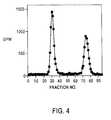
- FIG. 4illustrates urine from a diabetic patient showing both intact and fragmented albumin peaks from size exclusion chromatography.
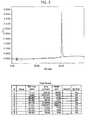
- FIG. 5illustrates a HPLC profile of albumin alone.
- FIG. 6illustrates the HPLC profile of plasma from normal, healthy volunteer showing albumin peaks.
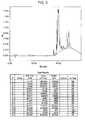
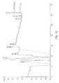
- FIG. 7shows the HPLC profile of urine from normal, healthy volunteer with fragmented products of albumin but no intact albumin peak.
- FIG. 8shows the HPLC profile of a urine sample from a normoalbuminuric diabetic patient showing albumin breakdown products and a small-modified albumin peak at approximately 39-44 minutes retention time.
- FIG. 9shows the HPLC profile of urine from a normoalbuminuric diabetic patient showing signs of kidney failure and the presence of the characteristic spiked albumin peak at approximately 39-44 minutes retention time.
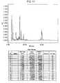
- FIG. 10illustrates a HPLC profile of a normoalbuminuric diabetic patient showing signs of kidney failure and the presence of the characteristic spiked modified albumin peak at approximately 39-44 minutes retention time.
- FIG. 11illustrates a HPLC of a macroalbuminuric diabetic patient showing high levels of the normal albumin as well as the characteristic spiked appearance at approximately 39-44 minutes retention time.
- FIG. 13illustrates a longitudinal study of a patient in which the modified protein was detected at a time prior to onset of diabetic nephropathy, indicating predisposition to diabetic nephropathy, and the delay in treatment caused by relying on conventional RIA methods.
- FIG. 14illustrates a longitudinal study of a patient in which the modified protein was detected at a time prior to onset of diabetic nephropathy, indicating predisposition to diabetic nephropathy, and the delay in treatment caused by relying on conventional RIA methods.
- FIG. 15shows the HPLC chromatogram used as a criterion of purity of the modified albumin of Example 4.
- proteinsincluding major plasma proteins such as albumin and immunoglobulin
- Tubular cellslie beyond the kidney filter and come in direct contact with the primary filtrate.
- proteinsare internalized by the tubular cells, they are directed towards the lysosomes, where they are partially degraded to various size fragments, and then regurgitated to outside the cell. These regurgitated fragments, of which there may be at least 60 different fragments generated from any one particular type of protein, are then excreted into the urine.
- the drugsmay also be useful in other renal diseases where lysosomal activities are affected, or in diabetes without renal complications in situations where lysosomal activity is turned off in non renal tissues.
- Such drugsinclude antiproliferative drugs, such as anti cancer drugs or antibodies to neutralize TGF beta.
- “Fragmented protein or fragment albumin”includes post-glomerular breakdown products after chemical, enzymatic or physical breakdown that occurs during renal passage. These components have a reduced size and/or may have changed hydrophobicity.
- “Intact albumin, modified albumin, or modified form of albumin” as used hereinmeans a compound having similar size and structural characteristics to native albumin, wherein the amino acid sequence is substantially the same as the native albumin. It is preferably a filtered intact protein. It elutes at or near the same position as native albumin on high-pressure liquid chromatography (HPLC) (FIG. 5 ).
- HPLChigh-pressure liquid chromatography
- the structurehas been modified biochemically either by minor enzyme mediated modification or addition to its basic structure and/or physically through a change in its three dimensional structure so that it escapes detection by conventionally used anti-albumin antibodies.
- Biochemical modificationmay be made by enzymes such as endo- or exo-peptidases.
- the 3D structure of albuminmay have been altered in some way. Ligands may have bound to the albumin, or it may be any combination of these.
- the modified albumin detected in the method of the inventionis not detectable by current and conventional radioimmunoassays using
- Conventional anti-albumin antibodiescan be purchased from any purveyor of immunochemicals.
- monoclonal antibody catalog numbers A6684clone no. HSA-11
- A2672clone no. HSA-9
- liquid whole serum, lyophilized fractionates, liquid IgG fraction, and the monoclonal antibodies in liquid ascites fluids formcan be obtained from Sigma, St. Louis, Mo., as found in the Immunochemicals section at pages 1151-1152 in the 1994 Sigma—Biochemicals Organic Compounds for Research and Diagnostic Reagents catalog.
- intact/modified albuminincludes albumin that is substantially full-length, fragmented, chemically modified, or physically modified.
- intact/modified albuminis meant to indicate albumin that is less than, equal to, or greater in molecular weight than the full-length albumin, and elutes at or near the native albumin position in a separation medium, such as chromatography, preferably HPLC, and most preferably hydrophobicity HPLC.
- fragmented albuminis meant to refer to the fragment of albumin that is not detected by conventional anti-albumin antibody, and its presence is detected in diagnosing an early stage of renal disease and/or renal complications of a disease. The detection of the presence of intact/modified albumin is an indication of a predisposition to renal disease.
- “Intact protein, modified protein or modified form of a protein” as used hereinincludes those forms of substantially full-length protein which are undetectable by conventional radioimmunoassay.
- the proteinincludes, but is not limited to, albumins, globulins ( ⁇ -globulin( ⁇ 1 -globulin, ⁇ 2 -globulin), ⁇ -globulins, ⁇ -globulins), euglobulins, pseudoglobulin I and II, fibrinogen, ⁇ 1 acid glycoprotein (orosomucoid), ⁇ 1 glycoprotein, ⁇ 1 lipoproteins, ceruloplasmin, ⁇ 2 19 S glycoprotein, ⁇ 1 transferrin, ⁇ 1 lipoprotein, immunoglobulins A, E, G, and M, protein hormones including growth hormone, insulin, parathyroid hormone and other proteins including horseradish peroxidase, lactate dehydrogenase, glucose oxidase, myoglobin, and lysozyme.
- Kidney diseaseas used herein includes any malfunction of the kidney. Kidney disease may be identified by the presence of intact or modified albumin in the urine. Preferably, an early diagnosis of the kidney disease may be made by detecting the presence of modified protein in the urine, or an increase in the modified protein in the urine over time.
- Low lysosome activityas used herein is compared against normal levels of lysosome activity and/or lysosome machinery that traffics protein to the lysosome in a normal individual. The activity is insufficient for the lysosome to fragment proteins so that intact protein is excreted at a greater amount than at normally low levels.
- “Lysosome-activating compound” as used hereinrefers to a compound that is beneficial to reactivation of the lysosome.
- the compoundmay work directly or indirectly on the lysosome resulting in activation of lysosomal function.
- These compoundsmay be selected from the group including, but not limited to, anticancer compounds, antiproliferation compounds, paracetamol, vitamin A (retinoic acid) or derivatives of retinol, or compounds, including antibodies, to neutralize TGF beta.
- Microalbuminuriais a condition where an individual excretes greater than 200 ⁇ g albumin/min in the urine as measured by conventional radioimmunoassay (RIA).
- Microalbuminuriais a condition where an individual excretes at least 20 ⁇ g albumin/min in the urine as measured by conventional radioimmunoassay (RIA). RIA measures down to 15.6 ng/ml and is able to measure albumin in urine of normal subjects who have clearance of less than 6 ⁇ g/min. However, when albumin excretion exceeds 20 ⁇ g/min, treatment of the kidney disease is limited and full recovery is difficult from this point.
- RIAradioimmunoassay
- “Microalbuminuric” as used hereinis a condition when albumin is detected in the urine at an excretion rate of at least 20 ⁇ g/min as measured by conventional RIA.
- “native” and “unmodified”are used interchangeably to describe a protein that is naturally found in an organism, preferably a human, which has not been modified by the filtering process of the renal glomeruli.
- Normal individualas used herein is an individual who does not have a disease in which intact protein found in urine is an indicator of the disease.
- the diseaseis kidney disease.
- Normal levels of lysosome activityare levels of lysosome activity found in undiseased kidney of a normal individual.
- Normal albuminuricas used herein means a condition where albumin is excreted in the urine and is not detectable by RIA, or less than 20 ⁇ g/min (as measured by RIA) is excreted.
- Propensity for a diseasemeans that a disease may result in an individual as judged by a determination of the presence and excretion rate of a modified protein such as modified albumin.
- Proteinuriaas used herein is the existence of protein in the urine, usually in the form of albumin, a protein that is soluble in water and can be coagulated by heat. Related to this, “specific proteinuria” refers to the existence of a particular protein in the urine.
- Radioimmunoassayas used herein is a method for detection and measurement of substances using radioactively labeled specific antibodies or antigens.
- Reactivation of the lysosomeincludes an activation of lysosome activity preferably so that breakdown of proteins, particularly albumin, is increased compared with an inactivated state of the lysosome.
- Restoreas used herein means to restore in full or in part so that the component being restored has an improved function compared with its previous function.
- the “sum of intact and intact modified protein” as used hereinrefers to the total amount of intact protein, and intact modified protein present in a biological sample.
- Total proteinrefers to a particular filtered protein present in native, unmodified, modified or fragmented form that is excreted in urine. It includes protein that is not detected by conventional radioimmunoassay or conventional methods, which are currently available to detect the protein. Preferably the protein is albumin.
- the diseases to be treatedinclude, but are not limited to renal disease (glomerulonephritis, bacterial and viral glomerulonephritides, IgA nephropathy and Henoch-Schönlein Purpura, membranoproliferative glomerulonephritis, membranous nephropathy, Sjögren's syndrome, diabetic nephropathy, nephrotic syndrome (minimal change disease, focal glomerulosclerosis, and related disorders), acute renal failure, acute tubulointerstitial nephritis, pyelonephritis, GU tract inflammatory disease, Pre-clampsia, renal graft rejection, leprosy, reflux nephropathy, nephrolithiasis), genetic renal disease (medullary cystic, medullar sponge, polycystic kidney disease (autosomal dominant polycystic kidney disease, autosomal recessive polycystic kidney disease, tub
- a method for determining a propensity for or early diagnosis of renal disease and/or renal complications of a diseaseincludes determining a change in the albumin content in a urine sample.
- the diseasemay be a kidney disease, although not necessarily limited to a kidney disease.
- albuminis used herein only as an example of a protein to be detected in urine.
- albumin in a patientis analyzed by conventional RIA, it is expected that a normoalbuminuric patient or normal individual would have albumin in the urine in the range of 3-10 ⁇ g/min in young people and greater in older people.
- normoalbuminuric patientsalso show levels of albumin in the urine if measured by HPLC. Applicant has found that these levels may be in the order of 5 ⁇ g/min.
- the level of intact/modified albuminwill increase to microalbuminuria levels in the order of 20 to 200 ⁇ g/min as determined by RIA.
- a patient suspected of having diabetic kidney diseasewill not show signs of kidney degeneration until well after 10 to 15 years when albumin is detected by currently available methods such as RIA methods.
- Urinary excretion rates of at least 20 ⁇ g/minmay be detected by RIA when an individual enters a microalbuminuric state. Again, by observing the excretion of modified albumin, a change in the kidney and possibly onset of a kidney disease may be detected.
- a normoalbuminuric subject, or normoalbuminuric diabetic patientmay continue to have a low albumin excretion rate of less than 20 ⁇ g/min as determined by RIA, for many years.
- the presence of albumin in the urineis a sign that functions of the kidney may be impaired. Once this level begins to change, treatment may be initiated.
- albuminIn a normal individual a small amount of albumin is detectable in the urine. Total filtered albumin appears mainly as fragmented albumin in urine. Some albumin may be detected in normoalbuminuric individuals. However, the excretion rate of albumin in urine in a normoalbuminuric individual may be as low as 5 ⁇ g/min. This level is generally detectable by RIA.
- the modified protein of the inventioncan be detected by a variety of methods that are well-known in the art, including, but not limited to chromatography, electrophoresis and sedimentation, or a combination of these, which are described in Karger B L, Hancock W S (eds.) High Resolution Separation and Analysis of biological Macromolecules. Part A Fundamentals in Methods in Enzymology, Vol. 270, 1996, Academic Press, San Diego, Calif., USA; Karger B L, Hancock W S (eds.) High Resolution Separation and Analysis of biological Macromolecules. Part B Applications in Methods in Enzymology, Vol.
- the electrophoresis methodincludes, but is not limited to, moving-boundary electrophoresis, zone electrophoresis, and isoelectric focusing.
- the chromatography methodincludes, but is not limited to, partition chromatography, adsorption chromatography, paper chromatography, thin-layer chromatography, gas-liquid chromatography, gel chromatography, ion-exchange chromatography, affinity chromatography, and hydrophobic interaction chromatography.
- the methodis a sizing gel chromatography and hydrophobic interaction chromatography. More preferably, the method is hydrophobic interaction chromatography using a HPLC column.
- the modified proteincan also be detected by the use of specific albumin dyes.
- specific albumin dyesSuch methods are described for example by Pegoraro, et al. American Journal of Kidney Diseases, Vo. 35, No. 4, April, 2000, pp. 739-744, the entire disclosure of which is hereby incorporated by reference.
- the modified albumin, as well as whole albuminare detectable by this dye method to provide the sum of modified albumin and whole or intact albumin.
- This detection methodmay be used with or without an initial separation of the albumin components form urine.
- Such dyesnormally do not detect fragments ⁇ 10,000 in molecular weight, but will detect the modified albumin.
- a dyesuch as Albumin Blue 580, is used.
- Such dyesare naturally non-fluorescent but fluoresce on binding to intact albumin as well as the modified albumin, but do not bind to globulins. Therefore, globulins do not interfere with the assay so that measurements can be made in unfractionated urine.
- a normoalbuminuric diabetic patienthas almost undetectable levels of modified or fragments of albumin when analyzed by conventional RIA. They appear to be normal. However, when the urine is tested by HPLC, the levels of modified albumin are much greater than found in a normal individual. This difference in albumin may be attributed to the inability of conventional RIA's to adequately detect all albumin (total albumin) in intact or modified forms. Thus, HPLC is preferred for generating a fragmentation profile.
- a fragmentation profile on HPLCis characterized by a series of peaks representing a number of species of albumin as fragments or in intact or modified forms.
- the method of determining a propensity for or early diagnosis of a kidney disease in a subjectis determined before the subject becomes microalbuminuric.
- Measuring albumin content in a sample by an HPLC method of the present inventionmay provide different results from its measurement by conventional RIA.
- a low level of albuminis observed in normal individuals.
- the level of modified albuminbegins to be detected and its level increases, and progresses toward microalbuminuria then a patient can be determined to have a propensity for kidney disease.
- the HPLC generated fragmentation profileis characterized by the absence of a peak in a region where full-length native albumin elutes. Instead, multiple fragmented albumin is detectable.
- a pure protein product(unmodified) produces essentially a single peak.
- albuminwas observed to elute in the range of 39-44 minutes (FIG. 5 ).
- a normal individualwould provide a distinct fragmentation profile indicative of an absence of kidney disease or no propensity for a kidney disease.
- an increasing amount of modified albumin first, and then native form laterare detectable.
- the fragmentation profilebegins to change and more products in the region of full-length albumin manifests as additional spikes or an enlarged peak indicative of more intact/modified albumin in the urine.
- the modified albuminmay appear in a region where native albumin elutes but may be manifest as multiple peaks indicating the presence of multiple forms of modified albumin.
- the propensity for kidney diseasemay be measured by determining the presence of or identifying at least one species of modified albumin. This may be determined or identified by the presence of a specific peak on a HPLC profile, preferably the peak is within the range of position that corresponds to the elution position of the native albumin.
- a HPLC column for detecting modified albumin or unmodified albuminmay be a hydrophobicity column, such as Zorbax 300 SB-CB (4.6 mm ⁇ 150 mm).
- a 50 ⁇ l sample loopmay be used.
- Elution solvents suitable for HPLC in detecting albumin and its breakdown productsmay include standard elution solvents such as acetonitrile solvents.
- a buffer of water/1% trifluoro acetic acid (TFA) followed by a buffer of 60% acetonitrile/0.09% TFAmay be used.
- TFAtrifluoro acetic acid
- a gradient of 0 to 100% of a 60% acetonitrile/0.09% TFAhas been found to be suitable.
- Suitable HPLC conditions for a hydrophobicity columnmay be as follows:
- the wavelength used in HPLCmay be approximately 214 nm.
- Modified albuminmay elute between 39-44 minutes (FIG. 5 ). Albumin fragments may elute much earlier, mainly at less than 20 minutes.
- the method for determining the propensity for kidney diseaseis applicable to any individual.
- Kidney diseasemay be caused by a number of factors including bacterial infection, allergic, congenital defects, stones, tumors, chemicals or from diabetes.
- the methodis applicable for determining a propensity for kidney disease in diabetic patients that may progress to a kidney disease.
- the individualis a normoalbuminuric diabetic.
- normal individualsmay be monitored for propensity for the disease by determining increased levels of intact or modified albumin in the urine.
- the method of the inventioncan be carried out using non-antibody separation procedures as described above.
- antibody specific for modified proteinmay also be used to detect the presence of the modified protein.
- the antibody to the modified proteinmay be obtained using the following method.
- the procedureis described specifically for albumin by way of example only, and can be readily applied to antibody production against any other protein in the urine.
- the methodseeks to determine which modified albumin molecule is the most sensitive marker to identify diabetic patients, for example, who will progress to kidney complications.
- the modified albuminis characterized by carrying out a quantitative separation of the modified albumin molecules, such as by preparative HPLC.
- the modified proteinsare analyzed for ligand binding, such as glycation.
- amino acid sequence of the individual modified proteinis determined, preferably by mass spectrometry using methods described in Karger B L, Hancock WS (eds.) High Resolution Separation and Analysis of biological Macromolecules. Part A Fundamentals in Methods in Enzymology, Vol. 270, 1996, Academic Press, San Diego, Calif., USA; or Karger B L, Hancock W S (eds.) High Resolution Separation and Analysis of biological Macromolecules. Part B Applications in Methods in Enzymology, Vol. 271, 1996, Academic Press, San Diego, Calif., USA, for example, which references are incorporated herein by reference in their entirety. In a preferred embodiment, there may be about 3 to 4 modified albumin species.
- the method of generating antibody against the modified albuminseeks to develop a diagnostic immunoassay for the modified albumin that predicts those diabetic patients, for example, that progress to kidney complications. To accomplish this, sufficient quantities of modified albumin is prepared by HPLC. Antibodies are made by sequential injection of the modified albumin in an animal such as a rabbit, to generate good titer, and the antibodies are isolated using conventional techniques using methods described in Goding J W, Monoclonal Antibodies: Principles and Practice.
- the obtained antibodiesmay be polyclonal antibodies or monoclonal antibodies.
- At least one species of a modified albuminis isolated and identified for use in determining a propensity for kidney disease.
- the isolated speciesmay be used to generate antibodies for use in immunoassays.
- the antibodiesmay be tagged with an enzymatic, radioactive, fluorescent or chemiluminescent label.
- the detection methodmay include, but is not limited to radioimmuoassay, immunoradiometric assay, fluorescent immunoassay, enzyme linked immunoassay, and protein A immunoassay.
- the assaysmay be carried out in the manner described in Goding J W, Monoclonal Antibodies: Principles and Practice. Production and Application of Monoclonal Antibodies in Cell Biology, Biochemistry and Immunology.
- kitsfor rapidly and accurately determining the presence or absence of modified protein such as modified albumin, in a sample quantitatively or non-quantitatively as desired.
- modified proteinsuch as modified albumin
- Each component of the kit(s)may be individually packaged in its own suitable container.
- the individual containermay also be labeled in a manner, which identifies the contents.
- the individually packaged componentsmay be placed in a larger container capable of holding all desired components.
- Associated with the kitmay be instructions, which explain how to use the kit. These instructions may be written on or attached to the kit.
- the inventionis also directed to a method of determining a treatment agent for renal disease and/or renal complications of a disease, comprising:
- the treatment agentmay be a lysosome activating agent that may act directly or indirectly to activate lysosome, and thereby cause the lysosome to digest post-glomerular filtered proteins, which is a sign of a healthy kidney.
- PKCprotein kinase C
- a lysosome-activating compoundfor use in reactivating lysosomes or processes that direct substrates to the lysosome or products away from the lysosome.
- compositioncomprising a lysosome-activating compound and a carrier.
- a method of preventing or treating kidney diseaseincluding administering an effective amount of a lysosome-activating compound to a subject.
- a method of screening a multiplicity of compounds to identify a compound capable of activating lysosomes or processes that direct substrates to the lysosome or products away from the lysosomeincluding the steps of:
- Lysosomesmay be associated with the breakdown of proteins, particularly albumin, in the kidney. In cases of microalbuminuria, substantial amounts of albumin escape lysosomal breakdown possibly due to a deactivated lysosome. Restoration of lysosomal breakdown may restore the balance in the kidney of cellular processes and tissue turnover.
- a lysosome-activating compoundmay be a compound that acts directly or indirectly on the lysosome. By acting indirectly, the compound may act on a component, which influences the activity of the lysosome. Nevertheless, the outcome results in an activation of the lysosome, thereby providing enhanced protein breakdown.
- compositioncomprising a lysosome-activating compound and a carrier.
- the compositionmay be a physiologically acceptable or pharmaceutically acceptable composition. However, it will be a composition which allows for stable storage of the lysosome activating compound. Where the composition is a pharmaceutically acceptable composition, it may be suitable for use in a method of preventing or treating kidney disease.
- a method of preventing or treating kidney diseaseincluding administering an effective amount of a lysosome-activating compound to a subject.
- the lysosome-activating compoundmay act by reactivating the lysosome so that cellular processes and tissue turnover are restored fully or in part, thereby resulting in the kidney being restored partially or fully.
- administering a lysosome activating compound to an animal having kidney diseasemay restore lysosome activity fully or in part.
- Methods of administeringmay be oral or parenteral.
- Oralmay include administering with tablets, capsules, powders, syrups, etc.
- Parenteral administrationmay include intravenous, intramuscular, subcutaneous or intraperitoneal routes.
- the changed activity of the lysosomeis preferably a change which enhances the activity of the lysosome so that albumin breakdown is improved.
- the ability to not only activate lysosome but also improve cellular processes and/or tissue turnoveris a characteristic of the most desirable lysosome activating compound.
- kidney disease in a subjectincluding:
- H[HSA]Human Serum Albumin
- Tritium radioactivitywas determined in 1 ml aqueous samples with 3 ml scintillant and measured on a Wallac 1410 liquid scintillation counter (Wallac Turku, Finland).
- FIG. 2illustrates the distribution of albumin in urine and in plasma.
- Example 3 H[HSA] as used in Example 1was injected into a normal, healthy volunteer and a diabetic patient. Samples of urine were collected and 3 H[HSA] was determined as in Example 1.
- the normal, healthy volunteer(FIG. 3) shows the excretion of fragments of albumin on a size exclusion chromatography as performed in Example 1.
- the diabetic patient(FIG. 4) shows the presence of substantially full-length and fragmented albumin on size exclusion chromatography.
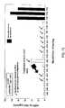
- excretion rates of albumin detectable by these methodswere in the order of 5 ⁇ g/min (control) and 1457 ⁇ g/min (diabetic).
- Urine sampleswere collected from normal, healthy volunteer, normoalbuminuric diabetic patients and from macroalbuminuric patients. Urine was collected midstream in 50 ml urine specimen containers. The urine was frozen until further use. Prior to HPLC analysis the urine was centrifuged at 5000 g.
- Sampleswere analyzed on HPLC using a hydrophobicity column Zorbax 300 SB-CB (4.6 mm ⁇ 150 mm). A 50 ⁇ l sample loop was used.
- a wavelength of 214 nmwas used.
- Urine from microalbuminuric patient which had an intact albumin concentration of 43.5 mg/L as determined by turbitimer (involving conventional immunochemical assay)was initially filtered through a 30 kDa membrane to separate the modified albumin from low molecular weight ( ⁇ 30,000) protein fragments in urine.
- the material that was retained by the filtergave a yield of intact albumin of 27.4 mg/L as determined by turbitimer assay.
- This retained materialwas then subjected to size exclusion chromatography on Sephadex G100. The material collected was the peak fraction that coelutes with intact albumin. This material gave a yield of 15.2 ml/L of albumin as determined by the turbitimer method.
- FIG. 5illustrates a HPLC profile of albumin alone. Essentially a single peak which elutes at approximately 39-44 minutes retention time was obtained.
- FIG. 6illustrates a HPLC profile of plasma showing a distinct albumin peak at approximately 39-44 minutes as well as other peaks corresponding to other plasma proteins.
- FIG. 7illustrates a HPLC profile of a normal, healthy volunteer showing no albumin peak in the urine sample. This individual breaks down the albumin excreted into the urine possibly via an active lysosome. Substantial fragmented products were evident showing prominence of some species, particularly of a species at approximately less than 14.5 minutes retention time.
- FIG. 9Another urine sample from normoalbuminuric diabetic patient (with albumin excretion rate of 17.04 ⁇ g/min) was analyzed (FIG. 9 ).
- RIA testsshow albumin excreted in the urine for this patient.
- HPLCHPLC
- an albumin or modified albumin peakis evident at approximately 39-44 minutes retention time.
- the method of the inventionshowed that the true albumin content in the urine sample was 81.3 mg/l.
- Treatment for the diseaseshould have begun on this individual. This peak begins to show a multiple peaked appearance.
- a smaller peak corresponding to intact albuminshows that modified albumin may represent the peak at 39-44 minutes.
- the presence of this albumin peakcompared with the profile of a normal, healthy volunteer having no albumin peak shows a change in the detectable levels of the amount of intact/modified albumin. This may signal a propensity for a kidney disease.
- a further urine sample from a normoalbuminuric diabetic patient(with an albumin excretion rate of 4.37 ⁇ g/min) was analyzed, and the HPLC profile is illustrated in FIG. 10 .
- modified albuminwas detected at approximately 39-44 minutes retention time showing multiple peaks.
- This patientagain did register normal albumin by RIA.
- conventional testindicates the presence of ⁇ 6 mg/l of albumin in the urine sample
- the method of the inventionshowed that the true albumin content in the urine sample was 491 mg/l. Treatment for the disease should have begun on this individual. It is clear that modified albumin assessment is necessary to identify these changes. This patient would be determined to have a propensity for kidney disease. As kidney disease progresses, the modified albumin peak will continue to increase.
- FIG. 11This is shown in FIG. 11 where a urine sample of a macroalbuminuric patient was analyzed. A quite significant albumin peak at approximately 39-44 minutes retention time showing multiple peaks was evident. The patient's albumin content was 1796 mg/l. Treatment for this individual is in progress.
- the method of the inventionresults in early detection of a propensity for a renal disease as illustrated by the longitudinal studies in FIGS. 12-14.
- FIGS. 12-14show situations in which the ACE inhibitor treatment for diabetes was begun later than it should have had the modified albumin detection method of the invention been used. Detecting modified protein using the method according to the invention is a more effective method for predicting the onset of a renal disease than using conventional RIA.
Landscapes
- Health & Medical Sciences (AREA)
- Life Sciences & Earth Sciences (AREA)
- Engineering & Computer Science (AREA)
- Chemical & Material Sciences (AREA)
- Medicinal Chemistry (AREA)
- General Health & Medical Sciences (AREA)
- Organic Chemistry (AREA)
- Veterinary Medicine (AREA)
- Chemical Kinetics & Catalysis (AREA)
- Nuclear Medicine, Radiotherapy & Molecular Imaging (AREA)
- General Chemical & Material Sciences (AREA)
- Pharmacology & Pharmacy (AREA)
- Public Health (AREA)
- Animal Behavior & Ethology (AREA)
- Immunology (AREA)
- Hematology (AREA)
- Urology & Nephrology (AREA)
- Bioinformatics & Cheminformatics (AREA)
- Biomedical Technology (AREA)
- Molecular Biology (AREA)
- Biochemistry (AREA)
- Biotechnology (AREA)
- Pathology (AREA)
- General Physics & Mathematics (AREA)
- Analytical Chemistry (AREA)
- Physics & Mathematics (AREA)
- Food Science & Technology (AREA)
- Microbiology (AREA)
- Cell Biology (AREA)
- Diabetes (AREA)
- Tropical Medicine & Parasitology (AREA)
- Cardiology (AREA)
- Heart & Thoracic Surgery (AREA)
- Dermatology (AREA)
- Neurology (AREA)
- Physiology (AREA)
- Virology (AREA)
- Proteomics, Peptides & Aminoacids (AREA)
- Endocrinology (AREA)
- Orthopedic Medicine & Surgery (AREA)
Abstract
Description
Claims (24)
Priority Applications (4)
| Application Number | Priority Date | Filing Date | Title |
|---|---|---|---|
| US09/892,797US6589748B2 (en) | 1998-12-21 | 2001-06-28 | Method for kidney disease detection and treatment |
| US10/391,202US20040029175A1 (en) | 1998-12-21 | 2003-03-19 | Method for kidney disease detection |
| US10/921,982US20050026225A1 (en) | 1998-12-21 | 2004-08-20 | Method for kidney disease detection |
| US11/020,316US20050191664A1 (en) | 1998-12-21 | 2004-12-27 | Method for kidney disease detection |
Applications Claiming Priority (5)
| Application Number | Priority Date | Filing Date | Title |
|---|---|---|---|
| AUPP7843 | 1997-07-10 | ||
| AUPP7843AAUPP784398A0 (en) | 1998-12-21 | 1998-12-21 | Kidney disease detection and treatment |
| AUPP7843/98 | 1998-12-21 | ||
| US09/415,217US6447989B1 (en) | 1998-12-21 | 1999-10-12 | Kidney disease detection and treatment |
| US09/892,797US6589748B2 (en) | 1998-12-21 | 2001-06-28 | Method for kidney disease detection and treatment |
Related Parent Applications (1)
| Application Number | Title | Priority Date | Filing Date |
|---|---|---|---|
| US09/415,217Continuation-In-PartUS6447989B1 (en) | 1998-12-21 | 1999-10-12 | Kidney disease detection and treatment |
Related Child Applications (1)
| Application Number | Title | Priority Date | Filing Date |
|---|---|---|---|
| US10/391,202Continuation-In-PartUS20040029175A1 (en) | 1998-12-21 | 2003-03-19 | Method for kidney disease detection |
Publications (2)
| Publication Number | Publication Date |
|---|---|
| US20020022236A1 US20020022236A1 (en) | 2002-02-21 |
| US6589748B2true US6589748B2 (en) | 2003-07-08 |
Family
ID=25645955
Family Applications (3)
| Application Number | Title | Priority Date | Filing Date |
|---|---|---|---|
| US09/893,346AbandonedUS20020012906A1 (en) | 1998-12-21 | 2001-06-28 | Method for kidney disease treatment by drug intervention |
| US09/892,797Expired - Fee RelatedUS6589748B2 (en) | 1998-12-21 | 2001-06-28 | Method for kidney disease detection and treatment |
| US10/721,351AbandonedUS20040106155A1 (en) | 1998-12-21 | 2003-11-26 | Method for kidney disease treatment by drug intervention |
Family Applications Before (1)
| Application Number | Title | Priority Date | Filing Date |
|---|---|---|---|
| US09/893,346AbandonedUS20020012906A1 (en) | 1998-12-21 | 2001-06-28 | Method for kidney disease treatment by drug intervention |
Family Applications After (1)
| Application Number | Title | Priority Date | Filing Date |
|---|---|---|---|
| US10/721,351AbandonedUS20040106155A1 (en) | 1998-12-21 | 2003-11-26 | Method for kidney disease treatment by drug intervention |
Country Status (13)
| Country | Link |
|---|---|
| US (3) | US20020012906A1 (en) |
| EP (1) | EP1141728B1 (en) |
| JP (2) | JP4160263B2 (en) |
| CN (1) | CN1331801A (en) |
| AT (1) | ATE376187T1 (en) |
| AU (1) | AU1674400A (en) |
| BR (1) | BR9916407A (en) |
| CA (1) | CA2356174A1 (en) |
| DE (1) | DE69937368T2 (en) |
| DK (1) | DK1141728T3 (en) |
| IL (1) | IL143892A0 (en) |
| MX (1) | MXPA01006404A (en) |
| WO (1) | WO2000037944A1 (en) |
Cited By (31)
| Publication number | Priority date | Publication date | Assignee | Title |
|---|---|---|---|---|
| US20030003588A1 (en)* | 2001-06-28 | 2003-01-02 | Comper Wayne D. | Method for kidney disease detection by protein profiling |
| US20060141011A1 (en)* | 2004-12-29 | 2006-06-29 | Jewell Dennis E | Combination of limited nutrients and enhanced dietary antioxidants to impart improved kidney health |
| US20060205818A1 (en)* | 2005-03-08 | 2006-09-14 | Burzynski Stanislaw R | Method for the treatment of von Hippel-Lindau (VHL) disease with phenylacetyl-derivatives |
| WO2006113752A1 (en) | 2005-04-19 | 2006-10-26 | Hill's Pet Nutrition, Inc. | Methods and compositions for the prevention and treatment of kidney disease |
| US20060286602A1 (en)* | 2004-05-10 | 2006-12-21 | Harald Mischak | Method and markers for the diagnosis of renal diseases |
| WO2007002836A2 (en) | 2005-06-29 | 2007-01-04 | Hill's Pet Nutrition, Inc. | Methods and compositions for the prevention and treatment of kidney disease |
| WO2006127424A3 (en)* | 2005-05-20 | 2007-06-28 | Hills Pet Nutrition Inc | Methods for promoting health or wellness in adult animals |
| US20080038323A1 (en)* | 2004-11-24 | 2008-02-14 | Zicker Steven C | Methods for Improving Liver Clearance of Xenobiotic Substances in an Animal |
| US20080206398A1 (en)* | 2004-12-30 | 2008-08-28 | Ryan Yamka | Methods for Enhancing the Quality of Life of a Senior Animal |
| US20080293621A1 (en)* | 2005-04-29 | 2008-11-27 | Timothy Arthur Allen | Methods for Prolonging Feline Life |
| US20090023165A1 (en)* | 2007-05-11 | 2009-01-22 | The Institutes For Pharmaceutical Discovery, Llc | Methods for Early Diagnosis of Kidney Disease |
| US20090111877A1 (en)* | 2004-12-30 | 2009-04-30 | Hill's Pet Nutrition, Inc. | Methods for Enhancing the Quality of Life of a Senior Animal |
| US20090227665A1 (en)* | 2005-08-17 | 2009-09-10 | Hill's Pet Nutrition, Inc. | Methods and Compositions For The Prevention and Treatment of Kidney Disease |
| US20100099196A1 (en)* | 2007-03-07 | 2010-04-22 | Harald Mischak | Process for normalizing the concentration of analytes in a urine sample |
| WO2010064239A1 (en) | 2008-12-04 | 2010-06-10 | Technion Research And Development Foundation Ltd. | Apparatus and methods for diagnosing renal disorders |
| US20100210021A1 (en)* | 2007-03-14 | 2010-08-19 | Harald Mischak | Process and markers for the diagnosis of kidney diseases |
| US20100227411A1 (en)* | 2007-10-09 | 2010-09-09 | Harald Mischak | Polypeptide markers for the diagnosis of prostate cancer |
| US20100248378A1 (en)* | 2007-10-19 | 2010-09-30 | Mosaiques Diagnostics And Therapeutics Ag | Method and marker for diagnosing diabetes mellitus |
| US20110036717A1 (en)* | 2008-03-19 | 2011-02-17 | Harald Mischak | Method and marker for diagnosis of tubular kidney damage and illness |
| US20110135785A1 (en)* | 2004-11-24 | 2011-06-09 | Hill's Pet Nutrition, Inc. | Methods for improving hepatic and immune function in an animal |
| US20110214990A1 (en)* | 2008-09-17 | 2011-09-08 | Mosaiques Diagnostics And Therapeutics Ag | Kidney cell carcinoma |
| WO2012170765A2 (en) | 2011-06-10 | 2012-12-13 | Oregon Health & Science University | Cmv glycoproteins and recombinant vectors |
| EP2568289A2 (en) | 2011-09-12 | 2013-03-13 | International AIDS Vaccine Initiative | Immunoselection of recombinant vesicular stomatitis virus expressing hiv-1 proteins by broadly neutralizing antibodies |
| EP2586461A1 (en) | 2011-10-27 | 2013-05-01 | Christopher L. Parks | Viral particles derived from an enveloped virus |
| EP2679596A1 (en) | 2012-06-27 | 2014-01-01 | Simon Hoffenberg | HIV-1 env glycoprotein variant |
| EP2848937A1 (en) | 2013-09-05 | 2015-03-18 | International Aids Vaccine Initiative | Methods of identifying novel HIV-1 immunogens |
| EP2873423A2 (en) | 2013-10-07 | 2015-05-20 | International Aids Vaccine Initiative | Soluble hiv-1 envelope glycoprotein trimers |
| EP3069730A2 (en) | 2015-03-20 | 2016-09-21 | International Aids Vaccine Initiative | Soluble hiv-1 envelope glycoprotein trimers |
| EP3072901A1 (en) | 2015-03-23 | 2016-09-28 | International Aids Vaccine Initiative | Soluble hiv-1 envelope glycoprotein trimers |
| US9689826B2 (en) | 2012-03-11 | 2017-06-27 | Technion Research And Development Foundation Ltd. | Detection of chronic kidney disease and disease progression |
| EP3187585A1 (en) | 2010-03-25 | 2017-07-05 | Oregon Health&Science University | Cmv glycoproteins and recombinant vectors |
Families Citing this family (47)
| Publication number | Priority date | Publication date | Assignee | Title |
|---|---|---|---|---|
| AUPP784398A0 (en) | 1998-12-21 | 1999-01-21 | Monash University | Kidney disease detection and treatment |
| AU1674400A (en) | 1998-12-21 | 2000-07-12 | Monash University | Kidney disease detection and treatment |
| US7250294B2 (en)* | 2000-05-17 | 2007-07-31 | Geron Corporation | Screening small molecule drugs using neural cells differentiated from human embryonic stem cells |
| CA2442074C (en) | 2001-03-28 | 2014-07-22 | Heska Corporation | Methods of detecting early renal disease in animals |
| ES2894356T3 (en)* | 2003-02-13 | 2022-02-14 | Becton Dickinson Co | Devices for the removal of components during blood collection and their uses |
| US7482128B2 (en) | 2003-03-27 | 2009-01-27 | Heska Corporation | Anti-feline albumin antibodies |
| DE60325906D1 (en) | 2003-08-08 | 2009-03-05 | Amgen Fremont Inc | ANTIBODIES TO PARATH-HORMONE (PTH) AND ITS USES |
| US7318925B2 (en)* | 2003-08-08 | 2008-01-15 | Amgen Fremont, Inc. | Methods of use for antibodies against parathyroid hormone |
| US20050112123A1 (en)* | 2003-10-06 | 2005-05-26 | Vaughan Michael R. | Methods of treating proteinuria |
| DK2313778T3 (en)* | 2008-07-18 | 2015-04-27 | Boston Medical Ct Corp | Diagnostics for membranous nephropathy |
| CN105067819B (en)* | 2008-08-28 | 2018-05-22 | 阿斯图特医药公司 | Methods and compositions for diagnosis and prognosis of renal injury and renal failure |
| EP2813848A3 (en)* | 2008-08-29 | 2015-03-11 | Astute Medical, Inc. | Methods and compositions for diagnosis and prognosis of renal injury and renal failure |
| WO2010048347A2 (en)* | 2008-10-21 | 2010-04-29 | Astute Medical, Inc. | Methods and compositions for diagnosis and prognosis of renal injury and renal failure |
| NZ607703A (en) | 2008-10-21 | 2014-09-26 | Astute Medical Inc | Methods and compositions for diagnosis and prognosis of renal injury and renal failure |
| BRPI0922021A2 (en) | 2008-11-10 | 2019-09-24 | Astute Medical Inc | method for assessing kidney condition in an individual, and use of one or more markers of kidney injury |
| CN104034901A (en)* | 2008-11-22 | 2014-09-10 | 阿斯图特医药公司 | Methods and compositions for diagnosis and prognosis of renal injury and renal failure |
| CA2748852C (en)* | 2009-01-28 | 2019-05-28 | Industrial Technology Research Institute | Biomarkers associated with nephropathy |
| US9229010B2 (en) | 2009-02-06 | 2016-01-05 | Astute Medical, Inc. | Methods and compositions for diagnosis and prognosis of renal injury and renal failure |
| CN102576011B (en)* | 2009-08-07 | 2014-09-17 | 阿斯图特医药公司 | Methods and compositions for diagnosis and prognosis of renal injury and renal failure |
| CA2772336A1 (en)* | 2009-08-28 | 2011-03-03 | Astute Medical, Inc. | Methods and compositions for diagnosis and prognosis of renal injury and renal failure |
| NZ624614A (en)* | 2009-11-07 | 2015-10-30 | Astute Medical Inc | Methods and compositions for diagnosis and prognosis of renal injury and renal failure |
| EA201290192A1 (en) | 2009-11-07 | 2013-02-28 | Астьют Медикал, Инк. | METHODS AND COMPOSITIONS FOR DIAGNOSIS AND PREDICTION OF KIDNEY DAMAGE AND RENAL FAILURE |
| EP3112871B1 (en)* | 2009-12-20 | 2020-07-29 | Astute Medical, Inc. | Methods and compositions for diagnosis and prognosis of renal injury and renal failure |
| MX366653B (en) | 2010-02-05 | 2019-07-17 | Astute Medical Inc | METHODS and COMPOSITIONS FOR DIAGNOSIS and PROGNOSIS OF RENAL INJURY and RENAL FAILURE. |
| AU2011220413B2 (en) | 2010-02-26 | 2015-07-23 | Astute Medical, Inc. | Methods and compositions for diagnosis and prognosis of renal injury and renal failure |
| WO2011162819A1 (en) | 2010-06-23 | 2011-12-29 | Astute Medical, Inc. | Methods and compositions for diagnosis and prognosis of renal injury and renal failure |
| EP2585827A4 (en) | 2010-06-23 | 2013-12-04 | Astute Medical Inc | METHODS AND COMPOSITIONS FOR DIAGNOSING AND PROGNOSING RENAL INJURY AND RENAL FAILURE |
| US8930222B2 (en)* | 2010-08-05 | 2015-01-06 | Abbott Laboratories | Method and system for managing patient healthcare |
| DE102010038014B4 (en) | 2010-10-06 | 2021-10-07 | Numares Ag | Use of specific substances as markers to determine the risk of kidney rejection |
| KR101215759B1 (en) | 2010-11-01 | 2012-12-26 | 서울대학교산학협력단 | Uses of Compositions Comprising Orosomucoid |
| CN102854325B (en)* | 2011-07-01 | 2015-01-07 | 复旦大学 | Method for detecting affinity indicators of reversible binding of insulin and serum protein and application thereof |
| WO2013067420A1 (en)* | 2011-11-03 | 2013-05-10 | Tumlin James A | Acth for treatment of kidney disease |
| EP2788759B1 (en) | 2011-12-08 | 2019-02-20 | Astute Medical, Inc. | Methods and uses for diagnosis of renal injury and renal failure |
| ES2681955T3 (en) | 2013-01-17 | 2018-09-17 | Astute Medical, Inc. | Methods and compositions for the diagnosis and prognosis of renal injury and renal insufficiency |
| JP2016188761A (en)* | 2013-08-23 | 2016-11-04 | 国立研究開発法人国立国際医療研究センター | Method and kit for detecting the onset or risk of developing diabetic nephropathy |
| CN104007258B (en)* | 2013-09-30 | 2016-07-06 | 中国医学科学院基础医学研究所 | The protein marker of focal segmental glomerulosclerosis |
| CN110007084B (en) | 2013-12-03 | 2022-11-18 | 阿斯图特医药公司 | Methods and compositions for diagnosis and prognosis of renal injury and renal failure |
| CN104849466A (en)* | 2014-02-14 | 2015-08-19 | 张曼 | Application of urine vitronectin in diagnosis and treatment of type 2 diabetes mellitus combined early renal injury |
| CA2955992A1 (en)* | 2014-07-23 | 2016-01-28 | University Of British Columbia | Biomarkers for anderson-fabry disease |
| EP3281011B1 (en) | 2015-04-09 | 2024-06-05 | Astute Medical, Inc. | Methods and compositions for diagnosis and prognosis of renal injury and renal failure |
| US11243217B2 (en) | 2016-06-06 | 2022-02-08 | Astute Medical, Inc. | Management of acute kidney injury using insulin-like growth factor-binding protein 7 and tissue inhibitor of metalloproteinase 2 |
| CN117577321A (en) | 2017-08-08 | 2024-02-20 | 费森尤斯医疗保健控股公司 | Systems and methods for treating and assessing the progression of chronic kidney disease |
| CN107561175A (en)* | 2017-08-10 | 2018-01-09 | 武汉大学 | A kind of evaluation method of glomerular sclerosis rat model |
| RU2697722C1 (en)* | 2018-12-06 | 2019-08-19 | Ирина Сергеевна Шатохина | Diagnostic technique of activity of inflammatory process in latent flow of chronic pyelonephritis |
| CN110632229A (en)* | 2019-09-24 | 2019-12-31 | 华兰生物工程重庆有限公司 | Method for detecting protein content of human immunoglobulin (pH4) infused in static environment by high performance liquid chromatograph |
| CN111714517A (en)* | 2020-04-14 | 2020-09-29 | 南开大学 | Application of a lysosome in the field of pharmaceutical preparation |
| RU2768464C1 (en)* | 2021-04-27 | 2022-03-24 | Федеральное государственное бюджетное образовательное учреждение высшего образования "Рязанский государственный медицинский университет имени академика И.П. Павлова" Министерства здравоохранения Российской Федерации | Method for diagnosing membranous nephropathy as one of forms of chronic glomerulonephritis |
Citations (6)
| Publication number | Priority date | Publication date | Assignee | Title |
|---|---|---|---|---|
| EP0514928A2 (en) | 1991-05-24 | 1992-11-25 | Wakamoto Pharmaceutical Co., Ltd. | Method of diagnosing renal diseases by detecting albumin fragments |
| JPH0682446A (en) | 1992-08-31 | 1994-03-22 | Wakamoto Pharmaceut Co Ltd | Diagnostic method for kidney disease |
| DE4336498A1 (en) | 1993-10-26 | 1995-04-27 | Bender & Co Gmbh | Method for diagnosing renal and vascular diseases |
| US5534431A (en) | 1994-02-28 | 1996-07-09 | The Board Of Regents Of The University Of Nebraska | Hybridomas and monoclonal antibodies specific for unique determinants of nephropathy-related immunoglobulin G and complexes thereof |
| US5908925A (en) | 1996-06-27 | 1999-06-01 | Exocell, Inc. | Genetically engineered immunoglobulins with specificity for glycated albumin |
| WO2000037944A1 (en) | 1998-12-21 | 2000-06-29 | Monash University | Kidney disease detection and treatment |
Family Cites Families (2)
| Publication number | Priority date | Publication date | Assignee | Title |
|---|---|---|---|---|
| US4994165A (en)* | 1989-02-16 | 1991-02-19 | Cornell Research Foundation, Inc. | Liquid junction coupling for capillary zone electrophoresis/ion spray spectrometry |
| US5800998A (en)* | 1996-11-12 | 1998-09-01 | Millennium Pharmaceuticals, Inc. | Assays for diagnosing type II diabetes in a subject |
- 1999
- 1999-12-20AUAU16744/00Apatent/AU1674400A/ennot_activeAbandoned
- 1999-12-20ATAT99959616Tpatent/ATE376187T1/ennot_activeIP Right Cessation
- 1999-12-20EPEP99959616Apatent/EP1141728B1/ennot_activeExpired - Lifetime
- 1999-12-20DKDK99959616Tpatent/DK1141728T3/enactive
- 1999-12-20ILIL14389299Apatent/IL143892A0/enunknown
- 1999-12-20DEDE69937368Tpatent/DE69937368T2/ennot_activeExpired - Lifetime
- 1999-12-20BRBR9916407-8Apatent/BR9916407A/ennot_activeApplication Discontinuation
- 1999-12-20CNCN99814862Apatent/CN1331801A/enactivePending
- 1999-12-20WOPCT/IB1999/002029patent/WO2000037944A1/enactiveIP Right Grant
- 1999-12-20CACA002356174Apatent/CA2356174A1/ennot_activeAbandoned
- 1999-12-20MXMXPA01006404Apatent/MXPA01006404A/ennot_activeIP Right Cessation
- 1999-12-20JPJP2000589950Apatent/JP4160263B2/ennot_activeExpired - Fee Related
- 2001
- 2001-06-28USUS09/893,346patent/US20020012906A1/ennot_activeAbandoned
- 2001-06-28USUS09/892,797patent/US6589748B2/ennot_activeExpired - Fee Related
- 2003
- 2003-11-26USUS10/721,351patent/US20040106155A1/ennot_activeAbandoned
- 2005
- 2005-09-16JPJP2005270160Apatent/JP4388515B2/ennot_activeExpired - Fee Related
Patent Citations (8)
| Publication number | Priority date | Publication date | Assignee | Title |
|---|---|---|---|---|
| EP0514928A2 (en) | 1991-05-24 | 1992-11-25 | Wakamoto Pharmaceutical Co., Ltd. | Method of diagnosing renal diseases by detecting albumin fragments |
| US5246835A (en) | 1991-05-24 | 1993-09-21 | Wakamoto Pharmaceutical Co., Ltd. | Method of diagnosing renal diseases |
| JPH0682446A (en) | 1992-08-31 | 1994-03-22 | Wakamoto Pharmaceut Co Ltd | Diagnostic method for kidney disease |
| DE4336498A1 (en) | 1993-10-26 | 1995-04-27 | Bender & Co Gmbh | Method for diagnosing renal and vascular diseases |
| US5534431A (en) | 1994-02-28 | 1996-07-09 | The Board Of Regents Of The University Of Nebraska | Hybridomas and monoclonal antibodies specific for unique determinants of nephropathy-related immunoglobulin G and complexes thereof |
| US5654158A (en) | 1994-02-28 | 1997-08-05 | Board Of Regents Of The University Of Nebraska | Methods for detection of nephropathy-related immunoglobulin G using monoclonal antibodies specific for nephropathy-related immunoglobulin G |
| US5908925A (en) | 1996-06-27 | 1999-06-01 | Exocell, Inc. | Genetically engineered immunoglobulins with specificity for glycated albumin |
| WO2000037944A1 (en) | 1998-12-21 | 2000-06-29 | Monash University | Kidney disease detection and treatment |
Non-Patent Citations (17)
| Title |
|---|
| "Alterations in Renal Degradation of Albumin in Early Experimental Diabetes in the Rat: A New Factor in the Mechanism of Albuminuria''" by Burne et al., Clinical Science (1998), vol. 95, pp. 67-72. |
| "Alterations in Renal Degradation of Albumin in Early Experimental Diabetes in the Rat: A New Factor in the Mechanism of Albuminuria″" by Burne et al., Clinical Science (1998), vol. 95, pp. 67-72. |
| "Evaluation of a New Fluorescent Dye Method to Measure Urinary Albumin in Lieu of Urinary Total Protein", Alfredo A. Pegoraro, MD et al., American Journal of Kidney Diseases, vol. 35, No. 4 (Apr.), 2000, pp. 739-744. |
| "Fractional Clearance of Albumin is Influenced by its Degradation During Renal Passage", by Osicka et al., Clinical Science (1997) vol. 93, pp. 557-564. |
| "Glomerular Capillary Wall Permeability to Albumin and Horseradish Peroxidase", by Osicka et al., Nephrology 1996, vol. 2, pp. 199-212. |
| "Glomerular Charge Selectivity for Anionic and Neutral Horseradish Peroxidase", by Osicka, et al., Kidney International, vol. 47 (1995), pp. 1630-1637. |
| "Protein Degradation During Renal Passage in Normal Kidneys in Inhibited in Experimental Albuminuria", by Osicka et al., Clinical Science (1997) vol. 93, pp. 65-72. |
| "Ramipril and aminoguanidine restore lysosomal processing in streptozotocin diabetic rats", T. M. Osicka et al., Diabetologia, (2001), pp. 230-236. |
| "The Return of Glomerular-Filtered Albumin to the Rat Renal Vein', by Eppel, et al., Kidney International, vol. 55, (1999), pp. 1861-1870. |
| BioRad Life Science Research Products catalog, p. 97. (1993). |
| British Medical Journal, Feb. 22, 1975. 1(5955): 419-22. Hall, C.L. et al., Urinary fibrin-fibrinogen degradation products in nephrotic syndrome. |
| Clinical Nephrology, Mar. 1979, 11(3): 140-41. Blainey, J.D. and Terry, J.M., Urinary fibrinogen degradation products and differential protein clearances in renal disease. |
| Fractional Clearance of High Molecular Weight Proteins in Conscious Rats Using a Continuous Infusion Method, by Burne et al., Kidney International, vol. 55, (1999), pp. 261-270. |
| Journal of Chromatography, Jul. 29, 1988. 429:3150-44. Weber, M.H., Urinary protein analysis. |
| Kidney International, Feb. 1991. 39(2):307-11. Ohta, K. et al., Urinary excretion of endothelin-1 in normal subjects and patients with renal disease. |
| Pharmacia Biotech BioDirectory catalog, p. 405. (1996). |
| Proceedings of the Society for Experimental Biology and Medicine, Dec. 1997. 216 (3):41423. Sabatino, B. and Van Liew, J.B., The renal handling of IgG in the aging rate and in experimental kidney disease. |
Cited By (52)
| Publication number | Priority date | Publication date | Assignee | Title |
|---|---|---|---|---|
| US20030003588A1 (en)* | 2001-06-28 | 2003-01-02 | Comper Wayne D. | Method for kidney disease detection by protein profiling |
| US20060286602A1 (en)* | 2004-05-10 | 2006-12-21 | Harald Mischak | Method and markers for the diagnosis of renal diseases |
| US20080038323A1 (en)* | 2004-11-24 | 2008-02-14 | Zicker Steven C | Methods for Improving Liver Clearance of Xenobiotic Substances in an Animal |
| US20110135785A1 (en)* | 2004-11-24 | 2011-06-09 | Hill's Pet Nutrition, Inc. | Methods for improving hepatic and immune function in an animal |
| US20060141011A1 (en)* | 2004-12-29 | 2006-06-29 | Jewell Dennis E | Combination of limited nutrients and enhanced dietary antioxidants to impart improved kidney health |
| US8647660B2 (en) | 2004-12-29 | 2014-02-11 | Hill's Pet Nutrition, Inc. | Combination of limited nutrients and enhanced dietary antioxidants to impart improved kidney health |
| US8668922B2 (en) | 2004-12-29 | 2014-03-11 | Hill's Pet Nutrition, Inc. | Combination of limited nutrients and enhanced dietary antioxidants to impart improved kidney health |
| US20080206398A1 (en)* | 2004-12-30 | 2008-08-28 | Ryan Yamka | Methods for Enhancing the Quality of Life of a Senior Animal |
| US8252742B2 (en) | 2004-12-30 | 2012-08-28 | Hill's Pet Nutrition, Inc. | Methods for enhancing the quality of life of a senior animal |
| US8148325B2 (en) | 2004-12-30 | 2012-04-03 | Hill's Pet Nutrition, Inc. | Methods for enhancing the quality of life of a senior animal |
| US20090111877A1 (en)* | 2004-12-30 | 2009-04-30 | Hill's Pet Nutrition, Inc. | Methods for Enhancing the Quality of Life of a Senior Animal |
| US8759283B2 (en) | 2004-12-30 | 2014-06-24 | Hill's Pet Nutrition, Inc. | Methods for detecting mRNA and/or protein levels of gene in a biological sample |
| US8669211B2 (en) | 2004-12-30 | 2014-03-11 | Hill's Pet Nutrition, Inc. | Methods for measuring enhancement in the quality of life of an animal |
| US20060205818A1 (en)* | 2005-03-08 | 2006-09-14 | Burzynski Stanislaw R | Method for the treatment of von Hippel-Lindau (VHL) disease with phenylacetyl-derivatives |
| US20080287368A1 (en)* | 2005-04-19 | 2008-11-20 | Shiguang Yu | Methods and Compositions For the Prevention and Treatment of Kidney Disease |
| WO2006113752A1 (en) | 2005-04-19 | 2006-10-26 | Hill's Pet Nutrition, Inc. | Methods and compositions for the prevention and treatment of kidney disease |
| US9272033B2 (en) | 2005-04-19 | 2016-03-01 | Hill's Pet Nutrition, Inc. | Methods and compositions for the prevention and treatment of kidney disease |
| RU2376035C2 (en)* | 2005-04-19 | 2009-12-20 | Хилл`С Пет Ньютришн, Инк. | Methods and compositions for prevention and treatment of kidney diseases in cats |
| US20080293621A1 (en)* | 2005-04-29 | 2008-11-27 | Timothy Arthur Allen | Methods for Prolonging Feline Life |
| RU2372794C2 (en)* | 2005-05-20 | 2009-11-20 | Хилл`С Пет Ньютришн, Инк. | Methods of strengthening health and general state of adult animals |
| US20080317905A1 (en)* | 2005-05-20 | 2008-12-25 | Ryan Michael Yamka | Methods for Promoting Health or Wellness in Adult Animals |
| WO2006127424A3 (en)* | 2005-05-20 | 2007-06-28 | Hills Pet Nutrition Inc | Methods for promoting health or wellness in adult animals |
| US9808027B2 (en) | 2005-05-20 | 2017-11-07 | Colgate-Palmolive Company | Methods for promoting health or wellness in adult animals |
| US20090197804A1 (en)* | 2005-06-29 | 2009-08-06 | Kim Gene Friesen | Methods and Compositions for the Prevention and Treatment of Kidney Disease |
| US20110171318A1 (en)* | 2005-06-29 | 2011-07-14 | Hill's Pet Nutrition, Inc. | Methods and compositions for the prevention and treatment of kidney disease |
| WO2007002836A2 (en) | 2005-06-29 | 2007-01-04 | Hill's Pet Nutrition, Inc. | Methods and compositions for the prevention and treatment of kidney disease |
| US9173427B2 (en) | 2005-06-29 | 2015-11-03 | Hill's Pet Nutrition, Inc. | Methods and compositions for the prevention and treatment of kidney disease |
| US9149062B2 (en) | 2005-06-29 | 2015-10-06 | Hill's Pet Nutrition, Inc. | Methods and compositions for the prevention and treatment of kidney disease |
| US8492432B2 (en) | 2005-08-17 | 2013-07-23 | Hill's Pet Nutrition, Inc. | Methods for the treatment of kidney disease |
| US8263646B2 (en) | 2005-08-17 | 2012-09-11 | Hill's Pet Nutrition, Inc. | Methods for the treatment of kidney disease |
| US8859613B2 (en) | 2005-08-17 | 2014-10-14 | Hill's Pet Nutrition, Inc. | Compositions for the treatment of kidney disease |
| US20090227665A1 (en)* | 2005-08-17 | 2009-09-10 | Hill's Pet Nutrition, Inc. | Methods and Compositions For The Prevention and Treatment of Kidney Disease |
| US20100099196A1 (en)* | 2007-03-07 | 2010-04-22 | Harald Mischak | Process for normalizing the concentration of analytes in a urine sample |
| US20100210021A1 (en)* | 2007-03-14 | 2010-08-19 | Harald Mischak | Process and markers for the diagnosis of kidney diseases |
| US9000134B2 (en)* | 2007-05-11 | 2015-04-07 | Wallace B. Haigh | Reagent and kit for early diagnosis of kidney disease |
| US20090023165A1 (en)* | 2007-05-11 | 2009-01-22 | The Institutes For Pharmaceutical Discovery, Llc | Methods for Early Diagnosis of Kidney Disease |
| US20100227411A1 (en)* | 2007-10-09 | 2010-09-09 | Harald Mischak | Polypeptide markers for the diagnosis of prostate cancer |
| US20100248378A1 (en)* | 2007-10-19 | 2010-09-30 | Mosaiques Diagnostics And Therapeutics Ag | Method and marker for diagnosing diabetes mellitus |
| US20110036717A1 (en)* | 2008-03-19 | 2011-02-17 | Harald Mischak | Method and marker for diagnosis of tubular kidney damage and illness |
| US20110214990A1 (en)* | 2008-09-17 | 2011-09-08 | Mosaiques Diagnostics And Therapeutics Ag | Kidney cell carcinoma |
| WO2010064239A1 (en) | 2008-12-04 | 2010-06-10 | Technion Research And Development Foundation Ltd. | Apparatus and methods for diagnosing renal disorders |
| US8481324B2 (en) | 2008-12-04 | 2013-07-09 | Technion Research And Development Foundation Ltd. | Apparatus and methods for diagnosing renal disorders |
| EP3187585A1 (en) | 2010-03-25 | 2017-07-05 | Oregon Health&Science University | Cmv glycoproteins and recombinant vectors |
| WO2012170765A2 (en) | 2011-06-10 | 2012-12-13 | Oregon Health & Science University | Cmv glycoproteins and recombinant vectors |
| EP2568289A2 (en) | 2011-09-12 | 2013-03-13 | International AIDS Vaccine Initiative | Immunoselection of recombinant vesicular stomatitis virus expressing hiv-1 proteins by broadly neutralizing antibodies |
| EP2586461A1 (en) | 2011-10-27 | 2013-05-01 | Christopher L. Parks | Viral particles derived from an enveloped virus |
| US9689826B2 (en) | 2012-03-11 | 2017-06-27 | Technion Research And Development Foundation Ltd. | Detection of chronic kidney disease and disease progression |
| EP2679596A1 (en) | 2012-06-27 | 2014-01-01 | Simon Hoffenberg | HIV-1 env glycoprotein variant |
| EP2848937A1 (en) | 2013-09-05 | 2015-03-18 | International Aids Vaccine Initiative | Methods of identifying novel HIV-1 immunogens |
| EP2873423A2 (en) | 2013-10-07 | 2015-05-20 | International Aids Vaccine Initiative | Soluble hiv-1 envelope glycoprotein trimers |
| EP3069730A2 (en) | 2015-03-20 | 2016-09-21 | International Aids Vaccine Initiative | Soluble hiv-1 envelope glycoprotein trimers |
| EP3072901A1 (en) | 2015-03-23 | 2016-09-28 | International Aids Vaccine Initiative | Soluble hiv-1 envelope glycoprotein trimers |
Also Published As
| Publication number | Publication date |
|---|---|
| DE69937368D1 (en) | 2007-11-29 |
| US20020012906A1 (en) | 2002-01-31 |
| CN1331801A (en) | 2002-01-16 |
| EP1141728A1 (en) | 2001-10-10 |
| ATE376187T1 (en) | 2007-11-15 |
| JP4388515B2 (en) | 2009-12-24 |
| CA2356174A1 (en) | 2000-06-29 |
| EP1141728B1 (en) | 2007-10-17 |
| JP4160263B2 (en) | 2008-10-01 |
| AU1674400A (en) | 2000-07-12 |
| DE69937368T2 (en) | 2008-07-17 |
| US20020022236A1 (en) | 2002-02-21 |
| EP1141728A4 (en) | 2005-01-12 |
| WO2000037944A1 (en) | 2000-06-29 |
| BR9916407A (en) | 2001-09-25 |
| MXPA01006404A (en) | 2003-06-06 |
| JP2006038877A (en) | 2006-02-09 |
| DK1141728T3 (en) | 2008-02-04 |
| JP2002533680A (en) | 2002-10-08 |
| US20040106155A1 (en) | 2004-06-03 |
| IL143892A0 (en) | 2002-04-21 |
Similar Documents
| Publication | Publication Date | Title |
|---|---|---|
| US6589748B2 (en) | Method for kidney disease detection and treatment | |
| US6447989B1 (en) | Kidney disease detection and treatment | |
| US20030003588A1 (en) | Method for kidney disease detection by protein profiling | |
| CA1303496C (en) | Ck-mm myocardial infarction immunoassay | |
| US5914241A (en) | Assays and kits for detecting analytes in the presence of cross-reacting substances | |
| US20050191664A1 (en) | Method for kidney disease detection | |
| EP1352238A2 (en) | Method for detection of vitamin d metabolites | |
| US7977072B2 (en) | Sandwich immunoassay for identifying partial proANP peptides | |
| MXPA06014775A (en) | Diagnostic method for disorders using copeptin. | |
| Arendt et al. | Increased endothelin plasma concentrations in patients with coronary artery disease or hyperlipoproteinemia without coronary events | |
| WO1993025911A1 (en) | Diagnosing alzheimer's disease and schizophrenia | |
| CA2393004A1 (en) | Method of diagnosing neurodegenerative disease | |
| EP1169647B1 (en) | Method for analyzing the amount of intraabdominal adipose tissue | |
| EP1898215B1 (en) | Method of assaying endocrine substance in specimen | |
| Ohta et al. | Measurement of intact rat osteocalcin in osteoblast (ROS17/2.8) cells and in ovariectomized rats with a sandwich enzyme immunoassay | |
| JPH0580053A (en) | Method for immunochemical detection of human endometrial cancer cells | |
| JP3998245B2 (en) | Method and kit for measuring oxidized apolipoprotein AI and oxidized lipoprotein containing the same | |
| ZA200309593B (en) | Method for kidney disease detection by protein profiling. | |
| JPH09318627A (en) | Screening method for metabolic error of osseous tissue in renal failure or basedow's disease | |
| JP2007279066A (en) | Method and kit for measuring appolipoprotein oxide ai and lipoprotein oxide containing it | |
| JPH0833398B2 (en) | Method and kit for measuring human tissue plasminogen activator / human plasminogen activator inhibitor complex |
Legal Events
| Date | Code | Title | Description |
|---|---|---|---|
| AS | Assignment | Owner name:MONASH UNIVERSITY, AUSTRALIA Free format text:ASSIGNMENT OF ASSIGNORS INTEREST;ASSIGNOR:COMPER, WAYNE D.;REEL/FRAME:012264/0255 Effective date:20010927 | |
| AS | Assignment | Owner name:NORTHSHORE ASSET MANAGEMENT, LLC, ILLINOIS Free format text:SECURITY AGREEMENT;ASSIGNOR:AUSAM BIOTECHNOLOGIES, INC.;REEL/FRAME:015074/0827 Effective date:20031201 | |
| AS | Assignment | Owner name:TRIDENT GROWTH FUND, LP, TEXAS Free format text:SECURITY AGREEMENT;ASSIGNOR:AUSAM BIOTECHNOLOGIES, INC.;REEL/FRAME:016182/0255 Effective date:20050421 | |
| FEPP | Fee payment procedure | Free format text:PAYOR NUMBER ASSIGNED (ORIGINAL EVENT CODE: ASPN); ENTITY STATUS OF PATENT OWNER: SMALL ENTITY | |
| FPAY | Fee payment | Year of fee payment:4 | |
| AS | Assignment | Owner name:COMPER, WAYNE, AUSTRALIA Free format text:ASSIGNMENT OF ASSIGNORS INTEREST;ASSIGNOR:MONASH UNIVERSITY;REEL/FRAME:023427/0192 Effective date:20081206 | |
| AS | Assignment | Owner name:NIPRO CORPORATION, JAPAN Free format text:ASSIGNMENT OF ASSIGNORS INTEREST;ASSIGNOR:COMPER, WAYNE;REEL/FRAME:023427/0759 Effective date:20090407 | |
| FPAY | Fee payment | Year of fee payment:8 | |
| SULP | Surcharge for late payment | Year of fee payment:7 | |
| REMI | Maintenance fee reminder mailed | ||
| LAPS | Lapse for failure to pay maintenance fees | ||
| STCH | Information on status: patent discontinuation | Free format text:PATENT EXPIRED DUE TO NONPAYMENT OF MAINTENANCE FEES UNDER 37 CFR 1.362 | |
| FP | Lapsed due to failure to pay maintenance fee | Effective date:20150708 |