US6569108B2 - Real time mechanical imaging of the prostate - Google Patents
Real time mechanical imaging of the prostateDownload PDFInfo
- Publication number
- US6569108B2 US6569108B2US09/819,419US81941901AUS6569108B2US 6569108 B2US6569108 B2US 6569108B2US 81941901 AUS81941901 AUS 81941901AUS 6569108 B2US6569108 B2US 6569108B2
- Authority
- US
- United States
- Prior art keywords
- prostate
- pressure
- tactile sensor
- probe
- real time
- Prior art date
- Legal status (The legal status is an assumption and is not a legal conclusion. Google has not performed a legal analysis and makes no representation as to the accuracy of the status listed.)
- Expired - Lifetime, expires
Links
Images


Classifications
- A—HUMAN NECESSITIES
- A61—MEDICAL OR VETERINARY SCIENCE; HYGIENE
- A61B—DIAGNOSIS; SURGERY; IDENTIFICATION
- A61B5/00—Measuring for diagnostic purposes; Identification of persons
- A61B5/03—Measuring fluid pressure within the body other than blood pressure, e.g. cerebral pressure ; Measuring pressure in body tissues or organs
- A61B5/036—Measuring fluid pressure within the body other than blood pressure, e.g. cerebral pressure ; Measuring pressure in body tissues or organs by means introduced into body tracts
- A—HUMAN NECESSITIES
- A61—MEDICAL OR VETERINARY SCIENCE; HYGIENE
- A61B—DIAGNOSIS; SURGERY; IDENTIFICATION
- A61B5/00—Measuring for diagnostic purposes; Identification of persons
- A61B5/06—Devices, other than using radiation, for detecting or locating foreign bodies ; Determining position of diagnostic devices within or on the body of the patient
- A61B5/061—Determining position of a probe within the body employing means separate from the probe, e.g. sensing internal probe position employing impedance electrodes on the surface of the body
- A—HUMAN NECESSITIES
- A61—MEDICAL OR VETERINARY SCIENCE; HYGIENE
- A61B—DIAGNOSIS; SURGERY; IDENTIFICATION
- A61B5/00—Measuring for diagnostic purposes; Identification of persons
- A61B5/103—Measuring devices for testing the shape, pattern, colour, size or movement of the body or parts thereof, for diagnostic purposes
- A61B5/11—Measuring movement of the entire body or parts thereof, e.g. head or hand tremor or mobility of a limb
- A61B5/1121—Determining geometric values, e.g. centre of rotation or angular range of movement
- A61B5/1122—Determining geometric values, e.g. centre of rotation or angular range of movement of movement trajectories
- A—HUMAN NECESSITIES
- A61—MEDICAL OR VETERINARY SCIENCE; HYGIENE
- A61B—DIAGNOSIS; SURGERY; IDENTIFICATION
- A61B5/00—Measuring for diagnostic purposes; Identification of persons
- A61B5/43—Detecting, measuring or recording for evaluating the reproductive systems
- A61B5/4375—Detecting, measuring or recording for evaluating the reproductive systems for evaluating the male reproductive system
- A61B5/4381—Prostate evaluation or disorder diagnosis
- A—HUMAN NECESSITIES
- A61—MEDICAL OR VETERINARY SCIENCE; HYGIENE
- A61B—DIAGNOSIS; SURGERY; IDENTIFICATION
- A61B8/00—Diagnosis using ultrasonic, sonic or infrasonic waves
- A61B8/42—Details of probe positioning or probe attachment to the patient
- A61B8/4245—Details of probe positioning or probe attachment to the patient involving determining the position of the probe, e.g. with respect to an external reference frame or to the patient
- A61B8/4254—Details of probe positioning or probe attachment to the patient involving determining the position of the probe, e.g. with respect to an external reference frame or to the patient using sensors mounted on the probe
- A—HUMAN NECESSITIES
- A61—MEDICAL OR VETERINARY SCIENCE; HYGIENE
- A61B—DIAGNOSIS; SURGERY; IDENTIFICATION
- A61B1/00—Instruments for performing medical examinations of the interior of cavities or tubes of the body by visual or photographical inspection, e.g. endoscopes; Illuminating arrangements therefor
- A61B1/31—Instruments for performing medical examinations of the interior of cavities or tubes of the body by visual or photographical inspection, e.g. endoscopes; Illuminating arrangements therefor for the rectum, e.g. proctoscopes, sigmoidoscopes, colonoscopes
- A—HUMAN NECESSITIES
- A61—MEDICAL OR VETERINARY SCIENCE; HYGIENE
- A61B—DIAGNOSIS; SURGERY; IDENTIFICATION
- A61B2562/00—Details of sensors; Constructional details of sensor housings or probes; Accessories for sensors
- A61B2562/02—Details of sensors specially adapted for in-vivo measurements
- A61B2562/0219—Inertial sensors, e.g. accelerometers, gyroscopes, tilt switches
- A—HUMAN NECESSITIES
- A61—MEDICAL OR VETERINARY SCIENCE; HYGIENE
- A61B—DIAGNOSIS; SURGERY; IDENTIFICATION
- A61B2562/00—Details of sensors; Constructional details of sensor housings or probes; Accessories for sensors
- A61B2562/02—Details of sensors specially adapted for in-vivo measurements
- A61B2562/0247—Pressure sensors
- A—HUMAN NECESSITIES
- A61—MEDICAL OR VETERINARY SCIENCE; HYGIENE
- A61B—DIAGNOSIS; SURGERY; IDENTIFICATION
- A61B2562/00—Details of sensors; Constructional details of sensor housings or probes; Accessories for sensors
- A61B2562/04—Arrangements of multiple sensors of the same type
- A61B2562/043—Arrangements of multiple sensors of the same type in a linear array
- A—HUMAN NECESSITIES
- A61—MEDICAL OR VETERINARY SCIENCE; HYGIENE
- A61B—DIAGNOSIS; SURGERY; IDENTIFICATION
- A61B5/00—Measuring for diagnostic purposes; Identification of persons
- A61B5/44—Detecting, measuring or recording for evaluating the integumentary system, e.g. skin, hair or nails
- A61B5/441—Skin evaluation, e.g. for skin disorder diagnosis
- A—HUMAN NECESSITIES
- A61—MEDICAL OR VETERINARY SCIENCE; HYGIENE
- A61B—DIAGNOSIS; SURGERY; IDENTIFICATION
- A61B8/00—Diagnosis using ultrasonic, sonic or infrasonic waves
- A61B8/08—Clinical applications
- A61B8/0825—Clinical applications for diagnosis of the breast, e.g. mammography
- A—HUMAN NECESSITIES
- A61—MEDICAL OR VETERINARY SCIENCE; HYGIENE
- A61B—DIAGNOSIS; SURGERY; IDENTIFICATION
- A61B8/00—Diagnosis using ultrasonic, sonic or infrasonic waves
- A61B8/08—Clinical applications
- A61B8/0833—Clinical applications involving detecting or locating foreign bodies or organic structures
- A—HUMAN NECESSITIES
- A61—MEDICAL OR VETERINARY SCIENCE; HYGIENE
- A61B—DIAGNOSIS; SURGERY; IDENTIFICATION
- A61B8/00—Diagnosis using ultrasonic, sonic or infrasonic waves
- A61B8/12—Diagnosis using ultrasonic, sonic or infrasonic waves in body cavities or body tracts, e.g. by using catheters
Definitions
- the present inventionrelates to a method and device for mechanically imaging the prostate. It is also applicable to mechanical imaging of tissues and glands, including but not limited to, through natural openings in a human being, i.e. mouth, ear(s), rectum, etc. channels. It is also applicable to determination of relative stiffness or elasticity of tissues or glands, i.e. breast. In all cases both human beings and animals, both alive and dead, can be a subject for mechanically imaging.
- DREdigital rectal examination
- Palpationis an examination using the sense of touch
- Palpationhas been a commonly used test by general practitioners and specialists and is recommended as a part of an annual general preventive physical examination for all men 40 years of age and older.
- the effectiveness and reliability of palpationis dependent on the level of skill of the examiner, since the finger as an instrument does not provide any quantitative information, and therefore the examiner must instinctively relate what he/she senses by the finger to their previous experience with palpation, as described in Littrup et al., The Benefit and Cost of Prostate Cancer Early Detection , CA Cancer Journ.
- U.S. Pat. No. 4,250,894describes an instrument for breast examination that uses a plurality of spaced piezoelectric strips which are pressed into the body being examined by a pressure member which applies a given periodic or steady stress to the tissue beneath the strips.
- Another approach to evaluate the elasticity of the tissuesuses indirect means, such as conventional imaging modalities (ultrasound or MRI) which are capable of detecting motion of a tissue subjected to an external force.
- imaging modalitiesultrasound or MRI
- One approachattempts to determine the relative stiffness or elasticity of tissue by applying ultrasound-imaging techniques while vibrating the tissue at low frequencies. See, e.g., J. J. Parker et al., U.S. Pat. No. 5,099,848; R. M. Learner et al., Sono - Elasticity: Medical Elasticity Images Derived From Ultrasound Signals in Mechanically Vibrated Targets , Acoustical Imaging, Vol. 16, 317 (1988); T. A.
- U.S. Pat. Nos. 6,142,959; 5,922,018 and 5,836,894 to Sarvazyan et al.describe devices for mechanical imaging of the prostate using a transrectal probe.
- the prostate imagingis achieved by evaluating the spatial changes of stress pattern over the prostate compressed by the probe.
- the devicesenable physicians to quantitatively and objectively characterize geometrical and mechanical features of the prostate.
- the physiciandoesn't have a feedback in real time, while prostate examination, which decreases the data collection efficacy.
- the present inventionrelates to a method for real time mechanically imaging the prostate with a transrectal probe.
- the present inventionprovides a means for electronic palpation of the prostate, objective and quantitative assessment of the prostate conditions using tactile sensors.
- the prostate imageis synthesized and displayed in real time during the examination process.
- the compact prostate examination devicecomprises a probe sized to fit within the rectum and having a head with a tactile sensor longer than the axial length of an average prostate, an electronic unit and a display. As the pressure sensing part of the head is pressed against and moved over the prostate, it generates signals characterizing mechanical structure of the examined area.
- An accelerometer-based motion tracking systemis mounted in the probe for determining the position of the pressure transducer array during prostate examination.
- the electronic unit incorporated into the handle of the probereceives the pressure and motion data to calculate mechanical and geometrical features of the prostate and displays it on the display.
- the tactile sensorincludes a plurality of accelerometers to be used as a motion tracking system for determination of probe coordinates relative to the examined prostate.
- the tactile sensorcomprises a magnetometer-based motion tracking system.
- the pressure responseis used in real time to generate mechanical imaging results.
- the method and devicecan be used for real time mechanically imaging any gland or tissue.
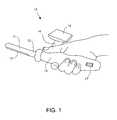
- FIG. 1is a perspective view of an embodiment of the probe of the present invention including a tactile sensor and electronic unit with a two-dimensional display attached to the probe handle.
- FIG. 2is a schematic diagram illustrating the relative position of a head with a tactile sensor during prostate examination.
- FIG. 3is a perspective view of real time pressure profiles which characterize cross-sections of the examined prostate obtained by pressing the probe against the prostate at the regions shown in FIG. 2 .
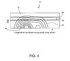
- FIG. 4is an illustration of a topographic picture for characterizing method of real time synthesizing a two-dimensional prostate image for the prostate device in accordance with an embodiment of the method of the present invention.
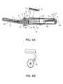
- FIG. 5Ais a cross-sectional side view of the probe including a tactile sensor with accelerometer based motion tracking system, and electronic unit with a two-dimensional display attached to the probe handle in accordance with a preferred embodiment of the device of the present invention.
- FIG. 5Bis a rear view of the probe shown in FIG. 5 A.
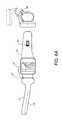
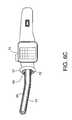
- FIG. 6Ais a side and rear view of the probe with a two-dimensional display folded into the review position.
- FIG. 6Bis a cross-sectional side view of the probe head including disposable pressure sensor head.
- FIG. 6Cis a cross-sectional view of the probe including a disposable pressure transducers.
- FIG. 7Ais a schematic diagram of an accelerometer based motion tracking system in accordance with an embodiment of the device of the present invention.
- FIG. 7Bis a schematic diagram of a magnetometer based motion tracking system in accordance with an embodiment of the device of the present invention.
- FIG. 8shows a flow chart describing steps for obtaining diagnostic information in accordance with an embodiment of the method of the present invention.
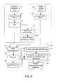
- FIG. 9is a schematic diagram illustrating the functional structure of the system in accordance with an embodiment of the device of the present invention.
- the method for transrectal imaging of the prostateis based on the technology of medical imaging described in U.S. Pat. Nos. 6,142,959; 5,922,018 and 5,836,894, which are incorporated herein by reference.
- This methodis referred to herein as mechanical imaging (MI).
- MImechanical imaging
- the essence of MIis the reconstruction of the internal structure of soft body tissues by measuring a surface stress pattern using a pressure sensing assembly.
- the pattern of mechanical stress and its changes as a function of applied pressure and timecontain comprehensive information on the mechanical properties and geometry of the internal structures of the body tissues.
- FIG. 1is a perspective view of a preferred embodiment of a prostate probe 10 sized to fit within the rectum.
- Probe 10comprises tactile sensor 13 mounted on head 11 .
- Tactile sensor 13generates signals in response to forces imposed on tactile sensor 13 as it is pressed against and moved over the prostate.
- Probe 10further comprises shaft 12 coupled to handle 16 .
- Control wheel 17is formed in handle 16 .
- Two-dimensional display 15is attached to handle 16 with display support 14 . Alternatively, the display can be built into the probe handle. The display or more than one display can be connected with or attached to, or mounted or laid onto a support or more than one support.
- An electronic unit(not shown FIG. 1) is incorporated inside the handle 16 .
- probe 10is inserted into the rectum and manipulated within the rectum using handle 16 .
- head 11is moved within the rectum and is pressed against the prostate.
- the electronic unitreceives electrical signals generated by tactile sensor 13 in response to forces imposed on tactile sensor 13 and calculates in real time a pressure profile which characterizes prostate cross-section.
- the pressure profileis visualized on display 15 .
- depth of insertion of probe 13is adjusted so that the prostate is located close to the center of tactile sensor 13 .
- the prostateis palpated in accordance with a predetermined trajectory to collect pressure response and motion data of probe 10 for calculating mechanical imaging of the prostate and its inner structure.
- tactile sensor 13includes a plurality of accelerometers to be used as a motion tracking system for determination of probe coordinates relative to the examined prostate.
- tactile sensor 13comprises a magnetometer based orientation tracking system system. Thereafter, pressure response data is used in real time to generate mechanical imaging results, as described in more detail below.
- probe 10can be applied against or adjacent to any gland or tissue such as by being applied through a natural opening of a subject or against a gland or tissue of a subject. Probe 10 can also be used during or after drug delivery and other types of treatments.
- FIG. 2is a schematic diagram illustrating the relative position of head 11 with tactile sensor 13 during prostate examination while periodically pressing probe 10 against prostate 23 in accordance with an embodiment of the method of the present invention.
- head 11 of probe 10is inserted into the rectum along axis Y to a predetermined depth without pressing against prostate 23 in order to minimize displacement of the prostate.
- head 11is pressed against prostate 23 at a site positioned on trajectory 24 to receive a real time cross-sectional view along axis Y of prostate 23 on display 15 .
- tactile sensor 13has a linear dimension greater than the axial length of prostate 23 .
- tactile sensor 13has a linear dimension exceeding the axial length of an average prostate, which typically varies between about 25 to about 45 mm.
- head 11 having tactile sensor 13 contacting prostate 23is moved along axis Y to adjust the location of prostate 23 close to the center of tactile sensor 13 .
- head 11is moved along trajectory 25 , 26 to laterally pass from one side of prostate 23 to the other while pressing against prostate 23 at a plurality of sites along trajectory 26 .
- Periodic pressing along trajectory 26provides prostate geometrical features along axis X.
- Another proceduresuch as oscillation of head 11 radially from sphincter 22 with simultaneous pressing against prostate 23 can be added to increase linear resolution of mechanical imaging along axis Y.
- FIG. 3perspective views of real time pressure profiles which characterize cross-sections 32 , 33 and 34 of the examined prostate obtained as the result of pressing the probe against the prostate are shown in coordinate system 21 with the origin placed at the sphincter 22 , as shown in FIG. 2 . All pressings against the prostate are represented in a vertical direction along axis Z.
- Cross-sections 32 , 33 and 34are calculated in real time for each pressing against the prostate and are represented on the display by lines of equal pressure A i in accordance with the procedure described below.
- FIG. 4illustrates a topographic picture for characterizing an embodiment of a method of real time synthesizing two-dimensional prostate image during pressing the probe against the prostate. It is advantageous to use a color scale for representation of the different pressure levels at pressure transducers of tactile sensor 13 for improved differentiation between pressure levels.
- the momentary pressure response datacan be visualized as color coded line 42 .
- the location of line 42 on topographic picture 41 with coordinates Y and F( ⁇ )represents the tactile sensor position relative to the examined prostate. Tactile sensor position and pressure on pressure transducers are changing as the head of the probe is pressed against the prostate.
- line 42is shifted along coordinate F( ⁇ ) on display 15 as shown by arrow 43 leaving a colored trace of the previous location of line 42 behind.
- Each line drawn on display 15gradually disappears losing its brightness and contrast within a time constant of about 1 second for improved visual perception of the different locations of line 42 .
- Coordinate Y on display 15is the longitudinal coordinate along tactile sensor 13 , as shown in FIG. 4 .
- Different parameterscan be used as the F( ⁇ ) coordinate.
- the coordinate F( ⁇ )is an average pressure from all pressure transducers or a portion of transducers of tactile sensor 13 .
- the coordinate F( ⁇ )is spatial coordinate Z (see FIG. 3 ).
- coordinate F( ⁇ )is the running time.
- Lines 44 of equal pressurecan be drawn on two-dimensional color display 15 .
- lines 44have a predetermined color to represent the prostate shape and lines 45 corresponding to a higher pressure have a predetermined second color to represent color regions with increased hardness inside the prostate. Pressure gradient analysis of pressure data can be used to determine the prostate shape and its inner structure.
- FIGS. 5A and 5B and FIGS. 6A and 6Billustrate in more detail a preferred embodiment of device 10 shown in FIG. 1 with tactile sensor 13 having an accelerometer based motion tracking system 50 .
- Accelerometer based motion tracking system 50comprises at least two three-axis accelerometers 53 , 55 which can be used to distinguish the gravity and inertia signal components.
- accelerometer 53can be located in handle 16 close to head 11 , so that during prostate examination accelerometer 53 is located near the sphincter and is more sensitive to angle orientation of probe 10 than to linear accelerations.
- Accelerometer 55preferably is located in handle 16 at a maximum distance from head 11 so that during prostate examination accelerometer 55 is more sensitive to angular acceleration of probe 10 than to linear acceleration. Additional accelerometers can be incorporated into shaft 12 or head 11 .
- Tactile sensor 13 of the device for real time mechanical imaging of the prostateis incorporated into head 11 .
- Tactile sensor 13comprises pressure transducer array 52 .
- Pressure transducer array 52preferably comprises a plurality of piezopolymer transducers, or micro-machined piezoresistive transducers, or capacitive pressure transducers covered by an elastic compound.
- Cover 56covers head 11 and shaft 12 .
- cover 56is flexible.
- Cover 56is held by fixing ring 57 to handle 16 .
- Cover 56can be removed from probe 10 after use and discarded before the next use. Thereafter, a new cover 56 can be placed over head 11 and shaft 12 before the next use for providing improved hygienics of the prostate examination.
- cover 56can be formed of thin elastic material such as latex.
- tactile sensor 13has a linear dimension exceeding the axial length of an average prostate, which typically varies between about 25 to about 45 mm.
- pressure sensing head 60includes pressure transducers 61 removably connected to handle 16 by electrical and mechanical connector 62 , as shown in FIG. 6 B. Pressure sensing head 60 including pressure transducers 61 can be disposable.
- probe head 65 unremovably connected to handle 16and pressure sensitive transducers 61 can be removable attached to handle 16 by electrical and mechanical connector 62 and lock 64 , as shown in FIG. 6 C.
- pressure sensitive transducers 61can be disposable.
- Electronic unit 54 , power source 58 and computer port 59preferably can be fitted in handle 16 .
- Electronic unit 54is coupled to tactile sensor 13 and accelerometer 53 , 55 .
- Power source 58is coupled to electronic unit 54 .
- Control wheel 17 mounted on handle 16can be used for producing “start” and “stop” signals during the prostate examination procedure, and for operating in review mode, as shown in FIG. 6 A.
- the physicianmay examine on display 15 stored data received from different patients or transfer data to an external computer.
- Display 15is connected to handle 16 through display support 14 equipped with hinge 60 to fix display 15 at two positions: an examination mode as shown in FIG. 5 and a review mode as shown in FIG. 6 A.
- display 15shows a real time pressure profile, which characterizes cross-section of examined prostate during pressing against the prostate with visualization of the prostate inner structure and prostate geometrical parameters.
- Accelerometer based motion tracking system 50includes at least two three-axis accelerometers 53 , 55 , which generate analog signals while moving the probe during prostate examination. As far as the distance 72 and relative orientation of accelerometers 53 and 55 are known, a three-dimensional trajectory can be evaluated by double integration of all analog signals received from accelerometers 53 , 55 and by restoration of probe head trajectory relative to a fixed point.
- an improved precision trajectorycan be determined by the following consideration. Since three-axis accelerometer 53 is located near the sphincter during the pressing against the prostate as shown by arrow 71 , accelerometer 53 with sensitivity axis along Y 1 can be used as a tilt sensor to determine elevation data 73 of the probe. Synchronous data analysis from accelerometers 53 , 55 with sensitivity axes Y 1 and Y 2 allows one to exclude the influence of angular acceleration on elevation data 73 .
- the vertical movement of the probecan be determined by double integration of signals from Z 1 of accelerometer 53 and Z 2 of accelerometer 55 taking into account changing in elevation of the probe. For more precise determination of coordinate Z to visualize prostate cross-section (as shown in FIG.
- the rotation data relative to axis 72 from tilt X 1 of accelerometer 53 and X 2 of accelerometer 55can be added.
- the differential signal from accelerometers X 1 and X 2are double integrated taking into account possible changing in rotation and elevation of the probe. It is useful to consider a point with maximum pressure on tactile sensor during pressing against the prostate as a starting spatial point with zero motion acceleration to calculate the trajectory of the probe relative to the point with maximum pressure in the tactile sensor.
- Magnetometer based orientation tracking system 75includes at least one triaxial magnetometer 74 which generates analog signals by changing the probe orientation relative to the Earth's magnetic field while moving the prove during prostate examination. As far as the probe orientation is known, a relative site of the pressing of the prostate can be determined. Alternatively, a motion tracking system can be based on a combination of accelerometers and magnetometers or a combination of gyroscopes and accelerometers.
- FIG. 8is a flow chart of the preferred method of calculating two-dimensional and three-dimensional prostate images.
- Pressure data from pressure transducer array of tactile sensor 13 and position data from motion tracking system 50are acquired in real time during prostate examination.
- Analog signals corresponding to pressures received from all pressure transducers 52 of tactile sensor 13form pressure data set 81 denoted by A(p i ,t), where p i is pressure signal for pressure transducer with number i at time t.
- Analog signals representing accelerations and angular orientation of the probe generated in response to movement of the probe during prostate examination received from motion tracking system 50form motion data set 82 denoted as B(b j ,t), where b j is signal corresponding to j accelerometer at time t.
- Accelerator based motion tracking system 50can receive responses from accelerometers used as tilt sensors, as described above.
- pressure data set 81is transformed into absolute pressure data P(i,t), where P is the force imposed on transducer i at time t, calculated in accordance with calibration data of pressure transducers 52 .
- data P(i,t)is processed by a conventional approximation method, such as described for example by T. J. Rivlin , An introduction to the Approximation of Functions , Dover Publications, Inc., New York (1969), to reduce noise originating from force measurement errors and from artefacts related to prostate movement.
- corrected data P(i,t)is transformed into a displayable format and represented on display 15 . All operations in block 86 take place in real time during prostate examination.
- the prostate examination data including said pressure data set P(i,t) and motion data set B(b j t)are accumulated in block 87 .
- the coordinates of each pressure transducer 52 of tactile sensor 13are calculated in the coordinate system with the origin at sphincter 22 (see FIG. 2) and the patterns of pressure responses P(x,y,z) of examined prostate are calculated.
- a pattern of pressure gradient responses represented by grad ⁇ P(x,y,z) ⁇is calculated from the pattern of pressure responses of the prostate by a conventional method such as described for example by D. Redfern and C. Campbel, The Matlab 5 Handbook , Springer-Verlag New York, Inc. (1998).
- the pattern of pressure responses of the prostate P(x,y,z)is corrected subject to distortions from the stiffer tissue inside the prostate which are revealed in the pressure gradient responses to determine prostate geometrical features.
- the prostate imageis synthesized from data generated in block 91 and data generated in block 90 . After having approximated smooth surfaces of equal pressures, it is possible to calculate hardness distribution inside the prostate using the pattern of pressure gradient responses.
- the surface of the examined prostatecan be obtained by calculating the first maximum of the second derivative of P(x,y,z) along normal to prostate surface by an iteration algorithm.
- the synthesized imagecan be displayed on a two-dimensional or linear display coupled to handle 16 .
- An average level of pressure applied to tactile sensor 13 , the position, real trajectory of pressure transducers and the predetermined pattern of trajectories for movement of the probecan be indicated in real time in a plane projection over the prostate image on the same display.
- FIG. 9is a schematic diagram of a preferred embodiment of electronic unit 54 mounted in handle 16 of probe 10 shown in FIG. 5.
- a plurality of pressure transducer elements 93forms tactile sensor 13 .
- Pressure sensing circuit 95is formed of a plurality of amplifiers, converters and integrators to amplify and convert respective signals generated by pressure transducer elements 93 for detecting the force imposed on each pressure transducer element 93 of tactile sensor 13 during prostate examination.
- a plurality of amplifiers 94amplify signals generated by respective accelerometers 53 , 54 of accelerator motion tracking system 50 , for detecting the position of the probe during pressing against the prostate and movement of the probe from one pressing site to another.
- the amplified signals from amplifiers 94 and 95are applied to multiplexer 96 .
- Multiplexed signalsare converted to digital signals by analog-to-digital converter 97 and fed to processor 98 .
- Processor 98is used for signal processing to calculate the position of each pressure transducer elements 93 during prostate examination, to approximate and correct mechanical images of the prostate and surrounding tissues, for separation and analysis of the prostate mechanical images, for determining the prostate geometrical features and mechanical features of prostate inner structures such as lesions, nodules, stiffer tissue and the like, and for prostate image synthesis, as described in the method illustrated in FIGS. 3, 4 and 8 .
- Display 15including a display screen and a controller, is connected to processor 98 , thereby displaying the real time prostate image, prostate examination process and the final results of the examination.
- Control wheel 17is connected to processor 98 through driver 99 for controlling the prostate examination process, data analysis and data review.
- Processor 98communicates with analog-to-digital converter 97 and multiplexer 96 for sending data and control signals.
- Storage unit 100can be used in electronic unit 54 for storing the results of the prostate examination generated by processor 98 having computer port 59 to transfer the stored data to an external computer.
Landscapes
- Health & Medical Sciences (AREA)
- Life Sciences & Earth Sciences (AREA)
- Engineering & Computer Science (AREA)
- Physics & Mathematics (AREA)
- General Health & Medical Sciences (AREA)
- Public Health (AREA)
- Veterinary Medicine (AREA)
- Pathology (AREA)
- Biomedical Technology (AREA)
- Heart & Thoracic Surgery (AREA)
- Medical Informatics (AREA)
- Molecular Biology (AREA)
- Surgery (AREA)
- Animal Behavior & Ethology (AREA)
- Biophysics (AREA)
- Geometry (AREA)
- Radiology & Medical Imaging (AREA)
- Hematology (AREA)
- Gynecology & Obstetrics (AREA)
- Reproductive Health (AREA)
- Human Computer Interaction (AREA)
- Nuclear Medicine, Radiotherapy & Molecular Imaging (AREA)
- Physiology (AREA)
- Dentistry (AREA)
- Oral & Maxillofacial Surgery (AREA)
- Measuring And Recording Apparatus For Diagnosis (AREA)
- Ultra Sonic Daignosis Equipment (AREA)
- Measurement Of The Respiration, Hearing Ability, Form, And Blood Characteristics Of Living Organisms (AREA)
Abstract
Description
Claims (33)
Priority Applications (6)
| Application Number | Priority Date | Filing Date | Title |
|---|---|---|---|
| US09/819,419US6569108B2 (en) | 2001-03-28 | 2001-03-28 | Real time mechanical imaging of the prostate |
| JP2002580806AJP4177116B2 (en) | 2001-03-28 | 2002-03-28 | Real-time mechanical imaging of the prostate |
| CA 2440849CA2440849A1 (en) | 2001-03-28 | 2002-03-28 | Real time mechanical imaging of the prostate |
| PCT/US2002/009296WO2002082998A1 (en) | 2001-03-28 | 2002-03-28 | Real time mechanical imaging of the prostate |
| EP20100177745EP2316335A1 (en) | 2001-03-28 | 2002-03-28 | Real-time mechanical imaging of the prostate |
| EP02761982AEP1379171A4 (en) | 2001-03-28 | 2002-03-28 | Real time mechanical imaging of the prostate |
Applications Claiming Priority (1)
| Application Number | Priority Date | Filing Date | Title |
|---|---|---|---|
| US09/819,419US6569108B2 (en) | 2001-03-28 | 2001-03-28 | Real time mechanical imaging of the prostate |
Publications (2)
| Publication Number | Publication Date |
|---|---|
| US20020143275A1 US20020143275A1 (en) | 2002-10-03 |
| US6569108B2true US6569108B2 (en) | 2003-05-27 |
Family
ID=25228101
Family Applications (1)
| Application Number | Title | Priority Date | Filing Date |
|---|---|---|---|
| US09/819,419Expired - LifetimeUS6569108B2 (en) | 2001-03-28 | 2001-03-28 | Real time mechanical imaging of the prostate |
Country Status (5)
| Country | Link |
|---|---|
| US (1) | US6569108B2 (en) |
| EP (2) | EP1379171A4 (en) |
| JP (1) | JP4177116B2 (en) |
| CA (1) | CA2440849A1 (en) |
| WO (1) | WO2002082998A1 (en) |
Cited By (72)
| Publication number | Priority date | Publication date | Assignee | Title |
|---|---|---|---|---|
| US20050228617A1 (en)* | 2004-04-02 | 2005-10-13 | Scott Kerwin | Methods and systems for tracking probe use |
| US20050256387A1 (en)* | 2002-01-29 | 2005-11-17 | Nihon University | Elasticity measuring device for biological tissue |
| US20060156833A1 (en)* | 2005-01-18 | 2006-07-20 | Chih-Ching Hsieh | Low-cost high precision twisting measuring device |
| US20070156067A1 (en)* | 2005-12-29 | 2007-07-05 | Dharmesh Dubey | Cervimeter |
| US20070239197A1 (en)* | 2006-04-10 | 2007-10-11 | Intrapartum Ventures, Llc | Method for cervical dilation and/or measurement |
| US20070255185A1 (en)* | 2005-12-29 | 2007-11-01 | Intrapartum Ventures, Llc. | Cervical dilation measurement apparatus |
| US20080221484A1 (en)* | 2005-05-06 | 2008-09-11 | Sarvazyan Armen P | Method and a dual-array transducer probe for real time mechanical imaging of prostate |
| WO2008131557A1 (en)* | 2007-05-01 | 2008-11-06 | Urodynamix Technologies Ltd. | Apparatus and methods for evaluating physiological conditions of tissue |
| US20090005707A1 (en)* | 2005-05-06 | 2009-01-01 | Sarvazyan Armen P | Method and device for real time mechanical imaging of prostate |
| US20090018444A1 (en)* | 2003-05-30 | 2009-01-15 | Takashi Osaka | Utrasound probe and ultrasound elasticity imaging apparatus |
| US20090143679A1 (en)* | 2007-09-28 | 2009-06-04 | The University Of British Columbia | Method and apparatus for imaging the mechanical properties of tissue from an endocavity |
| US20090182231A1 (en)* | 2004-10-06 | 2009-07-16 | Guided Therapy Systems, L.L.C. | Method and system for treating acne and sebaceous glands |
| US20090196459A1 (en)* | 2008-02-01 | 2009-08-06 | Perceptron, Inc. | Image manipulation and processing techniques for remote inspection device |
| US20110054357A1 (en)* | 2009-09-02 | 2011-03-03 | Artann Laboratories, Inc. | Methods for characterizing vaginal tissue elasticity |
| US20110196263A1 (en)* | 2009-09-02 | 2011-08-11 | Artann Laboratories, Inc. | Methods for assessment of pelvic organ conditions affecting the vagina |
| CN101317774B (en)* | 2003-05-30 | 2013-03-06 | 株式会社日立医药 | Ultrasonic diagnostic equipment |
| US8915870B2 (en) | 2004-10-06 | 2014-12-23 | Guided Therapy Systems, Llc | Method and system for treating stretch marks |
| US8915853B2 (en) | 2004-10-06 | 2014-12-23 | Guided Therapy Systems, Llc | Methods for face and neck lifts |
| US8932224B2 (en) | 2004-10-06 | 2015-01-13 | Guided Therapy Systems, Llc | Energy based hyperhidrosis treatment |
| US9011336B2 (en) | 2004-09-16 | 2015-04-21 | Guided Therapy Systems, Llc | Method and system for combined energy therapy profile |
| US9011337B2 (en) | 2011-07-11 | 2015-04-21 | Guided Therapy Systems, Llc | Systems and methods for monitoring and controlling ultrasound power output and stability |
| US9032818B2 (en) | 2012-07-05 | 2015-05-19 | Nextinput, Inc. | Microelectromechanical load sensor and methods of manufacturing the same |
| US9039617B2 (en) | 2009-11-24 | 2015-05-26 | Guided Therapy Systems, Llc | Methods and systems for generating thermal bubbles for improved ultrasound imaging and therapy |
| US9114247B2 (en) | 2004-09-16 | 2015-08-25 | Guided Therapy Systems, Llc | Method and system for ultrasound treatment with a multi-directional transducer |
| US9149658B2 (en) | 2010-08-02 | 2015-10-06 | Guided Therapy Systems, Llc | Systems and methods for ultrasound treatment |
| US9216276B2 (en) | 2007-05-07 | 2015-12-22 | Guided Therapy Systems, Llc | Methods and systems for modulating medicants using acoustic energy |
| US9263663B2 (en) | 2012-04-13 | 2016-02-16 | Ardent Sound, Inc. | Method of making thick film transducer arrays |
| US9272162B2 (en) | 1997-10-14 | 2016-03-01 | Guided Therapy Systems, Llc | Imaging, therapy, and temperature monitoring ultrasonic method |
| US9283410B2 (en) | 2004-10-06 | 2016-03-15 | Guided Therapy Systems, L.L.C. | System and method for fat and cellulite reduction |
| US9283409B2 (en) | 2004-10-06 | 2016-03-15 | Guided Therapy Systems, Llc | Energy based fat reduction |
| US9320537B2 (en) | 2004-10-06 | 2016-04-26 | Guided Therapy Systems, Llc | Methods for noninvasive skin tightening |
| US9452302B2 (en) | 2011-07-10 | 2016-09-27 | Guided Therapy Systems, Llc | Systems and methods for accelerating healing of implanted material and/or native tissue |
| US9487388B2 (en) | 2012-06-21 | 2016-11-08 | Nextinput, Inc. | Ruggedized MEMS force die |
| US9504446B2 (en) | 2010-08-02 | 2016-11-29 | Guided Therapy Systems, Llc | Systems and methods for coupling an ultrasound source to tissue |
| US9510802B2 (en) | 2012-09-21 | 2016-12-06 | Guided Therapy Systems, Llc | Reflective ultrasound technology for dermatological treatments |
| US9694212B2 (en) | 2004-10-06 | 2017-07-04 | Guided Therapy Systems, Llc | Method and system for ultrasound treatment of skin |
| US9700340B2 (en) | 2004-10-06 | 2017-07-11 | Guided Therapy Systems, Llc | System and method for ultra-high frequency ultrasound treatment |
| US9726647B2 (en) | 2015-03-17 | 2017-08-08 | Hemosonics, Llc | Determining mechanical properties via ultrasound-induced resonance |
| US9827449B2 (en) | 2004-10-06 | 2017-11-28 | Guided Therapy Systems, L.L.C. | Systems for treating skin laxity |
| US9867668B2 (en) | 2008-10-20 | 2018-01-16 | The Johns Hopkins University | Environment property estimation and graphical display |
| US9902611B2 (en) | 2014-01-13 | 2018-02-27 | Nextinput, Inc. | Miniaturized and ruggedized wafer level MEMs force sensors |
| US10004450B2 (en) | 2016-05-03 | 2018-06-26 | Texas Medical Center | Tactile sensing device for lumbar punctures |
| US10039938B2 (en) | 2004-09-16 | 2018-08-07 | Guided Therapy Systems, Llc | System and method for variable depth ultrasound treatment |
| US10383610B2 (en) | 2017-10-27 | 2019-08-20 | Intuitap Medical, Inc. | Tactile sensing and needle guidance device |
| US10420960B2 (en) | 2013-03-08 | 2019-09-24 | Ulthera, Inc. | Devices and methods for multi-focus ultrasound therapy |
| US10466119B2 (en) | 2015-06-10 | 2019-11-05 | Nextinput, Inc. | Ruggedized wafer level MEMS force sensor with a tolerance trench |
| US10537304B2 (en) | 2008-06-06 | 2020-01-21 | Ulthera, Inc. | Hand wand for ultrasonic cosmetic treatment and imaging |
| US10561862B2 (en) | 2013-03-15 | 2020-02-18 | Guided Therapy Systems, Llc | Ultrasound treatment device and methods of use |
| US10603521B2 (en) | 2014-04-18 | 2020-03-31 | Ulthera, Inc. | Band transducer ultrasound therapy |
| US10864385B2 (en) | 2004-09-24 | 2020-12-15 | Guided Therapy Systems, Llc | Rejuvenating skin by heating tissue for cosmetic treatment of the face and body |
| US10962524B2 (en) | 2011-02-15 | 2021-03-30 | HomoSonics LLC | Characterization of blood hemostasis and oxygen transport parameters |
| US10962427B2 (en) | 2019-01-10 | 2021-03-30 | Nextinput, Inc. | Slotted MEMS force sensor |
| US11207548B2 (en) | 2004-10-07 | 2021-12-28 | Guided Therapy Systems, L.L.C. | Ultrasound probe for treating skin laxity |
| US11221263B2 (en) | 2017-07-19 | 2022-01-11 | Nextinput, Inc. | Microelectromechanical force sensor having a strain transfer layer arranged on the sensor die |
| US11224895B2 (en) | 2016-01-18 | 2022-01-18 | Ulthera, Inc. | Compact ultrasound device having annular ultrasound array peripherally electrically connected to flexible printed circuit board and method of assembly thereof |
| US11235179B2 (en) | 2004-10-06 | 2022-02-01 | Guided Therapy Systems, Llc | Energy based skin gland treatment |
| US11241218B2 (en) | 2016-08-16 | 2022-02-08 | Ulthera, Inc. | Systems and methods for cosmetic ultrasound treatment of skin |
| US11243125B2 (en) | 2017-02-09 | 2022-02-08 | Nextinput, Inc. | Integrated piezoresistive and piezoelectric fusion force sensor |
| US11243126B2 (en) | 2017-07-27 | 2022-02-08 | Nextinput, Inc. | Wafer bonded piezoresistive and piezoelectric force sensor and related methods of manufacture |
| US11255737B2 (en) | 2017-02-09 | 2022-02-22 | Nextinput, Inc. | Integrated digital force sensors and related methods of manufacture |
| US11338156B2 (en) | 2004-10-06 | 2022-05-24 | Guided Therapy Systems, Llc | Noninvasive tissue tightening system |
| US11385108B2 (en) | 2017-11-02 | 2022-07-12 | Nextinput, Inc. | Sealed force sensor with etch stop layer |
| US11423686B2 (en) | 2017-07-25 | 2022-08-23 | Qorvo Us, Inc. | Integrated fingerprint and force sensor |
| US11579028B2 (en) | 2017-10-17 | 2023-02-14 | Nextinput, Inc. | Temperature coefficient of offset compensation for force sensor and strain gauge |
| US11717661B2 (en) | 2007-05-07 | 2023-08-08 | Guided Therapy Systems, Llc | Methods and systems for ultrasound assisted delivery of a medicant to tissue |
| US11724133B2 (en) | 2004-10-07 | 2023-08-15 | Guided Therapy Systems, Llc | Ultrasound probe for treatment of skin |
| US11874185B2 (en) | 2017-11-16 | 2024-01-16 | Nextinput, Inc. | Force attenuator for force sensor |
| US11883688B2 (en) | 2004-10-06 | 2024-01-30 | Guided Therapy Systems, Llc | Energy based fat reduction |
| US11944849B2 (en) | 2018-02-20 | 2024-04-02 | Ulthera, Inc. | Systems and methods for combined cosmetic treatment of cellulite with ultrasound |
| US12076591B2 (en) | 2018-01-26 | 2024-09-03 | Ulthera, Inc. | Systems and methods for simultaneous multi-focus ultrasound therapy in multiple dimensions |
| US12102473B2 (en) | 2008-06-06 | 2024-10-01 | Ulthera, Inc. | Systems for ultrasound treatment |
| US12377293B2 (en) | 2019-07-15 | 2025-08-05 | Ulthera, Inc. | Systems and methods for measuring elasticity with imaging of ultrasound multi-focus shearwaves in multiple dimensions |
Families Citing this family (34)
| Publication number | Priority date | Publication date | Assignee | Title |
|---|---|---|---|---|
| GB0313794D0 (en)* | 2003-06-14 | 2003-07-23 | Univ Dundee | Tactile sensor assembly |
| AT9316U1 (en)* | 2005-05-04 | 2007-08-15 | Ami Gmbh | DEVICE FOR USE IN THE LIGATURE OF INTRAMULAR ARTERIES |
| US20060276726A1 (en)* | 2005-06-03 | 2006-12-07 | Holsten Henry E | Tissue tension detection system |
| GB0520596D0 (en)* | 2005-10-11 | 2005-11-16 | Sussex Dev Services Llp | Location and stabilization device |
| US20070293792A1 (en)* | 2006-06-15 | 2007-12-20 | Sliwa John W | Prostate BPH and tumor detector also useable on other tissues |
| DE102007010046A1 (en)* | 2007-03-01 | 2008-09-04 | Siemens Ag | Prostate cancer detection apparatus |
| US20100137845A1 (en) | 2008-12-03 | 2010-06-03 | Immersion Corporation | Tool Having Multiple Feedback Devices |
| US8551002B2 (en)* | 2008-12-12 | 2013-10-08 | Immersion Corporation | Spatial array of sensors mounted on a tool |
| US8652046B2 (en)* | 2009-01-15 | 2014-02-18 | Immersion Corporation | Palpation algorithms for computer-augmented hand tools |
| FR2966717B1 (en) | 2010-10-27 | 2013-07-26 | Commissariat Energie Atomique | STERILE PROTECTION WITH LIGHT GUIDES FOR MEDICAL PROBE AND METHOD OF MAKING SAME |
| JP5826478B2 (en)* | 2010-10-28 | 2015-12-02 | 日立アロカメディカル株式会社 | Tissue insertion type ultrasonic probe |
| KR101223209B1 (en) | 2010-12-10 | 2013-01-17 | 한국과학기술원 | Mechanical Loading based Apparatus and Method with compensating geometry information for measurement of diagnostic property of matter |
| EP2491865A1 (en)* | 2011-02-24 | 2012-08-29 | Samsung Medison Co., Ltd. | Ultrasound system for providing image indicator |
| US20150112230A1 (en)* | 2011-11-28 | 2015-04-23 | Remendium Labs Llc | Treatment of male urinary incontinence and sexual dysfunction |
| CN109259803B (en)* | 2011-12-16 | 2021-04-16 | 佩里梅特里克斯有限责任公司 | System and method for determining structural characteristics of an object |
| KR20140008728A (en)* | 2012-07-11 | 2014-01-22 | 삼성전자주식회사 | Palpation apparatus and method using robot |
| KR101495131B1 (en) | 2013-03-29 | 2015-02-26 | 김범기 | A prostate cancer diagnosis system using a pressure sensor and a prostate cancer diagnosis method thereof |
| US10925579B2 (en)* | 2014-11-05 | 2021-02-23 | Otsuka Medical Devices Co., Ltd. | Systems and methods for real-time tracking of a target tissue using imaging before and during therapy delivery |
| US20170065249A1 (en)* | 2015-09-08 | 2017-03-09 | Advanced Tactile Imaging Inc. | Methods and probes for vaginal tactile and ultrasound imaging |
| RU2615727C2 (en)* | 2015-09-29 | 2017-04-07 | Общество с ограниченной ответственностью "Уровест" | Uroflowmeter |
| USD801526S1 (en) | 2015-09-30 | 2017-10-31 | Sussex Development Services Llp | Rectal obturator |
| US11395593B2 (en) | 2016-09-14 | 2022-07-26 | Mor Research Applications Ltd. | Device, system and method for detecting irregularities in soft tissue |
| GB2559405A (en)* | 2017-02-06 | 2018-08-08 | Owlstone Med Ltd | Improvements in or relating to preparation of subjects for medical or veterinary examination |
| US11510646B2 (en)* | 2017-04-05 | 2022-11-29 | Bk Medical Aps | Ultrasound imaging system probe cable and connector |
| WO2019084469A1 (en) | 2017-10-27 | 2019-05-02 | Renovia Inc. | Devices, systems, and methods for training pelvic floor muscles |
| MX2020006928A (en)* | 2017-12-30 | 2020-09-09 | Perimetrics Llc | Determination of structural characteristics of an object. |
| EP3873344A4 (en) | 2018-10-30 | 2022-06-29 | Renovia Inc. | Devices, systems, and methods for monitoring bladder function |
| JP7171948B2 (en)* | 2019-05-17 | 2022-11-15 | コーニンクレッカ フィリップス エヌ ヴェ | Ultrasound system and method for tracking movement of an object |
| DE102019007290A1 (en)* | 2019-10-21 | 2021-04-22 | Karl Storz Se & Co. Kg | Sensor-based surgery set and procedure |
| US12193830B2 (en) | 2020-09-22 | 2025-01-14 | Advanced Tactile Imaging Inc. | Method for characterization of the female pelvic floor with a biomechanical integrity score |
| ES2921203B2 (en) | 2021-02-15 | 2023-04-18 | Fundacion Para La Investigacion Del Hospital Univ Y Politecnico La Fe De La Comunidad Valenciana | Device for palpation of the prostate |
| JP2023148439A (en)* | 2022-03-30 | 2023-10-13 | 富士フイルム株式会社 | Ultrasonic diagnostic device and operation method thereof |
| KR20240120062A (en)* | 2023-01-31 | 2024-08-07 | 계명대학교 산학협력단 | Prostate cancer diagnosis system and method capable of predicting prostage cancer by digitally converting user's tactile sensation |
| CN116549008A (en)* | 2023-05-30 | 2023-08-08 | 无锡海斯凯尔医学技术有限公司 | Ultrasonic signal-based elastography system and method of use |
Citations (35)
| Publication number | Priority date | Publication date | Assignee | Title |
|---|---|---|---|---|
| US4250894A (en) | 1978-11-14 | 1981-02-17 | Yeda Research & Development Co., Ltd. | Instrument for viscoelastic measurement |
| US4423738A (en) | 1977-11-04 | 1984-01-03 | Sri International | Noninvasive blood pressure monitoring transducer |
| US4580574A (en) | 1983-08-12 | 1986-04-08 | Benjamin Gavish | Method and device for non-invasively monitoring the instantaneous fluctuations in the viscoelastic-related properties of a living tissue |
| US4711248A (en) | 1983-12-01 | 1987-12-08 | Biokinetics, Inc. | Physiological pressure monitor |
| US4722348A (en) | 1985-09-17 | 1988-02-02 | Sentron V.O.F. | Catheter tip pressure transducer |
| US4799491A (en) | 1986-11-06 | 1989-01-24 | Sri International | Blood pressure monitoring method and apparatus |
| US4802488A (en) | 1986-11-06 | 1989-02-07 | Sri International | Blood pressure monitoring method and apparatus |
| US4809710A (en) | 1988-01-11 | 1989-03-07 | Williamson Jeffrey L | Multilumen manometer catheter |
| US4860761A (en) | 1985-04-12 | 1989-08-29 | Omron Tateisi Electronics Co. | Pulse wave detecting apparatus for blood pressure measurement |
| US4869265A (en) | 1987-04-03 | 1989-09-26 | Western Clinical Engineering Ltd. | Biomedical pressure transducer |
| US4893634A (en) | 1987-06-01 | 1990-01-16 | Problamnaya Nauchno-Issledovatelskaya Laboratoria Vspomogatelnogo Krovoobraschenia | Device for cleansing measuring pressure in the colon |
| US4947851A (en) | 1988-02-19 | 1990-08-14 | Institute for Physical Chemistry | Method and device for acoustic testing of elasticity of biological tissues |
| US5067491A (en) | 1989-12-08 | 1991-11-26 | Becton, Dickinson And Company | Barrier coating on blood contacting devices |
| US5078142A (en) | 1989-11-21 | 1992-01-07 | Fischer Imaging Corporation | Precision mammographic needle biopsy system |
| US5099848A (en) | 1990-11-02 | 1992-03-31 | University Of Rochester | Method and apparatus for breast imaging and tumor detection using modal vibration analysis |
| US5107837A (en) | 1989-11-17 | 1992-04-28 | Board Of Regents, University Of Texas | Method and apparatus for measurement and imaging of tissue compressibility or compliance |
| US5115808A (en) | 1988-02-19 | 1992-05-26 | Institute Of General And Physical Chemistry | Method and device for noninvasive acoustic testing of elasticity of soft biological tissues |
| US5170790A (en) | 1990-04-06 | 1992-12-15 | Technomed International | Arm having an end movable in translation, and therapeutic treatment apparatus constituting an application thereof |
| US5178148A (en) | 1990-04-06 | 1993-01-12 | Technomed International | Method of automatically measuring the volume of a tumor or of a gland, in particular the prostate, a measuring device, and a method and apparatus constituting and application thereof |
| US5247937A (en) | 1989-11-17 | 1993-09-28 | Board Of Regents, The University Of Texas System | Transaxial compression technique for sound velocity estimation |
| US5265612A (en) | 1992-12-21 | 1993-11-30 | Medical Biophysics International | Intracavity ultrasonic device for elasticity imaging |
| US5278776A (en) | 1991-05-21 | 1994-01-11 | Jack Fisher | System and method for the measurement of mechanical properties of elastic materials |
| US5293870A (en) | 1989-11-17 | 1994-03-15 | Board Of Regents The University Of Texas System | Method and apparatus for elastographic measurement and imaging |
| WO1994014375A1 (en) | 1992-12-21 | 1994-07-07 | Medical Biophysics International | Method and apparatus for elasticity imaging |
| WO1995002992A1 (en) | 1993-07-22 | 1995-02-02 | Uromed Corporation | Characteristic of body part determined |
| US5402793A (en) | 1993-11-19 | 1995-04-04 | Advanced Technology Laboratories, Inc. | Ultrasonic transesophageal probe for the imaging and diagnosis of multiple scan planes |
| US5474070A (en) | 1989-11-17 | 1995-12-12 | The Board Of Regents Of The University Of Texas System | Method and apparatus for elastographic measurement and imaging |
| US5522399A (en) | 1994-09-26 | 1996-06-04 | Wilk; Peter J. | Catheterization device and associated assembly |
| US5526820A (en) | 1994-09-19 | 1996-06-18 | Myelotec, Inc. | Catheter with integral pressure sensor |
| US5785663A (en)* | 1992-12-21 | 1998-07-28 | Artann Corporation | Method and device for mechanical imaging of prostate |
| US5836894A (en)* | 1992-12-21 | 1998-11-17 | Artann Laboratories | Apparatus for measuring mechanical parameters of the prostate and for imaging the prostate using such parameters |
| US5922018A (en)* | 1992-12-21 | 1999-07-13 | Artann Corporation | Method for using a transrectal probe to mechanically image the prostate gland |
| US5989199A (en)* | 1996-11-27 | 1999-11-23 | Assurance Medical, Inc. | Tissue examination |
| US6063031A (en)* | 1997-10-14 | 2000-05-16 | Assurance Medical, Inc. | Diagnosis and treatment of tissue with instruments |
| US6142959A (en) | 1992-12-21 | 2000-11-07 | Armed L.L.C. | Device for palpation and mechanical imaging of the prostate |
Family Cites Families (8)
| Publication number | Priority date | Publication date | Assignee | Title |
|---|---|---|---|---|
| US4132224A (en)* | 1977-01-12 | 1979-01-02 | Randolph Robert G | Durometer for indentible tissue and the like |
| US5678565A (en)* | 1992-12-21 | 1997-10-21 | Artann Corporation | Ultrasonic elasticity imaging method and device |
| JPH0821732A (en)* | 1994-07-05 | 1996-01-23 | Data Tec:Kk | Attitude, azimuth, and position measuring apparatus |
| US6179790B1 (en)* | 1997-10-20 | 2001-01-30 | Assurance Medical, Inc. | Layer of material for use with tissue examination device |
| AU6028299A (en)* | 1998-09-08 | 2000-03-27 | Catholic University Of America, The | Method and system for tactile imaging for breast cancer examination and detection of prostate cancer |
| US6190334B1 (en)* | 1999-05-24 | 2001-02-20 | Rbp, Inc. | Method and apparatus for the imaging of tissue |
| US6595933B2 (en)* | 2000-03-31 | 2003-07-22 | Artann Laboratories | Self-palpation device for examination of breast with 3-D positioning system |
| NL1018864C2 (en)* | 2001-08-31 | 2003-03-03 | Technologiestichting Stw | Device and method for generating three-dimensional images with tissue hardness information. |
- 2001
- 2001-03-28USUS09/819,419patent/US6569108B2/ennot_activeExpired - Lifetime
- 2002
- 2002-03-28EPEP02761982Apatent/EP1379171A4/ennot_activeWithdrawn
- 2002-03-28JPJP2002580806Apatent/JP4177116B2/ennot_activeExpired - Fee Related
- 2002-03-28WOPCT/US2002/009296patent/WO2002082998A1/enactiveApplication Filing
- 2002-03-28CACA 2440849patent/CA2440849A1/ennot_activeAbandoned
- 2002-03-28EPEP20100177745patent/EP2316335A1/ennot_activeWithdrawn
Patent Citations (37)
| Publication number | Priority date | Publication date | Assignee | Title |
|---|---|---|---|---|
| US4423738A (en) | 1977-11-04 | 1984-01-03 | Sri International | Noninvasive blood pressure monitoring transducer |
| US4250894A (en) | 1978-11-14 | 1981-02-17 | Yeda Research & Development Co., Ltd. | Instrument for viscoelastic measurement |
| US4580574A (en) | 1983-08-12 | 1986-04-08 | Benjamin Gavish | Method and device for non-invasively monitoring the instantaneous fluctuations in the viscoelastic-related properties of a living tissue |
| US4711248A (en) | 1983-12-01 | 1987-12-08 | Biokinetics, Inc. | Physiological pressure monitor |
| US4860761A (en) | 1985-04-12 | 1989-08-29 | Omron Tateisi Electronics Co. | Pulse wave detecting apparatus for blood pressure measurement |
| US4722348A (en) | 1985-09-17 | 1988-02-02 | Sentron V.O.F. | Catheter tip pressure transducer |
| US4802488A (en) | 1986-11-06 | 1989-02-07 | Sri International | Blood pressure monitoring method and apparatus |
| US4799491A (en) | 1986-11-06 | 1989-01-24 | Sri International | Blood pressure monitoring method and apparatus |
| US4869265A (en) | 1987-04-03 | 1989-09-26 | Western Clinical Engineering Ltd. | Biomedical pressure transducer |
| US4893634A (en) | 1987-06-01 | 1990-01-16 | Problamnaya Nauchno-Issledovatelskaya Laboratoria Vspomogatelnogo Krovoobraschenia | Device for cleansing measuring pressure in the colon |
| US4809710A (en) | 1988-01-11 | 1989-03-07 | Williamson Jeffrey L | Multilumen manometer catheter |
| US5115808A (en) | 1988-02-19 | 1992-05-26 | Institute Of General And Physical Chemistry | Method and device for noninvasive acoustic testing of elasticity of soft biological tissues |
| US4947851A (en) | 1988-02-19 | 1990-08-14 | Institute for Physical Chemistry | Method and device for acoustic testing of elasticity of biological tissues |
| US5474070A (en) | 1989-11-17 | 1995-12-12 | The Board Of Regents Of The University Of Texas System | Method and apparatus for elastographic measurement and imaging |
| US5107837A (en) | 1989-11-17 | 1992-04-28 | Board Of Regents, University Of Texas | Method and apparatus for measurement and imaging of tissue compressibility or compliance |
| US5247937A (en) | 1989-11-17 | 1993-09-28 | Board Of Regents, The University Of Texas System | Transaxial compression technique for sound velocity estimation |
| US5293870A (en) | 1989-11-17 | 1994-03-15 | Board Of Regents The University Of Texas System | Method and apparatus for elastographic measurement and imaging |
| US5078142A (en) | 1989-11-21 | 1992-01-07 | Fischer Imaging Corporation | Precision mammographic needle biopsy system |
| US5067491A (en) | 1989-12-08 | 1991-11-26 | Becton, Dickinson And Company | Barrier coating on blood contacting devices |
| US5170790A (en) | 1990-04-06 | 1992-12-15 | Technomed International | Arm having an end movable in translation, and therapeutic treatment apparatus constituting an application thereof |
| US5178148A (en) | 1990-04-06 | 1993-01-12 | Technomed International | Method of automatically measuring the volume of a tumor or of a gland, in particular the prostate, a measuring device, and a method and apparatus constituting and application thereof |
| US5099848A (en) | 1990-11-02 | 1992-03-31 | University Of Rochester | Method and apparatus for breast imaging and tumor detection using modal vibration analysis |
| US5278776A (en) | 1991-05-21 | 1994-01-11 | Jack Fisher | System and method for the measurement of mechanical properties of elastic materials |
| US5524636A (en)* | 1992-12-21 | 1996-06-11 | Artann Corporation Dba Artann Laboratories | Method and apparatus for elasticity imaging |
| US5922018A (en)* | 1992-12-21 | 1999-07-13 | Artann Corporation | Method for using a transrectal probe to mechanically image the prostate gland |
| US6142959A (en) | 1992-12-21 | 2000-11-07 | Armed L.L.C. | Device for palpation and mechanical imaging of the prostate |
| US5836894A (en)* | 1992-12-21 | 1998-11-17 | Artann Laboratories | Apparatus for measuring mechanical parameters of the prostate and for imaging the prostate using such parameters |
| WO1994014375A1 (en) | 1992-12-21 | 1994-07-07 | Medical Biophysics International | Method and apparatus for elasticity imaging |
| US5785663A (en)* | 1992-12-21 | 1998-07-28 | Artann Corporation | Method and device for mechanical imaging of prostate |
| US5265612A (en) | 1992-12-21 | 1993-11-30 | Medical Biophysics International | Intracavity ultrasonic device for elasticity imaging |
| US5423332A (en) | 1993-07-22 | 1995-06-13 | Uromed Corporation | Device and method for determining the mass or volume of a body part |
| WO1995002992A1 (en) | 1993-07-22 | 1995-02-02 | Uromed Corporation | Characteristic of body part determined |
| US5402793A (en) | 1993-11-19 | 1995-04-04 | Advanced Technology Laboratories, Inc. | Ultrasonic transesophageal probe for the imaging and diagnosis of multiple scan planes |
| US5526820A (en) | 1994-09-19 | 1996-06-18 | Myelotec, Inc. | Catheter with integral pressure sensor |
| US5522399A (en) | 1994-09-26 | 1996-06-04 | Wilk; Peter J. | Catheterization device and associated assembly |
| US5989199A (en)* | 1996-11-27 | 1999-11-23 | Assurance Medical, Inc. | Tissue examination |
| US6063031A (en)* | 1997-10-14 | 2000-05-16 | Assurance Medical, Inc. | Diagnosis and treatment of tissue with instruments |
Non-Patent Citations (8)
| Title |
|---|
| Learner et al., "Sono Elasticity: Medical Elasticity Images Derived from Ultrasound Signals in Mechanically Vibrated Targets," Acoustical Imaging, vol. 16, 317 (1988). |
| Littrup et al., "The Benefit and Cost of Prostate Cancer Early Detection," CA Cancer Journ. for Clinicians, vol. 43, pp. 134-149 (1993). |
| R. Rubens et al., "Sonoelasticity Imaging of Prostate Cancer: In Vitro Results," Journal of Radiology, 195, No. 2, May 1995, pp. 379-383. |
| Sarvazyan et al., "A New Philosophy of Medical Imaging," Medical Hypotheses, vol. 36, pp. 327-335 (1991). |
| Sarvazyan et al., "Biophysical Bases of Elasticity Imaging," Acoustical Imaging, vol. 21, pp. 223-240 (1995). |
| Smith et al., "Interexaminer Variability of Digital Rectal Examination in Detecting Prostate Cancer," Urology, vol. 45, pp. 70-74 (1995). |
| T.A. Krouskop et al., "A Pulsed Doppler Ultrasonic System for Making Non-Invasive Measurement of Mechanical Properties of Soft Tissue," 24 J. Rehab. Res. Dev., vol. 24, 1 (1987). |
| Y. Yamakoshi et al., "Ultrasonic Imaging of Internal Vibration of Soft Tissue Under Forced Vibration," IEEE Transactions on Ultrasonics Ferroelectrics and Frequency Control, vol. 7, No. 2, p. 45 (1990). |
Cited By (163)
| Publication number | Priority date | Publication date | Assignee | Title |
|---|---|---|---|---|
| US9272162B2 (en) | 1997-10-14 | 2016-03-01 | Guided Therapy Systems, Llc | Imaging, therapy, and temperature monitoring ultrasonic method |
| US20050256387A1 (en)* | 2002-01-29 | 2005-11-17 | Nihon University | Elasticity measuring device for biological tissue |
| US7648470B2 (en)* | 2002-01-29 | 2010-01-19 | Nihon University | Elasticity measuring device for biological tissue |
| CN101317774B (en)* | 2003-05-30 | 2013-03-06 | 株式会社日立医药 | Ultrasonic diagnostic equipment |
| US8007438B2 (en)* | 2003-05-30 | 2011-08-30 | Hitachi Medical Corporation | Ultrasound probe and ultrasound elasticity imaging apparatus |
| US20090018444A1 (en)* | 2003-05-30 | 2009-01-15 | Takashi Osaka | Utrasound probe and ultrasound elasticity imaging apparatus |
| US20050228617A1 (en)* | 2004-04-02 | 2005-10-13 | Scott Kerwin | Methods and systems for tracking probe use |
| US9114247B2 (en) | 2004-09-16 | 2015-08-25 | Guided Therapy Systems, Llc | Method and system for ultrasound treatment with a multi-directional transducer |
| US10039938B2 (en) | 2004-09-16 | 2018-08-07 | Guided Therapy Systems, Llc | System and method for variable depth ultrasound treatment |
| US9011336B2 (en) | 2004-09-16 | 2015-04-21 | Guided Therapy Systems, Llc | Method and system for combined energy therapy profile |
| US11590370B2 (en) | 2004-09-24 | 2023-02-28 | Guided Therapy Systems, Llc | Rejuvenating skin by heating tissue for cosmetic treatment of the face and body |
| US9095697B2 (en) | 2004-09-24 | 2015-08-04 | Guided Therapy Systems, Llc | Methods for preheating tissue for cosmetic treatment of the face and body |
| US9895560B2 (en) | 2004-09-24 | 2018-02-20 | Guided Therapy Systems, Llc | Methods for rejuvenating skin by heating tissue for cosmetic treatment of the face and body |
| US10328289B2 (en) | 2004-09-24 | 2019-06-25 | Guided Therapy Systems, Llc | Rejuvenating skin by heating tissue for cosmetic treatment of the face and body |
| US10864385B2 (en) | 2004-09-24 | 2020-12-15 | Guided Therapy Systems, Llc | Rejuvenating skin by heating tissue for cosmetic treatment of the face and body |
| US10238894B2 (en) | 2004-10-06 | 2019-03-26 | Guided Therapy Systems, L.L.C. | Energy based fat reduction |
| US11338156B2 (en) | 2004-10-06 | 2022-05-24 | Guided Therapy Systems, Llc | Noninvasive tissue tightening system |
| US10888718B2 (en) | 2004-10-06 | 2021-01-12 | Guided Therapy Systems, L.L.C. | Ultrasound probe for treating skin laxity |
| US10960236B2 (en) | 2004-10-06 | 2021-03-30 | Guided Therapy Systems, Llc | System and method for noninvasive skin tightening |
| US10888717B2 (en) | 2004-10-06 | 2021-01-12 | Guided Therapy Systems, Llc | Probe for ultrasound tissue treatment |
| US10888716B2 (en) | 2004-10-06 | 2021-01-12 | Guided Therapy Systems, Llc | Energy based fat reduction |
| US11167155B2 (en) | 2004-10-06 | 2021-11-09 | Guided Therapy Systems, Llc | Ultrasound probe for treatment of skin |
| US10610705B2 (en) | 2004-10-06 | 2020-04-07 | Guided Therapy Systems, L.L.C. | Ultrasound probe for treating skin laxity |
| US10610706B2 (en) | 2004-10-06 | 2020-04-07 | Guided Therapy Systems, Llc | Ultrasound probe for treatment of skin |
| US10603519B2 (en) | 2004-10-06 | 2020-03-31 | Guided Therapy Systems, Llc | Energy based fat reduction |
| US10603523B2 (en) | 2004-10-06 | 2020-03-31 | Guided Therapy Systems, Llc | Ultrasound probe for tissue treatment |
| US10532230B2 (en) | 2004-10-06 | 2020-01-14 | Guided Therapy Systems, Llc | Methods for face and neck lifts |
| US10525288B2 (en) | 2004-10-06 | 2020-01-07 | Guided Therapy Systems, Llc | System and method for noninvasive skin tightening |
| US11179580B2 (en) | 2004-10-06 | 2021-11-23 | Guided Therapy Systems, Llc | Energy based fat reduction |
| US11207547B2 (en) | 2004-10-06 | 2021-12-28 | Guided Therapy Systems, Llc | Probe for ultrasound tissue treatment |
| US10265550B2 (en) | 2004-10-06 | 2019-04-23 | Guided Therapy Systems, L.L.C. | Ultrasound probe for treating skin laxity |
| US10252086B2 (en) | 2004-10-06 | 2019-04-09 | Guided Therapy Systems, Llc | Ultrasound probe for treatment of skin |
| US10245450B2 (en) | 2004-10-06 | 2019-04-02 | Guided Therapy Systems, Llc | Ultrasound probe for fat and cellulite reduction |
| US11235179B2 (en) | 2004-10-06 | 2022-02-01 | Guided Therapy Systems, Llc | Energy based skin gland treatment |
| US9827450B2 (en) | 2004-10-06 | 2017-11-28 | Guided Therapy Systems, L.L.C. | System and method for fat and cellulite reduction |
| US8915870B2 (en) | 2004-10-06 | 2014-12-23 | Guided Therapy Systems, Llc | Method and system for treating stretch marks |
| US8915853B2 (en) | 2004-10-06 | 2014-12-23 | Guided Therapy Systems, Llc | Methods for face and neck lifts |
| US8932224B2 (en) | 2004-10-06 | 2015-01-13 | Guided Therapy Systems, Llc | Energy based hyperhidrosis treatment |
| US11235180B2 (en) | 2004-10-06 | 2022-02-01 | Guided Therapy Systems, Llc | System and method for noninvasive skin tightening |
| US10046181B2 (en) | 2004-10-06 | 2018-08-14 | Guided Therapy Systems, Llc | Energy based hyperhidrosis treatment |
| US10046182B2 (en) | 2004-10-06 | 2018-08-14 | Guided Therapy Systems, Llc | Methods for face and neck lifts |
| US9039619B2 (en) | 2004-10-06 | 2015-05-26 | Guided Therapy Systems, L.L.C. | Methods for treating skin laxity |
| US20090182231A1 (en)* | 2004-10-06 | 2009-07-16 | Guided Therapy Systems, L.L.C. | Method and system for treating acne and sebaceous glands |
| US11400319B2 (en) | 2004-10-06 | 2022-08-02 | Guided Therapy Systems, Llc | Methods for lifting skin tissue |
| US11697033B2 (en) | 2004-10-06 | 2023-07-11 | Guided Therapy Systems, Llc | Methods for lifting skin tissue |
| US10010724B2 (en) | 2004-10-06 | 2018-07-03 | Guided Therapy Systems, L.L.C. | Ultrasound probe for treating skin laxity |
| US10010725B2 (en) | 2004-10-06 | 2018-07-03 | Guided Therapy Systems, Llc | Ultrasound probe for fat and cellulite reduction |
| US10010726B2 (en) | 2004-10-06 | 2018-07-03 | Guided Therapy Systems, Llc | Ultrasound probe for treatment of skin |
| US11717707B2 (en) | 2004-10-06 | 2023-08-08 | Guided Therapy Systems, Llc | System and method for noninvasive skin tightening |
| US9283410B2 (en) | 2004-10-06 | 2016-03-15 | Guided Therapy Systems, L.L.C. | System and method for fat and cellulite reduction |
| US9283409B2 (en) | 2004-10-06 | 2016-03-15 | Guided Therapy Systems, Llc | Energy based fat reduction |
| US9320537B2 (en) | 2004-10-06 | 2016-04-26 | Guided Therapy Systems, Llc | Methods for noninvasive skin tightening |
| US10010721B2 (en) | 2004-10-06 | 2018-07-03 | Guided Therapy Systems, L.L.C. | Energy based fat reduction |
| US9421029B2 (en) | 2004-10-06 | 2016-08-23 | Guided Therapy Systems, Llc | Energy based hyperhidrosis treatment |
| US9427601B2 (en) | 2004-10-06 | 2016-08-30 | Guided Therapy Systems, Llc | Methods for face and neck lifts |
| US9427600B2 (en) | 2004-10-06 | 2016-08-30 | Guided Therapy Systems, L.L.C. | Systems for treating skin laxity |
| US9440096B2 (en) | 2004-10-06 | 2016-09-13 | Guided Therapy Systems, Llc | Method and system for treating stretch marks |
| US9974982B2 (en) | 2004-10-06 | 2018-05-22 | Guided Therapy Systems, Llc | System and method for noninvasive skin tightening |
| US11883688B2 (en) | 2004-10-06 | 2024-01-30 | Guided Therapy Systems, Llc | Energy based fat reduction |
| US9833639B2 (en) | 2004-10-06 | 2017-12-05 | Guided Therapy Systems, L.L.C. | Energy based fat reduction |
| US9833640B2 (en) | 2004-10-06 | 2017-12-05 | Guided Therapy Systems, L.L.C. | Method and system for ultrasound treatment of skin |
| US9827449B2 (en) | 2004-10-06 | 2017-11-28 | Guided Therapy Systems, L.L.C. | Systems for treating skin laxity |
| US9522290B2 (en) | 2004-10-06 | 2016-12-20 | Guided Therapy Systems, Llc | System and method for fat and cellulite reduction |
| US9533175B2 (en) | 2004-10-06 | 2017-01-03 | Guided Therapy Systems, Llc | Energy based fat reduction |
| US9694212B2 (en) | 2004-10-06 | 2017-07-04 | Guided Therapy Systems, Llc | Method and system for ultrasound treatment of skin |
| US9694211B2 (en) | 2004-10-06 | 2017-07-04 | Guided Therapy Systems, L.L.C. | Systems for treating skin laxity |
| US9700340B2 (en) | 2004-10-06 | 2017-07-11 | Guided Therapy Systems, Llc | System and method for ultra-high frequency ultrasound treatment |
| US9707412B2 (en) | 2004-10-06 | 2017-07-18 | Guided Therapy Systems, Llc | System and method for fat and cellulite reduction |
| US9713731B2 (en) | 2004-10-06 | 2017-07-25 | Guided Therapy Systems, Llc | Energy based fat reduction |
| US11207548B2 (en) | 2004-10-07 | 2021-12-28 | Guided Therapy Systems, L.L.C. | Ultrasound probe for treating skin laxity |
| US11724133B2 (en) | 2004-10-07 | 2023-08-15 | Guided Therapy Systems, Llc | Ultrasound probe for treatment of skin |
| US20060156833A1 (en)* | 2005-01-18 | 2006-07-20 | Chih-Ching Hsieh | Low-cost high precision twisting measuring device |
| US7089807B2 (en)* | 2005-01-18 | 2006-08-15 | Chih-Ching Hsieh | Low-cost high precision twisting measuring device |
| US20080221484A1 (en)* | 2005-05-06 | 2008-09-11 | Sarvazyan Armen P | Method and a dual-array transducer probe for real time mechanical imaging of prostate |
| US7922674B2 (en)* | 2005-05-06 | 2011-04-12 | Artann Laboratories Inc | Method and device for real time mechanical imaging of prostate |
| US20090005707A1 (en)* | 2005-05-06 | 2009-01-01 | Sarvazyan Armen P | Method and device for real time mechanical imaging of prostate |
| US7819824B2 (en)* | 2005-05-06 | 2010-10-26 | Artann Laboratories Inc. | Method and a dual-array transducer probe for real time mechanical imaging of prostate |
| US7811239B2 (en) | 2005-12-29 | 2010-10-12 | Intrapartum, Llc | Cervical dilation measurement apparatus |
| US7527601B2 (en) | 2005-12-29 | 2009-05-05 | Intrapartum Ventures, Llc | Cervimeter |
| US20070255185A1 (en)* | 2005-12-29 | 2007-11-01 | Intrapartum Ventures, Llc. | Cervical dilation measurement apparatus |
| US20080188774A1 (en)* | 2005-12-29 | 2008-08-07 | Intrapartum Ventures, Llc | Cervical dilation measurement apparatus |
| US20070156068A1 (en)* | 2005-12-29 | 2007-07-05 | Intrapartum Ventures, Llc; | Cervimetry control apparatus |
| US20080114268A1 (en)* | 2005-12-29 | 2008-05-15 | Intrapartum Ventures, Llc | Cervical dilation measurement apparatus |
| US20070156067A1 (en)* | 2005-12-29 | 2007-07-05 | Dharmesh Dubey | Cervimeter |
| US7654970B2 (en) | 2005-12-29 | 2010-02-02 | Intrapartum, Llc | Cervical dilation measurement apparatus |
| US7749176B2 (en) | 2005-12-29 | 2010-07-06 | Intrapartum, Llc | Cervical dilation measurement apparatus |
| US7713216B2 (en) | 2006-04-10 | 2010-05-11 | Intrapartum, Llc | Method for cervical dilation and/or measurement |
| US20070239197A1 (en)* | 2006-04-10 | 2007-10-11 | Intrapartum Ventures, Llc | Method for cervical dilation and/or measurement |
| US20100256461A1 (en)* | 2007-05-01 | 2010-10-07 | Urodynamix Technologies Ltd. | Apparatus and methods for evaluating physiological conditions of tissue |
| WO2008131557A1 (en)* | 2007-05-01 | 2008-11-06 | Urodynamix Technologies Ltd. | Apparatus and methods for evaluating physiological conditions of tissue |
| US11717661B2 (en) | 2007-05-07 | 2023-08-08 | Guided Therapy Systems, Llc | Methods and systems for ultrasound assisted delivery of a medicant to tissue |
| US9216276B2 (en) | 2007-05-07 | 2015-12-22 | Guided Therapy Systems, Llc | Methods and systems for modulating medicants using acoustic energy |
| US8323199B2 (en)* | 2007-09-28 | 2012-12-04 | The University Of British Columbia | Method and apparatus for imaging the mechanical properties of tissue from an endocavity |
| US20090143679A1 (en)* | 2007-09-28 | 2009-06-04 | The University Of British Columbia | Method and apparatus for imaging the mechanical properties of tissue from an endocavity |
| US20090196459A1 (en)* | 2008-02-01 | 2009-08-06 | Perceptron, Inc. | Image manipulation and processing techniques for remote inspection device |
| US11723622B2 (en) | 2008-06-06 | 2023-08-15 | Ulthera, Inc. | Systems for ultrasound treatment |
| US11123039B2 (en) | 2008-06-06 | 2021-09-21 | Ulthera, Inc. | System and method for ultrasound treatment |
| US10537304B2 (en) | 2008-06-06 | 2020-01-21 | Ulthera, Inc. | Hand wand for ultrasonic cosmetic treatment and imaging |
| US12102473B2 (en) | 2008-06-06 | 2024-10-01 | Ulthera, Inc. | Systems for ultrasound treatment |
| US9867668B2 (en) | 2008-10-20 | 2018-01-16 | The Johns Hopkins University | Environment property estimation and graphical display |
| US20110196263A1 (en)* | 2009-09-02 | 2011-08-11 | Artann Laboratories, Inc. | Methods for assessment of pelvic organ conditions affecting the vagina |
| US8052622B2 (en) | 2009-09-02 | 2011-11-08 | Artann Laboratories Inc | Methods for characterizing vaginal tissue elasticity |
| US8187208B2 (en) | 2009-09-02 | 2012-05-29 | Artann Laboratories Inc. | Methods for assessment of pelvic organ conditions affecting the vagina |
| US20110054357A1 (en)* | 2009-09-02 | 2011-03-03 | Artann Laboratories, Inc. | Methods for characterizing vaginal tissue elasticity |
| US20120259247A1 (en)* | 2009-09-02 | 2012-10-11 | Artann Laboratories, Inc. | Methods for assessment of improvements in pelvic organ conditions after an interventional procedure |
| US8419659B2 (en)* | 2009-09-02 | 2013-04-16 | Artann Laboratories | Methods for assessment of improvements in pelvic organ conditions after an interventional procedure |
| US9039617B2 (en) | 2009-11-24 | 2015-05-26 | Guided Therapy Systems, Llc | Methods and systems for generating thermal bubbles for improved ultrasound imaging and therapy |
| US9345910B2 (en) | 2009-11-24 | 2016-05-24 | Guided Therapy Systems Llc | Methods and systems for generating thermal bubbles for improved ultrasound imaging and therapy |
| US9504446B2 (en) | 2010-08-02 | 2016-11-29 | Guided Therapy Systems, Llc | Systems and methods for coupling an ultrasound source to tissue |
| US9149658B2 (en) | 2010-08-02 | 2015-10-06 | Guided Therapy Systems, Llc | Systems and methods for ultrasound treatment |
| US10183182B2 (en) | 2010-08-02 | 2019-01-22 | Guided Therapy Systems, Llc | Methods and systems for treating plantar fascia |
| US10962524B2 (en) | 2011-02-15 | 2021-03-30 | HomoSonics LLC | Characterization of blood hemostasis and oxygen transport parameters |
| US11680940B2 (en) | 2011-02-15 | 2023-06-20 | Hemosonics Llc | Characterization of blood hemostasis and oxygen transport parameters |
| US9452302B2 (en) | 2011-07-10 | 2016-09-27 | Guided Therapy Systems, Llc | Systems and methods for accelerating healing of implanted material and/or native tissue |
| US9011337B2 (en) | 2011-07-11 | 2015-04-21 | Guided Therapy Systems, Llc | Systems and methods for monitoring and controlling ultrasound power output and stability |
| US9263663B2 (en) | 2012-04-13 | 2016-02-16 | Ardent Sound, Inc. | Method of making thick film transducer arrays |
| US9487388B2 (en) | 2012-06-21 | 2016-11-08 | Nextinput, Inc. | Ruggedized MEMS force die |
| US9493342B2 (en) | 2012-06-21 | 2016-11-15 | Nextinput, Inc. | Wafer level MEMS force dies |
| US9032818B2 (en) | 2012-07-05 | 2015-05-19 | Nextinput, Inc. | Microelectromechanical load sensor and methods of manufacturing the same |
| US9802063B2 (en) | 2012-09-21 | 2017-10-31 | Guided Therapy Systems, Llc | Reflective ultrasound technology for dermatological treatments |
| US9510802B2 (en) | 2012-09-21 | 2016-12-06 | Guided Therapy Systems, Llc | Reflective ultrasound technology for dermatological treatments |
| US10420960B2 (en) | 2013-03-08 | 2019-09-24 | Ulthera, Inc. | Devices and methods for multi-focus ultrasound therapy |
| US11517772B2 (en) | 2013-03-08 | 2022-12-06 | Ulthera, Inc. | Devices and methods for multi-focus ultrasound therapy |
| US11969609B2 (en) | 2013-03-08 | 2024-04-30 | Ulthera, Inc. | Devices and methods for multi-focus ultrasound therapy |
| US10561862B2 (en) | 2013-03-15 | 2020-02-18 | Guided Therapy Systems, Llc | Ultrasound treatment device and methods of use |
| US9902611B2 (en) | 2014-01-13 | 2018-02-27 | Nextinput, Inc. | Miniaturized and ruggedized wafer level MEMs force sensors |
| US11351401B2 (en) | 2014-04-18 | 2022-06-07 | Ulthera, Inc. | Band transducer ultrasound therapy |
| US10603521B2 (en) | 2014-04-18 | 2020-03-31 | Ulthera, Inc. | Band transducer ultrasound therapy |
| US12163925B2 (en) | 2015-03-17 | 2024-12-10 | Hemosonics Llc | Determining mechanical properties via ultrasound-induced resonance |
| US9726647B2 (en) | 2015-03-17 | 2017-08-08 | Hemosonics, Llc | Determining mechanical properties via ultrasound-induced resonance |
| US10495613B2 (en) | 2015-03-17 | 2019-12-03 | Hemosonics, Llc | Determining mechanical properties via ultrasound-induced resonance |
| US11002712B2 (en) | 2015-03-17 | 2021-05-11 | Hemosonics Llc | Determining mechanical properties via ultrasound-induced resonance |
| US11656206B2 (en) | 2015-03-17 | 2023-05-23 | Hemosonics Llc | Determining mechanical properties via ultrasound-induced resonance |
| US10466119B2 (en) | 2015-06-10 | 2019-11-05 | Nextinput, Inc. | Ruggedized wafer level MEMS force sensor with a tolerance trench |
| US11224895B2 (en) | 2016-01-18 | 2022-01-18 | Ulthera, Inc. | Compact ultrasound device having annular ultrasound array peripherally electrically connected to flexible printed circuit board and method of assembly thereof |
| US10004450B2 (en) | 2016-05-03 | 2018-06-26 | Texas Medical Center | Tactile sensing device for lumbar punctures |
| US11179097B2 (en) | 2016-05-03 | 2021-11-23 | Texas Medical Center | Tactile sensing device for lumbar punctures |
| US11241218B2 (en) | 2016-08-16 | 2022-02-08 | Ulthera, Inc. | Systems and methods for cosmetic ultrasound treatment of skin |
| US11808644B2 (en) | 2017-02-09 | 2023-11-07 | Qorvo Us, Inc. | Integrated piezoresistive and piezoelectric fusion force sensor |
| US11604104B2 (en) | 2017-02-09 | 2023-03-14 | Qorvo Us, Inc. | Integrated piezoresistive and piezoelectric fusion force sensor |
| US11946817B2 (en) | 2017-02-09 | 2024-04-02 | DecaWave, Ltd. | Integrated digital force sensors and related methods of manufacture |
| US11243125B2 (en) | 2017-02-09 | 2022-02-08 | Nextinput, Inc. | Integrated piezoresistive and piezoelectric fusion force sensor |
| US11255737B2 (en) | 2017-02-09 | 2022-02-22 | Nextinput, Inc. | Integrated digital force sensors and related methods of manufacture |
| US11221263B2 (en) | 2017-07-19 | 2022-01-11 | Nextinput, Inc. | Microelectromechanical force sensor having a strain transfer layer arranged on the sensor die |
| US11423686B2 (en) | 2017-07-25 | 2022-08-23 | Qorvo Us, Inc. | Integrated fingerprint and force sensor |
| US11946816B2 (en) | 2017-07-27 | 2024-04-02 | Nextinput, Inc. | Wafer bonded piezoresistive and piezoelectric force sensor and related methods of manufacture |
| US11609131B2 (en) | 2017-07-27 | 2023-03-21 | Qorvo Us, Inc. | Wafer bonded piezoresistive and piezoelectric force sensor and related methods of manufacture |
| US11243126B2 (en) | 2017-07-27 | 2022-02-08 | Nextinput, Inc. | Wafer bonded piezoresistive and piezoelectric force sensor and related methods of manufacture |
| US11898918B2 (en) | 2017-10-17 | 2024-02-13 | Nextinput, Inc. | Temperature coefficient of offset compensation for force sensor and strain gauge |
| US11579028B2 (en) | 2017-10-17 | 2023-02-14 | Nextinput, Inc. | Temperature coefficient of offset compensation for force sensor and strain gauge |
| US12203819B2 (en) | 2017-10-17 | 2025-01-21 | Nextinput, Inc. | Temperature coefficient of offset compensation for force sensor and strain gauge |
| US11000311B2 (en) | 2017-10-27 | 2021-05-11 | Intuitap Medical, Inc. | Tactile sensing and needle guidance device |
| US10383610B2 (en) | 2017-10-27 | 2019-08-20 | Intuitap Medical, Inc. | Tactile sensing and needle guidance device |
| US11965787B2 (en) | 2017-11-02 | 2024-04-23 | Nextinput, Inc. | Sealed force sensor with etch stop layer |
| US11385108B2 (en) | 2017-11-02 | 2022-07-12 | Nextinput, Inc. | Sealed force sensor with etch stop layer |
| US12332127B2 (en) | 2017-11-02 | 2025-06-17 | Nextinput, Inc. | Sealed force sensor with etch stop layer |
| US11874185B2 (en) | 2017-11-16 | 2024-01-16 | Nextinput, Inc. | Force attenuator for force sensor |
| US12076591B2 (en) | 2018-01-26 | 2024-09-03 | Ulthera, Inc. | Systems and methods for simultaneous multi-focus ultrasound therapy in multiple dimensions |
| US11944849B2 (en) | 2018-02-20 | 2024-04-02 | Ulthera, Inc. | Systems and methods for combined cosmetic treatment of cellulite with ultrasound |
| US10962427B2 (en) | 2019-01-10 | 2021-03-30 | Nextinput, Inc. | Slotted MEMS force sensor |
| US11698310B2 (en) | 2019-01-10 | 2023-07-11 | Nextinput, Inc. | Slotted MEMS force sensor |
| US12416534B2 (en) | 2019-01-10 | 2025-09-16 | Nextinput, Inc. | Slotted MEMS force sensor |
| US12377293B2 (en) | 2019-07-15 | 2025-08-05 | Ulthera, Inc. | Systems and methods for measuring elasticity with imaging of ultrasound multi-focus shearwaves in multiple dimensions |
Also Published As
| Publication number | Publication date |
|---|---|
| EP1379171A4 (en) | 2007-01-17 |
| WO2002082998A1 (en) | 2002-10-24 |
| EP2316335A1 (en) | 2011-05-04 |
| CA2440849A1 (en) | 2002-10-24 |
| EP1379171A1 (en) | 2004-01-14 |
| JP4177116B2 (en) | 2008-11-05 |
| US20020143275A1 (en) | 2002-10-03 |
| JP2004527302A (en) | 2004-09-09 |
Similar Documents
| Publication | Publication Date | Title |
|---|---|---|
| US6569108B2 (en) | Real time mechanical imaging of the prostate | |
| US6142959A (en) | Device for palpation and mechanical imaging of the prostate | |
| US6595933B2 (en) | Self-palpation device for examination of breast with 3-D positioning system | |
| US5785663A (en) | Method and device for mechanical imaging of prostate | |
| US6620115B2 (en) | Apparatus and method for mechanical imaging of breast | |
| JP4997225B2 (en) | Dual array transducer probe for real-time mechanical imaging of the prostate | |
| US5922018A (en) | Method for using a transrectal probe to mechanically image the prostate gland | |
| US5836894A (en) | Apparatus for measuring mechanical parameters of the prostate and for imaging the prostate using such parameters | |
| US20070038152A1 (en) | Tactile breast imager and method for use | |
| US20140052027A1 (en) | Method and device for real time mechanical imaging of prostate | |
| EP0444594B1 (en) | Method and apparatus for detecting and analyzing the swallowing activity in a subject | |
| US5423332A (en) | Device and method for determining the mass or volume of a body part | |
| US20090130642A1 (en) | Educational Simulator for Transthoracic Echocardiography | |
| EP0884976A1 (en) | Method and device for mechanical imaging of breast | |
| EP4518753A1 (en) | Devices and methods of diagnosing medical conditions | |
| JP2005270351A (en) | Method and device for ultrasonic three-dimensional imaging |
Legal Events
| Date | Code | Title | Description |
|---|---|---|---|
| AS | Assignment | Owner name:ARMED L.L.C., MINNESOTA Free format text:ASSIGNMENT OF ASSIGNORS INTEREST;ASSIGNORS:SARVAZYAN, ARMEN P.;EGOROV, VALDIMIR;REEL/FRAME:011672/0603 Effective date:20010328 | |
| AS | Assignment | Owner name:PROFILE, LLC, DELAWARE Free format text:ASSIGNMENT OF ASSIGNORS INTEREST;ASSIGNOR:ARMED, INC.;REEL/FRAME:012177/0344 Effective date:20010829 | |
| AS | Assignment | Owner name:ARMED L.L.C., MINNESOTA Free format text:CORRECTIVE ASSIGNMENT TO CORRECT REEL/FRAME 1167;ASSIGNORS:SARVAZYAN, ARMEN P.;EGOROV, VLADIMIR;REEL/FRAME:012803/0898 Effective date:20010328 | |
| STCF | Information on status: patent grant | Free format text:PATENTED CASE | |
| FEPP | Fee payment procedure | Free format text:PAT HOLDER CLAIMS SMALL ENTITY STATUS, ENTITY STATUS SET TO SMALL (ORIGINAL EVENT CODE: LTOS); ENTITY STATUS OF PATENT OWNER: SMALL ENTITY | |
| REMI | Maintenance fee reminder mailed | ||
| FPAY | Fee payment | Year of fee payment:4 | |
| SULP | Surcharge for late payment | ||
| AS | Assignment | Owner name:NATIONAL INSTITUTES OF HEALTH (NIH), U.S. DEPT. OF Free format text:CONFIRMATORY LICENSE;ASSIGNOR:ARTANN LABORATORIES, INC.;REEL/FRAME:020655/0097 Effective date:20011022 | |
| AS | Assignment | Owner name:PROUROCARE MEDICAL, INC., MINNESOTA Free format text:ASSET PURCHASE AGREEMENT;ASSIGNOR:PROFILE L.L.C.;REEL/FRAME:021194/0748 Effective date:20080403 | |
| AS | Assignment | Owner name:PROUROCARE MEDICAL INC., MINNESOTA Free format text:CORRECTIVE ASSIGNMENT TO CORRECT THE THE WRONG SPELLING OF PROUROCARE MEDICAL, IMC. PREVIOUSLY RECORDED ON REEL 021194 FRAME 0748. ASSIGNOR(S) HEREBY CONFIRMS THE ASSIGNMENT OF ASSIGNOR'S INTEREST.;ASSIGNOR:PROFILE L.L.C.;REEL/FRAME:021230/0051 Effective date:20080403 | |
| AS | Assignment | Owner name:ARMED, INC., MINNESOTA Free format text:MERGER;ASSIGNOR:ARMED, L.L.C. (A DELAWARE LIMITED LIABILITY COMPANY);REEL/FRAME:021428/0343 Effective date:20010102 Owner name:ARMED, L.L.C. (A DELAWARE LIMITED LIABILITY COMPAN Free format text:MERGER;ASSIGNOR:ARMED, L.L.C. (AN ALABAMA LIMITED LIABILITY COMPANY);REEL/FRAME:021428/0331 Effective date:19980505 | |
| FPAY | Fee payment | Year of fee payment:8 | |
| SULP | Surcharge for late payment | Year of fee payment:7 | |
| REMI | Maintenance fee reminder mailed | ||
| FPAY | Fee payment | Year of fee payment:12 |