US6565528B1 - Apparatus and method for delivering therapeutic and diagnostic agents - Google Patents
Apparatus and method for delivering therapeutic and diagnostic agentsDownload PDFInfo
- Publication number
- US6565528B1 US6565528B1US09/566,196US56619600AUS6565528B1US 6565528 B1US6565528 B1US 6565528B1US 56619600 AUS56619600 AUS 56619600AUS 6565528 B1US6565528 B1US 6565528B1
- Authority
- US
- United States
- Prior art keywords
- tissue
- implement
- drug
- accessing device
- agent
- Prior art date
- Legal status (The legal status is an assumption and is not a legal conclusion. Google has not performed a legal analysis and makes no representation as to the accuracy of the status listed.)
- Expired - Fee Related
Links
- 239000003814drugSubstances0.000titleclaimsabstractdescription45
- 238000000034methodMethods0.000titleclaimsabstractdescription29
- 229940124597therapeutic agentDrugs0.000titleclaimsabstractdescription16
- 239000000032diagnostic agentSubstances0.000titleclaimsabstractdescription14
- 229940039227diagnostic agentDrugs0.000titleclaimsabstractdescription14
- 230000001225therapeutic effectEffects0.000titledescription9
- 239000003795chemical substances by applicationSubstances0.000claimsabstractdescription80
- 238000012377drug deliveryMethods0.000claimsabstractdescription58
- 210000001519tissueAnatomy0.000claimsdescription151
- 238000005520cutting processMethods0.000claimsdescription35
- 229940079593drugDrugs0.000claimsdescription29
- 230000033001locomotionEffects0.000claimsdescription27
- 238000004891communicationMethods0.000claimsdescription13
- 238000010276constructionMethods0.000claimsdescription11
- 230000002107myocardial effectEffects0.000claimsdescription11
- 238000002604ultrasonographyMethods0.000claimsdescription9
- 239000007787solidSubstances0.000claimsdescription8
- 210000005003heart tissueAnatomy0.000claimsdescription6
- 230000004913activationEffects0.000claimsdescription5
- 239000012530fluidSubstances0.000claimsdescription5
- 230000000149penetrating effectEffects0.000claimsdescription5
- 238000003825pressingMethods0.000claimsdescription5
- 230000015572biosynthetic processEffects0.000claimsdescription4
- 230000005779cell damageEffects0.000claimsdescription4
- 210000000056organAnatomy0.000claimsdescription4
- 239000012781shape memory materialSubstances0.000claimsdescription4
- 230000000302ischemic effectEffects0.000claimsdescription3
- 108020004707nucleic acidsProteins0.000claimsdescription3
- 150000007523nucleic acidsChemical class0.000claimsdescription3
- 102000039446nucleic acidsHuman genes0.000claimsdescription3
- 230000000283vasomotionEffects0.000claimsdescription3
- 230000002491angiogenic effectEffects0.000claimsdescription2
- 230000000694effectsEffects0.000claimsdescription2
- 238000012423maintenanceMethods0.000claimsdescription2
- 210000002216heartAnatomy0.000abstractdescription10
- 210000004165myocardiumAnatomy0.000abstractdescription6
- 230000007246mechanismEffects0.000description15
- 239000000463materialSubstances0.000description12
- 108020004414DNAProteins0.000description8
- 230000008901benefitEffects0.000description6
- 238000002347injectionMethods0.000description6
- 239000007924injectionSubstances0.000description6
- 239000000203mixtureSubstances0.000description6
- 108090000623proteins and genesProteins0.000description6
- 150000001875compoundsChemical class0.000description4
- 230000005684electric fieldEffects0.000description4
- 229910001000nickel titaniumInorganic materials0.000description4
- BASFCYQUMIYNBI-UHFFFAOYSA-NplatinumChemical compound[Pt]BASFCYQUMIYNBI-UHFFFAOYSA-N0.000description4
- 238000012800visualizationMethods0.000description4
- 230000009471actionEffects0.000description3
- 229920002988biodegradable polymerPolymers0.000description3
- 239000004621biodegradable polymerSubstances0.000description3
- 210000005240left ventricleAnatomy0.000description3
- 239000003094microcapsuleSubstances0.000description3
- 229920000642polymerPolymers0.000description3
- 239000003981vehicleSubstances0.000description3
- 108091061960Naked DNAProteins0.000description2
- HZEWFHLRYVTOIW-UHFFFAOYSA-N[Ti].[Ni]Chemical compound[Ti].[Ni]HZEWFHLRYVTOIW-UHFFFAOYSA-N0.000description2
- 238000005299abrasionMethods0.000description2
- 238000010009beatingMethods0.000description2
- 210000005242cardiac chamberAnatomy0.000description2
- 210000004413cardiac myocyteAnatomy0.000description2
- 230000001010compromised effectEffects0.000description2
- 239000003405delayed action preparationSubstances0.000description2
- 230000000881depressing effectEffects0.000description2
- 230000000994depressogenic effectEffects0.000description2
- 230000003205diastolic effectEffects0.000description2
- 239000002502liposomeSubstances0.000description2
- 229920002521macromoleculePolymers0.000description2
- 230000003287optical effectEffects0.000description2
- 239000002245particleSubstances0.000description2
- 230000035515penetrationEffects0.000description2
- 229910052697platinumInorganic materials0.000description2
- 229920001308poly(aminoacid)Polymers0.000description2
- 229920001200poly(ethylene-vinyl acetate)Polymers0.000description2
- 229920000728polyesterPolymers0.000description2
- 102000004169proteins and genesHuman genes0.000description2
- 238000007789sealingMethods0.000description2
- 239000010935stainless steelSubstances0.000description2
- 229910001220stainless steelInorganic materials0.000description2
- 238000003860storageMethods0.000description2
- 238000001356surgical procedureMethods0.000description2
- 230000008961swellingEffects0.000description2
- 210000005166vasculatureAnatomy0.000description2
- LNAZSHAWQACDHT-XIYTZBAFSA-N(2r,3r,4s,5r,6s)-4,5-dimethoxy-2-(methoxymethyl)-3-[(2s,3r,4s,5r,6r)-3,4,5-trimethoxy-6-(methoxymethyl)oxan-2-yl]oxy-6-[(2r,3r,4s,5r,6r)-4,5,6-trimethoxy-2-(methoxymethyl)oxan-3-yl]oxyoxaneChemical compoundCO[C@@H]1[C@@H](OC)[C@H](OC)[C@@H](COC)O[C@H]1O[C@H]1[C@H](OC)[C@@H](OC)[C@H](O[C@H]2[C@@H]([C@@H](OC)[C@H](OC)O[C@@H]2COC)OC)O[C@@H]1COCLNAZSHAWQACDHT-XIYTZBAFSA-N0.000description1
- 102000009027AlbuminsHuman genes0.000description1
- 108010088751AlbuminsProteins0.000description1
- 229920002134Carboxymethyl cellulosePolymers0.000description1
- 102000008186CollagenHuman genes0.000description1
- 108010035532CollagenProteins0.000description1
- 108010010803GelatinProteins0.000description1
- 238000012695Interfacial polymerizationMethods0.000description1
- 239000004907Macro-emulsionSubstances0.000description1
- 229920002732PolyanhydridePolymers0.000description1
- 229920002614Polyether block amidePolymers0.000description1
- 229920000954PolyglycolidePolymers0.000description1
- 239000004642PolyimideSubstances0.000description1
- 102000007327ProtaminesHuman genes0.000description1
- 108010007568ProtaminesProteins0.000description1
- QAOWNCQODCNURD-UHFFFAOYSA-LSulfateChemical compound[O-]S([O-])(=O)=OQAOWNCQODCNURD-UHFFFAOYSA-L0.000description1
- 238000010521absorption reactionMethods0.000description1
- 230000002378acidificating effectEffects0.000description1
- 210000003484anatomyAnatomy0.000description1
- 230000006427angiogenic responseEffects0.000description1
- 210000001765aortic valveAnatomy0.000description1
- 210000001367arteryAnatomy0.000description1
- 238000000429assemblyMethods0.000description1
- 230000000712assemblyEffects0.000description1
- 238000005452bendingMethods0.000description1
- 239000000560biocompatible materialSubstances0.000description1
- 229920000249biocompatible polymerPolymers0.000description1
- 230000005540biological transmissionEffects0.000description1
- DQXBYHZEEUGOBF-UHFFFAOYSA-Nbut-3-enoic acid;etheneChemical compoundC=C.OC(=O)CC=CDQXBYHZEEUGOBF-UHFFFAOYSA-N0.000description1
- 239000001768carboxy methyl celluloseSubstances0.000description1
- 235000010948carboxy methyl celluloseNutrition0.000description1
- 239000008112carboxymethyl-celluloseSubstances0.000description1
- 210000004027cellAnatomy0.000description1
- 230000001413cellular effectEffects0.000description1
- 238000005354coacervationMethods0.000description1
- 229920001436collagenPolymers0.000description1
- 239000002131composite materialSubstances0.000description1
- 238000013270controlled releaseMethods0.000description1
- 238000007796conventional methodMethods0.000description1
- 210000004351coronary vesselAnatomy0.000description1
- 230000003292diminished effectEffects0.000description1
- 239000003937drug carrierSubstances0.000description1
- 239000003623enhancerSubstances0.000description1
- 239000005038ethylene vinyl acetateSubstances0.000description1
- 210000001105femoral arteryAnatomy0.000description1
- 239000000945fillerSubstances0.000description1
- 238000002594fluoroscopyMethods0.000description1
- 239000011888foilSubstances0.000description1
- -1for exampleProteins0.000description1
- 238000009472formulationMethods0.000description1
- 239000000499gelSubstances0.000description1
- 239000008273gelatinSubstances0.000description1
- 229920000159gelatinPolymers0.000description1
- 235000019322gelatineNutrition0.000description1
- 235000011852gelatine dessertsNutrition0.000description1
- 238000001415gene therapyMethods0.000description1
- 208000019622heart diseaseDiseases0.000description1
- 238000010438heat treatmentMethods0.000description1
- 239000000017hydrogelSubstances0.000description1
- 229940031574hydroxymethyl celluloseDrugs0.000description1
- 229920003063hydroxymethyl cellulosePolymers0.000description1
- 238000010348incorporationMethods0.000description1
- 230000001939inductive effectEffects0.000description1
- 238000001802infusionMethods0.000description1
- 230000003993interactionEffects0.000description1
- 238000007918intramuscular administrationMethods0.000description1
- 239000003550markerSubstances0.000description1
- 239000011159matrix materialSubstances0.000description1
- 239000012528membraneSubstances0.000description1
- 229910052751metalInorganic materials0.000description1
- 239000002184metalSubstances0.000description1
- 229910001092metal group alloyInorganic materials0.000description1
- 229920000609methyl cellulosePolymers0.000description1
- 239000001923methylcelluloseSubstances0.000description1
- 239000004530micro-emulsionSubstances0.000description1
- 239000004005microsphereSubstances0.000description1
- 230000004048modificationEffects0.000description1
- 238000012986modificationMethods0.000description1
- 239000002991molded plasticSubstances0.000description1
- 210000003205muscleAnatomy0.000description1
- 239000002088nanocapsuleSubstances0.000description1
- 239000002105nanoparticleSubstances0.000description1
- HLXZNVUGXRDIFK-UHFFFAOYSA-Nnickel titaniumChemical compound[Ti].[Ti].[Ti].[Ti].[Ti].[Ti].[Ti].[Ti].[Ti].[Ti].[Ti].[Ni].[Ni].[Ni].[Ni].[Ni].[Ni].[Ni].[Ni].[Ni].[Ni].[Ni].[Ni].[Ni].[Ni]HLXZNVUGXRDIFK-UHFFFAOYSA-N0.000description1
- 230000037361pathwayEffects0.000description1
- 229920000747poly(lactic acid)Polymers0.000description1
- 239000004633polyglycolic acidSubstances0.000description1
- 229920001721polyimidePolymers0.000description1
- 229920005862polyolPolymers0.000description1
- 150000003077polyolsChemical class0.000description1
- 230000002035prolonged effectEffects0.000description1
- 229940048914protamineDrugs0.000description1
- HNJBEVLQSNELDL-UHFFFAOYSA-Npyrrolidin-2-oneChemical compoundO=C1CCCN1HNJBEVLQSNELDL-UHFFFAOYSA-N0.000description1
- 210000002321radial arteryAnatomy0.000description1
- 231100000241scarToxicity0.000description1
- 229910052715tantalumInorganic materials0.000description1
- GUVRBAGPIYLISA-UHFFFAOYSA-Ntantalum atomChemical compound[Ta]GUVRBAGPIYLISA-UHFFFAOYSA-N0.000description1
- 230000009974thixotropic effectEffects0.000description1
- 210000000779thoracic wallAnatomy0.000description1
- 229920002554vinyl polymerPolymers0.000description1
Images


Classifications
- A—HUMAN NECESSITIES
- A61—MEDICAL OR VETERINARY SCIENCE; HYGIENE
- A61B—DIAGNOSIS; SURGERY; IDENTIFICATION
- A61B17/00—Surgical instruments, devices or methods
- A61B17/32—Surgical cutting instruments
- A61B17/3205—Excision instruments
- A61B17/3207—Atherectomy devices working by cutting or abrading; Similar devices specially adapted for non-vascular obstructions
- A—HUMAN NECESSITIES
- A61—MEDICAL OR VETERINARY SCIENCE; HYGIENE
- A61B—DIAGNOSIS; SURGERY; IDENTIFICATION
- A61B17/00—Surgical instruments, devices or methods
- A61B17/22—Implements for squeezing-off ulcers or the like on inner organs of the body; Implements for scraping-out cavities of body organs, e.g. bones; for invasive removal or destruction of calculus using mechanical vibrations; for removing obstructions in blood vessels, not otherwise provided for
- A61B17/221—Gripping devices in the form of loops or baskets for gripping calculi or similar types of obstructions
- A—HUMAN NECESSITIES
- A61—MEDICAL OR VETERINARY SCIENCE; HYGIENE
- A61B—DIAGNOSIS; SURGERY; IDENTIFICATION
- A61B17/00—Surgical instruments, devices or methods
- A61B17/00234—Surgical instruments, devices or methods for minimally invasive surgery
- A61B2017/00238—Type of minimally invasive operation
- A61B2017/00243—Type of minimally invasive operation cardiac
- A61B2017/00247—Making holes in the wall of the heart, e.g. laser Myocardial revascularization
- A—HUMAN NECESSITIES
- A61—MEDICAL OR VETERINARY SCIENCE; HYGIENE
- A61B—DIAGNOSIS; SURGERY; IDENTIFICATION
- A61B17/00—Surgical instruments, devices or methods
- A61B17/22—Implements for squeezing-off ulcers or the like on inner organs of the body; Implements for scraping-out cavities of body organs, e.g. bones; for invasive removal or destruction of calculus using mechanical vibrations; for removing obstructions in blood vessels, not otherwise provided for
- A61B2017/22072—Implements for squeezing-off ulcers or the like on inner organs of the body; Implements for scraping-out cavities of body organs, e.g. bones; for invasive removal or destruction of calculus using mechanical vibrations; for removing obstructions in blood vessels, not otherwise provided for with an instrument channel, e.g. for replacing one instrument by the other
- A61B2017/22074—Implements for squeezing-off ulcers or the like on inner organs of the body; Implements for scraping-out cavities of body organs, e.g. bones; for invasive removal or destruction of calculus using mechanical vibrations; for removing obstructions in blood vessels, not otherwise provided for with an instrument channel, e.g. for replacing one instrument by the other the instrument being only slidable in a channel, e.g. advancing optical fibre through a channel
- A61B2017/22077—Implements for squeezing-off ulcers or the like on inner organs of the body; Implements for scraping-out cavities of body organs, e.g. bones; for invasive removal or destruction of calculus using mechanical vibrations; for removing obstructions in blood vessels, not otherwise provided for with an instrument channel, e.g. for replacing one instrument by the other the instrument being only slidable in a channel, e.g. advancing optical fibre through a channel with a part piercing the tissue
- A—HUMAN NECESSITIES
- A61—MEDICAL OR VETERINARY SCIENCE; HYGIENE
- A61B—DIAGNOSIS; SURGERY; IDENTIFICATION
- A61B17/00—Surgical instruments, devices or methods
- A61B17/22—Implements for squeezing-off ulcers or the like on inner organs of the body; Implements for scraping-out cavities of body organs, e.g. bones; for invasive removal or destruction of calculus using mechanical vibrations; for removing obstructions in blood vessels, not otherwise provided for
- A61B2017/22082—Implements for squeezing-off ulcers or the like on inner organs of the body; Implements for scraping-out cavities of body organs, e.g. bones; for invasive removal or destruction of calculus using mechanical vibrations; for removing obstructions in blood vessels, not otherwise provided for after introduction of a substance
- A—HUMAN NECESSITIES
- A61—MEDICAL OR VETERINARY SCIENCE; HYGIENE
- A61B—DIAGNOSIS; SURGERY; IDENTIFICATION
- A61B17/00—Surgical instruments, devices or methods
- A61B17/22—Implements for squeezing-off ulcers or the like on inner organs of the body; Implements for scraping-out cavities of body organs, e.g. bones; for invasive removal or destruction of calculus using mechanical vibrations; for removing obstructions in blood vessels, not otherwise provided for
- A61B17/221—Gripping devices in the form of loops or baskets for gripping calculi or similar types of obstructions
- A61B2017/2212—Gripping devices in the form of loops or baskets for gripping calculi or similar types of obstructions having a closed distal end, e.g. a loop
- A—HUMAN NECESSITIES
- A61—MEDICAL OR VETERINARY SCIENCE; HYGIENE
- A61B—DIAGNOSIS; SURGERY; IDENTIFICATION
- A61B18/00—Surgical instruments, devices or methods for transferring non-mechanical forms of energy to or from the body
- A61B2018/00315—Surgical instruments, devices or methods for transferring non-mechanical forms of energy to or from the body for treatment of particular body parts
- A61B2018/00345—Vascular system
- A61B2018/00351—Heart
- A61B2018/00392—Transmyocardial revascularisation
- A—HUMAN NECESSITIES
- A61—MEDICAL OR VETERINARY SCIENCE; HYGIENE
- A61B—DIAGNOSIS; SURGERY; IDENTIFICATION
- A61B90/00—Instruments, implements or accessories specially adapted for surgery or diagnosis and not covered by any of the groups A61B1/00 - A61B50/00, e.g. for luxation treatment or for protecting wound edges
- A61B90/06—Measuring instruments not otherwise provided for
- A61B2090/064—Measuring instruments not otherwise provided for for measuring force, pressure or mechanical tension
- A—HUMAN NECESSITIES
- A61—MEDICAL OR VETERINARY SCIENCE; HYGIENE
- A61M—DEVICES FOR INTRODUCING MEDIA INTO, OR ONTO, THE BODY; DEVICES FOR TRANSDUCING BODY MEDIA OR FOR TAKING MEDIA FROM THE BODY; DEVICES FOR PRODUCING OR ENDING SLEEP OR STUPOR
- A61M25/00—Catheters; Hollow probes
- A61M25/0067—Catheters; Hollow probes characterised by the distal end, e.g. tips
- A61M25/0082—Catheter tip comprising a tool
- A61M25/0084—Catheter tip comprising a tool being one or more injection needles
- A61M2025/0089—Single injection needle protruding axially, i.e. along the longitudinal axis of the catheter, from the distal tip
- A—HUMAN NECESSITIES
- A61—MEDICAL OR VETERINARY SCIENCE; HYGIENE
- A61M—DEVICES FOR INTRODUCING MEDIA INTO, OR ONTO, THE BODY; DEVICES FOR TRANSDUCING BODY MEDIA OR FOR TAKING MEDIA FROM THE BODY; DEVICES FOR PRODUCING OR ENDING SLEEP OR STUPOR
- A61M2210/00—Anatomical parts of the body
- A61M2210/12—Blood circulatory system
- A61M2210/125—Heart
Definitions
- the present inventionis directed to an drug-delivery tool and method of delivering selected therapeutic and/or diagnostic agents to target sites in selected body tissues. More particularly, the invention provides for the creation of temporary cavities in desired layers of a selected tissue, for example, myocardial tissue of the heart, and for the delivery of one or more selected agents therein.
- Intra-muscular needle injection of therapeutic compoundsis well known in the medical arts, as is intra-coronary injection where pre-existing intra-coronary arteries provide perfusate conduits.
- intra-coronary injectionwhere pre-existing intra-coronary arteries provide perfusate conduits.
- the existing coronary artery in-flows to capillary bedsis often compromised.
- Newly developed gene and protein therapeutic agentshold promise in their ability to act on the surviving smaller capillary beds to grow and expand them.
- the intra-myocardial cellular latticelimits angiogenic response to about 5-10 mm and similar limits occur with direct needle injections in stunned or ischemic heart tissue. The physician must work within an environment of compromised capillary bed vascularity.
- Physiciansare further limited to some degree by drug viscosity—where the drug viscosity is too low, rapid wash-out can occur; and where too high, capillary occlusion can occur—as well as by high infusate pressure induced cellular damages.
- drug viscosityis too low, rapid wash-out can occur; and where too high, capillary occlusion can occur—as well as by high infusate pressure induced cellular damages.
- These problemsare not typical of common healthy muscle tissue injections in the arm or leg.
- the prior artteaches the creation of permanent channels with the use of lasers, radio frequency heating and mechanical cutting means. Such channels often compromise the capillaries that are sought to be accessed with a drug, wash out readily, and resolve ultimately as fibrous connective scar tissue. Needle and membrane tools may improve access to capillaries but offer no stretching forces and don't offer unobstructed capillary access.
- One embodiment of the inventionprovides a drug-delivery tool for delivering a drug to an internal member of a tissue, such as a heart-wall.
- the toolcomprises an accessing device having distal and proximal ends, an inner lumen extending therebetween, a drug-delivery reservoir adapted to hold such drug, and a user-control structure at the accessing device's proximal end.
- the toolfurther includes a tissue-penetrating implement carried at the accessing device's distal end for axial movement into and out of the lumen.
- the implementhas first and second expandable members which are disposed in a substantially co-extension condition, when the implement is disposed in a retracted condition within the lumen.
- the implementmay assume an expanded, spaced-apart condition when the implement is advanced to an extended condition out of the lumen. At least one of the members has a tip for penetrating such tissue.
- a first operative connectionexists between the control structure and the implement that is operable, upon user activation of the control structure, to advance the implement from its retracted to its extended condition.
- the accessing device's distal endis placed against a surface region of the tissue, the implement is advanced into the tissue, causing the two expandable members to expand to form a cavity within the tissue.
- a second operative connectionexists between the control structure and the reservoir that is operable, upon user activation of the control structure, to deliver drug from the reservoir into such cavity. Placement of the accessing device's distal end against a surface region of such tissue, and activation of the control structure results in the delivery of drug into a cavity within the tissue.
- the implementincludes at least two expandable elements which move away from one another as the implement is being advanced, from its retracted to its extended condition, into such tissue, to form a cavity in the tissue.
- the second expandable member of the tissue-penetrating implementdefines a lumen having a plurality of openings that permit direct communication of an drug passed into a cavity formed by the tool with at least about 90% of the surface area of the tissue directly bordering the drug receiving space.
- the accessing deviceis a flexible catheter accessing device; and further comprises a pull-wire assembly extending longitudinally through the catheter accessing device, the pull-wire assembly being is operable to deflect the distal end of the accessing device substantially within a plane; and one or more force contact transducers mounted at the distal end of the accessing device within the deflection plane.
- This embodimentmay further comprise one or more additional force contact transducers mounted at the distal end of the accessing device outside of the deflection plane.
- the first expandable memberfurther comprises construction from a shape memory material capable of a first remembered curved shape, and a second, stress induced linear shape causing the first expandable member to cut in an arc shape as it is advanced through a tissue upon extension from the confines of the accessing device lumen.
- the second expandable membercomprises a ribbed balloon, wherein each rib defines a lumen in fluid communication with the drug-delivery reservoir, and each rib further defines a plurality of exit ports from the rib lumen that the drug may perfuse through into the formed cavity.
- the first expandable memberis formed in a cork-screw shape tubular member defining a lumen within exiting at an end distal to the accessing device and in communication with the drug-delivery reservoir, the first expandable member is rotatable along its axis to permit it to screw into a tissue upon axial rotation, and upon stopping axial rotation, withdraw into the lumen of the accessing device thereby pulling the tissue up into the lumen of the accessing device until such tissue is sealably urged against the accessing implement's lumen edge causing a seal to form between the accessing implement's lumen edge and the tissue, and further causing a cavity to form between the distal region of the first expandable member and the tissue adjacent to that region.
- some of the expandable members of the tissue-penetrating implementdefine lumens with a plurality of openings in fluid communication with the drug-delivery reservoir such that a drug may be introduced into a formed cavity with at least about 90%, and preferably greater than about 95%, of the surface area of the tissue directly bordering the cavity.
- the accessing devicecan be, for example, a flexible catheter accessing device or the accessing device of an endoscope-type tool.
- the toolfurther includes (i) a pull-wire assembly extending longitudinally through the catheter accessing device, with the pull-wire assembly being operable to deflect a distal-end region of the accessing is device substantially within a plane; and (ii) one or more (for example, two) ultrasound or force contact transducers mounted on opposing sides of the orifice at the distal end of the accessing device within the deflection plane.
- one or more (for example, two) additional transducerscan be mounted at the distal end of the accessing device outside of the deflection plane.
- the drug-delivery toolfor delivering a selected diagnostic or therapeutic agent to a target site within a selected body tissue, such as myocardial tissue of the heart.
- the drug-delivery toolincludes an accessing device having proximal and distal ends, with a lumen extending between such ends and terminating at an orifice at the distal end.
- a tissue-penetrating implementis movable between a retracted condition, within a distal region of the lumen, and an extended condition, extending out of the orifice.
- the tissue-penetrating implementincludes a tip configured to penetrate a selected body tissue when (i) the distal end of the accessing device is placed thereagainst and (ii) the implement is advanced from its retracted condition to its extended condition.
- the tissue-penetrating implementincludes a first expandable member, disposed proximal of the tip, for following the tip to a target site as the tip penetrates the selected tissue.
- a second expandable member, also proximal to the tip of the implement,is adapted to expand radially as the implement is advanced to its extended condition, with a force sufficient to form a cavity at the target site by pressing the tissue adjacent the penetration site away from the longitudinal axis of the implement.
- An agent-delivery passage or conduitextends longitudinally through at least a member of the accessing device, with a distal end of the passage defining an exit port facing the expandable member of the tissue-penetrating implement.
- the tissue-penetrating implement of the drug-delivery toolincludes (i) a cutting or slicing tip at its distal-end region, and (ii) one or more resiliently flexible expandable members extending proximally therefrom, with the expandable members being adapted to expand radially outward in their normal state.
- the expandable memberscan be, for example, wires or filaments made of Nintinol, or the like. Movement of the tissue-penetrating implement can be effected using an actuation line attached at one end to a proximal end of the implement and attached at its other end to a manually operable deflection mechanism at a proximal end of the drug-delivery tool. By this construction, sliding movement of the line within the accessing device is transmitted to the implement—causing the implement to move.
- the agent-delivery passage of the drug-delivery toolcan be formed, for example, by an elongate conduit having an internal lumen that extends between the proximal end of the accessing device and a distal-end region of the accessing device.
- a conduitis adapted for sliding movement within the accessing device, coupled with movement of the tissue-penetrating implement.
- One embodiment of the drug-delivery toolparticularly useful for delivering a selected agent having a net negative charge (for example, DNA), further comprises first and second electrodes adapted to be placed in electrical communication with a power supply.
- the first electrodein this embodiment, is disposed at a distal region of the tissue-penetrating implement and the second electrode is disposed proximally of the implement.
- Generation of a positive charge at the first terminalis effective to draw at least a portion of the negatively charge species from a supply or holding reservoir, through the agent-delivery passage, and into the expandable member of the tissue-penetrating implement.
- the expandable member of the tissue-penetrating implementincludes a plurality of resiliently flexible expandable members (for example, wires or filaments of Nintinol, or the like) disposed at spaced positions about the longitudinal axis of the implement so as to define a cage or skeleton capable of holding the agent as it is placed in a cavity formed by the implement.
- the cageis provided with open regions between its expandable members sufficient to provide direct exposure of the agent to at least about 95% of the tissue bordering the cavity.
- Another general embodiment of the drug-delivery tool of the inventionincludes (i) an accessing device having proximal and distal ends, with a lumen extending therebetween and terminating at an orifice at the distal end; (ii) a tissue-penetrating implement movable between a retracted condition, within a distal region of the lumen, and an extended condition, extending out of the orifice; with the implement including (a) a tip configured to penetrate a selected body tissue when the distal end of the accessing device is placed thereagainst and the implement is moved from its retracted condition to its extended condition, and (b) a cage member disposed proximal of the tip for following the tip to a target site within such tissue, and adapted to assist in the formation and maintenance of a cavity at the target site by pressing the tissue at the target site away from the longitudinal axis of the implement as it is inserted therein and having sufficient rigidity to resist inwardly directed forces of the tissue tending to collapse the cavity; and (iii
- the cage membercan comprise, for example, a plurality of expandable elements disposed about the central, longitudinal axis of the implement, with open regions between adjacent expandable members. Preferably, at least about 95% of the cage member is open.
- the cage membercan be expandable (tending to flex outwardly), or generally non-expandable.
- the present inventionprovides a method for delivering a selected diagnostic or therapeutic agent to a target site within a selected body tissue.
- the methodincludes the steps of:
- At least about 90% (and preferably greater than 95%) of the surface area of the tissue bordering the cavityis directly exposed to the cavity, so that the agent delivered into the cavity can pass directly into the exposed tissue.
- Step (i) of the methodis preferably effected using a cutting or slicing implement, such as a blade edge or tip, that is configured to avoid the removal of tissue along the region of the cut or slice beyond the inherent cellular injury due to the cutting or slicing.
- a cutting or slicing implementsuch as a blade edge or tip
- the cut or slice formed in step (i)is made along a substantially linear axis, with the axis being oriented generally normal to the wall of the selected tissue. Ultrasound can be used to achieve such orientation.
- the agentcan be delivered using, for example, an elongate agent-delivery conduit defining a passage or lumen terminating at a distal orifice through which the agent can exit.
- an elongate agent-delivery conduitdefining a passage or lumen terminating at a distal orifice through which the agent can exit.
- the orificedoes not make substantial contact with the selected tissue, thereby maximizing the tissue surface area available for contact with the agent.
- the selected tissueis heart tissue (for example, myocardial tissue), and the cut or slice is formed from an endocardial wall, a septal wall, or an epicardial wall.
- heart tissuefor example, myocardial tissue
- the cut or sliceis formed from an endocardial wall, a septal wall, or an epicardial wall.
- the selected tissueis stunned, ischemic and/or hibernating organ tissue that has at least partially lost its normal capillary ability at vasomotion.
- the greater surface area and capillary access provided by practicing the present inventionpermits the agent to be moved through micro-capillaries even where assistance by natural vasomotion is greatly diminished or unavailable.
- the selected agentcan be, for example, an angiogenic agent (for example, a protein and/or nucleic acid).
- the agentis a nucleic acid, for example, naked DNA, intended for delivery to heart tissue.
- a further aspect of the present inventionprovides a method where the normal pressure drug tissue treatment area of 5-10 mm obtained with direct needle injection or TMR can be improved upon by creating a temporary cavity having significantly greater direct capillary access due to surface area, lack of non-perfusing delivery implement to cell contact patches and implement stretching force.
- FIG. 1is an elevational view of a steerable catheter assembly, with its distal end region enlarged and in section showing a tissue-penetrating implement therein, as taught by an embodiment of the present invention
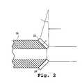
- FIG. 2is a side sectional view showing two angle-mounted ultrasound transducers on the distal end of a steerable catheter accessing device, in accordance with an embodiment of the present invention
- FIG. 3is a cross sectional view of the catheter assembly shown in FIG. 1, taken laterally across a mid-member of the catheter accessing device;
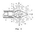
- FIG. 4is a side sectional view of the catheter-assembly distal-end region of FIG. 1, taken longitudinally therealong, with the tissue-penetrating implement inserted into a selected tissue to form a cavity therein for receiving a selected agent;
- FIG. 5Aillustrates a section of normal myocardial tissue
- FIG. 5Billustrates a section of myocardial tissue with a temporary cavity formed therein
- FIG. 6is a side elevational view, with members shown in cross section, of an endoscope-type agent delivery tool having a tissue-penetrating implement like that of the catheter assembly of FIG. 1;
- FIGS. 7 (A)- 7 (C)illustrate an accessing device, shown in section, with a movable implement for forming a cavity in a selected tissue and delivering a selected agent therein, in accordance with the teachings of one embodiment of the present invention.
- FIGS. 8 (A)- 8 (C)illustrate an accessing device, shown in section, with a movable implement for forming a cavity in a selected tissue and placing a selected agent therein, in accordance with an embodiment of the present invention.
- FIGS. 9 ( a-d )depict an embodiment having a force contact transducer.
- FIGS. 10 ( a-b )depict a corkscrew shaped expandable member embodiment.
- FIGS. 11 ( a-c )depict a balloon expandable member embodiment.
- FIGS. 12 ( a-c )depict an arc cutting embodiment.
- a catheter assembly(which may be disposable, in whole or in part), indicated generally by the reference numeral 12 , includes a control structure (hand unit) 14 attached to a steerable catheter accessing device 16 having a controllably deflectable distal-end member.
- a catheter assemblycan include steering components like those disclosed in U.S. Pat. No.
- a pull wire 18having an enlarged head member 18 a at its distal end, extends from the tip of catheter accessing device 16 , through a wire-guide channel 19 extending through catheter accessing device 16 , to control structure (hand unit) 14 , whereat the wire's proximal end is coupled to a deflection or steering actuator assembly.
- Rotation of a deflection knob 20which is threadedly mounted along a forward end of the hand unit, causes the pull wire to be pulled backward, or the catheter accessing device to be pushed forward, relative to one another, thereby inducing deflection of the distal end of the steerable catheter accessing device.
- FIG. 3another embodiment provides the pull wire extending longitudinally along the interior wall of the catheter accessing device (FIG. 3 ).
- Other steering mechanisms and arrangements, suitable for use herein,will be apparent to those skilled in the art.
- the catheteris further guided by a coaxial second catheter as described in co-pending application U.S. Ser. No. 09/052,971 and PCT publication WO 9949773A2 titled “Delivery catheter system for heart chamber” by Payne, filed Mar. 31, 1998, both herein incorporated in their entireties by reference.
- Catheter accessing device 16is dimensioned to be placed in the vasculature of a subject and steered therethrough until the tip is disposed adjacent a selected region of tissue, for example, a surface or wall within a heart chamber (such as against the endocardial wall within the heart's left ventricle).
- Visualization enhancement aidsincluding but not limited to radiopaque markers, tantalum and/or platinum bands, foils, and/or strips can be placed on the various components of drug-delivery tool—catheter assembly 12 , including on the deflectable end member of catheter accessing device 16 .
- a radio-opaque marker(not shown) made of platinum or other suitable radio-opaque material is disposed adjacent the tip for visualization via fluoroscopy or other methods.
- one or more ultra-sonic transducerscan be mounted on the catheter accessing device at or near its tip to assist in determining its location and/or placement (for example, degree of perpendicularity) with respect to a selected tissue in a subject, as well as to sense wall contact with, and/or wall thickness of, the tissue.
- Ultra-sonic transducer assemblies, and methods of using the same,are disclosed, for example, in published Canadian Patent Application No. 2,236,958, entitled, “Ultrasound Tool for Axial Ranging,” to Zanelli et al., and in co-pending U.S. patent application Ser. No.
- two transducersdenoted as 26 and 28 , are angle mounted at the tip of catheter accessing device 16 in the axis of pull-wire deflection. This construction permits an operator to determine, by comparing signal strength, whether the catheter tip region is perpendicular to a selected tissue surface or wall.
- this two-transducer arrangementprovides an operator with information useful for determining an appropriate adjustment direction for improving perpendicularity, as compared to single-transducer arrangements that, while capable of indicating perpendicularity by signal strength amplitude, are generally incapable of indicating a suitable direction in which to move the tip to improve perpendicularity.
- third and fourth transducersare added, off of the deflection axis, to aid an operator with rotational movement and rotational perpendicularity in the non-deflecting plane of the subject tissue surface.
- Each of the above ultrasound transducersmay preferably be substituted with force contact transducers described in co-pending U.S. patent application No. 60/191,610 by C.
- one or more elongate lumensmay extend between the proximal and distal ends of the catheter accessing device, with (i) at least one lumen being dimensioned to accommodate a cavity forming implement for axial movement along a region of the assembly's distal end, and (ii) at least one lumen being configured to permit passage of one or more selected therapeutic and/or diagnostic agents from an agent-supply region (for example, a reservoir in the hand unit) to, and out of, a terminal orifice at the assembly's distal end.
- agent-supply regionfor example, a reservoir in the hand unit
- a tissue-penetrating implement 48(described below) is adapted for movement within a distal-end region of lumen 22 .
- a selected agentcan be passed through the main lumen directly, i.e., in contact with the main lumen's interior walls, and/or indirectly, for example, using one or more additional lumens (for example, sub-lumens) extending coextensively and/or coaxially with the main lumen. An embodiment of the latter construction is also illustrated, in part, in FIGS.
- FIGS. 1, 3 , and 4each depict different aspects of an elongate, flexible agent-delivery conduit 30 is disposed substantially coaxially within catheter accessing device 16 , extending from control structure (hand unit) 14 to a distal region of lumen 22 .
- Conduit 30can be formed, for example, of a substantially inert polymeric material that resists collapse during bending or twisting, such as braided polyimide, braided PEBAX, or the like.
- Conduit 30defines a hollow, axial lumen or passage 32 , having a diameter within a range of from about 0.25 mm to about 1 mm (for example, about 0.5 mm), or from about 0.010′′ to about 0.040′′ (for example, about 0.020′′), that communicates at its proximal end with an agent-supply reservoir disposed in control structure (hand unit) 14 , and terminates at its distal end at an exit or infusion port 34 , through which a selected therapeutic and/or diagnostic agent can pass.
- conduit 30is adapted for reciprocal sliding movement within catheter accessing device 16 and, thus, is provided with an outer diameter less than the inner diameter of catheter accessing device 16 , for example, about 1 mm or less in certain constructions.
- control structure (hand unit) 14is provided with a fixed drug-delivery reservoir for holding a supply of a selected agent to be dispensed.
- a supply vesselsuch as syringe 36
- a connectorprovided in the unit's outer housing 38 .
- the connectoris preferably a substantially sterile connector, such as a standard Luer-type fitting or other known standard or proprietary connector.
- the supply reservoircomprises a syringe, pre-loaded with a selected agent, that can be removably fit into a holding area inside the housing.
- a dosage volume adjustment thumbscrew 40can be mounted in the housing 38 so as to be externally accessible for accurate, local and rapid dosage volume adjustment.
- a dosage volume scale or indicator, as at 42can be provided in the housing 38 .
- trigger mechanism 44is coupled to the proximal end of conduit 30 such that, upon being depressed, the conduit is pushed forward (advanced) within catheter accessing device 16 from a normal, retracted condition, depicted in FIG. 1, to a dispensing condition, shown in FIG. 4, whereat conduit orifice 34 can be positioned closely adjacent a selected tissue, such as 46 , against which catheter-accessing device orifice 24 has been placed.
- conduit 30shifts back to its normal condition.
- the distance traversed by conduit 30in each direction, is from about 2 to about 10 mm, and preferably about 5 mm.
- a tissue-penetrating implement, indicated generally as 48is also longitudinally movable within catheter accessing device 16 , between a retracted condition, within a distal region of lumen 22 (FIG. 1 ), and an extended (advanced) condition, passed through and extending out of orifice 24 (FIG. 4 ), over a stroke of about 4-6 mm, and preferably about 5 mm. Movement of implement 48 is effected by way of an elongate actuation line 50 , depicted in cross-section in FIG. 3, operatively coupled at one end to trigger mechanism 44 (FIG. 1) and extending axially through conduit 30 from control structure (hand unit) 14 to a proximal end of implement 48 .
- Preferred materials for forming the actuation lineare laterally flexible, permitting movement through tortuous pathways, and sufficiently incompressible along the longitudinal direction to provide for the efficient transmission of motion from the proximal end to the distal end.
- Suitable materialsinclude, for example, stainless steel or a braided composite.
- both conduit 30 and cutting implement 48takes place substantially simultaneously (i.e., these motions are coupled) with a single depression of trigger mechanism 44 .
- an agent held in a reservoir in the hand unitis dispensed from conduit 30 .
- such dispensingis effected immediately after (not before) the conduit and cutting implement have reached their respective extended conditions.
- the initial depressioncan actuate axial movement of the conduit and cutting implement, and the latter member of the depression can effect dispensing.
- both conduit 30 and cutting implement 48are retracted together with release of the trigger mechanism, and the dispensing of the selected agent is stopped.
- tip 52takes the form of a narrow, three-sided pyramid-like structure that tapers to a sharp point.
- tip 52could taper to a two-sided knife edge or blade, or any other suitable cutting or slicing structure.
- Preferred cutting or slicing structuresare configured to substantially avoid the removal of tissue beyond the cellular injury inherent in cutting.
- Implement 48further includes an expandable member, proximal of tip 52 , comprised of one or more resiliently flexible expandable elements or expandable members, three of which are visible (out of a total of four) at 54 in the embodiment of FIGS. 1 and 4.
- the expandable membersare arranged at spaced positions about the implement's longitudinal axis, and configured to flex outwardly, away from such axis, to collectively form a three dimensional support skeleton or cage.
- the expandable memberscan be, for example, narrow, elongate wires, filaments or ribbons, formed of a substantially inert, resiliently flexible material, such as a metal or metal alloy (for example, stainless steel, nickel-titanium, or similar material) or from an injection molded plastic.
- each expanderis turned inward and attached to the proximal side of tip 52 .
- the expandable memberWhen the expandable member is disposed at its retracted condition (FIG. 1 ), the expandable members are compressed toward the implement's longitudinal axis; and when advanced to its extended condition (FIG. 4 ), the expandable members are allowed to flex outward, so that, overall, the expandable member achieves a maximum diameter of about 1-3 mm, and preferably from about 1.75 mm to about 2 mm.
- the expandable memberbetween about 3-10 nickel-titanium (for example, as available commercially under the name “Nintinol”) filaments, each between about 4-5 mm in length and from about 0.003′′ to about 0.005′′ in diameter are employed as expandable members.
- the particular number, dimensions, and material composition of the expandable membersare not critical, provided only that the expandable members are capable of forming a cavity when inserted into a selected tissue (i.e., they have sufficient strength and spring capabilities), and, when in the expanded condition, a drug or other agent delivered into the region within the expandable members can move outwardly into the tissue about the cavity, with very little interference presented by the expandable members themselves, as shown in FIG. 4 with agent 58 in cavity 60 .
- the expandable memberspreferably occupy no more than about 10%, and more preferably less than about 3%, of the region defining the boundary between the cavity and the target tissue thereabout. In this way, the vast majority of the tissue boarding a cavity can be directly exposed to an agent delivered into the cavity.
- catheter accessing device 16is percutaneously introduced via femoral or radial artery access. This can be accomplished, for example, by way of the Seldinger technique ( Acta Radiologica , 38, [1953], 368-376; incorporated herein by reference), a variation thereof, or other conventional technique.
- a conventional guiding or shielding catheter(not shown) can be employed to assist in tracking the catheter tool through the patient's vasculature and into targeted regions of the heart.
- the catheter accessing device 16is slid across the aortic valve and into the left ventricle chamber.
- the distal end of the catheter accessing device 16is maneuvered so as to be substantially perpendicular to the endocardial wall 46 (FIG. 4 ), using fluoroscopic visualization and/or ultrasound guidance, and pressed thereagainst.
- Trigger mechanism 44is next depressed, causing cutting tip 52 to advance into the myocardial tissue, in the direction of arrow 64 , to a preset or adjustable depth.
- Expandable members 54follow cutting tip 52 into the myocardium and expand radially (for example, in the direction of arrows 66 ), creating a cavity about the axis of penetration (i.e., the axis of cutting or slicing). Once the cavity has been created, the expandable members serve to maintain the cavity by resisting heart contractile forces.
- the same trigger depressionserves to deliver a selected agent through conduit 30 into the cavity 60 . After allowing the agent to enter into the surrounding tissue for appropriate period of time, for example, typically less than about 2 minutes, the tissue-penetrating implement is withdrawn, at which point the cavity can close.
- Healthy myocardial tissueis illustrated in FIG. 5 A.
- healthy tissuecontains capillaries 70 , interstitial tissue 72 , and heart muscle cells 74 (See, for example, “Gray's Anatomy” (1959) at page 597).
- FIG. 5Bshows how a temporary cavity 60 can be created to directly access, for example, along the direction of arrows 68 , more capillaries 70 , more heart muscle cells 74 , and tissue surface area 76 . It should be appreciated that the creation of temporary cavities, as taught therein, provides direct access to a greater number of capillaries than has been possible by the prior techniques. As a result, the performance of the infusate tool is greatly enhanced.
- abrasion to the wall of the cavitiesmay aid in absorption of the agent. Accordingly, it may be desirable to configure the cutting tip and/or cavity expandable members of the invention so as to allow selective abrasion. This can also be accomplished, for example, by RF, thermal, acidic and/or ultrasonic means acting on the cutting tip and/or cavity expandable members.
- FIG. 6Another embodiment of the drug-delivery tool of the present invention is shown in FIG. 6, wherein the tool is embodied in an endoscope-type tool, shown generally at 80 .
- the drug-delivery tool of this embodimentis configured for intraoperative use, to be introduced thoracoscopically or through a thoracotomy, to form temporary cavities in a selected tissue.
- the toolincludes a proximal handpiece 82 (similar to the previously-described control structure (hand unit) 14 ) adapted to accommodate an drug-delivery reservoir syringe 84 , and a depressible trigger mechanism 86 .
- This particular surgical toolincorporates a reusable 5 mm thoracoscopic camera 88 axially mounted to provide an operator with a field of view 90 through lens 92 .
- a tissue-penetrating implement 48upon traversing the epicardial surface 98 of the heart, can create a temporary cavity for receiving a selected agent.
- the toolis adapted to permit a user to both extend the tissue-penetrating implement and dispense a drug or other agent, with a single depression of the trigger mechanism 86 . Additional details of the handpiece are presented in co-pending U.S.
- tissue-penetrating implementneed not retract.
- movement of the implement between its retracted and advanced conditionsneed only involve movement of the implement move from its retracted to its advanced condition.
- a selected therapeutic and/or diagnostic agentcomprising a charged species (for example, DNA) is held within the distal-end region of an accessing device and delivered into a cavity formed by in a selected tissue via an electrical field.
- a cavity-forming and delivery implementwhich can be incorporated in a catheter-type tool or an endoscope-type, such as previously described, is shown in FIGS. 7A-7C.
- the implementincludes drug-delivery reservoir or storage vessel 114 which opens into the region between a plurality of expandable members 116 via short passage 118 through a neck member 120 .
- First and second lead wiresextend through a flexible actuation accessing device 126 and terminate at respective terminals, or electrodes, fixed in the implement.
- the first terminalindicated as 123
- the second terminaldenoted as 125
- the catheter accessing device 134is introduced into a subject body and placed against an endocardial or epicardial wall 136 of the heart's left ventricle (FIG. 7 A).
- the vessel terminal 123is made positive (+) and the tip terminal 125 is made negative ( ⁇ ), thereby establishing an electrical field that maintains the negatively charged DNA in the vessel 114 .
- the lead wires 122 , 124 and regions about the terminals 123 , 125are shielded, using conventional materials, to limit the field's reach into the surrounding heart tissue. Such shielding about the forward (distal) region of the implement is indicated by back-hatching in the drawings.
- actuation accessing device 126is advanced, via a remote shifting mechanism (such as previously described), to push the slicing tip 128 through the wall 136 and into a region of myocardium 132 , with the expandable members 116 following the tip therein.
- a remote shifting mechanismsuch as previously described
- the polarityis reversed, so that the tip terminal 128 is positive (+) and the vessel terminal 123 is negative ( ⁇ ) (FIG. 7 B), thereby establishing an electrical field effective to draw the negatively charged DNA 130 toward the tip 128 .
- the electrical fieldis discontinued (FIG. 7 C), so that the DNA can move outwardly into the surrounding tissue and capillaries of the myocardium.
- a selected therapeutic and/or diagnostic agentis held within the distal-end region of an accessing device and placed in a cavity formed in a selected tissue.
- An exemplary cavity-forming and placement implementwhich can be incorporated in a catheter-type tool or an endoscope-type, such as previously described, is shown in FIGS. 8A-8C.
- the implementincludes a plurality of expandable members 142 attached at their rearward (proximal) ends to a flexible actuation accessing device 144 , and at their forward (distal) ends to a cutting/slicing tip 146 .
- the expandable members 142are arranged to serve as a cage or skeleton for containing a selected agent 148 , in solid or semi-solid form, as the catheter accessing device 150 is placed against a selected organ wall, as at 152 (FIG. 8 A).
- Actuation accessing device 144is then advanced, via a remote shifting mechanism, to push the slicing tip 146 of the implement through the wall 152 and into a selected layer of tissue 154 , with the expandable members 142 following the tip 146 therein (FIG. 8 B). Once a cavity has been formed in this manner, the agent 148 is allowed to move outward into the surrounding tissue and capillaries (FIGS. 8 B- 8 C).
- the agentcan be configured to for controlled release after placement, for example, via swelling and sloughing over a period of several minutes.
- controlled-release preparationsare formulated through the use of polymers to complex or absorb the selected gene sequence (with or without an associated carrier, for example, liposomes, etc.).
- the agentscan be formulated according to known methods to prepare pharmaceutically useful compositions, whereby these materials, or their functional derivatives, are combined in admixture with a pharmaceutically acceptable carrier vehicle. Suitable vehicles and their formulation, are described, for example, in Nicolau, C. et al. ( Crit. Rev. Ther. Drug Camier Syst . 6:239-271 (1989)), which is incorporated herein by reference.
- a pharmaceutically acceptable composition suitable for effective administrationsuch compositions will contain an effective amount of the desired gene sequence together with a suitable amount of carrier vehicle.
- FIG. 9 adepicts a preferred embodiment of the invention where accessing device 900 further comprises force contact transducer 902 mounted on distal end 904 of accessing device 900 .
- force contact transducer 902contacts tissue 906 causing detectable contact pressure to develop between force contact transducer 902 and tissue 906 .
- detectable pressuredetected by force contact transducer 902 is communicated back to the end user who then can further manipulate accessing device 900 to achieve perpendicularity between the thrust axis of accessing device 900 and tissue 906 .
- tissue-penetrating implement 908Upon achieving perpendicularity and contact force, tissue-penetrating implement 908 , with cutting tip 908 a , may be advanced to an extended condition, from a retracted position, thus causing the formation of cavity 910 in tissue 906 . Because accessing device 900 is urged against tissue 906 in a perpendicular manner, distal end 904 of accessing device 900 develops a seal for sealing in later delivered drug into cavity 910 .
- FIG. 9 cdepicts accessing device 900 without force contact transducer 902 . FIG. 9 c suggests how a non-perpendicular orientation of accessing device 900 with respect to tissue 906 could result in seepage of delivered drug 912 from cavity 910 .
- FIG. 9 csuggests how a non-perpendicular orientation of accessing device 900 with respect to tissue 906 could result in seepage of delivered drug 912 from cavity 910 .
- FIG. 9 dfurther depicts accessing device 900 without force contact transducer 902 urged against tissue 900 .
- Tissue 900is further depicted in two states, diastolic state tissue 906 a and systolic state tissue 906 b correlating to the movement of myocardial tissue in a beating heart.
- diastolic position tissue 906 aprovides a seal between tissue 906 and accessing device 900 .
- tissue 906moves away from accessing device 900 unless sufficient contact force exists between accessing device 900 and tissue 906 .
- Force contact transducer 902provides information to the user to enable the user to apply sufficient and perpendicular force to the accessing device to create a seal between accessing device 900 and tissue 906 during the movements of beating heart between tissue 900 a and 900 b states.
- FIG. 9 ddepicts how delivered drug 912 may be further ejected or pumped out of cavity 910 by the contractile actions between heart tissue 900 a and 900 b states.
- FIG. 10depicts another embodiment of the invention utilizing corkscrew shaped tissue-penetrating implement 1000 .
- Accessing device 1002houses tissue-penetrating implement 1000 that may be rotated within accessing device in either a retracted condition or an extended condition.
- FIG. 10 adepicts tissue-penetrating implement 1000 secured into tissue 1004 by screwing. As tissue-penetrating implement 1000 is withdrawn back towards a retracted condition, tissue 1004 is likewise pulled into lumen 1006 of accessing device 1002 thus creating seal 1006 between accessing device 1002 and tissue 1004 . Such pulling further creates cavity 1008 at distal end 1010 of tissue-penetrating implement 1000 . Cavity 1008 may then be filled with delivered-drug, not shown, delivered through lumen orifice 1012 to treat the walls of cavity 1008 with such drug.
- FIG. 11depicts another embodiment of the invention where the expandable members comprise a balloon structure with drug-delivery lumen orifices distributed along the surface of the expandable members.
- FIGS. 11 a and 11 bdepicts a tissue-penetrating implement 1101 comprising four radially distributed expandable members 1100 defining lumens 1102 with exit ports 1104 outwardly situated on balloon 1106 .
- Penetrating tip 1108is situated on the end of the balloon distal from accessing device 1110 , not shown.
- FIG. 11 cfurther shows yet another embodiment using a balloon as an expandable member and drug-delivery channel.
- Accessing tool 1110is urged against tissue 1112 , whereby tissue-penetrating implement 1101 comprises a balloon expandable member 1106 with distally situated exit ports 1104 and penetrating or cutting tip 1108 .
- FIG. 12depicts a preferred embodiment of the invention where tissue penetrating implement 1200 comprises at least one first expandable member 1202 made from a shape memory material composition having a first remembered arc shape and a second, stress induced, straight shape.
- First expandable member 1202assumes a stress induced straight shape when housed within lumen 1204 of accessing tool 1206 , but returns to its remembered shape upon extension beyond lumen 1204 .
- As first expandable member 1202 extends from lumen 1202it cuts an arc shaped path through tissue 1210 as first expandable member 1202 regains its remembered shape.
- Tissue-penetrating implement 1200has cutting tip 1208 situated distal to accessing tool 1206 for cutting tissue 1210 as tissue-penetrating implement 1200 is advanced into tissue 1210 when advanced from a retracted condition to an extended condition out of lumen 1204 .
- Second expandable member 1211extends from lumen 1204 coaxial to first expandable member 1202 . Adjacent tissue-penetration implement's distal end, first and second expandable members are positioned together either fixedly or slidably. When fixedly positioned, both expandable members 1202 and 1211 extend together, but expand longitudinally from one another to form cavity 1212 .
- first and second expandable members 1202 and 1211When first and second expandable members 1202 and 1211 are slidably positioned, the user may either extend one expandable member, preferably the first expandable member 1202 having cutting tip 1208 , and then extend second expandable member 1211 to follow along cut path 1216 created by previously extended first expandable member 1202 , expanding longitudinally away from first expandable member 1202 to create cavity 1212 where a drug may be infused from a drug-delivery reservoir, not shown, in fluid communication through a conduit with the distal region of accessing device 1222 .
- FIG. 12 bdepicts a variation where second expandable member further comprises construction from shape memory tube 1218 , such as nitinol or NiTi tubing, defining a longitudinal lumen in fluid communication with a drug-delivery reservoir, not shown, and terminating with exit ports 1220 adjacent to the distal end of second expandable member.
- shape memory tube 1218such as nitinol or NiTi tubing
- drugmay be delivered from the drug-delivery reservoir, not shown, to the cavity 1212 through the lumen and exit ports 1220 of second expandable member 1211 .
- FIG. 12 cdepicts a variation where first and second expandable members 1202 and 1211 are spaced-apart from one another by, for example, having two lumens, not shown, defined within accessing device 1222 .
- Force contact transducer 1224is located on the distal end of accessing device 1222 to assist a user in achieving the sufficient and perpendicular contact force with respect to tissue 1210 to create a seal between tissue 1210 and the distal end of accessing device 1222 .
- second expandable member 1211made from a shape memory material that assumes a stress induced straight shape when housed within lumen 1204 of accessing tool 1206 , but returns to its remembered shape upon extension beyond lumen 1204 , when configured as shown in FIG. 12, provides the ability to shepherd first expandable member 1202 further in its arc shape cutting path by applying lateral force to cutting tip 1208 as it cuts through tissue 1210 . This further prevents cutting tip from accidentally cutting too deep through a wall like tissue and thus perforating the wall and turning a cavity into a passage.
- Controlled deliverymay be exercised by selecting appropriate macromolecules (for example polyesters, polyamino acids, polyvinyl, pyrrolidone, ethylenevinylacetate, methylcellulose, carboxymethylcellulose, or protamine, sulfate) and the concentration of macromolecules as well as the methods of incorporation in order to control release.
- Another method to control the duration of action by controlled release preparationsis to incorporate the agent into particles of a polymeric material such as polyesters, polyamino acids, hydrogels, poly(lactic acid) or ethylene vinyl acetate copolymers.
- microcapsulesprepared, for example, by coacervation techniques or by interfacial polymerization, for example, hydroxymethylcellulose or gelatin microcapsules and poly(methylmethacylate) microcapsules, respectively, or in colloidal drug delivery systems, for example, liposomes, albumin microspheres, microemulsions, nanoparticles, and nanocapsules or in macroemulsions.
- the drug-delivery tool and method of the present inventionmay employ a wide variety of agents, for example, ranging from active compounds to markers to gene therapy compounds.
- agentscontemplated for use herein, are set forth in U.S. Pat. Nos. 5,840,059; 5,861,397; 5,846,946; 5,703,055; 5,693,622; 5,589,466; and 5,580,859, each expressly incorporated herein by reference.
- the inventionis employed to deliver one or more genes (for example, as so-called “naked DNA”) into cavities formed in the myocardium of a subject.
- the agentcan be delivered in a form that keeps the agent associated with the target tissue for a useful period of time, such as with a viscosity-enhancer to produce a thixotropic gel.
- the therapeutic or diagnostic agentis mixed with a viscous biocompatible polyol to maintain prolonged, high concentration of the agent in the channels and affect the kinetics of the agent-target region interaction.
- a cathetercould be employed to deliver an agent incorporated in a biocompatible polymer matrix.
- Suitable polymeric materialsare known in the art, for example, as set forth in U.S. Pat. No. 5,840,059, incorporated herein by reference.
- non-biodegradable polymerscan be employed as hollow reservoirs or other structures.
- conventional pharmacologically inert fillersmay be employed to tailor the time release characteristics of the agent.
- biodegradable polymerssuch as collagen, polylactic-polyglycolic acid, and polyanhydride.
- the agentcan be dispersed in a polymer which is configured to degrade over a useful period of time, releasing the agent.
- the agentis released by swelling and sloughing of the biodegradable polymer.
- Various means for employing polymer compounds to secure a therapeutic agentare disclosed, for example, in Levy et al., WO 94/21237 and in U.S. application Ser. No. 08/033,307, filed Mar. 15, 1993, which is hereby incorporated by reference.
- a biocompatible materialis delivered to seal and retain the agent within the cavity.
- a delivery lumencould be employed to deliver a sealing agent after delivery of the agent.
- the expandable members of the tissue-penetrating implementcan be configured not to expand, but rather to maintain a substantially constant configuration as it is moved between its retracted and advanced conditions.
- the cage or skeleton structure defined by the expandable memberscan serve, when inserted into a tissue, to help form a temporary cavity, and maintain the cavity as one or more selected agents are delivered and/or drawn therein.
Landscapes
- Health & Medical Sciences (AREA)
- Surgery (AREA)
- Life Sciences & Earth Sciences (AREA)
- Medical Informatics (AREA)
- Animal Behavior & Ethology (AREA)
- Engineering & Computer Science (AREA)
- Biomedical Technology (AREA)
- Heart & Thoracic Surgery (AREA)
- Vascular Medicine (AREA)
- Molecular Biology (AREA)
- Nuclear Medicine, Radiotherapy & Molecular Imaging (AREA)
- General Health & Medical Sciences (AREA)
- Public Health (AREA)
- Veterinary Medicine (AREA)
- Media Introduction/Drainage Providing Device (AREA)
- Infusion, Injection, And Reservoir Apparatuses (AREA)
- Surgical Instruments (AREA)
Abstract
Description
Claims (38)
Priority Applications (3)
| Application Number | Priority Date | Filing Date | Title |
|---|---|---|---|
| US09/566,196US6565528B1 (en) | 1999-05-07 | 2000-05-05 | Apparatus and method for delivering therapeutic and diagnostic agents |
| US10/370,646US20030225370A1 (en) | 1999-05-07 | 2003-02-20 | Apparatus and method for delivering therapeutic and diagnostic agents |
| US11/090,859US7211041B2 (en) | 1999-05-07 | 2005-03-25 | Apparatus and method for delivering therapeutic and diagnostic agents |
Applications Claiming Priority (2)
| Application Number | Priority Date | Filing Date | Title |
|---|---|---|---|
| US13317999P | 1999-05-07 | 1999-05-07 | |
| US09/566,196US6565528B1 (en) | 1999-05-07 | 2000-05-05 | Apparatus and method for delivering therapeutic and diagnostic agents |
Related Child Applications (1)
| Application Number | Title | Priority Date | Filing Date |
|---|---|---|---|
| US10/370,646ContinuationUS20030225370A1 (en) | 1999-05-07 | 2003-02-20 | Apparatus and method for delivering therapeutic and diagnostic agents |
Publications (1)
| Publication Number | Publication Date |
|---|---|
| US6565528B1true US6565528B1 (en) | 2003-05-20 |
Family
ID=22457373
Family Applications (3)
| Application Number | Title | Priority Date | Filing Date |
|---|---|---|---|
| US09/566,196Expired - Fee RelatedUS6565528B1 (en) | 1999-05-07 | 2000-05-05 | Apparatus and method for delivering therapeutic and diagnostic agents |
| US10/370,646AbandonedUS20030225370A1 (en) | 1999-05-07 | 2003-02-20 | Apparatus and method for delivering therapeutic and diagnostic agents |
| US11/090,859Expired - Fee RelatedUS7211041B2 (en) | 1999-05-07 | 2005-03-25 | Apparatus and method for delivering therapeutic and diagnostic agents |
Family Applications After (2)
| Application Number | Title | Priority Date | Filing Date |
|---|---|---|---|
| US10/370,646AbandonedUS20030225370A1 (en) | 1999-05-07 | 2003-02-20 | Apparatus and method for delivering therapeutic and diagnostic agents |
| US11/090,859Expired - Fee RelatedUS7211041B2 (en) | 1999-05-07 | 2005-03-25 | Apparatus and method for delivering therapeutic and diagnostic agents |
Country Status (2)
| Country | Link |
|---|---|
| US (3) | US6565528B1 (en) |
| WO (1) | WO2000067825A1 (en) |
Cited By (61)
| Publication number | Priority date | Publication date | Assignee | Title |
|---|---|---|---|---|
| US20020032478A1 (en)* | 2000-08-07 | 2002-03-14 | Percardia, Inc. | Myocardial stents and related methods of providing direct blood flow from a heart chamber to a coronary vessel |
| US20020045928A1 (en)* | 2000-05-04 | 2002-04-18 | Percardia, Inc. | Methods and devices for delivering a ventricular stent |
| US20020058897A1 (en)* | 1998-09-10 | 2002-05-16 | Percardia, Inc. | Designs for left ventricular conduit |
| US20030032972A1 (en)* | 2001-08-13 | 2003-02-13 | Birnbaum Peter L. | Transmyocardial revascularization gun |
| US20030083607A1 (en)* | 2001-10-29 | 2003-05-01 | Bobo Donald E. | Method and apparatus for providing medicament to tissue |
| US20030093033A1 (en)* | 2001-11-12 | 2003-05-15 | Bunyan Glenn Walter | Powered applicator |
| US20030105514A1 (en)* | 1998-09-10 | 2003-06-05 | Percardia, Inc. | Designs for left ventricular conduit |
| US20030137008A1 (en)* | 2000-03-28 | 2003-07-24 | Hidetoshi Nozaki | Solid state imaging device having a photodiode and a MOSFET and method of manufacturing the same |
| US20030225370A1 (en)* | 1999-05-07 | 2003-12-04 | Scimed Life Systems, Inc. | Apparatus and method for delivering therapeutic and diagnostic agents |
| US20040059411A1 (en)* | 2000-10-26 | 2004-03-25 | Strecker Ernst Peter | Implantable valve system |
| US20040133969A1 (en)* | 2003-01-10 | 2004-07-15 | Tyler Pipe Company, A Division Of Ransom Industries, Lp | Closet carrier system and method of assembly |
| US20040243158A1 (en)* | 2003-01-21 | 2004-12-02 | Angioscore, Inc., A Delaware Corporation | Apparatus and methods for treating hardened vascular lesions |
| US20050021071A1 (en)* | 2003-01-21 | 2005-01-27 | Angioscore, Inc. | Apparatus and methods for treating hardened vascular lesions |
| US6881199B2 (en) | 1998-09-10 | 2005-04-19 | Percardia, Inc. | Left ventricular conduit with blood vessel graft |
| US6945949B2 (en) | 1998-01-30 | 2005-09-20 | Percardia, Inc. | Left ventricular conduits to coronary arteries and methods for coronary bypass |
| US6949118B2 (en) | 2002-01-16 | 2005-09-27 | Percardia, Inc. | Encased implant and methods |
| US6964652B2 (en) | 1999-08-04 | 2005-11-15 | Percardia, Inc. | Left ventricular conduits and methods for delivery |
| US6976990B2 (en) | 2001-01-25 | 2005-12-20 | Percardia, Inc. | Intravascular ventriculocoronary bypass via a septal passageway |
| US7008397B2 (en) | 2002-02-13 | 2006-03-07 | Percardia, Inc. | Cardiac implant and methods |
| US7011095B2 (en) | 1998-09-10 | 2006-03-14 | Percardia, Inc. | Valve designs for left ventricular conduits |
| US7033372B1 (en) | 1999-08-04 | 2006-04-25 | Percardia, Inc. | Corkscrew reinforced left ventricle to coronary artery channel |
| AU2002301750B2 (en)* | 2001-11-12 | 2006-08-17 | N.J. Phillips Pty. Limited | A Powered Applicator |
| WO2006050396A3 (en)* | 2004-11-02 | 2006-08-31 | Medtronic Inc | Introduction of agent with medical device |
| US20060259005A1 (en)* | 2005-05-11 | 2006-11-16 | Angioscore, Inc. | Methods and systems for delivering substances into luminal walls |
| US20070255287A1 (en)* | 2006-04-26 | 2007-11-01 | Illuminoss Medical, Inc. | Apparatus and methods for reinforcing bone |
| US7326219B2 (en) | 2002-09-09 | 2008-02-05 | Wilk Patent Development | Device for placing transmyocardial implant |
| US20080039854A1 (en)* | 2006-04-26 | 2008-02-14 | Illuminoss Medical, Inc. | Apparatus and methods for delivery of reinforcing materials to bone |
| US20080125784A1 (en)* | 2006-11-10 | 2008-05-29 | Illuminoss Medical, Inc. | Systems and methods for internal bone fixation |
| US20090054900A1 (en)* | 2006-11-10 | 2009-02-26 | Illuminoss Medical, Inc. | Systems and Methods for Internal Bone Fixation |
| US20100152646A1 (en)* | 2008-02-29 | 2010-06-17 | Reshma Girijavallabhan | Intravitreal injection device and method |
| US20100265733A1 (en)* | 2009-04-06 | 2010-10-21 | Illuminoss Medical, Inc. | Attachment System for Light-Conducting Fibers |
| US20100324472A1 (en)* | 2007-11-14 | 2010-12-23 | Pathway Medical Technologies, Inc. | Delivery and administration of compositions using interventional catheters |
| US8403968B2 (en) | 2007-12-26 | 2013-03-26 | Illuminoss Medical, Inc. | Apparatus and methods for repairing craniomaxillofacial bones using customized bone plates |
| US8512338B2 (en) | 2009-04-07 | 2013-08-20 | Illuminoss Medical, Inc. | Photodynamic bone stabilization systems and methods for reinforcing bone |
| US8684965B2 (en) | 2010-06-21 | 2014-04-01 | Illuminoss Medical, Inc. | Photodynamic bone stabilization and drug delivery systems |
| US20140171908A1 (en)* | 2012-12-17 | 2014-06-19 | Robert G Matheny | Intra-Myocardial Agent Delivery Device, System and Method |
| US8870965B2 (en) | 2009-08-19 | 2014-10-28 | Illuminoss Medical, Inc. | Devices and methods for bone alignment, stabilization and distraction |
| US8936644B2 (en) | 2011-07-19 | 2015-01-20 | Illuminoss Medical, Inc. | Systems and methods for joint stabilization |
| US8939977B2 (en) | 2012-07-10 | 2015-01-27 | Illuminoss Medical, Inc. | Systems and methods for separating bone fixation devices from introducer |
| US9144442B2 (en) | 2011-07-19 | 2015-09-29 | Illuminoss Medical, Inc. | Photodynamic articular joint implants and methods of use |
| US9173977B2 (en) | 2010-04-19 | 2015-11-03 | Angioscore, Inc. | Coating formulations for scoring or cutting balloon catheters |
| US9179959B2 (en) | 2010-12-22 | 2015-11-10 | Illuminoss Medical, Inc. | Systems and methods for treating conditions and diseases of the spine |
| US9351756B2 (en) | 2010-09-21 | 2016-05-31 | Angioscore, Inc. | Method and system for treating valve stenosis |
| US9375328B2 (en) | 2001-11-09 | 2016-06-28 | Angioscore, Inc. | Balloon catheter with non-deployable stent |
| US9427289B2 (en) | 2007-10-31 | 2016-08-30 | Illuminoss Medical, Inc. | Light source |
| US9687281B2 (en) | 2012-12-20 | 2017-06-27 | Illuminoss Medical, Inc. | Distal tip for bone fixation devices |
| US10086178B2 (en) | 2001-11-09 | 2018-10-02 | Angioscore, Inc. | Balloon catheter with non-deployable stent |
| US10117668B2 (en) | 2013-10-08 | 2018-11-06 | The Spectranetics Corporation | Balloon catheter with non-deployable stent having improved stability |
| US10993805B2 (en) | 2008-02-26 | 2021-05-04 | Jenavalve Technology, Inc. | Stent for the positioning and anchoring of a valvular prosthesis in an implantation site in the heart of a patient |
| US11065138B2 (en) | 2016-05-13 | 2021-07-20 | Jenavalve Technology, Inc. | Heart valve prosthesis delivery system and method for delivery of heart valve prosthesis with introducer sheath and loading system |
| US11071572B2 (en) | 2018-06-27 | 2021-07-27 | Illuminoss Medical, Inc. | Systems and methods for bone stabilization and fixation |
| US11185405B2 (en) | 2013-08-30 | 2021-11-30 | Jenavalve Technology, Inc. | Radially collapsible frame for a prosthetic valve and method for manufacturing such a frame |
| US11197754B2 (en) | 2017-01-27 | 2021-12-14 | Jenavalve Technology, Inc. | Heart valve mimicry |
| US11337800B2 (en) | 2015-05-01 | 2022-05-24 | Jenavalve Technology, Inc. | Device and method with reduced pacemaker rate in heart valve replacement |
| US11357624B2 (en) | 2007-04-13 | 2022-06-14 | Jenavalve Technology, Inc. | Medical device for treating a heart valve insufficiency |
| US11517431B2 (en) | 2005-01-20 | 2022-12-06 | Jenavalve Technology, Inc. | Catheter system for implantation of prosthetic heart valves |
| US11564794B2 (en) | 2008-02-26 | 2023-01-31 | Jenavalve Technology, Inc. | Stent for the positioning and anchoring of a valvular prosthesis in an implantation site in the heart of a patient |
| US11589981B2 (en) | 2010-05-25 | 2023-02-28 | Jenavalve Technology, Inc. | Prosthetic heart valve and transcatheter delivered endoprosthesis comprising a prosthetic heart valve and a stent |
| US12121461B2 (en) | 2015-03-20 | 2024-10-22 | Jenavalve Technology, Inc. | Heart valve prosthesis delivery system and method for delivery of heart valve prosthesis with introducer sheath |
| US12171658B2 (en) | 2022-11-09 | 2024-12-24 | Jenavalve Technology, Inc. | Catheter system for sequential deployment of an expandable implant |
| US12414854B2 (en) | 2010-05-20 | 2025-09-16 | Jenavalve Technology, Inc. | Catheter system for introducing an expandable stent into the body of a patient |
Families Citing this family (33)
| Publication number | Priority date | Publication date | Assignee | Title |
|---|---|---|---|---|
| US7022088B2 (en) | 1999-08-05 | 2006-04-04 | Broncus Technologies, Inc. | Devices for applying energy to tissue |
| US7175644B2 (en) | 2001-02-14 | 2007-02-13 | Broncus Technologies, Inc. | Devices and methods for maintaining collateral channels in tissue |
| US7422563B2 (en) | 1999-08-05 | 2008-09-09 | Broncus Technologies, Inc. | Multifunctional tip catheter for applying energy to tissue and detecting the presence of blood flow |
| US7400931B2 (en)* | 2002-09-18 | 2008-07-15 | Cardiac Pacemakers, Inc. | Devices and methods to stimulate therapeutic angiogenesis for ischemia and heart failure |
| US8303511B2 (en)* | 2002-09-26 | 2012-11-06 | Pacesetter, Inc. | Implantable pressure transducer system optimized for reduced thrombosis effect |
| US8308682B2 (en) | 2003-07-18 | 2012-11-13 | Broncus Medical Inc. | Devices for maintaining patency of surgically created channels in tissue |
| US8409167B2 (en) | 2004-07-19 | 2013-04-02 | Broncus Medical Inc | Devices for delivering substances through an extra-anatomic opening created in an airway |
| US10646109B1 (en)* | 2004-07-19 | 2020-05-12 | Hypermed Imaging, Inc. | Device and method of balloon endoscopy |
| US7758541B2 (en) | 2004-08-17 | 2010-07-20 | Boston Scientific Scimed, Inc. | Targeted drug delivery device and method |
| US20070083192A1 (en)* | 2005-10-07 | 2007-04-12 | Eric Welch | Apparatus and method for ablation of targeted tissue |
| US7713232B2 (en) | 2005-11-04 | 2010-05-11 | Medrad, Inc. | System for washing and processing of cells for delivery thereof to tissue |
| US8182444B2 (en)* | 2005-11-04 | 2012-05-22 | Medrad, Inc. | Delivery of agents such as cells to tissue |
| US20070250035A1 (en)* | 2006-04-19 | 2007-10-25 | El-Nounou Fozan O | Devices and methods for intravascular drug delivery |
| US20080086111A1 (en)* | 2006-10-09 | 2008-04-10 | Medrad, Inc. | Fluid delivery systems and volume metering in cell delivery |
| US20080108950A1 (en)* | 2006-11-03 | 2008-05-08 | Rioux Robert F | Corkscrew helical inserter port |
| US9364586B2 (en) | 2007-05-31 | 2016-06-14 | Abbott Cardiovascular Systems Inc. | Method and apparatus for improving delivery of an agent to a kidney |
| US9144509B2 (en) | 2007-05-31 | 2015-09-29 | Abbott Cardiovascular Systems Inc. | Method and apparatus for delivering an agent to a kidney |
| US8216209B2 (en) | 2007-05-31 | 2012-07-10 | Abbott Cardiovascular Systems Inc. | Method and apparatus for delivering an agent to a kidney |
| US9149610B2 (en) | 2007-05-31 | 2015-10-06 | Abbott Cardiovascular Systems Inc. | Method and apparatus for improving delivery of an agent to a kidney |
| US8235983B2 (en) | 2007-07-12 | 2012-08-07 | Asthmatx, Inc. | Systems and methods for delivering energy to passageways in a patient |
| WO2010078409A1 (en)* | 2008-12-30 | 2010-07-08 | Cook Incorporated | Corkscrew injection needle |
| FR2962028B1 (en)* | 2010-07-05 | 2012-08-31 | Perouse Medical | MEDICAL DEVICE FOR INJECTING A FLUID INTO AN ORGANISM |
| US9345532B2 (en) | 2011-05-13 | 2016-05-24 | Broncus Medical Inc. | Methods and devices for ablation of tissue |
| US8709034B2 (en) | 2011-05-13 | 2014-04-29 | Broncus Medical Inc. | Methods and devices for diagnosing, monitoring, or treating medical conditions through an opening through an airway wall |
| WO2013078235A1 (en) | 2011-11-23 | 2013-05-30 | Broncus Medical Inc | Methods and devices for diagnosing, monitoring, or treating medical conditions through an opening through an airway wall |
| US9445897B2 (en) | 2012-05-01 | 2016-09-20 | Direct Flow Medical, Inc. | Prosthetic implant delivery device with introducer catheter |
| US9814618B2 (en) | 2013-06-06 | 2017-11-14 | Boston Scientific Scimed, Inc. | Devices for delivering energy and related methods of use |
| US20150005740A1 (en)* | 2013-06-28 | 2015-01-01 | Boston Scientific Scimed, Inc. | Injection devices and related methods of use |
| EP3606457A4 (en) | 2017-04-03 | 2021-04-21 | Broncus Medical Inc. | ELECTROSURGICAL ACCESS SHEATH |
| EP4018946A1 (en) | 2017-05-03 | 2022-06-29 | Medtronic Vascular, Inc. | Tissue-removing catheter |
| US11690645B2 (en) | 2017-05-03 | 2023-07-04 | Medtronic Vascular, Inc. | Tissue-removing catheter |
| CN118697424A (en) | 2018-11-16 | 2024-09-27 | 美敦力瓦斯科尔勒公司 | Tissue Removal Catheter |
| US11819236B2 (en) | 2019-05-17 | 2023-11-21 | Medtronic Vascular, Inc. | Tissue-removing catheter |
Citations (114)
| Publication number | Priority date | Publication date | Assignee | Title |
|---|---|---|---|---|
| US2491978A (en) | 1946-11-26 | 1949-12-20 | Helfman Hyman | Syringe holder |
| US3057349A (en) | 1959-12-14 | 1962-10-09 | Ismach Aaron | Multi-dose jet injection device |
| US3773034A (en) | 1971-11-24 | 1973-11-20 | Itt Research Institute | Steerable catheter |
| US3859996A (en) | 1973-07-18 | 1975-01-14 | Mizzy Inc | Multi-dose injector |
| US3952742A (en) | 1974-06-12 | 1976-04-27 | Taylor Duane F | Needle-carried, transthoracic, cannula-type cardiac resuscitation instrument |
| US4171852A (en) | 1977-06-27 | 1979-10-23 | Haentjens Walter D | Propulsion of slurry along a pipeline by ultrasonic sound waves |
| US4243035A (en) | 1979-06-18 | 1981-01-06 | Barrett Howard G | Syringe with integral swab |
| US4266541A (en) | 1978-09-19 | 1981-05-12 | Halen-Elliot Do Brazil Industria E Comercio Equipamentos De Precisao Ltda. | Pressure hypodermic injector for intermittent vaccination |
| US4296100A (en) | 1980-06-30 | 1981-10-20 | Franco Wayne P | Method of treating the heart for myocardial infarction |
| US4447225A (en) | 1982-03-22 | 1984-05-08 | Taff Barry E | Multidose jet injector |
| US4531936A (en) | 1981-01-29 | 1985-07-30 | Gordon Robert T | Device and method for the selective delivery of drugs to the myocardium |
| US4576591A (en) | 1983-07-06 | 1986-03-18 | Ivy-Gene Co., Inc. | Medicament implant applicator |
| US4657536A (en) | 1979-04-13 | 1987-04-14 | Regents Of The University Of Minnesota | Check valve catheter |
| US4680027A (en) | 1985-12-12 | 1987-07-14 | Injet Medical Products, Inc. | Needleless hypodermic injection device |
| US4770653A (en) | 1987-06-25 | 1988-09-13 | Medilase, Inc. | Laser angioplasty |
| US4838850A (en) | 1980-10-03 | 1989-06-13 | Henning Rosengart | Electromedical treatment apparatus |
| US4861339A (en) | 1985-09-25 | 1989-08-29 | Henke-Sass, Wolf Gmbh | Injection spray gun with adjustable pressure limitation |
| US4911148A (en) | 1989-03-14 | 1990-03-27 | Intramed Laboratories, Inc. | Deflectable-end endoscope with detachable flexible shaft assembly |
| US4946442A (en) | 1985-06-28 | 1990-08-07 | Olympus Optical Co., Ltd. | Endoscope treatment device |
| US4994033A (en) | 1989-05-25 | 1991-02-19 | Schneider (Usa) Inc. | Intravascular drug delivery dilatation catheter |
| US5034003A (en) | 1988-11-21 | 1991-07-23 | Raymond Denance | Injection device having a stabilizing sight which actuates a safety valve for the needle |
| US5061273A (en) | 1989-06-01 | 1991-10-29 | Yock Paul G | Angioplasty apparatus facilitating rapid exchanges |
| US5100792A (en) | 1984-11-13 | 1992-03-31 | Cornell Research Foundation, Inc. | Method for transporting substances into living cells and tissues |
| US5106370A (en) | 1991-01-23 | 1992-04-21 | Ideal Instruments, Inc. | Pellet carrier fed pellet implanter apparatus |
| US5114423A (en) | 1989-05-15 | 1992-05-19 | Advanced Cardiovascular Systems, Inc. | Dilatation catheter assembly with heated balloon |
| US5130141A (en) | 1988-05-24 | 1992-07-14 | Law Peter K | Compositions for and methods of treating muscle degeneration and weakness |
| US5179022A (en) | 1988-02-29 | 1993-01-12 | E. I. Du Pont De Nemours & Co. | Biolistic apparatus for delivering substances into cells and tissues in a non-lethal manner |
| US5183465A (en)* | 1990-12-28 | 1993-02-02 | Dimitrios Xanthakos | Apparatus for supporting and moving needles and trocars for penetrating the abdomen |
| US5185004A (en) | 1991-06-03 | 1993-02-09 | Danforth Biomedical, Inc. | Turn-limiting proximal adaptor for steerable catheter systems |
| US5197946A (en) | 1990-06-27 | 1993-03-30 | Shunro Tachibana | Injection instrument with ultrasonic oscillating element |
| US5203772A (en) | 1989-01-09 | 1993-04-20 | Pilot Cardiovascular Systems, Inc. | Steerable medical device |
| US5228883A (en) | 1991-05-02 | 1993-07-20 | Eli Lilly And Company | Portable drug delivery system |
| US5244460A (en) | 1991-11-27 | 1993-09-14 | The United States Of America As Represented By The Department Of Health And Human Services | Method to foster myocardial blood vessel growth and improve blood flow to the heart |
| US5307803A (en) | 1992-03-04 | 1994-05-03 | Intramed Laboratories | Deflecting endoscope |
| US5308324A (en) | 1989-01-09 | 1994-05-03 | Pilot Cardiovascular Systems, Inc. | Steerable medical device |
| US5322511A (en) | 1992-04-21 | 1994-06-21 | Sterling Winthrop Inc. | Portable hand-held power injector |
| US5328470A (en) | 1989-03-31 | 1994-07-12 | The Regents Of The University Of Michigan | Treatment of diseases by site-specific instillation of cells or site-specific transformation of cells and kits therefor |
| US5336178A (en) | 1992-11-02 | 1994-08-09 | Localmed, Inc. | Intravascular catheter with infusion array |
| US5356388A (en) | 1992-09-22 | 1994-10-18 | Target Therapeutics, Inc. | Perfusion catheter system |
| US5370685A (en) | 1991-07-16 | 1994-12-06 | Stanford Surgical Technologies, Inc. | Endovascular aortic valve replacement |
| US5372587A (en) | 1989-01-09 | 1994-12-13 | Pilot Cariovascular Systems, Inc. | Steerable medical device |
| US5378239A (en)* | 1990-04-12 | 1995-01-03 | Schneider (Usa) Inc. | Radially expandable fixation member constructed of recovery metal |
| US5380279A (en) | 1994-03-11 | 1995-01-10 | The Upjohn Company | Animal vaccination gun |
| US5389096A (en) | 1990-12-18 | 1995-02-14 | Advanced Cardiovascular Systems | System and method for percutaneous myocardial revascularization |
| US5395312A (en) | 1991-10-18 | 1995-03-07 | Desai; Ashvin | Surgical tool |
| US5429131A (en) | 1994-02-25 | 1995-07-04 | The Regents Of The University Of California | Magnetized electrode tip catheter |
| US5431168A (en) | 1993-08-23 | 1995-07-11 | Cordis-Webster, Inc. | Steerable open-lumen catheter |
| US5454790A (en)* | 1994-05-09 | 1995-10-03 | Innerdyne, Inc. | Method and apparatus for catheterization access |
| US5476100A (en) | 1994-07-07 | 1995-12-19 | Guided Medical Systems, Inc. | Catheter steerable by directional jets with remotely controlled closures |
| US5480381A (en) | 1991-08-23 | 1996-01-02 | Weston Medical Limited | Needle-less injector |
| US5480382A (en) | 1989-01-09 | 1996-01-02 | Pilot Cardiovascular Systems, Inc. | Steerable medical device |
| US5492119A (en) | 1993-12-22 | 1996-02-20 | Heart Rhythm Technologies, Inc. | Catheter tip stabilizing apparatus |
| US5499971A (en) | 1990-06-15 | 1996-03-19 | Cortrak Medical, Inc. | Method for iontophoretically delivering drug adjacent to a heart |
| US5507724A (en) | 1992-07-01 | 1996-04-16 | Genetronics, Inc. | Electroporation and iontophoresis apparatus and method for insertion of drugs and genes into cells |
| EP0489496B1 (en) | 1990-11-08 | 1996-08-14 | Possis Medical, Inc. | Water jet atherectomy device |
| US5569160A (en) | 1993-09-13 | 1996-10-29 | United States Surgical Corporation | Optical trocar |
| US5569217A (en) | 1993-10-28 | 1996-10-29 | Luther Medical Products, Inc. | Percutaneous port catheter assembly and method of use |
| US5580859A (en) | 1989-03-21 | 1996-12-03 | Vical Incorporated | Delivery of exogenous DNA sequences in a mammal |
| US5591195A (en) | 1995-10-30 | 1997-01-07 | Taheri; Syde | Apparatus and method for engrafting a blood vessel |
| US5630795A (en) | 1991-08-02 | 1997-05-20 | Olympus Optical Co., Ltd. | Cleaning tube apparatus for endoscope |
| US5642730A (en) | 1994-06-17 | 1997-07-01 | Trudell Medical Limited | Catheter system for delivery of aerosolized medicine for use with pressurized propellant canister |
| US5652225A (en) | 1994-10-04 | 1997-07-29 | St. Elizabeth's Medical Center Of Boston, Inc. | Methods and products for nucleic acid delivery |
| US5653684A (en)* | 1992-06-26 | 1997-08-05 | Schneider (Usa), Inc. | Catheter with expandable wire mesh tip |
| US5661133A (en) | 1991-11-12 | 1997-08-26 | The Regents Of The University Of Michigan | Expression of a protein in myocardium by injection of a gene |
| US5698531A (en) | 1989-03-31 | 1997-12-16 | The Regents Of The University Of Michigan | Treatment of diseases by site-specific instillation of cells or site-specific transformation of cells and kits therefor |
| US5713907A (en)* | 1995-07-20 | 1998-02-03 | Endotex Interventional Systems, Inc. | Apparatus and method for dilating a lumen and for inserting an intraluminal graft |
| US5725523A (en) | 1996-03-29 | 1998-03-10 | Mueller; Richard L. | Lateral-and posterior-aspect method and apparatus for laser-assisted transmyocardial revascularization and other surgical applications |
| US5739118A (en) | 1994-04-01 | 1998-04-14 | Apollon, Inc. | Compositions and methods for delivery of genetic material |
| US5760081A (en) | 1994-05-10 | 1998-06-02 | The General Hospital Corporation | Omega 3 fatty acids in the prevention of ventricular fibrillation |
| US5772590A (en) | 1992-06-30 | 1998-06-30 | Cordis Webster, Inc. | Cardiovascular catheter with laterally stable basket-shaped electrode array with puller wire |
| US5782802A (en) | 1996-03-22 | 1998-07-21 | Vitajet Corporation | Multiple use needle-less hypodermic injection device for individual users |
| US5782239A (en) | 1992-06-30 | 1998-07-21 | Cordis Webster, Inc. | Unique electrode configurations for cardiovascular electrode catheter with built-in deflection method and central puller wire |
| US5792453A (en) | 1995-02-28 | 1998-08-11 | The Regents Of The University Of California | Gene transfer-mediated angiogenesis therapy |
| US5797870A (en) | 1995-06-07 | 1998-08-25 | Indiana University Foundation | Pericardial delivery of therapeutic and diagnostic agents |
| US5803083A (en) | 1995-11-09 | 1998-09-08 | Cordis Corporation | Guiding catheter with ultrasound imaging capability |
| US5807306A (en) | 1992-11-09 | 1998-09-15 | Cortrak Medical, Inc. | Polymer matrix drug delivery apparatus |
| US5820592A (en) | 1996-07-16 | 1998-10-13 | Hammerslag; Gary R. | Angiographic and/or guide catheter |
| US5827216A (en) | 1995-06-07 | 1998-10-27 | Cormedics Corp. | Method and apparatus for accessing the pericardial space |
| US5833658A (en) | 1996-04-29 | 1998-11-10 | Levy; Robert J. | Catheters for the delivery of solutions and suspensions |
| US5837533A (en) | 1994-09-28 | 1998-11-17 | American Home Products Corporation | Complexes comprising a nucleic acid bound to a cationic polyamine having an endosome disruption agent |
| US5836905A (en) | 1994-06-20 | 1998-11-17 | Lemelson; Jerome H. | Apparatus and methods for gene therapy |
| US5840062A (en) | 1995-11-08 | 1998-11-24 | Gumaste; Anand V. | Solid state fluid delivery system |
| US5840059A (en) | 1995-06-07 | 1998-11-24 | Cardiogenesis Corporation | Therapeutic and diagnostic agent delivery |
| US5840031A (en) | 1993-07-01 | 1998-11-24 | Boston Scientific Corporation | Catheters for imaging, sensing electrical potentials and ablating tissue |
| US5843050A (en) | 1995-11-13 | 1998-12-01 | Micro Therapeutics, Inc. | Microcatheter |
| US5846225A (en) | 1997-02-19 | 1998-12-08 | Cornell Research Foundation, Inc. | Gene transfer therapy delivery device and method |
| US5846221A (en) | 1996-02-09 | 1998-12-08 | Catheter Imaging Systems, Inc. | Steerable catheter having disposable module and sterilizable handle and method of connecting same |
| US5854209A (en) | 1995-03-23 | 1998-12-29 | Biopure Corporation | Method for oxygenating tissue having reduced red blood cell flow |
| US5857464A (en) | 1995-06-07 | 1999-01-12 | Desai; Jawahar M. | Catheter for media injection |
| US5865811A (en) | 1996-12-19 | 1999-02-02 | Boehringer Ingelheim Vetmedica, Inc. | Multi-dose applicator with quick bottle attachment mechanism |
| US5871495A (en) | 1996-09-13 | 1999-02-16 | Eclipse Surgical Technologies, Inc. | Method and apparatus for mechanical transmyocardial revascularization of the heart |
| US5871462A (en) | 1995-06-07 | 1999-02-16 | Hydrocision, Inc. | Method for using a fluid jet cutting system |
| US5876373A (en)* | 1997-04-04 | 1999-03-02 | Eclipse Surgical Technologies, Inc. | Steerable catheter |
| US5882332A (en) | 1997-06-06 | 1999-03-16 | Wijay; Bandula | Drug infusion catheter and method |
| US5885272A (en) | 1990-10-30 | 1999-03-23 | Aita; Michael | System and method for percutaneous myocardial revascularization |
| US5891136A (en) | 1996-01-19 | 1999-04-06 | Ep Technologies, Inc. | Expandable-collapsible mesh electrode structures |
| US5891086A (en) | 1993-07-31 | 1999-04-06 | Weston Medical Limited | Needle-less injector |
| US5935063A (en) | 1997-10-29 | 1999-08-10 | Irvine Biomedical, Inc. | Electrode catheter system and methods thereof |
| US5941868A (en) | 1995-12-22 | 1999-08-24 | Localmed, Inc. | Localized intravascular delivery of growth factors for promotion of angiogenesis |
| US5951516A (en) | 1997-09-01 | 1999-09-14 | N.J. Phillips Pty. Limited | Applicator |
| US5971983A (en) | 1997-05-09 | 1999-10-26 | The Regents Of The University Of California | Tissue ablation device and method of use |
| US5972019A (en)* | 1996-07-25 | 1999-10-26 | Target Therapeutics, Inc. | Mechanical clot treatment device |
| US5993443A (en) | 1997-02-03 | 1999-11-30 | Eclipse Surgical Technologies, Inc. | Revascularization with heartbeat verification |
| US5997509A (en) | 1998-03-06 | 1999-12-07 | Cornell Research Foundation, Inc. | Minimally invasive gene therapy delivery device and method |
| US6004269A (en) | 1993-07-01 | 1999-12-21 | Boston Scientific Corporation | Catheters for imaging, sensing electrical potentials, and ablating tissue |
| US6007531A (en) | 1995-11-21 | 1999-12-28 | Catheter Imaging Systems, Inc. | Steerable catheter having disposable module and sterilizable handle and method of connecting same |
| US6024703A (en)* | 1997-05-07 | 2000-02-15 | Eclipse Surgical Technologies, Inc. | Ultrasound device for axial ranging |
| US6036677A (en) | 1997-03-07 | 2000-03-14 | Cardiogenesis Corporation | Catheter with flexible intermediate section |
| US6056742A (en) | 1997-02-03 | 2000-05-02 | Eclipse Surgical Technologies, Inc. | Revascularization with laser outputs |
| US6102887A (en) | 1998-08-11 | 2000-08-15 | Biocardia, Inc. | Catheter drug delivery system and method for use |
| US6183444B1 (en)* | 1998-05-16 | 2001-02-06 | Microheart, Inc. | Drug delivery module |
| US6203559B1 (en)* | 1998-10-05 | 2001-03-20 | Origin Medsystems | Method and apparatus for tissue dissection |
| US6266550B1 (en)* | 1998-01-16 | 2001-07-24 | Lumend, Inc. | Catheter apparatus for treating arterial occlusions |
| US6322548B1 (en)* | 1995-05-10 | 2001-11-27 | Eclipse Surgical Technologies | Delivery catheter system for heart chamber |
Family Cites Families (26)
| Publication number | Priority date | Publication date | Assignee | Title |
|---|---|---|---|---|
| DE9190181U1 (en) | 1990-12-10 | 1993-07-29 | Howmedica Inc. (n.d.Ges.d.Staates Delaware), New York, N.Y. | Device for interstitial laser energy delivery |
| NZ263685A (en) | 1993-03-15 | 1997-08-22 | Univ Michiganthe Board Of Rege | Device for controlling heart rhythm comprising eledevice for controlling heart rhythm comprising electrode assembly for attachment to the heart and a ctrode assembly for attachment to the heart and a controlled release matrix comprising an antiarrhytcontrolled release matrix comprising an antiarrhythmic agent hmic agent |
| TW404844B (en) | 1993-04-08 | 2000-09-11 | Oxford Biosciences Ltd | Needleless syringe |
| GB9314640D0 (en)* | 1993-07-15 | 1993-08-25 | Salim Aws S M | Tunnellimg catheter |
| US5396887A (en)* | 1993-09-23 | 1995-03-14 | Cardiac Pathways Corporation | Apparatus and method for detecting contact pressure |
| US5810767A (en) | 1994-05-11 | 1998-09-22 | Localmed, Inc. | Method and apparatus for pressurized intraluminal drug delivery |
| WO1997017099A1 (en) | 1995-11-06 | 1997-05-15 | C.R. Bard, Inc. | Balloon catheter for drug application |
| US5904670A (en)* | 1996-04-03 | 1999-05-18 | Xrt Corp. | Catheters and methods for guiding drugs and other agents to an intended site by deployable grooves |
| GB9619002D0 (en) | 1996-09-11 | 1996-10-23 | Oxford Biosciences Ltd | Particle delivery |
| US5944710A (en) | 1996-06-24 | 1999-08-31 | Genetronics, Inc. | Electroporation-mediated intravascular delivery |
| AU2458597A (en)* | 1996-07-17 | 1998-02-09 | Medtronic, Inc. | System for genetically treating cardiac conduction disturbances |
| AU3911097A (en) | 1996-08-08 | 1998-02-25 | Localmed, Inc. | Transmural drug delivery method and apparatus |
| US5861397A (en) | 1996-10-03 | 1999-01-19 | Vical Incorporated | Piperazine based cytofectins |
| CA2225521C (en)* | 1996-12-27 | 2004-04-06 | Eclipse Surgical Technologies, Inc. | Laser assisted drug delivery apparatus |
| DE19701494A1 (en) | 1997-01-17 | 1998-07-23 | Boehringer Mannheim Gmbh | Transdermal injection system |
| US5860957A (en) | 1997-02-07 | 1999-01-19 | Sarcos, Inc. | Multipathway electronically-controlled drug delivery system |
| EP0989879A4 (en) | 1997-06-19 | 2001-05-30 | Mile Creek Capital Llc | Drug delivery system including a drug transport enhancement mechanism |
| US5947928A (en) | 1997-06-19 | 1999-09-07 | Mile Creek Capital, Llc | Drug delivery system |
| WO1999004851A1 (en)* | 1997-07-22 | 1999-02-04 | Emed Corporation | Iontophoretic delivery of an agent into cardiac tissue |
| JP4255208B2 (en)* | 1997-07-24 | 2009-04-15 | レックス メディカル リミテッド パートナーシップ | Device for resecting subcutaneous target tissue mass |
| GB2327614B (en)* | 1997-07-30 | 2002-03-06 | Univ Dundee | A hypodermic needle |
| US6179809B1 (en) | 1997-09-24 | 2001-01-30 | Eclipse Surgical Technologies, Inc. | Drug delivery catheter with tip alignment |
| ES2255155T3 (en)* | 1998-02-05 | 2006-06-16 | Biosense Webster, Inc. | DEVICE FOR THE INTRACARDIAC ADMINISTRATION OF PHARMACOS. |
| EP1020202A3 (en) | 1998-07-10 | 2000-08-23 | Medtronic, Inc. | Medical device for transmyocardial revascularization |
| US6224566B1 (en)* | 1999-05-04 | 2001-05-01 | Cardiodyne, Inc. | Method and devices for creating a trap for confining therapeutic drugs and/or genes in the myocardium |
| WO2000067825A1 (en)* | 1999-05-07 | 2000-11-16 | Microheart, Inc. | Apparatus and method for delivering therapeutic and diagnostic agents |
- 2000
- 2000-05-05WOPCT/US2000/012345patent/WO2000067825A1/enactiveApplication Filing
- 2000-05-05USUS09/566,196patent/US6565528B1/ennot_activeExpired - Fee Related
- 2003
- 2003-02-20USUS10/370,646patent/US20030225370A1/ennot_activeAbandoned
- 2005
- 2005-03-25USUS11/090,859patent/US7211041B2/ennot_activeExpired - Fee Related
Patent Citations (119)
| Publication number | Priority date | Publication date | Assignee | Title |
|---|---|---|---|---|
| US2491978A (en) | 1946-11-26 | 1949-12-20 | Helfman Hyman | Syringe holder |
| US3057349A (en) | 1959-12-14 | 1962-10-09 | Ismach Aaron | Multi-dose jet injection device |
| US3773034A (en) | 1971-11-24 | 1973-11-20 | Itt Research Institute | Steerable catheter |
| US3859996A (en) | 1973-07-18 | 1975-01-14 | Mizzy Inc | Multi-dose injector |
| US3952742A (en) | 1974-06-12 | 1976-04-27 | Taylor Duane F | Needle-carried, transthoracic, cannula-type cardiac resuscitation instrument |
| US4171852A (en) | 1977-06-27 | 1979-10-23 | Haentjens Walter D | Propulsion of slurry along a pipeline by ultrasonic sound waves |
| US4266541A (en) | 1978-09-19 | 1981-05-12 | Halen-Elliot Do Brazil Industria E Comercio Equipamentos De Precisao Ltda. | Pressure hypodermic injector for intermittent vaccination |
| US4657536A (en) | 1979-04-13 | 1987-04-14 | Regents Of The University Of Minnesota | Check valve catheter |
| US4243035A (en) | 1979-06-18 | 1981-01-06 | Barrett Howard G | Syringe with integral swab |
| US4296100A (en) | 1980-06-30 | 1981-10-20 | Franco Wayne P | Method of treating the heart for myocardial infarction |
| US4838850A (en) | 1980-10-03 | 1989-06-13 | Henning Rosengart | Electromedical treatment apparatus |
| US4531936A (en) | 1981-01-29 | 1985-07-30 | Gordon Robert T | Device and method for the selective delivery of drugs to the myocardium |
| US4447225A (en) | 1982-03-22 | 1984-05-08 | Taff Barry E | Multidose jet injector |
| US4576591A (en) | 1983-07-06 | 1986-03-18 | Ivy-Gene Co., Inc. | Medicament implant applicator |
| US5100792A (en) | 1984-11-13 | 1992-03-31 | Cornell Research Foundation, Inc. | Method for transporting substances into living cells and tissues |
| US4946442A (en) | 1985-06-28 | 1990-08-07 | Olympus Optical Co., Ltd. | Endoscope treatment device |
| US4861339A (en) | 1985-09-25 | 1989-08-29 | Henke-Sass, Wolf Gmbh | Injection spray gun with adjustable pressure limitation |
| US4680027A (en) | 1985-12-12 | 1987-07-14 | Injet Medical Products, Inc. | Needleless hypodermic injection device |
| US4770653A (en) | 1987-06-25 | 1988-09-13 | Medilase, Inc. | Laser angioplasty |
| US5179022A (en) | 1988-02-29 | 1993-01-12 | E. I. Du Pont De Nemours & Co. | Biolistic apparatus for delivering substances into cells and tissues in a non-lethal manner |
| US5130141A (en) | 1988-05-24 | 1992-07-14 | Law Peter K | Compositions for and methods of treating muscle degeneration and weakness |
| US5034003A (en) | 1988-11-21 | 1991-07-23 | Raymond Denance | Injection device having a stabilizing sight which actuates a safety valve for the needle |
| US5372587A (en) | 1989-01-09 | 1994-12-13 | Pilot Cariovascular Systems, Inc. | Steerable medical device |
| US5480382A (en) | 1989-01-09 | 1996-01-02 | Pilot Cardiovascular Systems, Inc. | Steerable medical device |
| US5203772A (en) | 1989-01-09 | 1993-04-20 | Pilot Cardiovascular Systems, Inc. | Steerable medical device |
| US5308324A (en) | 1989-01-09 | 1994-05-03 | Pilot Cardiovascular Systems, Inc. | Steerable medical device |
| US4911148A (en) | 1989-03-14 | 1990-03-27 | Intramed Laboratories, Inc. | Deflectable-end endoscope with detachable flexible shaft assembly |
| US5580859A (en) | 1989-03-21 | 1996-12-03 | Vical Incorporated | Delivery of exogenous DNA sequences in a mammal |
| US5328470A (en) | 1989-03-31 | 1994-07-12 | The Regents Of The University Of Michigan | Treatment of diseases by site-specific instillation of cells or site-specific transformation of cells and kits therefor |
| US5698531A (en) | 1989-03-31 | 1997-12-16 | The Regents Of The University Of Michigan | Treatment of diseases by site-specific instillation of cells or site-specific transformation of cells and kits therefor |
| US5707969A (en) | 1989-03-31 | 1998-01-13 | The Regents Of The University Of Michigan | Treatment of diseases by site-specific instillation of cells or site-specific transformation of cells and kits therefor |
| US5114423A (en) | 1989-05-15 | 1992-05-19 | Advanced Cardiovascular Systems, Inc. | Dilatation catheter assembly with heated balloon |
| US4994033A (en) | 1989-05-25 | 1991-02-19 | Schneider (Usa) Inc. | Intravascular drug delivery dilatation catheter |
| US5061273A (en) | 1989-06-01 | 1991-10-29 | Yock Paul G | Angioplasty apparatus facilitating rapid exchanges |
| US5378239A (en)* | 1990-04-12 | 1995-01-03 | Schneider (Usa) Inc. | Radially expandable fixation member constructed of recovery metal |
| US5499971A (en) | 1990-06-15 | 1996-03-19 | Cortrak Medical, Inc. | Method for iontophoretically delivering drug adjacent to a heart |
| US5197946A (en) | 1990-06-27 | 1993-03-30 | Shunro Tachibana | Injection instrument with ultrasonic oscillating element |
| US5885272A (en) | 1990-10-30 | 1999-03-23 | Aita; Michael | System and method for percutaneous myocardial revascularization |
| EP0489496B1 (en) | 1990-11-08 | 1996-08-14 | Possis Medical, Inc. | Water jet atherectomy device |
| US5389096A (en) | 1990-12-18 | 1995-02-14 | Advanced Cardiovascular Systems | System and method for percutaneous myocardial revascularization |
| US5183465A (en)* | 1990-12-28 | 1993-02-02 | Dimitrios Xanthakos | Apparatus for supporting and moving needles and trocars for penetrating the abdomen |
| US5106370A (en) | 1991-01-23 | 1992-04-21 | Ideal Instruments, Inc. | Pellet carrier fed pellet implanter apparatus |
| US5228883A (en) | 1991-05-02 | 1993-07-20 | Eli Lilly And Company | Portable drug delivery system |
| US5185004A (en) | 1991-06-03 | 1993-02-09 | Danforth Biomedical, Inc. | Turn-limiting proximal adaptor for steerable catheter systems |
| US5370685A (en) | 1991-07-16 | 1994-12-06 | Stanford Surgical Technologies, Inc. | Endovascular aortic valve replacement |
| US5630795A (en) | 1991-08-02 | 1997-05-20 | Olympus Optical Co., Ltd. | Cleaning tube apparatus for endoscope |
| US5480381A (en) | 1991-08-23 | 1996-01-02 | Weston Medical Limited | Needle-less injector |
| US5395312A (en) | 1991-10-18 | 1995-03-07 | Desai; Ashvin | Surgical tool |
| US5661133B1 (en) | 1991-11-12 | 1999-06-01 | Univ Michigan | Collateral blood vessel formation in cardiac muscle by injecting a dna sequence encoding an angiogenic protein |
| US5661133A (en) | 1991-11-12 | 1997-08-26 | The Regents Of The University Of Michigan | Expression of a protein in myocardium by injection of a gene |
| US5244460A (en) | 1991-11-27 | 1993-09-14 | The United States Of America As Represented By The Department Of Health And Human Services | Method to foster myocardial blood vessel growth and improve blood flow to the heart |
| US5307803A (en) | 1992-03-04 | 1994-05-03 | Intramed Laboratories | Deflecting endoscope |
| US5322511A (en) | 1992-04-21 | 1994-06-21 | Sterling Winthrop Inc. | Portable hand-held power injector |
| US5653684A (en)* | 1992-06-26 | 1997-08-05 | Schneider (Usa), Inc. | Catheter with expandable wire mesh tip |
| US5772590A (en) | 1992-06-30 | 1998-06-30 | Cordis Webster, Inc. | Cardiovascular catheter with laterally stable basket-shaped electrode array with puller wire |
| US5782239A (en) | 1992-06-30 | 1998-07-21 | Cordis Webster, Inc. | Unique electrode configurations for cardiovascular electrode catheter with built-in deflection method and central puller wire |
| US5507724A (en) | 1992-07-01 | 1996-04-16 | Genetronics, Inc. | Electroporation and iontophoresis apparatus and method for insertion of drugs and genes into cells |
| US5356388A (en) | 1992-09-22 | 1994-10-18 | Target Therapeutics, Inc. | Perfusion catheter system |
| US5336178A (en) | 1992-11-02 | 1994-08-09 | Localmed, Inc. | Intravascular catheter with infusion array |
| US5807306A (en) | 1992-11-09 | 1998-09-15 | Cortrak Medical, Inc. | Polymer matrix drug delivery apparatus |
| US5840031A (en) | 1993-07-01 | 1998-11-24 | Boston Scientific Corporation | Catheters for imaging, sensing electrical potentials and ablating tissue |
| US6004269A (en) | 1993-07-01 | 1999-12-21 | Boston Scientific Corporation | Catheters for imaging, sensing electrical potentials, and ablating tissue |
| US5891086A (en) | 1993-07-31 | 1999-04-06 | Weston Medical Limited | Needle-less injector |
| US5431168A (en) | 1993-08-23 | 1995-07-11 | Cordis-Webster, Inc. | Steerable open-lumen catheter |
| US5569160A (en) | 1993-09-13 | 1996-10-29 | United States Surgical Corporation | Optical trocar |
| US5569217A (en) | 1993-10-28 | 1996-10-29 | Luther Medical Products, Inc. | Percutaneous port catheter assembly and method of use |
| US5492119A (en) | 1993-12-22 | 1996-02-20 | Heart Rhythm Technologies, Inc. | Catheter tip stabilizing apparatus |
| US5429131A (en) | 1994-02-25 | 1995-07-04 | The Regents Of The University Of California | Magnetized electrode tip catheter |
| US5380279A (en) | 1994-03-11 | 1995-01-10 | The Upjohn Company | Animal vaccination gun |
| US5739118A (en) | 1994-04-01 | 1998-04-14 | Apollon, Inc. | Compositions and methods for delivery of genetic material |
| US5454790A (en)* | 1994-05-09 | 1995-10-03 | Innerdyne, Inc. | Method and apparatus for catheterization access |
| US5760081A (en) | 1994-05-10 | 1998-06-02 | The General Hospital Corporation | Omega 3 fatty acids in the prevention of ventricular fibrillation |
| US5642730A (en) | 1994-06-17 | 1997-07-01 | Trudell Medical Limited | Catheter system for delivery of aerosolized medicine for use with pressurized propellant canister |
| US5836905A (en) | 1994-06-20 | 1998-11-17 | Lemelson; Jerome H. | Apparatus and methods for gene therapy |
| US5476100A (en) | 1994-07-07 | 1995-12-19 | Guided Medical Systems, Inc. | Catheter steerable by directional jets with remotely controlled closures |
| US5837533A (en) | 1994-09-28 | 1998-11-17 | American Home Products Corporation | Complexes comprising a nucleic acid bound to a cationic polyamine having an endosome disruption agent |
| US5652225A (en) | 1994-10-04 | 1997-07-29 | St. Elizabeth's Medical Center Of Boston, Inc. | Methods and products for nucleic acid delivery |
| US5792453A (en) | 1995-02-28 | 1998-08-11 | The Regents Of The University Of California | Gene transfer-mediated angiogenesis therapy |
| US5854209A (en) | 1995-03-23 | 1998-12-29 | Biopure Corporation | Method for oxygenating tissue having reduced red blood cell flow |
| US6322548B1 (en)* | 1995-05-10 | 2001-11-27 | Eclipse Surgical Technologies | Delivery catheter system for heart chamber |
| US5840059A (en) | 1995-06-07 | 1998-11-24 | Cardiogenesis Corporation | Therapeutic and diagnostic agent delivery |
| US5871462A (en) | 1995-06-07 | 1999-02-16 | Hydrocision, Inc. | Method for using a fluid jet cutting system |
| US5827216A (en) | 1995-06-07 | 1998-10-27 | Cormedics Corp. | Method and apparatus for accessing the pericardial space |
| US5797870A (en) | 1995-06-07 | 1998-08-25 | Indiana University Foundation | Pericardial delivery of therapeutic and diagnostic agents |
| US5857464A (en) | 1995-06-07 | 1999-01-12 | Desai; Jawahar M. | Catheter for media injection |
| US5713907A (en)* | 1995-07-20 | 1998-02-03 | Endotex Interventional Systems, Inc. | Apparatus and method for dilating a lumen and for inserting an intraluminal graft |
| US5591195A (en) | 1995-10-30 | 1997-01-07 | Taheri; Syde | Apparatus and method for engrafting a blood vessel |
| US5840062A (en) | 1995-11-08 | 1998-11-24 | Gumaste; Anand V. | Solid state fluid delivery system |
| US5803083A (en) | 1995-11-09 | 1998-09-08 | Cordis Corporation | Guiding catheter with ultrasound imaging capability |
| US5843050A (en) | 1995-11-13 | 1998-12-01 | Micro Therapeutics, Inc. | Microcatheter |
| US5860953A (en) | 1995-11-21 | 1999-01-19 | Catheter Imaging Systems, Inc. | Steerable catheter having disposable module and sterilizable handle and method of connecting same |
| US6017322A (en) | 1995-11-21 | 2000-01-25 | Catheter Imaging Systems, Inc. | Steerable catheter having disposable module and sterilizable handle and method of connecting same |
| US6007531A (en) | 1995-11-21 | 1999-12-28 | Catheter Imaging Systems, Inc. | Steerable catheter having disposable module and sterilizable handle and method of connecting same |
| US5941868A (en) | 1995-12-22 | 1999-08-24 | Localmed, Inc. | Localized intravascular delivery of growth factors for promotion of angiogenesis |
| US5891136A (en) | 1996-01-19 | 1999-04-06 | Ep Technologies, Inc. | Expandable-collapsible mesh electrode structures |
| US5846221A (en) | 1996-02-09 | 1998-12-08 | Catheter Imaging Systems, Inc. | Steerable catheter having disposable module and sterilizable handle and method of connecting same |
| US5782802A (en) | 1996-03-22 | 1998-07-21 | Vitajet Corporation | Multiple use needle-less hypodermic injection device for individual users |
| US5725523A (en) | 1996-03-29 | 1998-03-10 | Mueller; Richard L. | Lateral-and posterior-aspect method and apparatus for laser-assisted transmyocardial revascularization and other surgical applications |
| US5833658A (en) | 1996-04-29 | 1998-11-10 | Levy; Robert J. | Catheters for the delivery of solutions and suspensions |
| US5820592A (en) | 1996-07-16 | 1998-10-13 | Hammerslag; Gary R. | Angiographic and/or guide catheter |
| US5972019A (en)* | 1996-07-25 | 1999-10-26 | Target Therapeutics, Inc. | Mechanical clot treatment device |
| US5871495A (en) | 1996-09-13 | 1999-02-16 | Eclipse Surgical Technologies, Inc. | Method and apparatus for mechanical transmyocardial revascularization of the heart |
| US5865811A (en) | 1996-12-19 | 1999-02-02 | Boehringer Ingelheim Vetmedica, Inc. | Multi-dose applicator with quick bottle attachment mechanism |
| US5993443A (en) | 1997-02-03 | 1999-11-30 | Eclipse Surgical Technologies, Inc. | Revascularization with heartbeat verification |
| US6056742A (en) | 1997-02-03 | 2000-05-02 | Eclipse Surgical Technologies, Inc. | Revascularization with laser outputs |
| US5846225A (en) | 1997-02-19 | 1998-12-08 | Cornell Research Foundation, Inc. | Gene transfer therapy delivery device and method |
| US6036677A (en) | 1997-03-07 | 2000-03-14 | Cardiogenesis Corporation | Catheter with flexible intermediate section |
| US5876373A (en)* | 1997-04-04 | 1999-03-02 | Eclipse Surgical Technologies, Inc. | Steerable catheter |
| US6024703A (en)* | 1997-05-07 | 2000-02-15 | Eclipse Surgical Technologies, Inc. | Ultrasound device for axial ranging |
| US5971983A (en) | 1997-05-09 | 1999-10-26 | The Regents Of The University Of California | Tissue ablation device and method of use |
| US5882332A (en) | 1997-06-06 | 1999-03-16 | Wijay; Bandula | Drug infusion catheter and method |
| US5951516A (en) | 1997-09-01 | 1999-09-14 | N.J. Phillips Pty. Limited | Applicator |
| US5935063A (en) | 1997-10-29 | 1999-08-10 | Irvine Biomedical, Inc. | Electrode catheter system and methods thereof |
| US6006123A (en) | 1997-10-29 | 1999-12-21 | Irvine Biomedical, Inc. | Electrode catheter system and methods for using same |
| US6266550B1 (en)* | 1998-01-16 | 2001-07-24 | Lumend, Inc. | Catheter apparatus for treating arterial occlusions |
| US5997509A (en) | 1998-03-06 | 1999-12-07 | Cornell Research Foundation, Inc. | Minimally invasive gene therapy delivery device and method |
| US6183444B1 (en)* | 1998-05-16 | 2001-02-06 | Microheart, Inc. | Drug delivery module |
| US6102887A (en) | 1998-08-11 | 2000-08-15 | Biocardia, Inc. | Catheter drug delivery system and method for use |
| US6203559B1 (en)* | 1998-10-05 | 2001-03-20 | Origin Medsystems | Method and apparatus for tissue dissection |
Non-Patent Citations (20)
| Title |
|---|
| "TDMAC-Heparin Complex", Data Sheet #172, Apr. 2000. |
| Amiji et al., "Surface modification of polymeric biomaterials with poly(ethylene oxide), albumin, and heparin for reduced thrombogenicity", J. Biomater, Sci. Polymer Edn., vol. 4, No. 3, pp. 217-234 (1993). |
| Barbucci, Rolando, "Coating of commercially available materials with a new heparinizable materials", Journal of Biomedical Materials Research, vol. 25, pp. 1259-1274 (1991). |
| Bergstrom et al., Reduction of fibrinogen adsorption on PEG-coated polystyrene surfaces, Journal of Biomedical Materials Research, vol. 26, pp. 779-790 (1992). |
| Boretos, J.W., "Improved intravascular delivery of drug via a polyethylene jet catheter" 13th Annual Meeting of the Society for Biomaterials Jun. 2-6 pp. 128 (1987). |
| Dennis E., Chenoweth, "Complement Activation in Extracorporeal Circuits", Annals Of The New York Academy Of Sciences, vol. 516, pp. 306-313 (1987). |
| Helmus et al., "Medical Device Design-A Systems Approach: Central Venous Catheters", 22nd International SAMPE Technical Conference, Nov. 6-8, pp. 113-126 (1990). |
| Helmus et al., "Medical Device Design—A Systems Approach: Central Venous Catheters", 22nd International SAMPE Technical Conference, Nov. 6-8, pp. 113-126 (1990). |
| Horrow, "Heparin-Coated Cardiopulmonary Bypass Circuits", Journal of Cardiothoracic and Vascular Anesthesia, vol. 8, No. 2 (Apr.), pp. 213-222 (1994). |
| Hsu, Li-Chen, "Principles of heparin-coating techniques", Perfusion, vol. 6, pp. 209-219 (1991). |
| Hubbell, Jeffrey A., "Pharmacologic Modification of Materials", Cardiovasc Pathol, vol. 2, No. 3 (Suppl.) Jul.-Sep. 1993:121S-127S. |
| Ishihara et al., "Synthesis of phospholipid polymers having a urethane bond in the side chain as coating material on segmented polyurethane and their platelet adhesion-resistant properties", Biomaterials, vol. 16, No. 11, pp. 873-879 (1995). |
| Newman, J.H., and Ross, J.C., "Primary Pulmonary Hypertension: A Look at the Future" JACC 14(3):551-555 (1989). |
| Peng et al., "Role of polymers in improving the results of stenting in coronary arteries", Biomaterials, vol. 17, No. 7. Pp. 685-694 (1996). |
| Sanchez et al., "Control of contact activation on end-point immobilized heparin: The role of antithrombin and the specific antithrombin-binding sequence", Journal of Biomedical Materials Research, vol. 29, pp. 655-661 (1995). |
| Schumacher, B., et al., "Induction of Neoangiogenesis in Ischemic Myocardium by Human Growth Factors" Circulation 97:645-650 (1998). |
| Seldinger, S.I., "Catheter replacement of the needle in percutaneous arteriography" Acta Radiologica 39:368-376 (1953). |
| Tong et al. "Non-thrombogenic Hemofiltration System for Acute Renal Failure Treatment", ASAIO Journal, pp. M702-M706 (1992). |
| Toomasian et al. "Evaluation of Duraflo II Heparin Coating in Prolonged Extracorporeal Membrane Oxygenation", Trans Am Soc Artif Intern Organs, vol. XXXIV, pp. 410-414 (1988). |
| von Segesser, Ludwig K., "Heparin-Bonded Surfaces in Extracorporeal Membrane Oxygenation for Cardiac Support", http://ats.ctsnetjournals.org/cgi/content/full/61/1/330 (1996). |
Cited By (147)
| Publication number | Priority date | Publication date | Assignee | Title |
|---|---|---|---|---|
| US7294115B1 (en) | 1998-01-30 | 2007-11-13 | Percardia, Inc. | Methods of providing direct blood flow between a heart chamber and a coronary vessel |
| US6949080B2 (en) | 1998-01-30 | 2005-09-27 | Percardia, Inc. | Left ventricular conduits to coronary arteries and methods for coronary bypass |
| US6945949B2 (en) | 1998-01-30 | 2005-09-20 | Percardia, Inc. | Left ventricular conduits to coronary arteries and methods for coronary bypass |
| US6881199B2 (en) | 1998-09-10 | 2005-04-19 | Percardia, Inc. | Left ventricular conduit with blood vessel graft |
| US6926690B2 (en) | 1998-09-10 | 2005-08-09 | Percardia, Inc. | Transmyocardial shunt and its attachment mechanism, for left ventricular revascularization |
| US7736327B2 (en) | 1998-09-10 | 2010-06-15 | Jenavalve Technology, Inc. | Methods and conduits for flowing blood from a heart chamber to a blood vessel |
| US20030105514A1 (en)* | 1998-09-10 | 2003-06-05 | Percardia, Inc. | Designs for left ventricular conduit |
| US6953481B2 (en) | 1998-09-10 | 2005-10-11 | Percardia, Inc. | Designs for left ventricular conduit |
| US20020058897A1 (en)* | 1998-09-10 | 2002-05-16 | Percardia, Inc. | Designs for left ventricular conduit |
| US7347867B2 (en) | 1998-09-10 | 2008-03-25 | Wilk Patent And Development Corporation | Designs for left ventricular conduit |
| US7704222B2 (en) | 1998-09-10 | 2010-04-27 | Jenavalve Technology, Inc. | Methods and conduits for flowing blood from a heart chamber to a blood vessel |
| US7101402B2 (en) | 1998-09-10 | 2006-09-05 | Percardia, Inc. | Designs for left ventricular conduit |
| US8597226B2 (en) | 1998-09-10 | 2013-12-03 | Jenavalve Technology, Inc. | Methods and conduits for flowing blood from a heart chamber to a blood vessel |
| US8216174B2 (en) | 1998-09-10 | 2012-07-10 | Jenavalve Technology, Inc. | Methods and conduits for flowing blood from a heart chamber to a blood vessel |
| US7011095B2 (en) | 1998-09-10 | 2006-03-14 | Percardia, Inc. | Valve designs for left ventricular conduits |
| US20030225370A1 (en)* | 1999-05-07 | 2003-12-04 | Scimed Life Systems, Inc. | Apparatus and method for delivering therapeutic and diagnostic agents |
| US7033372B1 (en) | 1999-08-04 | 2006-04-25 | Percardia, Inc. | Corkscrew reinforced left ventricle to coronary artery channel |
| US6964652B2 (en) | 1999-08-04 | 2005-11-15 | Percardia, Inc. | Left ventricular conduits and methods for delivery |
| US20030137008A1 (en)* | 2000-03-28 | 2003-07-24 | Hidetoshi Nozaki | Solid state imaging device having a photodiode and a MOSFET and method of manufacturing the same |
| US6854467B2 (en) | 2000-05-04 | 2005-02-15 | Percardia, Inc. | Methods and devices for delivering a ventricular stent |
| US20020045928A1 (en)* | 2000-05-04 | 2002-04-18 | Percardia, Inc. | Methods and devices for delivering a ventricular stent |
| US20020032478A1 (en)* | 2000-08-07 | 2002-03-14 | Percardia, Inc. | Myocardial stents and related methods of providing direct blood flow from a heart chamber to a coronary vessel |
| US20040059411A1 (en)* | 2000-10-26 | 2004-03-25 | Strecker Ernst Peter | Implantable valve system |
| US7156872B2 (en)* | 2000-10-26 | 2007-01-02 | Ernst Peter Strecker | Implantable valve system |
| US6976990B2 (en) | 2001-01-25 | 2005-12-20 | Percardia, Inc. | Intravascular ventriculocoronary bypass via a septal passageway |
| US20030032972A1 (en)* | 2001-08-13 | 2003-02-13 | Birnbaum Peter L. | Transmyocardial revascularization gun |
| US20030083607A1 (en)* | 2001-10-29 | 2003-05-01 | Bobo Donald E. | Method and apparatus for providing medicament to tissue |
| US7776025B2 (en)* | 2001-10-29 | 2010-08-17 | Edwards Lifesciences Corporation | Method for providing medicament to tissue |
| US9375328B2 (en) | 2001-11-09 | 2016-06-28 | Angioscore, Inc. | Balloon catheter with non-deployable stent |
| US11571554B2 (en) | 2001-11-09 | 2023-02-07 | Angioscore, Inc. | Balloon catheter with non-deployable stent |
| US10086178B2 (en) | 2001-11-09 | 2018-10-02 | Angioscore, Inc. | Balloon catheter with non-deployable stent |
| AU2002301750B2 (en)* | 2001-11-12 | 2006-08-17 | N.J. Phillips Pty. Limited | A Powered Applicator |
| AU2002301750B9 (en)* | 2001-11-12 | 2006-09-14 | N.J. Phillips Pty. Limited | A Powered Applicator |
| US6783512B2 (en)* | 2001-11-12 | 2004-08-31 | N.J. Phillips Pty. Limited | Powered applicator |
| US20030093033A1 (en)* | 2001-11-12 | 2003-05-15 | Bunyan Glenn Walter | Powered applicator |
| US6949118B2 (en) | 2002-01-16 | 2005-09-27 | Percardia, Inc. | Encased implant and methods |
| US7008397B2 (en) | 2002-02-13 | 2006-03-07 | Percardia, Inc. | Cardiac implant and methods |
| US7326219B2 (en) | 2002-09-09 | 2008-02-05 | Wilk Patent Development | Device for placing transmyocardial implant |
| US20040133969A1 (en)* | 2003-01-10 | 2004-07-15 | Tyler Pipe Company, A Division Of Ransom Industries, Lp | Closet carrier system and method of assembly |
| US8454636B2 (en) | 2003-01-21 | 2013-06-04 | Angioscore, Inc. | Apparatus and methods for treating hardened vascular lesions |
| US7955350B2 (en) | 2003-01-21 | 2011-06-07 | Angioscore, Inc. | Apparatus and methods for treating hardened vascular lesions |
| US20040243158A1 (en)* | 2003-01-21 | 2004-12-02 | Angioscore, Inc., A Delaware Corporation | Apparatus and methods for treating hardened vascular lesions |
| US20050021071A1 (en)* | 2003-01-21 | 2005-01-27 | Angioscore, Inc. | Apparatus and methods for treating hardened vascular lesions |
| US8080026B2 (en) | 2003-01-21 | 2011-12-20 | Angioscore, Inc. | Apparatus and methods for treating hardened vascular lesions |
| US9962529B2 (en) | 2003-01-21 | 2018-05-08 | Angioscore, Inc. | Apparatus and methods for treating hardened vascular lesions |
| US10722694B2 (en) | 2003-01-21 | 2020-07-28 | Angioscore, Inc. | Apparatus and methods for treating hardened vascular lesions |
| US8721667B2 (en) | 2003-01-21 | 2014-05-13 | Angioscore, Inc. | Apparatus and methods for treating hardened vascular lesions |
| WO2006050396A3 (en)* | 2004-11-02 | 2006-08-31 | Medtronic Inc | Introduction of agent with medical device |
| US11517431B2 (en) | 2005-01-20 | 2022-12-06 | Jenavalve Technology, Inc. | Catheter system for implantation of prosthetic heart valves |
| US9586031B2 (en) | 2005-05-11 | 2017-03-07 | Angioscore, Inc. | Methods and systems for delivering substances into luminal walls |
| US11420030B2 (en) | 2005-05-11 | 2022-08-23 | Angioscore, Inc. | Methods and systems for delivering substances into luminal walls |
| US20060259005A1 (en)* | 2005-05-11 | 2006-11-16 | Angioscore, Inc. | Methods and systems for delivering substances into luminal walls |
| US10342960B2 (en) | 2005-05-11 | 2019-07-09 | Angioscore, Inc. | Methods and systems for delivering substances into luminal walls |
| US8864743B2 (en) | 2005-05-11 | 2014-10-21 | Angioscore, Inc. | Methods and systems for delivering substances into luminal walls |
| US10076641B2 (en) | 2005-05-11 | 2018-09-18 | The Spectranetics Corporation | Methods and systems for delivering substances into luminal walls |
| US8246628B2 (en) | 2006-04-26 | 2012-08-21 | Illuminoss Medical, Inc. | Apparatus for delivery of reinforcing materials to bone |
| US7806900B2 (en)* | 2006-04-26 | 2010-10-05 | Illuminoss Medical, Inc. | Apparatus and methods for delivery of reinforcing materials to bone |
| AU2007243353B2 (en)* | 2006-04-26 | 2012-05-31 | Illuminoss Medical, Inc. | Apparatus and methods for reinforcing bone |
| US7811290B2 (en)* | 2006-04-26 | 2010-10-12 | Illuminoss Medical, Inc. | Apparatus and methods for reinforcing bone |
| US8348956B2 (en) | 2006-04-26 | 2013-01-08 | Illuminoss Medical, Inc. | Apparatus and methods for reinforcing bone |
| US9724147B2 (en) | 2006-04-26 | 2017-08-08 | Illuminoss Medical, Inc. | Apparatus for delivery of reinforcing materials to bone |
| US11331132B2 (en) | 2006-04-26 | 2022-05-17 | Illuminoss Medical, Inc. | Apparatus for delivery of reinforcing materials to bone |
| US10456184B2 (en) | 2006-04-26 | 2019-10-29 | Illuminoss Medical, Inc. | Apparatus for delivery of reinforcing materials to bone |
| US20070255287A1 (en)* | 2006-04-26 | 2007-11-01 | Illuminoss Medical, Inc. | Apparatus and methods for reinforcing bone |
| US9265549B2 (en) | 2006-04-26 | 2016-02-23 | Illuminoss Medical, Inc. | Apparatus for delivery of reinforcing materials to bone |
| US20110009871A1 (en)* | 2006-04-26 | 2011-01-13 | Illuminoss Medical, Inc. | Apparatus and methods for reinforcing bone |
| US9254156B2 (en) | 2006-04-26 | 2016-02-09 | Illuminoss Medical, Inc. | Apparatus for delivery of reinforcing materials to bone |
| US8668701B2 (en) | 2006-04-26 | 2014-03-11 | Illuminoss Medical, Inc. | Apparatus for delivery of reinforcing materials to bone |
| US20100331850A1 (en)* | 2006-04-26 | 2010-12-30 | Illuminoss Medical, Inc. | Apparatus for delivery of reinforcing materials to bone |
| US20080039854A1 (en)* | 2006-04-26 | 2008-02-14 | Illuminoss Medical, Inc. | Apparatus and methods for delivery of reinforcing materials to bone |
| US8734460B2 (en) | 2006-11-10 | 2014-05-27 | Illuminoss Medical, Inc. | Systems and methods for internal bone fixation |
| US10543025B2 (en) | 2006-11-10 | 2020-01-28 | Illuminoss Medical, Inc. | Systems and methods for internal bone fixation |
| US12349946B2 (en) | 2006-11-10 | 2025-07-08 | Illuminoss Medical, Inc. | Systems and methods for internal bone fixation |
| US8366711B2 (en) | 2006-11-10 | 2013-02-05 | Illuminoss Medical, Inc. | Systems and methods for internal bone fixation |
| US9433450B2 (en) | 2006-11-10 | 2016-09-06 | Illuminoss Medical, Inc. | Systems and methods for internal bone fixation |
| US11793556B2 (en) | 2006-11-10 | 2023-10-24 | Illuminoss Medical, Inc. | Systems and methods for internal bone fixation |
| US8906031B2 (en) | 2006-11-10 | 2014-12-09 | Illuminoss Medical, Inc. | Systems and methods for internal bone fixation |
| US8906030B2 (en) | 2006-11-10 | 2014-12-09 | Illuminoss Medical, Inc. | Systems and methods for internal bone fixation |
| US20080125784A1 (en)* | 2006-11-10 | 2008-05-29 | Illuminoss Medical, Inc. | Systems and methods for internal bone fixation |
| US7879041B2 (en) | 2006-11-10 | 2011-02-01 | Illuminoss Medical, Inc. | Systems and methods for internal bone fixation |
| US20090054900A1 (en)* | 2006-11-10 | 2009-02-26 | Illuminoss Medical, Inc. | Systems and Methods for Internal Bone Fixation |
| US20110004213A1 (en)* | 2006-11-10 | 2011-01-06 | IlluminOss Medical , Inc. | Systems and methods for internal bone fixation |
| US9717542B2 (en) | 2006-11-10 | 2017-08-01 | Illuminoss Medical, Inc. | Systems and methods for internal bone fixation |
| US11259847B2 (en) | 2006-11-10 | 2022-03-01 | Illuminoss Medical, Inc. | Systems and methods for internal bone fixation |
| US7811284B2 (en) | 2006-11-10 | 2010-10-12 | Illuminoss Medical, Inc. | Systems and methods for internal bone fixation |
| US11357624B2 (en) | 2007-04-13 | 2022-06-14 | Jenavalve Technology, Inc. | Medical device for treating a heart valve insufficiency |
| US9427289B2 (en) | 2007-10-31 | 2016-08-30 | Illuminoss Medical, Inc. | Light source |
| US8613721B2 (en)* | 2007-11-14 | 2013-12-24 | Medrad, Inc. | Delivery and administration of compositions using interventional catheters |
| US20100324472A1 (en)* | 2007-11-14 | 2010-12-23 | Pathway Medical Technologies, Inc. | Delivery and administration of compositions using interventional catheters |
| US20140107481A1 (en)* | 2007-11-14 | 2014-04-17 | Medrad, Inc. | Delivery and administration of compositions using interventional catheters |
| US9005254B2 (en) | 2007-12-26 | 2015-04-14 | Illuminoss Medical, Inc. | Methods for repairing craniomaxillofacial bones using customized bone plate |
| US8672982B2 (en) | 2007-12-26 | 2014-03-18 | Illuminoss Medical, Inc. | Apparatus and methods for repairing craniomaxillofacial bones using customized bone plates |
| US8403968B2 (en) | 2007-12-26 | 2013-03-26 | Illuminoss Medical, Inc. | Apparatus and methods for repairing craniomaxillofacial bones using customized bone plates |
| US11154398B2 (en) | 2008-02-26 | 2021-10-26 | JenaValve Technology. Inc. | Stent for the positioning and anchoring of a valvular prosthesis in an implantation site in the heart of a patient |
| US10993805B2 (en) | 2008-02-26 | 2021-05-04 | Jenavalve Technology, Inc. | Stent for the positioning and anchoring of a valvular prosthesis in an implantation site in the heart of a patient |
| US11564794B2 (en) | 2008-02-26 | 2023-01-31 | Jenavalve Technology, Inc. | Stent for the positioning and anchoring of a valvular prosthesis in an implantation site in the heart of a patient |
| US12232957B2 (en) | 2008-02-26 | 2025-02-25 | Jenavalve Technology, Inc. | Stent for the positioning and anchoring of a valvular prosthesis in an implantation site in the heart of a patient |
| US20100152646A1 (en)* | 2008-02-29 | 2010-06-17 | Reshma Girijavallabhan | Intravitreal injection device and method |
| US8328402B2 (en) | 2009-04-06 | 2012-12-11 | Illuminoss Medical, Inc. | Attachment system for light-conducting fibers |
| US20100265733A1 (en)* | 2009-04-06 | 2010-10-21 | Illuminoss Medical, Inc. | Attachment System for Light-Conducting Fibers |
| US8936382B2 (en) | 2009-04-06 | 2015-01-20 | Illuminoss Medical, Inc. | Attachment system for light-conducting fibers |
| US8210729B2 (en) | 2009-04-06 | 2012-07-03 | Illuminoss Medical, Inc. | Attachment system for light-conducting fibers |
| US8512338B2 (en) | 2009-04-07 | 2013-08-20 | Illuminoss Medical, Inc. | Photodynamic bone stabilization systems and methods for reinforcing bone |
| US8574233B2 (en) | 2009-04-07 | 2013-11-05 | Illuminoss Medical, Inc. | Photodynamic bone stabilization systems and methods for reinforcing bone |
| US8870965B2 (en) | 2009-08-19 | 2014-10-28 | Illuminoss Medical, Inc. | Devices and methods for bone alignment, stabilization and distraction |
| US8915966B2 (en) | 2009-08-19 | 2014-12-23 | Illuminoss Medical, Inc. | Devices and methods for bone alignment, stabilization and distraction |
| US9125706B2 (en) | 2009-08-19 | 2015-09-08 | Illuminoss Medical, Inc. | Devices and methods for bone alignment, stabilization and distraction |
| US10314947B2 (en) | 2010-04-19 | 2019-06-11 | Angioscore, Inc. | Coating formulations for scoring or cutting balloon catheters |
| US9173977B2 (en) | 2010-04-19 | 2015-11-03 | Angioscore, Inc. | Coating formulations for scoring or cutting balloon catheters |
| US10471184B2 (en) | 2010-04-19 | 2019-11-12 | Angioscore, Inc. | Coating formulations for scoring or cutting balloon catheters |
| US12414854B2 (en) | 2010-05-20 | 2025-09-16 | Jenavalve Technology, Inc. | Catheter system for introducing an expandable stent into the body of a patient |
| US11589981B2 (en) | 2010-05-25 | 2023-02-28 | Jenavalve Technology, Inc. | Prosthetic heart valve and transcatheter delivered endoprosthesis comprising a prosthetic heart valve and a stent |
| US8684965B2 (en) | 2010-06-21 | 2014-04-01 | Illuminoss Medical, Inc. | Photodynamic bone stabilization and drug delivery systems |
| US9351756B2 (en) | 2010-09-21 | 2016-05-31 | Angioscore, Inc. | Method and system for treating valve stenosis |
| US10736652B2 (en) | 2010-09-21 | 2020-08-11 | Angioscore, Inc. | Method and system for treating valve stenosis |
| US9364254B2 (en) | 2010-09-21 | 2016-06-14 | Angioscore, Inc. | Method and system for treating valve stenosis |
| US10111689B2 (en) | 2010-12-22 | 2018-10-30 | Illuminoss Medical, Inc. | Systems and methods for treating conditions and diseases of the spine |
| US9179959B2 (en) | 2010-12-22 | 2015-11-10 | Illuminoss Medical, Inc. | Systems and methods for treating conditions and diseases of the spine |
| US9855080B2 (en) | 2010-12-22 | 2018-01-02 | Illuminoss Medical, Inc. | Systems and methods for treating conditions and diseases of the spine |
| US10772664B2 (en) | 2010-12-22 | 2020-09-15 | Illuminoss Medical, Inc. | Systems and methods for treating conditions and diseases of the spine |
| US8936644B2 (en) | 2011-07-19 | 2015-01-20 | Illuminoss Medical, Inc. | Systems and methods for joint stabilization |
| US9254195B2 (en) | 2011-07-19 | 2016-02-09 | Illuminoss Medical, Inc. | Systems and methods for joint stabilization |
| US9775661B2 (en) | 2011-07-19 | 2017-10-03 | Illuminoss Medical, Inc. | Devices and methods for bone restructure and stabilization |
| US11141207B2 (en) | 2011-07-19 | 2021-10-12 | Illuminoss Medical, Inc. | Photodynamic articular joint implants and methods of use |
| US9855145B2 (en) | 2011-07-19 | 2018-01-02 | IlluminsOss Medical, Inc. | Systems and methods for joint stabilization |
| US10292823B2 (en) | 2011-07-19 | 2019-05-21 | Illuminoss Medical, Inc. | Photodynamic articular joint implants and methods of use |
| US9144442B2 (en) | 2011-07-19 | 2015-09-29 | Illuminoss Medical, Inc. | Photodynamic articular joint implants and methods of use |
| US11559343B2 (en) | 2011-07-19 | 2023-01-24 | Illuminoss Medical, Inc. | Photodynamic articular joint implants and methods of use |
| US8939977B2 (en) | 2012-07-10 | 2015-01-27 | Illuminoss Medical, Inc. | Systems and methods for separating bone fixation devices from introducer |
| US20140171908A1 (en)* | 2012-12-17 | 2014-06-19 | Robert G Matheny | Intra-Myocardial Agent Delivery Device, System and Method |
| US12364521B2 (en) | 2012-12-20 | 2025-07-22 | Illuminoss Medical, Inc. | Distal tip for bone fixation devices |
| US10575882B2 (en) | 2012-12-20 | 2020-03-03 | Illuminoss Medical, Inc. | Distal tip for bone fixation devices |
| US9687281B2 (en) | 2012-12-20 | 2017-06-27 | Illuminoss Medical, Inc. | Distal tip for bone fixation devices |
| US11185405B2 (en) | 2013-08-30 | 2021-11-30 | Jenavalve Technology, Inc. | Radially collapsible frame for a prosthetic valve and method for manufacturing such a frame |
| US12318281B2 (en) | 2013-08-30 | 2025-06-03 | Jenavalve Technology, Inc. | Radially collapsible frame for a prosthetic valve and method for manufacturing such a frame |
| US10485571B2 (en) | 2013-10-08 | 2019-11-26 | Angioscore, Inc. | Balloon catheter with non-deployable stent having improved stability |
| US10117668B2 (en) | 2013-10-08 | 2018-11-06 | The Spectranetics Corporation | Balloon catheter with non-deployable stent having improved stability |
| US12121461B2 (en) | 2015-03-20 | 2024-10-22 | Jenavalve Technology, Inc. | Heart valve prosthesis delivery system and method for delivery of heart valve prosthesis with introducer sheath |
| US11337800B2 (en) | 2015-05-01 | 2022-05-24 | Jenavalve Technology, Inc. | Device and method with reduced pacemaker rate in heart valve replacement |
| US12343255B2 (en) | 2015-05-01 | 2025-07-01 | Jenavalve Technology, Inc. | Device and method with reduced pacemaker rate in heart valve replacement |
| US11065138B2 (en) | 2016-05-13 | 2021-07-20 | Jenavalve Technology, Inc. | Heart valve prosthesis delivery system and method for delivery of heart valve prosthesis with introducer sheath and loading system |
| US11197754B2 (en) | 2017-01-27 | 2021-12-14 | Jenavalve Technology, Inc. | Heart valve mimicry |
| US12433745B2 (en) | 2017-01-27 | 2025-10-07 | Jenavalve Technology, Inc. | Heart valve mimicry |
| US11419649B2 (en) | 2018-06-27 | 2022-08-23 | Illuminoss Medical, Inc. | Systems and methods for bone stabilization and fixation |
| US12161373B2 (en) | 2018-06-27 | 2024-12-10 | Illuminoss Medical, Inc. | Systems and methods for bone stabilization and fixation |
| US11071572B2 (en) | 2018-06-27 | 2021-07-27 | Illuminoss Medical, Inc. | Systems and methods for bone stabilization and fixation |
| US12171658B2 (en) | 2022-11-09 | 2024-12-24 | Jenavalve Technology, Inc. | Catheter system for sequential deployment of an expandable implant |
Also Published As
| Publication number | Publication date |
|---|---|
| WO2000067825A1 (en) | 2000-11-16 |
| US7211041B2 (en) | 2007-05-01 |
| US20050171474A1 (en) | 2005-08-04 |
| US20030225370A1 (en) | 2003-12-04 |
Similar Documents
| Publication | Publication Date | Title |
|---|---|---|
| US6565528B1 (en) | Apparatus and method for delivering therapeutic and diagnostic agents | |
| EP2685919B1 (en) | Transseptal puncturing device | |
| US6322548B1 (en) | Delivery catheter system for heart chamber | |
| US7147633B2 (en) | Method and apparatus for treatment of atrial fibrillation | |
| AU712738B2 (en) | Steerable catheter | |
| EP1104316B1 (en) | Catheter drug delivery system | |
| EP1187652B1 (en) | Devices for delivering a drug | |
| US6547787B1 (en) | Drug delivery catheters that attach to tissue and methods for their use | |
| US6723082B1 (en) | Delivery catheter system for heart chamber | |
| US20020177772A1 (en) | Drug delivery catheters that attach to tissue and methods for their use | |
| JP2001503301A (en) | Transvascular TMR device and method | |
| EP1367947B1 (en) | Apparatus for treatment of atrial fibrillation | |
| US20090118673A1 (en) | Needle injection catheter | |
| EP1020202A2 (en) | Medical device for transmyocardial revascularization | |
| CA2248328A1 (en) | Steerable catheter with tip alignment and surface contact detector |
Legal Events
| Date | Code | Title | Description |
|---|---|---|---|
| AS | Assignment | Owner name:MICROHEART, INC., CALIFORNIA Free format text:ASSIGNMENT OF ASSIGNORS INTEREST;ASSIGNOR:MUELLER, RICHARD;REEL/FRAME:010938/0579 Effective date:20000818 | |
| AS | Assignment | Owner name:SCIMED LIFE SYSTEMS, INC., MINNESOTA Free format text:ASSIGNMENT OF ASSIGNORS INTEREST;ASSIGNOR:BOSTON SCIENTIFIC CORPORATION;REEL/FRAME:013035/0284 Effective date:20020621 Owner name:BOSTON SCIENTIFIC CORPORATION, MASSACHUSETTS Free format text:ASSIGNMENT OF ASSIGNORS INTEREST;ASSIGNOR:MICROHEART, INC.;REEL/FRAME:013035/0286 Effective date:20020619 | |
| AS | Assignment | Owner name:BOSTON SCIENTIFIC CORPORATION, MASSACHUSETTS Free format text:ASSIGNMENT OF ASSIGNORS INTEREST;ASSIGNOR:MICROHEART, INC.;REEL/FRAME:013135/0872 Effective date:20020619 | |
| FPAY | Fee payment | Year of fee payment:4 | |
| AS | Assignment | Owner name:BOSTON SCIENTIFIC SCIMED, INC., MINNESOTA Free format text:CHANGE OF NAME;ASSIGNOR:SCIMED LIFE SYSTEMS, INC.;REEL/FRAME:018505/0868 Effective date:20050101 Owner name:BOSTON SCIENTIFIC SCIMED, INC.,MINNESOTA Free format text:CHANGE OF NAME;ASSIGNOR:SCIMED LIFE SYSTEMS, INC.;REEL/FRAME:018505/0868 Effective date:20050101 | |
| FEPP | Fee payment procedure | Free format text:PAYOR NUMBER ASSIGNED (ORIGINAL EVENT CODE: ASPN); ENTITY STATUS OF PATENT OWNER: LARGE ENTITY | |
| REMI | Maintenance fee reminder mailed | ||
| LAPS | Lapse for failure to pay maintenance fees | ||
| STCH | Information on status: patent discontinuation | Free format text:PATENT EXPIRED DUE TO NONPAYMENT OF MAINTENANCE FEES UNDER 37 CFR 1.362 | |
| FP | Lapsed due to failure to pay maintenance fee | Effective date:20110520 |