US6562033B2 - Intradiscal lesioning apparatus - Google Patents
Intradiscal lesioning apparatusDownload PDFInfo
- Publication number
- US6562033B2 US6562033B2US09/827,922US82792201AUS6562033B2US 6562033 B2US6562033 B2US 6562033B2US 82792201 AUS82792201 AUS 82792201AUS 6562033 B2US6562033 B2US 6562033B2
- Authority
- US
- United States
- Prior art keywords
- distal portion
- nucleus pulposus
- introducer
- disc
- bore
- Prior art date
- Legal status (The legal status is an assumption and is not a legal conclusion. Google has not performed a legal analysis and makes no representation as to the accuracy of the status listed.)
- Expired - Lifetime
Links
Images


Classifications
- A—HUMAN NECESSITIES
- A61—MEDICAL OR VETERINARY SCIENCE; HYGIENE
- A61B—DIAGNOSIS; SURGERY; IDENTIFICATION
- A61B18/00—Surgical instruments, devices or methods for transferring non-mechanical forms of energy to or from the body
- A61B18/04—Surgical instruments, devices or methods for transferring non-mechanical forms of energy to or from the body by heating
- A61B18/12—Surgical instruments, devices or methods for transferring non-mechanical forms of energy to or from the body by heating by passing a current through the tissue to be heated, e.g. high-frequency current
- A61B18/14—Probes or electrodes therefor
- A61B18/1482—Probes or electrodes therefor having a long rigid shaft for accessing the inner body transcutaneously in minimal invasive surgery, e.g. laparoscopy
- A—HUMAN NECESSITIES
- A61—MEDICAL OR VETERINARY SCIENCE; HYGIENE
- A61B—DIAGNOSIS; SURGERY; IDENTIFICATION
- A61B17/00—Surgical instruments, devices or methods
- A61B17/22—Implements for squeezing-off ulcers or the like on inner organs of the body; Implements for scraping-out cavities of body organs, e.g. bones; for invasive removal or destruction of calculus using mechanical vibrations; for removing obstructions in blood vessels, not otherwise provided for
- A61B17/22004—Implements for squeezing-off ulcers or the like on inner organs of the body; Implements for scraping-out cavities of body organs, e.g. bones; for invasive removal or destruction of calculus using mechanical vibrations; for removing obstructions in blood vessels, not otherwise provided for using mechanical vibrations, e.g. ultrasonic shock waves
- A61B17/22012—Implements for squeezing-off ulcers or the like on inner organs of the body; Implements for scraping-out cavities of body organs, e.g. bones; for invasive removal or destruction of calculus using mechanical vibrations; for removing obstructions in blood vessels, not otherwise provided for using mechanical vibrations, e.g. ultrasonic shock waves in direct contact with, or very close to, the obstruction or concrement
- A—HUMAN NECESSITIES
- A61—MEDICAL OR VETERINARY SCIENCE; HYGIENE
- A61B—DIAGNOSIS; SURGERY; IDENTIFICATION
- A61B18/00—Surgical instruments, devices or methods for transferring non-mechanical forms of energy to or from the body
- A61B18/04—Surgical instruments, devices or methods for transferring non-mechanical forms of energy to or from the body by heating
- A61B18/12—Surgical instruments, devices or methods for transferring non-mechanical forms of energy to or from the body by heating by passing a current through the tissue to be heated, e.g. high-frequency current
- A61B18/14—Probes or electrodes therefor
- A61B18/1477—Needle-like probes
- A—HUMAN NECESSITIES
- A61—MEDICAL OR VETERINARY SCIENCE; HYGIENE
- A61B—DIAGNOSIS; SURGERY; IDENTIFICATION
- A61B18/00—Surgical instruments, devices or methods for transferring non-mechanical forms of energy to or from the body
- A61B18/18—Surgical instruments, devices or methods for transferring non-mechanical forms of energy to or from the body by applying electromagnetic radiation, e.g. microwaves
- A61B18/1815—Surgical instruments, devices or methods for transferring non-mechanical forms of energy to or from the body by applying electromagnetic radiation, e.g. microwaves using microwaves
- A—HUMAN NECESSITIES
- A61—MEDICAL OR VETERINARY SCIENCE; HYGIENE
- A61B—DIAGNOSIS; SURGERY; IDENTIFICATION
- A61B18/00—Surgical instruments, devices or methods for transferring non-mechanical forms of energy to or from the body
- A61B18/18—Surgical instruments, devices or methods for transferring non-mechanical forms of energy to or from the body by applying electromagnetic radiation, e.g. microwaves
- A61B18/20—Surgical instruments, devices or methods for transferring non-mechanical forms of energy to or from the body by applying electromagnetic radiation, e.g. microwaves using laser
- A—HUMAN NECESSITIES
- A61—MEDICAL OR VETERINARY SCIENCE; HYGIENE
- A61B—DIAGNOSIS; SURGERY; IDENTIFICATION
- A61B17/00—Surgical instruments, devices or methods
- A61B17/00234—Surgical instruments, devices or methods for minimally invasive surgery
- A61B2017/00238—Type of minimally invasive operation
- A61B2017/00261—Discectomy
- A—HUMAN NECESSITIES
- A61—MEDICAL OR VETERINARY SCIENCE; HYGIENE
- A61B—DIAGNOSIS; SURGERY; IDENTIFICATION
- A61B18/00—Surgical instruments, devices or methods for transferring non-mechanical forms of energy to or from the body
- A61B2018/00315—Surgical instruments, devices or methods for transferring non-mechanical forms of energy to or from the body for treatment of particular body parts
- A61B2018/00434—Neural system
- A61B2018/0044—Spinal cord
- A—HUMAN NECESSITIES
- A61—MEDICAL OR VETERINARY SCIENCE; HYGIENE
- A61B—DIAGNOSIS; SURGERY; IDENTIFICATION
- A61B18/00—Surgical instruments, devices or methods for transferring non-mechanical forms of energy to or from the body
- A61B2018/00636—Sensing and controlling the application of energy
- A61B2018/00642—Sensing and controlling the application of energy with feedback, i.e. closed loop control
- A61B2018/00654—Sensing and controlling the application of energy with feedback, i.e. closed loop control with individual control of each of a plurality of energy emitting elements
- A—HUMAN NECESSITIES
- A61—MEDICAL OR VETERINARY SCIENCE; HYGIENE
- A61B—DIAGNOSIS; SURGERY; IDENTIFICATION
- A61B18/00—Surgical instruments, devices or methods for transferring non-mechanical forms of energy to or from the body
- A61B2018/00636—Sensing and controlling the application of energy
- A61B2018/00773—Sensed parameters
- A61B2018/00791—Temperature
- A61B2018/00797—Temperature measured by multiple temperature sensors
- A—HUMAN NECESSITIES
- A61—MEDICAL OR VETERINARY SCIENCE; HYGIENE
- A61B—DIAGNOSIS; SURGERY; IDENTIFICATION
- A61B18/00—Surgical instruments, devices or methods for transferring non-mechanical forms of energy to or from the body
- A61B18/04—Surgical instruments, devices or methods for transferring non-mechanical forms of energy to or from the body by heating
- A61B18/12—Surgical instruments, devices or methods for transferring non-mechanical forms of energy to or from the body by heating by passing a current through the tissue to be heated, e.g. high-frequency current
- A61B18/14—Probes or electrodes therefor
- A61B2018/1405—Electrodes having a specific shape
- A61B2018/1425—Needle
- A—HUMAN NECESSITIES
- A61—MEDICAL OR VETERINARY SCIENCE; HYGIENE
- A61B—DIAGNOSIS; SURGERY; IDENTIFICATION
- A61B18/00—Surgical instruments, devices or methods for transferring non-mechanical forms of energy to or from the body
- A61B18/04—Surgical instruments, devices or methods for transferring non-mechanical forms of energy to or from the body by heating
- A61B18/12—Surgical instruments, devices or methods for transferring non-mechanical forms of energy to or from the body by heating by passing a current through the tissue to be heated, e.g. high-frequency current
- A61B18/14—Probes or electrodes therefor
- A61B2018/1405—Electrodes having a specific shape
- A61B2018/1435—Spiral
Definitions
- the inventionrelates to an intradiscal lesioning apparatus for treating intervertebral disc disorders, such as localized tears or fissures in the annulus fibrosus, localized disc herniations, and circumferential bulging of discs.
- Each intervertebral discis composed of a central, gel-like nucleus pulposus surrounded by a tough fibrous semielastic annulus fibrosus.
- Common disordersinclude localized tears or fissures in the annulus fibrosus; localized disc herniations with contained or escaped extrusions of the nucleus pulposus; and chronic circumferential bulging of discs.
- a well-defined abnormalitycannot be found to solely explain the cause of the low back pain, making treatment and pain management very difficult.
- Isolated cases where a specific anatomic disorder can be diagnosedare the exception. Regrettably, most patients are merely treated symptomatically to reduce pain, rather than to eliminate the root cause of the condition.
- the intervertebral discsform about one-quarter the length of the vertebral column in a healthy adult human. Discs are thickest in the cervical and lumbar regions, where the movements of the vertebral column are greatest. With age the vertebral column, including the intervertebral discs, undergo various morphological and biochemical changes such as dehydration of the discs and concaving vertebral bodies. As a result, the size and configuration of the disc components vary considerably from person to person.
- the annulus fibrosusis composed of concentric layers of fibrocartilage, in which the collagen fibers are arranged in parallel strands running obliquely between vertebral bodies. The inclination is reversed in alternate layers thereby crossing over each other obliquely.
- the nucleus pulposusis an amorphous colloidal mass of gelatinous material containing glycosaminoglycans, collagen fibrils, mineral salt, water and cellular elements.
- the nucleus pulposusis normally under pressure and is contained within an ovoid cavity formed laterally by the annulus fibrosus and bounded by thin plates of hyaline cartilage covering the adjacent vertebrae.
- the annulus fibrosusis thinner nearer to the posterior than to the anterior margin of the disc, and many disc ruptures occur in the posterior region thereby exerting pressure on the adjacent nerve fibers.
- Denervating the discis less invasive, less costly, simpler to administer and does not require the fusing of adjacent vertebrae thereby better preserving the patient's freedom of movement.
- the prior artincludes probes that emit various forms of energy from within the nucleus pulposus such as, radio frequency electric current, microwave or thermal energy. It appears that the surface of the disc is devoid of temperature sensing neurological structures, probably since the disc is at core body temperature, and only mechanical and chemical stimulus-sensing nociceptors exist in the annulus fibrosus.
- U.S. Pat. No. 5,433,739 to Sluijter et aldescribes a method of relieving back pain through percutaneous insertion of a needle or electrode into the intervertebral disc under fluoroscopy or other imaging control.
- Radio frequency electrodes of the same typeare commonly used in neurosurgery, anesthesiology and cardiology to lesion neural tissue including an insulated shaft with an exposed tip conducting radio frequency current.
- a second dispersive electrode with large surface areais placed elsewhere on the patient's body to complete the circuit.
- the intensity of radio frequency current at the exposed tipcauses heating of the adjacent tissue and when the temperature increases sufficiently, the neural tissue is coagulated.
- the mechanismis direct interruption of the nerves by formation of a lesion and thus the transmissions of pain signals are blocked.
- percutaneous access to the discis by placing a needle or tube into the disc from the posterior lateral approach, but the limited access does not allow much room to manoeuvre. Once the tube pierces the tough annulus fibrosus, the tube is fixed and has very little freedom of movement. Thus, with a simple needle or electrode, access to only small portions of the central and anterior nucleus pulposus is available.
- U.S. Pat. Nos. 6,007,570; 6,073,051; 6,122,549 and 6,126,682 to Sharkey et aldescribe a flexible heating element that is inserted into the nucleus pulposus through a hollow tube that has been pierced through the annulus fibrosus.
- the flexible heating elementhas sufficient rigidity to be advanced longitudinally under force through the nucleus pulposus while having flexibility to be compliant to the inner wall of the annulus fibrosus.
- the heating elementis guided by sliding contact with the inner wall and ideally should not puncture or damage the annulus fibrosus during positioning.
- the shape, size and configuration of the nucleus pulposusmay vary considerably and obstacles such as a radial fissure or a fibrous lump, commonly existing in degenerated discs, may impede sliding contact of the heating element.
- the transition zoneis made of both fibrous material of the annulus fibrosus and gelatinous material of the nucleus pulposus.
- the Sharkey heating elementrelies on sliding contact with the inner wall of the annulus fibrosus to guide it into position and to bend the element into a configuration that closely engages the inner wall of the annulus fibrosus. As a result, the accuracy with which the Sharkey heating element can be placed is limited. Placement may be impeded by damage to the inner wall, ruptures or lack of wall rigidity.
- the inventionprovides an intradiscal lesioning device for percutaneous treatment of a patient's intervertebral disc that has a pathological condition that causes discogenic pain.
- An elongate introducer having a longitudinal hollow boreis surgically inserted from the patient's skin to extend through the annulus fibrosus thereby providing external surgical access to the nucleus pulposus through the introducer bore.
- An elongate probeslides through and flexibly conforms to the bore when longitudinally inserted through the bore of the introducer.
- the distal portion of the probeis capable of conforming to the bore in a longitudinally slidably confined configuration and being deployed into a predetermined configuration when released within the nucleus pulposus in which the distal portion forms at least one loop of a dimension able to remain within the nucleus pulposus without contacting the inner wall.
- the distal portionhouses lesioning devices for emitting energy in the disc when the distal portion is in the deployed configuration. Examples of suitable energy sources include: thermal energy; radio frequency electric current; microwave emission; ultrasound emission; radioactive emission; and optical emission.
- the deployed preform shapeincludes: a hook; a spiral coil; a helical coil; a cylindrical coil; a flat plate; an arcuate sheet; an elongate tape; a closed loop; an open loop; and a partial spherical basket.
- active actuatorsmay be included for progressively developing the trajectory of the distal portion in three-dimensional space, as the distal portion is longitudinally slidably released from the outer end of the introducer into the nucleus pulposus.
- mechanical activatorscan include: a cable extending from the distal end through the introducer; hydraulic actuators within the distal end; piezoelectric actuators within the distal end; and solenoid actuators within the distal end.
- the systemcomprises an introducer, a probe that enters the nucleus pulposus via the introducer, an intradiscal distal portion of the probe that resiliently rebounds to a shape that will give close access to the interior wall of the annulus fibrosus, an energy emission device within the distal portion that is supplied by a power source, temperature sensors and a monitoring system that measures temperature and controls the supply of energy.
- the introduceris an insertional apparatus that provides proximity to the intervertebral disc.
- the outer end of the introducermay be sharply beveled to facilitate percutaneous entry and passage through various tissues to reach the intervertebral disc and penetrate the tough, cartilaginous annulus fibrosus.
- the introducermay also have a stopper or handle on the inner end.
- the introducercan consist of a hollow needle-like device, optionally fitted with an internal removable obturator or trocar to prevent clogging during initial insertion.
- the hollow tubeacts as a guide for introducing the instrumentation probe.
- More complex variationscan include one or more temperature transducers, such as thermocouples or thermistors, at various locations along the length of the introducer with exposure to the outer surface of the probe for the purpose of measuring or monitoring the temperature at various locations within the intervertebral disc and surrounding tissue.
- temperature transducerssuch as thermocouples or thermistors
- the introducer cross-sectional sizeis sufficient to house the operational components of the probe while being as small as possible to be minimally invasive.
- the introducermay be of needle gauge in the range of 18 GA to 15 GA with an outer diameter in the range of 1.283 mm to 1.842 mm and an inner diameter in the range of 0.75 mm to 1.61 mm.
- the length of the tubeis sufficient to reach the intradiscal space from the exterior of a large human posterior-laterally at an angle of 25 to 50° lateral of the midsagittal plane, namely, in the range of 7 cm to 20 cm.
- the part of the introducer that comes into contact with any internal tissue of the patientis made of biocompatible and sterilizeable material, such as surgical steel.
- the introducerhas sufficient strength to sustain the action of piercing through tissues, notably the cartilaginous annulus fibrosus, to access the nucleus pulposus, various movements within the tissues such as moderate twisting and wiggling, and removal from the tissues.
- the probeconsists of a proximal end connected to a distal end.
- the proximal endpreferably includes an extension shaft that is stiff and strong enough to allow the user to apply longitudinal force to push the distal end out of the distal end of the introducer and into the nucleus pulposus.
- the extension shaftcan be a tube of 304 stainless steel, optionally with a sheath covering.
- the outside diameter of the probe extension shaftwill range from 0.7 mm to 1.5 mm, the wall thickness will range from 0.05 mm to 0.15 mm and the inside diameter will range from 0.4 mm to 1.4 mm.
- the length of the extension shaftwill be approximately the same length as the introducer, in the range of 7 to 20 cm. Wires to connect the energy source to the delivery mechanism and to connect the temperature sensors in the probe to the monitoring system can pass through the extension shaft.
- the distal end of the probe, or intradiscal portionwill be flexible enough to take a shape that allows close proximity with the inner wall of the annulus fibrosus.
- the probe distal portionpassively and resiliently rebounds to a preform shape to actively navigate through the nucleus pulposus.
- the distal portion shapeincludes but is not limited to a shape memory alloy loop, coil, basket, or manually extended loop and can be navigated using electronic, mechanical or hydraulic means.
- the distal portionwill also carry the components that supply thermal energy, temperature sensors or other means of denervating the annulus fibrosus with energy emissions under controlled conditions.
- the present inventionprovides several pre-form shapes and actively navigated shapes that can engage any selected target portion or the entire posterior lateral or anterior portions of the inner wall of the annulus fibrosus.
- a method for percutaneous treatment of a patient's intervertebral disccomprises surgically inserting and placing an elongate introducer, comprising a tube having an inner end, an outer end and a longitudinal hollow bore extending therebetween through the annulus fibrosus thereby providing external surgical access to the nucleus pulposus through the bore; inserting an elongate probe having a proximal portion and a distal portion into the bore, the probe slidably engaging and flexibly conforming to the bore when longitudinally inserted through the bore from the inner end to the outer end of the introducer; deploying a distal portion of the probe into a configuration within the nucleus pulposus in which the distal portion forms at least one loop of a dimension able to remain within the nucleus pulposus without depending on contact with the inner wall; connecting the probe to an energy source; and emitting energy from the energy source at the distal portion into the disc.
- FIG. 1is a lateral view of three human vertebrae with intervertebral discs illustrated and showing the nucleus pulposus in dashed outline.
- FIG. 2is a sectional view through an intervertebral disc along lines 2 , 2 of FIG. 1 also showing insertion of the introducer with probe initially entering the nucleus pulposus.
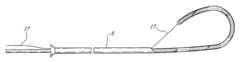
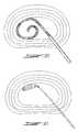
- FIG. 3is a detailed view of the introducer with probe fully inserted to illustrate the coil loop pre-form shape of this embodiment when allowed to freely rebound to its unconstrained configuration.
- FIG. 4is a further detailed view of the coil loop embodiment illustrated in FIG. 3 showing the helical resistant heat emitting elements within the coiled distal portion of the probe.
- FIG. 5is a detailed view of one embodiment of the probe showing longitudinal electrical wires connected to two separate helical thermal heating elements with elongate temperature sensors housed within a transparent flexible plastic casing.
- FIG. 6is a like detailed view of the distal portion of the probe, however, showing a central flexible metal core in dashed outline with two layers of external sheaths for electrical insulation and heat transfer capability.
- FIG. 7shows a second embodiment of the invention wherein the pre-formed shape is a single open loop to enable the distal portion of the probe to be positioned within the nucleus pulposus adjacent the posterior of the inner wall (see FIG. 22 ).
- FIG. 8is a third embodiment illustrating a pre-form shape as a closed loop with hooked end.
- FIGS. 9, 10 , 11 and 12show deployment steps for a fourth embodiment with preformed shape as an axially symmetric closed loop.
- FIG. 9is the first step of deployment of the fourth embodiment wherein the extension shaft of the probe has been extended slightly outwardly of the outer end of the introducer.
- FIG. 10shows the fourth embodiment of FIG. 9 wherein the extension shaft of the probe has been extended further outwardly of the outer end of the introducer and wherein the loop has a shape modified because of the pressure exerted on it by the inner wall, for example.
- FIG. 11shows the fourth embodiment of the FIG. 10 wherein the extension shaft of the probe has been extended further outwardly of the outer end of the introducer and wherein the loop has penetrated through the inner wall and into the fibrosus and is reaching its preform shape.
- FIG. 12shows the fourth embodiment of the FIG. 11 wherein the extension shaft of the probe has been extended further outwardly of the outer end of the introducer and wherein the loop has deployed fully into the disc.
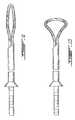
- FIG. 13shows a fifth embodiment of the invention with a pre-form in the shape of a semi-closed loop with active shape controlling cable attached to the extreme distal end of the probe and extending through the introducer enabling the surgeon to actively control the shape of the distal portion.
- FIG. 14shows the fifth embodiment of the invention with cable secured from the tip of the probe in order to fold the probe distal portion over into an open loop shape.
- FIG. 15shows the fifth embodiment with cable passing into the interior of the probe in contrast to passing the cable externally of the probe as in FIG. 13 .
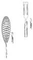
- FIG. 16shows a sixth embodiment of the invention wherein the pre-form shape is a helicoidal coil loop that collapses to withdraw through the introducer once the procedure has been completed.
- FIG. 17shows a seventh embodiment of the invention wherein the pre-form shape is a flat plate, arcuate sheet or elongate tape depending on the length and lateral flexibility of the distal end of the probe, wherein the heating elements are arranged to form a closed, semi-closed or open loop and showing beveled rearward edges that serve to roll or fold the distal end of the probe when retracted through the introducer.
- the pre-form shapeis a flat plate, arcuate sheet or elongate tape depending on the length and lateral flexibility of the distal end of the probe, wherein the heating elements are arranged to form a closed, semi-closed or open loop and showing beveled rearward edges that serve to roll or fold the distal end of the probe when retracted through the introducer.
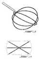
- FIG. 18is an eighth embodiment of the invention showing a pre-form shape of a partial spherical loop basket with six arcuate loop strands extending between outer and inner poles disposed longitudinally and being flexible enough to retract within the introducer when withdrawn.
- FIG. 19shows the eighth embodiment of FIG. 18 in an end view to indicate in dashed outline the ability of this embodiment to completely engage the inner wall of the annulus fibrosus.
- FIG. 20is a sectional view through an intervertebral disc showing insertion of the spiral coil loop embodiment that is shown in FIGS. 3 and 4.
- FIG. 21is a like sectional view showing the deployed configuration of the embodiment illustrated in FIG. 16 .
- FIG. 22is a like sectional view showing the deployed position of the embodiment illustrated in FIG. 7 .
- FIG. 23is like sectional view showing the deployed configuration of the embodiment illustrated in FIG. 12 .
- FIG. 1shows a lateral view of three human vertebrae 1 with intervertebral disc 2 between each vertebra 1 .
- the vertebral disc 2comprises two main structures with a central gelatinous nucleus pulposus 3 bounded by a tough annulus fibrosus 4 .
- the annulus fibrosus 4has a number of overlapping layers which are obliquely oriented spanning between the adjacent vertebra 1 in opposing overlapping orientations.
- the intradiscal lesioning deviceincludes an elongate introducer 5 having an inner beveled end inserted within the nucleus pulposus and an outer end which extends outwardly of the patient's body.
- a longitudinal hollow boreextends through the tube 5 and provides for percutaneous surgical insertion of the tube 5 through the annulus fibrosus 4 thereby providing external surgical access to the nucleus pulposus 3 through the bore.
- the elongate probe 6is pushed through to slidably engage and flexibly conform to the bore of the introducer 5 .
- the proximal portion 7 of the probe 6includes a handle and control means as well as markings 8 to visually indicate the extent of insertion of the probe 6 .
- the distal portion 9 of the probe 6is shown in FIG. 2 initially being longitudinally pushed out of the outer end of the introducer 5 into the soft gelatinous nucleus pulposus 3 .
- selected portions or the entire inner wall of the annulus fibrosus 4can be engaged.
- the distal portion 9may be pre-formed into a spiral coil shape to resiliently rebound to a spiral deployed configuration when released within the nucleus pulposus 3 while remaining capable of resiliently conforming to the bore within the tube 5 in a longitudinally slidable confined configuration therein.
- the distal portion 9can include passive or active means to change its shape once inserted into the nucleus pulposus 3 .
- Passive shape biasing meansto resiliently urge the distal portion 9 to adopt a selected pre-form shape, such as in FIG. 4 a spiral coil shape.
- passive shape biasing meanscan be constructed of various resilient materials such as shape memory metallic alloy, super elastic surgical rubber, resilient plastic and even natural rubber.
- a resilient solid fiber core 10 and resilient outer tubes 11 and 12can be provided depending on the necessary insulation and physical properties required.
- Resilient fibersmay also be woven in tubular shapes as in electrical co-axial cables or in an annular array of longitudinal resilient fibers or in a resilient protective sheet. Alternatively as shown in FIG.
- the entire probemay be molded as a solid plastic unit with embedded heating coils and control means.
- the passive shape biasing meanscan also be a resilient tube, a woven tube of resilient fibers, an annular array of longitudinal resilient fibers, or a resilient sheath.
- the distal portion 9 of the probe 6includes lesioning means for emitting energy toward the inner wall of the annulus fibrosus 4 when the distal portion 9 is in its deployed configuration (for example see FIGS. 20 through 23 ).
- the lesioning meanscomprise helical electrically powered heating coils 13 .
- the coils 13are individually controllable with separate electrical supply conduits 14 longitudinally through the length of the probe 6 .
- the probe 6may also contain various monitors such as a thermometer, thermistor, thermocouples or ammeters illustrated in FIGS. 5 and 6 as housed within a conduit 15 . If necessary, as shown in FIG. 5 further structural or resilient strength can be provided by a separate mounting wire 16 .
- the lesioning meansmay include any energy emitting device which may be of therapeutic use within the nucleus pulposus 3 such as thermal energy coils, radio frequency electric current, ammeters, microwave ammeters, ultrasound ammeters, radio active emission devices or optical emission devices.
- the intradiscal lesioning devicemay be designed to passively or actively adopt any number of deployed configurations.
- Various deployed configurationsare required due to the various shapes of the nucleus pulposus 3 , various sizes depending on the location of the injured intervertebral disc 2 , depending on the age of the patient, the clarity of definition between the nucleus pulposus and the annulus fibrosus 4 and depending on the extent of damage or rupture in the disc itself.
- FIGS. 3 and 4show an embodiment taking the pre-form shape of a spiral coil
- FIG. 7shows a pre-form shape of a simple overlapping open loop
- FIG. 8shows a device pre-formed into a hook shape with dual strands in a closed loop, however, it will be understood that a single strand hook shape can be provided by the embodiment shown in FIG. 7 .
- FIGS. 9, 10 , 11 and 12show the deployment of dual strand closed loops as they are being pushed out of the probe and as they attain their preform shape.
- FIG. 16shows a helical coil having varying diameter longitudinally, however, it will be understood that a coil of cylindrical shape or alternating diameter can be easily constructed.
- FIG. 17shows a relatively short flat plate configuration, however, it will be understood that an arcuate sheet or elongate tape may also be constructed in a like manner. As indicated in FIGS.
- FIGS. 18 and 19complex shapes of multiple strands can also be constructed provided that they are weak enough to collapse within the bore of the introducer when inserted and withdrawn from the nucleus pulposus while being rigid enough to be pushed longitudinally through the introducer into the nucleus pulposus.
- An advantage of the embodiment shown in FIGS. 18 and 19is the complete coverage of the interior wall of the annulus fibrosus with multiple strands which expand once installed.
- the lesioning devicemay also include active shape control means for progressively developing the trajectory of the distal portion 9 in three dimensional space as the distal portion is longitudinally slidably released from the outer end of the introducer 5 into the nucleus pulposus 3 .
- the active shape control meansis a cable that extends from the distal end through the introducer 5 .
- the cable 17extends from the extreme distal end of the probe through the introducer 5 .
- the cable 17is secured to the probe longitudinally spaced from the extreme distal end in order to enable the probe to fold over itself into an open loop.
- FIG. 15shows a further example of use of a cable that is threaded through the interior of the probe as opposed to extending parallel with the probe itself.
- mechanical activatorsare within the scope of the invention such as hydraulic actuators within the distal end, piezo-electric actuators or solenoid actuators. In all cases the actuators will serve to actively control the shape of the distal portion as it is released from the outer end of the introducer and is pushed through the nucleus pulposus.
- the inventionprovides means to control the shape, location and positioning of energy emitting devices within the distal portion of the probe thereby providing significant advantage over the prior art which relies on contact and sliding guidance of the distal end of the probe as it contacts the inner wall of the annulus fibrosus. Superior control over the shape and positioning of the distal portion serves to better locate and control the emission of thermal energy or other forms of energy to denervate the annulus fibrosus.
- the method for treating a condition of the intervertebral disc with an intradiscal lesioning apparatusutilizes the apparatus of the invention to modify the tissue at a selected location of the intervertebral disc.
- the patientis first diagnosed 30 using a number of methods to determine the condition of the disc. Examples of current diagnostic methods include discography, MRI and CT. An evaluation of the intensity of pain may also be used. If the disc shows signs of internal disruption, degeneration or non-protruding herniation the doctor may decide that the pain experienced is originating from the disc. If conventional therapy such as a combination of rest and physiotherapy is unsuccessful 31 a decision can be made to have the intradiscal therapy.
- the patientlies on the operating table in the prone position and the area of the back to be treated is cleaned, disinfected and draped 32 .
- a local anesthetic 33is administered to the area that is to be treated and the patient may be put under a mild sedative. It is important that the patient be conscious during the procedure to provide feedback to the practitioner to avoid damage of tissues around the disc such as the spinal cord or spinal nerves.
- An introduceris provided that is used for percutaneous, minimally invasive access to the disc.
- the introduceris a hollow shaft that is inserted through the skin approximately 10 to 15 cm lateral of the spine, travelling through the tissues of the back to the intervertebral disc that is to be treated.
- a trocarcan be used while inserting the introducer to stop tissue or fluid from entering the hollow lumen.
- the probehas an end that contains a functional component and the other end has a connection hub that connects the probe to a cable that leads to the energy source. Without connecting the cable to the probe, the probe is then slid into the lumen of the introducer 35 , functional component first. Placement of the probe can be aided by fluoroscopy. As the end of the probe containing the functional component is advanced out of the end of the introducer that is in the nucleus pulposus 36 , the length of the probe that is out of the introducer will assume its predefined shape 37 . The predefined shape is such that the functional component can be placed next to a selected region of the annulus fibrosus 38 .
- the probecan be connected to the cable, which in turn is connected to the energy source 39 .
- One of the embodimentshas a set of thermocouples imbedded in the introducer. If an introducer is used that contains temperature measuring elements it will have a connection hub on the end that is external to the patient's body. At this time the connection hub is connected to a cable that is connected to a measurement device 40 .
- the energy sourcecan be controlled by a program that creates a profile of amount of energy delivered 41 and uses the temperature of the disc tissue in a feedback loop.
- the form of energy to be usedis electricity such as AC electricity or radiofrequency AC electricity to heat a resistive thermal element
- one or more temperature sensorscan feedback to the power source controller and the desired amount of power can be sent 42 to the resistive thermal element in order to adjust the temperature to reach a set-point.
- the temperature profile of functional elementwould be designed to achieve the following criteria: the temperature at the region of the disc that contains nociceptors that are most likely to be causing discogenic pain is to be raised to at least 45° C.
- the temperature of the annulus fibrosus in the region of a fissure or disruptioncan be raised to at least 60° C. in order to constrict the collagen; the temperature must be raised slow enough so as to allow the heat to be dissipated to the outer third of the annulus fibrosus while not elevating the tissue in direct contact to the probe to cause thermal injury such as charring or causing an inflammatory response.
- the set-point for application of energycan change (increase, decrease or hold at the same level) according to a set profile.
- the feedback control systemdetermines the amount of energy delivered in relation to time. During the delivery of energy the patient is asked if any abnormal pain is experienced. This is done as a safeguard against injuring the spinal nerves near the disc.
- connection cablescan be disconnected 43 .
- the probeis then slowly removed from the introducer the deployed shape will be retracted through the introducer 44 .
- a solution of antibiotic 45can optionally be injected into the disc using the introducer. The reason for this is to avoid infection. The patient should then start noticing a difference in the pain felt 46 .
Landscapes
- Health & Medical Sciences (AREA)
- Surgery (AREA)
- Engineering & Computer Science (AREA)
- Life Sciences & Earth Sciences (AREA)
- Biomedical Technology (AREA)
- Molecular Biology (AREA)
- Nuclear Medicine, Radiotherapy & Molecular Imaging (AREA)
- Plasma & Fusion (AREA)
- Physics & Mathematics (AREA)
- Heart & Thoracic Surgery (AREA)
- Medical Informatics (AREA)
- Otolaryngology (AREA)
- Animal Behavior & Ethology (AREA)
- General Health & Medical Sciences (AREA)
- Public Health (AREA)
- Veterinary Medicine (AREA)
- Prostheses (AREA)
- Laser Surgery Devices (AREA)
Abstract
Description
Claims (11)
Priority Applications (1)
| Application Number | Priority Date | Filing Date | Title |
|---|---|---|---|
| US09/827,922US6562033B2 (en) | 2001-04-09 | 2001-04-09 | Intradiscal lesioning apparatus |
Applications Claiming Priority (1)
| Application Number | Priority Date | Filing Date | Title |
|---|---|---|---|
| US09/827,922US6562033B2 (en) | 2001-04-09 | 2001-04-09 | Intradiscal lesioning apparatus |
Publications (2)
| Publication Number | Publication Date |
|---|---|
| US20020147444A1 US20020147444A1 (en) | 2002-10-10 |
| US6562033B2true US6562033B2 (en) | 2003-05-13 |
Family
ID=25250488
Family Applications (1)
| Application Number | Title | Priority Date | Filing Date |
|---|---|---|---|
| US09/827,922Expired - LifetimeUS6562033B2 (en) | 2001-04-09 | 2001-04-09 | Intradiscal lesioning apparatus |
Country Status (1)
| Country | Link |
|---|---|
| US (1) | US6562033B2 (en) |
Cited By (132)
| Publication number | Priority date | Publication date | Assignee | Title |
|---|---|---|---|---|
| US20030014093A1 (en)* | 2001-05-29 | 2003-01-16 | Makin Inder Raj. S. | Excisional and ultrasound medical treatment system |
| US20030158545A1 (en)* | 2000-09-28 | 2003-08-21 | Arthrocare Corporation | Methods and apparatus for treating back pain |
| US20030212395A1 (en)* | 2000-05-12 | 2003-11-13 | Arthrocare Corporation | Systems and methods for electrosurgery |
| US20030216721A1 (en)* | 2002-01-15 | 2003-11-20 | The Regents Of The University Of Calfornia | System and method providing directional ultrasound therapy to skeletal joints |
| US20040024399A1 (en)* | 1995-04-13 | 2004-02-05 | Arthrocare Corporation | Method for repairing damaged intervertebral discs |
| US20040049180A1 (en)* | 1996-07-16 | 2004-03-11 | Arthrocare Corporation | Systems and methods for electrosurgical prevention of disc herniations |
| US20040087642A1 (en)* | 2002-10-24 | 2004-05-06 | Zeldis Jerome B. | Methods of using and compositions comprising a JNK inhibitor for the treatment, prevention, management and/or modification of pain |
| US20040102824A1 (en)* | 1996-08-13 | 2004-05-27 | Sharkey Hugh R. | Method for treating intervertebral discs |
| US20040127791A1 (en)* | 2001-05-29 | 2004-07-01 | Mast T. Douglas | Method for mapping temperature rise using pulse-echo ultrasound |
| US20040158248A1 (en)* | 2001-09-06 | 2004-08-12 | Ginn Richard S. | Apparatus and methods for treating spinal discs |
| US20040193151A1 (en)* | 2001-06-06 | 2004-09-30 | Oratec Interventions, Inc. | Intervertebral disc device employing looped probe |
| US20050240171A1 (en)* | 2004-04-23 | 2005-10-27 | Forrest Leonard E | Device and method for treatment of intervertebral disc disruption |
| US20060020246A1 (en)* | 2004-07-22 | 2006-01-26 | Mclucas Bruce | Angiographic catheter for uterine artery embolization |
| US20060064145A1 (en)* | 2004-09-21 | 2006-03-23 | Podhajsky Ronald J | Method for treatment of an intervertebral disc |
| US20060167500A1 (en)* | 2002-08-19 | 2006-07-27 | Bruce Towe | Neurostimulator |
| US20060184246A1 (en)* | 2004-06-10 | 2006-08-17 | Zwirkoski Paul A | Non-soft tissue repair |
| US20060217705A1 (en)* | 2005-02-17 | 2006-09-28 | Baylis Medical Company Inc. | Electrosurgical device with discontinuous flow density |
| US20060224219A1 (en)* | 2005-03-31 | 2006-10-05 | Sherwood Services Ag | Method of using neural stimulation during nucleoplasty procedures |
| US20060247643A1 (en)* | 2005-04-29 | 2006-11-02 | Jmea Corporation | Tissue repair system |
| US20060259026A1 (en)* | 2005-05-05 | 2006-11-16 | Baylis Medical Company Inc. | Electrosurgical treatment method and device |
| US20070016273A1 (en)* | 2005-05-26 | 2007-01-18 | Smith & Nephew, Inc. | Electrothermal intervertebral disc treatment |
| US20070027449A1 (en)* | 2002-03-05 | 2007-02-01 | Baylis Medical Company Inc. | Electrosurgical device and methods |
| US20070038222A1 (en)* | 2005-04-29 | 2007-02-15 | Jmea Corporation | Tissue Repair System |
| US20070123888A1 (en)* | 2004-10-15 | 2007-05-31 | Baxano, Inc. | Flexible tissue rasp |
| US20070135881A1 (en)* | 2005-01-11 | 2007-06-14 | Vilims Bradley D | Combination Electrical Stimulating And Infusion Medical Device and Method |
| US20070142791A1 (en)* | 2003-05-07 | 2007-06-21 | Aleeve Medical Inc. A Delaware Corporation | U-shaped disc shunt and delivery device |
| US20070203402A1 (en)* | 2002-03-05 | 2007-08-30 | Baylis Medical | Elongate member providing a variation in radiopacity |
| US7270659B2 (en) | 1995-06-07 | 2007-09-18 | Arthrocare Corporation | Methods for electrosurgical treatment of spinal tissue |
| US20070225312A1 (en)* | 2006-03-21 | 2007-09-27 | Ergonex Pharma Gmbh | Terguride / proterguride for the treatment of chronic pain |
| US20080009828A1 (en)* | 2004-04-16 | 2008-01-10 | Kyphon, Inc. | Spinal diagnostic methods and apparatus |
| US20080009927A1 (en)* | 2005-01-11 | 2008-01-10 | Vilims Bradley D | Combination Electrical Stimulating and Infusion Medical Device and Method |
| US20080125747A1 (en)* | 2006-11-28 | 2008-05-29 | Smith & Nephew, Inc.-Tn | Passive thermal spine catheter |
| US7387625B2 (en) | 1995-06-07 | 2008-06-17 | Arthrocare Corporation | Methods and apparatus for treating intervertebral discs |
| US7393351B2 (en) | 1995-06-07 | 2008-07-01 | Arthrocare Corporation | Apparatus and methods for treating cervical inter-vertebral discs |
| US20080200972A1 (en)* | 2005-01-11 | 2008-08-21 | Rittman William J | Combination electrical stimulating and infusion medical device and method |
| US20080215033A1 (en)* | 2004-04-16 | 2008-09-04 | Kyphon, Inc. | Spinal diagnostic methods and apparatus |
| US20080221490A1 (en)* | 2007-03-06 | 2008-09-11 | The Cleveland Clinic Foundation | Method and apparatus for repair of intervertebral discs |
| US20080228135A1 (en)* | 2007-03-05 | 2008-09-18 | Elizabeth Ann Snoderly | Apparatus for treating a damaged spinal disc |
| US7449019B2 (en)* | 1999-01-25 | 2008-11-11 | Smith & Nephew, Inc. | Intervertebral decompression |
| US7449021B2 (en) | 1996-07-16 | 2008-11-11 | Arthrocare Corporation | Systems and methods for electrosurgical tissue contraction within the spine |
| US20080312660A1 (en)* | 2007-06-15 | 2008-12-18 | Baxano, Inc. | Devices and methods for measuring the space around a nerve root |
| US7473250B2 (en) | 2004-05-21 | 2009-01-06 | Ethicon Endo-Surgery, Inc. | Ultrasound medical system and method |
| US20090024124A1 (en)* | 2005-07-14 | 2009-01-22 | Lefler Amy | Methods for treating the thoracic region of a patient's body |
| US20090030308A1 (en)* | 2005-09-21 | 2009-01-29 | The Regents Of The University Of California | Systems, compositions, and methods for local imaging and treatment of pain |
| US20090054962A1 (en)* | 2002-03-05 | 2009-02-26 | Baylis Medical Company Inc. | Methods for treating the thoracic region of a patient's body |
| US20090099613A1 (en)* | 2005-01-11 | 2009-04-16 | Vilims Bradley D | Combination electrical stimulating and infusion device and method |
| US7553307B2 (en) | 2004-10-15 | 2009-06-30 | Baxano, Inc. | Devices and methods for tissue modification |
| US20090182478A1 (en)* | 2008-01-15 | 2009-07-16 | Gm Global Technology Operations, Inc. | Axle torque based cruise control |
| US20090198248A1 (en)* | 2008-02-04 | 2009-08-06 | Yeung Jeffrey E | Device for disc shunt implantation and peri-shunt injection |
| US7578819B2 (en)* | 2005-05-16 | 2009-08-25 | Baxano, Inc. | Spinal access and neural localization |
| US7662177B2 (en) | 2006-04-12 | 2010-02-16 | Bacoustics, Llc | Apparatus and methods for pain relief using ultrasound waves in combination with cryogenic energy |
| US20100076422A1 (en)* | 2008-09-24 | 2010-03-25 | Tyco Healthcare Group Lp | Thermal Treatment of Nucleus Pulposus |
| US7708733B2 (en) | 2003-10-20 | 2010-05-04 | Arthrocare Corporation | Electrosurgical method and apparatus for removing tissue within a bone body |
| US20100145424A1 (en)* | 2004-09-21 | 2010-06-10 | Covidien Ag | Method for Treatment of an Intervertebral Disc |
| US7738969B2 (en) | 2004-10-15 | 2010-06-15 | Baxano, Inc. | Devices and methods for selective surgical removal of tissue |
| US20100198039A1 (en)* | 2007-05-04 | 2010-08-05 | Arizona Board Of Regents For And On Behalf Of Arizona State University | Systems and Methods for Wireless Transmission of Biopotentials |
| US7794456B2 (en) | 2003-05-13 | 2010-09-14 | Arthrocare Corporation | Systems and methods for electrosurgical intervertebral disc replacement |
| US7806839B2 (en) | 2004-06-14 | 2010-10-05 | Ethicon Endo-Surgery, Inc. | System and method for ultrasound therapy using grating lobes |
| US7846096B2 (en) | 2001-05-29 | 2010-12-07 | Ethicon Endo-Surgery, Inc. | Method for monitoring of medical treatment using pulse-echo ultrasound |
| US7857813B2 (en) | 2006-08-29 | 2010-12-28 | Baxano, Inc. | Tissue access guidewire system and method |
| US7879034B2 (en) | 2006-03-02 | 2011-02-01 | Arthrocare Corporation | Internally located return electrode electrosurgical apparatus, system and method |
| US7887538B2 (en) | 2005-10-15 | 2011-02-15 | Baxano, Inc. | Methods and apparatus for tissue modification |
| US20110071639A1 (en)* | 2004-04-23 | 2011-03-24 | Leonard Edward Forrest | Method and device for treatment of the spine |
| US20110071548A1 (en)* | 2009-09-22 | 2011-03-24 | Jmea Corporation | Tissue Repair System |
| US7918849B2 (en)* | 2004-10-15 | 2011-04-05 | Baxano, Inc. | Devices and methods for tissue access |
| US7938830B2 (en) | 2004-10-15 | 2011-05-10 | Baxano, Inc. | Powered tissue modification devices and methods |
| US7959577B2 (en) | 2007-09-06 | 2011-06-14 | Baxano, Inc. | Method, system, and apparatus for neural localization |
| US20110166603A1 (en)* | 2004-04-23 | 2011-07-07 | Leonard Edward Forrest | Method and device for placing materials in the spine |
| US8062300B2 (en) | 2006-05-04 | 2011-11-22 | Baxano, Inc. | Tissue removal with at least partially flexible devices |
| US8062298B2 (en) | 2005-10-15 | 2011-11-22 | Baxano, Inc. | Flexible tissue removal devices and methods |
| US8092456B2 (en) | 2005-10-15 | 2012-01-10 | Baxano, Inc. | Multiple pathways for spinal nerve root decompression from a single access point |
| US20120065570A1 (en)* | 2010-09-11 | 2012-03-15 | Yeung Jeffrey E | Disc shunt delivery with stepped needle |
| US8192436B2 (en) | 2007-12-07 | 2012-06-05 | Baxano, Inc. | Tissue modification devices |
| US8221397B2 (en) | 2004-10-15 | 2012-07-17 | Baxano, Inc. | Devices and methods for tissue modification |
| US8257356B2 (en) | 2004-10-15 | 2012-09-04 | Baxano, Inc. | Guidewire exchange systems to treat spinal stenosis |
| US8366712B2 (en) | 2005-10-15 | 2013-02-05 | Baxano, Inc. | Multiple pathways for spinal nerve root decompression from a single access point |
| US8394102B2 (en) | 2009-06-25 | 2013-03-12 | Baxano, Inc. | Surgical tools for treatment of spinal stenosis |
| US8398641B2 (en) | 2008-07-01 | 2013-03-19 | Baxano, Inc. | Tissue modification devices and methods |
| US8409206B2 (en) | 2008-07-01 | 2013-04-02 | Baxano, Inc. | Tissue modification devices and methods |
| US8430881B2 (en) | 2004-10-15 | 2013-04-30 | Baxano, Inc. | Mechanical tissue modification devices and methods |
| US8470043B2 (en) | 2008-12-23 | 2013-06-25 | Benvenue Medical, Inc. | Tissue removal tools and methods of use |
| US8480666B2 (en) | 2007-01-31 | 2013-07-09 | Covidien Lp | Thermal feedback systems and methods of using the same |
| US20130245739A1 (en)* | 2012-03-14 | 2013-09-19 | Anatoly Arber | Self-anchored stimulator lead and method of insertion |
| US8568416B2 (en) | 2004-10-15 | 2013-10-29 | Baxano Surgical, Inc. | Access and tissue modification systems and methods |
| US8613745B2 (en) | 2004-10-15 | 2013-12-24 | Baxano Surgical, Inc. | Methods, systems and devices for carpal tunnel release |
| US8702718B2 (en) | 2005-04-29 | 2014-04-22 | Jmea Corporation | Implantation system for tissue repair |
| US8801626B2 (en) | 2004-10-15 | 2014-08-12 | Baxano Surgical, Inc. | Flexible neural localization devices and methods |
| US8845639B2 (en) | 2008-07-14 | 2014-09-30 | Baxano Surgical, Inc. | Tissue modification devices |
| US8979838B2 (en) | 2010-05-24 | 2015-03-17 | Arthrocare Corporation | Symmetric switching electrode method and related system |
| US20150094706A1 (en)* | 2013-09-29 | 2015-04-02 | Covidien Lp | Medical treatment devices having adjustable length and/or diameter |
| US9101386B2 (en) | 2004-10-15 | 2015-08-11 | Amendia, Inc. | Devices and methods for treating tissue |
| US9113950B2 (en) | 2009-11-04 | 2015-08-25 | Regenerative Sciences, Llc | Therapeutic delivery device |
| US9133438B2 (en) | 2011-06-29 | 2015-09-15 | Biorestorative Therapies, Inc. | Brown fat cell compositions and methods |
| US9161773B2 (en) | 2008-12-23 | 2015-10-20 | Benvenue Medical, Inc. | Tissue removal tools and methods of use |
| US9247952B2 (en) | 2004-10-15 | 2016-02-02 | Amendia, Inc. | Devices and methods for tissue access |
| US9314253B2 (en) | 2008-07-01 | 2016-04-19 | Amendia, Inc. | Tissue modification devices and methods |
| US9446255B2 (en) | 2011-11-13 | 2016-09-20 | Arizona Board of Regents on Behalf Arizona State University | Controlled stimulation delivery from neurostimulator |
| US9456829B2 (en) | 2004-10-15 | 2016-10-04 | Amendia, Inc. | Powered tissue modification devices and methods |
| US9724107B2 (en) | 2008-09-26 | 2017-08-08 | Relievant Medsystems, Inc. | Nerve modulation systems |
| US9724151B2 (en) | 2013-08-08 | 2017-08-08 | Relievant Medsystems, Inc. | Modulating nerves within bone using bone fasteners |
| US9770280B2 (en) | 2002-01-23 | 2017-09-26 | The Regents Of The University Of California | Implantable thermal treatment method and apparatus |
| US9775627B2 (en) | 2012-11-05 | 2017-10-03 | Relievant Medsystems, Inc. | Systems and methods for creating curved paths through bone and modulating nerves within the bone |
| WO2018067496A1 (en) | 2016-10-04 | 2018-04-12 | Avent, Inc. | Cooled rf probes |
| US9949789B2 (en) | 2002-03-05 | 2018-04-24 | Avent, Inc. | Methods of treating the sacroiliac region of a patient's body |
| US10022566B2 (en) | 2010-08-31 | 2018-07-17 | Arizona Board Of Regents On Behalf Of Arizona State University | Apparatus, systems, and methods for current monitoring in ultrasound powered neurostimulation |
| US10080571B2 (en) | 2015-03-06 | 2018-09-25 | Warsaw Orthopedic, Inc. | Surgical instrument and method |
| US10111704B2 (en) | 2002-09-30 | 2018-10-30 | Relievant Medsystems, Inc. | Intraosseous nerve treatment |
| US10265099B2 (en) | 2008-09-26 | 2019-04-23 | Relievant Medsystems, Inc. | Systems for accessing nerves within bone |
| US10314605B2 (en) | 2014-07-08 | 2019-06-11 | Benvenue Medical, Inc. | Apparatus and methods for disrupting intervertebral disc tissue |
| US10390877B2 (en) | 2011-12-30 | 2019-08-27 | Relievant Medsystems, Inc. | Systems and methods for treating back pain |
| US10441339B2 (en) | 2015-11-17 | 2019-10-15 | Medtronic Holding Company Sárl | Spinal tissue ablation apparatus, system, and method |
| US10463423B2 (en) | 2003-03-28 | 2019-11-05 | Relievant Medsystems, Inc. | Thermal denervation devices and methods |
| US10588691B2 (en) | 2012-09-12 | 2020-03-17 | Relievant Medsystems, Inc. | Radiofrequency ablation of tissue within a vertebral body |
| US10716618B2 (en) | 2010-05-21 | 2020-07-21 | Stratus Medical, LLC | Systems and methods for tissue ablation |
| US10736688B2 (en) | 2009-11-05 | 2020-08-11 | Stratus Medical, LLC | Methods and systems for spinal radio frequency neurotomy |
| US10786300B2 (en) | 2015-04-13 | 2020-09-29 | Carlos Fernando Bazoberry | Radiofrequency denervation needle and method |
| USRE48460E1 (en) | 2002-09-30 | 2021-03-09 | Relievant Medsystems, Inc. | Method of treating an intraosseous nerve |
| US11007010B2 (en) | 2019-09-12 | 2021-05-18 | Relevant Medsysterns, Inc. | Curved bone access systems |
| US11224475B2 (en) | 2010-04-26 | 2022-01-18 | Medtronic Holding Company Sàrl | Electrosurgical device and methods |
| US11291496B2 (en) | 2002-03-05 | 2022-04-05 | Avent, Inc. | Methods of treating the sacroiliac region of a patient's body |
| US11471145B2 (en) | 2018-03-16 | 2022-10-18 | Spinal Elements, Inc. | Articulated instrumentation and methods of using the same |
| US11564811B2 (en) | 2015-02-06 | 2023-01-31 | Spinal Elements, Inc. | Graft material injector system and method |
| US11576716B2 (en) | 2013-03-15 | 2023-02-14 | Medtronic Holding Company Sàrl | Electrosurgical mapping tools and methods |
| US11583327B2 (en) | 2018-01-29 | 2023-02-21 | Spinal Elements, Inc. | Minimally invasive interbody fusion |
| US11771483B2 (en) | 2017-03-22 | 2023-10-03 | Spinal Elements, Inc. | Minimal impact access system to disc space |
| US11931016B2 (en) | 2013-03-07 | 2024-03-19 | Medtronic Holding Company Sàrl | Systems and methods for track coagulation |
| USRE49994E1 (en) | 2013-03-14 | 2024-06-04 | Spinal Elements, Inc. | Spinal fusion implants and devices and methods for deploying such implants |
| US12039731B2 (en) | 2020-12-22 | 2024-07-16 | Relievant Medsystems, Inc. | Prediction of candidates for spinal neuromodulation |
| US12035961B2 (en) | 2015-04-13 | 2024-07-16 | Carlos Fernando Bazoberry | Radiofrequency denervation needle and method |
| US12076074B2 (en) | 2010-04-26 | 2024-09-03 | Medtronic Holding Company Sàrl | Electrosurgical device and methods |
| US12082876B1 (en) | 2020-09-28 | 2024-09-10 | Relievant Medsystems, Inc. | Introducer drill |
| US12433668B1 (en) | 2021-11-08 | 2025-10-07 | Relievant Medsystems, Inc. | Impedance stoppage mitigation during radiofrequency tissue ablation procedures |
Families Citing this family (20)
| Publication number | Priority date | Publication date | Assignee | Title |
|---|---|---|---|---|
| US6306132B1 (en) | 1999-06-17 | 2001-10-23 | Vivant Medical | Modular biopsy and microwave ablation needle delivery apparatus adapted to in situ assembly and method of use |
| WO2001060235A2 (en)* | 2000-02-18 | 2001-08-23 | Fogarty Thomas J M D | Improved device for accurately marking tissue |
| US7128739B2 (en) | 2001-11-02 | 2006-10-31 | Vivant Medical, Inc. | High-strength microwave antenna assemblies and methods of use |
| US6878147B2 (en) | 2001-11-02 | 2005-04-12 | Vivant Medical, Inc. | High-strength microwave antenna assemblies |
| US6752767B2 (en) | 2002-04-16 | 2004-06-22 | Vivant Medical, Inc. | Localization element with energized tip |
| US7197363B2 (en) | 2002-04-16 | 2007-03-27 | Vivant Medical, Inc. | Microwave antenna having a curved configuration |
| US8613744B2 (en) | 2002-09-30 | 2013-12-24 | Relievant Medsystems, Inc. | Systems and methods for navigating an instrument through bone |
| US8808284B2 (en) | 2008-09-26 | 2014-08-19 | Relievant Medsystems, Inc. | Systems for navigating an instrument through bone |
| US7311703B2 (en)* | 2003-07-18 | 2007-12-25 | Vivant Medical, Inc. | Devices and methods for cooling microwave antennas |
| US7799019B2 (en) | 2005-05-10 | 2010-09-21 | Vivant Medical, Inc. | Reinforced high strength microwave antenna |
| US20070073282A1 (en)* | 2005-09-26 | 2007-03-29 | Starion Instruments Corporation | Resistive heating device and method for turbinate ablation |
| US8075556B2 (en)* | 2006-05-23 | 2011-12-13 | Andres Betts | High frequency epidural neuromodulation catheter for effectuating RF treatment in spinal canal and method of using same |
| US8068921B2 (en) | 2006-09-29 | 2011-11-29 | Vivant Medical, Inc. | Microwave antenna assembly and method of using the same |
| US7998139B2 (en) | 2007-04-25 | 2011-08-16 | Vivant Medical, Inc. | Cooled helical antenna for microwave ablation |
| US8353901B2 (en) | 2007-05-22 | 2013-01-15 | Vivant Medical, Inc. | Energy delivery conduits for use with electrosurgical devices |
| US9023024B2 (en) | 2007-06-20 | 2015-05-05 | Covidien Lp | Reflective power monitoring for microwave applications |
| US8651146B2 (en) | 2007-09-28 | 2014-02-18 | Covidien Lp | Cable stand-off |
| US8292880B2 (en) | 2007-11-27 | 2012-10-23 | Vivant Medical, Inc. | Targeted cooling of deployable microwave antenna |
| LT5605B (en)* | 2007-11-28 | 2009-11-25 | Kauno technologijos universitetas, , | An ultrasound wave guide for cleaning of internal blood vessels |
| JP2012529335A (en)* | 2009-06-09 | 2012-11-22 | ユー アンド アイ コーポレーション | Directionally adjustable electrode body and guide tube for selective removal of body tissue |
Citations (7)
| Publication number | Priority date | Publication date | Assignee | Title |
|---|---|---|---|---|
| US5167658A (en)* | 1991-01-31 | 1992-12-01 | Mdt Corporation | Method and apparatus for electrosurgical measurement |
| US5201729A (en) | 1990-01-12 | 1993-04-13 | Laserscope | Method for performing percutaneous diskectomy using a laser |
| US5433739A (en) | 1993-11-02 | 1995-07-18 | Sluijter; Menno E. | Method and apparatus for heating an intervertebral disc for relief of back pain |
| WO1999047058A2 (en) | 1998-03-19 | 1999-09-23 | Oratec Interventions, Inc. | Catheter for delivery of energy to a surgical site |
| US5980504A (en) | 1996-08-13 | 1999-11-09 | Oratec Interventions, Inc. | Method for manipulating tissue of an intervertebral disc |
| US6007570A (en) | 1996-08-13 | 1999-12-28 | Oratec Interventions, Inc. | Apparatus with functional element for performing function upon intervertebral discs |
| US6099514A (en) | 1996-08-13 | 2000-08-08 | Oratec Interventions, Inc. | Method and apparatus for delivering or removing material from the interior of an intervertebral disc |
- 2001
- 2001-04-09USUS09/827,922patent/US6562033B2/ennot_activeExpired - Lifetime
Patent Citations (12)
| Publication number | Priority date | Publication date | Assignee | Title |
|---|---|---|---|---|
| US5201729A (en) | 1990-01-12 | 1993-04-13 | Laserscope | Method for performing percutaneous diskectomy using a laser |
| US5167658A (en)* | 1991-01-31 | 1992-12-01 | Mdt Corporation | Method and apparatus for electrosurgical measurement |
| US5433739A (en) | 1993-11-02 | 1995-07-18 | Sluijter; Menno E. | Method and apparatus for heating an intervertebral disc for relief of back pain |
| US5980504A (en) | 1996-08-13 | 1999-11-09 | Oratec Interventions, Inc. | Method for manipulating tissue of an intervertebral disc |
| US6007570A (en) | 1996-08-13 | 1999-12-28 | Oratec Interventions, Inc. | Apparatus with functional element for performing function upon intervertebral discs |
| US6073051A (en) | 1996-08-13 | 2000-06-06 | Oratec Interventions, Inc. | Apparatus for treating intervertebal discs with electromagnetic energy |
| US6095149A (en) | 1996-08-13 | 2000-08-01 | Oratec Interventions, Inc. | Method for treating intervertebral disc degeneration |
| US6099514A (en) | 1996-08-13 | 2000-08-08 | Oratec Interventions, Inc. | Method and apparatus for delivering or removing material from the interior of an intervertebral disc |
| US6122549A (en) | 1996-08-13 | 2000-09-19 | Oratec Interventions, Inc. | Apparatus for treating intervertebral discs with resistive energy |
| US6126682A (en) | 1996-08-13 | 2000-10-03 | Oratec Interventions, Inc. | Method for treating annular fissures in intervertebral discs |
| US6258086B1 (en)* | 1996-10-23 | 2001-07-10 | Oratec Interventions, Inc. | Catheter for delivery of energy to a surgical site |
| WO1999047058A2 (en) | 1998-03-19 | 1999-09-23 | Oratec Interventions, Inc. | Catheter for delivery of energy to a surgical site |
Non-Patent Citations (20)
| Title |
|---|
| 510(k) Summary SpineCATH, Dec. 17, 1999, Document No. K993967, 4 pages. |
| 510(k) Summary: Radionics Disc catheter Electrode System, Document K001741, Oct. 23, 2000, 5 pages. |
| 510(k) Summary: SpineCATH Mar. 19, 1998 and Document #K974464, Jan. 23, 1998, 8 pages. |
| Abstract: Arthroscopic Electro-Thermal Surgery for Discogenic Low Back Pain: A preliminary Report. May 25, 1998, 1 page. |
| Derby, R., Eek, B., Chen, Y., O'Neill, C., Ryan D., Intradiscal Electrothermal Annuloplasty (IDET): A novel Approach for Treating Chronic Discogenic Back Pain. Neuromodulation. 2000, 3(2), 82-88. |
| http://www.orthoassociates.com/IDET.htm, Jun. 11, 2000, 5 pages. |
| IDET Procedure with Spine CATCH, Dec. 2000, Oratec Interventions Inc., 77 0004, Rev. 4, pp. 1-6. |
| IDET Reference Literature List with abstracts, Mar. 15, 2001, Oratec Interventions Inc., 77 0023, Rev. 09, pp. 1-5. |
| IDET Reference Literature List, Jun. 15, 2000, Oratec Interventions Inc., Doc #770023 Rev 0S, 7 pages. |
| Jeffrey A. Saal, MD, and Joel S. Saal, MD, "Intradiscal Electrothermal Therapy for the Treatment of Chronic Discogenic Low Back Pain", Operative Techniques in Orthopaedics, vol. 10, No. 4, Oct. 2000, pp. 271-281. |
| Michael Karasek, MD, and Nikolai Bogduk, MD, PhD, DSct, "Twelve-Month Follow-Up of a Controlled Trial of Intradiscal Thermal Anuloplasty for Back Pain Due to Internal Disc Disruption", Reprinted from Spine, vol. 25, No. 20, Oct. 2000, Lippincott Williams & Wilkins, pp. 2601-2607. |
| Nerve Ingrowth Into Diseased Intervertebral Disc in Chronic Back Pain, A.J. Freement, T.E. Peacock, P. Goupille, J.A. Hoyland, J. O'Brian, MIV Jayson., The Lancet, pp. 178-181. |
| Press Release: Spine Journal Studies Support Oratec's IDET Back Pain Treatment Two Peer Reviewed Articles Report Substantial Reduction in Pain. Oratec, Oct. 19, 2000, 1 page. |
| Radionics RF Lesioning Systems and RF Catheter Electrode System Photo. |
| Saal, Joel, Saal, Jeffrey. Management of Chronic Discogenic Low Back Pain with a Thermal Intradiscal Cathethe. Spine. 2000, 25(3), 382-388. |
| Spine vol. 21, No. 15, HOUPT, Jonathan C. et al. Experimental Study of Temperature Distributions and Thermal Transport During Radiofrequency Current Therapy of the Intervertebral Disc. pp. 1808-1813, 1996, Lippincott-Raven Publishers.m by J.C. Houpt, Scanner, E. McFarland. |
| SpineCATH Intradiscal catheter by Oratec, Instructions for use, Document No. 700242, Rev. 03,. |
| Wiesel S W, Sharp Criticism of Intradiscal Thermal Therapy Research. The Back Letter. 2000, 15(6), 61-64. Lippincott Williams & Wilkins, 1 page. |
| Wiesel, S.W., IDET: Is There Any Indication That Intradiscal Thermal Therapy Works? The Back Letter. 2000, 15(6), 64-65. Lippincott Williams & Wilkins, 3 pages. |
| Yahoo News Re: Oratec Dec. 20, 2000, Jan. 25, 2001, Mar. 3, 2001, 6 pages. |
Cited By (304)
| Publication number | Priority date | Publication date | Assignee | Title |
|---|---|---|---|---|
| US7318823B2 (en) | 1995-04-13 | 2008-01-15 | Arthrocare Corporation | Methods for repairing damaged intervertebral discs |
| US20040024399A1 (en)* | 1995-04-13 | 2004-02-05 | Arthrocare Corporation | Method for repairing damaged intervertebral discs |
| USRE40156E1 (en) | 1995-06-07 | 2008-03-18 | Arthrocare Corporation | Methods for repairing damaged intervertebral discs |
| US7387625B2 (en) | 1995-06-07 | 2008-06-17 | Arthrocare Corporation | Methods and apparatus for treating intervertebral discs |
| US7393351B2 (en) | 1995-06-07 | 2008-07-01 | Arthrocare Corporation | Apparatus and methods for treating cervical inter-vertebral discs |
| US7270659B2 (en) | 1995-06-07 | 2007-09-18 | Arthrocare Corporation | Methods for electrosurgical treatment of spinal tissue |
| US7449021B2 (en) | 1996-07-16 | 2008-11-11 | Arthrocare Corporation | Systems and methods for electrosurgical tissue contraction within the spine |
| US7357798B2 (en) | 1996-07-16 | 2008-04-15 | Arthrocare Corporation | Systems and methods for electrosurgical prevention of disc herniations |
| US20040049180A1 (en)* | 1996-07-16 | 2004-03-11 | Arthrocare Corporation | Systems and methods for electrosurgical prevention of disc herniations |
| US7400930B2 (en) | 1996-08-13 | 2008-07-15 | Oratec Interventions, Inc. | Method for treating intervertebral discs |
| US7282061B2 (en) | 1996-08-13 | 2007-10-16 | Oratec Interventions, Inc. | Method of treating intervertebral disc |
| US8187312B2 (en) | 1996-08-13 | 2012-05-29 | Neurotherm, Inc. | Method for treating intervertebral disc |
| US8226697B2 (en) | 1996-08-13 | 2012-07-24 | Neurotherm, Inc. | Method for treating intervertebral disc |
| US7647123B2 (en) | 1996-08-13 | 2010-01-12 | Oratec Interventions, Inc. | Method for treating intervertebral discs |
| US20040102824A1 (en)* | 1996-08-13 | 2004-05-27 | Sharkey Hugh R. | Method for treating intervertebral discs |
| US7449019B2 (en)* | 1999-01-25 | 2008-11-11 | Smith & Nephew, Inc. | Intervertebral decompression |
| US7462178B2 (en) | 2000-05-12 | 2008-12-09 | Arthrocare Corporation | Systems and methods for electrosurgical spine surgery |
| US20030212395A1 (en)* | 2000-05-12 | 2003-11-13 | Arthrocare Corporation | Systems and methods for electrosurgery |
| US7331956B2 (en)* | 2000-09-28 | 2008-02-19 | Arthrocare Corporation | Methods and apparatus for treating back pain |
| US20030158545A1 (en)* | 2000-09-28 | 2003-08-21 | Arthrocare Corporation | Methods and apparatus for treating back pain |
| US20130226040A1 (en)* | 2001-05-29 | 2013-08-29 | Slayton H. Michael | Tissue-Retaining Systems for Ultrasound Medical Treatment |
| US7211044B2 (en) | 2001-05-29 | 2007-05-01 | Ethicon Endo-Surgery, Inc. | Method for mapping temperature rise using pulse-echo ultrasound |
| US20040127791A1 (en)* | 2001-05-29 | 2004-07-01 | Mast T. Douglas | Method for mapping temperature rise using pulse-echo ultrasound |
| US9261596B2 (en) | 2001-05-29 | 2016-02-16 | T. Douglas Mast | Method for monitoring of medical treatment using pulse-echo ultrasound |
| US9005144B2 (en)* | 2001-05-29 | 2015-04-14 | Michael H. Slayton | Tissue-retaining systems for ultrasound medical treatment |
| US20030014093A1 (en)* | 2001-05-29 | 2003-01-16 | Makin Inder Raj. S. | Excisional and ultrasound medical treatment system |
| US7806892B2 (en)* | 2001-05-29 | 2010-10-05 | Ethicon Endo-Surgery, Inc. | Tissue-retaining system for ultrasound medical treatment |
| US7473224B2 (en) | 2001-05-29 | 2009-01-06 | Ethicon Endo-Surgery, Inc. | Deployable ultrasound medical transducers |
| US8333721B2 (en)* | 2001-05-29 | 2012-12-18 | Makin Inder Raj S | Tissue-retaining system for ultrasound medical treatment |
| US7846096B2 (en) | 2001-05-29 | 2010-12-07 | Ethicon Endo-Surgery, Inc. | Method for monitoring of medical treatment using pulse-echo ultrasound |
| US20100312151A1 (en)* | 2001-05-29 | 2010-12-09 | Makin Inder Raj S | Tissue-retaining system for ultrasound medical treatment |
| US20030018266A1 (en)* | 2001-05-29 | 2003-01-23 | Makin Inder Raj. S. | Faceted ultrasound medical transducer assembly |
| US20040193151A1 (en)* | 2001-06-06 | 2004-09-30 | Oratec Interventions, Inc. | Intervertebral disc device employing looped probe |
| US20040158248A1 (en)* | 2001-09-06 | 2004-08-12 | Ginn Richard S. | Apparatus and methods for treating spinal discs |
| US7211055B2 (en)* | 2002-01-15 | 2007-05-01 | The Regents Of The University Of California | System and method providing directional ultrasound therapy to skeletal joints |
| US10603522B2 (en) | 2002-01-15 | 2020-03-31 | The Regents Of The University Of California | Method of treating back pain with microwave sources |
| US10589131B2 (en) | 2002-01-15 | 2020-03-17 | The Regents Of The University Of California | Methods of delivering chemical denervation to the vertebral body |
| US20030216721A1 (en)* | 2002-01-15 | 2003-11-20 | The Regents Of The University Of Calfornia | System and method providing directional ultrasound therapy to skeletal joints |
| US8915949B2 (en) | 2002-01-15 | 2014-12-23 | The Regents Of The University Of California | Method for providing directional therapy to skeletal joints |
| US11052267B2 (en) | 2002-01-15 | 2021-07-06 | The Regents Of The University Of California | Back pain treatment using microwave sources |
| US10272271B2 (en) | 2002-01-15 | 2019-04-30 | The Regents Of The University Of California | Method for providing directional therapy to skeletal joints |
| US20060241576A1 (en)* | 2002-01-15 | 2006-10-26 | Diederich Chris J | System and method providing directional ultrasound therapy to skeletal joints |
| US9770280B2 (en) | 2002-01-23 | 2017-09-26 | The Regents Of The University Of California | Implantable thermal treatment method and apparatus |
| US10206739B2 (en) | 2002-03-05 | 2019-02-19 | Avent, Inc. | Electrosurgical device and methods |
| US9216053B2 (en) | 2002-03-05 | 2015-12-22 | Avent, Inc. | Elongate member providing a variation in radiopacity |
| US20090054962A1 (en)* | 2002-03-05 | 2009-02-26 | Baylis Medical Company Inc. | Methods for treating the thoracic region of a patient's body |
| US9364281B2 (en) | 2002-03-05 | 2016-06-14 | Avent, Inc. | Methods for treating the thoracic region of a patient's body |
| US11291496B2 (en) | 2002-03-05 | 2022-04-05 | Avent, Inc. | Methods of treating the sacroiliac region of a patient's body |
| US9820808B2 (en) | 2002-03-05 | 2017-11-21 | Avent, Inc. | Method for treating the thoracic region of a patient's body |
| US20070203402A1 (en)* | 2002-03-05 | 2007-08-30 | Baylis Medical | Elongate member providing a variation in radiopacity |
| US20070027449A1 (en)* | 2002-03-05 | 2007-02-01 | Baylis Medical Company Inc. | Electrosurgical device and methods |
| US9949789B2 (en) | 2002-03-05 | 2018-04-24 | Avent, Inc. | Methods of treating the sacroiliac region of a patient's body |
| US8774928B2 (en) | 2002-08-19 | 2014-07-08 | Arizona Board of Regents on Behalf Arizona State University | Neurostimulator |
| US8340773B2 (en) | 2002-08-19 | 2012-12-25 | Arizona Board Of Regents, A Body Corporate, Acting For And On Behalf Of Arizona State University | Neurostimulator |
| US20060167500A1 (en)* | 2002-08-19 | 2006-07-27 | Bruce Towe | Neurostimulator |
| US20100179628A1 (en)* | 2002-08-19 | 2010-07-15 | Arizona Board of Regents, a body corporate acting for and on behalf of Arizona State University | Neurostimulator |
| US8369956B2 (en) | 2002-08-19 | 2013-02-05 | Arizona Board Of Regents, A Body Corporate, Acting For And On Behalf Of Arizona State University | Neurostimulator |
| US9555258B2 (en) | 2002-08-19 | 2017-01-31 | Arizona Board Of Regents On Behalf Of Arizona State Unveristy | Neurostimulator |
| US10016612B2 (en) | 2002-08-19 | 2018-07-10 | Arizona Board Of Regents On Behalf Of Arizona State University | Neurostimulator |
| US7702395B2 (en)* | 2002-08-19 | 2010-04-20 | Arizona Board Of Regents, A Body Corporate, Acting For And On Behalf Of Arizona State University | Neurostimulator |
| US20110077722A1 (en)* | 2002-08-19 | 2011-03-31 | Arizona Board of Regents, a body corporate acting and on behalf of Arizona State University | Neurostimulator |
| US9457196B2 (en) | 2002-08-19 | 2016-10-04 | Arizona Board Of Regents On Behalf Of Arizona State University | Neurostimulator |
| US8626303B2 (en) | 2002-08-19 | 2014-01-07 | Arizona Board Of Regents, A Body Corporate, Acting For And On Behalf Of Arizona State University | Neurostimulator |
| US11596468B2 (en) | 2002-09-30 | 2023-03-07 | Relievant Medsystems, Inc. | Intraosseous nerve treatment |
| US10478246B2 (en) | 2002-09-30 | 2019-11-19 | Relievant Medsystems, Inc. | Ablation of tissue within vertebral body involving internal cooling |
| USRE48460E1 (en) | 2002-09-30 | 2021-03-09 | Relievant Medsystems, Inc. | Method of treating an intraosseous nerve |
| US10111704B2 (en) | 2002-09-30 | 2018-10-30 | Relievant Medsystems, Inc. | Intraosseous nerve treatment |
| US20040087642A1 (en)* | 2002-10-24 | 2004-05-06 | Zeldis Jerome B. | Methods of using and compositions comprising a JNK inhibitor for the treatment, prevention, management and/or modification of pain |
| US10463423B2 (en) | 2003-03-28 | 2019-11-05 | Relievant Medsystems, Inc. | Thermal denervation devices and methods |
| US20070142791A1 (en)* | 2003-05-07 | 2007-06-21 | Aleeve Medical Inc. A Delaware Corporation | U-shaped disc shunt and delivery device |
| US7794456B2 (en) | 2003-05-13 | 2010-09-14 | Arthrocare Corporation | Systems and methods for electrosurgical intervertebral disc replacement |
| US7951141B2 (en) | 2003-05-13 | 2011-05-31 | Arthrocare Corporation | Systems and methods for electrosurgical intervertebral disc replacement |
| AU2003248461B2 (en)* | 2003-08-29 | 2011-02-24 | Smith & Nephew, Inc. | Intervertebral decompression |
| US8801705B2 (en) | 2003-10-20 | 2014-08-12 | Arthrocare Corporation | Electrosurgical method and apparatus for removing tissue within a bone body |
| US7708733B2 (en) | 2003-10-20 | 2010-05-04 | Arthrocare Corporation | Electrosurgical method and apparatus for removing tissue within a bone body |
| US20080009828A1 (en)* | 2004-04-16 | 2008-01-10 | Kyphon, Inc. | Spinal diagnostic methods and apparatus |
| US20080009826A1 (en)* | 2004-04-16 | 2008-01-10 | Kyphon, Inc. | Spinal diagnostic methods and apparatus |
| US20090054935A1 (en)* | 2004-04-16 | 2009-02-26 | Kyphon Sarl | Spinal Diagnostic Methods and Apparatus |
| US7824390B2 (en) | 2004-04-16 | 2010-11-02 | Kyphon SÀRL | Spinal diagnostic methods and apparatus |
| US8157786B2 (en) | 2004-04-16 | 2012-04-17 | Kyphon Sarl | Spinal diagnostic methods and apparatus |
| US7955312B2 (en)* | 2004-04-16 | 2011-06-07 | Kyphon Sarl | Spinal diagnostic methods and apparatus |
| US7905874B2 (en)* | 2004-04-16 | 2011-03-15 | Kyphon Sarl | Spinal diagnostic methods and apparatus |
| US20080215033A1 (en)* | 2004-04-16 | 2008-09-04 | Kyphon, Inc. | Spinal diagnostic methods and apparatus |
| US20080077117A1 (en)* | 2004-04-16 | 2008-03-27 | Kyphon, Inc. | Spinal diagnostic methods and apparatus |
| US8500742B2 (en) | 2004-04-23 | 2013-08-06 | Leonard Edward Forrest | Device and method for treatment or evacuation of intervertebral disc or vertebral body |
| US8308690B2 (en) | 2004-04-23 | 2012-11-13 | Leonard Edward Forrest | Device and method treatment or evacuation of intervertebral disc |
| US8992479B2 (en) | 2004-04-23 | 2015-03-31 | Leonard Edward Forrest | Method for treatment or evacuation of intervertebral disc |
| US20110190753A1 (en)* | 2004-04-23 | 2011-08-04 | Leonard Edward Forrest | Device and method treatment or evacuation of intervertebral disc |
| US7905863B1 (en) | 2004-04-23 | 2011-03-15 | Leonard Edward Forrest | Device and method for treatment or evacuation of intervertebral disc |
| US20110166603A1 (en)* | 2004-04-23 | 2011-07-07 | Leonard Edward Forrest | Method and device for placing materials in the spine |
| US20110071639A1 (en)* | 2004-04-23 | 2011-03-24 | Leonard Edward Forrest | Method and device for treatment of the spine |
| US20110224741A1 (en)* | 2004-04-23 | 2011-09-15 | Leonard Edward Forrest | Device and Method for Treatment or Evacuation of Intervertebral Disc or Vertebral Body |
| US8523820B2 (en) | 2004-04-23 | 2013-09-03 | Leonard Edward Forrest | Method for treatment or evacuation of intervertebral disc |
| US7322962B2 (en) | 2004-04-23 | 2008-01-29 | Leonard Edward Forrest | Device and method for treatment of intervertebral disc disruption |
| US20050240171A1 (en)* | 2004-04-23 | 2005-10-27 | Forrest Leonard E | Device and method for treatment of intervertebral disc disruption |
| US8292931B2 (en) | 2004-04-23 | 2012-10-23 | Leonard Edward Forrest | Method and device for placing materials in the spine |
| US8257311B2 (en) | 2004-04-23 | 2012-09-04 | Leonard Edward Forrest | Method and device for treatment of the spine |
| US7473250B2 (en) | 2004-05-21 | 2009-01-06 | Ethicon Endo-Surgery, Inc. | Ultrasound medical system and method |
| US20060184246A1 (en)* | 2004-06-10 | 2006-08-17 | Zwirkoski Paul A | Non-soft tissue repair |
| US20100076497A1 (en)* | 2004-06-10 | 2010-03-25 | Zwirkoski Paul A | Device and Method for Securing a Fastener |
| US7682400B2 (en) | 2004-06-10 | 2010-03-23 | Spinal Ventures, Llc | Non-soft tissue repair |
| US8734520B2 (en) | 2004-06-10 | 2014-05-27 | Spinal Ventures, Llc | Device and method for securing a fastener |
| US9526539B2 (en) | 2004-06-10 | 2016-12-27 | Spinal Ventures, Llc | Non-soft tissue repair |
| US7806839B2 (en) | 2004-06-14 | 2010-10-05 | Ethicon Endo-Surgery, Inc. | System and method for ultrasound therapy using grating lobes |
| US9132287B2 (en) | 2004-06-14 | 2015-09-15 | T. Douglas Mast | System and method for ultrasound treatment using grating lobes |
| US20070169911A1 (en)* | 2004-06-22 | 2007-07-26 | Aleeva Medical Inc., A Delaware Corporation | U-shaped disc shunt and delivery device |
| US20060020246A1 (en)* | 2004-07-22 | 2006-01-26 | Mclucas Bruce | Angiographic catheter for uterine artery embolization |
| US20060064145A1 (en)* | 2004-09-21 | 2006-03-23 | Podhajsky Ronald J | Method for treatment of an intervertebral disc |
| US20100145424A1 (en)* | 2004-09-21 | 2010-06-10 | Covidien Ag | Method for Treatment of an Intervertebral Disc |
| US8100896B2 (en) | 2004-09-21 | 2012-01-24 | Covidien Ag | Method for treatment of an intervertebral disc |
| US10052116B2 (en) | 2004-10-15 | 2018-08-21 | Amendia, Inc. | Devices and methods for treating tissue |
| US7738969B2 (en) | 2004-10-15 | 2010-06-15 | Baxano, Inc. | Devices and methods for selective surgical removal of tissue |
| US8430881B2 (en) | 2004-10-15 | 2013-04-30 | Baxano, Inc. | Mechanical tissue modification devices and methods |
| US7963915B2 (en) | 2004-10-15 | 2011-06-21 | Baxano, Inc. | Devices and methods for tissue access |
| US9101386B2 (en) | 2004-10-15 | 2015-08-11 | Amendia, Inc. | Devices and methods for treating tissue |
| US7938830B2 (en) | 2004-10-15 | 2011-05-10 | Baxano, Inc. | Powered tissue modification devices and methods |
| US9247952B2 (en) | 2004-10-15 | 2016-02-02 | Amendia, Inc. | Devices and methods for tissue access |
| US7918849B2 (en)* | 2004-10-15 | 2011-04-05 | Baxano, Inc. | Devices and methods for tissue access |
| US8048080B2 (en) | 2004-10-15 | 2011-11-01 | Baxano, Inc. | Flexible tissue rasp |
| US11382647B2 (en) | 2004-10-15 | 2022-07-12 | Spinal Elements, Inc. | Devices and methods for treating tissue |
| US8652138B2 (en) | 2004-10-15 | 2014-02-18 | Baxano Surgical, Inc. | Flexible tissue rasp |
| US9320618B2 (en) | 2004-10-15 | 2016-04-26 | Amendia, Inc. | Access and tissue modification systems and methods |
| US8647346B2 (en) | 2004-10-15 | 2014-02-11 | Baxano Surgical, Inc. | Devices and methods for tissue modification |
| US8617163B2 (en) | 2004-10-15 | 2013-12-31 | Baxano Surgical, Inc. | Methods, systems and devices for carpal tunnel release |
| US8613745B2 (en) | 2004-10-15 | 2013-12-24 | Baxano Surgical, Inc. | Methods, systems and devices for carpal tunnel release |
| US8801626B2 (en) | 2004-10-15 | 2014-08-12 | Baxano Surgical, Inc. | Flexible neural localization devices and methods |
| US8579902B2 (en) | 2004-10-15 | 2013-11-12 | Baxano Signal, Inc. | Devices and methods for tissue modification |
| US8568416B2 (en) | 2004-10-15 | 2013-10-29 | Baxano Surgical, Inc. | Access and tissue modification systems and methods |
| US9345491B2 (en) | 2004-10-15 | 2016-05-24 | Amendia, Inc. | Flexible tissue rasp |
| US20070123888A1 (en)* | 2004-10-15 | 2007-05-31 | Baxano, Inc. | Flexible tissue rasp |
| US7740631B2 (en) | 2004-10-15 | 2010-06-22 | Baxano, Inc. | Devices and methods for tissue modification |
| US7738968B2 (en) | 2004-10-15 | 2010-06-15 | Baxano, Inc. | Devices and methods for selective surgical removal of tissue |
| US8192435B2 (en) | 2004-10-15 | 2012-06-05 | Baxano, Inc. | Devices and methods for tissue modification |
| US20110130758A9 (en)* | 2004-10-15 | 2011-06-02 | Baxano, Inc. | Flexible tissue rasp |
| US7553307B2 (en) | 2004-10-15 | 2009-06-30 | Baxano, Inc. | Devices and methods for tissue modification |
| US7555343B2 (en) | 2004-10-15 | 2009-06-30 | Baxano, Inc. | Devices and methods for selective surgical removal of tissue |
| US8221397B2 (en) | 2004-10-15 | 2012-07-17 | Baxano, Inc. | Devices and methods for tissue modification |
| US9456829B2 (en) | 2004-10-15 | 2016-10-04 | Amendia, Inc. | Powered tissue modification devices and methods |
| US8257356B2 (en) | 2004-10-15 | 2012-09-04 | Baxano, Inc. | Guidewire exchange systems to treat spinal stenosis |
| US9463041B2 (en) | 2004-10-15 | 2016-10-11 | Amendia, Inc. | Devices and methods for tissue access |
| US7819869B2 (en) | 2004-11-15 | 2010-10-26 | Kimberly-Clark Inc. | Methods of treating the sacroilac region of a patient's body |
| US20070055316A1 (en)* | 2004-11-15 | 2007-03-08 | Baylis Medical Company Inc. | Methods of treating the sacroiliac region of a patient's body |
| US8864759B2 (en) | 2004-11-15 | 2014-10-21 | Kimberly-Clark Inc. | Methods of treating the sacroiliac region of a patient's body |
| US7824404B2 (en)* | 2004-11-15 | 2010-11-02 | Kimberly-Clark, Inc. | Methods of treating the sacroiliac region of a patient's body |
| US20110040362A1 (en)* | 2004-11-15 | 2011-02-17 | Neil Godara | Methods of Treating the Sacroiliac Region of a Patient's Body |
| US8195307B2 (en) | 2005-01-11 | 2012-06-05 | Vilims Bradley D | Combination electrical stimulating and infusion device and method |
| US8323277B2 (en) | 2005-01-11 | 2012-12-04 | Vilims Bradley D | Combination electrical stimulating and infusion method |
| US20070135881A1 (en)* | 2005-01-11 | 2007-06-14 | Vilims Bradley D | Combination Electrical Stimulating And Infusion Medical Device and Method |
| US20080009927A1 (en)* | 2005-01-11 | 2008-01-10 | Vilims Bradley D | Combination Electrical Stimulating and Infusion Medical Device and Method |
| US20080200972A1 (en)* | 2005-01-11 | 2008-08-21 | Rittman William J | Combination electrical stimulating and infusion medical device and method |
| US8066702B2 (en) | 2005-01-11 | 2011-11-29 | Rittman Iii William J | Combination electrical stimulating and infusion medical device and method |
| US20090099613A1 (en)* | 2005-01-11 | 2009-04-16 | Vilims Bradley D | Combination electrical stimulating and infusion device and method |
| US20110196361A1 (en)* | 2005-01-11 | 2011-08-11 | Vilims Bradley D | Combination Electrical Stimulating and Infusion Medical Device and Method |
| US7945331B2 (en) | 2005-01-11 | 2011-05-17 | Bradley D. Vilims | Combination electrical stimulating and infusion medical device and method |
| US8951249B2 (en) | 2005-02-17 | 2015-02-10 | Avent Inc. | Electrosurgical device with discontinuous flow density |
| US20060217705A1 (en)* | 2005-02-17 | 2006-09-28 | Baylis Medical Company Inc. | Electrosurgical device with discontinuous flow density |
| US20090024071A1 (en)* | 2005-03-08 | 2009-01-22 | Yeung Jeffrey E | U-Shaped Disc Shunt and Delivery Device |
| US8361007B2 (en)* | 2005-03-08 | 2013-01-29 | Aleeva Medical Inc. | U-shaped disc shunt and delivery device |
| US20060224219A1 (en)* | 2005-03-31 | 2006-10-05 | Sherwood Services Ag | Method of using neural stimulation during nucleoplasty procedures |
| US20070038222A1 (en)* | 2005-04-29 | 2007-02-15 | Jmea Corporation | Tissue Repair System |
| US8317868B2 (en) | 2005-04-29 | 2012-11-27 | Jmea Corporation | Disc repair system |
| US8702718B2 (en) | 2005-04-29 | 2014-04-22 | Jmea Corporation | Implantation system for tissue repair |
| US8961530B2 (en) | 2005-04-29 | 2015-02-24 | Jmea Corporation | Implantation system for tissue repair |
| US7632313B2 (en) | 2005-04-29 | 2009-12-15 | Jmea Corporation | Disc repair system |
| US8177847B2 (en) | 2005-04-29 | 2012-05-15 | Jmea Corporation | Disc repair system |
| US7547326B2 (en) | 2005-04-29 | 2009-06-16 | Jmea Corporation | Disc annulus repair system |
| US8070818B2 (en) | 2005-04-29 | 2011-12-06 | Jmea Corporation | Disc annulus repair system |
| US7608108B2 (en) | 2005-04-29 | 2009-10-27 | Jmea Corporation | Tissue repair system |
| US20060247643A1 (en)* | 2005-04-29 | 2006-11-02 | Jmea Corporation | Tissue repair system |
| US20060247644A1 (en)* | 2005-04-29 | 2006-11-02 | Bhatnagar Mohit K | Disc annulus repair system |
| US20060259026A1 (en)* | 2005-05-05 | 2006-11-16 | Baylis Medical Company Inc. | Electrosurgical treatment method and device |
| US7578819B2 (en)* | 2005-05-16 | 2009-08-25 | Baxano, Inc. | Spinal access and neural localization |
| US8419653B2 (en) | 2005-05-16 | 2013-04-16 | Baxano, Inc. | Spinal access and neural localization |
| US20070016273A1 (en)* | 2005-05-26 | 2007-01-18 | Smith & Nephew, Inc. | Electrothermal intervertebral disc treatment |
| US20090024124A1 (en)* | 2005-07-14 | 2009-01-22 | Lefler Amy | Methods for treating the thoracic region of a patient's body |
| US20090030308A1 (en)* | 2005-09-21 | 2009-01-29 | The Regents Of The University Of California | Systems, compositions, and methods for local imaging and treatment of pain |
| US10123759B2 (en) | 2005-09-21 | 2018-11-13 | The Regents Of The University Of California | Systems, compositions, and methods for local imaging and treatment of pain |
| US9161735B2 (en) | 2005-09-21 | 2015-10-20 | The Regents Of The University Of California | Systems, compositions, and methods for local imaging and treatment of pain |
| US7887538B2 (en) | 2005-10-15 | 2011-02-15 | Baxano, Inc. | Methods and apparatus for tissue modification |
| US8366712B2 (en) | 2005-10-15 | 2013-02-05 | Baxano, Inc. | Multiple pathways for spinal nerve root decompression from a single access point |
| US9492151B2 (en) | 2005-10-15 | 2016-11-15 | Amendia, Inc. | Multiple pathways for spinal nerve root decompression from a single access point |
| US8062298B2 (en) | 2005-10-15 | 2011-11-22 | Baxano, Inc. | Flexible tissue removal devices and methods |
| US8092456B2 (en) | 2005-10-15 | 2012-01-10 | Baxano, Inc. | Multiple pathways for spinal nerve root decompression from a single access point |
| US9125682B2 (en) | 2005-10-15 | 2015-09-08 | Amendia, Inc. | Multiple pathways for spinal nerve root decompression from a single access point |
| CN101378701B (en)* | 2006-02-04 | 2011-12-07 | 阿利瓦医药有限公司 | U-Shaped Disc Bypass and Delivery Device |
| US8292887B2 (en) | 2006-03-02 | 2012-10-23 | Arthrocare Corporation | Internally located return electrode electrosurgical apparatus, system and method |
| US7901403B2 (en) | 2006-03-02 | 2011-03-08 | Arthrocare Corporation | Internally located return electrode electrosurgical apparatus, system and method |
| US7879034B2 (en) | 2006-03-02 | 2011-02-01 | Arthrocare Corporation | Internally located return electrode electrosurgical apparatus, system and method |
| US20070225312A1 (en)* | 2006-03-21 | 2007-09-27 | Ergonex Pharma Gmbh | Terguride / proterguride for the treatment of chronic pain |
| US7662177B2 (en) | 2006-04-12 | 2010-02-16 | Bacoustics, Llc | Apparatus and methods for pain relief using ultrasound waves in combination with cryogenic energy |
| US8062300B2 (en) | 2006-05-04 | 2011-11-22 | Baxano, Inc. | Tissue removal with at least partially flexible devices |
| US9351741B2 (en) | 2006-05-04 | 2016-05-31 | Amendia, Inc. | Flexible tissue removal devices and methods |
| US8585704B2 (en) | 2006-05-04 | 2013-11-19 | Baxano Surgical, Inc. | Flexible tissue removal devices and methods |
| US8551097B2 (en) | 2006-08-29 | 2013-10-08 | Baxano Surgical, Inc. | Tissue access guidewire system and method |
| US7857813B2 (en) | 2006-08-29 | 2010-12-28 | Baxano, Inc. | Tissue access guidewire system and method |
| US8845637B2 (en) | 2006-08-29 | 2014-09-30 | Baxano Surgical, Inc. | Tissue access guidewire system and method |
| US20080125747A1 (en)* | 2006-11-28 | 2008-05-29 | Smith & Nephew, Inc.-Tn | Passive thermal spine catheter |
| US8480666B2 (en) | 2007-01-31 | 2013-07-09 | Covidien Lp | Thermal feedback systems and methods of using the same |
| US8956350B2 (en) | 2007-01-31 | 2015-02-17 | Covidien Lp | Thermal feedback systems and methods of using the same |
| US9833287B2 (en) | 2007-01-31 | 2017-12-05 | Covidien Lp | Thermal feedback systems and methods of using the same |
| US8568402B2 (en) | 2007-01-31 | 2013-10-29 | Covidien Lp | Thermal feedback systems and methods of using the same |
| US20080228135A1 (en)* | 2007-03-05 | 2008-09-18 | Elizabeth Ann Snoderly | Apparatus for treating a damaged spinal disc |
| US8088084B2 (en) | 2007-03-06 | 2012-01-03 | The Cleveland Clinic Foundation | Method and apparatus for repair of intervertebral discs |
| US20080221490A1 (en)* | 2007-03-06 | 2008-09-11 | The Cleveland Clinic Foundation | Method and apparatus for repair of intervertebral discs |
| US20100198039A1 (en)* | 2007-05-04 | 2010-08-05 | Arizona Board Of Regents For And On Behalf Of Arizona State University | Systems and Methods for Wireless Transmission of Biopotentials |
| US9693708B2 (en) | 2007-05-04 | 2017-07-04 | Arizona Board Of Regents For And On Behalf Of Arizona State University | Systems and methods for wireless transmission of biopotentials |
| US20080312660A1 (en)* | 2007-06-15 | 2008-12-18 | Baxano, Inc. | Devices and methods for measuring the space around a nerve root |
| US8303516B2 (en) | 2007-09-06 | 2012-11-06 | Baxano, Inc. | Method, system and apparatus for neural localization |
| US7959577B2 (en) | 2007-09-06 | 2011-06-14 | Baxano, Inc. | Method, system, and apparatus for neural localization |
| US8192436B2 (en) | 2007-12-07 | 2012-06-05 | Baxano, Inc. | Tissue modification devices |
| US8663228B2 (en) | 2007-12-07 | 2014-03-04 | Baxano Surgical, Inc. | Tissue modification devices |
| US9463029B2 (en) | 2007-12-07 | 2016-10-11 | Amendia, Inc. | Tissue modification devices |
| US20090182478A1 (en)* | 2008-01-15 | 2009-07-16 | Gm Global Technology Operations, Inc. | Axle torque based cruise control |
| US20090198248A1 (en)* | 2008-02-04 | 2009-08-06 | Yeung Jeffrey E | Device for disc shunt implantation and peri-shunt injection |
| US8088150B2 (en)* | 2008-02-04 | 2012-01-03 | Aleeva Medical Inc. | Device for disc shunt implantation and peri-shunt injection |
| US8409206B2 (en) | 2008-07-01 | 2013-04-02 | Baxano, Inc. | Tissue modification devices and methods |
| US8398641B2 (en) | 2008-07-01 | 2013-03-19 | Baxano, Inc. | Tissue modification devices and methods |
| US9314253B2 (en) | 2008-07-01 | 2016-04-19 | Amendia, Inc. | Tissue modification devices and methods |
| US8845639B2 (en) | 2008-07-14 | 2014-09-30 | Baxano Surgical, Inc. | Tissue modification devices |
| US20100076422A1 (en)* | 2008-09-24 | 2010-03-25 | Tyco Healthcare Group Lp | Thermal Treatment of Nucleus Pulposus |
| US11471171B2 (en) | 2008-09-26 | 2022-10-18 | Relievant Medsystems, Inc. | Bipolar radiofrequency ablation systems for treatment within bone |
| US12303166B2 (en) | 2008-09-26 | 2025-05-20 | Relievant Medsystems, Inc. | Methods for accessing nerves within bone |
| US12329412B2 (en) | 2008-09-26 | 2025-06-17 | Relievant Medsystems, Inc. | Systems for accessing nerves within bone |
| US9724107B2 (en) | 2008-09-26 | 2017-08-08 | Relievant Medsystems, Inc. | Nerve modulation systems |
| US10905440B2 (en) | 2008-09-26 | 2021-02-02 | Relievant Medsystems, Inc. | Nerve modulation systems |
| US12161350B2 (en) | 2008-09-26 | 2024-12-10 | Relievant Medsystems, Inc. | Systems for treating nerves within bone using steam |
| US10028753B2 (en) | 2008-09-26 | 2018-07-24 | Relievant Medsystems, Inc. | Spine treatment kits |
| US10265099B2 (en) | 2008-09-26 | 2019-04-23 | Relievant Medsystems, Inc. | Systems for accessing nerves within bone |
| US8470043B2 (en) | 2008-12-23 | 2013-06-25 | Benvenue Medical, Inc. | Tissue removal tools and methods of use |
| US9161773B2 (en) | 2008-12-23 | 2015-10-20 | Benvenue Medical, Inc. | Tissue removal tools and methods of use |
| US8394102B2 (en) | 2009-06-25 | 2013-03-12 | Baxano, Inc. | Surgical tools for treatment of spinal stenosis |
| US8211126B2 (en) | 2009-09-22 | 2012-07-03 | Jmea Corporation | Tissue repair system |
| US8603118B2 (en) | 2009-09-22 | 2013-12-10 | Jmea Corporation | Tissue repair system |
| US20110071548A1 (en)* | 2009-09-22 | 2011-03-24 | Jmea Corporation | Tissue Repair System |
| US9113950B2 (en) | 2009-11-04 | 2015-08-25 | Regenerative Sciences, Llc | Therapeutic delivery device |
| US11806070B2 (en) | 2009-11-05 | 2023-11-07 | Stratus Medical, LLC | Methods and systems for spinal radio frequency neurotomy |
| US10925664B2 (en) | 2009-11-05 | 2021-02-23 | Stratus Medical, LLC | Methods for radio frequency neurotomy |
| US10736688B2 (en) | 2009-11-05 | 2020-08-11 | Stratus Medical, LLC | Methods and systems for spinal radio frequency neurotomy |
| US12076074B2 (en) | 2010-04-26 | 2024-09-03 | Medtronic Holding Company Sàrl | Electrosurgical device and methods |
| US11224475B2 (en) | 2010-04-26 | 2022-01-18 | Medtronic Holding Company Sàrl | Electrosurgical device and methods |
| US10966782B2 (en) | 2010-05-21 | 2021-04-06 | Stratus Medical, LLC | Needles and systems for radiofrequency neurotomy |
| US10716618B2 (en) | 2010-05-21 | 2020-07-21 | Stratus Medical, LLC | Systems and methods for tissue ablation |
| US8979838B2 (en) | 2010-05-24 | 2015-03-17 | Arthrocare Corporation | Symmetric switching electrode method and related system |
| US10022566B2 (en) | 2010-08-31 | 2018-07-17 | Arizona Board Of Regents On Behalf Of Arizona State University | Apparatus, systems, and methods for current monitoring in ultrasound powered neurostimulation |
| US20120065570A1 (en)* | 2010-09-11 | 2012-03-15 | Yeung Jeffrey E | Disc shunt delivery with stepped needle |
| US8449490B2 (en)* | 2010-09-11 | 2013-05-28 | Aleeva Medical Inc. | Disc shunt delivery with stepped needle |
| US11851682B2 (en) | 2011-06-29 | 2023-12-26 | Biorestorative Therapies, Inc. | Brown fat cell compositions and methods |
| US10597638B2 (en) | 2011-06-29 | 2020-03-24 | Biorestorative Therapies, Inc. | Brown fat cell compositions and methods |
| US9133438B2 (en) | 2011-06-29 | 2015-09-15 | Biorestorative Therapies, Inc. | Brown fat cell compositions and methods |
| US11066646B2 (en) | 2011-06-29 | 2021-07-20 | Biorestorative Therapies, Inc. | Brown fat cell compositions and methods |
| US11464989B2 (en) | 2011-11-13 | 2022-10-11 | Arizona Board Of Regents On Behalf Of Arizona State University | Controlled stimulation delivery from neurostimulator |
| US9446255B2 (en) | 2011-11-13 | 2016-09-20 | Arizona Board of Regents on Behalf Arizona State University | Controlled stimulation delivery from neurostimulator |
| US10441803B2 (en) | 2011-11-13 | 2019-10-15 | Arizona Board Of Regents On Behalf Of Arizona State University | Controlled stimulation delivery from neurostimulator |
| US10390877B2 (en) | 2011-12-30 | 2019-08-27 | Relievant Medsystems, Inc. | Systems and methods for treating back pain |
| US12059193B2 (en) | 2011-12-30 | 2024-08-13 | Relievant Medsystems, Inc. | Methods of denervating vertebral body using external energy source |
| US11471210B2 (en) | 2011-12-30 | 2022-10-18 | Relievant Medsystems, Inc. | Methods of denervating vertebral body using external energy source |
| US20130245739A1 (en)* | 2012-03-14 | 2013-09-19 | Anatoly Arber | Self-anchored stimulator lead and method of insertion |
| US11701168B2 (en) | 2012-09-12 | 2023-07-18 | Relievant Medsystems, Inc. | Radiofrequency ablation of tissue within a vertebral body |
| US10588691B2 (en) | 2012-09-12 | 2020-03-17 | Relievant Medsystems, Inc. | Radiofrequency ablation of tissue within a vertebral body |
| US11737814B2 (en) | 2012-09-12 | 2023-08-29 | Relievant Medsystems, Inc. | Cryotherapy treatment for back pain |
| US11690667B2 (en) | 2012-09-12 | 2023-07-04 | Relievant Medsystems, Inc. | Radiofrequency ablation of tissue within a vertebral body |
| US11974759B2 (en) | 2012-11-05 | 2024-05-07 | Relievant Medsystems, Inc. | Methods of navigation and treatment within a vertebral body |
| US11160563B2 (en) | 2012-11-05 | 2021-11-02 | Relievant Medsystems, Inc. | Systems for navigation and treatment within a vertebral body |
| US9775627B2 (en) | 2012-11-05 | 2017-10-03 | Relievant Medsystems, Inc. | Systems and methods for creating curved paths through bone and modulating nerves within the bone |
| US10357258B2 (en) | 2012-11-05 | 2019-07-23 | Relievant Medsystems, Inc. | Systems and methods for creating curved paths through bone |
| US10517611B2 (en) | 2012-11-05 | 2019-12-31 | Relievant Medsystems, Inc. | Systems for navigation and treatment within a vertebral body |
| US11234764B1 (en) | 2012-11-05 | 2022-02-01 | Relievant Medsystems, Inc. | Systems for navigation and treatment within a vertebral body |
| US11291502B2 (en) | 2012-11-05 | 2022-04-05 | Relievant Medsystems, Inc. | Methods of navigation and treatment within a vertebral body |
| US11931016B2 (en) | 2013-03-07 | 2024-03-19 | Medtronic Holding Company Sàrl | Systems and methods for track coagulation |
| USRE49994E1 (en) | 2013-03-14 | 2024-06-04 | Spinal Elements, Inc. | Spinal fusion implants and devices and methods for deploying such implants |
| US11576716B2 (en) | 2013-03-15 | 2023-02-14 | Medtronic Holding Company Sàrl | Electrosurgical mapping tools and methods |
| US11065046B2 (en) | 2013-08-08 | 2021-07-20 | Relievant Medsystems, Inc. | Modulating nerves within bone |
| US10456187B2 (en) | 2013-08-08 | 2019-10-29 | Relievant Medsystems, Inc. | Modulating nerves within bone using bone fasteners |
| US9724151B2 (en) | 2013-08-08 | 2017-08-08 | Relievant Medsystems, Inc. | Modulating nerves within bone using bone fasteners |
| US12193719B2 (en) | 2013-08-08 | 2025-01-14 | Relievant Medsystems, Inc. | Modulating nerves within bone |
| US20150094706A1 (en)* | 2013-09-29 | 2015-04-02 | Covidien Lp | Medical treatment devices having adjustable length and/or diameter |
| US9351786B2 (en)* | 2013-09-29 | 2016-05-31 | Covidien Lp | Medical treatment devices having adjustable length and/or diameter |
| US10314605B2 (en) | 2014-07-08 | 2019-06-11 | Benvenue Medical, Inc. | Apparatus and methods for disrupting intervertebral disc tissue |
| US12053196B2 (en) | 2014-07-08 | 2024-08-06 | Spinal Elements, Inc. | Apparatus and methods for disrupting inter vertebral disc tissue |
| US11224453B2 (en) | 2014-07-08 | 2022-01-18 | Spinal Elements, Inc. | Apparatus and methods for disrupting intervertebral disc tissue |
| US11564811B2 (en) | 2015-02-06 | 2023-01-31 | Spinal Elements, Inc. | Graft material injector system and method |
| US12121456B2 (en) | 2015-02-06 | 2024-10-22 | Spinal Elements, Inc. | Graft material injector system and method |
| US10080571B2 (en) | 2015-03-06 | 2018-09-25 | Warsaw Orthopedic, Inc. | Surgical instrument and method |
| US11653934B2 (en) | 2015-03-06 | 2023-05-23 | Warsaw Orthopedic, Inc. | Surgical instrument and method |
| US10667827B2 (en) | 2015-03-06 | 2020-06-02 | Warsaw Orthopedic, Inc. | Surgical instrument and method |
| US12433611B2 (en) | 2015-03-06 | 2025-10-07 | Warsaw Orthopedic, Inc. | Surgical instrument and method |
| US10786300B2 (en) | 2015-04-13 | 2020-09-29 | Carlos Fernando Bazoberry | Radiofrequency denervation needle and method |
| US12035961B2 (en) | 2015-04-13 | 2024-07-16 | Carlos Fernando Bazoberry | Radiofrequency denervation needle and method |
| US11213339B2 (en) | 2015-11-17 | 2022-01-04 | Medtronic Holding Company Sàrl | Spinal tissue ablation apparatus, system, and method |
| US10441339B2 (en) | 2015-11-17 | 2019-10-15 | Medtronic Holding Company Sárl | Spinal tissue ablation apparatus, system, and method |
| WO2018067496A1 (en) | 2016-10-04 | 2018-04-12 | Avent, Inc. | Cooled rf probes |
| US11771483B2 (en) | 2017-03-22 | 2023-10-03 | Spinal Elements, Inc. | Minimal impact access system to disc space |
| US11583327B2 (en) | 2018-01-29 | 2023-02-21 | Spinal Elements, Inc. | Minimally invasive interbody fusion |
| US12207856B2 (en) | 2018-01-29 | 2025-01-28 | Spinal Elements, Inc. | Minimally invasive interbody fusion |
| US12357291B2 (en) | 2018-03-16 | 2025-07-15 | Spinal Elements, Inc. | Articulated instrumentation and methods of using the same |
| US11471145B2 (en) | 2018-03-16 | 2022-10-18 | Spinal Elements, Inc. | Articulated instrumentation and methods of using the same |
| US11123103B2 (en) | 2019-09-12 | 2021-09-21 | Relievant Medsystems, Inc. | Introducer systems for bone access |
| US11207100B2 (en) | 2019-09-12 | 2021-12-28 | Relievant Medsystems, Inc. | Methods of detecting and treating back pain |
| US11202655B2 (en) | 2019-09-12 | 2021-12-21 | Relievant Medsystems, Inc. | Accessing and treating tissue within a vertebral body |
| US11007010B2 (en) | 2019-09-12 | 2021-05-18 | Relevant Medsysterns, Inc. | Curved bone access systems |
| US11426199B2 (en) | 2019-09-12 | 2022-08-30 | Relievant Medsystems, Inc. | Methods of treating a vertebral body |
| US12082876B1 (en) | 2020-09-28 | 2024-09-10 | Relievant Medsystems, Inc. | Introducer drill |
| US12039731B2 (en) | 2020-12-22 | 2024-07-16 | Relievant Medsystems, Inc. | Prediction of candidates for spinal neuromodulation |
| US12433668B1 (en) | 2021-11-08 | 2025-10-07 | Relievant Medsystems, Inc. | Impedance stoppage mitigation during radiofrequency tissue ablation procedures |
Also Published As
| Publication number | Publication date |
|---|---|
| US20020147444A1 (en) | 2002-10-10 |
Similar Documents
| Publication | Publication Date | Title |
|---|---|---|
| US6562033B2 (en) | Intradiscal lesioning apparatus | |
| US6517568B1 (en) | Method and apparatus for treating intervertebral discs | |
| US7449019B2 (en) | Intervertebral decompression | |
| US8082043B2 (en) | Method for treating intervertebral disc degeneration | |
| US6007570A (en) | Apparatus with functional element for performing function upon intervertebral discs | |
| US7896909B2 (en) | Method for treating intervertebral discs | |
| EP1006885B1 (en) | Apparatus for treating intervertebral discs | |
| WO1998017190A9 (en) | Method and apparatus for treating intervertebral discs | |
| EP1459692A1 (en) | Method and apparatus for treating intervertebral discs | |
| US20060064145A1 (en) | Method for treatment of an intervertebral disc | |
| JP2003523258A (en) | Apparatus and method for penetrating and performing functions within a disc | |
| EP1922006A2 (en) | Electrothermal intervertebral disc treatment | |
| US20240415539A1 (en) | Bone access systems, devices and methods | |
| CA2438913A1 (en) | Intervertebral decompression |
Legal Events
| Date | Code | Title | Description |
|---|---|---|---|
| AS | Assignment | Owner name:BAYLIS MEDICAL CO., CANADA Free format text:ASSIGNMENT OF ASSIGNORS INTEREST;ASSIGNORS:SHAH, KRISHAN;BAYLIS, FRANK HEADLEY;LEUNG, MARK;REEL/FRAME:011686/0517;SIGNING DATES FROM 20010329 TO 20010402 | |
| STCF | Information on status: patent grant | Free format text:PATENTED CASE | |
| FEPP | Fee payment procedure | Free format text:PAT HOLDER CLAIMS SMALL ENTITY STATUS, ENTITY STATUS SET TO SMALL (ORIGINAL EVENT CODE: LTOS); ENTITY STATUS OF PATENT OWNER: SMALL ENTITY | |
| FPAY | Fee payment | Year of fee payment:4 | |
| FEPP | Fee payment procedure | Free format text:PAYOR NUMBER ASSIGNED (ORIGINAL EVENT CODE: ASPN); ENTITY STATUS OF PATENT OWNER: SMALL ENTITY | |
| AS | Assignment | Owner name:KIMBERLY-CLARK INC.,CANADA Free format text:ASSIGNMENT OF ASSIGNORS INTEREST;ASSIGNOR:BAYLIS MEDICAL COMPANY INC.;REEL/FRAME:023915/0252 Effective date:20091001 Owner name:KIMBERLY-CLARK INC., CANADA Free format text:ASSIGNMENT OF ASSIGNORS INTEREST;ASSIGNOR:BAYLIS MEDICAL COMPANY INC.;REEL/FRAME:023915/0252 Effective date:20091001 | |
| FPAY | Fee payment | Year of fee payment:8 | |
| FPAY | Fee payment | Year of fee payment:12 | |
| AS | Assignment | Owner name:AVENT INC., GEORGIA Free format text:ASSIGNMENT OF ASSIGNORS INTEREST;ASSIGNOR:KIMBERLY-CLARK INC.;REEL/FRAME:034515/0310 Effective date:20141031 | |
| AS | Assignment | Owner name:AVENT, INC., GEORGIA Free format text:ASSIGNMENT OF ASSIGNORS INTEREST;ASSIGNOR:KIMBERLY-CLARK WORLDWIDE, INC.;REEL/FRAME:034754/0001 Effective date:20141030 | |
| AS | Assignment | Owner name:MORGAN STANLEY SENIOR FUNDING, INC., NEW YORK Free format text:SECURITY INTEREST;ASSIGNOR:AVENT, INC.;REEL/FRAME:035375/0867 Effective date:20150227 | |
| AS | Assignment | Owner name:CITIBANK, N.A., NEW YORK Free format text:INTELLECTUAL PROPERTY SECURITY INTEREST ASSIGNMENT AGREEMENT;ASSIGNOR:MORGAN STANLEY SENIOR FUNDING, INC.;REEL/FRAME:048173/0137 Effective date:20181029 | |
| AS | Assignment | Owner name:AVANOS MEDICAL SALES, LLC, GEORGIA Free format text:RELEASE BY SECURED PARTY;ASSIGNOR:CITIBANK, N.A.;REEL/FRAME:060557/0062 Effective date:20220624 Owner name:AVENT, INC., GEORGIA Free format text:RELEASE BY SECURED PARTY;ASSIGNOR:CITIBANK, N.A.;REEL/FRAME:060557/0062 Effective date:20220624 |