US6560354B1 - Apparatus and method for registration of images to physical space using a weighted combination of points and surfaces - Google Patents
Apparatus and method for registration of images to physical space using a weighted combination of points and surfacesDownload PDFInfo
- Publication number
- US6560354B1 US6560354B1US09/250,267US25026799AUS6560354B1US 6560354 B1US6560354 B1US 6560354B1US 25026799 AUS25026799 AUS 25026799AUS 6560354 B1US6560354 B1US 6560354B1
- Authority
- US
- United States
- Prior art keywords
- measurements
- patient
- taken
- image
- registration
- Prior art date
- Legal status (The legal status is an assumption and is not a legal conclusion. Google has not performed a legal analysis and makes no representation as to the accuracy of the status listed.)
- Expired - Fee Related
Links
Images


Classifications
- G—PHYSICS
- G06—COMPUTING OR CALCULATING; COUNTING
- G06T—IMAGE DATA PROCESSING OR GENERATION, IN GENERAL
- G06T7/00—Image analysis
- G06T7/30—Determination of transform parameters for the alignment of images, i.e. image registration
- G06T7/33—Determination of transform parameters for the alignment of images, i.e. image registration using feature-based methods
- G—PHYSICS
- G06—COMPUTING OR CALCULATING; COUNTING
- G06T—IMAGE DATA PROCESSING OR GENERATION, IN GENERAL
- G06T2207/00—Indexing scheme for image analysis or image enhancement
- G06T2207/10—Image acquisition modality
- G06T2207/10028—Range image; Depth image; 3D point clouds
- G—PHYSICS
- G06—COMPUTING OR CALCULATING; COUNTING
- G06T—IMAGE DATA PROCESSING OR GENERATION, IN GENERAL
- G06T2207/00—Indexing scheme for image analysis or image enhancement
- G06T2207/10—Image acquisition modality
- G06T2207/10072—Tomographic images
- G06T2207/10081—Computed x-ray tomography [CT]
- G—PHYSICS
- G06—COMPUTING OR CALCULATING; COUNTING
- G06T—IMAGE DATA PROCESSING OR GENERATION, IN GENERAL
- G06T2207/00—Indexing scheme for image analysis or image enhancement
- G06T2207/30—Subject of image; Context of image processing
- G06T2207/30004—Biomedical image processing
- G06T2207/30008—Bone
- G—PHYSICS
- G06—COMPUTING OR CALCULATING; COUNTING
- G06T—IMAGE DATA PROCESSING OR GENERATION, IN GENERAL
- G06T2207/00—Indexing scheme for image analysis or image enhancement
- G06T2207/30—Subject of image; Context of image processing
- G06T2207/30004—Biomedical image processing
- G06T2207/30016—Brain
- G—PHYSICS
- G06—COMPUTING OR CALCULATING; COUNTING
- G06T—IMAGE DATA PROCESSING OR GENERATION, IN GENERAL
- G06T2207/00—Indexing scheme for image analysis or image enhancement
- G06T2207/30—Subject of image; Context of image processing
- G06T2207/30204—Marker
- Y—GENERAL TAGGING OF NEW TECHNOLOGICAL DEVELOPMENTS; GENERAL TAGGING OF CROSS-SECTIONAL TECHNOLOGIES SPANNING OVER SEVERAL SECTIONS OF THE IPC; TECHNICAL SUBJECTS COVERED BY FORMER USPC CROSS-REFERENCE ART COLLECTIONS [XRACs] AND DIGESTS
- Y10—TECHNICAL SUBJECTS COVERED BY FORMER USPC
- Y10S—TECHNICAL SUBJECTS COVERED BY FORMER USPC CROSS-REFERENCE ART COLLECTIONS [XRACs] AND DIGESTS
- Y10S128/00—Surgery
- Y10S128/92—Computer assisted medical diagnostics
- Y10S128/922—Computer assisted medical diagnostics including image analysis
Definitions
- the present inventionis directed to an apparatus and method for the registration of images to physical space by the use of a weighted combination of points and surfaces.
- the present inventionis more particularly directed to the registration of images of patients' body parts to the actual patients' body parts for surgery although it is applicable to any use in which a geometrical transformation represents rigid-body motion or approximate rigid-body motion.
- Such imagescan be taken by X-ray computed tomography (CT) or the like.
- Registrationis the determination of a one-to-one mapping or transformation between the coordinates in one space and those in another such that points in the two spaces that correspond to the same anatomic point are mapped to each other.
- Registration of multimodal imagesmakes it possible to combine different types of structural information [such as X-ray computed tomography (CT) and magnetic resonance (MR) images] and functional information [such as positron emission tomography (PET) and single photon emission tomography (SPECT)] for diagnosis and surgical planning.
- CTcomputed tomography
- MRmagnetic resonance
- PETpositron emission tomography
- SPECTsingle photon emission tomography
- Surgical navigation systemsuse the image-to-physical transformation to track in real time the changing position of a surgical probe on a display of the preoperative images.
- Stereotactic proceduresuse the transformation to direct a needle (stereotactic biopsy) or energy (stereotactic radiosurgery) to a surgical target (e.g., tumor) located in the images.
- Image-guided stereotactic surgical procedureshave been performed since the early 1970's using stereotactic frame systems.
- Such systemsgenerally include a reference frame that provides rigid skull fixation using pins or screws and establishes a stereotactic coordinate system in physical space, a method for stereotactic image acquisition, and a system for mechanical direction of a probe or other surgical instrument to a defined intracranial point.
- Most current systemsrelate image space to the physical coordinate space established by the reference frame by attaching a localizing system consisting of N-shaped fiducials during image acquisition. Frames permit neurosurgeons to perform biopsies and to resect deep-seated and previously inaccessible lesions.
- Frame-based techniqueshave several limitations.
- the framesare bulky and may interfere with the surgical exposure. Patients complain about the weight of the frame and the pain associated with its application.
- the surgeonis typically limited to target points on a linear trajectory.
- frame-based stereotactic systemsdo not provide real-time feedback to the surgeon about anatomic structures encountered in the surgical field.
- Point-based image-to-physical registrationusing external anatomic landmarks (e.g., internal and external canthi, nasion), skin-affixed markers, or bone-implanted markers; a recent survey cites more than 70 publications devoted to point-based registration.
- Surface-based registrationinvolves determining corresponding surfaces in different images and/or physical space and computing the geometrical transformation that best matches those surfaces.
- researchershave performed surface-based image-to-physical registration using the skin or the outer skull surface. Points and surfaces can be easily and accurately acquired in physical space using 3-D probes (e.g., articulated mechanical, electromagnetic, ultrasonic, optical), stereo video cameras, and/or laser range-finders.
- Another object of the inventionis to allow 3-D image registration using a combination of points and surfaces.
- Yet another object of the inventionis to allow 3-D image registration which is not limited to points, lines and planes.
- Still another object of the inventionis to allow 3-D image registration through weighting of the various features to be registered.
- a still further object of the inventionis to allow 3-D image registration while avoiding the above-noted deficiencies in the prior art.
- the present claimed inventionis directed to a 3-D image registration method that uses a weighted combination of multiple geometrical features simultaneously.
- the methoduses an algorithm, called a weighted geometrical feature (WGF) registration algorithm, which has the capability of using points, curves (e.g., line segment sets), and surfaces (e.g., triangle sets).
- WGFweighted geometrical feature
- the result of the registrationis a transformation function which is used in guiding surgical operations.
- the present inventionis further directed to an apparatus for carrying out the algorithm and performing the surgical operations in accordance with the transformation function.
- extrinsic markerse.g., skin-affixed or bone-implanted markers
- the transformationcan be used for various purposes, e.g., tracking the changing position of a surgical probe on a display of the preoperative images, or directing a needle (stereotactic biopsy) or energy (stereotactic radiosurgery) to a surgical target (e.g., tumor) located in the images.
- a needlestereotactic biopsy
- energystereotactic radiosurgery
- weightscan be assigned to different points or different regions of a surface. That can be done using the present invention with one or more types of geometrical features.
- the present inventioncan be used to perform point-based registration, as the WGF algorithm handles point-based and surface-based registration as degenerate cases.
- the present inventionallows the user to assign different weights to different points. If each point has a measurement error that is independently random and normally distributed around the true position, then the maximum likelihood estimate of the transformation parameters is obtained by weighting the ith point by 1/ ⁇ i 2 , where ⁇ i 2 , is the variance of the measurement error of the ith point.
- the present inventioncan be used to perform surface-based registration. It might be possible to increase the accuracy of such registration by weighting surface points using information such as estimated surface point measurement error, surface model segmentation error, and surface curvature.
- the second important ideais that registration can be performed using multiple geometrical features simultaneously. That idea has two potential advantages.
- point-based registrationrequires a minimum of three noncolinear points.
- the position of a bone-implanted markercan be determined more accurately than that of a skin-affixed marker or an anatomic point landmark.
- the major disadvantage of using bone-implanted markersis that an invasive procedure is required to implant each marker.
- the present inventionallows registration to be performed using only one or two markers.
- a combination of geometrical featurescan improve both the accuracy and reproducibility of registration. For example, intraoperative surface information is sometimes restricted to a small patch due to surgical conditions (e.g., surgical drapes). Surface-based registration using limited surface information is almost certainly not very accurate.
- the WGF algorithmcan be used to improve registration accuracy in such circumstances by incorporating a single point (in that case a bone-implanted marker, but potentially an anatomical landmark or skin-affixed marker) that effectively serves as an “anchor.”
- the WGF registration algorithmwhich not only allows the combination of multiple types of geometrical information but also handles point-based and surface-based registration as degenerate cases, could form the foundation of a “flexible” surgical navigation system that allows the surgeon to use what he considers the method most appropriate for an individual clinical situation. For example, if a neurosurgeon wants to remove a superficial meningioma and decides that a 95% TRE (target registration error) of 5-6 mm is sufficient for his operation, he might choose to do a surface-based registration using the skin.
- TREtarget registration error
- hemight initially collect skin surface points, perform a skin surface-based registration, use that registration to make a skin flap, collect surface points over the skull that is exposed after making the skin flap, perform a bone-and-skin surface-based registration, and use that more accurate registration to elevate the craniotomy and perform surgery.
- the present inventionwould allow the surgeon to also use one or more anatomic landmarks and/or bone-implanted markers in a bone-skin-and-marker registration.
- three or more bone-implanted markerscould be used, possibly in combination with the skull surface.
- the inventionshould not be construed as limited only to that use.
- rigid-body transformationsare also useful in spinal surgery and in orthopedic surgery. Even soft tissues may be sufficiently rigid for the present invention to be used.
- the liveris not a rigid structure, liver motion during breathing is often approximately rigid (cranial-caudal translation) and may be sufficiently rigid for a radiosurgery application.
- the present inventioncan be applied to any surgical or non-surgical use in which a geometrical transformation represents rigid-body motion, exactly or approximately.
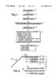
- FIG. 1shows a flow chart of operations performed in carrying out the registration according to a preferred embodiment of the present invention
- FIGS. 2A and 2Bshow the collection of point locations on the skin surface and the bone surface, respectively, of a patient about to undergo a craniotomy
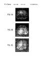
- FIGS. 3A and 3Bshow renderings of a patient's skull showing a distribution of skin surface points
- FIG. 3Cshows a rendering of the same patient's skull as in FIGS. 3A and 3B showing a distribution of bone surface points
- FIG. 4shows a flow chart of a weighted geometrical features (WGF) algorithm used in the operations of FIG. 1;
- WGFweighted geometrical features
- FIGS. 5-7show experimental results obtained from the WGF algorithm of FIG. 4.
- FIG. 8shows a block diagram of an apparatus for carrying out the method according to the preferred embodiment.
- FIG. 1is a flow chart showing an overview of operations to be performed according to the preferred embodiment.
- extrinsic markerse.g., skin-affixed or bone-affixed
- theyare attached to the patient in the region of the anatomy to be imaged and registered.
- a marker to be usefulit must be visible in the image acquired.
- Many types of markers, with different sizes, shapes and materials,are known in the art.
- CT imagesany material that attenuates radiation is potentially useful; metal spheres and hollow shapes filled with a solution containing iodine have commonly been used.
- contrast agentsthat change the relaxation parameter of the hydrogen protons whose spins are imaged have commonly been used.
- a bone-implanted marker systemtypically includes a post that is screwed into the bone, to which an imaging marker containing contrast agent is attached.
- the imaging markeris typically reversibly attached, and a different marker is attached to the post to acquire physical position information, as in step 107 to be described below.
- a three-dimensional imageis acquired by a suitable technique such as CT, MR, PET or SPECT.
- a suitable techniquesuch as CT, MR, PET or SPECT.
- the geometric features to be used for registrationare segmented; suitable techniques are known in the art. For example, if extrinsic markers are to be used, determine the centroid of each marker. If anatomical landmarks are to be used, identify the position of each landmark. If a surface is to be used, extract the surface. Surfaces such as the skin and bone can be easily and automatically extracted using published techniques.
- positional informationis taken on the physical features corresponding to those segmented at step 105 .
- Various techniques for doing soinvolve articulated mechanical probes, probes using electromagnetic, ultrasonic or optical radiation, stereo video cameras and laser range finders. A particularly useful technique will be explained below.
- weightsare assigned to the segmented features to emphasize or deemphasize various features. For example, if data from bone are considered to be more accurate than data from skin, bone can be assigned a large weight, while skin can be assigned a small weight. Techniques for assigning weights will be described in detail below.
- the image taken at step 103is registered with the physical positional information taken at step 107 by use of the weights assigned at step 109 .
- the technique usedis the weighted geometrical feature (WGF) algorithm, which will be described in detail below.
- Illustrative examples of the physical features to be registered at step 111include the following: exactly one marker plus other shapes or features; exactly two markers plus other shapes or features; exactly three markers where the markers are weighted differently; four or more markers where the markers are weighted differently; three or more anatomical landmarks weighted differently; three or more markers plus other shapes or features; exactly one surface with points which are weighted differently.
- one or two bone-implanted markers plus a surfaceshould prove useful in many applications, as should differently weighted landmarks.
- the output of step 111is a transformation that maps each point in the 3-D image to a physical point. That transformation can be used at step 113 for a variety of surgical and other tasks, such as tracking the changing position of a surgical probe on a display of the preoperative images, or directing a needle (stereotactic biopsy) or energy (stereotactic radiosurgery) to a surgical target (e.g., tumor) located in the images.
- a needlestereotactic biopsy
- energystereotactic radiosurgery
- the positional informationcan be acquired at step 107 using a technique which will now be explained with reference to FIGS. 2A, 2 B and 3 A- 3 C.
- the headis fixed in a Mayfield clamp, physical-space markers are attached to the marker posts, a localization probe is calibrated, and the markers are localized.
- the localization probeis equipped with an array of infrared emitting diodes (IREDs).
- IREDsinfrared emitting diodes
- Physical space tracking of the probeis accomplished with optical triangulation using an Optotrak 3020 (Northern Digital, Ontario, Canada).
- the physical-space markersare manufactured with a hemispherical divot whose center corresponds to the centroid of the image markers.
- the ACUSTAR I Advanced Neurosurgical Navigation Systemregisters images to each other and to the intraoperative physical coordinate system.
- the ACUSTAR system used in the preferred embodimenthas been enhanced so that it can also record selected probe tip positions after registration is performed.
- Several currently available systemscan be relatively easily modified to implement the present invention; they feature tracking of some sort (optical, electromagnetic, or articulated mechanical), and they use a registration transformation to perform a surgical task.
- FIG. 2Ashows such an operation for the collection of a point on the skin surface
- FIG. 2Bshows such an operation for the collection of a point on a bone surface.
- the probeis supported by both hands to help prevent it from compressing the skin.
- the arrows in FIGS. 2A and 2Bindicate two physical-space markers.
- the probeis supported by both hands to help prevent the probe tip from compressing the skin. It takes two or three seconds to acquire each point.
- the usertypically collects between 200 and 250 points for each skin surface acquisition and between 75 and 100 points for each bone surface.
- FIGS. 3A and 3Billustrate a typical distribution of recorded skin positions
- FIG. 3Cillustrates a typical distribution of recorded bone positions.
- the skin points shown in FIGS. 3A and 3Bare widely distributed over the top portion of the head, while the bone points are concentrated exclusively under the skin flap. Only one-third of the points are shown in each rendering. The markers are also visible (four in this case, although none, one, two, three, or any other suitable number could be used).
- the radius of the ball-point tip used for surface point collectionis 0.75 mm.
- the localization probeis calibrated by placing the ball-point tip in a hemispherical divot and pivoting the probe about the center of the divot.
- the position of the probe tip relative to the coordinate system of the probe(the probe coordinate system is set by the positions of infrared-emitting diodes on the probe handle) is determined by finding the most invariant point (in a least squares sense) in those pivot motions. That invariant point is the center of the ball-point tip. That is, the ball-point tip pivots about the center of the ball rather than a point on the surface of the ball.
- the weighted geometrical feature (WGF) registration algorithmis an extension of the iterative closest point (ICP) algorithm taught in P. J. Best et al, “A method for registration of 3-D shapes,” IEEE Trans. Pattern Anal. Machine Intell ., Vol. 14, pp. 239-256, 1992.
- the points to be used for triangle setsare the vertices.
- ⁇ w ij ⁇be a set of nonnegative weights associated with the points ⁇ p ij ⁇ .
- the weightscan be selected at step 109 in a variety of ways to emphasize or deemphasize various geometrical features; some techniques will be explained below.
- ⁇ iis a “correspondence” function.
- ⁇ iis defined to be the closest point operator; that is, ⁇ i finds the point y ij in the shape ⁇ i that is closest to the point ⁇ (p ij ).
- a multidimensional binary search treecan be used to speed up the search process, as is known in the art.
- the WGF algorithmis an iterative procedure which will now be explained with reference to FIG. 4 .
- the variable k and the third subscript on each pare iteration indices.
- the algorithmcan be repeated with multiple initial transformations ⁇ 0 to solve the problem of local minima, or in other words to correct for a situation in which an iteration arrives at a local minimum which is different from the global minimum.
- the transformation ⁇ kis computed between the points ⁇ p ij0 ⁇ and ⁇ y ijk ⁇ using the weights ⁇ w ij ⁇ .
- the points ⁇ p ij0 ⁇ and ⁇ y ijk ⁇ for all shapesare collectively considered as two corresponding point sets and registered using an extension of the closed-form solution to the “orthogonal Procrustes” problem developed in K. S. Arun et al, “Least-square fitting of two 3-D point sets,” IEEE Trans. Pattern Anal. Machine Intell ., Vol. 9, pp. 698-700, 1987. That extension allows the points to be weighted and is detailed in the above-referenced Maurer et al 1996 publication, which has already been incorporated by reference. That extension works in the following manner:
- the cost function d( ⁇ ) defined aboveis computed for ⁇ k and ⁇ k+1 , and it is determined whether d( ⁇ k ) ⁇ d( ⁇ k+1 ) ⁇ , where ⁇ is a predetermined tolerance. In other words, it is determined whether the iterative algorithm has converged to within ⁇ . If so, the algorithm ends with step 411 . Otherwise, k is incremented by one at step 413 , and another iteration begins with step 403 .
- the algorithmthus fits the recorded surface points p ij to a surface that is displaced from the actual surface ⁇ i by the radius r i of the ball-point tip. If the ⁇ is replaced by a+, the algorithm can fit recorded surface points to either a surface that is displaced outside the actual surface (the correct solution) or one that is displaced inside the actual surface (a false solution). The ⁇ forces the algorithm to fit the recorded surface points to a surface that is displaced outside the actual surface. The false solution always has a higher residual error than that of the correct solution, i.e., it is never the global minimum. Use of the ⁇ prevents the iterative search from terminating in the local minimum corresponding to the false solution.
- the present inventionhas been tested with data acquired from twelve patients that underwent craniotomies for the resection of cerebral lesions at Vanderbilt University Medical Center. All of the cases were temporal, front-temporal, or temporal-parietal lobe tumors. The patients were operated on in the supine position with the head oriented 90 degrees right or left (for the left and right tumors, respectively). The surgeries were performed using the ACUSTAR I Advanced Neurosurgical Navigation System which is used to provide intraoperative guidance during neurosurgery. Before the patient is imaged, the surgeon implants four plastic posts into the outer table of the skull of the patient with one end remaining outside the skin.
- the specific locations of the postswere determined by individual clinical circumstances, but generally the posts were widely separated and placed on both sides of the head, with two of the markers inferior and two superior to the region of surgical interest.
- Image markers that contain contrast fluid and generate high intensity in both X-ray computed tomography (CT) and magnetic resonance (MR) scansare attached to the posts just prior to image acquisition.
- CT imageswere acquired preoperatively for all patients. Imaging studies were performed the day before or the morning of the surgical procedure. The CT images were acquired using a Siemens Somatom Plus scanner. Each image volume contains between 41 and 48 transverse slices with 512 ⁇ 512 pixels. The voxel dimensions are typically 0.4 ⁇ 0.4 ⁇ 3.0 mm. All CT image volumes in the present example are stacks of image slices with no interslice gap or slice overlap. The gantry tilt angle was zero.
- the position of an image markeris defined as its centroid, which can be determined through known techniques.
- Triangle set representations of the skin and skull surfacesare automatically extracted from each CT image using a known tetrahedral decomposition method. That algorithm produces an oriented surface.
- TREtarget registration error
- the registration error of a transformation being evaluatedis calculated by using as a reference gold standard the transformation obtained using three of the four bone-implanted markers; the remaining marker is used in the various combinations.
- TREis calculated as the distance between the position of a point in CT mapped from physical space by the reference transformation and its position mapped by the transformation being evaluated.
- xbe the position of a target point in physical space.
- R 1 and t 1be the rotation matrix and translation vector, respectively, of the reference rigid-body transformation ⁇ 1 that maps coordinates in physical space to CT.
- R 2 and t 2be the rotation matrix and translation vector, respectively, of the evaluated rigid-body transformation ⁇ 2 .
- R 12R 2 R 1 ⁇ 1
- R 1is an orthogonal matrix
- R 1 ⁇ 1is equal to the transposition of R 1 , or R 1 t .
- the TRE at a given positionis generally defined as ⁇ y ⁇ .
- the TREis computed as the mean value of ⁇ y ⁇ at the center of every CT voxel inside the head that is within 75 mm of the center of the craniotomy.
- the center of the craniotomyis estimated as the centroid of the recorded bone surface points.
- the reference gold standardis obtained by using three of the four bone-implanted markers.
- the accuracy of the reference transformationis estimated as follows. Consider a set of fiducial points that is registered to another set of points that differs from the first set by position, orientation, and noise that is added to each point (identical, independent, zero-mean, isotropic, normally distributed noise). Let ⁇ 2 be the variance of the random noise. In the present case the random noise represents the error of determining the positions of the markers, and a is called the fiducial localization error (FLE). The root-mean-square (RMS) distance between fiducial points after registration is called the fiducial registration error (FRE).
- FLEfiducial localization error
- FREThe root-mean-square
- d kis the distance of the target point r from the kth principal axis of the fiducial point set
- f kis the RMS distance of the fiducials from the kth axis, or in other words, effectively the radius of gyration of the fiducial set about its kth principal axis.
- CT imagesare registered to physical space using the skin surface only, the bone surface only, and various weighted combinations of those surfaces and one bone-implanted marker position. Different weighted combinations are obtained by varying the inter-shape weights u i .
- the marker closest to the craniotomyis the one used.
- the recorded physical space surface points and marker positionare selected as the “data” shapes, and the CT extracted triangle set surfaces and localized marker position are selected as the “model” shapes.
- An initial transformationcan be determined by manually identifying three or more anatomic landmarks in the image and recording their approximate positions in physical space using the localization probe. The intraoperative positions were not recorded for any anatomic landmarks. However, that approach can be simulated as follows. 1) Identify the tip of the nose and the left and right tragi in the CT image. 2) Calculate the positions of those anatomic landmarks in physical space using the reference gold-standard transformation. 3) Perturb the physical space positions by adding random noise to each point (zero-mean, isotropic, normally distributed noise with a standard deviation of 10 mm). 4) Calculate a point-based registration using the identified image positions and the perturbed physical space positions. 5) Use the resulting approximate transformation as the initial transformation. That initial transformation typically differs from the reference transformation by a rotation on the order of 5 degrees and a translation on the order of 5 mm.
- the outlier problemcan be corrected as follows.
- Outliersare arbitrarily defined as points whose distance from the closest corresponding surface point after that first pass is greater than the mean distance plus two standard deviations.
- the intra-shape weight v ij corresponding to each outlieris set equal to 0, the intra-shape weight sets ⁇ v ij ⁇ are renormalized for each i, the weights w ij , are recomputed, and the WGF algorithm is reapplied.
- CT imageswere registered to physical space using various weighted combinations of the skin surface, bone surface, and one bone-implanted marker. Different weighted combinations were obtained by varying the inter-shape weights u i .
- the mean TRE values computed using the resulting transformationsare illustrated in FIGS. 5 and 6. Each symbol in those figures represents the mean TRE obtained from twelve patients.
- the skin, bone, and marker inter-shape weightsare distinguished with the subscripts s, b, and m, respectively.
- u s +u b1.0 for all registrations.
- the marker weight u mis 0.0 for the top curve (i.e., the marker is not used) and 0.1 for the bottom curve.
- the square symbols on the left and right axesrepresent skin only and bone only surface-based registrations, respectively.
- the square symbols between the axesrepresent skin-and-bone surface-based registrations.
- the triangle symbols on the left and right axesrepresent skin-and-marker and bone-and-marker registrations, respectively.
- the triangle symbols between the axesrepresent skin-bone-and-marker registrations.
- TRE (mm) Type of Registrationmean ⁇ SD 95% Skin surface only 3.1 ⁇ 1.4 5.5 Skin surface + 1 marker 2.2 ⁇ 1.0 3.8 Bone surface only 1.8 ⁇ 0.8 3.1 Bone surface + 1 marker 1.3 ⁇ 0.5 2.1 Skin + bone surfaces 1.5 ⁇ 0.6 2.5 Skin + bone surfaces + 1 marker 1.0 ⁇ 0.3 1.5
- the TRE valuescorrespond to the minima for the four curves in FIGS. 5 and 6.
- the 95% TRE valuesare calculated as mean+1.645 SD by assuming that the TRE values are normally distributed.
- the effect of the number of surface points on registration accuracywas also investigated. The results are shown in FIG. 7 .
- the curveswere generated by performing each registration with a randomly picked subset of the total number of surface points.
- the mean FRE for the reference gold standardsi.e., the registrations obtained using three bone-implanted markers, is 0.28 mm.
- TREwas estimated for the reference transformations for each patient with that value of FRE.
- a combination of geometrical featuresmight improve registration accuracy.
- a combination of featuresmight merely produce a registration with intermediate accuracy.
- Using multiple geometrical features simultaneouslycan improve the accuracy of CT-to-MR registration.
- the registration of head CT and MR image volumes using points and a surfacecan be significantly more accurate than registration using only points or a surface.
- a combination of geometrical featurescan also improve the accuracy of CT-to-physical registration. The improvement in accuracy obtained for CT-to-physical registration is relatively larger than that obtained for CT-to-MR registration.
- the observed TRE of the evaluated techniquewill be approximately ⁇ square root over (2) ⁇ times the TRE of the reference standard.
- the TRE of the reference standardis approximately 0.67 mm.
- ⁇ square root over (2) ⁇ 0.670.95 mm.
- the best observed TRE found in the present exampleis 1.02 mm and was obtained using the skin surface, the bone surface, and one bone-implanted marker (see FIG. 5 and Table 1). Thus it is likely that the accuracy of the hybrid registration is comparable to that of the reference gold standard.
- a combination of geometrical featurescan reduce not only the mean TRE but also the standard deviation and thus can improve not only registration accuracy but also reproducibility.
- Table 1lists 95% TRE values that were calculated as mean+1.645 SD by assuming that the TRE values are normally distributed. The 95% TRE values are an estimate of the worst TRE that will be observed in 95 out of 100 registrations.
- the bone surface-based mean TREimproved 0.5 mm—from 1.8 to 1.3 mm—by also using one marker.
- the 95% TREimproved 1.0 mm, from 3.1 to 2.1 mm.
- One feature of the present inventionis that a registration can be achieved using only one or two points.
- Point-based registrationrequires a minimum of three noncolinear points.
- the position of a bone-implanted markercan be determined much more accurately than that of a skin-affixed marker or an anatomic landmark.
- the major disadvantage of using bone-implanted markersis that an invasive procedure is required to implant each marker.
- the WGF algorithmallows registration to be performed using only one or two such markers.
- a robot-assisted total hip replacement systemperforms IP registration using three titanium pins, one implanted into the greater trochanter and two implanted into the condyles of the patient's femur. Implantation of the two markers in the condyles can be painful and difficult.
- the WGF algorithmcould potentially be applied in such a system to perform registration using a femur surface—determined, for example, using ultrasound—plus only one or two pins, thereby eliminating one or two pins. The accuracy and reproducibility of such an application would of course have to be carefully tested and validated.
- the WGF registration algorithmwhich not only allows the combination of multiple types of geometrical information but also handles point-based and surface-based registration as degenerate cases, could form the foundation of a “flexible” surgical navigation system that allows the surgeon to use what he considers the method most appropriate for an individual clinical situation. For example, if a neurosurgeon wants to remove a superficial meningioma and decides that a 95% TRE of 5-6 mm is sufficient for his operation, he might choose to do a surface-based registration using the skin.
- hemight initially collect skin surface points, perform a skin surface-based registration, use that registration to make a skin flap, collect surface points over the skull that is exposed after making the skin flap, perform a bone-and-skin surface-based registration, and use that more accurate registration to elevate the craniotomy and perform surgery.
- the present inventionwould allow the surgeon to also use one or more anatomic landmarks and/or bone-implanted markers in a bone-skin-and-marker registration.
- three or more bone-implanted markerscould be used, possibly in combination with the skull surface.
- FIG. 8shows a block diagram of one possible apparatus for carrying out the method described above in the context of a surgical procedure.
- the components of the apparatus shown in FIG. 8can be those set forth above, any equivalents, or any other suitable components, whether selected from the prior art or yet to be developed.
- a probe 803such as that described above is used to take positional data.
- a probe sensor 805detects the position of the probe each time positional data are taken.
- Scanning datasuch the data of a CT scan are input through a suitable input 807 .
- the usercan input information and commands, such as the desired weights of various points, through a user input 809 which can include a terminal with a keyboard or any other suitable user interface.
- the probe position sensor 805 , the CT scan input 807 and the user input 809supply their respective data to a central processing unit (CPU) 811 , which can be any suitable computing device sufficiently powerful to carry out the WGF algorithm.
- CPUcentral processing unit
- the result of the WGF algorithmcan be used in surgery in any suitable way; for example, it can be applied to a surgical navigation system 813 such as the ACUSTAR I system, a radiosurgery system 815 or a robot-assisted surgical system 817 such as the ROBODOC system.
- a surgical navigation system 813such as the ACUSTAR I system
- a radiosurgery system 815or a robot-assisted surgical system 817 such as the ROBODOC system.
- the particulars of the WGF algorithmcan be refined as needed, and any suitable way of taking the physical positions can be used instead of, or in addition to, the probe.
- the cost function or the search techniquecould be adjusted. It is possible that other search techniques known in the art could produce good registration results. It is also possible that an alternative cost function, e.g., a cost function where the L 2 norm (sum of squared values) is replaced by the L 1 norm (sum of absolute values), could produce good registration results. Therefore, the present invention should be construed as limited only by the appended claims and by applicable principles of law.
Landscapes
- Engineering & Computer Science (AREA)
- Computer Vision & Pattern Recognition (AREA)
- Physics & Mathematics (AREA)
- General Physics & Mathematics (AREA)
- Theoretical Computer Science (AREA)
- Apparatus For Radiation Diagnosis (AREA)
Abstract
Description
The present invention is directed to an apparatus and method for the registration of images to physical space by the use of a weighted combination of points and surfaces. The present invention is more particularly directed to the registration of images of patients' body parts to the actual patients' body parts for surgery although it is applicable to any use in which a geometrical transformation represents rigid-body motion or approximate rigid-body motion. Such images can be taken by X-ray computed tomography (CT) or the like.
Registration is the determination of a one-to-one mapping or transformation between the coordinates in one space and those in another such that points in the two spaces that correspond to the same anatomic point are mapped to each other. Registration of multimodal images makes it possible to combine different types of structural information [such as X-ray computed tomography (CT) and magnetic resonance (MR) images] and functional information [such as positron emission tomography (PET) and single photon emission tomography (SPECT)] for diagnosis and surgical planning. Registration of images acquired with the same modality at different times allows quantitative comparison of serial data for longitudinal monitoring of disease progression/regression and postoperative follow up.
Registration of preoperative images with the physical space occupied by the patient during surgery is a fundamental step in interactive, image-guided surgery techniques. Surgical navigation systems use the image-to-physical transformation to track in real time the changing position of a surgical probe on a display of the preoperative images. Stereotactic procedures use the transformation to direct a needle (stereotactic biopsy) or energy (stereotactic radiosurgery) to a surgical target (e.g., tumor) located in the images.
Many methods have been used to register medical images. Image-guided stereotactic surgical procedures have been performed since the early 1970's using stereotactic frame systems. Such systems generally include a reference frame that provides rigid skull fixation using pins or screws and establishes a stereotactic coordinate system in physical space, a method for stereotactic image acquisition, and a system for mechanical direction of a probe or other surgical instrument to a defined intracranial point. Most current systems relate image space to the physical coordinate space established by the reference frame by attaching a localizing system consisting of N-shaped fiducials during image acquisition. Frames permit neurosurgeons to perform biopsies and to resect deep-seated and previously inaccessible lesions.
Frame-based techniques, however, have several limitations. The frames are bulky and may interfere with the surgical exposure. Patients complain about the weight of the frame and the pain associated with its application. The surgeon is typically limited to target points on a linear trajectory. Perhaps most importantly, frame-based stereotactic systems do not provide real-time feedback to the surgeon about anatomic structures encountered in the surgical field.
To address such limitations, a number of frameless stereotactic systems have been developed over the last decade. Of the many frameless methods that have been used to register medical images, it appears that point-based and surface-based techniques are the most useful for image-to-physical registration. There is mounting evidence that voxel-intensity-based methods might be the easiest and most accurate way to perform mono- and multi-modality image-to-image registration, but their use for image-to-physical registration is probably rather limited until higher quality three-dimensional (3-D) intraoperative images become readily available. Point-based registration involves determining the coordinates of corresponding points in different images and/or physical space and computing the geometrical transformation that best aligns those points, generally in a least-squares sense. Many investigators have performed point-based image-to-physical registration using external anatomic landmarks (e.g., internal and external canthi, nasion), skin-affixed markers, or bone-implanted markers; a recent survey cites more than 70 publications devoted to point-based registration. Surface-based registration involves determining corresponding surfaces in different images and/or physical space and computing the geometrical transformation that best matches those surfaces. Researchers have performed surface-based image-to-physical registration using the skin or the outer skull surface. Points and surfaces can be easily and accurately acquired in physical space using 3-D probes (e.g., articulated mechanical, electromagnetic, ultrasonic, optical), stereo video cameras, and/or laser range-finders.
Most previously reported registration techniques that align three-dimensional (3-D) image volumes by matching geometrical features such as points, curves, or surfaces use a single type of feature. Patents have been issued for the idea of point-based registration using bone-implanted markers (e.g., G. S. Allen, “Method and Apparatus for Imaging the Anatomy,” U.S. Pat. No. 5,016,639, May 1991) and surface-based registration (e.g., C. A. Pelizzari and G. T. Y. Chen, “Means to Correlate Images from Scans Taken at Different Times Including Means to Determine the Minimum Distances between a Patient Anatomical Contour and a Correlating Surface,” U.S. Pat. No. 4,977,505, December 1990). An example of a surface commonly used for registration is the skin-air interface.
A technique has been developed to use multiple features simultaneously (C. R. Meyer, G. S. Leichtman, J. A. Brunberg, R. L. Wahl, and L. E. Quint, “Simultaneous usage of homologous points, lines, and planes for optimal, 3-D, linear registration of multimodality imaging data,”IEEE Transactions on Medical Imaging, 14: 1-11, 1995). However, that method uses points, lines, and planes and is accordingly less general and less useful than is desired, since lines are special cases of curves and planes are special cases of surfaces.
It will be readily apparent from the above discussion that a need still exists in the art for a method and apparatus for 3-D image registration that provides high accuracy while minimizing the need for invasive techniques and other sources of patient discomfort.
Accordingly, it is an object of the invention to allow accurate 3-D image registration without the need for frames and with only minimal need for markers.
Another object of the invention is to allow 3-D image registration using a combination of points and surfaces.
Yet another object of the invention is to allow 3-D image registration which is not limited to points, lines and planes.
Still another object of the invention is to allow 3-D image registration through weighting of the various features to be registered.
A still further object of the invention is to allow 3-D image registration while avoiding the above-noted deficiencies in the prior art.
To achieve the above and other objects, the present claimed invention is directed to a 3-D image registration method that uses a weighted combination of multiple geometrical features simultaneously. The method uses an algorithm, called a weighted geometrical feature (WGF) registration algorithm, which has the capability of using points, curves (e.g., line segment sets), and surfaces (e.g., triangle sets). The result of the registration is a transformation function which is used in guiding surgical operations. The present invention is further directed to an apparatus for carrying out the algorithm and performing the surgical operations in accordance with the transformation function.
The main idea of the present invention can be summarized briefly as follows:
1) If extrinsic markers (e.g., skin-affixed or bone-implanted markers) are to be used for registration, first attach such markers to the patient in the region of the anatomy to be imaged and registered.
2) Acquire a 3-D image (e.g., CT, MR, PET, SPECT) of the patient.
3) Segment the geometrical features to be used for registration using standard segmentation techniques.
4) Acquire positional information of geometrical features corresponding to those segmented in the above step using 3-D probes (e.g., articulated mechanical, electromagnetic, ultrasonic, optical), stereo video cameras, and/or laser range-finders.
5) Assign weights to the segmented geometrical features.
6) Register the first image with physical space (image-to-physical registration, or IP registration) using the weighted geometrical feature (WGF) registration algorithm.
7) The transformation can be used for various purposes, e.g., tracking the changing position of a surgical probe on a display of the preoperative images, or directing a needle (stereotactic biopsy) or energy (stereotactic radiosurgery) to a surgical target (e.g., tumor) located in the images.
There are two important ideas in the present invention. First, different weights can be assigned to different points or different regions of a surface. That can be done using the present invention with one or more types of geometrical features. For example, the present invention can be used to perform point-based registration, as the WGF algorithm handles point-based and surface-based registration as degenerate cases. Whereas all published work of which Applicants are aware weights all points equally, the present invention allows the user to assign different weights to different points. If each point has a measurement error that is independently random and normally distributed around the true position, then the maximum likelihood estimate of the transformation parameters is obtained by weighting the ith point by 1/σi2, where σi2, is the variance of the measurement error of the ith point. In other words, points which are more accurately measured (smaller σi2) are weighted more heavily (larger 1/σi2). Similarly, the present invention can be used to perform surface-based registration. It might be possible to increase the accuracy of such registration by weighting surface points using information such as estimated surface point measurement error, surface model segmentation error, and surface curvature.
The second important idea is that registration can be performed using multiple geometrical features simultaneously. That idea has two potential advantages. First, point-based registration requires a minimum of three noncolinear points. The position of a bone-implanted marker can be determined more accurately than that of a skin-affixed marker or an anatomic point landmark. The major disadvantage of using bone-implanted markers is that an invasive procedure is required to implant each marker. By combining surface information, the present invention allows registration to be performed using only one or two markers. Second, a combination of geometrical features can improve both the accuracy and reproducibility of registration. For example, intraoperative surface information is sometimes restricted to a small patch due to surgical conditions (e.g., surgical drapes). Surface-based registration using limited surface information is almost certainly not very accurate. The WGF algorithm can be used to improve registration accuracy in such circumstances by incorporating a single point (in that case a bone-implanted marker, but potentially an anatomical landmark or skin-affixed marker) that effectively serves as an “anchor.”
As mentioned previously, there is mounting evidence that voxel-intensity-based methods might be the easiest and most accurate way to perform both mono- and multi-modality image-to-image registration. However, their use for image-to-physical registration is probably rather limited until higher quality 3-D intraoperative images become readily available. Thus, for at least the next decade, it is believed that image-to-physical registration will be based on geometrical features such as points and surfaces. Points and surfaces can be easily and accurately acquired in physical space using 3-D probes (e.g., articulated mechanical, electromagnetic, ultrasonic, optical), stereo video cameras, and/or laser range-finders. All image-to-physical registration work of which Applicants are aware has used exclusively points or surfaces.
The WGF registration algorithm, which not only allows the combination of multiple types of geometrical information but also handles point-based and surface-based registration as degenerate cases, could form the foundation of a “flexible” surgical navigation system that allows the surgeon to use what he considers the method most appropriate for an individual clinical situation. For example, if a neurosurgeon wants to remove a superficial meningioma and decides that a 95% TRE (target registration error) of 5-6 mm is sufficient for his operation, he might choose to do a surface-based registration using the skin. If he decides that he needs additional accuracy, he might initially collect skin surface points, perform a skin surface-based registration, use that registration to make a skin flap, collect surface points over the skull that is exposed after making the skin flap, perform a bone-and-skin surface-based registration, and use that more accurate registration to elevate the craniotomy and perform surgery. The present invention would allow the surgeon to also use one or more anatomic landmarks and/or bone-implanted markers in a bone-skin-and-marker registration. In circumstances where the best possible accuracy is required, as may be the case for skull-base surgery performed using an operating microscope with video overlays containing information derived from preoperative images, three or more bone-implanted markers could be used, possibly in combination with the skull surface.
An algorithm has been developed, implemented, and tested. Its usefulness has been demonstrated by using it to perform both image-to-image (CT-MR) and image-to-physical (CT-physical space) registration of head images. Weighting different regions of a surface can improve the accuracy of surface-based registration. Registration using points and a surface can be significantly more accurate than registration using points or a surface.
While a preferred embodiment will be set forth with respect to registration of patients' heads to the actual patients' heads for surgery, the invention should not be construed as limited only to that use. For example, rigid-body transformations are also useful in spinal surgery and in orthopedic surgery. Even soft tissues may be sufficiently rigid for the present invention to be used. While the liver is not a rigid structure, liver motion during breathing is often approximately rigid (cranial-caudal translation) and may be sufficiently rigid for a radiosurgery application. For that matter, the present invention can be applied to any surgical or non-surgical use in which a geometrical transformation represents rigid-body motion, exactly or approximately.
Various aspects of the present invention have been described in the following publications, whose disclosures are hereby incorporated by reference in their entireties into the present disclosure:
C. R. Maurer, Jr. et al, “Registration of CT and MR brain images using a combination of points and surfaces,”Medical Imaging1995: Image Processing, Proc. SPIE 2434: 109-123, 1995;
C. R. Maurer, Jr. et al, “A method for registration of 3-D images using multiple geometrical features,”IEEE Transactions on Medical Imaging, 15: 836-849, 1996;
C. R. Maurer, Jr. et al, “Registration of head CT images to physical space using multiple geometrical features,”Medical Imaging1998: Image-Processing, Proc. SPIE 3338: 72-80, 1998; and
C. R. Maurer, Jr. et al, “Registration of head CT images to physical space using a combination of points and surfaces,”IEEE Transactions on Medical Imaging, 17: 753-761, 1998.
A preferred embodiment of the invention will be set forth in detail with reference to the drawings, in which:
FIG. 1 shows a flow chart of operations performed in carrying out the registration according to a preferred embodiment of the present invention;
FIGS. 2A and 2B show the collection of point locations on the skin surface and the bone surface, respectively, of a patient about to undergo a craniotomy;
FIGS. 3A and 3B show renderings of a patient's skull showing a distribution of skin surface points;
FIG. 3C shows a rendering of the same patient's skull as in FIGS. 3A and 3B showing a distribution of bone surface points;
FIG. 4 shows a flow chart of a weighted geometrical features (WGF) algorithm used in the operations of FIG. 1;
FIGS. 5-7 show experimental results obtained from the WGF algorithm of FIG. 4; and
FIG. 8 shows a block diagram of an apparatus for carrying out the method according to the preferred embodiment.
A preferred embodiment of the present invention will be set forth in detail with reference to the drawings, in which like reference numerals designate like components throughout.
FIG. 1 is a flow chart showing an overview of operations to be performed according to the preferred embodiment. Atstep 101, if one or more extrinsic markers (e.g., skin-affixed or bone-affixed) are to be used for registration, they are attached to the patient in the region of the anatomy to be imaged and registered. For a marker to be useful, it must be visible in the image acquired. Many types of markers, with different sizes, shapes and materials, are known in the art. For CT images, any material that attenuates radiation is potentially useful; metal spheres and hollow shapes filled with a solution containing iodine have commonly been used. For MR images, contrast agents that change the relaxation parameter of the hydrogen protons whose spins are imaged have commonly been used. For nuclear medicine scans (e.g., PET, SPECT) contrast agents are active radiation emitters. A bone-implanted marker system typically includes a post that is screwed into the bone, to which an imaging marker containing contrast agent is attached. The imaging marker is typically reversibly attached, and a different marker is attached to the post to acquire physical position information, as instep 107 to be described below.
Atstep 103, a three-dimensional image is acquired by a suitable technique such as CT, MR, PET or SPECT. Atstep 105, the geometric features to be used for registration are segmented; suitable techniques are known in the art. For example, if extrinsic markers are to be used, determine the centroid of each marker. If anatomical landmarks are to be used, identify the position of each landmark. If a surface is to be used, extract the surface. Surfaces such as the skin and bone can be easily and automatically extracted using published techniques.
Atstep 107, positional information is taken on the physical features corresponding to those segmented atstep 105. Various techniques for doing so involve articulated mechanical probes, probes using electromagnetic, ultrasonic or optical radiation, stereo video cameras and laser range finders. A particularly useful technique will be explained below.
Atstep 109, weights are assigned to the segmented features to emphasize or deemphasize various features. For example, if data from bone are considered to be more accurate than data from skin, bone can be assigned a large weight, while skin can be assigned a small weight. Techniques for assigning weights will be described in detail below.
Atstep 111, the image taken atstep 103 is registered with the physical positional information taken atstep 107 by use of the weights assigned atstep 109. The technique used is the weighted geometrical feature (WGF) algorithm, which will be described in detail below.
Illustrative examples of the physical features to be registered atstep 111 include the following: exactly one marker plus other shapes or features; exactly two markers plus other shapes or features; exactly three markers where the markers are weighted differently; four or more markers where the markers are weighted differently; three or more anatomical landmarks weighted differently; three or more markers plus other shapes or features; exactly one surface with points which are weighted differently. In particular, one or two bone-implanted markers plus a surface should prove useful in many applications, as should differently weighted landmarks.
The output ofstep 111 is a transformation that maps each point in the 3-D image to a physical point. That transformation can be used at step113 for a variety of surgical and other tasks, such as tracking the changing position of a surgical probe on a display of the preoperative images, or directing a needle (stereotactic biopsy) or energy (stereotactic radiosurgery) to a surgical target (e.g., tumor) located in the images.
The positional information can be acquired atstep 107 using a technique which will now be explained with reference to FIGS. 2A,2B and3A-3C.
Intraoperatively, the head is fixed in a Mayfield clamp, physical-space markers are attached to the marker posts, a localization probe is calibrated, and the markers are localized. The localization probe is equipped with an array of infrared emitting diodes (IREDs). Physical space tracking of the probe is accomplished with optical triangulation using an Optotrak 3020 (Northern Digital, Ontario, Canada). The physical-space markers are manufactured with a hemispherical divot whose center corresponds to the centroid of the image markers. The tip of the probe is a spherical ball. Intraoperative localization of each marker is performed by placing the ball-point tip into the divot and pressing a button on the probe handle.
The ACUSTAR I Advanced Neurosurgical Navigation System registers images to each other and to the intraoperative physical coordinate system. When the localizer tip is placed near or within the patient, triplanar reformatted images intersecting at the corresponding position in image coordinates are generated. The ACUSTAR system used in the preferred embodiment has been enhanced so that it can also record selected probe tip positions after registration is performed. Several currently available systems can be relatively easily modified to implement the present invention; they feature tracking of some sort (optical, electromagnetic, or articulated mechanical), and they use a registration transformation to perform a surgical task.
To collect a skin point, the surgeon places the probe tip on the surface and records its 3-D coordinates by pressing a button. FIG. 2A shows such an operation for the collection of a point on the skin surface, while FIG. 2B shows such an operation for the collection of a point on a bone surface. The probe is supported by both hands to help prevent it from compressing the skin. The arrows in FIGS. 2A and 2B indicate two physical-space markers. During the acquisition of skin points, the probe is supported by both hands to help prevent the probe tip from compressing the skin. It takes two or three seconds to acquire each point. The user typically collects between 200 and 250 points for each skin surface acquisition and between 75 and 100 points for each bone surface. It is preferable to collect surface points as uniformly distributed over the head (skin points) and exposed skull (bone points) as possible. FIGS. 3A and 3B illustrate a typical distribution of recorded skin positions, while FIG. 3C illustrates a typical distribution of recorded bone positions. The skin points shown in FIGS. 3A and 3B are widely distributed over the top portion of the head, while the bone points are concentrated exclusively under the skin flap. Only one-third of the points are shown in each rendering. The markers are also visible (four in this case, although none, one, two, three, or any other suitable number could be used). The radius of the ball-point tip used for surface point collection is 0.75 mm.
The localization probe is calibrated by placing the ball-point tip in a hemispherical divot and pivoting the probe about the center of the divot. The position of the probe tip relative to the coordinate system of the probe (the probe coordinate system is set by the positions of infrared-emitting diodes on the probe handle) is determined by finding the most invariant point (in a least squares sense) in those pivot motions. That invariant point is the center of the ball-point tip. That is, the ball-point tip pivots about the center of the ball rather than a point on the surface of the ball.
To understand the assignment of weights ofstep 109 and the registration ofstep 111, one must first understand the goal of the registration algorithm and the notation in which the goal is expressed. The weighted geometrical feature (WGF) registration algorithm is an extension of the iterative closest point (ICP) algorithm taught in P. J. Best et al, “A method for registration of 3-D shapes,”IEEE Trans. Pattern Anal. Machine Intell., Vol. 14, pp. 239-256, 1992. Let {ρi} for i=1, . . . , Nsbe a set of “data” shapes in the physical data taken from the patient; the “data” shapes are to be registered with another set of “model” shapes {χi} in the image for i=1, . . . , Ns. Each shape ρiis decomposed into a point set Pi={pij} for j=1, . . . , Nρiif it is not already in point set form. For example, the points to be used for triangle sets are the vertices. Let {wij} be a set of nonnegative weights associated with the points {pij}. The weights can be selected atstep 109 in a variety of ways to emphasize or deemphasize various geometrical features; some techniques will be explained below. The goal of the algorithm is to find the rigid-body transformation Γ that minimizes the cost function

where
and ∂iis a “correspondence” function. For the WGF registration algorithm, ∂iis defined to be the closest point operator; that is, ∂ifinds the point yijin the shape χithat is closest to the point Γ(pij). A multidimensional binary search tree can be used to speed up the search process, as is known in the art.
The weights wijcan be selected thus. Let {ui} for i=1, . . . , Nsbe a non-negative set of inter-shape weights. Let {vij} for j=1, . . . , Nρibe non-negative normalized sets of intra-shape weights. The weights are given by wij=uivij. The intra-shape weights can be set to be equal within each shape, so that all points within each shape are weighted equally, by setting vij=1/Nρi. The inter-shape weights uican be selected to emphasize one feature over another. If it is not clear how much emphasis each feature should receive, the uican be varied until an error curve reaches a minimum, as will be explained below when the experimental results are presented.
The WGF algorithm is an iterative procedure which will now be explained with reference to FIG.4. The procedure is initialized atstep 401 by setting k=1, pij0=pijand pij1=Γ0(pij0) where Γ0is some initial transformation. The variable k and the third subscript on each p are iteration indices. The algorithm can be repeated with multiple initial transformations Γ0to solve the problem of local minima, or in other words to correct for a situation in which an iteration arrives at a local minimum which is different from the global minimum.
The following steps are carried out iteratively.
Atstep 403, for each shape ρiand for j=1, . . . , Nρi, the closest points yijk=∂i(pijk, χi) are calculated.
Atstep 405, the transformation Γkis computed between the points {pij0} and {yijk} using the weights {wij}. Instep 405, the points {pij0} and {yijk} for all shapes are collectively considered as two corresponding point sets and registered using an extension of the closed-form solution to the “orthogonal Procrustes” problem developed in K. S. Arun et al, “Least-square fitting of two 3-D point sets,”IEEE Trans. Pattern Anal. Machine Intell., Vol. 9, pp. 698-700, 1987. That extension allows the points to be weighted and is detailed in the above-referenced Maurer et al 1996 publication, which has already been incorporated by reference. That extension works in the following manner:
1) Translate the point sets such that their new origins are their previous weighted centroids {overscore (p)} and {overscore (x)} and denote the translated points with prime marks:


wheretrepresents a transposition of a matrix.
3) Calculate the SVD of the weighted covariance matrix:
4) Calculate the rotation matrix:
If the determinant of R is +1, then R is the rotation matrix. If the determinant of R is −1, then the solution is a reflection rather than a rotation. In that case, Vtis obtained from V by changing the sign of the column of V corresponding to the most singular value of H, and the rotation matrix is given by
5) Calculate the translation vector
Atstep 407, the transformation Γkfound atstep 405 is applied to produce registered points pij,k+1=Γk(pij0).
Atstep 409, the cost function d(Γ) defined above is computed for Γkand Γk+1, and it is determined whether d(Γk)−d(Γk+1)<ε, where ε is a predetermined tolerance. In other words, it is determined whether the iterative algorithm has converged to within ε. If so, the algorithm ends withstep 411. Otherwise, k is incremented by one atstep 413, and another iteration begins withstep 403.
In practice, however, one modification needs to be made to the algorithm just set forth. If a conventional localization probe having a spherical ball at its tip is used to locate the intraoperative skin and bone surface points, those points as measured are the positions of the center of the spherical ball. Hence the recorded surface points pijare displaced from the actual surface χiby the radius riof the ball-point tip. That is true for any orientation of the localization probe relative to the surface normal at the point of contact. The goal is thus to find the rigid-body transformation that minimizes the cost function

Accordingly,step 403 is modified such that if the shape ρiis a surface, the closest points are computed as

The last term is added if the point pijklies outside the surface χi. Otherwise, it is subtracted. The algorithm thus fits the recorded surface points pijto a surface that is displaced from the actual surface χiby the radius riof the ball-point tip. If the±is replaced by a+, the algorithm can fit recorded surface points to either a surface that is displaced outside the actual surface (the correct solution) or one that is displaced inside the actual surface (a false solution). The±forces the algorithm to fit the recorded surface points to a surface that is displaced outside the actual surface. The false solution always has a higher residual error than that of the correct solution, i.e., it is never the global minimum. Use of the±prevents the iterative search from terminating in the local minimum corresponding to the false solution.
The present invention has been tested with data acquired from twelve patients that underwent craniotomies for the resection of cerebral lesions at Vanderbilt University Medical Center. All of the cases were temporal, front-temporal, or temporal-parietal lobe tumors. The patients were operated on in the supine position with the head oriented 90 degrees right or left (for the left and right tumors, respectively). The surgeries were performed using the ACUSTAR I Advanced Neurosurgical Navigation System which is used to provide intraoperative guidance during neurosurgery. Before the patient is imaged, the surgeon implants four plastic posts into the outer table of the skull of the patient with one end remaining outside the skin. The specific locations of the posts were determined by individual clinical circumstances, but generally the posts were widely separated and placed on both sides of the head, with two of the markers inferior and two superior to the region of surgical interest. Image markers that contain contrast fluid and generate high intensity in both X-ray computed tomography (CT) and magnetic resonance (MR) scans are attached to the posts just prior to image acquisition.
CT images were acquired preoperatively for all patients. Imaging studies were performed the day before or the morning of the surgical procedure. The CT images were acquired using a Siemens Somatom Plus scanner. Each image volume contains between 41 and 48 transverse slices with 512×512 pixels. The voxel dimensions are typically 0.4×0.4×3.0 mm. All CT image volumes in the present example are stacks of image slices with no interslice gap or slice overlap. The gantry tilt angle was zero.
The position of an image marker is defined as its centroid, which can be determined through known techniques. Triangle set representations of the skin and skull surfaces are automatically extracted from each CT image using a known tetrahedral decomposition method. That algorithm produces an oriented surface.
One measure of the accuracy of point-based and surface-based registrations is the value of the cost function at its minimum. A more clinically meaningful measure of registration accuracy is the distance between corresponding points other than those used to estimate the transformation parameters. Because such points might represent surgically targeted lesions, such points are called targets, and the corresponding accuracy measure is called a target registration error (TRE). The term “registration error,” without a modifier, will be understood to mean the TRE.
The registration error of a transformation being evaluated is calculated by using as a reference gold standard the transformation obtained using three of the four bone-implanted markers; the remaining marker is used in the various combinations. Specifically, TRE is calculated as the distance between the position of a point in CT mapped from physical space by the reference transformation and its position mapped by the transformation being evaluated. Let x be the position of a target point in physical space. Let R1and t1be the rotation matrix and translation vector, respectively, of the reference rigid-body transformation Γ1that maps coordinates in physical space to CT. Let R2and t2be the rotation matrix and translation vector, respectively, of the evaluated rigid-body transformation Γ2. Thus,
and
are the positions of the target point in CT mapped by the reference and evaluated transformations, respectively. The equation for y1is inverted to obtain
which can be plugged into the equation for y2to obtain
The target registration error (TRE) Δy is thus
or
where R12=R2R1−1, I is identity and Δt=t2−R12t1. Since R1is an orthogonal matrix, R1−1is equal to the transposition of R1, or R1t.
The TRE at a given position is generally defined as ∥Δy∥. For the purposes of the present invention, the TRE is computed as the mean value of ∥Δy∥ at the center of every CT voxel inside the head that is within 75 mm of the center of the craniotomy. The center of the craniotomy is estimated as the centroid of the recorded bone surface points.
The reference gold standard is obtained by using three of the four bone-implanted markers. The accuracy of the reference transformation is estimated as follows. Consider a set of fiducial points that is registered to another set of points that differs from the first set by position, orientation, and noise that is added to each point (identical, independent, zero-mean, isotropic, normally distributed noise). Let σ2be the variance of the random noise. In the present case the random noise represents the error of determining the positions of the markers, and a is called the fiducial localization error (FLE). The root-mean-square (RMS) distance between fiducial points after registration is called the fiducial registration error (FRE). It can be shown using perturbation theory that if the points are registered in a least-squares sense using a rigid-body transformation, there is a statistical relationship among the expected value of FRE, FLE, and the number of fiducials N that is described by

where the brackets denote the expected value. We note that the expected value of FRE is independent of the fiducial configuration. It has been shown using perturbation theory that there is a relationship among the expected value of TRE, FLE, the number of fiducials N, the configuration of the fiducials, and the position of the target point relative to the fiducials that is described by

where dkis the distance of the target point r from the kth principal axis of the fiducial point set, and fkis the RMS distance of the fiducials from the kth axis, or in other words, effectively the radius of gyration of the fiducial set about its kth principal axis. We can estimate TRE from FRE by rearranging those two equations to eliminate FLE and thus obtain
CT images are registered to physical space using the skin surface only, the bone surface only, and various weighted combinations of those surfaces and one bone-implanted marker position. Different weighted combinations are obtained by varying the inter-shape weights ui. The marker closest to the craniotomy is the one used. The recorded physical space surface points and marker position are selected as the “data” shapes, and the CT extracted triangle set surfaces and localized marker position are selected as the “model” shapes.
An initial transformation can be determined by manually identifying three or more anatomic landmarks in the image and recording their approximate positions in physical space using the localization probe. The intraoperative positions were not recorded for any anatomic landmarks. However, that approach can be simulated as follows. 1) Identify the tip of the nose and the left and right tragi in the CT image. 2) Calculate the positions of those anatomic landmarks in physical space using the reference gold-standard transformation. 3) Perturb the physical space positions by adding random noise to each point (zero-mean, isotropic, normally distributed noise with a standard deviation of 10 mm). 4) Calculate a point-based registration using the identified image positions and the perturbed physical space positions. 5) Use the resulting approximate transformation as the initial transformation. That initial transformation typically differs from the reference transformation by a rotation on the order of 5 degrees and a translation on the order of 5 mm.
Surface points that are incorrectly segmented or that lie on a non-overlapping surface can adversely affect registration accuracy. The outlier problem can be corrected as follows. The WGF algorithm is permitted to iterate until it converges within a tolerance ε=5×10⁻⁵mm. Outliers are arbitrarily defined as points whose distance from the closest corresponding surface point after that first pass is greater than the mean distance plus two standard deviations. The intra-shape weight vijcorresponding to each outlier is set equal to 0, the intra-shape weight sets {vij} are renormalized for each i, the weights wij, are recomputed, and the WGF algorithm is reapplied.
All results in the present disclosure are based on retrospective analysis of preoperative images and intraoperative measurements. Transformations calculated using the WGF algorithm were not used for intraoperative navigation.
CT images were registered to physical space using various weighted combinations of the skin surface, bone surface, and one bone-implanted marker. Different weighted combinations were obtained by varying the inter-shape weights ui. The mean TRE values computed using the resulting transformations are illustrated in FIGS. 5 and 6. Each symbol in those figures represents the mean TRE obtained from twelve patients. The skin, bone, and marker inter-shape weights are distinguished with the subscripts s, b, and m, respectively. In FIG. 5, us+ub=1.0 for all registrations. The marker weight umis 0.0 for the top curve (i.e., the marker is not used) and 0.1 for the bottom curve. The square symbols on the left and right axes represent skin only and bone only surface-based registrations, respectively. The square symbols between the axes represent skin-and-bone surface-based registrations. The triangle symbols on the left and right axes represent skin-and-marker and bone-and-marker registrations, respectively. The triangle symbols between the axes represent skin-bone-and-marker registrations. The skin-and-bone registration curve has a minimum at us=0.05, ub=0.95. The skin-bone-and-marker registration curve has a minimum at us=0.10, ub=0.90. In FIG. 6, us+um=1.0 for the top curve (ub=0.0, i.e., the bone-surface is not used), and ub+um=1.0 for the bottom curve (us=0.0, i.e., the skin-surface is not used). The skin-and-marker registration curve has a minimum at us=0.70, um=0.30. The bone-and-marker registration curve has a minimum at ub=0.90, um=0.10. Both of those curves are fairly flat between um=0.1 and 0.4. Therefore, FIG. 5 presents results using only a single value of umfor the skin-bone-and-marker registrations (bottom curve). Curves obtained using other values of umbetween 0.1 and 0.4 give very similar results. Several of the interesting findings are listed in Table 1, in which “SD” stands for “standard deviation”:
| TRE (mm) | |||
| Type of Registration | mean ± SD | 95% | ||
| Skin surface only | 3.1 ± 1.4 | 5.5 | ||
| Skin surface + 1 marker | 2.2 ± 1.0 | 3.8 | ||
| Bone surface only | 1.8 ± 0.8 | 3.1 | ||
| Bone surface + 1 marker | 1.3 ± 0.5 | 2.1 | ||
| Skin + bone surfaces | 1.5 ± 0.6 | 2.5 | ||
| Skin + bone surfaces + 1 marker | 1.0 ± 0.3 | 1.5 | ||
In Table 1, for the registrations performed using multiple geometrical features, the TRE values correspond to the minima for the four curves in FIGS. 5 and 6. The 95% TRE values are calculated as mean+1.645 SD by assuming that the TRE values are normally distributed.
Those results were obtained using 211±13 (mean±standard deviation) skin points and 83±8 bone points. Of those, 11±5 skin points and 3±2 bone points were classified as outliers and discarded as described above. Visual inspection revealed that about half of the skin outliers are located at the very top of the head in patients whose CT scan does not include the top several millimeters of the head. The remaining skin outliers, and all of the bone outliers, are surface points whose recorded positions are located at least 5 mm outside the corresponding surface and thus probably represent erroneous “air clicks,” i.e., points that were recorded while the probe tip was not touching the skin or bone surface.
The effect of the number of surface points on registration accuracy was also investigated. The results are shown in FIG.7. The curves were generated by performing each registration with a randomly picked subset of the total number of surface points.
The mean FRE for the reference gold standards, i.e., the registrations obtained using three bone-implanted markers, is 0.28 mm. TRE was estimated for the reference transformations for each patient with that value of FRE. The reference TRE computed for the twelve patients is 0.67±0.10 mm (mean±standard deviation; range=0.57 to 0.85 mm).
It is intuitively clear that a combination of geometrical features might improve registration accuracy. However, it is also intuitively clear that a combination of features might merely produce a registration with intermediate accuracy. Using multiple geometrical features simultaneously can improve the accuracy of CT-to-MR registration. Specifically, the registration of head CT and MR image volumes using points and a surface can be significantly more accurate than registration using only points or a surface. A combination of geometrical features can also improve the accuracy of CT-to-physical registration. The improvement in accuracy obtained for CT-to-physical registration is relatively larger than that obtained for CT-to-MR registration. If the registration error of an evaluated registration technique is approximately the same as that of the reference gold standard, and if the errors are independent, then the observed TRE of the evaluated technique will be approximately {square root over (2)} times the TRE of the reference standard. In the examples set forth above, the TRE of the reference standard is approximately 0.67 mm. We note that {square root over (2)}×0.67=0.95 mm. The best observed TRE found in the present example is 1.02 mm and was obtained using the skin surface, the bone surface, and one bone-implanted marker (see FIG.5 and Table 1). Thus it is likely that the accuracy of the hybrid registration is comparable to that of the reference gold standard.
A combination of geometrical features can reduce not only the mean TRE but also the standard deviation and thus can improve not only registration accuracy but also reproducibility. Table 1 lists 95% TRE values that were calculated as mean+1.645 SD by assuming that the TRE values are normally distributed. The 95% TRE values are an estimate of the worst TRE that will be observed in 95 out of 100 registrations. For example, the bone surface-based mean TRE improved 0.5 mm—from 1.8 to 1.3 mm—by also using one marker. The 95% TRE improved 1.0 mm, from 3.1 to 2.1 mm.
One feature of the present invention is that a registration can be achieved using only one or two points. Point-based registration requires a minimum of three noncolinear points. The position of a bone-implanted marker can be determined much more accurately than that of a skin-affixed marker or an anatomic landmark. The major disadvantage of using bone-implanted markers is that an invasive procedure is required to implant each marker. By combining surface information, the WGF algorithm allows registration to be performed using only one or two such markers. For example, a robot-assisted total hip replacement system (ROBODOC Surgical Assistant System, Integrated.Surgical Systems, Inc., Sacramento, Calif.) performs IP registration using three titanium pins, one implanted into the greater trochanter and two implanted into the condyles of the patient's femur. Implantation of the two markers in the condyles can be painful and difficult. The WGF algorithm could potentially be applied in such a system to perform registration using a femur surface—determined, for example, using ultrasound—plus only one or two pins, thereby eliminating one or two pins. The accuracy and reproducibility of such an application would of course have to be carefully tested and validated.
One interesting finding is that the use of a single very accurate point (a bone-implanted marker) allows very accurate surface-based registration to be achieved using very few surface points. For example, an accuracy of 2.4±0.9 mm (mean±SD) was achieved with only 10 bone surface points by combining a single marker (see FIG.7). That is rather remarkable given that all of our bone surface points are located within a relatively small surface patch and lie on an approximately spherical surface (see FIGS.3A-3C). It is probable that even better accuracy can be achieved with so few bone surface points if they are collected over a larger area and in regions with greater curvature.
The WGF registration algorithm, which not only allows the combination of multiple types of geometrical information but also handles point-based and surface-based registration as degenerate cases, could form the foundation of a “flexible” surgical navigation system that allows the surgeon to use what he considers the method most appropriate for an individual clinical situation. For example, if a neurosurgeon wants to remove a superficial meningioma and decides that a 95% TRE of 5-6 mm is sufficient for his operation, he might choose to do a surface-based registration using the skin. If he decides that he needs additional accuracy, he might initially collect skin surface points, perform a skin surface-based registration, use that registration to make a skin flap, collect surface points over the skull that is exposed after making the skin flap, perform a bone-and-skin surface-based registration, and use that more accurate registration to elevate the craniotomy and perform surgery. The present invention would allow the surgeon to also use one or more anatomic landmarks and/or bone-implanted markers in a bone-skin-and-marker registration. In circumstances where the best possible accuracy is required, as may be the case for skull-base surgery performed using an operating microscope with video overlays containing information derived from preoperative images, three or more bone-implanted markers could be used, possibly in combination with the skull surface.
FIG. 8 shows a block diagram of one possible apparatus for carrying out the method described above in the context of a surgical procedure. The components of the apparatus shown in FIG. 8 can be those set forth above, any equivalents, or any other suitable components, whether selected from the prior art or yet to be developed.
In theapparatus 801, aprobe 803 such as that described above is used to take positional data. Aprobe sensor 805 detects the position of the probe each time positional data are taken. Scanning data such the data of a CT scan are input through asuitable input 807. The user can input information and commands, such as the desired weights of various points, through auser input 809 which can include a terminal with a keyboard or any other suitable user interface. Theprobe position sensor 805, theCT scan input 807 and theuser input 809 supply their respective data to a central processing unit (CPU)811, which can be any suitable computing device sufficiently powerful to carry out the WGF algorithm. The result of the WGF algorithm can be used in surgery in any suitable way; for example, it can be applied to a surgical navigation system813 such as the ACUSTAR I system, aradiosurgery system 815 or a robot-assistedsurgical system 817 such as the ROBODOC system.
While a preferred embodiment of the present invention has been set forth in detail above, those skilled in the art who have reviewed the present disclosure will readily appreciate that other embodiments can be realized within the scope of the present invention. For example, the particulars of the WGF algorithm can be refined as needed, and any suitable way of taking the physical positions can be used instead of, or in addition to, the probe. The cost function or the search technique could be adjusted. It is possible that other search techniques known in the art could produce good registration results. It is also possible that an alternative cost function, e.g., a cost function where the L2norm (sum of squared values) is replaced by the L1norm (sum of absolute values), could produce good registration results. Therefore, the present invention should be construed as limited only by the appended claims and by applicable principles of law.
Claims (32)
1. A method of registering an image taken from a patient to a physical space in which the image is taken, the method comprising:
(a) taking the image;
(b) segmenting the image to obtain features of the image;
(c) taking a plurality of measurements on the patient;
(d) assigning weights to the features obtained in step (b), wherein not all of said weights are equal;
(e) using the weights assigned in step (d), registering the features obtained in step (b) with the measurements taken in step (c) to obtain a transformation function; and
(f) using the transformation function obtained in step (e), controlling a surgical procedure performed on the patient.
2. The method ofclaim 1 , wherein step (d) comprises assigning the weights to the features in accordance with estimated measurement errors of the features such that the weights increase as the estimated measurement errors decrease.
3. The method ofclaim 2 , wherein:
the measurements define a plurality of shapes ρi, each shape ρiof the plurality of shapes defining a point set {pij}; and
each point pijcorresponds to a weight wij=uivij, where uiis a weight of said shape ρiamong said plurality of shapes and wherein vijis a weight of said point pijwithin said shape ρi.
4. The method ofclaim 3 , wherein the measurements are taken from two or more of said shapes ρi, not all of said weights uiare equal, and for each i, all values of vijare equal.
5. The method ofclaim 1 , wherein:
the measurements comprise measurements of a plurality of points pij;
the features of the image comprise a plurality of shapes χi; and
step (e) comprises:
(i) selecting an initial transformation Γ0and calculating pij1=Γ0(pij);
(ii) for an iteration index k, beginning with k=1, calculating yijkas a closest point in χito pijkusing a closest point operator, determining a transformation Γkbetween {pij} and {yijk}, and calculating pij,k+1=Γk(pijk);
(iii) determining whether a cost function of Γkhas converged to within a predetermined tolerance;
(iv) if the cost function has converged to within the predetermined tolerance, taking Γkto be the transformation function of step (e); and
(v) if the cost function has not converged to within the predetermined tolerance, incrementing k and carrying out steps (e)(ii) and (e)(iii) with the incremented k.
6. The method ofclaim 5 , wherein at least some of the measurements are taken with a probe having a tip with a radius, and wherein the cost function and the closest point operator are corrected in accordance with the radius of the tip.
7. The method ofclaim 5 , wherein step (e) further comprises selecting a plurality of initial transformations and carrying out steps (e)(i)-(e)(v) for each of the plurality of initial transformations to avoid finding a local minimum of the cost function.
8. The method ofclaim 1 , wherein at least some of the measurements are taken by applying a hand-held probe to the patient and detecting locations of the hand-held probe.
9. The method ofclaim 8 , wherein the measurements taken with the hand-held probe comprise measurements taken on the patient's skin.
10. The method ofclaim 9 , wherein the measurements taken with the hand-held probe further comprise measurements taken on a bone surface of the patient.
11. The method ofclaim 10 , wherein the bone surface is exposed by lifting a skin flap so that the measurements on the bone surface can be taken.
12. The method ofclaim 1 , wherein the surgical procedure comprises navigating within the patient's body in accordance with the transformation function.
13. The method ofclaim 1 , wherein the surgical procedure comprises directing radiation at a location within the patient's body determined in accordance with the transformation function.
14. The method ofclaim 1 , wherein the surgical procedure comprises a robot-assisted surgical procedure controlled in accordance with the transformation function.
15. The method ofclaim 1 , wherein, in step (c), at least one of the plurality of measurements is taken without affixing a marker or a frame to the patient.
16. The method ofclaim 1 , wherein the measurements are taken from one marker affixed to the patient plus other shapes.
17. The method ofclaim 1 , wherein the measurements are taken from two markers affixed to the patient plus other shapes.
18. The method ofclaim 1 , wherein the measurements are taken from three markers affixed to the patient, and wherein step (d) comprises assigning different weights corresponding to the three markers.
19. The method ofclaim 1 , wherein the measurements are taken from at least four markers affixed to the patient and wherein step (d) comprises assigning different weights corresponding to the four or more markers.
20. The method ofclaim 1 , wherein the measurements are taken from at least three anatomical landmarks on the patient, and wherein step (d) comprises assigning different weights corresponding to the at least three anatomical landmarks.
21. The method ofclaim 1 , wherein the measurements are taken from at least three markers affixed to the patient plus other shapes.
22. The method ofclaim 1 , wherein the measurements are taken from a plurality of points on one surface, and wherein step (d) comprises assigning different weights corresponding to the plurality of points.
23. An apparatus for registering an image taken from a patient to a physical space in which the image is taken, the apparatus comprising:
image taking means for taking the image;
measurement taking means for taking a plurality of measurements on the patient;
processing means for segmenting the image to obtain features of the image, assigning weights to the features, wherein not all of said weights are equal, and using the weights to register the features with the measurements to obtain a transformation function; and
surgical means for using the transformation function to control a surgical procedure performed on the patient.
24. The apparatus ofclaim 23 , wherein the processing means comprises means for assigning the weights to the features in accordance with estimated measurement errors of the features such that the weights increase as the estimated measurement errors decrease.
25. The apparatus ofclaim 24 , wherein:
the measurements define a plurality of shapes ρi, each shape ρiof the plurality of shapes defining a point set {pij}; and
each point pijcorresponds to a weight wij=uivij, where uiis a weight of said shape ρiamong said plurality of shapes and wherein vijis a weight of said point pijwithin said shape ρi.
26. The apparatus ofclaim 25 , wherein the measurements are taken from two or more of said shapes ρi, not all of said weights uiare equal, and for each i, all values of vijare equal.
27. The apparatus ofclaim 23 , wherein:
the measurements comprise measurements of a plurality of points pij;
the features of the image comprise a plurality of shapes χi; and
the processing means comprises means for:
(i) selecting an initial transformation Γ0and calculating pij1=Γ0(pij);
(ii) for an iteration index k, beginning with k=1, calculating yijkas a closest point in χito pijkusing a closest point operator, determining a transformation Γkbetween {pij} and {yijk}, and calculating pij,k+1=Γk(pijk);
(iii) determining whether a cost function of Γkhas converged to within a predetermined tolerance;
(iv) if the cost function has converged to within the predetermined tolerance, taking Γkto be the transformation function of step (e); and
(v) if the cost function has not converged to within the predetermined tolerance, incrementing k and carrying out steps (e)(ii) and (e)(iii) with the incremented k.
28. The apparatus ofclaim 27 , wherein the measurement taking means comprises a probe having a tip with a radius, and wherein the cost function and the closest point operator are corrected in accordance with the radius of the tip.
29. The apparatus ofclaim 27 , wherein the processing means comprises means for selecting a plurality of initial transformations and carrying out steps (i)-(v) for each of the plurality of initial transformations to avoid finding a local minimum of the cost function.
30. The apparatus ofclaim 23 , wherein the surgical means comprises means for navigating within the patient's body in accordance with the transformation function.
31. The apparatus ofclaim 23 , wherein the surgical means comprises means for directing radiation at a location within the patient's body determined in accordance with the transformation function.
32. The apparatus ofclaim 23 , wherein the surgical means comprises a robot-assisted surgical device controlled in accordance with the transformation function.
Priority Applications (1)
| Application Number | Priority Date | Filing Date | Title |
|---|---|---|---|
| US09/250,267US6560354B1 (en) | 1999-02-16 | 1999-02-16 | Apparatus and method for registration of images to physical space using a weighted combination of points and surfaces |
Applications Claiming Priority (1)
| Application Number | Priority Date | Filing Date | Title |
|---|---|---|---|
| US09/250,267US6560354B1 (en) | 1999-02-16 | 1999-02-16 | Apparatus and method for registration of images to physical space using a weighted combination of points and surfaces |
Publications (1)
| Publication Number | Publication Date |
|---|---|
| US6560354B1true US6560354B1 (en) | 2003-05-06 |
Family
ID=22947044
Family Applications (1)
| Application Number | Title | Priority Date | Filing Date |
|---|---|---|---|
| US09/250,267Expired - Fee RelatedUS6560354B1 (en) | 1999-02-16 | 1999-02-16 | Apparatus and method for registration of images to physical space using a weighted combination of points and surfaces |
Country Status (1)
| Country | Link |
|---|---|
| US (1) | US6560354B1 (en) |
Cited By (311)
| Publication number | Priority date | Publication date | Assignee | Title |
|---|---|---|---|---|
| US20020054699A1 (en)* | 2000-09-26 | 2002-05-09 | Peter Roesch | Device and method of computing a transformation linking two images |
| US20030139661A1 (en)* | 2001-01-22 | 2003-07-24 | Yoav Kimchy | Ingestible device |
| US20040002642A1 (en)* | 2002-07-01 | 2004-01-01 | Doron Dekel | Video pose tracking system and method |
| US20040024310A1 (en)* | 2002-08-05 | 2004-02-05 | Siemens Aktiengesellschaft | Medical apparatus for tracking movement of a bone fragment in a displayed image |
| US20040028275A1 (en)* | 2002-07-08 | 2004-02-12 | Prateek Arora | Image processing method and apparatus |
| US20040054278A1 (en)* | 2001-01-22 | 2004-03-18 | Yoav Kimchy | Ingestible pill |
| US20040080548A1 (en)* | 2002-10-29 | 2004-04-29 | Daily Michael J. | Method and apparatus for augmented reality hybrid tracking system with fiducial-based heading correction |
| US20040116807A1 (en)* | 2002-10-17 | 2004-06-17 | Roni Amrami | Blood vessels wall imaging catheter |
| US20040184583A1 (en)* | 2003-03-05 | 2004-09-23 | Yoshihiko Nagamine | Patient positioning device and patient positioning method |
| US20050055174A1 (en)* | 2000-08-21 | 2005-03-10 | V Target Ltd. | Radioactive emission detector equipped with a position tracking system and utilization thereof with medical systems and in medical procedures |
| US20050065734A1 (en)* | 2003-09-18 | 2005-03-24 | Mirada Solutions Limited | Characterisation of progressive system dysfunction |
| US20050205792A1 (en)* | 2004-01-13 | 2005-09-22 | Benny Rousso | Multi-dimensional image reconstruction |
| US20050228270A1 (en)* | 2004-04-02 | 2005-10-13 | Lloyd Charles F | Method and system for geometric distortion free tracking of 3-dimensional objects from 2-dimensional measurements |
| US20060004275A1 (en)* | 2004-06-30 | 2006-01-05 | Vija A H | Systems and methods for localized image registration and fusion |
| US20060012380A1 (en)* | 2004-07-13 | 2006-01-19 | Heraeus Electro-Nite International N.V. | Transportable measuring probe, measuring system, process for determining measurement values, and use of the measuring system |
| WO2006024679A1 (en)* | 2004-07-29 | 2006-03-09 | Udiat Centre Diagnòstic S.A. | Digital stereotaxic biopsy system |
| WO2006004933A3 (en)* | 2004-06-29 | 2006-03-30 | Univ Wayne State | Adaptive digital subtraction for verification of intensity modulated radiation therapy |
| US20060074290A1 (en)* | 2004-10-04 | 2006-04-06 | Banner Health | Methodologies linking patterns from multi-modality datasets |
| US20060072808A1 (en)* | 2004-10-01 | 2006-04-06 | Marcus Grimm | Registration of first and second image data of an object |
| US20060163495A1 (en)* | 2005-01-24 | 2006-07-27 | Kazuo Hiramoto | Ion beam therapy system and its couch positioning method |
| US20060171586A1 (en)* | 2004-11-08 | 2006-08-03 | Bogdan Georgescu | Method of database-guided segmentation of anatomical structures having complex appearances |
| US20060237652A1 (en)* | 2000-08-21 | 2006-10-26 | Yoav Kimchy | Apparatus and methods for imaging and attenuation correction |
| US20060258933A1 (en)* | 2005-05-10 | 2006-11-16 | Advanced Clinical Solutions, Inc. | Method of defining a biological target for treatment |
| US20070016011A1 (en)* | 2005-05-18 | 2007-01-18 | Robert Schmidt | Instrument position recording in medical navigation |
| US20070058829A1 (en)* | 2005-09-13 | 2007-03-15 | Siemens Corporate Research Inc | Method and Apparatus for the Rigid and Non-Rigid Registration of 3D Shapes |
| US20070057942A1 (en)* | 2005-09-13 | 2007-03-15 | Siemens Corporate Research Inc | Method and Apparatus for the Rigid Registration of 3D Ear Impression Shapes with Skeletons |
| US20070057941A1 (en)* | 2005-09-13 | 2007-03-15 | Siemens Corporate Research Inc | Method and Apparatus for the Registration of 3D Ear Impression Models |
| US20070076929A1 (en)* | 2005-10-05 | 2007-04-05 | General Electric Company | System and method for automatic post processing image generation |
| US20070156047A1 (en)* | 2000-08-21 | 2007-07-05 | Michael Nagler | Radioactive-emission-measurement optimization to specific body structures |
| US20070201713A1 (en)* | 2005-09-13 | 2007-08-30 | Siemens Corporate Research Inc | Method and Apparatus for Aperture Detection of 3D Hearing Aid Shells |
| EP1837828A2 (en) | 2006-03-21 | 2007-09-26 | Biosense Webster, Inc. | Image registration using locally-weighted fitting |
| US20070262781A1 (en)* | 2005-12-20 | 2007-11-15 | Geisel Donald J | Apparatus and method to detect moisture in power generator stator bars |
| US20070297661A1 (en)* | 2006-05-31 | 2007-12-27 | The Board Of Trustees Of The Leland Stanford Junior University | Tomographic reconstruction for x-ray cone-beam scan data |
| US7356182B1 (en)* | 2002-12-06 | 2008-04-08 | Altera Corporation | Method and apparatus for measuring image complexity |
| US20080109013A1 (en)* | 2006-11-03 | 2008-05-08 | Dongshan Fu | Fiducial-less tracking of a volume of interest |
| US20080221570A1 (en)* | 2002-08-09 | 2008-09-11 | Vineet Kumar Sarin | Non-imaging tracking tools and method for hip replacement surgery |
| US20080262345A1 (en)* | 2003-07-21 | 2008-10-23 | The John Hopkins University | Image registration of multiple medical imaging modalities using a multiple degree-of-freedom-encoded fiducial device |
| US7450983B2 (en) | 2003-03-18 | 2008-11-11 | University Of Cincinnati | Automated brain MRI and CT prescriptions in Talairach space |
| US20080319311A1 (en)* | 2007-06-22 | 2008-12-25 | General Electric Company | System and method for accuracy verification for image based surgical navigation |
| US20090078875A1 (en)* | 2004-11-09 | 2009-03-26 | Spectrum Dynamics Llc | Radioimaging |
| US20090088632A1 (en)* | 2007-10-02 | 2009-04-02 | Siemens Corporate Research, Inc. | Method for Dynamic Road Mapping |
| US20090152471A1 (en)* | 2005-11-09 | 2009-06-18 | Spectrum Dynamics Llc | Dynamic Spect Camera |
| US20090190807A1 (en)* | 2005-07-19 | 2009-07-30 | Spectrum Dynamics Llc | Reconstruction Stabilizer and Active Vision |
| US20090202118A1 (en)* | 2007-12-07 | 2009-08-13 | Edward John Holupka | Method and apparatus for wireless image guidance |
| US7601966B2 (en) | 2006-06-28 | 2009-10-13 | Spectrum Dynamics Llc | Imaging techniques for reducing blind spots |
| US20090309520A1 (en)* | 2008-05-22 | 2009-12-17 | Vladimir Balakin | Magnetic field control method and apparatus used in conjunction with a charged particle cancer therapy system |
| US20090309038A1 (en)* | 2008-05-22 | 2009-12-17 | Dr. Vladimir Balakin | Charged particle beam extraction method and apparatus used in conjunction with a charged particle cancer therapy system |
| US20090314961A1 (en)* | 2008-05-22 | 2009-12-24 | Dr. Vladimir Balakin | Method and apparatus for intensity control of a charged particle beam extracted from a synchrotron |
| US20100006770A1 (en)* | 2008-05-22 | 2010-01-14 | Dr. Vladimir Balakin | Charged particle beam acceleration and extraction method and apparatus used in conjunction with a charged particle cancer therapy system |
| US20100008466A1 (en)* | 2008-05-22 | 2010-01-14 | Vladimir Balakin | Charged particle cancer therapy and patient breath monitoring method and apparatus |
| US20100008468A1 (en)* | 2008-05-22 | 2010-01-14 | Vladimir Balakin | Charged particle cancer therapy x-ray method and apparatus |
| US20100006106A1 (en)* | 2008-07-14 | 2010-01-14 | Dr. Vladimir Balakin | Semi-vertical positioning method and apparatus used in conjunction with a charged particle cancer therapy system |
| US20100014640A1 (en)* | 2008-05-22 | 2010-01-21 | Dr. Vladimir Balakin | Negative ion beam source vacuum method and apparatus used in conjunction with a charged particle cancer therapy system |
| US20100027745A1 (en)* | 2008-05-22 | 2010-02-04 | Vladimir Balakin | Charged particle cancer therapy and patient positioning method and apparatus |
| US20100046697A1 (en)* | 2008-05-22 | 2010-02-25 | Dr. Vladmir Balakin | X-ray tomography method and apparatus used in conjunction with a charged particle cancer therapy system |
| US20100059686A1 (en)* | 2008-05-22 | 2010-03-11 | Vladimir Balakin | Tandem accelerator method and apparatus used in conjunction with a charged particle cancer therapy system |
| US20100060209A1 (en)* | 2008-05-22 | 2010-03-11 | Vladimir Balakin | Rf accelerator method and apparatus used in conjunction with a charged particle cancer therapy system |
| US20100090122A1 (en)* | 2008-05-22 | 2010-04-15 | Vladimir | Multi-field charged particle cancer therapy method and apparatus |
| US20100091948A1 (en)* | 2008-05-22 | 2010-04-15 | Vladimir Balakin | Patient immobilization and repositioning method and apparatus used in conjunction with charged particle cancer therapy |
| US20100128846A1 (en)* | 2008-05-22 | 2010-05-27 | Vladimir Balakin | Synchronized x-ray / breathing method and apparatus used in conjunction with a charged particle cancer therapy system |
| US20100133446A1 (en)* | 2008-05-22 | 2010-06-03 | Dr. Viadimir Balakin | Ion beam focusing lens method and apparatus used in conjunction with a charged particle cancer therapy system |
| US20100133444A1 (en)* | 2008-05-22 | 2010-06-03 | Vladimir Balakin | Charged particle cancer therapy patient positioning method and apparatus |
| US20100245354A1 (en)* | 2004-01-13 | 2010-09-30 | Spectrum Dynamics Llc | Dynamic spect camera |
| US7872235B2 (en) | 2005-01-13 | 2011-01-18 | Spectrum Dynamics Llc | Multi-dimensional image reconstruction and analysis for expert-system diagnosis |
| US7943913B2 (en) | 2008-05-22 | 2011-05-17 | Vladimir Balakin | Negative ion source method and apparatus used in conjunction with a charged particle cancer therapy system |
| US20110150180A1 (en)* | 2008-05-22 | 2011-06-23 | Vladimir Yegorovich Balakin | X-ray method and apparatus used in conjunction with a charged particle cancer therapy system |
| US20110180720A1 (en)* | 2008-05-22 | 2011-07-28 | Vladimir Yegorovich Balakin | Charged particle beam acceleration method and apparatus as part of a charged particle cancer therapy system |
| US20110182410A1 (en)* | 2008-05-22 | 2011-07-28 | Vladimir Yegorovich Balakin | Charged particle cancer therapy beam path control method and apparatus |
| WO2011143073A3 (en)* | 2010-05-08 | 2011-12-29 | The Regents Of The University Of California | Method, system, and apparatus for pressure image registration |
| US8089054B2 (en) | 2008-05-22 | 2012-01-03 | Vladimir Balakin | Charged particle beam acceleration and extraction method and apparatus used in conjunction with a charged particle cancer therapy system |
| US8129699B2 (en) | 2008-05-22 | 2012-03-06 | Vladimir Balakin | Multi-field charged particle cancer therapy method and apparatus coordinated with patient respiration |
| US8178859B2 (en) | 2008-05-22 | 2012-05-15 | Vladimir Balakin | Proton beam positioning verification method and apparatus used in conjunction with a charged particle cancer therapy system |
| US8204500B2 (en) | 2005-12-28 | 2012-06-19 | Starhome Gmbh | Optimal voicemail deposit for roaming cellular telephony |
| US8229072B2 (en) | 2008-07-14 | 2012-07-24 | Vladimir Balakin | Elongated lifetime X-ray method and apparatus used in conjunction with a charged particle cancer therapy system |
| US8270770B1 (en)* | 2008-08-15 | 2012-09-18 | Adobe Systems Incorporated | Region-based dense feature correspondence |
| US8280124B2 (en) | 2004-06-01 | 2012-10-02 | Spectrum Dynamics Llc | Methods of view selection for radioactive emission measurements |
| US8338788B2 (en) | 2009-07-29 | 2012-12-25 | Spectrum Dynamics Llc | Method and system of optimized volumetric imaging |
| US20120330135A1 (en)* | 2010-03-03 | 2012-12-27 | Manuel Millahn | Method for enabling medical navigation with minimised invasiveness |
| US8373145B2 (en) | 2008-05-22 | 2013-02-12 | Vladimir Balakin | Charged particle cancer therapy system magnet control method and apparatus |
| US8374314B2 (en) | 2008-05-22 | 2013-02-12 | Vladimir Balakin | Synchronized X-ray / breathing method and apparatus used in conjunction with a charged particle cancer therapy system |
| US8378311B2 (en) | 2008-05-22 | 2013-02-19 | Vladimir Balakin | Synchrotron power cycling apparatus and method of use thereof |
| US8399866B2 (en) | 2008-05-22 | 2013-03-19 | Vladimir Balakin | Charged particle extraction apparatus and method of use thereof |
| US8445851B2 (en) | 2004-11-09 | 2013-05-21 | Spectrum Dynamics Llc | Radioimaging |
| US8489176B1 (en)* | 2000-08-21 | 2013-07-16 | Spectrum Dynamics Llc | Radioactive emission detector equipped with a position tracking system and utilization thereof with medical systems and in medical procedures |
| US8521253B2 (en) | 2007-10-29 | 2013-08-27 | Spectrum Dynamics Llc | Prostate imaging |
| US8519365B2 (en) | 2008-05-22 | 2013-08-27 | Vladimir Balakin | Charged particle cancer therapy imaging method and apparatus |
| US8565860B2 (en) | 2000-08-21 | 2013-10-22 | Biosensors International Group, Ltd. | Radioactive emission detector equipped with a position tracking system |
| US8571881B2 (en) | 2004-11-09 | 2013-10-29 | Spectrum Dynamics, Llc | Radiopharmaceutical dispensing, administration, and imaging |
| US8569717B2 (en) | 2008-05-22 | 2013-10-29 | Vladimir Balakin | Intensity modulated three-dimensional radiation scanning method and apparatus |
| US8598543B2 (en) | 2008-05-22 | 2013-12-03 | Vladimir Balakin | Multi-axis/multi-field charged particle cancer therapy method and apparatus |
| US8606349B2 (en) | 2004-11-09 | 2013-12-10 | Biosensors International Group, Ltd. | Radioimaging using low dose isotope |
| US8610075B2 (en) | 2006-11-13 | 2013-12-17 | Biosensors International Group Ltd. | Radioimaging applications of and novel formulations of teboroxime |
| US8615405B2 (en) | 2004-11-09 | 2013-12-24 | Biosensors International Group, Ltd. | Imaging system customization using data from radiopharmaceutical-associated data carrier |
| US8625739B2 (en) | 2008-07-14 | 2014-01-07 | Vladimir Balakin | Charged particle cancer therapy x-ray method and apparatus |
| US8624528B2 (en) | 2008-05-22 | 2014-01-07 | Vladimir Balakin | Method and apparatus coordinating synchrotron acceleration periods with patient respiration periods |
| US8637833B2 (en) | 2008-05-22 | 2014-01-28 | Vladimir Balakin | Synchrotron power supply apparatus and method of use thereof |
| US8642978B2 (en) | 2008-05-22 | 2014-02-04 | Vladimir Balakin | Charged particle cancer therapy dose distribution method and apparatus |
| US8644910B2 (en) | 2005-07-19 | 2014-02-04 | Biosensors International Group, Ltd. | Imaging protocols |
| US8688197B2 (en) | 2008-05-22 | 2014-04-01 | Vladimir Yegorovich Balakin | Charged particle cancer therapy patient positioning method and apparatus |
| US8710462B2 (en) | 2008-05-22 | 2014-04-29 | Vladimir Balakin | Charged particle cancer therapy beam path control method and apparatus |
| US8718231B2 (en) | 2008-05-22 | 2014-05-06 | Vladimir Balakin | X-ray tomography method and apparatus used in conjunction with a charged particle cancer therapy system |
| US8750453B2 (en) | 2003-08-12 | 2014-06-10 | Loma Linda University Medical Center | Path planning and collision avoidance for movement of instruments in a radiation therapy environment |
| CN103860268A (en)* | 2012-12-13 | 2014-06-18 | 中国科学院深圳先进技术研究院 | Marker point registration method, device and surgical navigation system |
| US8766217B2 (en) | 2008-05-22 | 2014-07-01 | Vladimir Yegorovich Balakin | Multi-field charged particle cancer therapy method and apparatus |
| US8791435B2 (en) | 2009-03-04 | 2014-07-29 | Vladimir Egorovich Balakin | Multi-field charged particle cancer therapy method and apparatus |
| US20140226886A1 (en)* | 2011-10-26 | 2014-08-14 | Koh Young Technology Inc. | Registration method of images for surgery |
| US8837793B2 (en) | 2005-07-19 | 2014-09-16 | Biosensors International Group, Ltd. | Reconstruction stabilizer and active vision |
| US8841866B2 (en) | 2008-05-22 | 2014-09-23 | Vladimir Yegorovich Balakin | Charged particle beam extraction method and apparatus used in conjunction with a charged particle cancer therapy system |
| US8894974B2 (en) | 2006-05-11 | 2014-11-25 | Spectrum Dynamics Llc | Radiopharmaceuticals for diagnosis and therapy |
| US8896239B2 (en) | 2008-05-22 | 2014-11-25 | Vladimir Yegorovich Balakin | Charged particle beam injection method and apparatus used in conjunction with a charged particle cancer therapy system |
| US8901509B2 (en) | 2008-05-22 | 2014-12-02 | Vladimir Yegorovich Balakin | Multi-axis charged particle cancer therapy method and apparatus |
| US8907309B2 (en) | 2009-04-17 | 2014-12-09 | Stephen L. Spotts | Treatment delivery control system and method of operation thereof |
| US8909325B2 (en) | 2000-08-21 | 2014-12-09 | Biosensors International Group, Ltd. | Radioactive emission detector equipped with a position tracking system and utilization thereof with medical systems and in medical procedures |
| US8933651B2 (en) | 2012-11-16 | 2015-01-13 | Vladimir Balakin | Charged particle accelerator magnet apparatus and method of use thereof |
| US8963112B1 (en) | 2011-05-25 | 2015-02-24 | Vladimir Balakin | Charged particle cancer therapy patient positioning method and apparatus |
| US8969834B2 (en) | 2008-05-22 | 2015-03-03 | Vladimir Balakin | Charged particle therapy patient constraint apparatus and method of use thereof |
| US8975600B2 (en) | 2008-05-22 | 2015-03-10 | Vladimir Balakin | Treatment delivery control system and method of operation thereof |
| US9040016B2 (en) | 2004-01-13 | 2015-05-26 | Biosensors International Group, Ltd. | Diagnostic kit and methods for radioimaging myocardial perfusion |
| US9044600B2 (en) | 2008-05-22 | 2015-06-02 | Vladimir Balakin | Proton tomography apparatus and method of operation therefor |
| US9056199B2 (en) | 2008-05-22 | 2015-06-16 | Vladimir Balakin | Charged particle treatment, rapid patient positioning apparatus and method of use thereof |
| US9078685B2 (en) | 2007-02-16 | 2015-07-14 | Globus Medical, Inc. | Method and system for performing invasive medical procedures using a surgical robot |
| US9084887B2 (en) | 2009-02-05 | 2015-07-21 | Loma Linda University Medical Center | Proton scattering analysis system |
| US9095040B2 (en) | 2008-05-22 | 2015-07-28 | Vladimir Balakin | Charged particle beam acceleration and extraction method and apparatus used in conjunction with a charged particle cancer therapy system |
| US9108048B2 (en) | 2010-08-06 | 2015-08-18 | Accuray Incorporated | Systems and methods for real-time tumor tracking during radiation treatment using ultrasound imaging |
| US20150254859A1 (en)* | 2013-07-31 | 2015-09-10 | Materials Engineering And Packaging, Llc | Template-less method for arbitrary radiopaque object tracking in dynamic imaging |
| US9155911B1 (en) | 2008-05-22 | 2015-10-13 | Vladimir Balakin | Ion source method and apparatus used in conjunction with a charged particle cancer therapy system |
| US9168392B1 (en) | 2008-05-22 | 2015-10-27 | Vladimir Balakin | Charged particle cancer therapy system X-ray apparatus and method of use thereof |
| US9177751B2 (en) | 2008-05-22 | 2015-11-03 | Vladimir Balakin | Carbon ion beam injector apparatus and method of use thereof |
| US9207193B2 (en) | 2010-02-12 | 2015-12-08 | Loma Linda University Medical Center | Systems and methodologies for proton computed tomography |
| US9213107B2 (en) | 2009-10-01 | 2015-12-15 | Loma Linda University Medical Center | Ion induced impact ionization detector and uses thereof |
| US20160027179A1 (en)* | 2014-07-24 | 2016-01-28 | Canon Kabushiki Kaisha | Image processing apparatus, image processing method and storage medium |
| US9275451B2 (en) | 2006-12-20 | 2016-03-01 | Biosensors International Group, Ltd. | Method, a system, and an apparatus for using and processing multidimensional data |
| US9274067B2 (en) | 2011-03-07 | 2016-03-01 | Loma Linda University Medical Center | Systems, devices and methods related to calibration of a proton computed tomography scanner |
| US9316743B2 (en) | 2004-11-09 | 2016-04-19 | Biosensors International Group, Ltd. | System and method for radioactive emission measurement |
| US9470801B2 (en) | 2004-01-13 | 2016-10-18 | Spectrum Dynamics Llc | Gating with anatomically varying durations |
| US9492114B2 (en) | 2004-06-18 | 2016-11-15 | Banner Health Systems, Inc. | Accelerated evaluation of treatments to prevent clinical onset of alzheimer's disease |
| US9498649B2 (en) | 2008-05-22 | 2016-11-22 | Vladimir Balakin | Charged particle cancer therapy patient constraint apparatus and method of use thereof |
| US9579525B2 (en) | 2008-05-22 | 2017-02-28 | Vladimir Balakin | Multi-axis charged particle cancer therapy method and apparatus |
| US9616252B2 (en) | 2008-05-22 | 2017-04-11 | Vladimir Balakin | Multi-field cancer therapy apparatus and method of use thereof |
| US9682254B2 (en) | 2008-05-22 | 2017-06-20 | Vladimir Balakin | Cancer surface searing apparatus and method of use thereof |
| EP2493387A4 (en)* | 2009-10-30 | 2017-07-19 | The Johns Hopkins University | Visual tracking and annotation of clinically important anatomical landmarks for surgical interventions |
| US9737731B2 (en) | 2010-04-16 | 2017-08-22 | Vladimir Balakin | Synchrotron energy control apparatus and method of use thereof |
| US9737272B2 (en) | 2008-05-22 | 2017-08-22 | W. Davis Lee | Charged particle cancer therapy beam state determination apparatus and method of use thereof |
| US9737733B2 (en) | 2008-05-22 | 2017-08-22 | W. Davis Lee | Charged particle state determination apparatus and method of use thereof |
| US9737734B2 (en) | 2008-05-22 | 2017-08-22 | Susan L. Michaud | Charged particle translation slide control apparatus and method of use thereof |
| US9744380B2 (en) | 2008-05-22 | 2017-08-29 | Susan L. Michaud | Patient specific beam control assembly of a cancer therapy apparatus and method of use thereof |
| US9782229B2 (en) | 2007-02-16 | 2017-10-10 | Globus Medical, Inc. | Surgical robot platform |
| US9782140B2 (en) | 2008-05-22 | 2017-10-10 | Susan L. Michaud | Hybrid charged particle / X-ray-imaging / treatment apparatus and method of use thereof |
| US9808321B2 (en) | 2015-07-24 | 2017-11-07 | Izi Medical Products, Llc | Dynamic reference frame for surgical navigation system |
| US9855444B2 (en) | 2008-05-22 | 2018-01-02 | Scott Penfold | X-ray detector for proton transit detection apparatus and method of use thereof |
| US9907981B2 (en) | 2016-03-07 | 2018-03-06 | Susan L. Michaud | Charged particle translation slide control apparatus and method of use thereof |
| US9910166B2 (en) | 2008-05-22 | 2018-03-06 | Stephen L. Spotts | Redundant charged particle state determination apparatus and method of use thereof |
| US9937362B2 (en) | 2008-05-22 | 2018-04-10 | W. Davis Lee | Dynamic energy control of a charged particle imaging/treatment apparatus and method of use thereof |
| US9974978B2 (en) | 2008-05-22 | 2018-05-22 | W. Davis Lee | Scintillation array apparatus and method of use thereof |
| US9981147B2 (en) | 2008-05-22 | 2018-05-29 | W. Davis Lee | Ion beam extraction apparatus and method of use thereof |
| US10029122B2 (en) | 2008-05-22 | 2018-07-24 | Susan L. Michaud | Charged particle—patient motion control system apparatus and method of use thereof |
| US10029124B2 (en) | 2010-04-16 | 2018-07-24 | W. Davis Lee | Multiple beamline position isocenterless positively charged particle cancer therapy apparatus and method of use thereof |
| US10037863B2 (en) | 2016-05-27 | 2018-07-31 | Mark R. Amato | Continuous ion beam kinetic energy dissipater apparatus and method of use thereof |
| US10070831B2 (en) | 2008-05-22 | 2018-09-11 | James P. Bennett | Integrated cancer therapy—imaging apparatus and method of use thereof |
| US10080615B2 (en) | 2015-08-12 | 2018-09-25 | Globus Medical, Inc. | Devices and methods for temporary mounting of parts to bone |
| US10086214B2 (en) | 2010-04-16 | 2018-10-02 | Vladimir Balakin | Integrated tomography—cancer treatment apparatus and method of use thereof |
| US10092776B2 (en) | 2008-05-22 | 2018-10-09 | Susan L. Michaud | Integrated translation/rotation charged particle imaging/treatment apparatus and method of use thereof |
| US10102640B2 (en) | 2016-11-29 | 2018-10-16 | Optinav Sp. Z O.O. | Registering three-dimensional image data of an imaged object with a set of two-dimensional projection images of the object |
| EP3391824A1 (en) | 2017-04-21 | 2018-10-24 | Biosense Webster (Israel) Ltd. | Registration of an anatomical image with a position-tracking coordinate system based on proximity to bone tissue |
| US20180303506A1 (en)* | 2017-04-24 | 2018-10-25 | Biosense Webster (Israel) Ltd. | Mapping of Nasal Passages Before and After a Procedure |
| CN108720924A (en)* | 2017-04-21 | 2018-11-02 | 韦伯斯特生物官能(以色列)有限公司 | Based on improving being registrated for anatomic image and position tracking coordinate system to the vision degree of approach of bone tissue |
| US10117632B2 (en) | 2016-02-03 | 2018-11-06 | Globus Medical, Inc. | Portable medical imaging system with beam scanning collimator |
| US10136954B2 (en) | 2012-06-21 | 2018-11-27 | Globus Medical, Inc. | Surgical tool systems and method |
| US10136865B2 (en) | 2004-11-09 | 2018-11-27 | Spectrum Dynamics Medical Limited | Radioimaging using low dose isotope |
| US10143854B2 (en) | 2008-05-22 | 2018-12-04 | Susan L. Michaud | Dual rotation charged particle imaging / treatment apparatus and method of use thereof |
| US10179250B2 (en) | 2010-04-16 | 2019-01-15 | Nick Ruebel | Auto-updated and implemented radiation treatment plan apparatus and method of use thereof |
| US10231791B2 (en) | 2012-06-21 | 2019-03-19 | Globus Medical, Inc. | Infrared signal based position recognition system for use with a robot-assisted surgery |
| EP3457406A1 (en) | 2017-09-13 | 2019-03-20 | Biosense Webster (Israel) Ltd. | Patient face as touchpad user interface |
| US10292778B2 (en) | 2014-04-24 | 2019-05-21 | Globus Medical, Inc. | Surgical instrument holder for use with a robotic surgical system |
| US10349906B2 (en) | 2010-04-16 | 2019-07-16 | James P. Bennett | Multiplexed proton tomography imaging apparatus and method of use thereof |
| US10357184B2 (en) | 2012-06-21 | 2019-07-23 | Globus Medical, Inc. | Surgical tool systems and method |
| US10376717B2 (en) | 2010-04-16 | 2019-08-13 | James P. Bennett | Intervening object compensating automated radiation treatment plan development apparatus and method of use thereof |
| US10448910B2 (en) | 2016-02-03 | 2019-10-22 | Globus Medical, Inc. | Portable medical imaging system |
| CN110458963A (en)* | 2019-08-16 | 2019-11-15 | 北京罗森博特科技有限公司 | The accurate positioning method and device of object in a kind of three-dimension navigation system |
| US10518109B2 (en) | 2010-04-16 | 2019-12-31 | Jillian Reno | Transformable charged particle beam path cancer therapy apparatus and method of use thereof |
| US10548551B2 (en) | 2008-05-22 | 2020-02-04 | W. Davis Lee | Depth resolved scintillation detector array imaging apparatus and method of use thereof |
| US10555710B2 (en) | 2010-04-16 | 2020-02-11 | James P. Bennett | Simultaneous multi-axes imaging apparatus and method of use thereof |
| US10556126B2 (en) | 2010-04-16 | 2020-02-11 | Mark R. Amato | Automated radiation treatment plan development apparatus and method of use thereof |
| US10569794B2 (en) | 2015-10-13 | 2020-02-25 | Globus Medical, Inc. | Stabilizer wheel assembly and methods of use |
| US10573023B2 (en) | 2018-04-09 | 2020-02-25 | Globus Medical, Inc. | Predictive visualization of medical imaging scanner component movement |
| US10580217B2 (en) | 2015-02-03 | 2020-03-03 | Globus Medical, Inc. | Surgeon head-mounted display apparatuses |
| US10589128B2 (en) | 2010-04-16 | 2020-03-17 | Susan L. Michaud | Treatment beam path verification in a cancer therapy apparatus and method of use thereof |
| US10603118B2 (en) | 2017-10-27 | 2020-03-31 | Synaptive Medical (Barbados) Inc. | Method for recovering patient registration |
| US10625097B2 (en) | 2010-04-16 | 2020-04-21 | Jillian Reno | Semi-automated cancer therapy treatment apparatus and method of use thereof |
| US10638988B2 (en) | 2010-04-16 | 2020-05-05 | Scott Penfold | Simultaneous/single patient position X-ray and proton imaging apparatus and method of use thereof |
| US10646283B2 (en) | 2018-02-19 | 2020-05-12 | Globus Medical Inc. | Augmented reality navigation systems for use with robotic surgical systems and methods of their use |
| US10660712B2 (en) | 2011-04-01 | 2020-05-26 | Globus Medical Inc. | Robotic system and method for spinal and other surgeries |
| US10675094B2 (en) | 2017-07-21 | 2020-06-09 | Globus Medical Inc. | Robot surgical platform |
| US10684380B2 (en) | 2008-05-22 | 2020-06-16 | W. Davis Lee | Multiple scintillation detector array imaging apparatus and method of use thereof |
| US10751551B2 (en) | 2010-04-16 | 2020-08-25 | James P. Bennett | Integrated imaging-cancer treatment apparatus and method of use thereof |
| US10810427B1 (en) | 2017-12-15 | 2020-10-20 | AI Incorporated | Methods for an autonomous robotic device to identify locations captured in an image |
| US10813704B2 (en) | 2013-10-04 | 2020-10-27 | Kb Medical, Sa | Apparatus and systems for precise guidance of surgical tools |
| US10842453B2 (en) | 2016-02-03 | 2020-11-24 | Globus Medical, Inc. | Portable medical imaging system |
| US10866119B2 (en) | 2016-03-14 | 2020-12-15 | Globus Medical, Inc. | Metal detector for detecting insertion of a surgical device into a hollow tube |
| US10893912B2 (en) | 2006-02-16 | 2021-01-19 | Globus Medical Inc. | Surgical tool systems and methods |
| US10898252B2 (en) | 2017-11-09 | 2021-01-26 | Globus Medical, Inc. | Surgical robotic systems for bending surgical rods, and related methods and devices |
| US10925681B2 (en) | 2015-07-31 | 2021-02-23 | Globus Medical Inc. | Robot arm and methods of use |
| US10939968B2 (en) | 2014-02-11 | 2021-03-09 | Globus Medical Inc. | Sterile handle for controlling a robotic surgical system from a sterile field |
| US10945742B2 (en) | 2014-07-14 | 2021-03-16 | Globus Medical Inc. | Anti-skid surgical instrument for use in preparing holes in bone tissue |
| US10973594B2 (en) | 2015-09-14 | 2021-04-13 | Globus Medical, Inc. | Surgical robotic systems and methods thereof |
| US11045179B2 (en) | 2019-05-20 | 2021-06-29 | Global Medical Inc | Robot-mounted retractor system |
| US11045267B2 (en) | 2012-06-21 | 2021-06-29 | Globus Medical, Inc. | Surgical robotic automation with tracking markers |
| US11058378B2 (en) | 2016-02-03 | 2021-07-13 | Globus Medical, Inc. | Portable medical imaging system |
| US11109922B2 (en) | 2012-06-21 | 2021-09-07 | Globus Medical, Inc. | Surgical tool systems and method |
| US11116576B2 (en) | 2012-06-21 | 2021-09-14 | Globus Medical Inc. | Dynamic reference arrays and methods of use |
| US11135447B2 (en)* | 2015-07-17 | 2021-10-05 | Koninklijke Philips N.V. | Guidance for lung cancer radiation |
| US11134862B2 (en) | 2017-11-10 | 2021-10-05 | Globus Medical, Inc. | Methods of selecting surgical implants and related devices |
| US11153555B1 (en) | 2020-05-08 | 2021-10-19 | Globus Medical Inc. | Extended reality headset camera system for computer assisted navigation in surgery |
| US11207150B2 (en) | 2020-02-19 | 2021-12-28 | Globus Medical, Inc. | Displaying a virtual model of a planned instrument attachment to ensure correct selection of physical instrument attachment |
| US11253327B2 (en) | 2012-06-21 | 2022-02-22 | Globus Medical, Inc. | Systems and methods for automatically changing an end-effector on a surgical robot |
| US11253216B2 (en) | 2020-04-28 | 2022-02-22 | Globus Medical Inc. | Fixtures for fluoroscopic imaging systems and related navigation systems and methods |
| US11266470B2 (en) | 2015-02-18 | 2022-03-08 | KB Medical SA | Systems and methods for performing minimally invasive spinal surgery with a robotic surgical system using a percutaneous technique |
| US11278360B2 (en) | 2018-11-16 | 2022-03-22 | Globus Medical, Inc. | End-effectors for surgical robotic systems having sealed optical components |
| US20220087750A1 (en)* | 2020-09-24 | 2022-03-24 | Stryker European Operations Limited | Technique For Guiding Acquisition of One More Registration Points On A Patient's Body |
| US11298196B2 (en) | 2012-06-21 | 2022-04-12 | Globus Medical Inc. | Surgical robotic automation with tracking markers and controlled tool advancement |
| US11317978B2 (en) | 2019-03-22 | 2022-05-03 | Globus Medical, Inc. | System for neuronavigation registration and robotic trajectory guidance, robotic surgery, and related methods and devices |
| US11317971B2 (en) | 2012-06-21 | 2022-05-03 | Globus Medical, Inc. | Systems and methods related to robotic guidance in surgery |
| US11317973B2 (en) | 2020-06-09 | 2022-05-03 | Globus Medical, Inc. | Camera tracking bar for computer assisted navigation during surgery |
| US11335182B2 (en)* | 2016-06-22 | 2022-05-17 | Outsight | Methods and systems for detecting intrusions in a monitored volume |
| US11337769B2 (en) | 2015-07-31 | 2022-05-24 | Globus Medical, Inc. | Robot arm and methods of use |
| US11337742B2 (en) | 2018-11-05 | 2022-05-24 | Globus Medical Inc | Compliant orthopedic driver |
| US11357548B2 (en) | 2017-11-09 | 2022-06-14 | Globus Medical, Inc. | Robotic rod benders and related mechanical and motor housings |
| US11382549B2 (en) | 2019-03-22 | 2022-07-12 | Globus Medical, Inc. | System for neuronavigation registration and robotic trajectory guidance, and related methods and devices |
| US11382699B2 (en) | 2020-02-10 | 2022-07-12 | Globus Medical Inc. | Extended reality visualization of optical tool tracking volume for computer assisted navigation in surgery |
| US11382700B2 (en) | 2020-05-08 | 2022-07-12 | Globus Medical Inc. | Extended reality headset tool tracking and control |
| US11382713B2 (en) | 2020-06-16 | 2022-07-12 | Globus Medical, Inc. | Navigated surgical system with eye to XR headset display calibration |
| US11395706B2 (en) | 2012-06-21 | 2022-07-26 | Globus Medical Inc. | Surgical robot platform |
| US11399900B2 (en) | 2012-06-21 | 2022-08-02 | Globus Medical, Inc. | Robotic systems providing co-registration using natural fiducials and related methods |
| US11419616B2 (en) | 2019-03-22 | 2022-08-23 | Globus Medical, Inc. | System for neuronavigation registration and robotic trajectory guidance, robotic surgery, and related methods and devices |
| US11426178B2 (en) | 2019-09-27 | 2022-08-30 | Globus Medical Inc. | Systems and methods for navigating a pin guide driver |
| US11439444B1 (en) | 2021-07-22 | 2022-09-13 | Globus Medical, Inc. | Screw tower and rod reduction tool |
| CN115363754A (en)* | 2021-05-21 | 2022-11-22 | 史赛克欧洲运营有限公司 | Techniques to provide user guidance to obtain registration between patient image data and a surgical tracking system |
| US11510684B2 (en) | 2019-10-14 | 2022-11-29 | Globus Medical, Inc. | Rotary motion passive end effector for surgical robots in orthopedic surgeries |
| US11510750B2 (en) | 2020-05-08 | 2022-11-29 | Globus Medical, Inc. | Leveraging two-dimensional digital imaging and communication in medicine imagery in three-dimensional extended reality applications |
| US11523785B2 (en) | 2020-09-24 | 2022-12-13 | Globus Medical, Inc. | Increased cone beam computed tomography volume length without requiring stitching or longitudinal C-arm movement |
| US11529195B2 (en) | 2017-01-18 | 2022-12-20 | Globus Medical Inc. | Robotic navigation of robotic surgical systems |
| US11571171B2 (en) | 2019-09-24 | 2023-02-07 | Globus Medical, Inc. | Compound curve cable chain |
| US11571265B2 (en) | 2019-03-22 | 2023-02-07 | Globus Medical Inc. | System for neuronavigation registration and robotic trajectory guidance, robotic surgery, and related methods and devices |
| US11602402B2 (en) | 2018-12-04 | 2023-03-14 | Globus Medical, Inc. | Drill guide fixtures, cranial insertion fixtures, and related methods and robotic systems |
| US11607149B2 (en) | 2012-06-21 | 2023-03-21 | Globus Medical Inc. | Surgical tool systems and method |
| US11620831B2 (en)* | 2020-04-29 | 2023-04-04 | Toyota Research Institute, Inc. | Register sets of low-level features without data association |
| US11628023B2 (en) | 2019-07-10 | 2023-04-18 | Globus Medical, Inc. | Robotic navigational system for interbody implants |
| US11628039B2 (en) | 2006-02-16 | 2023-04-18 | Globus Medical Inc. | Surgical tool systems and methods |
| US11648420B2 (en) | 2010-04-16 | 2023-05-16 | Vladimir Balakin | Imaging assisted integrated tomography—cancer treatment apparatus and method of use thereof |
| US11717350B2 (en) | 2020-11-24 | 2023-08-08 | Globus Medical Inc. | Methods for robotic assistance and navigation in spinal surgery and related systems |
| US11737831B2 (en) | 2020-09-02 | 2023-08-29 | Globus Medical Inc. | Surgical object tracking template generation for computer assisted navigation during surgical procedure |
| US11737766B2 (en) | 2014-01-15 | 2023-08-29 | Globus Medical Inc. | Notched apparatus for guidance of an insertable instrument along an axis during spinal surgery |
| US11744655B2 (en) | 2018-12-04 | 2023-09-05 | Globus Medical, Inc. | Drill guide fixtures, cranial insertion fixtures, and related methods and robotic systems |
| US11794338B2 (en) | 2017-11-09 | 2023-10-24 | Globus Medical Inc. | Robotic rod benders and related mechanical and motor housings |
| US11793588B2 (en) | 2020-07-23 | 2023-10-24 | Globus Medical, Inc. | Sterile draping of robotic arms |
| US11793570B2 (en) | 2012-06-21 | 2023-10-24 | Globus Medical Inc. | Surgical robotic automation with tracking markers |
| US11806084B2 (en) | 2019-03-22 | 2023-11-07 | Globus Medical, Inc. | System for neuronavigation registration and robotic trajectory guidance, and related methods and devices |
| US11813030B2 (en) | 2017-03-16 | 2023-11-14 | Globus Medical, Inc. | Robotic navigation of robotic surgical systems |
| US11819365B2 (en) | 2012-06-21 | 2023-11-21 | Globus Medical, Inc. | System and method for measuring depth of instrumentation |
| US11850009B2 (en) | 2021-07-06 | 2023-12-26 | Globus Medical, Inc. | Ultrasonic robotic surgical navigation |
| US11857266B2 (en) | 2012-06-21 | 2024-01-02 | Globus Medical, Inc. | System for a surveillance marker in robotic-assisted surgery |
| US11857149B2 (en) | 2012-06-21 | 2024-01-02 | Globus Medical, Inc. | Surgical robotic systems with target trajectory deviation monitoring and related methods |
| US11864839B2 (en) | 2012-06-21 | 2024-01-09 | Globus Medical Inc. | Methods of adjusting a virtual implant and related surgical navigation systems |
| US11864857B2 (en) | 2019-09-27 | 2024-01-09 | Globus Medical, Inc. | Surgical robot with passive end effector |
| US11864745B2 (en) | 2012-06-21 | 2024-01-09 | Globus Medical, Inc. | Surgical robotic system with retractor |
| US11872000B2 (en) | 2015-08-31 | 2024-01-16 | Globus Medical, Inc | Robotic surgical systems and methods |
| US11877807B2 (en) | 2020-07-10 | 2024-01-23 | Globus Medical, Inc | Instruments for navigated orthopedic surgeries |
| US11883217B2 (en) | 2016-02-03 | 2024-01-30 | Globus Medical, Inc. | Portable medical imaging system and method |
| US11890066B2 (en) | 2019-09-30 | 2024-02-06 | Globus Medical, Inc | Surgical robot with passive end effector |
| US11911115B2 (en) | 2021-12-20 | 2024-02-27 | Globus Medical Inc. | Flat panel registration fixture and method of using same |
| US11911112B2 (en) | 2020-10-27 | 2024-02-27 | Globus Medical, Inc. | Robotic navigational system |
| US11911225B2 (en) | 2012-06-21 | 2024-02-27 | Globus Medical Inc. | Method and system for improving 2D-3D registration convergence |
| US11941814B2 (en) | 2020-11-04 | 2024-03-26 | Globus Medical Inc. | Auto segmentation using 2-D images taken during 3-D imaging spin |
| US11944325B2 (en) | 2019-03-22 | 2024-04-02 | Globus Medical, Inc. | System for neuronavigation registration and robotic trajectory guidance, robotic surgery, and related methods and devices |
| US11974822B2 (en) | 2012-06-21 | 2024-05-07 | Globus Medical Inc. | Method for a surveillance marker in robotic-assisted surgery |
| US11974886B2 (en) | 2016-04-11 | 2024-05-07 | Globus Medical Inc. | Surgical tool systems and methods |
| EP4368137A1 (en) | 2022-11-08 | 2024-05-15 | Stryker European Operations Limited | Technique for determining a surface registration based on mechanically acquired tissue surface data |
| US11992373B2 (en) | 2019-12-10 | 2024-05-28 | Globus Medical, Inc | Augmented reality headset with varied opacity for navigated robotic surgery |
| US12004905B2 (en) | 2012-06-21 | 2024-06-11 | Globus Medical, Inc. | Medical imaging systems using robotic actuators and related methods |
| US12048493B2 (en) | 2022-03-31 | 2024-07-30 | Globus Medical, Inc. | Camera tracking system identifying phantom markers during computer assisted surgery navigation |
| US12064189B2 (en) | 2019-12-13 | 2024-08-20 | Globus Medical, Inc. | Navigated instrument for use in robotic guided surgery |
| US12070286B2 (en) | 2021-01-08 | 2024-08-27 | Globus Medical, Inc | System and method for ligament balancing with robotic assistance |
| US12070276B2 (en) | 2020-06-09 | 2024-08-27 | Globus Medical Inc. | Surgical object tracking in visible light via fiducial seeding and synthetic image registration |
| US12076091B2 (en) | 2020-10-27 | 2024-09-03 | Globus Medical, Inc. | Robotic navigational system |
| US12082886B2 (en) | 2017-04-05 | 2024-09-10 | Globus Medical Inc. | Robotic surgical systems for preparing holes in bone tissue and methods of their use |
| US12103480B2 (en) | 2022-03-18 | 2024-10-01 | Globus Medical Inc. | Omni-wheel cable pusher |
| US12133772B2 (en) | 2019-12-10 | 2024-11-05 | Globus Medical, Inc. | Augmented reality headset for navigated robotic surgery |
| US12150728B2 (en) | 2021-04-14 | 2024-11-26 | Globus Medical, Inc. | End effector for a surgical robot |
| US12161427B2 (en) | 2022-06-08 | 2024-12-10 | Globus Medical, Inc. | Surgical navigation system with flat panel registration fixture |
| US12184636B2 (en) | 2021-10-04 | 2024-12-31 | Globus Medical, Inc. | Validating credential keys based on combinations of credential value strings and input order strings |
| US12178523B2 (en) | 2021-04-19 | 2024-12-31 | Globus Medical, Inc. | Computer assisted surgical navigation system for spine procedures |
| US12201375B2 (en) | 2021-09-16 | 2025-01-21 | Globus Medical Inc. | Extended reality systems for visualizing and controlling operating room equipment |
| US12220120B2 (en) | 2012-06-21 | 2025-02-11 | Globus Medical, Inc. | Surgical robotic system with retractor |
| US12220176B2 (en) | 2019-12-10 | 2025-02-11 | Globus Medical, Inc. | Extended reality instrument interaction zone for navigated robotic |
| US12226169B2 (en) | 2022-07-15 | 2025-02-18 | Globus Medical, Inc. | Registration of 3D and 2D images for surgical navigation and robotic guidance without using radiopaque fiducials in the images |
| US12232820B2 (en) | 2021-12-01 | 2025-02-25 | Globus Medical, Inc. | Extended reality systems with three-dimensional visualizations of medical image scan slices |
| US12238087B2 (en) | 2021-10-04 | 2025-02-25 | Globus Medical, Inc. | Validating credential keys based on combinations of credential value strings and input order strings |
| US12251140B2 (en) | 2012-06-21 | 2025-03-18 | Globus Medical, Inc. | Methods for performing medical procedures using a surgical robot |
| US12262954B2 (en) | 2012-06-21 | 2025-04-01 | Globus Medical, Inc. | Surgical robotic automation with tracking markers |
| US12310683B2 (en) | 2012-06-21 | 2025-05-27 | Globus Medical, Inc. | Surgical tool systems and method |
| US12318150B2 (en) | 2022-10-11 | 2025-06-03 | Globus Medical Inc. | Camera tracking system for computer assisted surgery navigation |
| US12329469B2 (en) | 2014-12-02 | 2025-06-17 | Globus Medical Inc. | Robot assisted volume removal during surgery |
| US12329391B2 (en) | 2019-09-27 | 2025-06-17 | Globus Medical, Inc. | Systems and methods for robot-assisted knee arthroplasty surgery |
| US12329593B2 (en) | 2012-06-21 | 2025-06-17 | Globus Medical, Inc. | Surgical robotic automation with tracking markers |
| US12354263B2 (en) | 2022-07-15 | 2025-07-08 | Globus Medical Inc. | Registration of 3D and 2D images for surgical navigation and robotic guidance without using radiopaque fiducials in the images |
| US12394086B2 (en) | 2022-05-10 | 2025-08-19 | Globus Medical, Inc. | Accuracy check and automatic calibration of tracked instruments |
| US12396692B2 (en) | 2019-09-24 | 2025-08-26 | Globus Medical, Inc. | Compound curve cable chain |
| US12408929B2 (en) | 2019-09-27 | 2025-09-09 | Globus Medical, Inc. | Systems and methods for navigating a pin guide driver |
| US12414752B2 (en) | 2020-02-17 | 2025-09-16 | Globus Medical, Inc. | System and method of determining optimal 3-dimensional position and orientation of imaging device for imaging patient bones |
| US12430760B2 (en) | 2021-10-20 | 2025-09-30 | Globus Medical, Inc. | Registering intra-operative images transformed from pre-operative images of different imaging-modality for computer assisted navigation during surgery |
| US12444045B2 (en) | 2022-05-12 | 2025-10-14 | Globus Medical, Inc. | Interpolation of medical images |
Citations (14)
| Publication number | Priority date | Publication date | Assignee | Title |
|---|---|---|---|---|
| US4945914A (en) | 1987-11-10 | 1990-08-07 | Allen George S | Method and apparatus for providing related images over time of a portion of the anatomy using at least four fiducial implants |
| US4977505A (en) | 1988-05-24 | 1990-12-11 | Arch Development Corporation | Means to correlate images from scans taken at different times including means to determine the minimum distances between a patient anatomical contour and a correlating surface |
| US5142930A (en) | 1987-11-10 | 1992-09-01 | Allen George S | Interactive image-guided surgical system |
| US5222499A (en) | 1989-11-15 | 1993-06-29 | Allen George S | Method and apparatus for imaging the anatomy |
| US5361763A (en)* | 1993-03-02 | 1994-11-08 | Wisconsin Alumni Research Foundation | Method for segmenting features in an image |
| US5429134A (en)* | 1994-06-27 | 1995-07-04 | General Electric Company | Multi-phase fat suppressed MRI cardiac imaging |
| US5568384A (en)* | 1992-10-13 | 1996-10-22 | Mayo Foundation For Medical Education And Research | Biomedical imaging and analysis |
| US5590215A (en) | 1993-10-15 | 1996-12-31 | Allen; George S. | Method for providing medical images |
| US5778043A (en)* | 1996-09-20 | 1998-07-07 | Cosman; Eric R. | Radiation beam control system |
| US5951475A (en)* | 1997-09-25 | 1999-09-14 | International Business Machines Corporation | Methods and apparatus for registering CT-scan data to multiple fluoroscopic images |
| US5999840A (en)* | 1994-09-01 | 1999-12-07 | Massachusetts Institute Of Technology | System and method of registration of three-dimensional data sets |
| US6096050A (en)* | 1997-09-19 | 2000-08-01 | Surgical Navigation Specialist Inc. | Method and apparatus for correlating a body with an image of the body |
| US6144759A (en)* | 1997-02-14 | 2000-11-07 | U.S. Philips Corporation | Method of determining the transformation between an object and its three-dimensional representation, and device for carrying out the method |
| US6205411B1 (en)* | 1997-02-21 | 2001-03-20 | Carnegie Mellon University | Computer-assisted surgery planner and intra-operative guidance system |
- 1999
- 1999-02-16USUS09/250,267patent/US6560354B1/ennot_activeExpired - Fee Related
Patent Citations (23)
| Publication number | Priority date | Publication date | Assignee | Title |
|---|---|---|---|---|
| US5397329A (en) | 1987-11-10 | 1995-03-14 | Allen; George S. | Fiducial implant and system of such implants |
| US4991579A (en) | 1987-11-10 | 1991-02-12 | Allen George S | Method and apparatus for providing related images over time of a portion of the anatomy using fiducial implants |
| US5016639A (en) | 1987-11-10 | 1991-05-21 | Allen George S | Method and apparatus for imaging the anatomy |
| US5094241A (en) | 1987-11-10 | 1992-03-10 | Allen George S | Apparatus for imaging the anatomy |
| US5097839A (en) | 1987-11-10 | 1992-03-24 | Allen George S | Apparatus for imaging the anatomy |
| US5119817A (en) | 1987-11-10 | 1992-06-09 | Allen George S | Apparatus for imaging the anatomy |
| US5142930A (en) | 1987-11-10 | 1992-09-01 | Allen George S | Interactive image-guided surgical system |
| US5178164A (en) | 1987-11-10 | 1993-01-12 | Allen George S | Method for implanting a fiducial implant into a patient |
| US5211164A (en) | 1987-11-10 | 1993-05-18 | Allen George S | Method of locating a target on a portion of anatomy |
| US4945914A (en) | 1987-11-10 | 1990-08-07 | Allen George S | Method and apparatus for providing related images over time of a portion of the anatomy using at least four fiducial implants |
| US5230338A (en) | 1987-11-10 | 1993-07-27 | Allen George S | Interactive image-guided surgical system for displaying images corresponding to the placement of a surgical tool or the like |
| US4977505A (en) | 1988-05-24 | 1990-12-11 | Arch Development Corporation | Means to correlate images from scans taken at different times including means to determine the minimum distances between a patient anatomical contour and a correlating surface |
| US5222499A (en) | 1989-11-15 | 1993-06-29 | Allen George S | Method and apparatus for imaging the anatomy |
| US5568384A (en)* | 1992-10-13 | 1996-10-22 | Mayo Foundation For Medical Education And Research | Biomedical imaging and analysis |
| US5361763A (en)* | 1993-03-02 | 1994-11-08 | Wisconsin Alumni Research Foundation | Method for segmenting features in an image |
| US5590215A (en) | 1993-10-15 | 1996-12-31 | Allen; George S. | Method for providing medical images |
| US5429134A (en)* | 1994-06-27 | 1995-07-04 | General Electric Company | Multi-phase fat suppressed MRI cardiac imaging |
| US5999840A (en)* | 1994-09-01 | 1999-12-07 | Massachusetts Institute Of Technology | System and method of registration of three-dimensional data sets |
| US5778043A (en)* | 1996-09-20 | 1998-07-07 | Cosman; Eric R. | Radiation beam control system |
| US6144759A (en)* | 1997-02-14 | 2000-11-07 | U.S. Philips Corporation | Method of determining the transformation between an object and its three-dimensional representation, and device for carrying out the method |
| US6205411B1 (en)* | 1997-02-21 | 2001-03-20 | Carnegie Mellon University | Computer-assisted surgery planner and intra-operative guidance system |
| US6096050A (en)* | 1997-09-19 | 2000-08-01 | Surgical Navigation Specialist Inc. | Method and apparatus for correlating a body with an image of the body |
| US5951475A (en)* | 1997-09-25 | 1999-09-14 | International Business Machines Corporation | Methods and apparatus for registering CT-scan data to multiple fluoroscopic images |
Non-Patent Citations (7)
| Title |
|---|
| CR Maurer Jr., GB Aboutanos, BM Dawant, RJ Maciunas, JM Fitzpatrick. Registration of 3-D images using weighted geometrical features. IEEE Transactions on Medical Imaging, 15: 836-849, Dec. 1996. |
| CR Maurer Jr., GB Aboutanos, BM Dawant, RJ Maciunas, JM Fitzpatrick. Registration of CT and MR brain images using a combination of points and surfaces. Medical Imaging 1995: Image Processing, Proc. SPIE 2434: 109-123, 1995. |
| CR Maurer Jr., RJ Maciunas, JM Fitzpatrick. Registration of head CT images to physical space using a weighted combination of points and surfaces. IEEE Transactions on Medical Imaging, 17: 753-761, Oct. 1998. |
| CR Maurer Jr., RJ Maciunas, JM Fitzpatrick. Registration of head CT images to physical space using multiple geometrical features. Medical Imaging 1998: Image Processing, Proc. SPIE 3338: 72-80, 1998. |
| CR Meyer, GS Leichtman, JA Brunberg, RL Wahl, LE Quint. Simultaneous usage of homologous points, lines, and planes for optimal, 3-D, liner registration of multimodallity imaging data. IEEE Transactions on Medical Imaging, 14: 1-11, Mar. 1995. |
| KS Arun, TS Huang, SD Blostein. Least-squares fitting of two 3-D point sets. IEEE Transactions on Pattern Analysis and Machine Intelligence, 9: 698-700, Sep. 1987. |
| PJ Besl, NC McKay. A method of registraction of 3-D shapes. IEEE Transactions on Pattern Analysis and Machine Intelligence, 14: 239-256, Feb. 1992. |
Cited By (539)
| Publication number | Priority date | Publication date | Assignee | Title |
|---|---|---|---|---|
| US7652259B2 (en)* | 2000-08-21 | 2010-01-26 | Spectrum Dynamics Llc | Apparatus and methods for imaging and attenuation correction |
| US8565860B2 (en) | 2000-08-21 | 2013-10-22 | Biosensors International Group, Ltd. | Radioactive emission detector equipped with a position tracking system |
| US20070156047A1 (en)* | 2000-08-21 | 2007-07-05 | Michael Nagler | Radioactive-emission-measurement optimization to specific body structures |
| US9370333B2 (en) | 2000-08-21 | 2016-06-21 | Biosensors International Group, Ltd. | Radioactive-emission-measurement optimization to specific body structures |
| US7826889B2 (en) | 2000-08-21 | 2010-11-02 | Spectrum Dynamics Llc | Radioactive emission detector equipped with a position tracking system and utilization thereof with medical systems and in medical procedures |
| US20060237652A1 (en)* | 2000-08-21 | 2006-10-26 | Yoav Kimchy | Apparatus and methods for imaging and attenuation correction |
| US8909325B2 (en) | 2000-08-21 | 2014-12-09 | Biosensors International Group, Ltd. | Radioactive emission detector equipped with a position tracking system and utilization thereof with medical systems and in medical procedures |
| US8094894B2 (en) | 2000-08-21 | 2012-01-10 | Spectrum Dynamics Llc | Radioactive-emission-measurement optimization to specific body structures |
| US20050055174A1 (en)* | 2000-08-21 | 2005-03-10 | V Target Ltd. | Radioactive emission detector equipped with a position tracking system and utilization thereof with medical systems and in medical procedures |
| US8620046B2 (en) | 2000-08-21 | 2013-12-31 | Biosensors International Group, Ltd. | Radioactive-emission-measurement optimization to specific body structures |
| US8489176B1 (en)* | 2000-08-21 | 2013-07-16 | Spectrum Dynamics Llc | Radioactive emission detector equipped with a position tracking system and utilization thereof with medical systems and in medical procedures |
| US20020054699A1 (en)* | 2000-09-26 | 2002-05-09 | Peter Roesch | Device and method of computing a transformation linking two images |
| US6950542B2 (en)* | 2000-09-26 | 2005-09-27 | Koninklijke Philips Electronics, N.V. | Device and method of computing a transformation linking two images |
| US8036731B2 (en) | 2001-01-22 | 2011-10-11 | Spectrum Dynamics Llc | Ingestible pill for diagnosing a gastrointestinal tract |
| US20040054278A1 (en)* | 2001-01-22 | 2004-03-18 | Yoav Kimchy | Ingestible pill |
| US8055329B2 (en) | 2001-01-22 | 2011-11-08 | Spectrum Dynamics Llc | Ingestible device for radioimaging of the gastrointestinal tract |
| US20030139661A1 (en)* | 2001-01-22 | 2003-07-24 | Yoav Kimchy | Ingestible device |
| US6978167B2 (en)* | 2002-07-01 | 2005-12-20 | Claron Technology Inc. | Video pose tracking system and method |
| US20040002642A1 (en)* | 2002-07-01 | 2004-01-01 | Doron Dekel | Video pose tracking system and method |
| US7519225B2 (en)* | 2002-07-08 | 2009-04-14 | Ge Medical Systems Global Technology Company, Llc | Image processing method and apparatus for extracting an image |
| US20040028275A1 (en)* | 2002-07-08 | 2004-02-12 | Prateek Arora | Image processing method and apparatus |
| US20040024310A1 (en)* | 2002-08-05 | 2004-02-05 | Siemens Aktiengesellschaft | Medical apparatus for tracking movement of a bone fragment in a displayed image |
| US7340291B2 (en)* | 2002-08-05 | 2008-03-04 | Siemens Aktiengesellschaft | Medical apparatus for tracking movement of a bone fragment in a displayed image |
| US8271066B2 (en)* | 2002-08-09 | 2012-09-18 | Kinamed, Inc. | Non-imaging tracking tools and method for hip replacement surgery |
| US20080221570A1 (en)* | 2002-08-09 | 2008-09-11 | Vineet Kumar Sarin | Non-imaging tracking tools and method for hip replacement surgery |
| US20040116807A1 (en)* | 2002-10-17 | 2004-06-17 | Roni Amrami | Blood vessels wall imaging catheter |
| US7120875B2 (en)* | 2002-10-29 | 2006-10-10 | X-Labs Holdings, Llc | Method and apparatus for augmented reality hybrid tracking system with fiducial-based heading correction |
| US20040080548A1 (en)* | 2002-10-29 | 2004-04-29 | Daily Michael J. | Method and apparatus for augmented reality hybrid tracking system with fiducial-based heading correction |
| US7356182B1 (en)* | 2002-12-06 | 2008-04-08 | Altera Corporation | Method and apparatus for measuring image complexity |
| US7212608B2 (en)* | 2003-03-05 | 2007-05-01 | Hitachi, Ltd. | Patient positioning device and patient positioning method |
| US20060203958A1 (en)* | 2003-03-05 | 2006-09-14 | Yoshihiko Nagamine | Patient positioning device and patient positioning method |
| US20040184583A1 (en)* | 2003-03-05 | 2004-09-23 | Yoshihiko Nagamine | Patient positioning device and patient positioning method |
| US7212609B2 (en) | 2003-03-05 | 2007-05-01 | Hitachi, Ltd. | Patient positioning device and patient positioning method |
| US7450983B2 (en) | 2003-03-18 | 2008-11-11 | University Of Cincinnati | Automated brain MRI and CT prescriptions in Talairach space |
| US20150216621A1 (en)* | 2003-07-21 | 2015-08-06 | The Johns Hopkins University | Image registration of multiple medical imaging modalities using a multiple degree-of-freedom-encoded fiducial device |
| US8948471B2 (en)* | 2003-07-21 | 2015-02-03 | The John Hopkins University | Image registration of multiple medical imaging modalities using a multiple degree-of-freedom-encoded fiducial device |
| US20080262345A1 (en)* | 2003-07-21 | 2008-10-23 | The John Hopkins University | Image registration of multiple medical imaging modalities using a multiple degree-of-freedom-encoded fiducial device |
| US8750453B2 (en) | 2003-08-12 | 2014-06-10 | Loma Linda University Medical Center | Path planning and collision avoidance for movement of instruments in a radiation therapy environment |
| US10010720B2 (en) | 2003-08-12 | 2018-07-03 | Vision Rt Limited | Path planning and collision avoidance for movement of instruments in a radiation therapy environment |
| US9623263B2 (en) | 2003-08-12 | 2017-04-18 | Vision Rt Limited | Path planning and collision avoidance for movement of instruments in a radiation therapy environment |
| US7558680B2 (en)* | 2003-09-18 | 2009-07-07 | Siemens Medical Solutions Usa, Inc. | Characterisation of progressive system dysfunction |
| US20050065734A1 (en)* | 2003-09-18 | 2005-03-24 | Mirada Solutions Limited | Characterisation of progressive system dysfunction |
| US9040016B2 (en) | 2004-01-13 | 2015-05-26 | Biosensors International Group, Ltd. | Diagnostic kit and methods for radioimaging myocardial perfusion |
| US7176466B2 (en) | 2004-01-13 | 2007-02-13 | Spectrum Dynamics Llc | Multi-dimensional image reconstruction |
| US7968851B2 (en) | 2004-01-13 | 2011-06-28 | Spectrum Dynamics Llc | Dynamic spect camera |
| US8676292B2 (en) | 2004-01-13 | 2014-03-18 | Biosensors International Group, Ltd. | Multi-dimensional image reconstruction |
| US20050205792A1 (en)* | 2004-01-13 | 2005-09-22 | Benny Rousso | Multi-dimensional image reconstruction |
| US10964075B2 (en) | 2004-01-13 | 2021-03-30 | Spectrum Dynamics Llc | Gating with anatomically varying durations |
| US20100245354A1 (en)* | 2004-01-13 | 2010-09-30 | Spectrum Dynamics Llc | Dynamic spect camera |
| US9470801B2 (en) | 2004-01-13 | 2016-10-18 | Spectrum Dynamics Llc | Gating with anatomically varying durations |
| US20050228270A1 (en)* | 2004-04-02 | 2005-10-13 | Lloyd Charles F | Method and system for geometric distortion free tracking of 3-dimensional objects from 2-dimensional measurements |
| US8280124B2 (en) | 2004-06-01 | 2012-10-02 | Spectrum Dynamics Llc | Methods of view selection for radioactive emission measurements |
| US9943278B2 (en) | 2004-06-01 | 2018-04-17 | Spectrum Dynamics Medical Limited | Radioactive-emission-measurement optimization to specific body structures |
| US9492114B2 (en) | 2004-06-18 | 2016-11-15 | Banner Health Systems, Inc. | Accelerated evaluation of treatments to prevent clinical onset of alzheimer's disease |
| US9788784B2 (en) | 2004-06-18 | 2017-10-17 | Banner Health | Accelerated evaluation of treatments to prevent clinical onset of neurodegenerative diseases |
| WO2006004933A3 (en)* | 2004-06-29 | 2006-03-30 | Univ Wayne State | Adaptive digital subtraction for verification of intensity modulated radiation therapy |
| US8090429B2 (en)* | 2004-06-30 | 2012-01-03 | Siemens Medical Solutions Usa, Inc. | Systems and methods for localized image registration and fusion |
| US20060004275A1 (en)* | 2004-06-30 | 2006-01-05 | Vija A H | Systems and methods for localized image registration and fusion |
| US20060012380A1 (en)* | 2004-07-13 | 2006-01-19 | Heraeus Electro-Nite International N.V. | Transportable measuring probe, measuring system, process for determining measurement values, and use of the measuring system |
| US7307409B2 (en)* | 2004-07-13 | 2007-12-11 | Heraeus Electro-Nite International N.V. | Transportable measuring probe, measuring system, process for determining measurement values, and use of the measuring system |
| ES2253997A1 (en)* | 2004-07-29 | 2006-06-01 | Udiat Centre Diagnostic, S.A. | Digital stereotaxic biopsy system |
| ES2253997B1 (en)* | 2004-07-29 | 2007-07-16 | Udiat Centre Diagnostic, S.A. | DIGITAL SYSTEM TO PERFORM STEREOTAXIC BIOPSY. |
| US20070195928A1 (en)* | 2004-07-29 | 2007-08-23 | Manuel Lozano Fantoba | Digital Stereotaxic Biopsy System |
| US7702065B2 (en) | 2004-07-29 | 2010-04-20 | Consejo Superior De Investigaciones Cientificas | Digital stereotaxic biopsy system |
| WO2006024679A1 (en)* | 2004-07-29 | 2006-03-09 | Udiat Centre Diagnòstic S.A. | Digital stereotaxic biopsy system |
| US20060072808A1 (en)* | 2004-10-01 | 2006-04-06 | Marcus Grimm | Registration of first and second image data of an object |
| US10754928B2 (en) | 2004-10-04 | 2020-08-25 | Banner Health | Methodologies linking patterns from multi-modality datasets |
| US9471978B2 (en)* | 2004-10-04 | 2016-10-18 | Banner Health | Methodologies linking patterns from multi-modality datasets |
| US20060074290A1 (en)* | 2004-10-04 | 2006-04-06 | Banner Health | Methodologies linking patterns from multi-modality datasets |
| US7876934B2 (en)* | 2004-11-08 | 2011-01-25 | Siemens Medical Solutions Usa, Inc. | Method of database-guided segmentation of anatomical structures having complex appearances |
| US20060171586A1 (en)* | 2004-11-08 | 2006-08-03 | Bogdan Georgescu | Method of database-guided segmentation of anatomical structures having complex appearances |
| US8620679B2 (en) | 2004-11-09 | 2013-12-31 | Biosensors International Group, Ltd. | Radiopharmaceutical dispensing, administration, and imaging |
| US9316743B2 (en) | 2004-11-09 | 2016-04-19 | Biosensors International Group, Ltd. | System and method for radioactive emission measurement |
| US8445851B2 (en) | 2004-11-09 | 2013-05-21 | Spectrum Dynamics Llc | Radioimaging |
| US10136865B2 (en) | 2004-11-09 | 2018-11-27 | Spectrum Dynamics Medical Limited | Radioimaging using low dose isotope |
| US8000773B2 (en) | 2004-11-09 | 2011-08-16 | Spectrum Dynamics Llc | Radioimaging |
| US8423125B2 (en) | 2004-11-09 | 2013-04-16 | Spectrum Dynamics Llc | Radioimaging |
| US8615405B2 (en) | 2004-11-09 | 2013-12-24 | Biosensors International Group, Ltd. | Imaging system customization using data from radiopharmaceutical-associated data carrier |
| US20090078875A1 (en)* | 2004-11-09 | 2009-03-26 | Spectrum Dynamics Llc | Radioimaging |
| US8606349B2 (en) | 2004-11-09 | 2013-12-10 | Biosensors International Group, Ltd. | Radioimaging using low dose isotope |
| US8586932B2 (en) | 2004-11-09 | 2013-11-19 | Spectrum Dynamics Llc | System and method for radioactive emission measurement |
| US8571881B2 (en) | 2004-11-09 | 2013-10-29 | Spectrum Dynamics, Llc | Radiopharmaceutical dispensing, administration, and imaging |
| US8748826B2 (en) | 2004-11-17 | 2014-06-10 | Biosensor International Group, Ltd. | Radioimaging methods using teboroxime and thallium |
| US7872235B2 (en) | 2005-01-13 | 2011-01-18 | Spectrum Dynamics Llc | Multi-dimensional image reconstruction and analysis for expert-system diagnosis |
| US7405407B2 (en) | 2005-01-24 | 2008-07-29 | Hitachi, Ltd. | Ion beam therapy system and its couch positioning method |
| US7193227B2 (en)* | 2005-01-24 | 2007-03-20 | Hitachi, Ltd. | Ion beam therapy system and its couch positioning method |
| US20060163495A1 (en)* | 2005-01-24 | 2006-07-27 | Kazuo Hiramoto | Ion beam therapy system and its couch positioning method |
| US20070215819A1 (en)* | 2005-01-24 | 2007-09-20 | Kazuo Hiramoto | Ion beam therapy system and its couch positioning method |
| US20060258933A1 (en)* | 2005-05-10 | 2006-11-16 | Advanced Clinical Solutions, Inc. | Method of defining a biological target for treatment |
| US7831293B2 (en)* | 2005-05-10 | 2010-11-09 | Advanced Clinical Solutions, Inc. | Method of defining a biological target for treatment |
| US20070016011A1 (en)* | 2005-05-18 | 2007-01-18 | Robert Schmidt | Instrument position recording in medical navigation |
| US8111886B2 (en) | 2005-07-19 | 2012-02-07 | Spectrum Dynamics Llc | Reconstruction stabilizer and active vision |
| US20090190807A1 (en)* | 2005-07-19 | 2009-07-30 | Spectrum Dynamics Llc | Reconstruction Stabilizer and Active Vision |
| US8837793B2 (en) | 2005-07-19 | 2014-09-16 | Biosensors International Group, Ltd. | Reconstruction stabilizer and active vision |
| US8644910B2 (en) | 2005-07-19 | 2014-02-04 | Biosensors International Group, Ltd. | Imaging protocols |
| US20070057941A1 (en)* | 2005-09-13 | 2007-03-15 | Siemens Corporate Research Inc | Method and Apparatus for the Registration of 3D Ear Impression Models |
| US8086427B2 (en) | 2005-09-13 | 2011-12-27 | Siemens Corporation | Method and apparatus for the registration of 3D ear impression models |
| US20070201713A1 (en)* | 2005-09-13 | 2007-08-30 | Siemens Corporate Research Inc | Method and Apparatus for Aperture Detection of 3D Hearing Aid Shells |
| US7979244B2 (en) | 2005-09-13 | 2011-07-12 | Siemens Corporation | Method and apparatus for aperture detection of 3D hearing aid shells |
| US7801708B2 (en) | 2005-09-13 | 2010-09-21 | Siemens Corporation | Method and apparatus for the rigid and non-rigid registration of 3D shapes |
| US7991594B2 (en) | 2005-09-13 | 2011-08-02 | Siemens Corporation | Method and apparatus for the rigid registration of 3D ear impression shapes with skeletons |
| US20070057942A1 (en)* | 2005-09-13 | 2007-03-15 | Siemens Corporate Research Inc | Method and Apparatus for the Rigid Registration of 3D Ear Impression Shapes with Skeletons |
| US20070058829A1 (en)* | 2005-09-13 | 2007-03-15 | Siemens Corporate Research Inc | Method and Apparatus for the Rigid and Non-Rigid Registration of 3D Shapes |
| US20070076929A1 (en)* | 2005-10-05 | 2007-04-05 | General Electric Company | System and method for automatic post processing image generation |
| US20090152471A1 (en)* | 2005-11-09 | 2009-06-18 | Spectrum Dynamics Llc | Dynamic Spect Camera |
| US7705316B2 (en) | 2005-11-09 | 2010-04-27 | Spectrum Dynamics Llc | Dynamic SPECT camera |
| US20070262781A1 (en)* | 2005-12-20 | 2007-11-15 | Geisel Donald J | Apparatus and method to detect moisture in power generator stator bars |
| US8204500B2 (en) | 2005-12-28 | 2012-06-19 | Starhome Gmbh | Optimal voicemail deposit for roaming cellular telephony |
| US10893912B2 (en) | 2006-02-16 | 2021-01-19 | Globus Medical Inc. | Surgical tool systems and methods |
| US11628039B2 (en) | 2006-02-16 | 2023-04-18 | Globus Medical Inc. | Surgical tool systems and methods |
| US7855723B2 (en)* | 2006-03-21 | 2010-12-21 | Biosense Webster, Inc. | Image registration using locally-weighted fitting |
| CN105574878B (en)* | 2006-03-21 | 2020-04-28 | 韦伯斯特生物官能公司 | Image registration using local weighted fitting |
| EP1837828A2 (en) | 2006-03-21 | 2007-09-26 | Biosense Webster, Inc. | Image registration using locally-weighted fitting |
| US20070223794A1 (en)* | 2006-03-21 | 2007-09-27 | Assaf Preiss | Image registration using locally-weighted fitting |
| EP1837828A3 (en)* | 2006-03-21 | 2010-07-14 | Biosense Webster, Inc. | Image registration using locally-weighted fitting |
| CN105574878A (en)* | 2006-03-21 | 2016-05-11 | 韦伯斯特生物官能公司 | Image registration using locally-weighted fitting |
| US8894974B2 (en) | 2006-05-11 | 2014-11-25 | Spectrum Dynamics Llc | Radiopharmaceuticals for diagnosis and therapy |
| US20070297661A1 (en)* | 2006-05-31 | 2007-12-27 | The Board Of Trustees Of The Leland Stanford Junior University | Tomographic reconstruction for x-ray cone-beam scan data |
| US7409033B2 (en)* | 2006-05-31 | 2008-08-05 | The Board Of Trustees Of The Leland Stanford Junior University | Tomographic reconstruction for x-ray cone-beam scan data |
| US7601966B2 (en) | 2006-06-28 | 2009-10-13 | Spectrum Dynamics Llc | Imaging techniques for reducing blind spots |
| US8831706B2 (en)* | 2006-11-03 | 2014-09-09 | Accuray Incorporated | Fiducial-less tracking of a volume of interest |
| US20080109013A1 (en)* | 2006-11-03 | 2008-05-08 | Dongshan Fu | Fiducial-less tracking of a volume of interest |
| US8610075B2 (en) | 2006-11-13 | 2013-12-17 | Biosensors International Group Ltd. | Radioimaging applications of and novel formulations of teboroxime |
| US9275451B2 (en) | 2006-12-20 | 2016-03-01 | Biosensors International Group, Ltd. | Method, a system, and an apparatus for using and processing multidimensional data |
| US9782229B2 (en) | 2007-02-16 | 2017-10-10 | Globus Medical, Inc. | Surgical robot platform |
| US10172678B2 (en) | 2007-02-16 | 2019-01-08 | Globus Medical, Inc. | Method and system for performing invasive medical procedures using a surgical robot |
| US9078685B2 (en) | 2007-02-16 | 2015-07-14 | Globus Medical, Inc. | Method and system for performing invasive medical procedures using a surgical robot |
| US20080319311A1 (en)* | 2007-06-22 | 2008-12-25 | General Electric Company | System and method for accuracy verification for image based surgical navigation |
| US9468412B2 (en)* | 2007-06-22 | 2016-10-18 | General Electric Company | System and method for accuracy verification for image based surgical navigation |
| US20090088632A1 (en)* | 2007-10-02 | 2009-04-02 | Siemens Corporate Research, Inc. | Method for Dynamic Road Mapping |
| US8271068B2 (en)* | 2007-10-02 | 2012-09-18 | Siemens Aktiengesellschaft | Method for dynamic road mapping |
| US8521253B2 (en) | 2007-10-29 | 2013-08-27 | Spectrum Dynamics Llc | Prostate imaging |
| US20090202118A1 (en)* | 2007-12-07 | 2009-08-13 | Edward John Holupka | Method and apparatus for wireless image guidance |
| US8941084B2 (en) | 2008-05-22 | 2015-01-27 | Vladimir Balakin | Charged particle cancer therapy dose distribution method and apparatus |
| US20110182410A1 (en)* | 2008-05-22 | 2011-07-28 | Vladimir Yegorovich Balakin | Charged particle cancer therapy beam path control method and apparatus |
| US8374314B2 (en) | 2008-05-22 | 2013-02-12 | Vladimir Balakin | Synchronized X-ray / breathing method and apparatus used in conjunction with a charged particle cancer therapy system |
| US8378321B2 (en) | 2008-05-22 | 2013-02-19 | Vladimir Balakin | Charged particle cancer therapy and patient positioning method and apparatus |
| US8378311B2 (en) | 2008-05-22 | 2013-02-19 | Vladimir Balakin | Synchrotron power cycling apparatus and method of use thereof |
| US8384053B2 (en) | 2008-05-22 | 2013-02-26 | Vladimir Balakin | Charged particle beam extraction method and apparatus used in conjunction with a charged particle cancer therapy system |
| US8399866B2 (en) | 2008-05-22 | 2013-03-19 | Vladimir Balakin | Charged particle extraction apparatus and method of use thereof |
| US8415643B2 (en) | 2008-05-22 | 2013-04-09 | Vladimir Balakin | Charged particle beam acceleration and extraction method and apparatus used in conjunction with a charged particle cancer therapy system |
| US8373145B2 (en) | 2008-05-22 | 2013-02-12 | Vladimir Balakin | Charged particle cancer therapy system magnet control method and apparatus |
| US8421041B2 (en) | 2008-05-22 | 2013-04-16 | Vladimir Balakin | Intensity control of a charged particle beam extracted from a synchrotron |
| US8436327B2 (en) | 2008-05-22 | 2013-05-07 | Vladimir Balakin | Multi-field charged particle cancer therapy method and apparatus |
| US8373143B2 (en) | 2008-05-22 | 2013-02-12 | Vladimir Balakin | Patient immobilization and repositioning method and apparatus used in conjunction with charged particle cancer therapy |
| US8368038B2 (en) | 2008-05-22 | 2013-02-05 | Vladimir Balakin | Method and apparatus for intensity control of a charged particle beam extracted from a synchrotron |
| US8487278B2 (en) | 2008-05-22 | 2013-07-16 | Vladimir Yegorovich Balakin | X-ray method and apparatus used in conjunction with a charged particle cancer therapy system |
| US20100027745A1 (en)* | 2008-05-22 | 2010-02-04 | Vladimir Balakin | Charged particle cancer therapy and patient positioning method and apparatus |
| US9937362B2 (en) | 2008-05-22 | 2018-04-10 | W. Davis Lee | Dynamic energy control of a charged particle imaging/treatment apparatus and method of use thereof |
| US8519365B2 (en) | 2008-05-22 | 2013-08-27 | Vladimir Balakin | Charged particle cancer therapy imaging method and apparatus |
| US9910166B2 (en) | 2008-05-22 | 2018-03-06 | Stephen L. Spotts | Redundant charged particle state determination apparatus and method of use thereof |
| US8309941B2 (en) | 2008-05-22 | 2012-11-13 | Vladimir Balakin | Charged particle cancer therapy and patient breath monitoring method and apparatus |
| US8569717B2 (en) | 2008-05-22 | 2013-10-29 | Vladimir Balakin | Intensity modulated three-dimensional radiation scanning method and apparatus |
| US8581215B2 (en) | 2008-05-22 | 2013-11-12 | Vladimir Balakin | Charged particle cancer therapy patient positioning method and apparatus |
| US8288742B2 (en) | 2008-05-22 | 2012-10-16 | Vladimir Balakin | Charged particle cancer therapy patient positioning method and apparatus |
| US8598543B2 (en) | 2008-05-22 | 2013-12-03 | Vladimir Balakin | Multi-axis/multi-field charged particle cancer therapy method and apparatus |
| US9981147B2 (en) | 2008-05-22 | 2018-05-29 | W. Davis Lee | Ion beam extraction apparatus and method of use thereof |
| US20100014640A1 (en)* | 2008-05-22 | 2010-01-21 | Dr. Vladimir Balakin | Negative ion beam source vacuum method and apparatus used in conjunction with a charged particle cancer therapy system |
| US8198607B2 (en) | 2008-05-22 | 2012-06-12 | Vladimir Balakin | Tandem accelerator method and apparatus used in conjunction with a charged particle cancer therapy system |
| US8614554B2 (en) | 2008-05-22 | 2013-12-24 | Vladimir Balakin | Magnetic field control method and apparatus used in conjunction with a charged particle cancer therapy system |
| US8614429B2 (en) | 2008-05-22 | 2013-12-24 | Vladimir Balakin | Multi-axis/multi-field charged particle cancer therapy method and apparatus |
| US8188688B2 (en) | 2008-05-22 | 2012-05-29 | Vladimir Balakin | Magnetic field control method and apparatus used in conjunction with a charged particle cancer therapy system |
| US8178859B2 (en) | 2008-05-22 | 2012-05-15 | Vladimir Balakin | Proton beam positioning verification method and apparatus used in conjunction with a charged particle cancer therapy system |
| US9855444B2 (en) | 2008-05-22 | 2018-01-02 | Scott Penfold | X-ray detector for proton transit detection apparatus and method of use thereof |
| US8624528B2 (en) | 2008-05-22 | 2014-01-07 | Vladimir Balakin | Method and apparatus coordinating synchrotron acceleration periods with patient respiration periods |
| US10029122B2 (en) | 2008-05-22 | 2018-07-24 | Susan L. Michaud | Charged particle—patient motion control system apparatus and method of use thereof |
| US8637833B2 (en) | 2008-05-22 | 2014-01-28 | Vladimir Balakin | Synchrotron power supply apparatus and method of use thereof |
| US8637818B2 (en) | 2008-05-22 | 2014-01-28 | Vladimir Balakin | Magnetic field control method and apparatus used in conjunction with a charged particle cancer therapy system |
| US8642978B2 (en) | 2008-05-22 | 2014-02-04 | Vladimir Balakin | Charged particle cancer therapy dose distribution method and apparatus |
| US8144832B2 (en) | 2008-05-22 | 2012-03-27 | Vladimir Balakin | X-ray tomography method and apparatus used in conjunction with a charged particle cancer therapy system |
| US8129699B2 (en) | 2008-05-22 | 2012-03-06 | Vladimir Balakin | Multi-field charged particle cancer therapy method and apparatus coordinated with patient respiration |
| US8688197B2 (en) | 2008-05-22 | 2014-04-01 | Vladimir Yegorovich Balakin | Charged particle cancer therapy patient positioning method and apparatus |
| US8710462B2 (en) | 2008-05-22 | 2014-04-29 | Vladimir Balakin | Charged particle cancer therapy beam path control method and apparatus |
| US8718231B2 (en) | 2008-05-22 | 2014-05-06 | Vladimir Balakin | X-ray tomography method and apparatus used in conjunction with a charged particle cancer therapy system |
| US8129694B2 (en) | 2008-05-22 | 2012-03-06 | Vladimir Balakin | Negative ion beam source vacuum method and apparatus used in conjunction with a charged particle cancer therapy system |
| US8093564B2 (en) | 2008-05-22 | 2012-01-10 | Vladimir Balakin | Ion beam focusing lens method and apparatus used in conjunction with a charged particle cancer therapy system |
| US20100046697A1 (en)* | 2008-05-22 | 2010-02-25 | Dr. Vladmir Balakin | X-ray tomography method and apparatus used in conjunction with a charged particle cancer therapy system |
| US20090309520A1 (en)* | 2008-05-22 | 2009-12-17 | Vladimir Balakin | Magnetic field control method and apparatus used in conjunction with a charged particle cancer therapy system |
| US8766217B2 (en) | 2008-05-22 | 2014-07-01 | Vladimir Yegorovich Balakin | Multi-field charged particle cancer therapy method and apparatus |
| US9782140B2 (en) | 2008-05-22 | 2017-10-10 | Susan L. Michaud | Hybrid charged particle / X-ray-imaging / treatment apparatus and method of use thereof |
| US20090309038A1 (en)* | 2008-05-22 | 2009-12-17 | Dr. Vladimir Balakin | Charged particle beam extraction method and apparatus used in conjunction with a charged particle cancer therapy system |
| US20100059686A1 (en)* | 2008-05-22 | 2010-03-11 | Vladimir Balakin | Tandem accelerator method and apparatus used in conjunction with a charged particle cancer therapy system |
| US8089054B2 (en) | 2008-05-22 | 2012-01-03 | Vladimir Balakin | Charged particle beam acceleration and extraction method and apparatus used in conjunction with a charged particle cancer therapy system |
| US20090314961A1 (en)* | 2008-05-22 | 2009-12-24 | Dr. Vladimir Balakin | Method and apparatus for intensity control of a charged particle beam extracted from a synchrotron |
| US8841866B2 (en) | 2008-05-22 | 2014-09-23 | Vladimir Yegorovich Balakin | Charged particle beam extraction method and apparatus used in conjunction with a charged particle cancer therapy system |
| US8067748B2 (en) | 2008-05-22 | 2011-11-29 | Vladimir Balakin | Charged particle beam acceleration and extraction method and apparatus used in conjunction with a charged particle cancer therapy system |
| US8896239B2 (en) | 2008-05-22 | 2014-11-25 | Vladimir Yegorovich Balakin | Charged particle beam injection method and apparatus used in conjunction with a charged particle cancer therapy system |
| US8901509B2 (en) | 2008-05-22 | 2014-12-02 | Vladimir Yegorovich Balakin | Multi-axis charged particle cancer therapy method and apparatus |
| US8373146B2 (en) | 2008-05-22 | 2013-02-12 | Vladimir Balakin | RF accelerator method and apparatus used in conjunction with a charged particle cancer therapy system |
| US8045679B2 (en) | 2008-05-22 | 2011-10-25 | Vladimir Balakin | Charged particle cancer therapy X-ray method and apparatus |
| US20100006770A1 (en)* | 2008-05-22 | 2010-01-14 | Dr. Vladimir Balakin | Charged particle beam acceleration and extraction method and apparatus used in conjunction with a charged particle cancer therapy system |
| US20100008469A1 (en)* | 2008-05-22 | 2010-01-14 | Vladimir Balakin | Elongated lifetime x-ray method and apparatus used in conjunction with a charged particle cancer therapy system |
| US9974978B2 (en) | 2008-05-22 | 2018-05-22 | W. Davis Lee | Scintillation array apparatus and method of use thereof |
| US8957396B2 (en) | 2008-05-22 | 2015-02-17 | Vladimir Yegorovich Balakin | Charged particle cancer therapy beam path control method and apparatus |
| US10684380B2 (en) | 2008-05-22 | 2020-06-16 | W. Davis Lee | Multiple scintillation detector array imaging apparatus and method of use thereof |
| US8969834B2 (en) | 2008-05-22 | 2015-03-03 | Vladimir Balakin | Charged particle therapy patient constraint apparatus and method of use thereof |
| US8975600B2 (en) | 2008-05-22 | 2015-03-10 | Vladimir Balakin | Treatment delivery control system and method of operation thereof |
| US9018601B2 (en) | 2008-05-22 | 2015-04-28 | Vladimir Balakin | Multi-field charged particle cancer therapy method and apparatus coordinated with patient respiration |
| US20110180720A1 (en)* | 2008-05-22 | 2011-07-28 | Vladimir Yegorovich Balakin | Charged particle beam acceleration method and apparatus as part of a charged particle cancer therapy system |
| US9044600B2 (en) | 2008-05-22 | 2015-06-02 | Vladimir Balakin | Proton tomography apparatus and method of operation therefor |
| US9058910B2 (en) | 2008-05-22 | 2015-06-16 | Vladimir Yegorovich Balakin | Charged particle beam acceleration method and apparatus as part of a charged particle cancer therapy system |
| US9056199B2 (en) | 2008-05-22 | 2015-06-16 | Vladimir Balakin | Charged particle treatment, rapid patient positioning apparatus and method of use thereof |
| US20110150180A1 (en)* | 2008-05-22 | 2011-06-23 | Vladimir Yegorovich Balakin | X-ray method and apparatus used in conjunction with a charged particle cancer therapy system |
| US10070831B2 (en) | 2008-05-22 | 2018-09-11 | James P. Bennett | Integrated cancer therapy—imaging apparatus and method of use thereof |
| US9095040B2 (en) | 2008-05-22 | 2015-07-28 | Vladimir Balakin | Charged particle beam acceleration and extraction method and apparatus used in conjunction with a charged particle cancer therapy system |
| US7953205B2 (en) | 2008-05-22 | 2011-05-31 | Vladimir Balakin | Synchronized X-ray / breathing method and apparatus used in conjunction with a charged particle cancer therapy system |
| US20100008466A1 (en)* | 2008-05-22 | 2010-01-14 | Vladimir Balakin | Charged particle cancer therapy and patient breath monitoring method and apparatus |
| US10548551B2 (en) | 2008-05-22 | 2020-02-04 | W. Davis Lee | Depth resolved scintillation detector array imaging apparatus and method of use thereof |
| US9757594B2 (en) | 2008-05-22 | 2017-09-12 | Vladimir Balakin | Rotatable targeting magnet apparatus and method of use thereof in conjunction with a charged particle cancer therapy system |
| US9155911B1 (en) | 2008-05-22 | 2015-10-13 | Vladimir Balakin | Ion source method and apparatus used in conjunction with a charged particle cancer therapy system |
| US9168392B1 (en) | 2008-05-22 | 2015-10-27 | Vladimir Balakin | Charged particle cancer therapy system X-ray apparatus and method of use thereof |
| US9177751B2 (en) | 2008-05-22 | 2015-11-03 | Vladimir Balakin | Carbon ion beam injector apparatus and method of use thereof |
| US9744380B2 (en) | 2008-05-22 | 2017-08-29 | Susan L. Michaud | Patient specific beam control assembly of a cancer therapy apparatus and method of use thereof |
| US9737734B2 (en) | 2008-05-22 | 2017-08-22 | Susan L. Michaud | Charged particle translation slide control apparatus and method of use thereof |
| US9737733B2 (en) | 2008-05-22 | 2017-08-22 | W. Davis Lee | Charged particle state determination apparatus and method of use thereof |
| US20100008468A1 (en)* | 2008-05-22 | 2010-01-14 | Vladimir Balakin | Charged particle cancer therapy x-ray method and apparatus |
| US7943913B2 (en) | 2008-05-22 | 2011-05-17 | Vladimir Balakin | Negative ion source method and apparatus used in conjunction with a charged particle cancer therapy system |
| US10143854B2 (en) | 2008-05-22 | 2018-12-04 | Susan L. Michaud | Dual rotation charged particle imaging / treatment apparatus and method of use thereof |
| US7940894B2 (en) | 2008-05-22 | 2011-05-10 | Vladimir Balakin | Elongated lifetime X-ray method and apparatus used in conjunction with a charged particle cancer therapy system |
| US9314649B2 (en) | 2008-05-22 | 2016-04-19 | Vladimir Balakin | Fast magnet method and apparatus used in conjunction with a charged particle cancer therapy system |
| US7939809B2 (en) | 2008-05-22 | 2011-05-10 | Vladimir Balakin | Charged particle beam extraction method and apparatus used in conjunction with a charged particle cancer therapy system |
| US20100133444A1 (en)* | 2008-05-22 | 2010-06-03 | Vladimir Balakin | Charged particle cancer therapy patient positioning method and apparatus |
| US20100133446A1 (en)* | 2008-05-22 | 2010-06-03 | Dr. Viadimir Balakin | Ion beam focusing lens method and apparatus used in conjunction with a charged particle cancer therapy system |
| US20100128846A1 (en)* | 2008-05-22 | 2010-05-27 | Vladimir Balakin | Synchronized x-ray / breathing method and apparatus used in conjunction with a charged particle cancer therapy system |
| US9737272B2 (en) | 2008-05-22 | 2017-08-22 | W. Davis Lee | Charged particle cancer therapy beam state determination apparatus and method of use thereof |
| US20100091948A1 (en)* | 2008-05-22 | 2010-04-15 | Vladimir Balakin | Patient immobilization and repositioning method and apparatus used in conjunction with charged particle cancer therapy |
| US20100090122A1 (en)* | 2008-05-22 | 2010-04-15 | Vladimir | Multi-field charged particle cancer therapy method and apparatus |
| US9498649B2 (en) | 2008-05-22 | 2016-11-22 | Vladimir Balakin | Charged particle cancer therapy patient constraint apparatus and method of use thereof |
| US9543106B2 (en) | 2008-05-22 | 2017-01-10 | Vladimir Balakin | Tandem charged particle accelerator including carbon ion beam injector and carbon stripping foil |
| US9579525B2 (en) | 2008-05-22 | 2017-02-28 | Vladimir Balakin | Multi-axis charged particle cancer therapy method and apparatus |
| US9616252B2 (en) | 2008-05-22 | 2017-04-11 | Vladimir Balakin | Multi-field cancer therapy apparatus and method of use thereof |
| US20100060209A1 (en)* | 2008-05-22 | 2010-03-11 | Vladimir Balakin | Rf accelerator method and apparatus used in conjunction with a charged particle cancer therapy system |
| US9682254B2 (en) | 2008-05-22 | 2017-06-20 | Vladimir Balakin | Cancer surface searing apparatus and method of use thereof |
| US10092776B2 (en) | 2008-05-22 | 2018-10-09 | Susan L. Michaud | Integrated translation/rotation charged particle imaging/treatment apparatus and method of use thereof |
| US20100006106A1 (en)* | 2008-07-14 | 2010-01-14 | Dr. Vladimir Balakin | Semi-vertical positioning method and apparatus used in conjunction with a charged particle cancer therapy system |
| US8625739B2 (en) | 2008-07-14 | 2014-01-07 | Vladimir Balakin | Charged particle cancer therapy x-ray method and apparatus |
| US8627822B2 (en) | 2008-07-14 | 2014-01-14 | Vladimir Balakin | Semi-vertical positioning method and apparatus used in conjunction with a charged particle cancer therapy system |
| US8229072B2 (en) | 2008-07-14 | 2012-07-24 | Vladimir Balakin | Elongated lifetime X-ray method and apparatus used in conjunction with a charged particle cancer therapy system |
| US8270770B1 (en)* | 2008-08-15 | 2012-09-18 | Adobe Systems Incorporated | Region-based dense feature correspondence |
| US9878180B2 (en) | 2009-02-05 | 2018-01-30 | Loma Linda University Medical Center | Proton scattering analysis system |
| US9084887B2 (en) | 2009-02-05 | 2015-07-21 | Loma Linda University Medical Center | Proton scattering analysis system |
| US8791435B2 (en) | 2009-03-04 | 2014-07-29 | Vladimir Egorovich Balakin | Multi-field charged particle cancer therapy method and apparatus |
| US8907309B2 (en) | 2009-04-17 | 2014-12-09 | Stephen L. Spotts | Treatment delivery control system and method of operation thereof |
| US8338788B2 (en) | 2009-07-29 | 2012-12-25 | Spectrum Dynamics Llc | Method and system of optimized volumetric imaging |
| US8748827B2 (en) | 2009-07-29 | 2014-06-10 | Biosensors International Group, Ltd. | Method and system of optimized volumetric imaging |
| US8492725B2 (en) | 2009-07-29 | 2013-07-23 | Biosensors International Group Ltd. | Method and system of optimized volumetric imaging |
| US9213107B2 (en) | 2009-10-01 | 2015-12-15 | Loma Linda University Medical Center | Ion induced impact ionization detector and uses thereof |
| EP2493387A4 (en)* | 2009-10-30 | 2017-07-19 | The Johns Hopkins University | Visual tracking and annotation of clinically important anatomical landmarks for surgical interventions |
| US9207193B2 (en) | 2010-02-12 | 2015-12-08 | Loma Linda University Medical Center | Systems and methodologies for proton computed tomography |
| US10180505B2 (en) | 2010-02-12 | 2019-01-15 | Loma Linda University Medical Center | Systems and methodologies for proton computed tomography |
| US11065066B2 (en) | 2010-03-03 | 2021-07-20 | Smith & Nephew, Inc. | Method for enabling medical navigation with minimised invasiveness |
| US10537392B2 (en)* | 2010-03-03 | 2020-01-21 | Brainlab Ag | Method for enabling medical navigation with minimised invasiveness |
| US20120330135A1 (en)* | 2010-03-03 | 2012-12-27 | Manuel Millahn | Method for enabling medical navigation with minimised invasiveness |
| US10029124B2 (en) | 2010-04-16 | 2018-07-24 | W. Davis Lee | Multiple beamline position isocenterless positively charged particle cancer therapy apparatus and method of use thereof |
| US9737731B2 (en) | 2010-04-16 | 2017-08-22 | Vladimir Balakin | Synchrotron energy control apparatus and method of use thereof |
| US10188877B2 (en) | 2010-04-16 | 2019-01-29 | W. Davis Lee | Fiducial marker/cancer imaging and treatment apparatus and method of use thereof |
| US10349906B2 (en) | 2010-04-16 | 2019-07-16 | James P. Bennett | Multiplexed proton tomography imaging apparatus and method of use thereof |
| US10751551B2 (en) | 2010-04-16 | 2020-08-25 | James P. Bennett | Integrated imaging-cancer treatment apparatus and method of use thereof |
| US10638988B2 (en) | 2010-04-16 | 2020-05-05 | Scott Penfold | Simultaneous/single patient position X-ray and proton imaging apparatus and method of use thereof |
| US10625097B2 (en) | 2010-04-16 | 2020-04-21 | Jillian Reno | Semi-automated cancer therapy treatment apparatus and method of use thereof |
| US10589128B2 (en) | 2010-04-16 | 2020-03-17 | Susan L. Michaud | Treatment beam path verification in a cancer therapy apparatus and method of use thereof |
| US10086214B2 (en) | 2010-04-16 | 2018-10-02 | Vladimir Balakin | Integrated tomography—cancer treatment apparatus and method of use thereof |
| US10179250B2 (en) | 2010-04-16 | 2019-01-15 | Nick Ruebel | Auto-updated and implemented radiation treatment plan apparatus and method of use thereof |
| US10556126B2 (en) | 2010-04-16 | 2020-02-11 | Mark R. Amato | Automated radiation treatment plan development apparatus and method of use thereof |
| US10555710B2 (en) | 2010-04-16 | 2020-02-11 | James P. Bennett | Simultaneous multi-axes imaging apparatus and method of use thereof |
| US10518109B2 (en) | 2010-04-16 | 2019-12-31 | Jillian Reno | Transformable charged particle beam path cancer therapy apparatus and method of use thereof |
| US10376717B2 (en) | 2010-04-16 | 2019-08-13 | James P. Bennett | Intervening object compensating automated radiation treatment plan development apparatus and method of use thereof |
| US11648420B2 (en) | 2010-04-16 | 2023-05-16 | Vladimir Balakin | Imaging assisted integrated tomography—cancer treatment apparatus and method of use thereof |
| US10357666B2 (en) | 2010-04-16 | 2019-07-23 | W. Davis Lee | Fiducial marker / cancer imaging and treatment apparatus and method of use thereof |
| WO2011143073A3 (en)* | 2010-05-08 | 2011-12-29 | The Regents Of The University Of California | Method, system, and apparatus for pressure image registration |
| AU2011253255B2 (en)* | 2010-05-08 | 2014-08-14 | The Regents Of The University Of California | Method, system, and apparatus for pressure image registration |
| US10603512B2 (en) | 2010-08-06 | 2020-03-31 | Accuray Incorporated | Tracking during radiation treatment using ultrasound imaging |
| US9108048B2 (en) | 2010-08-06 | 2015-08-18 | Accuray Incorporated | Systems and methods for real-time tumor tracking during radiation treatment using ultrasound imaging |
| US11511132B2 (en) | 2010-08-06 | 2022-11-29 | Accuray Incorporated | Tumor tracking during radiation treatment using ultrasound imaging |
| US10702712B2 (en) | 2010-08-06 | 2020-07-07 | Accuray Incorporated | Tumor tracking during radiation treatment using ultrasound imaging |
| US9274067B2 (en) | 2011-03-07 | 2016-03-01 | Loma Linda University Medical Center | Systems, devices and methods related to calibration of a proton computed tomography scanner |
| US9880301B2 (en) | 2011-03-07 | 2018-01-30 | Loma Linda University Medical Center | Systems, devices and methods related to calibration of a proton computed tomography scanner |
| US11202681B2 (en) | 2011-04-01 | 2021-12-21 | Globus Medical, Inc. | Robotic system and method for spinal and other surgeries |
| US11744648B2 (en) | 2011-04-01 | 2023-09-05 | Globus Medicall, Inc. | Robotic system and method for spinal and other surgeries |
| US10660712B2 (en) | 2011-04-01 | 2020-05-26 | Globus Medical Inc. | Robotic system and method for spinal and other surgeries |
| US12096994B2 (en) | 2011-04-01 | 2024-09-24 | KB Medical SA | Robotic system and method for spinal and other surgeries |
| US8963112B1 (en) | 2011-05-25 | 2015-02-24 | Vladimir Balakin | Charged particle cancer therapy patient positioning method and apparatus |
| US9105092B2 (en)* | 2011-10-26 | 2015-08-11 | Koh Young Technology Inc. | Registration method of images for surgery |
| US20140226886A1 (en)* | 2011-10-26 | 2014-08-14 | Koh Young Technology Inc. | Registration method of images for surgery |
| US12016645B2 (en) | 2012-06-21 | 2024-06-25 | Globus Medical Inc. | Surgical robotic automation with tracking markers |
| US11284949B2 (en) | 2012-06-21 | 2022-03-29 | Globus Medical, Inc. | Surgical robot platform |
| US11684437B2 (en) | 2012-06-21 | 2023-06-27 | Globus Medical Inc. | Systems and methods for automatically changing an end-effector on a surgical robot |
| US11331153B2 (en) | 2012-06-21 | 2022-05-17 | Globus Medical, Inc. | Surgical robot platform |
| US12220120B2 (en) | 2012-06-21 | 2025-02-11 | Globus Medical, Inc. | Surgical robotic system with retractor |
| US12178518B2 (en) | 2012-06-21 | 2024-12-31 | Globus Medical Inc. | Systems and methods related to robotic guidance in surgery |
| US10485617B2 (en) | 2012-06-21 | 2019-11-26 | Globus Medical, Inc. | Surgical robot platform |
| US12336775B2 (en) | 2012-06-21 | 2025-06-24 | Globus Medical Inc. | Surgical robot platform |
| US10531927B2 (en) | 2012-06-21 | 2020-01-14 | Globus Medical, Inc. | Methods for performing invasive medical procedures using a surgical robot |
| US11135022B2 (en) | 2012-06-21 | 2021-10-05 | Globus Medical, Inc. | Surgical robot platform |
| US10357184B2 (en) | 2012-06-21 | 2019-07-23 | Globus Medical, Inc. | Surgical tool systems and method |
| US11864839B2 (en) | 2012-06-21 | 2024-01-09 | Globus Medical Inc. | Methods of adjusting a virtual implant and related surgical navigation systems |
| US12376916B2 (en) | 2012-06-21 | 2025-08-05 | Globus Medical, Inc. | System for a surveillance marker in robotic-assisted surgery |
| US11690687B2 (en) | 2012-06-21 | 2023-07-04 | Globus Medical Inc. | Methods for performing medical procedures using a surgical robot |
| US12310683B2 (en) | 2012-06-21 | 2025-05-27 | Globus Medical, Inc. | Surgical tool systems and method |
| US11317971B2 (en) | 2012-06-21 | 2022-05-03 | Globus Medical, Inc. | Systems and methods related to robotic guidance in surgery |
| US11607149B2 (en) | 2012-06-21 | 2023-03-21 | Globus Medical Inc. | Surgical tool systems and method |
| US11864745B2 (en) | 2012-06-21 | 2024-01-09 | Globus Medical, Inc. | Surgical robotic system with retractor |
| US11116576B2 (en) | 2012-06-21 | 2021-09-14 | Globus Medical Inc. | Dynamic reference arrays and methods of use |
| US11395706B2 (en) | 2012-06-21 | 2022-07-26 | Globus Medical Inc. | Surgical robot platform |
| US12251140B2 (en) | 2012-06-21 | 2025-03-18 | Globus Medical, Inc. | Methods for performing medical procedures using a surgical robot |
| US12409001B2 (en) | 2012-06-21 | 2025-09-09 | Globus Medical, Inc. | Surgical robot platform |
| US10639112B2 (en) | 2012-06-21 | 2020-05-05 | Globus Medical, Inc. | Infrared signal based position recognition system for use with a robot-assisted surgery |
| US12070285B2 (en) | 2012-06-21 | 2024-08-27 | Globus Medical, Inc. | Systems and methods for automatically changing an end-effector on a surgical robot |
| US11298196B2 (en) | 2012-06-21 | 2022-04-12 | Globus Medical Inc. | Surgical robotic automation with tracking markers and controlled tool advancement |
| US11109922B2 (en) | 2012-06-21 | 2021-09-07 | Globus Medical, Inc. | Surgical tool systems and method |
| US11103317B2 (en) | 2012-06-21 | 2021-08-31 | Globus Medical, Inc. | Surgical robot platform |
| US12004905B2 (en) | 2012-06-21 | 2024-06-11 | Globus Medical, Inc. | Medical imaging systems using robotic actuators and related methods |
| US12262954B2 (en) | 2012-06-21 | 2025-04-01 | Globus Medical, Inc. | Surgical robotic automation with tracking markers |
| US11744657B2 (en) | 2012-06-21 | 2023-09-05 | Globus Medical, Inc. | Infrared signal based position recognition system for use with a robot-assisted surgery |
| US10231791B2 (en) | 2012-06-21 | 2019-03-19 | Globus Medical, Inc. | Infrared signal based position recognition system for use with a robot-assisted surgery |
| US11103320B2 (en) | 2012-06-21 | 2021-08-31 | Globus Medical, Inc. | Infrared signal based position recognition system for use with a robot-assisted surgery |
| US11857149B2 (en) | 2012-06-21 | 2024-01-02 | Globus Medical, Inc. | Surgical robotic systems with target trajectory deviation monitoring and related methods |
| US10136954B2 (en) | 2012-06-21 | 2018-11-27 | Globus Medical, Inc. | Surgical tool systems and method |
| US12329593B2 (en) | 2012-06-21 | 2025-06-17 | Globus Medical, Inc. | Surgical robotic automation with tracking markers |
| US11974822B2 (en) | 2012-06-21 | 2024-05-07 | Globus Medical Inc. | Method for a surveillance marker in robotic-assisted surgery |
| US11684433B2 (en) | 2012-06-21 | 2023-06-27 | Globus Medical Inc. | Surgical tool systems and method |
| US11191598B2 (en) | 2012-06-21 | 2021-12-07 | Globus Medical, Inc. | Surgical robot platform |
| US10835326B2 (en) | 2012-06-21 | 2020-11-17 | Globus Medical Inc. | Surgical robot platform |
| US10835328B2 (en) | 2012-06-21 | 2020-11-17 | Globus Medical, Inc. | Surgical robot platform |
| US11857266B2 (en) | 2012-06-21 | 2024-01-02 | Globus Medical, Inc. | System for a surveillance marker in robotic-assisted surgery |
| US11793570B2 (en) | 2012-06-21 | 2023-10-24 | Globus Medical Inc. | Surgical robotic automation with tracking markers |
| US11911225B2 (en) | 2012-06-21 | 2024-02-27 | Globus Medical Inc. | Method and system for improving 2D-3D registration convergence |
| US11684431B2 (en) | 2012-06-21 | 2023-06-27 | Globus Medical, Inc. | Surgical robot platform |
| US11045267B2 (en) | 2012-06-21 | 2021-06-29 | Globus Medical, Inc. | Surgical robotic automation with tracking markers |
| US10912617B2 (en) | 2012-06-21 | 2021-02-09 | Globus Medical, Inc. | Surgical robot platform |
| US11253327B2 (en) | 2012-06-21 | 2022-02-22 | Globus Medical, Inc. | Systems and methods for automatically changing an end-effector on a surgical robot |
| US11819365B2 (en) | 2012-06-21 | 2023-11-21 | Globus Medical, Inc. | System and method for measuring depth of instrumentation |
| US11026756B2 (en) | 2012-06-21 | 2021-06-08 | Globus Medical, Inc. | Surgical robot platform |
| US11399900B2 (en) | 2012-06-21 | 2022-08-02 | Globus Medical, Inc. | Robotic systems providing co-registration using natural fiducials and related methods |
| US11819283B2 (en) | 2012-06-21 | 2023-11-21 | Globus Medical Inc. | Systems and methods related to robotic guidance in surgery |
| US8933651B2 (en) | 2012-11-16 | 2015-01-13 | Vladimir Balakin | Charged particle accelerator magnet apparatus and method of use thereof |
| CN103860268B (en)* | 2012-12-13 | 2017-10-03 | 中国科学院深圳先进技术研究院 | A kind of mark point method for registering, device and surgical navigation systems |
| CN103860268A (en)* | 2012-12-13 | 2014-06-18 | 中国科学院深圳先进技术研究院 | Marker point registration method, device and surgical navigation system |
| US11896363B2 (en) | 2013-03-15 | 2024-02-13 | Globus Medical Inc. | Surgical robot platform |
| US9471985B2 (en)* | 2013-07-31 | 2016-10-18 | Materials Engineering And Packaging, Llc | Template-less method for arbitrary radiopaque object tracking in dynamic imaging |
| US20150254859A1 (en)* | 2013-07-31 | 2015-09-10 | Materials Engineering And Packaging, Llc | Template-less method for arbitrary radiopaque object tracking in dynamic imaging |
| US10813704B2 (en) | 2013-10-04 | 2020-10-27 | Kb Medical, Sa | Apparatus and systems for precise guidance of surgical tools |
| US12295676B2 (en) | 2013-10-04 | 2025-05-13 | Kb Medical, Sa | Apparatus, systems, and methods for precise guidance of surgical tools |
| US11737766B2 (en) | 2014-01-15 | 2023-08-29 | Globus Medical Inc. | Notched apparatus for guidance of an insertable instrument along an axis during spinal surgery |
| US10939968B2 (en) | 2014-02-11 | 2021-03-09 | Globus Medical Inc. | Sterile handle for controlling a robotic surgical system from a sterile field |
| US10828116B2 (en) | 2014-04-24 | 2020-11-10 | Kb Medical, Sa | Surgical instrument holder for use with a robotic surgical system |
| US11793583B2 (en) | 2014-04-24 | 2023-10-24 | Globus Medical Inc. | Surgical instrument holder for use with a robotic surgical system |
| US10292778B2 (en) | 2014-04-24 | 2019-05-21 | Globus Medical, Inc. | Surgical instrument holder for use with a robotic surgical system |
| US10945742B2 (en) | 2014-07-14 | 2021-03-16 | Globus Medical Inc. | Anti-skid surgical instrument for use in preparing holes in bone tissue |
| US10424067B2 (en) | 2014-07-24 | 2019-09-24 | Canon Kabushiki Kaisha | Image processing apparatus, image processing method and storage medium |
| US20160027179A1 (en)* | 2014-07-24 | 2016-01-28 | Canon Kabushiki Kaisha | Image processing apparatus, image processing method and storage medium |
| US9767562B2 (en)* | 2014-07-24 | 2017-09-19 | Canon Kabushiki Kaisha | Image processing apparatus, image processing method and storage medium |
| JP2016022337A (en)* | 2014-07-24 | 2016-02-08 | キヤノン株式会社 | Image processor, image processing method and program |
| US12329469B2 (en) | 2014-12-02 | 2025-06-17 | Globus Medical Inc. | Robot assisted volume removal during surgery |
| US10580217B2 (en) | 2015-02-03 | 2020-03-03 | Globus Medical, Inc. | Surgeon head-mounted display apparatuses |
| US11062522B2 (en) | 2015-02-03 | 2021-07-13 | Global Medical Inc | Surgeon head-mounted display apparatuses |
| US12076095B2 (en) | 2015-02-18 | 2024-09-03 | Globus Medical, Inc. | Systems and methods for performing minimally invasive spinal surgery with a robotic surgical system using a percutaneous technique |
| US11266470B2 (en) | 2015-02-18 | 2022-03-08 | KB Medical SA | Systems and methods for performing minimally invasive spinal surgery with a robotic surgical system using a percutaneous technique |
| US11135447B2 (en)* | 2015-07-17 | 2021-10-05 | Koninklijke Philips N.V. | Guidance for lung cancer radiation |
| US9808321B2 (en) | 2015-07-24 | 2017-11-07 | Izi Medical Products, Llc | Dynamic reference frame for surgical navigation system |
| US11672622B2 (en) | 2015-07-31 | 2023-06-13 | Globus Medical, Inc. | Robot arm and methods of use |
| US12364562B2 (en) | 2015-07-31 | 2025-07-22 | Globus Medical, Inc. | Robot arm and methods of use |
| US11337769B2 (en) | 2015-07-31 | 2022-05-24 | Globus Medical, Inc. | Robot arm and methods of use |
| US10925681B2 (en) | 2015-07-31 | 2021-02-23 | Globus Medical Inc. | Robot arm and methods of use |
| US11751950B2 (en) | 2015-08-12 | 2023-09-12 | Globus Medical Inc. | Devices and methods for temporary mounting of parts to bone |
| US10080615B2 (en) | 2015-08-12 | 2018-09-25 | Globus Medical, Inc. | Devices and methods for temporary mounting of parts to bone |
| US10786313B2 (en) | 2015-08-12 | 2020-09-29 | Globus Medical, Inc. | Devices and methods for temporary mounting of parts to bone |
| US11872000B2 (en) | 2015-08-31 | 2024-01-16 | Globus Medical, Inc | Robotic surgical systems and methods |
| US10973594B2 (en) | 2015-09-14 | 2021-04-13 | Globus Medical, Inc. | Surgical robotic systems and methods thereof |
| US11066090B2 (en) | 2015-10-13 | 2021-07-20 | Globus Medical, Inc. | Stabilizer wheel assembly and methods of use |
| US10569794B2 (en) | 2015-10-13 | 2020-02-25 | Globus Medical, Inc. | Stabilizer wheel assembly and methods of use |
| US11801022B2 (en) | 2016-02-03 | 2023-10-31 | Globus Medical, Inc. | Portable medical imaging system |
| US10448910B2 (en) | 2016-02-03 | 2019-10-22 | Globus Medical, Inc. | Portable medical imaging system |
| US12016714B2 (en) | 2016-02-03 | 2024-06-25 | Globus Medical Inc. | Portable medical imaging system |
| US10687779B2 (en) | 2016-02-03 | 2020-06-23 | Globus Medical, Inc. | Portable medical imaging system with beam scanning collimator |
| US10117632B2 (en) | 2016-02-03 | 2018-11-06 | Globus Medical, Inc. | Portable medical imaging system with beam scanning collimator |
| US11986333B2 (en) | 2016-02-03 | 2024-05-21 | Globus Medical Inc. | Portable medical imaging system |
| US10842453B2 (en) | 2016-02-03 | 2020-11-24 | Globus Medical, Inc. | Portable medical imaging system |
| US10849580B2 (en) | 2016-02-03 | 2020-12-01 | Globus Medical Inc. | Portable medical imaging system |
| US11883217B2 (en) | 2016-02-03 | 2024-01-30 | Globus Medical, Inc. | Portable medical imaging system and method |
| US11058378B2 (en) | 2016-02-03 | 2021-07-13 | Globus Medical, Inc. | Portable medical imaging system |
| US11523784B2 (en) | 2016-02-03 | 2022-12-13 | Globus Medical, Inc. | Portable medical imaging system |
| US9907981B2 (en) | 2016-03-07 | 2018-03-06 | Susan L. Michaud | Charged particle translation slide control apparatus and method of use thereof |
| US11668588B2 (en) | 2016-03-14 | 2023-06-06 | Globus Medical Inc. | Metal detector for detecting insertion of a surgical device into a hollow tube |
| US11920957B2 (en) | 2016-03-14 | 2024-03-05 | Globus Medical, Inc. | Metal detector for detecting insertion of a surgical device into a hollow tube |
| US12044552B2 (en) | 2016-03-14 | 2024-07-23 | Globus Medical, Inc. | Metal detector for detecting insertion of a surgical device into a hollow tube |
| US10866119B2 (en) | 2016-03-14 | 2020-12-15 | Globus Medical, Inc. | Metal detector for detecting insertion of a surgical device into a hollow tube |
| US11974886B2 (en) | 2016-04-11 | 2024-05-07 | Globus Medical Inc. | Surgical tool systems and methods |
| US10037863B2 (en) | 2016-05-27 | 2018-07-31 | Mark R. Amato | Continuous ion beam kinetic energy dissipater apparatus and method of use thereof |
| US11335182B2 (en)* | 2016-06-22 | 2022-05-17 | Outsight | Methods and systems for detecting intrusions in a monitored volume |
| US10102640B2 (en) | 2016-11-29 | 2018-10-16 | Optinav Sp. Z O.O. | Registering three-dimensional image data of an imaged object with a set of two-dimensional projection images of the object |
| US11779408B2 (en) | 2017-01-18 | 2023-10-10 | Globus Medical, Inc. | Robotic navigation of robotic surgical systems |
| US12186032B2 (en) | 2017-01-18 | 2025-01-07 | Globus Medical Inc. | Robotic navigation of robotic surgical systems |
| US11529195B2 (en) | 2017-01-18 | 2022-12-20 | Globus Medical Inc. | Robotic navigation of robotic surgical systems |
| US11813030B2 (en) | 2017-03-16 | 2023-11-14 | Globus Medical, Inc. | Robotic navigation of robotic surgical systems |
| US12082886B2 (en) | 2017-04-05 | 2024-09-10 | Globus Medical Inc. | Robotic surgical systems for preparing holes in bone tissue and methods of their use |
| US10314658B2 (en) | 2017-04-21 | 2019-06-11 | Biosense Webster (Israel) Ltd. | Registration of an anatomical image with a position-tracking coordinate system based on visual proximity to bone tissue |
| CN108720862A (en)* | 2017-04-21 | 2018-11-02 | 韦伯斯特生物官能(以色列)有限公司 | It is registrated anatomic image and position tracking coordinate system based on the degree of approach with bone tissue |
| JP2018183581A (en)* | 2017-04-21 | 2018-11-22 | バイオセンス・ウエブスター・(イスラエル)・リミテッドBiosense Webster (Israel), Ltd. | Positioning of anatomical image with position-tracking coordinate system based on proximity to bone tissue |
| CN108720924A (en)* | 2017-04-21 | 2018-11-02 | 韦伯斯特生物官能(以色列)有限公司 | Based on improving being registrated for anatomic image and position tracking coordinate system to the vision degree of approach of bone tissue |
| US10643330B2 (en)* | 2017-04-21 | 2020-05-05 | Biosense Webster (Israel) Ltd. | Registration of an anatomical image with a position-tracking coordinate system based on proximity to bone tissue |
| CN108720862B (en)* | 2017-04-21 | 2023-10-31 | 韦伯斯特生物官能(以色列)有限公司 | Registering anatomical images and position tracking coordinate systems based on proximity to bone tissue |
| CN108720924B (en)* | 2017-04-21 | 2023-08-04 | 韦伯斯特生物官能(以色列)有限公司 | Improving registration of anatomical images to a position tracking coordinate system based on visual proximity to bone tissue |
| EP3391824A1 (en) | 2017-04-21 | 2018-10-24 | Biosense Webster (Israel) Ltd. | Registration of an anatomical image with a position-tracking coordinate system based on proximity to bone tissue |
| US11276169B2 (en)* | 2017-04-21 | 2022-03-15 | Biosense Webster (Israel) Ltd. | Registration of an anatomical image with a position-tracking coordinate system based on proximity to bone tissue |
| US20180308232A1 (en)* | 2017-04-21 | 2018-10-25 | Biosense Webster (Israel) Ltd. | Registration of an anatomical image with a position-tracking coordinate system based on proximity to bone tissue |
| US20180303506A1 (en)* | 2017-04-24 | 2018-10-25 | Biosense Webster (Israel) Ltd. | Mapping of Nasal Passages Before and After a Procedure |
| US10799256B2 (en)* | 2017-04-24 | 2020-10-13 | Biosense Webster (Israel) Ltd. | Mapping of nasal passages before and after a procedure |
| US11666352B2 (en) | 2017-04-24 | 2023-06-06 | Biosense Webster (Israel) Ltd. | Mapping of nasal passages before and after a procedure |
| US11771499B2 (en) | 2017-07-21 | 2023-10-03 | Globus Medical Inc. | Robot surgical platform |
| US11253320B2 (en) | 2017-07-21 | 2022-02-22 | Globus Medical Inc. | Robot surgical platform |
| US11135015B2 (en) | 2017-07-21 | 2021-10-05 | Globus Medical, Inc. | Robot surgical platform |
| US10675094B2 (en) | 2017-07-21 | 2020-06-09 | Globus Medical Inc. | Robot surgical platform |
| US12193756B2 (en) | 2017-07-21 | 2025-01-14 | Globus Medical, Inc. | Robot surgical platform |
| EP3457406A1 (en) | 2017-09-13 | 2019-03-20 | Biosense Webster (Israel) Ltd. | Patient face as touchpad user interface |
| US10452263B2 (en) | 2017-09-13 | 2019-10-22 | Biosense Webster (Israel) Ltd. | Patient face as touchpad user interface |
| US10603118B2 (en) | 2017-10-27 | 2020-03-31 | Synaptive Medical (Barbados) Inc. | Method for recovering patient registration |
| US11191595B2 (en) | 2017-10-27 | 2021-12-07 | Synaptive Medical Inc. | Method for recovering patient registration |
| US11382666B2 (en) | 2017-11-09 | 2022-07-12 | Globus Medical Inc. | Methods providing bend plans for surgical rods and related controllers and computer program products |
| US10898252B2 (en) | 2017-11-09 | 2021-01-26 | Globus Medical, Inc. | Surgical robotic systems for bending surgical rods, and related methods and devices |
| US11357548B2 (en) | 2017-11-09 | 2022-06-14 | Globus Medical, Inc. | Robotic rod benders and related mechanical and motor housings |
| US11794338B2 (en) | 2017-11-09 | 2023-10-24 | Globus Medical Inc. | Robotic rod benders and related mechanical and motor housings |
| US11134862B2 (en) | 2017-11-10 | 2021-10-05 | Globus Medical, Inc. | Methods of selecting surgical implants and related devices |
| US11786144B2 (en) | 2017-11-10 | 2023-10-17 | Globus Medical, Inc. | Methods of selecting surgical implants and related devices |
| US10810427B1 (en) | 2017-12-15 | 2020-10-20 | AI Incorporated | Methods for an autonomous robotic device to identify locations captured in an image |
| US11443508B1 (en) | 2017-12-15 | 2022-09-13 | AI Incorporated | Methods for an autonomous robotic device to identify locations captured in an image |
| US10646283B2 (en) | 2018-02-19 | 2020-05-12 | Globus Medical Inc. | Augmented reality navigation systems for use with robotic surgical systems and methods of their use |
| US11694355B2 (en) | 2018-04-09 | 2023-07-04 | Globus Medical, Inc. | Predictive visualization of medical imaging scanner component movement |
| US11100668B2 (en) | 2018-04-09 | 2021-08-24 | Globus Medical, Inc. | Predictive visualization of medical imaging scanner component movement |
| US10573023B2 (en) | 2018-04-09 | 2020-02-25 | Globus Medical, Inc. | Predictive visualization of medical imaging scanner component movement |
| US12121278B2 (en) | 2018-11-05 | 2024-10-22 | Globus Medical, Inc. | Compliant orthopedic driver |
| US11832863B2 (en) | 2018-11-05 | 2023-12-05 | Globus Medical, Inc. | Compliant orthopedic driver |
| US11337742B2 (en) | 2018-11-05 | 2022-05-24 | Globus Medical Inc | Compliant orthopedic driver |
| US11751927B2 (en) | 2018-11-05 | 2023-09-12 | Globus Medical Inc. | Compliant orthopedic driver |
| US12295677B2 (en) | 2018-11-16 | 2025-05-13 | Globus Medical, Inc. | End-effectors for surgical robotic systems having sealed optical components |
| US11278360B2 (en) | 2018-11-16 | 2022-03-22 | Globus Medical, Inc. | End-effectors for surgical robotic systems having sealed optical components |
| US11602402B2 (en) | 2018-12-04 | 2023-03-14 | Globus Medical, Inc. | Drill guide fixtures, cranial insertion fixtures, and related methods and robotic systems |
| US11969224B2 (en) | 2018-12-04 | 2024-04-30 | Globus Medical, Inc. | Drill guide fixtures, cranial insertion fixtures, and related methods and robotic systems |
| US12329476B2 (en) | 2018-12-04 | 2025-06-17 | Globus Medical, Inc. | Drill guide fixtures, cranial insertion fixtures, and related methods and robotic systems |
| US11744655B2 (en) | 2018-12-04 | 2023-09-05 | Globus Medical, Inc. | Drill guide fixtures, cranial insertion fixtures, and related methods and robotic systems |
| US11944325B2 (en) | 2019-03-22 | 2024-04-02 | Globus Medical, Inc. | System for neuronavigation registration and robotic trajectory guidance, robotic surgery, and related methods and devices |
| US11737696B2 (en) | 2019-03-22 | 2023-08-29 | Globus Medical, Inc. | System for neuronavigation registration and robotic trajectory guidance, and related methods and devices |
| US12268401B2 (en) | 2019-03-22 | 2025-04-08 | Globus Medical, Inc. | System for neuronavigation registration and robotic trajectory guidance, robotic surgery, and related methods and devices |
| US11850012B2 (en) | 2019-03-22 | 2023-12-26 | Globus Medical, Inc. | System for neuronavigation registration and robotic trajectory guidance, robotic surgery, and related methods and devices |
| US12268506B2 (en) | 2019-03-22 | 2025-04-08 | Globus Medical, Inc. | System for neuronavigation registration and robotic trajectory guidance, and related methods and devices |
| US11744598B2 (en) | 2019-03-22 | 2023-09-05 | Globus Medical, Inc. | System for neuronavigation registration and robotic trajectory guidance, robotic surgery, and related methods and devices |
| US11571265B2 (en) | 2019-03-22 | 2023-02-07 | Globus Medical Inc. | System for neuronavigation registration and robotic trajectory guidance, robotic surgery, and related methods and devices |
| US11806084B2 (en) | 2019-03-22 | 2023-11-07 | Globus Medical, Inc. | System for neuronavigation registration and robotic trajectory guidance, and related methods and devices |
| US11317978B2 (en) | 2019-03-22 | 2022-05-03 | Globus Medical, Inc. | System for neuronavigation registration and robotic trajectory guidance, robotic surgery, and related methods and devices |
| US11419616B2 (en) | 2019-03-22 | 2022-08-23 | Globus Medical, Inc. | System for neuronavigation registration and robotic trajectory guidance, robotic surgery, and related methods and devices |
| US12127803B2 (en) | 2019-03-22 | 2024-10-29 | Globus Medical, Inc. | System for neuronavigation registration and robotic trajectory guidance, robotic surgery, and related methods and devices |
| US11382549B2 (en) | 2019-03-22 | 2022-07-12 | Globus Medical, Inc. | System for neuronavigation registration and robotic trajectory guidance, and related methods and devices |
| US11045179B2 (en) | 2019-05-20 | 2021-06-29 | Global Medical Inc | Robot-mounted retractor system |
| US11628023B2 (en) | 2019-07-10 | 2023-04-18 | Globus Medical, Inc. | Robotic navigational system for interbody implants |
| US12076097B2 (en) | 2019-07-10 | 2024-09-03 | Globus Medical, Inc. | Robotic navigational system for interbody implants |
| CN110458963A (en)* | 2019-08-16 | 2019-11-15 | 北京罗森博特科技有限公司 | The accurate positioning method and device of object in a kind of three-dimension navigation system |
| CN110458963B (en)* | 2019-08-16 | 2020-06-05 | 北京罗森博特科技有限公司 | Method and device for accurately positioning target object in three-dimensional navigation system |
| US11571171B2 (en) | 2019-09-24 | 2023-02-07 | Globus Medical, Inc. | Compound curve cable chain |
| US12396692B2 (en) | 2019-09-24 | 2025-08-26 | Globus Medical, Inc. | Compound curve cable chain |
| US12329391B2 (en) | 2019-09-27 | 2025-06-17 | Globus Medical, Inc. | Systems and methods for robot-assisted knee arthroplasty surgery |
| US11864857B2 (en) | 2019-09-27 | 2024-01-09 | Globus Medical, Inc. | Surgical robot with passive end effector |
| US12408929B2 (en) | 2019-09-27 | 2025-09-09 | Globus Medical, Inc. | Systems and methods for navigating a pin guide driver |
| US11426178B2 (en) | 2019-09-27 | 2022-08-30 | Globus Medical Inc. | Systems and methods for navigating a pin guide driver |
| US11890066B2 (en) | 2019-09-30 | 2024-02-06 | Globus Medical, Inc | Surgical robot with passive end effector |
| US11844532B2 (en) | 2019-10-14 | 2023-12-19 | Globus Medical, Inc. | Rotary motion passive end effector for surgical robots in orthopedic surgeries |
| US11510684B2 (en) | 2019-10-14 | 2022-11-29 | Globus Medical, Inc. | Rotary motion passive end effector for surgical robots in orthopedic surgeries |
| US12121240B2 (en) | 2019-10-14 | 2024-10-22 | Globus Medical, Inc. | Rotary motion passive end effector for surgical robots in orthopedic surgeries |
| US11992373B2 (en) | 2019-12-10 | 2024-05-28 | Globus Medical, Inc | Augmented reality headset with varied opacity for navigated robotic surgery |
| US12336868B2 (en) | 2019-12-10 | 2025-06-24 | Globus Medical, Inc. | Augmented reality headset with varied opacity for navigated robotic surgery |
| US12220176B2 (en) | 2019-12-10 | 2025-02-11 | Globus Medical, Inc. | Extended reality instrument interaction zone for navigated robotic |
| US12133772B2 (en) | 2019-12-10 | 2024-11-05 | Globus Medical, Inc. | Augmented reality headset for navigated robotic surgery |
| US12064189B2 (en) | 2019-12-13 | 2024-08-20 | Globus Medical, Inc. | Navigated instrument for use in robotic guided surgery |
| US11382699B2 (en) | 2020-02-10 | 2022-07-12 | Globus Medical Inc. | Extended reality visualization of optical tool tracking volume for computer assisted navigation in surgery |
| US12414752B2 (en) | 2020-02-17 | 2025-09-16 | Globus Medical, Inc. | System and method of determining optimal 3-dimensional position and orientation of imaging device for imaging patient bones |
| US12295798B2 (en) | 2020-02-19 | 2025-05-13 | Globus Medical, Inc. | Displaying a virtual model of a planned instrument attachment to ensure correct selection of physical instrument attachment |
| US11207150B2 (en) | 2020-02-19 | 2021-12-28 | Globus Medical, Inc. | Displaying a virtual model of a planned instrument attachment to ensure correct selection of physical instrument attachment |
| US11690697B2 (en) | 2020-02-19 | 2023-07-04 | Globus Medical, Inc. | Displaying a virtual model of a planned instrument attachment to ensure correct selection of physical instrument attachment |
| US12310776B2 (en) | 2020-04-28 | 2025-05-27 | Globus Medical, Inc. | Fixtures for fluoroscopic imaging systems and related navigation systems and methods |
| US11253216B2 (en) | 2020-04-28 | 2022-02-22 | Globus Medical Inc. | Fixtures for fluoroscopic imaging systems and related navigation systems and methods |
| US11620831B2 (en)* | 2020-04-29 | 2023-04-04 | Toyota Research Institute, Inc. | Register sets of low-level features without data association |
| US11510750B2 (en) | 2020-05-08 | 2022-11-29 | Globus Medical, Inc. | Leveraging two-dimensional digital imaging and communication in medicine imagery in three-dimensional extended reality applications |
| US12349987B2 (en) | 2020-05-08 | 2025-07-08 | Globus Medical, Inc. | Extended reality headset tool tracking and control |
| US11153555B1 (en) | 2020-05-08 | 2021-10-19 | Globus Medical Inc. | Extended reality headset camera system for computer assisted navigation in surgery |
| US12115028B2 (en) | 2020-05-08 | 2024-10-15 | Globus Medical, Inc. | Leveraging two-dimensional digital imaging and communication in medicine imagery in three-dimensional extended reality applications |
| US12225181B2 (en) | 2020-05-08 | 2025-02-11 | Globus Medical, Inc. | Extended reality headset camera system for computer assisted navigation in surgery |
| US11382700B2 (en) | 2020-05-08 | 2022-07-12 | Globus Medical Inc. | Extended reality headset tool tracking and control |
| US11839435B2 (en) | 2020-05-08 | 2023-12-12 | Globus Medical, Inc. | Extended reality headset tool tracking and control |
| US11838493B2 (en) | 2020-05-08 | 2023-12-05 | Globus Medical Inc. | Extended reality headset camera system for computer assisted navigation in surgery |
| US12070276B2 (en) | 2020-06-09 | 2024-08-27 | Globus Medical Inc. | Surgical object tracking in visible light via fiducial seeding and synthetic image registration |
| US12239388B2 (en) | 2020-06-09 | 2025-03-04 | Globus Medical, Inc. | Camera tracking bar for computer assisted navigation during surgery |
| US11317973B2 (en) | 2020-06-09 | 2022-05-03 | Globus Medical, Inc. | Camera tracking bar for computer assisted navigation during surgery |
| US11382713B2 (en) | 2020-06-16 | 2022-07-12 | Globus Medical, Inc. | Navigated surgical system with eye to XR headset display calibration |
| US11877807B2 (en) | 2020-07-10 | 2024-01-23 | Globus Medical, Inc | Instruments for navigated orthopedic surgeries |
| US12376932B2 (en) | 2020-07-23 | 2025-08-05 | Globus Medical, Inc. | Sterile draping of robotic arms |
| US11793588B2 (en) | 2020-07-23 | 2023-10-24 | Globus Medical, Inc. | Sterile draping of robotic arms |
| US11737831B2 (en) | 2020-09-02 | 2023-08-29 | Globus Medical Inc. | Surgical object tracking template generation for computer assisted navigation during surgical procedure |
| US11890122B2 (en) | 2020-09-24 | 2024-02-06 | Globus Medical, Inc. | Increased cone beam computed tomography volume length without requiring stitching or longitudinal c-arm movement |
| EP3975120A1 (en)* | 2020-09-24 | 2022-03-30 | Stryker European Operations Limited | Technique for guiding acquisition of one or more registration points on a patient's body |
| US11523785B2 (en) | 2020-09-24 | 2022-12-13 | Globus Medical, Inc. | Increased cone beam computed tomography volume length without requiring stitching or longitudinal C-arm movement |
| US11826109B2 (en)* | 2020-09-24 | 2023-11-28 | Stryker European Operations Limited | Technique for guiding acquisition of one or more registration points on a patient's body |
| CN114246685A (en)* | 2020-09-24 | 2022-03-29 | 史赛克欧洲运营有限公司 | Techniques for guiding reacquisition of a registration point on a patient's body |
| US20220087750A1 (en)* | 2020-09-24 | 2022-03-24 | Stryker European Operations Limited | Technique For Guiding Acquisition of One More Registration Points On A Patient's Body |
| US12295765B2 (en) | 2020-09-24 | 2025-05-13 | Globus Medical Inc. | Increased cone beam computed tomography volume length without requiring stitching or longitudinal c-arm movement |
| CN114246685B (en)* | 2020-09-24 | 2024-09-27 | 史赛克欧洲运营有限公司 | Techniques for guiding reacquiring a registration point on a patient's body |
| US11911112B2 (en) | 2020-10-27 | 2024-02-27 | Globus Medical, Inc. | Robotic navigational system |
| US12076091B2 (en) | 2020-10-27 | 2024-09-03 | Globus Medical, Inc. | Robotic navigational system |
| US12299893B2 (en) | 2020-11-04 | 2025-05-13 | Globus Medical Inc. | Auto segmentation using 2-D images taken during 3-D imaging spin |
| US11941814B2 (en) | 2020-11-04 | 2024-03-26 | Globus Medical Inc. | Auto segmentation using 2-D images taken during 3-D imaging spin |
| US11717350B2 (en) | 2020-11-24 | 2023-08-08 | Globus Medical Inc. | Methods for robotic assistance and navigation in spinal surgery and related systems |
| US12070286B2 (en) | 2021-01-08 | 2024-08-27 | Globus Medical, Inc | System and method for ligament balancing with robotic assistance |
| US12161433B2 (en) | 2021-01-08 | 2024-12-10 | Globus Medical, Inc. | System and method for ligament balancing with robotic assistance |
| US12150728B2 (en) | 2021-04-14 | 2024-11-26 | Globus Medical, Inc. | End effector for a surgical robot |
| US12178523B2 (en) | 2021-04-19 | 2024-12-31 | Globus Medical, Inc. | Computer assisted surgical navigation system for spine procedures |
| CN115363754A (en)* | 2021-05-21 | 2022-11-22 | 史赛克欧洲运营有限公司 | Techniques to provide user guidance to obtain registration between patient image data and a surgical tracking system |
| US11857273B2 (en) | 2021-07-06 | 2024-01-02 | Globus Medical, Inc. | Ultrasonic robotic surgical navigation |
| US11850009B2 (en) | 2021-07-06 | 2023-12-26 | Globus Medical, Inc. | Ultrasonic robotic surgical navigation |
| US12262961B2 (en) | 2021-07-06 | 2025-04-01 | Globus Medical, Inc. | Ultrasonic robotic surgical navigation |
| US12310634B2 (en) | 2021-07-22 | 2025-05-27 | Globus Medical Inc. | Screw tower and rod reduction tool |
| US11439444B1 (en) | 2021-07-22 | 2022-09-13 | Globus Medical, Inc. | Screw tower and rod reduction tool |
| US11622794B2 (en) | 2021-07-22 | 2023-04-11 | Globus Medical, Inc. | Screw tower and rod reduction tool |
| US12201375B2 (en) | 2021-09-16 | 2025-01-21 | Globus Medical Inc. | Extended reality systems for visualizing and controlling operating room equipment |
| US12213745B2 (en) | 2021-09-16 | 2025-02-04 | Globus Medical, Inc. | Extended reality systems for visualizing and controlling operating room equipment |
| US12238087B2 (en) | 2021-10-04 | 2025-02-25 | Globus Medical, Inc. | Validating credential keys based on combinations of credential value strings and input order strings |
| US12184636B2 (en) | 2021-10-04 | 2024-12-31 | Globus Medical, Inc. | Validating credential keys based on combinations of credential value strings and input order strings |
| US12430760B2 (en) | 2021-10-20 | 2025-09-30 | Globus Medical, Inc. | Registering intra-operative images transformed from pre-operative images of different imaging-modality for computer assisted navigation during surgery |
| US12232820B2 (en) | 2021-12-01 | 2025-02-25 | Globus Medical, Inc. | Extended reality systems with three-dimensional visualizations of medical image scan slices |
| US12324634B2 (en) | 2021-12-20 | 2025-06-10 | Globus Medical, Inc. | Flat panel registration fixture and method of using same |
| US11918304B2 (en) | 2021-12-20 | 2024-03-05 | Globus Medical, Inc | Flat panel registration fixture and method of using same |
| US11911115B2 (en) | 2021-12-20 | 2024-02-27 | Globus Medical Inc. | Flat panel registration fixture and method of using same |
| US12295673B2 (en) | 2021-12-20 | 2025-05-13 | Globus Medical, Inc. | Robotic fluoroscopic navigation |
| US12103480B2 (en) | 2022-03-18 | 2024-10-01 | Globus Medical Inc. | Omni-wheel cable pusher |
| US12048493B2 (en) | 2022-03-31 | 2024-07-30 | Globus Medical, Inc. | Camera tracking system identifying phantom markers during computer assisted surgery navigation |
| US12394086B2 (en) | 2022-05-10 | 2025-08-19 | Globus Medical, Inc. | Accuracy check and automatic calibration of tracked instruments |
| US12444045B2 (en) | 2022-05-12 | 2025-10-14 | Globus Medical, Inc. | Interpolation of medical images |
| US12161427B2 (en) | 2022-06-08 | 2024-12-10 | Globus Medical, Inc. | Surgical navigation system with flat panel registration fixture |
| US12354263B2 (en) | 2022-07-15 | 2025-07-08 | Globus Medical Inc. | Registration of 3D and 2D images for surgical navigation and robotic guidance without using radiopaque fiducials in the images |
| US12226169B2 (en) | 2022-07-15 | 2025-02-18 | Globus Medical, Inc. | Registration of 3D and 2D images for surgical navigation and robotic guidance without using radiopaque fiducials in the images |
| US12318150B2 (en) | 2022-10-11 | 2025-06-03 | Globus Medical Inc. | Camera tracking system for computer assisted surgery navigation |
| EP4368137A1 (en) | 2022-11-08 | 2024-05-15 | Stryker European Operations Limited | Technique for determining a surface registration based on mechanically acquired tissue surface data |
Similar Documents
| Publication | Publication Date | Title |
|---|---|---|
| US6560354B1 (en) | Apparatus and method for registration of images to physical space using a weighted combination of points and surfaces | |
| Maurer et al. | Registration of head CT images to physical space using a weighted combination of points and surfaces [image-guided surgery] | |
| Maurer et al. | Registration of head volume images using implantable fiducial markers | |
| US8548563B2 (en) | Method for registering a physical space to image space | |
| Susil et al. | A single image registration method for CT guided interventions | |
| US7225012B1 (en) | Methods and systems for image-guided surgical interventions | |
| US6782287B2 (en) | Method and apparatus for tracking a medical instrument based on image registration | |
| US6390982B1 (en) | Ultrasonic guidance of target structures for medical procedures | |
| US6546279B1 (en) | Computer controlled guidance of a biopsy needle | |
| US8311611B2 (en) | Method for performing multiple registrations in a navigated procedure | |
| Tan et al. | A frameless stereotactic approach to neurosurgical planning based on retrospective patient-image registration | |
| US7646899B2 (en) | Apparatus and methods of determining marker orientation in fiducial registration | |
| Winter et al. | Registration of CT and intraoperative 3-D ultrasound images of the spine using evolutionary and gradient-based methods | |
| US8024026B2 (en) | Dynamic reference method and system for use with surgical procedures | |
| Penney et al. | Cadaver validation of intensity-based ultrasound to CT registration | |
| Muratore et al. | Three-dimensional image registration of phantom vertebrae for image-guided surgery: a preliminary study | |
| Schumann | State of the art of ultrasound-based registration in computer assisted orthopedic interventions | |
| CN116035832A (en) | Apparatus and method for registering live and scanned images | |
| Maurer Jr et al. | Registration of head CT images to physical space using multiple geometrical features | |
| Ettinger | Hierarchical three-dimensional medical image registration | |
| Firle et al. | Mutual-information-based registration for ultrasound and CT datasets | |
| Maurer Jr | Registration of multimodal three-dimensional medical images using points and surfaces | |
| Edwards et al. | Guiding therapeutic procedures | |
| Wen et al. | Intelligent Coordinate Registration Method for Computer-Assisted Surgery | |
| Muratore et al. | Effect of ultrasonic transducer frequency on the registration of ultrasound to CT vertebral images |
Legal Events
| Date | Code | Title | Description |
|---|---|---|---|
| AS | Assignment | Owner name:UNIVERSITY OF ROCHESTER, NEW YORK Free format text:ASSIGNMENT OF ASSIGNORS INTEREST;ASSIGNORS:MAURER, CALVIN R.;MACIUNAS, ROBERT J.;FITZPATRICK, J. MICHAEL;REEL/FRAME:009932/0479;SIGNING DATES FROM 19990330 TO 19990409 | |
| FPAY | Fee payment | Year of fee payment:4 | |
| FPAY | Fee payment | Year of fee payment:8 | |
| FEPP | Fee payment procedure | Free format text:PAYOR NUMBER ASSIGNED (ORIGINAL EVENT CODE: ASPN); ENTITY STATUS OF PATENT OWNER: SMALL ENTITY Free format text:PAYER NUMBER DE-ASSIGNED (ORIGINAL EVENT CODE: RMPN); ENTITY STATUS OF PATENT OWNER: SMALL ENTITY | |
| REMI | Maintenance fee reminder mailed | ||
| LAPS | Lapse for failure to pay maintenance fees | ||
| STCH | Information on status: patent discontinuation | Free format text:PATENT EXPIRED DUE TO NONPAYMENT OF MAINTENANCE FEES UNDER 37 CFR 1.362 | |
| FP | Lapsed due to failure to pay maintenance fee | Effective date:20150506 |