US6553095B2 - Automatic exposure control for dental panoramic and cephalographic x-ray equipment - Google Patents
Automatic exposure control for dental panoramic and cephalographic x-ray equipmentDownload PDFInfo
- Publication number
- US6553095B2 US6553095B2US10/024,873US2487301AUS6553095B2US 6553095 B2US6553095 B2US 6553095B2US 2487301 AUS2487301 AUS 2487301AUS 6553095 B2US6553095 B2US 6553095B2
- Authority
- US
- United States
- Prior art keywords
- density
- dsu
- dcu
- correction
- projections
- Prior art date
- Legal status (The legal status is an assumption and is not a legal conclusion. Google has not performed a legal analysis and makes no representation as to the accuracy of the status listed.)
- Expired - Fee Related
Links
- 238000000034methodMethods0.000claimsabstractdescription35
- 238000004088simulationMethods0.000claimsabstractdescription18
- 230000005855radiationEffects0.000claimsabstractdescription11
- 238000003384imaging methodMethods0.000claimsdescription26
- 230000008569processEffects0.000claimsdescription16
- 210000003484anatomyAnatomy0.000claimsdescription15
- 230000000875corresponding effectEffects0.000claimsdescription12
- 210000000988bone and boneAnatomy0.000claimsdescription9
- 238000005259measurementMethods0.000claimsdescription9
- 238000004846x-ray emissionMethods0.000claimsdescription6
- 238000002601radiographyMethods0.000claimsdescription5
- 230000035945sensitivityEffects0.000claimsdescription5
- 238000004458analytical methodMethods0.000claimsdescription4
- 238000010521absorption reactionMethods0.000claimsdescription3
- 239000000654additiveSubstances0.000claimsdescription2
- 230000000996additive effectEffects0.000claimsdescription2
- 230000005540biological transmissionEffects0.000claimsdescription2
- 230000002596correlated effectEffects0.000claimsdescription2
- 230000010354integrationEffects0.000claimsdescription2
- 230000007704transitionEffects0.000claimsdescription2
- 230000000977initiatory effectEffects0.000claims2
- 230000003213activating effectEffects0.000claims1
- 230000000415inactivating effectEffects0.000claims1
- 229910000497AmalgamInorganic materials0.000description2
- OAICVXFJPJFONN-UHFFFAOYSA-NPhosphorusChemical compound[P]OAICVXFJPJFONN-UHFFFAOYSA-N0.000description2
- 238000001514detection methodMethods0.000description2
- 238000010586diagramMethods0.000description2
- 239000007943implantSubstances0.000description2
- 230000000149penetrating effectEffects0.000description2
- 230000004044responseEffects0.000description2
- 210000001738temporomandibular jointAnatomy0.000description2
- 208000001132OsteoporosisDiseases0.000description1
- 230000008901benefitEffects0.000description1
- 238000006243chemical reactionMethods0.000description1
- 230000000694effectsEffects0.000description1
- 210000003823hyoid boneAnatomy0.000description1
- 238000001727in vivoMethods0.000description1
- 230000005764inhibitory processEffects0.000description1
- 230000001788irregularEffects0.000description1
- 210000001847jawAnatomy0.000description1
- 210000002050maxillaAnatomy0.000description1
- 210000003582temporal boneAnatomy0.000description1
- 238000004148unit processMethods0.000description1
Images


Classifications
- A—HUMAN NECESSITIES
- A61—MEDICAL OR VETERINARY SCIENCE; HYGIENE
- A61B—DIAGNOSIS; SURGERY; IDENTIFICATION
- A61B6/00—Apparatus or devices for radiation diagnosis; Apparatus or devices for radiation diagnosis combined with radiation therapy equipment
- A61B6/50—Apparatus or devices for radiation diagnosis; Apparatus or devices for radiation diagnosis combined with radiation therapy equipment specially adapted for specific body parts; specially adapted for specific clinical applications
- A61B6/501—Apparatus or devices for radiation diagnosis; Apparatus or devices for radiation diagnosis combined with radiation therapy equipment specially adapted for specific body parts; specially adapted for specific clinical applications for diagnosis of the head, e.g. neuroimaging or craniography
- A—HUMAN NECESSITIES
- A61—MEDICAL OR VETERINARY SCIENCE; HYGIENE
- A61B—DIAGNOSIS; SURGERY; IDENTIFICATION
- A61B6/00—Apparatus or devices for radiation diagnosis; Apparatus or devices for radiation diagnosis combined with radiation therapy equipment
- A61B6/50—Apparatus or devices for radiation diagnosis; Apparatus or devices for radiation diagnosis combined with radiation therapy equipment specially adapted for specific body parts; specially adapted for specific clinical applications
- A61B6/51—Apparatus or devices for radiation diagnosis; Apparatus or devices for radiation diagnosis combined with radiation therapy equipment specially adapted for specific body parts; specially adapted for specific clinical applications for dentistry
- A—HUMAN NECESSITIES
- A61—MEDICAL OR VETERINARY SCIENCE; HYGIENE
- A61B—DIAGNOSIS; SURGERY; IDENTIFICATION
- A61B6/00—Apparatus or devices for radiation diagnosis; Apparatus or devices for radiation diagnosis combined with radiation therapy equipment
- A61B6/54—Control of apparatus or devices for radiation diagnosis
- A61B6/542—Control of apparatus or devices for radiation diagnosis involving control of exposure
- A61B6/544—Control of apparatus or devices for radiation diagnosis involving control of exposure dependent on patient size
- H—ELECTRICITY
- H05—ELECTRIC TECHNIQUES NOT OTHERWISE PROVIDED FOR
- H05G—X-RAY TECHNIQUE
- H05G1/00—X-ray apparatus involving X-ray tubes; Circuits therefor
- H05G1/08—Electrical details
- H05G1/26—Measuring, controlling or protecting
- H05G1/30—Controlling
- H05G1/36—Temperature of anode; Brightness of image power
- H—ELECTRICITY
- H05—ELECTRIC TECHNIQUES NOT OTHERWISE PROVIDED FOR
- H05G—X-RAY TECHNIQUE
- H05G1/00—X-ray apparatus involving X-ray tubes; Circuits therefor
- H05G1/08—Electrical details
- H05G1/26—Measuring, controlling or protecting
- H05G1/30—Controlling
- H05G1/38—Exposure time
- H05G1/42—Exposure time using arrangements for switching when a predetermined dose of radiation has been applied, e.g. in which the switching instant is determined by measuring the electrical energy supplied to the tube
- H05G1/44—Exposure time using arrangements for switching when a predetermined dose of radiation has been applied, e.g. in which the switching instant is determined by measuring the electrical energy supplied to the tube in which the switching instant is determined by measuring the amount of radiation directly
- H—ELECTRICITY
- H05—ELECTRIC TECHNIQUES NOT OTHERWISE PROVIDED FOR
- H05G—X-RAY TECHNIQUE
- H05G1/00—X-ray apparatus involving X-ray tubes; Circuits therefor
- H05G1/08—Electrical details
- H05G1/26—Measuring, controlling or protecting
- H05G1/30—Controlling
- H05G1/46—Combined control of different quantities, e.g. exposure time as well as voltage or current
Definitions
- the densityis greatly influenced by (1) the individual patient characteristics (age, sex, race and size), particularly on the bone structure where osteoporosis phenomena may be present. Additionally (2) great variations in density occur during the exposure, caused by the different x-ray transmission of the various anatomical regions (temporo-mandibular joint, styloid process, hyoid bone, spine vertebrae, etc.) exposed during the scanning process. Finally (3) various irregularities in the patient denture (amalgam fillings, implants, missing teeth, etc.) may induce false detection and consequently erroneous operation of the automatic exposure control apparatus. While the automatic exposure control apparatus should effectively correct the density variations of the first two kinds, it is desirable that it has a high level of rejection for variations of the third kind (artefacts).
- cephalographybeing a stationary radiographic technique
- density variations of the first kindare present, in conjunction also with the different patient projections (antero-posterior or latero-lateral).
- the automatic exposure control apparatuscan operate effectively in the several projection modalities (standard and child projections, transversal slicing, temporo-mandibular joint projections, sinus projections, frontal and orthogonal projections, cephalography antero-posterior and letero-lateral) as provided by the modern dental x-ray panoramic and cephalographic equipments.
- projection modalitiesstandard and child projections, transversal slicing, temporo-mandibular joint projections, sinus projections, frontal and orthogonal projections, cephalography antero-posterior and letero-lateral
- the automatic exposure control apparatusis able to compensate for the different sensitivities of the compatible image detectors (radiographic film speed factor, phosphor plate x-ray detectors, etc.).
- Prior art automatic exposure apparatus for panoramic x-ray equipmentdetermine imaging parameters (e.g. kV and mA of an x-ray tube, speed of the film drive or of the rotating arm supporting the x-ray tube and the film drive) by measuring the radiation passed through a patient either on the basis of a single sample taken at predefined imaging moment, or by continuous measurement, or by identification and measuring on a selected portion of the jaw (preferably the ramus of the mandibula).
- imaging parameterse.g. kV and mA of an x-ray tube, speed of the film drive or of the rotating arm supporting the x-ray tube and the film drive
- Automatic exposure control by correction of the x-ray tube voltage (kV)is preferable, as it has faster response and provides x-ray energy modulation, with varying penetrating power. Adjustment of the speed of rotating arm or film drive requires huge computational capability, not practical in case of multi-projection equipment. Automatic exposure controls where adjustments are effected on the basis of a single sample are very sensitive to variations in the positioning of the patients and do not compensate for differences in the anatomy of the same. Automatic exposure apparatus with continuous control are very critical, due to the intrinsic difficulty in providing accurate and reproducible dynamic correction in all the anatomical regions, particularly in the spine, without producing asymmetry of the image density or other undesirable effects (vertical bands, artefact shadows, etc.). Identification of particular bone locations is restricted to regions with well defined anatomical structure and cannot be easily extended to varying anatomies as practically needed in multi-projection equipment.
- the object of the inventionis an automatic exposure control apparatus and method for dental panoramic and cephalographic x-ray equipment, capable of producing optimisation of the grey scale of the latent image in a consistent and reproducible way for all the available projections and regardless of the patient size and anatomy, providing high level of rejection of the artefacts generated by structures inserted into the patient denture.
- the inventionis founded on the following basic assumptions:
- the optimal controlled parameterin panoramic projections the optimal controlled parameter is the tube voltage (kV), for its faster dynamic response and varying penetrating power; in cephalographic projections the optimal controlled parameter is the exposure time, which is directly proportional to the output dose.
- Table 1shows the status of enable of the functional units in the different operational phases
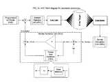
- FIG. 1is a system diagram showing a x-ray equipment incorporating the apparatus of the invention
- FIGS. 2 a / 2 bis a block diagram showing the basic functional units of the apparatus of the invention.
- FIG. 3is a plot showing an example of a programmed kV profile.
- FIG. 4shows the time sequence during Cephalography.
- the x-ray diagnostic system illustrated in FIG. 1is capable of performing dental panoramic radiography, using narrow beam scanning technique, transversal slicing radiography, using linear tomographic technique, and cephalography, using conventional x-ray technique.
- a x-ray generator 1mechanically coupled to the image receptor 2 a by the rotating arm.
- the rotating armrotates around the patient during the panoramic and transversal slicing scanning process.
- the image receptoris moved with variable speed during the panoramic sequence. The details of the panoramic technique are well known to those experienced in the matter, and will not further described.
- the same x-ray generator 1can be oriented towards a second image receptor 2 b.
- the Radiation Detectors 3composed of one or more detectors such as photo diodes, are located behind the image detectors, one for panoramic and transversal slicing, and one for cephalography.
- the vertical and horizontal positions of the radiation detectorsare located within the x-ray beam and are chosen to provide a signal corresponding to the absorption of the bone structure, namely the maxillary bone for the panoramic projections and the temporal bone for the cephalographic projection, which are of major diagnostic interest for the dentist.
- the output signals of Radiation Detectors 3are amplified by amplifiers 4 and the amplified signals are fed to the control unit 5 .
- the control unit 5provides analog to digital conversion at fast rate (e.g. every 40 msec) of the measured value of radiation intensity, and correction of the exposure factors, e.g. the tube voltage, in synchronisation with the imaging process.
- the automatic exposure control apparatusincorporates several functions, which are illustrated in FIG. 2 and are described below:
- This functional unitis applicable only to panoramic projections. It provides a pre-programmed modulated profile of the tube voltage (kV) during the imaging process, to be used as a reference for the AEC operation in the panoramic projections.
- kVtube voltage
- a different PKPmay be available for each available projection, and eventually for each available patient size.
- PKPhas been implemented by adopting a tube voltage profile which has proven by test on phantom and in vivo to provide an optimised image density, e.g. at the roots of the teeth, along the various anatomical regions, such as the rami of the mandibula and the spine.
- This functional unitprovides simulation of the density of the latent image on the x-ray receptor. It processes the radiation detector amplified signal either by combining it with the speed of the cassette drive in case of panoramic projections, or by integrating it in the time in case of cephalographic projections.
- the parameters of the simulationi.e. the gain
- the parameters of the simulationare separately adjustable for each different projection and for the sensitivity of each different image detector type (e.g. the speed of the applied screen-film combination, or the phosphor plate sensitivity).
- upper and lower limitsmay be applied to the range of acceptable density values. Such limits may preferably be adjustable for each different projection.
- This functional unitprocesses the density simulation output generated by the DSU, by comparing it with a reference value and generating a density error. Then the computational unit translates the density error into a correction of the applicable imaging parameter.
- the density erroris translated into a correction of the tube voltage, by applying to it a proportional control and/or an integrative control named PSC and described below.
- PSCproportional control and/or an integrative control
- panoramic projectionssuch correction is applied with certain upper and lower limits over the Programmed kV Profile, as further described below within the ARU functional unit.
- the proportional controlwill be programmable for each projection.
- the density erroris translated into a correction of the exposure time, by comparing the actual integrated density level generated by the DSU with a threshold value, and generating a stop of x-ray emission in case that the threshold is exceeded.
- This functional unitis applicable only to panoramic projections. It performs on selected portions of the anatomy an integration of the density error generated by the DCU and generates in real-time a correction quantity to be added to the Programmed kV Profile.
- the PSC functionis intended to operate in a region of measurement corresponding to the anatomical region where there is a homogeneous bone structure to which the absorption characteristic of the patient can be correlated.
- the location of the region of measurementmay be done by applying a pre-programmed interval, during which the density correction values within an acceptable range are integrated.
- the start of the region of measurementmay be located automatically during the imaging process, by performing an analysis of the density simulation generated by the DSU and its derivative, and identifying the negative transition of the derivative corresponding to start of the bone structure. After that the start of the region of measurement is identified, a pre-programmed interval is applied, during which the density correction values within an acceptable range are integrated.
- the PSC operationis preferably adjusted for each projection, by using programmable parameters for the start and end of the seek zone, the gain of the integrator, and the range of acceptable values of samples.
- ARUArtefacts Rejection Unit
- This functional unitoperates both in panoramic and cephalographic projections. It aims to perform a rejection of spurious correction of the imaging parameter, as may be generated by artefacts of non-anatomical structures inserted into the patient denture, such as amalgam fillings, implants, etc.
- panoramic projectionsIn panoramic projections it analyses the correction of the imaging parameter generated by the DCU and its derivative, and rejects those samples exceeding threshold limits both in absolute value and in slew rate, by clipping them to a predefined level.
- the ARU operationis preferably adjusted for each panoramic projection, by using programmable threshold limits.
- cephalographic projectionsit prevents the exposure time to exceed a predefined maximum level, by generating a stop of x-ray emission when such maximum level would be exceed.
- the ARU operationis preferably adjusted for each cephalographic projection, by using a programmable exposure time limit as a percentage of the initial exposure time setting.
- the PSC controlis inactive, while the P control (proportional) of the DCU may be active.
- Phase where the PSC control is activeto provide a correction for the patient size.
- the PKPwill result shifted upwards or downwards depending on the values of the integrated samples.
- the PKP, DSU, DCU and ARUare active.
- the P control of the DCUmay be inactive.
- the DSU, DCU and ARU functionsare active.
- the PSC controlis inactive, while the P control of the DCU may be active to provide further correction in specific anatomical regions, such as the spine.
- the imaging parametersare predefined by the user through the user interface, either manually or pre-programmed depending on the selection of the patient size.
- the imaging parametersi.e. the tube current
- the sensitivity of the image detectori.e. the speed of the film-screen combination.
- the tube voltage and currentwill stay stable at the values defined by the user, while the exposure time will be corrected by the AEC.
- DSU, DCU and ARUare active.
- the density simulation generated by the DSUis analysed by the DCU and compared with the threshold value.
- the time sequence during Cephalographyis illustrated in FIG. 4 .
Landscapes
- Health & Medical Sciences (AREA)
- Life Sciences & Earth Sciences (AREA)
- Engineering & Computer Science (AREA)
- General Health & Medical Sciences (AREA)
- Medical Informatics (AREA)
- Radiology & Medical Imaging (AREA)
- Heart & Thoracic Surgery (AREA)
- High Energy & Nuclear Physics (AREA)
- Physics & Mathematics (AREA)
- Nuclear Medicine, Radiotherapy & Molecular Imaging (AREA)
- Optics & Photonics (AREA)
- Pathology (AREA)
- Toxicology (AREA)
- Biomedical Technology (AREA)
- Biophysics (AREA)
- Molecular Biology (AREA)
- Surgery (AREA)
- Animal Behavior & Ethology (AREA)
- Public Health (AREA)
- Veterinary Medicine (AREA)
- Dentistry (AREA)
- Oral & Maxillofacial Surgery (AREA)
- Neurology (AREA)
- Neurosurgery (AREA)
- Apparatus For Radiation Diagnosis (AREA)
Abstract
Description
This application is a continuation application to U.S. application Ser. No. 09/679,768 filed on Oct. 5, 2000, and claims the benefit of provisional application No. 60/158,706, filed Oct. 8, 1999.
In dental panoramic and cephalographic radiography it is of utmost importance to ensure by an automatic method the optimal density of the latent image on the image detector, to allow proper detection of the anatomical details and consistency of the diagnostic outcome, and to minimise the need of repeating the x-ray examination so imparting unnecessary dose to the patient.
In dental panoramic radiography, using narrow beam scanning technique, the density is greatly influenced by (1) the individual patient characteristics (age, sex, race and size), particularly on the bone structure where osteoporosis phenomena may be present. Additionally (2) great variations in density occur during the exposure, caused by the different x-ray transmission of the various anatomical regions (temporo-mandibular joint, styloid process, hyoid bone, spine vertebrae, etc.) exposed during the scanning process. Finally (3) various irregularities in the patient denture (amalgam fillings, implants, missing teeth, etc.) may induce false detection and consequently erroneous operation of the automatic exposure control apparatus. While the automatic exposure control apparatus should effectively correct the density variations of the first two kinds, it is desirable that it has a high level of rejection for variations of the third kind (artefacts).
In cephalography, being a stationary radiographic technique, mainly density variations of the first kind are present, in conjunction also with the different patient projections (antero-posterior or latero-lateral).
One important feature is that the automatic exposure control apparatus can operate effectively in the several projection modalities (standard and child projections, transversal slicing, temporo-mandibular joint projections, sinus projections, frontal and orthogonal projections, cephalography antero-posterior and letero-lateral) as provided by the modern dental x-ray panoramic and cephalographic equipments.
Another desirable feature is that the automatic exposure control apparatus is able to compensate for the different sensitivities of the compatible image detectors (radiographic film speed factor, phosphor plate x-ray detectors, etc.).
Prior art automatic exposure apparatus for panoramic x-ray equipment determine imaging parameters (e.g. kV and mA of an x-ray tube, speed of the film drive or of the rotating arm supporting the x-ray tube and the film drive) by measuring the radiation passed through a patient either on the basis of a single sample taken at predefined imaging moment, or by continuous measurement, or by identification and measuring on a selected portion of the jaw (preferably the ramus of the mandibula).
Automatic exposure control by correction of the x-ray tube voltage (kV) is preferable, as it has faster response and provides x-ray energy modulation, with varying penetrating power. Adjustment of the speed of rotating arm or film drive requires huge computational capability, not practical in case of multi-projection equipment. Automatic exposure controls where adjustments are effected on the basis of a single sample are very sensitive to variations in the positioning of the patients and do not compensate for differences in the anatomy of the same. Automatic exposure apparatus with continuous control are very critical, due to the intrinsic difficulty in providing accurate and reproducible dynamic correction in all the anatomical regions, particularly in the spine, without producing asymmetry of the image density or other undesirable effects (vertical bands, artefact shadows, etc.). Identification of particular bone locations is restricted to regions with well defined anatomical structure and cannot be easily extended to varying anatomies as practically needed in multi-projection equipment.
The object of the invention is an automatic exposure control apparatus and method for dental panoramic and cephalographic x-ray equipment, capable of producing optimisation of the grey scale of the latent image in a consistent and reproducible way for all the available projections and regardless of the patient size and anatomy, providing high level of rejection of the artefacts generated by structures inserted into the patient denture.
The invention is founded on the following basic assumptions:
(a) in panoramic projections the optimal controlled parameter is the tube voltage (kV), for its faster dynamic response and varying penetrating power; in cephalographic projections the optimal controlled parameter is the exposure time, which is directly proportional to the output dose.
(b) both in panoramic and cephalographic projections it is preferable that the user selects the patient size, so pre-setting a programmed value of the controlled parameter.
(c) in panoramic projections, due to inherent criticality induced by the individual anatomic differences, the variance in positioning of the patient, the presence of irregular structures in the patient denture, it is wise to support the automatic exposure control operation with a variable kV profile pre-programmed for each available projection.
(d) for increased safety and reliability the automatic exposure control operates in a limited range around the programmed value (and programmed profile) of the controlled parameter.
Herefollowing is a description in greater detail of the invention, based on the exemplary embodiment illustrated in the attached drawings. By the disclosure and the appended claims the features and innovations of the invention will be outlined.
Table 1 shows the status of enable of the functional units in the different operational phases
FIG. 1 is a system diagram showing a x-ray equipment incorporating the apparatus of the invention;
FIGS. 2a/2bis a block diagram showing the basic functional units of the apparatus of the invention;
FIG. 3 is a plot showing an example of a programmed kV profile.
FIG. 4 shows the time sequence during Cephalography.
The x-ray diagnostic system illustrated in FIG. 1 is capable of performing dental panoramic radiography, using narrow beam scanning technique, transversal slicing radiography, using linear tomographic technique, and cephalography, using conventional x-ray technique.
It is equipped with ax-ray generator 1, mechanically coupled to theimage receptor 2aby the rotating arm. The rotating arm rotates around the patient during the panoramic and transversal slicing scanning process. The image receptor is moved with variable speed during the panoramic sequence. The details of the panoramic technique are well known to those experienced in the matter, and will not further described.
In case of cephalography, thesame x-ray generator 1 can be oriented towards a second image receptor2b.
TheRadiation Detectors 3, composed of one or more detectors such as photo diodes, are located behind the image detectors, one for panoramic and transversal slicing, and one for cephalography. The vertical and horizontal positions of the radiation detectors are located within the x-ray beam and are chosen to provide a signal corresponding to the absorption of the bone structure, namely the maxillary bone for the panoramic projections and the temporal bone for the cephalographic projection, which are of major diagnostic interest for the dentist.
The output signals ofRadiation Detectors 3 are amplified byamplifiers 4 and the amplified signals are fed to thecontrol unit 5.
Thecontrol unit 5 provides analog to digital conversion at fast rate (e.g. every 40 msec) of the measured value of radiation intensity, and correction of the exposure factors, e.g. the tube voltage, in synchronisation with the imaging process.
The automatic exposure control apparatus (herefollowing AEC) incorporates several functions, which are illustrated in FIG.2 and are described below:
(a) PKP (Programmed kV Profile).
This functional unit is applicable only to panoramic projections. It provides a pre-programmed modulated profile of the tube voltage (kV) during the imaging process, to be used as a reference for the AEC operation in the panoramic projections. A different PKP may be available for each available projection, and eventually for each available patient size.
PKP has been implemented by adopting a tube voltage profile which has proven by test on phantom and in vivo to provide an optimised image density, e.g. at the roots of the teeth, along the various anatomical regions, such as the rami of the mandibula and the spine.
(b) DSU (Density Simulation Unit).
This functional unit provides simulation of the density of the latent image on the x-ray receptor. It processes the radiation detector amplified signal either by combining it with the speed of the cassette drive in case of panoramic projections, or by integrating it in the time in case of cephalographic projections.
Preferably the parameters of the simulation (i.e. the gain) are separately adjustable for each different projection and for the sensitivity of each different image detector type (e.g. the speed of the applied screen-film combination, or the phosphor plate sensitivity).
In order to introduce a safety control and inhibition of the AEC operation, upper and lower limits may be applied to the range of acceptable density values. Such limits may preferably be adjustable for each different projection.
(c) DCU (Density Correction Unit).
This functional unit processes the density simulation output generated by the DSU, by comparing it with a reference value and generating a density error. Then the computational unit translates the density error into a correction of the applicable imaging parameter.
In case of panoramic projections the density error is translated into a correction of the tube voltage, by applying to it a proportional control and/or an integrative control named PSC and described below. In panoramic projections such correction is applied with certain upper and lower limits over the Programmed kV Profile, as further described below within the ARU functional unit. Preferably the proportional control will be programmable for each projection.
In cephalographic projections the density error is translated into a correction of the exposure time, by comparing the actual integrated density level generated by the DSU with a threshold value, and generating a stop of x-ray emission in case that the threshold is exceeded.
(d) PSC (Patient Size Correction).
This functional unit is applicable only to panoramic projections. It performs on selected portions of the anatomy an integration of the density error generated by the DCU and generates in real-time a correction quantity to be added to the Programmed kV Profile.
The PSC function is intended to operate in a region of measurement corresponding to the anatomical region where there is a homogeneous bone structure to which the absorption characteristic of the patient can be correlated.
In a first embodiment the location of the region of measurement may be done by applying a pre-programmed interval, during which the density correction values within an acceptable range are integrated.
In another embodiment the start of the region of measurement may be located automatically during the imaging process, by performing an analysis of the density simulation generated by the DSU and its derivative, and identifying the negative transition of the derivative corresponding to start of the bone structure. After that the start of the region of measurement is identified, a pre-programmed interval is applied, during which the density correction values within an acceptable range are integrated.
The PSC operation is preferably adjusted for each projection, by using programmable parameters for the start and end of the seek zone, the gain of the integrator, and the range of acceptable values of samples.
(e) ARU (Artefacts Rejection Unit)
This functional unit operates both in panoramic and cephalographic projections. It aims to perform a rejection of spurious correction of the imaging parameter, as may be generated by artefacts of non-anatomical structures inserted into the patient denture, such as amalgam fillings, implants, etc.
In panoramic projections it analyses the correction of the imaging parameter generated by the DCU and its derivative, and rejects those samples exceeding threshold limits both in absolute value and in slew rate, by clipping them to a predefined level. The ARU operation is preferably adjusted for each panoramic projection, by using programmable threshold limits.
In cephalographic projections it prevents the exposure time to exceed a predefined maximum level, by generating a stop of x-ray emission when such maximum level would be exceed. The ARU operation is preferably adjusted for each cephalographic projection, by using a programmable exposure time limit as a percentage of the initial exposure time setting.
Based on the functional units above, the following operational phases are applicable in panoramic and transversal slicing projections:
A) Phase corresponding to the initial part of the imaging process, where the PKP corresponding the patient size selected by the user is applied, and the DSU, DCU and ARU functions are activated. The PSC control is inactive, while the P control (proportional) of the DCU may be active.
B) Phase where the PSC control is active, to provide a correction for the patient size. At the end of this phase the PKP will result shifted upwards or downwards depending on the values of the integrated samples. The PKP, DSU, DCU and ARU are active. The P control of the DCU may be inactive.
C) Phase where the PKP corrected with the additive term generated by the PSC is applied. The DSU, DCU and ARU functions are active. The PSC control is inactive, while the P control of the DCU may be active to provide further correction in specific anatomical regions, such as the spine.
The sequence of phases is illustrated in FIG. 3, together with an exemplary plot of the actual PKP. The status of enable of the functional units is illustrated in Table 1 below.
| Phase | PKP | DSU | DCU | ARU | PSC | P | ||
| A | Y | Y | Y/N | Y | N | Y/N | ||
| B | Y | Y | Y | Y | Y | Y/N | ||
| C | Y | Y | Y/N | Y | N | Y/N | ||
| Y = ACTIVE | ||||||||
| N = INACTIVE | ||||||||
In Cephalography the imaging parameters (tube voltage, tube current, exposure time) are predefined by the user through the user interface, either manually or pre-programmed depending on the selection of the patient size. Preferably the imaging parameters (i.e. the tube current) will be corrected depending on the sensitivity of the image detector (i.e. the speed of the film-screen combination). During the imaging process the tube voltage and current will stay stable at the values defined by the user, while the exposure time will be corrected by the AEC.
Based on the functional units above, the following operational phases are applicable in cephalographic projections:
A) Phase corresponding to the intial part of the imaging process. DSU, DCU and ARU are active. The density simulation generated by the DSU is analysed by the DCU and compared with the threshold value.
B) Phase corresponding to the stop of the exposure. If the integrated density simulation generated by the DSU is exceeding the threshold level, then the DCU generates the stop of the exposure. If the exposure time exceeds the programmed maximum limit then the ARU generates the stop of x-ray emission. After the end of exposure all functions get inactive.
The time sequence during Cephalography is illustrated in FIG.4.
Claims (12)
1. A dental x-ray device having an x-ray radiation source and image receptors for panoramic and cephalographic radiography of bone structure of a patient, and for performing multiple projections where the density of the latent image is controlled by the imaging parameters, comprising an apparatus for the automatic control of the exposure (AEC) comprising:
an x-ray radiation detector located behind an image receptor and providing a signal corresponding to the x-ray absorption characteristic of the bone structure; a control unit for analysing the radiation detector signal and providing adjustment of the imaging parameters by incorporating as functional units:
(a) A programmed kV profile unit (PKP) being the functional unit providing pre-programmed modulated profile of the x-ray source tube voltage (kV) during the imaging process, correlated to the standard patient anatomy; said PKP being a reference for the AEC operation during the imaging process in the panoramic projections;
(b) a density simulation unit (DSU) being the functional unit providing simulation of the density of the latent image on the x-ray receptor, and processing a radiation detector-amplified signal by combining it with the speed of the cassette drive in case of panoramic projections, or by integrating it in the time in case of cephalographic projections;
(c) a density correction unit (DCU) being the functional unit processing the DSU density simulation output by comparing with a reference value, and generating a density error, and translating the density error into a correction of the applicable imaging parameter using a computational unit; in panoramic projections said density error is translated into a correction of the tube voltage, by applying to it a proportional control or an integrative control (PSC); in panoramic projections correction is applied with upper and lower limits over said PKP, within an ARU functional unit; in cephalographic projections said density error is translated into a correction of the exposure time, by comparing the actual integrated density level generated by the DSU with a threshold value, and generating a stop of x-ray emission when said threshold is exceeded;
(d) said PSC being a patient size correction functional unit operating in panoramic projections and performing on selected portions of the anatomy corresponding to a region of measurement an integration of the density error generated by the DCU and generating in real-time a correction quantity to be added to said PKP; in the region of measurement the density correction values are analysed and samples within a selected range are integrated;
(e) said ARU being an artefacts rejection unit and being a functional unit providing rejection of spurious correction of the imaging parameter, as generated by artefacts of non-anatomical structures inserted into the bone structure; in panoramic projections said ARU analyses the correction of the imaging parameter generated by the DCU and its derivative, and rejects samples exceeding threshold limits both in absolute value and in slew rate, by clipping them to a predefined level; in cephalographic projections it prevents the exposure time to exceed a predefined maximum level, by generating a stop of x-ray emission when such maximum level would be exceed.
2. A method for the automatic control of the exposure in panoramic projections employing the device ofclaim 1 , comprising the steps of:
A) selecting by the user interface the patient size, by which the initial setting of the imaging parameter and the programmed kV profile (PKP) are defined;
B) initiating the imaging process, where the PKP corresponding the patient size selected by the user is applied; the DSU, DCU and ARU functions are active; the PSC control is inactive, while the P control (proportional) of the DCU may be active;
C) activating in the applicable region of measurement the PSC function, to provide a real time correction of the PKP corresponding to the actual patient size; at the end of this phase the PKP result is shifted upwards or downwards depending on the values of the integrated samples; the PKP, DSU, DCU and ARU are active; The P control of the DCU may be inactive;
D) disabling the PSC function, while the PKP corrected with the additive term generated by the PSC is applied; the DSU, DCU and ARU functions are active; the PSC control is inactive, while the P control of the DCU may be active to provide further correction in specific anatomical regions.
3. A method for the automatic control of the exposure in cephalographic projections employing the device ofclaim 1 , comprising the steps of:
A) selecting through the user interface the imaging parameters (tube voltage, tube current, exposure time), manually or by pre-programmed control depending on the selection of the patient size;
B) initiating the imaging process, where the tube voltage and current are kept stable at the values pre-set by the user; DSU and DCU and ARU are active; the density simulation generated by the DSU is analysed by the DCU and compared with the threshold value;
C) Terminating the imaging process by generation of a stop of exposure signal; if the integrated density simulation generated by the DSU exceeds the threshold level, then the DCU generates the stop of the exposure; if the exposure time exceeds the programmed maximum limit then the ARU generates the stop of x-ray emission; and, after the end of exposure inactivating all functions.
4. The apparatus as set forth inclaim 1 wherein a different PKP is programmable for each panoramic projection, and optionally for each patient size.
5. The apparatus and method as set forth inclaim 1 wherein a range of acceptable density values is applied to the DSU density simulation, and such range of acceptable density values is programmable for each projection.
6. The apparatus and method as set forth inclaim 1 wherein the parameters for the DSU simulation are programmable for each projection.
7. The apparatus and method as set forth inclaim 1 wherein the parameters for the DSU simulation are programmable for each compatible image detector type, by taking into account its particular sensitivity.
8. The apparatus and method as set forth inclaim 1 wherein the parameters for the DSU simulation are programmable for each compatible image detector holder type, by taking into account its particular x-ray transmission characteristic.
9. The apparatus and method as set forth inclaim 1 wherein the parameters for the proportional control of the DCU correction are programmable for each projection.
10. The apparatus and method as set forth inclaim 1 wherein the parameters for the PSC operation are programmable for each projection.
11. The apparatus and method as set forth inclaim 1 wherein the start of the PSC region of measurement may be located automatically during the imaging process, by performing an analysis of the density simulation generated by the DSU and its derivative, and identifying the negative transition of the derivative corresponding to start of the bone structure.
12. The apparatus and method as set forth inclaim 1 wherein the parameters for the ARU operation are programmable for each projection.
Priority Applications (1)
| Application Number | Priority Date | Filing Date | Title |
|---|---|---|---|
| US10/024,873US6553095B2 (en) | 1999-10-08 | 2001-12-19 | Automatic exposure control for dental panoramic and cephalographic x-ray equipment |
Applications Claiming Priority (3)
| Application Number | Priority Date | Filing Date | Title |
|---|---|---|---|
| US15870699P | 1999-10-08 | 1999-10-08 | |
| US67976800A | 2000-10-05 | 2000-10-05 | |
| US10/024,873US6553095B2 (en) | 1999-10-08 | 2001-12-19 | Automatic exposure control for dental panoramic and cephalographic x-ray equipment |
Related Parent Applications (1)
| Application Number | Title | Priority Date | Filing Date |
|---|---|---|---|
| US67976800AContinuation | 1999-10-08 | 2000-10-05 |
Publications (2)
| Publication Number | Publication Date |
|---|---|
| US20020085673A1 US20020085673A1 (en) | 2002-07-04 |
| US6553095B2true US6553095B2 (en) | 2003-04-22 |
Family
ID=22569340
Family Applications (1)
| Application Number | Title | Priority Date | Filing Date |
|---|---|---|---|
| US10/024,873Expired - Fee RelatedUS6553095B2 (en) | 1999-10-08 | 2001-12-19 | Automatic exposure control for dental panoramic and cephalographic x-ray equipment |
Country Status (5)
| Country | Link |
|---|---|
| US (1) | US6553095B2 (en) |
| EP (1) | EP1219147B1 (en) |
| CA (1) | CA2388256A1 (en) |
| DE (1) | DE60031787T2 (en) |
| WO (1) | WO2001028298A1 (en) |
Cited By (9)
| Publication number | Priority date | Publication date | Assignee | Title |
|---|---|---|---|---|
| US20030091142A1 (en)* | 2001-11-15 | 2003-05-15 | Jianying Li | System and method of medical imaging having task and/or patient size dependent processing |
| US20040037389A1 (en)* | 2002-03-05 | 2004-02-26 | Marie-Anne De Smet | Process and device for testing parts by X rays |
| US20040096035A1 (en)* | 2002-11-15 | 2004-05-20 | Canon Kabushiki Kaisha | X-ray imaging apparatus |
| US20050078792A1 (en)* | 2001-11-23 | 2005-04-14 | Pekka Strommer | Automatic exposure method and automatic exposure system |
| US20070183590A1 (en)* | 2006-02-08 | 2007-08-09 | Gray Joel E | Dental image quality and dose analyzer |
| US20080107240A1 (en)* | 2006-10-11 | 2008-05-08 | Franz Atzinger | X-ray arrangement with a converter for converting system parameters into image chain parameters and associated X-ray method |
| US20110142197A1 (en)* | 2009-12-15 | 2011-06-16 | Midmark Corporation | Patient positioning system for panoramic dental radiation imaging system |
| US20110142198A1 (en)* | 2009-12-15 | 2011-06-16 | Midmark Corporation | Removable radiation sensor for dental imaging systems |
| US20110142199A1 (en)* | 2009-12-15 | 2011-06-16 | Midmark Corporation | Motion system for panoramic dental radiation imaging system |
Families Citing this family (13)
| Publication number | Priority date | Publication date | Assignee | Title |
|---|---|---|---|---|
| JP2004536643A (en)* | 2001-07-25 | 2004-12-09 | デンツプライ インターナショナル インコーポレーテッド | Real-time digital X-ray imaging device |
| CA2491759A1 (en) | 2002-07-25 | 2004-02-19 | Gendex Corporation | Real-time digital x-ray imaging apparatus and method |
| DE10313109A1 (en)* | 2003-03-24 | 2004-10-21 | Sirona Dental Systems Gmbh | X-ray sensitive camera and X-ray device |
| WO2004103034A1 (en)* | 2003-05-16 | 2004-11-25 | Philips Intellectual Property & Standards Gmbh | Method and device for exposing x-ray images |
| US7372942B2 (en)* | 2004-03-29 | 2008-05-13 | Siemens Medical Solutions Usa, Inc. | Medical imaging system with dosimetry for estimating circuit board life |
| US8417010B1 (en) | 2006-01-12 | 2013-04-09 | Diagnoscan, LLC | Digital x-ray diagnosis and evaluation of dental disease |
| DE102007033957A1 (en)* | 2007-07-19 | 2009-01-22 | Sirona Dental Systems Gmbh | Dental X-ray emitter's light exposure parameter e.g. light exposure time, generating method, involves performing correlation between light exposure parameters and photograph requirements based on generated photograph |
| US7480365B1 (en)* | 2007-08-06 | 2009-01-20 | Carestream Health, Inc. | Dose reduced digital medical image simulations |
| EP2198783B1 (en)* | 2008-12-19 | 2016-10-12 | Cefla S.C. | Apparatus and method for digital X-ray scanning |
| KR101094180B1 (en)* | 2009-11-10 | 2011-12-14 | 주식회사바텍 | Method and device for acquiring panoramic image |
| US9579073B2 (en)* | 2010-05-07 | 2017-02-28 | Apteryx, Inc. | System and method for dentition specific image enhancement |
| US11363938B2 (en)* | 2013-03-14 | 2022-06-21 | Ormco Corporation | Feedback control mechanism for adjustment of imaging parameters in a dental imaging system |
| EP4186435A1 (en)* | 2021-11-26 | 2023-05-31 | Sirona Dental Systems GmbH | Device and method for generating dental x-ray images with highly specific dose application |
Citations (120)
| Publication number | Priority date | Publication date | Assignee | Title |
|---|---|---|---|---|
| US3894235A (en) | 1973-06-08 | 1975-07-08 | Siemens Ag | X-ray diagnostic apparatus for the preparation of x-ray exposures including a timer switch for determining the exposure time |
| US3911273A (en) | 1973-04-27 | 1975-10-07 | Siemens Ag | X-ray diagnostic apparatus for preparing x-ray exposures including an automatic illuminating device and automatic adjustment of the exposure voltage |
| US3974385A (en) | 1972-12-06 | 1976-08-10 | Siemens Aktiengesellschaft | X-ray diagnostic apparatus |
| US3987281A (en) | 1974-07-29 | 1976-10-19 | The United States Of America As Represented By The Department Of Health, Education And Welfare | Method of radiation therapy treatment planning |
| US3991314A (en) | 1972-09-19 | 1976-11-09 | Siemens Aktiengesellschaft | X-ray diagnosis apparatus for X-raying and exposure |
| US4021672A (en) | 1974-10-02 | 1977-05-03 | Siemens Aktiengesellschaft | Dental X-ray diagnostic installation |
| US4061920A (en) | 1975-03-15 | 1977-12-06 | U.S. Philips Corporation | X-ray installation comprising an image intensifier/image pick-up tube system and an automatic X-ray exposure device |
| US4070578A (en) | 1976-07-30 | 1978-01-24 | Timothy John G | Detector array and method |
| US4097741A (en) | 1975-10-20 | 1978-06-27 | Siemens Aktiengesellschaft | X-ray diagnostics system for X-ray photographs |
| US4104531A (en) | 1976-10-04 | 1978-08-01 | Thoro-Ray Inc. | Electron beam target carrier with ceramic window for dental or medical X-ray use |
| US4158138A (en)* | 1977-10-25 | 1979-06-12 | Cgr Medical Corporation | Microprocessor controlled X-ray generator |
| US4160997A (en) | 1974-05-14 | 1979-07-10 | Robert Schwartz | Intraoral fluoroscope |
| US4160906A (en) | 1977-06-23 | 1979-07-10 | General Electric Company | Anatomically coordinated user dominated programmer for diagnostic x-ray apparatus |
| US4188537A (en) | 1976-10-15 | 1980-02-12 | Siemens Aktiengesellschaft | Dental apparatus for x-ray diagnosis |
| US4247780A (en) | 1979-06-08 | 1981-01-27 | The United States Of America As Represented By The Department Of Health, Education And Welfare | Feedback controlled geometry registration system for radiographs |
| US4259582A (en) | 1979-11-02 | 1981-03-31 | Albert Richard D | Plural image signal system for scanning x-ray apparatus |
| US4352987A (en) | 1977-12-27 | 1982-10-05 | Kabushiki Kaisha Morita Seisakusho | Structure of dental X-ray apparatus |
| US4454606A (en) | 1983-05-23 | 1984-06-12 | General Electric Company | Reconfigurable x-ray AEC compensation |
| US4475224A (en) | 1981-10-30 | 1984-10-02 | Siemens Aktiengesellschaft | Dental x-ray diagnostic installation |
| US4486896A (en) | 1981-08-13 | 1984-12-04 | U.S. Philips Corporation | X-Ray generator incorporating automatic correction of a dose-determining exposure parameter |
| US4495632A (en) | 1980-06-27 | 1985-01-22 | Kabushiki Kaisha Morita Seisakusho | Radiographic apparatus for photographing entire jaws |
| US4501010A (en) | 1981-10-30 | 1985-02-19 | Siemens Aktiengesellschaft | Dental X-ray diagnostic installation |
| US4589121A (en) | 1984-02-01 | 1986-05-13 | Kabushiki Kaisha Morita Seisakusho | Dental panoramic X-ray photographing apparatus |
| US4641331A (en) | 1983-08-02 | 1987-02-03 | Kabushiki Kaisha Morita Seisakusho | Automatic exposure device for a panoramic X-ray photographing device |
| US4675888A (en) | 1984-01-06 | 1987-06-23 | Instrumentarium Corp. | Patient support system in a narrow-beam tomographic imaging apparatus |
| EP0229497A1 (en) | 1985-12-11 | 1987-07-22 | FutureTech Industries, Inc. | X-ray imaging system and method |
| US4741007A (en) | 1985-05-31 | 1988-04-26 | Planmeca Oy | Panoramic X-ray, tomography especially for dental photography |
| US4783793A (en) | 1985-09-13 | 1988-11-08 | Planmeca Oy | X-ray apparatus for panoramic tomography including control system |
| US4797905A (en) | 1986-01-10 | 1989-01-10 | U.S. Philips Corporation | X-ray generator incorporating dose rate control |
| US4811372A (en) | 1985-12-20 | 1989-03-07 | Siemens Aktiengesellschaft | Dental X-ray diagnostic installation for producing panoramic tomograms of the jaw of a patient |
| US4813060A (en) | 1985-12-20 | 1989-03-14 | Siemens Aktiengesellschaft | Dental x-ray diagnostic apparatus for producing panorama tomograms of the jaw of a patient |
| US4815115A (en) | 1986-02-11 | 1989-03-21 | Radiante Oy | Method of photographing an object with a panoramic X-ray apparatus fitted with automatic exposure |
| US4823369A (en) | 1987-02-16 | 1989-04-18 | Siemens Aktiengesellschaft | Dental x-ray diagnostics installation for producing panorama slice exposures of the jaw of a patient |
| US4847881A (en) | 1985-12-20 | 1989-07-11 | Siemens Aktiengesellschaft | Dental x-ray diagnostics installation for producing panorama tomograms of the jaw of a patient |
| US4856038A (en) | 1986-09-26 | 1989-08-08 | Siemens Aktiengesellschaft | Dental x-ray diagnostics installation for producing panoramic exposures of slices in the skull |
| US4878234A (en) | 1987-02-16 | 1989-10-31 | Siemens Aktiengesellschaft | Dental x-ray diagnostics installation for producing panorama slice exposures of the jaw of a patient |
| US4905265A (en) | 1985-12-11 | 1990-02-27 | General Imaging Corporation | X-ray imaging system and solid state detector therefor |
| US4930146A (en) | 1989-07-10 | 1990-05-29 | General Electric Company | X-ray tube current control with constant loop gain |
| EP0373717A1 (en) | 1988-12-08 | 1990-06-20 | Koninklijke Philips Electronics N.V. | Dental x-ray image detection system |
| US4980905A (en) | 1989-02-16 | 1990-12-25 | General Electric Company | X-ray imaging apparatus dose calibration method |
| US4985907A (en) | 1988-05-06 | 1991-01-15 | U.S. Philips Corporation | Dental X-ray apparatus for panoramic tomography |
| US4995062A (en) | 1988-08-12 | 1991-02-19 | Siemens Aktiengesellschaft | X-ray diagnostics installation for producing panorama tomograms of the jaw of a patient |
| US5005195A (en) | 1989-03-10 | 1991-04-02 | Expert Image Systems, Inc. | Digital readout system for radiographic imaging |
| US5018177A (en) | 1989-06-01 | 1991-05-21 | Board Of Regents, The University Of Texas System | Apparatus and method for producing digital panoramic x-ray images |
| US5043582A (en) | 1985-12-11 | 1991-08-27 | General Imagining Corporation | X-ray imaging system and solid state detector therefor |
| US5090047A (en) | 1990-10-23 | 1992-02-18 | Applied Research Company | Apparatus for reproducibly positioning an image receptor for intraoral diagnostics |
| US5090040A (en) | 1989-03-10 | 1992-02-18 | Expert Image Systems, Inc. | Data acquisition system for radiographic imaging |
| US5093852A (en) | 1988-11-08 | 1992-03-03 | Kabushiki Kaisha Morita Seisakusho | Medical panoramic radiographing device |
| US5195114A (en) | 1990-10-05 | 1993-03-16 | Kabushiki Kaisha Morita Seisakusho | Digital panoramic radiographic apparatus |
| US5214686A (en) | 1991-12-13 | 1993-05-25 | Wake Forest University | Three-dimensional panoramic dental radiography method and apparatus which avoids the subject's spine |
| US5267296A (en) | 1992-10-13 | 1993-11-30 | Digiray Corporation | Method and apparatus for digital control of scanning X-ray imaging systems |
| US5293312A (en) | 1991-06-20 | 1994-03-08 | Waggener Robert G | Method and apparatus for computing tomographic scans |
| US5331166A (en) | 1991-10-25 | 1994-07-19 | Kabushiki Kaisha Morita Seisakusho | Dental X-ray image detecting device with an automatic exposure function |
| EP0415075B1 (en) | 1989-08-03 | 1994-10-12 | Siemens Aktiengesellschaft | Dental X-ray diagnostic apparatus |
| US5386448A (en) | 1992-06-12 | 1995-01-31 | Orion-Yhtyma Oy | Method for imaging an object by means of a panoramic apparatus equipped with exposure automatics |
| USD355964S (en) | 1992-03-27 | 1995-02-28 | Regam Medical Systems Ab | Capsule for housing electro-medical equipment for radio-diagnosis or the like |
| US5425065A (en) | 1991-08-19 | 1995-06-13 | Instrumentarium Corp.- Imaging Division | Automatic exposure apparatus for panoramic x-ray equipment |
| US5434418A (en) | 1992-10-16 | 1995-07-18 | Schick; David | Intra-oral sensor for computer aided radiography |
| US5454022A (en) | 1993-03-26 | 1995-09-26 | Eastman Kodak Company | Method of taking X-ray images with a CCD image sensor, and a CCD image sensor system |
| EP0673623A1 (en) | 1994-03-24 | 1995-09-27 | Regam Medical Systems Ab | Scanning layer forming radiography |
| US5473660A (en) | 1994-06-01 | 1995-12-05 | U.S. Philips Corporation | Image sensing device |
| US5511106A (en) | 1993-07-06 | 1996-04-23 | Siemens Aktiengesellschaft | X-ray diagnostics installation for producing x-ray exposures of body parts of a patient |
| US5513252A (en) | 1992-06-01 | 1996-04-30 | Siemens Aktiengesellschaft | Dental x-ray diagnostics installation |
| US5519751A (en) | 1992-10-15 | 1996-05-21 | Hamamatsu Photonics K.K. | Medical X-ray image processing apparatus |
| US5519437A (en) | 1992-05-11 | 1996-05-21 | Regam Medical Ab | Method for compensation of dark current of CCD-sensor in dental x-raying |
| US5541974A (en) | 1994-06-15 | 1996-07-30 | Siemens Aktiengesellschaft | Real-time x-ray video imaging system having a CCD solid state image sensor |
| US5579366A (en) | 1993-07-06 | 1996-11-26 | Siemens Aktiengesellschaft | Line detector camera for employment in, particularly, dental x-ray diagnostics installations |
| US5583905A (en) | 1990-11-22 | 1996-12-10 | Kabushiki Kaisha Toshiba | X-ray imaging apparatus capable of eliminating unwanted X-ray image signal components |
| US5600699A (en) | 1995-02-09 | 1997-02-04 | J. Morita Manufacturing Corporation | Panoramic X-ray imaging apparatus |
| US5602896A (en) | 1994-12-22 | 1997-02-11 | U.S. Philips Corporation | Composing an image from sub-images |
| US5608455A (en) | 1992-07-30 | 1997-03-04 | Fuji Photo Film Co., Ltd | Interline transfer CCD image sensor with reduced dark current |
| US5617462A (en) | 1995-08-07 | 1997-04-01 | Oec Medical Systems, Inc. | Automatic X-ray exposure control system and method of use |
| US5625662A (en) | 1995-11-20 | 1997-04-29 | General Electric Company | Modulating x-ray tube current in a CT system |
| EP0776149A1 (en) | 1995-11-21 | 1997-05-28 | Loral Fairchild Corporation | X-ray image sensor system and method |
| US5640018A (en) | 1995-02-09 | 1997-06-17 | J. Morita Manufacturing Corporation | Image detecting device and medical X-ray imaging apparatus |
| US5664001A (en) | 1995-03-24 | 1997-09-02 | J. Morita Manufacturing Corporation | Medical X-ray imaging apparatus |
| US5663998A (en) | 1995-08-29 | 1997-09-02 | J. Morita Manufacturing Corporation | X-ray imaging apparatus and automatic density correction method |
| US5668375A (en) | 1996-08-26 | 1997-09-16 | General Electric Company | Fast scan reset for a large area x-ray detector |
| US5677940A (en) | 1995-03-24 | 1997-10-14 | J. Morita Manufacturing Corporation | Digital X-ray imaging apparatus |
| DE4433545C2 (en) | 1994-09-20 | 1997-11-20 | Luecken Hans Joerg Von | Device for converting time-variable, strip-shaped X-ray image information into static overall images |
| US5694448A (en) | 1995-08-01 | 1997-12-02 | Eev Limited | Imaging apparatus |
| US5742659A (en) | 1996-08-26 | 1998-04-21 | Universities Research Assoc., Inc. | High resolution biomedical imaging system with direct detection of x-rays via a charge coupled device |
| US5744806A (en) | 1994-09-08 | 1998-04-28 | Afp Imaging Corporation | Method for multisensor arrangements |
| US5751783A (en) | 1996-12-20 | 1998-05-12 | General Electric Company | Detector for automatic exposure control on an x-ray imaging system |
| US5757011A (en) | 1995-02-10 | 1998-05-26 | Orbit Semiconductor, Inc. | X-ray onset detector and method |
| DE19648052A1 (en) | 1996-11-20 | 1998-06-04 | Siemens Ag | Three dimensional reconstruction of position of partial objects or sub=structures e.g. teeth or bones of skull |
| DE19754463A1 (en) | 1996-12-10 | 1998-06-18 | Morita Mfg | X-ray imaging instrument |
| DE4238268C2 (en) | 1992-11-12 | 1998-07-02 | Siemens Ag | Method and device for acceptance and constancy testing of filmless dental X-ray devices |
| US5784429A (en) | 1995-11-22 | 1998-07-21 | J. Morita Manufacturing Corporation | Dental panoramic X-ray imaging apparatus |
| US5796430A (en) | 1993-06-02 | 1998-08-18 | Hitachi, Ltd. | Video camera with a function to correct defective pixels of solid state image pickup device and method of correcting defective pixels of solid state image pickup device |
| US5812191A (en) | 1994-06-01 | 1998-09-22 | Simage Oy | Semiconductor high-energy radiation imaging device |
| US5828721A (en) | 1994-08-10 | 1998-10-27 | Sirona Dental Systems Gmbh & Co. Kg | Radiation diagnostics installation and method for producing panorama tomograms |
| US5828720A (en) | 1995-12-14 | 1998-10-27 | Orion-Yhtyma Oy | Exposure automatics for an X-ray apparatus |
| EP0632994B1 (en) | 1993-07-06 | 1999-01-07 | Sirona Dental Systems GmbH & Co.KG | X-ray diagnostic device for producing X-rays of body parts of a patient |
| US5864146A (en) | 1996-11-13 | 1999-01-26 | University Of Massachusetts Medical Center | System for quantitative radiographic imaging |
| DE19731927A1 (en) | 1997-07-24 | 1999-02-11 | Sirona Dental Systems Gmbh | X-ray diagnostic device for tomosynthesis |
| DE19734717A1 (en) | 1997-08-11 | 1999-02-25 | Sirona Dental Systems Gmbh | Method for compensating the dark current when creating dental panoramic and / or cephalometric slice images |
| US5887049A (en) | 1996-11-12 | 1999-03-23 | California Institute Of Technology | Self-triggered X-ray sensor |
| US5892227A (en) | 1994-09-29 | 1999-04-06 | Yissum Research Development Company Of The Hebrew University Of Jerusalem | Radiation detection system and processes for preparing the same |
| US5912942A (en) | 1997-06-06 | 1999-06-15 | Schick Technologies, Inc. | X-ray detection system using active pixel sensors |
| US5923722A (en) | 1996-08-05 | 1999-07-13 | Siemens Aktiengesellschaft | X-ray diagnostic apparatus with control of x-ray emission dependent on the afterglow in the solid-state detector |
| US5930330A (en) | 1995-09-29 | 1999-07-27 | New Mexico Biophysics | Method and apparatus for multitaxis scanning system |
| US5933471A (en) | 1997-02-26 | 1999-08-03 | International Business Machines Corporation | System and method for reducing reconstruction artifacts in computed tomography images |
| US5949811A (en)* | 1996-10-08 | 1999-09-07 | Hitachi Medical Corporation | X-ray apparatus |
| US5969360A (en) | 1997-11-26 | 1999-10-19 | Direct Radiography Corp. | Readout sequence for residual image elimination in a radiation detection panel |
| US5974166A (en) | 1996-06-26 | 1999-10-26 | Matsushita Electric Industrial Co., Ltd. | X-ray imaging apparatus and recording medium therefore |
| GB2304017B (en) | 1995-08-01 | 1999-11-17 | Eev Ltd | Imaging apparatus |
| US6002742A (en) | 1991-06-03 | 1999-12-14 | Afp Imaging Corporation | Method and device for triggering of X-ray image sensor |
| US6035013A (en) | 1994-06-01 | 2000-03-07 | Simage O.Y. | Radiographic imaging devices, systems and methods |
| US6035012A (en)* | 1998-05-14 | 2000-03-07 | Gen Electric | Artifact correction for highly attenuating objects |
| US6047042A (en) | 1998-03-25 | 2000-04-04 | Continental X-Ray Corporation | Automatic exposure and brightness control for fluoroscopic and radio-graphic imaging |
| US6055292A (en) | 1997-02-17 | 2000-04-25 | Sirona Dental Systems Gmbh & Co. Kg | Method and apparatus for producing X-ray exposures of body parts of a human |
| US6081739A (en) | 1998-05-21 | 2000-06-27 | Lemchen; Marc S. | Scanning device or methodology to produce an image incorporating correlated superficial, three dimensional surface and x-ray images and measurements of an object |
| US6093019A (en) | 1997-12-23 | 2000-07-25 | Integra Medical | Dental imaging system with digital motion video |
| US6094468A (en)* | 1997-06-26 | 2000-07-25 | U.S. Philips Corporation | Adjustable computer tomography device |
| JP3109057B2 (en) | 1996-11-27 | 2000-11-13 | 大日本塗料株式会社 | Cold-drying aqueous coating composition |
| US6173035B1 (en) | 1997-09-30 | 2001-01-09 | Kabushiki Kaisha Morita Seisakusho | Panoramic radiographic apparatus and digital sensor cassette used for same apparatus |
| US6208710B1 (en)* | 1998-07-21 | 2001-03-27 | Kabushiki Kaisha Toshiba | X-ray diagnostic apparatus and radiation diagnostic apparatus |
| US6233310B1 (en)* | 1999-07-12 | 2001-05-15 | General Electric Company | Exposure management and control system and method |
| EP0685201B1 (en) | 1994-05-31 | 2002-02-13 | Trophy Radiologie | Radiodiagnostic device having a scintillator with a CCD sensor |
- 2000
- 2000-10-05CACA002388256Apatent/CA2388256A1/ennot_activeAbandoned
- 2000-10-05DEDE60031787Tpatent/DE60031787T2/ennot_activeExpired - Lifetime
- 2000-10-05EPEP00968731Apatent/EP1219147B1/ennot_activeExpired - Lifetime
- 2000-10-05WOPCT/US2000/027474patent/WO2001028298A1/enactiveIP Right Grant
- 2001
- 2001-12-19USUS10/024,873patent/US6553095B2/ennot_activeExpired - Fee Related
Patent Citations (131)
| Publication number | Priority date | Publication date | Assignee | Title |
|---|---|---|---|---|
| US3991314A (en) | 1972-09-19 | 1976-11-09 | Siemens Aktiengesellschaft | X-ray diagnosis apparatus for X-raying and exposure |
| US3974385A (en) | 1972-12-06 | 1976-08-10 | Siemens Aktiengesellschaft | X-ray diagnostic apparatus |
| US3911273A (en) | 1973-04-27 | 1975-10-07 | Siemens Ag | X-ray diagnostic apparatus for preparing x-ray exposures including an automatic illuminating device and automatic adjustment of the exposure voltage |
| US3894235A (en) | 1973-06-08 | 1975-07-08 | Siemens Ag | X-ray diagnostic apparatus for the preparation of x-ray exposures including a timer switch for determining the exposure time |
| US4160997A (en) | 1974-05-14 | 1979-07-10 | Robert Schwartz | Intraoral fluoroscope |
| US3987281A (en) | 1974-07-29 | 1976-10-19 | The United States Of America As Represented By The Department Of Health, Education And Welfare | Method of radiation therapy treatment planning |
| US4021672A (en) | 1974-10-02 | 1977-05-03 | Siemens Aktiengesellschaft | Dental X-ray diagnostic installation |
| US4061920A (en) | 1975-03-15 | 1977-12-06 | U.S. Philips Corporation | X-ray installation comprising an image intensifier/image pick-up tube system and an automatic X-ray exposure device |
| US4097741A (en) | 1975-10-20 | 1978-06-27 | Siemens Aktiengesellschaft | X-ray diagnostics system for X-ray photographs |
| US4070578A (en) | 1976-07-30 | 1978-01-24 | Timothy John G | Detector array and method |
| US4104531A (en) | 1976-10-04 | 1978-08-01 | Thoro-Ray Inc. | Electron beam target carrier with ceramic window for dental or medical X-ray use |
| US4188537A (en) | 1976-10-15 | 1980-02-12 | Siemens Aktiengesellschaft | Dental apparatus for x-ray diagnosis |
| US4160906A (en) | 1977-06-23 | 1979-07-10 | General Electric Company | Anatomically coordinated user dominated programmer for diagnostic x-ray apparatus |
| US4158138A (en)* | 1977-10-25 | 1979-06-12 | Cgr Medical Corporation | Microprocessor controlled X-ray generator |
| US4352987A (en) | 1977-12-27 | 1982-10-05 | Kabushiki Kaisha Morita Seisakusho | Structure of dental X-ray apparatus |
| US4247780A (en) | 1979-06-08 | 1981-01-27 | The United States Of America As Represented By The Department Of Health, Education And Welfare | Feedback controlled geometry registration system for radiographs |
| US4259582A (en) | 1979-11-02 | 1981-03-31 | Albert Richard D | Plural image signal system for scanning x-ray apparatus |
| US4495632A (en) | 1980-06-27 | 1985-01-22 | Kabushiki Kaisha Morita Seisakusho | Radiographic apparatus for photographing entire jaws |
| US4486896A (en) | 1981-08-13 | 1984-12-04 | U.S. Philips Corporation | X-Ray generator incorporating automatic correction of a dose-determining exposure parameter |
| US4475224A (en) | 1981-10-30 | 1984-10-02 | Siemens Aktiengesellschaft | Dental x-ray diagnostic installation |
| US4501010A (en) | 1981-10-30 | 1985-02-19 | Siemens Aktiengesellschaft | Dental X-ray diagnostic installation |
| US4454606A (en) | 1983-05-23 | 1984-06-12 | General Electric Company | Reconfigurable x-ray AEC compensation |
| US4641331A (en) | 1983-08-02 | 1987-02-03 | Kabushiki Kaisha Morita Seisakusho | Automatic exposure device for a panoramic X-ray photographing device |
| US4675888A (en) | 1984-01-06 | 1987-06-23 | Instrumentarium Corp. | Patient support system in a narrow-beam tomographic imaging apparatus |
| US4589121A (en) | 1984-02-01 | 1986-05-13 | Kabushiki Kaisha Morita Seisakusho | Dental panoramic X-ray photographing apparatus |
| US4741007A (en) | 1985-05-31 | 1988-04-26 | Planmeca Oy | Panoramic X-ray, tomography especially for dental photography |
| US4783793A (en) | 1985-09-13 | 1988-11-08 | Planmeca Oy | X-ray apparatus for panoramic tomography including control system |
| US4905265A (en) | 1985-12-11 | 1990-02-27 | General Imaging Corporation | X-ray imaging system and solid state detector therefor |
| CA1259711A (en) | 1985-12-11 | 1989-09-19 | John D. Cox | X-ray imaging system and solid state detector therefor |
| US5043582A (en) | 1985-12-11 | 1991-08-27 | General Imagining Corporation | X-ray imaging system and solid state detector therefor |
| EP0229497A1 (en) | 1985-12-11 | 1987-07-22 | FutureTech Industries, Inc. | X-ray imaging system and method |
| US4811372A (en) | 1985-12-20 | 1989-03-07 | Siemens Aktiengesellschaft | Dental X-ray diagnostic installation for producing panoramic tomograms of the jaw of a patient |
| US4813060A (en) | 1985-12-20 | 1989-03-14 | Siemens Aktiengesellschaft | Dental x-ray diagnostic apparatus for producing panorama tomograms of the jaw of a patient |
| US4847881A (en) | 1985-12-20 | 1989-07-11 | Siemens Aktiengesellschaft | Dental x-ray diagnostics installation for producing panorama tomograms of the jaw of a patient |
| US4797905A (en) | 1986-01-10 | 1989-01-10 | U.S. Philips Corporation | X-ray generator incorporating dose rate control |
| EP0234603B1 (en) | 1986-01-10 | 1992-07-08 | Philips Patentverwaltung GmbH | X-ray generator with control of dosing power |
| US4815115A (en) | 1986-02-11 | 1989-03-21 | Radiante Oy | Method of photographing an object with a panoramic X-ray apparatus fitted with automatic exposure |
| US4856038A (en) | 1986-09-26 | 1989-08-08 | Siemens Aktiengesellschaft | Dental x-ray diagnostics installation for producing panoramic exposures of slices in the skull |
| US4878234A (en) | 1987-02-16 | 1989-10-31 | Siemens Aktiengesellschaft | Dental x-ray diagnostics installation for producing panorama slice exposures of the jaw of a patient |
| US4823369A (en) | 1987-02-16 | 1989-04-18 | Siemens Aktiengesellschaft | Dental x-ray diagnostics installation for producing panorama slice exposures of the jaw of a patient |
| US4985907A (en) | 1988-05-06 | 1991-01-15 | U.S. Philips Corporation | Dental X-ray apparatus for panoramic tomography |
| US4995062A (en) | 1988-08-12 | 1991-02-19 | Siemens Aktiengesellschaft | X-ray diagnostics installation for producing panorama tomograms of the jaw of a patient |
| US5093852A (en) | 1988-11-08 | 1992-03-03 | Kabushiki Kaisha Morita Seisakusho | Medical panoramic radiographing device |
| EP0373717A1 (en) | 1988-12-08 | 1990-06-20 | Koninklijke Philips Electronics N.V. | Dental x-ray image detection system |
| US4980905A (en) | 1989-02-16 | 1990-12-25 | General Electric Company | X-ray imaging apparatus dose calibration method |
| US5005195A (en) | 1989-03-10 | 1991-04-02 | Expert Image Systems, Inc. | Digital readout system for radiographic imaging |
| US5090040A (en) | 1989-03-10 | 1992-02-18 | Expert Image Systems, Inc. | Data acquisition system for radiographic imaging |
| US5018177A (en) | 1989-06-01 | 1991-05-21 | Board Of Regents, The University Of Texas System | Apparatus and method for producing digital panoramic x-ray images |
| EP0408167B1 (en) | 1989-07-10 | 1995-09-20 | General Electric Company | X-ray tube current control with constant loop gain |
| US4930146A (en) | 1989-07-10 | 1990-05-29 | General Electric Company | X-ray tube current control with constant loop gain |
| EP0415075B1 (en) | 1989-08-03 | 1994-10-12 | Siemens Aktiengesellschaft | Dental X-ray diagnostic apparatus |
| US5195114A (en) | 1990-10-05 | 1993-03-16 | Kabushiki Kaisha Morita Seisakusho | Digital panoramic radiographic apparatus |
| US5090047A (en) | 1990-10-23 | 1992-02-18 | Applied Research Company | Apparatus for reproducibly positioning an image receptor for intraoral diagnostics |
| US5583905A (en) | 1990-11-22 | 1996-12-10 | Kabushiki Kaisha Toshiba | X-ray imaging apparatus capable of eliminating unwanted X-ray image signal components |
| US6002742A (en) | 1991-06-03 | 1999-12-14 | Afp Imaging Corporation | Method and device for triggering of X-ray image sensor |
| US5293312A (en) | 1991-06-20 | 1994-03-08 | Waggener Robert G | Method and apparatus for computing tomographic scans |
| US5425065A (en) | 1991-08-19 | 1995-06-13 | Instrumentarium Corp.- Imaging Division | Automatic exposure apparatus for panoramic x-ray equipment |
| US5331166A (en) | 1991-10-25 | 1994-07-19 | Kabushiki Kaisha Morita Seisakusho | Dental X-ray image detecting device with an automatic exposure function |
| US5214686A (en) | 1991-12-13 | 1993-05-25 | Wake Forest University | Three-dimensional panoramic dental radiography method and apparatus which avoids the subject's spine |
| USD355964S (en) | 1992-03-27 | 1995-02-28 | Regam Medical Systems Ab | Capsule for housing electro-medical equipment for radio-diagnosis or the like |
| US5519437A (en) | 1992-05-11 | 1996-05-21 | Regam Medical Ab | Method for compensation of dark current of CCD-sensor in dental x-raying |
| US5513252A (en) | 1992-06-01 | 1996-04-30 | Siemens Aktiengesellschaft | Dental x-ray diagnostics installation |
| EP0574368B1 (en) | 1992-06-12 | 1997-04-09 | Orion-Yhtymä Oy | A method for imaging an object by means of a panoramic apparatus equipped with exposure automatics |
| US5386448A (en) | 1992-06-12 | 1995-01-31 | Orion-Yhtyma Oy | Method for imaging an object by means of a panoramic apparatus equipped with exposure automatics |
| US5608455A (en) | 1992-07-30 | 1997-03-04 | Fuji Photo Film Co., Ltd | Interline transfer CCD image sensor with reduced dark current |
| US5267296A (en) | 1992-10-13 | 1993-11-30 | Digiray Corporation | Method and apparatus for digital control of scanning X-ray imaging systems |
| US5490197A (en) | 1992-10-13 | 1996-02-06 | Albert; Richard D. | Method and apparatus for digital control of scanning x-ray imaging systems |
| US5519751A (en) | 1992-10-15 | 1996-05-21 | Hamamatsu Photonics K.K. | Medical X-ray image processing apparatus |
| US5434418A (en) | 1992-10-16 | 1995-07-18 | Schick; David | Intra-oral sensor for computer aided radiography |
| DE4238268C2 (en) | 1992-11-12 | 1998-07-02 | Siemens Ag | Method and device for acceptance and constancy testing of filmless dental X-ray devices |
| US5454022A (en) | 1993-03-26 | 1995-09-26 | Eastman Kodak Company | Method of taking X-ray images with a CCD image sensor, and a CCD image sensor system |
| US5796430A (en) | 1993-06-02 | 1998-08-18 | Hitachi, Ltd. | Video camera with a function to correct defective pixels of solid state image pickup device and method of correcting defective pixels of solid state image pickup device |
| US5511106A (en) | 1993-07-06 | 1996-04-23 | Siemens Aktiengesellschaft | X-ray diagnostics installation for producing x-ray exposures of body parts of a patient |
| EP0632995B1 (en) | 1993-07-06 | 1999-04-21 | Sirona Dental Systems GmbH & Co.KG | Dental X-ray diagnostic device |
| US5579366A (en) | 1993-07-06 | 1996-11-26 | Siemens Aktiengesellschaft | Line detector camera for employment in, particularly, dental x-ray diagnostics installations |
| EP0634671B1 (en) | 1993-07-06 | 1999-08-18 | Sirona Dental Systems GmbH | Line detector camera for application particularly in dental X-ray diagnostic devices |
| EP0632994B1 (en) | 1993-07-06 | 1999-01-07 | Sirona Dental Systems GmbH & Co.KG | X-ray diagnostic device for producing X-rays of body parts of a patient |
| EP0673623A1 (en) | 1994-03-24 | 1995-09-27 | Regam Medical Systems Ab | Scanning layer forming radiography |
| EP0685201B1 (en) | 1994-05-31 | 2002-02-13 | Trophy Radiologie | Radiodiagnostic device having a scintillator with a CCD sensor |
| US5812191A (en) | 1994-06-01 | 1998-09-22 | Simage Oy | Semiconductor high-energy radiation imaging device |
| US5473660A (en) | 1994-06-01 | 1995-12-05 | U.S. Philips Corporation | Image sensing device |
| US6035013A (en) | 1994-06-01 | 2000-03-07 | Simage O.Y. | Radiographic imaging devices, systems and methods |
| US5541974A (en) | 1994-06-15 | 1996-07-30 | Siemens Aktiengesellschaft | Real-time x-ray video imaging system having a CCD solid state image sensor |
| US5828721A (en) | 1994-08-10 | 1998-10-27 | Sirona Dental Systems Gmbh & Co. Kg | Radiation diagnostics installation and method for producing panorama tomograms |
| US5744806A (en) | 1994-09-08 | 1998-04-28 | Afp Imaging Corporation | Method for multisensor arrangements |
| DE4433545C2 (en) | 1994-09-20 | 1997-11-20 | Luecken Hans Joerg Von | Device for converting time-variable, strip-shaped X-ray image information into static overall images |
| US5892227A (en) | 1994-09-29 | 1999-04-06 | Yissum Research Development Company Of The Hebrew University Of Jerusalem | Radiation detection system and processes for preparing the same |
| US5602896A (en) | 1994-12-22 | 1997-02-11 | U.S. Philips Corporation | Composing an image from sub-images |
| US5640018A (en) | 1995-02-09 | 1997-06-17 | J. Morita Manufacturing Corporation | Image detecting device and medical X-ray imaging apparatus |
| US5600699A (en) | 1995-02-09 | 1997-02-04 | J. Morita Manufacturing Corporation | Panoramic X-ray imaging apparatus |
| US5757011A (en) | 1995-02-10 | 1998-05-26 | Orbit Semiconductor, Inc. | X-ray onset detector and method |
| US5677940A (en) | 1995-03-24 | 1997-10-14 | J. Morita Manufacturing Corporation | Digital X-ray imaging apparatus |
| US5664001A (en) | 1995-03-24 | 1997-09-02 | J. Morita Manufacturing Corporation | Medical X-ray imaging apparatus |
| GB2304017B (en) | 1995-08-01 | 1999-11-17 | Eev Ltd | Imaging apparatus |
| US5694448A (en) | 1995-08-01 | 1997-12-02 | Eev Limited | Imaging apparatus |
| US5617462A (en) | 1995-08-07 | 1997-04-01 | Oec Medical Systems, Inc. | Automatic X-ray exposure control system and method of use |
| US5663998A (en) | 1995-08-29 | 1997-09-02 | J. Morita Manufacturing Corporation | X-ray imaging apparatus and automatic density correction method |
| US5930330A (en) | 1995-09-29 | 1999-07-27 | New Mexico Biophysics | Method and apparatus for multitaxis scanning system |
| US5625662A (en) | 1995-11-20 | 1997-04-29 | General Electric Company | Modulating x-ray tube current in a CT system |
| EP0776149A1 (en) | 1995-11-21 | 1997-05-28 | Loral Fairchild Corporation | X-ray image sensor system and method |
| US5773832A (en) | 1995-11-21 | 1998-06-30 | Loral Fairchild Corporation | Advanced CCD-based x-ray image sensor system |
| US5784429A (en) | 1995-11-22 | 1998-07-21 | J. Morita Manufacturing Corporation | Dental panoramic X-ray imaging apparatus |
| DE19648076C2 (en) | 1995-11-22 | 2002-01-17 | Morita Mfg | Dental panoramic X-ray imaging device |
| US5828720A (en) | 1995-12-14 | 1998-10-27 | Orion-Yhtyma Oy | Exposure automatics for an X-ray apparatus |
| US5974166A (en) | 1996-06-26 | 1999-10-26 | Matsushita Electric Industrial Co., Ltd. | X-ray imaging apparatus and recording medium therefore |
| US5923722A (en) | 1996-08-05 | 1999-07-13 | Siemens Aktiengesellschaft | X-ray diagnostic apparatus with control of x-ray emission dependent on the afterglow in the solid-state detector |
| US5742659A (en) | 1996-08-26 | 1998-04-21 | Universities Research Assoc., Inc. | High resolution biomedical imaging system with direct detection of x-rays via a charge coupled device |
| US5668375A (en) | 1996-08-26 | 1997-09-16 | General Electric Company | Fast scan reset for a large area x-ray detector |
| US5949811A (en)* | 1996-10-08 | 1999-09-07 | Hitachi Medical Corporation | X-ray apparatus |
| US5887049A (en) | 1996-11-12 | 1999-03-23 | California Institute Of Technology | Self-triggered X-ray sensor |
| US5864146A (en) | 1996-11-13 | 1999-01-26 | University Of Massachusetts Medical Center | System for quantitative radiographic imaging |
| DE19648052A1 (en) | 1996-11-20 | 1998-06-04 | Siemens Ag | Three dimensional reconstruction of position of partial objects or sub=structures e.g. teeth or bones of skull |
| JP3109057B2 (en) | 1996-11-27 | 2000-11-13 | 大日本塗料株式会社 | Cold-drying aqueous coating composition |
| DE19754463A1 (en) | 1996-12-10 | 1998-06-18 | Morita Mfg | X-ray imaging instrument |
| US5751783A (en) | 1996-12-20 | 1998-05-12 | General Electric Company | Detector for automatic exposure control on an x-ray imaging system |
| EP0858773B1 (en) | 1997-02-17 | 2002-09-11 | Sirona Dental Systems GmbH | Device for producing X-rays of parts of a human body |
| US6055292A (en) | 1997-02-17 | 2000-04-25 | Sirona Dental Systems Gmbh & Co. Kg | Method and apparatus for producing X-ray exposures of body parts of a human |
| US5933471A (en) | 1997-02-26 | 1999-08-03 | International Business Machines Corporation | System and method for reducing reconstruction artifacts in computed tomography images |
| US5912942A (en) | 1997-06-06 | 1999-06-15 | Schick Technologies, Inc. | X-ray detection system using active pixel sensors |
| US6069935A (en) | 1997-06-06 | 2000-05-30 | Schick Technologies, Inc. | Method for reading out data from an x-ray detector |
| US6094468A (en)* | 1997-06-26 | 2000-07-25 | U.S. Philips Corporation | Adjustable computer tomography device |
| DE19731927A1 (en) | 1997-07-24 | 1999-02-11 | Sirona Dental Systems Gmbh | X-ray diagnostic device for tomosynthesis |
| DE19734717A1 (en) | 1997-08-11 | 1999-02-25 | Sirona Dental Systems Gmbh | Method for compensating the dark current when creating dental panoramic and / or cephalometric slice images |
| US6173035B1 (en) | 1997-09-30 | 2001-01-09 | Kabushiki Kaisha Morita Seisakusho | Panoramic radiographic apparatus and digital sensor cassette used for same apparatus |
| US5969360A (en) | 1997-11-26 | 1999-10-19 | Direct Radiography Corp. | Readout sequence for residual image elimination in a radiation detection panel |
| US6093019A (en) | 1997-12-23 | 2000-07-25 | Integra Medical | Dental imaging system with digital motion video |
| US6047042A (en) | 1998-03-25 | 2000-04-04 | Continental X-Ray Corporation | Automatic exposure and brightness control for fluoroscopic and radio-graphic imaging |
| US6035012A (en)* | 1998-05-14 | 2000-03-07 | Gen Electric | Artifact correction for highly attenuating objects |
| US6081739A (en) | 1998-05-21 | 2000-06-27 | Lemchen; Marc S. | Scanning device or methodology to produce an image incorporating correlated superficial, three dimensional surface and x-ray images and measurements of an object |
| US6208710B1 (en)* | 1998-07-21 | 2001-03-27 | Kabushiki Kaisha Toshiba | X-ray diagnostic apparatus and radiation diagnostic apparatus |
| US6233310B1 (en)* | 1999-07-12 | 2001-05-15 | General Electric Company | Exposure management and control system and method |
Cited By (19)
| Publication number | Priority date | Publication date | Assignee | Title |
|---|---|---|---|---|
| US20030091142A1 (en)* | 2001-11-15 | 2003-05-15 | Jianying Li | System and method of medical imaging having task and/or patient size dependent processing |
| US6687327B2 (en)* | 2001-11-15 | 2004-02-03 | Ge Medical Systems Global Technology Co., Llc | System and method of medical imaging having task and/or patient size dependent processing |
| US20050078792A1 (en)* | 2001-11-23 | 2005-04-14 | Pekka Strommer | Automatic exposure method and automatic exposure system |
| US7209543B2 (en)* | 2001-11-23 | 2007-04-24 | Planmed Oy | Automatic exposure method and automatic exposure system |
| US20040037389A1 (en)* | 2002-03-05 | 2004-02-26 | Marie-Anne De Smet | Process and device for testing parts by X rays |
| US7209539B2 (en)* | 2002-03-05 | 2007-04-24 | Airbus France | Process and device for testing parts by X rays |
| US20040096035A1 (en)* | 2002-11-15 | 2004-05-20 | Canon Kabushiki Kaisha | X-ray imaging apparatus |
| US6944266B2 (en)* | 2002-11-15 | 2005-09-13 | Canon Kabushiki Kaisha | X-ray imaging apparatus |
| US20070183590A1 (en)* | 2006-02-08 | 2007-08-09 | Gray Joel E | Dental image quality and dose analyzer |
| WO2007092224A3 (en)* | 2006-02-08 | 2007-12-13 | Joel E Gray | Dental image quality and dose analyzer |
| US7503694B2 (en) | 2006-02-08 | 2009-03-17 | Gray Joel E | Dental image quality and dose analyzer |
| US20080107240A1 (en)* | 2006-10-11 | 2008-05-08 | Franz Atzinger | X-ray arrangement with a converter for converting system parameters into image chain parameters and associated X-ray method |
| US8023620B2 (en)* | 2006-10-11 | 2011-09-20 | Siemens Aktiengesellschaft | X-ray arrangement with a converter and associated X-ray method |
| US20110142197A1 (en)* | 2009-12-15 | 2011-06-16 | Midmark Corporation | Patient positioning system for panoramic dental radiation imaging system |
| US20110142198A1 (en)* | 2009-12-15 | 2011-06-16 | Midmark Corporation | Removable radiation sensor for dental imaging systems |
| US20110142199A1 (en)* | 2009-12-15 | 2011-06-16 | Midmark Corporation | Motion system for panoramic dental radiation imaging system |
| US8251583B2 (en) | 2009-12-15 | 2012-08-28 | Midmark Corporation | Removable radiation sensor for dental imaging systems |
| US8509381B2 (en) | 2009-12-15 | 2013-08-13 | Midmark Corporation | Patient positioning system for panoramic dental radiation imaging system |
| US8515007B2 (en) | 2009-12-15 | 2013-08-20 | Midmark Corporation | Motion system for panoramic dental radiation imaging system |
Also Published As
| Publication number | Publication date |
|---|---|
| DE60031787D1 (en) | 2006-12-21 |
| CA2388256A1 (en) | 2001-04-19 |
| EP1219147A1 (en) | 2002-07-03 |
| US20020085673A1 (en) | 2002-07-04 |
| DE60031787T2 (en) | 2007-02-22 |
| EP1219147B1 (en) | 2006-11-08 |
| WO2001028298A1 (en) | 2001-04-19 |
Similar Documents
| Publication | Publication Date | Title |
|---|---|---|
| US6553095B2 (en) | Automatic exposure control for dental panoramic and cephalographic x-ray equipment | |
| JP3917852B2 (en) | Medical diagnostic method and apparatus for controlling dual energy irradiation technique based on image information | |
| US6459765B1 (en) | Automatic exposure control and optimization in digital x-ray radiography | |
| JP5575369B2 (en) | Control of dose rate in X-ray system | |
| EP0942682B1 (en) | Adjustable computer tomography device | |
| US5617462A (en) | Automatic X-ray exposure control system and method of use | |
| JP5203946B2 (en) | Method and apparatus for automatic exposure control | |
| US5425065A (en) | Automatic exposure apparatus for panoramic x-ray equipment | |
| JP3786960B2 (en) | X-ray inspection apparatus having an exposure control circuit | |
| JP2001043993A (en) | Tube current adjusting method and device and x-ray ct device | |
| JPH09281617A (en) | Method and apparatus for automatic exposure adjustment in radiography | |
| EP0291502A1 (en) | A method and device for controlling the x-radiation of an x-ray apparatus, in particular that of a mammographic apparatus. | |
| JP2023501675A (en) | Radiological imaging method | |
| JP3402776B2 (en) | X-ray diagnostic equipment | |
| WO2008044439A1 (en) | Apparatus for determining bone salt content | |
| GB2342715A (en) | Integrated automatic exposure control | |
| US7224770B2 (en) | X-ray apparatus with adapted waiting time between successive exposures | |
| US6256406B1 (en) | Exposure compensation for digital radiography systems using selective scanning of sensor arrays | |
| JP2000261724A (en) | X-ray apparatus and imaging condition setting method | |
| JP3913078B2 (en) | Dynamic X-ray imaging method and control device for performing dynamic X-ray imaging | |
| Goshima et al. | Sensitometric response of the Sens-A-Ray, a charge-coupled imaging device, to changes in beam energy | |
| Suleiman et al. | Assessing patient exposure in fluoroscopy | |
| KR20220040842A (en) | A dental X-ray scanning apparatus having preview function | |
| JPH08213189A (en) | X-ray equipment | |
| Nishikawa et al. | Dynamic range required for intra-oral radiographic systems |
Legal Events
| Date | Code | Title | Description |
|---|---|---|---|
| AS | Assignment | Owner name:DENTSPLY RESEARCH & DEVELOPMENT CORP., CALIFORNIA Free format text:ASSIGNMENT OF ASSIGNORS INTEREST;ASSIGNORS:RINALDI, GERARDO;ROTONDO, GIUSEPPE;VENTURINO, GIANFRANCE;REEL/FRAME:013458/0726 Effective date:20020913 | |
| AS | Assignment | Owner name:PDEX ACQUISITION CORPORATION, DELAWARE Free format text:ASSIGNMENT OF ASSIGNORS INTEREST;ASSIGNOR:DENTSPLY RESEARCH & DEVELOPMENT CORP.;REEL/FRAME:015209/0440 Effective date:20040601 | |
| AS | Assignment | Owner name:GENDEX CORPORATION, DISTRICT OF COLUMBIA Free format text:ASSIGNMENT OF ASSIGNORS INTEREST;ASSIGNOR:PDEX ACQUISITION CORPORATION;REEL/FRAME:015494/0616 Effective date:20040830 | |
| FPAY | Fee payment | Year of fee payment:4 | |
| REMI | Maintenance fee reminder mailed | ||
| LAPS | Lapse for failure to pay maintenance fees | ||
| STCH | Information on status: patent discontinuation | Free format text:PATENT EXPIRED DUE TO NONPAYMENT OF MAINTENANCE FEES UNDER 37 CFR 1.362 | |
| FP | Lapsed due to failure to pay maintenance fee | Effective date:20110422 |