US6537310B1 - Endoluminal implantable devices and method of making same - Google Patents
Endoluminal implantable devices and method of making sameDownload PDFInfo
- Publication number
- US6537310B1 US6537310B1US09/532,164US53216400AUS6537310B1US 6537310 B1US6537310 B1US 6537310B1US 53216400 AUS53216400 AUS 53216400AUS 6537310 B1US6537310 B1US 6537310B1
- Authority
- US
- United States
- Prior art keywords
- stent
- graft
- substrate
- web
- openings
- Prior art date
- Legal status (The legal status is an assumption and is not a legal conclusion. Google has not performed a legal analysis and makes no representation as to the accuracy of the status listed.)
- Expired - Lifetime
Links
- 238000004519manufacturing processMethods0.000titleclaimsabstractdescription23
- 239000000463materialSubstances0.000claimsabstractdescription96
- 238000000034methodMethods0.000claimsabstractdescription66
- 229910052751metalInorganic materials0.000claimsabstractdescription39
- 239000002184metalSubstances0.000claimsabstractdescription39
- 238000001771vacuum depositionMethods0.000claimsabstractdescription13
- 239000000758substrateSubstances0.000claimsdescription82
- 238000000151depositionMethods0.000claimsdescription44
- XKRFYHLGVUSROY-UHFFFAOYSA-NArgonChemical compound[Ar]XKRFYHLGVUSROY-UHFFFAOYSA-N0.000claimsdescription10
- 238000005530etchingMethods0.000claimsdescription10
- 238000010884ion-beam techniqueMethods0.000claimsdescription8
- 238000004544sputter depositionMethods0.000claimsdescription8
- IJGRMHOSHXDMSA-UHFFFAOYSA-NAtomic nitrogenChemical compoundN#NIJGRMHOSHXDMSA-UHFFFAOYSA-N0.000claimsdescription6
- 229910052786argonInorganic materials0.000claimsdescription6
- 238000002207thermal evaporationMethods0.000claimsdescription6
- 239000011261inert gasSubstances0.000claimsdescription3
- 229910052757nitrogenInorganic materials0.000claimsdescription3
- 238000001465metallisationMethods0.000claimsdescription2
- 229910052754neonInorganic materials0.000claimsdescription2
- GKAOGPIIYCISHV-UHFFFAOYSA-Nneon atomChemical compound[Ne]GKAOGPIIYCISHV-UHFFFAOYSA-N0.000claimsdescription2
- 229910052724xenonInorganic materials0.000claimsdescription2
- FHNFHKCVQCLJFQ-UHFFFAOYSA-Nxenon atomChemical compound[Xe]FHNFHKCVQCLJFQ-UHFFFAOYSA-N0.000claimsdescription2
- 210000004369bloodAnatomy0.000abstractdescription15
- 239000008280bloodSubstances0.000abstractdescription15
- 230000017531blood circulationEffects0.000abstractdescription9
- 210000001519tissueAnatomy0.000abstractdescription4
- 210000001124body fluidAnatomy0.000abstractdescription2
- 239000010839body fluidSubstances0.000abstractdescription2
- 239000002356single layerSubstances0.000abstract1
- 239000010408filmSubstances0.000description40
- 230000008021depositionEffects0.000description38
- 238000013508migrationMethods0.000description26
- 230000005012migrationEffects0.000description26
- 230000001413cellular effectEffects0.000description21
- 238000000576coating methodMethods0.000description17
- OKTJSMMVPCPJKN-UHFFFAOYSA-NCarbonChemical compound[C]OKTJSMMVPCPJKN-UHFFFAOYSA-N0.000description15
- 239000011248coating agentSubstances0.000description15
- 229910052799carbonInorganic materials0.000description14
- 230000015572biosynthetic processEffects0.000description13
- 210000004027cellAnatomy0.000description13
- 210000002889endothelial cellAnatomy0.000description13
- 150000002739metalsChemical class0.000description13
- 229910001220stainless steelInorganic materials0.000description12
- 239000010935stainless steelSubstances0.000description12
- BASFCYQUMIYNBI-UHFFFAOYSA-NplatinumChemical compound[Pt]BASFCYQUMIYNBI-UHFFFAOYSA-N0.000description10
- 230000008569processEffects0.000description10
- 102000004169proteins and genesHuman genes0.000description10
- 108090000623proteins and genesProteins0.000description10
- 230000035876healingEffects0.000description9
- 238000004140cleaningMethods0.000description8
- 229910001000nickel titaniumInorganic materials0.000description8
- 239000000654additiveSubstances0.000description7
- 230000000996additive effectEffects0.000description7
- 239000013590bulk materialSubstances0.000description7
- 238000001727in vivoMethods0.000description7
- 239000000203mixtureSubstances0.000description7
- 230000004044responseEffects0.000description7
- 239000000126substanceSubstances0.000description7
- PXHVJJICTQNCMI-UHFFFAOYSA-NNickelChemical compound[Ni]PXHVJJICTQNCMI-UHFFFAOYSA-N0.000description6
- 239000000560biocompatible materialSubstances0.000description6
- 238000009826distributionMethods0.000description6
- 239000012530fluidSubstances0.000description6
- 208000014674injuryDiseases0.000description6
- 230000037361pathwayEffects0.000description6
- 208000037803restenosisDiseases0.000description6
- 238000001179sorption measurementMethods0.000description6
- 230000002885thrombogenetic effectEffects0.000description6
- 230000002792vascularEffects0.000description6
- 208000007536ThrombosisDiseases0.000description5
- RTAQQCXQSZGOHL-UHFFFAOYSA-NTitaniumChemical compound[Ti]RTAQQCXQSZGOHL-UHFFFAOYSA-N0.000description5
- 201000010099diseaseDiseases0.000description5
- 208000037265diseases, disorders, signs and symptomsDiseases0.000description5
- 238000005566electron beam evaporationMethods0.000description5
- 230000000873masking effectEffects0.000description5
- HLXZNVUGXRDIFK-UHFFFAOYSA-Nnickel titaniumChemical compound[Ti].[Ti].[Ti].[Ti].[Ti].[Ti].[Ti].[Ti].[Ti].[Ti].[Ti].[Ni].[Ni].[Ni].[Ni].[Ni].[Ni].[Ni].[Ni].[Ni].[Ni].[Ni].[Ni].[Ni].[Ni]HLXZNVUGXRDIFK-UHFFFAOYSA-N0.000description5
- 229910052697platinumInorganic materials0.000description5
- -1polytetrafluoroethylenePolymers0.000description5
- 229910052719titaniumInorganic materials0.000description5
- 239000010936titaniumSubstances0.000description5
- 230000008733traumaEffects0.000description5
- 102000004506Blood ProteinsHuman genes0.000description4
- 108010017384Blood ProteinsProteins0.000description4
- 229910045601alloyInorganic materials0.000description4
- 239000000956alloySubstances0.000description4
- 238000002399angioplastyMethods0.000description4
- 238000003486chemical etchingMethods0.000description4
- 230000001276controlling effectEffects0.000description4
- 230000003511endothelial effectEffects0.000description4
- 230000003993interactionEffects0.000description4
- 210000005166vasculatureAnatomy0.000description4
- 230000001464adherent effectEffects0.000description3
- 238000007796conventional methodMethods0.000description3
- 238000005336crackingMethods0.000description3
- 238000009760electrical discharge machiningMethods0.000description3
- 210000003038endotheliumAnatomy0.000description3
- 210000001650focal adhesionAnatomy0.000description3
- 102000006495integrinsHuman genes0.000description3
- 108010044426integrinsProteins0.000description3
- 238000010849ion bombardmentMethods0.000description3
- 150000002500ionsChemical class0.000description3
- 238000000608laser ablationMethods0.000description3
- 238000003754machiningMethods0.000description3
- 238000004377microelectronicMethods0.000description3
- 229910052759nickelInorganic materials0.000description3
- 238000000059patterningMethods0.000description3
- 238000012545processingMethods0.000description3
- 102000005962receptorsHuman genes0.000description3
- 108020003175receptorsProteins0.000description3
- 125000006850spacer groupChemical group0.000description3
- 229910052715tantalumInorganic materials0.000description3
- GUVRBAGPIYLISA-UHFFFAOYSA-Ntantalum atomChemical compound[Ta]GUVRBAGPIYLISA-UHFFFAOYSA-N0.000description3
- 239000010409thin filmSubstances0.000description3
- 238000012876topographyMethods0.000description3
- 238000003466weldingMethods0.000description3
- 206010053567CoagulopathiesDiseases0.000description2
- 229920004934Dacron®Polymers0.000description2
- 102000008946FibrinogenHuman genes0.000description2
- 108010049003FibrinogenProteins0.000description2
- HTTJABKRGRZYRN-UHFFFAOYSA-NHeparinChemical compoundOC1C(NC(=O)C)C(O)OC(COS(O)(=O)=O)C1OC1C(OS(O)(=O)=O)C(O)C(OC2C(C(OS(O)(=O)=O)C(OC3C(C(O)C(O)C(O3)C(O)=O)OS(O)(=O)=O)C(CO)O2)NS(O)(=O)=O)C(C(O)=O)O1HTTJABKRGRZYRN-UHFFFAOYSA-N0.000description2
- KDLHZDBZIXYQEI-UHFFFAOYSA-NPalladiumChemical compound[Pd]KDLHZDBZIXYQEI-UHFFFAOYSA-N0.000description2
- 238000002679ablationMethods0.000description2
- 229910052782aluminiumInorganic materials0.000description2
- XAGFODPZIPBFFR-UHFFFAOYSA-NaluminiumChemical compound[Al]XAGFODPZIPBFFR-UHFFFAOYSA-N0.000description2
- 238000000137annealingMethods0.000description2
- QVGXLLKOCUKJST-UHFFFAOYSA-Natomic oxygenChemical compound[O]QVGXLLKOCUKJST-UHFFFAOYSA-N0.000description2
- 230000008512biological responseEffects0.000description2
- 210000004204blood vesselAnatomy0.000description2
- 238000005266castingMethods0.000description2
- 230000021164cell adhesionEffects0.000description2
- 210000000170cell membraneAnatomy0.000description2
- 238000006243chemical reactionMethods0.000description2
- 230000035602clottingEffects0.000description2
- 238000010622cold drawingMethods0.000description2
- 238000005137deposition processMethods0.000description2
- 230000009977dual effectEffects0.000description2
- 230000008020evaporationEffects0.000description2
- 238000001704evaporationMethods0.000description2
- 229920000295expanded polytetrafluoroethylenePolymers0.000description2
- 230000002349favourable effectEffects0.000description2
- 229940012952fibrinogenDrugs0.000description2
- 239000007789gasSubstances0.000description2
- 239000003102growth factorSubstances0.000description2
- 238000010438heat treatmentMethods0.000description2
- 229960002897heparinDrugs0.000description2
- 229920000669heparinPolymers0.000description2
- 229930195733hydrocarbonNatural products0.000description2
- 150000002430hydrocarbonsChemical class0.000description2
- 206010020718hyperplasiaDiseases0.000description2
- 238000002513implantationMethods0.000description2
- 230000001965increasing effectEffects0.000description2
- 238000005304joiningMethods0.000description2
- 238000010030laminatingMethods0.000description2
- 238000003698laser cuttingMethods0.000description2
- 238000001755magnetron sputter depositionMethods0.000description2
- 238000012986modificationMethods0.000description2
- 230000004048modificationEffects0.000description2
- 239000000178monomerSubstances0.000description2
- 230000003647oxidationEffects0.000description2
- 238000007254oxidation reactionMethods0.000description2
- 239000001301oxygenSubstances0.000description2
- 229910052760oxygenInorganic materials0.000description2
- 239000003973paintSubstances0.000description2
- 230000036961partial effectEffects0.000description2
- 238000001020plasma etchingMethods0.000description2
- 229920000728polyesterPolymers0.000description2
- 239000005020polyethylene terephthalateSubstances0.000description2
- 229920000642polymerPolymers0.000description2
- 238000006116polymerization reactionMethods0.000description2
- 230000006916protein interactionEffects0.000description2
- 230000005855radiationEffects0.000description2
- 230000002829reductive effectEffects0.000description2
- 229910000077silaneInorganic materials0.000description2
- 239000007787solidSubstances0.000description2
- 238000012546transferMethods0.000description2
- 125000000391vinyl groupChemical group[H]C([*])=C([H])[H]0.000description2
- 239000011800void materialSubstances0.000description2
- XLYOFNOQVPJJNP-UHFFFAOYSA-NwaterSubstancesOXLYOFNOQVPJJNP-UHFFFAOYSA-N0.000description2
- 102000009027AlbuminsHuman genes0.000description1
- 108010088751AlbuminsProteins0.000description1
- 206010002329AneurysmDiseases0.000description1
- 229910000975Carbon steelInorganic materials0.000description1
- VYZAMTAEIAYCRO-UHFFFAOYSA-NChromiumChemical compound[Cr]VYZAMTAEIAYCRO-UHFFFAOYSA-N0.000description1
- 102000008186CollagenHuman genes0.000description1
- 108010035532CollagenProteins0.000description1
- 108010067306FibronectinsProteins0.000description1
- 102000016359FibronectinsHuman genes0.000description1
- 108090000288GlycoproteinsProteins0.000description1
- 102000003886GlycoproteinsHuman genes0.000description1
- 108010000487High-Molecular-Weight KininogenProteins0.000description1
- 102100035792Kininogen-1Human genes0.000description1
- FYYHWMGAXLPEAU-UHFFFAOYSA-NMagnesiumChemical compound[Mg]FYYHWMGAXLPEAU-UHFFFAOYSA-N0.000description1
- ZOKXTWBITQBERF-UHFFFAOYSA-NMolybdenumChemical compound[Mo]ZOKXTWBITQBERF-UHFFFAOYSA-N0.000description1
- 229910020018Nb ZrInorganic materials0.000description1
- XUIMIQQOPSSXEZ-UHFFFAOYSA-NSiliconChemical compound[Si]XUIMIQQOPSSXEZ-UHFFFAOYSA-N0.000description1
- BQCADISMDOOEFD-UHFFFAOYSA-NSilverChemical compound[Ag]BQCADISMDOOEFD-UHFFFAOYSA-N0.000description1
- 229910001362Ta alloysInorganic materials0.000description1
- 229910001069Ti alloyInorganic materials0.000description1
- 208000027418Wounds and injuryDiseases0.000description1
- 238000004833X-ray photoelectron spectroscopyMethods0.000description1
- QCWXUUIWCKQGHC-UHFFFAOYSA-NZirconiumChemical compound[Zr]QCWXUUIWCKQGHC-UHFFFAOYSA-N0.000description1
- HZEWFHLRYVTOIW-UHFFFAOYSA-N[Ti].[Ni]Chemical compound[Ti].[Ni]HZEWFHLRYVTOIW-UHFFFAOYSA-N0.000description1
- 230000004913activationEffects0.000description1
- 239000000853adhesiveSubstances0.000description1
- 238000004026adhesive bondingMethods0.000description1
- 230000001070adhesive effectEffects0.000description1
- 239000003570airSubstances0.000description1
- 150000001336alkenesChemical class0.000description1
- 239000012080ambient airSubstances0.000description1
- 150000001413amino acidsChemical group0.000description1
- 208000007474aortic aneurysmDiseases0.000description1
- 238000004630atomic force microscopyMethods0.000description1
- 239000010953base metalSubstances0.000description1
- 230000008901benefitEffects0.000description1
- 239000012620biological materialSubstances0.000description1
- 239000010962carbon steelSubstances0.000description1
- 230000010261cell growthEffects0.000description1
- 230000010267cellular communicationEffects0.000description1
- 230000008614cellular interactionEffects0.000description1
- 239000000919ceramicSubstances0.000description1
- 239000003795chemical substances by applicationSubstances0.000description1
- 229910052804chromiumInorganic materials0.000description1
- 239000011651chromiumSubstances0.000description1
- 230000015271coagulationEffects0.000description1
- 238000005345coagulationMethods0.000description1
- 229910017052cobaltInorganic materials0.000description1
- 239000010941cobaltSubstances0.000description1
- GUTLYIVDDKVIGB-UHFFFAOYSA-Ncobalt atomChemical compound[Co]GUTLYIVDDKVIGB-UHFFFAOYSA-N0.000description1
- 229920001436collagenPolymers0.000description1
- 239000002131composite materialSubstances0.000description1
- 150000001875compoundsChemical class0.000description1
- 239000012141concentrateSubstances0.000description1
- 238000010276constructionMethods0.000description1
- 238000011109contaminationMethods0.000description1
- 230000007797corrosionEffects0.000description1
- 238000005260corrosionMethods0.000description1
- 238000002788crimpingMethods0.000description1
- 230000006378damageEffects0.000description1
- 230000003247decreasing effectEffects0.000description1
- 238000001212derivatisationMethods0.000description1
- 238000013461designMethods0.000description1
- 238000011161developmentMethods0.000description1
- 238000010586diagramMethods0.000description1
- 238000004090dissolutionMethods0.000description1
- 238000001312dry etchingMethods0.000description1
- 230000000694effectsEffects0.000description1
- 239000003792electrolyteSubstances0.000description1
- 238000010894electron beam technologyMethods0.000description1
- 238000000609electron-beam lithographyMethods0.000description1
- 230000002708enhancing effectEffects0.000description1
- 230000007613environmental effectEffects0.000description1
- 230000007717exclusionEffects0.000description1
- 230000001747exhibiting effectEffects0.000description1
- 239000012850fabricated materialSubstances0.000description1
- 239000002783friction materialSubstances0.000description1
- PCHJSUWPFVWCPO-UHFFFAOYSA-NgoldChemical compound[Au]PCHJSUWPFVWCPO-UHFFFAOYSA-N0.000description1
- 229910052737goldInorganic materials0.000description1
- 239000010931goldSubstances0.000description1
- 229920000578graft copolymerPolymers0.000description1
- 230000012010growthEffects0.000description1
- 230000023597hemostasisEffects0.000description1
- 230000002209hydrophobic effectEffects0.000description1
- 239000012535impuritySubstances0.000description1
- 238000010348incorporationMethods0.000description1
- 230000003834intracellular effectEffects0.000description1
- 238000007737ion beam depositionMethods0.000description1
- 230000001788irregularEffects0.000description1
- 238000003475laminationMethods0.000description1
- 239000000314lubricantSubstances0.000description1
- 229910052749magnesiumInorganic materials0.000description1
- 239000011777magnesiumSubstances0.000description1
- WPBNNNQJVZRUHP-UHFFFAOYSA-Lmanganese(2+);methyl n-[[2-(methoxycarbonylcarbamothioylamino)phenyl]carbamothioyl]carbamate;n-[2-(sulfidocarbothioylamino)ethyl]carbamodithioateChemical compound[Mn+2].[S-]C(=S)NCCNC([S-])=S.COC(=O)NC(=S)NC1=CC=CC=C1NC(=S)NC(=O)OCWPBNNNQJVZRUHP-UHFFFAOYSA-L0.000description1
- 230000007246mechanismEffects0.000description1
- 230000008018meltingEffects0.000description1
- 238000002844meltingMethods0.000description1
- 150000001247metal acetylidesChemical class0.000description1
- 238000005459micromachiningMethods0.000description1
- 229910052750molybdenumInorganic materials0.000description1
- 239000011733molybdenumSubstances0.000description1
- 229910052758niobiumInorganic materials0.000description1
- 239000010955niobiumSubstances0.000description1
- GUCVJGMIXFAOAE-UHFFFAOYSA-Nniobium atomChemical compound[Nb]GUCVJGMIXFAOAE-UHFFFAOYSA-N0.000description1
- 230000001453nonthrombogenic effectEffects0.000description1
- RVTZCBVAJQQJTK-UHFFFAOYSA-Noxygen(2-);zirconium(4+)Chemical compound[O-2].[O-2].[Zr+4]RVTZCBVAJQQJTK-UHFFFAOYSA-N0.000description1
- 238000012856packingMethods0.000description1
- 229910052763palladiumInorganic materials0.000description1
- 238000002161passivationMethods0.000description1
- 238000000206photolithographyMethods0.000description1
- 229920002120photoresistant polymerPolymers0.000description1
- 238000005240physical vapour depositionMethods0.000description1
- 238000007747platingMethods0.000description1
- 229920001296polysiloxanePolymers0.000description1
- 229920001343polytetrafluoroethylenePolymers0.000description1
- 239000004810polytetrafluoroethyleneSubstances0.000description1
- 229920002635polyurethanePolymers0.000description1
- 239000004814polyurethaneSubstances0.000description1
- 239000002243precursorSubstances0.000description1
- 230000035755proliferationEffects0.000description1
- 230000001737promoting effectEffects0.000description1
- 238000005546reactive sputteringMethods0.000description1
- 230000009257reactivityEffects0.000description1
- 230000001105regulatory effectEffects0.000description1
- 238000007634remodelingMethods0.000description1
- 238000005096rolling processMethods0.000description1
- 229910052706scandiumInorganic materials0.000description1
- SIXSYDAISGFNSX-UHFFFAOYSA-Nscandium atomChemical compound[Sc]SIXSYDAISGFNSX-UHFFFAOYSA-N0.000description1
- 238000005204segregationMethods0.000description1
- 239000012781shape memory materialSubstances0.000description1
- 229910001285shape-memory alloyInorganic materials0.000description1
- 230000011664signalingEffects0.000description1
- 230000007727signaling mechanismEffects0.000description1
- 229910052710siliconInorganic materials0.000description1
- 239000010703siliconSubstances0.000description1
- 229910052709silverInorganic materials0.000description1
- 239000004332silverSubstances0.000description1
- 210000000329smooth muscle myocyteAnatomy0.000description1
- 238000005476solderingMethods0.000description1
- 238000000992sputter etchingMethods0.000description1
- 238000005477sputtering targetMethods0.000description1
- 108010053226substrate adhesion moleculesProteins0.000description1
- 229920002994synthetic fiberPolymers0.000description1
- WILOFBYLLUPEHC-UHFFFAOYSA-Ntantalum titanium zirconiumChemical compound[Ti].[Zr].[Ta]WILOFBYLLUPEHC-UHFFFAOYSA-N0.000description1
- 239000013077target materialSubstances0.000description1
- 238000012360testing methodMethods0.000description1
- 238000000427thin-film depositionMethods0.000description1
- 230000036962time dependentEffects0.000description1
- 238000005011time of flight secondary ion mass spectroscopyMethods0.000description1
- 238000002042time-of-flight secondary ion mass spectrometryMethods0.000description1
- 238000011282treatmentMethods0.000description1
- 238000007514turningMethods0.000description1
- 229910052720vanadiumInorganic materials0.000description1
- GPPXJZIENCGNKB-UHFFFAOYSA-NvanadiumChemical compound[V]#[V]GPPXJZIENCGNKB-UHFFFAOYSA-N0.000description1
- 230000006444vascular growthEffects0.000description1
- 210000003462veinAnatomy0.000description1
- 238000009941weavingMethods0.000description1
- 229910052726zirconiumInorganic materials0.000description1
- ZVWKZXLXHLZXLS-UHFFFAOYSA-Nzirconium nitrideChemical compound[Zr]#NZVWKZXLXHLZXLS-UHFFFAOYSA-N0.000description1
- 229910001928zirconium oxideInorganic materials0.000description1
Images


Classifications
- A—HUMAN NECESSITIES
- A61—MEDICAL OR VETERINARY SCIENCE; HYGIENE
- A61F—FILTERS IMPLANTABLE INTO BLOOD VESSELS; PROSTHESES; DEVICES PROVIDING PATENCY TO, OR PREVENTING COLLAPSING OF, TUBULAR STRUCTURES OF THE BODY, e.g. STENTS; ORTHOPAEDIC, NURSING OR CONTRACEPTIVE DEVICES; FOMENTATION; TREATMENT OR PROTECTION OF EYES OR EARS; BANDAGES, DRESSINGS OR ABSORBENT PADS; FIRST-AID KITS
- A61F2/00—Filters implantable into blood vessels; Prostheses, i.e. artificial substitutes or replacements for parts of the body; Appliances for connecting them with the body; Devices providing patency to, or preventing collapsing of, tubular structures of the body, e.g. stents
- A61F2/02—Prostheses implantable into the body
- A61F2/04—Hollow or tubular parts of organs, e.g. bladders, tracheae, bronchi or bile ducts
- A61F2/06—Blood vessels
- A61F2/07—Stent-grafts
- A—HUMAN NECESSITIES
- A61—MEDICAL OR VETERINARY SCIENCE; HYGIENE
- A61F—FILTERS IMPLANTABLE INTO BLOOD VESSELS; PROSTHESES; DEVICES PROVIDING PATENCY TO, OR PREVENTING COLLAPSING OF, TUBULAR STRUCTURES OF THE BODY, e.g. STENTS; ORTHOPAEDIC, NURSING OR CONTRACEPTIVE DEVICES; FOMENTATION; TREATMENT OR PROTECTION OF EYES OR EARS; BANDAGES, DRESSINGS OR ABSORBENT PADS; FIRST-AID KITS
- A61F2/00—Filters implantable into blood vessels; Prostheses, i.e. artificial substitutes or replacements for parts of the body; Appliances for connecting them with the body; Devices providing patency to, or preventing collapsing of, tubular structures of the body, e.g. stents
- A61F2/0077—Special surfaces of prostheses, e.g. for improving ingrowth
- A—HUMAN NECESSITIES
- A61—MEDICAL OR VETERINARY SCIENCE; HYGIENE
- A61F—FILTERS IMPLANTABLE INTO BLOOD VESSELS; PROSTHESES; DEVICES PROVIDING PATENCY TO, OR PREVENTING COLLAPSING OF, TUBULAR STRUCTURES OF THE BODY, e.g. STENTS; ORTHOPAEDIC, NURSING OR CONTRACEPTIVE DEVICES; FOMENTATION; TREATMENT OR PROTECTION OF EYES OR EARS; BANDAGES, DRESSINGS OR ABSORBENT PADS; FIRST-AID KITS
- A61F2/00—Filters implantable into blood vessels; Prostheses, i.e. artificial substitutes or replacements for parts of the body; Appliances for connecting them with the body; Devices providing patency to, or preventing collapsing of, tubular structures of the body, e.g. stents
- A61F2/82—Devices providing patency to, or preventing collapsing of, tubular structures of the body, e.g. stents
- A61F2/86—Stents in a form characterised by the wire-like elements; Stents in the form characterised by a net-like or mesh-like structure
- A61F2/90—Stents in a form characterised by the wire-like elements; Stents in the form characterised by a net-like or mesh-like structure characterised by a net-like or mesh-like structure
- A61F2/91—Stents in a form characterised by the wire-like elements; Stents in the form characterised by a net-like or mesh-like structure characterised by a net-like or mesh-like structure made from perforated sheets or tubes, e.g. perforated by laser cuts or etched holes
- A61F2/915—Stents in a form characterised by the wire-like elements; Stents in the form characterised by a net-like or mesh-like structure characterised by a net-like or mesh-like structure made from perforated sheets or tubes, e.g. perforated by laser cuts or etched holes with bands having a meander structure, adjacent bands being connected to each other
- A—HUMAN NECESSITIES
- A61—MEDICAL OR VETERINARY SCIENCE; HYGIENE
- A61F—FILTERS IMPLANTABLE INTO BLOOD VESSELS; PROSTHESES; DEVICES PROVIDING PATENCY TO, OR PREVENTING COLLAPSING OF, TUBULAR STRUCTURES OF THE BODY, e.g. STENTS; ORTHOPAEDIC, NURSING OR CONTRACEPTIVE DEVICES; FOMENTATION; TREATMENT OR PROTECTION OF EYES OR EARS; BANDAGES, DRESSINGS OR ABSORBENT PADS; FIRST-AID KITS
- A61F2/00—Filters implantable into blood vessels; Prostheses, i.e. artificial substitutes or replacements for parts of the body; Appliances for connecting them with the body; Devices providing patency to, or preventing collapsing of, tubular structures of the body, e.g. stents
- A61F2/82—Devices providing patency to, or preventing collapsing of, tubular structures of the body, e.g. stents
- A61F2/86—Stents in a form characterised by the wire-like elements; Stents in the form characterised by a net-like or mesh-like structure
- A61F2/90—Stents in a form characterised by the wire-like elements; Stents in the form characterised by a net-like or mesh-like structure characterised by a net-like or mesh-like structure
- A—HUMAN NECESSITIES
- A61—MEDICAL OR VETERINARY SCIENCE; HYGIENE
- A61F—FILTERS IMPLANTABLE INTO BLOOD VESSELS; PROSTHESES; DEVICES PROVIDING PATENCY TO, OR PREVENTING COLLAPSING OF, TUBULAR STRUCTURES OF THE BODY, e.g. STENTS; ORTHOPAEDIC, NURSING OR CONTRACEPTIVE DEVICES; FOMENTATION; TREATMENT OR PROTECTION OF EYES OR EARS; BANDAGES, DRESSINGS OR ABSORBENT PADS; FIRST-AID KITS
- A61F2/00—Filters implantable into blood vessels; Prostheses, i.e. artificial substitutes or replacements for parts of the body; Appliances for connecting them with the body; Devices providing patency to, or preventing collapsing of, tubular structures of the body, e.g. stents
- A61F2/0077—Special surfaces of prostheses, e.g. for improving ingrowth
- A61F2002/0086—Special surfaces of prostheses, e.g. for improving ingrowth for preferentially controlling or promoting the growth of specific types of cells or tissues
- A—HUMAN NECESSITIES
- A61—MEDICAL OR VETERINARY SCIENCE; HYGIENE
- A61F—FILTERS IMPLANTABLE INTO BLOOD VESSELS; PROSTHESES; DEVICES PROVIDING PATENCY TO, OR PREVENTING COLLAPSING OF, TUBULAR STRUCTURES OF THE BODY, e.g. STENTS; ORTHOPAEDIC, NURSING OR CONTRACEPTIVE DEVICES; FOMENTATION; TREATMENT OR PROTECTION OF EYES OR EARS; BANDAGES, DRESSINGS OR ABSORBENT PADS; FIRST-AID KITS
- A61F2/00—Filters implantable into blood vessels; Prostheses, i.e. artificial substitutes or replacements for parts of the body; Appliances for connecting them with the body; Devices providing patency to, or preventing collapsing of, tubular structures of the body, e.g. stents
- A61F2/02—Prostheses implantable into the body
- A61F2/04—Hollow or tubular parts of organs, e.g. bladders, tracheae, bronchi or bile ducts
- A61F2/06—Blood vessels
- A61F2/07—Stent-grafts
- A61F2002/072—Encapsulated stents, e.g. wire or whole stent embedded in lining
- A—HUMAN NECESSITIES
- A61—MEDICAL OR VETERINARY SCIENCE; HYGIENE
- A61F—FILTERS IMPLANTABLE INTO BLOOD VESSELS; PROSTHESES; DEVICES PROVIDING PATENCY TO, OR PREVENTING COLLAPSING OF, TUBULAR STRUCTURES OF THE BODY, e.g. STENTS; ORTHOPAEDIC, NURSING OR CONTRACEPTIVE DEVICES; FOMENTATION; TREATMENT OR PROTECTION OF EYES OR EARS; BANDAGES, DRESSINGS OR ABSORBENT PADS; FIRST-AID KITS
- A61F2/00—Filters implantable into blood vessels; Prostheses, i.e. artificial substitutes or replacements for parts of the body; Appliances for connecting them with the body; Devices providing patency to, or preventing collapsing of, tubular structures of the body, e.g. stents
- A61F2/82—Devices providing patency to, or preventing collapsing of, tubular structures of the body, e.g. stents
- A61F2/86—Stents in a form characterised by the wire-like elements; Stents in the form characterised by a net-like or mesh-like structure
- A61F2/90—Stents in a form characterised by the wire-like elements; Stents in the form characterised by a net-like or mesh-like structure characterised by a net-like or mesh-like structure
- A61F2/91—Stents in a form characterised by the wire-like elements; Stents in the form characterised by a net-like or mesh-like structure characterised by a net-like or mesh-like structure made from perforated sheets or tubes, e.g. perforated by laser cuts or etched holes
- A61F2/915—Stents in a form characterised by the wire-like elements; Stents in the form characterised by a net-like or mesh-like structure characterised by a net-like or mesh-like structure made from perforated sheets or tubes, e.g. perforated by laser cuts or etched holes with bands having a meander structure, adjacent bands being connected to each other
- A61F2002/91533—Stents in a form characterised by the wire-like elements; Stents in the form characterised by a net-like or mesh-like structure characterised by a net-like or mesh-like structure made from perforated sheets or tubes, e.g. perforated by laser cuts or etched holes with bands having a meander structure, adjacent bands being connected to each other characterised by the phase between adjacent bands
- A61F2002/91541—Adjacent bands are arranged out of phase
- A—HUMAN NECESSITIES
- A61—MEDICAL OR VETERINARY SCIENCE; HYGIENE
- A61F—FILTERS IMPLANTABLE INTO BLOOD VESSELS; PROSTHESES; DEVICES PROVIDING PATENCY TO, OR PREVENTING COLLAPSING OF, TUBULAR STRUCTURES OF THE BODY, e.g. STENTS; ORTHOPAEDIC, NURSING OR CONTRACEPTIVE DEVICES; FOMENTATION; TREATMENT OR PROTECTION OF EYES OR EARS; BANDAGES, DRESSINGS OR ABSORBENT PADS; FIRST-AID KITS
- A61F2/00—Filters implantable into blood vessels; Prostheses, i.e. artificial substitutes or replacements for parts of the body; Appliances for connecting them with the body; Devices providing patency to, or preventing collapsing of, tubular structures of the body, e.g. stents
- A61F2/82—Devices providing patency to, or preventing collapsing of, tubular structures of the body, e.g. stents
- A61F2/86—Stents in a form characterised by the wire-like elements; Stents in the form characterised by a net-like or mesh-like structure
- A61F2/90—Stents in a form characterised by the wire-like elements; Stents in the form characterised by a net-like or mesh-like structure characterised by a net-like or mesh-like structure
- A61F2/91—Stents in a form characterised by the wire-like elements; Stents in the form characterised by a net-like or mesh-like structure characterised by a net-like or mesh-like structure made from perforated sheets or tubes, e.g. perforated by laser cuts or etched holes
- A61F2/915—Stents in a form characterised by the wire-like elements; Stents in the form characterised by a net-like or mesh-like structure characterised by a net-like or mesh-like structure made from perforated sheets or tubes, e.g. perforated by laser cuts or etched holes with bands having a meander structure, adjacent bands being connected to each other
- A61F2002/9155—Adjacent bands being connected to each other
- A—HUMAN NECESSITIES
- A61—MEDICAL OR VETERINARY SCIENCE; HYGIENE
- A61F—FILTERS IMPLANTABLE INTO BLOOD VESSELS; PROSTHESES; DEVICES PROVIDING PATENCY TO, OR PREVENTING COLLAPSING OF, TUBULAR STRUCTURES OF THE BODY, e.g. STENTS; ORTHOPAEDIC, NURSING OR CONTRACEPTIVE DEVICES; FOMENTATION; TREATMENT OR PROTECTION OF EYES OR EARS; BANDAGES, DRESSINGS OR ABSORBENT PADS; FIRST-AID KITS
- A61F2220/00—Fixations or connections for prostheses classified in groups A61F2/00 - A61F2/26 or A61F2/82 or A61F9/00 or A61F11/00 or subgroups thereof
- A61F2220/0025—Connections or couplings between prosthetic parts, e.g. between modular parts; Connecting elements
- A61F2220/0041—Connections or couplings between prosthetic parts, e.g. between modular parts; Connecting elements using additional screws, bolts, dowels or rivets, e.g. connecting screws
- A—HUMAN NECESSITIES
- A61—MEDICAL OR VETERINARY SCIENCE; HYGIENE
- A61F—FILTERS IMPLANTABLE INTO BLOOD VESSELS; PROSTHESES; DEVICES PROVIDING PATENCY TO, OR PREVENTING COLLAPSING OF, TUBULAR STRUCTURES OF THE BODY, e.g. STENTS; ORTHOPAEDIC, NURSING OR CONTRACEPTIVE DEVICES; FOMENTATION; TREATMENT OR PROTECTION OF EYES OR EARS; BANDAGES, DRESSINGS OR ABSORBENT PADS; FIRST-AID KITS
- A61F2220/00—Fixations or connections for prostheses classified in groups A61F2/00 - A61F2/26 or A61F2/82 or A61F9/00 or A61F11/00 or subgroups thereof
- A61F2220/0025—Connections or couplings between prosthetic parts, e.g. between modular parts; Connecting elements
- A61F2220/005—Connections or couplings between prosthetic parts, e.g. between modular parts; Connecting elements using adhesives
- A—HUMAN NECESSITIES
- A61—MEDICAL OR VETERINARY SCIENCE; HYGIENE
- A61F—FILTERS IMPLANTABLE INTO BLOOD VESSELS; PROSTHESES; DEVICES PROVIDING PATENCY TO, OR PREVENTING COLLAPSING OF, TUBULAR STRUCTURES OF THE BODY, e.g. STENTS; ORTHOPAEDIC, NURSING OR CONTRACEPTIVE DEVICES; FOMENTATION; TREATMENT OR PROTECTION OF EYES OR EARS; BANDAGES, DRESSINGS OR ABSORBENT PADS; FIRST-AID KITS
- A61F2220/00—Fixations or connections for prostheses classified in groups A61F2/00 - A61F2/26 or A61F2/82 or A61F9/00 or A61F11/00 or subgroups thereof
- A61F2220/0025—Connections or couplings between prosthetic parts, e.g. between modular parts; Connecting elements
- A61F2220/0058—Connections or couplings between prosthetic parts, e.g. between modular parts; Connecting elements soldered or brazed or welded
- A—HUMAN NECESSITIES
- A61—MEDICAL OR VETERINARY SCIENCE; HYGIENE
- A61F—FILTERS IMPLANTABLE INTO BLOOD VESSELS; PROSTHESES; DEVICES PROVIDING PATENCY TO, OR PREVENTING COLLAPSING OF, TUBULAR STRUCTURES OF THE BODY, e.g. STENTS; ORTHOPAEDIC, NURSING OR CONTRACEPTIVE DEVICES; FOMENTATION; TREATMENT OR PROTECTION OF EYES OR EARS; BANDAGES, DRESSINGS OR ABSORBENT PADS; FIRST-AID KITS
- A61F2220/00—Fixations or connections for prostheses classified in groups A61F2/00 - A61F2/26 or A61F2/82 or A61F9/00 or A61F11/00 or subgroups thereof
- A61F2220/0025—Connections or couplings between prosthetic parts, e.g. between modular parts; Connecting elements
- A61F2220/0075—Connections or couplings between prosthetic parts, e.g. between modular parts; Connecting elements sutured, ligatured or stitched, retained or tied with a rope, string, thread, wire or cable
- A—HUMAN NECESSITIES
- A61—MEDICAL OR VETERINARY SCIENCE; HYGIENE
- A61L—METHODS OR APPARATUS FOR STERILISING MATERIALS OR OBJECTS IN GENERAL; DISINFECTION, STERILISATION OR DEODORISATION OF AIR; CHEMICAL ASPECTS OF BANDAGES, DRESSINGS, ABSORBENT PADS OR SURGICAL ARTICLES; MATERIALS FOR BANDAGES, DRESSINGS, ABSORBENT PADS OR SURGICAL ARTICLES
- A61L27/00—Materials for grafts or prostheses or for coating grafts or prostheses
- A61L27/02—Inorganic materials
- A61L27/04—Metals or alloys
- A—HUMAN NECESSITIES
- A61—MEDICAL OR VETERINARY SCIENCE; HYGIENE
- A61L—METHODS OR APPARATUS FOR STERILISING MATERIALS OR OBJECTS IN GENERAL; DISINFECTION, STERILISATION OR DEODORISATION OF AIR; CHEMICAL ASPECTS OF BANDAGES, DRESSINGS, ABSORBENT PADS OR SURGICAL ARTICLES; MATERIALS FOR BANDAGES, DRESSINGS, ABSORBENT PADS OR SURGICAL ARTICLES
- A61L27/00—Materials for grafts or prostheses or for coating grafts or prostheses
- A61L27/36—Materials for grafts or prostheses or for coating grafts or prostheses containing ingredients of undetermined constitution or reaction products thereof, e.g. transplant tissue, natural bone, extracellular matrix
- A61L27/3641—Materials for grafts or prostheses or for coating grafts or prostheses containing ingredients of undetermined constitution or reaction products thereof, e.g. transplant tissue, natural bone, extracellular matrix characterised by the site of application in the body
- A61L27/3679—Hollow organs, e.g. bladder, esophagus, urether, uterus, intestine
- A—HUMAN NECESSITIES
- A61—MEDICAL OR VETERINARY SCIENCE; HYGIENE
- A61L—METHODS OR APPARATUS FOR STERILISING MATERIALS OR OBJECTS IN GENERAL; DISINFECTION, STERILISATION OR DEODORISATION OF AIR; CHEMICAL ASPECTS OF BANDAGES, DRESSINGS, ABSORBENT PADS OR SURGICAL ARTICLES; MATERIALS FOR BANDAGES, DRESSINGS, ABSORBENT PADS OR SURGICAL ARTICLES
- A61L27/00—Materials for grafts or prostheses or for coating grafts or prostheses
- A61L27/50—Materials characterised by their function or physical properties, e.g. injectable or lubricating compositions, shape-memory materials, surface modified materials
- A61L27/507—Materials characterised by their function or physical properties, e.g. injectable or lubricating compositions, shape-memory materials, surface modified materials for artificial blood vessels
- A—HUMAN NECESSITIES
- A61—MEDICAL OR VETERINARY SCIENCE; HYGIENE
- A61L—METHODS OR APPARATUS FOR STERILISING MATERIALS OR OBJECTS IN GENERAL; DISINFECTION, STERILISATION OR DEODORISATION OF AIR; CHEMICAL ASPECTS OF BANDAGES, DRESSINGS, ABSORBENT PADS OR SURGICAL ARTICLES; MATERIALS FOR BANDAGES, DRESSINGS, ABSORBENT PADS OR SURGICAL ARTICLES
- A61L31/00—Materials for other surgical articles, e.g. stents, stent-grafts, shunts, surgical drapes, guide wires, materials for adhesion prevention, occluding devices, surgical gloves, tissue fixation devices
- A61L31/02—Inorganic materials
- A61L31/022—Metals or alloys
- Y—GENERAL TAGGING OF NEW TECHNOLOGICAL DEVELOPMENTS; GENERAL TAGGING OF CROSS-SECTIONAL TECHNOLOGIES SPANNING OVER SEVERAL SECTIONS OF THE IPC; TECHNICAL SUBJECTS COVERED BY FORMER USPC CROSS-REFERENCE ART COLLECTIONS [XRACs] AND DIGESTS
- Y10—TECHNICAL SUBJECTS COVERED BY FORMER USPC
- Y10T—TECHNICAL SUBJECTS COVERED BY FORMER US CLASSIFICATION
- Y10T29/00—Metal working
- Y10T29/49—Method of mechanical manufacture
- Y10T29/49826—Assembling or joining
- Y10T29/49908—Joining by deforming
- Y10T29/49936—Surface interlocking
Definitions
- the present inventionpertains generally to implantable medical devices and, more particularly, to implantable medical devices which are capable of being implanted utilizing minimally-invasive delivery techniques. More particularly, the present invention relates to endoluminal grafts, stent-grafts and stent-graft-type devices that are implanted into anatomical passageways using minimally invasive delivery techniques. More specifically, the present invention comprises endoluminal grafts, stent-grafts and stent-graft-type devices that are fabricated entirely of biocompatible metals or of biocompatible materials which exhibit biological response and material characteristics substantially the same as biocompatible metals, such as for example composite materials.
- endoluminal stents and stent-graftsare frequently used post-angioplasty in order to provide a structural support for a blood vessel and reduce the incidence of restenosis following percutaneous balloon angioplasty.
- a principal example of the present inventionare endovascular stents which are introduced to a site of disease or trauma within the body's vasculature from an introductory location remote from the disease or trauma site using an introductory catheter, passed through the vasculature communicating between the remote introductory location and the disease or trauma site, and released from the introductory catheter at the disease or trauma site to maintain patency of the blood vessel at the site of disease or trauma.
- Stent-graftsare delivered and deployed under similar circumstances and are utilized to maintain patency of an anatomic passageway, for example, by reducing restenosis following angioplasty, or when used to exclude an aneurysm, such as in aortic aneurysm exclusion applications.
- endoluminal stentsWhile the use of endoluminal stents has successfully decreased the rate of restenosis in angioplasty patients, it has been found that a significant restenosis rate continues to exist even with the use of endoluminal stents. It is generally believed that the post-stenting restenosis rate is due, in major part, to a failure of the endothelial layer to regrow over the stent and the incidence of smooth muscle cell-related neointimal growth on the luminal surfaces of the stent. Damage to the endothelium, the natural nonthrombogenic lining of the arterial lumen, is a significant factor contributing to restenosis at the situs of a stent.
- Endothelial lossexposes thrombogenic arterial wall proteins, which, along with the generally thrombogenic nature of many prosthetic materials, such as stainless steel, titanium, tantalum, Nitinol, etc. customarily used in manufacturing stents, initiates platelet deposition and activation of the coagulation cascade, which results in thrombus formation, ranging from partial covering of the luminal surface of the stent to an occlusive thrombus. Additionally, endothelial loss at the site of the stent has been implicated in the development of neointimal hyperplasia at the stent situs.
- endoluminal stentsare manufactured of stainless steel, which is known to be thrombogenic.
- most stentsminimize the metal surface area that contacts blood, in order to minimize thrombus formation after implantation.
- Stent-graftsare essentially endoluminal stents with a discrete covering on either or both of the luminal and abluminal surfaces of the stent that occludes the open spaces, or interstices, between adjacent structural members of the endoluminal stent. It is known in the art to fabricate stent-grafts by covering the stent with endogenous vein or a synthetic material, such as woven polyester known as DACRON, or with expanded polytetrafluoroethylene. Additionally, it is known in the art to cover the stent with a biological material, such as a xenograft or collagen. A primary purpose for covering stents with grafts is to reduce the thrombogenic effect of the stent material. However, conventional grafts are not a complete solution to enhancing the healing response of the devices.
- a graft fabricated of biocompatible metals or of biocompatible materials which exhibit in vivo biological and mechanical responses substantially the same as biocompatible metalshereinafter referred to as “metal-like materials”
- metal-like materialsa stent-graft device in which a structural component, or stent, and a graft component are each fabricated of metal or metal-like materials
- a stent-graft-type devicein which a structural support, such as a stent, defines openings which are subtended by a web, with both the stent and the web being formed as a single, integral, monolithic structure and fabricated of metals or of metal-like materials, this particular embodiment is hereinafter referred to as a “web-stent.”
- Graftis intended to indicate any type of tubular member which exhibits integral columnar and circumferential strength and which has openings which pass through the thickness of the tubular member.
- a graft memberis formed as a discrete thin sheet or tube of biocompatible metals or metal-like material.
- a plurality of openingsis provided which pass transversely through the graft member.
- the plurality of openingsmay be random or may be patterned. It is preferable that the size of each of the plurality of openings be such as to permit cellular migration through each opening, without permitting fluid flow there through. In this manner, blood cannot flow through the plurality of openings, but various cells or proteins may freely pass through the plurality of openings to promote graft healing in vivo.
- first and second graft membersare employed, with an outer diameter of a first graft member being smaller than the inner diameter of a second graft member, such that the first graft member is concentrically engageable within a lumen of the second graft member.
- Both the first and second graft membershave a pattern of a plurality of openings passing there through.
- the first and second graft membersare positioned concentrically with respect to one another, with the plurality of patterned openings being positioned out of phase relative to one another such as to create a tortuous cellular migration pathway through the wall of the concentrically engaged first and second graft members.
- additional cellular migration pathwaysthat communicate between the plurality of openings in the first and second graft members.
- These additional cellular migration pathwaysmay be imparted as 1) a plurality of projections formed on either the luminal surface of the second graft or the abluminal surface of the first graft, or both, which serve as spacers and act to maintain an annular opening between the first and second graft members that permits cellular migration and cellular communication between the plurality of openings in the first and second graft members, or 2) a plurality of microgrooves, which may be random, radial, helical, or longitudinal relative to the longitudinal axis of the first and second graft members, the plurality of microgrooves being of a sufficient size to permit cellular migration and propagation along the groove without permitting fluid flow there through, the microgrooves serve as cellular migration conduits between the plurality of openings in the first and second
- a graft membermay be formed as either a thin sheet of material or as a tubular member, and mechanically joined to cover a plurality of structural support members.
- the graft membermay be used to cover either a luminal or abluminal surface, or both, of an endoluminal device.
- a stent-graft in accordance with the present inventionmay be formed by conjoining a discrete graft member with a plurality of structural support members, such as a stent, by mechanically joining the graft member to regions of the plurality of structural support members.
- a stent-graftmay be formed by first forming, such as by vacuum deposition methods or by etching a pre-existing material blank, a graft member as a contiguous thin sheet or tube which projects outwardly from at least one aspect of the plurality of structural members.
- the thin sheetis then everted over the structural members and brought into a position adjacent a terminal portion of the plurality of structural members such that it covers one or both of the putative luminal or abluminal surfaces of the plurality of structural members.
- the graft memberis then mechanically joined at an opposing end, i.e., the putative proximal or the putative distal end of the plurality of structural members.
- the stent-graftis formed entirely of a metal or metal-like material, which, as opposed to using conventional synthetic polymeric graft materials, the inventive graft material exhibits improved healing response.
- a stent-graft-type devicetermed a “web-stent” in which there is at least one of a plurality of structural members that provide a primary means of structural support for the webbed-stent device.
- the plurality of structural membersmay be arranged in any manner as is known in the art of stent fabrication, e.g., single element forming a circle or ellipse, a single or plural elements which form a tubular diamond-like or undulating pattern, in which adjacent structural members are spaced apart forming open regions or interstices between adjacent structural members.
- the interstices or open regions between adjacent structural membersare subtended by a web of material that is the same material or a material exhibiting similar biological and mechanical response as the material that forms the plurality of structural members.
- the webmay be formed within all or a portion of the interstitial area or open regions between the plurality of structural support members.
- the present inventionprovides a method of fabricating the graft, stent-graft and web-stent devices of the present invention.
- the inventive methodconsists of forming the device by vacuum deposition of a film, either as a planar sheet or as a tube, of a biocompatible material, such as nickel-titanium alloys. The thickness of the deposited material is determined by the particular embodiment being fabricated. After the deposited film is created, either additive or subtractive methodologies are employed to define: the structural members, the interstitial web regions, the graft regions and/or a plurality of openings through the deposited film.
- a pre-fabricated starting film of a biocompatible materialsuch as Nitinol
- the thickness of the deposited or pre-fabricated starting filmmay be less than that where a web-stent is being formed, due to the absence of structural members in the graft member.
- structural membersmay be formed by alternative methods.
- the structural membersmay be formed by additive techniques by applying a pattern of structural members onto a film, such as by vacuum deposition techniques or conventional metal forming techniques, such as laminating or casting.
- Second, subtractive or selective removal techniquesmay be employed to remove material from patterned regions on a film, such as by etching a pattern of interstitial regions between adjacent structural members until a thinner film is created which forms the web subtending the plurality of structural members.
- a pre-existing stentis employed as the structural members, obviously, the structural members do not need to be fabricated or formed.
- the graft, the plurality of structural members and the webare fabricated of the same or similar metals or metal-like materials.
- the materials employedhave substantially homogenous surface profiles at the blood or tissue contact surfaces thereof.
- a substantially homogeneous surface profileis achieved by controlling heterogeneities along the blood or tissue-contacting surface of the material.
- the heterogeneities that are controlled in accordance with an embodiment of the present inventioninclude: grain size, grain phase, grain material composition, stent-material composition, and surface topography at the blood flow surface of the stent.
- the present inventionprovides methods of making endoluminal devices having controlled heterogeneities in the device material along the blood flow or tissue-contacting surface of the device. Material heterogeneities are preferably controlled by using conventional methods of vacuum deposition of materials onto a substrate.
- the surface of a solid, homogeneous materialcan be conceptualized as having unsaturated inter-atomic and intermolecular bonds forming a reactive plane ready to interact with the environment.
- a perfectly clean surfaceis unattainable because of immediate adsorption of airborne species, upon exposure to ambient air, of O, O 2 , CO 2 , SO 2 , NO, hydrocarbons and other more complex reactive molecules.
- Reaction with oxygenimplies the formation of oxides on a metal surface, a self-limiting process, known as passivation.
- An oxidized surfaceis also reactive with air, by adsorbing simple, organic airborne compounds. Assuming the existence of bulk material of homogeneous subsurface and surface composition, oxygen and hydrocarbons may adsorb homogeneously. Therefore, further exposure to another environment, such as the vascular compartment, may be followed by a uniform biological response.
- stentsare made from bulk metals made by conventional methods, and stent precursors, such as hypotubes, are made by many steps that introduce processing aides to the metals.
- olefins trapped by cold drawing and transformed into carbides or elemental carbon deposit by heat treatmenttypically yield large carbon rich areas in 316L stainless steel tubing manufactured by cold drawing process.
- the conventional stentshave marked surface and subsurface heterogeneity resulting from manufacturing processes (friction material transfer from tooling, inclusion of lubricants, chemical segregation from heat treatments). This results in formation of surface and subsurface inclusions with chemical composition and, therefore, reactivity different from the bulk material.
- Oxidation, organic contamination, water and electrolytic interaction, protein adsorption and cellular interactionmay, therefore, be altered on the surface of such inclusion spots.
- Unpredictable distribution of inclusionssuch as those mentioned above provide an unpredictable and uncontrolled heterogeneous surface available for interaction with plasma proteins and cells. Specifically, these inclusions interrupt the regular distribution pattern of surface free energy and electrostatic charges on the metal surface that determine the nature and extent of plasma protein interaction. Plasma proteins deposit nonspecifically on surfaces according to their relative affinity for polar or non-polar areas and their concentration in blood.
- Vroman LA replacement process known as the Vroman effect, Vroman L., The importance of surfaces in contact phase reactions, Seminars of Thrombosis and Hemostasis 1987; 13(1): 79-85, determines a time-dependent sequential replacement of predominant proteins at an artificial surface, starting with albumin, following with IgG, fibrinogen and ending with high molecular weight kininogen.
- some of the adsorbed proteinshave receptors available for cell attachment and therefore constitute adhesive sites. Examples are: fibrinogen glycoprotein receptor IIbIIIa for platelets and fibronectin RGD sequence for many blood activated cells. Since the coverage of an artificial surface with endothelial cells is a favorable end-point in the healing process, favoring endothelialization in device design is desirable in implantable vascular device manufacturing.
- endothelial cellsmigrate and proliferate to cover denuded areas until confluence is achieved.
- Migrationquantitatively more important than proliferation, proceeds under normal blood flow roughly at a rate of 25 ⁇ m/hr or 2.5 times the diameter of an EC, which is nominally 10 ⁇ m.
- ECmigrate by a rolling motion of the cell membrane, coordinated by a complex system of intracellular filaments attached to clusters of cell membrane integrin receptors, specifically focal contact points.
- the integrins within the focal contact sitesare expressed according to complex signaling mechanisms and eventually couple to specific amino acid sequences in substrate adhesion molecules (such as RGD, mentioned above).
- An EChas roughly 16-22% of its cell surface represented by integrin clusters. Davies, P.
- the underlying stent substrateis characterized by uncontrolled heterogeneities on the surface thereof.
- coatingsmerely are laid upon the heterogeneous stent surface, and inherently conform to the topographical heterogeneities in the stent surface and mirror these heterogeneities at the blood contact surface of the resulting coating.
- Thisis conceptually similar to adding a coat of fresh paint over an old coating of blistered paint; the fresh coating will conform to the blistering and eventually, blister and delaminate from the underlying substrate.
- topographical heterogeneitiesare typically telegraphed through a surface coating.
- Chemical heterogeneitiesmay not be telegraphed through a surface coating but may be exposed due to cracking or peeling of the adherent layer, depending upon the particular chemical heterogeneity.
- the current inventionentails creating materials specifically designed for manufacture of grafts, stents, stent-grafts and other endoluminal devices.
- the manufacture of grafts, stents, stent-grafts and other endoluminal devicesis controlled to attain a regular, homogeneous atomic and molecular pattern of distribution along their surface. This avoids the marked variations in surface composition, creating predictable oxidation and organic adsorption patterns and has predictable interactions with water, electrolytes, proteins and cells.
- EC migrationis supported by a homogeneous distribution of binding domains that serve as natural or implanted cell attachment sites, in order to promote unimpeded migration and attachment.
- binding domainsshould have a repeating pattern along the blood contact surface of no less than 1 ⁇ m radius and 2 ⁇ m border-to-border spacing between binding domains.
- the inter-binding domain spacingis less than the nominal diameter of an endothelial cell in order to ensure that at any given time, a portion of an endothelial cell is in proximity to a binding domain.
- a web-stent devicein which there is at least one of a plurality of structural members that provides a primary means of structural support for the web-stent device.
- the plurality of structural membersis spaced apart to form open regions or interstices between adjacent structural members.
- a web of materialthat is the same or similar to the material which forms the plurality of structural members, subtends the interstices or open regions between adjacent structural members.
- the webmay be formed within all or a portion of the interstitial area or open regions between the plurality of structural support members.
- Both the plurality of interconnected structural members and the webmay be formed of initially substantially planar materials or of initially substantially cylindrical materials.
- a stent-graft devicein which a graft member is formed as a film of material and mechanically joined to one or both of the proximal and distal ends of the plurality of structural support members, and covers that surface of the plurality of structural support members which is to form either the luminal or abluminal surface of the stent-graft device.
- the graft membermay be formed either separately or as a contiguous thin-film projecting from the plurality of structural members.
- the thin filmis either abluminally everted or luminally inverted and brought into a position adjacent to the plurality of structural members such that it covers either, or both, the luminal or abluminal surfaces or the plurality of structural members, then is attached at an opposing end, i.e., the putative proximal or the putative distal end of the plurality of structural members.
- a graftformed as a discrete thin sheet or tube of biocompatible metal or metal-like materials.
- a plurality of openingsis provided which pass transversely through the graft member.
- the plurality of openingsmay be random or may be patterned. It is preferable that the size of each of the plurality of openings be such as to permit cellular migration through each opening, without permitting fluid flow there through. In this manner, blood cannot flow through the plurality of openings, but various cells or proteins may freely pass through the plurality of openings to promote graft healing in vivo.
- first and second graft membersare employed, with an outer diameter of a first graft member being smaller than the an inner diameter of a second graft member, such that the first graft member is concentrically engageable within a lumen of the second graft member.
- Both the first and second graft membershave a plurality of patterned openings passing there through.
- the first and second graft membersare positioned concentrically with respect to one another, with the plurality of patterned openings being positioned out of phase relative to one another such as to create a tortuous cellular migration pathway through the wall of the concentrically engaged first and second graft members.
- additional cellular migration pathwaysthat communicate between the plurality of openings in the first and second graft members.
- These additional cellular migration pathwaysmay be imparted as 1) a plurality of projections formed on either the luminal surface of the second graft or the abluminal surface of the first graft, or both, which serve as spacers and act to maintain an annular opening between the first and second graft members and permit cellular migration in order to communicate between the plurality of openings in the first and second graft members, or 2) a plurality of microgrooves, which may be random, radial, helical, or longitudinal relative to the longitudinal axis of the first and second graft members, the plurality of microgrooves being of a sufficient size to permit cellular migration and propagation along the groove without permitting fluid flow there through, the microgrooves serve as cellular migration conduits between the plurality of openings in the first and second graft members.
- the present inventionalso provides a method of fabricating the web-stent device which entails providing a planar or tubular film of a biocompatible material, such as forming the film by vacuum deposition, then removing interstitial regions until a thinner film region is created which forms a web subtending a plurality of structural members.
- a pre-existing conventionally produced sheet or tube of a biocompatible materialsuch as Nitinol, may be etched until a thinner film is created in the etched regions, thereby forming the interstitial web areas of the web-stent device.
- an implantable endoluminal devicethat is fabricated from materials that present a blood or tissue contact surface that is substantially homogeneous in material constitution. More particularly, the present invention provides an endoluminal graft, stent, stent-graft and web-stent that is made of a material having controlled heterogeneities along the blood flow or tissue-contacting surface of the stent.
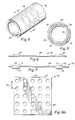
- FIG. 1is a photographic top plan view of a web-stent in accordance with the present invention.
- FIG. 2is a perspective view of a preferred embodiment of the web-stent of the present invention.
- FIG. 3is a perspective view of a stent-graft in accordance with the present invention.
- FIG. 4is a perspective view of an alternative embodiment of the inventive stent-graft.
- FIG. 5is a cross-sectional view taken along line 5 — 5 of FIG. 4 .
- FIG. 6is a cross-sectional view illustrating a pair of support members and a section of interstitial web between adjacent supporting members.
- FIG. 7is a cross-sectional view illustrating a pair of support members and a section of interstitial web between adjacent supporting members in accordance with an alternative embodiment of the present invention.
- FIG. 8Ais a top-plan view of a graft or web region with a plurality of openings passing there through.
- FIG. 8Bis a top plan view of an alternative embodiment of a graft or web region of the present invention with a plurality of openings passing there through.
- FIG. 8Cis a top plan view of a third embodiment of a graft or web region of the present invention with a plurality of openings passing there through.
- FIG. 9Ais a transverse cross-sectional view of a first embodiment of a graft member in accordance with the present invention.
- FIG. 9Bis a transverse cross-sectional view of a second embodiment of a graft member in accordance with the present invention.
- FIG. 10is a flow chart diagrammatically illustrating the method of fabricating the graft, stent-graft and/or web-stent of the present invention.
- stent, web-stent and stent-graft deviceswhich preferably exhibit substantially homogenous surface properties.
- inventive graft, stent, stent-graft and web-stent devicesmay be made utilizing a pre-fabricated film or a deposited film, either in a planar or cylindrical conformation, then either adding a pattern of support members to the film or removing at least some regions of the film to create thinner regions in the starting film and defining relatively thinner and thicker film regions, such as thinner web regions between adjacent structural members formed by thicker film regions and/or relatively thinner graft regions.
- An additive methodologymay include vacuum deposition or lamination of a pattern of support members upon the planar or cylindrical film.
- a subtractive methodologyincludes etching unwanted regions of material by masking regions to form the structural members and expose unmasked regions to the etchant. Additionally, in order to improve in vivo healing, it is advantageous to impart openings passing through the web or the graft.
- the openingsare preferably produced during the process of forming the web or the graft.
- the openings in the web or the graftmay be formed by conventional methods such as photolithographic processes, by masking and etching techniques, by mechanical means, such as laser ablation, EDM, or micromachining, etc.
- Suitable deposition methodologiesas are known in the microelectronic and vacuum coating fabrication arts and incorporated herein by reference, are plasma deposition and physical vapor deposition which are utilized to impart a metal layer onto the stent pattern.
- a vacuum deposited devicethat is fabricated of a material having substantially homogeneous surface properties across the blood contact surface of the device.
- Current manufacturing methods for fabricating endoluminal stentsfail to achieve the desired material properties of the present invention.
- stentsare fabricated from bulk metals that are processed in a manner that incorporates processing aides to the base metal.
- stentsare made from hypotubes formed from bulk metals, by machining a series of slots or patterns into the hyptotube to accommodate radial expansion, or by weaving wires into a mesh pattern.
- the present inventionconsists of a stent made of a bulk material having controlled heterogeneities on the luminal surface thereof. Heterogeneities are controlled by fabricating the bulk material of the stent to have defined grain sizes that yield areas or sites along the surface of the stent having optimal protein binding capability.
- the characteristically desirable properties of the inventive stentare: (a) optimum mechanical properties consistent with or exceeding regulatory approval criteria, (b) controlling discontinuities, such as cracking or pinholes, (c) a fatigue life of 400 MM cycles as measured by simulated accelerated testing, (d) corrosion resistance, (e) biocompatibility without having biologically significant impurities in the material, (f) a substantially non-frictional abluminal surface to facilitate atraumatic vascular crossing and tracking and compatible with transcatheter techniques for stent introduction, (g) radiopaque at selected sites and MRI compatible, (h) have a luminal surface which is optimized for surface energy and microtopography, (i) minimal manufacturing and material cost consistent with achieving the desired material properties, and (j) high process yields.
- Controlling the surface profile of an endoluminal deviceis significant because blood protein interactions with surfaces of endoluminal devices appear to be the initial step in a chain of events leading to tissue incorporation of the endovascular device.
- the present inventionis based, in part, upon the relationship between surface energy of the material used to make the endoluminal device and protein adsorption at the surface of the endoluminal device.
- the present inventorshave found that a relationship exists between surface free energy and protein adsorption on metals commonly used in fabrication of endoluminal devices.
- specific electrostatic forces resident on the surface of metal endoluminal stentshave been found to influence blood interactions with the stent surface and the vascular wall.
- the inventive grafts, stent-grafts and web-stentshave surface profiles which are achieved by fabricating the graft, stent-graft and web-stent by the same metal deposition methodologies as are used and standard in the microelectronic and nano-fabrication vacuum coating arts, and which are hereby incorporated by reference.
- the preferred deposition methodologiesinclude ion-beam assisted evaporative deposition and sputtering techniques.
- ion beam-assisted evaporative depositionit is preferable to employ dual and simultaneous thermal electron beam evaporation with simultaneous ion bombardment of the material being deposited using an inert gas, such as argon, xenon, nitrogen or neon.
- an inert gassuch as argon, xenon, nitrogen or neon.
- Bombardment with inert gas ions during depositionserves to reduce void content by increasing the atomic packing density in the deposited material.
- the reduced void content in the deposited materialallows the mechanical properties of that deposited material to be similar to the bulk material properties. Deposition rates up to 20 nm/sec are achievable using ion beam-assisted evaporative deposition techniques.
- a 200-micron thick stainless steel filmmay be deposited within about four hours of deposition time.
- Alternate deposition processes which may be employed to form the stent in accordance with the present inventionare cathodic arc, laser ablation, and direct ion beam deposition.
- the crystalline structure of the deposited filmaffects the mechanical properties of the deposited film. These mechanical properties of the deposited film may be modified by post-process treatment, such as by, for example, annealing.
- Materials to make the inventive graft, stent-graft and web-stentare chosen for their biocompatibility, mechanical properties, i.e., tensile strength, yield strength, and their ease of deposition include, without limitation, the following: elemental titanium, vanadium, aluminum, nickel, tantalum, zirconium, chromium, silver, gold, silicon, magnesium, niobium, scandium, platinum, cobalt, palladium, manganese, molybdenum and alloys thereof, such as zirconium-titanium-tantalum alloys, nitinol, and stainless steel.
- the chamber pressure, the deposition pressure and the partial pressure of the process gasesare controlled to optimize deposition of the desired species onto the substrate.
- both the reactive and non-reactive gasesare controlled and the inert or non-reactive gaseous species introduced into the deposition chamber are typically argon and nitrogen.
- the substratemay be either stationary or moveable; either rotated about its longitudinal axis, moved in an X-Y plane, planatarily or rotationally moved within the deposition chamber to facilitate deposition or patterning of the deposited material onto the substrate.
- the deposited materialmaybe deposited either as a uniform solid film onto the substrate, or patterned by (a) imparting either a positive or negative pattern onto the substrate, such as by etching or photolithography techniques applied to the substrate surface to create a positive or negative image of the desired pattern or (b) using a mask or set of masks which are either stationary or moveable relative to the substrate to define the pattern applied to the substrate. Patterning may be employed to achieve complex finished geometries of the resultant structural supports, web-regions or graft, both in the context of spatial orientation of patterns of regions of relative thickness and thinness, such as by varying the thickness of the film over its length to impart different mechanical characteristics under different delivery, deployment or in vivo environmental conditions.
- the devicemay be removed from the substrate after device formation by any of a variety of methods.
- the substratemay be removed by chemical means, such as etching or dissolution, by ablation, by machining or by ultrasonic energy.
- a sacrificial layer of a materialsuch as carbon, aluminum or organic based materials, such as photoresists, may be deposited intermediate the substrate and the stent and the sacrificial layer removed by melting, chemical means, ablation, machining or other suitable means to free the stent from the substrate.
- the resulting devicemay then be subjected to post-deposition processing to modify the crystalline structure, such as by annealing, or to modify the surface topography, such as by etching to expose a heterogeneous surface of the device.
- FIGS. 1 and 2there is illustrated a web-stent 20 in accordance with the present invention.
- the web-stent 20is formed of a material blank 10 , which has been either pre-manufactured or has been vacuum deposited as a planar or cylindrical film.
- the web-stent 20is formed by masking regions of the material blank which are to form a plurality of structural members 22 , and then etching the unmasked regions which then form interstitial webs 24 which subtend interstitial regions between adjacent structural members 22 .
- the interstitial webs 24are etched to a material thickness that is less than the thickness of the plurality of structural members 22 .
- the openingsmay be imparted as a random pattern or as a regular pattern in the interstitial web 24 , as will be discussed hereinafter.
- Stent-graft 30is formed either from a tubular or planar material blank, which is etched to form the plurality of structural members 32 and interstitial regions 34 between the structural members 32 .
- a proximal 36 or a distal 38 graft region of the stentare provided and project outwardly from terminal structural members 32 .
- the proximal graft region 36 and the distal graft region 38are preferably etched to a reduced thickness of less than the thickness of the structural members, and are made with openings passing there through which promote cellular migration, as will be discussed hereinafter.
- FIGS. 4 and 5An alternative embodiment of the invention is illustrated in FIGS. 4 and 5.
- the alternative embodiment of the stent-graft 30involves covering the luminal and abluminal surfaces of a plurality of structural supports 32 with a luminal graft 36 and an abluminal graft 38 .
- the luminal graft 36may initially be formed as the proximal graft region 36 in FIG. 3 and be luminally inverted 39 and passed into the lumen defined by the structural members 32 .
- the abluminal graft 38may initially be formed as the distal graft region 38 in FIG.
- the luminal graft 36 and the abluminal graft 38may be formed as either pre-fabricated discrete graft members made of biocompatible metal or metal-like materials that are either tubular or planar then formed into a tube and concentrically engaged about the plurality of structural members 32 . Portions of each of the abluminal graft 38 and the luminal graft 36 are mechanically joined to the plurality of structural members 32 or to one and other, thereby effectively encapsulating the plurality of structural members 32 between the luminal graft 36 and the abluminal graft 38 .
- each of the abluminal graft 38 and luminal graft 36are mechanically joined to and co-terminus with a terminal portion of the plurality of structural members 32 .
- Mechanical joiningmay be accomplished by methods such as welding, suturing, adhesive bonding, soldering, thermobonding, riveting, crimping, or dovetailing.
- the interstitial regions 34may be subtended by a web 34 , as discussed hereinabove, with reference to FIGS. 1 and 2.
- relatively thinner regionsmay be formed by removing bulk material by ion milling, laser ablation, EDM, laser machine, electron beam lithography, reactive ion etching, sputtering or equivalent methods which are capable of reducing the thickness of the material in either the graft region or the interstitial web region between the structural members.
- the structural membersmay be added to the defined interstitial web or graft regions to form the device, or the interstitial web or graft regions may be added to pre-existing structural members. Additive methods that may be employed include conventional metal forming techniques, including laminating, plating, or casting.
- initial bulk material configurationsincluding a substantially planar sheet substrate, an arcuate substrate or a tubular substrate, which is then processed by either subtractive or additive techniques discussed above.
- both the circumferential or hoop strength of the resultant device, as well as the longitudinal or columnar strength of the deviceare enhanced over conventional stent-graft devices.
- Additional advantages of the present invention, depending upon fabrication methods,may include: controlled homogeneity and/or heterogeneity of the material used to form the device by deposition methodologies, enhanced ability to control dimensional and mechanical characteristics of the device, the ability to fabricate complex device conformations, ability to pattern and control the porosity of the web and/or graft regions, and a monolithic one-piece construction of a device which yields a minimized device profile and cross-sectional area.
- the devices of the present inventionhave relatively thicker and thinner regions, in which the thinner regions permit radial collapse of the device for endoluminal delivery.
- the inventive deviceexhibits superior column strength that permits smaller introducer size and more readily facilitates deployment of the device.
- the web and/or graft regions, 44 , 54 between adjacent structural members 42 , 52may be co-planar with either the luminal or abluminal surface of the structural members 42 , or may be positioned intermediate the luminal 51 and abluminal 56 surfaces of the structural members 52 .
- the web regions of the inventive web-stent, the graft regions of the inventive stent-graft and the inventive grafthave a plurality of openings which pass through the thickness of the material used to fabricated the inventive devices.
- Each of the plurality of openingsis dimensioned to permit cellular migration through the opening without permitting blood leakage or seepage through the plurality of openings.
- the plurality of openingsmay be random or may be patterned. However, in order to control the effective porosity of the device, it is desirable to impart a pattern of openings in the material used to fabricate the inventive device.
- FIGS. 8A-8Cdepict several examples of patterned openings in a section of material used to make the inventive web-stent, graft regions of the stent-graft, and the inventive graft.
- FIG. 8Adepicts a material 60 with a plurality of circular openings 64 passing through the material substrate 62 .
- the plurality of circular openingsis patterned in a regular array of rows and columns with regular inter-opening spacing 65 between adjacent openings.
- the diameter of each of the plurality of openingsis about 19 ⁇ m, with an inter-opening spacing in each row and column of about 34 ⁇ m on center.
- the thickness of the material 62is approximately 10 ⁇ m.
- the material 62which again is approximately 10 ⁇ m in thickness, has a plurality of openings 66 and 67 passing there through.
- the pattern of the plurality of openings 66 and 67is an alternating slot pattern in which the plurality of openings 66 are arrayed adjacent one and other forming a y-axis oriented array 68 relative to the material 62 , while a plurality of openings 67 are arrayed adjacent one and other forming an x-axis oriented array 69 relative to the material 62 .
- FIG. 8Cillustrates a material 60 in which the material substrate 62 has a regular array of a plurality of diamond-shaped openings 63 passing through the material substrate 62 .
- the dimension of the plurality of diamond-shaped openings 63is of sufficient size to permit cellular migration through the openings 63 , while preventing blood flow or seepage through the plurality of openings 63 .
- FIGS. 9A and 9Billustrate alternate preferred embodiments of the graft 70 and graft 80 in accordance with the present invention.
- Graft 70consists generally of concentrically positioned luminal graft member 74 and abluminal graft member 72 and an interfacial region 74 where the luminal surface of the abluminal graft member 72 and the abluminal surface of the luminal graft member 74 are in immediate juxtaposition with one and other.
- Both the luminal 74 and the abluminal 72 graft membersare fabricated in accordance with the methodologies described above, and are provided with a plurality of patterned openings 73 in the abluminal graft member 72 and a plurality of patterned openings 75 in the luminal graft member 74 .
- the plurality of patterned openings 74 and 75are positioned out of phase relative to one another. By positioning the plurality of patterned openings 74 and 75 in an out-of-phase relationship, there is no continuous opening that passes through the interfacial region 76 which would permit blood flow or seepage from the lumen of the graft. However, in order to permit cellular migration from the abluminal surface of the graft to the lumen of the graft, the interfacial region 76 should have microroughness [not shown] which is oriented either randomly or selectively, such as helically or circumferential, about the interfacial region 76 .
- the microroughnesspreferably has a peak-to-valley depth of between about 5 ⁇ to about 65 ⁇ , most preferably between about 10 ⁇ to 15 ⁇ , may be either on the luminal surface of the abluminal graft 72 or on the abluminal surface of the luminal graft 74 , or both.
- the microroughnessspans the surface area region between adjacent pairs of openings 74 , 75 , and the microroughness depth permits cellular migration across the surfaces between adjacent openings 74 and 75 .
- the microroughnessis not large enough to permit fluid passage through the inter-opening regions at the interface between the luminal graft 74 and the abluminal graft 72 .
- This property of permitting cellular growthis similar to the difference between the porosity of expanded polytetrafluoroethylene grafts which do not require pre-clotting, and the much larger porosity of polyester or DACRON grafts which require pre-clotting to prevent fluid seepage there from.
- FIG. 9Billustrates an alternative embodiment of the inventive graft 80 in which an abluminal graft member 82 is concentrically positioned about a luminal graft member 84 .
- Each of the abluminal graft member 82 and the luminal graft member 84having a plurality of patterned openings 83 , 85 , respectively, passing there through.
- the plurality of patterned openings 83 and 85are positioned in an out-of-phase relationship to one and other in order to prevent forming a continuous opening between the luminal and abluminal surfaces of the graft 80 .
- annular open region 87is positioned intermediate the luminal graft member 84 and the abluminal graft member 82 .
- the annular open region 87is created by providing a plurality of microprojections 86 that project either radially inward from the luminal surface of the abluminal graft member 82 or radially outward from the abluminal surface of the luminal graft member 84 .
- the plurality of microprojections 86act as spacers which abut the opposing surface of either the luminal graft member 84 or the abluminal graft member 82 which bound the annular open region 87 .
- the height of the microprojections 86 and, therefore, the size of the annular open region 87are dimensioned such that cells may migrate through the annular open region 87 , while blood flow or seepage will not occur between the lumen and the abluminal surface of the graft 80 .
- the size of the plurality of openings in the luminal graft member 74 , 84may be different than the size of the plurality of openings in the abluminal graft member 72 , 82 .
- the plurality of openings in the abluminal graft member 74 , 84preferably have a larger size than the plurality of openings in the luminal graft member 72 , 84 , while still retaining the out-of-phase relationship between the plurality of openings in the luminal 72 , 82 and the abluminal 74 , 84 graft members.
- the luminal 72 , 82 and the abluminal 74 , 84 graft membershave openings having diameters of between about 5 ⁇ m and 100 ⁇ m.
- a third membermay be interposed between the luminal 72 , 82 and the abluminal 82 , 84 graft members.
- the third memberwill preferably have a very fine plurality of openings, such as on the order of between 2-10 ⁇ , and permits use of a higher porosity in the luminal and abluminal grafts, without the need to maintain an out-of-phase relationship between the openings in the luminal 72 , 82 and the abluminal 74 , 84 graft members.
- a starting blank of materialmay be formed either by providing a pre-fabricated material blank of a biocompatible metal or metal-like material 92 or by vacuum depositing a starting blank of a biocompatible metal or metal like material film 94 . Then a determination is made whether to employ an additive or a subtractive method 96 for forming the graft, stent-graft or web-stent. If an additive method is selected 97 , the structural supports are built upon the starting blank 100 , either by vacuum deposition techniques or by conventional metal forming techniques.
- the regions to remainare masked 98 , then the unmasked regions are removed, such as by chemical etching or sputtering, to form the interstitial web regions, graft regions and/or openings in either the interstitial web regions and/or graft regions 99 .
- a ceramic cylindrical substrateis introduced into a deposition chamber with capabilities of glow discharge substrate cleaning and sputter deposition of carbon and stainless steel.
- the deposition chamberis evacuated to a pressure less than or equal to 2 ⁇ 10 ⁇ 7 Torr. Pre-cleaning of the substrate is conducted under vacuum by glow discharge.
- the substrate temperatureis controlled to achieve a temperature between about 300 and 1100 degrees Centigrade.
- a bias voltagebetween ⁇ 1000 and +1000 volts is applied to the substrate sufficient to cause energetic species arriving at the surface of the substrate to have hyperthermal energy between 0.1 eV and about 700 eV, preferably between 5-50 eV.
- the deposition sourcesare circumferential and are oriented to deposit from the target circumferentially about the substrate.
- the deposition pressureis maintained between 0.1 and 10 mTorr.
- a sacrificial carbon layer of substantially uniform thickness ( ⁇ 5%) between 10 and 500 Angstromsis deposited circumferentially on the substrate.
- a cylindrical film of stainless steelis deposited onto the sacrificial carbon layer on the cylindrical substrate at a deposition rate between about 10 to 100 microns/hour.
- the substrateis removed from the deposition chamber and heated to volatilize the intermediate sacrificial carbon layer between the substrate and the film.
- the stainless steel filmis removed from the substrate and exhibits material properties similar to the bulk stainless steel target and surface properties characterized by controlled heterogeneities in grain size, material composition and surface topography. A series of patterns are then machined into the resultant stainless steel film to form a stent by electrical discharge machining (EDM) or laser cutting the film.
- EDMelectrical discharge machining
- Example 2The same operating conditions are followed as in Example 1, except that the substrate is tubular and selected to have a coefficient of thermal expansion different than that of the resultant stent.
- No intermediate layer of sacrificial carbonis deposited onto the substrate, and the outer surface of the substrate is etched with a pattern of recesses defining a desired stent pattern.
- the substrateis mounted onto a rotational jig within the deposition chamber and rotated at a uniform rate during deposition. Tantalum is used as the target material and deposited into the recesses of the substrate from a single stationary source. After deposition, the temperature of the substrate and the deposited stent are controlled to impart diametric differential in the substrate and stent and permit removal of the stent from the substrate.
- a cylindrical substrateis introduced into a deposition chamber that has capabilities of: substrate rotation and precise positioning, glow discharge substrate cleaning, ion beam-assisted evaporative deposition, and cylindrical magnetron sputtering.
- the deposition sourcesare (a) dual electron beam evaporative sources placed adjacent to one another at the base of the deposition chamber at a fixed distance from the substrate, these are used with simultaneous argon ion impingement onto the substrate from a controlled ion beam source, and (b) a cylindrical magnetron sputtering source with a carbon target capable of circumferentially coating a carbon sacrificial layer of substantially uniform thickness of between 10 and 200 Angstroms onto the substrate.
- the substrate temperatureis controlled to achieve a substrate temperature between about 300 and 1100 degrees Centigrade.
- the deposition chamberis evacuated to a pressure less than or equal to 2 ⁇ 10 ⁇ 7 Torr.
- a pre-cleaning of the substrateis conducted under vacuum by glow discharge.
- the substrateis rotated to ensure uniform cleaning and subsequent uniform deposition thickness.
- After cleaning the substrateis moved into the magnetron and coated with the carbon layer.
- the substrateis then moved into position to receive the stent-forming metal coating with simultaneous ion bombardment.
- One electron beam evaporation sourcecontains titanium while the other source contains nickel.
- the evaporation rates of each of the titanium and nickel evaporation sourcesare separately controlled to form a nitinol alloy on the substrate as the stent-forming metal.
- the deposition sourceis a single electron beam evaporation source containing platinum and is used with simultaneous argon ion impingement onto the substrate from a controlled ion beam source.
- the substrate temperatureis controlled to achieve a substrate temperature between about 300 and 1100 degrees Centigrade.
- the deposition chamberis evacuated to a pressure less than or equal to 2 ⁇ 10 ⁇ 7 Torr.
- a pre-cleaning of the substrateis conducted under vacuum by glow discharge. After cleaning the substrate is moved into position within the deposition chamber and coated with platinum from the electron beam evaporation source with simultaneous argon ion bombardment, with the electron beam evaporation source passing platinum through a pattern mask corresponding to a stent pattern which is interposed between the source and the substrate to pass a pattern of platinum onto the substrate.
- the patterned stentis removed from the substrate and rolled about a forming substrate to a cylindrical shape and opposing ends of the planar stent material are brought into juxtaposition with one another and may be attached by laser welding or left uncoupled.
- Example 4The same conditions are employed as in Example 4, except that a uniform layer of stent-forming material is deposited having a thickness of 150 microns without patterning of the stent onto the deposited layer. Rather, a negative mask is applied to the deposited stent-forming material, and a chemical etchant is introduced to etch a pattern of structural elements into the stent-forming metal. The etchant is permitted to react with the metal until a thinner film web having a thickness of between 2-75 microns, is present between adjacent structural elements. After the thinner film web is formed, the etching is stopped, and the resultant stent-graft is removed and formed into a tubular shape.
- Example 5The same conditions as in Example 5 are followed, except that reactive ion etching is employed to form the thinner film web.
- Example 5The same conditions are followed as in Example 5, except that the structural elements are defined in an intermediate region of a tubular substrate, and interstitial regions between adjacent structural elements are etched by chemical etching until interstitial openings are formed between adjacent structural elements while masking the structural elements and proximal and distal regions of the tubular substrate. Proximal and distal graft regions are formed adjacent the intermediate region of the tubular substrate and contiguous with the plurality of structural elements, by masking the structural elements and interstitial openings and chemical etching the proximal and distal regions of the tubular substrate to yield a thinner film of material in the proximal and distal regions of the tubular substrate.
- proximal and distal graft regionsare then everted, with the proximal graft region being inverted luminally through the lumen of the structural members and the distal graft region being everted abluminally over the structural members.
- the proximal graft regionis mechanically joined to the distal terminal end of the plurality of structural members, while the distal graft region is mechanically joined to the proximal terminal end of the plurality of structural members, thereby encapsulating the plurality of structural members between the everted proximal and distal graft regions.
- a pre-fabricated self-expanding superelastic shape memory alloy stentis provided.
- Two cylindrical hypotubes of a superelastic shape memory material similar to that of the stentare chemically etched to a substantially uniform thickness of 10 ⁇ m, with a first hypotube having an inner diameter which is of sufficient size to accommodate the outer diameter of the stent, and a second hypotube having an outer diameter dimensioned to accommodate the inner diameter of the stent.
- the etched hypotubesare then placed into a vacuum chamber and a cylindrical pattern mask having a regular array of circular openings, each circular opening having a diameter of about 25 ⁇ m, is positioned concentrically about each of the cylindrical hypotubes.
- the etched hypotubesare reactive ion etched to transfer the masked pattern to the etched hypotube and impart a pattern of circular openings that pass through the wall thickness of the etched hypotubes corresponding to the mask pattern.
- the stent, and first and second etched and reactive ion etched hypotubesare concentrically engaged upon one and other, with the second hypotube being concentrically positioned within the lumen of the stent and the first hypotube being concentrically positioned about the abluminal surface of the stent.
- Proximal and distal ends of the stent, the first hypotube and the second hypotubeare mechanically joined by welding and then trimmed by laser cutting to ensure that the proximal and distal ends are co-terminus.
- a cylindrical mandrelis provided which is coated with a sacrificial layer.
- a plurality of patterned recessesis defined in the sacrificial layer.
- the mandrelis introduced into a deposition chamber and a nickel-titanium alloy is vacuum deposited onto the mandrel, while rotating the mandrel, until a uniform adherent layer of the deposited nickel-titanium alloy covers the cylindrical mandrel.
- the sacrificial layeris removed, and the uniform adherent layer disengaged from the cylindrical mandrel, yielding the inventive graft, with openings corresponding to the plurality of patterned recesses in the graft material.
Landscapes
- Health & Medical Sciences (AREA)
- Engineering & Computer Science (AREA)
- Biomedical Technology (AREA)
- Life Sciences & Earth Sciences (AREA)
- General Health & Medical Sciences (AREA)
- Cardiology (AREA)
- Oral & Maxillofacial Surgery (AREA)
- Transplantation (AREA)
- Heart & Thoracic Surgery (AREA)
- Vascular Medicine (AREA)
- Veterinary Medicine (AREA)
- Animal Behavior & Ethology (AREA)
- Public Health (AREA)
- Optics & Photonics (AREA)
- Physics & Mathematics (AREA)
- Gastroenterology & Hepatology (AREA)
- Pulmonology (AREA)
- Prostheses (AREA)
- Materials For Medical Uses (AREA)
- Media Introduction/Drainage Providing Device (AREA)
Abstract
Description
Claims (9)
Priority Applications (32)
| Application Number | Priority Date | Filing Date | Title |
|---|---|---|---|
| US09/532,164US6537310B1 (en) | 1999-11-19 | 2000-03-20 | Endoluminal implantable devices and method of making same |
| AU2001245884AAU2001245884B2 (en) | 2000-03-20 | 2001-03-20 | Endoluminal implantable devices and method of making same |
| EP01918860.6AEP1267749B2 (en) | 2000-03-20 | 2001-03-20 | Endoluminal implantable devices and method of making same |
| ES01918860TES2338524T3 (en) | 2000-03-20 | 2001-03-20 | ENDOLUMINAL IMPLANTABLE DEVICES AND PROCEDURES FOR MANUFACTURING THEM. |
| PCT/US2001/008914WO2001074274A2 (en) | 2000-03-20 | 2001-03-20 | Endoluminal implantable devices and method of making same |
| CA2403341ACA2403341C (en) | 2000-03-20 | 2001-03-20 | Endoluminal implantable devices and method of making same |
| AT01918860TATE448751T1 (en) | 2000-03-20 | 2001-03-20 | IMPLANTABLE ENDOLUMINAL DEVICES AND PRODUCTION METHOD THEREOF |
| JP2001572021AJP5186074B2 (en) | 2000-03-20 | 2001-03-20 | Intraluminal implantable device and method of manufacturing the same |
| AU4588401AAU4588401A (en) | 2000-03-20 | 2001-03-20 | Endoluminal implantable devices and method of making same |
| DE60140525TDE60140525D1 (en) | 2000-03-20 | 2001-03-20 | IMPLANTABLE ENDOLUMINAL DEVICES AND MANUFACTURING METHOD THEREFOR |
| US10/135,316US7300457B2 (en) | 1999-11-19 | 2002-04-29 | Self-supporting metallic implantable grafts, compliant implantable medical devices and methods of making same |
| US10/136,246US6695865B2 (en) | 2000-03-20 | 2002-04-29 | Embolic protection device |
| US10/135,626US6936066B2 (en) | 1999-11-19 | 2002-04-29 | Complaint implantable medical devices and methods of making same |
| US10/289,974US7491226B2 (en) | 1999-11-19 | 2002-11-06 | Endoluminal implantable stent-grafts |
| US10/289,843US7641680B2 (en) | 1999-11-19 | 2002-11-06 | Endoluminal implantable stent-grafts |
| US11/157,139US7641682B2 (en) | 1999-11-19 | 2005-06-20 | Compliant implantable medical devices and methods of making same |
| US11/875,685US7670690B2 (en) | 1999-11-19 | 2007-10-19 | High strength vacuum deposited nitinol alloy films and method of making same |
| US11/875,629US8083908B2 (en) | 1999-11-19 | 2007-10-19 | High strength vacuum deposited nitinol alloy films and method of making same |
| US12/210,789US10172730B2 (en) | 1999-11-19 | 2008-09-15 | Stents with metallic covers and methods of making same |
| US12/652,582US8715335B2 (en) | 1999-11-19 | 2010-01-05 | Endoluminal implantable stent-grafts |
| US13/168,865US10092390B2 (en) | 1999-11-19 | 2011-06-24 | Method of making implantable medical devices having controlled surface properties |
| US13/168,844US9662230B2 (en) | 1999-11-19 | 2011-06-24 | Implantable medical devices having controlled surface properties for improved healing response |
| US13/673,168US9132001B2 (en) | 1999-11-19 | 2012-11-09 | Metallic implantable grafts and method of making same |
| US13/908,933US8910363B2 (en) | 1999-11-19 | 2013-06-03 | Compliant implantable medical devices and methods of making same |
| US14/177,978US9375330B2 (en) | 1999-11-19 | 2014-02-11 | Methods of making medical devices |
| US14/571,572US10106884B2 (en) | 1999-11-19 | 2014-12-16 | Compliant implantable medical devices and methods of making same |
| US14/852,371US9668852B2 (en) | 1999-11-19 | 2015-09-11 | Metallic implantable grafts and method of making same |
| US15/194,117US20170130322A1 (en) | 1999-11-19 | 2016-06-27 | Methods of making medical devices |
| US15/608,337US10874532B2 (en) | 1999-11-19 | 2017-05-30 | Implantable medical devices having controlled surface properties for improved healing response |
| US16/140,822US10363125B2 (en) | 1999-11-19 | 2018-09-25 | Method of making implantable medical devices having controlled surface properties |
| US16/167,102US10745799B2 (en) | 1999-11-19 | 2018-10-22 | Compliant implantable medical devices and methods of making same |
| US16/222,286US20190125559A1 (en) | 1999-11-19 | 2018-12-17 | Stents with metallic covers and methods of making same |
Applications Claiming Priority (2)
| Application Number | Priority Date | Filing Date | Title |
|---|---|---|---|
| US09/443,929US6379383B1 (en) | 1999-11-19 | 1999-11-19 | Endoluminal device exhibiting improved endothelialization and method of manufacture thereof |
| US09/532,164US6537310B1 (en) | 1999-11-19 | 2000-03-20 | Endoluminal implantable devices and method of making same |
Related Parent Applications (2)
| Application Number | Title | Priority Date | Filing Date |
|---|---|---|---|
| US09/443,929Continuation-In-PartUS6379383B1 (en) | 1999-11-19 | 1999-11-19 | Endoluminal device exhibiting improved endothelialization and method of manufacture thereof |
| US12/210,789Continuation-In-PartUS10172730B2 (en) | 1999-11-19 | 2008-09-15 | Stents with metallic covers and methods of making same |
Related Child Applications (9)
| Application Number | Title | Priority Date | Filing Date |
|---|---|---|---|
| US09/443,929Continuation-In-PartUS6379383B1 (en) | 1999-11-19 | 1999-11-19 | Endoluminal device exhibiting improved endothelialization and method of manufacture thereof |
| US09/853,985Continuation-In-PartUS6849085B2 (en) | 1999-11-19 | 2001-05-11 | Self-supporting laminated films, structural materials and medical devices manufactured therefrom and method of making same |
| US10/136,246Continuation-In-PartUS6695865B2 (en) | 2000-03-20 | 2002-04-29 | Embolic protection device |
| US10/135,626Continuation-In-PartUS6936066B2 (en) | 1999-11-19 | 2002-04-29 | Complaint implantable medical devices and methods of making same |
| US10/135,316Continuation-In-PartUS7300457B2 (en) | 1999-11-19 | 2002-04-29 | Self-supporting metallic implantable grafts, compliant implantable medical devices and methods of making same |
| US10/289,974ContinuationUS7491226B2 (en) | 1999-11-19 | 2002-11-06 | Endoluminal implantable stent-grafts |
| US10/289,974DivisionUS7491226B2 (en) | 1999-11-19 | 2002-11-06 | Endoluminal implantable stent-grafts |
| US10/289,843ContinuationUS7641680B2 (en) | 1999-11-19 | 2002-11-06 | Endoluminal implantable stent-grafts |
| US11/157,139Continuation-In-PartUS7641682B2 (en) | 1999-11-19 | 2005-06-20 | Compliant implantable medical devices and methods of making same |
Publications (1)
| Publication Number | Publication Date |
|---|---|
| US6537310B1true US6537310B1 (en) | 2003-03-25 |
Family
ID=24120612
Family Applications (8)
| Application Number | Title | Priority Date | Filing Date |
|---|---|---|---|
| US09/532,164Expired - LifetimeUS6537310B1 (en) | 1999-11-19 | 2000-03-20 | Endoluminal implantable devices and method of making same |
| US10/289,974Expired - LifetimeUS7491226B2 (en) | 1999-11-19 | 2002-11-06 | Endoluminal implantable stent-grafts |
| US10/289,843Expired - LifetimeUS7641680B2 (en) | 1999-11-19 | 2002-11-06 | Endoluminal implantable stent-grafts |
| US12/652,582Expired - LifetimeUS8715335B2 (en) | 1999-11-19 | 2010-01-05 | Endoluminal implantable stent-grafts |
| US13/168,844Expired - LifetimeUS9662230B2 (en) | 1999-11-19 | 2011-06-24 | Implantable medical devices having controlled surface properties for improved healing response |
| US13/168,865Expired - Fee RelatedUS10092390B2 (en) | 1999-11-19 | 2011-06-24 | Method of making implantable medical devices having controlled surface properties |
| US15/608,337Expired - Fee RelatedUS10874532B2 (en) | 1999-11-19 | 2017-05-30 | Implantable medical devices having controlled surface properties for improved healing response |
| US16/140,822Expired - Fee RelatedUS10363125B2 (en) | 1999-11-19 | 2018-09-25 | Method of making implantable medical devices having controlled surface properties |
Family Applications After (7)
| Application Number | Title | Priority Date | Filing Date |
|---|---|---|---|
| US10/289,974Expired - LifetimeUS7491226B2 (en) | 1999-11-19 | 2002-11-06 | Endoluminal implantable stent-grafts |
| US10/289,843Expired - LifetimeUS7641680B2 (en) | 1999-11-19 | 2002-11-06 | Endoluminal implantable stent-grafts |
| US12/652,582Expired - LifetimeUS8715335B2 (en) | 1999-11-19 | 2010-01-05 | Endoluminal implantable stent-grafts |
| US13/168,844Expired - LifetimeUS9662230B2 (en) | 1999-11-19 | 2011-06-24 | Implantable medical devices having controlled surface properties for improved healing response |
| US13/168,865Expired - Fee RelatedUS10092390B2 (en) | 1999-11-19 | 2011-06-24 | Method of making implantable medical devices having controlled surface properties |
| US15/608,337Expired - Fee RelatedUS10874532B2 (en) | 1999-11-19 | 2017-05-30 | Implantable medical devices having controlled surface properties for improved healing response |
| US16/140,822Expired - Fee RelatedUS10363125B2 (en) | 1999-11-19 | 2018-09-25 | Method of making implantable medical devices having controlled surface properties |
Country Status (9)
| Country | Link |
|---|---|
| US (8) | US6537310B1 (en) |
| EP (1) | EP1267749B2 (en) |
| JP (1) | JP5186074B2 (en) |
| AT (1) | ATE448751T1 (en) |
| AU (2) | AU4588401A (en) |
| CA (1) | CA2403341C (en) |
| DE (1) | DE60140525D1 (en) |
| ES (1) | ES2338524T3 (en) |
| WO (1) | WO2001074274A2 (en) |
Cited By (210)
| Publication number | Priority date | Publication date | Assignee | Title |
|---|---|---|---|---|
| US20010044650A1 (en)* | 2001-01-12 | 2001-11-22 | Simso Eric J. | Stent for in-stent restenosis |
| US20030018381A1 (en)* | 2000-01-25 | 2003-01-23 | Scimed Life Systems, Inc. | Manufacturing medical devices by vapor deposition |
| US20030088308A1 (en)* | 2000-09-18 | 2003-05-08 | Inflow Dynamics Inc. | Primarily niobium stent |
| US20030135260A1 (en)* | 2002-01-16 | 2003-07-17 | Kohler Robert Edward | Encased implant and methods |
| US20030144728A1 (en)* | 2000-09-18 | 2003-07-31 | Inflow Dynamics Inc. | Metal stent with surface layer of noble metal oxide and method of fabrication |
| US20040000046A1 (en)* | 2002-06-27 | 2004-01-01 | Stinson Jonathan S. | Methods of making medical devices |
| US20040014253A1 (en)* | 2002-07-17 | 2004-01-22 | Vikas Gupta | Three dimensional thin film devices and methods of fabrication |
| US20040158310A1 (en)* | 2003-02-06 | 2004-08-12 | Jan Weber | Medical device with magnetic resonance visibility enhancing structure |
| WO2004028340A3 (en)* | 2002-09-26 | 2004-08-26 | Advanced Bio Prosthetic Surfac | High strength vacuum deposited nitionol alloy films, medical thin film graft materials and method of making same |
| US20040181274A1 (en)* | 2003-03-11 | 2004-09-16 | Scimed Life Systems, Inc. | Covered stent |
| US20040241448A1 (en)* | 2003-05-27 | 2004-12-02 | Nissan Motor Co., Ltd. | Rolling element |
| US20040242435A1 (en)* | 2003-05-29 | 2004-12-02 | Nissan Motor Co., Ltd. | Hard-carbon coated machine tool and cutting oil composition therefor |
| US20050005892A1 (en)* | 2003-05-23 | 2005-01-13 | Nissan Motor Co., Ltd. | Piston for internal combustion engine |
| US20050025975A1 (en)* | 2003-07-31 | 2005-02-03 | Nissan Motor Co., Ltd. | Gear |
| US20050033407A1 (en)* | 2003-08-07 | 2005-02-10 | Scimed Life Systems, Inc. | Stent designs which enable the visibility of the inside of the stent during MRI |
| US20050035222A1 (en)* | 2003-04-15 | 2005-02-17 | Nissan Motor Co., Ltd. | Fuel injection valve |
| US20050038497A1 (en)* | 2003-08-11 | 2005-02-17 | Scimed Life Systems, Inc. | Deformation medical device without material deformation |
| US20050037879A1 (en)* | 2003-08-13 | 2005-02-17 | Nissan Motor Co., Ltd. | Chain drive system |
| US20050056241A1 (en)* | 2003-08-08 | 2005-03-17 | Nissan Motor Co., Ltd. | Valve train for internal combustion engine |
| US20050065437A1 (en)* | 2003-09-24 | 2005-03-24 | Scimed Life Systems, Inc. | Medical device with markers for magnetic resonance visibility |
| US20050064196A1 (en)* | 2003-08-21 | 2005-03-24 | Jean Martin | Low-friction sliding member and low-friction sliding mechanism using same |
| US20050085775A1 (en)* | 2002-02-14 | 2005-04-21 | Ishay Ostfeld | Indwelling device |
| US20050100701A1 (en)* | 2003-08-08 | 2005-05-12 | Nissan Motor Co., Ltd. | Sliding member and production process thereof |
| US20050118426A1 (en)* | 1999-04-09 | 2005-06-02 | Shojiro Miyake | Slidably movable member and method of producing same |
| US20050131513A1 (en)* | 2003-12-16 | 2005-06-16 | Cook Incorporated | Stent catheter with a permanently affixed conductor |
| US20050159807A1 (en)* | 2002-08-30 | 2005-07-21 | Bales Thomas O. | Helical stent having improved flexibility and expandability |
| US20050186241A1 (en)* | 2000-11-17 | 2005-08-25 | Boyle Christopher T. | Device for in vivo delivery of bioactive agents and method of manufacture thereof |
| US20050197687A1 (en)* | 2004-03-02 | 2005-09-08 | Masoud Molaei | Medical devices including metallic films and methods for making same |
| US20050197689A1 (en)* | 2004-03-02 | 2005-09-08 | Masoud Molaei | Medical devices including metallic films and methods for making same |
| US20050197690A1 (en)* | 2004-03-02 | 2005-09-08 | Masoud Molaei | Medical devices including metallic films and methods for making same |
| US20050213854A1 (en)* | 2002-11-06 | 2005-09-29 | Nissan Motor Co., Ltd. | Low-friction sliding mechanism |
| US20050232968A1 (en)* | 2002-09-26 | 2005-10-20 | Advanced Bio Prosthetic Surfaces, Ltd. | Implantable materials having engineered surfaces and method of making same |
| US20050283225A1 (en)* | 2004-06-18 | 2005-12-22 | Scimed Life Systems, Inc. | Medical devices and methods of making the same |
| US20050288773A1 (en)* | 2004-01-22 | 2005-12-29 | Glocker David A | Radiopaque coating for biomedical devices |
| US20060004466A1 (en)* | 2004-06-28 | 2006-01-05 | Glocker David A | Porous coatings for biomedical implants |
| US20060015175A1 (en)* | 1999-11-19 | 2006-01-19 | Advanced Bio Prosthetic Surfaces, Ltd. | Compliant implantable medical devices and methods of making same |
| US20060020323A1 (en)* | 2002-11-25 | 2006-01-26 | Boyle Christopher T | Implantable expandable medical devices having regions of differential mechanical properties and methods of making same |
| US20060052865A1 (en)* | 2004-09-09 | 2006-03-09 | Banas Christopher E | Stents with metallic covers and methods of making same |
| US20060064154A1 (en)* | 2004-09-01 | 2006-03-23 | Pst, Llc | Stent and method for manufacturing the stent |
| US20060064155A1 (en)* | 2004-09-01 | 2006-03-23 | Pst, Llc | Stent and method for manufacturing the stent |
| US20060085035A1 (en)* | 2004-10-18 | 2006-04-20 | Viola Frank J | Compression anastomosis device and method |
| US20060118210A1 (en)* | 2004-10-04 | 2006-06-08 | Johnson A D | Portable energy storage devices and methods |
| US20060125144A1 (en)* | 2004-12-14 | 2006-06-15 | Jan Weber | Stent and stent manufacturing methods |
| US20060142851A1 (en)* | 2004-12-29 | 2006-06-29 | Masoud Molaei | Medical devices including metallic films and methods for making same |
| US20060142842A1 (en)* | 2004-12-29 | 2006-06-29 | Masoud Molaei | Medical devices including metallic films and methods for making same |
| US20060142838A1 (en)* | 2004-12-29 | 2006-06-29 | Masoud Molaei | Medical devices including metallic films and methods for loading and deploying same |
| US20060142845A1 (en)* | 2004-12-29 | 2006-06-29 | Masoud Molaei | Medical devices including metallic films and methods for making same |
| US20060155364A1 (en)* | 2002-03-26 | 2006-07-13 | Thoratec Corporation | Flexible stent and method of making the same |
| US20060213522A1 (en)* | 2002-08-08 | 2006-09-28 | Leticia Menchaca | Thin film intrauterine device |
| US20060232374A1 (en)* | 2005-03-31 | 2006-10-19 | Johnson A D | Tear-resistant thin film methods of fabrication |
| US7134381B2 (en) | 2003-08-21 | 2006-11-14 | Nissan Motor Co., Ltd. | Refrigerant compressor and friction control process therefor |
| US20060259131A1 (en)* | 2005-05-16 | 2006-11-16 | Masoud Molaei | Medical devices including metallic films and methods for making same |
| US20060263604A1 (en)* | 2003-08-06 | 2006-11-23 | Martin Jean M | Low-friction sliding mechanism, low-friction agent composition and method of friction reduction |
| US20060264945A1 (en)* | 2005-05-18 | 2006-11-23 | Edidin Avram A | Selectively-expandable bone scaffold |
| US20070009564A1 (en)* | 2005-06-22 | 2007-01-11 | Mcclain James B | Drug/polymer composite materials and methods of making the same |
| US20070106374A1 (en)* | 2004-01-22 | 2007-05-10 | Isoflux, Inc. | Radiopaque coating for biomedical devices |
| US20070112411A1 (en)* | 2004-02-09 | 2007-05-17 | Obermiller F J | Stent graft devices having collagen coating |
| US7228786B2 (en) | 2003-06-06 | 2007-06-12 | Nissan Motor Co., Ltd. | Engine piston-pin sliding structure |
| US20070137740A1 (en)* | 2004-05-06 | 2007-06-21 | Atini Alloy Company | Single crystal shape memory alloy devices and methods |
| US20070149951A1 (en)* | 2005-12-27 | 2007-06-28 | Mina Wu | Variable stiffness guidewire |
| US20070178221A1 (en)* | 2006-01-31 | 2007-08-02 | Sims Daniel D | Methods of making medical devices |
| US7255083B2 (en) | 2002-10-16 | 2007-08-14 | Nissan Motor Co., Ltd. | Sliding structure for automotive engine |
| US20070207321A1 (en)* | 2004-03-30 | 2007-09-06 | Yoshinori Abe | Method For Treating Surface Of Material, Surface-Treated Material, Medical Material, And Medical Instrument |
| US7284525B2 (en) | 2003-08-13 | 2007-10-23 | Nissan Motor Co., Ltd. | Structure for connecting piston to crankshaft |
| US20070276469A1 (en)* | 2006-05-26 | 2007-11-29 | Dirk Tenne | Occlusion device combination of stent and mesh with diamond-shaped porosity |
| US20070276470A1 (en)* | 2006-05-26 | 2007-11-29 | Dirk Tenne | Occlusion device combination of stent and mesh having offset parallelogram porosity |
| US7308515B2 (en) | 2005-07-20 | 2007-12-11 | Quanta Computer Inc. | Devices and methods for signal switching and processing |
| US20070288049A1 (en)* | 2006-06-12 | 2007-12-13 | Richard Champion Davis | Modified headpiece for hydraulic coil deployment system |
| US20070293928A1 (en)* | 2006-06-14 | 2007-12-20 | Damian Tomlin | Retrieval device with sidewall grippers |
| US7318514B2 (en) | 2003-08-22 | 2008-01-15 | Nissan Motor Co., Ltd. | Low-friction sliding member in transmission, and transmission oil therefor |
| US20080075557A1 (en)* | 2006-09-22 | 2008-03-27 | Johnson A David | Constant load bolt |
| US20080095919A1 (en)* | 2006-10-23 | 2008-04-24 | Mcclain James B | Holder For Electrically Charging A Substrate During Coating |
| US20080140179A1 (en)* | 2006-12-12 | 2008-06-12 | Ladisa John F | Apparatus and method for minimizing flow disturbances in a stented region of a lumen |
| US20080213062A1 (en)* | 2006-09-22 | 2008-09-04 | Tini Alloy Company | Constant load fastener |
| US7422403B1 (en) | 2003-10-23 | 2008-09-09 | Tini Alloy Company | Non-explosive releasable coupling device |
| US20080220039A1 (en)* | 2004-09-17 | 2008-09-11 | Sherman Darren R | Thin Film Medical Devices Manufactured on Application Specific Core Shapes |
| US7441888B1 (en) | 2005-05-09 | 2008-10-28 | Tini Alloy Company | Eyeglass frame |
| US20090041824A1 (en)* | 2007-08-07 | 2009-02-12 | Arsenal Medical, Inc. | Method and apparatus for composite drug delivery medical devices |
| US20090095493A1 (en)* | 2007-01-25 | 2009-04-16 | Tini Alloy Company | Frangible shape memory alloy fire sprinkler valve actuator |
| US7540899B1 (en) | 2005-05-25 | 2009-06-02 | Tini Alloy Company | Shape memory alloy thin film, method of fabrication, and articles of manufacture |
| US20090139613A1 (en)* | 2007-12-03 | 2009-06-04 | Tini Alloy Company | Hyperelastic shape setting devices and fabrication methods |
| US20090171432A1 (en)* | 2005-12-22 | 2009-07-02 | Von Segesser Ludwig K | Stent-valves for valve replacement and associated methods and systems for surgery |
| US20090186069A1 (en)* | 2006-04-26 | 2009-07-23 | Micell Technologies, Inc. | Coatings Containing Multiple Drugs |
| US20090192627A1 (en)* | 2008-01-29 | 2009-07-30 | Taewoong Medical Co., Ltd | Double-tube type stent |
| US20090204200A1 (en)* | 2006-02-14 | 2009-08-13 | Angiomed Gmbh & Co. Medizintechnik Kg | Highly Flexible Stent and Method of Manufacture |
| US7586828B1 (en) | 2003-10-23 | 2009-09-08 | Tini Alloy Company | Magnetic data storage system |
| US20090292351A1 (en)* | 2008-04-17 | 2009-11-26 | Micell Technologies, Inc. | Stents having bioabsorbable layers |
| US20090304772A1 (en)* | 2002-09-26 | 2009-12-10 | Advanced Bio Prosthetic Surfaces, A Wholly Owned Subsidiary Of Palmaz Scientific, Inc. | Implantable materials having engineered surfaces and method of making same |
| US20100006304A1 (en)* | 2007-01-25 | 2010-01-14 | Alfred David Johnson | Sprinkler valve with active actuation |
| US20100015200A1 (en)* | 2008-07-17 | 2010-01-21 | Micell Technologies, Inc. | Drug Delivery Medical Device |
| US20100063580A1 (en)* | 2007-01-08 | 2010-03-11 | Mcclain James B | Stents having biodegradable layers |
| US20100106255A1 (en)* | 2008-10-24 | 2010-04-29 | Dubin Marc G | Self-expanding frontal sinus stent and insertion tool |
| US20100106235A1 (en)* | 2008-10-27 | 2010-04-29 | Aga Medical Corporation | Multi-layer device with gap for treating a target site and associated method |
| US7766935B2 (en) | 2006-06-12 | 2010-08-03 | Codman & Shurtleff, Inc. | Modified headpiece for hydraulic coil deployment system |
| US20100211164A1 (en)* | 2007-04-17 | 2010-08-19 | Mcclain James B | Stents having biodegradable layers |
| US7785317B2 (en) | 2006-03-29 | 2010-08-31 | Codman & Shurtleff, Inc. | Joined metal tubing and method of manufacture |
| US20100241220A1 (en)* | 2009-03-23 | 2010-09-23 | Mcclain James B | Peripheral Stents Having Layers |
| US20100256748A1 (en)* | 2009-04-01 | 2010-10-07 | Micell Technologies, Inc. | Coated stents |
| US20100272778A1 (en)* | 2007-04-17 | 2010-10-28 | Micell Technologies, Inc. | Stents having controlled elution |
| US20100298928A1 (en)* | 2007-10-19 | 2010-11-25 | Micell Technologies, Inc. | Drug Coated Stents |
| US20110083767A1 (en)* | 2007-12-03 | 2011-04-14 | Alfred David Johnson | Hyperelastic shape setting devices and fabrication methods |
| US7931683B2 (en) | 2007-07-27 | 2011-04-26 | Boston Scientific Scimed, Inc. | Articles having ceramic coated surfaces |
| US7938855B2 (en) | 2007-11-02 | 2011-05-10 | Boston Scientific Scimed, Inc. | Deformable underlayer for stent |
| US7942926B2 (en) | 2007-07-11 | 2011-05-17 | Boston Scientific Scimed, Inc. | Endoprosthesis coating |
| US20110135806A1 (en)* | 2009-12-03 | 2011-06-09 | David Grewe | Manufacturing methods for covering endoluminal prostheses |
| US20110153005A1 (en)* | 2009-12-21 | 2011-06-23 | Claus Harder | Medical implant, coating method and implantation method |
| US20110159069A1 (en)* | 2008-12-26 | 2011-06-30 | Shaw Wendy J | Medical Implants and Methods of Making Medical Implants |
| US20110166641A1 (en)* | 2007-02-12 | 2011-07-07 | C.R. Bard Inc. | Highly flexible stent and method of manufacture |
| US7976915B2 (en) | 2007-05-23 | 2011-07-12 | Boston Scientific Scimed, Inc. | Endoprosthesis with select ceramic morphology |
| US7981150B2 (en) | 2006-11-09 | 2011-07-19 | Boston Scientific Scimed, Inc. | Endoprosthesis with coatings |
| US7985252B2 (en) | 2008-07-30 | 2011-07-26 | Boston Scientific Scimed, Inc. | Bioerodible endoprosthesis |
| US20110184451A1 (en)* | 2008-07-17 | 2011-07-28 | Phenox Gmbh | Membrane implant for treatment of cerebral artery aneurysms |
| US20110190864A1 (en)* | 2010-02-02 | 2011-08-04 | Micell Technologies, Inc. | Stent and stent delivery system with improved deliverability |
| US7998192B2 (en) | 2008-05-09 | 2011-08-16 | Boston Scientific Scimed, Inc. | Endoprostheses |
| US8002823B2 (en) | 2007-07-11 | 2011-08-23 | Boston Scientific Scimed, Inc. | Endoprosthesis coating |
| US8002821B2 (en) | 2006-09-18 | 2011-08-23 | Boston Scientific Scimed, Inc. | Bioerodible metallic ENDOPROSTHESES |
| US8007674B2 (en) | 2007-07-30 | 2011-08-30 | Tini Alloy Company | Method and devices for preventing restenosis in cardiovascular stents |
| US20110238161A1 (en)* | 2010-03-26 | 2011-09-29 | Battelle Memorial Institute | System and method for enhanced electrostatic deposition and surface coatings |
| US8029554B2 (en) | 2007-11-02 | 2011-10-04 | Boston Scientific Scimed, Inc. | Stent with embedded material |
| US8048150B2 (en) | 2006-04-12 | 2011-11-01 | Boston Scientific Scimed, Inc. | Endoprosthesis having a fiber meshwork disposed thereon |
| US20110269207A1 (en)* | 2010-04-29 | 2011-11-03 | The Government Of The United States Of America, As Represented By The Secretary Of The Navy | Cell and biofactor printable biopapers |
| US8052743B2 (en) | 2006-08-02 | 2011-11-08 | Boston Scientific Scimed, Inc. | Endoprosthesis with three-dimensional disintegration control |
| US8052745B2 (en) | 2007-09-13 | 2011-11-08 | Boston Scientific Scimed, Inc. | Endoprosthesis |
| US8052744B2 (en) | 2006-09-15 | 2011-11-08 | Boston Scientific Scimed, Inc. | Medical devices and methods of making the same |
| US8057534B2 (en) | 2006-09-15 | 2011-11-15 | Boston Scientific Scimed, Inc. | Bioerodible endoprostheses and methods of making the same |
| US8067054B2 (en) | 2007-04-05 | 2011-11-29 | Boston Scientific Scimed, Inc. | Stents with ceramic drug reservoir layer and methods of making and using the same |
| US8066763B2 (en) | 1998-04-11 | 2011-11-29 | Boston Scientific Scimed, Inc. | Drug-releasing stent with ceramic-containing layer |
| US8070797B2 (en) | 2007-03-01 | 2011-12-06 | Boston Scientific Scimed, Inc. | Medical device with a porous surface for delivery of a therapeutic agent |
| US8071156B2 (en) | 2009-03-04 | 2011-12-06 | Boston Scientific Scimed, Inc. | Endoprostheses |
| US8080055B2 (en) | 2006-12-28 | 2011-12-20 | Boston Scientific Scimed, Inc. | Bioerodible endoprostheses and methods of making the same |
| US8089029B2 (en) | 2006-02-01 | 2012-01-03 | Boston Scientific Scimed, Inc. | Bioabsorbable metal medical device and method of manufacture |
| US8128689B2 (en) | 2006-09-15 | 2012-03-06 | Boston Scientific Scimed, Inc. | Bioerodible endoprosthesis with biostable inorganic layers |
| US8187620B2 (en) | 2006-03-27 | 2012-05-29 | Boston Scientific Scimed, Inc. | Medical devices comprising a porous metal oxide or metal material and a polymer coating for delivering therapeutic agents |
| US8216632B2 (en) | 2007-11-02 | 2012-07-10 | Boston Scientific Scimed, Inc. | Endoprosthesis coating |
| US8221822B2 (en) | 2007-07-31 | 2012-07-17 | Boston Scientific Scimed, Inc. | Medical device coating by laser cladding |
| US8231980B2 (en) | 2008-12-03 | 2012-07-31 | Boston Scientific Scimed, Inc. | Medical implants including iridium oxide |
| US8236046B2 (en) | 2008-06-10 | 2012-08-07 | Boston Scientific Scimed, Inc. | Bioerodible endoprosthesis |
| US8267992B2 (en) | 2009-03-02 | 2012-09-18 | Boston Scientific Scimed, Inc. | Self-buffering medical implants |
| US8287937B2 (en) | 2009-04-24 | 2012-10-16 | Boston Scientific Scimed, Inc. | Endoprosthese |
| US8303643B2 (en) | 2001-06-27 | 2012-11-06 | Remon Medical Technologies Ltd. | Method and device for electrochemical formation of therapeutic species in vivo |
| US20120282391A1 (en)* | 2011-05-03 | 2012-11-08 | Palmaz Scientific, Inc. | Endoluminal implantable surfaces, stents, and grafts and method of making same |
| US20120290074A1 (en)* | 2011-05-09 | 2012-11-15 | Palmaz Scientific, Inc. | Implantable medical device having enhanced endothelial migration features and methods of making the same |
| US8333799B2 (en) | 2007-02-12 | 2012-12-18 | C. R. Bard, Inc. | Highly flexible stent and method of manufacture |
| US20130004469A1 (en)* | 2010-03-15 | 2013-01-03 | Indiana University Research And Technology Corporation | Engineered lumenized vascular networks and support matrix |
| US8349099B1 (en) | 2006-12-01 | 2013-01-08 | Ormco Corporation | Method of alloying reactive components |
| US20130013054A1 (en)* | 2009-04-17 | 2013-01-10 | Medtronic Vascular, Inc. | Endovascular Implant Having an Integral Graft Component and Method of Manufacture |
| US8353949B2 (en) | 2006-09-14 | 2013-01-15 | Boston Scientific Scimed, Inc. | Medical devices with drug-eluting coating |
| US8382824B2 (en) | 2008-10-03 | 2013-02-26 | Boston Scientific Scimed, Inc. | Medical implant having NANO-crystal grains with barrier layers of metal nitrides or fluorides |
| US8431149B2 (en) | 2007-03-01 | 2013-04-30 | Boston Scientific Scimed, Inc. | Coated medical devices for abluminal drug delivery |
| US20130131784A1 (en)* | 1999-11-19 | 2013-05-23 | Advanced Bio Prosthetic Surface, Ltd., a wholly owned subsidiary of Palmaz Scientific, Inc. | Metallic implantable grafts and method of making same |
| US8449603B2 (en) | 2008-06-18 | 2013-05-28 | Boston Scientific Scimed, Inc. | Endoprosthesis coating |
| US8458879B2 (en) | 2001-07-03 | 2013-06-11 | Advanced Bio Prosthetic Surfaces, Ltd., A Wholly Owned Subsidiary Of Palmaz Scientific, Inc. | Method of fabricating an implantable medical device |
| US8556969B2 (en) | 2007-11-30 | 2013-10-15 | Ormco Corporation | Biocompatible copper-based single-crystal shape memory alloys |
| US8574615B2 (en) | 2006-03-24 | 2013-11-05 | Boston Scientific Scimed, Inc. | Medical devices having nanoporous coatings for controlled therapeutic agent delivery |
| US8668732B2 (en) | 2010-03-23 | 2014-03-11 | Boston Scientific Scimed, Inc. | Surface treated bioerodible metal endoprostheses |
| US8679517B2 (en) | 2002-09-26 | 2014-03-25 | Palmaz Scientific, Inc. | Implantable materials having engineered surfaces made by vacuum deposition and method of making same |
| US20140121760A1 (en)* | 2012-10-31 | 2014-05-01 | W. L. Gore & Associates, Inc. | Devices and methods related to deposited support structures |
| US8758429B2 (en) | 2005-07-15 | 2014-06-24 | Micell Technologies, Inc. | Polymer coatings containing drug powder of controlled morphology |
| US8771343B2 (en) | 2006-06-29 | 2014-07-08 | Boston Scientific Scimed, Inc. | Medical devices with selective titanium oxide coatings |
| US8795577B2 (en) | 2007-11-30 | 2014-08-05 | Cook Medical Technologies Llc | Needle-to-needle electrospinning |
| US8808726B2 (en) | 2006-09-15 | 2014-08-19 | Boston Scientific Scimed. Inc. | Bioerodible endoprostheses and methods of making the same |
| US8815275B2 (en) | 2006-06-28 | 2014-08-26 | Boston Scientific Scimed, Inc. | Coatings for medical devices comprising a therapeutic agent and a metallic material |
| US8815273B2 (en) | 2007-07-27 | 2014-08-26 | Boston Scientific Scimed, Inc. | Drug eluting medical devices having porous layers |
| US8840660B2 (en) | 2006-01-05 | 2014-09-23 | Boston Scientific Scimed, Inc. | Bioerodible endoprostheses and methods of making the same |
| US8900651B2 (en) | 2007-05-25 | 2014-12-02 | Micell Technologies, Inc. | Polymer films for medical device coating |
| US8900292B2 (en) | 2007-08-03 | 2014-12-02 | Boston Scientific Scimed, Inc. | Coating for medical device having increased surface area |
| US8920491B2 (en) | 2008-04-22 | 2014-12-30 | Boston Scientific Scimed, Inc. | Medical devices having a coating of inorganic material |
| US8932346B2 (en) | 2008-04-24 | 2015-01-13 | Boston Scientific Scimed, Inc. | Medical devices having inorganic particle layers |
| US9175427B2 (en) | 2011-11-14 | 2015-11-03 | Cook Medical Technologies Llc | Electrospun patterned stent graft covering |
| US9284409B2 (en) | 2007-07-19 | 2016-03-15 | Boston Scientific Scimed, Inc. | Endoprosthesis having a non-fouling surface |
| US9510856B2 (en) | 2008-07-17 | 2016-12-06 | Micell Technologies, Inc. | Drug delivery medical device |
| US9636309B2 (en) | 2010-09-09 | 2017-05-02 | Micell Technologies, Inc. | Macrolide dosage forms |
| US9839513B2 (en) | 2007-10-25 | 2017-12-12 | Symetis Sa | Stents, valved-stents and methods and systems for delivery thereof |
| US10117972B2 (en) | 2011-07-15 | 2018-11-06 | Micell Technologies, Inc. | Drug delivery medical device |
| US10124197B2 (en) | 2012-08-31 | 2018-11-13 | TiNi Allot Company | Fire sprinkler valve actuator |
| US10154918B2 (en) | 2012-12-28 | 2018-12-18 | Cook Medical Technologies Llc | Endoluminal prosthesis with fiber matrix |
| US10172730B2 (en) | 1999-11-19 | 2019-01-08 | Vactronix Scientific, Llc | Stents with metallic covers and methods of making same |
| US10188772B2 (en) | 2011-10-18 | 2019-01-29 | Micell Technologies, Inc. | Drug delivery medical device |
| US10232092B2 (en) | 2010-04-22 | 2019-03-19 | Micell Technologies, Inc. | Stents and other devices having extracellular matrix coating |
| US10258464B2 (en) | 2012-03-22 | 2019-04-16 | Symetis Sa | Transcatheter stent-valves |
| US10272606B2 (en) | 2013-05-15 | 2019-04-30 | Micell Technologies, Inc. | Bioabsorbable biomedical implants |
| US10376359B2 (en) | 2009-11-02 | 2019-08-13 | Symetis Sa | Aortic bioprosthesis and systems for delivery thereof |
| US10433987B2 (en) | 2002-08-30 | 2019-10-08 | C. R. Bard, Inc. | Highly flexible stent and method of manufacture |
| US10464100B2 (en) | 2011-05-31 | 2019-11-05 | Micell Technologies, Inc. | System and process for formation of a time-released, drug-eluting transferable coating |
| US10485968B2 (en) | 2015-10-20 | 2019-11-26 | The University Of Melbourne | Medical device for sensing and or stimulating tissue |
| US10575783B2 (en) | 2011-10-04 | 2020-03-03 | Synchron Australia Pty Limited | Methods for sensing or stimulating activity of tissue |
| US10716662B2 (en) | 2007-08-21 | 2020-07-21 | Boston Scientific Limited | Stent-valves for valve replacement and associated methods and systems for surgery |
| US10729530B2 (en) | 2015-10-20 | 2020-08-04 | Nicholas Lachlan OPIE | Endovascular device for sensing and or stimulating tissue |
| US10835396B2 (en) | 2005-07-15 | 2020-11-17 | Micell Technologies, Inc. | Stent with polymer coating containing amorphous rapamycin |
| US10959715B2 (en) | 2012-10-31 | 2021-03-30 | W. L. Gore & Associates, Inc. | Devices and methods related to deposited support structures |
| US10993805B2 (en) | 2008-02-26 | 2021-05-04 | Jenavalve Technology, Inc. | Stent for the positioning and anchoring of a valvular prosthesis in an implantation site in the heart of a patient |
| US11040230B2 (en) | 2012-08-31 | 2021-06-22 | Tini Alloy Company | Fire sprinkler valve actuator |
| US11039943B2 (en) | 2013-03-12 | 2021-06-22 | Micell Technologies, Inc. | Bioabsorbable biomedical implants |
| US11065138B2 (en) | 2016-05-13 | 2021-07-20 | Jenavalve Technology, Inc. | Heart valve prosthesis delivery system and method for delivery of heart valve prosthesis with introducer sheath and loading system |
| US11185405B2 (en) | 2013-08-30 | 2021-11-30 | Jenavalve Technology, Inc. | Radially collapsible frame for a prosthetic valve and method for manufacturing such a frame |
| US11197754B2 (en) | 2017-01-27 | 2021-12-14 | Jenavalve Technology, Inc. | Heart valve mimicry |
| US11207176B2 (en) | 2012-03-22 | 2021-12-28 | Boston Scientific Scimed, Inc. | Transcatheter stent-valves and methods, systems and devices for addressing para-valve leakage |
| US11337800B2 (en) | 2015-05-01 | 2022-05-24 | Jenavalve Technology, Inc. | Device and method with reduced pacemaker rate in heart valve replacement |
| US11357624B2 (en) | 2007-04-13 | 2022-06-14 | Jenavalve Technology, Inc. | Medical device for treating a heart valve insufficiency |
| US11426494B2 (en) | 2007-01-08 | 2022-08-30 | MT Acquisition Holdings LLC | Stents having biodegradable layers |
| US11517431B2 (en) | 2005-01-20 | 2022-12-06 | Jenavalve Technology, Inc. | Catheter system for implantation of prosthetic heart valves |
| US11564794B2 (en) | 2008-02-26 | 2023-01-31 | Jenavalve Technology, Inc. | Stent for the positioning and anchoring of a valvular prosthesis in an implantation site in the heart of a patient |
| US11589981B2 (en) | 2010-05-25 | 2023-02-28 | Jenavalve Technology, Inc. | Prosthetic heart valve and transcatheter delivered endoprosthesis comprising a prosthetic heart valve and a stent |
| US11744594B2 (en) | 2012-11-16 | 2023-09-05 | W.L. Gore & Associates, Inc. | Space filling devices |
| US11904118B2 (en) | 2010-07-16 | 2024-02-20 | Micell Medtech Inc. | Drug delivery medical device |
| US12121461B2 (en) | 2015-03-20 | 2024-10-22 | Jenavalve Technology, Inc. | Heart valve prosthesis delivery system and method for delivery of heart valve prosthesis with introducer sheath |
| US12171658B2 (en) | 2022-11-09 | 2024-12-24 | Jenavalve Technology, Inc. | Catheter system for sequential deployment of an expandable implant |
| US12396838B2 (en) | 2015-10-20 | 2025-08-26 | The University Of Melbourne | Methods of transmitting neural activity |
| US12397153B2 (en) | 2015-10-20 | 2025-08-26 | The University Of Melbourne | Medical device for sensing and or stimulating tissue |
| US12414854B2 (en) | 2010-05-20 | 2025-09-16 | Jenavalve Technology, Inc. | Catheter system for introducing an expandable stent into the body of a patient |
Families Citing this family (109)
| Publication number | Priority date | Publication date | Assignee | Title |
|---|---|---|---|---|
| EP0891752B1 (en)* | 1997-07-17 | 2005-01-12 | Schneider (Europe) GmbH | Stent and method for manufacturing such a stent |
| US6537310B1 (en)* | 1999-11-19 | 2003-03-25 | Advanced Bio Prosthetic Surfaces, Ltd. | Endoluminal implantable devices and method of making same |
| AU6120001A (en)* | 2000-05-04 | 2001-11-12 | Univ Oregon Health Sciences | Endovascular stent graft |
| US6764446B2 (en)* | 2000-10-16 | 2004-07-20 | Remon Medical Technologies Ltd | Implantable pressure sensors and methods for making and using them |
| US8470019B1 (en)* | 2001-11-30 | 2013-06-25 | Advanced Cardiovascular Systems, Inc. | TiNxOy modified surface for an implantable device and a method of producing the same |
| WO2003088820A2 (en)* | 2002-04-19 | 2003-10-30 | Broncus Technologies, Inc. | Devices for maintaining surgically created openings |
| US7690621B2 (en)* | 2003-04-15 | 2010-04-06 | Board Of Trustees Operating Michigan State University | Prestrained thin-film shape memory actuator using polymeric substrates |
| US7377937B2 (en)* | 2003-04-22 | 2008-05-27 | Medtronic Vascular, Inc. | Stent-graft assembly with elution openings |
| US8308682B2 (en) | 2003-07-18 | 2012-11-13 | Broncus Medical Inc. | Devices for maintaining patency of surgically created channels in tissue |
| CA2558128C (en)* | 2004-03-02 | 2013-01-08 | Boston Scientific Scimed, Inc. | Medical devices including metallic films and polymer layers |
| US8715340B2 (en)* | 2004-03-31 | 2014-05-06 | Merlin Md Pte Ltd. | Endovascular device with membrane |
| WO2005094725A1 (en) | 2004-03-31 | 2005-10-13 | Merlin Md Pte Ltd | A method for treating aneurysms |
| US8500751B2 (en)* | 2004-03-31 | 2013-08-06 | Merlin Md Pte Ltd | Medical device |
| US8409167B2 (en) | 2004-07-19 | 2013-04-02 | Broncus Medical Inc | Devices for delivering substances through an extra-anatomic opening created in an airway |
| JP2008513135A (en)* | 2004-09-20 | 2008-05-01 | バイエル・ヘルスケア・エルエルシー | Blood glucose sensor dispensing device with serrated knife |
| MX2007003191A (en)* | 2004-09-20 | 2007-10-16 | Johnson & Johnson | Thin film medical device and delivery system. |
| US8968390B2 (en)* | 2004-09-27 | 2015-03-03 | Medinol Ltd. | Covering for an endoprosthetic device and methods of using for aneurysm treatment |
| CN101094623B (en)* | 2004-09-28 | 2012-08-08 | 科迪斯公司 | Thin film medical device and delivery system |
| CA2609687C (en) | 2005-05-24 | 2014-04-22 | Inspire M.D Ltd. | Stent apparatuses for treatment via body lumens and methods of use |
| US8043323B2 (en) | 2006-10-18 | 2011-10-25 | Inspiremd Ltd. | In vivo filter assembly |
| US8961586B2 (en)* | 2005-05-24 | 2015-02-24 | Inspiremd Ltd. | Bifurcated stent assemblies |
| US20060276910A1 (en)* | 2005-06-01 | 2006-12-07 | Jan Weber | Endoprostheses |
| US8187318B2 (en)* | 2005-08-31 | 2012-05-29 | Advanced Bio Prosthetic Surfaces, Ltd. | Covered stent with proximal and distal attachment, delivery catheter, and method of making same |
| AU2012258286B2 (en)* | 2005-08-31 | 2014-09-04 | Advanced Bio Prosthetic Surfaces, Ltd., A Wholly Owned Subsidiary Of Palmaz Scientific, Inc. | Covered stent with proximal and distal attachment, delivery catheter, and method of making same |
| US20070061006A1 (en)* | 2005-09-14 | 2007-03-15 | Nathan Desatnik | Methods of making shape memory films by chemical vapor deposition and shape memory devices made thereby |
| US9867530B2 (en) | 2006-08-14 | 2018-01-16 | Volcano Corporation | Telescopic side port catheter device with imaging system and method for accessing side branch occlusions |
| WO2008047369A2 (en)* | 2006-10-18 | 2008-04-24 | Inspiremd Ltd. | Knitted stent jackets |
| US20100324664A1 (en)* | 2006-10-18 | 2010-12-23 | Asher Holzer | Bifurcated Stent Assemblies |
| EP3292837B1 (en) | 2006-11-22 | 2022-11-09 | Inspire M.D Ltd | Optimized stent jacket |
| US8057531B2 (en) | 2007-06-29 | 2011-11-15 | Abbott Cardiovascular Systems Inc. | Stent having circumferentially deformable struts |
| US9596993B2 (en) | 2007-07-12 | 2017-03-21 | Volcano Corporation | Automatic calibration systems and methods of use |
| WO2009009802A1 (en) | 2007-07-12 | 2009-01-15 | Volcano Corporation | Oct-ivus catheter for concurrent luminal imaging |
| EP2178442B1 (en) | 2007-07-12 | 2017-09-06 | Volcano Corporation | Catheter for in vivo imaging |
| FR2919501B1 (en)* | 2007-08-02 | 2010-12-31 | Oreal | USE OF HESPERIDINE OR ONE OF ITS DERIVATIVES FOR THE PREVENTION AND / OR TREATMENT OF RELEASED SKINS |
| US10716573B2 (en) | 2008-05-01 | 2020-07-21 | Aneuclose | Janjua aneurysm net with a resilient neck-bridging portion for occluding a cerebral aneurysm |
| US10028747B2 (en) | 2008-05-01 | 2018-07-24 | Aneuclose Llc | Coils with a series of proximally-and-distally-connected loops for occluding a cerebral aneurysm |
| DE102009004188A1 (en)* | 2009-01-09 | 2010-07-15 | Acandis Gmbh & Co. Kg | Medical implant and method of making such an implant |
| US9358140B1 (en) | 2009-11-18 | 2016-06-07 | Aneuclose Llc | Stent with outer member to embolize an aneurysm |
| WO2011163236A2 (en) | 2010-06-21 | 2011-12-29 | Zorion Medical, Inc. | Bioabsorbable implants |
| US8986369B2 (en) | 2010-12-01 | 2015-03-24 | Zorion Medical, Inc. | Magnesium-based absorbable implants |
| US11141063B2 (en) | 2010-12-23 | 2021-10-12 | Philips Image Guided Therapy Corporation | Integrated system architectures and methods of use |
| US11040140B2 (en) | 2010-12-31 | 2021-06-22 | Philips Image Guided Therapy Corporation | Deep vein thrombosis therapeutic methods |
| US8709034B2 (en) | 2011-05-13 | 2014-04-29 | Broncus Medical Inc. | Methods and devices for diagnosing, monitoring, or treating medical conditions through an opening through an airway wall |
| US9345532B2 (en) | 2011-05-13 | 2016-05-24 | Broncus Medical Inc. | Methods and devices for ablation of tissue |
| US9360630B2 (en) | 2011-08-31 | 2016-06-07 | Volcano Corporation | Optical-electrical rotary joint and methods of use |
| WO2013078235A1 (en) | 2011-11-23 | 2013-05-30 | Broncus Medical Inc | Methods and devices for diagnosing, monitoring, or treating medical conditions through an opening through an airway wall |
| US10987208B2 (en) | 2012-04-06 | 2021-04-27 | Merlin Md Pte Ltd. | Devices and methods for treating an aneurysm |
| CA2887421A1 (en) | 2012-10-05 | 2014-04-10 | David Welford | Systems and methods for amplifying light |
| US9367965B2 (en) | 2012-10-05 | 2016-06-14 | Volcano Corporation | Systems and methods for generating images of tissue |
| US9292918B2 (en) | 2012-10-05 | 2016-03-22 | Volcano Corporation | Methods and systems for transforming luminal images |
| US9307926B2 (en) | 2012-10-05 | 2016-04-12 | Volcano Corporation | Automatic stent detection |
| US9286673B2 (en) | 2012-10-05 | 2016-03-15 | Volcano Corporation | Systems for correcting distortions in a medical image and methods of use thereof |
| US11272845B2 (en) | 2012-10-05 | 2022-03-15 | Philips Image Guided Therapy Corporation | System and method for instant and automatic border detection |
| US10070827B2 (en) | 2012-10-05 | 2018-09-11 | Volcano Corporation | Automatic image playback |
| US9324141B2 (en) | 2012-10-05 | 2016-04-26 | Volcano Corporation | Removal of A-scan streaking artifact |
| US10568586B2 (en) | 2012-10-05 | 2020-02-25 | Volcano Corporation | Systems for indicating parameters in an imaging data set and methods of use |
| US20140100454A1 (en) | 2012-10-05 | 2014-04-10 | Volcano Corporation | Methods and systems for establishing parameters for three-dimensional imaging |
| US9858668B2 (en) | 2012-10-05 | 2018-01-02 | Volcano Corporation | Guidewire artifact removal in images |
| US9840734B2 (en) | 2012-10-22 | 2017-12-12 | Raindance Technologies, Inc. | Methods for analyzing DNA |
| EP2931132B1 (en) | 2012-12-13 | 2023-07-05 | Philips Image Guided Therapy Corporation | System for targeted cannulation |
| US10942022B2 (en) | 2012-12-20 | 2021-03-09 | Philips Image Guided Therapy Corporation | Manual calibration of imaging system |
| WO2014113188A2 (en) | 2012-12-20 | 2014-07-24 | Jeremy Stigall | Locating intravascular images |
| US10939826B2 (en) | 2012-12-20 | 2021-03-09 | Philips Image Guided Therapy Corporation | Aspirating and removing biological material |
| EP2934310A4 (en) | 2012-12-20 | 2016-10-12 | Nathaniel J Kemp | Optical coherence tomography system that is reconfigurable between different imaging modes |
| EP2934311B1 (en) | 2012-12-20 | 2020-04-15 | Volcano Corporation | Smooth transition catheters |
| US11406498B2 (en) | 2012-12-20 | 2022-08-09 | Philips Image Guided Therapy Corporation | Implant delivery system and implants |
| EP2936241B1 (en) | 2012-12-21 | 2020-10-21 | Nathaniel J. Kemp | Power-efficient optical buffering using a polarisation-maintaining active optical switch |
| JP2016501625A (en) | 2012-12-21 | 2016-01-21 | ジェローム マイ, | Ultrasound imaging with variable line density |
| JP2016507892A (en) | 2012-12-21 | 2016-03-10 | デイビッド ウェルフォード, | System and method for narrowing the wavelength emission of light |
| US10332228B2 (en) | 2012-12-21 | 2019-06-25 | Volcano Corporation | System and method for graphical processing of medical data |
| US10058284B2 (en) | 2012-12-21 | 2018-08-28 | Volcano Corporation | Simultaneous imaging, monitoring, and therapy |
| CA2895769A1 (en) | 2012-12-21 | 2014-06-26 | Douglas Meyer | Rotational ultrasound imaging catheter with extended catheter body telescope |
| EP2934323A4 (en) | 2012-12-21 | 2016-08-17 | Andrew Hancock | SYSTEM AND METHOD FOR MULTIPLE PROCESSING OF IMAGE SIGNALS |
| US9486143B2 (en) | 2012-12-21 | 2016-11-08 | Volcano Corporation | Intravascular forward imaging device |
| US10413317B2 (en) | 2012-12-21 | 2019-09-17 | Volcano Corporation | System and method for catheter steering and operation |
| US9612105B2 (en) | 2012-12-21 | 2017-04-04 | Volcano Corporation | Polarization sensitive optical coherence tomography system |
| US10219924B2 (en) | 2012-12-26 | 2019-03-05 | Stryker Corporation | Multilayer stent |
| WO2014138555A1 (en) | 2013-03-07 | 2014-09-12 | Bernhard Sturm | Multimodal segmentation in intravascular images |
| US10226597B2 (en) | 2013-03-07 | 2019-03-12 | Volcano Corporation | Guidewire with centering mechanism |
| EP2967391A4 (en) | 2013-03-12 | 2016-11-02 | Donna Collins | SYSTEMS AND METHODS FOR DIAGNOSING CORONARY MICROVASCULAR DISEASE |
| US20140276923A1 (en) | 2013-03-12 | 2014-09-18 | Volcano Corporation | Vibrating catheter and methods of use |
| US11026591B2 (en) | 2013-03-13 | 2021-06-08 | Philips Image Guided Therapy Corporation | Intravascular pressure sensor calibration |
| US9301687B2 (en) | 2013-03-13 | 2016-04-05 | Volcano Corporation | System and method for OCT depth calibration |
| WO2014159819A1 (en) | 2013-03-13 | 2014-10-02 | Jinhyoung Park | System and methods for producing an image from a rotational intravascular ultrasound device |
| US12343198B2 (en) | 2013-03-14 | 2025-07-01 | Philips Image Guided Therapy Corporation | Delivery catheter having imaging capabilities |
| US10292677B2 (en) | 2013-03-14 | 2019-05-21 | Volcano Corporation | Endoluminal filter having enhanced echogenic properties |
| JP2016512751A (en)* | 2013-03-14 | 2016-05-09 | パルマズ サイエンティフィック, インコーポレイテッドPalmaz Scientific, Inc. | Integrated medical device, method for manufacturing the same, and method for using the same |
| US20160030151A1 (en) | 2013-03-14 | 2016-02-04 | Volcano Corporation | Filters with echogenic characteristics |
| US10219887B2 (en) | 2013-03-14 | 2019-03-05 | Volcano Corporation | Filters with echogenic characteristics |
| US9724203B2 (en) | 2013-03-15 | 2017-08-08 | Smed-Ta/Td, Llc | Porous tissue ingrowth structure |
| US9681966B2 (en) | 2013-03-15 | 2017-06-20 | Smed-Ta/Td, Llc | Method of manufacturing a tubular medical implant |
| US9907684B2 (en) | 2013-05-08 | 2018-03-06 | Aneuclose Llc | Method of radially-asymmetric stent expansion |
| CN104207866B (en)* | 2013-05-30 | 2016-12-07 | 深圳市先健生物材料技术有限公司 | The medical apparatus and instruments of biological absorbable or the manufacture method of its absorbable parts |
| EP3060176B1 (en)* | 2013-10-23 | 2018-12-05 | NSVascular, Inc | Three-dimensional thin-film nitinol devices |
| USD888245S1 (en) | 2014-03-14 | 2020-06-23 | Vactronix Scientific, Llc | Stent device |
| US9579427B2 (en)* | 2014-06-28 | 2017-02-28 | Cordis Corporation | Thin-film composite retrievable endovascular devices and method of use |
| KR102558002B1 (en) | 2016-02-17 | 2023-07-20 | 삼성전자주식회사 | Filter and device including the filter |
| WO2018049111A1 (en)* | 2016-09-09 | 2018-03-15 | W. L. Gore & Associates, Inc. | Total arch concept |
| USD858770S1 (en)* | 2017-03-08 | 2019-09-03 | Paragon 28, Inc. | Graft implant |
| US12279946B2 (en) | 2017-07-31 | 2025-04-22 | Loria Hair Implant Company Llc | Hair implants comprising enhanced anchoring and medical safety features |
| MX2020001324A (en) | 2017-07-31 | 2020-10-28 | Loria Products Llc | Hair implants comprising enhanced anchoring and medical safety features. |
| US11564789B2 (en) | 2017-07-31 | 2023-01-31 | Loria Products Llc | Hair implants comprising enhanced anchoring and medical safety features |
| WO2019045695A1 (en)* | 2017-08-29 | 2019-03-07 | C.R. Bard, Inc. | Reconfigurable stent, and systems and methods for use thereof |
| JP7569219B2 (en)* | 2018-05-31 | 2024-10-17 | エンドロジックス リミテッド ライアビリティ カンパニー | STENT GRAFT SYSTEM HAVING CONSTRAINTS IN A CHANNEL AND METHOD THEREOF - Patent application |
| US10555802B1 (en)* | 2019-03-07 | 2020-02-11 | John H. Shadduck | Urologic stents and methods of use |
| USD968608S1 (en) | 2019-08-12 | 2022-11-01 | Lifenet Health | Medical implant device |
| USD917050S1 (en)* | 2019-08-26 | 2021-04-20 | Loria Products Llc | Hair implant |
| USD968609S1 (en)* | 2019-09-12 | 2022-11-01 | Lifenet Health | Medical implant device |
| EP4568716A1 (en)* | 2022-09-02 | 2025-06-18 | Edwards Lifesciences Corporation | Improved radiopacity in implantable medical devices |
Citations (96)
| Publication number | Priority date | Publication date | Assignee | Title |
|---|---|---|---|---|
| DE1452370A1 (en) | 1965-06-26 | 1969-03-13 | Forrer Hans Georg | Process for the production of thin needles |
| JPS5155724A (en) | 1974-11-12 | 1976-05-17 | Tokuriki Shoten Goshi | Shikayokingokin oyobi sonoseizohoho |
| US4510182A (en) | 1981-08-27 | 1985-04-09 | Ruhrchemie Aktiengesellschaft | Method for the production of homogeneous coatings of two or more metals and/or metal compounds |
| JPS6188135A (en) | 1984-10-05 | 1986-05-06 | Nec Corp | Production of semiconductor biosensor |
| US4751099A (en) | 1985-12-28 | 1988-06-14 | National Aerospace Laboratories of Science and Technology Agency | Method of producing a functionally gradient material |
| US4846834A (en) | 1986-05-27 | 1989-07-11 | Clemson University | Method for promoting tissue adhesion to soft tissue implants |
| EP0400947A1 (en) | 1989-05-31 | 1990-12-05 | De Beers Industrial Diamond Division (Proprietary) Limited | Diamond growth |
| EP0442303A1 (en) | 1990-02-13 | 1991-08-21 | General Electric Company | CVD Diamond workpieces and their fabrication |
| US5049251A (en) | 1988-06-10 | 1991-09-17 | Fujitsu Limited | Sputtering method for fabricating thin film |
| US5061914A (en) | 1989-06-27 | 1991-10-29 | Tini Alloy Company | Shape-memory alloy micro-actuator |
| US5084151A (en) | 1985-11-26 | 1992-01-28 | Sorin Biomedica S.P.A. | Method and apparatus for forming prosthetic device having a biocompatible carbon film thereon |
| US5133845A (en) | 1986-12-12 | 1992-07-28 | Sorin Biomedica, S.P.A. | Method for making prosthesis of polymeric material coated with biocompatible carbon |
| US5158750A (en) | 1990-06-06 | 1992-10-27 | Praxair S.T. Technology, Inc. | Boron nitride crucible |
| US5242710A (en) | 1990-06-25 | 1993-09-07 | Lanxide Technology Company, Lp | Methods for making self-supporting composite bodies and articles produced thereby |
| US5277933A (en) | 1990-06-25 | 1994-01-11 | Lanxide Technology Company, Lp | Method for forming a self-supporting body using vapor-phase parent metals and solid oxidants |
| US5329514A (en) | 1991-07-31 | 1994-07-12 | Canon Kabushiki Kaisha | Information processing apparatus, and electrode substrate and information recording medium used in the apparatus |
| US5370684A (en) | 1986-12-12 | 1994-12-06 | Sorin Biomedica S.P.A. | Prosthesis of polymeric material coated with biocompatible carbon |
| US5376463A (en) | 1991-03-26 | 1994-12-27 | Hughes Aircraft Company | Anisometric metal needles with L-shaped cross-section |
| US5387247A (en) | 1983-10-25 | 1995-02-07 | Sorin Biomedia S.P.A. | Prosthetic device having a biocompatible carbon film thereon and a method of and apparatus for forming such device |
| US5421955A (en) | 1991-10-28 | 1995-06-06 | Advanced Cardiovascular Systems, Inc. | Expandable stents and method for making same |
| US5477864A (en) | 1989-12-21 | 1995-12-26 | Smith & Nephew Richards, Inc. | Cardiovascular guidewire of enhanced biocompatibility |
| US5540820A (en) | 1990-11-30 | 1996-07-30 | Hitachi, Ltd. | Thin film forming method |
| US5545210A (en) | 1994-09-22 | 1996-08-13 | Advanced Coronary Technology, Inc. | Method of implanting a permanent shape memory alloy stent |
| US5569295A (en) | 1993-12-28 | 1996-10-29 | Advanced Cardiovascular Systems, Inc. | Expandable stents and method for making same |
| US5593442A (en) | 1995-06-05 | 1997-01-14 | Localmed, Inc. | Radially expansible and articulated vessel scaffold |
| US5605714A (en) | 1994-03-29 | 1997-02-25 | Southwest Research Institute | Treatments to reduce thrombogeneticity in heart valves made from titanium and its alloys |
| WO1997007257A1 (en) | 1995-08-18 | 1997-02-27 | The Secretary Of State For Defence | Preparation of structural materials by physical vapour deposition process |
| US5607463A (en) | 1993-03-30 | 1997-03-04 | Medtronic, Inc. | Intravascular medical device |
| US5607445A (en) | 1992-06-18 | 1997-03-04 | American Biomed, Inc. | Stent for supporting a blood vessel |
| US5609629A (en) | 1995-06-07 | 1997-03-11 | Med Institute, Inc. | Coated implantable medical device |
| US5628788A (en) | 1995-11-07 | 1997-05-13 | Corvita Corporation | Self-expanding endoluminal stent-graft |
| US5630840A (en) | 1993-01-19 | 1997-05-20 | Schneider (Usa) Inc | Clad composite stent |
| US5647858A (en) | 1989-07-25 | 1997-07-15 | Smith & Nephew, Inc. | Zirconium oxide and zirconium nitride coated catheters |
| US5656036A (en) | 1992-09-01 | 1997-08-12 | Expandable Grafts Partnership | Apparatus for occluding vessels |
| US5683453A (en) | 1992-01-08 | 1997-11-04 | Expandable Grafts Partnership | Apparatus for bilateral intra-aortic bypass |
| US5685961A (en) | 1992-03-27 | 1997-11-11 | P & D Medical Coatings, Inc. | Method for fabrication of metallized medical devices |
| WO1997044692A2 (en) | 1996-05-09 | 1997-11-27 | President And Fellows Of Harvard College | Fabrication of small-scale coils and bands as photomasks on optical fibers for generation of in-fiber gratings, electromagnets as micro-nmr coils, microtransformers, and intra-vascular stents |
| US5723219A (en) | 1995-12-19 | 1998-03-03 | Talison Research | Plasma deposited film networks |
| US5725573A (en) | 1994-03-29 | 1998-03-10 | Southwest Research Institute | Medical implants made of metal alloys bearing cohesive diamond like carbon coatings |
| WO1998013537A1 (en) | 1996-09-24 | 1998-04-02 | Fotomechanix Limited | Method of producing a hollow structure |
| US5735896A (en) | 1994-08-15 | 1998-04-07 | Biotronik | Biocompatible prosthesis |
| US5744515A (en) | 1995-05-26 | 1998-04-28 | Bsi Corporation | Method and implantable article for promoting endothelialization |
| US5765418A (en) | 1994-05-16 | 1998-06-16 | Medtronic, Inc. | Method for making an implantable medical device from a refractory metal |
| US5772864A (en) | 1996-02-23 | 1998-06-30 | Meadox Medicals, Inc. | Method for manufacturing implantable medical devices |
| US5776161A (en) | 1995-10-16 | 1998-07-07 | Instent, Inc. | Medical stents, apparatus and method for making same |
| US5780807A (en) | 1994-11-28 | 1998-07-14 | Advanced Cardiovascular Systems, Inc. | Method and apparatus for direct laser cutting of metal stents |
| US5782908A (en) | 1995-08-22 | 1998-07-21 | Medtronic, Inc. | Biocompatible medical article and method |
| US5788558A (en) | 1995-11-13 | 1998-08-04 | Localmed, Inc. | Apparatus and method for polishing lumenal prostheses |
| US5798042A (en) | 1994-03-07 | 1998-08-25 | Regents Of The University Of California | Microfabricated filter with specially constructed channel walls, and containment well and capsule constructed with such filters |
| US5811151A (en) | 1996-05-31 | 1998-09-22 | Medtronic, Inc. | Method of modifying the surface of a medical device |
| WO1998045506A1 (en) | 1997-04-08 | 1998-10-15 | Interventional Technologies, Inc. | Method for manufacturing a stent |
| US5824045A (en) | 1996-10-21 | 1998-10-20 | Inflow Dynamics Inc. | Vascular and endoluminal stents |
| US5824049A (en) | 1995-06-07 | 1998-10-20 | Med Institute, Inc. | Coated implantable medical device |
| US5840009A (en) | 1995-12-05 | 1998-11-24 | Isostent, Inc. | Radioisotope stent with increased radiation field strength at the ends of the stent |
| US5843289A (en) | 1996-01-22 | 1998-12-01 | Etex Corporation | Surface modification of medical implants |
| US5843117A (en) | 1996-02-14 | 1998-12-01 | Inflow Dynamics Inc. | Implantable vascular and endoluminal stents and process of fabricating the same |
| US5855600A (en) | 1997-08-01 | 1999-01-05 | Inflow Dynamics Inc. | Flexible implantable stent with composite design |
| US5855955A (en) | 1995-06-07 | 1999-01-05 | Lanxide Technology Company L.P. | Method for making self-supporting composite bodies |
| US5855802A (en) | 1996-05-30 | 1999-01-05 | International Business Machines Corporation | Method and apparatus for forming a tubular article having a perforated annular wall |
| US5858556A (en) | 1997-01-21 | 1999-01-12 | Uti Corporation | Multilayer composite tubular structure and method of making |
| US5868782A (en) | 1996-12-24 | 1999-02-09 | Global Therapeutics, Inc. | Radially expandable axially non-contracting surgical stent |
| US5876432A (en) | 1994-04-01 | 1999-03-02 | Gore Enterprise Holdings, Inc. | Self-expandable helical intravascular stent and stent-graft |
| US5879370A (en) | 1994-02-25 | 1999-03-09 | Fischell; Robert E. | Stent having a multiplicity of undulating longitudinals |
| US5891507A (en) | 1997-07-28 | 1999-04-06 | Iowa-India Investments Company Limited | Process for coating a surface of a metallic stent |
| US5895406A (en) | 1996-01-26 | 1999-04-20 | Cordis Corporation | Axially flexible stent |
| US5899935A (en) | 1997-08-04 | 1999-05-04 | Schneider (Usa) Inc. | Balloon expandable braided stent with restraint |
| WO1999023977A1 (en) | 1997-11-07 | 1999-05-20 | Expandable Grafts Partnership | Intravascular stent and method for manufacturing an intravascular stent |
| US5907893A (en)* | 1996-01-30 | 1999-06-01 | Medtronic, Inc. | Methods for the manufacture of radially expansible stents |
| US5913896A (en) | 1995-11-28 | 1999-06-22 | Medtronic, Inc. | Interwoven dual sinusoidal helix stent |
| US5919225A (en) | 1994-09-08 | 1999-07-06 | Gore Enterprise Holdings, Inc. | Procedures for introducing stents and stent-grafts |
| US5925063A (en) | 1997-09-26 | 1999-07-20 | Khosravi; Farhad | Coiled sheet valve, filter or occlusive device and methods of use |
| US5932299A (en) | 1996-04-23 | 1999-08-03 | Katoot; Mohammad W. | Method for modifying the surface of an object |
| US5938697A (en) | 1998-03-04 | 1999-08-17 | Scimed Life Systems, Inc. | Stent having variable properties |
| US5938682A (en) | 1996-01-26 | 1999-08-17 | Cordis Corporation | Axially flexible stent |
| US5945153A (en) | 1994-07-11 | 1999-08-31 | Southwest Research Institute | Non-irritating antimicrobial coating for medical implants and a process for preparing same |
| US5951881A (en) | 1996-07-22 | 1999-09-14 | President And Fellows Of Harvard College | Fabrication of small-scale cylindrical articles |
| US5955588A (en) | 1997-12-22 | 1999-09-21 | Innerdyne, Inc. | Non-thrombogenic coating composition and methods for using same |
| JPH11267462A (en) | 1998-03-20 | 1999-10-05 | Hitachi Ltd | Plasma potential fixing device and plasma potential fixing method |
| US5968091A (en) | 1996-03-26 | 1999-10-19 | Corvita Corp. | Stents and stent grafts having enhanced hoop strength and methods of making the same |
| US5972027A (en) | 1997-09-30 | 1999-10-26 | Scimed Life Systems, Inc | Porous stent drug delivery system |
| US5972018A (en) | 1994-03-17 | 1999-10-26 | Medinol Ltd. | Flexible expandable stent |
| US6013855A (en)* | 1996-08-06 | 2000-01-11 | United States Surgical | Grafting of biocompatible hydrophilic polymers onto inorganic and metal surfaces |
| US6019784A (en) | 1996-04-04 | 2000-02-01 | Electroformed Stents, Inc. | Process for making electroformed stents |
| US6022370A (en) | 1996-10-01 | 2000-02-08 | Numed, Inc. | Expandable stent |
| US6027526A (en) | 1996-04-10 | 2000-02-22 | Advanced Cardiovascular Systems, Inc. | Stent having varied amounts of structural strength along its length |
| US6033433A (en) | 1997-04-25 | 2000-03-07 | Scimed Life Systems, Inc. | Stent configurations including spirals |
| US6042597A (en) | 1998-10-23 | 2000-03-28 | Scimed Life Systems, Inc. | Helical stent design |
| US6059808A (en) | 1996-04-10 | 2000-05-09 | Laboratoires Nycomed Sa | Implantable device and delivery system to reestablish or maintain a bodily canal |
| US6066169A (en) | 1998-06-02 | 2000-05-23 | Ave Connaught | Expandable stent having articulated connecting rods |
| US6071305A (en) | 1996-11-25 | 2000-06-06 | Alza Corporation | Directional drug delivery stent and method of use |
| US6086773A (en) | 1998-05-22 | 2000-07-11 | Bmc Industries, Inc. | Method and apparatus for etching-manufacture of cylindrical elements |
| US6096175A (en) | 1998-07-17 | 2000-08-01 | Micro Therapeutics, Inc. | Thin film stent |
| US6108642A (en) | 1998-02-02 | 2000-08-22 | Network Sciences Company, Inc. | Device for selectively blocking remote purchase requests |
| WO2001053559A1 (en) | 2000-01-24 | 2001-07-26 | Smart Therapeutics, Inc. | Thin-film shape memory alloy device and method |
| WO2001055473A1 (en) | 2000-01-25 | 2001-08-02 | Boston Scientific Limited | Manufacturing medical devices by vapor deposition |
| WO2001056502A1 (en) | 2000-02-01 | 2001-08-09 | Endotex Interventional Systems, Inc. | Micro-porous mesh stent with hybrid structure |
Family Cites Families (98)
| Publication number | Priority date | Publication date | Assignee | Title |
|---|---|---|---|---|
| US3860507A (en)* | 1972-11-29 | 1975-01-14 | Rca Corp | Rf sputtering apparatus and method |
| JPS5350519Y2 (en) | 1974-10-28 | 1978-12-04 | ||
| US4167045A (en)† | 1977-08-26 | 1979-09-11 | Interface Biomedical Laboratories Corp. | Cardiac and vascular prostheses |
| US5104399A (en) | 1986-12-10 | 1992-04-14 | Endovascular Technologies, Inc. | Artificial graft and implantation method |
| JPS6188135U (en) | 1985-03-28 | 1986-06-09 | ||
| US5133732A (en)* | 1987-10-19 | 1992-07-28 | Medtronic, Inc. | Intravascular stent |
| CA1322628C (en)* | 1988-10-04 | 1993-10-05 | Richard A. Schatz | Expandable intraluminal graft |
| US5289831A (en)* | 1989-03-09 | 1994-03-01 | Vance Products Incorporated | Surface-treated stent, catheter, cannula, and the like |
| US6344053B1 (en) | 1993-12-22 | 2002-02-05 | Medtronic Ave, Inc. | Endovascular support device and method |
| US5221449A (en)* | 1990-10-26 | 1993-06-22 | International Business Machines Corporation | Method of making Alpha-Ta thin films |
| US5304220A (en) | 1991-07-03 | 1994-04-19 | Maginot Thomas J | Method and apparatus for implanting a graft prosthesis in the body of a patient |
| US5456713A (en) | 1991-10-25 | 1995-10-10 | Cook Incorporated | Expandable transluminal graft prosthesis for repairs of aneurysm and method for implanting |
| US5693084A (en) | 1991-10-25 | 1997-12-02 | Cook Incorporated | Expandable transluminal graft prosthesis for repair of aneurysm |
| US5683448A (en) | 1992-02-21 | 1997-11-04 | Boston Scientific Technology, Inc. | Intraluminal stent and graft |
| US5540712A (en)* | 1992-05-01 | 1996-07-30 | Nitinol Medical Technologies, Inc. | Stent and method and apparatus for forming and delivering the same |
| US5681575A (en) | 1992-05-19 | 1997-10-28 | Westaim Technologies Inc. | Anti-microbial coating for medical devices |
| JPH06188135A (en) | 1992-12-21 | 1994-07-08 | Takaoka Electric Mfg Co Ltd | Transformer iron core manufacturing method |
| WO1994021196A2 (en)* | 1993-03-18 | 1994-09-29 | C.R. Bard, Inc. | Endovascular stents |
| US5480423A (en) | 1993-05-20 | 1996-01-02 | Boston Scientific Corporation | Prosthesis delivery |
| KR0149048B1 (en) | 1993-05-21 | 1998-08-17 | 박성희 | Proteins expressed in human cortical thymocytes |
| US5735892A (en)* | 1993-08-18 | 1998-04-07 | W. L. Gore & Associates, Inc. | Intraluminal stent graft |
| US6159565A (en) | 1993-08-18 | 2000-12-12 | W. L. Gore & Associates, Inc. | Thin-wall intraluminal graft |
| US5376469A (en) | 1993-09-30 | 1994-12-27 | The Board Of Trustees Of The Leland Stanford Junior University | Direct electrochemical conversion of carbon to electrical energy in a high temperature fuel cell |
| US5507769A (en) | 1994-10-18 | 1996-04-16 | Stentco, Inc. | Method and apparatus for forming an endoluminal bifurcated graft |
| US6001123A (en) | 1994-04-01 | 1999-12-14 | Gore Enterprise Holdings Inc. | Folding self-expandable intravascular stent-graft |
| JP2825452B2 (en)* | 1994-04-25 | 1998-11-18 | アドヴァンスド カーディオヴァスキュラー システムズ インコーポレーテッド | Radiopak stent marker |
| US6475232B1 (en) | 1996-12-10 | 2002-11-05 | Purdue Research Foundation | Stent with reduced thrombogenicity |
| ATE310839T1 (en) | 1994-04-29 | 2005-12-15 | Scimed Life Systems Inc | STENT WITH COLLAGEN |
| JP3577353B2 (en) | 1995-01-27 | 2004-10-13 | テルモ株式会社 | In-vivo stent |
| US5702419A (en) | 1994-09-21 | 1997-12-30 | Wake Forest University | Expandable, intraluminal stents |
| US5522882A (en)* | 1994-10-21 | 1996-06-04 | Impra, Inc. | Method and apparatus for balloon expandable stent-graft delivery |
| IL115755A0 (en)† | 1994-10-27 | 1996-01-19 | Medinol Ltd | X-ray visible stent |
| AU3783195A (en) | 1994-11-15 | 1996-05-23 | Advanced Cardiovascular Systems Inc. | Intraluminal stent for attaching a graft |
| DE4446036C2 (en)* | 1994-12-23 | 1999-06-02 | Ruesch Willy Ag | Placeholder for placement in a body tube |
| US5591226A (en)* | 1995-01-23 | 1997-01-07 | Schneider (Usa) Inc. | Percutaneous stent-graft and method for delivery thereof |
| US5556414A (en) | 1995-03-08 | 1996-09-17 | Wayne State University | Composite intraluminal graft |
| US6124523A (en)* | 1995-03-10 | 2000-09-26 | Impra, Inc. | Encapsulated stent |
| JP3507503B2 (en)* | 1995-03-10 | 2004-03-15 | インプラ・インコーポレーテッド | Sealable stent for body cavity, method for producing the same, and method for introducing the same into body cavity |
| US6264684B1 (en) | 1995-03-10 | 2001-07-24 | Impra, Inc., A Subsidiary Of C.R. Bard, Inc. | Helically supported graft |
| US6039755A (en) | 1997-02-05 | 2000-03-21 | Impra, Inc., A Division Of C.R. Bard, Inc. | Radially expandable tubular polytetrafluoroethylene grafts and method of making same |
| US6451047B2 (en) | 1995-03-10 | 2002-09-17 | Impra, Inc. | Encapsulated intraluminal stent-graft and methods of making same |
| US5631203A (en)* | 1995-05-04 | 1997-05-20 | Phillips Petroleum Company | Metallocene compounds and preparation thereof containing terminal alkynes |
| JPH11511352A (en) | 1995-08-24 | 1999-10-05 | インプラ・インコーポレーテッド | Covered intraluminal stent and method of assembling the same |
| US5824036A (en) | 1995-09-29 | 1998-10-20 | Datascope Corp | Stent for intraluminal grafts and device and methods for delivering and assembling same |
| US6348066B1 (en) | 1995-11-07 | 2002-02-19 | Corvita Corporation | Modular endoluminal stent-grafts and methods for their use |
| US5788626A (en) | 1995-11-21 | 1998-08-04 | Schneider (Usa) Inc | Method of making a stent-graft covered with expanded polytetrafluoroethylene |
| EP0950385A3 (en)* | 1995-12-14 | 1999-10-27 | Prograft Medical, Inc. | Stent-graft deployment apparatus and method |
| US6042605A (en) | 1995-12-14 | 2000-03-28 | Gore Enterprose Holdings, Inc. | Kink resistant stent-graft |
| US6033582A (en) | 1996-01-22 | 2000-03-07 | Etex Corporation | Surface modification of medical implants |
| JP2899682B2 (en)* | 1996-03-22 | 1999-06-02 | 科学技術庁金属材料技術研究所長 | Ti-Ni based shape memory alloy and method for producing the same |
| US5718159A (en)† | 1996-04-30 | 1998-02-17 | Schneider (Usa) Inc. | Process for manufacturing three-dimensional braided covered stent |
| US5769884A (en)* | 1996-06-27 | 1998-06-23 | Cordis Corporation | Controlled porosity endovascular implant |
| US5928279A (en) | 1996-07-03 | 1999-07-27 | Baxter International Inc. | Stented, radially expandable, tubular PTFE grafts |
| US5922020A (en)* | 1996-08-02 | 1999-07-13 | Localmed, Inc. | Tubular prosthesis having improved expansion and imaging characteristics |
| US6007573A (en)* | 1996-09-18 | 1999-12-28 | Microtherapeutics, Inc. | Intracranial stent and method of use |
| US5807404A (en)* | 1996-09-19 | 1998-09-15 | Medinol Ltd. | Stent with variable features to optimize support and method of making such stent |
| EP1011524A4 (en)* | 1996-09-20 | 2001-04-25 | Medical Inc Converge | Radially expanding prostheses and systems for their deployment |
| US5954764A (en) | 1996-09-20 | 1999-09-21 | Parodi; Juan Carlos | Device for concurrently placing an endovascular expander with an endovascular prosthesis |
| US6152995A (en)* | 1999-03-22 | 2000-11-28 | Idatech Llc | Hydrogen-permeable metal membrane and method for producing the same |
| US6197013B1 (en)* | 1996-11-06 | 2001-03-06 | Setagon, Inc. | Method and apparatus for drug and gene delivery |
| IT1289815B1 (en)* | 1996-12-30 | 1998-10-16 | Sorin Biomedica Cardio Spa | ANGIOPLASTIC STENT AND RELATED PRODUCTION PROCESS |
| IT1291001B1 (en)* | 1997-01-09 | 1998-12-14 | Sorin Biomedica Cardio Spa | ANGIOPLASTIC STENT AND ITS PRODUCTION PROCESS |
| US5957974A (en)† | 1997-01-23 | 1999-09-28 | Schneider (Usa) Inc | Stent graft with braided polymeric sleeve |
| DE19703482A1 (en)† | 1997-01-31 | 1998-08-06 | Ernst Peter Prof Dr M Strecker | Stent |
| US5824054A (en)* | 1997-03-18 | 1998-10-20 | Endotex Interventional Systems, Inc. | Coiled sheet graft stent and methods of making and use |
| US5843172A (en)* | 1997-04-15 | 1998-12-01 | Advanced Cardiovascular Systems, Inc. | Porous medicated stent |
| US6240616B1 (en) | 1997-04-15 | 2001-06-05 | Advanced Cardiovascular Systems, Inc. | Method of manufacturing a medicated porous metal prosthesis |
| US6013054A (en)* | 1997-04-28 | 2000-01-11 | Advanced Cardiovascular Systems, Inc. | Multifurcated balloon catheter |
| US5741327A (en)* | 1997-05-06 | 1998-04-21 | Global Therapeutics, Inc. | Surgical stent featuring radiopaque markers |
| JPH1142284A (en) | 1997-07-25 | 1999-02-16 | Ube Ind Ltd | Vascular prosthesis with stent |
| US5980564A (en) | 1997-08-01 | 1999-11-09 | Schneider (Usa) Inc. | Bioabsorbable implantable endoprosthesis with reservoir |
| US5897911A (en) | 1997-08-11 | 1999-04-27 | Advanced Cardiovascular Systems, Inc. | Polymer-coated stent structure |
| JPH11137694A (en)* | 1997-11-13 | 1999-05-25 | Takiron Co Ltd | In-vivo decomposable and absorbable shape memory stent |
| JP2002508209A (en)* | 1997-12-15 | 2002-03-19 | プロリフィックス メディカル, インコーポレイテッド | Vascular stent for reduction of restenosis |
| DE19803186C1 (en)† | 1998-01-28 | 1999-06-17 | Bosch Gmbh Robert | Structured wafer production used in micro-mechanical sensor structure manufacture, without need for wafer edge lacquering or doping |
| WO1999039765A2 (en) | 1998-02-10 | 1999-08-12 | Implant Sciences Corporation | Soft x-ray emitting medical devices |
| US6106642A (en) | 1998-02-19 | 2000-08-22 | Boston Scientific Limited | Process for the improved ductility of nitinol |
| US6488701B1 (en) | 1998-03-31 | 2002-12-03 | Medtronic Ave, Inc. | Stent-graft assembly with thin-walled graft component and method of manufacture |
| US6129756A (en) | 1998-03-16 | 2000-10-10 | Teramed, Inc. | Biluminal endovascular graft system |
| DE19916086B4 (en) | 1998-04-11 | 2004-11-11 | Inflow Dynamics Inc. | Implantable prosthesis, especially vascular prosthesis (stent) |
| JPH11299901A (en)† | 1998-04-16 | 1999-11-02 | Johnson & Johnson Medical Kk | Stent and method for manufacturing the same |
| AU756080B2 (en) | 1998-06-04 | 2003-01-02 | New York University | Endovascular thin film devices and methods for treating and preventing stroke |
| US6168633B1 (en)* | 1998-08-10 | 2001-01-02 | Itzhak Shoher | Composite surface composition for an implant structure |
| US6156064A (en) | 1998-08-14 | 2000-12-05 | Schneider (Usa) Inc | Stent-graft-membrane and method of making the same |
| US6159239A (en) | 1998-08-14 | 2000-12-12 | Prodesco, Inc. | Woven stent/graft structure |
| US6143022A (en) | 1998-08-24 | 2000-11-07 | Medtronic Ave, Inc. | Stent-graft assembly with dual configuration graft component and method of manufacture |
| US6273909B1 (en)* | 1998-10-05 | 2001-08-14 | Teramed Inc. | Endovascular graft system |
| US6322585B1 (en) | 1998-11-16 | 2001-11-27 | Endotex Interventional Systems, Inc. | Coiled-sheet stent-graft with slidable exo-skeleton |
| US6287335B1 (en)* | 1999-04-26 | 2001-09-11 | William J. Drasler | Intravascular folded tubular endoprosthesis |
| US6398802B1 (en) | 1999-06-21 | 2002-06-04 | Scimed Life Systems, Inc. | Low profile delivery system for stent and graft deployment |
| US6733513B2 (en)* | 1999-11-04 | 2004-05-11 | Advanced Bioprosthetic Surfaces, Ltd. | Balloon catheter having metal balloon and method of making same |
| US6428569B1 (en)* | 1999-11-09 | 2002-08-06 | Scimed Life Systems Inc. | Micro structure stent configurations |
| US6379383B1 (en)* | 1999-11-19 | 2002-04-30 | Advanced Bio Prosthetic Surfaces, Ltd. | Endoluminal device exhibiting improved endothelialization and method of manufacture thereof |
| US6537310B1 (en)* | 1999-11-19 | 2003-03-25 | Advanced Bio Prosthetic Surfaces, Ltd. | Endoluminal implantable devices and method of making same |
| US6355058B1 (en)* | 1999-12-30 | 2002-03-12 | Advanced Cardiovascular Systems, Inc. | Stent with radiopaque coating consisting of particles in a binder |
| US6790298B2 (en)* | 2000-07-10 | 2004-09-14 | Tini Alloy Company | Method of fabrication of free standing shape memory alloy thin film |
| US6620634B2 (en)* | 2002-01-17 | 2003-09-16 | Tini Alloy Company | Method of accurately measuring compositions of thin film shape memory alloys |
| US6746890B2 (en)* | 2002-07-17 | 2004-06-08 | Tini Alloy Company | Three dimensional thin film devices and methods of fabrication |
- 2000
- 2000-03-20USUS09/532,164patent/US6537310B1/ennot_activeExpired - Lifetime
- 2001
- 2001-03-20WOPCT/US2001/008914patent/WO2001074274A2/enactiveIP Right Grant
- 2001-03-20DEDE60140525Tpatent/DE60140525D1/ennot_activeExpired - Lifetime
- 2001-03-20ATAT01918860Tpatent/ATE448751T1/ennot_activeIP Right Cessation
- 2001-03-20ESES01918860Tpatent/ES2338524T3/ennot_activeExpired - Lifetime
- 2001-03-20JPJP2001572021Apatent/JP5186074B2/ennot_activeExpired - Lifetime
- 2001-03-20CACA2403341Apatent/CA2403341C/ennot_activeExpired - Lifetime
- 2001-03-20AUAU4588401Apatent/AU4588401A/enactivePending
- 2001-03-20EPEP01918860.6Apatent/EP1267749B2/ennot_activeExpired - Lifetime
- 2001-03-20AUAU2001245884Apatent/AU2001245884B2/ennot_activeExpired
- 2002
- 2002-11-06USUS10/289,974patent/US7491226B2/ennot_activeExpired - Lifetime
- 2002-11-06USUS10/289,843patent/US7641680B2/ennot_activeExpired - Lifetime
- 2010
- 2010-01-05USUS12/652,582patent/US8715335B2/ennot_activeExpired - Lifetime
- 2011
- 2011-06-24USUS13/168,844patent/US9662230B2/ennot_activeExpired - Lifetime
- 2011-06-24USUS13/168,865patent/US10092390B2/ennot_activeExpired - Fee Related
- 2017
- 2017-05-30USUS15/608,337patent/US10874532B2/ennot_activeExpired - Fee Related
- 2018
- 2018-09-25USUS16/140,822patent/US10363125B2/ennot_activeExpired - Fee Related
Patent Citations (115)
| Publication number | Priority date | Publication date | Assignee | Title |
|---|---|---|---|---|
| DE1452370A1 (en) | 1965-06-26 | 1969-03-13 | Forrer Hans Georg | Process for the production of thin needles |
| JPS5155724A (en) | 1974-11-12 | 1976-05-17 | Tokuriki Shoten Goshi | Shikayokingokin oyobi sonoseizohoho |
| US4510182A (en) | 1981-08-27 | 1985-04-09 | Ruhrchemie Aktiengesellschaft | Method for the production of homogeneous coatings of two or more metals and/or metal compounds |
| US5387247A (en) | 1983-10-25 | 1995-02-07 | Sorin Biomedia S.P.A. | Prosthetic device having a biocompatible carbon film thereon and a method of and apparatus for forming such device |
| JPS6188135A (en) | 1984-10-05 | 1986-05-06 | Nec Corp | Production of semiconductor biosensor |
| US5084151A (en) | 1985-11-26 | 1992-01-28 | Sorin Biomedica S.P.A. | Method and apparatus for forming prosthetic device having a biocompatible carbon film thereon |
| US4751099A (en) | 1985-12-28 | 1988-06-14 | National Aerospace Laboratories of Science and Technology Agency | Method of producing a functionally gradient material |
| US4846834A (en) | 1986-05-27 | 1989-07-11 | Clemson University | Method for promoting tissue adhesion to soft tissue implants |
| US5370684A (en) | 1986-12-12 | 1994-12-06 | Sorin Biomedica S.P.A. | Prosthesis of polymeric material coated with biocompatible carbon |
| US5133845A (en) | 1986-12-12 | 1992-07-28 | Sorin Biomedica, S.P.A. | Method for making prosthesis of polymeric material coated with biocompatible carbon |
| US5049251A (en) | 1988-06-10 | 1991-09-17 | Fujitsu Limited | Sputtering method for fabricating thin film |
| EP0400947A1 (en) | 1989-05-31 | 1990-12-05 | De Beers Industrial Diamond Division (Proprietary) Limited | Diamond growth |
| US5061914A (en) | 1989-06-27 | 1991-10-29 | Tini Alloy Company | Shape-memory alloy micro-actuator |
| US5647858A (en) | 1989-07-25 | 1997-07-15 | Smith & Nephew, Inc. | Zirconium oxide and zirconium nitride coated catheters |
| US5649951A (en) | 1989-07-25 | 1997-07-22 | Smith & Nephew Richards, Inc. | Zirconium oxide and zirconium nitride coated stents |
| US5477864A (en) | 1989-12-21 | 1995-12-26 | Smith & Nephew Richards, Inc. | Cardiovascular guidewire of enhanced biocompatibility |
| US5782910A (en) | 1989-12-21 | 1998-07-21 | Smith & Nephew, Inc. | Cardiovascular implants of enhanced biocompatibility |
| US5690670A (en) | 1989-12-21 | 1997-11-25 | Davidson; James A. | Stents of enhanced biocompatibility and hemocompatibility |
| EP0442303A1 (en) | 1990-02-13 | 1991-08-21 | General Electric Company | CVD Diamond workpieces and their fabrication |
| US5158750A (en) | 1990-06-06 | 1992-10-27 | Praxair S.T. Technology, Inc. | Boron nitride crucible |
| US5277933A (en) | 1990-06-25 | 1994-01-11 | Lanxide Technology Company, Lp | Method for forming a self-supporting body using vapor-phase parent metals and solid oxidants |
| US5242710A (en) | 1990-06-25 | 1993-09-07 | Lanxide Technology Company, Lp | Methods for making self-supporting composite bodies and articles produced thereby |
| US5540820A (en) | 1990-11-30 | 1996-07-30 | Hitachi, Ltd. | Thin film forming method |
| US5376463A (en) | 1991-03-26 | 1994-12-27 | Hughes Aircraft Company | Anisometric metal needles with L-shaped cross-section |
| US5329514A (en) | 1991-07-31 | 1994-07-12 | Canon Kabushiki Kaisha | Information processing apparatus, and electrode substrate and information recording medium used in the apparatus |
| US5514154A (en) | 1991-10-28 | 1996-05-07 | Advanced Cardiovascular Systems, Inc. | Expandable stents |
| US5603721A (en) | 1991-10-28 | 1997-02-18 | Advanced Cardiovascular Systems, Inc. | Expandable stents and method for making same |
| US6066167A (en) | 1991-10-28 | 2000-05-23 | Advanced Cardiovascular Systems, Inc. | Expandable stents |
| US5728158A (en) | 1991-10-28 | 1998-03-17 | Advanced Cardiovascular Systems, Inc. | Expandable stents |
| US5421955B1 (en) | 1991-10-28 | 1998-01-20 | Advanced Cardiovascular System | Expandable stents and method for making same |
| US6066168A (en) | 1991-10-28 | 2000-05-23 | Advanced Cardiovascular Systems, Inc. | Expandable stents and method for making same |
| US6056776A (en) | 1991-10-28 | 2000-05-02 | Advanced Cardiovascular System, Inc. | Expandable stents and method for making same |
| US5421955A (en) | 1991-10-28 | 1995-06-06 | Advanced Cardiovascular Systems, Inc. | Expandable stents and method for making same |
| US5683453A (en) | 1992-01-08 | 1997-11-04 | Expandable Grafts Partnership | Apparatus for bilateral intra-aortic bypass |
| US5685961A (en) | 1992-03-27 | 1997-11-11 | P & D Medical Coatings, Inc. | Method for fabrication of metallized medical devices |
| US5607445A (en) | 1992-06-18 | 1997-03-04 | American Biomed, Inc. | Stent for supporting a blood vessel |
| US5656036A (en) | 1992-09-01 | 1997-08-12 | Expandable Grafts Partnership | Apparatus for occluding vessels |
| US5630840A (en) | 1993-01-19 | 1997-05-20 | Schneider (Usa) Inc | Clad composite stent |
| US5607463A (en) | 1993-03-30 | 1997-03-04 | Medtronic, Inc. | Intravascular medical device |
| US5569295A (en) | 1993-12-28 | 1996-10-29 | Advanced Cardiovascular Systems, Inc. | Expandable stents and method for making same |
| US5879370A (en) | 1994-02-25 | 1999-03-09 | Fischell; Robert E. | Stent having a multiplicity of undulating longitudinals |
| US5798042A (en) | 1994-03-07 | 1998-08-25 | Regents Of The University Of California | Microfabricated filter with specially constructed channel walls, and containment well and capsule constructed with such filters |
| US5972018A (en) | 1994-03-17 | 1999-10-26 | Medinol Ltd. | Flexible expandable stent |
| US5725573A (en) | 1994-03-29 | 1998-03-10 | Southwest Research Institute | Medical implants made of metal alloys bearing cohesive diamond like carbon coatings |
| US5605714A (en) | 1994-03-29 | 1997-02-25 | Southwest Research Institute | Treatments to reduce thrombogeneticity in heart valves made from titanium and its alloys |
| US5876432A (en) | 1994-04-01 | 1999-03-02 | Gore Enterprise Holdings, Inc. | Self-expandable helical intravascular stent and stent-graft |
| US5765418A (en) | 1994-05-16 | 1998-06-16 | Medtronic, Inc. | Method for making an implantable medical device from a refractory metal |
| US5824056A (en) | 1994-05-16 | 1998-10-20 | Medtronic, Inc. | Implantable medical device formed from a refractory metal having a thin coating disposed thereon |
| US5984905A (en) | 1994-07-11 | 1999-11-16 | Southwest Research Institute | Non-irritating antimicrobial coating for medical implants and a process for preparing same |
| US5945153A (en) | 1994-07-11 | 1999-08-31 | Southwest Research Institute | Non-irritating antimicrobial coating for medical implants and a process for preparing same |
| US5735896A (en) | 1994-08-15 | 1998-04-07 | Biotronik | Biocompatible prosthesis |
| US5849206A (en) | 1994-08-15 | 1998-12-15 | Biotronik Mess- Und Therapiegerate Gmbh & Co. Ingenieurburo Berlin | Method of producing a biocompatible prosthesis |
| US6015429A (en) | 1994-09-08 | 2000-01-18 | Gore Enterprise Holdings, Inc. | Procedures for introducing stents and stent-grafts |
| US5919225A (en) | 1994-09-08 | 1999-07-06 | Gore Enterprise Holdings, Inc. | Procedures for introducing stents and stent-grafts |
| US5545210A (en) | 1994-09-22 | 1996-08-13 | Advanced Coronary Technology, Inc. | Method of implanting a permanent shape memory alloy stent |
| US5780807A (en) | 1994-11-28 | 1998-07-14 | Advanced Cardiovascular Systems, Inc. | Method and apparatus for direct laser cutting of metal stents |
| US5744515A (en) | 1995-05-26 | 1998-04-28 | Bsi Corporation | Method and implantable article for promoting endothelialization |
| US5593442A (en) | 1995-06-05 | 1997-01-14 | Localmed, Inc. | Radially expansible and articulated vessel scaffold |
| US5873904A (en) | 1995-06-07 | 1999-02-23 | Cook Incorporated | Silver implantable medical device |
| US5609629A (en) | 1995-06-07 | 1997-03-11 | Med Institute, Inc. | Coated implantable medical device |
| US5824049A (en) | 1995-06-07 | 1998-10-20 | Med Institute, Inc. | Coated implantable medical device |
| US5855955A (en) | 1995-06-07 | 1999-01-05 | Lanxide Technology Company L.P. | Method for making self-supporting composite bodies |
| WO1997007257A1 (en) | 1995-08-18 | 1997-02-27 | The Secretary Of State For Defence | Preparation of structural materials by physical vapour deposition process |
| US5782908A (en) | 1995-08-22 | 1998-07-21 | Medtronic, Inc. | Biocompatible medical article and method |
| US5776161A (en) | 1995-10-16 | 1998-07-07 | Instent, Inc. | Medical stents, apparatus and method for making same |
| US5628788A (en) | 1995-11-07 | 1997-05-13 | Corvita Corporation | Self-expanding endoluminal stent-graft |
| US5788558A (en) | 1995-11-13 | 1998-08-04 | Localmed, Inc. | Apparatus and method for polishing lumenal prostheses |
| US5913896A (en) | 1995-11-28 | 1999-06-22 | Medtronic, Inc. | Interwoven dual sinusoidal helix stent |
| US5840009A (en) | 1995-12-05 | 1998-11-24 | Isostent, Inc. | Radioisotope stent with increased radiation field strength at the ends of the stent |
| US5962138A (en) | 1995-12-19 | 1999-10-05 | Talison Research, Inc. | Plasma deposited substrate structure |
| US5723219A (en) | 1995-12-19 | 1998-03-03 | Talison Research | Plasma deposited film networks |
| US5843289A (en) | 1996-01-22 | 1998-12-01 | Etex Corporation | Surface modification of medical implants |
| US5938682A (en) | 1996-01-26 | 1999-08-17 | Cordis Corporation | Axially flexible stent |
| US5895406A (en) | 1996-01-26 | 1999-04-20 | Cordis Corporation | Axially flexible stent |
| US5907893A (en)* | 1996-01-30 | 1999-06-01 | Medtronic, Inc. | Methods for the manufacture of radially expansible stents |
| US5843117A (en) | 1996-02-14 | 1998-12-01 | Inflow Dynamics Inc. | Implantable vascular and endoluminal stents and process of fabricating the same |
| US5772864A (en) | 1996-02-23 | 1998-06-30 | Meadox Medicals, Inc. | Method for manufacturing implantable medical devices |
| US5968091A (en) | 1996-03-26 | 1999-10-19 | Corvita Corp. | Stents and stent grafts having enhanced hoop strength and methods of making the same |
| US6019784A (en) | 1996-04-04 | 2000-02-01 | Electroformed Stents, Inc. | Process for making electroformed stents |
| US6027526A (en) | 1996-04-10 | 2000-02-22 | Advanced Cardiovascular Systems, Inc. | Stent having varied amounts of structural strength along its length |
| US6059808A (en) | 1996-04-10 | 2000-05-09 | Laboratoires Nycomed Sa | Implantable device and delivery system to reestablish or maintain a bodily canal |
| US5932299A (en) | 1996-04-23 | 1999-08-03 | Katoot; Mohammad W. | Method for modifying the surface of an object |
| WO1997044692A2 (en) | 1996-05-09 | 1997-11-27 | President And Fellows Of Harvard College | Fabrication of small-scale coils and bands as photomasks on optical fibers for generation of in-fiber gratings, electromagnets as micro-nmr coils, microtransformers, and intra-vascular stents |
| US5855802A (en) | 1996-05-30 | 1999-01-05 | International Business Machines Corporation | Method and apparatus for forming a tubular article having a perforated annular wall |
| US5866113A (en) | 1996-05-31 | 1999-02-02 | Medtronic, Inc. | Medical device with biomolecule-coated surface graft matrix |
| US5811151A (en) | 1996-05-31 | 1998-09-22 | Medtronic, Inc. | Method of modifying the surface of a medical device |
| US5951881A (en) | 1996-07-22 | 1999-09-14 | President And Fellows Of Harvard College | Fabrication of small-scale cylindrical articles |
| US6013855A (en)* | 1996-08-06 | 2000-01-11 | United States Surgical | Grafting of biocompatible hydrophilic polymers onto inorganic and metal surfaces |
| WO1998013537A1 (en) | 1996-09-24 | 1998-04-02 | Fotomechanix Limited | Method of producing a hollow structure |
| US6022370A (en) | 1996-10-01 | 2000-02-08 | Numed, Inc. | Expandable stent |
| US5824045A (en) | 1996-10-21 | 1998-10-20 | Inflow Dynamics Inc. | Vascular and endoluminal stents |
| US6071305A (en) | 1996-11-25 | 2000-06-06 | Alza Corporation | Directional drug delivery stent and method of use |
| US5868782A (en) | 1996-12-24 | 1999-02-09 | Global Therapeutics, Inc. | Radially expandable axially non-contracting surgical stent |
| US5858556A (en) | 1997-01-21 | 1999-01-12 | Uti Corporation | Multilayer composite tubular structure and method of making |
| WO1998045506A1 (en) | 1997-04-08 | 1998-10-15 | Interventional Technologies, Inc. | Method for manufacturing a stent |
| US6033433A (en) | 1997-04-25 | 2000-03-07 | Scimed Life Systems, Inc. | Stent configurations including spirals |
| US5891507A (en) | 1997-07-28 | 1999-04-06 | Iowa-India Investments Company Limited | Process for coating a surface of a metallic stent |
| US5855600A (en) | 1997-08-01 | 1999-01-05 | Inflow Dynamics Inc. | Flexible implantable stent with composite design |
| US5899935A (en) | 1997-08-04 | 1999-05-04 | Schneider (Usa) Inc. | Balloon expandable braided stent with restraint |
| US5925063A (en) | 1997-09-26 | 1999-07-20 | Khosravi; Farhad | Coiled sheet valve, filter or occlusive device and methods of use |
| US5972027A (en) | 1997-09-30 | 1999-10-26 | Scimed Life Systems, Inc | Porous stent drug delivery system |
| WO1999023977A1 (en) | 1997-11-07 | 1999-05-20 | Expandable Grafts Partnership | Intravascular stent and method for manufacturing an intravascular stent |
| US5955588A (en) | 1997-12-22 | 1999-09-21 | Innerdyne, Inc. | Non-thrombogenic coating composition and methods for using same |
| US6108642A (en) | 1998-02-02 | 2000-08-22 | Network Sciences Company, Inc. | Device for selectively blocking remote purchase requests |
| US5938697A (en) | 1998-03-04 | 1999-08-17 | Scimed Life Systems, Inc. | Stent having variable properties |
| JPH11267462A (en) | 1998-03-20 | 1999-10-05 | Hitachi Ltd | Plasma potential fixing device and plasma potential fixing method |
| US6086773A (en) | 1998-05-22 | 2000-07-11 | Bmc Industries, Inc. | Method and apparatus for etching-manufacture of cylindrical elements |
| US6066169A (en) | 1998-06-02 | 2000-05-23 | Ave Connaught | Expandable stent having articulated connecting rods |
| US6096175A (en) | 1998-07-17 | 2000-08-01 | Micro Therapeutics, Inc. | Thin film stent |
| US6042597A (en) | 1998-10-23 | 2000-03-28 | Scimed Life Systems, Inc. | Helical stent design |
| WO2001053559A1 (en) | 2000-01-24 | 2001-07-26 | Smart Therapeutics, Inc. | Thin-film shape memory alloy device and method |
| US20010039449A1 (en) | 2000-01-24 | 2001-11-08 | A. David Johnson | Thin-film shape memory alloy device and method |
| WO2001055473A1 (en) | 2000-01-25 | 2001-08-02 | Boston Scientific Limited | Manufacturing medical devices by vapor deposition |
| WO2001056502A1 (en) | 2000-02-01 | 2001-08-09 | Endotex Interventional Systems, Inc. | Micro-porous mesh stent with hybrid structure |
| US6312463B1 (en) | 2000-02-01 | 2001-11-06 | Endotex Interventional Systems, Inc. | Micro-porous mesh stent with hybrid structure |
Non-Patent Citations (54)
| Title |
|---|
| "A Concise History of Vacuum Coating Technology, Part 2: 1940 to 1975" by D. Mattox, www.svc.org/Historyof Vac2.html, pp 1-15. |
| "Adhesion of Bovine Serus Albumin on Coated DLC (Diamond-Like) and Uncoated (SiO2/ TiO2) Sensor Chips", http://www.phytis.com/stent4.htm, pp. 1-2. |
| "Amorphous Carbon and C:N Thin Films" http://www.glue.umd.edu/~astan/avs01.htm. |
| "Amorphous Carbon and C:N Thin Films" http://www.glue.umd.edu/˜astan/avs01.htm. |
| "Anomalous Plastic and Elastic Behaviors of Sputter-deposited TiN with 10 or 20 Inserted Thin A1 Layers Evaluated by Nanoindentation" by E. Kusano, et al., AVS 47th International Symposium, Paper No. TF-TuA3 (Oct. 3, 2000). |
| "Applications of Shape-Memory Alloy Thin Films" by A.D. Johnson and V.V. Martynov, Proceedings of the Second International Conference on Shape Memory and Superelastic Technologies Asilomar Conference Center, Pacific Grove, California, USA, pp. 1-8 (1997). |
| "Benefits From Diamond-Like Coated Stainless Steal Stents", http://www.phytis.com/stents0.htm, pp. 1-2. |
| "Biocompatibility of Cardiac Cells on Silane-Modified Surfaces" AVS 46th International Symposium, Paper BI-WeM5 (Oct. 27, 1999). |
| "Biofilm-Titanium Chemistry of Adhesion Using X-ray Photoelectron Spectroscopy" AVS 46th International Sympopsium, Paper No. BI-FrM10. |
| "Biofilm—Titanium Chemistry of Adhesion Using X-ray Photoelectron Spectroscopy" AVS 46th International Sympopsium, Paper No. BI-FrM10. |
| "Biofunctionalization of Surfaces with Peptide Amphilphiles" AVS 46th International Symposium, Paper No. BI-WeM7 (Oct. 27, 1999). |
| "Cell Response to Chemically and Topographically Modified Surfaces" by D.S. Sutherland, et al., AVS 47th International Symposium, Paper No. BI+EL-TuA3 (Oct. 3, 2000). |
| "Constitutive Parts of a Shape Memory Alloy Titanium Nickel Thin Film Catheter" by L. Buchaillot, et al., Proceedings of the Second International Conference on Shape Memory and Superelastic Technologies Asilomar Conference Center, Pacific Grove, California, USA, pp. 183-188 (1997). |
| "Directions for Use, Diamond As(R) Stent", http://www.phytis.com/direcuse.htm, pp. 1-8. |
| "Directions for Use, Diamond As® Stent", http://www.phytis.com/direcuse.htm, pp. 1-8. |
| "Endothelial Cell Organization on Micropatterned Protein Surfaces" by R. Daw, et al., AVS 47th International Symposium, Paper No. BI-WeP21 (Oct. 4, 2000). |
| "Expertise Concerning the Implementation of the Phytis Diamond As Stent Performed at the Institute for Experimental Medicine (IEM)", http://www.phytis.com/stent9.htm, pp. 1. |
| "Fabrication of Small-Scale Coils and Bands as Photomasks on Optical Fibers for Generation of In-Fiber Gratings, Electromagnets as Micro-NMR Coils, Microtransformers, and Intra-Vascular Stents" www.techtransfer.harvard.edu/cgi-bin/TALSearch.cgi?full_report=1&case=72, Case No. 1263. |
| "Flow Cytometric Investigation", http://www.phytis.com/stent6.htm, pp. 1-3. |
| "Focused Ion Beam NonaFabrication", http://www.glue.umd.edu/~astan/avs04.htm. |
| "Focused Ion Beam NonaFabrication", http://www.glue.umd.edu/˜astan/avs04.htm. |
| "Invulnerability and Resistance of DLC-Coating", http://www.phytis.com/stent3.htm, pp. 1-2. |
| "Liquid Sources for Chemical Vapor Deposition of Group 6 Metals and Metal Nitrides" by Gordon, et al., www.techtransfer.harvard.edu/cgi-bin/TAL Search.cgi?full_report=1&case=3, Case No. 1709. |
| "Material In Use and Its Biocompatibility", http://www.phytis.com/stent5.htm, pp. 1-2. |
| "Microstructure of Ti-Rich TiNi Films" by A. Ishida, et al., Proceedings of the Second International Conference on Shape Memory and Superelastic Technologies Asilomar Conference Center, Pacific Grove, California, USA, pp. 161-166 (1997). |
| "Model Surfaces for Studying and Controlling the Adhesion of Cells" by M. Mrksich, AVS 47th International Symposium, Invited Paper No. BI+EL-TuA1 (Oct. 3, 2000). |
| "Multicomponent Film Deposition by Target Biasing", IBM Technical Disclosure Bulletin, pp. 1-2 (Jul. 1980). |
| "Nanoscale Patterning of Gold for Attachment of Supported Lipid Bilayers" AVS 46th International Symposium, Paper No. BI-FrM10. |
| "Oriented nickel-tetanium shape memory alloy films prepared by annealing during deposition", by Kathleen Gisser, et al., Applied Physics Letters, vol. 61, Issue 14, pp. 1632-1634 (Abstract view). |
| "Phytis L.D.A. Home Page information", http://www.phytis.com/content/htm, pp. 1-15. |
| "Plasma Copolymer Surfaces for Cell Culture" AVS 46th International Symposium, Paper No. Paper BI-WeM9 (Oct. 27, 1999). |
| "Plasma Co-polymer Surfaces for the Controlled Adsorption of Common Proteins" AVS 46th International Symposium, Paper No. BI-FrM2 (Oct. 29, 1999). |
| "Pre-clinical and Clinical Evaluation", http://www.phytis.com/stent2.htm, pp. 1-2. |
| "Progress in Thin Film Shape Memory Microactuators" by Johnson, et al., www.sma-mems.com/recent.htm (Overview), pp 1-5. |
| "Reactions of Biological Cells to Nanostructures", by Curtis, et al., AVS 46th International Symposium, Paper BI-WeM2 (Oct. 27, 1999). |
| "Recent Progress in the Application of Thin Film Shape Memory Alloys" by A.D. Johnson and J.D. Busch, Proceedings of the First International Conference on Shape Memory and Superelastic Technologies Asilomar Conference Center, Pacific Grove, California, USA, pp. 299-310 (1994). |
| "Relative importance of bombardment energy and intensity in ion plating", K.S. Fancey, et al., Journal of Vacuum Science & Technology A: Vacuum, Surfaces and Films, vol. 13, Issue 2, pp. 428-435 (Abstract view) March 1995. |
| "Risk Analysis of Stents With a Diamond-Like Coated Surface For Use in Prosthetic Implants", http://www.phytis.com/risk.htm, pp. 1-6. |
| "Shape Memory Properties in NiTI Sputter-deposited Film", by J.D. Busch and A.D. Johnson, J Appl. Phys, vol. 68, No. 12, pp. 6224-6226 (Dec. 15, 1990). |
| "Sputter-deposition of TiNi, TiNiPd and TiPd films displaying the two-way shape-memory effect" by E. Quandt, et al., Sensors and Actuators, A 53, pp. 434-439 (1996). |
| "Sputtering Targets High-Quality Thin Film Materials" by AMETEK Specialty Metal Products online at www.ametek84.com/fd-sputtering.html, pp. 1-3. |
| "Stents: Literature", http://www.phytis.com/liter.htm, pp. 1-8. |
| "The Characteristics of NiTi HCD-Deposited SMA Films" by H. Weixin, et al., Proceedings of the Second International Conference on Shape Memory and Superelastic Technologies Asilomar Conference Center, Pacific Grove, California, USA, pp. 167-172 (1997). |
| "The Effect of HCD Technological Factors on the NiTi SMA Film Thickness" by Q. Pingshan, et al., Proceedings of the Second International Conference on Shape Memory and Superelastic Technologies Asilomar Conference Center, Pacific Grove, California, USA, pp. 173-176 (1997). |
| "The Effects of Ion Irradiation on NiTi Shape Memory Alloy Thin Films" by F. Goldberg and E. Knystautas, Proceedings of the Second International Conference on Shape Memory and Superelastic Technologies Asilomar Conference Center, Pacific Grove, California, USA, pp. 177-182 (1997). |
| "The influence of ion irradiation during film growth on the chemical stability of film/substrate systems" by W. Ensinger, Surface and Coatings Technology, vol. 80, pp. 35-48 (1996). |
| "The Nanomechanical Properties of Thin Films" by J.E. Houston, AVS 47th International Symposium, Paper No. TF-TuA1 (Oct. 3, 2000). |
| "The New Phytis Stent", http://www.phytis.com/stent1.htm, pp. 1-2. |
| "Thin Film Shape Memory Alloy Microactuators" by TiNi Alloy Company (online). |
| "Thin film shape memory microvalves with adjustable operation temperature" by M. Kohl, D. Dittman, E. Quandt, B. Winzek, Sensors and Actuators, vol. 83, No. 1-3, (May 2000), pp. 214-219. |
| "Thin-film Processing of TiNi Shape Memory Alloy" by J.A. Waker and K.J. Gabriel, Sensors and Actuators, A21-A23, pp. 243-246 (1990). |
| "Tissue Formation of Hepatocytes on Micro-Porous Films of Polylactide" by T. Nishikawa, et al., AVS 47th International Symposium, Paper No. BI+EL-TuA10 (Oct. 3, 2000). |
| "Vacuum Conditions for Sputtering Thin Film TiNi", Journal of Vacuum Science and Technology, JVTS A Online, pp. 1-2 (Abstract view). |
| Multilayer Ceramic/Metallic Coatings by Ion Beam-Assisted, Electron Beam Physical Vapor (EB-PVD) Deposition, Penn State Appled Research Laboratory, pp. 1-4 (1997). |
Cited By (401)
| Publication number | Priority date | Publication date | Assignee | Title |
|---|---|---|---|---|
| US8066763B2 (en) | 1998-04-11 | 2011-11-29 | Boston Scientific Scimed, Inc. | Drug-releasing stent with ceramic-containing layer |
| US7273655B2 (en) | 1999-04-09 | 2007-09-25 | Shojiro Miyake | Slidably movable member and method of producing same |
| US20050118426A1 (en)* | 1999-04-09 | 2005-06-02 | Shojiro Miyake | Slidably movable member and method of producing same |
| US7641682B2 (en) | 1999-11-19 | 2010-01-05 | Advanced Bio Prosthetic Surfaces, Ltd. | Compliant implantable medical devices and methods of making same |
| US10106884B2 (en)* | 1999-11-19 | 2018-10-23 | Vactronix Scientific, Llc | Compliant implantable medical devices and methods of making same |
| US10172730B2 (en) | 1999-11-19 | 2019-01-08 | Vactronix Scientific, Llc | Stents with metallic covers and methods of making same |
| US20060015175A1 (en)* | 1999-11-19 | 2006-01-19 | Advanced Bio Prosthetic Surfaces, Ltd. | Compliant implantable medical devices and methods of making same |
| US10745799B2 (en) | 1999-11-19 | 2020-08-18 | Vactronix Scientific, Llc | Compliant implantable medical devices and methods of making same |
| US8910363B2 (en)* | 1999-11-19 | 2014-12-16 | Advanced Bio Prosthetic Surfaces, Ltd. | Compliant implantable medical devices and methods of making same |
| US9375330B2 (en) | 1999-11-19 | 2016-06-28 | Advanced Bio Prosthetic Surfaces, Ltd. | Methods of making medical devices |
| US9132001B2 (en)* | 1999-11-19 | 2015-09-15 | Advanced Bio Prosthetic Surfaces, Ltd., A Wholly Owned Subsidiary Of Palmaz Scientific, Inc. | Metallic implantable grafts and method of making same |
| US20130131784A1 (en)* | 1999-11-19 | 2013-05-23 | Advanced Bio Prosthetic Surface, Ltd., a wholly owned subsidiary of Palmaz Scientific, Inc. | Metallic implantable grafts and method of making same |
| US6938668B2 (en)* | 2000-01-25 | 2005-09-06 | Scimed Life Systems, Inc. | Manufacturing medical devices by vapor deposition |
| US20030018381A1 (en)* | 2000-01-25 | 2003-01-23 | Scimed Life Systems, Inc. | Manufacturing medical devices by vapor deposition |
| US7604703B2 (en) | 2000-09-18 | 2009-10-20 | Boston Scientific Scimed, Inc. | Primarily niobium stent |
| US7402173B2 (en) | 2000-09-18 | 2008-07-22 | Boston Scientific Scimed, Inc. | Metal stent with surface layer of noble metal oxide and method of fabrication |
| US7101391B2 (en) | 2000-09-18 | 2006-09-05 | Inflow Dynamics Inc. | Primarily niobium stent |
| US20030088308A1 (en)* | 2000-09-18 | 2003-05-08 | Inflow Dynamics Inc. | Primarily niobium stent |
| US20030144728A1 (en)* | 2000-09-18 | 2003-07-31 | Inflow Dynamics Inc. | Metal stent with surface layer of noble metal oxide and method of fabrication |
| US20060196581A1 (en)* | 2000-09-18 | 2006-09-07 | Torsten Scheuermann | Primarily niobium stent |
| US9107605B2 (en)* | 2000-11-17 | 2015-08-18 | Advanced Bio Prosthetic Surfaces, Ltd., A Wholly Owned Subsidiary Of Palmaz Scientific, Inc. | Device for in vivo delivery of bioactive agents and method of manufacture thereof |
| US20050186241A1 (en)* | 2000-11-17 | 2005-08-25 | Boyle Christopher T. | Device for in vivo delivery of bioactive agents and method of manufacture thereof |
| US20010044650A1 (en)* | 2001-01-12 | 2001-11-22 | Simso Eric J. | Stent for in-stent restenosis |
| US8303643B2 (en) | 2001-06-27 | 2012-11-06 | Remon Medical Technologies Ltd. | Method and device for electrochemical formation of therapeutic species in vivo |
| US8458879B2 (en) | 2001-07-03 | 2013-06-11 | Advanced Bio Prosthetic Surfaces, Ltd., A Wholly Owned Subsidiary Of Palmaz Scientific, Inc. | Method of fabricating an implantable medical device |
| US6949118B2 (en)* | 2002-01-16 | 2005-09-27 | Percardia, Inc. | Encased implant and methods |
| US20030135260A1 (en)* | 2002-01-16 | 2003-07-17 | Kohler Robert Edward | Encased implant and methods |
| US20050085775A1 (en)* | 2002-02-14 | 2005-04-21 | Ishay Ostfeld | Indwelling device |
| US8523823B2 (en) | 2002-02-14 | 2013-09-03 | Ishay Ostfeld | Indwelling device |
| US7288111B1 (en)* | 2002-03-26 | 2007-10-30 | Thoratec Corporation | Flexible stent and method of making the same |
| US20060155364A1 (en)* | 2002-03-26 | 2006-07-13 | Thoratec Corporation | Flexible stent and method of making the same |
| US7758629B2 (en) | 2002-03-26 | 2010-07-20 | Thoratec Corporation | Flexible stent and method of making the same |
| US20040000046A1 (en)* | 2002-06-27 | 2004-01-01 | Stinson Jonathan S. | Methods of making medical devices |
| US20090292352A1 (en)* | 2002-06-27 | 2009-11-26 | Boston Scientific Scimed, Inc. | Methods of making medical devices |
| US6865810B2 (en)* | 2002-06-27 | 2005-03-15 | Scimed Life Systems, Inc. | Methods of making medical devices |
| US20040014253A1 (en)* | 2002-07-17 | 2004-01-22 | Vikas Gupta | Three dimensional thin film devices and methods of fabrication |
| US6746890B2 (en) | 2002-07-17 | 2004-06-08 | Tini Alloy Company | Three dimensional thin film devices and methods of fabrication |
| US20060213522A1 (en)* | 2002-08-08 | 2006-09-28 | Leticia Menchaca | Thin film intrauterine device |
| US8038707B2 (en) | 2002-08-30 | 2011-10-18 | C.R. Bard, Inc. | Helical stent having improved flexibility and expandability |
| US8512391B2 (en) | 2002-08-30 | 2013-08-20 | C. R. Bard, Inc. | Helical stent having struts in a transition zone that progressively increase in length |
| US9554927B2 (en) | 2002-08-30 | 2017-01-31 | C.R. Bard, Inc. | Helical stent having improved flexibility and expandability |
| US10433987B2 (en) | 2002-08-30 | 2019-10-08 | C. R. Bard, Inc. | Highly flexible stent and method of manufacture |
| US20050159807A1 (en)* | 2002-08-30 | 2005-07-21 | Bales Thomas O. | Helical stent having improved flexibility and expandability |
| US20090264986A1 (en)* | 2002-08-30 | 2009-10-22 | C.R. Bard, Inc. | Helical stent having improved flexibility and expandability |
| US10463509B2 (en) | 2002-08-30 | 2019-11-05 | C. R. Bard, Inc. | Helical stent having improved flexibility and expandability |
| US20050232968A1 (en)* | 2002-09-26 | 2005-10-20 | Advanced Bio Prosthetic Surfaces, Ltd. | Implantable materials having engineered surfaces and method of making same |
| US8268340B2 (en) | 2002-09-26 | 2012-09-18 | Advanced Bio Prosthetic Surfaces, Ltd. | Implantable materials having engineered surfaces and method of making same |
| US8932347B2 (en) | 2002-09-26 | 2015-01-13 | Advanced Bio Prosthetic Surfaces, Ltd. | Implantable materials having engineered surfaces and method of making same |
| WO2004028340A3 (en)* | 2002-09-26 | 2004-08-26 | Advanced Bio Prosthetic Surfac | High strength vacuum deposited nitionol alloy films, medical thin film graft materials and method of making same |
| US10039866B2 (en) | 2002-09-26 | 2018-08-07 | Vactronix Scientific, Llc | Implantable materials having engineered surfaces and method of making same |
| US10729824B2 (en) | 2002-09-26 | 2020-08-04 | Vactronix Scientific, Llc. | Implantable materials having engineered surfaces and method of making same |
| US10682443B2 (en) | 2002-09-26 | 2020-06-16 | Vactronix Scientific, Llc | Implantable biomaterials having functional surfaces |
| US9272077B2 (en) | 2002-09-26 | 2016-03-01 | Palmaz Scientific, Inc. | Implantable materials having engineered surfaces and method of making same |
| US8679517B2 (en) | 2002-09-26 | 2014-03-25 | Palmaz Scientific, Inc. | Implantable materials having engineered surfaces made by vacuum deposition and method of making same |
| US10314949B2 (en) | 2002-09-26 | 2019-06-11 | Vactronix Scientific, Llc | Implantable biomaterials having functional surfaces |
| US20090304772A1 (en)* | 2002-09-26 | 2009-12-10 | Advanced Bio Prosthetic Surfaces, A Wholly Owned Subsidiary Of Palmaz Scientific, Inc. | Implantable materials having engineered surfaces and method of making same |
| US10034967B2 (en) | 2002-09-26 | 2018-07-31 | Vactronix Scientific, Llc | Implantable biomaterials having engineered functional surfaces |
| US8709066B2 (en) | 2002-09-26 | 2014-04-29 | Advanced Bio Prosthetic Surfaces, Ltd. | Implantable materials having engineered surfaces comprising a pattern of features and method of making same |
| US8147859B2 (en) | 2002-09-26 | 2012-04-03 | Advanced Bio Prosthetic Surfaces, Ltd. | Implantable material having patterned surface of raised elements and photochemically altered elements and method of making same |
| US7255083B2 (en) | 2002-10-16 | 2007-08-14 | Nissan Motor Co., Ltd. | Sliding structure for automotive engine |
| US8152377B2 (en) | 2002-11-06 | 2012-04-10 | Nissan Motor Co., Ltd. | Low-friction sliding mechanism |
| US7322749B2 (en) | 2002-11-06 | 2008-01-29 | Nissan Motor Co., Ltd. | Low-friction sliding mechanism |
| US20050213854A1 (en)* | 2002-11-06 | 2005-09-29 | Nissan Motor Co., Ltd. | Low-friction sliding mechanism |
| US20110028361A1 (en)* | 2002-11-06 | 2011-02-03 | Nissan Motor Co., Ltd. | Low-friction sliding mechanism |
| US11045338B2 (en) | 2002-11-25 | 2021-06-29 | Vactronix Scientific | Implantable expandable medical devices having regions of differential mechanical properties and methods of making same |
| US8529616B2 (en) | 2002-11-25 | 2013-09-10 | Advanced Bio Prosthetic Surfaces, Ltd., a wholly owned subsidary of Palmaz Scientific, Inc. | Implantable expandable medical devices having regions of differential mechanical properties and methods of making same |
| US9532890B2 (en) | 2002-11-25 | 2017-01-03 | Vactronix Scientific, Inc. | Implantable expandable medical devices having regions of differential mechanical properties and methods of making same |
| US20060020323A1 (en)* | 2002-11-25 | 2006-01-26 | Boyle Christopher T | Implantable expandable medical devices having regions of differential mechanical properties and methods of making same |
| US8348990B2 (en) | 2002-11-25 | 2013-01-08 | Advanced Bio Prosthetic Surfaces, Ltd. | Implantable expandable medical devices having regions of differential mechanical properties and methods of making same |
| US20070123975A1 (en)* | 2003-02-06 | 2007-05-31 | Jan Weber | Medical device with magnetic resonance visibility enhancing structure |
| US20040158310A1 (en)* | 2003-02-06 | 2004-08-12 | Jan Weber | Medical device with magnetic resonance visibility enhancing structure |
| US7172624B2 (en) | 2003-02-06 | 2007-02-06 | Boston Scientific Scimed, Inc. | Medical device with magnetic resonance visibility enhancing structure |
| US20080147167A1 (en)* | 2003-02-10 | 2008-06-19 | Boston Scientific Scimed, Inc. | Metal Stent with Surface Layer of Noble Metal Oxide and Method of Fabrication |
| US7582112B2 (en) | 2003-02-10 | 2009-09-01 | Boston Scientific Scimed, Inc. | Metal stent with surface layer of noble metal oxide and method of fabrication |
| US20040181274A1 (en)* | 2003-03-11 | 2004-09-16 | Scimed Life Systems, Inc. | Covered stent |
| US7318836B2 (en)* | 2003-03-11 | 2008-01-15 | Boston Scientific Scimed, Inc. | Covered stent |
| US20080114447A1 (en)* | 2003-03-11 | 2008-05-15 | Boston Scientific Scimed, Inc. | Covered stent |
| US8147538B2 (en) | 2003-03-11 | 2012-04-03 | Boston Scientific Scimed, Inc. | Covered stent |
| US20050035222A1 (en)* | 2003-04-15 | 2005-02-17 | Nissan Motor Co., Ltd. | Fuel injection valve |
| US7500472B2 (en) | 2003-04-15 | 2009-03-10 | Nissan Motor Co., Ltd. | Fuel injection valve |
| US7406940B2 (en) | 2003-05-23 | 2008-08-05 | Nissan Motor Co., Ltd. | Piston for internal combustion engine |
| US20050005892A1 (en)* | 2003-05-23 | 2005-01-13 | Nissan Motor Co., Ltd. | Piston for internal combustion engine |
| US7427162B2 (en) | 2003-05-27 | 2008-09-23 | Nissan Motor Co., Ltd. | Rolling element |
| US20040241448A1 (en)* | 2003-05-27 | 2004-12-02 | Nissan Motor Co., Ltd. | Rolling element |
| US20040242435A1 (en)* | 2003-05-29 | 2004-12-02 | Nissan Motor Co., Ltd. | Hard-carbon coated machine tool and cutting oil composition therefor |
| US7228786B2 (en) | 2003-06-06 | 2007-06-12 | Nissan Motor Co., Ltd. | Engine piston-pin sliding structure |
| US8096205B2 (en) | 2003-07-31 | 2012-01-17 | Nissan Motor Co., Ltd. | Gear |
| US20050025975A1 (en)* | 2003-07-31 | 2005-02-03 | Nissan Motor Co., Ltd. | Gear |
| US20080276755A1 (en)* | 2003-07-31 | 2008-11-13 | Nissan Motor Co., Ltd. | Gear |
| US8206035B2 (en) | 2003-08-06 | 2012-06-26 | Nissan Motor Co., Ltd. | Low-friction sliding mechanism, low-friction agent composition and method of friction reduction |
| US20060263604A1 (en)* | 2003-08-06 | 2006-11-23 | Martin Jean M | Low-friction sliding mechanism, low-friction agent composition and method of friction reduction |
| US20050033407A1 (en)* | 2003-08-07 | 2005-02-10 | Scimed Life Systems, Inc. | Stent designs which enable the visibility of the inside of the stent during MRI |
| US7479157B2 (en) | 2003-08-07 | 2009-01-20 | Boston Scientific Scimed, Inc. | Stent designs which enable the visibility of the inside of the stent during MRI |
| US20090054277A1 (en)* | 2003-08-08 | 2009-02-26 | Nissan Motor Co., Ltd. | Sliding member and production process thereof |
| US7146956B2 (en) | 2003-08-08 | 2006-12-12 | Nissan Motor Co., Ltd. | Valve train for internal combustion engine |
| US7458585B2 (en) | 2003-08-08 | 2008-12-02 | Nissan Motor Co., Ltd. | Sliding member and production process thereof |
| US20050100701A1 (en)* | 2003-08-08 | 2005-05-12 | Nissan Motor Co., Ltd. | Sliding member and production process thereof |
| US8575076B2 (en) | 2003-08-08 | 2013-11-05 | Nissan Motor Co., Ltd. | Sliding member and production process thereof |
| US20050056241A1 (en)* | 2003-08-08 | 2005-03-17 | Nissan Motor Co., Ltd. | Valve train for internal combustion engine |
| US20050038497A1 (en)* | 2003-08-11 | 2005-02-17 | Scimed Life Systems, Inc. | Deformation medical device without material deformation |
| US20050037879A1 (en)* | 2003-08-13 | 2005-02-17 | Nissan Motor Co., Ltd. | Chain drive system |
| US7284525B2 (en) | 2003-08-13 | 2007-10-23 | Nissan Motor Co., Ltd. | Structure for connecting piston to crankshaft |
| US7572200B2 (en) | 2003-08-13 | 2009-08-11 | Nissan Motor Co., Ltd. | Chain drive system |
| US7771821B2 (en) | 2003-08-21 | 2010-08-10 | Nissan Motor Co., Ltd. | Low-friction sliding member and low-friction sliding mechanism using same |
| US20050064196A1 (en)* | 2003-08-21 | 2005-03-24 | Jean Martin | Low-friction sliding member and low-friction sliding mechanism using same |
| US7134381B2 (en) | 2003-08-21 | 2006-11-14 | Nissan Motor Co., Ltd. | Refrigerant compressor and friction control process therefor |
| US7318514B2 (en) | 2003-08-22 | 2008-01-15 | Nissan Motor Co., Ltd. | Low-friction sliding member in transmission, and transmission oil therefor |
| US7650976B2 (en) | 2003-08-22 | 2010-01-26 | Nissan Motor Co., Ltd. | Low-friction sliding member in transmission, and transmission oil therefor |
| US20050065437A1 (en)* | 2003-09-24 | 2005-03-24 | Scimed Life Systems, Inc. | Medical device with markers for magnetic resonance visibility |
| US7586828B1 (en) | 2003-10-23 | 2009-09-08 | Tini Alloy Company | Magnetic data storage system |
| US7422403B1 (en) | 2003-10-23 | 2008-09-09 | Tini Alloy Company | Non-explosive releasable coupling device |
| US20080113084A1 (en)* | 2003-12-16 | 2008-05-15 | Cook Incorporated | Process of Electrostatically Coating A Stent On a Catheter |
| US20050131513A1 (en)* | 2003-12-16 | 2005-06-16 | Cook Incorporated | Stent catheter with a permanently affixed conductor |
| US7879387B2 (en) | 2003-12-16 | 2011-02-01 | Cook Incorporated | Process of electrostatically coating a stent on a catheter |
| US20070106374A1 (en)* | 2004-01-22 | 2007-05-10 | Isoflux, Inc. | Radiopaque coating for biomedical devices |
| US20050288773A1 (en)* | 2004-01-22 | 2005-12-29 | Glocker David A | Radiopaque coating for biomedical devices |
| US10420636B2 (en)* | 2004-02-09 | 2019-09-24 | Cook Medical Technologies Llc | Stent graft devices having collagen coating |
| US20070112411A1 (en)* | 2004-02-09 | 2007-05-17 | Obermiller F J | Stent graft devices having collagen coating |
| US20050197687A1 (en)* | 2004-03-02 | 2005-09-08 | Masoud Molaei | Medical devices including metallic films and methods for making same |
| US20050197689A1 (en)* | 2004-03-02 | 2005-09-08 | Masoud Molaei | Medical devices including metallic films and methods for making same |
| US8998973B2 (en) | 2004-03-02 | 2015-04-07 | Boston Scientific Scimed, Inc. | Medical devices including metallic films |
| US20050197690A1 (en)* | 2004-03-02 | 2005-09-08 | Masoud Molaei | Medical devices including metallic films and methods for making same |
| US8591568B2 (en)* | 2004-03-02 | 2013-11-26 | Boston Scientific Scimed, Inc. | Medical devices including metallic films and methods for making same |
| US20100136212A1 (en)* | 2004-03-30 | 2010-06-03 | Yoshinori Abe | Method for fabricating material |
| US20070207321A1 (en)* | 2004-03-30 | 2007-09-06 | Yoshinori Abe | Method For Treating Surface Of Material, Surface-Treated Material, Medical Material, And Medical Instrument |
| US8221823B2 (en)* | 2004-03-30 | 2012-07-17 | Toyo Advanced Technologies Co., Ltd. | Method for fabricating material |
| US20070137740A1 (en)* | 2004-05-06 | 2007-06-21 | Atini Alloy Company | Single crystal shape memory alloy devices and methods |
| US7544257B2 (en) | 2004-05-06 | 2009-06-09 | Tini Alloy Company | Single crystal shape memory alloy devices and methods |
| US7632361B2 (en) | 2004-05-06 | 2009-12-15 | Tini Alloy Company | Single crystal shape memory alloy devices and methods |
| US20090171294A1 (en)* | 2004-05-06 | 2009-07-02 | Johnson A David | Single crystal shape memory alloy devices and methods |
| US20050283225A1 (en)* | 2004-06-18 | 2005-12-22 | Scimed Life Systems, Inc. | Medical devices and methods of making the same |
| US7585318B2 (en) | 2004-06-18 | 2009-09-08 | Boston Scientific Scimed, Inc. | Medical devices and methods of making the same |
| US20060004466A1 (en)* | 2004-06-28 | 2006-01-05 | Glocker David A | Porous coatings for biomedical implants |
| US8894824B2 (en) | 2004-06-28 | 2014-11-25 | Isoflux, Inc. | Porous coatings for biomedical implants |
| US20060074480A1 (en)* | 2004-09-01 | 2006-04-06 | Pst, Llc | Stent and method for manufacturing the stent |
| US20060064158A1 (en)* | 2004-09-01 | 2006-03-23 | Pst, Llc | Stent and method for manufacturing the stent |
| US10342685B2 (en) | 2004-09-01 | 2019-07-09 | Angiomed Gmbh & Co. Medizintechnik Kg | Stent and method for manufacturing the stent |
| US10864095B2 (en) | 2004-09-01 | 2020-12-15 | Angiomed Gmbh & Co. Medizintechnik Kg | Stent and method for manufacturing the stent |
| US20060064154A1 (en)* | 2004-09-01 | 2006-03-23 | Pst, Llc | Stent and method for manufacturing the stent |
| US20060064155A1 (en)* | 2004-09-01 | 2006-03-23 | Pst, Llc | Stent and method for manufacturing the stent |
| US20060060266A1 (en)* | 2004-09-01 | 2006-03-23 | Pst, Llc | Stent and method for manufacturing the stent |
| US7763067B2 (en) | 2004-09-01 | 2010-07-27 | C. R. Bard, Inc. | Stent and method for manufacturing the stent |
| US7780721B2 (en) | 2004-09-01 | 2010-08-24 | C. R. Bard, Inc. | Stent and method for manufacturing the stent |
| US9486339B2 (en) | 2004-09-01 | 2016-11-08 | Angiomed Gmbh & Co. Medizintechnik Kg | Stent and method for manufacturing the stent |
| EP2316391A1 (en) | 2004-09-01 | 2011-05-04 | Angiomed GmbH & Co. Medizintechnik KG | Stent and method for manufacturing the stent |
| US20060052865A1 (en)* | 2004-09-09 | 2006-03-09 | Banas Christopher E | Stents with metallic covers and methods of making same |
| WO2006029375A3 (en)* | 2004-09-09 | 2007-09-13 | Advanced Bio Prosthetic Surfac | Stents with metallic covers |
| US20080220039A1 (en)* | 2004-09-17 | 2008-09-11 | Sherman Darren R | Thin Film Medical Devices Manufactured on Application Specific Core Shapes |
| US20060118210A1 (en)* | 2004-10-04 | 2006-06-08 | Johnson A D | Portable energy storage devices and methods |
| US7285125B2 (en) | 2004-10-18 | 2007-10-23 | Tyco Healthcare Group Lp | Compression anastomosis device and method |
| US9023068B2 (en) | 2004-10-18 | 2015-05-05 | Covidien Lp | Compression anastomosis device and method |
| US20060085035A1 (en)* | 2004-10-18 | 2006-04-20 | Viola Frank J | Compression anastomosis device and method |
| US20080004641A1 (en)* | 2004-10-18 | 2008-01-03 | Tyco Healthcare Group Lp | Compression anastomosis device and method |
| US8109948B2 (en) | 2004-10-18 | 2012-02-07 | Tyco Healthcare Group Lp | Compression anastomosis device and method |
| US20060125144A1 (en)* | 2004-12-14 | 2006-06-15 | Jan Weber | Stent and stent manufacturing methods |
| US20060142838A1 (en)* | 2004-12-29 | 2006-06-29 | Masoud Molaei | Medical devices including metallic films and methods for loading and deploying same |
| US8632580B2 (en)* | 2004-12-29 | 2014-01-21 | Boston Scientific Scimed, Inc. | Flexible medical devices including metallic films |
| US7901447B2 (en) | 2004-12-29 | 2011-03-08 | Boston Scientific Scimed, Inc. | Medical devices including a metallic film and at least one filament |
| US20060142845A1 (en)* | 2004-12-29 | 2006-06-29 | Masoud Molaei | Medical devices including metallic films and methods for making same |
| US8992592B2 (en) | 2004-12-29 | 2015-03-31 | Boston Scientific Scimed, Inc. | Medical devices including metallic films |
| US20060142842A1 (en)* | 2004-12-29 | 2006-06-29 | Masoud Molaei | Medical devices including metallic films and methods for making same |
| US20060142851A1 (en)* | 2004-12-29 | 2006-06-29 | Masoud Molaei | Medical devices including metallic films and methods for making same |
| US8864815B2 (en) | 2004-12-29 | 2014-10-21 | Boston Scientific Scimed, Inc. | Medical devices including metallic film and at least one filament |
| US20110144740A1 (en)* | 2004-12-29 | 2011-06-16 | Boston Scientific Scimed, Inc. | Medical Devices Including Metallic Film and at Least One Filament |
| US11517431B2 (en) | 2005-01-20 | 2022-12-06 | Jenavalve Technology, Inc. | Catheter system for implantation of prosthetic heart valves |
| US20060232374A1 (en)* | 2005-03-31 | 2006-10-19 | Johnson A D | Tear-resistant thin film methods of fabrication |
| US7763342B2 (en) | 2005-03-31 | 2010-07-27 | Tini Alloy Company | Tear-resistant thin film methods of fabrication |
| US7441888B1 (en) | 2005-05-09 | 2008-10-28 | Tini Alloy Company | Eyeglass frame |
| US20060259131A1 (en)* | 2005-05-16 | 2006-11-16 | Masoud Molaei | Medical devices including metallic films and methods for making same |
| US20100204784A1 (en)* | 2005-05-16 | 2010-08-12 | Boston Scientific Scimed, Inc. | Medical devices including metallic films |
| US8152841B2 (en) | 2005-05-16 | 2012-04-10 | Boston Scientific Scimed, Inc. | Medical devices including metallic films |
| US7854760B2 (en) | 2005-05-16 | 2010-12-21 | Boston Scientific Scimed, Inc. | Medical devices including metallic films |
| US8187327B2 (en) | 2005-05-18 | 2012-05-29 | Kyphon Sarl | Selectively-expandable bone scaffold |
| US20060264945A1 (en)* | 2005-05-18 | 2006-11-23 | Edidin Avram A | Selectively-expandable bone scaffold |
| US7540899B1 (en) | 2005-05-25 | 2009-06-02 | Tini Alloy Company | Shape memory alloy thin film, method of fabrication, and articles of manufacture |
| US20070009564A1 (en)* | 2005-06-22 | 2007-01-11 | Mcclain James B | Drug/polymer composite materials and methods of making the same |
| US8758429B2 (en) | 2005-07-15 | 2014-06-24 | Micell Technologies, Inc. | Polymer coatings containing drug powder of controlled morphology |
| US11911301B2 (en) | 2005-07-15 | 2024-02-27 | Micell Medtech Inc. | Polymer coatings containing drug powder of controlled morphology |
| US10898353B2 (en) | 2005-07-15 | 2021-01-26 | Micell Technologies, Inc. | Polymer coatings containing drug powder of controlled morphology |
| US10835396B2 (en) | 2005-07-15 | 2020-11-17 | Micell Technologies, Inc. | Stent with polymer coating containing amorphous rapamycin |
| US9827117B2 (en) | 2005-07-15 | 2017-11-28 | Micell Technologies, Inc. | Polymer coatings containing drug powder of controlled morphology |
| US7308515B2 (en) | 2005-07-20 | 2007-12-11 | Quanta Computer Inc. | Devices and methods for signal switching and processing |
| US10299922B2 (en) | 2005-12-22 | 2019-05-28 | Symetis Sa | Stent-valves for valve replacement and associated methods and systems for surgery |
| US9216082B2 (en) | 2005-12-22 | 2015-12-22 | Symetis Sa | Stent-valves for valve replacement and associated methods and systems for surgery |
| US10265167B2 (en) | 2005-12-22 | 2019-04-23 | Symetis Sa | Stent-valves for valve replacement and associated methods and systems for surgery |
| US10314701B2 (en) | 2005-12-22 | 2019-06-11 | Symetis Sa | Stent-valves for valve replacement and associated methods and systems for surgery |
| US20090171432A1 (en)* | 2005-12-22 | 2009-07-02 | Von Segesser Ludwig K | Stent-valves for valve replacement and associated methods and systems for surgery |
| US9839515B2 (en) | 2005-12-22 | 2017-12-12 | Symetis, SA | Stent-valves for valve replacement and associated methods and systems for surgery |
| US20070149951A1 (en)* | 2005-12-27 | 2007-06-28 | Mina Wu | Variable stiffness guidewire |
| US7867176B2 (en) | 2005-12-27 | 2011-01-11 | Cordis Corporation | Variable stiffness guidewire |
| US8840660B2 (en) | 2006-01-05 | 2014-09-23 | Boston Scientific Scimed, Inc. | Bioerodible endoprostheses and methods of making the same |
| US20070178221A1 (en)* | 2006-01-31 | 2007-08-02 | Sims Daniel D | Methods of making medical devices |
| US20110056909A1 (en)* | 2006-01-31 | 2011-03-10 | Advanced Bio Prosthetic Surfaces, Ltd., A Wholly Owned Subsidiary Of Palmaz Scientific, Inc. | Methods of making medical devices |
| US8647700B2 (en) | 2006-01-31 | 2014-02-11 | Advanced Bio Prosthetic Surfaces, Ltd. | Methods of making medical devices |
| US8247020B2 (en) | 2006-01-31 | 2012-08-21 | Advanced Bio Prosthetic Surfaces, Ltd. | Methods of making medical devices |
| WO2007089912A3 (en)* | 2006-01-31 | 2009-01-15 | Advanced Bio Prosthetic Surfac | Methods of making medical devices |
| US7736687B2 (en) | 2006-01-31 | 2010-06-15 | Advance Bio Prosthetic Surfaces, Ltd. | Methods of making medical devices |
| US8089029B2 (en) | 2006-02-01 | 2012-01-03 | Boston Scientific Scimed, Inc. | Bioabsorbable metal medical device and method of manufacture |
| US9456911B2 (en) | 2006-02-14 | 2016-10-04 | Angiomed Gmbh & Co. Medizintechnik | Highly flexible stent and method of manufacture |
| US20090204200A1 (en)* | 2006-02-14 | 2009-08-13 | Angiomed Gmbh & Co. Medizintechnik Kg | Highly Flexible Stent and Method of Manufacture |
| US10390978B2 (en) | 2006-02-14 | 2019-08-27 | Angiomed Gmbh & Co. Medizintechnik Kg | Highly flexible stent and method of manufacture |
| US11103371B2 (en) | 2006-02-14 | 2021-08-31 | Angiomed Gmbh & Co. Medizintechnik Kg | Highly flexible stent and method of manufacture |
| US8574615B2 (en) | 2006-03-24 | 2013-11-05 | Boston Scientific Scimed, Inc. | Medical devices having nanoporous coatings for controlled therapeutic agent delivery |
| US8187620B2 (en) | 2006-03-27 | 2012-05-29 | Boston Scientific Scimed, Inc. | Medical devices comprising a porous metal oxide or metal material and a polymer coating for delivering therapeutic agents |
| US7785317B2 (en) | 2006-03-29 | 2010-08-31 | Codman & Shurtleff, Inc. | Joined metal tubing and method of manufacture |
| US8048150B2 (en) | 2006-04-12 | 2011-11-01 | Boston Scientific Scimed, Inc. | Endoprosthesis having a fiber meshwork disposed thereon |
| US11850333B2 (en) | 2006-04-26 | 2023-12-26 | Micell Medtech Inc. | Coatings containing multiple drugs |
| US20090186069A1 (en)* | 2006-04-26 | 2009-07-23 | Micell Technologies, Inc. | Coatings Containing Multiple Drugs |
| US9415142B2 (en) | 2006-04-26 | 2016-08-16 | Micell Technologies, Inc. | Coatings containing multiple drugs |
| US8852625B2 (en) | 2006-04-26 | 2014-10-07 | Micell Technologies, Inc. | Coatings containing multiple drugs |
| US9737645B2 (en) | 2006-04-26 | 2017-08-22 | Micell Technologies, Inc. | Coatings containing multiple drugs |
| US11007307B2 (en) | 2006-04-26 | 2021-05-18 | Micell Technologies, Inc. | Coatings containing multiple drugs |
| US20070276470A1 (en)* | 2006-05-26 | 2007-11-29 | Dirk Tenne | Occlusion device combination of stent and mesh having offset parallelogram porosity |
| US8118859B2 (en) | 2006-05-26 | 2012-02-21 | Codman & Shurtleff, Inc. | Occlusion device combination of stent and mesh having offset parallelogram porosity |
| US20070276469A1 (en)* | 2006-05-26 | 2007-11-29 | Dirk Tenne | Occlusion device combination of stent and mesh with diamond-shaped porosity |
| US8690938B2 (en) | 2006-05-26 | 2014-04-08 | DePuy Synthes Products, LLC | Occlusion device combination of stent and mesh with diamond-shaped porosity |
| US8920457B2 (en) | 2006-06-12 | 2014-12-30 | Depuy Synthes Products Llc | Modified headpiece for hydraulic coil deployment system |
| US7766935B2 (en) | 2006-06-12 | 2010-08-03 | Codman & Shurtleff, Inc. | Modified headpiece for hydraulic coil deployment system |
| US20100262179A1 (en)* | 2006-06-12 | 2010-10-14 | Codman & Shurtleff, Inc. | Modified headpiece for hydraulic coil deployment system |
| US7670353B2 (en) | 2006-06-12 | 2010-03-02 | Codman & Shurtleff, Inc. | Modified headpiece for hydraulic coil deployment system |
| US20070288049A1 (en)* | 2006-06-12 | 2007-12-13 | Richard Champion Davis | Modified headpiece for hydraulic coil deployment system |
| US8585732B2 (en) | 2006-06-14 | 2013-11-19 | DePuy Synthes Products, LLC | Retrieval device with sidewall grippers |
| US20070293928A1 (en)* | 2006-06-14 | 2007-12-20 | Damian Tomlin | Retrieval device with sidewall grippers |
| US8815275B2 (en) | 2006-06-28 | 2014-08-26 | Boston Scientific Scimed, Inc. | Coatings for medical devices comprising a therapeutic agent and a metallic material |
| US8771343B2 (en) | 2006-06-29 | 2014-07-08 | Boston Scientific Scimed, Inc. | Medical devices with selective titanium oxide coatings |
| US8052743B2 (en) | 2006-08-02 | 2011-11-08 | Boston Scientific Scimed, Inc. | Endoprosthesis with three-dimensional disintegration control |
| US8353949B2 (en) | 2006-09-14 | 2013-01-15 | Boston Scientific Scimed, Inc. | Medical devices with drug-eluting coating |
| US8808726B2 (en) | 2006-09-15 | 2014-08-19 | Boston Scientific Scimed. Inc. | Bioerodible endoprostheses and methods of making the same |
| US8128689B2 (en) | 2006-09-15 | 2012-03-06 | Boston Scientific Scimed, Inc. | Bioerodible endoprosthesis with biostable inorganic layers |
| US8057534B2 (en) | 2006-09-15 | 2011-11-15 | Boston Scientific Scimed, Inc. | Bioerodible endoprostheses and methods of making the same |
| US8052744B2 (en) | 2006-09-15 | 2011-11-08 | Boston Scientific Scimed, Inc. | Medical devices and methods of making the same |
| US8002821B2 (en) | 2006-09-18 | 2011-08-23 | Boston Scientific Scimed, Inc. | Bioerodible metallic ENDOPROSTHESES |
| US20080213062A1 (en)* | 2006-09-22 | 2008-09-04 | Tini Alloy Company | Constant load fastener |
| US20080075557A1 (en)* | 2006-09-22 | 2008-03-27 | Johnson A David | Constant load bolt |
| US9539593B2 (en) | 2006-10-23 | 2017-01-10 | Micell Technologies, Inc. | Holder for electrically charging a substrate during coating |
| US20080095919A1 (en)* | 2006-10-23 | 2008-04-24 | Mcclain James B | Holder For Electrically Charging A Substrate During Coating |
| US7981150B2 (en) | 2006-11-09 | 2011-07-19 | Boston Scientific Scimed, Inc. | Endoprosthesis with coatings |
| US8685183B1 (en) | 2006-12-01 | 2014-04-01 | Ormco Corporation | Method of alloying reactive components |
| US8349099B1 (en) | 2006-12-01 | 2013-01-08 | Ormco Corporation | Method of alloying reactive components |
| US10190199B2 (en) | 2006-12-01 | 2019-01-29 | Ormco Corporation | Method of alloying reactive components |
| US9340858B2 (en) | 2006-12-01 | 2016-05-17 | Ormco Corporation | Method of alloying reactive components |
| US20080140179A1 (en)* | 2006-12-12 | 2008-06-12 | Ladisa John F | Apparatus and method for minimizing flow disturbances in a stented region of a lumen |
| US8080055B2 (en) | 2006-12-28 | 2011-12-20 | Boston Scientific Scimed, Inc. | Bioerodible endoprostheses and methods of making the same |
| US8715339B2 (en) | 2006-12-28 | 2014-05-06 | Boston Scientific Scimed, Inc. | Bioerodible endoprostheses and methods of making the same |
| US10617795B2 (en) | 2007-01-08 | 2020-04-14 | Micell Technologies, Inc. | Stents having biodegradable layers |
| US11426494B2 (en) | 2007-01-08 | 2022-08-30 | MT Acquisition Holdings LLC | Stents having biodegradable layers |
| US9737642B2 (en)* | 2007-01-08 | 2017-08-22 | Micell Technologies, Inc. | Stents having biodegradable layers |
| US20100063580A1 (en)* | 2007-01-08 | 2010-03-11 | Mcclain James B | Stents having biodegradable layers |
| US8684101B2 (en) | 2007-01-25 | 2014-04-01 | Tini Alloy Company | Frangible shape memory alloy fire sprinkler valve actuator |
| US20100025050A2 (en)* | 2007-01-25 | 2010-02-04 | Alfred Johnson | Frangible Shape Memory Alloy Fire Sprinkler Valve Actuator |
| US8584767B2 (en) | 2007-01-25 | 2013-11-19 | Tini Alloy Company | Sprinkler valve with active actuation |
| US20090095493A1 (en)* | 2007-01-25 | 2009-04-16 | Tini Alloy Company | Frangible shape memory alloy fire sprinkler valve actuator |
| US20100006304A1 (en)* | 2007-01-25 | 2010-01-14 | Alfred David Johnson | Sprinkler valve with active actuation |
| US8333799B2 (en) | 2007-02-12 | 2012-12-18 | C. R. Bard, Inc. | Highly flexible stent and method of manufacture |
| US11896482B2 (en) | 2007-02-12 | 2024-02-13 | Boston Scientific Medical Device Limited | Stent-valves for valve replacement and associated methods and systems for surgery |
| US20110166641A1 (en)* | 2007-02-12 | 2011-07-07 | C.R. Bard Inc. | Highly flexible stent and method of manufacture |
| US8328865B2 (en) | 2007-02-12 | 2012-12-11 | C. R. Bard, Inc. | Highly flexible stent and method of manufacture |
| US8431149B2 (en) | 2007-03-01 | 2013-04-30 | Boston Scientific Scimed, Inc. | Coated medical devices for abluminal drug delivery |
| US8070797B2 (en) | 2007-03-01 | 2011-12-06 | Boston Scientific Scimed, Inc. | Medical device with a porous surface for delivery of a therapeutic agent |
| US8067054B2 (en) | 2007-04-05 | 2011-11-29 | Boston Scientific Scimed, Inc. | Stents with ceramic drug reservoir layer and methods of making and using the same |
| US11357624B2 (en) | 2007-04-13 | 2022-06-14 | Jenavalve Technology, Inc. | Medical device for treating a heart valve insufficiency |
| US9486338B2 (en) | 2007-04-17 | 2016-11-08 | Micell Technologies, Inc. | Stents having controlled elution |
| US20100272778A1 (en)* | 2007-04-17 | 2010-10-28 | Micell Technologies, Inc. | Stents having controlled elution |
| US20100211164A1 (en)* | 2007-04-17 | 2010-08-19 | Mcclain James B | Stents having biodegradable layers |
| US9775729B2 (en) | 2007-04-17 | 2017-10-03 | Micell Technologies, Inc. | Stents having controlled elution |
| US9433516B2 (en) | 2007-04-17 | 2016-09-06 | Micell Technologies, Inc. | Stents having controlled elution |
| US7976915B2 (en) | 2007-05-23 | 2011-07-12 | Boston Scientific Scimed, Inc. | Endoprosthesis with select ceramic morphology |
| US8900651B2 (en) | 2007-05-25 | 2014-12-02 | Micell Technologies, Inc. | Polymer films for medical device coating |
| US8002823B2 (en) | 2007-07-11 | 2011-08-23 | Boston Scientific Scimed, Inc. | Endoprosthesis coating |
| US7942926B2 (en) | 2007-07-11 | 2011-05-17 | Boston Scientific Scimed, Inc. | Endoprosthesis coating |
| US9284409B2 (en) | 2007-07-19 | 2016-03-15 | Boston Scientific Scimed, Inc. | Endoprosthesis having a non-fouling surface |
| US7931683B2 (en) | 2007-07-27 | 2011-04-26 | Boston Scientific Scimed, Inc. | Articles having ceramic coated surfaces |
| US8815273B2 (en) | 2007-07-27 | 2014-08-26 | Boston Scientific Scimed, Inc. | Drug eluting medical devices having porous layers |
| US8007674B2 (en) | 2007-07-30 | 2011-08-30 | Tini Alloy Company | Method and devices for preventing restenosis in cardiovascular stents |
| US10610620B2 (en) | 2007-07-30 | 2020-04-07 | Monarch Biosciences, Inc. | Method and devices for preventing restenosis in cardiovascular stents |
| US8221822B2 (en) | 2007-07-31 | 2012-07-17 | Boston Scientific Scimed, Inc. | Medical device coating by laser cladding |
| US8900292B2 (en) | 2007-08-03 | 2014-12-02 | Boston Scientific Scimed, Inc. | Coating for medical device having increased surface area |
| US20090041824A1 (en)* | 2007-08-07 | 2009-02-12 | Arsenal Medical, Inc. | Method and apparatus for composite drug delivery medical devices |
| US10716662B2 (en) | 2007-08-21 | 2020-07-21 | Boston Scientific Limited | Stent-valves for valve replacement and associated methods and systems for surgery |
| US8052745B2 (en) | 2007-09-13 | 2011-11-08 | Boston Scientific Scimed, Inc. | Endoprosthesis |
| US20100298928A1 (en)* | 2007-10-19 | 2010-11-25 | Micell Technologies, Inc. | Drug Coated Stents |
| US11452598B2 (en) | 2007-10-25 | 2022-09-27 | Symetis Sa | Stents, valved-stents and methods and systems for delivery thereof |
| US10709557B2 (en) | 2007-10-25 | 2020-07-14 | Symetis Sa | Stents, valved-stents and methods and systems for delivery thereof |
| US12171657B2 (en) | 2007-10-25 | 2024-12-24 | Symetis Sa | Stents, valved-stents and methods and systems for delivery thereof |
| US9839513B2 (en) | 2007-10-25 | 2017-12-12 | Symetis Sa | Stents, valved-stents and methods and systems for delivery thereof |
| US10219897B2 (en) | 2007-10-25 | 2019-03-05 | Symetis Sa | Stents, valved-stents and methods and systems for delivery thereof |
| US8029554B2 (en) | 2007-11-02 | 2011-10-04 | Boston Scientific Scimed, Inc. | Stent with embedded material |
| US7938855B2 (en) | 2007-11-02 | 2011-05-10 | Boston Scientific Scimed, Inc. | Deformable underlayer for stent |
| US8216632B2 (en) | 2007-11-02 | 2012-07-10 | Boston Scientific Scimed, Inc. | Endoprosthesis coating |
| US8556969B2 (en) | 2007-11-30 | 2013-10-15 | Ormco Corporation | Biocompatible copper-based single-crystal shape memory alloys |
| US9539372B2 (en) | 2007-11-30 | 2017-01-10 | Ormco Corporation | Biocompatible copper-based single-crystal shape memory alloys |
| US8795577B2 (en) | 2007-11-30 | 2014-08-05 | Cook Medical Technologies Llc | Needle-to-needle electrospinning |
| US7842143B2 (en) | 2007-12-03 | 2010-11-30 | Tini Alloy Company | Hyperelastic shape setting devices and fabrication methods |
| US8382917B2 (en) | 2007-12-03 | 2013-02-26 | Ormco Corporation | Hyperelastic shape setting devices and fabrication methods |
| US9127338B2 (en) | 2007-12-03 | 2015-09-08 | Ormco Corporation | Hyperelastic shape setting devices and fabrication methods |
| US20110226379A2 (en)* | 2007-12-03 | 2011-09-22 | Alfred Johnson | Hyperelastic shape setting devices and fabrication methods |
| US20090139613A1 (en)* | 2007-12-03 | 2009-06-04 | Tini Alloy Company | Hyperelastic shape setting devices and fabrication methods |
| US20110083767A1 (en)* | 2007-12-03 | 2011-04-14 | Alfred David Johnson | Hyperelastic shape setting devices and fabrication methods |
| US8043355B2 (en)* | 2008-01-29 | 2011-10-25 | Taewoong Medical Co., Ltd. | Double-tube type stent |
| US20090192627A1 (en)* | 2008-01-29 | 2009-07-30 | Taewoong Medical Co., Ltd | Double-tube type stent |
| US11564794B2 (en) | 2008-02-26 | 2023-01-31 | Jenavalve Technology, Inc. | Stent for the positioning and anchoring of a valvular prosthesis in an implantation site in the heart of a patient |
| US11154398B2 (en) | 2008-02-26 | 2021-10-26 | JenaValve Technology. Inc. | Stent for the positioning and anchoring of a valvular prosthesis in an implantation site in the heart of a patient |
| US10993805B2 (en) | 2008-02-26 | 2021-05-04 | Jenavalve Technology, Inc. | Stent for the positioning and anchoring of a valvular prosthesis in an implantation site in the heart of a patient |
| US12232957B2 (en) | 2008-02-26 | 2025-02-25 | Jenavalve Technology, Inc. | Stent for the positioning and anchoring of a valvular prosthesis in an implantation site in the heart of a patient |
| US10350333B2 (en) | 2008-04-17 | 2019-07-16 | Micell Technologies, Inc. | Stents having bioabsorable layers |
| US20090292351A1 (en)* | 2008-04-17 | 2009-11-26 | Micell Technologies, Inc. | Stents having bioabsorbable layers |
| US9789233B2 (en) | 2008-04-17 | 2017-10-17 | Micell Technologies, Inc. | Stents having bioabsorbable layers |
| US8920491B2 (en) | 2008-04-22 | 2014-12-30 | Boston Scientific Scimed, Inc. | Medical devices having a coating of inorganic material |
| US8932346B2 (en) | 2008-04-24 | 2015-01-13 | Boston Scientific Scimed, Inc. | Medical devices having inorganic particle layers |
| US7998192B2 (en) | 2008-05-09 | 2011-08-16 | Boston Scientific Scimed, Inc. | Endoprostheses |
| US8236046B2 (en) | 2008-06-10 | 2012-08-07 | Boston Scientific Scimed, Inc. | Bioerodible endoprosthesis |
| US8449603B2 (en) | 2008-06-18 | 2013-05-28 | Boston Scientific Scimed, Inc. | Endoprosthesis coating |
| US20100015200A1 (en)* | 2008-07-17 | 2010-01-21 | Micell Technologies, Inc. | Drug Delivery Medical Device |
| US9510856B2 (en) | 2008-07-17 | 2016-12-06 | Micell Technologies, Inc. | Drug delivery medical device |
| US9981071B2 (en) | 2008-07-17 | 2018-05-29 | Micell Technologies, Inc. | Drug delivery medical device |
| US20110184451A1 (en)* | 2008-07-17 | 2011-07-28 | Phenox Gmbh | Membrane implant for treatment of cerebral artery aneurysms |
| US9486431B2 (en) | 2008-07-17 | 2016-11-08 | Micell Technologies, Inc. | Drug delivery medical device |
| US10076429B2 (en)* | 2008-07-17 | 2018-09-18 | Phenox Gmbh | Membrane implant for treatment of cerebral artery aneurysms |
| US10350391B2 (en) | 2008-07-17 | 2019-07-16 | Micell Technologies, Inc. | Drug delivery medical device |
| US7985252B2 (en) | 2008-07-30 | 2011-07-26 | Boston Scientific Scimed, Inc. | Bioerodible endoprosthesis |
| US8382824B2 (en) | 2008-10-03 | 2013-02-26 | Boston Scientific Scimed, Inc. | Medical implant having NANO-crystal grains with barrier layers of metal nitrides or fluorides |
| US20100106255A1 (en)* | 2008-10-24 | 2010-04-29 | Dubin Marc G | Self-expanding frontal sinus stent and insertion tool |
| US20100106235A1 (en)* | 2008-10-27 | 2010-04-29 | Aga Medical Corporation | Multi-layer device with gap for treating a target site and associated method |
| US9427304B2 (en)* | 2008-10-27 | 2016-08-30 | St. Jude Medical, Cardiology Division, Inc. | Multi-layer device with gap for treating a target site and associated method |
| US8231980B2 (en) | 2008-12-03 | 2012-07-31 | Boston Scientific Scimed, Inc. | Medical implants including iridium oxide |
| US20110159069A1 (en)* | 2008-12-26 | 2011-06-30 | Shaw Wendy J | Medical Implants and Methods of Making Medical Implants |
| US8834913B2 (en) | 2008-12-26 | 2014-09-16 | Battelle Memorial Institute | Medical implants and methods of making medical implants |
| US8267992B2 (en) | 2009-03-02 | 2012-09-18 | Boston Scientific Scimed, Inc. | Self-buffering medical implants |
| US8071156B2 (en) | 2009-03-04 | 2011-12-06 | Boston Scientific Scimed, Inc. | Endoprostheses |
| US20100241220A1 (en)* | 2009-03-23 | 2010-09-23 | Mcclain James B | Peripheral Stents Having Layers |
| US20100256748A1 (en)* | 2009-04-01 | 2010-10-07 | Micell Technologies, Inc. | Coated stents |
| US10653820B2 (en) | 2009-04-01 | 2020-05-19 | Micell Technologies, Inc. | Coated stents |
| US9981072B2 (en) | 2009-04-01 | 2018-05-29 | Micell Technologies, Inc. | Coated stents |
| US8480727B2 (en)* | 2009-04-17 | 2013-07-09 | Medtronic Vascular, Inc. | Endovascular implant having an integral graft component and method of manufacture |
| US20130013054A1 (en)* | 2009-04-17 | 2013-01-10 | Medtronic Vascular, Inc. | Endovascular Implant Having an Integral Graft Component and Method of Manufacture |
| US8287937B2 (en) | 2009-04-24 | 2012-10-16 | Boston Scientific Scimed, Inc. | Endoprosthese |
| US10376359B2 (en) | 2009-11-02 | 2019-08-13 | Symetis Sa | Aortic bioprosthesis and systems for delivery thereof |
| US8637109B2 (en) | 2009-12-03 | 2014-01-28 | Cook Medical Technologies Llc | Manufacturing methods for covering endoluminal prostheses |
| US20110135806A1 (en)* | 2009-12-03 | 2011-06-09 | David Grewe | Manufacturing methods for covering endoluminal prostheses |
| US20110153005A1 (en)* | 2009-12-21 | 2011-06-23 | Claus Harder | Medical implant, coating method and implantation method |
| US11369498B2 (en) | 2010-02-02 | 2022-06-28 | MT Acquisition Holdings LLC | Stent and stent delivery system with improved deliverability |
| US20110190864A1 (en)* | 2010-02-02 | 2011-08-04 | Micell Technologies, Inc. | Stent and stent delivery system with improved deliverability |
| US9267099B2 (en)* | 2010-03-15 | 2016-02-23 | Indiana University Research And Technology Corporation | Engineered lumenized vascular networks and support matrix |
| US10196596B2 (en) | 2010-03-15 | 2019-02-05 | National Institutes of Health (NIH), U.S. Dept. of Health and Human Services (DHHS), The United States of America NIH Division of Extramural Inventions and Technology Resources (DEITR) | Engineered lumenized vascular networks and support matrix |
| US20130004469A1 (en)* | 2010-03-15 | 2013-01-03 | Indiana University Research And Technology Corporation | Engineered lumenized vascular networks and support matrix |
| US8668732B2 (en) | 2010-03-23 | 2014-03-11 | Boston Scientific Scimed, Inc. | Surface treated bioerodible metal endoprostheses |
| US9687864B2 (en) | 2010-03-26 | 2017-06-27 | Battelle Memorial Institute | System and method for enhanced electrostatic deposition and surface coatings |
| US8795762B2 (en) | 2010-03-26 | 2014-08-05 | Battelle Memorial Institute | System and method for enhanced electrostatic deposition and surface coatings |
| US20110238161A1 (en)* | 2010-03-26 | 2011-09-29 | Battelle Memorial Institute | System and method for enhanced electrostatic deposition and surface coatings |
| US10232092B2 (en) | 2010-04-22 | 2019-03-19 | Micell Technologies, Inc. | Stents and other devices having extracellular matrix coating |
| US20110269207A1 (en)* | 2010-04-29 | 2011-11-03 | The Government Of The United States Of America, As Represented By The Secretary Of The Navy | Cell and biofactor printable biopapers |
| US8669086B2 (en)* | 2010-04-29 | 2014-03-11 | The United States Of America, As Represented By The Secretary Of The Navy | Cell and biofactor printable biopapers |
| US12414854B2 (en) | 2010-05-20 | 2025-09-16 | Jenavalve Technology, Inc. | Catheter system for introducing an expandable stent into the body of a patient |
| US11589981B2 (en) | 2010-05-25 | 2023-02-28 | Jenavalve Technology, Inc. | Prosthetic heart valve and transcatheter delivered endoprosthesis comprising a prosthetic heart valve and a stent |
| US11904118B2 (en) | 2010-07-16 | 2024-02-20 | Micell Medtech Inc. | Drug delivery medical device |
| US9636309B2 (en) | 2010-09-09 | 2017-05-02 | Micell Technologies, Inc. | Macrolide dosage forms |
| US10293050B2 (en) | 2010-09-09 | 2019-05-21 | Micell Technologies, Inc. | Macrolide dosage forms |
| AU2012250739B2 (en)* | 2011-05-03 | 2016-12-08 | Vactronix Scientific, Llc | Endoluminal implantable surfaces and method of making the same |
| US20120282391A1 (en)* | 2011-05-03 | 2012-11-08 | Palmaz Scientific, Inc. | Endoluminal implantable surfaces, stents, and grafts and method of making same |
| US20140332499A1 (en)* | 2011-05-03 | 2014-11-13 | Palmaz Scientific, Inc. | Endoluminal implantable surfaces, stents, and grafts and method of making the same |
| US8728563B2 (en)* | 2011-05-03 | 2014-05-20 | Palmaz Scientific, Inc. | Endoluminal implantable surfaces, stents, and grafts and method of making same |
| US20120290074A1 (en)* | 2011-05-09 | 2012-11-15 | Palmaz Scientific, Inc. | Implantable medical device having enhanced endothelial migration features and methods of making the same |
| US10206772B2 (en) | 2011-05-09 | 2019-02-19 | Vactronix Scientific, Llc | Implantable medical device having enhanced endothelial migration features and methods of making the same |
| US9439789B2 (en) | 2011-05-09 | 2016-09-13 | Palmaz Scientific, Inc. | Implantable medical device having enhanced endothelial migration features and methods of making the same |
| US10786343B2 (en) | 2011-05-09 | 2020-09-29 | Vactronix Scientific, Llc | Implantable medical device having enhanced endothelial migration features and methods of making the same |
| US8632583B2 (en)* | 2011-05-09 | 2014-01-21 | Palmaz Scientific, Inc. | Implantable medical device having enhanced endothelial migration features and methods of making the same |
| US10464100B2 (en) | 2011-05-31 | 2019-11-05 | Micell Technologies, Inc. | System and process for formation of a time-released, drug-eluting transferable coating |
| US10729819B2 (en) | 2011-07-15 | 2020-08-04 | Micell Technologies, Inc. | Drug delivery medical device |
| US10117972B2 (en) | 2011-07-15 | 2018-11-06 | Micell Technologies, Inc. | Drug delivery medical device |
| US10575783B2 (en) | 2011-10-04 | 2020-03-03 | Synchron Australia Pty Limited | Methods for sensing or stimulating activity of tissue |
| US10188772B2 (en) | 2011-10-18 | 2019-01-29 | Micell Technologies, Inc. | Drug delivery medical device |
| US9175427B2 (en) | 2011-11-14 | 2015-11-03 | Cook Medical Technologies Llc | Electrospun patterned stent graft covering |
| US11957573B2 (en) | 2012-03-22 | 2024-04-16 | Boston Scientific Medical Device Limited | Relating to transcatheter stent-valves |
| US11207176B2 (en) | 2012-03-22 | 2021-12-28 | Boston Scientific Scimed, Inc. | Transcatheter stent-valves and methods, systems and devices for addressing para-valve leakage |
| US10898321B2 (en) | 2012-03-22 | 2021-01-26 | Symetis Sa | Transcatheter stent-valves |
| US10258464B2 (en) | 2012-03-22 | 2019-04-16 | Symetis Sa | Transcatheter stent-valves |
| US11040230B2 (en) | 2012-08-31 | 2021-06-22 | Tini Alloy Company | Fire sprinkler valve actuator |
| US10124197B2 (en) | 2012-08-31 | 2018-11-13 | TiNi Allot Company | Fire sprinkler valve actuator |
| US10959715B2 (en) | 2012-10-31 | 2021-03-30 | W. L. Gore & Associates, Inc. | Devices and methods related to deposited support structures |
| AU2013338009B2 (en)* | 2012-10-31 | 2016-11-17 | W. L. Gore & Associates, Inc. | Devices and methods related to deposited support structures |
| US9700441B2 (en)* | 2012-10-31 | 2017-07-11 | W. L. Gore & Associates, Inc. | Devices and methods related to deposited support structures |
| US20140121760A1 (en)* | 2012-10-31 | 2014-05-01 | W. L. Gore & Associates, Inc. | Devices and methods related to deposited support structures |
| US11744594B2 (en) | 2012-11-16 | 2023-09-05 | W.L. Gore & Associates, Inc. | Space filling devices |
| US10154918B2 (en) | 2012-12-28 | 2018-12-18 | Cook Medical Technologies Llc | Endoluminal prosthesis with fiber matrix |
| US11039943B2 (en) | 2013-03-12 | 2021-06-22 | Micell Technologies, Inc. | Bioabsorbable biomedical implants |
| US10272606B2 (en) | 2013-05-15 | 2019-04-30 | Micell Technologies, Inc. | Bioabsorbable biomedical implants |
| US12318281B2 (en) | 2013-08-30 | 2025-06-03 | Jenavalve Technology, Inc. | Radially collapsible frame for a prosthetic valve and method for manufacturing such a frame |
| US11185405B2 (en) | 2013-08-30 | 2021-11-30 | Jenavalve Technology, Inc. | Radially collapsible frame for a prosthetic valve and method for manufacturing such a frame |
| US12121461B2 (en) | 2015-03-20 | 2024-10-22 | Jenavalve Technology, Inc. | Heart valve prosthesis delivery system and method for delivery of heart valve prosthesis with introducer sheath |
| US12343255B2 (en) | 2015-05-01 | 2025-07-01 | Jenavalve Technology, Inc. | Device and method with reduced pacemaker rate in heart valve replacement |
| US11337800B2 (en) | 2015-05-01 | 2022-05-24 | Jenavalve Technology, Inc. | Device and method with reduced pacemaker rate in heart valve replacement |
| US11938016B2 (en) | 2015-10-20 | 2024-03-26 | The University Of Melbourne | Endovascular device for sensing and or stimulating tissue |
| US10729530B2 (en) | 2015-10-20 | 2020-08-04 | Nicholas Lachlan OPIE | Endovascular device for sensing and or stimulating tissue |
| US11141584B2 (en) | 2015-10-20 | 2021-10-12 | The University Of Melbourne | Medical device for sensing and or stimulating tissue |
| US10485968B2 (en) | 2015-10-20 | 2019-11-26 | The University Of Melbourne | Medical device for sensing and or stimulating tissue |
| US12396838B2 (en) | 2015-10-20 | 2025-08-26 | The University Of Melbourne | Methods of transmitting neural activity |
| US12397153B2 (en) | 2015-10-20 | 2025-08-26 | The University Of Melbourne | Medical device for sensing and or stimulating tissue |
| US11065138B2 (en) | 2016-05-13 | 2021-07-20 | Jenavalve Technology, Inc. | Heart valve prosthesis delivery system and method for delivery of heart valve prosthesis with introducer sheath and loading system |
| US11197754B2 (en) | 2017-01-27 | 2021-12-14 | Jenavalve Technology, Inc. | Heart valve mimicry |
| US12433745B2 (en) | 2017-01-27 | 2025-10-07 | Jenavalve Technology, Inc. | Heart valve mimicry |
| US12171658B2 (en) | 2022-11-09 | 2024-12-24 | Jenavalve Technology, Inc. | Catheter system for sequential deployment of an expandable implant |
Also Published As
| Publication number | Publication date |
|---|---|
| EP1267749A2 (en) | 2003-01-02 |
| WO2001074274A2 (en) | 2001-10-11 |
| AU4588401A (en) | 2001-10-15 |
| EP1267749B1 (en) | 2009-11-18 |
| US20030130718A1 (en) | 2003-07-10 |
| US20100191317A1 (en) | 2010-07-29 |
| US8715335B2 (en) | 2014-05-06 |
| US10874532B2 (en) | 2020-12-29 |
| ES2338524T3 (en) | 2010-05-10 |
| JP5186074B2 (en) | 2013-04-17 |
| US9662230B2 (en) | 2017-05-30 |
| US10363125B2 (en) | 2019-07-30 |
| WO2001074274A3 (en) | 2002-02-28 |
| EP1267749B2 (en) | 2017-07-26 |
| US20030074053A1 (en) | 2003-04-17 |
| US7641680B2 (en) | 2010-01-05 |
| US20120136427A1 (en) | 2012-05-31 |
| US20120135130A1 (en) | 2012-05-31 |
| JP2003528690A (en) | 2003-09-30 |
| AU2001245884B2 (en) | 2006-04-27 |
| DE60140525D1 (en) | 2009-12-31 |
| CA2403341A1 (en) | 2001-10-11 |
| ATE448751T1 (en) | 2009-12-15 |
| US20170258570A1 (en) | 2017-09-14 |
| US20190021844A1 (en) | 2019-01-24 |
| CA2403341C (en) | 2010-05-25 |
| US7491226B2 (en) | 2009-02-17 |
| US10092390B2 (en) | 2018-10-09 |
Similar Documents
| Publication | Publication Date | Title |
|---|---|---|
| US10363125B2 (en) | Method of making implantable medical devices having controlled surface properties | |
| US10945828B2 (en) | Self-supporting laminated films, structural materials and medical devices manufactured therefrom and methods of making same | |
| AU2001245884A1 (en) | Endoluminal implantable devices and method of making same | |
| US6379383B1 (en) | Endoluminal device exhibiting improved endothelialization and method of manufacture thereof | |
| US6849085B2 (en) | Self-supporting laminated films, structural materials and medical devices manufactured therefrom and method of making same | |
| CA2616781C (en) | Endoluminal device exhibiting improved endothelialization and method of manufacture thereof |
Legal Events
| Date | Code | Title | Description |
|---|---|---|---|
| STCF | Information on status: patent grant | Free format text:PATENTED CASE | |
| AS | Assignment | Owner name:ADVANCED BIO PROSTHETIC SURFACES, LTD., TEXAS Free format text:ASSIGNMENT OF ASSIGNORS INTEREST;ASSIGNORS:PALMAZ, JULIO C.;BOYLE, CHRISTOPHER T.;WISEMAN, ROGER W.;AND OTHERS;REEL/FRAME:013837/0326;SIGNING DATES FROM 20030210 TO 20030219 | |
| CC | Certificate of correction | ||
| FEPP | Fee payment procedure | Free format text:PAYOR NUMBER ASSIGNED (ORIGINAL EVENT CODE: ASPN); ENTITY STATUS OF PATENT OWNER: SMALL ENTITY Free format text:PAT HOLDER NO LONGER CLAIMS SMALL ENTITY STATUS, ENTITY STATUS SET TO UNDISCOUNTED (ORIGINAL EVENT CODE: STOL); ENTITY STATUS OF PATENT OWNER: SMALL ENTITY | |
| FPAY | Fee payment | Year of fee payment:4 | |
| REMI | Maintenance fee reminder mailed | ||
| FEPP | Fee payment procedure | Free format text:PAT HOLDER CLAIMS SMALL ENTITY STATUS, ENTITY STATUS SET TO SMALL (ORIGINAL EVENT CODE: LTOS); ENTITY STATUS OF PATENT OWNER: SMALL ENTITY | |
| FPAY | Fee payment | Year of fee payment:8 | |
| SULP | Surcharge for late payment | Year of fee payment:7 | |
| REMI | Maintenance fee reminder mailed | ||
| FPAY | Fee payment | Year of fee payment:12 | |
| SULP | Surcharge for late payment | Year of fee payment:11 | |
| AS | Assignment | Owner name:SPI DALLAS INVESTMENTS, LP, TEXAS Free format text:SECURITY INTEREST;ASSIGNORS:PALMAZ SCIENTIFIC INC.;ADVANCED BIO PROSTHETIC SURFACES, LTD.;ABPS VENTURE ONE, LTD.;REEL/FRAME:036384/0818 Effective date:20150722 | |
| AS | Assignment | Owner name:SPI DALLAS INVESTMENTS, LP, TEXAS Free format text:SECURITY INTEREST;ASSIGNORS:PALMAZ SCIENTIFIC INC.;ADVANCED BIO PROSTHETIC SURFACES, LTD.;ABPS VENTURE ONE, LTD.;REEL/FRAME:036434/0813 Effective date:20150722 Owner name:LENNOX CAPITAL PARTNERS, LP, TEXAS Free format text:CORRECTIVE ASSIGNMENT TO CORRECT THE NAME OF ASSIGNEE PREVIOUSLY RECORDED AT REEL: 036384 FRAME: 0818. ASSIGNOR(S) HEREBY CONFIRMS THE SECURITY INTEREST;ASSIGNORS:PALMAZ SCIENTIFIC INC.;ADVANCED BIO PROSTHETIC SURFACES, LTD.;ABPS VENTURE ONE, LTD.;REEL/FRAME:036465/0091 Effective date:20150722 | |
| AS | Assignment | Owner name:PALMAZ, JULIO, CALIFORNIA Free format text:SECURITY INTEREST;ASSIGNORS:PALMAZ SCIENTIFIC INC.;ADVANCED BIO PROSTHETIC SURFACES, LTD.;ABPS VENTURE ONE, LTD.;REEL/FRAME:037820/0400 Effective date:20150917 | |
| AS | Assignment | Owner name:OAK COURT PARTNERS, LTD., NEVADA Free format text:SECURITY INTEREST;ASSIGNORS:PALMAZ SCIENTIFIC INC.;ADVANCED BIO PROSTHETIC SURFACES, LTD.;ABPS VENTURE ONE, LTD.;REEL/FRAME:037827/0568 Effective date:20150917 Owner name:OAK COURT PARTNERS, LTD., NEVADA Free format text:SECURITY INTEREST;ASSIGNORS:PALMAZ SCIENTIFIC INC.;ADVANCED BIO PROSTHETIC SURFACES, LTD.;ABPS VENTURE ONE, LTD.;REEL/FRAME:037836/0646 Effective date:20150917 | |
| AS | Assignment | Owner name:OAK COURT PARTNERS, LTD., TEXAS Free format text:SECURITY INTEREST;ASSIGNORS:PALMAZ SCIENTIFIC INC.;ADVANCED BIO PROSTHETIC SURFACES, LTD.;ABPS VENTURE ONE, LTD.;REEL/FRAME:037839/0278 Effective date:20151230 | |
| AS | Assignment | Owner name:VACTRONIX SCIENTIFIC, INC., CALIFORNIA Free format text:ASSIGNMENT OF ASSIGNORS INTEREST;ASSIGNOR:ADVANCED BIO PROSTHETIC SURFACES, LTD.;REEL/FRAME:042117/0331 Effective date:20160805 | |
| AS | Assignment | Owner name:VACTRONIX SCIENTIFIC, LLC, TEXAS Free format text:ASSIGNMENT OF ASSIGNORS INTEREST;ASSIGNOR:VACTRONIX SCIENTIFIC, INC.;REEL/FRAME:045846/0917 Effective date:20180516 |