US6537300B2 - Implantable obstruction device for septal defects - Google Patents
Implantable obstruction device for septal defectsDownload PDFInfo
- Publication number
- US6537300B2 US6537300B2US09/867,951US86795101AUS6537300B2US 6537300 B2US6537300 B2US 6537300B2US 86795101 AUS86795101 AUS 86795101AUS 6537300 B2US6537300 B2US 6537300B2
- Authority
- US
- United States
- Prior art keywords
- catheter
- delivery member
- obstruction
- elongated delivery
- puncturing
- Prior art date
- Legal status (The legal status is an assumption and is not a legal conclusion. Google has not performed a legal analysis and makes no representation as to the accuracy of the status listed.)
- Expired - Lifetime
Links
Images


Classifications
- A—HUMAN NECESSITIES
- A61—MEDICAL OR VETERINARY SCIENCE; HYGIENE
- A61B—DIAGNOSIS; SURGERY; IDENTIFICATION
- A61B17/00—Surgical instruments, devices or methods
- A61B17/0057—Implements for plugging an opening in the wall of a hollow or tubular organ, e.g. for sealing a vessel puncture or closing a cardiac septal defect
- A61B2017/00575—Implements for plugging an opening in the wall of a hollow or tubular organ, e.g. for sealing a vessel puncture or closing a cardiac septal defect for closure at remote site, e.g. closing atrial septum defects
- A61B2017/00592—Elastic or resilient implements
- A—HUMAN NECESSITIES
- A61—MEDICAL OR VETERINARY SCIENCE; HYGIENE
- A61B—DIAGNOSIS; SURGERY; IDENTIFICATION
- A61B17/00—Surgical instruments, devices or methods
- A61B17/0057—Implements for plugging an opening in the wall of a hollow or tubular organ, e.g. for sealing a vessel puncture or closing a cardiac septal defect
- A61B2017/00575—Implements for plugging an opening in the wall of a hollow or tubular organ, e.g. for sealing a vessel puncture or closing a cardiac septal defect for closure at remote site, e.g. closing atrial septum defects
- A61B2017/00615—Implements with an occluder on one side of the opening and holding means therefor on the other
- A—HUMAN NECESSITIES
- A61—MEDICAL OR VETERINARY SCIENCE; HYGIENE
- A61B—DIAGNOSIS; SURGERY; IDENTIFICATION
- A61B17/00—Surgical instruments, devices or methods
- A61B17/0057—Implements for plugging an opening in the wall of a hollow or tubular organ, e.g. for sealing a vessel puncture or closing a cardiac septal defect
- A61B2017/00575—Implements for plugging an opening in the wall of a hollow or tubular organ, e.g. for sealing a vessel puncture or closing a cardiac septal defect for closure at remote site, e.g. closing atrial septum defects
- A61B2017/00623—Introducing or retrieving devices therefor
Definitions
- the present inventiondeals with an implantable medical device. While the device could be utilized in the context of a variety of body spaces, and particularly in the context of a variety of septal defects, the present description, for the sake of brevity, will be focused primarily on the treatment of ventricular septal defects. Accordingly, the present invention deals with an implantable medical device for at least partially obstructing a ventricular septal defect.
- a ventricular septal defectis characterized by incomplete closure (i.e., a hole) in the intraventricular septum, the heart muscle forming a wall between ventricles within the heart.
- the intraventricular septumis meant to prevent blood passing from one ventricle to the next.
- a septal defectcan undesirably allow blood to flow from one ventricle to the other, forcing some heart chambers to pump extra blood. This increase in blood can potentially cause the heart to dilate, a weakening of the heart muscle, and pressures in the pulmonary arteries to increase (pulmonary hypertension).
- an undesirable mixing of oxygen-depleted blood from the veins with oxygenated blood going to the arteriesis a potential problem. In many instances, these consequences can be minimized or even avoided through a natural or treatment-based obstruction of the septal defect.
- ventricular septal defectsThe size of ventricular septal defects is variable. Small-to-medium sized defects often close naturally and spontaneously. Many of the larger defects, however, require surgical treatment. If a substantial sized defect is not properly treated, then pressures in the pulmonary arteries may become very high and induce undesirable changes in the arteries themselves. Eventually, if the defect is not corrected, then conditions can deteriorate until even a successful closure of the defect will no longer improve the patient outcome.
- Intravascular devicessuch as catheters and guide wires, have been used to deliver a variety of these devices to a specific location, such as within a particular ventricle, within a patient's heart.
- a variety of simple and complex devicesare known to be deliverable to a septal defect through a catheter.
- One class of catheter-delivered devices designed for the treatment of septal defectsare self-expanding defect obstructing devices.
- a rod-like elementis typically connected to these devices and utilized to push the devices from the end of a delivery catheter into a location proximate a septal defect, thereby causing an expansion of the device as it leaves the catheter.
- the expanded devicesare typically maneuvered relative the defect until a secured position, a position where the device will stay in place and cause an obstruction of blood flow through the defect, is located.
- the expanding devicesWhen the expanding devices have been maneuvered to a secured position, they are typically detached from any catheter, guide wire, or rod-like element utilized for intravascular placement.
- the expanding devicesare left in a location proximate the septal defect and are intended to obstruct blood flow through the defect.
- Some implantable self-expanding defect obstructing devicesinclude separate extending portions that expand on both sides of a septal defect and into both of the heart chambers that are connected by the defect.
- Other devicesare balloon-actuated devices, wherein expansion occurs as a result of inflation of extending members.
- Still other devicesinclude mechanically expanding extending members that collapse (i.e., during delivery through a catheter) and can be extended (i.e., in a location proximate a septal defect) utilizing a mechanically maneuverable frame.
- Other devicesare constructed of shape-memory based material, allowing the device to be manipulated into a collapsed shape and inserted into a catheter. Upon being pushed out of the catheter, these devices regain their original shape (i.e., a shape convenient for obstructing a septal defect).
- One aspect of the present inventionpertains to an implantable medical device for at least partially obstructing a septal defect.
- the implantable medical deviceincludes an obstruction mechanism connected to a non-linear elongated tissue-puncturing end.
- the implantable deviceincludes an elongated delivery member having a distal end.
- An obstruction mechanismis connected to a coil that includes a puncturing end.
- the obstruction mechanismincludes a ring-shaped structure having an interior portion. A material covering substantially fills the interior portion of the ring-shaped structure. A connection between the distal end of the elongated delivery member and the obstruction mechanism enables the obstruction mechanism to be rotated.
- Yet another aspect of the present inventionpertains to a method for at least partially obstructing a septal defect in a heart by implanting a medical device.
- the methodfirst includes the step of placing a distal end of a catheter in a location proximate the septal defect.
- an elongated delivery memberis utilized to push an obstruction device through the catheter until a puncturing member portion of the obstruction device extends from the distal end of the catheter.
- an area of tissue proximate the septal defectis punctured.
- the obstruction deviceis rotated such that a non-linear tissue engaging section of the obstruction device, which is connected to the puncturing member, becomes substantially embedded in the area of tissue proximate the septal defect.
- the catheter and elongated delivery memberare removed from the heart.
- FIG. 1is a partial sectioned view of a heart, wherein a catheter is shown extending toward a ventricular septal defect.
- FIG. 2is a perspective side view of an implantable medical device.
- FIG. 3is a partial sectioned view of the implantable medical device inserted within the catheter.
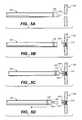
- FIG. 4Ais a side view of an embodiment of the implantable medical device, wherein the implantable medical device is attached to an elongated delivery member that includes an electrolytic joint.
- FIG. 4Bis a side view of an embodiment of the implantable medical device and an elongated delivery member, wherein the delivery member includes a first threaded member and the medical device includes a second threaded member that functionally corresponds to the first threaded member.
- FIGS. 5A to 5 Dare partial sectioned views of the ventricular septal defect, shown relative the catheter, and illustrate various procedural elements associated with using the implantable medical device.
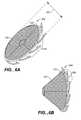
- FIG. 6Ais a perspective side view of another embodiment of an implantable medical device.
- FIG. 6Bis a side view of the implantable medical device of FIG. 6A in a collapsed delivery position.
- FIG. 1illustrates a partial sectioned view of a heart, wherein a catheter 100 extends toward a ventricular septal defect 105 .
- Catheter 100is shown having a radio-opaque band 115 at a distal end 120 .
- radio-opaque band 115assists in the guidance of catheter 100 through a vascular system and through heart 110 utilizing principles of radiography or fluoroscopy.
- distal end 120 of catheter 100has been guided so as to extend to a position proximate ventricular septal defect 105 .
- the present inventionwill be described in the context of ventricular septal defects, the scope of the present invention should not be limited to that context.
- the present inventioncould just as easily be applied in the context of atrial septal defects.
- the present inventioncould be applied in the context of a variety of other body spaces.
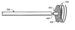
- FIG. 2illustrates a perspective side view of an implantable medical device 200 in accordance with an embodiment of the present invention.
- Device 200includes an obstruction mechanism 205 connected to a non-linear elongated tissue-puncturing end 210 .
- Non-linear elongated tissue-puncturing end 210comprises a coil 215 that includes a tissue engaging section 220 and a puncturing member 225 .
- Tissue engaging section 220illustratively interconnects obstruction mechanism 205 and puncturing member 225 .
- obstruction mechanism 205includes a material supporting member 230 attached to a material covering 235 .
- Material supporting member 230is illustratively a ring-shaped structure having an interior portion (portion inside of the ring) that is substantially filled by material covering 235 .
- material covering 235is, by design, configured to physically obstruct blood flow through septal defect 105 (FIG. 1 ), when device 200 has been implanted relative to the defect.
- material covering 235is a sheet of polytetrafluroethylene (PTFE) or other biocompatible material (degradable or not).
- material covering 235can be constructed of a material designed to act as a therapeutic agent.
- material covering 235can be constructed of or contain a bioactive material, such as a drug, protein, cells or genetic material, useful for the medical treatment of a ventricular septal defect or other medical disorder.
- material covering 235can be constructed of, or have an attached collection of, living cells that promote tissue regeneration within the human body.
- the cellscould be a number of types including but not limited to fibroblast, endothelial cells, smooth muscle cells or stemt cells.
- material covering 235can be constructed of or include a different bioactive material selected or designed to encourage cell growth at the site of a septal defect.
- the materialcan illustratively be a natural bio-material, such as collagen, gelatin, fibrin, fibronectin, fibriogen, hyaluronic acid, polysaccharides, or proteoglycans, elastin or any combination thereof; or a combination of natural bio-materials and synthetic absorbable materials.
- material covering 235can be constructed of or include a material that encourages cell growth within a targeted portion of a septal defect and then is specifically designed or selected to be biologically absorbed by the human body. While there are many materials that can be utilized as material covering 235 , two that are biologically absorbable and encourage cell growth are polylactic acid (PLA) and polyglycolic acid (PGA). In accordance with one embodiment, a mixture or composite composition comprising PLA and PGA could be utilized. Other potential materials that could be incorporated into material covering 235 , and that may encourage cell growth, include polymers containing e-caprolactone, trimethylene carbonate, and p-dioxanone. The materials listed above should be considered only examples of the many materials within the scope of the present invention that could be utilized in the construction of material covering 235 .
- non-linear elongated tissue-puncturing end 210could be formed to include a single-layer coil 215 (or double-layer, quadruple-layer, etc.) tissue engaging section 220 rather than the three-layer coil 215 configuration illustrated in FIG. 2 .
- material supporting member 230could be formed in a shape other than the illustrated ring-shaped structure.
- material covering 235need not completely fill an interior portion of a material supporting member 230 .
- material covering 235could be a therapeutic agent disposed circumferentially on material supporting member 230 .
- a therapeutic agentcould be further disposed on at least one portion of non-linear elongated tissue-puncturing end 210 .
- Implantable medical device 200is illustratively of a size and overall flexibility to be deliverable through a tubular delivery device, such as catheter 100 in FIG. 1 .
- FIG. 3is an illustration of implantable medical device 200 as it is being delivered through catheter 100 .
- the same reference numeralsare used in FIG. 3 for elements that are the same or similar to those elements illustrated in FIGS. 1 and 2.
- an elongated delivery member 300is being utilized to push medical device 200 through catheter 100 towards catheter distal end 120 .
- device 200is being delivered with coil 215 in a non-compressed state.
- coil 215is constructed of a material having shape-memory characteristics, such as nitinol.
- shape-memory characteristicssuch as nitinol.
- different super-elastic or pseudo-elastic shape recovery alloys, or shape memory polymersi.e., urethanes
- Other materials having shape-memory characteristicsi.e., certain metals should be considered within the scope of the present invention.
- Utilizing shape-memory material in the construction of coil 215enables device 200 to be delivered through catheter 100 with coil 215 in a compressed state, wherein frictional forces between catheter 100 and device 200 , created while device 200 is being pushed through catheter 100 , causes the compression of coil 215 .
- coil 215assumes a non-compressed shape.
- a distal end 305 of elongated delivery member 300could connect to implantable medical device 200 .
- Distal end 305illustratively engages material covering 235 in a non-fixed manner such that delivery member 300 can be disengaged from material covering 235 and device 200 simply by proximally withdrawing delivery member 300 .
- FIG. 4Ain accordance with an embodiment of the present invention, an alternate connection between elongated delivery member 300 and medical device 200 is illustrated.
- the same reference numeralsare used in FIG. 4A for elements that are the same or similar to those elements illustrated in previously described embodiments.
- FIG. 4Ais a side view of medical device 200 , which is fixedly attached to elongated delivery member 300 .
- distal end 305 of delivery member 300includes a portion that is fixedly attached to material covering 235 of medical device 200 .
- Severable joint 400interconnects distal end 305 with the rest of delivery member 300 .
- Severable joint 400illustratively includes means for severing medical device 200 from delivery member 300 .
- severable joint 400is an electrolytically severable joint, wherein severable joint 400 is constructed of a material that is more susceptible to dissolution via electrolysis in blood (or other ionic media) than the material used to construct medical device 200 and delivery member 300 (including distal end 305 ). Accordingly, in response to an electrolytic control signal, severable joint 400 dissolves, thereby disengaging medical device 200 from all or most of delivery member 300 .
- severable joint 400attaches directly to material covering 235 and distal end 205 is connected to joint 400 and located just proximal thereof.
- FIG. 4Bin accordance with an embodiment of the present invention, yet another alternative connection between elongated delivery member 300 and medical device 200 is illustrated.
- the same reference numeralsare used in FIG. 4B for elements that are the same or similar to those elements illustrated in previously described embodiments.
- FIG. 4Bis a side view of an embodiment of medical device 200 and elongated delivery member 300 .
- Distal end 305 of delivery member 300includes a first threaded member 405 .
- a second threaded member 410is fixedly connected to material covering 235 of device 200 and functionally corresponds to the first threaded member.
- the first and second threaded members 405 and 410can be desirably engaged and disengaged by rotating delivery member 300 and engaging and disengaging the threaded members 405 and 410 .
- further rotation of delivery member 300enables rotation of device 200 . For reasons described below in relation to FIGS. 5A-5D, such rotation of device 200 can be desirable during implantation of medical device 200 .
- FIGS. 5A-5Dillustrate a series of partial sectioned views of ventricular septal defect 105 , shown relative to catheter 100 .
- the same reference numeralsare used in FIGS. 5A-5D for elements that are the same or similar to those illustrated in previously described embodiments.
- catheter 100is initially steered into a location such that distal end 120 is placed proximate septal defect 105 .
- the positioning of catheter 100is aided by the use of a steerable guide wire (not illustrated).
- radio-opaque band 115may be used to assist in the steering of catheter 100 .
- any guide wire that has been utilizedis typically removed.
- medical device 200is then pushed through catheter 100 .
- Medical device 200is illustratively pushed until puncturing member 225 extends from distal end 120 of catheter 100 (see FIG. 5 A).
- tissue proximate septal defect 105is punctured.
- medical device 200is rotated such that a substantial portion of the coil 215 portion of medical device 200 becomes embedded in the tissue proximate septal defect 105 .
- medical device 200is physically and gradually transferred out of catheter 100 .
- device 200could be rotated during the implantation process.
- medical device 200is rotated by first ensuring maintenance of a secure engagement between distal end 305 of delivery member 300 and material covering 235 , and then rotating delivery member 300 .
- the engagement between distal end 305 and material covering 235could illustratively be a frictional engagement (FIG. 3 ), a severable joint engagement (FIG. 4 A), a threaded engagement (FIG. 4 B), or another similar engagement.
- medical device 200is rotated by first ensuring maintenance of a frictional engagement between distal end 120 of catheter 100 and a circumference of device 200 . Then, catheter 100 is rotated, thereby rotating device 200 .
- FIG. 5Billustrates device 200 after it has been rotated out of the grip of catheter 100 and into an embedded position within tissue proximate septal defect 105 .
- FIG. 5Cillustrates device 200 after it has been rotated into an embedded position within tissue proximate septal defect 105 .
- the next stepis to sever joint 400 in order to eliminate all connections between member 300 and device 200 .
- FIG. 5Dillustrates device 200 after delivery member 300 has been rotated so as to disengage threaded member 405 from 410 .
- Device 200is left embedded in the tissue proximate defect 105 .
- a subsequent step in each of the above-described embodimentsis to remove catheter 100 and delivery member 300 from heart 110 (FIG. 1 ).
- Medical device 200is left embedded in tissue relative to septal defect 105 such that septal defect 105 at least partially obstructs blood flow from one side of defect 105 to the other.
- FIG. 6Ais a perspective side view of an implantable medical device 600 in accordance with another embodiment of the present invention.
- the same reference numeralsare used in FIG. 6A for elements that are the same or similar to those elements illustrated in previously described Figures.
- Medical device 600includes a material supporting member 230 attached to a material covering 235 .
- Material supporting member 230 and material coveringare configured and operate as described above in relation to other embodiments of the present invention.
- Device 600illustratively can be attached to a delivery member (such as delivery member 300 ) as described above in relation to previous embodiments.
- Medical device 600differs from previous embodiments.
- medical device 600includes a radius 620 that is significantly larger than that of a catheter (such as catheter 100 in FIG. 3) or other delivery mechanism through which device 600 might be delivered.
- radius 620is up to three times the diameter of an associated delivery device.
- Medical device 600includes an attached plurality of non-linear elongated tissue-engaging mechanisms 625 (an illustrative few have been labeled) disposed around a periphery of material supporting member 230 .
- mechanisms 625are configured to engage tissue proximate a septal defect (such as defect 105 in FIG. 1) in a manner that enables the defect to be at least partially obstructed by device 600 .
- material covering 235could illustratively be constructed of a material suitable to supplement device 600 and further encourage obstruction of the defect.
- FIG. 6Bis a side view of implantable medical device 600 in a collapsed delivery or folded cone shape or position.
- device 600illustratively includes material covering 235 , material supporting member 230 and non-linear elongated tissue-engaging mechanisms 625 .
- device 600because device 600 includes a diameter 620 that is greater than the diameter of an associated delivery mechanism (such as catheter 100 in FIG. 1 ), device 600 is illustratively collapsible into a shape suitable for delivery.
- FIG. 6Bis an illustration of medical device 600 in an embodiment of a collapsed position.
- medical device 600includes material having shape memory characteristics that cause device 600 to transform from the FIG. 6B collapsed configuration to the FIG. 6A non-collapsed configuration, the transformation illustratively occurring as device 600 exits or is pushed out of a delivery device (such as catheter 100 in FIG. 1 ).
Landscapes
- Health & Medical Sciences (AREA)
- Surgery (AREA)
- Life Sciences & Earth Sciences (AREA)
- Medical Informatics (AREA)
- Nuclear Medicine, Radiotherapy & Molecular Imaging (AREA)
- Engineering & Computer Science (AREA)
- Biomedical Technology (AREA)
- Heart & Thoracic Surgery (AREA)
- Cardiology (AREA)
- Molecular Biology (AREA)
- Animal Behavior & Ethology (AREA)
- General Health & Medical Sciences (AREA)
- Public Health (AREA)
- Veterinary Medicine (AREA)
- Prostheses (AREA)
- Surgical Instruments (AREA)
Abstract
Description
The present invention deals with an implantable medical device. While the device could be utilized in the context of a variety of body spaces, and particularly in the context of a variety of septal defects, the present description, for the sake of brevity, will be focused primarily on the treatment of ventricular septal defects. Accordingly, the present invention deals with an implantable medical device for at least partially obstructing a ventricular septal defect.
A ventricular septal defect is characterized by incomplete closure (i.e., a hole) in the intraventricular septum, the heart muscle forming a wall between ventricles within the heart. The intraventricular septum is meant to prevent blood passing from one ventricle to the next. A septal defect can undesirably allow blood to flow from one ventricle to the other, forcing some heart chambers to pump extra blood. This increase in blood can potentially cause the heart to dilate, a weakening of the heart muscle, and pressures in the pulmonary arteries to increase (pulmonary hypertension). In addition, when the intraventricular septum is broken, an undesirable mixing of oxygen-depleted blood from the veins with oxygenated blood going to the arteries is a potential problem. In many instances, these consequences can be minimized or even avoided through a natural or treatment-based obstruction of the septal defect.
The size of ventricular septal defects is variable. Small-to-medium sized defects often close naturally and spontaneously. Many of the larger defects, however, require surgical treatment. If a substantial sized defect is not properly treated, then pressures in the pulmonary arteries may become very high and induce undesirable changes in the arteries themselves. Eventually, if the defect is not corrected, then conditions can deteriorate until even a successful closure of the defect will no longer improve the patient outcome.
Different implantable medical devices have been developed for obstructing ventricular septal defects. Intravascular devices, such as catheters and guide wires, have been used to deliver a variety of these devices to a specific location, such as within a particular ventricle, within a patient's heart. A variety of simple and complex devices are known to be deliverable to a septal defect through a catheter.
One class of catheter-delivered devices designed for the treatment of septal defects are self-expanding defect obstructing devices. A rod-like element is typically connected to these devices and utilized to push the devices from the end of a delivery catheter into a location proximate a septal defect, thereby causing an expansion of the device as it leaves the catheter. The expanded devices are typically maneuvered relative the defect until a secured position, a position where the device will stay in place and cause an obstruction of blood flow through the defect, is located. When the expanding devices have been maneuvered to a secured position, they are typically detached from any catheter, guide wire, or rod-like element utilized for intravascular placement. The expanding devices are left in a location proximate the septal defect and are intended to obstruct blood flow through the defect.
Some implantable self-expanding defect obstructing devices include separate extending portions that expand on both sides of a septal defect and into both of the heart chambers that are connected by the defect. Other devices are balloon-actuated devices, wherein expansion occurs as a result of inflation of extending members. Still other devices include mechanically expanding extending members that collapse (i.e., during delivery through a catheter) and can be extended (i.e., in a location proximate a septal defect) utilizing a mechanically maneuverable frame. Other devices are constructed of shape-memory based material, allowing the device to be manipulated into a collapsed shape and inserted into a catheter. Upon being pushed out of the catheter, these devices regain their original shape (i.e., a shape convenient for obstructing a septal defect).
Designing an effective implantable medical device for the obstruction of a septal defect presents special challenges. Many self-expanding devices suffer from deployment problems (i.e., incomplete opening of extending members or an error in the functionality of the extending member deployment mechanics). Many lack the ability to be precisely and effectively positioned relative a septal defect. In many instances, the shape of known implantable devices fails to effectively accommodate the often complex shape of a septal defect. With most known devices, recovery of a deployed device is difficult if not impossible. Many known devices require highly complex manufacture processes.
One aspect of the present invention pertains to an implantable medical device for at least partially obstructing a septal defect. The implantable medical device includes an obstruction mechanism connected to a non-linear elongated tissue-puncturing end.
Another aspect of the present invention pertains to an implantable device, deliverable via a vascular catheter, of a size and overall flexibility to lodge in an area of tissue located proximate a septal defect, and suitable for at least partially obstructing the septal defect. The implantable device includes an elongated delivery member having a distal end. An obstruction mechanism is connected to a coil that includes a puncturing end. The obstruction mechanism includes a ring-shaped structure having an interior portion. A material covering substantially fills the interior portion of the ring-shaped structure. A connection between the distal end of the elongated delivery member and the obstruction mechanism enables the obstruction mechanism to be rotated.
Yet another aspect of the present invention pertains to a method for at least partially obstructing a septal defect in a heart by implanting a medical device. The method first includes the step of placing a distal end of a catheter in a location proximate the septal defect. Next, an elongated delivery member is utilized to push an obstruction device through the catheter until a puncturing member portion of the obstruction device extends from the distal end of the catheter. Then, with the puncturing member, an area of tissue proximate the septal defect is punctured. Next, the obstruction device is rotated such that a non-linear tissue engaging section of the obstruction device, which is connected to the puncturing member, becomes substantially embedded in the area of tissue proximate the septal defect. Finally, the catheter and elongated delivery member are removed from the heart.
FIG. 1 is a partial sectioned view of a heart, wherein a catheter is shown extending toward a ventricular septal defect.
FIG. 2 is a perspective side view of an implantable medical device.
FIG. 3 is a partial sectioned view of the implantable medical device inserted within the catheter.
FIG. 4A is a side view of an embodiment of the implantable medical device, wherein the implantable medical device is attached to an elongated delivery member that includes an electrolytic joint.
FIG. 4B is a side view of an embodiment of the implantable medical device and an elongated delivery member, wherein the delivery member includes a first threaded member and the medical device includes a second threaded member that functionally corresponds to the first threaded member.
FIGS. 5A to5D are partial sectioned views of the ventricular septal defect, shown relative the catheter, and illustrate various procedural elements associated with using the implantable medical device.
FIG. 6A is a perspective side view of another embodiment of an implantable medical device.
FIG. 6B is a side view of the implantable medical device of FIG. 6A in a collapsed delivery position.
FIG. 1 illustrates a partial sectioned view of a heart, wherein acatheter 100 extends toward a ventricularseptal defect 105.Catheter 100 is shown having a radio-opaque band 115 at adistal end 120. As is known in the art, radio-opaque band 115 assists in the guidance ofcatheter 100 through a vascular system and throughheart 110 utilizing principles of radiography or fluoroscopy. As is illustrated,distal end 120 ofcatheter 100 has been guided so as to extend to a position proximate ventricularseptal defect 105.
While, for the sake of brevity, the present invention will be described in the context of ventricular septal defects, the scope of the present invention should not be limited to that context. For instance, the present invention could just as easily be applied in the context of atrial septal defects. In addition the present invention could be applied in the context of a variety of other body spaces.
FIG. 2 illustrates a perspective side view of an implantablemedical device 200 in accordance with an embodiment of the present invention.Device 200 includes anobstruction mechanism 205 connected to a non-linear elongated tissue-puncturingend 210.
Non-linear elongated tissue-puncturingend 210 comprises acoil 215 that includes atissue engaging section 220 and a puncturingmember 225.Tissue engaging section 220 illustratively interconnectsobstruction mechanism 205 and puncturingmember 225.
In accordance with the FIG. 2 embodiment of the present invention,obstruction mechanism 205 includes amaterial supporting member 230 attached to a material covering235.Material supporting member 230 is illustratively a ring-shaped structure having an interior portion (portion inside of the ring) that is substantially filled by material covering235. In accordance with one embodiment, material covering235 is, by design, configured to physically obstruct blood flow through septal defect105 (FIG.1), whendevice 200 has been implanted relative to the defect. In accordance with one embodiment, material covering235 is a sheet of polytetrafluroethylene (PTFE) or other biocompatible material (degradable or not).
In accordance with another embodiment of the present invention, material covering235 can be constructed of a material designed to act as a therapeutic agent. Illustratively, material covering235 can be constructed of or contain a bioactive material, such as a drug, protein, cells or genetic material, useful for the medical treatment of a ventricular septal defect or other medical disorder.
In accordance with one embodiment, material covering235 can be constructed of, or have an attached collection of, living cells that promote tissue regeneration within the human body. Illustratively, the cells could be a number of types including but not limited to fibroblast, endothelial cells, smooth muscle cells or stemt cells.
In accordance with other embodiments, material covering235 can be constructed of or include a different bioactive material selected or designed to encourage cell growth at the site of a septal defect. The material can illustratively be a natural bio-material, such as collagen, gelatin, fibrin, fibronectin, fibriogen, hyaluronic acid, polysaccharides, or proteoglycans, elastin or any combination thereof; or a combination of natural bio-materials and synthetic absorbable materials.
In accordance with still other embodiments, material covering235 can be constructed of or include a material that encourages cell growth within a targeted portion of a septal defect and then is specifically designed or selected to be biologically absorbed by the human body. While there are many materials that can be utilized as material covering235, two that are biologically absorbable and encourage cell growth are polylactic acid (PLA) and polyglycolic acid (PGA). In accordance with one embodiment, a mixture or composite composition comprising PLA and PGA could be utilized. Other potential materials that could be incorporated into material covering235, and that may encourage cell growth, include polymers containing e-caprolactone, trimethylene carbonate, and p-dioxanone. The materials listed above should be considered only examples of the many materials within the scope of the present invention that could be utilized in the construction of material covering235.
It should be noted that implantable medical devices having configurations other than the precise configuration illustrated bydevice 200 in FIG. 2 should be considered within the scope of the present invention. For example, non-linear elongated tissue-puncturingend 210 could be formed to include a single-layer coil215 (or double-layer, quadruple-layer, etc.)tissue engaging section 220 rather than the three-layer coil 215 configuration illustrated in FIG.2. In addition,material supporting member 230 could be formed in a shape other than the illustrated ring-shaped structure. Finally, material covering235 need not completely fill an interior portion of amaterial supporting member 230. For instance, in accordance with one embodiment of the present invention, material covering235 could be a therapeutic agent disposed circumferentially onmaterial supporting member 230. In accordance with one embodiment, a therapeutic agent could be further disposed on at least one portion of non-linear elongated tissue-puncturingend 210.
Implantablemedical device 200 is illustratively of a size and overall flexibility to be deliverable through a tubular delivery device, such ascatheter 100 in FIG.1. FIG. 3 is an illustration of implantablemedical device 200 as it is being delivered throughcatheter 100. The same reference numerals are used in FIG. 3 for elements that are the same or similar to those elements illustrated in FIGS. 1 and 2.
In FIG. 3, anelongated delivery member 300 is being utilized to pushmedical device 200 throughcatheter 100 towards catheterdistal end 120. As illustrated,device 200 is being delivered withcoil 215 in a non-compressed state. In accordance with one embodiment of the present invention,coil 215 is constructed of a material having shape-memory characteristics, such as nitinol. In addition to or in place of nitinol, different super-elastic or pseudo-elastic shape recovery alloys, or shape memory polymers (i.e., urethanes) could be utilized in the construction ofcoil 215. Other materials having shape-memory characteristics (i.e., certain metals) should be considered within the scope of the present invention.
Utilizing shape-memory material in the construction ofcoil 215 enablesdevice 200 to be delivered throughcatheter 100 withcoil 215 in a compressed state, wherein frictional forces betweencatheter 100 anddevice 200, created whiledevice 200 is being pushed throughcatheter 100, causes the compression ofcoil 215. Illustratively, due to an incorporation of material having shape-memory characteristics, asdevice 200 is pushed fromdistal end 120 ofcatheter 100 and constriction forces betweencatheter 100 anddevice 200 are eliminated,coil 215 assumes a non-compressed shape.
In accordance with embodiments of the present invention, there are several different ways that adistal end 305 ofelongated delivery member 300 could connect to implantablemedical device 200. In accordance with the embodiment pictured in FIG. 3, there is no fixed connection betweendistal end 305 anddevice 200.Distal end 305 illustratively engages material covering235 in a non-fixed manner such thatdelivery member 300 can be disengaged from material covering235 anddevice 200 simply by proximally withdrawingdelivery member 300.
Turning to FIG. 4A, in accordance with an embodiment of the present invention, an alternate connection betweenelongated delivery member 300 andmedical device 200 is illustrated. The same reference numerals are used in FIG. 4A for elements that are the same or similar to those elements illustrated in previously described embodiments.
FIG. 4A is a side view ofmedical device 200, which is fixedly attached toelongated delivery member 300. Illustratively,distal end 305 ofdelivery member 300 includes a portion that is fixedly attached to material covering235 ofmedical device 200. Severable joint400 interconnectsdistal end 305 with the rest ofdelivery member 300. Severable joint400 illustratively includes means for severingmedical device 200 fromdelivery member 300. For example, in accordance with one embodiment, severable joint400 is an electrolytically severable joint, wherein severable joint400 is constructed of a material that is more susceptible to dissolution via electrolysis in blood (or other ionic media) than the material used to constructmedical device 200 and delivery member300 (including distal end305). Accordingly, in response to an electrolytic control signal, severable joint400 dissolves, thereby disengagingmedical device 200 from all or most ofdelivery member 300. In accordance with one embodiment, severable joint400 attaches directly to material covering235 anddistal end 205 is connected to joint400 and located just proximal thereof.
Turning to FIG. 4B, in accordance with an embodiment of the present invention, yet another alternative connection betweenelongated delivery member 300 andmedical device 200 is illustrated. The same reference numerals are used in FIG. 4B for elements that are the same or similar to those elements illustrated in previously described embodiments.
FIG. 4B is a side view of an embodiment ofmedical device 200 andelongated delivery member 300.Distal end 305 ofdelivery member 300 includes a first threadedmember 405. A second threadedmember 410 is fixedly connected to material covering235 ofdevice 200 and functionally corresponds to the first threaded member. Illustratively, the first and second threadedmembers delivery member 300 and engaging and disengaging the threadedmembers delivery member 300 enables rotation ofdevice 200. For reasons described below in relation to FIGS. 5A-5D, such rotation ofdevice 200 can be desirable during implantation ofmedical device 200.
FIGS. 5A-5D illustrate a series of partial sectioned views of ventricularseptal defect 105, shown relative tocatheter 100. The same reference numerals are used in FIGS. 5A-5D for elements that are the same or similar to those illustrated in previously described embodiments.
With reference to FIGS. 5A-5D and to the previously described Figures, procedural elements associated with implantingmedical device 200, in accordance with embodiments of the present invention, will now be described.
As is represented by FIG. 1,catheter 100 is initially steered into a location such thatdistal end 120 is placed proximateseptal defect 105. Typically, the positioning ofcatheter 100 is aided by the use of a steerable guide wire (not illustrated). As was discussed above in relation to FIG. 1, radio-opaque band 115 may be used to assist in the steering ofcatheter 100.
Whencatheter 100 has been positioned relative toseptal defect 105, any guide wire that has been utilized is typically removed. Next, as was discussed in relation to FIG. 3, utilizingelongated delivery member 300,medical device 200 is then pushed throughcatheter 100.Medical device 200 is illustratively pushed until puncturingmember 225 extends fromdistal end 120 of catheter100 (see FIG.5A).
Next, with puncturingmember 225, an area of tissue proximateseptal defect 105 is punctured. After the tissue has been punctured,medical device 200 is rotated such that a substantial portion of thecoil 215 portion ofmedical device 200 becomes embedded in the tissue proximateseptal defect 105. In accordance with one embodiment, during the rotation step,medical device 200 is physically and gradually transferred out ofcatheter 100.
In accordance with additional embodiments of the present invention, there are several ways in whichdevice 200 could be rotated during the implantation process. In accordance with one embodiment,medical device 200 is rotated by first ensuring maintenance of a secure engagement betweendistal end 305 ofdelivery member 300 and material covering235, and then rotatingdelivery member 300. The engagement betweendistal end 305 and material covering235 could illustratively be a frictional engagement (FIG.3), a severable joint engagement (FIG.4A), a threaded engagement (FIG.4B), or another similar engagement.
In accordance with another embodiment,medical device 200 is rotated by first ensuring maintenance of a frictional engagement betweendistal end 120 ofcatheter 100 and a circumference ofdevice 200. Then,catheter 100 is rotated, thereby rotatingdevice 200. FIG. 5B illustratesdevice 200 after it has been rotated out of the grip ofcatheter 100 and into an embedded position within tissue proximateseptal defect 105.
In accordance with embodiments whereinmedical device 200 is fixedly connected todelivery member 300 through a severable joint400, FIG. 5C illustratesdevice 200 after it has been rotated into an embedded position within tissue proximateseptal defect 105. Illustratively, the next step is to sever joint400 in order to eliminate all connections betweenmember 300 anddevice 200.
In accordance with embodiments whereinmedical device 200 is connected todelivery member 300 through a threaded connection between threadedmembers device 200 afterdelivery member 300 has been rotated so as to disengage threadedmember 405 from410.Device 200 is left embedded in the tissueproximate defect 105.
Illustratively, a subsequent step in each of the above-described embodiments is to removecatheter 100 anddelivery member 300 from heart110 (FIG.1).Medical device 200 is left embedded in tissue relative toseptal defect 105 such thatseptal defect 105 at least partially obstructs blood flow from one side ofdefect 105 to the other.
FIG. 6A is a perspective side view of an implantablemedical device 600 in accordance with another embodiment of the present invention. The same reference numerals are used in FIG. 6A for elements that are the same or similar to those elements illustrated in previously described Figures.
FIG. 6B is a side view of implantablemedical device 600 in a collapsed delivery or folded cone shape or position. The same reference numerals are used in FIG. 6B for elements that are the same or similar to those elements illustrated in previously described Figures. In FIG. 6B,device 600 illustratively includes material covering235,material supporting member 230 and non-linear elongated tissue-engagingmechanisms 625.
In accordance with an embodiment of the present invention, becausedevice 600 includes adiameter 620 that is greater than the diameter of an associated delivery mechanism (such ascatheter 100 in FIG.1),device 600 is illustratively collapsible into a shape suitable for delivery. FIG. 6B is an illustration ofmedical device 600 in an embodiment of a collapsed position. In accordance with one embodiment,medical device 600 includes material having shape memory characteristics that causedevice 600 to transform from the FIG. 6B collapsed configuration to the FIG. 6A non-collapsed configuration, the transformation illustratively occurring asdevice 600 exits or is pushed out of a delivery device (such ascatheter 100 in FIG.1).
It should be pointed out that while diameter620 (FIG. 6A) and the collapsed position illustrated in FIG. 6B have been depicted withmedical device 600, these features and the associated characteristics could just as easily be applied in the context of previously described embodiments, such as in the context ofdevice 200.
Although the present invention has been described with reference to preferred embodiments, workers skilled in the art will recognize that changes may be made in form and detail without departing from the spirit and scope of the invention.
Claims (14)
1. An implantable medical device for at least partially obstructing a septal defect, comprising:
an obstruction mechanism; and
a non-linear elongated tissue-puncturing end connected to the obstruction mechanism, wherein the non-linear elongated tissue-puncturing end includes a coil having a tissue engaging section and a puncturing member, and wherein the tissue engaging section interconnects the obstruction mechanism and the puncturing member.
2. An implantable device, deliverable via a vascular catheter, of a size and overall flexibility to lodge in an area of tissue located proximate a septal defect, and suitable for at least partially obstructing the septal defect, comprising:
an elongated delivery member having a distal end;
an obstruction mechanism comprising a ring-shaped structure having an interior portion and a connection to a coil that includes a puncturing member;
a material covering that substantially fills the interior portion of the ring-shaped structure; and
a connection between the distal end of the elongated delivery member and the obstruction mechanism, wherein the connection enables the obstruction mechanism to be rotated.
3. The implantable device ofclaim 2 , wherein the connection comprises an engagement between a first threaded member disposed on the distal end of the elongated delivery member and a functionally corresponding second threaded member disposed on the material covering.
4. The implantable device ofclaim 2 , wherein the connection comprises a frictional engagement between the distal end of the elongated delivery member and the material covering.
5. The implantable device ofclaim 2 , wherein the material covering is constructed of a material that is a therapeutic agent.
6. The implantable device ofclaim 2 , wherein the material covering is constructed of a bioactive material.
7. The implantable device ofclaim 6 , wherein the bioactive material is a biologically absorbable material that encourages cell growth.
8. A method for at least partially obstructing a septal defect in a heart by implanting a medical device, comprising:
placing a distal end of a catheter in a location proximate the septal defect;
utilizing an elongated delivery member to push an obstruction device through the catheter until a puncturing member portion of the obstruction device extends from the distal end of the catheter;
puncturing, with the puncturing member, an area of tissue proximate the septal defect;
rotating the obstruction device such that a non-linear tissue engaging section of the obstruction device, which is connected to the puncturing member, becomes substantially embedded in the area of tissue proximate the septal defect; and
removing the catheter and elongated delivery member from the heart.
9. The method ofclaim 8 , wherein rotating the obstruction device comprises:
maintaining a frictional engagement between the catheter and a circumference of the obstruction device; and
rotating the catheter.
10. The method ofclaim 8 , wherein rotating the obstruction device comprises:
maintaining an engagement between the elongated delivery member and the obstruction device;
and
rotating the elongated delivery member.
11. The method ofclaim 10 , wherein maintaining an engagement between the elongated delivery member and the obstruction device comprises:
maintaining a frictional engagement between the elongated delivery member and a material covering portion of the obstruction device.
12. The method ofclaim 10 , wherein maintaining an engagement between the elongated delivery member and the obstruction device comprises:
maintaining a threaded engagement between a first threaded member disposed on the distal end of the elongated delivery member and a second threaded member that functionally corresponds to the first threaded member and is disposed on the obstruction device.
13. The method ofclaim 12 , wherein removing the catheter and elongated delivery member from the heart comprises:
disengaging the first threaded member from the second threaded member;
removing the elongated delivery member from the catheter and from the heart; and
removing the catheter from the heart.
14. The method ofclaim 10 , wherein removing the catheter and elongated delivery member the heart comprises:
disengaging the elongated delivery member from the obstruction device by severing a joint therebetween;
removing the elongated delivery member from the catheter and from the heart; and
removing the catheter from the heart.
Priority Applications (2)
| Application Number | Priority Date | Filing Date | Title |
|---|---|---|---|
| US09/867,951US6537300B2 (en) | 2001-05-30 | 2001-05-30 | Implantable obstruction device for septal defects |
| PCT/US2002/017146WO2002096295A1 (en) | 2001-05-30 | 2002-05-29 | Implantable septal defects closure device |
Applications Claiming Priority (1)
| Application Number | Priority Date | Filing Date | Title |
|---|---|---|---|
| US09/867,951US6537300B2 (en) | 2001-05-30 | 2001-05-30 | Implantable obstruction device for septal defects |
Publications (2)
| Publication Number | Publication Date |
|---|---|
| US20020183786A1 US20020183786A1 (en) | 2002-12-05 |
| US6537300B2true US6537300B2 (en) | 2003-03-25 |
Family
ID=25350783
Family Applications (1)
| Application Number | Title | Priority Date | Filing Date |
|---|---|---|---|
| US09/867,951Expired - LifetimeUS6537300B2 (en) | 2001-05-30 | 2001-05-30 | Implantable obstruction device for septal defects |
Country Status (2)
| Country | Link |
|---|---|
| US (1) | US6537300B2 (en) |
| WO (1) | WO2002096295A1 (en) |
Cited By (51)
| Publication number | Priority date | Publication date | Assignee | Title |
|---|---|---|---|---|
| US20030195530A1 (en)* | 2000-04-26 | 2003-10-16 | Microvena Corporation, A Minnesota Corporation, And Into Ev3 Inc., A Delaware Corpora | Septal defect occluder |
| US20040220595A1 (en)* | 1999-09-20 | 2004-11-04 | Frazier Andrew G.C. | Method of securing tissue |
| US20040220596A1 (en)* | 2003-02-04 | 2004-11-04 | Frazier Andrew G.C. | Patent foramen ovale closure system |
| US20050010248A1 (en)* | 2003-07-10 | 2005-01-13 | Scimed Life Systems, Inc. | System for closing an opening in a body cavity |
| US20050033327A1 (en)* | 1999-09-07 | 2005-02-10 | John Gainor | Retrievable septal defect closure device |
| US20050113868A1 (en)* | 2003-11-20 | 2005-05-26 | Devellian Carol A. | Device, with electrospun fabric, for a percutaneous transluminal procedure, and methods thereof |
| US20050125032A1 (en)* | 2003-10-10 | 2005-06-09 | Whisenant Brian K. | Patent foramen ovale (PFO) closure devices, delivery apparatus and related methods and systems |
| US20050187568A1 (en)* | 2004-02-20 | 2005-08-25 | Klenk Alan R. | Devices and methods for closing a patent foramen ovale with a coil-shaped closure device |
| US20050192627A1 (en)* | 2003-10-10 | 2005-09-01 | Whisenant Brian K. | Patent foramen ovale closure devices, delivery apparatus and related methods and systems |
| US20050267495A1 (en)* | 2004-05-17 | 2005-12-01 | Gateway Medical, Inc. | Systems and methods for closing internal tissue defects |
| US20060009799A1 (en)* | 2003-05-19 | 2006-01-12 | Kleshinski Stephen J | Embolic filtering method and apparatus |
| US20060052821A1 (en)* | 2001-09-06 | 2006-03-09 | Ovalis, Inc. | Systems and methods for treating septal defects |
| US20060079915A1 (en)* | 2001-12-26 | 2006-04-13 | Chin Albert K | Temporary anastomotic seal and method |
| US20060106418A1 (en)* | 2002-07-31 | 2006-05-18 | Abbott Laboratories Vascular Enterprises, Limited | Apparatus for sealing surgical punctures |
| US20060200197A1 (en)* | 2005-03-02 | 2006-09-07 | St. Jude Medical, Inc. | Remote body tissue engaging methods and apparatus |
| US20070106327A1 (en)* | 2001-08-01 | 2007-05-10 | Ev3 Endovascular, Inc. | Tissue opening occluder |
| US20070129755A1 (en)* | 2005-12-05 | 2007-06-07 | Ovalis, Inc. | Clip-based systems and methods for treating septal defects |
| US20080015633A1 (en)* | 2001-09-06 | 2008-01-17 | Ryan Abbott | Systems and Methods for Treating Septal Defects |
| US20080161825A1 (en)* | 2006-11-20 | 2008-07-03 | Stout Medical Group, L.P. | Anatomical measurement tool |
| US20090036923A1 (en)* | 2007-05-04 | 2009-02-05 | Jabba Ronald J | Systems and methods for accommodating anatomical characteristics in the treatment of septal defects |
| US20090088794A1 (en)* | 2007-10-01 | 2009-04-02 | Boston Scientific Scimed, Inc | Connective Tissue Closure Device and Method |
| DE102008034534A1 (en)* | 2008-07-18 | 2010-01-21 | Aesculap Ag | Puncture closure for closing a hollow organ having a puncture opening, in particular a blood vessel |
| US7740640B2 (en) | 2001-09-06 | 2010-06-22 | Ovalis, Inc. | Clip apparatus for closing septal defects and methods of use |
| US7846179B2 (en) | 2005-09-01 | 2010-12-07 | Ovalis, Inc. | Suture-based systems and methods for treating septal defects |
| US20110118766A1 (en)* | 2009-11-15 | 2011-05-19 | Thoratec Corporation | Attachment System, Device and Method |
| US20110118833A1 (en)* | 2009-11-15 | 2011-05-19 | Thoratec Corporation | Attachment device and method |
| US20110118829A1 (en)* | 2009-11-15 | 2011-05-19 | Thoratec Corporation | Attachment device and method |
| US8114123B2 (en) | 2003-09-19 | 2012-02-14 | St. Jude Medical, Inc. | Apparatus and methods for tissue gathering and securing |
| US8398672B2 (en) | 2003-11-12 | 2013-03-19 | Nitinol Devices And Components, Inc. | Method for anchoring a medical device |
| US8579936B2 (en) | 2005-07-05 | 2013-11-12 | ProMed, Inc. | Centering of delivery devices with respect to a septal defect |
| US8747483B2 (en) | 2001-09-07 | 2014-06-10 | ProMed, Inc. | Needle apparatus for closing septal defects and methods for using such apparatus |
| US8858489B2 (en) | 2007-04-24 | 2014-10-14 | Emory University | Conduit device and system for implanting a conduit device in a tissue wall |
| US9044236B2 (en) | 2011-05-18 | 2015-06-02 | Thoratec Corporation | Coring knife |
| US9089329B2 (en) | 2012-04-23 | 2015-07-28 | Thoratec Corporation | Engagement device and method for deployment of anastomotic clips |
| US9125648B2 (en) | 2011-02-25 | 2015-09-08 | Thoratec Corporation | Coupling system, applicator tool, attachment ring and method for connecting a conduit to biological tissue |
| US9138228B2 (en) | 2004-08-11 | 2015-09-22 | Emory University | Vascular conduit device and system for implanting |
| US9320875B2 (en) | 2011-02-01 | 2016-04-26 | Emory University | Systems for implanting and using a conduit within a tissue wall |
| US9532773B2 (en) | 2011-01-28 | 2017-01-03 | Apica Cardiovascular Limited | Systems for sealing a tissue wall puncture |
| US9649211B2 (en) | 2009-11-04 | 2017-05-16 | Confluent Medical Technologies, Inc. | Alternating circumferential bridge stent design and methods for use thereof |
| US10028733B2 (en) | 2015-05-28 | 2018-07-24 | National University Of Ireland, Galway | Fistula treatment device |
| US10028741B2 (en) | 2013-01-25 | 2018-07-24 | Apica Cardiovascular Limited | Systems and methods for percutaneous access, stabilization and closure of organs |
| US10092427B2 (en) | 2009-11-04 | 2018-10-09 | Confluent Medical Technologies, Inc. | Alternating circumferential bridge stent design and methods for use thereof |
| US10485909B2 (en) | 2014-10-31 | 2019-11-26 | Thoratec Corporation | Apical connectors and instruments for use in a heart wall |
| US10518012B2 (en) | 2013-03-15 | 2019-12-31 | Apk Advanced Medical Technologies, Inc. | Devices, systems, and methods for implanting and using a connector in a tissue wall |
| US10667896B2 (en) | 2015-11-13 | 2020-06-02 | Cardiac Pacemakers, Inc. | Bioabsorbable left atrial appendage closure with endothelialization promoting surface |
| US11234706B2 (en) | 2018-02-14 | 2022-02-01 | Boston Scientific Scimed, Inc. | Occlusive medical device |
| US11452512B2 (en) | 2017-06-09 | 2022-09-27 | Signum Surgical Limited | Implant for closing an opening in tissue |
| US11517319B2 (en) | 2017-09-23 | 2022-12-06 | Universität Zürich | Medical occluder device |
| US11701096B2 (en) | 2015-05-28 | 2023-07-18 | National University Of Ireland, Galway | Fistula treatment device |
| US11944315B2 (en) | 2019-09-26 | 2024-04-02 | Universität Zürich | Left atrial appendage occlusion devices |
| US12402885B2 (en) | 2017-09-23 | 2025-09-02 | Universität Zürich | Medical occlusion device |
Families Citing this family (147)
| Publication number | Priority date | Publication date | Assignee | Title |
|---|---|---|---|---|
| US6939361B1 (en) | 1999-09-22 | 2005-09-06 | Nmt Medical, Inc. | Guidewire for a free standing intervascular device having an integral stop mechanism |
| US6391048B1 (en) | 2000-01-05 | 2002-05-21 | Integrated Vascular Systems, Inc. | Integrated vascular device with puncture site closure component and sealant and methods of use |
| US9579091B2 (en) | 2000-01-05 | 2017-02-28 | Integrated Vascular Systems, Inc. | Closure system and methods of use |
| US8758400B2 (en) | 2000-01-05 | 2014-06-24 | Integrated Vascular Systems, Inc. | Closure system and methods of use |
| US7842068B2 (en) | 2000-12-07 | 2010-11-30 | Integrated Vascular Systems, Inc. | Apparatus and methods for providing tactile feedback while delivering a closure device |
| US6461364B1 (en) | 2000-01-05 | 2002-10-08 | Integrated Vascular Systems, Inc. | Vascular sheath with bioabsorbable puncture site closure apparatus and methods of use |
| DE60144328D1 (en) | 2000-09-08 | 2011-05-12 | Abbott Vascular Inc | Surgical clamp |
| US6626918B1 (en) | 2000-10-06 | 2003-09-30 | Medical Technology Group | Apparatus and methods for positioning a vascular sheath |
| US6623510B2 (en) | 2000-12-07 | 2003-09-23 | Integrated Vascular Systems, Inc. | Closure device and methods for making and using them |
| US8690910B2 (en) | 2000-12-07 | 2014-04-08 | Integrated Vascular Systems, Inc. | Closure device and methods for making and using them |
| US7806904B2 (en) | 2000-12-07 | 2010-10-05 | Integrated Vascular Systems, Inc. | Closure device |
| US7211101B2 (en) | 2000-12-07 | 2007-05-01 | Abbott Vascular Devices | Methods for manufacturing a clip and clip |
| US6695867B2 (en) | 2002-02-21 | 2004-02-24 | Integrated Vascular Systems, Inc. | Plunger apparatus and methods for delivering a closure device |
| US7905900B2 (en)* | 2003-01-30 | 2011-03-15 | Integrated Vascular Systems, Inc. | Clip applier and methods of use |
| IES20010547A2 (en) | 2001-06-07 | 2002-12-11 | Christy Cummins | Surgical Staple |
| US7993365B2 (en) | 2001-06-08 | 2011-08-09 | Morris Innovative, Inc. | Method and apparatus for sealing access |
| AU2002323634A1 (en)* | 2001-09-06 | 2003-03-24 | Nmt Medical, Inc. | Flexible delivery system |
| EP1467661A4 (en) | 2001-12-19 | 2008-11-05 | Nmt Medical Inc | Septal occluder and associated methods |
| US7318833B2 (en) | 2001-12-19 | 2008-01-15 | Nmt Medical, Inc. | PFO closure device with flexible thrombogenic joint and improved dislodgement resistance |
| EP1471835A4 (en) | 2002-01-14 | 2008-03-19 | Nmt Medical Inc | Patent foramen ovale (pfo) closure method and device |
| JP2005521447A (en) | 2002-03-25 | 2005-07-21 | エヌエムティー メディカル インコーポレイテッド | Closure clip of patent foramen ovale (PFO) |
| WO2003101312A1 (en) | 2002-06-03 | 2003-12-11 | Nmt Medical, Inc. | Device with biological tissue scaffold for intracardiac defect closure |
| IES20030424A2 (en) | 2002-06-04 | 2003-12-10 | Robert Stevenson | Blood vessel closure clip and delivery device |
| AU2003240549A1 (en) | 2002-06-05 | 2003-12-22 | Nmt Medical, Inc. | Patent foramen ovale (pfo) closure device with radial and circumferential support |
| US20040127855A1 (en)* | 2002-10-10 | 2004-07-01 | Nmt Medical, Inc. | Hemostasis valve |
| EP1556117A1 (en) | 2002-10-25 | 2005-07-27 | NMT Medical, Inc. | Expandable sheath tubing |
| US7695446B2 (en) | 2002-12-02 | 2010-04-13 | Gi Dynamics, Inc. | Methods of treatment using a bariatric sleeve |
| US7678068B2 (en) | 2002-12-02 | 2010-03-16 | Gi Dynamics, Inc. | Atraumatic delivery devices |
| US7608114B2 (en) | 2002-12-02 | 2009-10-27 | Gi Dynamics, Inc. | Bariatric sleeve |
| WO2004049982A2 (en) | 2002-12-02 | 2004-06-17 | Gi Dynamics, Inc. | Bariatric sleeve |
| US7025791B2 (en)* | 2002-12-02 | 2006-04-11 | Gi Dynamics, Inc. | Bariatric sleeve |
| US7766973B2 (en) | 2005-01-19 | 2010-08-03 | Gi Dynamics, Inc. | Eversion resistant sleeves |
| WO2004052213A1 (en) | 2002-12-09 | 2004-06-24 | Nmt Medical, Inc. | Septal closure devices |
| US8562646B2 (en)* | 2002-12-19 | 2013-10-22 | Boston Scientific Scimed, Inc. | Anchoring to soft tissue |
| US8202293B2 (en) | 2003-01-30 | 2012-06-19 | Integrated Vascular Systems, Inc. | Clip applier and methods of use |
| US8758398B2 (en) | 2006-09-08 | 2014-06-24 | Integrated Vascular Systems, Inc. | Apparatus and method for delivering a closure element |
| US8398656B2 (en) | 2003-01-30 | 2013-03-19 | Integrated Vascular Systems, Inc. | Clip applier and methods of use |
| US7857828B2 (en) | 2003-01-30 | 2010-12-28 | Integrated Vascular Systems, Inc. | Clip applier and methods of use |
| US8821534B2 (en) | 2010-12-06 | 2014-09-02 | Integrated Vascular Systems, Inc. | Clip applier having improved hemostasis and methods of use |
| US8905937B2 (en) | 2009-02-26 | 2014-12-09 | Integrated Vascular Systems, Inc. | Methods and apparatus for locating a surface of a body lumen |
| US20040176788A1 (en)* | 2003-03-07 | 2004-09-09 | Nmt Medical, Inc. | Vacuum attachment system |
| US7658747B2 (en) | 2003-03-12 | 2010-02-09 | Nmt Medical, Inc. | Medical device for manipulation of a medical implant |
| US7473266B2 (en) | 2003-03-14 | 2009-01-06 | Nmt Medical, Inc. | Collet-based delivery system |
| EP2481356B1 (en) | 2003-07-14 | 2013-09-11 | W.L. Gore & Associates, Inc. | Tubular patent foramen ovale (PFO) closure device with catch system |
| US9861346B2 (en) | 2003-07-14 | 2018-01-09 | W. L. Gore & Associates, Inc. | Patent foramen ovale (PFO) closure device with linearly elongating petals |
| US8480706B2 (en) | 2003-07-14 | 2013-07-09 | W.L. Gore & Associates, Inc. | Tubular patent foramen ovale (PFO) closure device with catch system |
| CA2536368A1 (en) | 2003-08-19 | 2005-03-03 | Nmt Medical, Inc. | Expandable sheath tubing |
| CA2538707A1 (en) | 2003-09-11 | 2005-04-21 | Nmt Medical, Inc. | Suture sever tube |
| JP2007504885A (en) | 2003-09-11 | 2007-03-08 | エヌエムティー メディカル, インコーポレイティッド | Devices, systems and methods for suturing tissue |
| US7419498B2 (en) | 2003-10-21 | 2008-09-02 | Nmt Medical, Inc. | Quick release knot attachment system |
| US7666203B2 (en) | 2003-11-06 | 2010-02-23 | Nmt Medical, Inc. | Transseptal puncture apparatus |
| US8292910B2 (en) | 2003-11-06 | 2012-10-23 | Pressure Products Medical Supplies, Inc. | Transseptal puncture apparatus |
| US8057420B2 (en) | 2003-12-09 | 2011-11-15 | Gi Dynamics, Inc. | Gastrointestinal implant with drawstring |
| US20050273119A1 (en) | 2003-12-09 | 2005-12-08 | Nmt Medical, Inc. | Double spiral patent foramen ovale closure clamp |
| AU2004305450B2 (en) | 2003-12-09 | 2009-01-08 | Gi Dynamics, Inc. | Intestinal sleeve |
| EP1713401A2 (en) | 2004-01-30 | 2006-10-25 | NMT Medical, Inc. | Devices, systems, and methods for closure of cardiac openings |
| US20050192626A1 (en) | 2004-01-30 | 2005-09-01 | Nmt Medical, Inc. | Devices, systems, and methods for closure of cardiac openings |
| EP1737349A1 (en) | 2004-03-03 | 2007-01-03 | NMT Medical, Inc. | Delivery/recovery system for septal occluder |
| US20050267524A1 (en) | 2004-04-09 | 2005-12-01 | Nmt Medical, Inc. | Split ends closure device |
| US8361110B2 (en) | 2004-04-26 | 2013-01-29 | W.L. Gore & Associates, Inc. | Heart-shaped PFO closure device |
| US8308760B2 (en) | 2004-05-06 | 2012-11-13 | W.L. Gore & Associates, Inc. | Delivery systems and methods for PFO closure device with two anchors |
| US7842053B2 (en) | 2004-05-06 | 2010-11-30 | Nmt Medical, Inc. | Double coil occluder |
| US7842069B2 (en) | 2004-05-07 | 2010-11-30 | Nmt Medical, Inc. | Inflatable occluder |
| EP1748732A1 (en) | 2004-05-07 | 2007-02-07 | NMT Medical, Inc. | Catching mechanisms for tubular septal occluder |
| US7704268B2 (en) | 2004-05-07 | 2010-04-27 | Nmt Medical, Inc. | Closure device with hinges |
| IES20040368A2 (en) | 2004-05-25 | 2005-11-30 | James E Coleman | Surgical stapler |
| US20050283218A1 (en)* | 2004-06-22 | 2005-12-22 | Williams Michael S | Implantable chamber for biological induction or enhancement of muscle contraction |
| ATE506042T1 (en) | 2004-07-09 | 2011-05-15 | Gi Dynamics Inc | DEVICES FOR PLACEMENT OF A GASTROINTESTINAL SLEEVE |
| EP1799145B1 (en) | 2004-09-17 | 2016-12-21 | GI Dynamics, Inc. | Gastrointestinal anchor |
| EP1827247B8 (en) | 2004-09-24 | 2020-05-06 | W.L. Gore & Associates, Inc. | Occluder device double securement system for delivery/recovery of such occluder device |
| US7771382B2 (en) | 2005-01-19 | 2010-08-10 | Gi Dynamics, Inc. | Resistive anti-obesity devices |
| WO2006102213A1 (en) | 2005-03-18 | 2006-09-28 | Nmt Medical, Inc. | Catch member for pfo occluder |
| US8372113B2 (en) | 2005-03-24 | 2013-02-12 | W.L. Gore & Associates, Inc. | Curved arm intracardiac occluder |
| US7976488B2 (en) | 2005-06-08 | 2011-07-12 | Gi Dynamics, Inc. | Gastrointestinal anchor compliance |
| US8926633B2 (en) | 2005-06-24 | 2015-01-06 | Abbott Laboratories | Apparatus and method for delivering a closure element |
| US8313497B2 (en) | 2005-07-01 | 2012-11-20 | Abbott Laboratories | Clip applier and methods of use |
| US8920442B2 (en) | 2005-08-24 | 2014-12-30 | Abbott Vascular Inc. | Vascular opening edge eversion methods and apparatuses |
| US9456811B2 (en) | 2005-08-24 | 2016-10-04 | Abbott Vascular Inc. | Vascular closure methods and apparatuses |
| US9259267B2 (en) | 2005-09-06 | 2016-02-16 | W.L. Gore & Associates, Inc. | Devices and methods for treating cardiac tissue |
| WO2007030433A2 (en) | 2005-09-06 | 2007-03-15 | Nmt Medical, Inc. | Removable intracardiac rf device |
| US20070167981A1 (en) | 2005-12-22 | 2007-07-19 | Nmt Medical, Inc. | Catch members for occluder devices |
| US8814947B2 (en) | 2006-03-31 | 2014-08-26 | W.L. Gore & Associates, Inc. | Deformable flap catch mechanism for occluder device |
| US8870913B2 (en) | 2006-03-31 | 2014-10-28 | W.L. Gore & Associates, Inc. | Catch system with locking cap for patent foramen ovale (PFO) occluder |
| US8551135B2 (en) | 2006-03-31 | 2013-10-08 | W.L. Gore & Associates, Inc. | Screw catch mechanism for PFO occluder and method of use |
| US8808310B2 (en) | 2006-04-20 | 2014-08-19 | Integrated Vascular Systems, Inc. | Resettable clip applier and reset tools |
| US7819836B2 (en) | 2006-06-23 | 2010-10-26 | Gi Dynamics, Inc. | Resistive anti-obesity devices |
| US8556930B2 (en) | 2006-06-28 | 2013-10-15 | Abbott Laboratories | Vessel closure device |
| US20080215086A1 (en) | 2006-09-29 | 2008-09-04 | Daniel Olsen | Single disc intraluminal fixation patent foramen ovale closure device |
| EP2097012A4 (en) | 2006-11-07 | 2012-08-15 | David Stephen Celermajer | Devices and methods for the treatment of heart failure |
| US20110257723A1 (en) | 2006-11-07 | 2011-10-20 | Dc Devices, Inc. | Devices and methods for coronary sinus pressure relief |
| US8801647B2 (en) | 2007-02-22 | 2014-08-12 | Gi Dynamics, Inc. | Use of a gastrointestinal sleeve to treat bariatric surgery fistulas and leaks |
| US9005242B2 (en) | 2007-04-05 | 2015-04-14 | W.L. Gore & Associates, Inc. | Septal closure device with centering mechanism |
| US9138562B2 (en) | 2007-04-18 | 2015-09-22 | W.L. Gore & Associates, Inc. | Flexible catheter system |
| US8226681B2 (en) | 2007-06-25 | 2012-07-24 | Abbott Laboratories | Methods, devices, and apparatus for managing access through tissue |
| US8858490B2 (en) | 2007-07-18 | 2014-10-14 | Silk Road Medical, Inc. | Systems and methods for treating a carotid artery |
| EP2497520B1 (en) | 2007-07-18 | 2022-04-13 | Silk Road Medical, Inc. | Systems for establishing retrograde carotid arterial blood flow |
| US20090157101A1 (en) | 2007-12-17 | 2009-06-18 | Abbott Laboratories | Tissue closure system and methods of use |
| US8893947B2 (en) | 2007-12-17 | 2014-11-25 | Abbott Laboratories | Clip applier and methods of use |
| US7841502B2 (en) | 2007-12-18 | 2010-11-30 | Abbott Laboratories | Modular clip applier |
| US8998933B2 (en)* | 2008-02-28 | 2015-04-07 | Medtronic, Inc. | Surgical fastening clips, systems and methods for proximating tissue |
| US20130165967A1 (en) | 2008-03-07 | 2013-06-27 | W.L. Gore & Associates, Inc. | Heart occlusion devices |
| US9282965B2 (en) | 2008-05-16 | 2016-03-15 | Abbott Laboratories | Apparatus and methods for engaging tissue |
| US8118832B1 (en) | 2008-06-16 | 2012-02-21 | Morris Innovative, Inc. | Method and apparatus for sealing access |
| US8574245B2 (en) | 2008-08-13 | 2013-11-05 | Silk Road Medical, Inc. | Suture delivery device |
| EP2323566A2 (en) | 2008-08-13 | 2011-05-25 | Silk Road Medical, Inc. | Suture delivery device |
| US8398676B2 (en) | 2008-10-30 | 2013-03-19 | Abbott Vascular Inc. | Closure device |
| US8323312B2 (en) | 2008-12-22 | 2012-12-04 | Abbott Laboratories | Closure device |
| US8858594B2 (en) | 2008-12-22 | 2014-10-14 | Abbott Laboratories | Curved closure device |
| US9486191B2 (en) | 2009-01-09 | 2016-11-08 | Abbott Vascular, Inc. | Closure devices |
| US9089311B2 (en) | 2009-01-09 | 2015-07-28 | Abbott Vascular Inc. | Vessel closure devices and methods |
| US9414820B2 (en) | 2009-01-09 | 2016-08-16 | Abbott Vascular Inc. | Closure devices, systems, and methods |
| US9173644B2 (en) | 2009-01-09 | 2015-11-03 | Abbott Vascular Inc. | Closure devices, systems, and methods |
| US20100179589A1 (en) | 2009-01-09 | 2010-07-15 | Abbott Vascular Inc. | Rapidly eroding anchor |
| US20100185234A1 (en) | 2009-01-16 | 2010-07-22 | Abbott Vascular Inc. | Closure devices, systems, and methods |
| US20100191168A1 (en) | 2009-01-29 | 2010-07-29 | Trustees Of Tufts College | Endovascular cerebrospinal fluid shunt |
| WO2010099437A1 (en)* | 2009-02-27 | 2010-09-02 | Silk Road Medical, Inc. | Vessel closure clip device |
| US9636094B2 (en) | 2009-06-22 | 2017-05-02 | W. L. Gore & Associates, Inc. | Sealing device and delivery system |
| US20120029556A1 (en) | 2009-06-22 | 2012-02-02 | Masters Steven J | Sealing device and delivery system |
| US20110054492A1 (en) | 2009-08-26 | 2011-03-03 | Abbott Laboratories | Medical device for repairing a fistula |
| US20110184504A1 (en) | 2010-01-22 | 2011-07-28 | Medtronic Vascular, Inc. | Methods and Apparatus for Providing an Arteriovenous Fistula |
| US8303624B2 (en) | 2010-03-15 | 2012-11-06 | Abbott Cardiovascular Systems, Inc. | Bioabsorbable plug |
| US8758399B2 (en) | 2010-08-02 | 2014-06-24 | Abbott Cardiovascular Systems, Inc. | Expandable bioabsorbable plug apparatus and method |
| US8603116B2 (en) | 2010-08-04 | 2013-12-10 | Abbott Cardiovascular Systems, Inc. | Closure device with long tines |
| US8597340B2 (en) | 2010-09-17 | 2013-12-03 | Boston Scientific Scimed, Inc. | Torque mechanism actuated bioabsorbable vascular closure device |
| US12303119B2 (en) | 2011-02-10 | 2025-05-20 | Corvia Medical, Inc. | Apparatus and methods to create and maintain an intra-atrial pressure relief opening |
| WO2012109557A2 (en) | 2011-02-10 | 2012-08-16 | Dc Devices, Inc. | Apparatus and methods to create and maintain an intra-atrial pressure relief opening |
| US9149276B2 (en) | 2011-03-21 | 2015-10-06 | Abbott Cardiovascular Systems, Inc. | Clip and deployment apparatus for tissue closure |
| US9770232B2 (en) | 2011-08-12 | 2017-09-26 | W. L. Gore & Associates, Inc. | Heart occlusion devices |
| WO2013027107A1 (en)* | 2011-08-23 | 2013-02-28 | Simcha Milo | Device for creating temporary access and then closure |
| US9332976B2 (en) | 2011-11-30 | 2016-05-10 | Abbott Cardiovascular Systems, Inc. | Tissue closure device |
| US9821145B2 (en) | 2012-03-23 | 2017-11-21 | Pressure Products Medical Supplies Inc. | Transseptal puncture apparatus and method for using the same |
| US10159479B2 (en) | 2012-08-09 | 2018-12-25 | Silk Road Medical, Inc. | Suture delivery device |
| US9364209B2 (en) | 2012-12-21 | 2016-06-14 | Abbott Cardiovascular Systems, Inc. | Articulating suturing device |
| US10828019B2 (en) | 2013-01-18 | 2020-11-10 | W.L. Gore & Associates, Inc. | Sealing device and delivery system |
| US9737696B2 (en) | 2014-01-15 | 2017-08-22 | Tufts Medical Center, Inc. | Endovascular cerebrospinal fluid shunt |
| JP6637430B2 (en) | 2014-01-15 | 2020-01-29 | タフツ メディカル センター, インク.Tufts Medical Center, Inc. | Intravascular cerebrospinal fluid shunt |
| WO2015175537A1 (en) | 2014-05-16 | 2015-11-19 | Silk Road Medical, Inc. | Vessel access and closure assist system and method |
| US9808230B2 (en) | 2014-06-06 | 2017-11-07 | W. L. Gore & Associates, Inc. | Sealing device and delivery system |
| CN107148293B (en) | 2014-10-31 | 2020-08-11 | 西瑞维斯克有限责任公司 | Method and system for treating hydrocephalus |
| US20160338706A1 (en)* | 2015-05-20 | 2016-11-24 | Edwards Lifesciences Corporation | Atrial septal closure device for re-access |
| US10272230B2 (en) | 2015-10-30 | 2019-04-30 | Cerevasc, Llc | Systems and methods for treating hydrocephalus |
| WO2018071600A1 (en) | 2016-10-11 | 2018-04-19 | Cerevasc, Llc | Methods and systems for treating hydrocephalus |
| EP3762083B1 (en) | 2018-03-08 | 2025-02-12 | CereVasc, Inc. | Systems for minimally invasive drug delivery to a subarachnoid space |
| US11253265B2 (en) | 2019-06-18 | 2022-02-22 | DePuy Synthes Products, Inc. | Pull wire detachment for intravascular devices |
| US11426174B2 (en) | 2019-10-03 | 2022-08-30 | DePuy Synthes Products, Inc. | Medical device delivery member with flexible stretch resistant mechanical release |
| US20230310005A1 (en)* | 2022-03-29 | 2023-10-05 | DePuy Synthes Products, Inc. | Systems and methods for embolic implant deployment |
| US12402886B2 (en) | 2022-06-23 | 2025-09-02 | DePuy Synthes Products, Inc. | Detachment indicator for implant deployment |
Citations (21)
| Publication number | Priority date | Publication date | Assignee | Title |
|---|---|---|---|---|
| US3874388A (en)* | 1973-02-12 | 1975-04-01 | Ochsner Med Found Alton | Shunt defect closure system |
| US5108420A (en) | 1991-02-01 | 1992-04-28 | Temple University | Aperture occlusion device |
| US5383928A (en) | 1992-06-10 | 1995-01-24 | Emory University | Stent sheath for local drug delivery |
| US5634936A (en) | 1995-02-06 | 1997-06-03 | Scimed Life Systems, Inc. | Device for closing a septal defect |
| US5695499A (en)* | 1994-10-27 | 1997-12-09 | Schneider (Usa) Inc. | Medical device supported by spirally wound wire |
| US5853422A (en)* | 1996-03-22 | 1998-12-29 | Scimed Life Systems, Inc. | Apparatus and method for closing a septal defect |
| US5855599A (en) | 1997-09-02 | 1999-01-05 | Sitek, Inc. | Silicon micro machined occlusion implant |
| EP0920842A1 (en) | 1996-07-09 | 1999-06-09 | Boris Petrovich Kruchinin | Micro-surgery fixation device, variants and manipulation push-bar for mounting the same |
| US5925060A (en)* | 1998-03-13 | 1999-07-20 | B. Braun Celsa | Covered self-expanding vascular occlusion device |
| US5944738A (en) | 1998-02-06 | 1999-08-31 | Aga Medical Corporation | Percutaneous catheter directed constricting occlusion device |
| WO1999047075A1 (en) | 1998-03-17 | 1999-09-23 | Medicorp S.A. | Reversible-action endoprosthesis delivery device |
| US5976147A (en) | 1997-07-11 | 1999-11-02 | Johnson & Johnson Professional, Inc | Modular instrumentation for bone preparation and implant trial reduction of orthopedic implants |
| WO2000012012A1 (en) | 1998-08-28 | 2000-03-09 | Gore Enterprise Holdings, Inc. | Self-expanding defect closure device |
| US6077291A (en) | 1992-01-21 | 2000-06-20 | Regents Of The University Of Minnesota | Septal defect closure device |
| US6080182A (en) | 1996-12-20 | 2000-06-27 | Gore Enterprise Holdings, Inc. | Self-expanding defect closure device and method of making and using |
| US6129672A (en)* | 1991-11-08 | 2000-10-10 | Mayo Foundation For Medical Education And Research | Volumetric image ultrasound transducer underfluid catheter system |
| EP1046375A1 (en) | 1999-04-19 | 2000-10-25 | Nissho Corporation | Occlusion device |
| WO2001030266A1 (en) | 1999-10-27 | 2001-05-03 | Atritech, Inc. | Filter apparatus for ostium of left atrial appendage |
| US6283983B1 (en)* | 1995-10-13 | 2001-09-04 | Transvascular, Inc. | Percutaneous in-situ coronary bypass method and apparatus |
| US6306177B1 (en)* | 1994-05-06 | 2001-10-23 | Advanced Bio Surfaces, Inc. | Biomaterial system for in situ tissue repair |
| US6312402B1 (en)* | 1998-09-24 | 2001-11-06 | Ekos Corporation | Ultrasound catheter for improving blood flow to the heart |
- 2001
- 2001-05-30USUS09/867,951patent/US6537300B2/ennot_activeExpired - Lifetime
- 2002
- 2002-05-29WOPCT/US2002/017146patent/WO2002096295A1/ennot_activeApplication Discontinuation
Patent Citations (21)
| Publication number | Priority date | Publication date | Assignee | Title |
|---|---|---|---|---|
| US3874388A (en)* | 1973-02-12 | 1975-04-01 | Ochsner Med Found Alton | Shunt defect closure system |
| US5108420A (en) | 1991-02-01 | 1992-04-28 | Temple University | Aperture occlusion device |
| US6129672A (en)* | 1991-11-08 | 2000-10-10 | Mayo Foundation For Medical Education And Research | Volumetric image ultrasound transducer underfluid catheter system |
| US6077291A (en) | 1992-01-21 | 2000-06-20 | Regents Of The University Of Minnesota | Septal defect closure device |
| US5383928A (en) | 1992-06-10 | 1995-01-24 | Emory University | Stent sheath for local drug delivery |
| US6306177B1 (en)* | 1994-05-06 | 2001-10-23 | Advanced Bio Surfaces, Inc. | Biomaterial system for in situ tissue repair |
| US5695499A (en)* | 1994-10-27 | 1997-12-09 | Schneider (Usa) Inc. | Medical device supported by spirally wound wire |
| US5634936A (en) | 1995-02-06 | 1997-06-03 | Scimed Life Systems, Inc. | Device for closing a septal defect |
| US6283983B1 (en)* | 1995-10-13 | 2001-09-04 | Transvascular, Inc. | Percutaneous in-situ coronary bypass method and apparatus |
| US5853422A (en)* | 1996-03-22 | 1998-12-29 | Scimed Life Systems, Inc. | Apparatus and method for closing a septal defect |
| EP0920842A1 (en) | 1996-07-09 | 1999-06-09 | Boris Petrovich Kruchinin | Micro-surgery fixation device, variants and manipulation push-bar for mounting the same |
| US6080182A (en) | 1996-12-20 | 2000-06-27 | Gore Enterprise Holdings, Inc. | Self-expanding defect closure device and method of making and using |
| US5976147A (en) | 1997-07-11 | 1999-11-02 | Johnson & Johnson Professional, Inc | Modular instrumentation for bone preparation and implant trial reduction of orthopedic implants |
| US5855599A (en) | 1997-09-02 | 1999-01-05 | Sitek, Inc. | Silicon micro machined occlusion implant |
| US5944738A (en) | 1998-02-06 | 1999-08-31 | Aga Medical Corporation | Percutaneous catheter directed constricting occlusion device |
| US5925060A (en)* | 1998-03-13 | 1999-07-20 | B. Braun Celsa | Covered self-expanding vascular occlusion device |
| WO1999047075A1 (en) | 1998-03-17 | 1999-09-23 | Medicorp S.A. | Reversible-action endoprosthesis delivery device |
| WO2000012012A1 (en) | 1998-08-28 | 2000-03-09 | Gore Enterprise Holdings, Inc. | Self-expanding defect closure device |
| US6312402B1 (en)* | 1998-09-24 | 2001-11-06 | Ekos Corporation | Ultrasound catheter for improving blood flow to the heart |
| EP1046375A1 (en) | 1999-04-19 | 2000-10-25 | Nissho Corporation | Occlusion device |
| WO2001030266A1 (en) | 1999-10-27 | 2001-05-03 | Atritech, Inc. | Filter apparatus for ostium of left atrial appendage |
Cited By (103)
| Publication number | Priority date | Publication date | Assignee | Title |
|---|---|---|---|---|
| US6911037B2 (en) | 1999-09-07 | 2005-06-28 | Ev3, Inc. | Retrievable septal defect closure device |
| US20050033327A1 (en)* | 1999-09-07 | 2005-02-10 | John Gainor | Retrievable septal defect closure device |
| US7377936B2 (en) | 1999-09-07 | 2008-05-27 | Ev3, Inc. | Retrievable septal defect closure device |
| US20040220595A1 (en)* | 1999-09-20 | 2004-11-04 | Frazier Andrew G.C. | Method of securing tissue |
| US9421004B2 (en) | 1999-09-20 | 2016-08-23 | Atritech Inc. | Method of closing an opening in a wall of the heart |
| US7025756B2 (en) | 1999-09-20 | 2006-04-11 | Ev 3 Sunnyvale, Inc. | Method of securing tissue |
| US20030195530A1 (en)* | 2000-04-26 | 2003-10-16 | Microvena Corporation, A Minnesota Corporation, And Into Ev3 Inc., A Delaware Corpora | Septal defect occluder |
| US20070112382A1 (en)* | 2001-08-01 | 2007-05-17 | Microvena Corporation | Tissue opening occluder |
| US20070106327A1 (en)* | 2001-08-01 | 2007-05-10 | Ev3 Endovascular, Inc. | Tissue opening occluder |
| US7887562B2 (en)* | 2001-08-01 | 2011-02-15 | Ev3 Endovascular, Inc. | Tissue opening occluder |
| US20080058866A1 (en)* | 2001-08-01 | 2008-03-06 | Young Michelle M | Tissue opening occluder |
| US7288105B2 (en) | 2001-08-01 | 2007-10-30 | Ev3 Endovascular, Inc. | Tissue opening occluder |
| US7582103B2 (en)* | 2001-08-01 | 2009-09-01 | Ev3 Endovascular, Inc. | Tissue opening occluder |
| US8758401B2 (en) | 2001-09-06 | 2014-06-24 | ProMed, Inc. | Systems and methods for treating septal defects |
| US20060052821A1 (en)* | 2001-09-06 | 2006-03-09 | Ovalis, Inc. | Systems and methods for treating septal defects |
| US7678132B2 (en) | 2001-09-06 | 2010-03-16 | Ovalis, Inc. | Systems and methods for treating septal defects |
| US7686828B2 (en) | 2001-09-06 | 2010-03-30 | Ovalis, Inc. | Systems and methods for treating septal defects |
| US7740640B2 (en) | 2001-09-06 | 2010-06-22 | Ovalis, Inc. | Clip apparatus for closing septal defects and methods of use |
| US20080015633A1 (en)* | 2001-09-06 | 2008-01-17 | Ryan Abbott | Systems and Methods for Treating Septal Defects |
| US20120296346A1 (en)* | 2001-09-06 | 2012-11-22 | Ginn Richard S | Clip Apparatus for Closing Septal Defects and Methods of Use |
| US8747483B2 (en) | 2001-09-07 | 2014-06-10 | ProMed, Inc. | Needle apparatus for closing septal defects and methods for using such apparatus |
| US9345461B2 (en) | 2001-12-26 | 2016-05-24 | Maquet Cardiovascular Llc | Temporary anastomotic seal and method |
| US7544203B2 (en)* | 2001-12-26 | 2009-06-09 | Maquet Cardiovascular Llc | Temporary seal and method for facilitating anastomosis |
| US20060206121A1 (en)* | 2001-12-26 | 2006-09-14 | Chin Albert K | Temporary seal and method for facilitating anastomosis |
| US7947062B2 (en) | 2001-12-26 | 2011-05-24 | Maquet Cardiovascular Llc | Temporary anastomotic seal and method |
| US20060079915A1 (en)* | 2001-12-26 | 2006-04-13 | Chin Albert K | Temporary anastomotic seal and method |
| US11123052B2 (en) | 2001-12-26 | 2021-09-21 | Maquet Cardiovascular Llc | Temporary anastomotic seal and method |
| US8114125B2 (en) | 2002-07-31 | 2012-02-14 | Abbott Laboratories Vascular Enterprises Limited | Apparatus for sealing surgical punctures |
| US20080208225A1 (en)* | 2002-07-31 | 2008-08-28 | Abbott Laboratories Vascular Enterprises, Ltd. | Apparatus for sealing surgical punctures |
| US20080208226A1 (en)* | 2002-07-31 | 2008-08-28 | Abbott Laboratories Vascular Enterprises, Ltd. | Apparatus for sealing surgical punctures |
| US20060106418A1 (en)* | 2002-07-31 | 2006-05-18 | Abbott Laboratories Vascular Enterprises, Limited | Apparatus for sealing surgical punctures |
| US8118833B2 (en)* | 2002-07-31 | 2012-02-21 | Abbott Laboratories Vascular Enterprises Limited | Apparatus for sealing surgical punctures |
| US8147513B2 (en) | 2002-07-31 | 2012-04-03 | Abbott Laboratories Vascular Enterprises Limited | Apparatus for sealing surgical punctures |
| US20040220596A1 (en)* | 2003-02-04 | 2004-11-04 | Frazier Andrew G.C. | Patent foramen ovale closure system |
| US7780700B2 (en) | 2003-02-04 | 2010-08-24 | ev3 Endovascular, Inc | Patent foramen ovale closure system |
| US20100234885A1 (en)* | 2003-02-04 | 2010-09-16 | Ev3 Endovascular, Inc. | Patent foramen ovale closure system |
| US20090275976A1 (en)* | 2003-05-19 | 2009-11-05 | Stout Medical Group, L.P. | Embolic filtering method and apparatus |
| US7648532B2 (en) | 2003-05-19 | 2010-01-19 | Septrx, Inc. | Tissue distention device and related methods for therapeutic intervention |
| US20060009799A1 (en)* | 2003-05-19 | 2006-01-12 | Kleshinski Stephen J | Embolic filtering method and apparatus |
| US8758395B2 (en) | 2003-05-19 | 2014-06-24 | Septrx, Inc. | Embolic filtering method and apparatus |
| US7942897B2 (en)* | 2003-07-10 | 2011-05-17 | Boston Scientific Scimed, Inc. | System for closing an opening in a body cavity |
| US20050010248A1 (en)* | 2003-07-10 | 2005-01-13 | Scimed Life Systems, Inc. | System for closing an opening in a body cavity |
| US8114123B2 (en) | 2003-09-19 | 2012-02-14 | St. Jude Medical, Inc. | Apparatus and methods for tissue gathering and securing |
| US20050125032A1 (en)* | 2003-10-10 | 2005-06-09 | Whisenant Brian K. | Patent foramen ovale (PFO) closure devices, delivery apparatus and related methods and systems |
| US20050192627A1 (en)* | 2003-10-10 | 2005-09-01 | Whisenant Brian K. | Patent foramen ovale closure devices, delivery apparatus and related methods and systems |
| US8409239B2 (en) | 2003-11-12 | 2013-04-02 | Nitinol Devices And Components, Inc. | Medical device anchor and delivery system |
| US9283065B2 (en) | 2003-11-12 | 2016-03-15 | Nitinol Devices And Components, Inc. | Medical device anchor and delivery system |
| US8398672B2 (en) | 2003-11-12 | 2013-03-19 | Nitinol Devices And Components, Inc. | Method for anchoring a medical device |
| US20050113868A1 (en)* | 2003-11-20 | 2005-05-26 | Devellian Carol A. | Device, with electrospun fabric, for a percutaneous transluminal procedure, and methods thereof |
| US20050251201A1 (en)* | 2004-02-20 | 2005-11-10 | Roue Chad C | Devices and methods for closing a patent foramen ovale using a countertraction element |
| US20050187568A1 (en)* | 2004-02-20 | 2005-08-25 | Klenk Alan R. | Devices and methods for closing a patent foramen ovale with a coil-shaped closure device |
| US20050267495A1 (en)* | 2004-05-17 | 2005-12-01 | Gateway Medical, Inc. | Systems and methods for closing internal tissue defects |
| US9138228B2 (en) | 2004-08-11 | 2015-09-22 | Emory University | Vascular conduit device and system for implanting |
| US8920434B2 (en) | 2005-03-02 | 2014-12-30 | St. Jude Medical, Cardiology Division, Inc. | Remote body tissue engaging methods and apparatus |
| US20060200197A1 (en)* | 2005-03-02 | 2006-09-07 | St. Jude Medical, Inc. | Remote body tissue engaging methods and apparatus |
| US8579936B2 (en) | 2005-07-05 | 2013-11-12 | ProMed, Inc. | Centering of delivery devices with respect to a septal defect |
| US7846179B2 (en) | 2005-09-01 | 2010-12-07 | Ovalis, Inc. | Suture-based systems and methods for treating septal defects |
| US20070129755A1 (en)* | 2005-12-05 | 2007-06-07 | Ovalis, Inc. | Clip-based systems and methods for treating septal defects |
| US20080161825A1 (en)* | 2006-11-20 | 2008-07-03 | Stout Medical Group, L.P. | Anatomical measurement tool |
| US11027103B2 (en) | 2007-04-24 | 2021-06-08 | Emory University | Conduit device and system for implanting a conduit device in a tissue wall |
| US9950146B2 (en) | 2007-04-24 | 2018-04-24 | Emory Univeristy | Conduit device and system for implanting a conduit device in a tissue wall |
| US9308015B2 (en) | 2007-04-24 | 2016-04-12 | Emory University | Conduit device and system for implanting a conduit device in a tissue wall |
| US8858489B2 (en) | 2007-04-24 | 2014-10-14 | Emory University | Conduit device and system for implanting a conduit device in a tissue wall |
| US20090036923A1 (en)* | 2007-05-04 | 2009-02-05 | Jabba Ronald J | Systems and methods for accommodating anatomical characteristics in the treatment of septal defects |
| US8469994B2 (en) | 2007-10-01 | 2013-06-25 | Boston Scientific Scimed, Inc. | Connective tissue closure device and method |
| US20090088794A1 (en)* | 2007-10-01 | 2009-04-02 | Boston Scientific Scimed, Inc | Connective Tissue Closure Device and Method |
| US20110087275A1 (en)* | 2007-10-01 | 2011-04-14 | Boston Scientific Scimed, Inc. | Connective tissue closure device and method |
| US7875054B2 (en)* | 2007-10-01 | 2011-01-25 | Boston Scientific Scimed, Inc. | Connective tissue closure device and method |
| US20100016887A1 (en)* | 2008-07-18 | 2010-01-21 | Aesculap Ag | Puncture seals for closing hollow organs having a puncture opening |
| US8672964B2 (en) | 2008-07-18 | 2014-03-18 | Aesculap Ag | Puncture seals for closing hollow organs having a puncture opening |
| DE102008034534A1 (en)* | 2008-07-18 | 2010-01-21 | Aesculap Ag | Puncture closure for closing a hollow organ having a puncture opening, in particular a blood vessel |
| US10092427B2 (en) | 2009-11-04 | 2018-10-09 | Confluent Medical Technologies, Inc. | Alternating circumferential bridge stent design and methods for use thereof |
| US9649211B2 (en) | 2009-11-04 | 2017-05-16 | Confluent Medical Technologies, Inc. | Alternating circumferential bridge stent design and methods for use thereof |
| US20110118829A1 (en)* | 2009-11-15 | 2011-05-19 | Thoratec Corporation | Attachment device and method |
| US20110144680A1 (en)* | 2009-11-15 | 2011-06-16 | Thoratec Corporation | Coring Knife |
| US20110118766A1 (en)* | 2009-11-15 | 2011-05-19 | Thoratec Corporation | Attachment System, Device and Method |
| US20110118833A1 (en)* | 2009-11-15 | 2011-05-19 | Thoratec Corporation | Attachment device and method |
| US20110125256A1 (en)* | 2009-11-15 | 2011-05-26 | Thoratec Corporation | Slitting Tool |
| US10010660B2 (en) | 2009-11-15 | 2018-07-03 | Tc1 Llc | Coring knife |
| US9682180B2 (en) | 2009-11-15 | 2017-06-20 | Thoratec Corporation | Attachment system, device and method |
| US9532773B2 (en) | 2011-01-28 | 2017-01-03 | Apica Cardiovascular Limited | Systems for sealing a tissue wall puncture |
| US10357232B2 (en) | 2011-01-28 | 2019-07-23 | Apica Cardiovascular Limited | Systems for sealing a tissue wall puncture |
| US10499949B2 (en) | 2011-02-01 | 2019-12-10 | Emory University | Systems for implanting and using a conduit within a tissue wall |
| US9320875B2 (en) | 2011-02-01 | 2016-04-26 | Emory University | Systems for implanting and using a conduit within a tissue wall |
| US9848870B2 (en) | 2011-02-25 | 2017-12-26 | Thoratec Corporation | Coupling system, applicator tool, attachment ring and method for connecting a conduit to biological tissue |
| US10772623B2 (en) | 2011-02-25 | 2020-09-15 | Tc1 Llc | Coupling system, applicator tool, attachment ring and method for connecting a conduit to biological tissue |
| US9125648B2 (en) | 2011-02-25 | 2015-09-08 | Thoratec Corporation | Coupling system, applicator tool, attachment ring and method for connecting a conduit to biological tissue |
| US9044236B2 (en) | 2011-05-18 | 2015-06-02 | Thoratec Corporation | Coring knife |
| US9089329B2 (en) | 2012-04-23 | 2015-07-28 | Thoratec Corporation | Engagement device and method for deployment of anastomotic clips |
| US10028741B2 (en) | 2013-01-25 | 2018-07-24 | Apica Cardiovascular Limited | Systems and methods for percutaneous access, stabilization and closure of organs |
| US11116542B2 (en) | 2013-01-25 | 2021-09-14 | Apica Cardiovascular Limited | Systems and methods for percutaneous access, stabilization and closure of organs |
| US10518012B2 (en) | 2013-03-15 | 2019-12-31 | Apk Advanced Medical Technologies, Inc. | Devices, systems, and methods for implanting and using a connector in a tissue wall |
| US10485909B2 (en) | 2014-10-31 | 2019-11-26 | Thoratec Corporation | Apical connectors and instruments for use in a heart wall |
| US11701096B2 (en) | 2015-05-28 | 2023-07-18 | National University Of Ireland, Galway | Fistula treatment device |
| US10028733B2 (en) | 2015-05-28 | 2018-07-24 | National University Of Ireland, Galway | Fistula treatment device |
| US11166704B2 (en) | 2015-05-28 | 2021-11-09 | National University Of Ireland, Galway | Fistula treatment device |
| US10667896B2 (en) | 2015-11-13 | 2020-06-02 | Cardiac Pacemakers, Inc. | Bioabsorbable left atrial appendage closure with endothelialization promoting surface |
| US11452512B2 (en) | 2017-06-09 | 2022-09-27 | Signum Surgical Limited | Implant for closing an opening in tissue |
| US11517319B2 (en) | 2017-09-23 | 2022-12-06 | Universität Zürich | Medical occluder device |
| US12402885B2 (en) | 2017-09-23 | 2025-09-02 | Universität Zürich | Medical occlusion device |
| US11234706B2 (en) | 2018-02-14 | 2022-02-01 | Boston Scientific Scimed, Inc. | Occlusive medical device |
| US11944315B2 (en) | 2019-09-26 | 2024-04-02 | Universität Zürich | Left atrial appendage occlusion devices |
| US12426887B2 (en) | 2019-09-26 | 2025-09-30 | Universität Zürich | Left atrial appendage occlusion devices |
Also Published As
| Publication number | Publication date |
|---|---|
| US20020183786A1 (en) | 2002-12-05 |
| WO2002096295A1 (en) | 2002-12-05 |
Similar Documents
| Publication | Publication Date | Title |
|---|---|---|
| US6537300B2 (en) | Implantable obstruction device for septal defects | |
| JP7382830B2 (en) | Transcatheter device for interatrial anastomosis | |
| US20230270427A1 (en) | Devices and methods for occluding or promoting fluid flow | |
| CN102573660B (en) | Vascular Closure Devices and Methods | |
| JP5222290B2 (en) | Blood vessel filter | |
| US8192478B2 (en) | System for improving cardiac function | |
| US10905573B2 (en) | Stent | |
| US8398700B2 (en) | Intravascular flow modifier and reinforcement device and deployment system for same | |
| AU2007210970B2 (en) | Fistula grafts and related methods and systems for treating fistulae | |
| WO2021091566A1 (en) | Method and septostomy device for creating an interatrial aperture | |
| JP2009512521A (en) | Radiopaque bioabsorbable occluder | |
| WO2016187575A1 (en) | Atrial septal closure for re-access | |
| WO2014143368A1 (en) | Percutaneous catheter directed collapsible medical closure device | |
| EP3021762B1 (en) | Vasculature closure devices | |
| EP4104772B1 (en) | Devices for the treatment of vascular abnormalities | |
| JP2004049710A (en) | Therapeutic plug for dissociative aortic aneurysm |
Legal Events
| Date | Code | Title | Description |
|---|---|---|---|
| AS | Assignment | Owner name:SCIMED LIFE SYSTEMS, INC., MINNESOTA Free format text:ASSIGNMENT OF ASSIGNORS INTEREST;ASSIGNOR:GIRTON, TIMOTHY;REEL/FRAME:011866/0184 Effective date:20010529 | |
| STCF | Information on status: patent grant | Free format text:PATENTED CASE | |
| FPAY | Fee payment | Year of fee payment:4 | |
| AS | Assignment | Owner name:BOSTON SCIENTIFIC SCIMED, INC., MINNESOTA Free format text:CHANGE OF NAME;ASSIGNOR:SCIMED LIFE SYSTEMS, INC.;REEL/FRAME:018505/0868 Effective date:20050101 Owner name:BOSTON SCIENTIFIC SCIMED, INC.,MINNESOTA Free format text:CHANGE OF NAME;ASSIGNOR:SCIMED LIFE SYSTEMS, INC.;REEL/FRAME:018505/0868 Effective date:20050101 | |
| FPAY | Fee payment | Year of fee payment:8 | |
| FPAY | Fee payment | Year of fee payment:12 |