US6537198B1 - Splint assembly for improving cardiac function in hearts, and method for implanting the splint assembly - Google Patents
Splint assembly for improving cardiac function in hearts, and method for implanting the splint assemblyDownload PDFInfo
- Publication number
- US6537198B1 US6537198B1US09/532,049US53204900AUS6537198B1US 6537198 B1US6537198 B1US 6537198B1US 53204900 AUS53204900 AUS 53204900AUS 6537198 B1US6537198 B1US 6537198B1
- Authority
- US
- United States
- Prior art keywords
- heart
- elongate member
- approximately
- tension member
- cable
- Prior art date
- Legal status (The legal status is an assumption and is not a legal conclusion. Google has not performed a legal analysis and makes no representation as to the accuracy of the status listed.)
- Expired - Lifetime
Links
- 230000004217heart functionEffects0.000titleclaimsabstractdescription11
- 238000000034methodMethods0.000titledescription52
- 210000002216heartAnatomy0.000claimsabstractdescription164
- 210000005242cardiac chamberAnatomy0.000claimsabstractdescription34
- 230000000712assemblyEffects0.000claimsabstractdescription33
- 238000000429assemblyMethods0.000claimsabstractdescription33
- 239000000463materialSubstances0.000claimsdescription24
- 229920000295expanded polytetrafluoroethylenePolymers0.000claimsdescription7
- 229920000728polyesterPolymers0.000claimsdescription7
- 239000004699Ultra-high molecular weight polyethyleneSubstances0.000claimsdescription3
- -1polyethylenePolymers0.000claimsdescription3
- 229920000785ultra high molecular weight polyethylenePolymers0.000claimsdescription3
- 229920000106Liquid crystal polymerPolymers0.000claimsdescription2
- 239000004977Liquid-crystal polymers (LCPs)Substances0.000claimsdescription2
- 239000004698PolyethyleneSubstances0.000claims1
- 229920000573polyethylenePolymers0.000claims1
- 239000012815thermoplastic materialSubstances0.000claims1
- 239000003550markerSubstances0.000abstractdescription30
- 239000000523sampleSubstances0.000abstractdescription22
- 238000002513implantationMethods0.000abstractdescription15
- 230000000149penetrating effectEffects0.000abstractdescription11
- 230000000007visual effectEffects0.000abstract1
- 210000001519tissueAnatomy0.000description15
- 210000005240left ventricleAnatomy0.000description13
- 206010019280Heart failuresDiseases0.000description11
- 230000009467reductionEffects0.000description11
- 230000000747cardiac effectEffects0.000description10
- 230000002861ventricularEffects0.000description10
- 230000007246mechanismEffects0.000description9
- 238000001356surgical procedureMethods0.000description9
- 239000000853adhesiveSubstances0.000description8
- 230000001070adhesive effectEffects0.000description8
- 206010002329AneurysmDiseases0.000description7
- 210000004165myocardiumAnatomy0.000description7
- 238000005452bendingMethods0.000description6
- 230000006378damageEffects0.000description6
- 230000006870functionEffects0.000description6
- 210000005003heart tissueAnatomy0.000description6
- 239000007943implantSubstances0.000description6
- 238000011282treatmentMethods0.000description6
- 238000002604ultrasonographyMethods0.000description6
- 206010061216InfarctionDiseases0.000description5
- 239000004696Poly ether ether ketoneSubstances0.000description5
- 239000008280bloodSubstances0.000description5
- 210000004369bloodAnatomy0.000description5
- 230000008859changeEffects0.000description5
- 230000008602contractionEffects0.000description5
- 238000007373indentationMethods0.000description5
- 230000007574infarctionEffects0.000description5
- 210000004115mitral valveAnatomy0.000description5
- 210000003205muscleAnatomy0.000description5
- 210000003540papillary muscleAnatomy0.000description5
- 229920002530polyetherether ketonePolymers0.000description5
- 230000008569processEffects0.000description5
- 238000005086pumpingMethods0.000description5
- 238000004804windingMethods0.000description5
- 230000008901benefitEffects0.000description4
- 230000003205diastolic effectEffects0.000description4
- 208000037265diseases, disorders, signs and symptomsDiseases0.000description4
- 238000002474experimental methodMethods0.000description4
- 238000002054transplantationMethods0.000description4
- JOYRKODLDBILNP-UHFFFAOYSA-NEthyl urethaneChemical compoundCCOC(N)=OJOYRKODLDBILNP-UHFFFAOYSA-N0.000description3
- 206010027727Mitral valve incompetenceDiseases0.000description3
- 206010028851NecrosisDiseases0.000description3
- 208000007536ThrombosisDiseases0.000description3
- 230000015572biosynthetic processEffects0.000description3
- 230000007797corrosionEffects0.000description3
- 238000005260corrosionMethods0.000description3
- 125000004122cyclic groupChemical group0.000description3
- 201000010099diseaseDiseases0.000description3
- 239000002934diureticSubstances0.000description3
- 229940030606diureticsDrugs0.000description3
- 238000003384imaging methodMethods0.000description3
- 230000007794irritationEffects0.000description3
- 230000017074necrotic cell deathEffects0.000description3
- 230000035515penetrationEffects0.000description3
- 230000036316preloadEffects0.000description3
- 229910001220stainless steelInorganic materials0.000description3
- 239000010935stainless steelSubstances0.000description3
- 238000012800visualizationMethods0.000description3
- UUUHXMGGBIUAPW-UHFFFAOYSA-N1-[1-[2-[[5-amino-2-[[1-[5-(diaminomethylideneamino)-2-[[1-[3-(1h-indol-3-yl)-2-[(5-oxopyrrolidine-2-carbonyl)amino]propanoyl]pyrrolidine-2-carbonyl]amino]pentanoyl]pyrrolidine-2-carbonyl]amino]-5-oxopentanoyl]amino]-3-methylpentanoyl]pyrrolidine-2-carbonChemical compoundC1CCC(C(=O)N2C(CCC2)C(O)=O)N1C(=O)C(C(C)CC)NC(=O)C(CCC(N)=O)NC(=O)C1CCCN1C(=O)C(CCCN=C(N)N)NC(=O)C1CCCN1C(=O)C(CC=1C2=CC=CC=C2NC=1)NC(=O)C1CCC(=O)N1UUUHXMGGBIUAPW-UHFFFAOYSA-N0.000description2
- 206010001541AkinesiaDiseases0.000description2
- 206010007513Cardiac aneurysmDiseases0.000description2
- 208000012661DyskinesiaDiseases0.000description2
- 239000004593EpoxySubstances0.000description2
- 206010016803Fluid overloadDiseases0.000description2
- 206010020880HypertrophyDiseases0.000description2
- 206010048858Ischaemic cardiomyopathyDiseases0.000description2
- 102000004270Peptidyl-Dipeptidase AHuman genes0.000description2
- 108090000882Peptidyl-Dipeptidase AProteins0.000description2
- 239000004952PolyamideSubstances0.000description2
- 239000004642PolyimideSubstances0.000description2
- 230000005856abnormalityEffects0.000description2
- DHKHKXVYLBGOIT-UHFFFAOYSA-Nacetaldehyde Diethyl AcetalNatural productsCCOC(C)OCCDHKHKXVYLBGOIT-UHFFFAOYSA-N0.000description2
- 125000002777acetyl groupChemical class[H]C([H])([H])C(*)=O0.000description2
- 239000000560biocompatible materialSubstances0.000description2
- 230000017531blood circulationEffects0.000description2
- 230000001684chronic effectEffects0.000description2
- 238000010276constructionMethods0.000description2
- 210000004351coronary vesselAnatomy0.000description2
- 230000007423decreaseEffects0.000description2
- 238000009826distributionMethods0.000description2
- 230000001747exhibiting effectEffects0.000description2
- 230000000302ischemic effectEffects0.000description2
- 230000007774longtermEffects0.000description2
- 230000005012migrationEffects0.000description2
- 238000013508migrationMethods0.000description2
- 210000000107myocyteAnatomy0.000description2
- 230000036961partial effectEffects0.000description2
- 230000000144pharmacologic effectEffects0.000description2
- 229920002647polyamidePolymers0.000description2
- 229920001721polyimidePolymers0.000description2
- 230000002829reductive effectEffects0.000description2
- 210000005241right ventricleAnatomy0.000description2
- 210000002235sarcomereAnatomy0.000description2
- 239000007787solidSubstances0.000description2
- 239000003356suture materialSubstances0.000description2
- 230000003313weakening effectEffects0.000description2
- 239000005541ACE inhibitorSubstances0.000description1
- 206010007559Cardiac failure congestiveDiseases0.000description1
- 229910000684Cobalt-chromeInorganic materials0.000description1
- 229910001182Mo alloyInorganic materials0.000description1
- 206010028311Muscle hypertrophyDiseases0.000description1
- 208000029549Muscle injuryDiseases0.000description1
- 229930182556PolyacetalNatural products0.000description1
- 206010059162Ventricular dyskinesiaDiseases0.000description1
- 206010068767Viral cardiomyopathyDiseases0.000description1
- 230000006978adaptationEffects0.000description1
- 230000003044adaptive effectEffects0.000description1
- 230000002411adverseEffects0.000description1
- 229910045601alloyInorganic materials0.000description1
- 239000000956alloySubstances0.000description1
- 230000004075alterationEffects0.000description1
- 229940044094angiotensin-converting-enzyme inhibitorDrugs0.000description1
- 238000000137annealingMethods0.000description1
- 230000009286beneficial effectEffects0.000description1
- 230000036770blood supplyEffects0.000description1
- 210000004204blood vesselAnatomy0.000description1
- 230000022900cardiac muscle contractionEffects0.000description1
- 230000002612cardiopulmonary effectEffects0.000description1
- 230000015556catabolic processEffects0.000description1
- 230000001413cellular effectEffects0.000description1
- 210000000038chestAnatomy0.000description1
- PRQRQKBNBXPISG-UHFFFAOYSA-Nchromium cobalt molybdenum nickelChemical compound[Cr].[Co].[Ni].[Mo]PRQRQKBNBXPISG-UHFFFAOYSA-N0.000description1
- 239000010952cobalt-chromeSubstances0.000description1
- 238000005336crackingMethods0.000description1
- 238000005520cutting processMethods0.000description1
- 238000006731degradation reactionMethods0.000description1
- 238000011161developmentMethods0.000description1
- 230000035487diastolic blood pressureEffects0.000description1
- 230000010339dilationEffects0.000description1
- 238000011038discontinuous diafiltration by volume reductionMethods0.000description1
- 208000035475disorderDiseases0.000description1
- 238000002651drug therapyMethods0.000description1
- 230000000142dyskinetic effectEffects0.000description1
- 230000000694effectsEffects0.000description1
- 230000010102embolizationEffects0.000description1
- 238000012282endovascular techniqueMethods0.000description1
- 238000005265energy consumptionMethods0.000description1
- 210000003238esophagusAnatomy0.000description1
- 210000003722extracellular fluidAnatomy0.000description1
- 238000000227grindingMethods0.000description1
- 210000002837heart atriumAnatomy0.000description1
- 208000019622heart diseaseDiseases0.000description1
- 229920006253high performance fiberPolymers0.000description1
- 238000002650immunosuppressive therapyMethods0.000description1
- 230000008595infiltrationEffects0.000description1
- 238000001764infiltrationMethods0.000description1
- 239000003112inhibitorSubstances0.000description1
- 230000002401inhibitory effectEffects0.000description1
- 230000003993interactionEffects0.000description1
- 230000002452interceptive effectEffects0.000description1
- 208000028867ischemiaDiseases0.000description1
- 230000007257malfunctionEffects0.000description1
- 238000004519manufacturing processMethods0.000description1
- 238000005259measurementMethods0.000description1
- 229910052751metalInorganic materials0.000description1
- 239000002184metalSubstances0.000description1
- 230000012042muscle hypertrophyEffects0.000description1
- 230000003387muscularEffects0.000description1
- 230000002107myocardial effectEffects0.000description1
- 150000002823nitratesChemical class0.000description1
- 210000000056organAnatomy0.000description1
- 238000011458pharmacological treatmentMethods0.000description1
- 230000008288physiological mechanismEffects0.000description1
- 229920002492poly(sulfone)Polymers0.000description1
- 229920000306polymethylpentenePolymers0.000description1
- 239000011116polymethylpenteneSubstances0.000description1
- 229920006324polyoxymethylenePolymers0.000description1
- 229920001343polytetrafluoroethylenePolymers0.000description1
- 239000004810polytetrafluoroethyleneSubstances0.000description1
- 230000002028prematureEffects0.000description1
- 238000003825pressingMethods0.000description1
- 230000002441reversible effectEffects0.000description1
- 230000000630rising effectEffects0.000description1
- 230000035807sensationEffects0.000description1
- 238000004904shorteningMethods0.000description1
- 210000002027skeletal muscleAnatomy0.000description1
- 230000007480spreadingEffects0.000description1
- 238000003892spreadingMethods0.000description1
- 230000004936stimulating effectEffects0.000description1
- 238000005728strengtheningMethods0.000description1
- 230000035488systolic blood pressureEffects0.000description1
- 238000005496temperingMethods0.000description1
- 229920001169thermoplasticPolymers0.000description1
- 239000004416thermosoftening plasticSubstances0.000description1
- 230000008719thickeningEffects0.000description1
- 210000000779thoracic wallAnatomy0.000description1
- 230000000451tissue damageEffects0.000description1
- 231100000827tissue damageToxicity0.000description1
- 238000012546transferMethods0.000description1
- 230000007704transitionEffects0.000description1
- 230000002792vascularEffects0.000description1
- 210000005166vasculatureAnatomy0.000description1
- 229940124549vasodilatorDrugs0.000description1
- 239000003071vasodilator agentSubstances0.000description1
- 210000003462veinAnatomy0.000description1
- 210000000596ventricular septumAnatomy0.000description1
- 230000003612virological effectEffects0.000description1
Images


Classifications
- A—HUMAN NECESSITIES
- A61—MEDICAL OR VETERINARY SCIENCE; HYGIENE
- A61F—FILTERS IMPLANTABLE INTO BLOOD VESSELS; PROSTHESES; DEVICES PROVIDING PATENCY TO, OR PREVENTING COLLAPSING OF, TUBULAR STRUCTURES OF THE BODY, e.g. STENTS; ORTHOPAEDIC, NURSING OR CONTRACEPTIVE DEVICES; FOMENTATION; TREATMENT OR PROTECTION OF EYES OR EARS; BANDAGES, DRESSINGS OR ABSORBENT PADS; FIRST-AID KITS
- A61F2/00—Filters implantable into blood vessels; Prostheses, i.e. artificial substitutes or replacements for parts of the body; Appliances for connecting them with the body; Devices providing patency to, or preventing collapsing of, tubular structures of the body, e.g. stents
- A61F2/02—Prostheses implantable into the body
- A61F2/24—Heart valves ; Vascular valves, e.g. venous valves; Heart implants, e.g. passive devices for improving the function of the native valve or the heart muscle; Transmyocardial revascularisation [TMR] devices; Valves implantable in the body
- A61F2/2478—Passive devices for improving the function of the heart muscle, i.e. devices for reshaping the external surface of the heart, e.g. bags, strips or bands
- A61F2/2487—Devices within the heart chamber, e.g. splints
- A—HUMAN NECESSITIES
- A61—MEDICAL OR VETERINARY SCIENCE; HYGIENE
- A61B—DIAGNOSIS; SURGERY; IDENTIFICATION
- A61B17/00—Surgical instruments, devices or methods
- A61B17/00234—Surgical instruments, devices or methods for minimally invasive surgery
- A—HUMAN NECESSITIES
- A61—MEDICAL OR VETERINARY SCIENCE; HYGIENE
- A61B—DIAGNOSIS; SURGERY; IDENTIFICATION
- A61B17/00—Surgical instruments, devices or methods
- A61B17/04—Surgical instruments, devices or methods for suturing wounds; Holders or packages for needles or suture materials
- A61B17/0401—Suture anchors, buttons or pledgets, i.e. means for attaching sutures to bone, cartilage or soft tissue; Instruments for applying or removing suture anchors
- A—HUMAN NECESSITIES
- A61—MEDICAL OR VETERINARY SCIENCE; HYGIENE
- A61B—DIAGNOSIS; SURGERY; IDENTIFICATION
- A61B17/00—Surgical instruments, devices or methods
- A61B17/064—Surgical staples, i.e. penetrating the tissue
- A—HUMAN NECESSITIES
- A61—MEDICAL OR VETERINARY SCIENCE; HYGIENE
- A61B—DIAGNOSIS; SURGERY; IDENTIFICATION
- A61B17/00—Surgical instruments, devices or methods
- A61B17/068—Surgical staplers, e.g. containing multiple staples or clamps
- A61B17/0682—Surgical staplers, e.g. containing multiple staples or clamps for applying U-shaped staples or clamps, e.g. without a forming anvil
- A—HUMAN NECESSITIES
- A61—MEDICAL OR VETERINARY SCIENCE; HYGIENE
- A61B—DIAGNOSIS; SURGERY; IDENTIFICATION
- A61B17/00—Surgical instruments, devices or methods
- A61B17/00234—Surgical instruments, devices or methods for minimally invasive surgery
- A61B2017/00238—Type of minimally invasive operation
- A61B2017/00243—Type of minimally invasive operation cardiac
- A—HUMAN NECESSITIES
- A61—MEDICAL OR VETERINARY SCIENCE; HYGIENE
- A61B—DIAGNOSIS; SURGERY; IDENTIFICATION
- A61B17/00—Surgical instruments, devices or methods
- A61B17/04—Surgical instruments, devices or methods for suturing wounds; Holders or packages for needles or suture materials
- A61B17/0401—Suture anchors, buttons or pledgets, i.e. means for attaching sutures to bone, cartilage or soft tissue; Instruments for applying or removing suture anchors
- A61B2017/0404—Buttons
- A—HUMAN NECESSITIES
- A61—MEDICAL OR VETERINARY SCIENCE; HYGIENE
- A61B—DIAGNOSIS; SURGERY; IDENTIFICATION
- A61B17/00—Surgical instruments, devices or methods
- A61B17/04—Surgical instruments, devices or methods for suturing wounds; Holders or packages for needles or suture materials
- A61B17/0401—Suture anchors, buttons or pledgets, i.e. means for attaching sutures to bone, cartilage or soft tissue; Instruments for applying or removing suture anchors
- A61B2017/0412—Suture anchors, buttons or pledgets, i.e. means for attaching sutures to bone, cartilage or soft tissue; Instruments for applying or removing suture anchors having anchoring barbs or pins extending outwardly from suture anchor body
- A—HUMAN NECESSITIES
- A61—MEDICAL OR VETERINARY SCIENCE; HYGIENE
- A61B—DIAGNOSIS; SURGERY; IDENTIFICATION
- A61B17/00—Surgical instruments, devices or methods
- A61B17/04—Surgical instruments, devices or methods for suturing wounds; Holders or packages for needles or suture materials
- A61B17/0401—Suture anchors, buttons or pledgets, i.e. means for attaching sutures to bone, cartilage or soft tissue; Instruments for applying or removing suture anchors
- A61B2017/0427—Suture anchors, buttons or pledgets, i.e. means for attaching sutures to bone, cartilage or soft tissue; Instruments for applying or removing suture anchors having anchoring barbs or pins extending outwardly from the anchor body
- A61B2017/0437—Suture anchors, buttons or pledgets, i.e. means for attaching sutures to bone, cartilage or soft tissue; Instruments for applying or removing suture anchors having anchoring barbs or pins extending outwardly from the anchor body the barbs being resilient or spring-like
- A—HUMAN NECESSITIES
- A61—MEDICAL OR VETERINARY SCIENCE; HYGIENE
- A61B—DIAGNOSIS; SURGERY; IDENTIFICATION
- A61B17/00—Surgical instruments, devices or methods
- A61B17/04—Surgical instruments, devices or methods for suturing wounds; Holders or packages for needles or suture materials
- A61B17/0401—Suture anchors, buttons or pledgets, i.e. means for attaching sutures to bone, cartilage or soft tissue; Instruments for applying or removing suture anchors
- A61B2017/0464—Suture anchors, buttons or pledgets, i.e. means for attaching sutures to bone, cartilage or soft tissue; Instruments for applying or removing suture anchors for soft tissue
- A—HUMAN NECESSITIES
- A61—MEDICAL OR VETERINARY SCIENCE; HYGIENE
- A61B—DIAGNOSIS; SURGERY; IDENTIFICATION
- A61B17/00—Surgical instruments, devices or methods
- A61B17/04—Surgical instruments, devices or methods for suturing wounds; Holders or packages for needles or suture materials
- A61B17/0469—Suturing instruments for use in minimally invasive surgery, e.g. endoscopic surgery
- A61B2017/048—Suturing instruments for use in minimally invasive surgery, e.g. endoscopic surgery for reducing heart wall tension, e.g. sutures with a pad on each extremity
- A—HUMAN NECESSITIES
- A61—MEDICAL OR VETERINARY SCIENCE; HYGIENE
- A61B—DIAGNOSIS; SURGERY; IDENTIFICATION
- A61B17/00—Surgical instruments, devices or methods
- A61B17/04—Surgical instruments, devices or methods for suturing wounds; Holders or packages for needles or suture materials
- A61B2017/0496—Surgical instruments, devices or methods for suturing wounds; Holders or packages for needles or suture materials for tensioning sutures
- A—HUMAN NECESSITIES
- A61—MEDICAL OR VETERINARY SCIENCE; HYGIENE
- A61B—DIAGNOSIS; SURGERY; IDENTIFICATION
- A61B90/00—Instruments, implements or accessories specially adapted for surgery or diagnosis and not covered by any of the groups A61B1/00 - A61B50/00, e.g. for luxation treatment or for protecting wound edges
- A61B90/39—Markers, e.g. radio-opaque or breast lesions markers
Definitions
- the present inventionpertains to a device, and a method for placing the device, for treating a failing heart.
- the device and its related method of the present inventionare directed toward reducing the wall stress in a failing heart.
- the devicereduces the radius of curvature and/or alters the geometry or shape of the heart to thereby reduce wall stress in the heart and improve the heart's pumping performance.
- Heart failureis a common course for the progression of many forms of heart disease.
- Heart failuremay be considered as the condition in which an abnormality of cardiac function is responsible for the inability of the heart to pump blood at a rate commensurate with the requirements of the metabolizing tissues, or can do so only at an abnormally elevated filling pressure.
- the process of ventricular dilatationis generally the result of chronic volume overload or specific damage to the myocardium.
- cardiac output requirementsfor example, that of an athlete
- damage to the myocardium or chronic volume overloadthere are increased requirements put on the contracting myocardium to such a level that this compensated state is never achieved and the heart continues to dilate.
- the basic problem with a large dilated left ventricleis that there is a significant increase in wall tension and/or stress both during diastolic filling and during systolic contraction.
- the adaptation of muscle hypertrophy (thickening) and ventricular dilatationmaintain a fairly constant wall tension for systolic contraction.
- the ongoing dilatationis greater than the hypertrophy and the result is a rising wall tension requirement for systolic contraction. This is felt to be an ongoing insult to the muscle myocyte resulting in further muscle damage.
- the increase in wall stressalso occurs during diastolic filling. Additionally, because of the lack of cardiac output, a rise in ventricular filling pressure generally results from several physiologic mechanisms.
- diureticshave been used to reduce the workload of the heart by reducing blood volume and preload.
- preloadis defined in several ways including left ventricular end diastolic pressure (LVEDP), or indirectly by left ventricular end diastolic volume (LVEDV).
- LEDPleft ventricular end diastolic pressure
- LVEDVleft ventricular end diastolic volume
- Diureticsreduce extra cellular fluid which builds in congestive heart failure patients increasing preload conditions.
- Nitrates, arteriolar vasodilators, angiotensin converting enzyme (ACE) inhibitorshave been used to treat heart failure through the reduction of cardiac workload by reducing afterload.
- Afterloadmay be defined as the tension or stress required in the wall of the ventricle during ejection. Inotropes function to increase cardiac output by increasing the force and speed of cardiac muscle contraction.
- Assist devicesinclude mechanical pumps. Mechanical pumps reduce the load on the heart by performing all or part of the pumping function normally done by the heart. Currently, mechanical pumps are used to sustain the patient while a donor heart for transplantation becomes available for the patient.
- Heart transplantationhas serious limitations including restricted availability of organs and adverse effects of immunosuppressive therapies required following heart transplantation.
- Cardiomyoplastyinvolves wrapping the heart with skeletal muscle and electrically stimulating the muscle to contract synchronously with the heart in order to help the pumping function of the heart.
- the Batista partial left ventriculectomysurgically remodels the left ventricle by removing a segment of the muscular wall. This procedure reduces the diameter of the dilated heart, which in turn reduces the loading of the heart.
- this extremely invasive procedurereduces muscle mass of the heart.
- Infarctionoccurs when blood supply to the heart tissue has been obstructed resulting in a region of tissue that loses its ability to contract (referred to as infarcted tissue).
- infarcted tissuea region of tissue that loses its ability to contract.
- the presence of infarcted tissuemay lead to three conditions in the heart causing cardiac malfunction. These conditions are ventricular aneurysms (ventricular dyskinesia), non-aneurysmal ischemic or infarcted myocardium (ventricular akinesia), and mitral regurgitation.
- a ventricular aneurysmis formed when the infarction weakens the heart wall to such an extent that the tissue stretches and thins, causing, for example, the left ventricular wall to expand during systole (dyskinesia) and form a bulge in the heart wall.
- Non-aneurysmal ischemic or infarcted myocardiumoccurs when a major coronary artery is occluded and results in infarction in the myocardial tissue, but without a bulging aneurysm.
- mitral regurgitationis a condition whereby blood leaks through the mitral valve due to an improper positioning of the valve structures that causes it not to close entirely. If the infarcted or aneurysmal region is located in the vicinity of the mitral valve, geometric abnormalities may cause the mitral valve to alter its normal position and dimension, and may lead to annular dilatation and the development of mitral regurgitation.
- the “Dor” and “Jatene” procedureshave recently been employed to treat heart conditions resulting from aneurysms and other infarctions.
- the aneurysmis removed and an endocardial patch is placed to cover the dyskinetic septal wall portion of the aneurysm. In this manner, at least the portion of stroke volume “lost” to dyskinesia is restored.
- a purse string sutureis placed at the base of the aneurysm. The infarcted septal wall is circumferentially reduced by inbrication with sutures.
- One aspect of the present inventionpertains to a non-pharmacological, passive apparatus and method for the treatment of a failing heart due to dilatation.
- the deviceis configured to reduce the tension in the heart wall, and thereby reverse, stop or slow the disease process of a failing heart as it reduces the energy consumption of the failing heart, decreases isovolumetric contraction, increases isotonic contraction (sarcomere shortening), which in turn increases stroke volume.
- the devicereduces wall tension by changing chamber geometry or shape and/or changing the radius of curvature or cross-section of a heart chamber. These changes may occur during the entire cardiac cycle.
- the apparatuses of the present inventionwhich reduce heart wall stress in this way can be referred to generally as “splints.” Splints can be grouped as either “full cycle splints,” which engage the heart to produce these changes throughout the cardiac cycle, or “restrictive splints,” which engage the heart wall for only a portion of the cardiac cycle to produce these changes.
- One aspect of the present inventionincludes an apparatus for improving cardiac function includes an elongate member configured to extend transverse a heart chamber, a first heart-engaging assembly attached to one end of the elongate member and configured to engage a first exterior location of a heart wall, and a second heart-engaging assembly configured to be secured onto the elongate member and to engage a second exterior location of the heart wall.
- the apparatusfurther includes a fixation member configured to penetrate the elongate member to thereby hold at least one of the first and second heart-engaging assemblies in a fixed position along the length of the elongate member.
- an apparatus for improving cardiac functionincludes an elongate member configured to extend transverse a heart chamber, wherein the elongate member is made of a plurality of filament bundles of approximately 180 denier.
- the apparatusfurther includes a first heart-engaging assembly attached to one end of the elongate member and configured to engage a first exterior location of a heart wall and a second hear-tengaging assembly configured to be secured onto the elongate member and to engage a second exterior location of the heart wall.
- an apparatus for improving cardiac functionincludes an elongate member attached to a leader member at one end thereof and being configured to extend transverse a heart chamber, a first heart-engaging assembly attached to the other end of the elongate member and configured to engage a first exterior location of a heart wall, and a second heart-engaging assembly configured to slidably receive the leader member and the elongate member and to thereby be secured to the elongate member and to engage a second exterior location of the heart wall.
- the second heart-engaging assemblyis configured to be secured to the elongate member such that a length of the elongate member between the first and second heart-engaging assemblies can be adjusted during placement of the elongate member transverse the heart chamber.
- Another embodiment of the present inventionincludes an apparatus for determining and marking locations on a heart wall.
- the apparatusincludes a marker delivery mechanism configured to hold a marker and an actuator operatively connected to the marker delivery mechanism for delivering a marker to the location.
- the distal end of the delivery mechanismis configured to be visible relative to internal heart structures.
- Yet another embodiment of the present inventionincludes a tool for fixing an elongate member to a housing comprising an engagement member configured to engage a fixation member to be advanced within the housing, a wire having a first end secured to the engagement member and being configured to pass through the housing, and a handle connected to a second end of the wire.
- the engagement member and the wireare further configured to move through the housing to advance the fixation member within the housing and into engagement with the elongate member when the handle is actuated.
- a method for placing a splint assembly transverse a heart chamberincludes providing an elongate member with a leader member attached to a first end and a first heart-engaging assembly attached to a second end and guiding the leader member through first and second exterior locations on a heart wall so as to extend the elongate member transverse to the heart chamber.
- the methodfurther includes adjusting the length of the elongate member extending through the heart chamber by securing a second heart-engaging assembly to the elongate member at a position along the length of the elongate member exterior the chamber at the second location.
- instruments and related methods for implanting the device for treating a heart and improving cardiac functionalso are disclosed.
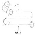
- FIG. 1is a plan view of an embodiment of the splint assembly and leader assembly according to the present invention
- FIG. 2is a cross-sectional view of a leader tube and a portion of the splint assembly showing the connection of a fixed pad assembly to the tension member;
- FIG. 2Ais a detailed view of section A—A of FIG. 2 showing the connection of the leader tube to the tension member;
- FIG. 3is a perspective view of the fixed pad assembly of FIG. 2 looking into the pin channels of the assembly;
- FIG. 4is a magnified view of an embodiment of the cable forming the tension member according to the present invention.
- FIG. 5is a lateral cross-sectional view of an embodiment of an adjustable pad assembly according to the present invention.
- FIG. 6is a vertical cross-sectional view of the adjustable pad assembly of FIG. 5 in a pre-deployment configuration according to the present invention
- FIG. 7is a vertical cross-sectional view of the adjustable pad assembly of FIG. 5 in a post-deployment configuration according to the present invention.
- FIG. 8is a perspective view of an embodiment of a deployment tool engaged with an adjustable pad assembly according to the present invention.
- FIG. 9is a cross-sectional view of an embodiment of the deployment tool of FIG. 8, showing the inner components thereof;
- FIG. 10is a perspective view of an embodiment of an probe/marking device according to the present invention.
- FIG. 10Ais a perspective view of the probe tip in FIG. 10 looking down onto the probe tip;
- FIG. 11is a perspective view of an embodiment of a tube used for housing a marker of the type shown in FIG. 12 prior to delivery according to the present invention
- FIG. 12is a perspective view of an embodiment of a marker according to the present invention.
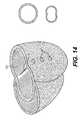
- FIG. 13is a cross-sectional view of the heart showing a preferred placement of one of the splint assemblies according to an embodiment of the present invention.
- FIG. 14is a cut-away perspective view of the heart showing a preferred placement of three splint assemblies for treatment of the heart according to the present invention and showing the cross-sectional shape of the left ventricle before and after placement of the splint assemblies with respect thereto.
- the various aspects of the invention to be discussed hereingenerally pertain to devices and methods for treating heart conditions, including, for example, dilatation and other similar heart failure conditions.
- the device of the present inventionpreferably operates passively in that, once placed in the heart, it does not require an active stimulus, either mechanical, electrical, or otherwise, to function. Implanting one or more of these devices alters the shape or geometry of the heart, both locally and globally, and thereby increases the heart's efficiency. That is, the heart experiences an increased pumping efficiency through an alteration in its shape or geometry and concomitant reduction in stress on the heart walls.
- the implanted device for treating the heartpreferably is a passive device, it is contemplated that the inventive tools and instruments used for implanting the device and the method of using these tools and instruments can be used to implant other treatment devices, such as active devices and the like.
- the inventive device and methodsoffer numerous advantages over the existing treatments for various heart conditions.
- the deviceis relatively easy to manufacture and use, and the related inventive surgical techniques and tools for implanting the device do not require the invasive procedures of current surgical techniques.
- the surgical techniquedoes not require removing portions of the heart tissue, nor does it necessarily require opening the heart chamber or stopping the heart during operation.
- the surgical techniques of the present inventionalso are less risky to the patient than other techniques.
- the disclosed inventive methods and related devicesinvolve geometric reshaping of the heart.
- substantially the entire chamber geometryis altered to return the heart to a more normal state of stress.
- Models of this geometric reshapingwhich includes a reduction in radius of curvature of the chamber walls, can be found in U.S. Pat. No. 5,961,440, issued Oct. 5, 1999 and entitled “Heart Wall Tension Reduction Apparatus and Method,” the complete disclosure of which is incorporated herein by reference.
- the heart wallsPrior to reshaping the chamber geometry, the heart walls experience high stress due to a combination of both the relatively large increased diameter of the chamber and the thinning of the chamber wall.
- Geometric reshapingreduces the stress in the walls of the heart chamber to increase the heart's pumping efficiency, as well as to stop further dilatation of the heart.
- left ventriclehas been selected for illustrative purposes because a large number of the disorders that the present invention treats occur in the left ventricle.
- FIG. 1shows a splint assembly 1 that ultimately is placed in the heart and a leader assembly 10 connected to splint assembly 1 prior to placement of splint assembly 1 and aiding in the delivery of splint assembly 1 into the heart.
- Splint assembly 1includes an elongate tension member 2 (shown by the thicker line), a fixed pad assembly 3 , and an adjustable pad assembly 4 .
- Fixed pad assembly 3is disposed at one end of tension member 2 (which will be referred to as the proximal end of assembly 1 ), while adjustable pad assembly 4 is disposed to slidably engage tension member 2 and secure to tension member 2 opposite to the end attached to fixed pad assembly 3 (which will be referred to as the distal end of assembly 1 ).
- splint assembly 1is placed transverse a heart chamber to induce a shape change of the heart chamber, for example, the left ventricle, to reduce stress on the heart wall and thereby improve cardiac function.
- a shape change of the heart chamberfor example, the left ventricle
- three splint assemblies 1are placed relative to the left ventricle LV in the manner illustrated to alter the shape of the ventricle from an essentially circular cross-section to an essentially bi-lobed cross-section.
- three splint assembliesare shown in FIG. 14, it is contemplated that any number of splint assemblies may be placed as desired, depending on the condition of the heart and the desired shape change results. Details on methods and tools used to determine the location for placement of the splint assembly 1 with respect to the heart chamber will be described later in this specification.
- Splint leader assembly 10includes a leader tube 5 (shown by the thinner line in FIG. 1 leading to tension member 2 ) and a stop band 7 .
- Leader tube 5facilitates the advancement of tension member 2 through the heart wall and across the heart chamber, as will be described. Once tension member 2 has been placed with respect to the heart and adjustable pad assembly 4 has been secured into place, leader assembly 10 and any excess tension member length can be severed and removed from tension member 2 , for example, by thermal cutting or the like.
- leader tube 5is made of a high strength, substantially rigid, polymeric tubing, such as polyetheretherketone (PEEK), polyamide, polyimide, acetal, urethane, polyester, or other suitable like material.
- Leader tube 5has an inner diameter of approximately 0.015 inches, an outer diameter of approximately 0.031 inches, and a length of approximately 24 inches.
- leader tube 5is hollow and heat-set into a coil shape.
- the coil shapeprovides leader tube 5 with a more compact configuration prior to implantation of splint assembly 1 .
- This compact configurationis especially important as the splint assembly rests in a sterile environment prior to the implantation procedure.
- the leader tube 5will be less cumbersome for a surgeon to handle due to its compactness.
- Stop band 7engages with a measuring/tightening device which will be described in more detail later during a discussion of the implantation procedure of splint assembly 1 .
- stop band 7is swaged about leader tube 5 and further secured, if necessary, through the use of an adhesive, such as, for example urethane or epoxy, or other suitable adhesive.
- an adhesivesuch as, for example urethane or epoxy, or other suitable adhesive.
- a “backfill” or fillet 7 ′ of adhesiveis placed at the distal end of stop band 7 . This fillet 7 ′ permits a smooth engagement of stop band 7 to the measuring and tightening device. The measuring and tightening device and the engagement of stop band 7 with the device will be described later.
- a portion of leader tube 5contains a mandrel 6 within the lumen of leader tube 5 .
- Mandrel 6is secured to leader tube 5 by, for example, a suitable adhesive, such as, epoxy, or other suitable means such as a friction fit within tube 5 .
- a suitable adhesivesuch as, epoxy, or other suitable means such as a friction fit within tube 5 .
- Mandrel 6provides stiffness and support to leader tube 5 .
- mandrel 6provides a base structure upon which stop band 7 can be swaged, thereby strengthening stop band 7 .
- mandrel 6is made of stainless steel or other suitable material offering stiffness and support.
- Mandrel 6extends within a proximal portion of leader tube 5 (closest to splint assembly 1 ) to a point within leader tube 5 slightly past stop band 7 before the coiled portion of leader tube 5 . Mandrel 6 also extends from leader tube 5 and into a proximal end of tension member 2 to connect leader tube 5 with tension member 2 .
- mandrel 6includes a larger diameter portion 6 ′ at its proximal end within tension member 2 .
- This larger diameter portionis formed by centerless grinding of all but the proximal end of the wire forming mandrel 6 .
- this wireis fabricated from a 0.020 inch diameter wire, with the ground portion having a diameter of 0.010 inch.
- Mandrel 6is fixed within a distal end of tension member 2 , which includes a covering 11 surrounding an inner cable 11 ′. Mandrel 6 and a surrounding metallic tube 9 are covered with an adhesive 9 ′ and inserted approximately 0.3 inches inside the distal end of tension member 2 .
- An external metallic tube 12is placed around a distal portion of covering 11 and cable 11 ′ and is swaged down and secured thereto.
- An adhesive 12 ′is disposed between external metallic tube 12 and covering 11 to more firmly secure tension member 2 and tube 12 .
- adhesive 12 ′′can be disposed at the distal end of metallic tube 12 to form a tapered connection between metallic tube 12 and leader tube 5 .
- Tension member 2and particularly cable 11 ′, serves as the primary load-bearing component of the splint assembly. Therefore, cable 11 ′ preferably has a braided-cable construction, for example, a multifilar braided polymeric construction.
- the filaments forming cable 11 ′should be high performance fibers.
- filaments of ultra high molecular weight polyethylenesuch as, for example, SpectraTM or DyneemaTM, or some other suitable like material, such as polyester (e.g. DacronTM) or liquid crystal polymers (e.g. VectranTM), for example, will be used to form the braided cable.
- Filamentspreferably are combined in yarn bundles of approximately 50 individual filaments, with each yarn bundle being approximately 180 denier.
- two bundlescan be paired together (referred to as 2-ply) and then braided with approximately 16 total bundle pairs to form cable 11 ′.
- the preferred braidincludes approximately 20 to 50 picks per inch, and more preferably approximately 30 picks per inch, wherein one pick measured along the length of cable 11 ′ is shown in FIG. 4 .
- making the braid as describedresults in an average diameter of cable 11 ′ of A approximately 0.030 to 0.080 inches, and preferably 0.055 inches, having approximately 1600 individual filaments.
- the braided cable 11 ′appears somewhat oval.
- FIG. 4shows a magnified view of a cable made according to the preferred embodiment described.
- the preferred embodiment of cable 11 ′provides cable 11 ′ with several significant properties.
- the ultra high molecular weight polyethyleneprovides cable 11 ′ with high strength characteristics.
- cable 11 ′is able to withstand the constant tension that will be placed upon it during use within the heart.
- this materialhas a high creep resistance, a high corrosion resistance, high fatigue resistance and is biostable. It is contemplated that other materials having similar properties also may be used to form cable 11 ′ and are within the scope of this invention.
- Implantation in the heartsubjects cable 11 ′, and therefore tension member 2 , to a dynamic, and often cyclic, bending and stressing environment.
- a multifilar structureresults in lower bending stresses than would otherwise occur in a solid structure.
- a braided multifilar structuredissipates concentrated loads to adjacent filaments within relatively short distances as compared with a twisted multifilar structure.
- the braided structurealso provides a simple, yet effective, way to anchor tension member 2 to pad assemblies 3 and 4 , as will be explained in greater detail shortly.
- a cable 11 ′ of the preferred diameter range of 0.030 to 0.080 inches, and most preferably 0.055 inchesresults in a high break strength and a high resistance to creep failure under expected stress conditions when placed in the heart. This resistance to creep strength allows cable 11 ′ to maintain its shape throughout implantation and use of the device. Furthermore, a cable of the preferred diameter range permits pins to penetrate the cable to hold it in place in the fixed pad assembly. If the diameter were too small, the pins may pull on portions of cable 11 ′, thus distorting the uniform shape of the cable. Additionally, it is important that cable 11 ′ not have too large of a diameter.
- the preferred combination of yarn density and material, together with the preferred pick count and cable diameterresults in an optimal tension member performance. That is, the tension member is capable of withstanding the cyclical stresses occurring within the heart chamber without breaking or weakening and a strong connection between the tension member and the pad assemblies can be achieved. Also, damage to internal vascular structure and the heart tissue, and obstruction of blood flow within the heart chamber can be avoided.
- the preferred parameters for the braid structurehave been described above, it is contemplated that other combinations of material, yarn density, number of bundles, and pick count may be used, as long as the desired characteristics with respect to strength of the braid and interaction of the braid with the heart and blood are achieved.
- Covering 11 surrounding cable 11 ′also provides tension member 2 with properties that facilitate implantation and use in the heart. Because tension member 2 will be in blood contact as it resides within a chamber of the heart, covering 11 preferably provides tension member 2 with resistance to thrombus generation. Furthermore, as a result of the relative motion that occurs between the heart and the portions of tension member 2 passing through the heart chamber wall, irritation of the heart wall may result. To alleviate such irritation, covering 11 preferably allows for tissue ingrowth to establish a relatively firm bond between the tension member and the heart wall, thus reducing relative motion between the two.
- covering 11preferably is made of a porous expanded polytetrafluoroethylene (ePTFE) sleeve having an inner diameter of approximately 0.040 inches and a wall thickness of approximately 0.005 inches prior to placement around cable 11 ′.
- the inner diameter of covering 11stretches to fit around cable 11 ′, which preferably has a diameter of about 0.055 inches, resulting in a frictional fit between covering 11 and cable 11 ′.
- covering 11made of ePTFE, has an internodal distance of between 20 and 70 microns, and most preferably approximately 45 microns.
- This preferred internodal spacingachieves both secure tissue ingrowth of the adjacent heart wall by allowing cellular infiltration and creating a tissue surface on the outsideof the tension member 2 .
- the preferred internodal spacingalso achieves a high resistance to thrombus.
- a coveringis biostable and tends not to degrade or corrode in the body.
- cable 11 ′primarily bears the loads placed on tension member 2
- covering 11also must be adapted to withstand the cyclic bending environment occurring in the heart.
- the porous nature of covering 11particularly having the internodal spacings discussed above, enables bending without creating high stress regions that may otherwise result in fatigue cracking of the covering if a solid structure were used.
- expanded PTFEhas been described as the preferred material with which to make covering 11 , other suitable materials exhibiting similar characteristics also are within the scope of the invention.
- splint assembly 1shown in FIG. 1 include fixed pad assembly 3 and adjustable pad assembly 4 .
- These pad assembliesessentially function as anchors that engage with the heart wall, providing a surface adjacent the exterior of the heart wall to which the tension member connects and which does not penetrate the heart wall.
- FIGS. 2 and 3show details of fixed pad assembly 3 and its connection to tension member 2 .
- fixed pad assembly 3includes a pad base 15 made of a rigid thermoplastic such as polyetheretherketone (PEEK), or other suitable like material, such as, for example, polysulfone, polymethylpentene, or polyacetal (Celon).
- PEEKpolyetheretherketone
- Pad base 15preferably has a generally disc-shaped configuration with a diameter of approximately 1 cm to 3 cm, preferably approximately 1.9 cm, and a thickness of approximately 0.3 cm to 1.5 cm, preferably 0.9 cm.
- a surface 16 adjacent the heart wallpreferably is slightly convex with a radius of curvature of approximately 0.25 in to 1.0 in, preferably approximately 0.5 in.
- the preferred ranges for the diameter of pad base 15 discussed aboveresults in optimal shape change and compressive forces on the heart chamber.
- the pad base diameteris too large, i.e., above the high end of the preferred range discussed above, an optimal bi-lobed shape change to the heart chamber does not result. That is, the heart wall at the locations of excessively large pads tend to flatten out such that the radius of curvature at those locations is essentially zero.
- Overly large pad base diametersalso make it difficult to place the pad assembly to avoid damaging vasculature of the heart.
- a channel 17extends through approximately the center of pad base 15 from an outer surface 19 to inner convex surface 16 .
- Channel 17has a diameter of approximately 0.062 inches through which tension member 2 passes.
- channel 17has a slightly rounded, or tapered, opening 17 ′ leading into channel 17 .
- the tapered opening 17 ′has a radius of curvature of approximately 0.062 inches at the inlet into the pad and a diameter of approximately 0.064 inches.
- the openingtapers to the channel 17 .
- This tapered openingwhich has a diameter larger than tension member 2 permits tension member 2 to gently curve around inner surface 16 as relative bending occurs, as opposed to having a sharp bend that would otherwise result if the diameter were not enlarged in this region.
- This tapered openingdecreases localized stresses in the region of tension member 2 near the opening to channel 17 that would occur during cyclical motion of the heart. Also, the diameter of channel 17 is slightly larger than the diameter of tension member 2 to permit room for the pins to penetrate the tension member to secure the tension member and pad together.
- Channels 18extend in direction parallel to surfaces 16 and 19 across pad base 15 .
- Channels 18house fixation members, such as sharpened pins 14 .
- Channels 18preferably have a smaller diameter than the pin diameters to create a press fit during connection of fixed pad assembly 3 to tension member 2 .
- channels 18preferably have a diameter of approximately 0.028 inches, as opposed to pin diameters of approximately 0.030 inches.
- a preferred embodiment of pad base 15includes a circumferential groove 20 adjacent to outer surface 19 , as shown in FIG. 2 .
- Circumferential groove 20accomodates windings of suture 21 to be secured to pad base 15 .
- a pad covering 13(shown in FIG. 2) can be placed over inner surface 16 and sides of pad base 15 and secured with respect thereto via suture windings 21 . Any excess pad covering extending past suture windings 21 can be trimmed off.
- Pad covering 13preferably is made of a velour woven polyester material, such as DacronTM, or other suitable like material, such as, for example, expanded polytetrafluoroethylene (ePTFE).
- the pad coveringfacilitates ingrowth of the heart wall tissue to secure pad base and thereby prevent long-term, motion-induced irritation of the outside of the heart wall.
- a hole disposed in approximately the center of the pad coveringenables the passage of tension member 2 .
- a similar pad coveringconnects in the same manner to a circumferential groove and sutures located on adjustable pad assembly 4 , as will be described later.
- fixation memberssuch as pins 14
- pins 14can be sharpened on their ends to more easily pierce through covering 11 and cable 11 ′.
- tension member 2folds over within pad base 15 in a U-shaped configuration. In this way, pins 14 each penetrate at an additional site along tension member 2 to provide a stronger connection between tension member 2 and fixed pad assembly 3 .
- the penetration of each pin 14 through two points of braided cable 11provides a reliable connection. This is due to the fact that the braided structure tends to transfer the contact load produced by pins 14 against the filament bundles and to all of the filaments forming braided cable 11 ′, essentially resulting in a load distribution between the pins and filaments.
- a reliable connectioncould be produced using only a single pin penetrating through the tension member at a single location.
- providing more than one pin and folding tension member 2 into the U-shaped configuration so that each pin intersects tension member 2 at more than one locationoffers additional strength to the connection.
- this configurationserves as a safety back-up should the connection at a single pin/cable interface become unsecure. Unless a failure at one of the pin sites occurs, however, it is expected that the intersection between the distal-most pin and the location where that pin first intersects tension member 2 , as tension member 2 enters pad base 15 , will carry substantially all of the load transferred by tension member 2 .
- This intersection siteis labeled as 14 a in FIG. 2 .
- pins 14it is preferable for pins 14 to penetrate tension member 2 at approximately the center of the cable to provide the most secure connection. Thus, approximately half of the filament bundles comprising the entire cable will be on one side of a pin and half on the other side. Such a placement of pins 14 assists in inhibiting distortion of the braided structure resulting from a non-equal distribution of the load on the various filaments. Additionally, to inhibit distortion of tension cable 11 ′, preferably a relatively dense braid is formed (i.e., in terms of both pick count and number of bundles) so pins 14 can penetrate and be secured without risk of pulling out or unraveling the braid.
- some length of cable 11 ′should extend on either side of pins 14 so that pins 14 are not easily pulled through the braid along the length of the braid.
- at least one of pins 14penetrates cable 11 ′ at a location that leaves a length of cable of approximately 1 to 2 centimeters.
- the folded over configuration of tension member 2 within fixed pad assembly 3serves to prevent both pins 14 from becoming disengaged with tension member 2 as a result of cable 11 ′ unraveling at its end.
- tension member 2can be thermally treated or otherwise fused together at its end.
- fixed pad assembly 3includes two pins 14 of approximately 0.025 to 0.035 inches in diameter and length slightly less than the pad base diameter, as shown in FIG. 2 .
- Pins 14can be formed of a relatively hard, corrosion-resistant material, such as, for example, a cobalt-nickel-chromium-molybdenum alloy, such as MP 35N, other cobalt-chrome alloys, stainless steel, or other suitable materials having similar characteristics. At least one end of each pin preferably is sharpened to facilitate penetration of tension cable 11 ′ and covering 11 .
- adjustable pad assembly 4and its connection to tension member 2 will now be described.
- the general outer configuration of adjustable pad assembly 4is similar to that of fixed pad assembly 3 . That is, adjustable pad assembly 4 includes a convex inner surface 47 that engages with an exterior of the heart wall when splint assembly 1 is implanted in the heart. Also, near an outer surface 48 of adjustable pad assembly 4 is a circumferential groove 80 with suture windings 81 . Although not shown in the Figures, it is contemplated that a pad covering of the type described with reference to fixed pad assembly 3 will be provided and secured via suture windings 81 to facilitate tissue ingrowth between adjustable pad assembly 4 and the heart wall.
- FIGS. 5-7depict the inner components of adjustable pad base 24 .
- pad base 24includes a plurality of channels.
- a channel 25 through which tension member 2 passesextends through pad base 24 in a manner similar to channel 17 passing through pad base 15 of fixed pad assembly 3 .
- FIGS. 5 and 6more clearly show the tapered opening 25 ′ of this channel 25 (similar to channel 17 ), which permits tension member 2 to gently curve at the surface of adjustable pad assembly 4 .
- channel 25 in adjustable pad assembly 4includes a further reduction in diameter, preferably approximately 0.059 inches, near an outer surface 48 of pad base 15 . This reduction in diameter helps to assure that the braid of cable 11 ′ is centered in channel 25 prior to fixation of tension member 2 to adjustable pad assembly 4 .
- cable 11 ′It is important for cable 11 ′ to be centered within channel 25 to ensure that a fixation or securement member, for example in the form of a staple 23 , used to secure tension member 2 to adjustable pad assembly 4 , penetrates cable 11 ′ in its center. In this manner, the yarn bundles of cable 11 ′ evenly divide on either side of staple 23 to evenly distribute the load to tension member 2 .
- Channel 17 in fixed pad base 3does not require such a reduced diameter region since the securement of tension member 2 to fixed pad assembly 3 occurs under controlled factory conditions prior to implantation of splint assembly 1 in the heart.
- a pair of staple leg channels 26extend through pad base 24 substantially perpendicular to channel 25 and parallel to inner surface 47 and outer surface 48 of pad base 24 .
- the pair of channels 26merges into a single staple leg channel disposed on one side of channel 25 .
- Channels 26receive legs 27 of a staple 23 and are sized to enable staple 23 to slide along the channels from a retracted position (shown in FIG. 6) to an advanced position (shown in FIG. 7) upon actuation of a deployment tool 22 , as will be described.
- a channel 28extends between and parallel to staple leg channels 26 .
- Channel 28is formed within one side of pad base 24 (on the side of a base 23 ′ of staple 23 opposite to the side from which legs 27 extend) and extends for approximately two-thirds of the distance measured from the side of pad base 24 to the center of pad base 24 .
- a deployment tool channel 31begins at the end of channel 28 and extends through the remaining diameter of pad base 24 . Deployment tool channel is radially offset from channel 28 and extends through pad base 24 so as to avoid intersection with channel 25 .
- a pre-deployment safety pin 32is located approximately in the center of the portion of channel 28 .
- Pre-deployment safety pin 32extends essentially perpendicularly to channel 28 and engages with base 23 ′ of staple 23 between staple legs 27 prior to actuation of staple 23 . This engagement tends to hold staple 23 in place to prevent premature advancement.
- a side channel 33also is formed in pad base 24 .
- Side channel 33extends perpendicularly to staple leg channels 26 .
- Side channel 33begins on either surface 47 (which engages the heart wall) or surface 48 (which faces away from the heart wall) and extends to approximately the end of channel 28 , near the beginning of deployment tool channel 31 .
- a post-deployment safety pin 34is disposed in side channel 33 .
- Post-deployment safety pin 34has a deflected configuration as it resides in side channel 33 , pressing on the side of one of staple legs 27 .
- tension member 2After tension member 2 has been extended transverse a heart chamber, for example, the left ventricle, such that the free end of tension member 2 extends through the heart wall at a location opposite to fixed pad assembly 3 , leader tube 5 and tension member 2 are fed through channel 25 of adjustable pad assembly 4 .
- a measuring/tightening devicewhich will be described in more detail shortly, is used to determine and adjust the proper tension member length between fixed pad assembly 3 and adjustable pad assembly 4 .
- This lengthis the length that is desired for the final implantation of splint assembly 1 in the heart. The determination of this length and the measuring and tightening procedure have been described in prior U.S. application Ser. No. 09/123,977, filed Jul.
- the final length of the tension member when placed in the heartcorresponds to a predetermined desired percentage reduction of the heart chamber diameter and the actual measured heart chamber diameter, which may differ from patient to patient.
- adjustable pad assembly 4is placed in the proper position on tension member 2 , with surface 47 engaging the outer surface of the heart wall.
- a deployment tool 22shown in FIGS. 8 and 9 is used to secure adjustable pad assembly 4 into place on tension member 2 .
- a deployment tool 22is pre-engaged with adjustable pad assembly 4 . That is, deployment tool 22 includes an engagement collar 35 disposed in channel 28 , as shown in FIG. 5, and an actuator wire 36 (not shown in FIG. 5) connected to engagement collar 35 at one end and to an actuator knob 37 at the other end.
- engagement collar 35rests against staple base 23 ′ and actuator wire extends from engagement collar 35 and through the entire diameter of pad base 24 exiting at a side 45 of pad base 24 .
- deployment tool channel 31ends in a countersink region 38 .
- actuator wire 36runs through the center of, but is not affixed to, an outer coil 39 .
- a collar 40surrounds both outer coil 39 and actuator wire 36 .
- Collar 40rests within countersink region 38 , essentially providing an abutment surface within pad base 24 that creates a counter-resistant force enabling actuator wire 36 to be pulled through pad base 24 .
- actuator wire 36 and outer coil 39connect to a threaded base portion 41 of deployment tool 22 .
- Outer coil 39terminates within threaded base portion 41 .
- Actuator wire 36continues through threaded base portion 41 and into an actuator 37 .
- Actuator 37also includes threads that engage with the threads of threaded base portion 41 at an interface 30 .
- Actuator wire 36fixedly attaches by a crimp fit 42 to a proximal end of actuator 37 .
- An actuator knob 43preferably is fixedly mounted to the distal end of actuator 37 to provide an ergonomic surface for a user to actuate deployment tool 22 .
- actuator knob 43is turned. This rotates actuator 37 with respect to threaded base portion 41 , essentially unscrewing actuator 37 from threaded base portion 41 . As actuator 37 turns, actuator wire 36 is pulled through outer coil 39 , exerting a force on engagement collar 35 . Continued rotation of actuator 37 further pulls on actuator wire 36 and engagement collar 35 . Engagement collar 35 thus moves staple 23 within adjustable pad assembly 4 to move staple legs 27 along channels 26 . Staple legs 27 eventually are pulled across channel 25 , piercing through tension member 2 in approximately the center thereof.
- actuator wire 36preferably rotates with respect to engagement collar 35 as actuator 37 turns without exerting any rotational forces upon engagement collar 35 .
- engagement collar 35simply pushes against staple 23 , but does not translate with respect to the staple surface.
- abrasive forces acting on staple 23are avoided, which otherwise may cause scratches and lead to corrosion of the staple.
- excessive torque on actuator wire 36 by engagement collar 35will be prevented.
- Abutment wall 29prevents staple 23 from moving in the other direction through pad base 24 .
- pre-deployment safety pin 32does not lie entirely flush within channel 28 but rather deflects back up so that it extends slightly outside of channel 28 .
- pre-deployment safety pin 32serves as a safety backup to maintain the advanced position of staple 23 if post-deployment safety pin 34 should for some reason fail to do so.
- both pre-deployment and post-deployment safety pinsare press-fit within pad base 24 .
- the pre-deployment safety pinpreferably has a diameter of approximately 0.025 inches and the post-deployment safety pin preferably has a diameter of approximately 0.016 inches.
- the pre-deployment pinpreferably is made of annealed MP35N and the post-deployment pin preferably is made of spring-tempered MP35N.
- the annealing of pre-deployment pin 32facilitates the formation of the permanent bend upon deflection resulting from advancing staple 23 .
- the spring-tempering of post-deployment pin 34enables the pin to recover to a relatively straight condition once the staple is fully deployed.
- pinscould be spring-activated with a bias in a particular direction such that the pins would move into their appropriate positions during deployment of staple 23 .
- tension member 2itself serves as the primary mechanism for preventing retraction of staple 23 . That is, the force exerted by tension member 2 onto staple 23 as a result of tightening tension member 2 in place with respect to the heart chamber tends to prevent staple 23 from moving. Due to this relatively large force exerted between tension member 2 and staple 23 , it is preferred to leave a length of tension member 2 of approximately 1 cm to 2 cm extending beyond the distal end of the distal most staple leg 27 to prevent staple 23 from pulling through the length of tension member 2 .
- tension member 2is even more desirable in the connection of tension member 2 to adjustable pad assembly 4 than it is for fixed pad assembly 3 since tension member 2 is not folded over in the former. Moreover, as will be explained, it is preferred to melt or otherwise fuse the end of tension member 2 to prevent unraveling of cable 11 ′.
- engagement collar 35drops into deployment tool channel 31 .
- actuator wire 36By continuing to pull on actuator wire 36 , engagement collar 35 can be pulled through deployment tool channel 31 and the entire deployment tool (including actuator wire 36 and engagement collar 35 ) can be removed from adjustable pad assembly 4 .
- leader assembly 10can be separated. from tension member 2 using a conventional cauterizing pen, or other suitable like instrument.
- the end of tension member 2 left protruding from adjustable pad assembly 4is also heated, or melted, and essentially fused together as a result of using the cauterizing pen to separate leader assembly 10 .
- the filaments of cable 11 and covering 11are joined to prevent potential unraveling of the tension member braid.
- Other suitable mechanisms for severing the leader from the tension member and for fusing the tension member endalso are within the scope of the invention. For example, although it is preferred to use the single step to both sever the leader assembly and fuse the tension member, it is also contemplated that the severing and fusing steps can be performed as separate steps.
- FIGS. 10-12Another aspect of the invention, described herein with reference to FIGS. 10-12, includes various location and identification tools for assisting in the optimal placement of splint assembly 1 with respect to a heart chamber to avoid damage to both internal cardiac structures, such as the papillary muscles, and external structures, such as blood vessels. Moreover, the tools assist in the placement of splint assemblies to effectively bisect the ventricle to result in optimal radius reduction and stress reduction.
- the splint assemblywill be placed across the left ventricle in a plane essentially longitudinally bisecting the ventricle.
- the splint assemblyshould extend from a location proximate to the anterior papillary muscle on the ventricle free wall to a location proximate to the posterior ventricular septum.
- the preferred location for the splint assembly near the anterior papillary muscleis just lateral to that muscle, while the preferred location near the septum is on the posterior free wall of the right ventricle.
- FIG. 13shows this preferred placement of a splint assembly.
- three individual splint assemblies 1are implanted.
- the upper-most (basal) splint assemblyis placed according to the description above.
- the remaining two splint assemblieswill be positioned in an equidistant relationship between the basal splint assembly and the apex of the left ventricle.
- the three splint assembliesessentially bisect the ventricle, producing optimal radius and stress reduction without excessive ventricular volume reduction.
- the positioning of the splint assemblies in this wayalso avoids interference with the mitral valve structure, including the chordae tendonae. Additionally, the positions described effectively avoid significant coronary arteries or veins.
- splint assemblies of the present inventioncan be employed to implant the splint assemblies of the present invention, including minimally invasive techniques through access ports, or endovascular techniques not requiring opening of the chest wall. These splint assemblies also could be implanted as an adjunct to other surgical procedures, such as, for example, CABG or mitral valve replacement.
- a preferred method of implanting the splint assembliesincludes through an open chest sternotomy without cardiopulmonary bypass. The description of the identification and location tools and methods for implanting the splint assemblies that follows thus relates to such an open sternotomy procedure.
- a preferred external imaging methodincludes the use of ultrasound probes. Ultrasound probes can either be used direcly in contact with the outside of the heart or can be positioned in the patient's esophagus (transesophageal).
- a probe/marking device 50shown in FIG. 10, operates to both locate positions on the heart wall for splint placement and simultaneously deliver a marker into the heart wall to mark each location.
- Marker 60shown in FIG. 12, is initially preloaded within a tube 51 , shown in FIG. 11 .
- Tube 51houses the entire structure of marker 60 with the exception of a penetrating tip 65 , which extends from a distal end of tube 51 . Marker 60 thus can easily be removed from tube 51 .
- Mounted on an exterior surface of tube 51is an advancer button 54 , the function of which will become apparent shortly.
- Probe/marking device 50includes a hollow handle portion 53 , a shaft 52 , and a probe tip 58 .
- Handle 53defines a slot 57 which opens at a proximal end and is configured to receive advancer button 54 , as will be described shortly.
- Shaft 52preferably is relatively rigid and may be curved as well, as shown in FIG. 10, to facilitate access to the exterior heart wall.
- Probe tip 58preferably has an essentially conical shape with three flat angular faces. As shown in FIG. 10A, the three angular faces meet at the distal most portion of tip 58 to define an opening 56 through which marker 60 is configured to pass upon delivery.
- Probe tip 58should be made of a material having a density that is sufficiently different than the myocardium in order to enhance echovisualization.
- a preferred material exhibiting this characteristicis a metal, such as stainless steel, for example, with a polyester covering such as DacronTM, or other suitable like material, that provides a gripping surface with respect to the heart tissue, to help stabilize the position of tip 58 with respect to the heart wall during indentation.
- the diameter of the proximal portion of tip 58 that connects to shaft 52is approximately 0.375 in. This configuration aids in creating a distinct, localized deflection upon contact and indentation of the heart wall.
- the flat angular facesenhance the visualization of probe tip 58 using ultrasound since the reflections off of the edges of the faces produce more discrete lines on the echo-image and the flat faces tend to reflect the ultrasound signals to a greater extent than a curved face.
- a physicianinserts the distal end of a tube 51 preloaded with marker 60 into the proximal end of handle 53 of probe/marking device 50 .
- advancer button 54slides into slot 57 .
- Tube 51is inserted into probe/marking device 50 until a base 55 of advancer button 54 , which connects advancer button 54 to tube 51 , engages with a detent mechanism (not shown) formed in handle 53 .
- marker tip 66is disposed at a location just proximal to the distal end of probe tip 58 .
- a portion of the proximal end of tube 51extends from the proximal end of handle 53 of probe/marking device 50 .
- Probe tip 58is then iteratively pressed against the outside of the heart wall to create localized indentations. Using ultrasound or other like imaging method, the physician can concurrently visualize these localized indentations, as well as tip 58 , to determine the position of the indentations relative to internal heart structures. In addition, the flat faces and preferred material of probe tip 58 tend to create a shadow that can be seen in the ultrasound picture, thus improving visualization.
- advancer button 54Upon finding a desired splint placement location, advancer button 54 is moved forward until it abuts the end of slot 57 defined by handle 53 . Moving advancer button 54 forward within slot 57 in turn moves tube 51 forward so as to move marker tip 66 through opening 56 of probe tip 58 . Marker tip 65 thus moves past probe tip 58 and into the heart wall.
- FIG. 12shows details of a preferred marker 60 to be used with inventive probe/marking device 50 .
- Marker 60includes a penetrating tip 65 and a suture line 70 .
- Penetrating tip 65includes a sharpened, conical-shaped end 66 to facilitate shallow penetration into the wall of the heart by spreading the heart wall tissue.
- the remaining portion 67 of penetrating tip 65 extending from conical-shaped end 66has a cylindrical configuration.
- a series of deflectable barbs 68protrude radially around the surface of cylindrical portion 67 .
- Barbs 68essentially act as gripping members to engage with the heart wall and securely hold marker 60 in place until removal is desired. Upon a sufficient force pulling marker 60 away from the heart wall, barbs 68 relatively easily disengage from heart wall to free marker 60 .
- Penetrating tip 65preferably is constructed from PEEK, or other suitable biocompatible material, such as, for example, polyimide, polyamide, acetal, urethane, or polyester, and has a length less than the thickness of the heart wall and preferably of approximately 0.156 inches, and a diameter preferably of approximately 0.030 inches.
- Barbs 68preferably extend from cylindrical portion 67 of penetrating tip 65 at angles ranging from approximately 10 degrees to approximately 45 degrees, and have respective lengths of 0.005 inches measured from the outer surface of cylindrical portion 67 .
- a suture line 70attaches to a proximal end of penetrating tip 65 .
- Suture line 70can either be formed by necking and drawing the back end of penetrating tip 65 or can be a standard suture material secured directly to penetrating tip 65 by, for example, a knot.
- a standard suture materialincludes 3-0 polyester, but other materials known in the art also may be utilized.
- Suture line 70has a length of approximately 2 to 14 inches, thus allowing the marker to be seen more readily, as well as enabling a surgeon to locate the marker through tactile sensation.
- the preloading of marker 60 into tube 51is preferably performed at a factory prior to implantation, with a number of marker-preloaded tubes 51 being supplied for the splint implantation procedure. Most preferably, 6 preloaded tubes 51 will be supplied for an implantation procedure to deliver six markers corresponding to the intersection points of each of three splint assemblies implanted into the heart, as shown in FIG. 14 . After each marker 60 is delivered, tube 51 is removed from probe/marking device 50 and disposed of as appropriate. A new preloaded tube 51 is thereafter inserted for delivery of the next marker 60 .
- the locating and marking procedurecan be repeated at each splint assembly positioning location on both sides of the heart chamber in the manner described above.
- the various splint assembliesare then delivered to each of the locations of the heart indicated by each of the delivered markers.
- the markersare removed once delivery of the splint assembly is complete.
- a splint assemblyproceeds in the following manner. Once markers have been delivered to heart wall on both sides of the chamber, an alignment clamp is positioned around the heart at those locations.
- the alignment clampincludes a guide tube, through which a needle is first delivered at the marker locations to penetrate the heart wall. The needle is extended transverse the heart such that each end of the needle penetrates locations on the heart wall corresponding to the ends of the splint assembly to be implanted.
- the needledefines a lumen extending along its length, through which leader tube 5 and tension member 2 are inserted via the guide tube in the alignment clamp. Once leader tube 5 extends through the second marker location, it can be pulled which in turn pulls tension member 2 across the heart wall. Leader tube 5 should be pulled until fixed pad assembly 3 engages the exterior surface of the heart wall. The needle can then be removed from the heart by pulling it off the free end of leader tube 5 . Similarly, markers 60 also can be removed from the heart wall, either before or after removing the needle.
- leader tube 5is fed into a measuring and tightening device, which also has been described previously in U.S. application Ser. No. 09/123,977.
- stop band 7 on leader tube 5engages with the measuring and tightening device, and leader tube 5 can be pulled a predetermined distance to tighten tension member 2 to a desired length between fixed pad assembly 3 and adjustable pad assembly 4 .
- Stop band 7is originally affixed to leader tube 5 at a known distance from fixed pad assembly 3 .
- Stop band 7serves as a marker to align with a measurement scale on the measuring and tightening device.
- adjustable pad assembly 4previously fed onto leader tube 5 and tension member 2 , is secured to tension member 2 adjacent the exterior of the heart wall in the manner described above with reference to the description of FIGS. 5-9
- the methods described above to place the splint assemblies with respect to the heartcan be repeated for other desired numbers of splint assemblies to achieve a particular configuration.
- the length of the tension members extending between the fixed and adjustable pad assembliesalso can be optimally determined based upon the size and condition of the patient's heart. It should also be noted that although the left ventricle has been referred to here for illustrative purposes, the apparatus and methods of this invention can be used to splint multiple chambers of a patient's heart, including the right ventricle or either atrium.
- FIGS. 13 and 14are illustrative only and may be shifted or rotated about a vertical axis generally disposed through the left ventricle and still avoid the major coronay vessels and papillary muscles.
- inventive device and methodscan be implanted to treat a heart having aneurysms or infarcted regions similar to those described in prior U.S. application Ser. No. 09/422,328 discussed earlier herein.
- the various components of the splint assembly to be implanted in the heartshould be made of biocompatible material that can remain in the human body indefinitely. Any surface engaging portions of the heart should be atraumatic in order to avoid tissue damage.
Landscapes
- Health & Medical Sciences (AREA)
- Life Sciences & Earth Sciences (AREA)
- Cardiology (AREA)
- Surgery (AREA)
- General Health & Medical Sciences (AREA)
- Public Health (AREA)
- Heart & Thoracic Surgery (AREA)
- Biomedical Technology (AREA)
- Veterinary Medicine (AREA)
- Animal Behavior & Ethology (AREA)
- Engineering & Computer Science (AREA)
- Nuclear Medicine, Radiotherapy & Molecular Imaging (AREA)
- Molecular Biology (AREA)
- Medical Informatics (AREA)
- Oral & Maxillofacial Surgery (AREA)
- Transplantation (AREA)
- Vascular Medicine (AREA)
- Rheumatology (AREA)
- Prostheses (AREA)
- Measuring Pulse, Heart Rate, Blood Pressure Or Blood Flow (AREA)
- Medicines Containing Plant Substances (AREA)
- External Artificial Organs (AREA)
- Machines For Laying And Maintaining Railways (AREA)
Abstract
Description
Claims (25)
Priority Applications (8)
| Application Number | Priority Date | Filing Date | Title |
|---|---|---|---|
| US09/532,049US6537198B1 (en) | 2000-03-21 | 2000-03-21 | Splint assembly for improving cardiac function in hearts, and method for implanting the splint assembly |
| EP01920566AEP1265534B1 (en) | 2000-03-21 | 2001-03-20 | A splint assembly for improving cardiac function in hearts |
| AT01920566TATE268143T1 (en) | 2000-03-21 | 2001-03-20 | SPLINT TO IMPROVE CARDIOLOGICAL FUNCTIONS OF THE HEART |
| DE60103618TDE60103618T2 (en) | 2000-03-21 | 2001-03-20 | RAIL TO IMPROVE CARDIOLOGICAL FUNCTIONS FOR THE HEART |
| AU2001247602AAU2001247602A1 (en) | 2000-03-21 | 2001-03-20 | A splint assembly for improving cardiac function in hearts, and method for implanting the splint assembly |
| PCT/US2001/008892WO2001070116A1 (en) | 2000-03-21 | 2001-03-20 | A splint assembly for improving cardiac function in hearts, and method for implanting the splint assembly |
| US10/278,847US7044905B2 (en) | 2000-03-21 | 2002-10-24 | Splint assembly for improving cardiac function in hearts, and method for implanting the splint assembly |
| US11/368,445US20060149123A1 (en) | 2000-03-21 | 2006-03-07 | Splint assembly for improving cardiac function in hearts, and method for implanting the splint assembly |
Applications Claiming Priority (1)
| Application Number | Priority Date | Filing Date | Title |
|---|---|---|---|
| US09/532,049US6537198B1 (en) | 2000-03-21 | 2000-03-21 | Splint assembly for improving cardiac function in hearts, and method for implanting the splint assembly |
Related Child Applications (1)
| Application Number | Title | Priority Date | Filing Date |
|---|---|---|---|
| US10/278,847ContinuationUS7044905B2 (en) | 2000-03-21 | 2002-10-24 | Splint assembly for improving cardiac function in hearts, and method for implanting the splint assembly |
Publications (1)
| Publication Number | Publication Date |
|---|---|
| US6537198B1true US6537198B1 (en) | 2003-03-25 |
Family
ID=24120173
Family Applications (3)
| Application Number | Title | Priority Date | Filing Date |
|---|---|---|---|
| US09/532,049Expired - LifetimeUS6537198B1 (en) | 2000-03-21 | 2000-03-21 | Splint assembly for improving cardiac function in hearts, and method for implanting the splint assembly |
| US10/278,847Expired - LifetimeUS7044905B2 (en) | 2000-03-21 | 2002-10-24 | Splint assembly for improving cardiac function in hearts, and method for implanting the splint assembly |
| US11/368,445AbandonedUS20060149123A1 (en) | 2000-03-21 | 2006-03-07 | Splint assembly for improving cardiac function in hearts, and method for implanting the splint assembly |
Family Applications After (2)
| Application Number | Title | Priority Date | Filing Date |
|---|---|---|---|
| US10/278,847Expired - LifetimeUS7044905B2 (en) | 2000-03-21 | 2002-10-24 | Splint assembly for improving cardiac function in hearts, and method for implanting the splint assembly |
| US11/368,445AbandonedUS20060149123A1 (en) | 2000-03-21 | 2006-03-07 | Splint assembly for improving cardiac function in hearts, and method for implanting the splint assembly |
Country Status (6)
| Country | Link |
|---|---|
| US (3) | US6537198B1 (en) |
| EP (1) | EP1265534B1 (en) |
| AT (1) | ATE268143T1 (en) |
| AU (1) | AU2001247602A1 (en) |
| DE (1) | DE60103618T2 (en) |
| WO (1) | WO2001070116A1 (en) |
Cited By (210)
| Publication number | Priority date | Publication date | Assignee | Title |
|---|---|---|---|---|
| US20030032979A1 (en)* | 1998-07-29 | 2003-02-13 | Myocor, Inc. | Transventricular implant tools and devices |
| US20030050685A1 (en)* | 1999-08-09 | 2003-03-13 | Nikolic Serjan D. | Method for improving cardiac function |
| US20030105384A1 (en)* | 1999-08-09 | 2003-06-05 | Sharkey Hugh R. | Method of improving cardiac function using a porous membrane |
| US6723038B1 (en) | 2000-10-06 | 2004-04-20 | Myocor, Inc. | Methods and devices for improving mitral valve function |
| US20040092973A1 (en)* | 2002-09-23 | 2004-05-13 | Nmt Medical, Inc. | Septal puncture device |
| US20040122474A1 (en)* | 2002-12-19 | 2004-06-24 | Scimed Life Systems, Inc. | Anchoring to soft tissue |
| US20040127982A1 (en)* | 2002-10-01 | 2004-07-01 | Ample Medical, Inc. | Devices, systems, and methods for reshaping a heart valve annulus |
| US20040133274A1 (en)* | 2002-11-15 | 2004-07-08 | Webler William E. | Cord locking mechanism for use in small systems |
| US20040138745A1 (en)* | 2001-10-01 | 2004-07-15 | Ample Medical, Inc. | Methods and devices for heart valve treatments |
| US6764510B2 (en) | 2002-01-09 | 2004-07-20 | Myocor, Inc. | Devices and methods for heart valve treatment |
| US6776754B1 (en)* | 2000-10-04 | 2004-08-17 | Wilk Patent Development Corporation | Method for closing off lower portion of heart ventricle |
| US20040181237A1 (en)* | 2003-03-12 | 2004-09-16 | Sean Forde | Medical device for manipulation of a medical implant |
| US20040249286A1 (en)* | 2003-03-31 | 2004-12-09 | Kabushiki Kaisha Toshiba | Ultrasonic endocavity probe having adjustable length insert |
| US20040260393A1 (en)* | 2000-09-20 | 2004-12-23 | Ample Medical, Inc. | Devices, systems, and methods for reshaping a heart valve annulus |
| US20040260317A1 (en)* | 2003-06-20 | 2004-12-23 | Elliot Bloom | Tensioning device, system, and method for treating mitral valve regurgitation |
| US20040267329A1 (en)* | 2001-09-07 | 2004-12-30 | Mardil, Inc. | Method and apparatus for external heart stabilization |
| US20050010287A1 (en)* | 2000-09-20 | 2005-01-13 | Ample Medical, Inc. | Devices, systems, and methods for supplementing, repairing, or replacing a native heart valve leaflet |
| US20050015109A1 (en)* | 2003-07-16 | 2005-01-20 | Samuel Lichtenstein | Methods and devices for altering blood flow through the left ventricle |
| US20050055089A1 (en)* | 2000-09-20 | 2005-03-10 | Ample Medical, Inc. | Devices, systems, and methods for reshaping a heart valve annulus |
| US20050059984A1 (en)* | 2003-09-11 | 2005-03-17 | Andrzej Chanduszko | Devices, systems, and methods for suturing tissue |
| US20050070844A1 (en)* | 2003-09-30 | 2005-03-31 | Mina Chow | Deflectable catheter assembly and method of making same |
| US20050080402A1 (en)* | 2001-04-27 | 2005-04-14 | Myomend, Inc. | Prevention of myocardial infarction induced ventricular expansion and remodeling |
| US20050101984A1 (en)* | 2003-11-06 | 2005-05-12 | Nmt Medical, Inc. | Transseptal puncture apparatus |
| US20050154252A1 (en)* | 2004-01-09 | 2005-07-14 | Cardiokinetix, Inc. | Ventricular partitioning device |
| US6939361B1 (en) | 1999-09-22 | 2005-09-06 | Nmt Medical, Inc. | Guidewire for a free standing intervascular device having an integral stop mechanism |
| US20050197716A1 (en)* | 2004-03-03 | 2005-09-08 | Cardiokinetix, Inc. | Inflatable ventricular partitioning device |
| US20050216079A1 (en)* | 2000-09-20 | 2005-09-29 | Ample Medical, Inc. | Heart valve annulus device and method of using same |
| US20050222489A1 (en)* | 2003-10-01 | 2005-10-06 | Ample Medical, Inc. | Devices, systems, and methods for reshaping a heart valve annulus, including the use of a bridge implant |
| US20050228422A1 (en)* | 2002-11-26 | 2005-10-13 | Ample Medical, Inc. | Devices, systems, and methods for reshaping a heart valve annulus, including the use of magnetic tools |
| US20060014998A1 (en)* | 2002-08-01 | 2006-01-19 | Sharkey Hugh R | Multiple partitioning devices for heart treatment |
| US20060030881A1 (en)* | 2004-08-05 | 2006-02-09 | Cardiokinetix, Inc. | Ventricular partitioning device |
| US20060030885A1 (en)* | 2002-10-15 | 2006-02-09 | Hyde Gregory M | Apparatuses and methods for heart valve repair |
| US20060089711A1 (en)* | 2004-10-27 | 2006-04-27 | Medtronic Vascular, Inc. | Multifilament anchor for reducing a compass of a lumen or structure in mammalian body |
| US20060106279A1 (en)* | 2004-05-14 | 2006-05-18 | Ample Medical, Inc. | Devices, systems, and methods for reshaping a heart valve annulus, including the use of a bridge implant having an adjustable bridge stop |
| US20060106278A1 (en)* | 2004-05-14 | 2006-05-18 | Ample Medical, Inc. | Devices, systems, and methods for reshaping a heart valve annulus, including the use of an adjustable bridge implant system |
| US20060106447A1 (en)* | 2004-01-26 | 2006-05-18 | Nmt Medical, Inc. | Adjustable stiffness medical system |
| US20060199995A1 (en)* | 2005-03-02 | 2006-09-07 | Venkataramana Vijay | Percutaneous cardiac ventricular geometry restoration device and treatment for heart failure |
| US7112219B2 (en) | 2002-11-12 | 2006-09-26 | Myocor, Inc. | Devices and methods for heart valve treatment |
| US20060229491A1 (en)* | 2002-08-01 | 2006-10-12 | Cardiokinetix, Inc. | Method for treating myocardial rupture |
| US20060264980A1 (en)* | 1999-08-09 | 2006-11-23 | Alexander Khairkhahan | System for improving cardiac function |
| US20060270896A1 (en)* | 2005-05-31 | 2006-11-30 | Ethicon, Inc. | Method and device for deployment of a sub-pericardial sack |
| US20060281965A1 (en)* | 2005-06-10 | 2006-12-14 | Alexander Khairkhahan | Peripheral seal for a ventricular partitioning device |
| US20070025009A1 (en)* | 2005-07-29 | 2007-02-01 | Fuji Photo Film Co., Ltd. | Magnetic recorder |
| US20070055303A1 (en)* | 2000-10-06 | 2007-03-08 | Myocor, Inc. | Endovascular splinting devices and methods |
| US20070066863A1 (en)* | 2005-08-31 | 2007-03-22 | Medtronic Vascular, Inc. | Device for treating mitral valve regurgitation |
| US20070161846A1 (en)* | 1999-08-09 | 2007-07-12 | Serjan Nikolic | Cardiac device and methods of use thereof |
| US7247134B2 (en) | 2002-11-12 | 2007-07-24 | Myocor, Inc. | Devices and methods for heart valve treatment |
| US20070198058A1 (en)* | 2006-02-21 | 2007-08-23 | Daniel Gelbart | Method and device for closing holes in tissue |
| US20070198082A1 (en)* | 2005-12-15 | 2007-08-23 | The Cleveland Clinic Foundation | Apparatus and method for treating a regurgitant valve |
| US20070203391A1 (en)* | 2006-02-24 | 2007-08-30 | Medtronic Vascular, Inc. | System for Treating Mitral Valve Regurgitation |
| US20070265658A1 (en)* | 2006-05-12 | 2007-11-15 | Aga Medical Corporation | Anchoring and tethering system |
| US20070270688A1 (en)* | 2006-05-19 | 2007-11-22 | Daniel Gelbart | Automatic atherectomy system |
| US20080004534A1 (en)* | 2006-06-28 | 2008-01-03 | Daniel Gelbart | Intra-cardiac mapping and ablation method |
| US20080004697A1 (en)* | 2006-06-28 | 2008-01-03 | Samuel Victor Lichtenstein | Method for anchoring a mitral valve |
| US7320665B2 (en) | 2005-03-02 | 2008-01-22 | Venkataramana Vijay | Cardiac Ventricular Geometry Restoration Device and Treatment for Heart Failure |
| US20080027268A1 (en)* | 2004-04-05 | 2008-01-31 | Genesee Biomedical, Inc. | Method and Apparaus for the Surgical Treatment of Congestive Heart Failure |
| US7331972B1 (en) | 2002-11-15 | 2008-02-19 | Abbott Cardiovascular Systems Inc. | Heart valve chord cutter |
| US20080045778A1 (en)* | 2006-08-02 | 2008-02-21 | Samuel Victor Lichtenstein | System for improving diastolic dysfunction |
| US7335213B1 (en) | 2002-11-15 | 2008-02-26 | Abbott Cardiovascular Systems Inc. | Apparatus and methods for heart valve repair |
| US7341584B1 (en) | 2003-05-30 | 2008-03-11 | Thomas David Starkey | Device and method to limit filling of the heart |
| US20080091057A1 (en)* | 2006-10-11 | 2008-04-17 | Cardiac Pacemakers, Inc. | Method and apparatus for passive left atrial support |
| US20080125861A1 (en)* | 2002-11-15 | 2008-05-29 | Webler William E | Valve aptation assist device |
| US20080228266A1 (en)* | 2007-03-13 | 2008-09-18 | Mitralign, Inc. | Plication assistance devices and methods |
| US7431726B2 (en) | 2003-12-23 | 2008-10-07 | Mitralign, Inc. | Tissue fastening systems and methods utilizing magnetic guidance |
| US20080293996A1 (en)* | 2006-02-06 | 2008-11-27 | Evans Michael A | Systems and methods for volume reduction |
| US20080300607A1 (en)* | 2005-12-28 | 2008-12-04 | C.R. Bard, Inc. | Apparatus and Method for Introducing Implants |
| US20080319254A1 (en)* | 2002-08-01 | 2008-12-25 | Cardiokinetix, Inc. | Therapeutic methods and devices following myocardial infarction |
| US20090054723A1 (en)* | 1999-08-09 | 2009-02-26 | Alexander Khairkhahan | Retrievable devices for improving cardiac function |
| US20090062596A1 (en)* | 2007-09-05 | 2009-03-05 | Leinsing Karl R | Heart band with fillable chambers to modify heart valve function |
| US20090076597A1 (en)* | 2007-09-19 | 2009-03-19 | Jonathan Micheal Dahlgren | System for mechanical adjustment of medical implants |
| US20090105729A1 (en)* | 2007-10-18 | 2009-04-23 | John Zentgraf | Minimally invasive repair of a valve leaflet in a beating heart |
| US20090131930A1 (en)* | 2007-11-16 | 2009-05-21 | Daniel Gelbart | Medical device for use in bodily lumens, for example an atrium |
| US20090192441A1 (en)* | 2008-01-25 | 2009-07-30 | Daniel Gelbart | Liposuction system |
| US20090228099A1 (en)* | 2000-09-20 | 2009-09-10 | Ample Medical, Inc. | Devices, systems, and methods for retaining a native heart valve leaflet |
| US20090234318A1 (en)* | 2007-10-19 | 2009-09-17 | Guided Delivery Systems, Inc. | Systems and methods for cardiac remodeling |
| US20090254195A1 (en)* | 1999-08-09 | 2009-10-08 | Alexander Khairkhahan | System for improving cardiac function by sealing a partitioning membrane within a ventricle |
| US20090281517A1 (en)* | 1999-08-18 | 2009-11-12 | Intrinsic Therapeutics, Inc. | System and method for repairing an intervertebral disc |
| US20090287040A1 (en)* | 2005-06-10 | 2009-11-19 | Alexander Khairkhahan | Peripheral seal for a ventricular partitioning device |
| US20090287304A1 (en)* | 2008-05-13 | 2009-11-19 | Kardium Inc. | Medical Device for Constricting Tissue or a Bodily Orifice, for example a mitral valve |
| US20090287179A1 (en)* | 2003-10-01 | 2009-11-19 | Ample Medical, Inc. | Devices, systems, and methods for reshaping a heart valve annulus, including the use of magnetic tools |
| US20100004664A1 (en)* | 2005-12-28 | 2010-01-07 | Intrinsic Therapeutics, Inc. | Anchoring system for disc repair |
| US20100049259A1 (en)* | 2007-09-07 | 2010-02-25 | Intrinsic Therapeutics, Inc. | Method for vertebral endplate reconstruction |
| US7695425B2 (en) | 1997-01-02 | 2010-04-13 | Edwards Lifesciences Llc | Heart wall tension reduction apparatus and method |
| US20100174297A1 (en)* | 2005-01-21 | 2010-07-08 | Giovanni Speziali | Thorascopic Heart Valve Repair Method and Apparatus |
| US20100185278A1 (en)* | 2009-01-21 | 2010-07-22 | Tendyne Medical | Apical Papillary Msucle Attachment for Left Ventricular Reduction |
| US20100210899A1 (en)* | 2009-01-21 | 2010-08-19 | Tendyne Medical, Inc. | Method for percutaneous lateral access to the left ventricle for treatment of mitral insufficiency by papillary muscle alignment |
| US20100274227A1 (en)* | 2009-02-13 | 2010-10-28 | Alexander Khairkhahan | Delivery catheter handle cover |
| US20110004296A1 (en)* | 2007-09-13 | 2011-01-06 | Georg Lutter | Heart Valve Stent |
| US20110015476A1 (en)* | 2009-03-04 | 2011-01-20 | Jeff Franco | Devices and Methods for Treating Cardiomyopathy |
| US7883539B2 (en) | 1997-01-02 | 2011-02-08 | Edwards Lifesciences Llc | Heart wall tension reduction apparatus and method |
| US20110087066A1 (en)* | 2000-08-09 | 2011-04-14 | Boutillette Michael P | Devices and methods for delivering an endocardial device |
| US20110125271A1 (en)* | 1999-08-18 | 2011-05-26 | Intrinsic Therapeutics, Inc. | Method of performing an anchor implantation procedure within a disc |
| US7981152B1 (en) | 2004-12-10 | 2011-07-19 | Advanced Cardiovascular Systems, Inc. | Vascular delivery system for accessing and delivering devices into coronary sinus and other vascular sites |
| US20110190879A1 (en)* | 2010-02-03 | 2011-08-04 | Edwards Lifesciences Corporation | Devices and Methods for Treating a Heart |
| US8025698B2 (en) | 1999-08-18 | 2011-09-27 | Intrinsic Therapeutics, Inc. | Method of rehabilitating an anulus fibrosus |
| US8187324B2 (en) | 2002-11-15 | 2012-05-29 | Advanced Cardiovascular Systems, Inc. | Telescoping apparatus for delivering and adjusting a medical device in a vessel |
| US8206280B2 (en) | 2007-11-13 | 2012-06-26 | C. R. Bard, Inc. | Adjustable tissue support member |
| US8226711B2 (en) | 1997-12-17 | 2012-07-24 | Edwards Lifesciences, Llc | Valve to myocardium tension members device and method |
| US8231678B2 (en) | 1999-08-18 | 2012-07-31 | Intrinsic Therapeutics, Inc. | Method of treating a herniated disc |
| US8292910B2 (en) | 2003-11-06 | 2012-10-23 | Pressure Products Medical Supplies, Inc. | Transseptal puncture apparatus |
| US8323341B2 (en) | 2007-09-07 | 2012-12-04 | Intrinsic Therapeutics, Inc. | Impaction grafting for vertebral fusion |
| US8460371B2 (en) | 2002-10-21 | 2013-06-11 | Mitralign, Inc. | Method and apparatus for performing catheter-based annuloplasty using local plications |
| US8480559B2 (en) | 2006-09-13 | 2013-07-09 | C. R. Bard, Inc. | Urethral support system |
| US8790242B2 (en) | 2009-10-26 | 2014-07-29 | Cardiokinetix, Inc. | Ventricular volume reduction |
| US8845512B2 (en) | 2005-11-14 | 2014-09-30 | C. R. Bard, Inc. | Sling anchor system |
| US8864822B2 (en) | 2003-12-23 | 2014-10-21 | Mitralign, Inc. | Devices and methods for introducing elements into tissue |
| USD717954S1 (en) | 2013-10-14 | 2014-11-18 | Mardil, Inc. | Heart treatment device |
| US8911461B2 (en) | 2007-03-13 | 2014-12-16 | Mitralign, Inc. | Suture cutter and method of cutting suture |
| US8940002B2 (en) | 2010-09-30 | 2015-01-27 | Kardium Inc. | Tissue anchor system |
| US8951285B2 (en) | 2005-07-05 | 2015-02-10 | Mitralign, Inc. | Tissue anchor, anchoring system and methods of using the same |
| US8979923B2 (en) | 2002-10-21 | 2015-03-17 | Mitralign, Inc. | Tissue fastening systems and methods utilizing magnetic guidance |
| US9011423B2 (en) | 2012-05-21 | 2015-04-21 | Kardium, Inc. | Systems and methods for selecting, activating, or selecting and activating transducers |
| US9011531B2 (en) | 2012-02-13 | 2015-04-21 | Mitraspan, Inc. | Method and apparatus for repairing a mitral valve |
| US9044221B2 (en) | 2010-12-29 | 2015-06-02 | Neochord, Inc. | Exchangeable system for minimally invasive beating heart repair of heart valve leaflets |
| US9050066B2 (en) | 2010-06-07 | 2015-06-09 | Kardium Inc. | Closing openings in anatomical tissue |
| US9072511B2 (en) | 2011-03-25 | 2015-07-07 | Kardium Inc. | Medical kit for constricting tissue or a bodily orifice, for example, a mitral valve |
| US9119633B2 (en) | 2006-06-28 | 2015-09-01 | Kardium Inc. | Apparatus and method for intra-cardiac mapping and ablation |
| US9149602B2 (en) | 2005-04-22 | 2015-10-06 | Advanced Cardiovascular Systems, Inc. | Dual needle delivery system |
| US9198592B2 (en) | 2012-05-21 | 2015-12-01 | Kardium Inc. | Systems and methods for activating transducers |
| US9204964B2 (en) | 2009-10-01 | 2015-12-08 | Kardium Inc. | Medical device, kit and method for constricting tissue or a bodily orifice, for example, a mitral valve |
| US9332992B2 (en) | 2004-08-05 | 2016-05-10 | Cardiokinetix, Inc. | Method for making a laminar ventricular partitioning device |
| US9332993B2 (en) | 2004-08-05 | 2016-05-10 | Cardiokinetix, Inc. | Devices and methods for delivering an endocardial device |
| US9358112B2 (en) | 2001-04-24 | 2016-06-07 | Mitralign, Inc. | Method and apparatus for catheter-based annuloplasty using local plications |
| US9370425B2 (en) | 2012-10-12 | 2016-06-21 | Mardil, Inc. | Cardiac treatment system and method |
| US9452016B2 (en) | 2011-01-21 | 2016-09-27 | Kardium Inc. | Catheter system |
| US9480559B2 (en) | 2011-08-11 | 2016-11-01 | Tendyne Holdings, Inc. | Prosthetic valves and related inventions |
| US9480525B2 (en) | 2011-01-21 | 2016-11-01 | Kardium, Inc. | High-density electrode-based medical device system |
| US9486306B2 (en) | 2013-04-02 | 2016-11-08 | Tendyne Holdings, Inc. | Inflatable annular sealing device for prosthetic mitral valve |
| US9492228B2 (en) | 2011-01-21 | 2016-11-15 | Kardium Inc. | Enhanced medical device for use in bodily cavities, for example an atrium |
| US9526611B2 (en) | 2013-10-29 | 2016-12-27 | Tendyne Holdings, Inc. | Apparatus and methods for delivery of transcatheter prosthetic valves |
| USD777926S1 (en) | 2012-01-20 | 2017-01-31 | Kardium Inc. | Intra-cardiac procedure device |
| USD777925S1 (en) | 2012-01-20 | 2017-01-31 | Kardium Inc. | Intra-cardiac procedure device |
| US9597181B2 (en) | 2013-06-25 | 2017-03-21 | Tendyne Holdings, Inc. | Thrombus management and structural compliance features for prosthetic heart valves |
| US9610159B2 (en) | 2013-05-30 | 2017-04-04 | Tendyne Holdings, Inc. | Structural members for prosthetic mitral valves |
| US9675454B2 (en) | 2012-07-30 | 2017-06-13 | Tendyne Holdings, Inc. | Delivery systems and methods for transcatheter prosthetic valves |
| US9693865B2 (en) | 2013-01-09 | 2017-07-04 | 4 Tech Inc. | Soft tissue depth-finding tool |
| US9694121B2 (en) | 1999-08-09 | 2017-07-04 | Cardiokinetix, Inc. | Systems and methods for improving cardiac function |
| US9737403B2 (en) | 2006-03-03 | 2017-08-22 | Mardil, Inc. | Self-adjusting attachment structure for a cardiac support device |
| US9801720B2 (en) | 2014-06-19 | 2017-10-31 | 4Tech Inc. | Cardiac tissue cinching |
| US9827092B2 (en) | 2011-12-16 | 2017-11-28 | Tendyne Holdings, Inc. | Tethers for prosthetic mitral valve |
| US9895221B2 (en) | 2012-07-28 | 2018-02-20 | Tendyne Holdings, Inc. | Multi-component designs for heart valve retrieval device, sealing structures and stent assembly |
| US9907547B2 (en) | 2014-12-02 | 2018-03-06 | 4Tech Inc. | Off-center tissue anchors |
| US9907681B2 (en) | 2013-03-14 | 2018-03-06 | 4Tech Inc. | Stent with tether interface |
| US9986993B2 (en) | 2014-02-11 | 2018-06-05 | Tendyne Holdings, Inc. | Adjustable tether and epicardial pad system for prosthetic heart valve |
| US10022226B2 (en) | 2005-06-09 | 2018-07-17 | Bioventrix, Inc. | Method and apparatus for closing off a portion of a heart ventricle |
| US10022114B2 (en) | 2013-10-30 | 2018-07-17 | 4Tech Inc. | Percutaneous tether locking |
| US10028783B2 (en) | 2006-06-28 | 2018-07-24 | Kardium Inc. | Apparatus and method for intra-cardiac mapping and ablation |
| US10052095B2 (en) | 2013-10-30 | 2018-08-21 | 4Tech Inc. | Multiple anchoring-point tension system |
| US10058323B2 (en) | 2010-01-22 | 2018-08-28 | 4 Tech Inc. | Tricuspid valve repair using tension |
| US10064696B2 (en) | 2000-08-09 | 2018-09-04 | Edwards Lifesciences Corporation | Devices and methods for delivering an endocardial device |
| US10076414B2 (en) | 2012-02-13 | 2018-09-18 | Mitraspan, Inc. | Method and apparatus for repairing a mitral valve |
| US10172621B2 (en) | 2007-09-21 | 2019-01-08 | Mvrx, Inc. | Devices, systems, and methods for reshaping a heart valve annulus, including the use of magnetic tools |
| US10201423B2 (en) | 2015-03-11 | 2019-02-12 | Mvrx, Inc. | Devices, systems, and methods for reshaping a heart valve annulus |
| US10201419B2 (en) | 2014-02-05 | 2019-02-12 | Tendyne Holdings, Inc. | Apparatus and methods for transfemoral delivery of prosthetic mitral valve |
| US10219902B2 (en) | 2005-03-25 | 2019-03-05 | Mvrx, Inc. | Devices, systems, and methods for reshaping a heart valve anulus, including the use of a bridge implant having an adjustable bridge stop |
| US10238491B2 (en) | 2010-01-22 | 2019-03-26 | 4Tech Inc. | Tricuspid valve repair using tension |
| US10278818B2 (en) | 2015-12-10 | 2019-05-07 | Mvrx, Inc. | Devices, systems, and methods for reshaping a heart valve annulus |
| US10307147B2 (en) | 1999-08-09 | 2019-06-04 | Edwards Lifesciences Corporation | System for improving cardiac function by sealing a partitioning membrane within a ventricle |
| US10327894B2 (en) | 2015-09-18 | 2019-06-25 | Tendyne Holdings, Inc. | Methods for delivery of prosthetic mitral valves |
| US10368936B2 (en) | 2014-11-17 | 2019-08-06 | Kardium Inc. | Systems and methods for selecting, activating, or selecting and activating transducers |
| US10405978B2 (en) | 2010-01-22 | 2019-09-10 | 4Tech Inc. | Tricuspid valve repair using tension |
| US10463494B2 (en) | 2013-04-02 | 2019-11-05 | Tendyne Holdings, Inc. | Prosthetic heart valve and systems and methods for delivering the same |
| US10463489B2 (en) | 2013-04-02 | 2019-11-05 | Tendyne Holdings, Inc. | Prosthetic heart valve and systems and methods for delivering the same |
| US10470877B2 (en) | 2016-05-03 | 2019-11-12 | Tendyne Holdings, Inc. | Apparatus and methods for anterior valve leaflet management |
| US10478293B2 (en) | 2013-04-04 | 2019-11-19 | Tendyne Holdings, Inc. | Retrieval and repositioning system for prosthetic heart valve |
| US10517728B2 (en) | 2014-03-10 | 2019-12-31 | Tendyne Holdings, Inc. | Devices and methods for positioning and monitoring tether load for prosthetic mitral valve |
| US10555718B2 (en) | 2013-10-17 | 2020-02-11 | Tendyne Holdings, Inc. | Apparatus and methods for alignment and deployment of intracardiac devices |
| US10588620B2 (en) | 2018-03-23 | 2020-03-17 | Neochord, Inc. | Device for suture attachment for minimally invasive heart valve repair |
| US10610356B2 (en) | 2015-02-05 | 2020-04-07 | Tendyne Holdings, Inc. | Expandable epicardial pads and devices and methods for delivery of same |
| US10610358B2 (en) | 2015-12-28 | 2020-04-07 | Tendyne Holdings, Inc. | Atrial pocket closures for prosthetic heart valves |
| US10610354B2 (en) | 2013-08-01 | 2020-04-07 | Tendyne Holdings, Inc. | Epicardial anchor devices and methods |
| US10667905B2 (en) | 2015-04-16 | 2020-06-02 | Tendyne Holdings, Inc. | Apparatus and methods for delivery, repositioning, and retrieval of transcatheter prosthetic valves |
| US10695178B2 (en) | 2011-06-01 | 2020-06-30 | Neochord, Inc. | Minimally invasive repair of heart valve leaflets |
| US10722184B2 (en) | 2014-11-17 | 2020-07-28 | Kardium Inc. | Systems and methods for selecting, activating, or selecting and activating transducers |
| US10751183B2 (en) | 2014-09-28 | 2020-08-25 | Edwards Lifesciences Corporation | Apparatuses for treating cardiac dysfunction |
| US10765517B2 (en) | 2015-10-01 | 2020-09-08 | Neochord, Inc. | Ringless web for repair of heart valves |
| US10786351B2 (en) | 2015-01-07 | 2020-09-29 | Tendyne Holdings, Inc. | Prosthetic mitral valves and apparatus and methods for delivery of same |
| US10799675B2 (en) | 2016-03-21 | 2020-10-13 | Edwards Lifesciences Corporation | Cam controlled multi-direction steerable handles |
| US10827977B2 (en) | 2012-05-21 | 2020-11-10 | Kardium Inc. | Systems and methods for activating transducers |
| US10835714B2 (en) | 2016-03-21 | 2020-11-17 | Edwards Lifesciences Corporation | Multi-direction steerable handles for steering catheters |
| US10898330B2 (en) | 2017-03-28 | 2021-01-26 | Edwards Lifesciences Corporation | Positioning, deploying, and retrieving implantable devices |
| US10918373B2 (en) | 2013-08-31 | 2021-02-16 | Edwards Lifesciences Corporation | Devices and methods for locating and implanting tissue anchors at mitral valve commissure |
| US10966709B2 (en) | 2018-09-07 | 2021-04-06 | Neochord, Inc. | Device for suture attachment for minimally invasive heart valve repair |
| US11039921B2 (en) | 2016-06-13 | 2021-06-22 | Tendyne Holdings, Inc. | Sequential delivery of two-part prosthetic mitral valve |
| US11065116B2 (en) | 2016-07-12 | 2021-07-20 | Tendyne Holdings, Inc. | Apparatus and methods for trans-septal retrieval of prosthetic heart valves |
| US11090157B2 (en) | 2016-06-30 | 2021-08-17 | Tendyne Holdings, Inc. | Prosthetic heart valves and apparatus and methods for delivery of same |
| US11096782B2 (en) | 2015-12-03 | 2021-08-24 | Tendyne Holdings, Inc. | Frame features for prosthetic mitral valves |
| US11110251B2 (en) | 2017-09-19 | 2021-09-07 | Edwards Lifesciences Corporation | Multi-direction steerable handles for steering catheters |
| US11154399B2 (en) | 2017-07-13 | 2021-10-26 | Tendyne Holdings, Inc. | Prosthetic heart valves and apparatus and methods for delivery of same |
| US11173030B2 (en) | 2018-05-09 | 2021-11-16 | Neochord, Inc. | Suture length adjustment for minimally invasive heart valve repair |
| US11179236B2 (en) | 2009-12-08 | 2021-11-23 | Colorado State University Research Foundation | Device and system for transcatheter mitral valve replacement |
| US11191639B2 (en) | 2017-08-28 | 2021-12-07 | Tendyne Holdings, Inc. | Prosthetic heart valves with tether coupling features |
| US11219746B2 (en) | 2016-03-21 | 2022-01-11 | Edwards Lifesciences Corporation | Multi-direction steerable handles for steering catheters |
| US11224510B2 (en) | 2013-04-02 | 2022-01-18 | Tendyne Holdings, Inc. | Prosthetic heart valve and systems and methods for delivering the same |
| US11253360B2 (en) | 2018-05-09 | 2022-02-22 | Neochord, Inc. | Low profile tissue anchor for minimally invasive heart valve repair |
| US11259929B2 (en)* | 2005-08-19 | 2022-03-01 | Bioventrix, Inc. | Method and device for treating dysfunctional cardiac tissue |
| US11259867B2 (en) | 2011-01-21 | 2022-03-01 | Kardium Inc. | High-density electrode-based medical device system |
| US11376126B2 (en) | 2019-04-16 | 2022-07-05 | Neochord, Inc. | Transverse helical cardiac anchor for minimally invasive heart valve repair |
| US11389232B2 (en) | 2006-06-28 | 2022-07-19 | Kardium Inc. | Apparatus and method for intra-cardiac mapping and ablation |
| US11589989B2 (en) | 2017-03-31 | 2023-02-28 | Neochord, Inc. | Minimally invasive heart valve repair in a beating heart |
| US11648110B2 (en) | 2019-12-05 | 2023-05-16 | Tendyne Holdings, Inc. | Braided anchor for mitral valve |
| US11648114B2 (en) | 2019-12-20 | 2023-05-16 | Tendyne Holdings, Inc. | Distally loaded sheath and loading funnel |
| US11660190B2 (en) | 2007-03-13 | 2023-05-30 | Edwards Lifesciences Corporation | Tissue anchors, systems and methods, and devices |
| US11678980B2 (en) | 2020-08-19 | 2023-06-20 | Tendyne Holdings, Inc. | Fully-transseptal apical pad with pulley for tensioning |
| US11766331B2 (en) | 2020-05-27 | 2023-09-26 | Politecnico Di Milano | Device and assembly to repair a heart valve |
| US11844913B2 (en) | 2012-03-23 | 2023-12-19 | Boston Scientific Medical Device Limited | Transseptal puncture apparatus and method for using the same |
| US11951002B2 (en) | 2020-03-30 | 2024-04-09 | Tendyne Holdings, Inc. | Apparatus and methods for valve and tether fixation |
| US12171662B1 (en) | 2023-10-26 | 2024-12-24 | Approxima Srl | Heart chamber reshaping and valve repair system |
| US12208007B2 (en) | 2020-01-16 | 2025-01-28 | Neochord, Inc. | Helical cardiac anchors for minimally invasive heart valve repair |
| US12350149B2 (en) | 2009-05-04 | 2025-07-08 | Edwards Lifesciences Innovation (Israel) Ltd. | Method and apparatus for repairing a heart valve |
Families Citing this family (71)
| Publication number | Priority date | Publication date | Assignee | Title |
|---|---|---|---|---|
| US20060161159A1 (en)* | 1999-02-02 | 2006-07-20 | Dreyfuss Peter J | PEEK ribbed suture anchor |
| US8172856B2 (en) | 2002-08-02 | 2012-05-08 | Cedars-Sinai Medical Center | Methods and apparatus for atrioventricular valve repair |
| EP1933756B1 (en) | 2005-08-19 | 2016-07-20 | CHF Technologies Inc. | Steerable lesion excluding heart implants for congestive heart failure |
| US7695510B2 (en)* | 2005-10-11 | 2010-04-13 | Medtronic Vascular, Inc. | Annuloplasty device having shape-adjusting tension filaments |
| US9375313B2 (en)* | 2006-09-08 | 2016-06-28 | The Regents Of The University Of California | Intramyocardial patterning for global cardiac resizing and reshaping |
| US20090012413A1 (en)* | 2006-09-08 | 2009-01-08 | Sabbah Hani N | Cardiac patterning for improving diastolic function |
| WO2008121880A2 (en)* | 2007-03-30 | 2008-10-09 | Micardia Corporation | Adjustable annuloplasty ring and activation system |
| CA2682160C (en)* | 2007-04-11 | 2017-04-04 | Henry Ford Health System | Cardiac repair, resizing and reshaping using the venous system of the heart |
| US20090259210A1 (en)* | 2008-04-10 | 2009-10-15 | Sabbah Hani N | Method, apparatus and kits for forming structural members within the cardiac venous system |
| US8801665B2 (en)* | 2008-04-10 | 2014-08-12 | Henry Ford Health System | Apparatus and method for controlled depth of injection into myocardial tissue |
| US11272979B2 (en) | 2008-04-29 | 2022-03-15 | Virginia Tech Intellectual Properties, Inc. | System and method for estimating tissue heating of a target ablation zone for electrical-energy based therapies |
| US9598691B2 (en) | 2008-04-29 | 2017-03-21 | Virginia Tech Intellectual Properties, Inc. | Irreversible electroporation to create tissue scaffolds |
| US10238447B2 (en) | 2008-04-29 | 2019-03-26 | Virginia Tech Intellectual Properties, Inc. | System and method for ablating a tissue site by electroporation with real-time monitoring of treatment progress |
| US10702326B2 (en) | 2011-07-15 | 2020-07-07 | Virginia Tech Intellectual Properties, Inc. | Device and method for electroporation based treatment of stenosis of a tubular body part |
| US10272178B2 (en) | 2008-04-29 | 2019-04-30 | Virginia Tech Intellectual Properties Inc. | Methods for blood-brain barrier disruption using electrical energy |
| US10117707B2 (en) | 2008-04-29 | 2018-11-06 | Virginia Tech Intellectual Properties, Inc. | System and method for estimating tissue heating of a target ablation zone for electrical-energy based therapies |
| US8992517B2 (en) | 2008-04-29 | 2015-03-31 | Virginia Tech Intellectual Properties Inc. | Irreversible electroporation to treat aberrant cell masses |
| US10245098B2 (en) | 2008-04-29 | 2019-04-02 | Virginia Tech Intellectual Properties, Inc. | Acute blood-brain barrier disruption using electrical energy based therapy |
| US11254926B2 (en) | 2008-04-29 | 2022-02-22 | Virginia Tech Intellectual Properties, Inc. | Devices and methods for high frequency electroporation |
| US9198733B2 (en) | 2008-04-29 | 2015-12-01 | Virginia Tech Intellectual Properties, Inc. | Treatment planning for electroporation-based therapies |
| US9867652B2 (en) | 2008-04-29 | 2018-01-16 | Virginia Tech Intellectual Properties, Inc. | Irreversible electroporation using tissue vasculature to treat aberrant cell masses or create tissue scaffolds |
| US9283051B2 (en) | 2008-04-29 | 2016-03-15 | Virginia Tech Intellectual Properties, Inc. | System and method for estimating a treatment volume for administering electrical-energy based therapies |
| US8394138B2 (en)* | 2008-09-05 | 2013-03-12 | Cook Medical Technologies Llc | Multi-strand helical stent |
| US8641753B2 (en) | 2009-01-31 | 2014-02-04 | Cook Medical Technologies Llc | Preform for and an endoluminal prosthesis |
| US8632534B2 (en) | 2009-04-03 | 2014-01-21 | Angiodynamics, Inc. | Irreversible electroporation (IRE) for congestive obstructive pulmonary disease (COPD) |
| US11382681B2 (en) | 2009-04-09 | 2022-07-12 | Virginia Tech Intellectual Properties, Inc. | Device and methods for delivery of high frequency electrical pulses for non-thermal ablation |
| US11638603B2 (en) | 2009-04-09 | 2023-05-02 | Virginia Tech Intellectual Properties, Inc. | Selective modulation of intracellular effects of cells using pulsed electric fields |
| WO2010138919A2 (en) | 2009-05-28 | 2010-12-02 | Angiodynamics, Inc. | System and method for synchronizing energy delivery to the cardiac rhythm |
| US9895189B2 (en) | 2009-06-19 | 2018-02-20 | Angiodynamics, Inc. | Methods of sterilization and treating infection using irreversible electroporation |
| WO2011081814A1 (en) | 2009-12-28 | 2011-07-07 | Cook Medical Technologies Llc | Endoluminal device with kink-resistant regions |
| CN102762455B (en) | 2010-02-19 | 2016-05-18 | 空中客车德国运营有限责任公司 | Lavatory fixtures for vehicles |
| US8425455B2 (en) | 2010-03-30 | 2013-04-23 | Angiodynamics, Inc. | Bronchial catheter and method of use |
| US8579964B2 (en) | 2010-05-05 | 2013-11-12 | Neovasc Inc. | Transcatheter mitral valve prosthesis |
| EP2627274B1 (en) | 2010-10-13 | 2022-12-14 | AngioDynamics, Inc. | System for electrically ablating tissue of a patient |
| WO2012088149A2 (en) | 2010-12-20 | 2012-06-28 | Virginia Tech Intellectual Properties, Inc. | High-frequency electroporation for cancer therapy |
| US9554897B2 (en) | 2011-04-28 | 2017-01-31 | Neovasc Tiara Inc. | Methods and apparatus for engaging a valve prosthesis with tissue |
| US9308087B2 (en) | 2011-04-28 | 2016-04-12 | Neovasc Tiara Inc. | Sequentially deployed transcatheter mitral valve prosthesis |
| AU2012203620B9 (en) | 2011-06-24 | 2014-10-02 | Cook Medical Technologies Llc | Helical Stent |
| US9078665B2 (en) | 2011-09-28 | 2015-07-14 | Angiodynamics, Inc. | Multiple treatment zone ablation probe |
| US9414881B2 (en) | 2012-02-08 | 2016-08-16 | Angiodynamics, Inc. | System and method for increasing a target zone for electrical ablation |
| US9345573B2 (en) | 2012-05-30 | 2016-05-24 | Neovasc Tiara Inc. | Methods and apparatus for loading a prosthesis onto a delivery system |
| US20140046347A1 (en)* | 2012-08-10 | 2014-02-13 | W. L. Gore & Associates, Inc. | Devices, systems and methods for engaging tissue |
| US9888956B2 (en) | 2013-01-22 | 2018-02-13 | Angiodynamics, Inc. | Integrated pump and generator device and method of use |
| US10080657B2 (en) | 2013-03-07 | 2018-09-25 | Cedars-Sinai Medical Center | Catheter based apical approach heart prostheses delivery system |
| WO2014138482A1 (en) | 2013-03-07 | 2014-09-12 | Cedars-Sinai Medical Center | Method and apparatus for percutaneous delivery and deployment of a cardiovascular prosthesis |
| US9572665B2 (en) | 2013-04-04 | 2017-02-21 | Neovasc Tiara Inc. | Methods and apparatus for delivering a prosthetic valve to a beating heart |
| WO2015175570A1 (en) | 2014-05-12 | 2015-11-19 | Virginia Tech Intellectual Properties, Inc. | Selective modulation of intracellular effects of cells using pulsed electric fields |
| US12114911B2 (en) | 2014-08-28 | 2024-10-15 | Angiodynamics, Inc. | System and method for ablating a tissue site by electroporation with real-time pulse monitoring |
| WO2016040526A1 (en) | 2014-09-10 | 2016-03-17 | Cedars-Sinai Medical Center | Method and apparatus for percutaneous delivery and deployment of a cardiac valve prosthesis |
| US10758265B2 (en) | 2014-11-14 | 2020-09-01 | Cedars-Sinai Medical Center | Cardiovascular access and device delivery system |
| US10694972B2 (en) | 2014-12-15 | 2020-06-30 | Virginia Tech Intellectual Properties, Inc. | Devices, systems, and methods for real-time monitoring of electrophysical effects during tissue treatment |
| EP3324855B1 (en)* | 2015-07-23 | 2024-03-20 | Cedars-Sinai Medical Center | Device for securing heart valve leaflets |
| CA3007660A1 (en) | 2015-12-15 | 2017-06-22 | Neovasc Tiara Inc. | Transseptal delivery system |
| US10433952B2 (en) | 2016-01-29 | 2019-10-08 | Neovasc Tiara Inc. | Prosthetic valve for avoiding obstruction of outflow |
| US10905492B2 (en) | 2016-11-17 | 2021-02-02 | Angiodynamics, Inc. | Techniques for irreversible electroporation using a single-pole tine-style internal device communicating with an external surface electrode |
| CA3042588A1 (en) | 2016-11-21 | 2018-05-24 | Neovasc Tiara Inc. | Methods and systems for rapid retraction of a transcatheter heart valve delivery system |
| WO2018140535A1 (en) | 2017-01-25 | 2018-08-02 | Cedars-Sinai Medical Center | Device for securing heart valve leaflets |
| CA3073834A1 (en) | 2017-08-25 | 2019-02-28 | Neovasc Tiara Inc. | Sequentially deployed transcatheter mitral valve prosthesis |
| US11607537B2 (en) | 2017-12-05 | 2023-03-21 | Virginia Tech Intellectual Properties, Inc. | Method for treating neurological disorders, including tumors, with electroporation |
| US11291544B2 (en) | 2018-02-02 | 2022-04-05 | Cedars-Sinai Medical Center | Delivery platforms, devices, and methods for tricuspid valve repair |
| US12390262B2 (en) | 2018-03-13 | 2025-08-19 | Virginia Tech Intellectual Properties, Inc. | Treatment planning system for immunotherapy enhancement via non-thermal ablation |
| US11925405B2 (en) | 2018-03-13 | 2024-03-12 | Virginia Tech Intellectual Properties, Inc. | Treatment planning system for immunotherapy enhancement via non-thermal ablation |
| US11311329B2 (en) | 2018-03-13 | 2022-04-26 | Virginia Tech Intellectual Properties, Inc. | Treatment planning for immunotherapy based treatments using non-thermal ablation techniques |
| CN113271890B (en) | 2018-11-08 | 2024-08-30 | 内奥瓦斯克迪亚拉公司 | Ventricular deployment of transcatheter mitral valve prosthesis |
| CA3132873A1 (en) | 2019-03-08 | 2020-09-17 | Neovasc Tiara Inc. | Retrievable prosthesis delivery system |
| CA3135753C (en) | 2019-04-01 | 2023-10-24 | Neovasc Tiara Inc. | Controllably deployable prosthetic valve |
| US11491006B2 (en) | 2019-04-10 | 2022-11-08 | Neovasc Tiara Inc. | Prosthetic valve with natural blood flow |
| US11779742B2 (en) | 2019-05-20 | 2023-10-10 | Neovasc Tiara Inc. | Introducer with hemostasis mechanism |
| JP7520897B2 (en) | 2019-06-20 | 2024-07-23 | ニオバスク ティアラ インコーポレイテッド | Thin prosthetic mitral valve |
| US11950835B2 (en) | 2019-06-28 | 2024-04-09 | Virginia Tech Intellectual Properties, Inc. | Cycled pulsing to mitigate thermal damage for multi-electrode irreversible electroporation therapy |
| US12214189B2 (en) | 2019-07-24 | 2025-02-04 | Virginia Tech Intellectual Properties, Inc. | Fourier analysis spectroscopy for monitoring tissue impedance changes and treatment outcome during electroporation-based-therapies |
Citations (101)
| Publication number | Priority date | Publication date | Assignee | Title |
|---|---|---|---|---|
| US4192293A (en) | 1978-09-05 | 1980-03-11 | Manfred Asrican | Cardiac assist device |
| US4261342A (en) | 1978-10-26 | 1981-04-14 | Iker Aranguren Duo | Process for installing mitral valves in their anatomical space by attaching cords to an artificial stent |
| US4372293A (en) | 1980-12-24 | 1983-02-08 | Vijil Rosales Cesar A | Apparatus and method for surgical correction of ptotic breasts |
| US4409974A (en) | 1981-06-29 | 1983-10-18 | Freedland Jeffrey A | Bone-fixating surgical implant device |
| US4536893A (en) | 1982-03-03 | 1985-08-27 | Roberto Parravicini | Implant device for substaining the activity of the myocardium |
| US4690134A (en) | 1985-07-01 | 1987-09-01 | Snyders Robert V | Ventricular assist device |
| US4705040A (en) | 1985-11-18 | 1987-11-10 | Medi-Tech, Incorporated | Percutaneous fixation of hollow organs |
| DE3614292C1 (en) | 1986-04-26 | 1987-11-19 | Alexander Prof Dr Bernhard | Holder for unframed biological mitral valve implant |
| US4936857A (en) | 1987-02-23 | 1990-06-26 | Kulik Yaroslav P | Prosthetic pericardium |
| US4944753A (en) | 1988-09-26 | 1990-07-31 | Burgess Frank M | Method for producing retro-sternal space |
| US4960424A (en) | 1988-06-30 | 1990-10-02 | Grooters Ronald K | Method of replacing a defective atrio-ventricular valve with a total atrio-ventricular valve bioprosthesis |
| US4997431A (en) | 1989-08-30 | 1991-03-05 | Angeion Corporation | Catheter |
| US5104407A (en) | 1989-02-13 | 1992-04-14 | Baxter International Inc. | Selectively flexible annuloplasty ring |
| US5131905A (en) | 1990-07-16 | 1992-07-21 | Grooters Ronald K | External cardiac assist device |
| USRE34021E (en) | 1985-11-18 | 1992-08-04 | Abbott Laboratories | Percutaneous fixation of hollow organs |
| US5169381A (en) | 1991-03-29 | 1992-12-08 | Snyders Robert V | Ventricular assist device |
| US5192314A (en) | 1991-12-12 | 1993-03-09 | Daskalakis Michael K | Synthetic intraventricular implants and method of inserting |
| US5250049A (en) | 1992-01-10 | 1993-10-05 | Michael Roger H | Bone and tissue connectors |
| US5284488A (en) | 1992-12-23 | 1994-02-08 | Sideris Eleftherios B | Adjustable devices for the occlusion of cardiac defects |
| US5385528A (en) | 1993-06-17 | 1995-01-31 | Wilk; Peter J. | Intrapericardial assist device and associated method |
| US5433727A (en) | 1994-08-16 | 1995-07-18 | Sideris; Eleftherios B. | Centering buttoned device for the occlusion of large defects for occluding |
| US5450860A (en) | 1993-08-31 | 1995-09-19 | W. L. Gore & Associates, Inc. | Device for tissue repair and method for employing same |
| US5452733A (en) | 1993-02-22 | 1995-09-26 | Stanford Surgical Technologies, Inc. | Methods for performing thoracoscopic coronary artery bypass |
| US5458574A (en) | 1994-03-16 | 1995-10-17 | Heartport, Inc. | System for performing a cardiac procedure |
| DE4234127C2 (en) | 1992-10-09 | 1996-02-22 | Herbert Dr Vetter | Heart valve prosthesis |
| US5496305A (en) | 1985-03-22 | 1996-03-05 | Massachusetts Institue Of Technology | Catheter for laser angiosurgery |
| US5509428A (en) | 1994-05-31 | 1996-04-23 | Dunlop; Richard W. | Method and apparatus for the creation of tricuspid regurgitation |
| US5533958A (en) | 1993-06-17 | 1996-07-09 | Wilk; Peter J. | Intrapericardial assist device and associated method |
| EP0583012B1 (en) | 1987-02-27 | 1996-07-10 | VASCOR, Inc. | Ventricular assist device |
| US5571215A (en) | 1993-02-22 | 1996-11-05 | Heartport, Inc. | Devices and methods for intracardiac procedures |
| US5584803A (en) | 1991-07-16 | 1996-12-17 | Heartport, Inc. | System for cardiac procedures |
| US5593424A (en) | 1994-08-10 | 1997-01-14 | Segmed, Inc. | Apparatus and method for reducing and stabilizing the circumference of a vascular structure |
| DE29619294U1 (en) | 1996-11-07 | 1997-07-17 | Caić, Pero, 63450 Hanau | Heart cuff |
| US5665092A (en) | 1993-03-01 | 1997-09-09 | Mangiardi; John R. | Marker for surgical procedures |
| US5682906A (en) | 1993-02-22 | 1997-11-04 | Heartport, Inc. | Methods of performing intracardiac procedures on an arrested heart |
| US5702343A (en) | 1996-10-02 | 1997-12-30 | Acorn Medical, Inc. | Cardiac reinforcement device |
| US5718725A (en) | 1992-12-03 | 1998-02-17 | Heartport, Inc. | Devices and methods for intracardiac procedures |
| US5755783A (en) | 1996-07-29 | 1998-05-26 | Stobie; Robert | Suture rings for rotatable artificial heart valves |
| US5758663A (en) | 1992-04-10 | 1998-06-02 | Wilk; Peter J. | Coronary artery by-pass method |
| US5800528A (en) | 1995-06-13 | 1998-09-01 | Abiomed R & D, Inc. | Passive girdle for heart ventricle for therapeutic aid to patients having ventricular dilatation |
| US5800334A (en) | 1993-06-17 | 1998-09-01 | Wilk; Peter J. | Intrapericardial assist device and associated method |
| US5800531A (en) | 1996-09-30 | 1998-09-01 | Baxter International Inc. | Bioprosthetic heart valve implantation device |
| US5814097A (en) | 1992-12-03 | 1998-09-29 | Heartport, Inc. | Devices and methods for intracardiac procedures |
| US5849005A (en) | 1995-06-07 | 1998-12-15 | Heartport, Inc. | Method and apparatus for minimizing the risk of air embolism when performing a procedure in a patient's thoracic cavity |
| US5855614A (en) | 1993-02-22 | 1999-01-05 | Heartport, Inc. | Method and apparatus for thoracoscopic intracardiac procedures |
| US5855601A (en) | 1996-06-21 | 1999-01-05 | The Trustees Of Columbia University In The City Of New York | Artificial heart valve and method and device for implanting the same |
| US5865791A (en) | 1995-06-07 | 1999-02-02 | E.P. Technologies Inc. | Atrial appendage stasis reduction procedure and devices |
| US5902229A (en) | 1998-03-30 | 1999-05-11 | Cardio Technologies, Inc. | Drive system for controlling cardiac compression |
| US5928281A (en) | 1997-03-27 | 1999-07-27 | Baxter International Inc. | Tissue heart valves |
| US5957977A (en) | 1996-01-02 | 1999-09-28 | University Of Cincinnati | Activation device for the natural heart including internal and external support structures |
| US5961440A (en) | 1997-01-02 | 1999-10-05 | Myocor, Inc. | Heart wall tension reduction apparatus and method |
| US5961549A (en) | 1997-04-03 | 1999-10-05 | Baxter International Inc. | Multi-leaflet bioprosthetic heart valve |
| US5972030A (en) | 1993-02-22 | 1999-10-26 | Heartport, Inc. | Less-invasive devices and methods for treatment of cardiac valves |
| US5972022A (en) | 1994-09-26 | 1999-10-26 | Ethicon, Inc. | Tissue attachment device having elastomeric section |
| US5971911A (en) | 1993-06-17 | 1999-10-26 | Wilk; Peter J. | Intrapericardial assist device and associated method |
| US5971910A (en) | 1996-10-18 | 1999-10-26 | Cardio Technologies, Inc. | Method and apparatus for assisting a heart to pump blood by applying substantially uniform pressure to the ventricles |
| US5984857A (en) | 1991-09-30 | 1999-11-16 | Thoratec Laboratories Corporation | Step-down skeletal muscle energy conversion system |
| US6001126A (en) | 1997-12-24 | 1999-12-14 | Baxter International Inc. | Stentless bioprosthetic heart valve with coronary protuberances and related methods for surgical repair of defective heart valves |
| US6024096A (en) | 1998-05-01 | 2000-02-15 | Correstore Inc | Anterior segment ventricular restoration apparatus and method |
| US6024756A (en) | 1996-03-22 | 2000-02-15 | Scimed Life Systems, Inc. | Method of reversibly closing a septal defect |
| US6045497A (en) | 1997-01-02 | 2000-04-04 | Myocor, Inc. | Heart wall tension reduction apparatus and method |
| DE19947885A1 (en) | 1998-10-05 | 2000-04-06 | Cardiothoracic Sys Inc | Heart positioning and manipulating unit for use during operations comprises asymmetrically expandable section with primary and secondary sections that contact posterior and lateroposterior heart sections |
| US6050936A (en) | 1997-01-02 | 2000-04-18 | Myocor, Inc. | Heart wall tension reduction apparatus |
| US6071303A (en) | 1996-12-08 | 2000-06-06 | Hearten Medical, Inc. | Device for the treatment of infarcted tissue and method of treating infarcted tissue |
| US6077214A (en) | 1998-07-29 | 2000-06-20 | Myocor, Inc. | Stress reduction apparatus and method |
| US6085754A (en) | 1998-07-13 | 2000-07-11 | Acorn Cardiovascular, Inc. | Cardiac disease treatment method |
| US6095968A (en) | 1998-04-10 | 2000-08-01 | Cardio Technologies, Inc. | Reinforcement device |
| US6110100A (en) | 1998-04-22 | 2000-08-29 | Scimed Life Systems, Inc. | System for stress relieving the heart muscle and for controlling heart function |
| US6123662A (en) | 1998-07-13 | 2000-09-26 | Acorn Cardiovascular, Inc. | Cardiac disease treatment and device |
| US6125852A (en) | 1993-02-22 | 2000-10-03 | Heartport, Inc. | Minimally-invasive devices and methods for treatment of congestive heart failure |
| US6155968A (en) | 1998-07-23 | 2000-12-05 | Wilk; Peter J. | Method and device for improving cardiac function |
| US6155972A (en) | 1999-02-02 | 2000-12-05 | Acorn Cardiovascular, Inc. | Cardiac constraint jacket construction |
| US6169922B1 (en) | 1998-11-18 | 2001-01-02 | Acorn Cardiovascular, Inc. | Defibrillating cardiac jacket with interwoven electrode grids |
| US6174279B1 (en) | 1999-09-21 | 2001-01-16 | Acorn Cardiovascular, Inc. | Cardiac constraint with tension indicator |
| US6179791B1 (en) | 1999-09-21 | 2001-01-30 | Acorn Cardiovascular, Inc. | Device for heart measurement |
| US6182664B1 (en) | 1996-02-19 | 2001-02-06 | Edwards Lifesciences Corporation | Minimally invasive cardiac valve surgery procedure |
| US6183512B1 (en) | 1999-04-16 | 2001-02-06 | Edwards Lifesciences Corporation | Flexible annuloplasty system |
| US6190408B1 (en) | 1998-03-05 | 2001-02-20 | The University Of Cincinnati | Device and method for restructuring the heart chamber geometry |
| US6193648B1 (en) | 1999-09-21 | 2001-02-27 | Acorn Cardiovascular, Inc. | Cardiac constraint with draw string tensioning |
| US6206820B1 (en) | 1995-10-18 | 2001-03-27 | Fraunhofer Gesellschaft Zur Foerderung Der Angewandten Forschung | Device for supporting cardiac function having elastic filling chambers |
| US6217610B1 (en) | 1994-07-29 | 2001-04-17 | Edwards Lifesciences Corporation | Expandable annuloplasty ring |
| US6221104B1 (en) | 1998-05-01 | 2001-04-24 | Cor Restore, Inc. | Anterior and interior segment cardiac restoration apparatus and method |
| US6230714B1 (en) | 1998-11-18 | 2001-05-15 | Acorn Cardiovascular, Inc. | Cardiac constraint with prior venus occlusion methods |
| US6231602B1 (en) | 1999-04-16 | 2001-05-15 | Edwards Lifesciences Corporation | Aortic annuloplasty ring |
| US6238334B1 (en) | 1997-11-03 | 2001-05-29 | Cardio Technologies, Inc. | Method and apparatus for assisting a heart to pump blood |
| US6241654B1 (en) | 1999-07-07 | 2001-06-05 | Acorn Cardiovasculr, Inc. | Cardiac reinforcement devices and methods |
| US6250308B1 (en) | 1998-06-16 | 2001-06-26 | Cardiac Concepts, Inc. | Mitral valve annuloplasty ring and method of implanting |
| US6258021B1 (en) | 1993-06-17 | 2001-07-10 | Peter J. Wilk | Intrapericardial assist method |
| US6269819B1 (en) | 1997-06-27 | 2001-08-07 | The Trustees Of Columbia University In The City Of New York | Method and apparatus for circulatory valve repair |
| US20010014811A1 (en) | 1999-03-03 | 2001-08-16 | Hany Hussein | Novel device and method for treatment of congestive heart failure |
| US20010018611A1 (en) | 1999-06-30 | 2001-08-30 | Solem Jan Otto | Method and device for treatment of mitral insufficiency |
| US6283993B1 (en) | 1989-07-31 | 2001-09-04 | Edwards Lifesciences Corporation | Annuloplasty ring delivery system |
| US6293906B1 (en) | 2000-01-14 | 2001-09-25 | Acorn Cardiovascular, Inc. | Delivery of cardiac constraint jacket |
| US6314322B1 (en) | 1998-03-02 | 2001-11-06 | Abiomed, Inc. | System and method for treating dilated cardiomyopathy using end diastolic volume (EDV) sensing |
| US6312447B1 (en) | 1999-10-13 | 2001-11-06 | The General Hospital Corporation | Devices and methods for percutaneous mitral valve repair |
| US20010041821A1 (en) | 1993-06-17 | 2001-11-15 | Wilk Peter J. | Intrapericardial assist method |
| US20010044568A1 (en) | 2000-01-31 | 2001-11-22 | Langberg Jonathan J. | Endoluminal ventricular retention |
| US6332893B1 (en) | 1997-12-17 | 2001-12-25 | Myocor, Inc. | Valve to myocardium tension members device and method |
| US20020007216A1 (en) | 1996-01-02 | 2002-01-17 | Melvin David Boyd | Heart wall actuation device for the natural heart |
| US20020013571A1 (en) | 1999-04-09 | 2002-01-31 | Evalve, Inc. | Methods and devices for capturing and fixing leaflets in valve repair |
| US6343605B1 (en) | 2000-08-08 | 2002-02-05 | Scimed Life Systems, Inc. | Percutaneous transluminal myocardial implantation device and method |
Family Cites Families (95)
| Publication number | Priority date | Publication date | Assignee | Title |
|---|---|---|---|---|
| US670065A (en)* | 1900-05-03 | 1901-03-19 | Richard Schulz | Water-tube boiler. |
| US5389096A (en)* | 1990-12-18 | 1995-02-14 | Advanced Cardiovascular Systems | System and method for percutaneous myocardial revascularization |
| US20020029783A1 (en)* | 1993-02-22 | 2002-03-14 | Stevens John H. | Minimally-invasive devices and methods for treatment of congestive heart failure |
| FR2708458B1 (en)* | 1993-08-03 | 1995-09-15 | Seguin Jacques | Prosthetic ring for cardiac surgery. |
| WO1997024083A1 (en) | 1993-11-01 | 1997-07-10 | Cox James L | Method of replacing heart valves using flexible tubes |
| US5480424A (en) | 1993-11-01 | 1996-01-02 | Cox; James L. | Heart valve replacement using flexible tubes |
| AU699189B2 (en) | 1993-12-17 | 1998-11-26 | Heartport, Inc. | System for cardiac procedures |
| US5593435A (en)* | 1994-07-29 | 1997-01-14 | Baxter International Inc. | Distensible annuloplasty ring for surgical remodelling of an atrioventricular valve and nonsurgical method for post-implantation distension thereof to accommodate patient growth |
| IL115680A (en)* | 1994-10-21 | 1999-03-12 | St Jude Medical | Rotatable cuff assembly for a heart valve prosthesis |
| US5713954A (en)* | 1995-06-13 | 1998-02-03 | Abiomed R&D, Inc. | Extra cardiac ventricular assist device |
| US6520904B1 (en)* | 1996-01-02 | 2003-02-18 | The University Of Cincinnati | Device and method for restructuring heart chamber geometry |
| US5972019A (en) | 1996-07-25 | 1999-10-26 | Target Therapeutics, Inc. | Mechanical clot treatment device |
| US5827268A (en) | 1996-10-30 | 1998-10-27 | Hearten Medical, Inc. | Device for the treatment of patent ductus arteriosus and method of using the device |
| EP0839497A1 (en)* | 1996-11-01 | 1998-05-06 | EndoSonics Corporation | A method for measuring volumetric fluid flow and its velocity profile in a lumen or other body cavity |
| US7883539B2 (en) | 1997-01-02 | 2011-02-08 | Edwards Lifesciences Llc | Heart wall tension reduction apparatus and method |
| US6183411B1 (en) | 1998-09-21 | 2001-02-06 | Myocor, Inc. | External stress reduction device and method |
| US6406420B1 (en)* | 1997-01-02 | 2002-06-18 | Myocor, Inc. | Methods and devices for improving cardiac function in hearts |
| US5928224A (en) | 1997-01-24 | 1999-07-27 | Hearten Medical, Inc. | Device for the treatment of damaged heart valve leaflets and methods of using the device |
| US6245102B1 (en)* | 1997-05-07 | 2001-06-12 | Iowa-India Investments Company Ltd. | Stent, stent graft and stent valve |
| EP0991373B1 (en)* | 1997-06-21 | 2004-09-15 | Acorn Cardiovascular, Inc. | Bag for at least partially enveloping a heart |
| AU9225598A (en) | 1997-09-04 | 1999-03-22 | Endocore, Inc. | Artificial chordae replacement |
| FR2768324B1 (en) | 1997-09-12 | 1999-12-10 | Jacques Seguin | SURGICAL INSTRUMENT FOR PERCUTANEOUSLY FIXING TWO AREAS OF SOFT TISSUE, NORMALLY MUTUALLY REMOTE, TO ONE ANOTHER |
| US6019722A (en)* | 1997-09-17 | 2000-02-01 | Guidant Corporation | Device to permit offpump beating heart coronary bypass surgery |
| US6338712B2 (en)* | 1997-09-17 | 2002-01-15 | Origin Medsystems, Inc. | Device to permit offpump beating heart coronary bypass surgery |
| US6086532A (en)* | 1997-09-26 | 2000-07-11 | Ep Technologies, Inc. | Systems for recording use of structures deployed in association with heart tissue |
| US6174332B1 (en)* | 1997-12-05 | 2001-01-16 | St. Jude Medical, Inc. | Annuloplasty ring with cut zone |
| US5944738A (en) | 1998-02-06 | 1999-08-31 | Aga Medical Corporation | Percutaneous catheter directed constricting occlusion device |
| US6544167B2 (en)* | 1998-05-01 | 2003-04-08 | Correstore, Inc. | Ventricular restoration patch |
| US6511426B1 (en)* | 1998-06-02 | 2003-01-28 | Acuson Corporation | Medical diagnostic ultrasound system and method for versatile processing |
| DE69921826T2 (en) | 1998-07-13 | 2005-12-08 | Acorn Cardiovascular, Inc., St. Paul | DEVICE FOR TREATING HEART DISEASES |
| US6165183A (en) | 1998-07-15 | 2000-12-26 | St. Jude Medical, Inc. | Mitral and tricuspid valve repair |
| US6260552B1 (en)* | 1998-07-29 | 2001-07-17 | Myocor, Inc. | Transventricular implant tools and devices |
| US6251061B1 (en) | 1998-09-09 | 2001-06-26 | Scimed Life Systems, Inc. | Cardiac assist device using field controlled fluid |
| US6360749B1 (en)* | 1998-10-09 | 2002-03-26 | Swaminathan Jayaraman | Modification of properties and geometry of heart tissue to influence heart function |
| US6685627B2 (en)* | 1998-10-09 | 2004-02-03 | Swaminathan Jayaraman | Modification of properties and geometry of heart tissue to influence heart function |
| BR9915314A (en) | 1998-11-04 | 2002-01-29 | Cardio Tech Inc | Apparatus to help a heart pump blood, method to create it and its use |
| FR2785521B1 (en) | 1998-11-10 | 2001-01-05 | Sofradim Production | SUSPENSION DEVICE FOR THE TREATMENT OF PROLAPSUS AND URINARY INCONTINENCES |
| US6432039B1 (en) | 1998-12-21 | 2002-08-13 | Corset, Inc. | Methods and apparatus for reinforcement of the heart ventricles |
| WO2000042951A1 (en) | 1999-01-26 | 2000-07-27 | Edwards Lifesciences Corporation | Anatomical orifice sizers and methods of orifice sizing |
| WO2000042950A2 (en) | 1999-01-26 | 2000-07-27 | Edwards Lifesciences Corporation | Flexible heart valve |
| US6695768B1 (en)* | 1999-03-30 | 2004-02-24 | Robert A. Levine | Adjustable periventricular ring/ring like device/method for control of ischemic mitral regurgitation and congestive heart disease |
| FR2791883B1 (en) | 1999-04-08 | 2001-08-10 | Ethicon Inc | FLEXIBLE PROSTHESIS IN PARTICULAR FOR CELIOSCOPIC HERNIA TREATMENT |
| AU770243B2 (en)* | 1999-04-09 | 2004-02-19 | Evalve, Inc. | Methods and apparatus for cardiac valve repair |
| US6331157B2 (en) | 1999-04-15 | 2001-12-18 | Heartport, Inc. | Apparatus and methods for off-pump cardiac surgery |
| US6994669B1 (en)* | 1999-04-15 | 2006-02-07 | Heartport, Inc. | Apparatus and method for cardiac surgery |
| US6577902B1 (en) | 1999-04-16 | 2003-06-10 | Tony R. Brown | Device for shaping infarcted heart tissue and method of using the device |
| US6260820B1 (en)* | 1999-05-21 | 2001-07-17 | Nordstrom Valves, Inc. | Valve with rotatable valve member and method for forming same |
| SE514718C2 (en)* | 1999-06-29 | 2001-04-09 | Jan Otto Solem | Apparatus for treating defective closure of the mitral valve apparatus |
| US6997951B2 (en)* | 1999-06-30 | 2006-02-14 | Edwards Lifesciences Ag | Method and device for treatment of mitral insufficiency |
| US6258023B1 (en)* | 1999-07-08 | 2001-07-10 | Chase Medical, Inc. | Device and method for isolating a surface of a beating heart during surgery |
| US6350281B1 (en) | 1999-09-14 | 2002-02-26 | Edwards Lifesciences Corp. | Methods and apparatus for measuring valve annuluses during heart valve-replacement surgery |
| US6231561B1 (en) | 1999-09-20 | 2001-05-15 | Appriva Medical, Inc. | Method and apparatus for closing a body lumen |
| US6626930B1 (en) | 1999-10-21 | 2003-09-30 | Edwards Lifesciences Corporation | Minimally invasive mitral valve repair method and apparatus |
| US20010041914A1 (en) | 1999-11-22 | 2001-11-15 | Frazier Andrew G.C. | Tissue patch deployment catheter |
| US6702732B1 (en)* | 1999-12-22 | 2004-03-09 | Paracor Surgical, Inc. | Expandable cardiac harness for treating congestive heart failure |
| US6409759B1 (en)* | 1999-12-30 | 2002-06-25 | St. Jude Medical, Inc. | Harvested tissue heart valve with sewing rim |
| JP2003520645A (en) | 2000-01-25 | 2003-07-08 | エドワーズ ライフサイエンシーズ コーポレイション | Bioactive coating prevents tissue overgrowth on artificial heart valves |
| WO2001054562A2 (en) | 2000-01-27 | 2001-08-02 | Heartport, Inc. | Apparatus and methods for cardiac surgery |
| US6406422B1 (en)* | 2000-03-02 | 2002-06-18 | Levram Medical Devices, Ltd. | Ventricular-assist method and apparatus |
| WO2001067985A1 (en)* | 2000-03-10 | 2001-09-20 | Paracor Surgical, Inc. | Expandable cardiac harness for treating congestive heart failure |
| AU2001265264A1 (en) | 2000-05-31 | 2001-12-11 | Cardioclasp, Inc. | Devices and methods for assisting natural heart function |
| US20030023132A1 (en)* | 2000-05-31 | 2003-01-30 | Melvin David B. | Cyclic device for restructuring heart chamber geometry |
| US6902522B1 (en) | 2000-06-12 | 2005-06-07 | Acorn Cardiovascular, Inc. | Cardiac disease treatment and device |
| US6730016B1 (en)* | 2000-06-12 | 2004-05-04 | Acorn Cardiovascular, Inc. | Cardiac disease treatment and device |
| US6482146B1 (en) | 2000-06-13 | 2002-11-19 | Acorn Cardiovascular, Inc. | Cardiac disease treatment and device |
| US6846296B1 (en)* | 2000-09-14 | 2005-01-25 | Abiomed, Inc. | Apparatus and method for detachably securing a device to a natural heart |
| US6913608B2 (en)* | 2000-10-23 | 2005-07-05 | Viacor, Inc. | Automated annular plication for mitral valve repair |
| US6673009B1 (en)* | 2000-11-08 | 2004-01-06 | Acorn Cardiovascular, Inc. | Adjustment clamp |
| US6994093B2 (en)* | 2001-02-28 | 2006-02-07 | Chase Medical, L.P. | Ventricular restoration shaping apparatus and method of use |
| US6786924B2 (en)* | 2001-03-15 | 2004-09-07 | Medtronic, Inc. | Annuloplasty band and method |
| US6619291B2 (en)* | 2001-04-24 | 2003-09-16 | Edwin J. Hlavka | Method and apparatus for catheter-based annuloplasty |
| US6800090B2 (en)* | 2001-05-14 | 2004-10-05 | Cardiac Dimensions, Inc. | Mitral valve therapy device, system and method |
| US6676702B2 (en)* | 2001-05-14 | 2004-01-13 | Cardiac Dimensions, Inc. | Mitral valve therapy assembly and method |
| ITMI20011012A1 (en)* | 2001-05-17 | 2002-11-17 | Ottavio Alfieri | ANNULAR PROSTHESIS FOR MITRAL VALVE |
| US6858039B2 (en)* | 2002-07-08 | 2005-02-22 | Edwards Lifesciences Corporation | Mitral valve annuloplasty ring having a posterior bow |
| US6685620B2 (en)* | 2001-09-25 | 2004-02-03 | The Foundry Inc. | Ventricular infarct assist device and methods for using it |
| US7052487B2 (en)* | 2001-10-26 | 2006-05-30 | Cohn William E | Method and apparatus for reducing mitral regurgitation |
| US7635387B2 (en)* | 2001-11-01 | 2009-12-22 | Cardiac Dimensions, Inc. | Adjustable height focal tissue deflector |
| US7179282B2 (en)* | 2001-12-05 | 2007-02-20 | Cardiac Dimensions, Inc. | Device and method for modifying the shape of a body organ |
| US6764510B2 (en)* | 2002-01-09 | 2004-07-20 | Myocor, Inc. | Devices and methods for heart valve treatment |
| US7011688B2 (en)* | 2002-05-10 | 2006-03-14 | C.R. Bard, Inc. | Prosthetic repair fabric |
| US6682475B2 (en)* | 2002-06-11 | 2004-01-27 | Acorn Cardiovascular, Inc. | Tension indicator for cardiac support device and method therefore |
| US7081084B2 (en)* | 2002-07-16 | 2006-07-25 | University Of Cincinnati | Modular power system and method for a heart wall actuation system for the natural heart |
| US20040015041A1 (en)* | 2002-07-18 | 2004-01-22 | The University Of Cincinnati | Protective sheath apparatus and method for use with a heart wall actuation system for the natural heart |
| US20040015040A1 (en)* | 2002-07-18 | 2004-01-22 | The University Of Cincinnati | Flexible, torsionable cardiac framework for heart wall actuation of the natural heart |
| US6988982B2 (en)* | 2002-08-19 | 2006-01-24 | Cardioenergetics | Heart wall actuation system for the natural heart with shape limiting elements |
| US7087064B1 (en)* | 2002-10-15 | 2006-08-08 | Advanced Cardiovascular Systems, Inc. | Apparatuses and methods for heart valve repair |
| US7112219B2 (en)* | 2002-11-12 | 2006-09-26 | Myocor, Inc. | Devices and methods for heart valve treatment |
| US7485143B2 (en)* | 2002-11-15 | 2009-02-03 | Abbott Cardiovascular Systems Inc. | Apparatuses and methods for heart valve repair |
| US7887582B2 (en)* | 2003-06-05 | 2011-02-15 | Cardiac Dimensions, Inc. | Device and method for modifying the shape of a body organ |
| US20050004665A1 (en)* | 2003-07-02 | 2005-01-06 | Lishan Aklog | Annuloplasty rings and methods for repairing cardiac valves |
| US8052751B2 (en)* | 2003-07-02 | 2011-11-08 | Flexcor, Inc. | Annuloplasty rings for repairing cardiac valves |
| US20050010283A1 (en)* | 2003-07-11 | 2005-01-13 | Vedic Biotechnology, Inc. | Heart failure mitral annuloplasty ring with multiple sets of suture placement indicia |
| US20050010286A1 (en)* | 2003-07-11 | 2005-01-13 | Vedic Biotechnology, Inc. | Heart failure mitral annuloplasty ring with removable central posterior portion |
| US20050038509A1 (en)* | 2003-08-14 | 2005-02-17 | Ashe Kassem Ali | Valve prosthesis including a prosthetic leaflet |
- 2000
- 2000-03-21USUS09/532,049patent/US6537198B1/ennot_activeExpired - Lifetime
- 2001
- 2001-03-20DEDE60103618Tpatent/DE60103618T2/ennot_activeExpired - Lifetime
- 2001-03-20EPEP01920566Apatent/EP1265534B1/ennot_activeExpired - Lifetime
- 2001-03-20AUAU2001247602Apatent/AU2001247602A1/ennot_activeAbandoned
- 2001-03-20WOPCT/US2001/008892patent/WO2001070116A1/enactiveIP Right Grant
- 2001-03-20ATAT01920566Tpatent/ATE268143T1/ennot_activeIP Right Cessation
- 2002
- 2002-10-24USUS10/278,847patent/US7044905B2/ennot_activeExpired - Lifetime
- 2006
- 2006-03-07USUS11/368,445patent/US20060149123A1/ennot_activeAbandoned
Patent Citations (126)
| Publication number | Priority date | Publication date | Assignee | Title |
|---|---|---|---|---|
| US4192293A (en) | 1978-09-05 | 1980-03-11 | Manfred Asrican | Cardiac assist device |
| US4261342A (en) | 1978-10-26 | 1981-04-14 | Iker Aranguren Duo | Process for installing mitral valves in their anatomical space by attaching cords to an artificial stent |
| US4372293A (en) | 1980-12-24 | 1983-02-08 | Vijil Rosales Cesar A | Apparatus and method for surgical correction of ptotic breasts |
| US4409974A (en) | 1981-06-29 | 1983-10-18 | Freedland Jeffrey A | Bone-fixating surgical implant device |
| US4536893A (en) | 1982-03-03 | 1985-08-27 | Roberto Parravicini | Implant device for substaining the activity of the myocardium |
| US5496305A (en) | 1985-03-22 | 1996-03-05 | Massachusetts Institue Of Technology | Catheter for laser angiosurgery |
| US4690134A (en) | 1985-07-01 | 1987-09-01 | Snyders Robert V | Ventricular assist device |
| US4705040A (en) | 1985-11-18 | 1987-11-10 | Medi-Tech, Incorporated | Percutaneous fixation of hollow organs |
| USRE34021E (en) | 1985-11-18 | 1992-08-04 | Abbott Laboratories | Percutaneous fixation of hollow organs |
| DE3614292C1 (en) | 1986-04-26 | 1987-11-19 | Alexander Prof Dr Bernhard | Holder for unframed biological mitral valve implant |
| US4936857A (en) | 1987-02-23 | 1990-06-26 | Kulik Yaroslav P | Prosthetic pericardium |
| EP0583012B1 (en) | 1987-02-27 | 1996-07-10 | VASCOR, Inc. | Ventricular assist device |
| US4960424A (en) | 1988-06-30 | 1990-10-02 | Grooters Ronald K | Method of replacing a defective atrio-ventricular valve with a total atrio-ventricular valve bioprosthesis |
| US4944753A (en) | 1988-09-26 | 1990-07-31 | Burgess Frank M | Method for producing retro-sternal space |
| US5104407A (en) | 1989-02-13 | 1992-04-14 | Baxter International Inc. | Selectively flexible annuloplasty ring |
| US5104407B1 (en) | 1989-02-13 | 1999-09-21 | Baxter Int | Selectively flexible annuloplasty ring |
| US6283993B1 (en) | 1989-07-31 | 2001-09-04 | Edwards Lifesciences Corporation | Annuloplasty ring delivery system |
| US5106386A (en) | 1989-08-30 | 1992-04-21 | Angelase, Inc. | Catheter |
| US4997431A (en) | 1989-08-30 | 1991-03-05 | Angeion Corporation | Catheter |
| US5131905A (en) | 1990-07-16 | 1992-07-21 | Grooters Ronald K | External cardiac assist device |
| US5169381A (en) | 1991-03-29 | 1992-12-08 | Snyders Robert V | Ventricular assist device |
| US5584803A (en) | 1991-07-16 | 1996-12-17 | Heartport, Inc. | System for cardiac procedures |
| US5984857A (en) | 1991-09-30 | 1999-11-16 | Thoratec Laboratories Corporation | Step-down skeletal muscle energy conversion system |
| US5192314A (en) | 1991-12-12 | 1993-03-09 | Daskalakis Michael K | Synthetic intraventricular implants and method of inserting |
| US5250049A (en) | 1992-01-10 | 1993-10-05 | Michael Roger H | Bone and tissue connectors |
| US5758663A (en) | 1992-04-10 | 1998-06-02 | Wilk; Peter J. | Coronary artery by-pass method |
| DE4234127C2 (en) | 1992-10-09 | 1996-02-22 | Herbert Dr Vetter | Heart valve prosthesis |
| US5718725A (en) | 1992-12-03 | 1998-02-17 | Heartport, Inc. | Devices and methods for intracardiac procedures |
| US5814097A (en) | 1992-12-03 | 1998-09-29 | Heartport, Inc. | Devices and methods for intracardiac procedures |
| US5284488A (en) | 1992-12-23 | 1994-02-08 | Sideris Eleftherios B | Adjustable devices for the occlusion of cardiac defects |
| US6125852A (en) | 1993-02-22 | 2000-10-03 | Heartport, Inc. | Minimally-invasive devices and methods for treatment of congestive heart failure |
| US5571215A (en) | 1993-02-22 | 1996-11-05 | Heartport, Inc. | Devices and methods for intracardiac procedures |
| US6079414A (en) | 1993-02-22 | 2000-06-27 | Heartport, Inc. | Method for thoracoscopic intracardiac procedures including septal defect |
| US5972030A (en) | 1993-02-22 | 1999-10-26 | Heartport, Inc. | Less-invasive devices and methods for treatment of cardiac valves |
| US5682906A (en) | 1993-02-22 | 1997-11-04 | Heartport, Inc. | Methods of performing intracardiac procedures on an arrested heart |
| US5452733A (en) | 1993-02-22 | 1995-09-26 | Stanford Surgical Technologies, Inc. | Methods for performing thoracoscopic coronary artery bypass |
| US5855614A (en) | 1993-02-22 | 1999-01-05 | Heartport, Inc. | Method and apparatus for thoracoscopic intracardiac procedures |
| US5665092A (en) | 1993-03-01 | 1997-09-09 | Mangiardi; John R. | Marker for surgical procedures |
| US20010041821A1 (en) | 1993-06-17 | 2001-11-15 | Wilk Peter J. | Intrapericardial assist method |
| US6258021B1 (en) | 1993-06-17 | 2001-07-10 | Peter J. Wilk | Intrapericardial assist method |
| US5800334A (en) | 1993-06-17 | 1998-09-01 | Wilk; Peter J. | Intrapericardial assist device and associated method |
| US5533958A (en) | 1993-06-17 | 1996-07-09 | Wilk; Peter J. | Intrapericardial assist device and associated method |
| US5971911A (en) | 1993-06-17 | 1999-10-26 | Wilk; Peter J. | Intrapericardial assist device and associated method |
| US5385528A (en) | 1993-06-17 | 1995-01-31 | Wilk; Peter J. | Intrapericardial assist device and associated method |
| US5450860A (en) | 1993-08-31 | 1995-09-19 | W. L. Gore & Associates, Inc. | Device for tissue repair and method for employing same |
| US5458574A (en) | 1994-03-16 | 1995-10-17 | Heartport, Inc. | System for performing a cardiac procedure |
| US5509428A (en) | 1994-05-31 | 1996-04-23 | Dunlop; Richard W. | Method and apparatus for the creation of tricuspid regurgitation |
| US6217610B1 (en) | 1994-07-29 | 2001-04-17 | Edwards Lifesciences Corporation | Expandable annuloplasty ring |
| US5593424A (en) | 1994-08-10 | 1997-01-14 | Segmed, Inc. | Apparatus and method for reducing and stabilizing the circumference of a vascular structure |
| US5433727A (en) | 1994-08-16 | 1995-07-18 | Sideris; Eleftherios B. | Centering buttoned device for the occlusion of large defects for occluding |
| US5972022A (en) | 1994-09-26 | 1999-10-26 | Ethicon, Inc. | Tissue attachment device having elastomeric section |
| US5849005A (en) | 1995-06-07 | 1998-12-15 | Heartport, Inc. | Method and apparatus for minimizing the risk of air embolism when performing a procedure in a patient's thoracic cavity |
| US5865791A (en) | 1995-06-07 | 1999-02-02 | E.P. Technologies Inc. | Atrial appendage stasis reduction procedure and devices |
| US6224540B1 (en) | 1995-06-13 | 2001-05-01 | Abiomed, Inc. | Passive girdle for heart ventricle for therapeutic aid to patients having ventricular dilatation |
| US5800528A (en) | 1995-06-13 | 1998-09-01 | Abiomed R & D, Inc. | Passive girdle for heart ventricle for therapeutic aid to patients having ventricular dilatation |
| US6206820B1 (en) | 1995-10-18 | 2001-03-27 | Fraunhofer Gesellschaft Zur Foerderung Der Angewandten Forschung | Device for supporting cardiac function having elastic filling chambers |
| US6221103B1 (en) | 1996-01-02 | 2001-04-24 | The University Of Cincinnati | Device and method for restructuring heart chamber geometry |
| US20020007216A1 (en) | 1996-01-02 | 2002-01-17 | Melvin David Boyd | Heart wall actuation device for the natural heart |
| US5957977A (en) | 1996-01-02 | 1999-09-28 | University Of Cincinnati | Activation device for the natural heart including internal and external support structures |
| US20010003986A1 (en) | 1996-02-19 | 2001-06-21 | Cosgrove Delos M. | Minimally invasive cardiac surgery procedure |
| US6182664B1 (en) | 1996-02-19 | 2001-02-06 | Edwards Lifesciences Corporation | Minimally invasive cardiac valve surgery procedure |
| US6024756A (en) | 1996-03-22 | 2000-02-15 | Scimed Life Systems, Inc. | Method of reversibly closing a septal defect |
| US6117159A (en) | 1996-03-22 | 2000-09-12 | Scimed Life Systems, Inc. | Apparatus and method for closing a septal defect |
| US5855601A (en) | 1996-06-21 | 1999-01-05 | The Trustees Of Columbia University In The City Of New York | Artificial heart valve and method and device for implanting the same |
| US5755783A (en) | 1996-07-29 | 1998-05-26 | Stobie; Robert | Suture rings for rotatable artificial heart valves |
| US6143025A (en) | 1996-07-29 | 2000-11-07 | Edwards Lifesciences Corporation | Suture rings for rotatable artificial heart valves |
| US6197053B1 (en) | 1996-09-30 | 2001-03-06 | Edwards Lifesciences Corporation | Bioprosthetic heart valve implantation device |
| US5800531A (en) | 1996-09-30 | 1998-09-01 | Baxter International Inc. | Bioprosthetic heart valve implantation device |
| US6126590A (en) | 1996-10-02 | 2000-10-03 | Acorn Cardiovascular, Inc. | Cardiac reinforcement device |
| US5702343A (en) | 1996-10-02 | 1997-12-30 | Acorn Medical, Inc. | Cardiac reinforcement device |
| US6165121A (en) | 1996-10-02 | 2000-12-26 | Acorn Cardiovascular, Inc. | Cardiac reinforcement device |
| US6165122A (en) | 1996-10-02 | 2000-12-26 | Acorn Cardiovascular, Inc. | Cardiac reinforcement device |
| US6077218A (en) | 1996-10-02 | 2000-06-20 | Acorn Cardiovascular, Inc. | Cardiac reinforcement device |
| US5971910A (en) | 1996-10-18 | 1999-10-26 | Cardio Technologies, Inc. | Method and apparatus for assisting a heart to pump blood by applying substantially uniform pressure to the ventricles |
| DE29619294U1 (en) | 1996-11-07 | 1997-07-17 | Caić, Pero, 63450 Hanau | Heart cuff |
| US6071303A (en) | 1996-12-08 | 2000-06-06 | Hearten Medical, Inc. | Device for the treatment of infarcted tissue and method of treating infarcted tissue |
| US6050936A (en) | 1997-01-02 | 2000-04-18 | Myocor, Inc. | Heart wall tension reduction apparatus |
| US6045497A (en) | 1997-01-02 | 2000-04-04 | Myocor, Inc. | Heart wall tension reduction apparatus and method |
| US5961440A (en) | 1997-01-02 | 1999-10-05 | Myocor, Inc. | Heart wall tension reduction apparatus and method |
| US6261222B1 (en)* | 1997-01-02 | 2001-07-17 | Myocor, Inc. | Heart wall tension reduction apparatus and method |
| US6162168A (en) | 1997-01-02 | 2000-12-19 | Myocor, Inc. | Heart wall tension reduction apparatus |
| US6059715A (en) | 1997-01-02 | 2000-05-09 | Myocor, Inc. | Heart wall tension reduction apparatus |
| US6165120A (en) | 1997-01-02 | 2000-12-26 | Myocor, Inc. | Heart wall tension reduction apparatus and method |
| US6165119A (en) | 1997-01-02 | 2000-12-26 | Myocor, Inc. | Heart wall tension reduction apparatus and method |
| US6332863B1 (en)* | 1997-01-02 | 2001-12-25 | Myocor, Inc. | Heart wall tension reduction kit |
| US5928281A (en) | 1997-03-27 | 1999-07-27 | Baxter International Inc. | Tissue heart valves |
| US6102944A (en) | 1997-03-27 | 2000-08-15 | Edwards Lifesciences Corporation | Methods of tissue heart valve assembly |
| US5961549A (en) | 1997-04-03 | 1999-10-05 | Baxter International Inc. | Multi-leaflet bioprosthetic heart valve |
| US6245105B1 (en) | 1997-04-03 | 2001-06-12 | Edwards Lifesciences Corporation | Method of testing bioprosthetic heart valve leaflets |
| US6269819B1 (en) | 1997-06-27 | 2001-08-07 | The Trustees Of Columbia University In The City Of New York | Method and apparatus for circulatory valve repair |
| US6238334B1 (en) | 1997-11-03 | 2001-05-29 | Cardio Technologies, Inc. | Method and apparatus for assisting a heart to pump blood |
| US6332893B1 (en) | 1997-12-17 | 2001-12-25 | Myocor, Inc. | Valve to myocardium tension members device and method |
| US6001126A (en) | 1997-12-24 | 1999-12-14 | Baxter International Inc. | Stentless bioprosthetic heart valve with coronary protuberances and related methods for surgical repair of defective heart valves |
| US6314322B1 (en) | 1998-03-02 | 2001-11-06 | Abiomed, Inc. | System and method for treating dilated cardiomyopathy using end diastolic volume (EDV) sensing |
| US6190408B1 (en) | 1998-03-05 | 2001-02-20 | The University Of Cincinnati | Device and method for restructuring the heart chamber geometry |
| US5902229A (en) | 1998-03-30 | 1999-05-11 | Cardio Technologies, Inc. | Drive system for controlling cardiac compression |
| US6095968A (en) | 1998-04-10 | 2000-08-01 | Cardio Technologies, Inc. | Reinforcement device |
| US6110100A (en) | 1998-04-22 | 2000-08-29 | Scimed Life Systems, Inc. | System for stress relieving the heart muscle and for controlling heart function |
| US6221104B1 (en) | 1998-05-01 | 2001-04-24 | Cor Restore, Inc. | Anterior and interior segment cardiac restoration apparatus and method |
| US6024096A (en) | 1998-05-01 | 2000-02-15 | Correstore Inc | Anterior segment ventricular restoration apparatus and method |
| US20010034551A1 (en) | 1998-06-16 | 2001-10-25 | Cardiac Concepts, Inc. | Mitral valve annuloplasty ring and method of implanting |
| US6250308B1 (en) | 1998-06-16 | 2001-06-26 | Cardiac Concepts, Inc. | Mitral valve annuloplasty ring and method of implanting |
| US6123662A (en) | 1998-07-13 | 2000-09-26 | Acorn Cardiovascular, Inc. | Cardiac disease treatment and device |
| US20010029314A1 (en) | 1998-07-13 | 2001-10-11 | Acorn Cardiovascular, Inc. | Cardiac disease treatment and device |
| US6085754A (en) | 1998-07-13 | 2000-07-11 | Acorn Cardiovascular, Inc. | Cardiac disease treatment method |
| US6155968A (en) | 1998-07-23 | 2000-12-05 | Wilk; Peter J. | Method and device for improving cardiac function |
| US6077214A (en) | 1998-07-29 | 2000-06-20 | Myocor, Inc. | Stress reduction apparatus and method |
| US6264602B1 (en)* | 1998-07-29 | 2001-07-24 | Myocor, Inc. | Stress reduction apparatus and method |
| DE19947885A1 (en) | 1998-10-05 | 2000-04-06 | Cardiothoracic Sys Inc | Heart positioning and manipulating unit for use during operations comprises asymmetrically expandable section with primary and secondary sections that contact posterior and lateroposterior heart sections |
| US6169922B1 (en) | 1998-11-18 | 2001-01-02 | Acorn Cardiovascular, Inc. | Defibrillating cardiac jacket with interwoven electrode grids |
| US6230714B1 (en) | 1998-11-18 | 2001-05-15 | Acorn Cardiovascular, Inc. | Cardiac constraint with prior venus occlusion methods |
| US6155972A (en) | 1999-02-02 | 2000-12-05 | Acorn Cardiovascular, Inc. | Cardiac constraint jacket construction |
| US20010014811A1 (en) | 1999-03-03 | 2001-08-16 | Hany Hussein | Novel device and method for treatment of congestive heart failure |
| US20020013571A1 (en) | 1999-04-09 | 2002-01-31 | Evalve, Inc. | Methods and devices for capturing and fixing leaflets in valve repair |
| US6183512B1 (en) | 1999-04-16 | 2001-02-06 | Edwards Lifesciences Corporation | Flexible annuloplasty system |
| US6231602B1 (en) | 1999-04-16 | 2001-05-15 | Edwards Lifesciences Corporation | Aortic annuloplasty ring |
| US20010018611A1 (en) | 1999-06-30 | 2001-08-30 | Solem Jan Otto | Method and device for treatment of mitral insufficiency |
| US6241654B1 (en) | 1999-07-07 | 2001-06-05 | Acorn Cardiovasculr, Inc. | Cardiac reinforcement devices and methods |
| US6193648B1 (en) | 1999-09-21 | 2001-02-27 | Acorn Cardiovascular, Inc. | Cardiac constraint with draw string tensioning |
| US6174279B1 (en) | 1999-09-21 | 2001-01-16 | Acorn Cardiovascular, Inc. | Cardiac constraint with tension indicator |
| US6179791B1 (en) | 1999-09-21 | 2001-01-30 | Acorn Cardiovascular, Inc. | Device for heart measurement |
| US6312447B1 (en) | 1999-10-13 | 2001-11-06 | The General Hospital Corporation | Devices and methods for percutaneous mitral valve repair |
| US20010047122A1 (en) | 2000-01-14 | 2001-11-29 | Acorn Cardiovascular, Inc. | Delivery of cardiac constraint jacket |
| US6293906B1 (en) | 2000-01-14 | 2001-09-25 | Acorn Cardiovascular, Inc. | Delivery of cardiac constraint jacket |
| US20010044568A1 (en) | 2000-01-31 | 2001-11-22 | Langberg Jonathan J. | Endoluminal ventricular retention |
| US6343605B1 (en) | 2000-08-08 | 2002-02-05 | Scimed Life Systems, Inc. | Percutaneous transluminal myocardial implantation device and method |
Non-Patent Citations (75)
| Title |
|---|
| "Congestive Heart Failure in the United States: A New Epidemic" Data Fact Sheet, National Heart, Lung, and Blood Institute, National Institutes of Health, Dec. 9, 1996, pp. 1-6. |
| "Heart "jacket' could help stop heart failure progression," Clinica, 916, Jul. 10, 2000. |
| "Reversible Cardiomyopathy," Thoratec'Heartbeat, vol. 10.2, Aug. 1996, 4 pages. |
| ABIOMED, Inc. Annual Report 1996, 32 pages. |
| Acorn Cardiovascular Highlights, Abstracts, Apr. 19, 1999. |
| Acorn Cardiovascular Highlights, Abstracts, Mar. 10, 1999. |
| Acorn Cardiovascular Highlights, Abstracts, Nov. 9, 1999. |
| Acorn Cardiovascular Highlights, Abstracts, Oct. 1, 1999. |
| Acorn Cardiovascular, Inc., "Acorn Cardiovascular Summary", undated. |
| Acorn Cardiovascular, Inc., "Acorn Cariovascular Company Overview", undated. |
| Alonso-Lej, M.D., "Adjustable Annuloplasty for Tricuspid Insufficiency," The Annals of Thoracic Surgery, vol. 46, No. 3, Sep. 1988, 2 pages. |
| Bach et al., "Early Improvement in Congestive Heart Failure after Correction of Secondary Mitral Regurgitation in End-Stage Cardiomyopathy," American Heart Journal, Jun. 1995, pp. 1165-1170. |
| Bailey et al. "The Surgical Correction of Mitral Insufficiency By The Use of Pericardial Grafts", The Journal of Thoracic Surgery, 28:551-603, Dec. 1954. |
| Bailey et al., "Closed Intracardiac Tactile Surgery", Diseases of the Chest, XXII:1-24, Jul. 1952. |
| Batista et al., "Partial Left Ventriculectomy to Improve Left Ventricular Function in End-Stage Heart Disease," J. Card. Surg., 1996:11:96-98. |
| Batista, MD et al., "Partial Left Ventriculectomy to Treat End-Stage Heart Disease", Ann. Thorac. Surg., 64:634-8, 1997. |
| Bearnson et al., "Development of a Prototype Magnetically Suspended Rotor Ventricular Assist Device," ASAIO Journal, 1996, pp. 275-280. |
| Bocchi et al., "Clinical Outcome after Surgical Remodeling of Left Ventricle in Candidates to Heart Transplantation with Idiopathic Dilated Cardiomypathy-Short Term Results," date even with or prior to Jan. 2, 1997, 1 page. |
| Bolling et al., "Surgery for Acquired Heart Disease/Early Outcome of Mitral Valve Reconstruction in Patients with End-Stage Cardiomyopathy," The Journal of Thoracic and Cardiovascular Surgery, vol. 109, No. 4, Apr. 1995, pp. 676-683. |
| Boyd et al., "Tricuspid Annuloplasty," The Journal of Thoracic Cardiovascular Surgery, vol. 68, No. 3, Sep. 1974, 8 pages. |
| Brochure entitled "Thoratec Ventricular Assist Device System-Because Heart Patients Come in All Sizes," date even with or prior to Jan. 2, 1997, 5 pages. |
| Burnett et al., "Improved Survival After Hemopump Insertion in Patients Experiencing Postcardiotomy Cardiogenic Shock During Cardiopulmonary Bypass," From the Section of Transplantation, Division of Cardiovascular Surgery, Texas Heart Institute and St. Luke's Episcopal Hospital, Texas, dated even with or prior to Jan. 2, 1997, pp. 626-628. |
| Carpentier et al., "Myocardial Substitution with a Stimulated Skeletal Muscle: First Successful Clinical Case," Letter to the Editor, p. 1267, Sep. 25, 1996. |
| Chachques et al., "Latissimus Dorsi Dynamic Cardiomyoplasty," Ann. Thorac. Surg., 1989:47:600-604. |
| Cox, "Left Ventricular Aneurysms: Pathophysiologic Observations and Standard Resection," Seminars in Thoracic and Cardiovascular Surgery, vol. 9, No. 2, Apr., 1997, pp. 113-122. |
| Deeb et al., "Clinical Experience with the Nimbus Pump," From the University of Michigan Medical Center Section of Thoracic Surgery and Division of Cardiology, Ann Arbor, Michigan, date even with or prior to Jan. 2, 1997, pp. 632-636. |
| Dickstein et al., "Heart Reduction Surgery: An Analysis of the Impact on Cardiac Function," The Journal of Thoracic and Cardiovascular Surgery, vol. 113, No. 6, Jun. 1997, 9 pages. |
| Doty, M.D., "Septation of the univentricular heart," The Journal of Thoracic and Cardiovascular Surgery, vol. 78, No. 3, Sep., 1979, pp. 423-430. |
| Edie, M.D. et al., "Surgical repair of single ventricle," The Journal of Thoracic and Cardiovascular Surgery, vol. 66, No. 3, Sep., 1973, pp. 350-360. |
| Farrar et al., "A New Skeletal Muscle Linear-Pull Energy Convertor as Power Source for Prosthetic Support Devices," The Journal of Heart & Lung Transplantation, vol. 11, No. 5, Sep., 1992, pp. 341-349. |
| Feldt, M.D., "Current status of the septation procedure for univentricular heart," The Journal of Thoracic and Cardiovascular Surgery, vol. 82, No. 1, Jul., 1981, pp. 93-97. |
| Glenn et al., "The Surgical Treatment of Mitral Insufficiency: The Fate of A Vascularized Transchamber Intracardiac Graft", Annals of Surgery, 141:510-518, Apr. 1955. |
| Harken et al., "The Surgical Correction of Mitral Insufficiency", Surgical forum, 4:4-7, 1953. |
| Harken et al., "The Surgical Correction of Mitral Insufficiency", The Journal of Thoracic Surgery, 28:604-627, 1954. |
| Huikuri, "Effect of Mitral Valve Replacement on Left Ventricular Function in Mitral Regurgitation," Br. Heart J., vol. 49, 1983, pp. 328-333. |
| Ianuzzo et al., "On Preconditioning of Skeletal Muscle: Application to Dynamic Cardiomyoplasty," Invited Commentary, J. Card. Surg., 1996:11:109-110. |
| Ianuzzo et al., "Preservation of the Latissimus Dorsi Muscle During Cardiomyoplasty Surgery," J. Card. Surg., 1996:11:99-108. |
| Kay et al., "Surgical Treatment of Mitral Insufficiency", Surgery, 37:697-706, May 1955. |
| Kay et al., "Surgical Treatment of Mitral Insufficiency", The Journal of Thoracic Surgery, 29: 618-620, 1955. |
| Kormos et al., "Experience with Univentricular Support in Mortally III Cardiac Transplant Candidates," Ann. Thorac. Surg., 1990:49:261-71. |
| Kurlansky et al., "Adjustable Annuloplasty for Tricuspid Insufficiency," Ann. Thorac. Surg., 44:404-406,Oct. 1987. |
| Lev, M.D., et al., "Single (Primitive) Ventricle," Circulation, vol. 39, May, 1969, pp. 577-591. |
| Lucas et al., "Long-Term Follow-Up (12 to 35 Weeks) After Dynamic Cardiomyoplasty," JACC, vol. 22, No. 3, Sep. 1993:758-67. |
| Masahiro et al., "Surgery for Acquired Heart Disease/Effects of Preserving Mitral Apparatus on Ventricular Systolic Function in Mitral Valve Operations in Dogs," The Journal of Thoracic and Cardiovascular Surgery, vol. 106, No. 6, Dec. 1993, pp. 1138-1146. |
| McCarthy et al., "Clinical Experience with the Novacor Ventricular Assist System," J. Thorac. Cardiovasc. Surg., 1991:102-578-87. |
| McCarthy et al., "Device Based Left Ventricular Shape Change Immediately Reduces Left Ventricular Volume and Increases Ejction Fraction in a Pacing Induced Cardiomyopathy Model in Dogs: A Pilot Study," JACC, Feb. 2000. |
| McCarthy et al., "Early Results with Partial Left Ventriculectomy," From the Departments of Thoracic and Cardiovascular Surgery, Cardiology, and Transplant Center, Cleveland Clinic Foundation, Presented at the 77th Annual Meeting of the American Association of Thoracic Surgeons, May 1997, 33 pages. |
| McGoon, M.D. et al., "Correction of the univentricular heart having two atrioventricular valves," The Journal of Thoracic and Cardiovascular Surgery, vol. 74, No. 2, Aug., 1977, pp. 218-226. |
| Medtronic, Inc. 1996 Annual Shareholders Report, 79 pages. |
| Melvin, "Ventricular Radius Reduction Without Restriction: A Computational Analysis," ASAIO Journal, 45:160-165, 1999. |
| Melvin, DB et al., "Reduction of Ventricular Wall Tensile Stress by Geometric Remodeling Device," Poster Text, ASAIO, 1999. |
| Melvin, DB, "Ventricular Radius-Reduction Without Resection, A Computational Assessment", undated. |
| Moreira et al., "Latissimus Dorsi Cardiomyoplasty in the Treatment of Patients with Dilated Cardiomyopathy," Supplement IV Circulation, Sep. 25, 1996, 7 pgs. |
| Phillips et al., "Hemopump Support for the Failing Heart," From the Department of Cardiovascular Medicine and Surgery, Mercy Hospital Medical Center, Des Moines, Iowa, date even with or prior to Jan. 2, 1997, pp. 629-631. |
| Pitarys II et al., "Long-Term Effects of Excision of the Mitral Apparatus on Global and Regional Ventricular Function in Humans," JACC, vol. 15, No. 3, Mar. 1, 1990, pp. 557-563. |
| Press Release dated Apr. 27, 1995, "ABIOMED's Temporary Artificial Heart System Reaches 1,000 Patient Milestone; BVS-5000 in More Than 100 U.S. Medical Centers," 1 page. |
| Press Release dated Aug. 11, 1995, "ABIOMED Receives Grant from NIH to Develop Disposable Bearingless Centrifugal Blood Pump," 1 page. |
| Press Release dated Aug. 25, 1995, "ABIOMED Wins Research Grant from NIH to Develop Suturing Instrument for Abdominal surgery," 1 page. |
| Press Release dated Jun. 9, 1995, "ABIOMED Receives Grant from National Institutes of Health to Develop a Laser Welding Technique for Tissue Repair," 1 page. |
| Press Release dated May 17, 1996, "ABIOMED Receives FDA Approval to Expand Indications for Use of Cardiac Assist System," 1 page. |
| Press Release dated Oct. 3, 1994, "Heartmate System Becomes First Implantable Cardiac-Assist Device to be Approved for Commercial Sale in the U.S.," 2 pages. |
| Press Release dated Oct. 3, 1995, "ABIOMED Wins $4.35 Million Contract from the National Heart, Lund and Blood Institutes to Develop Implantable Heart Booster," 1 page. |
| Press Release dated Sep. 16, 1996, "ABIOMED Wins $8.5 Million Federal Contract to Qualify its Artificial Heart for Human Trials," 5 pages. |
| Press Release dated Sep. 26, 1996, ABIOMED's Temporary Artificial Heart System Reaches 200 U.S. Medical Center Milestone, 1 page. |
| Press Release dated Sep. 29, 1995, "ABIOMED" Wins NIH Grant to Develop Calcification-Resistant Plastic Heart Valve, 1 page. |
| Sakakibara et al., "A Muscle Powered Cardiac Assist Device for Right Ventricular Support: Total Assist or Partial Assist?," Tran. Am.Soc. Artif. Intern. Organs, vol. XXXVI, 1990, pp. 372-375. |
| Sakakibara, "A Surgical Approach to the Correction of Mitral Insufficiency", Annals of Surgery, 142:196-203, 1955. |
| Savage, M.D., "Repair of left ventricular aneurysm," The Journal of Thoracic and Cardiovascular Surgery, vol. 104, No. 3, Sep., 1992, pp. 752-762. |
| Schuler et al., "Temporal Response of Left Ventricular Performance to Mitral Valve Surgery," vol. 59, No. 6, Jun. 1979, pp. 1218-1231. |
| Shumacker, "Cardiac Aneurysms," The Evolution of Cardiac Surgery, 1992, pp. 159-165. |
| Shumacker, Jr., "Attempts to Control Mitral Regurgitation", The Evolution of Cardiac Surgery, 203-210, 1992. |
| Tsai et al., "Surface Modifying Additives for Improved Device-Blood Compatibility," ASAIO Journal, 1994, pp. 619-624. |
| US 6,197,052, 3/2001, Cosgrove et al. (withdrawn) |
| Wampler et al., "Treatment of Cardiogenic Shock with the Hemopump Left Ventricular Assist Device," Ann. Thorac. Surg., 1991:52:506-13. |
| Westaby with Bosher, "Landmarks in Cardiac Surgery," 1997, pp. 198-199. |
Cited By (557)
| Publication number | Priority date | Publication date | Assignee | Title |
|---|---|---|---|---|
| US7695425B2 (en) | 1997-01-02 | 2010-04-13 | Edwards Lifesciences Llc | Heart wall tension reduction apparatus and method |
| US8460173B2 (en) | 1997-01-02 | 2013-06-11 | Edwards Lifesciences, Llc | Heart wall tension reduction apparatus and method |
| US7883539B2 (en) | 1997-01-02 | 2011-02-08 | Edwards Lifesciences Llc | Heart wall tension reduction apparatus and method |
| US8267852B2 (en) | 1997-01-02 | 2012-09-18 | Edwards Lifesciences, Llc | Heart wall tension reduction apparatus and method |
| US8226711B2 (en) | 1997-12-17 | 2012-07-24 | Edwards Lifesciences, Llc | Valve to myocardium tension members device and method |
| US20050148815A1 (en)* | 1998-07-29 | 2005-07-07 | Myocor, Inc. | Transventricular implant tools and devices |
| US20030032979A1 (en)* | 1998-07-29 | 2003-02-13 | Myocor, Inc. | Transventricular implant tools and devices |
| US7722523B2 (en) | 1998-07-29 | 2010-05-25 | Edwards Lifesciences Llc | Transventricular implant tools and devices |
| US20070161846A1 (en)* | 1999-08-09 | 2007-07-12 | Serjan Nikolic | Cardiac device and methods of use thereof |
| US8672827B2 (en) | 1999-08-09 | 2014-03-18 | Cardiokinetix, Inc. | Cardiac device and methods of use thereof |
| US7303526B2 (en) | 1999-08-09 | 2007-12-04 | Cardiokinetix, Inc. | Device for improving cardiac function |
| US20100121132A1 (en)* | 1999-08-09 | 2010-05-13 | Serjan Nikolic | Cardiac device and methods of use thereof |
| US20100048987A1 (en)* | 1999-08-09 | 2010-02-25 | Alexander Khairkhahan | Retrievable cardiac devices |
| US8192478B2 (en) | 1999-08-09 | 2012-06-05 | Cardiokinetix, Inc. | System for improving cardiac function |
| US20030163191A1 (en)* | 1999-08-09 | 2003-08-28 | Nikolic Serjan D. | Method for improving cardiac function |
| US9694121B2 (en) | 1999-08-09 | 2017-07-04 | Cardiokinetix, Inc. | Systems and methods for improving cardiac function |
| US7279007B2 (en) | 1999-08-09 | 2007-10-09 | Cardioklnetix, Inc. | Method for improving cardiac function |
| US20070213578A1 (en)* | 1999-08-09 | 2007-09-13 | Alexander Khairkhahan | System for improving cardiac function |
| US20070213815A1 (en)* | 1999-08-09 | 2007-09-13 | Alexander Khairkhahan | System for improving cardiac function |
| US9872767B2 (en) | 1999-08-09 | 2018-01-23 | Edwards Lifesciences Corporation | Retrievable cardiac devices |
| US10307147B2 (en) | 1999-08-09 | 2019-06-04 | Edwards Lifesciences Corporation | System for improving cardiac function by sealing a partitioning membrane within a ventricle |
| US8747454B2 (en) | 1999-08-09 | 2014-06-10 | Cardiokinetix, Inc. | System for improving cardiac function |
| US6852076B2 (en)* | 1999-08-09 | 2005-02-08 | Cardiokinetix, Inc. | Method for improving cardiac function |
| US20090254195A1 (en)* | 1999-08-09 | 2009-10-08 | Alexander Khairkhahan | System for improving cardiac function by sealing a partitioning membrane within a ventricle |
| US8246671B2 (en) | 1999-08-09 | 2012-08-21 | Cardiokinetix, Inc. | Retrievable cardiac devices |
| US7674222B2 (en) | 1999-08-09 | 2010-03-09 | Cardiokinetix, Inc. | Cardiac device and methods of use thereof |
| US8257428B2 (en) | 1999-08-09 | 2012-09-04 | Cardiokinetix, Inc. | System for improving cardiac function |
| US20030109770A1 (en)* | 1999-08-09 | 2003-06-12 | Sharkey Hugh R. | Device with a porous membrane for improving cardiac function |
| US8657873B2 (en) | 1999-08-09 | 2014-02-25 | Cardiokinetix, Inc. | System for improving cardiac function |
| US20030105384A1 (en)* | 1999-08-09 | 2003-06-05 | Sharkey Hugh R. | Method of improving cardiac function using a porous membrane |
| US20100262168A1 (en)* | 1999-08-09 | 2010-10-14 | Alexander Khairkhahan | Sealing and filling ventricular partitioning devices to improve cardiac function |
| US9017394B2 (en) | 1999-08-09 | 2015-04-28 | Cardiokinetix, Inc. | Retrievable cardiac devices |
| US10307253B2 (en) | 1999-08-09 | 2019-06-04 | Edwards Lifesciences Corporation | System for improving cardiac function by sealing a partitioning membrane within a ventricle |
| US8377114B2 (en) | 1999-08-09 | 2013-02-19 | Cardiokinetix, Inc. | Sealing and filling ventricular partitioning devices to improve cardiac function |
| US8500795B2 (en) | 1999-08-09 | 2013-08-06 | Cardiokinetix, Inc. | Retrievable devices for improving cardiac function |
| US8500790B2 (en) | 1999-08-09 | 2013-08-06 | Cardiokinetix, Inc. | Retrievable cardiac devices |
| US8388672B2 (en) | 1999-08-09 | 2013-03-05 | Cardiokinetix, Inc. | System for improving cardiac function by sealing a partitioning membrane within a ventricle |
| US20060264980A1 (en)* | 1999-08-09 | 2006-11-23 | Alexander Khairkhahan | System for improving cardiac function |
| US7887477B2 (en) | 1999-08-09 | 2011-02-15 | Cardiokinetix, Inc. | Method of improving cardiac function using a porous membrane |
| US20090054723A1 (en)* | 1999-08-09 | 2009-02-26 | Alexander Khairkhahan | Retrievable devices for improving cardiac function |
| US20090062601A1 (en)* | 1999-08-09 | 2009-03-05 | Alexander Khairkhahan | System for improving cardiac function |
| US20030050682A1 (en)* | 1999-08-09 | 2003-03-13 | Sharkey Hugh R. | Device for improving cardiac function |
| US20030050685A1 (en)* | 1999-08-09 | 2003-03-13 | Nikolic Serjan D. | Method for improving cardiac function |
| US20090281517A1 (en)* | 1999-08-18 | 2009-11-12 | Intrinsic Therapeutics, Inc. | System and method for repairing an intervertebral disc |
| US8025698B2 (en) | 1999-08-18 | 2011-09-27 | Intrinsic Therapeutics, Inc. | Method of rehabilitating an anulus fibrosus |
| US8021425B2 (en)* | 1999-08-18 | 2011-09-20 | Intrinsic Therapeutics, Inc. | Versatile method of repairing an intervertebral disc |
| US8409284B2 (en) | 1999-08-18 | 2013-04-02 | Intrinsic Therapeutics, Inc. | Methods of repairing herniated segments in the disc |
| US9333087B2 (en) | 1999-08-18 | 2016-05-10 | Intrinsic Therapeutics, Inc. | Herniated disc repair |
| US8231678B2 (en) | 1999-08-18 | 2012-07-31 | Intrinsic Therapeutics, Inc. | Method of treating a herniated disc |
| US9706947B2 (en) | 1999-08-18 | 2017-07-18 | Intrinsic Therapeutics, Inc. | Method of performing an anchor implantation procedure within a disc |
| US20110125271A1 (en)* | 1999-08-18 | 2011-05-26 | Intrinsic Therapeutics, Inc. | Method of performing an anchor implantation procedure within a disc |
| US8257437B2 (en) | 1999-08-18 | 2012-09-04 | Intrinsic Therapeutics, Inc. | Methods of intervertebral disc augmentation |
| US6939361B1 (en) | 1999-09-22 | 2005-09-06 | Nmt Medical, Inc. | Guidewire for a free standing intervascular device having an integral stop mechanism |
| US10064696B2 (en) | 2000-08-09 | 2018-09-04 | Edwards Lifesciences Corporation | Devices and methods for delivering an endocardial device |
| US9078660B2 (en) | 2000-08-09 | 2015-07-14 | Cardiokinetix, Inc. | Devices and methods for delivering an endocardial device |
| US20110087066A1 (en)* | 2000-08-09 | 2011-04-14 | Boutillette Michael P | Devices and methods for delivering an endocardial device |
| US20050010287A1 (en)* | 2000-09-20 | 2005-01-13 | Ample Medical, Inc. | Devices, systems, and methods for supplementing, repairing, or replacing a native heart valve leaflet |
| US7887583B2 (en) | 2000-09-20 | 2011-02-15 | Mvrx, Inc. | Heart valve annulus device and method of using same |
| US20090228099A1 (en)* | 2000-09-20 | 2009-09-10 | Ample Medical, Inc. | Devices, systems, and methods for retaining a native heart valve leaflet |
| US9610161B2 (en) | 2000-09-20 | 2017-04-04 | Mvrx, Inc. | Devices, systems, and methods for supplementing, repairing or replacing a native heart valve leaflet |
| US8956407B2 (en) | 2000-09-20 | 2015-02-17 | Mvrx, Inc. | Methods for reshaping a heart valve annulus using a tensioning implant |
| US20050055089A1 (en)* | 2000-09-20 | 2005-03-10 | Ample Medical, Inc. | Devices, systems, and methods for reshaping a heart valve annulus |
| US20080065204A1 (en)* | 2000-09-20 | 2008-03-13 | Ample Medical, Inc. | Devices, systems, and methods for supplementing, repairing, or replacing a native heart valve leaflet |
| US20050216079A1 (en)* | 2000-09-20 | 2005-09-29 | Ample Medical, Inc. | Heart valve annulus device and method of using same |
| US9861475B2 (en) | 2000-09-20 | 2018-01-09 | Mvrx Inc. | Devices, systems, and methods for reshaping a heart valve annulus |
| US20050267573A9 (en)* | 2000-09-20 | 2005-12-01 | Ample Medical, Inc. | Devices, systems, and methods for supplementing, repairing, or replacing a native heart valve leaflet |
| US8142494B2 (en) | 2000-09-20 | 2012-03-27 | Mvrx, Inc. | Devices, systems, and methods for retaining a native heart valve leaflet |
| US8784482B2 (en) | 2000-09-20 | 2014-07-22 | Mvrx, Inc. | Method of reshaping a heart valve annulus using an intravascular device |
| US20040260393A1 (en)* | 2000-09-20 | 2004-12-23 | Ample Medical, Inc. | Devices, systems, and methods for reshaping a heart valve annulus |
| US7381220B2 (en) | 2000-09-20 | 2008-06-03 | Ample Medical, Inc. | Devices, systems, and methods for supplementing, repairing, or replacing a native heart valve leaflet |
| US8016882B2 (en) | 2000-09-20 | 2011-09-13 | Mvrx, Inc. | Devices, systems, and methods for supplementing, repairing, or replacing a native heart valve leaflet |
| US9498331B2 (en) | 2000-09-20 | 2016-11-22 | Mvrx, Inc. | Devices, systems, and methods for reshaping a heart valve annulus |
| US8979925B2 (en) | 2000-09-20 | 2015-03-17 | Mvrx, Inc. | Devices, systems, and methods for reshaping a heart valve annulus |
| US8858622B2 (en) | 2000-09-20 | 2014-10-14 | Mvrx, Inc. | Devices, systems, and methods for reshaping a heart valve annulus, including the use of magnetic tools |
| US6776754B1 (en)* | 2000-10-04 | 2004-08-17 | Wilk Patent Development Corporation | Method for closing off lower portion of heart ventricle |
| US7766812B2 (en) | 2000-10-06 | 2010-08-03 | Edwards Lifesciences Llc | Methods and devices for improving mitral valve function |
| US20070055303A1 (en)* | 2000-10-06 | 2007-03-08 | Myocor, Inc. | Endovascular splinting devices and methods |
| EP2014242A2 (en) | 2000-10-06 | 2009-01-14 | Myocor, Inc. | Endovascular splinting devices |
| US6723038B1 (en) | 2000-10-06 | 2004-04-20 | Myocor, Inc. | Methods and devices for improving mitral valve function |
| US9198757B2 (en) | 2000-10-06 | 2015-12-01 | Edwards Lifesciences, Llc | Methods and devices for improving mitral valve function |
| US9358112B2 (en) | 2001-04-24 | 2016-06-07 | Mitralign, Inc. | Method and apparatus for catheter-based annuloplasty using local plications |
| US7988727B2 (en)* | 2001-04-27 | 2011-08-02 | Cormend Technologies, Llc | Prevention of myocardial infarction induced ventricular expansion and remodeling |
| US20050080402A1 (en)* | 2001-04-27 | 2005-04-14 | Myomend, Inc. | Prevention of myocardial infarction induced ventricular expansion and remodeling |
| US9289298B2 (en) | 2001-09-07 | 2016-03-22 | Mardil, Inc. | Method and apparatus for external stabilization of the heart |
| US8092367B2 (en) | 2001-09-07 | 2012-01-10 | Mardil, Inc. | Method for external stabilization of the base of the heart |
| US8128553B2 (en) | 2001-09-07 | 2012-03-06 | Mardil, Inc. | Method and apparatus for external stabilization of the heart |
| US20040267329A1 (en)* | 2001-09-07 | 2004-12-30 | Mardil, Inc. | Method and apparatus for external heart stabilization |
| US10292821B2 (en) | 2001-09-07 | 2019-05-21 | Phoenix Cardiac Devices, Inc. | Method and apparatus for external stabilization of the heart |
| US7381182B2 (en) | 2001-09-07 | 2008-06-03 | Mardil, Inc. | Method and apparatus for external heart stabilization |
| US8715160B2 (en) | 2001-09-07 | 2014-05-06 | Mardil, Inc. | Method and apparatus for external stabilization of the heart |
| US20080140188A1 (en)* | 2001-10-01 | 2008-06-12 | Ample Medical, Inc. | Devices, systems, and methods for reshaping a heart valve annulus |
| US7291168B2 (en) | 2001-10-01 | 2007-11-06 | Ample Medical, Inc. | Methods and devices for heart valve treatments |
| US20080140190A1 (en)* | 2001-10-01 | 2008-06-12 | Ample Medical, Inc. | Methods and devices for heart valve treatments |
| US20040138745A1 (en)* | 2001-10-01 | 2004-07-15 | Ample Medical, Inc. | Methods and devices for heart valve treatments |
| US8163013B2 (en) | 2001-10-01 | 2012-04-24 | Mvrx, Inc. | Devices, systems, and methods for reshaping a heart valve annulus, including the use of magnetic tools |
| US8506624B2 (en) | 2002-01-09 | 2013-08-13 | Edwards Lifesciences, Llc | Devices and methods for heart valve treatment |
| US7678145B2 (en) | 2002-01-09 | 2010-03-16 | Edwards Lifesciences Llc | Devices and methods for heart valve treatment |
| US20040243229A1 (en)* | 2002-01-09 | 2004-12-02 | Myocor, Inc. | Devices and methods for heart valve treatment |
| US6764510B2 (en) | 2002-01-09 | 2004-07-20 | Myocor, Inc. | Devices and methods for heart valve treatment |
| US20070050022A1 (en)* | 2002-01-09 | 2007-03-01 | Myocor, Inc. | Devices and methods for heart valve treatment |
| US8070805B2 (en) | 2002-01-09 | 2011-12-06 | Edwards Lifesciences Llc | Devices and methods for heart valve treatment |
| US7077862B2 (en) | 2002-01-09 | 2006-07-18 | Myocor, Inc. | Devices and methods for heart valve treatment |
| US8529430B2 (en) | 2002-08-01 | 2013-09-10 | Cardiokinetix, Inc. | Therapeutic methods and devices following myocardial infarction |
| US8827892B2 (en) | 2002-08-01 | 2014-09-09 | Cardiokinetix, Inc. | Therapeutic methods and devices following myocardial infarction |
| US7862500B2 (en) | 2002-08-01 | 2011-01-04 | Cardiokinetix, Inc. | Multiple partitioning devices for heart treatment |
| US20060014998A1 (en)* | 2002-08-01 | 2006-01-19 | Sharkey Hugh R | Multiple partitioning devices for heart treatment |
| US9592123B2 (en) | 2002-08-01 | 2017-03-14 | Cardiokinetix, Inc. | Therapeutic methods and devices following myocardial infarction |
| US20060229491A1 (en)* | 2002-08-01 | 2006-10-12 | Cardiokinetix, Inc. | Method for treating myocardial rupture |
| US20080319254A1 (en)* | 2002-08-01 | 2008-12-25 | Cardiokinetix, Inc. | Therapeutic methods and devices following myocardial infarction |
| US20040092973A1 (en)* | 2002-09-23 | 2004-05-13 | Nmt Medical, Inc. | Septal puncture device |
| US20040127982A1 (en)* | 2002-10-01 | 2004-07-01 | Ample Medical, Inc. | Devices, systems, and methods for reshaping a heart valve annulus |
| US20060106456A9 (en)* | 2002-10-01 | 2006-05-18 | Ample Medical, Inc. | Devices, systems, and methods for reshaping a heart valve annulus |
| US20070050019A1 (en)* | 2002-10-15 | 2007-03-01 | Hyde Gregory M | Apparatuses and methods for heart valve repair |
| US20100222876A1 (en)* | 2002-10-15 | 2010-09-02 | Abbott Cardiovascular Systems Inc. | Apparatuses and methods for heart valve repair |
| US8133272B2 (en) | 2002-10-15 | 2012-03-13 | Advanced Cardiovascular Systems, Inc. | Apparatuses and methods for heart valve repair |
| US7087064B1 (en)* | 2002-10-15 | 2006-08-08 | Advanced Cardiovascular Systems, Inc. | Apparatuses and methods for heart valve repair |
| US20060030885A1 (en)* | 2002-10-15 | 2006-02-09 | Hyde Gregory M | Apparatuses and methods for heart valve repair |
| US7740638B2 (en) | 2002-10-15 | 2010-06-22 | Abbott Cardiovascular Systems Inc. | Apparatuses and methods for heart valve repair |
| US8979923B2 (en) | 2002-10-21 | 2015-03-17 | Mitralign, Inc. | Tissue fastening systems and methods utilizing magnetic guidance |
| US8460371B2 (en) | 2002-10-21 | 2013-06-11 | Mitralign, Inc. | Method and apparatus for performing catheter-based annuloplasty using local plications |
| US10028833B2 (en) | 2002-10-21 | 2018-07-24 | Mitralign, Inc. | Tissue fastening systems and methods utilizing magnetic guidance |
| US7112219B2 (en) | 2002-11-12 | 2006-09-26 | Myocor, Inc. | Devices and methods for heart valve treatment |
| US7247134B2 (en) | 2002-11-12 | 2007-07-24 | Myocor, Inc. | Devices and methods for heart valve treatment |
| US7666224B2 (en) | 2002-11-12 | 2010-02-23 | Edwards Lifesciences Llc | Devices and methods for heart valve treatment |
| US8070804B2 (en) | 2002-11-15 | 2011-12-06 | Abbott Cardiovascular Systems Inc. | Apparatus and methods for heart valve repair |
| US7927370B2 (en) | 2002-11-15 | 2011-04-19 | Advanced Cardiovascular Systems, Inc. | Valve aptation assist device |
| US20080125861A1 (en)* | 2002-11-15 | 2008-05-29 | Webler William E | Valve aptation assist device |
| US7335213B1 (en) | 2002-11-15 | 2008-02-26 | Abbott Cardiovascular Systems Inc. | Apparatus and methods for heart valve repair |
| US7404824B1 (en) | 2002-11-15 | 2008-07-29 | Advanced Cardiovascular Systems, Inc. | Valve aptation assist device |
| US20110184512A1 (en)* | 2002-11-15 | 2011-07-28 | Webler William E | Valve aptation assist device |
| US20040133274A1 (en)* | 2002-11-15 | 2004-07-08 | Webler William E. | Cord locking mechanism for use in small systems |
| US7331972B1 (en) | 2002-11-15 | 2008-02-19 | Abbott Cardiovascular Systems Inc. | Heart valve chord cutter |
| US7942928B2 (en) | 2002-11-15 | 2011-05-17 | Advanced Cardiovascular Systems, Inc. | Valve aptation assist device |
| US8579967B2 (en) | 2002-11-15 | 2013-11-12 | Advanced Cardiovascular Systems, Inc. | Valve aptation assist device |
| US7914577B2 (en) | 2002-11-15 | 2011-03-29 | Advanced Cardiovascular Systems, Inc. | Apparatuses and methods for heart valve repair |
| US7485143B2 (en) | 2002-11-15 | 2009-02-03 | Abbott Cardiovascular Systems Inc. | Apparatuses and methods for heart valve repair |
| US20050038506A1 (en)* | 2002-11-15 | 2005-02-17 | Webler William E. | Apparatuses and methods for heart valve repair |
| US8187324B2 (en) | 2002-11-15 | 2012-05-29 | Advanced Cardiovascular Systems, Inc. | Telescoping apparatus for delivering and adjusting a medical device in a vessel |
| US7828819B2 (en) | 2002-11-15 | 2010-11-09 | Advanced Cardiovascular Systems, Inc. | Cord locking mechanism for use in small systems |
| US20070123978A1 (en)* | 2002-11-15 | 2007-05-31 | Cox Daniel L | Apparatuses and methods for heart valve repair |
| US20050228422A1 (en)* | 2002-11-26 | 2005-10-13 | Ample Medical, Inc. | Devices, systems, and methods for reshaping a heart valve annulus, including the use of magnetic tools |
| US20040122474A1 (en)* | 2002-12-19 | 2004-06-24 | Scimed Life Systems, Inc. | Anchoring to soft tissue |
| US8562646B2 (en) | 2002-12-19 | 2013-10-22 | Boston Scientific Scimed, Inc. | Anchoring to soft tissue |
| US7658747B2 (en) | 2003-03-12 | 2010-02-09 | Nmt Medical, Inc. | Medical device for manipulation of a medical implant |
| US20040181237A1 (en)* | 2003-03-12 | 2004-09-16 | Sean Forde | Medical device for manipulation of a medical implant |
| US7625342B2 (en)* | 2003-03-31 | 2009-12-01 | Kabushiki Kaisha Toshiba | Ultrasonic endocavity probe having adjustable length insert |
| US20040249286A1 (en)* | 2003-03-31 | 2004-12-09 | Kabushiki Kaisha Toshiba | Ultrasonic endocavity probe having adjustable length insert |
| US7341584B1 (en) | 2003-05-30 | 2008-03-11 | Thomas David Starkey | Device and method to limit filling of the heart |
| US20040260317A1 (en)* | 2003-06-20 | 2004-12-23 | Elliot Bloom | Tensioning device, system, and method for treating mitral valve regurgitation |
| US7316706B2 (en) | 2003-06-20 | 2008-01-08 | Medtronic Vascular, Inc. | Tensioning device, system, and method for treating mitral valve regurgitation |
| US20070083076A1 (en)* | 2003-07-16 | 2007-04-12 | Samuel Lichtenstein | Methods and devices for altering blood flow through the left ventricle |
| US20050015109A1 (en)* | 2003-07-16 | 2005-01-20 | Samuel Lichtenstein | Methods and devices for altering blood flow through the left ventricle |
| US7513867B2 (en) | 2003-07-16 | 2009-04-07 | Kardium, Inc. | Methods and devices for altering blood flow through the left ventricle |
| US7691112B2 (en) | 2003-09-11 | 2010-04-06 | Nmt Medical, Inc. | Devices, systems, and methods for suturing tissue |
| US20050059984A1 (en)* | 2003-09-11 | 2005-03-17 | Andrzej Chanduszko | Devices, systems, and methods for suturing tissue |
| US20050070844A1 (en)* | 2003-09-30 | 2005-03-31 | Mina Chow | Deflectable catheter assembly and method of making same |
| US8016784B1 (en) | 2003-09-30 | 2011-09-13 | Abbott Cardiovascular Systems Inc. | Deflectable catheter assembly having compression compensation mechanism |
| US7998112B2 (en) | 2003-09-30 | 2011-08-16 | Abbott Cardiovascular Systems Inc. | Deflectable catheter assembly and method of making same |
| US20050222489A1 (en)* | 2003-10-01 | 2005-10-06 | Ample Medical, Inc. | Devices, systems, and methods for reshaping a heart valve annulus, including the use of a bridge implant |
| US20100161044A1 (en)* | 2003-10-01 | 2010-06-24 | Mvrx, Inc. | Devices, systems, and methods for reshaping a heart valve annulus |
| US20050222488A1 (en)* | 2003-10-01 | 2005-10-06 | Ample Medical, Inc. | Devices, systems, and methods for reshaping a heart valve annulus |
| US20090287179A1 (en)* | 2003-10-01 | 2009-11-19 | Ample Medical, Inc. | Devices, systems, and methods for reshaping a heart valve annulus, including the use of magnetic tools |
| US10219905B2 (en) | 2003-10-01 | 2019-03-05 | Mvrx, Inc. | Devices, systems, and methods for reshaping a heart valve annulus |
| US7691144B2 (en) | 2003-10-01 | 2010-04-06 | Mvrx, Inc. | Devices, systems, and methods for reshaping a heart valve annulus |
| US8292910B2 (en) | 2003-11-06 | 2012-10-23 | Pressure Products Medical Supplies, Inc. | Transseptal puncture apparatus |
| US20050101984A1 (en)* | 2003-11-06 | 2005-05-12 | Nmt Medical, Inc. | Transseptal puncture apparatus |
| US8992556B2 (en) | 2003-11-06 | 2015-03-31 | Pressure Products Medical Supplies, Inc. | Transseptal puncture apparatus |
| US7666203B2 (en) | 2003-11-06 | 2010-02-23 | Nmt Medical, Inc. | Transseptal puncture apparatus |
| US8157829B2 (en) | 2003-11-06 | 2012-04-17 | Pressure Products Medical Supplies, Inc. | Transseptal puncture apparatus |
| US7431726B2 (en) | 2003-12-23 | 2008-10-07 | Mitralign, Inc. | Tissue fastening systems and methods utilizing magnetic guidance |
| US8864822B2 (en) | 2003-12-23 | 2014-10-21 | Mitralign, Inc. | Devices and methods for introducing elements into tissue |
| US8142493B2 (en) | 2003-12-23 | 2012-03-27 | Mitralign, Inc. | Method of heart valve repair |
| US20050154252A1 (en)* | 2004-01-09 | 2005-07-14 | Cardiokinetix, Inc. | Ventricular partitioning device |
| US20080228205A1 (en)* | 2004-01-09 | 2008-09-18 | Cardiokinetix, Inc. | Ventricular partitioning device |
| US7399271B2 (en) | 2004-01-09 | 2008-07-15 | Cardiokinetix, Inc. | Ventricular partitioning device |
| US20060106447A1 (en)* | 2004-01-26 | 2006-05-18 | Nmt Medical, Inc. | Adjustable stiffness medical system |
| US7976455B2 (en) | 2004-03-03 | 2011-07-12 | Cardiokinetix, Inc. | Inflatable ventricular partitioning device |
| US20090187063A1 (en)* | 2004-03-03 | 2009-07-23 | Alexander Khairkhahan | Inflatable ventricular partitioning device |
| US20050197716A1 (en)* | 2004-03-03 | 2005-09-08 | Cardiokinetix, Inc. | Inflatable ventricular partitioning device |
| US7762943B2 (en) | 2004-03-03 | 2010-07-27 | Cardiokinetix, Inc. | Inflatable ventricular partitioning device |
| US20080027268A1 (en)* | 2004-04-05 | 2008-01-31 | Genesee Biomedical, Inc. | Method and Apparaus for the Surgical Treatment of Congestive Heart Failure |
| US7758491B2 (en) | 2004-04-05 | 2010-07-20 | Genesee Biomedical, Inc. | Method and apparatus for the surgical treatment of congestive heart failure |
| US20090326648A1 (en)* | 2004-05-14 | 2009-12-31 | Ample Medical, Inc. | Devices, systems, and methods for reshaping a heart valve annulus, including the use of an adjustable bridge implant system |
| US20110295059A1 (en)* | 2004-05-14 | 2011-12-01 | Mvrx, Inc. | Devices, systems, and methods for reshaping a heart valve annulus, including the use of a bridge implant having an adjustable bridge stop |
| US20060106278A1 (en)* | 2004-05-14 | 2006-05-18 | Ample Medical, Inc. | Devices, systems, and methods for reshaping a heart valve annulus, including the use of an adjustable bridge implant system |
| US9179896B2 (en)* | 2004-05-14 | 2015-11-10 | Mvrx, Inc. | Devices, systems, and methods for reshaping a heart valve annulus, including the use of a bridge implant having an adjustable bridge stop |
| US20060106279A1 (en)* | 2004-05-14 | 2006-05-18 | Ample Medical, Inc. | Devices, systems, and methods for reshaping a heart valve annulus, including the use of a bridge implant having an adjustable bridge stop |
| US9597184B2 (en) | 2004-05-14 | 2017-03-21 | Mvrx, Inc. | Devices, systems, and methods for reshaping a heart valve annulus, including the use of a bridge implant having an adjustable bridge stop |
| US7897086B2 (en) | 2004-08-05 | 2011-03-01 | Cardiokinetix, Inc. | Method of making a laminar ventricular partitioning device |
| US20060030881A1 (en)* | 2004-08-05 | 2006-02-09 | Cardiokinetix, Inc. | Ventricular partitioning device |
| US9332993B2 (en) | 2004-08-05 | 2016-05-10 | Cardiokinetix, Inc. | Devices and methods for delivering an endocardial device |
| US9332992B2 (en) | 2004-08-05 | 2016-05-10 | Cardiokinetix, Inc. | Method for making a laminar ventricular partitioning device |
| US20060089711A1 (en)* | 2004-10-27 | 2006-04-27 | Medtronic Vascular, Inc. | Multifilament anchor for reducing a compass of a lumen or structure in mammalian body |
| US7981152B1 (en) | 2004-12-10 | 2011-07-19 | Advanced Cardiovascular Systems, Inc. | Vascular delivery system for accessing and delivering devices into coronary sinus and other vascular sites |
| US20100174297A1 (en)* | 2005-01-21 | 2010-07-08 | Giovanni Speziali | Thorascopic Heart Valve Repair Method and Apparatus |
| US9364213B2 (en) | 2005-01-21 | 2016-06-14 | Mayo Foundation For Medical Education And Research | Thorascopic heart valve repair method |
| US10582924B2 (en) | 2005-01-21 | 2020-03-10 | Mayo Foundation For Medical Education And Research | Thorascopic heart valve repair method |
| US8465500B2 (en) | 2005-01-21 | 2013-06-18 | Mayo Foundation For Medical Education And Research | Thorascopic heart valve repair method and apparatus |
| US11534156B2 (en) | 2005-01-21 | 2022-12-27 | Mayo Foundation For Medical Education And Research | Thorascopic heart valve repair method and apparatus |
| US8968338B2 (en) | 2005-01-21 | 2015-03-03 | Mayo Foundation For Medical Education And Research | Thorascopic heart valve repair method and apparatus |
| US9700300B2 (en) | 2005-01-21 | 2017-07-11 | Mayo Foundation For Medical Education And Research | Thorascopic heart valve repair apparatus |
| US20080177130A1 (en)* | 2005-03-02 | 2008-07-24 | Venkataramana Vijay | Cardiac Ventricular Geometry Restoration Device and Treatment for Heart Failure |
| US20060199995A1 (en)* | 2005-03-02 | 2006-09-07 | Venkataramana Vijay | Percutaneous cardiac ventricular geometry restoration device and treatment for heart failure |
| US7320665B2 (en) | 2005-03-02 | 2008-01-22 | Venkataramana Vijay | Cardiac Ventricular Geometry Restoration Device and Treatment for Heart Failure |
| US8007428B2 (en) | 2005-03-02 | 2011-08-30 | Venkataramana Vijay | Cardiac ventricular geometry restoration device and treatment for heart failure |
| US10398437B2 (en) | 2005-03-25 | 2019-09-03 | Mvrx, Inc. | Devices, systems, and methods for reshaping a heart valve annulus, including the use of magnetic tools |
| US10219902B2 (en) | 2005-03-25 | 2019-03-05 | Mvrx, Inc. | Devices, systems, and methods for reshaping a heart valve anulus, including the use of a bridge implant having an adjustable bridge stop |
| US9149602B2 (en) | 2005-04-22 | 2015-10-06 | Advanced Cardiovascular Systems, Inc. | Dual needle delivery system |
| US9950144B2 (en) | 2005-04-22 | 2018-04-24 | Advanced Cardiovascular Systems, Inc. | Dual needle delivery system |
| US7959555B2 (en) | 2005-05-31 | 2011-06-14 | Ethicon, Inc. | Method and device for deployment of a sub-pericardial sack |
| US7621866B2 (en) | 2005-05-31 | 2009-11-24 | Ethicon, Inc. | Method and device for deployment of a sub-pericardial sack |
| US20060270896A1 (en)* | 2005-05-31 | 2006-11-30 | Ethicon, Inc. | Method and device for deployment of a sub-pericardial sack |
| US20100036194A1 (en)* | 2005-05-31 | 2010-02-11 | Ethicon, Inc. | Method and device for deployment of a sub-pericardial sack |
| US11793643B2 (en) | 2005-06-09 | 2023-10-24 | Bioventrix, Inc. | Method and apparatus for closing off a portion of a heart ventricle |
| US10022226B2 (en) | 2005-06-09 | 2018-07-17 | Bioventrix, Inc. | Method and apparatus for closing off a portion of a heart ventricle |
| US10624745B2 (en) | 2005-06-09 | 2020-04-21 | Bioventrix, Inc. | Method and apparatus for closing off a portion of a heart ventricle |
| US8398537B2 (en) | 2005-06-10 | 2013-03-19 | Cardiokinetix, Inc. | Peripheral seal for a ventricular partitioning device |
| US20060281965A1 (en)* | 2005-06-10 | 2006-12-14 | Alexander Khairkhahan | Peripheral seal for a ventricular partitioning device |
| US7582051B2 (en) | 2005-06-10 | 2009-09-01 | Cardiokinetix, Inc. | Peripheral seal for a ventricular partitioning device |
| US20090287040A1 (en)* | 2005-06-10 | 2009-11-19 | Alexander Khairkhahan | Peripheral seal for a ventricular partitioning device |
| US8951286B2 (en) | 2005-07-05 | 2015-02-10 | Mitralign, Inc. | Tissue anchor and anchoring system |
| US9814454B2 (en) | 2005-07-05 | 2017-11-14 | Mitralign, Inc. | Tissue anchor and anchoring system |
| US10695046B2 (en) | 2005-07-05 | 2020-06-30 | Edwards Lifesciences Corporation | Tissue anchor and anchoring system |
| US8951285B2 (en) | 2005-07-05 | 2015-02-10 | Mitralign, Inc. | Tissue anchor, anchoring system and methods of using the same |
| US9259218B2 (en) | 2005-07-05 | 2016-02-16 | Mitralign, Inc. | Tissue anchor and anchoring system |
| US20070025009A1 (en)* | 2005-07-29 | 2007-02-01 | Fuji Photo Film Co., Ltd. | Magnetic recorder |
| US11259929B2 (en)* | 2005-08-19 | 2022-03-01 | Bioventrix, Inc. | Method and device for treating dysfunctional cardiac tissue |
| US20070078297A1 (en)* | 2005-08-31 | 2007-04-05 | Medtronic Vascular, Inc. | Device for Treating Mitral Valve Regurgitation |
| US20070066863A1 (en)* | 2005-08-31 | 2007-03-22 | Medtronic Vascular, Inc. | Device for treating mitral valve regurgitation |
| US8845512B2 (en) | 2005-11-14 | 2014-09-30 | C. R. Bard, Inc. | Sling anchor system |
| US20070198082A1 (en)* | 2005-12-15 | 2007-08-23 | The Cleveland Clinic Foundation | Apparatus and method for treating a regurgitant valve |
| US7901454B2 (en)* | 2005-12-15 | 2011-03-08 | The Cleveland Clinic Foundation | Apparatus and method for treating a regurgitant valve |
| US10470804B2 (en) | 2005-12-28 | 2019-11-12 | Intrinsic Therapeutics, Inc. | Bone anchor delivery systems and methods |
| US20080300607A1 (en)* | 2005-12-28 | 2008-12-04 | C.R. Bard, Inc. | Apparatus and Method for Introducing Implants |
| US9039741B2 (en) | 2005-12-28 | 2015-05-26 | Intrinsic Therapeutics, Inc. | Bone anchor systems |
| US11185354B2 (en) | 2005-12-28 | 2021-11-30 | Intrinsic Therapeutics, Inc. | Bone anchor delivery systems and methods |
| US9610106B2 (en) | 2005-12-28 | 2017-04-04 | Intrinsic Therapeutics, Inc. | Bone anchor systems |
| US20100004664A1 (en)* | 2005-12-28 | 2010-01-07 | Intrinsic Therapeutics, Inc. | Anchoring system for disc repair |
| US20100010501A2 (en)* | 2005-12-28 | 2010-01-14 | C.R. Bard, Inc. | Apparatus and method for introducing implants |
| US8114082B2 (en) | 2005-12-28 | 2012-02-14 | Intrinsic Therapeutics, Inc. | Anchoring system for disc repair |
| US8394146B2 (en) | 2005-12-28 | 2013-03-12 | Intrinsic Therapeutics, Inc. | Vertebral anchoring methods |
| US20080293996A1 (en)* | 2006-02-06 | 2008-11-27 | Evans Michael A | Systems and methods for volume reduction |
| US7938767B2 (en) | 2006-02-06 | 2011-05-10 | Northwind Ventures | Systems and methods for volume reduction |
| US20110178362A1 (en)* | 2006-02-06 | 2011-07-21 | Evans Michael A | Systems and methods for volume reduction |
| US20100222789A1 (en)* | 2006-02-21 | 2010-09-02 | Kardium Inc. | Method and device for closing holes in tissue |
| US20070198058A1 (en)* | 2006-02-21 | 2007-08-23 | Daniel Gelbart | Method and device for closing holes in tissue |
| US9572557B2 (en) | 2006-02-21 | 2017-02-21 | Kardium Inc. | Method and device for closing holes in tissue |
| US7749249B2 (en) | 2006-02-21 | 2010-07-06 | Kardium Inc. | Method and device for closing holes in tissue |
| US8337524B2 (en) | 2006-02-21 | 2012-12-25 | Kardium Inc. | Method and device for closing holes in tissue |
| US20070203391A1 (en)* | 2006-02-24 | 2007-08-30 | Medtronic Vascular, Inc. | System for Treating Mitral Valve Regurgitation |
| US9737403B2 (en) | 2006-03-03 | 2017-08-22 | Mardil, Inc. | Self-adjusting attachment structure for a cardiac support device |
| US10806580B2 (en) | 2006-03-03 | 2020-10-20 | Mardil, Inc. | Self-adjusting attachment structure for a cardiac support device |
| US20070265658A1 (en)* | 2006-05-12 | 2007-11-15 | Aga Medical Corporation | Anchoring and tethering system |
| US8150499B2 (en) | 2006-05-19 | 2012-04-03 | Kardium Inc. | Automatic atherectomy system |
| US20070270688A1 (en)* | 2006-05-19 | 2007-11-22 | Daniel Gelbart | Automatic atherectomy system |
| US8532746B2 (en) | 2006-05-19 | 2013-09-10 | Kardium Inc. | Automatic atherectomy system |
| US20110125172A1 (en)* | 2006-05-19 | 2011-05-26 | Kardium Inc. | Automatic atherectomy system |
| US10828094B2 (en) | 2006-06-28 | 2020-11-10 | Kardium Inc. | Apparatus and method for intra-cardiac mapping and ablation |
| US10828093B2 (en) | 2006-06-28 | 2020-11-10 | Kardium Inc. | Apparatus and method for intra-cardiac mapping and ablation |
| US9987083B2 (en) | 2006-06-28 | 2018-06-05 | Kardium Inc. | Apparatus and method for intra-cardiac mapping and ablation |
| US10028783B2 (en) | 2006-06-28 | 2018-07-24 | Kardium Inc. | Apparatus and method for intra-cardiac mapping and ablation |
| US9987084B2 (en) | 2006-06-28 | 2018-06-05 | Kardium Inc. | Apparatus and method for intra-cardiac mapping and ablation |
| US10820941B2 (en) | 2006-06-28 | 2020-11-03 | Kardium Inc. | Apparatus and method for intra-cardiac mapping and ablation |
| US20080004534A1 (en)* | 2006-06-28 | 2008-01-03 | Daniel Gelbart | Intra-cardiac mapping and ablation method |
| US8449605B2 (en) | 2006-06-28 | 2013-05-28 | Kardium Inc. | Method for anchoring a mitral valve |
| US20080004697A1 (en)* | 2006-06-28 | 2008-01-03 | Samuel Victor Lichtenstein | Method for anchoring a mitral valve |
| US9192468B2 (en) | 2006-06-28 | 2015-11-24 | Kardium Inc. | Method for anchoring a mitral valve |
| US8920411B2 (en) | 2006-06-28 | 2014-12-30 | Kardium Inc. | Apparatus and method for intra-cardiac mapping and ablation |
| US8672998B2 (en) | 2006-06-28 | 2014-03-18 | Kardium Inc. | Method for anchoring a mitral valve |
| US9119634B2 (en) | 2006-06-28 | 2015-09-01 | Kardium Inc. | Apparatus and method for intra-cardiac mapping and ablation |
| US9119633B2 (en) | 2006-06-28 | 2015-09-01 | Kardium Inc. | Apparatus and method for intra-cardiac mapping and ablation |
| US11399890B2 (en) | 2006-06-28 | 2022-08-02 | Kardium Inc. | Apparatus and method for intra-cardiac mapping and ablation |
| US11389232B2 (en) | 2006-06-28 | 2022-07-19 | Kardium Inc. | Apparatus and method for intra-cardiac mapping and ablation |
| US11389231B2 (en) | 2006-06-28 | 2022-07-19 | Kardium Inc. | Apparatus and method for intra-cardiac mapping and ablation |
| US7837610B2 (en) | 2006-08-02 | 2010-11-23 | Kardium Inc. | System for improving diastolic dysfunction |
| US11033392B2 (en) | 2006-08-02 | 2021-06-15 | Kardium Inc. | System for improving diastolic dysfunction |
| US20110087203A1 (en)* | 2006-08-02 | 2011-04-14 | Kardium Inc. | System for improving diastolic dysfunction |
| US20080045778A1 (en)* | 2006-08-02 | 2008-02-21 | Samuel Victor Lichtenstein | System for improving diastolic dysfunction |
| US8480559B2 (en) | 2006-09-13 | 2013-07-09 | C. R. Bard, Inc. | Urethral support system |
| US20080091057A1 (en)* | 2006-10-11 | 2008-04-17 | Cardiac Pacemakers, Inc. | Method and apparatus for passive left atrial support |
| US20080228266A1 (en)* | 2007-03-13 | 2008-09-18 | Mitralign, Inc. | Plication assistance devices and methods |
| US9750608B2 (en) | 2007-03-13 | 2017-09-05 | Mitralign, Inc. | Systems and methods for introducing elements into tissue |
| US8845723B2 (en) | 2007-03-13 | 2014-09-30 | Mitralign, Inc. | Systems and methods for introducing elements into tissue |
| US11660190B2 (en) | 2007-03-13 | 2023-05-30 | Edwards Lifesciences Corporation | Tissue anchors, systems and methods, and devices |
| US8911461B2 (en) | 2007-03-13 | 2014-12-16 | Mitralign, Inc. | Suture cutter and method of cutting suture |
| US9358111B2 (en) | 2007-03-13 | 2016-06-07 | Mitralign, Inc. | Tissue anchors, systems and methods, and devices |
| US20090062596A1 (en)* | 2007-09-05 | 2009-03-05 | Leinsing Karl R | Heart band with fillable chambers to modify heart valve function |
| US8092363B2 (en) | 2007-09-05 | 2012-01-10 | Mardil, Inc. | Heart band with fillable chambers to modify heart valve function |
| USRE46927E1 (en) | 2007-09-05 | 2018-07-03 | Mardil, Inc. | Heart band with fillable chambers to modify heart valve function |
| US9226832B2 (en) | 2007-09-07 | 2016-01-05 | Intrinsic Therapeutics, Inc. | Interbody fusion material retention methods |
| US8361155B2 (en) | 2007-09-07 | 2013-01-29 | Intrinsic Therapeutics, Inc. | Soft tissue impaction methods |
| US10716685B2 (en) | 2007-09-07 | 2020-07-21 | Intrinsic Therapeutics, Inc. | Bone anchor delivery systems |
| US8454612B2 (en) | 2007-09-07 | 2013-06-04 | Intrinsic Therapeutics, Inc. | Method for vertebral endplate reconstruction |
| US20110196492A1 (en)* | 2007-09-07 | 2011-08-11 | Intrinsic Therapeutics, Inc. | Bone anchoring systems |
| US10076424B2 (en) | 2007-09-07 | 2018-09-18 | Intrinsic Therapeutics, Inc. | Impaction systems |
| US20100049259A1 (en)* | 2007-09-07 | 2010-02-25 | Intrinsic Therapeutics, Inc. | Method for vertebral endplate reconstruction |
| US8323341B2 (en) | 2007-09-07 | 2012-12-04 | Intrinsic Therapeutics, Inc. | Impaction grafting for vertebral fusion |
| US10456248B2 (en) | 2007-09-13 | 2019-10-29 | Georg Lutter | Truncated cone heart valve stent |
| US9078749B2 (en) | 2007-09-13 | 2015-07-14 | Georg Lutter | Truncated cone heart valve stent |
| US9730792B2 (en) | 2007-09-13 | 2017-08-15 | Georg Lutter | Truncated cone heart valve stent |
| US12383398B2 (en) | 2007-09-13 | 2025-08-12 | Georg Lutter | Truncated cone heart valve stent |
| US9095433B2 (en) | 2007-09-13 | 2015-08-04 | Georg Lutter | Truncated cone heart valve stent |
| US20110004296A1 (en)* | 2007-09-13 | 2011-01-06 | Georg Lutter | Heart Valve Stent |
| US11213387B2 (en) | 2007-09-13 | 2022-01-04 | Georg Lutter | Truncated cone heart valve stent |
| US9254192B2 (en) | 2007-09-13 | 2016-02-09 | Georg Lutter | Truncated cone heart valve stent |
| US20090076597A1 (en)* | 2007-09-19 | 2009-03-19 | Jonathan Micheal Dahlgren | System for mechanical adjustment of medical implants |
| US10172621B2 (en) | 2007-09-21 | 2019-01-08 | Mvrx, Inc. | Devices, systems, and methods for reshaping a heart valve annulus, including the use of magnetic tools |
| US8758393B2 (en) | 2007-10-18 | 2014-06-24 | Neochord, Inc. | Minimally invasive repair of a valve leaflet in a beating heart |
| US20090105729A1 (en)* | 2007-10-18 | 2009-04-23 | John Zentgraf | Minimally invasive repair of a valve leaflet in a beating heart |
| US12426874B2 (en) | 2007-10-18 | 2025-09-30 | Neochord, Inc. | Minimally invasive repair of a valve leaflet in a beating heart |
| US11419602B2 (en) | 2007-10-18 | 2022-08-23 | Neochord, Inc. | Minimally invasive repair of a valve leaflet in a beating heart |
| US10507018B2 (en) | 2007-10-18 | 2019-12-17 | Neochord, Inc. | Minimally invasive repair of a valve leaflet in a beating heart |
| US9192374B2 (en) | 2007-10-18 | 2015-11-24 | Neochord, Inc. | Minimally invasive repair of a valve leaflet in a beating heart |
| US9125632B2 (en) | 2007-10-19 | 2015-09-08 | Guided Delivery Systems, Inc. | Systems and methods for cardiac remodeling |
| US20090234318A1 (en)* | 2007-10-19 | 2009-09-17 | Guided Delivery Systems, Inc. | Systems and methods for cardiac remodeling |
| US8574149B2 (en) | 2007-11-13 | 2013-11-05 | C. R. Bard, Inc. | Adjustable tissue support member |
| US8206280B2 (en) | 2007-11-13 | 2012-06-26 | C. R. Bard, Inc. | Adjustable tissue support member |
| US9877779B2 (en) | 2007-11-16 | 2018-01-30 | Kardium Inc. | Medical device for use in bodily lumens, for example an atrium |
| US11751940B2 (en) | 2007-11-16 | 2023-09-12 | Kardium Inc. | Medical device for use in bodily lumens, for example an atrium |
| US11076913B2 (en) | 2007-11-16 | 2021-08-03 | Kardium Inc. | Medical device for use in bodily lumens, for example an atrium |
| US11331141B2 (en) | 2007-11-16 | 2022-05-17 | Kardium Inc. | Medical device for use in bodily lumens, for example an atrium |
| US20090131930A1 (en)* | 2007-11-16 | 2009-05-21 | Daniel Gelbart | Medical device for use in bodily lumens, for example an atrium |
| US11304751B2 (en) | 2007-11-16 | 2022-04-19 | Kardium Inc. | Medical device for use in bodily lumens, for example an atrium |
| US10828096B2 (en) | 2007-11-16 | 2020-11-10 | Kardium Inc. | Medical device for use in bodily lumens, for example an atrium |
| US9603661B2 (en) | 2007-11-16 | 2017-03-28 | Kardium Inc. | Medical device for use in bodily lumens, for example an atrium |
| US11413091B2 (en) | 2007-11-16 | 2022-08-16 | Kardium Inc. | Medical device for use in bodily lumens, for example an atrium |
| US11432874B2 (en) | 2007-11-16 | 2022-09-06 | Kardium Inc. | Medical device for use in bodily lumens, for example an atrium |
| US11801091B2 (en) | 2007-11-16 | 2023-10-31 | Kardium Inc. | Medical device for use in bodily lumens, for example an atrium |
| US10828098B2 (en) | 2007-11-16 | 2020-11-10 | Kardium Inc. | Medical device for use in bodily lumens, for example an atrium |
| US9750569B2 (en) | 2007-11-16 | 2017-09-05 | Kardium Inc. | Medical device for use in bodily lumens, for example an atrium |
| US8906011B2 (en) | 2007-11-16 | 2014-12-09 | Kardium Inc. | Medical device for use in bodily lumens, for example an atrium |
| US8932287B2 (en) | 2007-11-16 | 2015-01-13 | Kardium Inc. | Medical device for use in bodily lumens, for example an atrium |
| US9585717B2 (en) | 2007-11-16 | 2017-03-07 | Kardium Inc. | Medical device for use in bodily lumens, for example an atrium |
| US10499986B2 (en) | 2007-11-16 | 2019-12-10 | Kardium Inc. | Medical device for use in bodily lumens, for example an atrium |
| US9820810B2 (en) | 2007-11-16 | 2017-11-21 | Kardium Inc. | Medical device for use in bodily lumens, for example an atrium |
| US10828095B2 (en) | 2007-11-16 | 2020-11-10 | Kardium Inc. | Medical device for use in bodily lumens, for example an atrium |
| US10828097B2 (en) | 2007-11-16 | 2020-11-10 | Kardium Inc. | Medical device for use in bodily lumens, for example an atrium |
| US9839474B2 (en) | 2007-11-16 | 2017-12-12 | Kardium Inc. | Medical device for use in bodily lumens, for example an atrium |
| US11633231B2 (en) | 2007-11-16 | 2023-04-25 | Kardium Inc. | Medical device for use in bodily lumens, for example an atrium |
| US8489172B2 (en) | 2008-01-25 | 2013-07-16 | Kardium Inc. | Liposuction system |
| US20090192441A1 (en)* | 2008-01-25 | 2009-07-30 | Daniel Gelbart | Liposuction system |
| US20110022166A1 (en)* | 2008-05-13 | 2011-01-27 | Kardium Inc. | Medical device for constricting tissue or a bodily orifice, for example a mitral valve |
| US20090287304A1 (en)* | 2008-05-13 | 2009-11-19 | Kardium Inc. | Medical Device for Constricting Tissue or a Bodily Orifice, for example a mitral valve |
| US9744038B2 (en) | 2008-05-13 | 2017-08-29 | Kardium Inc. | Medical device for constricting tissue or a bodily orifice, for example a mitral valve |
| WO2010090878A3 (en)* | 2009-01-21 | 2010-11-04 | Tendyne Medical, Inc. | Apical papillary muscle attachment for left ventricular reduction |
| US20100185278A1 (en)* | 2009-01-21 | 2010-07-22 | Tendyne Medical | Apical Papillary Msucle Attachment for Left Ventricular Reduction |
| US20100210899A1 (en)* | 2009-01-21 | 2010-08-19 | Tendyne Medical, Inc. | Method for percutaneous lateral access to the left ventricle for treatment of mitral insufficiency by papillary muscle alignment |
| US20100274227A1 (en)* | 2009-02-13 | 2010-10-28 | Alexander Khairkhahan | Delivery catheter handle cover |
| US20110015476A1 (en)* | 2009-03-04 | 2011-01-20 | Jeff Franco | Devices and Methods for Treating Cardiomyopathy |
| US12350149B2 (en) | 2009-05-04 | 2025-07-08 | Edwards Lifesciences Innovation (Israel) Ltd. | Method and apparatus for repairing a heart valve |
| US9204964B2 (en) | 2009-10-01 | 2015-12-08 | Kardium Inc. | Medical device, kit and method for constricting tissue or a bodily orifice, for example, a mitral valve |
| US10687941B2 (en) | 2009-10-01 | 2020-06-23 | Kardium Inc. | Medical device, kit and method for constricting tissue or a bodily orifice, for example, a mitral valve |
| US10813758B2 (en) | 2009-10-01 | 2020-10-27 | Kardium Inc. | Medical device, kit and method for constricting tissue or a bodily orifice, for example, a mitral valve |
| US9867703B2 (en) | 2009-10-01 | 2018-01-16 | Kardium Inc. | Medical device, kit and method for constricting tissue or a bodily orifice, for example, a mitral valve |
| US10028835B2 (en) | 2009-10-26 | 2018-07-24 | Edwards Lifesciences Corporation | Ventricular volume reduction |
| US9364327B2 (en) | 2009-10-26 | 2016-06-14 | Cardiokinetix, Inc. | Ventricular volume reduction |
| US9039597B2 (en) | 2009-10-26 | 2015-05-26 | Cardiokinetix, Inc. | Ventricular volume reduction |
| US8790242B2 (en) | 2009-10-26 | 2014-07-29 | Cardiokinetix, Inc. | Ventricular volume reduction |
| US11179236B2 (en) | 2009-12-08 | 2021-11-23 | Colorado State University Research Foundation | Device and system for transcatheter mitral valve replacement |
| US10433963B2 (en) | 2010-01-22 | 2019-10-08 | 4Tech Inc. | Tissue anchor and delivery tool |
| US10238491B2 (en) | 2010-01-22 | 2019-03-26 | 4Tech Inc. | Tricuspid valve repair using tension |
| US10058323B2 (en) | 2010-01-22 | 2018-08-28 | 4 Tech Inc. | Tricuspid valve repair using tension |
| US10405978B2 (en) | 2010-01-22 | 2019-09-10 | 4Tech Inc. | Tricuspid valve repair using tension |
| US12016776B2 (en) | 2010-02-03 | 2024-06-25 | Edwards Lifesciences Corporation | Devices for remodeling a valve annulus and ventricle |
| US20110190879A1 (en)* | 2010-02-03 | 2011-08-04 | Edwards Lifesciences Corporation | Devices and Methods for Treating a Heart |
| US9107749B2 (en) | 2010-02-03 | 2015-08-18 | Edwards Lifesciences Corporation | Methods for treating a heart |
| US10548730B2 (en) | 2010-02-03 | 2020-02-04 | Edwards Lifesciences Corporation | Devices for remodeling a valve annulus and ventricle |
| US9050066B2 (en) | 2010-06-07 | 2015-06-09 | Kardium Inc. | Closing openings in anatomical tissue |
| US10603022B2 (en) | 2010-06-07 | 2020-03-31 | Kardium Inc. | Closing openings in anatomical tissue |
| US9918706B2 (en) | 2010-06-07 | 2018-03-20 | Kardium Inc. | Closing openings in anatomical tissue |
| US8940002B2 (en) | 2010-09-30 | 2015-01-27 | Kardium Inc. | Tissue anchor system |
| US10130474B2 (en) | 2010-12-29 | 2018-11-20 | Neochord, Inc. | Exchangeable system for minimally invasive beating heart repair of heart valve leaflets |
| US10080659B1 (en) | 2010-12-29 | 2018-09-25 | Neochord, Inc. | Devices and methods for minimally invasive repair of heart valves |
| US9044221B2 (en) | 2010-12-29 | 2015-06-02 | Neochord, Inc. | Exchangeable system for minimally invasive beating heart repair of heart valve leaflets |
| US12349955B2 (en) | 2011-01-21 | 2025-07-08 | Kardium Inc. | Enhanced medical device for use in bodily cavities, for example an atrium |
| US9452016B2 (en) | 2011-01-21 | 2016-09-27 | Kardium Inc. | Catheter system |
| US9526573B2 (en) | 2011-01-21 | 2016-12-27 | Kardium Inc. | Enhanced medical device for use in bodily cavities, for example an atrium |
| US9480525B2 (en) | 2011-01-21 | 2016-11-01 | Kardium, Inc. | High-density electrode-based medical device system |
| US9492227B2 (en) | 2011-01-21 | 2016-11-15 | Kardium Inc. | Enhanced medical device for use in bodily cavities, for example an atrium |
| US10485608B2 (en) | 2011-01-21 | 2019-11-26 | Kardium Inc. | Catheter system |
| US12178490B2 (en) | 2011-01-21 | 2024-12-31 | Kardium Inc. | Enhanced medical device for use in bodily cavities, for example an atrium |
| US11596463B2 (en) | 2011-01-21 | 2023-03-07 | Kardium Inc. | Enhanced medical device for use in bodily cavities, for example an atrium |
| US11896295B2 (en) | 2011-01-21 | 2024-02-13 | Kardium Inc. | High-density electrode-based medical device system |
| US9675401B2 (en) | 2011-01-21 | 2017-06-13 | Kardium Inc. | Enhanced medical device for use in bodily cavities, for example an atrium |
| US12383325B2 (en) | 2011-01-21 | 2025-08-12 | Kardium Inc. | Enhanced medical device for use in bodily cavities, for example an atrium |
| US9492228B2 (en) | 2011-01-21 | 2016-11-15 | Kardium Inc. | Enhanced medical device for use in bodily cavities, for example an atrium |
| US11399881B2 (en) | 2011-01-21 | 2022-08-02 | Kardium Inc. | Enhanced medical device for use in bodily cavities, for example an atrium |
| US9486273B2 (en) | 2011-01-21 | 2016-11-08 | Kardium Inc. | High-density electrode-based medical device system |
| US11350989B2 (en) | 2011-01-21 | 2022-06-07 | Kardium Inc. | Catheter system |
| US11259867B2 (en) | 2011-01-21 | 2022-03-01 | Kardium Inc. | High-density electrode-based medical device system |
| US11298173B2 (en) | 2011-01-21 | 2022-04-12 | Kardium Inc. | Enhanced medical device for use in bodily cavities, for example an atrium |
| US11607261B2 (en) | 2011-01-21 | 2023-03-21 | Kardium Inc. | Enhanced medical device for use in bodily cavities, for example an atrium |
| US12059202B2 (en) | 2011-01-21 | 2024-08-13 | Kardium Inc. | Catheter system |
| US9072511B2 (en) | 2011-03-25 | 2015-07-07 | Kardium Inc. | Medical kit for constricting tissue or a bodily orifice, for example, a mitral valve |
| US10058318B2 (en) | 2011-03-25 | 2018-08-28 | Kardium Inc. | Medical kit for constricting tissue or a bodily orifice, for example, a mitral valve |
| US11974920B2 (en) | 2011-06-01 | 2024-05-07 | Neochord, Inc. | Minimally invasive repair of heart valve leaflets |
| US10695178B2 (en) | 2011-06-01 | 2020-06-30 | Neochord, Inc. | Minimally invasive repair of heart valve leaflets |
| US9833315B2 (en) | 2011-08-11 | 2017-12-05 | Tendyne Holdings, Inc. | Prosthetic valves and related inventions |
| US10617519B2 (en) | 2011-08-11 | 2020-04-14 | Tendyne Holdings, Inc. | Prosthetic valves and related inventions |
| US12059343B2 (en) | 2011-08-11 | 2024-08-13 | Tendyne Holdings, Inc. | Prosthetic valves and related inventions |
| US11311374B2 (en) | 2011-08-11 | 2022-04-26 | Tendyne Holdings, Inc. | Prosthetic valves and related inventions |
| US11364116B2 (en) | 2011-08-11 | 2022-06-21 | Tendyne Holdings, Inc. | Prosthetic valves and related inventions |
| US11382737B2 (en) | 2011-08-11 | 2022-07-12 | Tendyne Holdings, Inc. | Prosthetic valves and related inventions |
| US9480559B2 (en) | 2011-08-11 | 2016-11-01 | Tendyne Holdings, Inc. | Prosthetic valves and related inventions |
| US12121434B2 (en) | 2011-08-11 | 2024-10-22 | Tendyne Holdings, Inc. | Prosthetic valves and related inventions |
| US10639145B2 (en) | 2011-08-11 | 2020-05-05 | Tendyne Holdings, Inc. | Prosthetic valves and related inventions |
| US11135055B2 (en) | 2011-08-11 | 2021-10-05 | Tendyne Holdings, Inc. | Prosthetic valves and related inventions |
| US11123181B2 (en) | 2011-08-11 | 2021-09-21 | Tendyne Holdings, Inc. | Prosthetic valves and related inventions |
| US11484404B2 (en) | 2011-08-11 | 2022-11-01 | Tendyne Holdings, Inc. | Prosthetic valves and related inventions |
| US11123180B2 (en) | 2011-08-11 | 2021-09-21 | Tendyne Holdings, Inc. | Prosthetic valves and related inventions |
| US20210177588A1 (en)* | 2011-12-16 | 2021-06-17 | Tendyne Holdings, Inc. | Tethers For Prosthetic Mitral Valve |
| US9827092B2 (en) | 2011-12-16 | 2017-11-28 | Tendyne Holdings, Inc. | Tethers for prosthetic mitral valve |
| US10952844B2 (en) | 2011-12-16 | 2021-03-23 | Tendyne Holdings, Inc. | Tethers for prosthetic mitral valve |
| USD777925S1 (en) | 2012-01-20 | 2017-01-31 | Kardium Inc. | Intra-cardiac procedure device |
| USD777926S1 (en) | 2012-01-20 | 2017-01-31 | Kardium Inc. | Intra-cardiac procedure device |
| US10076414B2 (en) | 2012-02-13 | 2018-09-18 | Mitraspan, Inc. | Method and apparatus for repairing a mitral valve |
| US9011531B2 (en) | 2012-02-13 | 2015-04-21 | Mitraspan, Inc. | Method and apparatus for repairing a mitral valve |
| US11844913B2 (en) | 2012-03-23 | 2023-12-19 | Boston Scientific Medical Device Limited | Transseptal puncture apparatus and method for using the same |
| US12324636B2 (en) | 2012-05-21 | 2025-06-10 | Kardium Inc. | Systems and methods for selecting, activating, or selecting and activating transducers |
| US9259264B2 (en) | 2012-05-21 | 2016-02-16 | Kardium Inc. | Systems and methods for activating transducers |
| US10470826B2 (en) | 2012-05-21 | 2019-11-12 | Kardium Inc. | Systems and methods for selecting, activating, or selecting and activating transducers |
| US9439713B2 (en) | 2012-05-21 | 2016-09-13 | Kardium Inc. | Systems and methods for activating transducers |
| US11690684B2 (en) | 2012-05-21 | 2023-07-04 | Kardium Inc. | Systems and methods for selecting, activating, or selecting and activating transducers |
| US11672485B2 (en) | 2012-05-21 | 2023-06-13 | Kardium Inc. | Systems and methods for activating transducers |
| US12376796B2 (en) | 2012-05-21 | 2025-08-05 | Kardium Inc. | Systems and methods for activating transducers |
| US11633238B2 (en) | 2012-05-21 | 2023-04-25 | Kardium Inc. | Systems and methods for selecting, activating, or selecting and activating transducers |
| US9011423B2 (en) | 2012-05-21 | 2015-04-21 | Kardium, Inc. | Systems and methods for selecting, activating, or selecting and activating transducers |
| US9445862B2 (en) | 2012-05-21 | 2016-09-20 | Kardium Inc. | Systems and methods for selecting, activating, or selecting and activating transducers |
| US12376795B2 (en) | 2012-05-21 | 2025-08-05 | Kardium Inc. | Systems and methods for activating transducers |
| US9980679B2 (en) | 2012-05-21 | 2018-05-29 | Kardium Inc. | Systems and methods for activating transducers |
| US9017321B2 (en) | 2012-05-21 | 2015-04-28 | Kardium, Inc. | Systems and methods for activating transducers |
| US10568576B2 (en) | 2012-05-21 | 2020-02-25 | Kardium Inc. | Systems and methods for activating transducers |
| US9532831B2 (en) | 2012-05-21 | 2017-01-03 | Kardium Inc. | Systems and methods for activating transducers |
| US9888972B2 (en) | 2012-05-21 | 2018-02-13 | Kardium Inc. | Systems and methods for selecting, activating, or selecting and activating transducers |
| US9017320B2 (en) | 2012-05-21 | 2015-04-28 | Kardium, Inc. | Systems and methods for activating transducers |
| US10827977B2 (en) | 2012-05-21 | 2020-11-10 | Kardium Inc. | Systems and methods for activating transducers |
| US9572509B2 (en) | 2012-05-21 | 2017-02-21 | Kardium Inc. | Systems and methods for activating transducers |
| US11154248B2 (en) | 2012-05-21 | 2021-10-26 | Kardium Inc. | Systems and methods for activating transducers |
| US9198592B2 (en) | 2012-05-21 | 2015-12-01 | Kardium Inc. | Systems and methods for activating transducers |
| US10918446B2 (en) | 2012-05-21 | 2021-02-16 | Kardium Inc. | Systems and methods for selecting, activating, or selecting and activating transducers |
| US11589821B2 (en) | 2012-05-21 | 2023-02-28 | Kardium Inc. | Systems and methods for activating transducers |
| US11805974B2 (en) | 2012-05-21 | 2023-11-07 | Kardium Inc. | Systems and methods for selecting, activating, or selecting and activating transducers |
| US12226172B2 (en) | 2012-05-21 | 2025-02-18 | Kardium Inc. | Systems and methods for selecting, activating, or selecting and activating transducers |
| US9693832B2 (en) | 2012-05-21 | 2017-07-04 | Kardium Inc. | Systems and methods for selecting, activating, or selecting and activating transducers |
| US9895221B2 (en) | 2012-07-28 | 2018-02-20 | Tendyne Holdings, Inc. | Multi-component designs for heart valve retrieval device, sealing structures and stent assembly |
| US11759318B2 (en) | 2012-07-28 | 2023-09-19 | Tendyne Holdings, Inc. | Multi-component designs for heart valve retrieval device, sealing structures and stent assembly |
| US9675454B2 (en) | 2012-07-30 | 2017-06-13 | Tendyne Holdings, Inc. | Delivery systems and methods for transcatheter prosthetic valves |
| US10219900B2 (en) | 2012-07-30 | 2019-03-05 | Tendyne Holdings, Inc. | Delivery systems and methods for transcatheter prosthetic valves |
| US11090155B2 (en) | 2012-07-30 | 2021-08-17 | Tendyne Holdings, Inc. | Delivery systems and methods for transcatheter prosthetic valves |
| US9421102B2 (en) | 2012-10-12 | 2016-08-23 | Mardil, Inc. | Cardiac treatment system and method |
| US9421101B2 (en) | 2012-10-12 | 2016-08-23 | Mardil, Inc. | Cardiac treatment system |
| US11517437B2 (en) | 2012-10-12 | 2022-12-06 | Diaxamed, Llc | Cardiac treatment system |
| US10064723B2 (en) | 2012-10-12 | 2018-09-04 | Mardil, Inc. | Cardiac treatment system and method |
| US10405981B2 (en) | 2012-10-12 | 2019-09-10 | Mardil, Inc. | Cardiac treatment system |
| US10420644B2 (en) | 2012-10-12 | 2019-09-24 | Mardil, Inc. | Cardiac treatment system and method |
| US9370425B2 (en) | 2012-10-12 | 2016-06-21 | Mardil, Inc. | Cardiac treatment system and method |
| US11406500B2 (en) | 2012-10-12 | 2022-08-09 | Diaxamed, Llc | Cardiac treatment system and method |
| US9844437B2 (en) | 2012-10-12 | 2017-12-19 | Mardil, Inc. | Cardiac treatment system and method |
| US10449050B2 (en) | 2013-01-09 | 2019-10-22 | 4 Tech Inc. | Soft tissue depth-finding tool |
| US9693865B2 (en) | 2013-01-09 | 2017-07-04 | 4 Tech Inc. | Soft tissue depth-finding tool |
| US9788948B2 (en) | 2013-01-09 | 2017-10-17 | 4 Tech Inc. | Soft tissue anchors and implantation techniques |
| US9907681B2 (en) | 2013-03-14 | 2018-03-06 | 4Tech Inc. | Stent with tether interface |
| US10463494B2 (en) | 2013-04-02 | 2019-11-05 | Tendyne Holdings, Inc. | Prosthetic heart valve and systems and methods for delivering the same |
| US9486306B2 (en) | 2013-04-02 | 2016-11-08 | Tendyne Holdings, Inc. | Inflatable annular sealing device for prosthetic mitral valve |
| US11224510B2 (en) | 2013-04-02 | 2022-01-18 | Tendyne Holdings, Inc. | Prosthetic heart valve and systems and methods for delivering the same |
| US11311379B2 (en) | 2013-04-02 | 2022-04-26 | Tendyne Holdings, Inc. | Prosthetic heart valve and systems and methods for delivering the same |
| US10463489B2 (en) | 2013-04-02 | 2019-11-05 | Tendyne Holdings, Inc. | Prosthetic heart valve and systems and methods for delivering the same |
| US10478293B2 (en) | 2013-04-04 | 2019-11-19 | Tendyne Holdings, Inc. | Retrieval and repositioning system for prosthetic heart valve |
| US11364119B2 (en) | 2013-04-04 | 2022-06-21 | Tendyne Holdings, Inc. | Retrieval and repositioning system for prosthetic heart valve |
| US9610159B2 (en) | 2013-05-30 | 2017-04-04 | Tendyne Holdings, Inc. | Structural members for prosthetic mitral valves |
| US11617645B2 (en) | 2013-05-30 | 2023-04-04 | Tendyne Holdings, Inc. | Structural members for prosthetic mitral valves |
| US10405976B2 (en) | 2013-05-30 | 2019-09-10 | Tendyne Holdings, Inc. | Structural members for prosthetic mitral valves |
| US9597181B2 (en) | 2013-06-25 | 2017-03-21 | Tendyne Holdings, Inc. | Thrombus management and structural compliance features for prosthetic heart valves |
| US11471281B2 (en) | 2013-06-25 | 2022-10-18 | Tendyne Holdings, Inc. | Thrombus management and structural compliance features for prosthetic heart valves |
| US10595996B2 (en) | 2013-06-25 | 2020-03-24 | Tendyne Holdings, Inc. | Thrombus management and structural compliance features for prosthetic heart valves |
| US12274615B2 (en) | 2013-08-01 | 2025-04-15 | Tendyne Holdings, Inc. | Epicardial anchor devices and methods |
| US10610354B2 (en) | 2013-08-01 | 2020-04-07 | Tendyne Holdings, Inc. | Epicardial anchor devices and methods |
| US11612480B2 (en) | 2013-08-01 | 2023-03-28 | Tendyne Holdings, Inc. | Epicardial anchor devices and methods |
| US10918373B2 (en) | 2013-08-31 | 2021-02-16 | Edwards Lifesciences Corporation | Devices and methods for locating and implanting tissue anchors at mitral valve commissure |
| USD717954S1 (en) | 2013-10-14 | 2014-11-18 | Mardil, Inc. | Heart treatment device |
| US11246562B2 (en) | 2013-10-17 | 2022-02-15 | Tendyne Holdings, Inc. | Apparatus and methods for alignment and deployment of intracardiac devices |
| US10555718B2 (en) | 2013-10-17 | 2020-02-11 | Tendyne Holdings, Inc. | Apparatus and methods for alignment and deployment of intracardiac devices |
| US9526611B2 (en) | 2013-10-29 | 2016-12-27 | Tendyne Holdings, Inc. | Apparatus and methods for delivery of transcatheter prosthetic valves |
| US10363135B2 (en) | 2013-10-29 | 2019-07-30 | Tendyne Holdings, Inc. | Apparatus and methods for delivery of transcatheter prosthetic valves |
| US11096783B2 (en) | 2013-10-29 | 2021-08-24 | Tendyne Holdings, Inc. | Apparatus and methods for delivery of transcatheter prosthetic valves |
| US10022114B2 (en) | 2013-10-30 | 2018-07-17 | 4Tech Inc. | Percutaneous tether locking |
| US10052095B2 (en) | 2013-10-30 | 2018-08-21 | 4Tech Inc. | Multiple anchoring-point tension system |
| US10201419B2 (en) | 2014-02-05 | 2019-02-12 | Tendyne Holdings, Inc. | Apparatus and methods for transfemoral delivery of prosthetic mitral valve |
| US11589985B2 (en) | 2014-02-05 | 2023-02-28 | Tendyne Holdings, Inc. | Apparatus and methods for transfemoral delivery of prosthetic mitral valve |
| US11464628B2 (en) | 2014-02-05 | 2022-10-11 | Tendyne Holdings, Inc. | Expandable epicardial pads and devices and methods for delivery of same |
| US11045183B2 (en) | 2014-02-11 | 2021-06-29 | Tendyne Holdings, Inc. | Adjustable tether and epicardial pad system for prosthetic heart valve |
| US9986993B2 (en) | 2014-02-11 | 2018-06-05 | Tendyne Holdings, Inc. | Adjustable tether and epicardial pad system for prosthetic heart valve |
| US11382753B2 (en) | 2014-03-10 | 2022-07-12 | Tendyne Holdings, Inc. | Devices and methods for positioning and monitoring tether load for prosthetic mitral valve |
| US10517728B2 (en) | 2014-03-10 | 2019-12-31 | Tendyne Holdings, Inc. | Devices and methods for positioning and monitoring tether load for prosthetic mitral valve |
| US9801720B2 (en) | 2014-06-19 | 2017-10-31 | 4Tech Inc. | Cardiac tissue cinching |
| US10751183B2 (en) | 2014-09-28 | 2020-08-25 | Edwards Lifesciences Corporation | Apparatuses for treating cardiac dysfunction |
| US11690720B2 (en) | 2014-09-28 | 2023-07-04 | Edwards Lifesciences Corporation | Systems and methods for treating cardiac dysfunction |
| US12310853B2 (en) | 2014-09-28 | 2025-05-27 | Edwards Lifesciences Corporation | Systems and methods for treating cardiac dysfunction |
| US10722184B2 (en) | 2014-11-17 | 2020-07-28 | Kardium Inc. | Systems and methods for selecting, activating, or selecting and activating transducers |
| US10751006B2 (en) | 2014-11-17 | 2020-08-25 | Kardium Inc. | Systems and methods for selecting, activating, or selecting and activating transducers |
| US10368936B2 (en) | 2014-11-17 | 2019-08-06 | Kardium Inc. | Systems and methods for selecting, activating, or selecting and activating transducers |
| US12133745B2 (en) | 2014-11-17 | 2024-11-05 | Kardium Inc. | Systems and methods for selecting, activating, or selecting and activating transducers |
| US11026638B2 (en) | 2014-11-17 | 2021-06-08 | Kardium Inc. | Systems and methods for selecting, activating, or selecting and activating transducers |
| US11026637B2 (en) | 2014-11-17 | 2021-06-08 | Kardium Inc. | Systems and methods for selecting, activating, or selecting and activating transducers |
| US12383208B2 (en) | 2014-11-17 | 2025-08-12 | Kardium Inc. | Systems and methods for selecting, activating, or selecting and activating transducers |
| US10758191B2 (en) | 2014-11-17 | 2020-09-01 | Kardium Inc. | Systems and methods for selecting, activating, or selecting and activating transducers |
| US9907547B2 (en) | 2014-12-02 | 2018-03-06 | 4Tech Inc. | Off-center tissue anchors |
| US10786351B2 (en) | 2015-01-07 | 2020-09-29 | Tendyne Holdings, Inc. | Prosthetic mitral valves and apparatus and methods for delivery of same |
| US10610356B2 (en) | 2015-02-05 | 2020-04-07 | Tendyne Holdings, Inc. | Expandable epicardial pads and devices and methods for delivery of same |
| US10201423B2 (en) | 2015-03-11 | 2019-02-12 | Mvrx, Inc. | Devices, systems, and methods for reshaping a heart valve annulus |
| US11083578B2 (en) | 2015-03-11 | 2021-08-10 | Mvrx, Inc. | Devices, systems, and methods for reshaping a heart valve annulus |
| US10667905B2 (en) | 2015-04-16 | 2020-06-02 | Tendyne Holdings, Inc. | Apparatus and methods for delivery, repositioning, and retrieval of transcatheter prosthetic valves |
| US11523902B2 (en) | 2015-04-16 | 2022-12-13 | Tendyne Holdings, Inc. | Apparatus and methods for delivery, repositioning, and retrieval of transcatheter prosthetic valves |
| US10327894B2 (en) | 2015-09-18 | 2019-06-25 | Tendyne Holdings, Inc. | Methods for delivery of prosthetic mitral valves |
| US11318012B2 (en) | 2015-09-18 | 2022-05-03 | Tendyne Holdings, Inc. | Apparatus and methods for delivery of prosthetic mitral valve |
| US10765517B2 (en) | 2015-10-01 | 2020-09-08 | Neochord, Inc. | Ringless web for repair of heart valves |
| US11484409B2 (en) | 2015-10-01 | 2022-11-01 | Neochord, Inc. | Ringless web for repair of heart valves |
| US11096782B2 (en) | 2015-12-03 | 2021-08-24 | Tendyne Holdings, Inc. | Frame features for prosthetic mitral valves |
| US10278818B2 (en) | 2015-12-10 | 2019-05-07 | Mvrx, Inc. | Devices, systems, and methods for reshaping a heart valve annulus |
| US11793639B2 (en) | 2015-12-10 | 2023-10-24 | Mvrx, Inc. | Devices, systems and methods for reshaping a heart valve annulus |
| US10799354B2 (en) | 2015-12-10 | 2020-10-13 | Mvrx, Inc. | Devices, systems, and methods for reshaping a heart valve annulus |
| US11464629B2 (en) | 2015-12-28 | 2022-10-11 | Tendyne Holdings, Inc. | Atrial pocket closures for prosthetic heart valves |
| US10610358B2 (en) | 2015-12-28 | 2020-04-07 | Tendyne Holdings, Inc. | Atrial pocket closures for prosthetic heart valves |
| US11219746B2 (en) | 2016-03-21 | 2022-01-11 | Edwards Lifesciences Corporation | Multi-direction steerable handles for steering catheters |
| US10835714B2 (en) | 2016-03-21 | 2020-11-17 | Edwards Lifesciences Corporation | Multi-direction steerable handles for steering catheters |
| US10799675B2 (en) | 2016-03-21 | 2020-10-13 | Edwards Lifesciences Corporation | Cam controlled multi-direction steerable handles |
| US11253354B2 (en) | 2016-05-03 | 2022-02-22 | Tendyne Holdings, Inc. | Apparatus and methods for anterior valve leaflet management |
| US10470877B2 (en) | 2016-05-03 | 2019-11-12 | Tendyne Holdings, Inc. | Apparatus and methods for anterior valve leaflet management |
| US11039921B2 (en) | 2016-06-13 | 2021-06-22 | Tendyne Holdings, Inc. | Sequential delivery of two-part prosthetic mitral valve |
| US11701226B2 (en) | 2016-06-30 | 2023-07-18 | Tendyne Holdings, Inc. | Prosthetic heart valves and apparatus and methods for delivery of same |
| US11090157B2 (en) | 2016-06-30 | 2021-08-17 | Tendyne Holdings, Inc. | Prosthetic heart valves and apparatus and methods for delivery of same |
| US11065116B2 (en) | 2016-07-12 | 2021-07-20 | Tendyne Holdings, Inc. | Apparatus and methods for trans-septal retrieval of prosthetic heart valves |
| US10898330B2 (en) | 2017-03-28 | 2021-01-26 | Edwards Lifesciences Corporation | Positioning, deploying, and retrieving implantable devices |
| US12208009B2 (en) | 2017-03-31 | 2025-01-28 | Neochord, Inc. | Minimally invasive heart valve repair in a beating heart |
| US11589989B2 (en) | 2017-03-31 | 2023-02-28 | Neochord, Inc. | Minimally invasive heart valve repair in a beating heart |
| US11154399B2 (en) | 2017-07-13 | 2021-10-26 | Tendyne Holdings, Inc. | Prosthetic heart valves and apparatus and methods for delivery of same |
| US11191639B2 (en) | 2017-08-28 | 2021-12-07 | Tendyne Holdings, Inc. | Prosthetic heart valves with tether coupling features |
| US11110251B2 (en) | 2017-09-19 | 2021-09-07 | Edwards Lifesciences Corporation | Multi-direction steerable handles for steering catheters |
| US11612389B2 (en) | 2018-03-23 | 2023-03-28 | Neochord, Inc. | Device for suture attachment for minimally invasive heart valve repair |
| US12310577B2 (en) | 2018-03-23 | 2025-05-27 | Neochord, Inc. | Device for suture attachment for minimally invasive heart valve repair |
| US10588620B2 (en) | 2018-03-23 | 2020-03-17 | Neochord, Inc. | Device for suture attachment for minimally invasive heart valve repair |
| US11253360B2 (en) | 2018-05-09 | 2022-02-22 | Neochord, Inc. | Low profile tissue anchor for minimally invasive heart valve repair |
| US11173030B2 (en) | 2018-05-09 | 2021-11-16 | Neochord, Inc. | Suture length adjustment for minimally invasive heart valve repair |
| US11957584B2 (en) | 2018-05-09 | 2024-04-16 | Neochord, Inc. | Suture length adjustment for minimally invasive heart valve repair |
| US12137897B2 (en) | 2018-09-07 | 2024-11-12 | Neochord, Inc. | Device for suture attachment for minimally invasive heart valve repair |
| US10966709B2 (en) | 2018-09-07 | 2021-04-06 | Neochord, Inc. | Device for suture attachment for minimally invasive heart valve repair |
| US11918468B2 (en) | 2019-04-16 | 2024-03-05 | Neochord, Inc. | Transverse helical cardiac anchor for minimally invasive heart valve repair |
| US11376126B2 (en) | 2019-04-16 | 2022-07-05 | Neochord, Inc. | Transverse helical cardiac anchor for minimally invasive heart valve repair |
| US11648110B2 (en) | 2019-12-05 | 2023-05-16 | Tendyne Holdings, Inc. | Braided anchor for mitral valve |
| US11648114B2 (en) | 2019-12-20 | 2023-05-16 | Tendyne Holdings, Inc. | Distally loaded sheath and loading funnel |
| US12208007B2 (en) | 2020-01-16 | 2025-01-28 | Neochord, Inc. | Helical cardiac anchors for minimally invasive heart valve repair |
| US11951002B2 (en) | 2020-03-30 | 2024-04-09 | Tendyne Holdings, Inc. | Apparatus and methods for valve and tether fixation |
| US12290440B2 (en) | 2020-05-27 | 2025-05-06 | Approxima Srl | Device and assembly to repair a heart valve |
| US11766331B2 (en) | 2020-05-27 | 2023-09-26 | Politecnico Di Milano | Device and assembly to repair a heart valve |
| US12251310B2 (en) | 2020-05-27 | 2025-03-18 | Approxima Srl | Device and assembly to repair a heart valve |
| US11678980B2 (en) | 2020-08-19 | 2023-06-20 | Tendyne Holdings, Inc. | Fully-transseptal apical pad with pulley for tensioning |
| US12171662B1 (en) | 2023-10-26 | 2024-12-24 | Approxima Srl | Heart chamber reshaping and valve repair system |
Also Published As
| Publication number | Publication date |
|---|---|
| EP1265534B1 (en) | 2004-06-02 |
| US7044905B2 (en) | 2006-05-16 |
| US20060149123A1 (en) | 2006-07-06 |
| WO2001070116A1 (en) | 2001-09-27 |
| US20030050529A1 (en) | 2003-03-13 |
| AU2001247602A1 (en) | 2001-10-03 |
| EP1265534A1 (en) | 2002-12-18 |
| ATE268143T1 (en) | 2004-06-15 |
| DE60103618D1 (en) | 2004-07-08 |
| DE60103618T2 (en) | 2005-06-16 |
Similar Documents
| Publication | Publication Date | Title |
|---|---|---|
| US6537198B1 (en) | Splint assembly for improving cardiac function in hearts, and method for implanting the splint assembly | |
| US6406420B1 (en) | Methods and devices for improving cardiac function in hearts | |
| EP1322259B1 (en) | Endovascular splinting devices | |
| EP3377001B1 (en) | Systems and devices for setting an anchor | |
| US10398437B2 (en) | Devices, systems, and methods for reshaping a heart valve annulus, including the use of magnetic tools | |
| US9198757B2 (en) | Methods and devices for improving mitral valve function | |
| US6537314B2 (en) | Percutaneous mitral annuloplasty and cardiac reinforcement | |
| US7316706B2 (en) | Tensioning device, system, and method for treating mitral valve regurgitation | |
| US20130030522A1 (en) | Devices and methods for heart treatments | |
| US20110029071A1 (en) | Elongated body for deployment in a coronary sinus | |
| WO2001028455A1 (en) | Methods and devices for improving cardiac function in hearts | |
| US20060129188A1 (en) | Remodeling a cardiac annulus | |
| AU2006230087A1 (en) | Devices, systems, and methods for reshaping a heart valve annulus | |
| US10219902B2 (en) | Devices, systems, and methods for reshaping a heart valve anulus, including the use of a bridge implant having an adjustable bridge stop | |
| WO2011047201A2 (en) | Devices and methods for treatment of cardiomyopathy | |
| US20230255616A1 (en) | Intra-lumen suture knot deployment | |
| US20230414825A1 (en) | Suture encapsulation processes and systems | |
| US20230372102A1 (en) | Cardiac anchoring solutions | |
| HK1261569A1 (en) | Systems and devices for setting an anchor | |
| HK1261569B (en) | Systems and devices for setting an anchor |
Legal Events
| Date | Code | Title | Description |
|---|---|---|---|
| AS | Assignment | Owner name:MYOCOR, INC., MINNESOTA Free format text:ASSIGNMENT OF ASSIGNORS INTEREST;ASSIGNORS:SCHWEICH, CYRIL J.;MORTIER, TODD J.;VIDLUND, ROBERT M.;AND OTHERS;REEL/FRAME:011086/0309;SIGNING DATES FROM 20000811 TO 20000818 | |
| STCF | Information on status: patent grant | Free format text:PATENTED CASE | |
| CC | Certificate of correction | ||
| FEPP | Fee payment procedure | Free format text:PAT HOLDER CLAIMS SMALL ENTITY STATUS, ENTITY STATUS SET TO SMALL (ORIGINAL EVENT CODE: LTOS); ENTITY STATUS OF PATENT OWNER: LARGE ENTITY | |
| FPAY | Fee payment | Year of fee payment:4 | |
| AS | Assignment | Owner name:VENTURE LENDING & LEASING IV, INC., CALIFORNIA Free format text:SECURITY AGREEMMENT;ASSIGNOR:MYOCOR, INC.;REEL/FRAME:019805/0072 Effective date:20070820 Owner name:VENTURE LENDING & LEASING IV, INC.,CALIFORNIA Free format text:SECURITY AGREEMMENT;ASSIGNOR:MYOCOR, INC.;REEL/FRAME:019805/0072 Effective date:20070820 | |
| FEPP | Fee payment procedure | Free format text:PAT HOLDER NO LONGER CLAIMS SMALL ENTITY STATUS, ENTITY STATUS SET TO UNDISCOUNTED (ORIGINAL EVENT CODE: STOL); ENTITY STATUS OF PATENT OWNER: LARGE ENTITY | |
| FEPP | Fee payment procedure | Free format text:PAYER NUMBER DE-ASSIGNED (ORIGINAL EVENT CODE: RMPN); ENTITY STATUS OF PATENT OWNER: LARGE ENTITY Free format text:PAYOR NUMBER ASSIGNED (ORIGINAL EVENT CODE: ASPN); ENTITY STATUS OF PATENT OWNER: LARGE ENTITY | |
| AS | Assignment | Owner name:EDWARDS LIFESCIENCES LLC, CALIFORNIA Free format text:ASSIGNMENT OF ASSIGNORS INTEREST;ASSIGNOR:MYOCOR, INC.;REEL/FRAME:022277/0011 Effective date:20081029 Owner name:EDWARDS LIFESCIENCES LLC,CALIFORNIA Free format text:ASSIGNMENT OF ASSIGNORS INTEREST;ASSIGNOR:MYOCOR, INC.;REEL/FRAME:022277/0011 Effective date:20081029 | |
| FPAY | Fee payment | Year of fee payment:8 | |
| FPAY | Fee payment | Year of fee payment:12 |