US6514767B1 - Surface enhanced spectroscopy-active composite nanoparticles - Google Patents
Surface enhanced spectroscopy-active composite nanoparticlesDownload PDFInfo
- Publication number
- US6514767B1 US6514767B1US09/680,782US68078200AUS6514767B1US 6514767 B1US6514767 B1US 6514767B1US 68078200 AUS68078200 AUS 68078200AUS 6514767 B1US6514767 B1US 6514767B1
- Authority
- US
- United States
- Prior art keywords
- active
- sers
- metal nanoparticle
- molecule
- particle
- Prior art date
- Legal status (The legal status is an assumption and is not a legal conclusion. Google has not performed a legal analysis and makes no representation as to the accuracy of the status listed.)
- Expired - Lifetime, expires
Links
- 239000002105nanoparticleSubstances0.000titledescription34
- 239000002131composite materialSubstances0.000titledescription9
- 239000002082metal nanoparticleSubstances0.000claimsabstractdescription44
- 239000012491analyteSubstances0.000claimsabstractdescription33
- 239000008393encapsulating agentSubstances0.000claimsabstractdescription31
- 238000004611spectroscopical analysisMethods0.000claimsabstract5
- 238000001069Raman spectroscopyMethods0.000claimsdescription58
- 239000011324beadSubstances0.000claimsdescription47
- 239000011521glassSubstances0.000claimsdescription47
- 239000002245particleSubstances0.000claimsdescription46
- 238000000034methodMethods0.000claimsdescription42
- 238000004416surface enhanced Raman spectroscopyMethods0.000claimsdescription33
- 239000002184metalSubstances0.000claimsdescription26
- 229910052751metalInorganic materials0.000claimsdescription26
- 238000003556assayMethods0.000claimsdescription25
- 238000006243chemical reactionMethods0.000claimsdescription22
- 239000007787solidSubstances0.000claimsdescription21
- 238000000479surface-enhanced Raman spectrumMethods0.000claimsdescription18
- 239000003153chemical reaction reagentSubstances0.000claimsdescription15
- 238000000576coating methodMethods0.000claimsdescription8
- 229910052737goldInorganic materials0.000claimsdescription7
- 150000002739metalsChemical class0.000claimsdescription7
- 150000001875compoundsChemical class0.000claimsdescription6
- 239000000463materialSubstances0.000claimsdescription6
- 238000001228spectrumMethods0.000claimsdescription6
- 239000011248coating agentSubstances0.000claimsdescription5
- 229920000642polymerPolymers0.000claimsdescription5
- 229910052709silverInorganic materials0.000claimsdescription5
- 150000007523nucleic acidsChemical class0.000claimsdescription4
- 102000004169proteins and genesHuman genes0.000claimsdescription4
- 108090000623proteins and genesProteins0.000claimsdescription4
- 239000002356single layerSubstances0.000claimsdescription4
- 229910044991metal oxideInorganic materials0.000claimsdescription3
- 150000004706metal oxidesChemical class0.000claimsdescription3
- 229910052976metal sulfideInorganic materials0.000claimsdescription3
- 108020004707nucleic acidsProteins0.000claimsdescription3
- 102000039446nucleic acidsHuman genes0.000claimsdescription3
- 229910052782aluminiumInorganic materials0.000claims3
- 229910052804chromiumInorganic materials0.000claims3
- 239000000956alloySubstances0.000claims1
- 229910045601alloyInorganic materials0.000claims1
- 230000003287optical effectEffects0.000abstractdescription21
- 239000010931goldSubstances0.000description33
- 238000001237Raman spectrumMethods0.000description31
- 230000000694effectsEffects0.000description26
- 239000000243solutionSubstances0.000description24
- 239000000126substanceSubstances0.000description17
- 238000003786synthesis reactionMethods0.000description17
- 230000015572biosynthetic processEffects0.000description16
- MLIREBYILWEBDM-UHFFFAOYSA-Ncyanoacetic acidChemical compoundOC(=O)CC#NMLIREBYILWEBDM-UHFFFAOYSA-N0.000description14
- 239000000084colloidal systemSubstances0.000description11
- RAXXELZNTBOGNW-UHFFFAOYSA-NimidazoleNatural productsC1=CNC=N1RAXXELZNTBOGNW-UHFFFAOYSA-N0.000description11
- 241000894007speciesSpecies0.000description10
- PEDCQBHIVMGVHV-UHFFFAOYSA-NGlycerineChemical compoundOCC(O)COPEDCQBHIVMGVHV-UHFFFAOYSA-N0.000description9
- HEMHJVSKTPXQMS-UHFFFAOYSA-MSodium hydroxideChemical compound[OH-].[Na+]HEMHJVSKTPXQMS-UHFFFAOYSA-M0.000description9
- 238000001514detection methodMethods0.000description9
- 239000000203mixtureSubstances0.000description9
- 238000002360preparation methodMethods0.000description9
- 238000005119centrifugationMethods0.000description8
- PCHJSUWPFVWCPO-UHFFFAOYSA-NgoldChemical compound[Au]PCHJSUWPFVWCPO-UHFFFAOYSA-N0.000description8
- VGGSQFUCUMXWEO-UHFFFAOYSA-NEtheneChemical compoundC=CVGGSQFUCUMXWEO-UHFFFAOYSA-N0.000description7
- 239000005977EthyleneSubstances0.000description7
- VYPSYNLAJGMNEJ-UHFFFAOYSA-NSilicium dioxideChemical compoundO=[Si]=OVYPSYNLAJGMNEJ-UHFFFAOYSA-N0.000description7
- 230000005284excitationEffects0.000description7
- 150000002611lead compoundsChemical class0.000description7
- 239000011541reaction mixtureSubstances0.000description7
- 239000002904solventSubstances0.000description7
- QGZKDVFQNNGYKY-UHFFFAOYSA-NAmmoniaChemical compoundNQGZKDVFQNNGYKY-UHFFFAOYSA-N0.000description6
- LFQSCWFLJHTTHZ-UHFFFAOYSA-NEthanolChemical compoundCCOLFQSCWFLJHTTHZ-UHFFFAOYSA-N0.000description6
- 208000009119Giant Axonal NeuropathyDiseases0.000description6
- 238000005530etchingMethods0.000description6
- 201000003382giant axonal neuropathy 1Diseases0.000description6
- WHMDPDGBKYUEMW-UHFFFAOYSA-Npyridine-2-thiolChemical compoundSC1=CC=CC=N1WHMDPDGBKYUEMW-UHFFFAOYSA-N0.000description6
- 238000002835absorbanceMethods0.000description5
- 230000008901benefitEffects0.000description5
- 238000003018immunoassayMethods0.000description5
- 230000003595spectral effectEffects0.000description5
- 239000000758substrateSubstances0.000description5
- JUJWROOIHBZHMG-UHFFFAOYSA-NPyridineChemical compoundC1=CC=NC=C1JUJWROOIHBZHMG-UHFFFAOYSA-N0.000description4
- BOTDANWDWHJENH-UHFFFAOYSA-NTetraethyl orthosilicateChemical compoundCCO[Si](OCC)(OCC)OCCBOTDANWDWHJENH-UHFFFAOYSA-N0.000description4
- 230000010354integrationEffects0.000description4
- 239000000523sampleSubstances0.000description4
- 230000035945sensitivityEffects0.000description4
- 239000006228supernatantSubstances0.000description4
- IKYAJDOSWUATPI-UHFFFAOYSA-N3-[dimethoxy(methyl)silyl]propane-1-thiolChemical compoundCO[Si](C)(OC)CCCSIKYAJDOSWUATPI-UHFFFAOYSA-N0.000description3
- SJECZPVISLOESU-UHFFFAOYSA-N3-trimethoxysilylpropan-1-amineChemical compoundCO[Si](OC)(OC)CCCNSJECZPVISLOESU-UHFFFAOYSA-N0.000description3
- BQCADISMDOOEFD-UHFFFAOYSA-NSilverChemical compound[Ag]BQCADISMDOOEFD-UHFFFAOYSA-N0.000description3
- 238000003917TEM imageMethods0.000description3
- 229910021529ammoniaInorganic materials0.000description3
- 230000001413cellular effectEffects0.000description3
- 238000005538encapsulationMethods0.000description3
- 235000019441ethanolNutrition0.000description3
- 238000002474experimental methodMethods0.000description3
- 239000000796flavoring agentSubstances0.000description3
- 235000019634flavorsNutrition0.000description3
- 238000000684flow cytometryMethods0.000description3
- 239000010410layerSubstances0.000description3
- 238000004519manufacturing processMethods0.000description3
- 230000000704physical effectEffects0.000description3
- 230000009919sequestrationEffects0.000description3
- 239000000377silicon dioxideSubstances0.000description3
- 239000004332silverSubstances0.000description3
- 238000010561standard procedureMethods0.000description3
- 238000003756stirringMethods0.000description3
- NWUYHJFMYQTDRP-UHFFFAOYSA-N1,2-bis(ethenyl)benzene;1-ethenyl-2-ethylbenzene;styreneChemical compoundC=CC1=CC=CC=C1.CCC1=CC=CC=C1C=C.C=CC1=CC=CC=C1C=CNWUYHJFMYQTDRP-UHFFFAOYSA-N0.000description2
- 229910004042HAuCl4Inorganic materials0.000description2
- 229910004044HAuCl4.3H2OInorganic materials0.000description2
- 241001114003SeiraSpecies0.000description2
- 239000004115Sodium SilicateSubstances0.000description2
- GWEVSGVZZGPLCZ-UHFFFAOYSA-NTitan oxideChemical compoundO=[Ti]=OGWEVSGVZZGPLCZ-UHFFFAOYSA-N0.000description2
- 238000010521absorption reactionMethods0.000description2
- 239000002253acidSubstances0.000description2
- 230000002776aggregationEffects0.000description2
- 238000004220aggregationMethods0.000description2
- 125000000217alkyl groupChemical group0.000description2
- -1alkyl trichlorosilanesChemical class0.000description2
- 239000000427antigenSubstances0.000description2
- 102000036639antigensHuman genes0.000description2
- 108091007433antigensProteins0.000description2
- QZPSXPBJTPJTSZ-UHFFFAOYSA-Naqua regiaChemical compoundCl.O[N+]([O-])=OQZPSXPBJTPJTSZ-UHFFFAOYSA-N0.000description2
- 238000002820assay formatMethods0.000description2
- 238000004166bioassayMethods0.000description2
- 239000003729cation exchange resinSubstances0.000description2
- 230000021615conjugationEffects0.000description2
- 238000000502dialysisMethods0.000description2
- 239000000975dyeSubstances0.000description2
- 230000002708enhancing effectEffects0.000description2
- 125000000524functional groupChemical group0.000description2
- 239000003446ligandSubstances0.000description2
- 230000007246mechanismEffects0.000description2
- 238000001782photodegradationMethods0.000description2
- UMJSCPRVCHMLSP-UHFFFAOYSA-NpyridineNatural productsCOC1=CC=CN=C1UMJSCPRVCHMLSP-UHFFFAOYSA-N0.000description2
- 230000004044responseEffects0.000description2
- NLJMYIDDQXHKNR-UHFFFAOYSA-Ksodium citrateChemical compoundO.O.[Na+].[Na+].[Na+].[O-]C(=O)CC(O)(CC([O-])=O)C([O-])=ONLJMYIDDQXHKNR-UHFFFAOYSA-K0.000description2
- NTHWMYGWWRZVTN-UHFFFAOYSA-Nsodium silicateChemical compound[Na+].[Na+].[O-][Si]([O-])=ONTHWMYGWWRZVTN-UHFFFAOYSA-N0.000description2
- 229910052911sodium silicateInorganic materials0.000description2
- 238000001179sorption measurementMethods0.000description2
- 238000004415surface enhanced infrared absorption spectroscopyMethods0.000description2
- 239000000725suspensionSubstances0.000description2
- XOLBLPGZBRYERU-UHFFFAOYSA-Ntin dioxideChemical compoundO=[Sn]=OXOLBLPGZBRYERU-UHFFFAOYSA-N0.000description2
- 238000004627transmission electron microscopyMethods0.000description2
- MGFJDEHFNMWYBD-OWOJBTEDSA-N4-[(e)-2-pyridin-4-ylethenyl]pyridineChemical groupC=1C=NC=CC=1/C=C/C1=CC=NC=C1MGFJDEHFNMWYBD-OWOJBTEDSA-N0.000description1
- URYAFVKLYSEINW-UHFFFAOYSA-NChlorfenetholChemical compoundC=1C=C(Cl)C=CC=1C(O)(C)C1=CC=C(Cl)C=C1URYAFVKLYSEINW-UHFFFAOYSA-N0.000description1
- KRKNYBCHXYNGOX-UHFFFAOYSA-KCitrateChemical compound[O-]C(=O)CC(O)(CC([O-])=O)C([O-])=OKRKNYBCHXYNGOX-UHFFFAOYSA-K0.000description1
- RYGMFSIKBFXOCR-UHFFFAOYSA-NCopperChemical compound[Cu]RYGMFSIKBFXOCR-UHFFFAOYSA-N0.000description1
- GRYLNZFGIOXLOG-UHFFFAOYSA-NNitric acidChemical compoundO[N+]([O-])=OGRYLNZFGIOXLOG-UHFFFAOYSA-N0.000description1
- 108020004711Nucleic Acid ProbesProteins0.000description1
- 108091034117OligonucleotideProteins0.000description1
- 241000270295SerpentesSpecies0.000description1
- JLCPHMBAVCMARE-UHFFFAOYSA-N[3-[[3-[[3-[[3-[[3-[[3-[[3-[[3-[[3-[[3-[[3-[[5-(2-amino-6-oxo-1H-purin-9-yl)-3-[[3-[[3-[[3-[[3-[[3-[[5-(2-amino-6-oxo-1H-purin-9-yl)-3-[[5-(2-amino-6-oxo-1H-purin-9-yl)-3-hydroxyoxolan-2-yl]methoxy-hydroxyphosphoryl]oxyoxolan-2-yl]methoxy-hydroxyphosphoryl]oxy-5-(5-methyl-2,4-dioxopyrimidin-1-yl)oxolan-2-yl]methoxy-hydroxyphosphoryl]oxy-5-(6-aminopurin-9-yl)oxolan-2-yl]methoxy-hydroxyphosphoryl]oxy-5-(6-aminopurin-9-yl)oxolan-2-yl]methoxy-hydroxyphosphoryl]oxy-5-(6-aminopurin-9-yl)oxolan-2-yl]methoxy-hydroxyphosphoryl]oxy-5-(6-aminopurin-9-yl)oxolan-2-yl]methoxy-hydroxyphosphoryl]oxyoxolan-2-yl]methoxy-hydroxyphosphoryl]oxy-5-(5-methyl-2,4-dioxopyrimidin-1-yl)oxolan-2-yl]methoxy-hydroxyphosphoryl]oxy-5-(4-amino-2-oxopyrimidin-1-yl)oxolan-2-yl]methoxy-hydroxyphosphoryl]oxy-5-(5-methyl-2,4-dioxopyrimidin-1-yl)oxolan-2-yl]methoxy-hydroxyphosphoryl]oxy-5-(5-methyl-2,4-dioxopyrimidin-1-yl)oxolan-2-yl]methoxy-hydroxyphosphoryl]oxy-5-(6-aminopurin-9-yl)oxolan-2-yl]methoxy-hydroxyphosphoryl]oxy-5-(6-aminopurin-9-yl)oxolan-2-yl]methoxy-hydroxyphosphoryl]oxy-5-(4-amino-2-oxopyrimidin-1-yl)oxolan-2-yl]methoxy-hydroxyphosphoryl]oxy-5-(4-amino-2-oxopyrimidin-1-yl)oxolan-2-yl]methoxy-hydroxyphosphoryl]oxy-5-(4-amino-2-oxopyrimidin-1-yl)oxolan-2-yl]methoxy-hydroxyphosphoryl]oxy-5-(6-aminopurin-9-yl)oxolan-2-yl]methoxy-hydroxyphosphoryl]oxy-5-(4-amino-2-oxopyrimidin-1-yl)oxolan-2-yl]methyl [5-(6-aminopurin-9-yl)-2-(hydroxymethyl)oxolan-3-yl] hydrogen phosphatePolymersCc1cn(C2CC(OP(O)(=O)OCC3OC(CC3OP(O)(=O)OCC3OC(CC3O)n3cnc4c3nc(N)[nH]c4=O)n3cnc4c3nc(N)[nH]c4=O)C(COP(O)(=O)OC3CC(OC3COP(O)(=O)OC3CC(OC3COP(O)(=O)OC3CC(OC3COP(O)(=O)OC3CC(OC3COP(O)(=O)OC3CC(OC3COP(O)(=O)OC3CC(OC3COP(O)(=O)OC3CC(OC3COP(O)(=O)OC3CC(OC3COP(O)(=O)OC3CC(OC3COP(O)(=O)OC3CC(OC3COP(O)(=O)OC3CC(OC3COP(O)(=O)OC3CC(OC3COP(O)(=O)OC3CC(OC3COP(O)(=O)OC3CC(OC3COP(O)(=O)OC3CC(OC3COP(O)(=O)OC3CC(OC3COP(O)(=O)OC3CC(OC3CO)n3cnc4c(N)ncnc34)n3ccc(N)nc3=O)n3cnc4c(N)ncnc34)n3ccc(N)nc3=O)n3ccc(N)nc3=O)n3ccc(N)nc3=O)n3cnc4c(N)ncnc34)n3cnc4c(N)ncnc34)n3cc(C)c(=O)[nH]c3=O)n3cc(C)c(=O)[nH]c3=O)n3ccc(N)nc3=O)n3cc(C)c(=O)[nH]c3=O)n3cnc4c3nc(N)[nH]c4=O)n3cnc4c(N)ncnc34)n3cnc4c(N)ncnc34)n3cnc4c(N)ncnc34)n3cnc4c(N)ncnc34)O2)c(=O)[nH]c1=OJLCPHMBAVCMARE-UHFFFAOYSA-N0.000description1
- 238000000862absorption spectrumMethods0.000description1
- 238000004458analytical methodMethods0.000description1
- 238000013459approachMethods0.000description1
- 239000007864aqueous solutionSubstances0.000description1
- 238000000149argon plasma sinteringMethods0.000description1
- 238000003491arrayMethods0.000description1
- 125000004429atomChemical group0.000description1
- 230000008033biological extinctionEffects0.000description1
- 230000005540biological transmissionEffects0.000description1
- 230000008859changeEffects0.000description1
- 239000003638chemical reducing agentSubstances0.000description1
- 229910052802copperInorganic materials0.000description1
- 239000010949copperSubstances0.000description1
- 239000011258core-shell materialSubstances0.000description1
- 230000008878couplingEffects0.000description1
- 238000010168coupling processMethods0.000description1
- 238000005859coupling reactionMethods0.000description1
- 238000004163cytometryMethods0.000description1
- 238000000354decomposition reactionMethods0.000description1
- 230000001419dependent effectEffects0.000description1
- 238000001212derivatisationMethods0.000description1
- 239000003989dielectric materialSubstances0.000description1
- 238000009792diffusion processMethods0.000description1
- 230000007613environmental effectEffects0.000description1
- 230000005281excited stateEffects0.000description1
- 238000001506fluorescence spectroscopyMethods0.000description1
- 239000007850fluorescent dyeSubstances0.000description1
- 239000003574free electronSubstances0.000description1
- 238000007306functionalization reactionMethods0.000description1
- 125000005842heteroatomChemical group0.000description1
- 238000005286illuminationMethods0.000description1
- 150000002460imidazolesChemical class0.000description1
- 238000007654immersionMethods0.000description1
- 238000002329infrared spectrumMethods0.000description1
- 230000003993interactionEffects0.000description1
- 230000002452interceptive effectEffects0.000description1
- 230000002427irreversible effectEffects0.000description1
- 230000000155isotopic effectEffects0.000description1
- 230000004807localizationEffects0.000description1
- 238000003760magnetic stirringMethods0.000description1
- 238000005259measurementMethods0.000description1
- 229910021645metal ionInorganic materials0.000description1
- 239000002923metal particleSubstances0.000description1
- 238000002156mixingMethods0.000description1
- 238000012544monitoring processMethods0.000description1
- 229910017604nitric acidInorganic materials0.000description1
- 239000002853nucleic acid probeSubstances0.000description1
- 238000005457optimizationMethods0.000description1
- 229920003023plasticPolymers0.000description1
- 229920001467poly(styrenesulfonates)Polymers0.000description1
- 230000008569processEffects0.000description1
- 102000004196processed proteins & peptidesHuman genes0.000description1
- 108090000765processed proteins & peptidesProteins0.000description1
- 238000010791quenchingMethods0.000description1
- 230000000171quenching effectEffects0.000description1
- 239000000376reactantSubstances0.000description1
- 230000009467reductionEffects0.000description1
- 230000002441reversible effectEffects0.000description1
- 238000004062sedimentationMethods0.000description1
- 238000011896sensitive detectionMethods0.000description1
- 238000000926separation methodMethods0.000description1
- 229910052814silicon oxideInorganic materials0.000description1
- 239000001509sodium citrateSubstances0.000description1
- 238000003860storageMethods0.000description1
- 238000002198surface plasmon resonance spectroscopyMethods0.000description1
- 238000012546transferMethods0.000description1
- 230000007704transitionEffects0.000description1
- XLYOFNOQVPJJNP-UHFFFAOYSA-NwaterSubstancesOXLYOFNOQVPJJNP-UHFFFAOYSA-N0.000description1
Images


Classifications
- G—PHYSICS
- G01—MEASURING; TESTING
- G01N—INVESTIGATING OR ANALYSING MATERIALS BY DETERMINING THEIR CHEMICAL OR PHYSICAL PROPERTIES
- G01N21/00—Investigating or analysing materials by the use of optical means, i.e. using sub-millimetre waves, infrared, visible or ultraviolet light
- G01N21/62—Systems in which the material investigated is excited whereby it emits light or causes a change in wavelength of the incident light
- G01N21/63—Systems in which the material investigated is excited whereby it emits light or causes a change in wavelength of the incident light optically excited
- G01N21/65—Raman scattering
- G01N21/658—Raman scattering enhancement Raman, e.g. surface plasmons
- G—PHYSICS
- G01—MEASURING; TESTING
- G01N—INVESTIGATING OR ANALYSING MATERIALS BY DETERMINING THEIR CHEMICAL OR PHYSICAL PROPERTIES
- G01N33/00—Investigating or analysing materials by specific methods not covered by groups G01N1/00 - G01N31/00
- G01N33/48—Biological material, e.g. blood, urine; Haemocytometers
- G01N33/50—Chemical analysis of biological material, e.g. blood, urine; Testing involving biospecific ligand binding methods; Immunological testing
- G01N33/53—Immunoassay; Biospecific binding assay; Materials therefor
- G01N33/543—Immunoassay; Biospecific binding assay; Materials therefor with an insoluble carrier for immobilising immunochemicals
- G01N33/54313—Immunoassay; Biospecific binding assay; Materials therefor with an insoluble carrier for immobilising immunochemicals the carrier being characterised by its particulate form
- G01N33/54346—Nanoparticles
- G—PHYSICS
- G01—MEASURING; TESTING
- G01N—INVESTIGATING OR ANALYSING MATERIALS BY DETERMINING THEIR CHEMICAL OR PHYSICAL PROPERTIES
- G01N33/00—Investigating or analysing materials by specific methods not covered by groups G01N1/00 - G01N31/00
- G01N33/48—Biological material, e.g. blood, urine; Haemocytometers
- G01N33/50—Chemical analysis of biological material, e.g. blood, urine; Testing involving biospecific ligand binding methods; Immunological testing
- G01N33/53—Immunoassay; Biospecific binding assay; Materials therefor
- G01N33/543—Immunoassay; Biospecific binding assay; Materials therefor with an insoluble carrier for immobilising immunochemicals
- G01N33/551—Immunoassay; Biospecific binding assay; Materials therefor with an insoluble carrier for immobilising immunochemicals the carrier being inorganic
- G01N33/553—Metal or metal coated
- Y—GENERAL TAGGING OF NEW TECHNOLOGICAL DEVELOPMENTS; GENERAL TAGGING OF CROSS-SECTIONAL TECHNOLOGIES SPANNING OVER SEVERAL SECTIONS OF THE IPC; TECHNICAL SUBJECTS COVERED BY FORMER USPC CROSS-REFERENCE ART COLLECTIONS [XRACs] AND DIGESTS
- Y10—TECHNICAL SUBJECTS COVERED BY FORMER USPC
- Y10T—TECHNICAL SUBJECTS COVERED BY FORMER US CLASSIFICATION
- Y10T436/00—Chemistry: analytical and immunological testing
- Y10T436/13—Tracers or tags
Definitions
- the inventionis directed to surface enhanced spectroscopy-active composite nanoparticles, methods of manufacture of the particles, and uses of the particles (including their use as molecular or cellular optical tags). More particularly, it is directed to the area of submicron-sized tags or labels that can be covalently or non-covalently affixed to entities of interest for the purpose of quantitation, location, identification, and/or tracking.
- the vast majority of the incident photonsare elastically scattered without a change in frequency. This is termed Rayleigh scattering.
- the energy of some of the incident photons(approximately 1 in every 10 7 incident photons) is coupled into distinct vibrational modes of the molecule's bonds.
- Such couplingcauses some of the incident light to be inelastically scattered by the molecule with a range of frequencies that differ from the range of the incident light. This is termed the Raman effect.
- the Raman effectBy plotting the frequency of such inelastically scattered light against its intensity, the unique Raman spectrum of the molecule under observation is obtained. Analysis of the Raman spectrum of an unknown sample can yield information about the sample's molecular composition.
- the incident illumination for Raman spectroscopycan be concentrated to a small spot if the spectroscope is built with the configuration of a microscope. Since the Raman signal scales linearly with laser power, light intensity at the sample can be very high in order to optimize sensitivity of the instrument. Moreover, because the Raman response of a molecule occurs essentially instantaneously (without any long-lived highly energetic intermediate states), photobleaching of the Raman-active molecule—even by this high intensity light—is impossible. This places Raman spectroscopy in stark contrast to fluorescence spectroscopy, where photobleaching dramatically limits many applications.
- the Raman effectcan be significantly enhanced by bringing the Raman-active molecule(s) close ( ⁇ 50 ⁇ ) to a structured metal surface; this field decays exponentially away from the surface. Bringing molecules in close proximity to metal surfaces is typically achieved through adsorption of the Raman-active molecule onto suitably roughened gold, silver or copper or other free electron metals. Surface-enhancement of the Raman activity is observed with metal colloidal particles, metal films on dielectric substrates, and with metal particle arrays.
- SERSsurface-enhanced Raman scattering
- SERSallows detection of molecules attached to the surface of a single gold or silver nanoparticle.
- a Raman enhancing metal that has associated or bound to it a Raman-active molecule(s)is referred to as a SERS-active nanoparticle.
- Such SERS-active nanoparticlescan have utility as optical tags.
- SERS-active nanoparticlescan be used in immunoassays when conjugated to an antibody against a target molecule of interest. If the target of interest is immobilized on a solid support, then the interaction between a single target molecule and a single nanoparticle-bound antibody could be detected by searching for the Raman-active molecule's unique Raman spectrum.
- SERS-active nanoparticlesmay be used in multiplexed assay formats.
- SERS-active nanoparticlesoffer the potential for unprecedented sensitivity, stability, and multiplexing functionality, when used as optical tags in chemical assays.
- SERS-active nanoparticles made from metalspresent formidable practical problems when used in such assays.
- Metal nanoparticlesare exceedingly sensitive to aggregation in aqueous solution; once aggregated, it is not possible to re-disperse them.
- the chemical compositions of some Raman-active moleculesare incompatible with the chemistries used to attach other molecules (such as proteins) to metal nanoparticles. This restricts the choices of Raman-active molecules, attachment chemistries, and other molecules to be attached to the metal nanoparticle.
- the most significant problem with the use of metal nanoparticles as Raman tagsis the similarity of the Raman spectra of molecules to be coupled to the nanoparticles.
- the Raman spectra of the secondary antibodies to which the nanoparticles are attachedwould be highly similar, and thus impossible to deconvolute.
- the parts of the secondary antibodies that are different, i.e., the antigen-binding domains,are typically too far away from the metal surface to be significantly enhanced.
- Raman-based tagsA second fundamental problem with Raman-based tags is the weakness of the Raman signal; it is not possible to detect single molecules (or even thousands of molecules) by Raman without using surface enhancement. Ideally, one would like a tag that exhibits the enhancement factors associated with SERS and the ability to attach such a tag to a freely diffusing species (which would clearly not be possible with macroscopic SERS-active surfaces).
- the present inventionis directed to surface enhanced spectroscopy-active composite nanoparticles, including SERS-active composite nanoparticles (SACNs). Also included within the scope of this invention are methods of manufacture of the particles, and uses of the particles (including their use as molecular or cellular optical tags).
- SACNsSERS-active composite nanoparticles
- the submicron-sized-tags or labels of the inventioncan be covalently or non-covalently affixed to entities of interest(that may range in size from molecules to macroscopic objects) for the purpose of quantitation, location, identification, and/or tracking.
- the present inventionovercomes the problems encountered when using a spectroscopy-active species, such as a Raman scattering species, as an optical tag.
- the inventionprovides novel SES-active composite nanoparticles, including SERS-active composite nanoparticles (SACNs).
- SACNsSERS-active composite nanoparticles
- Such nanoparticleseach comprise a SES-active metal nanoparticle, a submonolayer, monolayer, or multilayer of spectroscopy-active species in close proximity to the metal surface, and an encapsulating shell comprising a polymer, glass, or any other dielectric material.
- This places the spectroscopy-active molecule(alternately referred to herein as the “analyte”; not to be confused with the species in solution that is ultimately being quantified) at the interface between the metal nanoparticle and the encapsulant.
- the encapsulantis glass.
- the resulting glass-coated analyte-loaded nanoparticlesretain the activity of the SES-active metal nanoparticles, but tightly sequester this activity from the exterior surface of the nanoparticle.
- SERSsurface active Raman scattering
- the resulting GANsexhibits SERS activity, but the Raman-active analyte is located at the interface between the metal nanoparticle and the encapsulant.
- the analyte moleculecan be chosen to exhibit extremely simple Raman spectra, because there is no need for the species to absorb visible light. This, in turn, allows multiple GANs particles, each with different analyte molecules, to be fabricated such that the Raman spectrum of each analyte can be distinguished in a mixture of different types of GANs particles.
- GANsare easily handled and stored. They are also aggregation resistant, stabilized against decomposition of the analyte in solvent and air, chemically inert, and can be centrifuged and redispersed without loss of SERS activity.
- the glass shells of GANsmay be readily derivatized by standard techniques. This allows GANs to be conjugated to molecules (including biomolecules such as proteins and nucleic acids) or to solid supports without interfering with the Raman activity of the GANs. Unlike metal nanoparticles, GANs can be evaporated to dryness, and then completely redispersed in solvent. Using the techniques provided herein, it is possible to fabricate GANs that are individually detectable using SERS.
- the SACNs provided by the present inventionare uniquely identifiable nanoparticles. They can be used in virtually any situation in which it is necessary to label molecules or objects (including beads and other types of solid support), with an optical tag. Biomolecules can be conjugated readily to the exterior of SACNs by standard techniques, thereby allowing the particles to function as optical tags in biological assays. SACNs can be used in virtually any assay that uses an optical tag, such as a fluorescent label. However, as optical tags, SACNs have several distinct advantages over fluorescent labels. These advantages include vastly more sensitive detection, chemical uniformity, and the absolute resistance of the SERS activity to photobleaching or photodegradation.
- a further benefit of using SACNs as optical tagsis the ease with which individual SACNs having different SERS-activities may be resolved from one another. At least twenty different SACNs are resolvable from one another using a simple Raman spectroscope. This enables multiplexed assays to be performed using a panel of different SACNs, each having a unique and distinguishable SERS-activity.
- SACNscan serve as novel “cleaveless” optical tags in bead-based combinatorial chemical syntheses.
- each synthetic step in the schemecan be accompanied by the conjugation of a unique SACN to the bead.
- the reaction history of the bead, and hence the identity of the synthesized compound,can then be determined by reading the SERS spectrum of the bead, without first requiring that the SACNs are cleaved from the bead.
- FIG. 1Ashows transmission electron microscopes of GANs comprising 35 nm Au cores with 40 nm glass.
- FIG. 1Bshows 60 nm Au cores with 16 nm glass.
- FIG. 2shows transmission electron micrographs of 35 nm Au, 8 nm glass GANs following centrifugation through a 50% glycerol solution.
- FIG. 3demonstrates the resistance to acid etching of the gold core of GANs particles with a 35 nm Au core, and 8 nm shell of glass. Etching of the gold core results in a decrease in the absorbance; this is plotted in FIG. 3A (the time after the addition of the etch solution is indicated). The rate of Au etching is shown in FIG. 3B as a plot of absorbance versus time in etch solution.
- FIG. 4shows the Raman spectrum of GANs comprising a 40 nm Au core coated with trans-4,4′-bis(pyridyl)ethylene (BPE) encapsulated in 4 nm of glass.
- Trace Ashows the characteristic BPE Raman signal;
- trace Bshows the Raman signal from the same particles without the BPE analyte.
- FIG. 5shows the Raman spectrum of a suspension of GANs comprising 40 nm Au coated with trans-4,4′-bis(pyridyl)ethylene (BPE)/4 nm glass (Trace A); supernatant from a first centrifugation of the GANs (Trace B); and supernatant from a second centrifugation of the GANs (Trace C).
- FIG. 6illustrates the Raman spectra of GANs (80 nm Au core/20 nm glass/2-mercaptopyridine as the analyte ) and of a 50 mM solution of 2-mercaptopyridine absorbed onto a conventional three-layer SERS substrate.
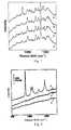
- FIG. 7illustrates the Raman spectra of the following four types (“flavors”) of GANs particles: (A) GANs tagged with furonitrile; (B) GANs tagged with furonitrile (66%) and cyanoacetic acid (33%); (B) GANs tagged with furonitrile (33%) and cyanoacetic acid (66%); and (D) GANs tagged with cyanoacetic acid.
- FIG. 8illustrates the Raman spectra of GANs (40 nm Au core/4 nm glass) (a) tagged with trans-4,4′-bis(pyridyl)ethylene (BPE-GANs) or (b) tagged with imidazole (IM-GANs) or (c) untagged.
- the present inventionis directed to surface enhanced spectroscopy-active composite nanoparticles, including SERS-active composite nanoparticles (SACNs). Also included within the scope of this invention are methods of manufacture of the particles, and uses of the particles (including their use as molecular or cellular optical tags).
- SACNsSERS-active composite nanoparticles
- the submicron-sized tags or labels of the inventioncan be covalently or non-covalently affixed to entities of interest (that may range in size from molecules to macroscopic objects) for the purpose of quantitation, location, identification, and/or tracking.
- SERS-active composite nanoparticlesare comprised of a metal nanoparticle that has attached or associated with its surface one or more Raman-active molecules (alternately referred to herein as “analytes”). This complex of Raman enhancing metal and analyte (referred to as a SERS-active metal nanoparticle) is then coated or encapsulated by an encapsulant.
- the encapsulantis a glass material, and the SACN is referred to then as a glass-coated analyte loaded nanoparticle (GAN).
- SACNsare provided by growing or otherwise placing a shell of a suitable encapsulant over a SERS-active metal nanoparticle core.
- the metal nanoparticle coreis preferably a gold or silver sphere of 20-200 nm in diameter. Most preferred is an oblate or prolate metal spheroid of the same materials.
- SERS using red incident light⁇ 633 nm
- the optimal SERS responseis obtained with 63 nm diameter gold nanoparticle cores.
- Metal nanoparticles of the desired sizecan be grown as metal colloids by a number of techniques well known in the art. For example, chemical or photochemical reduction of metal ions in solution using any number of reducing agents has been described.
- Nanoparticle syntheseshave been carried out in constrained volumes, e.g. inside a vesicle. Nanoparticles can be made via electrical discharge in solution. Dozens of other methods have been described, dating back to the mid-1800's.
- the encapsulantdoes not measurably alter the SERS activity of the metal nanoparticle.
- the advantages of the present inventionare still achieved even if the encapsulant has some measurable effect, provided it does not interfere with the SERS activity, or does not add significant complexity to the Raman spectrum.
- the encapsulantshould be readily modified in order to attach molecules, including biomolecules, to its exterior surface. Suitable encapsulants include, but are not limited to, glass, polymers, metals, metal oxides (such as TiO 2 and SnO 2 ), and metal sulfides.
- the encapsulationis carried out after, or during, adsorption to the core nanoparticle of the Raman-active analyte that is to provide the SERS activity of the SACN.
- the Raman-active analyteis sequestered from the surrounding solvent as a coating on the surface of the metal nanoparticle core.
- Such a configurationprovides the metal nanoparticle core with stable SERS activity.
- a Raman-active analytecan form a sub-monolayer, a complete monolayer, or a multilayer assembly on the surface of the metal nanoparticle core.
- a Raman-active analytecan comprise a single species of Raman-active molecule, or it can be a mixture of different species of Raman-active molecules.
- the encapsulantis glass (e.g. SiO x ).
- the metal nanoparticle coresare preferably treated first with a glass primer (that is, a material that can lead to growth of a uniform coating of glass, or can improve adhesion of the glass coat to the particle, or both). Glass is then grown over the metal nanoparticle by standard techniques well known in the art.
- the resulting SACNsare referred to as glass analyte-loaded nanoparticles (GANs).
- glass and many other materialscontain functional groups amenable to molecular attachment.
- immersion of glass in baseallows covalent attachment of alkyl trichlorosilanes or alkyl trialkoxysilanes, with additional functionality available on the end of the alkyl group.
- glass surfacescan be modified with all forms of biomolecules and biomolecular superstructures including cells, as well as oxides, metals, polymers, etc.
- surfaces of glasscan be modified with well-organized monomolecular layers.
- glass coatingssupport essentially any and all forms of chemical functionalization (derivatization). This is equally true for many different forms of encapsulant.
- SACN particlescan be affixed to any species with chemically reactive functionality. All chemical functional groups are reactive under certain conditions. There is thus no limitation to the species that can be immobilized on the encapsulant surface.
- the thickness of the encapsulantcan be easily varied depending on the physical properties required of the SACN. For example, coatings that are too thick—on the order of 1 micron or more—might preclude obtaining intense Raman spectra. Coatings too thin might lead to interference in the Raman spectrum of the analyte by the molecules on the enscapsulant surface. At the same time, physical properties such as sedimentation coefficient will clearly be affected by the thickness of encapsulant. In general, the thicker the encapsulant, the more effective the sequestration of the Raman-active analyte(s) on the metal nanoparticle core from the surrounding solvent.
- the preferred glass thicknessranges from 1-40 mn.
- the GANscomprise 60 nm diameter gold particles encapsulated by a 16 nm thick sphere of glass.
- the optimization of the dimensions of the SACNsis readily accomplished by one skilled in the art. Accordingly, one might alter the composition of the particle, or its size and shape in accordance with the invention to optimize the intensity of the Raman analyte molecule used as a tag. Indeed, it is known that core-shell nanoparticles (i.e. Au/AuS) support SERS and have very different optical properties compared to pure metal nanoparticles.
- SERS from prolate spheroidsis enhanced relative to spheres with the same major axis. It is further known that single particle enhancements are strongly wavelength-dependent. Thus, one might “tune” the particle size to achieve maximum signal for a given excitation wavelength.
- the present inventionspecifically contemplates the formation of a panel of at least 20 different SACNs, each having a unique SERS spectrum. Because the Raman bands of many molecules are extremely narrow (for example, CN ⁇ is less than 1 nm at FWHM), it is possible to synthesize a panel of SACNs, wherein each contains a Raman analyte that is spaced 20 wavenumbers away in the spectrum from its closest neighbor. For example, a GANs particle with 13 CN as the analyte is easily distinguished from a GANs with 12 CN as the analyte, and as well easily distinguishable from one with C 15 N.
- the inventioncontemplates the use of Raman-active analytes that have isotopic compositions distinct from naturally abundant species. For example, as described above, 13 CN is completely resolvable from any natural 12 CN that may be present in the background.
- isotopesas well as ratios of isotopes can be equally effectively used to identify unique SACNs.
- each population of SACNs in the panelis unique, the other properties of the SACNs are kept uniform across the panel. Because the SERS-activity of each SACN is sequestered from the surrounding milieu by the encapsulant, individual populations do not have different solvent or storage requirements. Also, each SACN has the same exterior shell, simplifying the choice of chemistry either for attachment of molecules to the SACNs or attachment of the SACNs to solid supports.
- SACNsprovided by the present invention can be used in virtually any assay in which a detectable tag or label is required.
- SACNsare used in biological and chemical assays as replacements for standard fluorescent tags.
- SACNspossess a number of characteristics that make them far superior to prior art optical tags based on fluorophores. For example, assays using fluorophore detection are commonly hampered by the presence of autofluorescence and other background effects.
- assaysrequire use of a number of different fluorophores; different fluorophores commonly require different attachment chemistries and have different environmental requirements and sensitivities. Particularly noteworthy is the quenching of fluorescent activity that is observed when some fluorophores are conjugated to proteins.
- SACNscannot be photobleached or photodegraded, they have uniform chemical and physical properties, and they can be readily resolved from the background.
- SACN detectionis significantly more sensitive than fluorophore detection. Indeed, it is possible to tag a single molecule with a single SACN, and then detect the presence of that molecule using Raman spectroscopy. Such simple single molecule resolution is without parallel in the fluorophore detection art.
- SACNscan be used as optical tags.
- sandwich assaysa target to be detected is captured by a solid surface.
- An antibody (or other ligand) to the same targetis attached to a SACN, and then contacted with the solid support.
- the presence of the SACN SERS signal at the solid supportindicates the presence of the antigen.
- SACNscan be conjugated to any molecule that is used to detect the presence of a specific target in an assay.
- SACNsare conjugated to nucleic acid molecules. In this way, they can be used in virtually any assay known in the art that detects specific nucleic acid sequences using optically-tagged nucleic acid probes.
- SACNsare especially suitable for multiplexed chemical assays in which the identity of SACNs encodes the identity of the target of the assay.
- Prior art multiplexed assays that use fluorophores to encode target identityare subject to a number of severe constraints imposed by the physical and chemical properties of the fluorophores. Specifically, different fluorophores have different excitation maxima, so coincident excitation of multiple fluorescent tags is not possible. Moreover, fluorescence emission occurs in broad spectral bands, so the bands from one fluorophore often overlap with those of another. As a result, resolving even three different fluorescence activities requires sophisticated optics to separate and then detect the individual emission wavelengths.
- multiplexed assays that use fluorophoresrely on positional information to reveal target identity.
- multiplexed assays with fluorophoresuse a solid support on which ligands are arranged in defined positions. The location of fluorophore signal reveals the identity of the target; the size of the fluorophore signal at that location indicates the amount of the target.
- the synthesis of solid supports with reagents localized at specific positionsis expensive and time-consuming. There are limits on the number of features that may be defined on a single surface.
- the SACNs of the present inventionoffer remarkable spectral diversity and resolvability.
- SACNscan be used in multiplexed assays to yield quantitative and qualitative information without requiring the position-specific localization of reagents.
- Each SACN coupled to a target-specific reagentcan encode the identity of that specific target, and the intensity of a particular Raman signal reveals the quantity of that target.
- the identity of targets captured on the solid supportcan be determined by using a different flavor of SACN for each target.
- SACNsare perfectly suited for use in multiplexing applications, they need not be used to encode identity in this manner. They can be used simply as replacements for fluorophores in multiplexed assays in which reagents are localized to specific positions on solid supports. When used in this way, the SACNs offer vastly more sensitive target detection than fluorophores.
- Flow cytometryis an example of a multiplexed assay format in which the diversity and resolvability of SACNs can be fully exploited.
- populations of beadsare provided to which primary antibodies against the targets to be detected are conjugated.
- the beadsare contacted with the assay solution containing the targets, and also with a second set of antibodies against the targets.
- Each secondary antibodyis conjugated to a GAN that encodes the identity of the target to which it will bind.
- the beadsare then passed through a flow cytometer that acquires the Raman spectrum of each bead.
- the Raman spectrometercan sample all frequency space of each bead, it is even possible to place many different primary antibodies on a single bead; the Raman spectrum of each bead can be decoded to determine which SACNs are present and in what quantity; this in turn reveals how much of each target is bound to a single bead. It will be understood that there are many variations of this basic scheme, including the use of reagents other than antibodies to bind to the targets of interest. Accordingly, SACNs can be used in a multitude of variations on this scheme in which it is necessary or useful to tag a reagent.
- the SACNsare used as optical tags for Microvolume Laser Scanning Cytometry (MLSC), rather than flow cytometry.
- MLSCis described in U.S. patent application Ser. No. 09/378,259, filed Aug. 20, 1999, and U.S. patent application Ser. No. 09/558,094, filed Apr. 26, 2000, both incorporated herein by reference in their entirety.
- a Raman microscopescans a capillary containing the reagents described above for the flow cytometry applications. The Raman microscope measures the Raman spectrum of each bead in the capillary, thereby obtaining quantitative data for each target to be detected. Again, it is the Raman signal of each SACN that encodes target identity; position specific reagents are not required.
- SACNsare used as optical tags in the solid support-based combinatorial chemical (“combi-chem”) synthesis of libraries of novel compounds.
- One such methodis known as “split and pool” synthesis.
- a preparation of suitably derivatized resinous beadsis randomly divided into multiple populations, and each population is introduced into a different reaction mixture.
- Different reaction mixturescan contain different reagents, or the same reagents but different reaction conditions.
- the beadsare then washed, recombined and divided again into a set of reaction mixtures. Because of the random manner in which the beads are distributed, each bead will experience a unique reaction history.
- the resultis a bead-based library comprising all of the compounds synthesized using the different permutations of the reaction mixtures.
- the librarymay then be screened to identify lead compounds with the desired activity.
- the lead compoundsin turn, may be analyzed to determine their composition and structure.
- the combi-chem methodhas been used to synthesize libraries of peptides, benzodiazapenes, and so on.
- each reaction mixturecontains a unique identifier molecule that becomes attached to the bead during the reaction step.
- the identifier moleculescan be cleaved from the bead of interest, and the reaction history of the bead can be determined by detecting the individual identifier molecules liberated from the bead.
- prior art methodshave used short oligonucleotides to encode reaction histories.
- oligomersmust be cleaved from the beads, amplified, and then sequenced in order to decode the reaction history; this is a time-consuming process. Because such identifier molecules must first be cleaved from the bead, it is necessary to choose a chemistry in which (a) cleaving the identifier from the bead does not modify or cleave the lead compound from the bead; and/or (b) cleaving the lead compound from the bead does not modify or cleave the identifier molecule. Moreover, the chemistry used to couple the identifier, and often just the presence of the identifier molecules themselves on the surface of the beads, may interfere with the actual combi-chem reactions. Such considerations place considerable restraints on all aspects of the chemistry used in encoded combi-chem synthesis.
- the SACNs provided by the present inventioncan be used to encode the reaction history of beads in such combinatorial schemes.
- Each reaction mixturecan contain a unique species of SACNs, such that each reaction step is accompanied by the attachment of a number of SACNs to the bead upon which the combinatorial synthesis takes place.
- reaction mixture Acan be encoded by SACN 1 when used at step 1 in the synthesis scheme, and by SACN 2 when used at step 2 in the synthesis scheme, and so on up to SACN n when used at step n in the synthesis scheme.
- the individual beadsmay be screened for the desired lead compound activity. Beads with the desired lead compound activity are then examined by Raman spectroscopy. The Raman spectrum of each bead is then automatically decoded to detect the individual species of SACNs that have bound to each bead. This information reveals the reaction history of the bead, and hence the structure of the lead compound.
- HAuCl 4 .3H 2 Otrisodium citrate dihydrate, sodium hydroxide, trans-1,2-bis(4-pyridyl)ethylene (BPE), pyridine, 2-mercaptopyridine, sodium silicate, tetraethyl orthosilicate (TEOS), and ammonia were obtained from Sigma-Aldrich. BPE was recrystallized several times before use. Dowex cation exchange resin (16-40 mesh) was obtained from J. T. Baker. Pure ethyl alcohol (EtOH) was purchased from Pharmco.
- Colloid preparation12-nm colloidal Au (nearly spherical, with a standard deviation less than 2 nm) was prepared from HAuCl 4 .3H 2 O reduced by citrate as described in Grabar et al, Analytical Chemistry 67:735-743 (1995), incorporated herein by reference in its entirety. Colloid >12 nm was prepared as follows: 3 ml of 12 mM HAuCl 4 was added for every 97 ml of H 2 O. The solution was then brought to a boil under vigorous stirring and 1 ml of 12-nm Au colloid as a seed and 0.5 ml of 1% sodium citrate per 100 ml of HAuCl 4 solution was added and boiled for 10 minutes. The size of the resulting particles was determined by transmission electron microscopy using Gatan or NIH Image software. Finally, the citrate ions surrounding the Au colloid were removed with dialysis, 7 exchanges of at least 4 hours each.
- GANs preparationAll reactions were performed in plastic Erylenmeyer flasks. Any amount of colloid could be used in a preparation and the subsequent reactants added in appropriate amounts based on the surface area and concentration of the Au colloid.
- a typical experimentused 25 ml of dialyzed, 50-nm, 0.17 nM Au colloid.
- the pH of the colloidwas adjusted from 5 to 7 with the addition of 50 ⁇ L of 0.1 M NaOH.
- the colloidwas rendered vitreophilic with the addition 125 ⁇ L of 0.5 mM MPTMS (or APTMS, or MPMDMS).
- 167 ⁇ L of a 0.5 mM solution of the Raman tag (BPE, pyridine, or 2-mercaptopyridine)was added.
- a 0.54% solution of active silicawas prepared by mixing 1 g of sodium silicate with 50 ml of 3 M NaOH and lowering the pH to 10 with cation exchange resin.
- One ml of the active silicawas added and the resulting solution was approximately pH 9. The solution remained stirring for 15 minutes and then was allowed to stand.
- FIG. 1Ashows GANs comprising 35 nm Au cores with 40 nm glass.
- FIG. 1Bshows 60 nm Au cores with 16 nm glass.
- FIG. 2illustrates 35 nm Au, 8 nm glass GANs following centrifugation through a 50% glycerol solution.
- FIG. 3demonstrates one such experiment for a batch of GANs particles with a 35 nm Au core, and 8 nm shell of glass.
- an etch solution50 ⁇ l HNO 3 and 150 ⁇ l HCl.
- the absorbance of the solutionwas measured ( ⁇ max 546 nm) at various times after addition of the etch solution. Etching of the gold core results in a decrease in the absorbance; this is plotted in FIG. 3A (the time after the addition of the etch solution is indicated). The rate of Au etching is shown in FIG. 3B as a plot of absorbance versus time in etch solution (right). Additional studies performed by the inventors have shown that etching of a Au core by aqua regia does not occur with a 20 nm glass shell over a four hour time period.
- GANscomprising a 40 nm Au core coated with trans-4,4′-bis(pyridyl)ethylene (BPE) encapsulated in 4 nm of glass were synthesized, and examined by Raman spectroscopy.
- the Raman spectrum obtained using 20 mW of 632.8 nm excitation, with a 3 mm lens and 30 second integrationis plotted in FIG. 4 .
- Trace A on the graphshows the characteristic BPE Raman signal; trace B shows the Raman signal from the same particles without the BPE analyte. It can be seen that the GANs without the BPE analyte give essentially no Raman signal.
- FIG. 5shows the Raman spectrum of a suspension of GANs comprising 40 nm Au coated with trans-4,4′-bis(pyridyl)ethylene (BPE)/4 nm glass (Trace A); supernatant from a first centrifugation of the GANs (Trace B); and supernatant from a second centrifugation of the GANs (Trace C). It can be seen that the BPE signal does not leave the GAN during each centrifugation step, indicating that the BPE has adhered to the Au core and is tightly sequestered there by glass encapsulation.
- BPEtrans-4,4′-bis(pyridyl)ethylene
- GANs80 nm Au core/20 nm glass
- 2-mercaptopyridineas the Raman-active analyte were analyzed by Raman spectroscopy using 25 mW of 632.8 nm excitation with a 3 mm lens and 60 seconds of integration.
- the Raman spectrum of the GAN preparationwas then compared with the Raman spectrum obtained when a 50 mM solution of 2-mercaptopyridine is absorbed onto a conventional three-layer SERS substrate (25 mW 632.8 nm excitation, 3 mm lens, 30-seconds integration).
- FIG. 6shows the two Raman spectra. It can be seen that the two spectra have identical features and intensities, illustrating that GANs are effective SERS substrates.
- SERS spectra of the following four flavors of GANs particleswere obtained using 26 mW of 632.8 nm excitation, a 3-mm lens, and 30-second integration: (A) GANs tagged with furonitrile; (B) GANs tagged with furonitrile (66%) and cyanoacetic acid (33%); (B) GANs tagged with furonitrile (33%) and cyanoacetic acid (66%); and (D) GANs tagged with cyanoacetic acid. The percentages indicated are the relative concentrations of each compound in the tagging solution added.
- FIG. 7shows that the furonitrile and cyanoacteic acid have relatively the same signal intensity and have similar spectral profiles. The fact that the spectra of B and C are very similar to the spectrum of D indicates that cyanoacetic acid has a better affinity for the Au nanoparticle than furonitrile.
- GANs40 nm Au core/4 nm glass
- BPE-GANstrans-4,4′-bis(pyridyl)ethylene
- IM-GANsimidazole
- SERS spectra of these Raman-active analytesare shown in FIG. 8, along with the SERS spectrum of untagged GANs (c) of the same dimensions.
- BPE-GANs and IM-GANsboth show the characteristic Raman bands of their respective Raman-active analytes; untagged GANs do not show these bands.
Landscapes
- Health & Medical Sciences (AREA)
- Immunology (AREA)
- Life Sciences & Earth Sciences (AREA)
- Chemical & Material Sciences (AREA)
- Engineering & Computer Science (AREA)
- Urology & Nephrology (AREA)
- Molecular Biology (AREA)
- Hematology (AREA)
- Biomedical Technology (AREA)
- Biochemistry (AREA)
- Analytical Chemistry (AREA)
- General Physics & Mathematics (AREA)
- Physics & Mathematics (AREA)
- General Health & Medical Sciences (AREA)
- Pathology (AREA)
- Biotechnology (AREA)
- Cell Biology (AREA)
- Microbiology (AREA)
- Food Science & Technology (AREA)
- Medicinal Chemistry (AREA)
- Nanotechnology (AREA)
- Nuclear Medicine, Radiotherapy & Molecular Imaging (AREA)
- Inorganic Chemistry (AREA)
- Investigating, Analyzing Materials By Fluorescence Or Luminescence (AREA)
- Powder Metallurgy (AREA)
- Other Investigation Or Analysis Of Materials By Electrical Means (AREA)
Abstract
Description
Claims (24)
Priority Applications (10)
| Application Number | Priority Date | Filing Date | Title |
|---|---|---|---|
| US09/680,782US6514767B1 (en) | 1999-10-06 | 2000-10-06 | Surface enhanced spectroscopy-active composite nanoparticles |
| US10/345,821US7192778B2 (en) | 1999-10-06 | 2003-01-16 | Surface enhanced spectroscopy-active composite nanoparticles |
| US11/113,601US8497131B2 (en) | 1999-10-06 | 2005-04-25 | Surface enhanced spectroscopy-active composite nanoparticles comprising Raman-active reporter molecules |
| US11/132,471US7443489B2 (en) | 1999-10-06 | 2005-05-18 | Surface enhanced spectroscopy-active composite nanoparticles |
| US11/132,510US20050272160A1 (en) | 1999-10-06 | 2005-05-18 | Surface enhanced spectroscopy-active composite nanoparticles |
| US11/132,974US9201013B2 (en) | 1999-10-06 | 2005-05-18 | Method for tagging material with surface-enhanced spectroscopy (SES)-active composite nanoparticles |
| US12/245,555US20090121193A1 (en) | 1999-10-06 | 2008-10-03 | Surface enhanced spectroscopy-active composite nanoparticles |
| US12/245,538US8918161B2 (en) | 1999-10-06 | 2008-10-03 | Methods of use for surface enhanced spectroscopy-active composite nanoparticles |
| US14/578,694US20150323465A1 (en) | 1999-10-06 | 2014-12-22 | Surface enhanced spectroscopy-active composite nanoparticles |
| US14/950,730US20160077011A1 (en) | 1999-10-06 | 2015-11-24 | Method for tagging material with surface-enhanced spectroscopy (ses)-active composite nanoparticles |
Applications Claiming Priority (3)
| Application Number | Priority Date | Filing Date | Title |
|---|---|---|---|
| US15793199P | 1999-10-06 | 1999-10-06 | |
| US19039500P | 2000-03-17 | 2000-03-17 | |
| US09/680,782US6514767B1 (en) | 1999-10-06 | 2000-10-06 | Surface enhanced spectroscopy-active composite nanoparticles |
Related Child Applications (1)
| Application Number | Title | Priority Date | Filing Date |
|---|---|---|---|
| US10/345,821Continuation-In-PartUS7192778B2 (en) | 1999-10-06 | 2003-01-16 | Surface enhanced spectroscopy-active composite nanoparticles |
Publications (1)
| Publication Number | Publication Date |
|---|---|
| US6514767B1true US6514767B1 (en) | 2003-02-04 |
Family
ID=26854607
Family Applications (1)
| Application Number | Title | Priority Date | Filing Date |
|---|---|---|---|
| US09/680,782Expired - LifetimeUS6514767B1 (en) | 1999-10-06 | 2000-10-06 | Surface enhanced spectroscopy-active composite nanoparticles |
Country Status (11)
| Country | Link |
|---|---|
| US (1) | US6514767B1 (en) |
| EP (3) | EP2295954B1 (en) |
| JP (1) | JP4786096B2 (en) |
| AU (1) | AU8000400A (en) |
| CA (1) | CA2386186A1 (en) |
| CY (1) | CY1118092T1 (en) |
| DE (1) | DE60037267T2 (en) |
| DK (2) | DK2295954T3 (en) |
| ES (2) | ES2584553T3 (en) |
| PT (2) | PT2295954T (en) |
| WO (1) | WO2001025758A1 (en) |
Cited By (188)
| Publication number | Priority date | Publication date | Assignee | Title |
|---|---|---|---|---|
| US20020153089A1 (en)* | 2001-04-19 | 2002-10-24 | Fuji Photo Film Co., Ltd. | Method and apparatus for producing laminates |
| US20020192676A1 (en)* | 2001-06-18 | 2002-12-19 | Madonna Angelo J. | Method for determining if a type of bacteria is present in a mixture |
| US20030078739A1 (en)* | 2001-10-05 | 2003-04-24 | Surromed, Inc. | Feature list extraction from data sets such as spectra |
| US6623977B1 (en)* | 1999-11-05 | 2003-09-23 | Real-Time Analyzers, Inc. | Material for surface-enhanced Raman spectroscopy, and SER sensors and method for preparing same |
| US20030187237A1 (en)* | 2002-03-26 | 2003-10-02 | Selena Chan | Methods and device for DNA sequencing using surface enhanced raman scattering (SERS) |
| US20030215816A1 (en)* | 2002-05-20 | 2003-11-20 | Narayan Sundararajan | Method for sequencing nucleic acids by observing the uptake of nucleotides modified with bulky groups |
| US20040063214A1 (en)* | 2002-09-30 | 2004-04-01 | Berlin Andrew Arthur | Spectroscopic analysis system and method |
| US20040110208A1 (en)* | 2002-03-26 | 2004-06-10 | Selena Chan | Methods and device for DNA sequencing using surface enhanced Raman scattering (SERS) |
| US20040120880A1 (en)* | 2002-03-26 | 2004-06-24 | Yuegang Zhang | Sorting of single-walled carbon nanotubes using optical dipole traps |
| US20040142484A1 (en)* | 2002-09-30 | 2004-07-22 | Intel Corporation | Spectroscopic analysis system and method |
| US20040161143A1 (en)* | 1999-07-21 | 2004-08-19 | Dietz Louis J. | System for microvolume laser scanning cytometry |
| US20040161750A1 (en)* | 2003-02-14 | 2004-08-19 | Lei Sun | Biomolecule analysis by rolling circle amplification and SERS detection |
| US20050032226A1 (en)* | 1999-10-01 | 2005-02-10 | Natan Michael J. | Encoded nanoparticles in paper manufacture |
| US20050130163A1 (en)* | 2002-07-12 | 2005-06-16 | Smith William E. | Serrs reactive particles |
| US20050138996A1 (en)* | 2003-12-29 | 2005-06-30 | Intel Corporation | Use of arrays of atomic force microscope/scanning tunneling microscope tips to scan nanocodes |
| US20050147981A1 (en)* | 2003-12-31 | 2005-07-07 | Intel Corporation | Methods and compositions for detecting nucleic acids using scanning probe microscopy and nanocodes |
| US20050148100A1 (en)* | 2003-12-30 | 2005-07-07 | Intel Corporation | Methods and devices for using Raman-active probe constructs to assay biological samples |
| US20050147977A1 (en)* | 2003-12-29 | 2005-07-07 | Tae-Woong Koo | Methods and compositions for nucleic acid detection and sequence analysis |
| US20050147980A1 (en)* | 2003-12-30 | 2005-07-07 | Intel Corporation | Nucleic acid sequencing by Raman monitoring of uptake of nucleotides during molecular replication |
| US20050148064A1 (en)* | 2003-12-29 | 2005-07-07 | Intel Corporation | Microfluid molecular-flow fractionator and bioreactor with integrated active/passive diffusion barrier |
| US20050147976A1 (en)* | 2003-12-29 | 2005-07-07 | Xing Su | Methods for determining nucleotide sequence information |
| US20050147964A1 (en)* | 2003-12-30 | 2005-07-07 | Intel Corporation | Methods for identifying a peptide that binds a geometrical shape |
| US20050158870A1 (en)* | 2001-01-26 | 2005-07-21 | Surromed, Inc. | Surface-enhanced spectroscopy-active sandwich nanoparticles |
| US20050171433A1 (en)* | 2004-01-08 | 2005-08-04 | Boppart Stephen A. | Multi-functional plasmon-resonant contrast agents for optical coherence tomography |
| US20050187407A1 (en)* | 2000-11-27 | 2005-08-25 | Aldrich Jane V. | Methods of synthesizing and using derivatives of [2-(2-aminoethoxy)ethoxy] acetic acid |
| WO2005013337A3 (en)* | 2003-03-06 | 2005-08-25 | Rensselaer Polytech Inst | Rapid generation of nanoparticles from bulk solids at room temperature |
| US20050191665A1 (en)* | 2003-12-29 | 2005-09-01 | Xing Su | Composite organic-inorganic nanoclusters |
| US20050196876A1 (en)* | 2003-12-29 | 2005-09-08 | Intel Corporation | Detection of biomolecules using porous biosensors and Raman spectroscopy |
| US20050209789A1 (en)* | 2001-08-24 | 2005-09-22 | Hastings Curtis A | Peak selection in multidimensional data |
| US20050221506A1 (en)* | 2004-03-30 | 2005-10-06 | Intel Corporation | Surface modification of metals for biomolecule detection using surface enhanced Raman scattering (SERS) |
| US20050221503A1 (en)* | 2003-10-24 | 2005-10-06 | Drachev Vladimir P | Plasmonic and/or microcavity enhanced optical protein sensing |
| US20050221333A1 (en)* | 2004-03-31 | 2005-10-06 | Intel Corporation | Microfluidic apparatus, systems, and methods for performing molecular reactions |
| US20050217990A1 (en)* | 2004-03-31 | 2005-10-06 | Intel Corporation | Fabrication and use of semipermeable membranes and gels for the control of electrolysis |
| US20050221507A1 (en)* | 2004-03-30 | 2005-10-06 | Intel Corporation | Method to detect molecular binding by surface-enhanced Raman spectroscopy |
| US20050221473A1 (en)* | 2004-03-30 | 2005-10-06 | Intel Corporation | Sensor array integrated circuits |
| US20050272160A1 (en)* | 1999-10-06 | 2005-12-08 | Natan Michael J | Surface enhanced spectroscopy-active composite nanoparticles |
| US20050280817A1 (en)* | 2004-04-02 | 2005-12-22 | Uwe Horchner | Polychromic laser scanning system and method of use |
| US20050282229A1 (en)* | 2002-05-01 | 2005-12-22 | Xing Su | Methods and device for analyte characterization |
| US20060019247A1 (en)* | 2002-05-20 | 2006-01-26 | Xing Su | Method and apparatus for nucleic acid sequencing and identification |
| US20060033910A1 (en)* | 2003-12-29 | 2006-02-16 | Lei Sun | Multiplexed detection of analytes in fluid solution |
| US20060038979A1 (en)* | 2004-08-05 | 2006-02-23 | Michael Natan | Nanoparticles as covert taggants in currency, bank notes, and related documents |
| US20060046313A1 (en)* | 2004-08-26 | 2006-03-02 | Intel Corporation | Cellular analysis using Raman surface scanning |
| US20060046311A1 (en)* | 2004-08-26 | 2006-03-02 | Intel Corporation | Biomolecule analysis using Raman surface scanning |
| US20060050268A1 (en)* | 2004-09-07 | 2006-03-09 | The Regents Of The University Of California | Nanosensors based on functionalized nanoparticles and Surface Enhanced Raman Scattering |
| US20060054506A1 (en)* | 1999-10-06 | 2006-03-16 | Natan Michael J | Surface enhanced spectrometry-active composite nanoparticles |
| US20060063271A1 (en)* | 2002-09-12 | 2006-03-23 | Putnam Martin A | Method and apparatus for aligning microbeads in order to interrogate the same |
| US20060073612A1 (en)* | 2004-10-06 | 2006-04-06 | General Electric Company | Raman-active particles and methods of making and using them |
| US20060073336A1 (en)* | 2003-12-29 | 2006-04-06 | Jingwu Zhang | External modification of composite organic inorganic nanoclusters |
| US20060072177A1 (en)* | 2002-08-20 | 2006-04-06 | Putnam Martin A | Diffraction grating-based encoded microparticle assay stick |
| US20060097154A1 (en)* | 2000-11-27 | 2006-05-11 | Curtis Hastings | Median filter for liquid chromatography-mass spectrometry data |
| US20060118630A1 (en)* | 2004-11-16 | 2006-06-08 | Illumina, Inc. | Holographically encoded elements for microarray and other tagging labeling applications, and method and apparatus for making and reading the same |
| US20060119913A1 (en)* | 2003-08-20 | 2006-06-08 | Illumina, Inc. | Fourier scattering methods for encoding microbeads and methods and apparatus for reading the same |
| US20060134806A1 (en)* | 2004-12-20 | 2006-06-22 | General Electric Company | Method of separating unattached Raman-active tag from bioassay or other reaction mixture |
| US20060133953A1 (en)* | 2003-12-30 | 2006-06-22 | Intel Corporation | Biosensor utilizing a resonator having a functionalized surface |
| US20060134324A1 (en)* | 2004-11-17 | 2006-06-22 | Illumina, Inc. | Filament with easily removed protective coating and methods for stripping the same |
| US20060131222A1 (en)* | 2002-05-09 | 2006-06-22 | Ppd Biomarker Discovery Sciences, Llc | Methods for time-alignment of liquid chromatography-mass spectrometry data |
| US20060147941A1 (en)* | 2004-12-30 | 2006-07-06 | Intel Corporation | Methods and apparatus for SERS assay of biological analytes |
| US20060166302A1 (en)* | 2005-01-27 | 2006-07-27 | Raman Systems, Inc. | Handheld raman blood analyzer |
| US20060192969A1 (en)* | 2005-02-28 | 2006-08-31 | Marks Daniel L | Distinguishing non-resonant four-wave-mixing noise in coherent stokes and anti-stokes Raman scattering |
| US20060216835A1 (en)* | 2005-03-24 | 2006-09-28 | General Electric Company | Method of separating unattached Raman-active tag from bioassay or other reaction mixture |
| US20060216697A1 (en)* | 2005-03-24 | 2006-09-28 | General Electric Company | Method of separating unattached Raman-active tag from bioassay or other reaction mixture |
| US20060234248A1 (en)* | 2003-12-29 | 2006-10-19 | Lei Sun | Composite organic inorganic nanoclusters |
| US20060240401A1 (en)* | 2005-01-27 | 2006-10-26 | Clarke Richard H | Handheld raman body fluid analyzer |
| US20060252065A1 (en)* | 2004-10-21 | 2006-11-09 | Yiping Zhao | Surface enhanced Raman spectroscopy (SERS) systems, substrates, fabrication thereof, and methods of use thereof |
| US20060259246A1 (en)* | 2000-11-28 | 2006-11-16 | Ppd Biomarker Discovery Sciences, Llc | Methods for efficiently mining broad data sets for biological markers |
| US20060285635A1 (en)* | 2005-04-15 | 2006-12-21 | Boppart Stephen A | Contrast enhanced spectroscopic optical coherence tomography |
| WO2006060734A3 (en)* | 2004-12-03 | 2007-01-18 | Univ Boston | Nanostructured substrate for for surface enhanced raman scattering |
| US20070020771A1 (en)* | 2005-06-24 | 2007-01-25 | Applied Nanoworks, Inc. | Nanoparticles and method of making thereof |
| US20070048797A1 (en)* | 2004-08-11 | 2007-03-01 | Xing Su | Composite organic inorganic nanoclusters as carriers and identifiers of tester molecules |
| US20070048746A1 (en)* | 2005-09-01 | 2007-03-01 | Intel Corporation | Multiplex data collection and analysis in bioanalyte detection |
| US20070059203A1 (en)* | 2005-09-09 | 2007-03-15 | General Electric Company | Raman-active lateral flow device and methods of detection |
| US20070058165A1 (en)* | 2005-09-09 | 2007-03-15 | General Electric Company | Raman-active lateral flow device and methods of detection and making |
| US20070059725A1 (en)* | 2005-03-31 | 2007-03-15 | Voorhees Kent J | Apparatus and method for detecting microscopic organisms using bacteriophage |
| US20070121181A1 (en)* | 2005-11-22 | 2007-05-31 | Cyvera Corporation | Method and apparatus for labeling using optical identification elements characterized by X-ray diffraction |
| US20070140951A1 (en)* | 2003-12-11 | 2007-06-21 | The Trustees Of Columbia University In The City Of New York | Nano-sized particles, processes of making, compositions and uses thereof |
| KR100733085B1 (en) | 2005-07-29 | 2007-06-28 | 재단법인서울대학교산학협력재단 | Surface enhanced raman scattering nano-tagging particle and method for preparing thereof |
| US20070155022A1 (en)* | 2005-12-30 | 2007-07-05 | Mineo Yamakawa | Degenerate binding detection and protein identification using Raman spectroscopy nanoparticle labels |
| US7253119B2 (en) | 2001-09-05 | 2007-08-07 | Rensselaer Polytechnic Institute | Passivated nanoparticles, method of fabrication thereof, and devices incorporating nanoparticles |
| WO2007090058A2 (en) | 2006-01-27 | 2007-08-09 | Oxonica, Inc. | Lateral flow immunoassay with encapsulated detection modality |
| WO2007092941A2 (en) | 2006-02-08 | 2007-08-16 | Oxonica, Inc. | Sers nanotag assays |
| US7259845B2 (en) | 1999-04-23 | 2007-08-21 | Ppd Biomarker Discovery Sciences Llc | Disposable optical cuvette cartridge with low fluorescence material |
| US20070205839A1 (en)* | 2002-04-30 | 2007-09-06 | Hrl Laboratories, Llc | Method for fabricating quartz-based nanoresonators |
| US20070224683A1 (en)* | 2005-01-27 | 2007-09-27 | Prescient Medical, Inc. | Raman spectroscopic test strip systems |
| US20070236789A1 (en)* | 2006-04-10 | 2007-10-11 | Moon John A | Optical scanner with improved scan time |
| US20070259437A1 (en)* | 2005-12-14 | 2007-11-08 | Oxonica, Inc. | Nanoparticulate Chemical Sensors Using SERS |
| US20070275370A1 (en)* | 2002-04-12 | 2007-11-29 | Madonna Angelo J | Method for detecting concentrations of a target bacterium that uses phages to infect target bacterial cells |
| US20080064120A1 (en)* | 2006-02-06 | 2008-03-13 | Clarke Richard H | Raman spectroscopic lateral flow test strip assays |
| US20080060781A1 (en)* | 2005-09-01 | 2008-03-13 | United Technologies Corporation | Investment Casting Pattern Manufacture |
| US20080074662A1 (en)* | 2006-09-21 | 2008-03-27 | Honeywell International Inc. | Sers analyzer |
| US20080096289A1 (en)* | 2006-10-24 | 2008-04-24 | Honeywell International Inc. | Core-shell nanoparticles for detection based on sers |
| US20080129990A1 (en)* | 2003-01-22 | 2008-06-05 | Cyvera Corporation | Hybrid random bead/chip based microarray |
| US20080149850A1 (en)* | 2006-12-22 | 2008-06-26 | Commissariat A L'energie Atomique | Optical coding device by plasmon effect and authentication method using the device |
| US20080152534A1 (en)* | 2006-12-21 | 2008-06-26 | Jingwu Zhang | Self-assembling raman-active nanoclusters |
| US20080165656A1 (en)* | 2002-09-12 | 2008-07-10 | Moon John A | Method of Manufacturing of a Diffraction Grating-Based Optical Identification Element |
| WO2008116093A2 (en) | 2007-03-20 | 2008-09-25 | Becton, Dickinson And Company | Assays using surface-enhanced raman spectroscopy (sers)-active particles |
| US20080241569A1 (en)* | 2007-03-30 | 2008-10-02 | Haoyu Qin | Encapsulation of raman active nanoparticles |
| US20080245769A1 (en)* | 2006-07-17 | 2008-10-09 | Applied Nanoworks, Inc. | Nanoparticles and method of making thereof |
| US20080305489A1 (en)* | 2007-06-06 | 2008-12-11 | Becton, Dickinson And Company | Near-infrared dyes as surface enhanced raman scattering reporters |
| WO2009009198A2 (en) | 2007-04-18 | 2009-01-15 | Becton, Dickinson And Company | Sers nanotag assays |
| US20090045340A1 (en)* | 2007-07-31 | 2009-02-19 | Shohei Terada | Electron microscope with electron spectrometer |
| US20090069605A1 (en)* | 2007-09-12 | 2009-03-12 | Shanghai Huayi Acrylic Acid Co., Ltd. | Catalyst composition and producing process thereof for use in manufacturing methacrolein |
| US20090068461A1 (en)* | 2003-10-16 | 2009-03-12 | The University Of Akron | Carbon nanotubes on carbon nanofiber substrate |
| US20090073520A1 (en)* | 2004-11-17 | 2009-03-19 | Illumina, Inc. | Encoded microparticles and a method for fabricating |
| WO2008019161A3 (en)* | 2006-01-10 | 2009-04-02 | Oxonica Inc | Fuel identification with surface enhanced raman spectroscopy tags |
| WO2009040114A2 (en) | 2007-09-24 | 2009-04-02 | Julius-Maximilians-Universität Würzburg | Compounds and markers for surface-enhanced raman scattering |
| US20090140206A1 (en)* | 2003-08-18 | 2009-06-04 | Shuming Nie | Surface enhanced Raman spectroscopy (SERS)-active composite nanoparticles, methods of fabrication thereof, and methods of use thereof |
| US20090147254A1 (en)* | 2007-06-14 | 2009-06-11 | Hrl Laboratories, Llc. | Integrated quartz biological sensor and method |
| US20090149344A1 (en)* | 2005-07-28 | 2009-06-11 | Yiping Zhao | Surface enhanced raman spectroscopy (sers) systems and methods of use thereof |
| US20090168592A1 (en)* | 2007-12-28 | 2009-07-02 | Michael Craig Burrell | Agitator for portable substance identification system and method |
| US20090168052A1 (en)* | 2007-12-28 | 2009-07-02 | Michael Craig Burrell | System and method for improved biodetection |
| US20090168058A1 (en)* | 2007-12-28 | 2009-07-02 | Michael Craig Burrell | System and method for improved biodetection |
| US20090169433A1 (en)* | 2007-12-28 | 2009-07-02 | Sankaran Kumar | System for improved biodetection |
| US20090194589A1 (en)* | 2002-08-20 | 2009-08-06 | Illumina, Inc. | Optical reader system for substrates having an optically readable code |
| US20090221920A1 (en)* | 2008-01-18 | 2009-09-03 | Boppart Stephen A | Low-coherence interferometry and optical coherence tomography for image-guided surgical treatment of solid tumors |
| US20090246753A1 (en)* | 2008-01-11 | 2009-10-01 | Colorado School Of Mines | Detection of Phage Amplification by SERS Nanoparticles |
| US7602952B2 (en) | 2004-11-16 | 2009-10-13 | Illumina, Inc. | Scanner having spatial light modulator |
| US20090258341A1 (en)* | 2008-04-03 | 2009-10-15 | Colorado School Of Mines | Compositions and Methods for Detecting Bacteria |
| US7619819B2 (en) | 2002-08-20 | 2009-11-17 | Illumina, Inc. | Method and apparatus for drug product tracking using encoded optical identification elements |
| US7623908B2 (en) | 2003-01-24 | 2009-11-24 | The Board Of Trustees Of The University Of Illinois | Nonlinear interferometric vibrational imaging |
| WO2009142604A1 (en)* | 2008-05-23 | 2009-11-26 | Nanyang Technological University | Polymer encapsulated particles as surface enhanced raman scattering probes |
| US20090297626A1 (en)* | 2006-11-03 | 2009-12-03 | The Trustees Of Columbia University In The City Of New York | Methods for preparing metal oxides |
| US20090305231A1 (en)* | 2008-04-09 | 2009-12-10 | Becton, Dickinson And Company | Sensitive Immunoassays Using Coated Nanoparticles |
| US7723100B2 (en) | 2006-01-13 | 2010-05-25 | Becton, Dickinson And Company | Polymer coated SERS nanotag |
| US20100151472A1 (en)* | 2008-11-12 | 2010-06-17 | Nodality, Inc. | Detection Composition |
| US7751057B2 (en) | 2008-01-18 | 2010-07-06 | The Board Of Trustees Of The University Of Illinois | Magnetomotive optical coherence tomography |
| US7787129B2 (en) | 2006-01-31 | 2010-08-31 | The Board Of Trustees Of The University Of Illinois | Method and apparatus for measurement of optical properties in tissue |
| US7791802B2 (en) | 2004-02-19 | 2010-09-07 | Illumina, Inc. | Optical identification element having a non-waveguide substrate |
| US20100248379A1 (en)* | 2004-05-19 | 2010-09-30 | Vladimir Poponin | Optical sensor with layered plasmon structure for enhanced detection of chemical groups by sers |
| US20100279272A1 (en)* | 2008-02-13 | 2010-11-04 | Michael Craig Burrell | Multiplexed analysis methods using sers-active nanoparticles |
| US20100291537A1 (en)* | 2004-11-16 | 2010-11-18 | Glauco Souza | Methods and compositions related to phage-nanoparticle assemblies |
| US7872804B2 (en) | 2002-08-20 | 2011-01-18 | Illumina, Inc. | Encoded particle having a grating with variations in the refractive index |
| US7879764B2 (en) | 2004-12-28 | 2011-02-01 | Intel Corporation | Electrically active combinatorial chemical (EACC) chip for biochemical analyte detection |
| US20110026019A1 (en)* | 2008-03-20 | 2011-02-03 | Drexel University | Method for the formation of sers substrates |
| US20110069307A1 (en)* | 2009-09-22 | 2011-03-24 | Authentix, Inc. | Dipyrromethenes and Azadipyrromethenes as Markers for Petroleum Products |
| US7923260B2 (en) | 2002-08-20 | 2011-04-12 | Illumina, Inc. | Method of reading encoded particles |
| US20110128535A1 (en)* | 2009-11-30 | 2011-06-02 | David Eugene Baker | Nano-Structured Substrates, Articles, and Methods Thereof |
| US7994877B1 (en) | 2008-11-10 | 2011-08-09 | Hrl Laboratories, Llc | MEMS-based quartz hybrid filters and a method of making the same |
| US20110207231A1 (en)* | 2008-11-17 | 2011-08-25 | Cabot Security Materials Inc. | Melamine Assay Methods and Systems |
| US20110228264A1 (en)* | 2010-03-22 | 2011-09-22 | Cabot Security Materials Inc. | SERS Nanotags With Improved Buoyancy in Liquids |
| US8033715B2 (en) | 2007-11-08 | 2011-10-11 | Illinois Institute Of Technology | Nanoparticle based thermal history indicators |
| US8115934B2 (en) | 2008-01-18 | 2012-02-14 | The Board Of Trustees Of The University Of Illinois | Device and method for imaging the ear using optical coherence tomography |
| US8138016B2 (en) | 2006-08-09 | 2012-03-20 | Hrl Laboratories, Llc | Large area integration of quartz resonators with electronics |
| US8151640B1 (en) | 2008-02-05 | 2012-04-10 | Hrl Laboratories, Llc | MEMS on-chip inertial navigation system with error correction |
| US8176607B1 (en) | 2009-10-08 | 2012-05-15 | Hrl Laboratories, Llc | Method of fabricating quartz resonators |
| CN102608097A (en)* | 2012-02-03 | 2012-07-25 | 济南大学 | Probe with adjustable Raman scattering effect, and preparation method thereof |
| US8268388B1 (en)* | 2007-08-29 | 2012-09-18 | The United States Of America As Represented By The Secretary Of The Navy | Method to incorporate a fluorescent internal standard on substrates |
| WO2013165615A2 (en) | 2012-04-12 | 2013-11-07 | Becton Dickinson And Company | Methods, systems, and devices for detecting and identifying microorganisms in microbiological culture samples |
| US20130306463A1 (en)* | 2010-12-15 | 2013-11-21 | William Marsh Rice University | Purifying a fluid using a heat carrier comprising an electromagnetic radiation-absorbing complex |
| US8766745B1 (en) | 2007-07-25 | 2014-07-01 | Hrl Laboratories, Llc | Quartz-based disk resonator gyro with ultra-thin conductive outer electrodes and method of making same |
| US8769802B1 (en) | 2008-02-21 | 2014-07-08 | Hrl Laboratories, Llc | Method of fabrication an ultra-thin quartz resonator |
| CN104204780A (en)* | 2012-03-30 | 2014-12-10 | 庄信万丰股份有限公司 | Tracer and method of identifying tracer in product |
| US8912711B1 (en) | 2010-06-22 | 2014-12-16 | Hrl Laboratories, Llc | Thermal stress resistant resonator, and a method for fabricating same |
| USD731672S1 (en) | 2012-04-12 | 2015-06-09 | Becton, Dickinson And Company | Vessel assembly |
| US9062392B2 (en) | 2003-12-30 | 2015-06-23 | Intel Corporation | Methods for isolating a peptide methods for identifying a peptide |
| US9239327B2 (en) | 2009-05-18 | 2016-01-19 | Sicpa Holding Sa | Particles comprising hollow surface-enhanced spectroscopy (SES)-active core for long wavelength SERS |
| US9250074B1 (en) | 2013-04-12 | 2016-02-02 | Hrl Laboratories, Llc | Resonator assembly comprising a silicon resonator and a quartz resonator |
| US9261403B2 (en) | 2010-08-31 | 2016-02-16 | Sicpa Holding Sa | Inline spectroscopic reader and methods |
| US9283570B2 (en) | 2010-05-25 | 2016-03-15 | Sicpa Holding Sa | Nanoparticle separation methods and compositions |
| US9482619B2 (en)* | 2011-09-22 | 2016-11-01 | East China University Of Science And Technology | Metallic nanoparticles and methods for their preparation and use |
| US9494581B2 (en) | 2004-08-24 | 2016-11-15 | University Of Wyoming | System and method for Raman spectroscopy assay using paramagnetic particles |
| US9545458B2 (en) | 2010-12-15 | 2017-01-17 | Willam Marsh Rice University | Waste remediation |
| US9599470B1 (en) | 2013-09-11 | 2017-03-21 | Hrl Laboratories, Llc | Dielectric high Q MEMS shell gyroscope structure |
| US9726609B2 (en) | 2010-03-22 | 2017-08-08 | Sicpa Holding Sa | Wavelength selective SERS nanotags |
| US9739473B2 (en) | 2009-12-15 | 2017-08-22 | William Marsh Rice University | Electricity generation using electromagnetic radiation |
| WO2017147417A1 (en)* | 2016-02-26 | 2017-08-31 | Prozyme, Inc. | Use of bispyridines to improve labeling of nucleophiles |
| US20170275573A1 (en)* | 2016-03-24 | 2017-09-28 | The Procter & Gamble Company | Process for making a liquid laundry detergent composition |
| US9863662B2 (en) | 2010-12-15 | 2018-01-09 | William Marsh Rice University | Generating a heated fluid using an electromagnetic radiation-absorbing complex |
| KR20180018378A (en)* | 2016-08-09 | 2018-02-21 | 한양대학교 에리카산학협력단 | Directional Assembly of Gold Nanorods and their Anisotropic Bimetal Nanorod Cluster-Polymer Hybrid Nanostructures for Biophotonics, process for producing the same, and use thereof |
| US9977097B1 (en) | 2014-02-21 | 2018-05-22 | Hrl Laboratories, Llc | Micro-scale piezoelectric resonating magnetometer |
| US9991863B1 (en) | 2014-04-08 | 2018-06-05 | Hrl Laboratories, Llc | Rounded and curved integrated tethers for quartz resonators |
| US10031191B1 (en) | 2015-01-16 | 2018-07-24 | Hrl Laboratories, Llc | Piezoelectric magnetometer capable of sensing a magnetic field in multiple vectors |
| US10105456B2 (en) | 2012-12-19 | 2018-10-23 | Sloan-Kettering Institute For Cancer Research | Multimodal particles, methods and uses thereof |
| US10110198B1 (en) | 2015-12-17 | 2018-10-23 | Hrl Laboratories, Llc | Integrated quartz MEMS tuning fork resonator/oscillator |
| US10175307B1 (en) | 2016-01-15 | 2019-01-08 | Hrl Laboratories, Llc | FM demodulation system for quartz MEMS magnetometer |
| KR20190034111A (en)* | 2017-09-22 | 2019-04-01 | 한양대학교 에리카산학협력단 | Controlled Nanoclusters of Anisotropic Bimetal Nanostars for Surface-enhanced Raman Scattering-based Biosensing and the Method Thereof |
| US10266398B1 (en) | 2007-07-25 | 2019-04-23 | Hrl Laboratories, Llc | ALD metal coatings for high Q MEMS structures |
| US10308505B1 (en) | 2014-08-11 | 2019-06-04 | Hrl Laboratories, Llc | Method and apparatus for the monolithic encapsulation of a micro-scale inertial navigation sensor suite |
| US10322194B2 (en) | 2012-08-31 | 2019-06-18 | Sloan-Kettering Institute For Cancer Research | Particles, methods and uses thereof |
| US10502734B2 (en) | 2014-06-03 | 2019-12-10 | Consejo Superior De Investigaciones Cientificas | System for biodetection applications |
| US10620107B2 (en) | 2014-05-05 | 2020-04-14 | The Regents Of The University Of California | Determining fluid reservoir connectivity using nanowire probes |
| EP3656735A1 (en) | 2004-04-23 | 2020-05-27 | Becton, Dickinson and Company | Surface enhanced spectroscopy-active composite nanoparticles |
| US10688202B2 (en) | 2014-07-28 | 2020-06-23 | Memorial Sloan-Kettering Cancer Center | Metal(loid) chalcogen nanoparticles as universal binders for medical isotopes |
| EP2325136B1 (en)* | 2009-04-10 | 2020-11-18 | Korea Atomic Energy Research Institute | Conductive nanocomposite and manufacturing method thereof |
| US10888227B2 (en) | 2013-02-20 | 2021-01-12 | Memorial Sloan Kettering Cancer Center | Raman-triggered ablation/resection systems and methods |
| US10912947B2 (en) | 2014-03-04 | 2021-02-09 | Memorial Sloan Kettering Cancer Center | Systems and methods for treatment of disease via application of mechanical force by controlled rotation of nanoparticles inside cells |
| US10919089B2 (en) | 2015-07-01 | 2021-02-16 | Memorial Sloan Kettering Cancer Center | Anisotropic particles, methods and uses thereof |
| US20230103705A1 (en)* | 2019-09-05 | 2023-04-06 | Korea Research Institute Of Standards And Science | Raman-active particle for surface-enhanced raman scattering and method of producing the same |
| EP3234929B1 (en)* | 2014-12-16 | 2024-05-22 | Giesecke+Devrient Currency Technology GmbH | Device and method for verifying feature substances |
Families Citing this family (11)
| Publication number | Priority date | Publication date | Assignee | Title |
|---|---|---|---|---|
| US6707548B2 (en) | 2001-02-08 | 2004-03-16 | Array Bioscience Corporation | Systems and methods for filter based spectrographic analysis |
| JP4787938B2 (en) | 2003-03-28 | 2011-10-05 | ザ・プロウボウスト・フェロウズ・ファウンデーション・スカラーズ・アンド・ザ・アザー・メンバーズ・オブ・ボード・オブ・ザ・カレッジ・オブ・ザ・ホリー・アンド・アンデバイデッド・トリニティ・オブ・クイーン | Detecting sensor using silver nanoparticles |
| KR20090033227A (en)* | 2006-06-19 | 2009-04-01 | 캐보트 코포레이션 | Photovoltaic conductive features and methods of forming the same |
| CA2694357A1 (en)* | 2007-07-24 | 2009-02-26 | Northwestern University | Coated colloidal materials |
| US9265729B2 (en)* | 2007-12-06 | 2016-02-23 | The University Of Tokushima | Nanofunctional silica particles and manufacturing method thereof |
| GB201017875D0 (en) | 2010-10-22 | 2010-12-01 | Johnson Matthey Plc | Method of identifying a material |
| JP5854350B2 (en)* | 2010-12-08 | 2016-02-09 | 公立大学法人大阪府立大学 | Pressure detecting device, temperature detecting device, pressure detecting method, and temperature detecting method using metal nanoparticle integrated structure |
| BR112014010696A2 (en) | 2011-11-02 | 2017-04-25 | Univ Cape Town | method to identify an analyte molecule in a biological sample, sensor to capture an analyte of interest from a biological sample, and |
| JP5831340B2 (en)* | 2012-04-05 | 2015-12-09 | 住友金属鉱山株式会社 | Silver powder |
| DE102019122079B4 (en)* | 2019-08-16 | 2021-04-08 | Leibniz-Institut Für Polymerforschung Dresden E.V. | METHOD FOR DETERMINING NANOPOLYMER PARTICLES |
| CN112296349B (en)* | 2020-09-24 | 2022-04-01 | 河北工程大学 | Preparation method of finger-ring-shaped Au nano ring |
Citations (8)
| Publication number | Priority date | Publication date | Assignee | Title |
|---|---|---|---|---|
| US5023139A (en) | 1989-04-04 | 1991-06-11 | Research Corporation Technologies, Inc. | Nonlinear optical materials |
| US5266498A (en) | 1989-10-27 | 1993-11-30 | Abbott Laboratories | Ligand binding assay for an analyte using surface-enhanced scattering (SERS) signal |
| US5609907A (en) | 1995-02-09 | 1997-03-11 | The Penn State Research Foundation | Self-assembled metal colloid monolayers |
| US5825790A (en) | 1994-03-18 | 1998-10-20 | Brown University Research Foundation | Optical sources having a strongly scattering gain medium providing laser-like action |
| US5828450A (en) | 1995-07-19 | 1998-10-27 | Kyoto Dai-Ichi Kagaku Co., Ltd. | Spectral measuring apparatus and automatic analyzer |
| WO1999021934A1 (en) | 1997-10-28 | 1999-05-06 | The University Of Melbourne | Stabilized particles and methods of preparation and use thereof |
| US6149868A (en) | 1997-10-28 | 2000-11-21 | The Penn State Research Foundation | Surface enhanced raman scattering from metal nanoparticle-analyte-noble metal substrate sandwiches |
| US6219137B1 (en)* | 1998-12-03 | 2001-04-17 | Lockheed Martin Energy Research Corporation | Nanoprobe for surface-enhanced Raman spectroscopy in medical diagnostic and drug screening |
Family Cites Families (5)
| Publication number | Priority date | Publication date | Assignee | Title |
|---|---|---|---|---|
| US5376556A (en)* | 1989-10-27 | 1994-12-27 | Abbott Laboratories | Surface-enhanced Raman spectroscopy immunoassay |
| JP2965502B2 (en)* | 1996-02-20 | 1999-10-18 | 株式会社京都第一科学 | Analysis method by surface enhanced Raman scattering measurement |
| JP4068677B2 (en)* | 1996-10-25 | 2008-03-26 | アークレイ株式会社 | Gene analysis method and gene analysis kit used therefor |
| JPH10160737A (en)* | 1996-12-03 | 1998-06-19 | Dainippon Printing Co Ltd | Measurement chip for optical analyzer and method for manufacturing the same |
| WO1999027350A1 (en)* | 1997-11-26 | 1999-06-03 | The Dow Chemical Company | Online measurement of crystallinity in semicrystalline polymers by raman spectroscopy |
- 2000
- 2000-10-06ESES10183847.2Tpatent/ES2584553T3/ennot_activeExpired - Lifetime
- 2000-10-06EPEP10183847.2Apatent/EP2295954B1/ennot_activeExpired - Lifetime
- 2000-10-06DKDK10183847.2Tpatent/DK2295954T3/enactive
- 2000-10-06PTPT101838472Tpatent/PT2295954T/enunknown
- 2000-10-06EPEP00970656Apatent/EP1226422B1/ennot_activeExpired - Lifetime
- 2000-10-06DEDE60037267Tpatent/DE60037267T2/ennot_activeExpired - Lifetime
- 2000-10-06AUAU80004/00Apatent/AU8000400A/ennot_activeAbandoned
- 2000-10-06USUS09/680,782patent/US6514767B1/ennot_activeExpired - Lifetime
- 2000-10-06PTPT00970656Tpatent/PT1226422E/enunknown
- 2000-10-06DKDK00970656Tpatent/DK1226422T3/enactive
- 2000-10-06ESES00970656Tpatent/ES2296648T3/ennot_activeExpired - Lifetime
- 2000-10-06WOPCT/US2000/027757patent/WO2001025758A1/enactiveIP Right Grant
- 2000-10-06EPEP07007315Apatent/EP1804053A1/ennot_activeCeased
- 2000-10-06CACA002386186Apatent/CA2386186A1/ennot_activeAbandoned
- 2000-10-06JPJP2001528673Apatent/JP4786096B2/ennot_activeExpired - Lifetime
- 2016
- 2016-07-26CYCY20161100732Tpatent/CY1118092T1/enunknown
Patent Citations (8)
| Publication number | Priority date | Publication date | Assignee | Title |
|---|---|---|---|---|
| US5023139A (en) | 1989-04-04 | 1991-06-11 | Research Corporation Technologies, Inc. | Nonlinear optical materials |
| US5266498A (en) | 1989-10-27 | 1993-11-30 | Abbott Laboratories | Ligand binding assay for an analyte using surface-enhanced scattering (SERS) signal |
| US5825790A (en) | 1994-03-18 | 1998-10-20 | Brown University Research Foundation | Optical sources having a strongly scattering gain medium providing laser-like action |
| US5609907A (en) | 1995-02-09 | 1997-03-11 | The Penn State Research Foundation | Self-assembled metal colloid monolayers |
| US5828450A (en) | 1995-07-19 | 1998-10-27 | Kyoto Dai-Ichi Kagaku Co., Ltd. | Spectral measuring apparatus and automatic analyzer |
| WO1999021934A1 (en) | 1997-10-28 | 1999-05-06 | The University Of Melbourne | Stabilized particles and methods of preparation and use thereof |
| US6149868A (en) | 1997-10-28 | 2000-11-21 | The Penn State Research Foundation | Surface enhanced raman scattering from metal nanoparticle-analyte-noble metal substrate sandwiches |
| US6219137B1 (en)* | 1998-12-03 | 2001-04-17 | Lockheed Martin Energy Research Corporation | Nanoprobe for surface-enhanced Raman spectroscopy in medical diagnostic and drug screening |
Non-Patent Citations (26)
| Title |
|---|
| Akbarian et al. "Porous sol-gel silicates containing gold particels as matrixes for surface-enhanced Raman spectroscopy", J. Raman Spectr., 1996, v. 26, No 10, pp. 775-783.** |
| Chan et al. (1998) Science 281:2016-2018. |
| Emory et al. (1997) Analytical Chemistry 69:2631-2635. |
| Emory et al. (1998) J. American Chemical Society 120:8009-8010. |
| Emory et al. (1998) J. Phys Chem. B 102:493-497. |
| Felidj et al. "A new approach to determine nanoparticle shape and size distributions of SERS-active gold-silver mixed colloids", New J. Chem., 1998, pp. 725-732.** |
| Freeman et al. "Ag-clad Au nanoparticles: novel aggregation, optical. An D surface-enhanced Raman scattering properties", J. Phys. Chem., 1996, v. 100, pp. 718-724.** |
| Grabar et al. (1995) Analytical Chemistry 67:735-743. |
| Kneipp et al. (1995) Applied Spectroscopy 49:780-784. |
| Kneipp et al. (1996) Phys. Rev. Letters 76:2444-2447. |
| Kneipp et al. (1997) Phys. Rev. Letters 78:1667-1670. |
| Kneipp et al. (1998) Applied Spectroscopy 52:1493-1497. |
| Kneipp et al. (1998) Applied Spectroscopy 52:175-178. |
| Kneipp et al. (1998) Exp. Techn. Phys. 36 2:161-166. |
| Kneipp et al. (1998) Phys. Rev. E 57:6281-6284. |
| Kneipp et al. (1999) Chem. Review 99:2957-2975. |
| Kneipp et al. (1999) Current Science 77:915-924. |
| Lyon et al. (1997) Anal. Chem. 69:3400-3405. |
| Nie et al. (1997) Annu. Rev. Biophys. Biomol. Struct. 26:567-596. |
| Nie et al. (1997) Science 275:1102-1106. |
| Shibata et al. "Preparation of Silica Microspheres Containing Ag Nanoparticles", J. Sol-Gel technol., 1998, v. 11, pp. 279-287.** |
| Stober et al. (1968) J. Colloid Interface Sci. 26:62-69. |
| U.S. patent application Ser. No. 09/378,259, Dietz et al., filed Aug. 20, 1999. |
| U.S. patent application Ser. No. 09/558,094, Dietz et al., filed Apr. 26, 2000. |
| Ung et al. (1998) Langmuir 14:3740-3748. |
| Van Duyne et al. (1993) J. Chem Phys. 99:2101-2115. |
Cited By (370)
| Publication number | Priority date | Publication date | Assignee | Title |
|---|---|---|---|---|
| US7259845B2 (en) | 1999-04-23 | 2007-08-21 | Ppd Biomarker Discovery Sciences Llc | Disposable optical cuvette cartridge with low fluorescence material |
| US7336812B2 (en) | 1999-07-21 | 2008-02-26 | Ppd Biomarker Discovery Sciences, Llc | System for microvolume laser scanning cytometry |
| US20040161143A1 (en)* | 1999-07-21 | 2004-08-19 | Dietz Louis J. | System for microvolume laser scanning cytometry |
| US20050032226A1 (en)* | 1999-10-01 | 2005-02-10 | Natan Michael J. | Encoded nanoparticles in paper manufacture |
| US20110172523A1 (en)* | 1999-10-06 | 2011-07-14 | Oxonica, Inc. | Surface enhanced spectroscopy-active composite nanoparticles |
| US20090121193A1 (en)* | 1999-10-06 | 2009-05-14 | Oxonica, Inc. | Surface enhanced spectroscopy-active composite nanoparticles |
| US8918161B2 (en) | 1999-10-06 | 2014-12-23 | Becton, Dickinson And Company | Methods of use for surface enhanced spectroscopy-active composite nanoparticles |
| US7443489B2 (en) | 1999-10-06 | 2008-10-28 | Oxonica, Inc. | Surface enhanced spectroscopy-active composite nanoparticles |
| US20050272160A1 (en)* | 1999-10-06 | 2005-12-08 | Natan Michael J | Surface enhanced spectroscopy-active composite nanoparticles |
| US20060054506A1 (en)* | 1999-10-06 | 2006-03-16 | Natan Michael J | Surface enhanced spectrometry-active composite nanoparticles |
| US8497131B2 (en) | 1999-10-06 | 2013-07-30 | Becton, Dickinson And Company | Surface enhanced spectroscopy-active composite nanoparticles comprising Raman-active reporter molecules |
| US9201013B2 (en) | 1999-10-06 | 2015-12-01 | Becton, Dickinson And Company | Method for tagging material with surface-enhanced spectroscopy (SES)-active composite nanoparticles |
| US6623977B1 (en)* | 1999-11-05 | 2003-09-23 | Real-Time Analyzers, Inc. | Material for surface-enhanced Raman spectroscopy, and SER sensors and method for preparing same |
| US20060097154A1 (en)* | 2000-11-27 | 2006-05-11 | Curtis Hastings | Median filter for liquid chromatography-mass spectrometry data |
| US20050187407A1 (en)* | 2000-11-27 | 2005-08-25 | Aldrich Jane V. | Methods of synthesizing and using derivatives of [2-(2-aminoethoxy)ethoxy] acetic acid |
| US7253404B2 (en) | 2000-11-27 | 2007-08-07 | Ppd Biomarker Discovery Sciences, Llc | Median filter for liquid chromatography-mass spectrometry data |
| US20060259246A1 (en)* | 2000-11-28 | 2006-11-16 | Ppd Biomarker Discovery Sciences, Llc | Methods for efficiently mining broad data sets for biological markers |
| US20050208665A1 (en)* | 2001-01-26 | 2005-09-22 | Natan Michael J | Surface-enhanced spectroscopy-active sandwich nanoparticles |
| US20050208663A1 (en)* | 2001-01-26 | 2005-09-22 | Natan Michael J | Surface-enhanced spectroscopy-active sandwich nanoparticles |
| US9297766B2 (en) | 2001-01-26 | 2016-03-29 | Becton, Dickinson And Company | Method of tagging materials with surface-enhanced spectroscopy-active sandwich particles |
| US20050221494A1 (en)* | 2001-01-26 | 2005-10-06 | Natan Michael J | Surface-enhanced spectroscopy-active sandwich nanoparticles |
| US20050158870A1 (en)* | 2001-01-26 | 2005-07-21 | Surromed, Inc. | Surface-enhanced spectroscopy-active sandwich nanoparticles |
| US20020153089A1 (en)* | 2001-04-19 | 2002-10-24 | Fuji Photo Film Co., Ltd. | Method and apparatus for producing laminates |
| US20020192676A1 (en)* | 2001-06-18 | 2002-12-19 | Madonna Angelo J. | Method for determining if a type of bacteria is present in a mixture |
| US7197401B2 (en) | 2001-08-24 | 2007-03-27 | Ppd Biomarker Discovery Sciences, Llc | Peak selection in multidimensional data |
| US20050209789A1 (en)* | 2001-08-24 | 2005-09-22 | Hastings Curtis A | Peak selection in multidimensional data |
| US7834468B2 (en) | 2001-09-05 | 2010-11-16 | Rensselaer Polytechnic Institute | Passivated nanoparticles, method of fabrication thereof, and devices incorporating nanoparticles |
| US7253119B2 (en) | 2001-09-05 | 2007-08-07 | Rensselaer Polytechnic Institute | Passivated nanoparticles, method of fabrication thereof, and devices incorporating nanoparticles |
| US20070248826A1 (en)* | 2001-09-05 | 2007-10-25 | Rensselaer Polytechnic Institute | Passivated nanoparticles, method of fabrication thereof, and devices incorporating nanoparticles |
| US20030078739A1 (en)* | 2001-10-05 | 2003-04-24 | Surromed, Inc. | Feature list extraction from data sets such as spectra |
| US7476501B2 (en) | 2002-03-26 | 2009-01-13 | Intel Corporation | Methods and device for DNA sequencing using surface enhanced raman scattering (SERS) |
| US20060068440A1 (en)* | 2002-03-26 | 2006-03-30 | Intel Corporation | Methods and device for DNA sequencing using surface enhanced Raman scattering (SERS) |
| US20040110208A1 (en)* | 2002-03-26 | 2004-06-10 | Selena Chan | Methods and device for DNA sequencing using surface enhanced Raman scattering (SERS) |
| US6974926B2 (en) | 2002-03-26 | 2005-12-13 | Intel Corporation | Sorting of single-walled carbon nanotubes using optical dipole traps |
| US20040120880A1 (en)* | 2002-03-26 | 2004-06-24 | Yuegang Zhang | Sorting of single-walled carbon nanotubes using optical dipole traps |
| US20030187237A1 (en)* | 2002-03-26 | 2003-10-02 | Selena Chan | Methods and device for DNA sequencing using surface enhanced raman scattering (SERS) |
| US20070275370A1 (en)* | 2002-04-12 | 2007-11-29 | Madonna Angelo J | Method for detecting concentrations of a target bacterium that uses phages to infect target bacterial cells |
| US7972773B2 (en) | 2002-04-12 | 2011-07-05 | Colorado School Of Mines | Method for detecting concentrations of a target bacterium that uses phages to infect target bacterial cells |
| US7750535B2 (en) | 2002-04-30 | 2010-07-06 | Hrl Laboratories, Llc | Quartz-based nanoresonator |
| US20070205839A1 (en)* | 2002-04-30 | 2007-09-06 | Hrl Laboratories, Llc | Method for fabricating quartz-based nanoresonators |
| US8278055B2 (en) | 2002-05-01 | 2012-10-02 | Intel Corporation | Methods and device for analyte characterization |
| US20050282229A1 (en)* | 2002-05-01 | 2005-12-22 | Xing Su | Methods and device for analyte characterization |
| US20060131222A1 (en)* | 2002-05-09 | 2006-06-22 | Ppd Biomarker Discovery Sciences, Llc | Methods for time-alignment of liquid chromatography-mass spectrometry data |
| US7628914B2 (en) | 2002-05-09 | 2009-12-08 | Ppd Biomarker Discovery Sciences, Llc | Methods for time-alignment of liquid chromatography-mass spectrometry data |
| US20060019247A1 (en)* | 2002-05-20 | 2006-01-26 | Xing Su | Method and apparatus for nucleic acid sequencing and identification |
| US20030215816A1 (en)* | 2002-05-20 | 2003-11-20 | Narayan Sundararajan | Method for sequencing nucleic acids by observing the uptake of nucleotides modified with bulky groups |
| US20070059733A1 (en)* | 2002-05-20 | 2007-03-15 | Intel Corporation | Method for sequencing nucleic acids by observing the uptake of nucleotides modified with bulky groups |
| US20050026163A1 (en)* | 2002-05-20 | 2005-02-03 | Narayanan Sundararajan | Method for sequencing nucleic acids by observing the uptake of nucleotides modified with bulky groups |
| US7005264B2 (en) | 2002-05-20 | 2006-02-28 | Intel Corporation | Method and apparatus for nucleic acid sequencing and identification |
| US20040209280A1 (en)* | 2002-05-20 | 2004-10-21 | Narayan Sundararajan | Method for sequencing nucleic acids by observing the uptake of nucleotides modified with bulky groups |
| US20050130163A1 (en)* | 2002-07-12 | 2005-06-16 | Smith William E. | Serrs reactive particles |
| US20090194589A1 (en)* | 2002-08-20 | 2009-08-06 | Illumina, Inc. | Optical reader system for substrates having an optically readable code |
| US7872804B2 (en) | 2002-08-20 | 2011-01-18 | Illumina, Inc. | Encoded particle having a grating with variations in the refractive index |
| US7923260B2 (en) | 2002-08-20 | 2011-04-12 | Illumina, Inc. | Method of reading encoded particles |
| US20110114729A1 (en)* | 2002-08-20 | 2011-05-19 | Illumina, Inc. | Optical reader system for substrates having an optically readable code |
| US8333325B2 (en) | 2002-08-20 | 2012-12-18 | Illumina, Inc. | Optical reader system for substrates having an optically readable code |
| US7900836B2 (en) | 2002-08-20 | 2011-03-08 | Illumina, Inc. | Optical reader system for substrates having an optically readable code |
| US8614852B2 (en) | 2002-08-20 | 2013-12-24 | Illumina, Inc. | Elongated microparticles having an optically detectable code configured to at least one of reflect or filter light |
| US20060072177A1 (en)* | 2002-08-20 | 2006-04-06 | Putnam Martin A | Diffraction grating-based encoded microparticle assay stick |
| US20100246007A1 (en)* | 2002-08-20 | 2010-09-30 | Illumina Corporation | composition including an item and an encoded optical substrate and a method for identifying an item |
| US20110003394A1 (en)* | 2002-08-20 | 2011-01-06 | Illumina, Inc. | Encoded microparticles |
| US7619819B2 (en) | 2002-08-20 | 2009-11-17 | Illumina, Inc. | Method and apparatus for drug product tracking using encoded optical identification elements |
| US8498052B2 (en) | 2002-08-20 | 2013-07-30 | Illumina, Inc. | Composition including an item and an encoded optical substrate and a method for identifying an item |
| US7901630B2 (en) | 2002-08-20 | 2011-03-08 | Illumina, Inc. | Diffraction grating-based encoded microparticle assay stick |
| US20080165656A1 (en)* | 2002-09-12 | 2008-07-10 | Moon John A | Method of Manufacturing of a Diffraction Grating-Based Optical Identification Element |
| US20100255603A9 (en)* | 2002-09-12 | 2010-10-07 | Putnam Martin A | Method and apparatus for aligning microbeads in order to interrogate the same |
| US7898735B2 (en) | 2002-09-12 | 2011-03-01 | Illumina, Inc. | Methods and systems for writing an optical code within or on a fiber substrate |
| US8470605B2 (en) | 2002-09-12 | 2013-06-25 | Illumina, Inc. | Optical reader for reading encoded microparticles |
| US20060063271A1 (en)* | 2002-09-12 | 2006-03-23 | Putnam Martin A | Method and apparatus for aligning microbeads in order to interrogate the same |
| US8088628B2 (en) | 2002-09-30 | 2012-01-03 | Intel Corporation | Stimulated and coherent anti-stokes raman spectroscopic methods for the detection of molecules |
| US20040063214A1 (en)* | 2002-09-30 | 2004-04-01 | Berlin Andrew Arthur | Spectroscopic analysis system and method |
| US20040142484A1 (en)* | 2002-09-30 | 2004-07-22 | Intel Corporation | Spectroscopic analysis system and method |
| US8513002B2 (en) | 2002-09-30 | 2013-08-20 | Intel Corporation | Devices and methods for dual excitation Raman spectroscopy |
| US20070165209A1 (en)* | 2003-01-16 | 2007-07-19 | Oxonica, Inc. | Nanoparticles As Covert Taggants In Currency, Bank Notes, And Related Documents |
| US20110058172A1 (en)* | 2003-01-22 | 2011-03-10 | Illumina, Inc. | Methods of identifying analytes and using encoded particles |
| US8049893B2 (en) | 2003-01-22 | 2011-11-01 | Illumina, Inc. | Methods of identifying analytes and using encoded particles |
| US20100099574A1 (en)* | 2003-01-22 | 2010-04-22 | Cyvera Corporation | Methods of identifying an analyte and nucleic acid analysis |
| US7659983B2 (en) | 2003-01-22 | 2010-02-09 | Electronics And Telecommunications Resarch Institute | Hybrid random bead/chip based microarray |
| US9268983B2 (en) | 2003-01-22 | 2016-02-23 | Illumina, Inc. | Optical system and method for reading encoded microbeads |
| US7843567B2 (en) | 2003-01-22 | 2010-11-30 | Illumina, Inc. | Methods of identifying an analyte and nucleic acid analysis |
| US20080129990A1 (en)* | 2003-01-22 | 2008-06-05 | Cyvera Corporation | Hybrid random bead/chip based microarray |
| US7623908B2 (en) | 2003-01-24 | 2009-11-24 | The Board Of Trustees Of The University Of Illinois | Nonlinear interferometric vibrational imaging |
| US20060099636A1 (en)* | 2003-02-14 | 2006-05-11 | Intel Corporation | Biomolecular analysis by rolling circle amplification and sers detection |
| US20040161750A1 (en)* | 2003-02-14 | 2004-08-19 | Lei Sun | Biomolecule analysis by rolling circle amplification and SERS detection |
| US7192703B2 (en) | 2003-02-14 | 2007-03-20 | Intel Corporation, Inc. | Biomolecule analysis by rolling circle amplification and SERS detection |
| US20070056465A1 (en)* | 2003-03-06 | 2007-03-15 | Rensselaer Polytechnic Institute | Rapid generation of nanoparticles from bulk solids at room temperature |
| WO2005013337A3 (en)* | 2003-03-06 | 2005-08-25 | Rensselaer Polytech Inst | Rapid generation of nanoparticles from bulk solids at room temperature |
| US9046541B1 (en) | 2003-04-30 | 2015-06-02 | Hrl Laboratories, Llc | Method for producing a disk resonator gyroscope |
| US7588827B2 (en)* | 2003-08-18 | 2009-09-15 | Emory University | Surface enhanced Raman spectroscopy (SERS)-active composite nanoparticles, methods of fabrication thereof, and methods of use thereof |
| US20090140206A1 (en)* | 2003-08-18 | 2009-06-04 | Shuming Nie | Surface enhanced Raman spectroscopy (SERS)-active composite nanoparticles, methods of fabrication thereof, and methods of use thereof |
| US20060119913A1 (en)* | 2003-08-20 | 2006-06-08 | Illumina, Inc. | Fourier scattering methods for encoding microbeads and methods and apparatus for reading the same |
| US8081792B2 (en) | 2003-08-20 | 2011-12-20 | Illumina, Inc. | Fourier scattering methods for encoding microbeads and methods and apparatus for reading the same |
| US8565475B2 (en) | 2003-08-20 | 2013-10-22 | Illumina, Inc. | Optical system and method for reading encoded microbeads |
| US20090068461A1 (en)* | 2003-10-16 | 2009-03-12 | The University Of Akron | Carbon nanotubes on carbon nanofiber substrate |
| US20050221503A1 (en)* | 2003-10-24 | 2005-10-06 | Drachev Vladimir P | Plasmonic and/or microcavity enhanced optical protein sensing |
| US7298474B2 (en) | 2003-10-24 | 2007-11-20 | Purdue Research Foundation | Plasmonic and/or microcavity enhanced optical protein sensing |
| US8182786B2 (en) | 2003-12-11 | 2012-05-22 | The Trustees Of Columbia University In The City Of New York | Nano-sized particles, processes of making, compositions and uses thereof |
| US20070140951A1 (en)* | 2003-12-11 | 2007-06-21 | The Trustees Of Columbia University In The City Of New York | Nano-sized particles, processes of making, compositions and uses thereof |
| US20070279626A9 (en)* | 2003-12-29 | 2007-12-06 | Lei Sun | Multiplexed detection of analytes in fluid solution |
| US20050148064A1 (en)* | 2003-12-29 | 2005-07-07 | Intel Corporation | Microfluid molecular-flow fractionator and bioreactor with integrated active/passive diffusion barrier |
| US20100322825A1 (en)* | 2003-12-29 | 2010-12-23 | Mineo Yamakawa | Microfluidic molecular-flow fractionator and bioreactor with integrated active/passive diffusion barrier |
| US20050138996A1 (en)* | 2003-12-29 | 2005-06-30 | Intel Corporation | Use of arrays of atomic force microscope/scanning tunneling microscope tips to scan nanocodes |
| US20060234248A1 (en)* | 2003-12-29 | 2006-10-19 | Lei Sun | Composite organic inorganic nanoclusters |
| US20060033910A1 (en)* | 2003-12-29 | 2006-02-16 | Lei Sun | Multiplexed detection of analytes in fluid solution |
| US20050147977A1 (en)* | 2003-12-29 | 2005-07-07 | Tae-Woong Koo | Methods and compositions for nucleic acid detection and sequence analysis |
| US7843562B2 (en) | 2003-12-29 | 2010-11-30 | Intel Corporation | Detection of biomolecules using porous biosensors and Raman spectroscopy |
| US20090069481A1 (en)* | 2003-12-29 | 2009-03-12 | Jingwu Zhang | External modification of composite organic inorganic nanoclusters |
| US20050147976A1 (en)* | 2003-12-29 | 2005-07-07 | Xing Su | Methods for determining nucleotide sequence information |
| US7271896B2 (en) | 2003-12-29 | 2007-09-18 | Intel Corporation | Detection of biomolecules using porous biosensors and raman spectroscopy |
| US20060073336A1 (en)* | 2003-12-29 | 2006-04-06 | Jingwu Zhang | External modification of composite organic inorganic nanoclusters |
| US20050191665A1 (en)* | 2003-12-29 | 2005-09-01 | Xing Su | Composite organic-inorganic nanoclusters |
| US20050196876A1 (en)* | 2003-12-29 | 2005-09-08 | Intel Corporation | Detection of biomolecules using porous biosensors and Raman spectroscopy |
| US20100240870A1 (en)* | 2003-12-29 | 2010-09-23 | Xing Su | Composite organic-inorganic nanoclusters |
| US20070128615A1 (en)* | 2003-12-29 | 2007-06-07 | Intel Corporation | Methods for determining nucleotide sequence information |
| US7790286B2 (en)* | 2003-12-29 | 2010-09-07 | Intel Corporation | External modification of composite organic inorganic nanoclusters |
| US20080108131A1 (en)* | 2003-12-29 | 2008-05-08 | Intel Corporation | Detection of biomolecules using porous biosensors and Raman spectroscopy |
| US7361410B2 (en)* | 2003-12-29 | 2008-04-22 | Intel Corporation | External modification of composite organic inorganic nanoclusters comprising raman active organic compound |
| US7302832B2 (en) | 2003-12-29 | 2007-12-04 | Intel Corporation | Use of arrays of atomic force microscope/scanning tunneling microscope tips to scan nanocodes |
| US20080076119A9 (en)* | 2003-12-29 | 2008-03-27 | Lei Sun | Composite organic inorganic nanoclusters |
| US20060133953A1 (en)* | 2003-12-30 | 2006-06-22 | Intel Corporation | Biosensor utilizing a resonator having a functionalized surface |
| US8940234B2 (en) | 2003-12-30 | 2015-01-27 | Intel Corporation | Biosensor utilizing a resonator having a functionalized surface |
| US20070105132A1 (en)* | 2003-12-30 | 2007-05-10 | Intel Corporation | Nucleic acid sequencing by Raman monitoring of uptake of nucleotides during molecular replication |
| US20110086438A1 (en)* | 2003-12-30 | 2011-04-14 | Yuegang Zhang | Biosensor utilizing a resonator having a functionalized surface |
| US7914740B2 (en) | 2003-12-30 | 2011-03-29 | Intel Corporation | Biosensor utilizing a resonator having a functionalized surface |
| US8173436B2 (en) | 2003-12-30 | 2012-05-08 | Intel Corporation | Biosensor utilizing a resonator having a functionalized surface |
| US20050148100A1 (en)* | 2003-12-30 | 2005-07-07 | Intel Corporation | Methods and devices for using Raman-active probe constructs to assay biological samples |
| US20050147980A1 (en)* | 2003-12-30 | 2005-07-07 | Intel Corporation | Nucleic acid sequencing by Raman monitoring of uptake of nucleotides during molecular replication |
| US7871569B2 (en) | 2003-12-30 | 2011-01-18 | Intel Corporation | Biosensor utilizing a resonator having a functionalized surface |
| US9062392B2 (en) | 2003-12-30 | 2015-06-23 | Intel Corporation | Methods for isolating a peptide methods for identifying a peptide |
| US20060133952A1 (en)* | 2003-12-30 | 2006-06-22 | Intel Corporation | Biosensor utilizing a resonator having a functionalized surface |
| US9267944B2 (en) | 2003-12-30 | 2016-02-23 | Intel Corporation | Biosensor utilizing a resonator having a functionalized surface |
| US20050147964A1 (en)* | 2003-12-30 | 2005-07-07 | Intel Corporation | Methods for identifying a peptide that binds a geometrical shape |
| US7381529B2 (en) | 2003-12-31 | 2008-06-03 | Intel Corporation | Methods and compositions for detecting nucleic acids using scanning probe microscopy and nanocodes |
| US20050147981A1 (en)* | 2003-12-31 | 2005-07-07 | Intel Corporation | Methods and compositions for detecting nucleic acids using scanning probe microscopy and nanocodes |
| US7610074B2 (en) | 2004-01-08 | 2009-10-27 | The Board Of Trustees Of The University Of Illinois | Multi-functional plasmon-resonant contrast agents for optical coherence tomography |
| US20050171433A1 (en)* | 2004-01-08 | 2005-08-04 | Boppart Stephen A. | Multi-functional plasmon-resonant contrast agents for optical coherence tomography |
| US7791802B2 (en) | 2004-02-19 | 2010-09-07 | Illumina, Inc. | Optical identification element having a non-waveguide substrate |
| US7432112B2 (en) | 2004-03-30 | 2008-10-07 | Intel Corporation | Surface modification of metals for biomolecule detection using surface enhanced Raman scattering (SERS) |
| US7427513B2 (en) | 2004-03-30 | 2008-09-23 | Intel Corporation | Surface modification of metals for biomolecule detection using surface enhanced Raman scattering (SERS) |
| US20050221508A1 (en)* | 2004-03-30 | 2005-10-06 | Intel Corporation | Surface modification of metals for biomolecule detection using surface enhanced Raman scattering (SERS) |
| US7351591B2 (en) | 2004-03-30 | 2008-04-01 | Intel Corporation | Surface modification of metals for biomolecule detection using surface enhanced Raman scattering (SERS) |
| US20050221506A1 (en)* | 2004-03-30 | 2005-10-06 | Intel Corporation | Surface modification of metals for biomolecule detection using surface enhanced Raman scattering (SERS) |
| US7432113B2 (en) | 2004-03-30 | 2008-10-07 | Intel Corporation | Surface modification of metals for biomolecule detection using surface enhanced Raman scattering (SERS) |
| US20050221473A1 (en)* | 2004-03-30 | 2005-10-06 | Intel Corporation | Sensor array integrated circuits |
| US20050221507A1 (en)* | 2004-03-30 | 2005-10-06 | Intel Corporation | Method to detect molecular binding by surface-enhanced Raman spectroscopy |
| US20050221509A1 (en)* | 2004-03-30 | 2005-10-06 | Intel Corporation | Surface modification of metals for biomolecule detection using surface enhanced Raman scattering (SERS) |
| US20100151454A1 (en)* | 2004-03-31 | 2010-06-17 | Narayanan Sundararajan | Microfluidic apparatus, systems, and methods for performing molecular reactions |
| US7442339B2 (en) | 2004-03-31 | 2008-10-28 | Intel Corporation | Microfluidic apparatus, Raman spectroscopy systems, and methods for performing molecular reactions |
| US7402229B2 (en) | 2004-03-31 | 2008-07-22 | Intel Corporation | Fabrication and use of semipermeable membranes and gels for the control of electrolysis in a microfluidic device |
| US7976691B2 (en) | 2004-03-31 | 2011-07-12 | Intel Corporation | Fabrication and use of semipermeable membranes and gels for the control of electrolysis |
| US20050217990A1 (en)* | 2004-03-31 | 2005-10-06 | Intel Corporation | Fabrication and use of semipermeable membranes and gels for the control of electrolysis |
| US20050221333A1 (en)* | 2004-03-31 | 2005-10-06 | Intel Corporation | Microfluidic apparatus, systems, and methods for performing molecular reactions |
| US20070144910A1 (en)* | 2004-03-31 | 2007-06-28 | Intel Corporation | Fabrication and use of semipermeable membranes and gels for the control of electrolysis |
| US20060213776A1 (en)* | 2004-03-31 | 2006-09-28 | Intel Corporation | Fabrication and use of semipermeable membranes and gels for the control of electrolysis |
| US7248360B2 (en)* | 2004-04-02 | 2007-07-24 | Ppd Biomarker Discovery Sciences, Llc | Polychronic laser scanning system and method of use |
| US20050280817A1 (en)* | 2004-04-02 | 2005-12-22 | Uwe Horchner | Polychromic laser scanning system and method of use |
| EP3656735A1 (en) | 2004-04-23 | 2020-05-27 | Becton, Dickinson and Company | Surface enhanced spectroscopy-active composite nanoparticles |
| US8093065B2 (en)* | 2004-05-19 | 2012-01-10 | Vladimir Poponin | Microbead optical sensor with layered plasmon structure for enhanced detection of chemical groups by SERS |
| US20100248379A1 (en)* | 2004-05-19 | 2010-09-30 | Vladimir Poponin | Optical sensor with layered plasmon structure for enhanced detection of chemical groups by sers |
| US8383416B2 (en) | 2004-05-19 | 2013-02-26 | Vladimir Poponin | Microbead optical sensor with layered plasmon structure for enhanced detection of chemical groups by SERS |
| US20060038979A1 (en)* | 2004-08-05 | 2006-02-23 | Michael Natan | Nanoparticles as covert taggants in currency, bank notes, and related documents |
| WO2006086008A3 (en)* | 2004-08-05 | 2007-01-04 | Nanoplex Technologies Inc | Nanoparticles as covert taggants in currency, bank notes, and related documents |
| US20070048797A1 (en)* | 2004-08-11 | 2007-03-01 | Xing Su | Composite organic inorganic nanoclusters as carriers and identifiers of tester molecules |
| US9494581B2 (en) | 2004-08-24 | 2016-11-15 | University Of Wyoming | System and method for Raman spectroscopy assay using paramagnetic particles |
| US20060046313A1 (en)* | 2004-08-26 | 2006-03-02 | Intel Corporation | Cellular analysis using Raman surface scanning |
| US20060046311A1 (en)* | 2004-08-26 | 2006-03-02 | Intel Corporation | Biomolecule analysis using Raman surface scanning |
| US7776547B2 (en) | 2004-08-26 | 2010-08-17 | Intel Corporation | Cellular analysis using Raman surface scanning |
| US7301624B2 (en) | 2004-09-07 | 2007-11-27 | Lawrence Livermore National Security, Llc | Nanosensors based on functionalized nanoparticles and surface enhanced raman scattering |
| US20060050268A1 (en)* | 2004-09-07 | 2006-03-09 | The Regents Of The University Of California | Nanosensors based on functionalized nanoparticles and Surface Enhanced Raman Scattering |
| WO2007018551A3 (en)* | 2004-09-13 | 2007-06-28 | Intel Corp | External modification of composite organic inorganic nanoclusters |
| US20060073612A1 (en)* | 2004-10-06 | 2006-04-06 | General Electric Company | Raman-active particles and methods of making and using them |
| US7285427B2 (en) | 2004-10-06 | 2007-10-23 | General Electric Company | Raman-active particles and methods of making and using them |
| WO2006042111A3 (en)* | 2004-10-06 | 2006-06-08 | Gen Electric | Raman-active particles and methods of making and using them |
| US20060252065A1 (en)* | 2004-10-21 | 2006-11-09 | Yiping Zhao | Surface enhanced Raman spectroscopy (SERS) systems, substrates, fabrication thereof, and methods of use thereof |
| US7738096B2 (en) | 2004-10-21 | 2010-06-15 | University Of Georgia Research Foundation, Inc. | Surface enhanced Raman spectroscopy (SERS) systems, substrates, fabrication thereof, and methods of use thereof |
| US20060118630A1 (en)* | 2004-11-16 | 2006-06-08 | Illumina, Inc. | Holographically encoded elements for microarray and other tagging labeling applications, and method and apparatus for making and reading the same |
| US20100291537A1 (en)* | 2004-11-16 | 2010-11-18 | Glauco Souza | Methods and compositions related to phage-nanoparticle assemblies |
| US7602952B2 (en) | 2004-11-16 | 2009-10-13 | Illumina, Inc. | Scanner having spatial light modulator |
| US7604173B2 (en) | 2004-11-16 | 2009-10-20 | Illumina, Inc. | Holographically encoded elements for microarray and other tagging labeling applications, and method and apparatus for making and reading the same |
| US20090073520A1 (en)* | 2004-11-17 | 2009-03-19 | Illumina, Inc. | Encoded microparticles and a method for fabricating |
| US20060134324A1 (en)* | 2004-11-17 | 2006-06-22 | Illumina, Inc. | Filament with easily removed protective coating and methods for stripping the same |
| US7796333B2 (en) | 2004-11-17 | 2010-09-14 | Illumina, Inc. | Encoded microparticles and a method for fabricating |
| US7867770B2 (en) | 2004-12-03 | 2011-01-11 | Trustees Of Boston University | Nanostructured substrate for surface enhanced raman scattering |
| US20080096005A1 (en)* | 2004-12-03 | 2008-04-24 | Premasiri W R | Nanostructured Substrate For Surface Enhanced Raman Scattering |
| WO2006060734A3 (en)* | 2004-12-03 | 2007-01-18 | Univ Boston | Nanostructured substrate for for surface enhanced raman scattering |
| US20060134806A1 (en)* | 2004-12-20 | 2006-06-22 | General Electric Company | Method of separating unattached Raman-active tag from bioassay or other reaction mixture |
| US20110136693A1 (en)* | 2004-12-28 | 2011-06-09 | Xing Su | Electrically active combinatorial chemical (eacc) chip for biochemical analyte detection |
| US10655166B2 (en) | 2004-12-28 | 2020-05-19 | Intel Corporation | Electrically active combinatorial chemical (EACC) chip for biochemical analyte detection |
| US7879764B2 (en) | 2004-12-28 | 2011-02-01 | Intel Corporation | Electrically active combinatorial chemical (EACC) chip for biochemical analyte detection |
| US20060147941A1 (en)* | 2004-12-30 | 2006-07-06 | Intel Corporation | Methods and apparatus for SERS assay of biological analytes |
| US20060240401A1 (en)* | 2005-01-27 | 2006-10-26 | Clarke Richard H | Handheld raman body fluid analyzer |
| US7688440B2 (en) | 2005-01-27 | 2010-03-30 | Prescient Medical, Inc. | Raman spectroscopic test strip systems |
| US7524671B2 (en) | 2005-01-27 | 2009-04-28 | Prescient Medical, Inc. | Handheld raman blood analyzer |
| US7651851B2 (en) | 2005-01-27 | 2010-01-26 | Prescient Medical, Inc. | Handheld Raman body fluid analyzer |
| US20070224683A1 (en)* | 2005-01-27 | 2007-09-27 | Prescient Medical, Inc. | Raman spectroscopic test strip systems |
| US20060166302A1 (en)* | 2005-01-27 | 2006-07-27 | Raman Systems, Inc. | Handheld raman blood analyzer |
| US7586618B2 (en) | 2005-02-28 | 2009-09-08 | The Board Of Trustees Of The University Of Illinois | Distinguishing non-resonant four-wave-mixing noise in coherent stokes and anti-stokes Raman scattering |
| US20060192969A1 (en)* | 2005-02-28 | 2006-08-31 | Marks Daniel L | Distinguishing non-resonant four-wave-mixing noise in coherent stokes and anti-stokes Raman scattering |
| US20060216697A1 (en)* | 2005-03-24 | 2006-09-28 | General Electric Company | Method of separating unattached Raman-active tag from bioassay or other reaction mixture |
| US20060216835A1 (en)* | 2005-03-24 | 2006-09-28 | General Electric Company | Method of separating unattached Raman-active tag from bioassay or other reaction mixture |
| US8092990B2 (en) | 2005-03-31 | 2012-01-10 | Colorado School Of Mines | Apparatus and method for detecting microscopic organisms using bacteriophage |
| US20070059725A1 (en)* | 2005-03-31 | 2007-03-15 | Voorhees Kent J | Apparatus and method for detecting microscopic organisms using bacteriophage |
| US7725169B2 (en) | 2005-04-15 | 2010-05-25 | The Board Of Trustees Of The University Of Illinois | Contrast enhanced spectroscopic optical coherence tomography |
| US20060285635A1 (en)* | 2005-04-15 | 2006-12-21 | Boppart Stephen A | Contrast enhanced spectroscopic optical coherence tomography |
| US20070020771A1 (en)* | 2005-06-24 | 2007-01-25 | Applied Nanoworks, Inc. | Nanoparticles and method of making thereof |
| US20090149344A1 (en)* | 2005-07-28 | 2009-06-11 | Yiping Zhao | Surface enhanced raman spectroscopy (sers) systems and methods of use thereof |
| US7583379B2 (en) | 2005-07-28 | 2009-09-01 | University Of Georgia Research Foundation | Surface enhanced raman spectroscopy (SERS) systems and methods of use thereof |
| KR100733085B1 (en) | 2005-07-29 | 2007-06-28 | 재단법인서울대학교산학협력재단 | Surface enhanced raman scattering nano-tagging particle and method for preparing thereof |
| US7410763B2 (en) | 2005-09-01 | 2008-08-12 | Intel Corporation | Multiplex data collection and analysis in bioanalyte detection |
| US20080060781A1 (en)* | 2005-09-01 | 2008-03-13 | United Technologies Corporation | Investment Casting Pattern Manufacture |
| US20090005260A1 (en)* | 2005-09-01 | 2009-01-01 | Xing Su | Multiplex data collection and analysis in bioanalyte detection |
| US8062841B2 (en) | 2005-09-01 | 2011-11-22 | Intel Corporation | Multiplex data collection and analysis in bioanalyte detection |
| US20080262989A1 (en)* | 2005-09-01 | 2008-10-23 | Xing Su | Multiplex data collection and analysis in bioanalyte detection |
| US20070048746A1 (en)* | 2005-09-01 | 2007-03-01 | Intel Corporation | Multiplex data collection and analysis in bioanalyte detection |
| US7355703B2 (en) | 2005-09-09 | 2008-04-08 | Ge Homeland Protection, Inc. | Raman-active lateral flow device and methods of detection and making |
| US20070058165A1 (en)* | 2005-09-09 | 2007-03-15 | General Electric Company | Raman-active lateral flow device and methods of detection and making |
| US20070059203A1 (en)* | 2005-09-09 | 2007-03-15 | General Electric Company | Raman-active lateral flow device and methods of detection |
| US7518721B2 (en) | 2005-09-09 | 2009-04-14 | Ge Homeland Protection, Inc. | Raman-active lateral flow device and methods of detection |
| US20070121181A1 (en)* | 2005-11-22 | 2007-05-31 | Cyvera Corporation | Method and apparatus for labeling using optical identification elements characterized by X-ray diffraction |
| US20100025482A1 (en)* | 2005-11-22 | 2010-02-04 | Illumina Corporation | Composition including an item and an encoded optical identification element that is physically associated with the item |
| US7623624B2 (en) | 2005-11-22 | 2009-11-24 | Illumina, Inc. | Method and apparatus for labeling using optical identification elements characterized by X-ray diffraction |
| US20070259437A1 (en)* | 2005-12-14 | 2007-11-08 | Oxonica, Inc. | Nanoparticulate Chemical Sensors Using SERS |
| US8409863B2 (en)* | 2005-12-14 | 2013-04-02 | Becton, Dickinson And Company | Nanoparticulate chemical sensors using SERS |
| US20070155022A1 (en)* | 2005-12-30 | 2007-07-05 | Mineo Yamakawa | Degenerate binding detection and protein identification using Raman spectroscopy nanoparticle labels |
| WO2008019161A3 (en)* | 2006-01-10 | 2009-04-02 | Oxonica Inc | Fuel identification with surface enhanced raman spectroscopy tags |
| US7723100B2 (en) | 2006-01-13 | 2010-05-25 | Becton, Dickinson And Company | Polymer coated SERS nanotag |
| WO2007090058A2 (en) | 2006-01-27 | 2007-08-09 | Oxonica, Inc. | Lateral flow immunoassay with encapsulated detection modality |
| US7787129B2 (en) | 2006-01-31 | 2010-08-31 | The Board Of Trustees Of The University Of Illinois | Method and apparatus for measurement of optical properties in tissue |
| US20080064120A1 (en)* | 2006-02-06 | 2008-03-13 | Clarke Richard H | Raman spectroscopic lateral flow test strip assays |
| EP2644615A2 (en) | 2006-02-08 | 2013-10-02 | Becton, Dickinson and Company | SERS nanotag assays |
| EP3241838A1 (en) | 2006-02-08 | 2017-11-08 | Becton Dickinson and Company | Sers nanotag assays |
| WO2007092941A2 (en) | 2006-02-08 | 2007-08-16 | Oxonica, Inc. | Sers nanotag assays |
| US20120164624A1 (en)* | 2006-02-08 | 2012-06-28 | Oxonica, Inc. | SERS Nanotag Assays |
| EP1981994A4 (en)* | 2006-02-08 | 2010-09-08 | Becton Dickinson Co | BIOESSAIS USING SEN NANOMARKERS |
| US20070236789A1 (en)* | 2006-04-10 | 2007-10-11 | Moon John A | Optical scanner with improved scan time |
| US7830575B2 (en) | 2006-04-10 | 2010-11-09 | Illumina, Inc. | Optical scanner with improved scan time |
| WO2008001978A1 (en)* | 2006-06-28 | 2008-01-03 | Seoul National University Industry Foundation | Surface enhanced raman scattering nano-tagging particle and method for preparing thereof |
| US7982870B2 (en) | 2006-06-28 | 2011-07-19 | Seoul National University Industry Foundation | Surface enhanced raman scattering nano-tagging particle and method for preparing thereof |
| US20100321683A1 (en)* | 2006-06-28 | 2010-12-23 | Seoul National University Industry Foundation | Surface enhanced raman scattering nano-tagging particle and method for preparing thereof |
| US20080245769A1 (en)* | 2006-07-17 | 2008-10-09 | Applied Nanoworks, Inc. | Nanoparticles and method of making thereof |
| US8138016B2 (en) | 2006-08-09 | 2012-03-20 | Hrl Laboratories, Llc | Large area integration of quartz resonators with electronics |
| US20080074662A1 (en)* | 2006-09-21 | 2008-03-27 | Honeywell International Inc. | Sers analyzer |
| US7502106B2 (en) | 2006-09-21 | 2009-03-10 | Honeywell International Inc. | SERS analyzer |
| US7727776B2 (en) | 2006-10-24 | 2010-06-01 | Honeywell International Inc. | Core-shell nanoparticles for detection based on SERS |
| US20080096289A1 (en)* | 2006-10-24 | 2008-04-24 | Honeywell International Inc. | Core-shell nanoparticles for detection based on sers |
| US20090297626A1 (en)* | 2006-11-03 | 2009-12-03 | The Trustees Of Columbia University In The City Of New York | Methods for preparing metal oxides |
| US20080152534A1 (en)* | 2006-12-21 | 2008-06-26 | Jingwu Zhang | Self-assembling raman-active nanoclusters |
| US7863584B2 (en)* | 2006-12-22 | 2011-01-04 | Commissariat A L'energie Atomique | Optical coding device by plasmon effect and authentication method using the device |
| US20080149850A1 (en)* | 2006-12-22 | 2008-06-26 | Commissariat A L'energie Atomique | Optical coding device by plasmon effect and authentication method using the device |
| US9823253B2 (en) | 2007-03-20 | 2017-11-21 | Becton, Dickinson And Company | Assays using surface-enhanced raman spectroscopy (SERS)-active particles |
| WO2008116093A2 (en) | 2007-03-20 | 2008-09-25 | Becton, Dickinson And Company | Assays using surface-enhanced raman spectroscopy (sers)-active particles |
| US11016095B2 (en) | 2007-03-20 | 2021-05-25 | Becton Dickinson And Company | Assays using surface-enhanced raman spectroscopy (SERS)-active particles |
| EP2461163A2 (en) | 2007-03-20 | 2012-06-06 | Becton, Dickinson and Company | Assays using surface-enhanced raman spectroscopy (sers)-active particles |
| EP2755031A2 (en) | 2007-03-20 | 2014-07-16 | Becton Dickinson And Company | Assay using surface-enhanced Raman spectroscopy (SERS)-active particles |
| US20080241569A1 (en)* | 2007-03-30 | 2008-10-02 | Haoyu Qin | Encapsulation of raman active nanoparticles |
| WO2009009198A2 (en) | 2007-04-18 | 2009-01-15 | Becton, Dickinson And Company | Sers nanotag assays |
| US8988679B2 (en) | 2007-04-18 | 2015-03-24 | Becton, Dickinson And Company | SERS nanotag assays |
| US20100177306A1 (en)* | 2007-04-18 | 2010-07-15 | Natan Michael J | SERS Nanotag Assays |
| US8962342B2 (en) | 2007-06-06 | 2015-02-24 | Beckton, Dickinson And Company | Near-infrared dyes as surface enhanced raman scattering reporters |
| US20080305489A1 (en)* | 2007-06-06 | 2008-12-11 | Becton, Dickinson And Company | Near-infrared dyes as surface enhanced raman scattering reporters |
| US9546957B2 (en) | 2007-06-06 | 2017-01-17 | Becton, Dickinson And Company | Near-infrared dyes as surface enhanced raman scattering reporters |
| US7884930B2 (en) | 2007-06-14 | 2011-02-08 | Hrl Laboratories, Llc | Integrated quartz biological sensor and method |
| US20090147254A1 (en)* | 2007-06-14 | 2009-06-11 | Hrl Laboratories, Llc. | Integrated quartz biological sensor and method |
| US8766745B1 (en) | 2007-07-25 | 2014-07-01 | Hrl Laboratories, Llc | Quartz-based disk resonator gyro with ultra-thin conductive outer electrodes and method of making same |
| US10266398B1 (en) | 2007-07-25 | 2019-04-23 | Hrl Laboratories, Llc | ALD metal coatings for high Q MEMS structures |
| US20110095182A1 (en)* | 2007-07-31 | 2011-04-28 | Hitachi High-Technologies Corporation | Electron microscope with electron spectrometer |
| US7888641B2 (en)* | 2007-07-31 | 2011-02-15 | Hitachi High-Technologies Corporation | Electron microscope with electron spectrometer |
| US20090045340A1 (en)* | 2007-07-31 | 2009-02-19 | Shohei Terada | Electron microscope with electron spectrometer |
| US8344320B2 (en)* | 2007-07-31 | 2013-01-01 | Hitachi High-Technologies Corporation | Electron microscope with electron spectrometer |
| US8268388B1 (en)* | 2007-08-29 | 2012-09-18 | The United States Of America As Represented By The Secretary Of The Navy | Method to incorporate a fluorescent internal standard on substrates |
| US20090069605A1 (en)* | 2007-09-12 | 2009-03-12 | Shanghai Huayi Acrylic Acid Co., Ltd. | Catalyst composition and producing process thereof for use in manufacturing methacrolein |
| WO2009040114A2 (en) | 2007-09-24 | 2009-04-02 | Julius-Maximilians-Universität Würzburg | Compounds and markers for surface-enhanced raman scattering |
| US8033715B2 (en) | 2007-11-08 | 2011-10-11 | Illinois Institute Of Technology | Nanoparticle based thermal history indicators |
| US20090168052A1 (en)* | 2007-12-28 | 2009-07-02 | Michael Craig Burrell | System and method for improved biodetection |
| US20090168592A1 (en)* | 2007-12-28 | 2009-07-02 | Michael Craig Burrell | Agitator for portable substance identification system and method |
| US7852470B2 (en) | 2007-12-28 | 2010-12-14 | Morpho Detection, Inc. | System and method for improved biodetection |
| US20090168058A1 (en)* | 2007-12-28 | 2009-07-02 | Michael Craig Burrell | System and method for improved biodetection |
| US7847932B2 (en) | 2007-12-28 | 2010-12-07 | Morpho Detection, Inc. | System and method for improved biodetection |
| US20090169433A1 (en)* | 2007-12-28 | 2009-07-02 | Sankaran Kumar | System for improved biodetection |
| US20090246753A1 (en)* | 2008-01-11 | 2009-10-01 | Colorado School Of Mines | Detection of Phage Amplification by SERS Nanoparticles |
| US8697434B2 (en) | 2008-01-11 | 2014-04-15 | Colorado School Of Mines | Detection of phage amplification by SERS nanoparticles |
| US20090221920A1 (en)* | 2008-01-18 | 2009-09-03 | Boppart Stephen A | Low-coherence interferometry and optical coherence tomography for image-guided surgical treatment of solid tumors |
| US11779219B2 (en) | 2008-01-18 | 2023-10-10 | The Board Of Trustees Of The University Of Illinois | Low-coherence interferometry and optical coherence tomography for image-guided surgical treatment of solid tumors |
| US8115934B2 (en) | 2008-01-18 | 2012-02-14 | The Board Of Trustees Of The University Of Illinois | Device and method for imaging the ear using optical coherence tomography |
| US7751057B2 (en) | 2008-01-18 | 2010-07-06 | The Board Of Trustees Of The University Of Illinois | Magnetomotive optical coherence tomography |
| US8983580B2 (en) | 2008-01-18 | 2015-03-17 | The Board Of Trustees Of The University Of Illinois | Low-coherence interferometry and optical coherence tomography for image-guided surgical treatment of solid tumors |
| US8522612B1 (en) | 2008-02-05 | 2013-09-03 | Hrl Laboratories, Llc | MEMS on-chip inertial navigation system with error correction |
| US8151640B1 (en) | 2008-02-05 | 2012-04-10 | Hrl Laboratories, Llc | MEMS on-chip inertial navigation system with error correction |
| US20100279272A1 (en)* | 2008-02-13 | 2010-11-04 | Michael Craig Burrell | Multiplexed analysis methods using sers-active nanoparticles |
| US8769802B1 (en) | 2008-02-21 | 2014-07-08 | Hrl Laboratories, Llc | Method of fabrication an ultra-thin quartz resonator |
| US8559002B2 (en) | 2008-03-20 | 2013-10-15 | Drexel University | Method for the formation of SERS substrates |
| US9086380B2 (en) | 2008-03-20 | 2015-07-21 | Drexel University | Surface-enhanced raman scattering substrates |
| US20110026019A1 (en)* | 2008-03-20 | 2011-02-03 | Drexel University | Method for the formation of sers substrates |
| US9441204B2 (en) | 2008-04-03 | 2016-09-13 | Colorado School Of Mines | Compositions and methods for detecting Yersinia pestis bacteria |
| US20090258341A1 (en)* | 2008-04-03 | 2009-10-15 | Colorado School Of Mines | Compositions and Methods for Detecting Bacteria |
| EP2952897A1 (en) | 2008-04-09 | 2015-12-09 | Becton, Dickinson and Company | Sensitive immunoassays using coated nanoparticles |
| US20090305231A1 (en)* | 2008-04-09 | 2009-12-10 | Becton, Dickinson And Company | Sensitive Immunoassays Using Coated Nanoparticles |
| WO2009142604A1 (en)* | 2008-05-23 | 2009-11-26 | Nanyang Technological University | Polymer encapsulated particles as surface enhanced raman scattering probes |
| US20110151586A1 (en)* | 2008-05-23 | 2011-06-23 | Nanyang Technological Yniversity | Polymer encapsulated particles as surface enhanced raman scattering probes |
| US10724903B2 (en) | 2008-05-23 | 2020-07-28 | Nanyang Technological University | Polymer encapsulated particles as surface enhanced Raman scattering probes |
| US7994877B1 (en) | 2008-11-10 | 2011-08-09 | Hrl Laboratories, Llc | MEMS-based quartz hybrid filters and a method of making the same |
| US8782876B1 (en) | 2008-11-10 | 2014-07-22 | Hrl Laboratories, Llc | Method of manufacturing MEMS based quartz hybrid filters |
| US20100151472A1 (en)* | 2008-11-12 | 2010-06-17 | Nodality, Inc. | Detection Composition |
| US8309306B2 (en) | 2008-11-12 | 2012-11-13 | Nodality, Inc. | Detection composition |
| US8574916B2 (en) | 2008-11-17 | 2013-11-05 | Cabot Security Materials Inc. | Melamine assay methods and systems |
| US20110207231A1 (en)* | 2008-11-17 | 2011-08-25 | Cabot Security Materials Inc. | Melamine Assay Methods and Systems |
| EP2325136B1 (en)* | 2009-04-10 | 2020-11-18 | Korea Atomic Energy Research Institute | Conductive nanocomposite and manufacturing method thereof |
| US9239327B2 (en) | 2009-05-18 | 2016-01-19 | Sicpa Holding Sa | Particles comprising hollow surface-enhanced spectroscopy (SES)-active core for long wavelength SERS |
| US9222043B2 (en) | 2009-09-22 | 2015-12-29 | Authentix, Inc. | Dipyrromethenes and azadipyrromethenes as markers for petroleum products |
| US20110069307A1 (en)* | 2009-09-22 | 2011-03-24 | Authentix, Inc. | Dipyrromethenes and Azadipyrromethenes as Markers for Petroleum Products |
| US8176607B1 (en) | 2009-10-08 | 2012-05-15 | Hrl Laboratories, Llc | Method of fabricating quartz resonators |
| US8593037B1 (en) | 2009-10-08 | 2013-11-26 | Hrl Laboratories, Llc | Resonator with a fluid cavity therein |
| US20110128535A1 (en)* | 2009-11-30 | 2011-06-02 | David Eugene Baker | Nano-Structured Substrates, Articles, and Methods Thereof |
| US9739473B2 (en) | 2009-12-15 | 2017-08-22 | William Marsh Rice University | Electricity generation using electromagnetic radiation |
| US20110228264A1 (en)* | 2010-03-22 | 2011-09-22 | Cabot Security Materials Inc. | SERS Nanotags With Improved Buoyancy in Liquids |
| US9726609B2 (en) | 2010-03-22 | 2017-08-08 | Sicpa Holding Sa | Wavelength selective SERS nanotags |
| US9283570B2 (en) | 2010-05-25 | 2016-03-15 | Sicpa Holding Sa | Nanoparticle separation methods and compositions |
| US8912711B1 (en) | 2010-06-22 | 2014-12-16 | Hrl Laboratories, Llc | Thermal stress resistant resonator, and a method for fabricating same |
| US9261403B2 (en) | 2010-08-31 | 2016-02-16 | Sicpa Holding Sa | Inline spectroscopic reader and methods |
| US9545458B2 (en) | 2010-12-15 | 2017-01-17 | Willam Marsh Rice University | Waste remediation |
| US9863662B2 (en) | 2010-12-15 | 2018-01-09 | William Marsh Rice University | Generating a heated fluid using an electromagnetic radiation-absorbing complex |
| US20130306463A1 (en)* | 2010-12-15 | 2013-11-21 | William Marsh Rice University | Purifying a fluid using a heat carrier comprising an electromagnetic radiation-absorbing complex |
| US9482619B2 (en)* | 2011-09-22 | 2016-11-01 | East China University Of Science And Technology | Metallic nanoparticles and methods for their preparation and use |
| CN102608097B (en)* | 2012-02-03 | 2014-04-02 | 济南大学 | Probe with adjustable Raman scattering effect, and preparation method thereof |
| CN102608097A (en)* | 2012-02-03 | 2012-07-25 | 济南大学 | Probe with adjustable Raman scattering effect, and preparation method thereof |
| CN104204780B (en)* | 2012-03-30 | 2017-06-16 | 庄信万丰股份有限公司 | The method of tracer in tracer and marked product |
| CN104204780A (en)* | 2012-03-30 | 2014-12-10 | 庄信万丰股份有限公司 | Tracer and method of identifying tracer in product |
| US10267740B2 (en)* | 2012-03-30 | 2019-04-23 | Johnson Matthey Public Limited Company | Tracer and method of identifying tracer in product |
| US20150077745A1 (en)* | 2012-03-30 | 2015-03-19 | Johnson Matthey Public Limited Company | Tracer and method of identifying tracer in product |
| EP3674708A1 (en) | 2012-04-12 | 2020-07-01 | Becton, Dickinson and Company | Methods, systems, and devices for detecting and identifying microorganisms in microbiological culture samples |
| US10962533B2 (en) | 2012-04-12 | 2021-03-30 | Becton, Dickinson And Company | Methods, systems, and devices for detecting and identifying microorganisms in microbiological culture samples |
| USD932049S1 (en) | 2012-04-12 | 2021-09-28 | Becton, Dickinson And Company | Vessel assembly |
| WO2013165615A2 (en) | 2012-04-12 | 2013-11-07 | Becton Dickinson And Company | Methods, systems, and devices for detecting and identifying microorganisms in microbiological culture samples |
| US9874555B2 (en) | 2012-04-12 | 2018-01-23 | Becton, Dickinson And Company | Methods, systems, and devices for detecting and identifying microorganisms in microbiological culture samples |
| USD773683S1 (en) | 2012-04-12 | 2016-12-06 | Becton, Dickinson And Company | Vessel |
| USD731672S1 (en) | 2012-04-12 | 2015-06-09 | Becton, Dickinson And Company | Vessel assembly |
| USD796687S1 (en) | 2012-04-12 | 2017-09-05 | Becton, Dickinson And Company | Basket |
| USD881408S1 (en) | 2012-04-12 | 2020-04-14 | Becton, Dickinson And Company | Cap |
| EP3254762A1 (en) | 2012-04-12 | 2017-12-13 | Becton Dickinson and Company | Methods, systems, and devices for detecting and identifying microorganisms in microbiological culture samples |
| US10322194B2 (en) | 2012-08-31 | 2019-06-18 | Sloan-Kettering Institute For Cancer Research | Particles, methods and uses thereof |
| US10105456B2 (en) | 2012-12-19 | 2018-10-23 | Sloan-Kettering Institute For Cancer Research | Multimodal particles, methods and uses thereof |
| US10888227B2 (en) | 2013-02-20 | 2021-01-12 | Memorial Sloan Kettering Cancer Center | Raman-triggered ablation/resection systems and methods |
| US9250074B1 (en) | 2013-04-12 | 2016-02-02 | Hrl Laboratories, Llc | Resonator assembly comprising a silicon resonator and a quartz resonator |
| US9599470B1 (en) | 2013-09-11 | 2017-03-21 | Hrl Laboratories, Llc | Dielectric high Q MEMS shell gyroscope structure |
| US9977097B1 (en) | 2014-02-21 | 2018-05-22 | Hrl Laboratories, Llc | Micro-scale piezoelectric resonating magnetometer |
| US10912947B2 (en) | 2014-03-04 | 2021-02-09 | Memorial Sloan Kettering Cancer Center | Systems and methods for treatment of disease via application of mechanical force by controlled rotation of nanoparticles inside cells |
| US9991863B1 (en) | 2014-04-08 | 2018-06-05 | Hrl Laboratories, Llc | Rounded and curved integrated tethers for quartz resonators |
| US10620107B2 (en) | 2014-05-05 | 2020-04-14 | The Regents Of The University Of California | Determining fluid reservoir connectivity using nanowire probes |
| US10502734B2 (en) | 2014-06-03 | 2019-12-10 | Consejo Superior De Investigaciones Cientificas | System for biodetection applications |
| US12117441B2 (en) | 2014-06-03 | 2024-10-15 | Consejo Superior De Investigaciones Cientificas | System for biodetection applications |
| US10688202B2 (en) | 2014-07-28 | 2020-06-23 | Memorial Sloan-Kettering Cancer Center | Metal(loid) chalcogen nanoparticles as universal binders for medical isotopes |
| US10308505B1 (en) | 2014-08-11 | 2019-06-04 | Hrl Laboratories, Llc | Method and apparatus for the monolithic encapsulation of a micro-scale inertial navigation sensor suite |
| US11117800B2 (en) | 2014-08-11 | 2021-09-14 | Hrl Laboratories, Llc | Method and apparatus for the monolithic encapsulation of a micro-scale inertial navigation sensor suite |
| EP3234929B1 (en)* | 2014-12-16 | 2024-05-22 | Giesecke+Devrient Currency Technology GmbH | Device and method for verifying feature substances |
| US10031191B1 (en) | 2015-01-16 | 2018-07-24 | Hrl Laboratories, Llc | Piezoelectric magnetometer capable of sensing a magnetic field in multiple vectors |
| US10919089B2 (en) | 2015-07-01 | 2021-02-16 | Memorial Sloan Kettering Cancer Center | Anisotropic particles, methods and uses thereof |
| US10581402B1 (en) | 2015-12-17 | 2020-03-03 | Hrl Laboratories, Llc | Integrated quartz MEMS tuning fork resonator/oscillator |
| US10110198B1 (en) | 2015-12-17 | 2018-10-23 | Hrl Laboratories, Llc | Integrated quartz MEMS tuning fork resonator/oscillator |
| US10175307B1 (en) | 2016-01-15 | 2019-01-08 | Hrl Laboratories, Llc | FM demodulation system for quartz MEMS magnetometer |
| US11092595B2 (en) | 2016-02-26 | 2021-08-17 | Agilent Technologies, Inc. | Use of bispyridines to improve labeling of nucleophiles |
| WO2017147417A1 (en)* | 2016-02-26 | 2017-08-31 | Prozyme, Inc. | Use of bispyridines to improve labeling of nucleophiles |
| US10344252B2 (en)* | 2016-03-24 | 2019-07-09 | The Procter & Gamble Company | Process for making a liquid laundry detergent composition |
| US20170275573A1 (en)* | 2016-03-24 | 2017-09-28 | The Procter & Gamble Company | Process for making a liquid laundry detergent composition |
| KR20180018375A (en)* | 2016-08-09 | 2018-02-21 | 한양대학교 에리카산학협력단 | Controlled Self-assembly of Anisotropic Bimetal-Polymer Hybrid Nanostructures for Surface Enhanced Raman Scattering-based Biosensing |
| KR20180018377A (en)* | 2016-08-09 | 2018-02-21 | 한양대학교 에리카산학협력단 | Anisotropic Nanostructures with Bimetallic Nanocluster and Conducting Polymer Compartment for Photonics-based Biosensing, process for producing the same, and use thereof |
| KR20180018378A (en)* | 2016-08-09 | 2018-02-21 | 한양대학교 에리카산학협력단 | Directional Assembly of Gold Nanorods and their Anisotropic Bimetal Nanorod Cluster-Polymer Hybrid Nanostructures for Biophotonics, process for producing the same, and use thereof |
| KR20190034111A (en)* | 2017-09-22 | 2019-04-01 | 한양대학교 에리카산학협력단 | Controlled Nanoclusters of Anisotropic Bimetal Nanostars for Surface-enhanced Raman Scattering-based Biosensing and the Method Thereof |
| KR102153948B1 (en) | 2017-09-22 | 2020-09-09 | 한양대학교 에리카산학협력단 | Controlled Nanoclusters of Anisotropic Bimetal Nanostars for Surface-enhanced Raman Scattering-based Biosensing and the Method Thereof |
| US20230103705A1 (en)* | 2019-09-05 | 2023-04-06 | Korea Research Institute Of Standards And Science | Raman-active particle for surface-enhanced raman scattering and method of producing the same |
| US12270762B2 (en)* | 2019-09-05 | 2025-04-08 | Korea Research Institute Of Standards And Science | Raman-active particle for surface-enhanced Raman scattering and method of producing the same |
Also Published As
| Publication number | Publication date |
|---|---|
| CY1118092T1 (en) | 2017-06-28 |
| EP1226422A4 (en) | 2005-03-23 |
| ES2584553T3 (en) | 2016-09-28 |
| WO2001025758A1 (en) | 2001-04-12 |
| DE60037267D1 (en) | 2008-01-10 |
| JP2003511557A (en) | 2003-03-25 |
| EP2295954B1 (en) | 2016-04-27 |
| DE60037267T2 (en) | 2008-10-09 |
| PT2295954T (en) | 2016-08-03 |
| EP1226422A1 (en) | 2002-07-31 |
| EP1804053A1 (en) | 2007-07-04 |
| DK2295954T3 (en) | 2016-08-15 |
| EP1226422B1 (en) | 2007-11-28 |
| JP4786096B2 (en) | 2011-10-05 |
| EP2295954A1 (en) | 2011-03-16 |
| CA2386186A1 (en) | 2001-04-12 |
| PT1226422E (en) | 2008-03-04 |
| DK1226422T3 (en) | 2008-04-14 |
| AU8000400A (en) | 2001-05-10 |
| ES2296648T3 (en) | 2008-05-01 |
Similar Documents
| Publication | Publication Date | Title |
|---|---|---|
| US6514767B1 (en) | Surface enhanced spectroscopy-active composite nanoparticles | |
| US7192778B2 (en) | Surface enhanced spectroscopy-active composite nanoparticles | |
| US9297766B2 (en) | Method of tagging materials with surface-enhanced spectroscopy-active sandwich particles | |
| EP1660320B1 (en) | Surface-enhanced raman spectroscopy (sers)-active composite nanoparticles, methods of fabrication thereof and methods of use thereof | |
| Wang et al. | Nanoparticles for multiplex diagnostics and imaging | |
| US8988679B2 (en) | SERS nanotag assays | |
| Shaw et al. | Statistical correlation between SERS intensity and nanoparticle cluster size | |
| Yang et al. | Ultrasensitive multiplex SERS immunoassay based on porous Au–Ag alloy nanoparticle–amplified Raman signal probe and encoded photonic crystal beads | |
| Wang et al. | Gold-nanorod-enhanced Raman spectroscopy encoded micro-quartz pieces for the multiplex detection of biomolecules |
Legal Events
| Date | Code | Title | Description |
|---|---|---|---|
| AS | Assignment | Owner name:PENN STATE RESEARCH FOUNDATION, THE, PENNSYLVANIA Free format text:ASSIGNMENT OF ASSIGNORS INTEREST;ASSIGNOR:NATAN, MICHAEL J.;REEL/FRAME:011546/0372 Effective date:20010111 | |
| AS | Assignment | Owner name:SURROMED, INC., CALIFORNIA Free format text:ASSIGNMENT OF ASSIGNORS INTEREST;ASSIGNOR:PENN STATE RESEARCH FOUNDATION, THE;REEL/FRAME:012884/0653 Effective date:20020321 | |
| STCF | Information on status: patent grant | Free format text:PATENTED CASE | |
| FEPP | Fee payment procedure | Free format text:PAT HOLDER NO LONGER CLAIMS SMALL ENTITY STATUS, ENTITY STATUS SET TO UNDISCOUNTED (ORIGINAL EVENT CODE: STOL); ENTITY STATUS OF PATENT OWNER: LARGE ENTITY | |
| REFU | Refund | Free format text:REFUND - SURCHARGE, PETITION TO ACCEPT PYMT AFTER EXP, UNINTENTIONAL (ORIGINAL EVENT CODE: R2551); ENTITY STATUS OF PATENT OWNER: LARGE ENTITY | |
| FPAY | Fee payment | Year of fee payment:4 | |
| AS | Assignment | Owner name:BECTON, DICKINSON AND COMPANY, NEW JERSEY Free format text:ASSIGNMENT OF ASSIGNORS INTEREST;ASSIGNOR:OXONICA INC.;REEL/FRAME:022299/0978 Effective date:20090216 Owner name:BECTON, DICKINSON AND COMPANY,NEW JERSEY Free format text:ASSIGNMENT OF ASSIGNORS INTEREST;ASSIGNOR:OXONICA INC.;REEL/FRAME:022299/0978 Effective date:20090216 | |
| FPAY | Fee payment | Year of fee payment:8 | |
| FPAY | Fee payment | Year of fee payment:12 |