US6507636B1 - Rapid X-ray diffraction screening method of polymorph libraries created in multi-well plates - Google Patents
Rapid X-ray diffraction screening method of polymorph libraries created in multi-well platesDownload PDFInfo
- Publication number
- US6507636B1 US6507636B1US09/501,634US50163400AUS6507636B1US 6507636 B1US6507636 B1US 6507636B1US 50163400 AUS50163400 AUS 50163400AUS 6507636 B1US6507636 B1US 6507636B1
- Authority
- US
- United States
- Prior art keywords
- plate
- base plate
- well plate
- well
- masking
- Prior art date
- Legal status (The legal status is an assumption and is not a legal conclusion. Google has not performed a legal analysis and makes no representation as to the accuracy of the status listed.)
- Expired - Fee Related
Links
- 238000002441X-ray diffractionMethods0.000titleclaimsabstractdescription15
- 238000000034methodMethods0.000titleclaimsabstractdescription14
- 238000012216screeningMethods0.000titledescription3
- 230000000873masking effectEffects0.000claimsabstractdescription35
- 239000000463materialSubstances0.000claimsabstractdescription26
- 239000013078crystalSubstances0.000claimsabstractdescription9
- 230000015572biosynthetic processEffects0.000claimsabstract2
- 238000002425crystallisationMethods0.000claimsdescription18
- 230000008025crystallizationEffects0.000claimsdescription16
- 229920006254polymer filmPolymers0.000claimsdescription11
- 230000005540biological transmissionEffects0.000claimsdescription7
- 238000013519translationMethods0.000claimsdescription6
- 150000001875compoundsChemical class0.000claimsdescription5
- 229920000642polymerPolymers0.000claimsdescription5
- 229920001343polytetrafluoroethylenePolymers0.000claimsdescription3
- 239000004810polytetrafluoroethyleneSubstances0.000claimsdescription3
- 229910052594sapphireInorganic materials0.000claimsdescription3
- 239000010980sapphireSubstances0.000claimsdescription3
- 239000008186active pharmaceutical agentSubstances0.000claimsdescription2
- 238000004458analytical methodMethods0.000claimsdescription2
- -1polytetrafluoroethylenePolymers0.000claimsdescription2
- 239000010453quartzSubstances0.000claimsdescription2
- 229910052710siliconInorganic materials0.000claimsdescription2
- 239000010703siliconSubstances0.000claimsdescription2
- VYPSYNLAJGMNEJ-UHFFFAOYSA-Nsilicon dioxideInorganic materialsO=[Si]=OVYPSYNLAJGMNEJ-UHFFFAOYSA-N0.000claimsdescription2
- 239000007788liquidSubstances0.000claims2
- 239000002184metalSubstances0.000claims2
- 238000001556precipitationMethods0.000claims2
- 229940079593drugDrugs0.000claims1
- 229930014626natural productNatural products0.000claims1
- 150000002894organic compoundsChemical class0.000claims1
- 239000003960organic solventSubstances0.000claims1
- XLYOFNOQVPJJNP-UHFFFAOYSA-NwaterSubstancesOXLYOFNOQVPJJNP-UHFFFAOYSA-N0.000claims1
- 238000012512characterization methodMethods0.000abstractdescription9
- 239000002244precipitateSubstances0.000abstractdescription8
- 238000003491arrayMethods0.000abstractdescription3
- 239000002904solventSubstances0.000description13
- 239000000126substanceSubstances0.000description13
- 238000002474experimental methodMethods0.000description11
- 239000000758substrateSubstances0.000description5
- SSXZWAYXQSKEMV-FMIVXFBMSA-N(e)-1-(4-methylphenyl)-3-phenylprop-2-en-1-oneChemical compoundC1=CC(C)=CC=C1C(=O)\C=C\C1=CC=CC=C1SSXZWAYXQSKEMV-FMIVXFBMSA-N0.000description3
- 229910001369BrassInorganic materials0.000description3
- 238000000333X-ray scatteringMethods0.000description3
- 239000010951brassSubstances0.000description3
- 238000002360preparation methodMethods0.000description3
- LFQSCWFLJHTTHZ-UHFFFAOYSA-NEthanolChemical compoundCCOLFQSCWFLJHTTHZ-UHFFFAOYSA-N0.000description2
- 238000002050diffraction methodMethods0.000description2
- 238000004090dissolutionMethods0.000description2
- 238000002844meltingMethods0.000description2
- 230000008018meltingEffects0.000description2
- 238000000386microscopyMethods0.000description2
- 238000000935solvent evaporationMethods0.000description2
- 239000012780transparent materialSubstances0.000description2
- 238000012935AveragingMethods0.000description1
- 238000004566IR spectroscopyMethods0.000description1
- 238000001069Raman spectroscopyMethods0.000description1
- XUIMIQQOPSSXEZ-UHFFFAOYSA-NSiliconChemical compound[Si]XUIMIQQOPSSXEZ-UHFFFAOYSA-N0.000description1
- 238000010521absorption reactionMethods0.000description1
- 238000006243chemical reactionMethods0.000description1
- 239000007795chemical reaction productSubstances0.000description1
- 239000002131composite materialSubstances0.000description1
- 239000002178crystalline materialSubstances0.000description1
- 238000000151depositionMethods0.000description1
- 238000013461designMethods0.000description1
- 238000001514detection methodMethods0.000description1
- 238000009792diffusion processMethods0.000description1
- 229940126534drug productDrugs0.000description1
- 229940088679drug related substanceDrugs0.000description1
- 230000000694effectsEffects0.000description1
- 238000001704evaporationMethods0.000description1
- 230000008020evaporationEffects0.000description1
- 230000001747exhibiting effectEffects0.000description1
- 238000000227grindingMethods0.000description1
- 238000013537high throughput screeningMethods0.000description1
- 230000010354integrationEffects0.000description1
- 230000005499meniscusEffects0.000description1
- 239000007769metal materialSubstances0.000description1
- 238000002156mixingMethods0.000description1
- 239000000203mixtureSubstances0.000description1
- 229910021421monocrystalline siliconInorganic materials0.000description1
- 238000000399optical microscopyMethods0.000description1
- 239000000825pharmaceutical preparationSubstances0.000description1
- 230000000704physical effectEffects0.000description1
- 238000000634powder X-ray diffractionMethods0.000description1
- 238000011165process developmentMethods0.000description1
- 239000000047productSubstances0.000description1
- 230000005855radiationEffects0.000description1
- 238000001953recrystallisationMethods0.000description1
- 238000011160researchMethods0.000description1
- 229920006395saturated elastomerPolymers0.000description1
- 238000007789sealingMethods0.000description1
- 238000004467single crystal X-ray diffractionMethods0.000description1
- 239000007787solidSubstances0.000description1
- 238000004611spectroscopical analysisMethods0.000description1
- 230000009897systematic effectEffects0.000description1
- 230000002123temporal effectEffects0.000description1
- 230000036962time dependentEffects0.000description1
- 230000000007visual effectEffects0.000description1
- 238000011179visual inspectionMethods0.000description1
Images

Classifications
- G—PHYSICS
- G01—MEASURING; TESTING
- G01N—INVESTIGATING OR ANALYSING MATERIALS BY DETERMINING THEIR CHEMICAL OR PHYSICAL PROPERTIES
- G01N23/00—Investigating or analysing materials by the use of wave or particle radiation, e.g. X-rays or neutrons, not covered by groups G01N3/00 – G01N17/00, G01N21/00 or G01N22/00
- G01N23/20—Investigating or analysing materials by the use of wave or particle radiation, e.g. X-rays or neutrons, not covered by groups G01N3/00 – G01N17/00, G01N21/00 or G01N22/00 by using diffraction of the radiation by the materials, e.g. for investigating crystal structure; by using scattering of the radiation by the materials, e.g. for investigating non-crystalline materials; by using reflection of the radiation by the materials
- B—PERFORMING OPERATIONS; TRANSPORTING
- B01—PHYSICAL OR CHEMICAL PROCESSES OR APPARATUS IN GENERAL
- B01L—CHEMICAL OR PHYSICAL LABORATORY APPARATUS FOR GENERAL USE
- B01L3/00—Containers or dishes for laboratory use, e.g. laboratory glassware; Droppers
- B01L3/50—Containers for the purpose of retaining a material to be analysed, e.g. test tubes
- B01L3/508—Containers for the purpose of retaining a material to be analysed, e.g. test tubes rigid containers not provided for above
- B01L3/5085—Containers for the purpose of retaining a material to be analysed, e.g. test tubes rigid containers not provided for above for multiple samples, e.g. microtitration plates
- B01L3/50853—Containers for the purpose of retaining a material to be analysed, e.g. test tubes rigid containers not provided for above for multiple samples, e.g. microtitration plates with covers or lids
- C—CHEMISTRY; METALLURGY
- C30—CRYSTAL GROWTH
- C30B—SINGLE-CRYSTAL GROWTH; UNIDIRECTIONAL SOLIDIFICATION OF EUTECTIC MATERIAL OR UNIDIRECTIONAL DEMIXING OF EUTECTOID MATERIAL; REFINING BY ZONE-MELTING OF MATERIAL; PRODUCTION OF A HOMOGENEOUS POLYCRYSTALLINE MATERIAL WITH DEFINED STRUCTURE; SINGLE CRYSTALS OR HOMOGENEOUS POLYCRYSTALLINE MATERIAL WITH DEFINED STRUCTURE; AFTER-TREATMENT OF SINGLE CRYSTALS OR A HOMOGENEOUS POLYCRYSTALLINE MATERIAL WITH DEFINED STRUCTURE; APPARATUS THEREFOR
- C30B29/00—Single crystals or homogeneous polycrystalline material with defined structure characterised by the material or by their shape
- C30B29/54—Organic compounds
- C30B29/58—Macromolecular compounds
- C—CHEMISTRY; METALLURGY
- C30—CRYSTAL GROWTH
- C30B—SINGLE-CRYSTAL GROWTH; UNIDIRECTIONAL SOLIDIFICATION OF EUTECTIC MATERIAL OR UNIDIRECTIONAL DEMIXING OF EUTECTOID MATERIAL; REFINING BY ZONE-MELTING OF MATERIAL; PRODUCTION OF A HOMOGENEOUS POLYCRYSTALLINE MATERIAL WITH DEFINED STRUCTURE; SINGLE CRYSTALS OR HOMOGENEOUS POLYCRYSTALLINE MATERIAL WITH DEFINED STRUCTURE; AFTER-TREATMENT OF SINGLE CRYSTALS OR A HOMOGENEOUS POLYCRYSTALLINE MATERIAL WITH DEFINED STRUCTURE; APPARATUS THEREFOR
- C30B7/00—Single-crystal growth from solutions using solvents which are liquid at normal temperature, e.g. aqueous solutions
- B—PERFORMING OPERATIONS; TRANSPORTING
- B01—PHYSICAL OR CHEMICAL PROCESSES OR APPARATUS IN GENERAL
- B01J—CHEMICAL OR PHYSICAL PROCESSES, e.g. CATALYSIS OR COLLOID CHEMISTRY; THEIR RELEVANT APPARATUS
- B01J2219/00—Chemical, physical or physico-chemical processes in general; Their relevant apparatus
- B01J2219/00274—Sequential or parallel reactions; Apparatus and devices for combinatorial chemistry or for making arrays; Chemical library technology
- B—PERFORMING OPERATIONS; TRANSPORTING
- B01—PHYSICAL OR CHEMICAL PROCESSES OR APPARATUS IN GENERAL
- B01L—CHEMICAL OR PHYSICAL LABORATORY APPARATUS FOR GENERAL USE
- B01L2300/00—Additional constructional details
- B01L2300/16—Surface properties and coatings
- B01L2300/168—Specific optical properties, e.g. reflective coatings
- B—PERFORMING OPERATIONS; TRANSPORTING
- B01—PHYSICAL OR CHEMICAL PROCESSES OR APPARATUS IN GENERAL
- B01L—CHEMICAL OR PHYSICAL LABORATORY APPARATUS FOR GENERAL USE
- B01L2300/00—Additional constructional details
- B01L2300/18—Means for temperature control
- B01L2300/1838—Means for temperature control using fluid heat transfer medium
- B01L2300/185—Means for temperature control using fluid heat transfer medium using a liquid as fluid
Definitions
- the present inventiongenerally relates to the characterization of crystalline materials that have been formed on a single substrate surface at predefined positions. More specifically, the invention is directed to the rapid screening of polymorph libraries using X-ray micro-diffraction methods.
- Polymorphismis the commonly used description for the occurrence of multiple crystal forms of the same chemical compound, distinguishable through physical characterization methods like melting point, rate of dissolution, infra-red and raman spectroscopy, and most pronounced single crystal and powder X-ray diffraction.
- Polymorph screensare conducted through crystallization experiments by systematic variation of parameters like solvent, temperature or crystallization method, and crystalline products are characterized using thermo-microscopy, spectroscopic and diffraction methods.
- the combination of different solvents, crystallization methods and temperaturesresults in several dozen to several hundred possibly even several thousands of crystallization attempts.
- Multi-well platesare the preferred containment for these crystallization experiments, combining the advantage of easy storage and transportability with the option to work with small quantities of substance on the micro-gram scale.
- conventional multi-well platesthe problem remains however that for crystalline, polycrystalline or amorphous materials, samples have to be removed from the wells and transferred to special sample holders. This procedure is unsuitable for high throughput screening since larger amounts of sample are necessary and only a few samples can be analysed per day and diffractometer.
- the present inventionprovides a device in form of a multi-well plate with detachable base plate, for producing an array of crystalline, polycrystalline or amorphous samples.
- the present inventionalso provides a method for characterizing such an array that has been formed on a substrate at predefined positions using X-ray micro-diffraction.
- the inventionis directed to the rapid screening of polymorph libraries, prepared using standard crystallization techniques, including but not limited to solvent evaporation, gas phase vapor diffusion, temporal and spatial temperature gradients.
- the multi-well plateis constructed in such a way, that crystalline or polycrystalline precipitates form on the removable substrate, which acts simultaneously as the bottom face to each well, which allows depending on the choice of substrate the characterization of said precipitates by X-ray diffraction in either transmission or reflection geometry.
- the base plateis made of single crystal silicon oriented in the (1 1 1) direction in order to minimise diffuse X-ray scattering originating from the sample support.
- X-ray diffraction analysisis carried out in reflection geometry.
- the base plateis made from optically transparent sapphire, shown to be virtually free of X-ray scattering artifacts in the background of the diffractogram.
- X-ray diffraction analysisis also carried out in reflection geometry.
- the base plateis made from an optically and X-ray transparent polymer film including but not limited to polyacetate for visual inspection of crystalline samples using optical microscopy.
- X-ray diffraction characterization of the formed crystalline or polycrystalline materialscan be carried out either in transmission or in reflection geometry.
- X-ray diffractograms from each sample spot depositedare obtained by placing the single substrate into a parallel X-ray beam, by means of a xyz-sample translation stage. Diffracted X-rays are detected by an area detector, for example using a multi-wire gas proportional detector. Diffraction images are converted to diffractograms tabulating intensity versus Bragg angle 2è. The identity of or difference between characterized crystalline forms is established through standard pattern matching procedures.
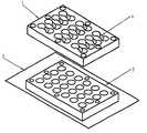
- FIG. 1is a perspective view of one configuration of a multi-well plate according to the present invention.
- the masking plate 1is secured to the base plate 2 , which is made from a thin polymer film, via a separate pressurizing plate 3 .
- Channels 4interconnect neighboring wells.
- the multi-well plate used in the present inventioncomprises the following main components: a removable base plate connected to a masking plate with openings forming an array.
- the base plate and masking plateare connected in such way that a liquid-tight connection is formed.
- the masking plateis covered by a removable lid providing a gas tight seal between the individual voids created by the composite of base plate, masking plate and lid and between said voids and the surrounding atmosphere.
- the material the base plate is made ofcan be selected from a variety of materials depending on the geometry used in the X-ray diffraction experiment. These materials must exhibit chemical and mechanical stability towards the chemical substances used in the crystallization experiment.
- the material the masking plate is made ofcan be selected from a variety of materials exhibiting chemical and mechanical stability towards the chemical substances used in the crystallization experiment.
- the material the lid is made ofcan be selected from materials which are not penetrated by air and vapors of the solvents used in the crystallization experiment. Generally an optically transparent material is preferred for the base plate as well as the lid, as it allows the observation of individual wells using light transmission microscopy.
- the base plateis made from a disc cut from a single crystal including but not limited to silicon or quartz.
- An alternative material for the base plateis sapphire.
- This plateis secured to the masking plate via a seperate additional pressurising plate which distributes an equal pressure over the whole contact area of all three plates. The connection is made by means of several bolts all tightened to the same tension.
- an additional gasketis inserted, repeating the pattern of the masking plate, or individual gaskets are inserted at the position of each opening of the masking plate.
- the lidis made preferably from an optically transparent material to enable visual observation of the wells. Again depending on the surface finish of the top surface of the masking plate and the bottom surface of the lid plate an additional gasket is inserted, repeating the pattern of the masking plate, or individual gaskets are inserted at the position of each opening of the masking plate.
- the base plateis made from a thin polymer film consisting of polyacetate, which exhibits in thicknesses as low as 0.015 mm no amorphous background scattering or regions in the diffractogram associated with crystallinity of the polymer.
- Other polymer filmsare also suitable for the purpose of forming the base plate, also some characteristic X-ray scattering will normally be observed.
- the polymer filmis secured to the masking plate via a separate additional pressurising plate which distributes an equal pressure over the whole contact area of the polymer film and the masking plate. Gaskets as described above are used where necessary.
- a material with as low as possible an average atomic number and as low as mechanically and chemically possible thicknessis used.
- the masking plateis made from a metallic material in order to conduct heat efficiently.
- optional heat exchangersare connected to the masking plate, in order to control and to change the temperature of the multiwell plate during the crystallization experiment.
- a temperature gradientis formed in a transverse direction along the masking plate by applying two different temperatures to either end of the masking plate.
- the openingsare lined with polytetrafluoroethylene (PTFE), or another chemically inert polymer to enhance the chemical resistivity of the masking plate while preserving the heat conducting properties.
- PTFEpolytetrafluoroethylene
- Arrays of crystalline, polycrystalline or even amorphous precipitatesare transferred to the X-ray micro-diffractometer for the purpose of characterization using X-ray diffraction.
- the base plateis separated from the remaining components of the multi-well plate.
- the essentially flat geometry of the base platethen permits the incident and diffracted X-ray beams to access the sample over a wide angular range in the case of reflection geometry.
- theseare positioned perpendicular to the X-ray beam via a clamping frame attached to the translation stage of the diffractometer.
- X-ray micro-diffractometerrequirements are the detection of micro-gram quantities of crystalline, polycrystalline or amorphous sample, automated movement of the sample support to the location of each predefined multi-well position, and large angular range of Bragg-angles including at least 4 to 120° 2 ⁇ combined with narrow X-ray beam diameter in the case of reflection geometry.
- the small beam diameteris needed to match the area illuminated by the X-ray beam to the dimensions of the sample when working in reflection geometry at small incident beam angles.
- the xyz-stageallows to analyse sequentially and fully automated each crystallization well.
- the sampleSince no sample preparation, i.e. grinding is applied, the sample will necessarily show spotty diffraction rings, possibly suffer from preferred orientation and in some cases show pronounced single crystal reflection.
- the area detectorcovers a considerable segment of the Laue-cones, which allows for averaging out the above effects during the integration process, which in turn results in a conventional diffractogram.
- the recorded diffractogramscan be compared to collections of known or of calculated diffractograms in order to confirm the identity of the obtained crystal form.
- the comparisonscan be made visually or by computer algorithms.
- the following exampleillustrates the preparation of an array of polycrystalline material from a single substance using a multi-well plate, for the subsequent characterization of said material by X-ray diffraction analysis.
- the compound 4′-methylchalcone 1is prepared according to the description of Kostanecki, St. v. and Rossbach G. (Chemische Berichte 29, 2245-2247, 1896). Recrystallization from ethanol yields 1 in 94.5% purity by GC.
- the multi-well plate used in this exampleconsists of a 0.03 mm thick polyacetate film sandwiched between two 10 mm thick brass plates, held in place by six M6 bolts. Both brass plates feature a 5 ⁇ 8 array of matching holes with 6 mm diameter.
- the upper brass plate, the masking platealso features on the top face channels with semicircular cross-section connecting adjoining holes in pairs.
- the bottom face of the masking plateis equipped with O-rings providing individual seals between the masking plate and the polymer film.
- 100 ⁇ l saturated ethanolic solution of 1are pipetted into the wells of the multi-well plate and covered with perforated parafilm®.
- the solventevaporates completely within 24 h and polycrystalline precipitates form on the bottom face of the filled wells.
- the polymer film with the attached crystalline precipitatesis removed from the multi-well plate assembly and positioned into the X-ray beam of a Bruker AXS GADDS (Bruker AXS Inc., 5465 East Cheryl Parkway, Madison, Wis. 53711, United States of America) diffractometer by means of a clamping frame.
- a Bruker AXS GADDS(Bruker AXS Inc., 5465 East Cheryl Parkway, Madison, Wis. 53711, United States of America) diffractometer by means of a clamping frame.
- the diffractometerconsists of a sealed Copper-radiation X-ray tube, crossed multi-layer parallel beam X-ray optics (Göbel mirrors) with a variable X-ray beam diamter between 0.05 mm and 0.5 mm, xyz-motorized programmable sample translation stage and photon counting multi-wire gas proportional X-ray detector.
- the recording of diffraction patterns at angles as low as 3 deg. 2-theta (for Cu-radiation)requires also a shortened X-ray collimator.
- the spatial resolution of the area detectoris about 0.1 mm in the 1024 ⁇ 1024 pixel storage mode and diffraction patterns are recorded typically at 200 mm distance; this distance can be varied between 60 and 300 mm.
- Part of the systemis also an alignment laser and video microscope which permits to center the desired part of the sample exactly in the center of the goniometer.
- Crystallization parametersinclude but are not limited to, change of solubility of the substance by change of concentration of the substance in solvent through solvent evaporation; change of solubility of the substance by adding precipitant; change of solubility of the substance through change of temperature.
- the rate of evaporationis controlled by differently sized appertures fixed to the top site of the multi-well plate.
- precipitantsare added in one of several ways depending on the difference in density and desired rate of mixing between the two solvent systems. Either by layering one solvent upon the other, or by allowing solvent vapor to diffuse between wells connected pairwise, above the meniscus of the solvents, and sealing of these well-pairs to the surrounding atmosphere to prevent loss of solvent.
- the temperature of multi-well plateis changed either with a time dependent gradient or sudden change or with a spatial gradient which puts different wells of the multi-well plate at different temperatures.
Landscapes
- Chemical & Material Sciences (AREA)
- Crystallography & Structural Chemistry (AREA)
- Health & Medical Sciences (AREA)
- Engineering & Computer Science (AREA)
- Organic Chemistry (AREA)
- Metallurgy (AREA)
- Analytical Chemistry (AREA)
- Materials Engineering (AREA)
- General Health & Medical Sciences (AREA)
- General Physics & Mathematics (AREA)
- Pathology (AREA)
- Hematology (AREA)
- Clinical Laboratory Science (AREA)
- Chemical Kinetics & Catalysis (AREA)
- Immunology (AREA)
- Biochemistry (AREA)
- Life Sciences & Earth Sciences (AREA)
- Physics & Mathematics (AREA)
- Analysing Materials By The Use Of Radiation (AREA)
Abstract
Description
Claims (19)
Priority Applications (1)
| Application Number | Priority Date | Filing Date | Title |
|---|---|---|---|
| US09/501,634US6507636B1 (en) | 2000-02-10 | 2000-02-10 | Rapid X-ray diffraction screening method of polymorph libraries created in multi-well plates |
Applications Claiming Priority (1)
| Application Number | Priority Date | Filing Date | Title |
|---|---|---|---|
| US09/501,634US6507636B1 (en) | 2000-02-10 | 2000-02-10 | Rapid X-ray diffraction screening method of polymorph libraries created in multi-well plates |
Publications (1)
| Publication Number | Publication Date |
|---|---|
| US6507636B1true US6507636B1 (en) | 2003-01-14 |
Family
ID=23994385
Family Applications (1)
| Application Number | Title | Priority Date | Filing Date |
|---|---|---|---|
| US09/501,634Expired - Fee RelatedUS6507636B1 (en) | 2000-02-10 | 2000-02-10 | Rapid X-ray diffraction screening method of polymorph libraries created in multi-well plates |
Country Status (1)
| Country | Link |
|---|---|
| US (1) | US6507636B1 (en) |
Cited By (25)
| Publication number | Priority date | Publication date | Assignee | Title |
|---|---|---|---|---|
| US20020067800A1 (en)* | 2000-10-19 | 2002-06-06 | Janet Newman | Apparatus and method for identification of crystals by in-situ X-ray diffraction |
| US20020183938A1 (en)* | 2000-04-07 | 2002-12-05 | Kobylecki Ryszard Jurek | Investigating different physical and/or chemical forms of materials |
| US20030116497A1 (en)* | 2001-08-10 | 2003-06-26 | Carlson Eric D. | Apparatuses and methods for creating and testing pre-formulations and systems for same |
| US20030147496A1 (en)* | 2001-06-29 | 2003-08-07 | Bruker Axs, Inc. | Diffraction system for biological crystal screening |
| US20030190758A1 (en)* | 2000-12-28 | 2003-10-09 | Stahly G. Patrick | Methods of searching for solid forms and screening a sample according to its forms |
| US20030219099A1 (en)* | 2002-03-21 | 2003-11-27 | Bruker Axs, Inc. | Transmission mode X-ray diffraction screening system |
| US6718008B1 (en)* | 2002-04-22 | 2004-04-06 | Bruker Axs, Inc. | X-ray diffraction screening system with retractable x-ray shield |
| EP1376108A3 (en)* | 2002-06-17 | 2004-04-21 | Rigaku Corporation | Crystal Evaluation Device |
| US20040103130A1 (en)* | 2002-08-06 | 2004-05-27 | Igor Ivanisevic | System and method for matching diffraction patterns |
| US20040208284A1 (en)* | 2003-04-17 | 2004-10-21 | Bruker Axs Gmbh | X-ray optical system for combinatorial screening of a sample library |
| US20050002487A1 (en)* | 2002-01-15 | 2005-01-06 | Erwin Blomsma | Method for performing power diffraction analysis |
| US20050069084A1 (en)* | 2001-10-03 | 2005-03-31 | Erwin Blomsma | Method for performing a transmission diffraction analysis |
| US20060023837A1 (en)* | 2004-07-30 | 2006-02-02 | Bruker Axs, Inc. | Combinatorial screening system with X-ray diffraction and Raman spectroscopy |
| WO2006021506A1 (en)* | 2004-08-26 | 2006-03-02 | Siemens Aktiengesellschaft | Reactor for producing surface reactions, whereby the reaction opening has a certain geometry, and use of the same |
| US20060129329A1 (en)* | 2001-04-09 | 2006-06-15 | Kobylecki Ryszard J | Investigating different physical and/or chemical forms of materials |
| US20060140821A1 (en)* | 2004-12-17 | 2006-06-29 | Rosso Victor W | Powder X-ray diffraction sample holder |
| US20070021929A1 (en)* | 2000-01-07 | 2007-01-25 | Transform Pharmaceuticals, Inc. | Computing methods for control of high-throughput experimental processing, digital analysis, and re-arraying comparative samples in computer-designed arrays |
| US20070020662A1 (en)* | 2000-01-07 | 2007-01-25 | Transform Pharmaceuticals, Inc. | Computerized control of high-throughput experimental processing and digital analysis of comparative samples for a compound of interest |
| US20070134803A1 (en)* | 2003-10-06 | 2007-06-14 | Solvias Ag | Process for the parallel detection of crystalline forms of molecular solids |
| US20080273662A1 (en)* | 2007-05-04 | 2008-11-06 | Xradia, Inc. | CD-GISAXS System and Method |
| US20080316465A1 (en)* | 2007-06-06 | 2008-12-25 | Alice Wernicki | Sample holder and sample preparation device |
| US20090010388A1 (en)* | 2007-06-06 | 2009-01-08 | Stahly Barbara C | Microplate and methods of using the same |
| US20090205412A1 (en)* | 2006-07-17 | 2009-08-20 | Avantium International B.V. | Method For Obtaining And Analyzing Solids, Preferably Crystals |
| US20160019994A1 (en)* | 2014-06-26 | 2016-01-21 | The Board Of Trustees Of The Leland Stanford Junior University | High Density Grids |
| US9339816B2 (en) | 2008-10-28 | 2016-05-17 | Grünenthal GmbH | Reactor array for producing and analyzing products |
Citations (10)
| Publication number | Priority date | Publication date | Assignee | Title |
|---|---|---|---|---|
| US4278883A (en)* | 1979-12-27 | 1981-07-14 | The United States Of America As Represented By The Secretary Of The Interior | Sample mount for X-ray diffraction |
| US4634599A (en)* | 1985-05-02 | 1987-01-06 | General Electric Company | Method for making ordered monolayers of macromolecules |
| US4862488A (en)* | 1986-03-18 | 1989-08-29 | U.S. Philips Corporation | Device for measuring the orientation of bulk monocrystalline materials using the Laue method |
| US4961210A (en)* | 1990-02-28 | 1990-10-02 | The United States Of America As Represented By The Secretary Of The Navy | High resolution technique and instrument for measuring lattice parameters in single crystals |
| US5221410A (en)* | 1991-10-09 | 1993-06-22 | Schering Corporation | Crystal forming device |
| US5414747A (en)* | 1993-02-22 | 1995-05-09 | The Penn State Research Foundation | Method and apparatus for in-process analysis of polycrystalline films and coatings by x-ray diffraction |
| US5597457A (en)* | 1995-01-23 | 1997-01-28 | The Regents Of The University Of California | System and method for forming synthetic protein crystals to determine the conformational structure by crystallography |
| US6005914A (en)* | 1997-03-28 | 1999-12-21 | Philips Electronics North America Corporation | Arc diffractometer |
| US6111930A (en)* | 1998-08-29 | 2000-08-29 | Bruker Axs Analytical X-Ray Systems Gmbh | Automatic sample changer for an X-ray diffractometer |
| US6168914B1 (en)* | 1997-12-19 | 2001-01-02 | Glaxo Wellcome Inc. | System and method for solid-phase parallel synthesis of a combinatorial collection of compounds |
- 2000
- 2000-02-10USUS09/501,634patent/US6507636B1/ennot_activeExpired - Fee Related
Patent Citations (10)
| Publication number | Priority date | Publication date | Assignee | Title |
|---|---|---|---|---|
| US4278883A (en)* | 1979-12-27 | 1981-07-14 | The United States Of America As Represented By The Secretary Of The Interior | Sample mount for X-ray diffraction |
| US4634599A (en)* | 1985-05-02 | 1987-01-06 | General Electric Company | Method for making ordered monolayers of macromolecules |
| US4862488A (en)* | 1986-03-18 | 1989-08-29 | U.S. Philips Corporation | Device for measuring the orientation of bulk monocrystalline materials using the Laue method |
| US4961210A (en)* | 1990-02-28 | 1990-10-02 | The United States Of America As Represented By The Secretary Of The Navy | High resolution technique and instrument for measuring lattice parameters in single crystals |
| US5221410A (en)* | 1991-10-09 | 1993-06-22 | Schering Corporation | Crystal forming device |
| US5414747A (en)* | 1993-02-22 | 1995-05-09 | The Penn State Research Foundation | Method and apparatus for in-process analysis of polycrystalline films and coatings by x-ray diffraction |
| US5597457A (en)* | 1995-01-23 | 1997-01-28 | The Regents Of The University Of California | System and method for forming synthetic protein crystals to determine the conformational structure by crystallography |
| US6005914A (en)* | 1997-03-28 | 1999-12-21 | Philips Electronics North America Corporation | Arc diffractometer |
| US6168914B1 (en)* | 1997-12-19 | 2001-01-02 | Glaxo Wellcome Inc. | System and method for solid-phase parallel synthesis of a combinatorial collection of compounds |
| US6111930A (en)* | 1998-08-29 | 2000-08-29 | Bruker Axs Analytical X-Ray Systems Gmbh | Automatic sample changer for an X-ray diffractometer |
Cited By (49)
| Publication number | Priority date | Publication date | Assignee | Title |
|---|---|---|---|---|
| US20070020662A1 (en)* | 2000-01-07 | 2007-01-25 | Transform Pharmaceuticals, Inc. | Computerized control of high-throughput experimental processing and digital analysis of comparative samples for a compound of interest |
| US20070021929A1 (en)* | 2000-01-07 | 2007-01-25 | Transform Pharmaceuticals, Inc. | Computing methods for control of high-throughput experimental processing, digital analysis, and re-arraying comparative samples in computer-designed arrays |
| US20020183938A1 (en)* | 2000-04-07 | 2002-12-05 | Kobylecki Ryszard Jurek | Investigating different physical and/or chemical forms of materials |
| US6965832B2 (en) | 2000-04-07 | 2005-11-15 | Millennium Pharmaceuticals, Inc. | Investigating different physical and/or chemical forms of materials |
| US20060025934A1 (en)* | 2000-04-07 | 2006-02-02 | Kobylecki Ryszard J | Investigating different physical and/or chemical forms of materials |
| US20020067800A1 (en)* | 2000-10-19 | 2002-06-06 | Janet Newman | Apparatus and method for identification of crystals by in-situ X-ray diffraction |
| US20030190758A1 (en)* | 2000-12-28 | 2003-10-09 | Stahly G. Patrick | Methods of searching for solid forms and screening a sample according to its forms |
| US6750064B2 (en)* | 2000-12-28 | 2004-06-15 | S.S.C.I. Inc. | Methods of screening for possible solid forms |
| US20040209304A1 (en)* | 2000-12-28 | 2004-10-21 | Purdue Research Foundation | Methods of searching for solid forms and screening a sample according to its forms |
| US20060129329A1 (en)* | 2001-04-09 | 2006-06-15 | Kobylecki Ryszard J | Investigating different physical and/or chemical forms of materials |
| US20030147496A1 (en)* | 2001-06-29 | 2003-08-07 | Bruker Axs, Inc. | Diffraction system for biological crystal screening |
| US6836532B2 (en)* | 2001-06-29 | 2004-12-28 | Bruker Axs, Inc. | Diffraction system for biological crystal screening |
| US20070077657A1 (en)* | 2001-08-10 | 2007-04-05 | Symyx Technologies, Inc. | Apparatuses and methods for creating and testing pre-formulations and systems for same |
| US7549978B2 (en) | 2001-08-10 | 2009-06-23 | Symyx Technologies, Inc. | Needle assembly |
| US6939515B2 (en) | 2001-08-10 | 2005-09-06 | Symyx Technologies, Inc. | Apparatuses and methods for creating and testing pre-formulations and systems for same |
| US20030116497A1 (en)* | 2001-08-10 | 2003-06-26 | Carlson Eric D. | Apparatuses and methods for creating and testing pre-formulations and systems for same |
| US20050211622A1 (en)* | 2001-08-10 | 2005-09-29 | Symyx Technologies, Inc. | Apparatuses and methods for creating and testing pre-formulations and systems for same |
| US7079621B2 (en) | 2001-10-03 | 2006-07-18 | Avantium International B.V. | Vertical transmission diffraction analysis |
| US20050069084A1 (en)* | 2001-10-03 | 2005-03-31 | Erwin Blomsma | Method for performing a transmission diffraction analysis |
| US20050002487A1 (en)* | 2002-01-15 | 2005-01-06 | Erwin Blomsma | Method for performing power diffraction analysis |
| US7702071B2 (en) | 2002-01-15 | 2010-04-20 | Avantium International B.V. | Method for performing power diffraction analysis |
| US6859520B2 (en) | 2002-03-21 | 2005-02-22 | Bruker Axs, Inc. | Transmission mode X-ray diffraction screening system |
| US20030219099A1 (en)* | 2002-03-21 | 2003-11-27 | Bruker Axs, Inc. | Transmission mode X-ray diffraction screening system |
| WO2003081221A3 (en)* | 2002-03-21 | 2004-01-22 | Bruker Axs Inc | Transmission mode x-ray diffraction screening system |
| EP1353166A3 (en)* | 2002-04-09 | 2004-01-14 | Bruker AXS, Inc. | Diffraction system for biological crystal screening |
| US6718008B1 (en)* | 2002-04-22 | 2004-04-06 | Bruker Axs, Inc. | X-ray diffraction screening system with retractable x-ray shield |
| US20040258203A1 (en)* | 2002-06-17 | 2004-12-23 | Akihito Yamano | Crystal evaluating device |
| EP1376108A3 (en)* | 2002-06-17 | 2004-04-21 | Rigaku Corporation | Crystal Evaluation Device |
| US7715527B2 (en) | 2002-08-06 | 2010-05-11 | Aptuit (Kansas City), Llc | System and method for matching diffraction patterns |
| US20040103130A1 (en)* | 2002-08-06 | 2004-05-27 | Igor Ivanisevic | System and method for matching diffraction patterns |
| US7372941B2 (en)* | 2002-08-06 | 2008-05-13 | Ssci, Inc. | System and method for matching diffraction patterns |
| US20080120051A1 (en)* | 2002-08-06 | 2008-05-22 | Ssci, Inc. | System and Method for Matching Diffraction Patterns |
| US20040208284A1 (en)* | 2003-04-17 | 2004-10-21 | Bruker Axs Gmbh | X-ray optical system for combinatorial screening of a sample library |
| US20070134803A1 (en)* | 2003-10-06 | 2007-06-14 | Solvias Ag | Process for the parallel detection of crystalline forms of molecular solids |
| US7892354B2 (en)* | 2003-10-06 | 2011-02-22 | Solvias Ag | Process for the parallel detection of crystalline forms of molecular solids |
| US7269245B2 (en)* | 2004-07-30 | 2007-09-11 | Bruker Axs, Inc. | Combinatorial screening system and X-ray diffraction and Raman spectroscopy |
| EP1632770A3 (en)* | 2004-07-30 | 2007-05-16 | Bruker AXS, Inc. | Combination screening system with X-ray diffraction and raman spectroscopy |
| US20060023837A1 (en)* | 2004-07-30 | 2006-02-02 | Bruker Axs, Inc. | Combinatorial screening system with X-ray diffraction and Raman spectroscopy |
| WO2006021506A1 (en)* | 2004-08-26 | 2006-03-02 | Siemens Aktiengesellschaft | Reactor for producing surface reactions, whereby the reaction opening has a certain geometry, and use of the same |
| US20060140821A1 (en)* | 2004-12-17 | 2006-06-29 | Rosso Victor W | Powder X-ray diffraction sample holder |
| US20090205412A1 (en)* | 2006-07-17 | 2009-08-20 | Avantium International B.V. | Method For Obtaining And Analyzing Solids, Preferably Crystals |
| US20080273662A1 (en)* | 2007-05-04 | 2008-11-06 | Xradia, Inc. | CD-GISAXS System and Method |
| US7920676B2 (en)* | 2007-05-04 | 2011-04-05 | Xradia, Inc. | CD-GISAXS system and method |
| US20090010388A1 (en)* | 2007-06-06 | 2009-01-08 | Stahly Barbara C | Microplate and methods of using the same |
| US20080316465A1 (en)* | 2007-06-06 | 2008-12-25 | Alice Wernicki | Sample holder and sample preparation device |
| US8018588B2 (en) | 2007-06-06 | 2011-09-13 | Aptuit, Inc. | Sample holder and sample preparation device |
| US9339816B2 (en) | 2008-10-28 | 2016-05-17 | Grünenthal GmbH | Reactor array for producing and analyzing products |
| US20160019994A1 (en)* | 2014-06-26 | 2016-01-21 | The Board Of Trustees Of The Leland Stanford Junior University | High Density Grids |
| US9869648B2 (en)* | 2014-06-26 | 2018-01-16 | The Board Of Trustees Of The Leland Stanford Junior University | High density grids |
Similar Documents
| Publication | Publication Date | Title |
|---|---|---|
| US6507636B1 (en) | Rapid X-ray diffraction screening method of polymorph libraries created in multi-well plates | |
| US6602714B1 (en) | Viscosity and mass sensor for the high-throughput synthesis, screening and characterization of combinatorial libraries | |
| US7409041B2 (en) | Methods of transmission mode X-ray diffraction analysis and apparatuses therefor | |
| US6977723B2 (en) | Apparatus and method for high-throughput preparation and spectroscopic classification and characterization of compositions | |
| US7033840B1 (en) | Reaction calorimeter and differential scanning calorimeter for the high-throughput synthesis, screening and characterization of combinatorial libraries | |
| US8675192B2 (en) | Method and device for high speed quantitative measurement of biomolecular targets on or in biological analysis medium | |
| EP1327012A1 (en) | Apparatus and method for identification of crystals by in-situ x-ray diffraction | |
| US6677162B1 (en) | Process of parallel sample preparation | |
| US20030106492A1 (en) | Apparatus and method for high-throughput preparation, visualization and screening of compositions | |
| US20050095696A9 (en) | Apparatus and method for high-throughput preparation and characterization of compositions | |
| US7597852B2 (en) | Substrate for sample analyses | |
| CA2459241C (en) | Sealing system with flow channels | |
| US20090010388A1 (en) | Microplate and methods of using the same | |
| US6806093B2 (en) | Process of parallel sample preparation | |
| EP1466166B2 (en) | Method for performing powder diffraction analysis | |
| US7410804B2 (en) | Process of parallel sample presentation | |
| CA2460647C (en) | Method for performing a transmission diffraction analysis | |
| HUP0400376A2 (en) | Device for performing catalytic screening | |
| EP1172646A1 (en) | Screening crystallisation conditions of organic compounds | |
| EP1720007B1 (en) | Method and apparatus for X-ray diffraction analysis | |
| EP1467205A1 (en) | Polymorph characterization | |
| EP1327877A1 (en) | Method for successively performing powder diffraction analysis on a plurality of samples | |
| US20060040394A1 (en) | Method of searching for and generating polymrophs of a substance | |
| US20060140821A1 (en) | Powder X-ray diffraction sample holder | |
| AU2002246760A1 (en) | Apparatus and method for identification of crystals by in-SITU X-ray diffraction |
Legal Events
| Date | Code | Title | Description |
|---|---|---|---|
| AS | Assignment | Owner name:STUDIENGESELLSCHAFT KOHLE MBH, GERMANY Free format text:ASSIGNMENT OF ASSIGNORS INTEREST;ASSIGNOR:LEHMANN, CHRISTIAN W.;REEL/FRAME:010596/0846 Effective date:20000204 | |
| AS | Assignment | Owner name:SYMYX TECHNOLOGIES, INC., CALIFORNIA Free format text:ASSIGNMENT OF ASSIGNORS INTEREST;ASSIGNOR:LEHMANN, CHRISTIAN W.;REEL/FRAME:014162/0822 Effective date:20030512 | |
| AS | Assignment | Owner name:SYMYX TECHNOLOGIES, INC., CALIFORNIA Free format text:ASSIGNMENT OF ASSIGNORS INTEREST;ASSIGNOR:STUDIENGESELLSCHAFT KOHLE MBH;REEL/FRAME:013835/0023 Effective date:20030528 | |
| FEPP | Fee payment procedure | Free format text:PAYOR NUMBER ASSIGNED (ORIGINAL EVENT CODE: ASPN); ENTITY STATUS OF PATENT OWNER: LARGE ENTITY | |
| FEPP | Fee payment procedure | Free format text:PAT HOLDER NO LONGER CLAIMS SMALL ENTITY STATUS, ENTITY STATUS SET TO UNDISCOUNTED (ORIGINAL EVENT CODE: STOL); ENTITY STATUS OF PATENT OWNER: LARGE ENTITY | |
| REFU | Refund | Free format text:REFUND - SURCHARGE, PETITION TO ACCEPT PYMT AFTER EXP, UNINTENTIONAL (ORIGINAL EVENT CODE: R2551); ENTITY STATUS OF PATENT OWNER: LARGE ENTITY | |
| CC | Certificate of correction | ||
| FPAY | Fee payment | Year of fee payment:4 | |
| AS | Assignment | Owner name:SYMYX SOLUTIONS, INC., CALIFORNIA Free format text:ASSIGNMENT OF ASSIGNORS INTEREST;ASSIGNOR:SYMYX TECHNOLOGIES, INC.;REEL/FRAME:022939/0564 Effective date:20090701 Owner name:SYMYX SOLUTIONS, INC.,CALIFORNIA Free format text:ASSIGNMENT OF ASSIGNORS INTEREST;ASSIGNOR:SYMYX TECHNOLOGIES, INC.;REEL/FRAME:022939/0564 Effective date:20090701 | |
| AS | Assignment | Owner name:FREESLATE, INC.,CALIFORNIA Free format text:ASSIGNMENT OF ASSIGNORS INTEREST;ASSIGNOR:SYMYX SOLUTIONS, INC.;REEL/FRAME:024057/0911 Effective date:20100301 Owner name:FREESLATE, INC., CALIFORNIA Free format text:ASSIGNMENT OF ASSIGNORS INTEREST;ASSIGNOR:SYMYX SOLUTIONS, INC.;REEL/FRAME:024057/0911 Effective date:20100301 | |
| REMI | Maintenance fee reminder mailed | ||
| LAPS | Lapse for failure to pay maintenance fees | ||
| STCH | Information on status: patent discontinuation | Free format text:PATENT EXPIRED DUE TO NONPAYMENT OF MAINTENANCE FEES UNDER 37 CFR 1.362 | |
| FP | Lapsed due to failure to pay maintenance fee | Effective date:20110114 | |
| AS | Assignment | Owner name:APTUIT (KANSAS CITY), LLC, CONNECTICUT Free format text:RELEASE OF SECURITY INTEREST;ASSIGNOR:GENERAL ELECTRIC CAPITAL CORPORATION;REEL/FRAME:027793/0505 Effective date:20120217 Owner name:APTUIT (WEST LAFAYETTE), LLC, DELAWARE Free format text:RELEASE OF SECURITY INTEREST;ASSIGNOR:GENERAL ELECTRIC CAPITAL CORPORATION;REEL/FRAME:027793/0505 Effective date:20120217 |