US6478746B2 - Acoustic sensor array for non-invasive detection of coronary artery disease - Google Patents
Acoustic sensor array for non-invasive detection of coronary artery diseaseDownload PDFInfo
- Publication number
- US6478746B2 US6478746B2US09/734,448US73444800AUS6478746B2US 6478746 B2US6478746 B2US 6478746B2US 73444800 AUS73444800 AUS 73444800AUS 6478746 B2US6478746 B2US 6478746B2
- Authority
- US
- United States
- Prior art keywords
- window
- array
- acoustic
- acoustic window
- sensors
- Prior art date
- Legal status (The legal status is an assumption and is not a legal conclusion. Google has not performed a legal analysis and makes no representation as to the accuracy of the status listed.)
- Expired - Lifetime, expires
Links
- 238000001514detection methodMethods0.000titledescription4
- 208000029078coronary artery diseaseDiseases0.000titledescription3
- 238000000034methodMethods0.000claimsabstractdescription13
- 230000003601intercostal effectEffects0.000claimsdescription23
- 210000004072lungAnatomy0.000claimsdescription6
- 238000013461designMethods0.000abstractdescription13
- 239000002033PVDF binderSubstances0.000description16
- 229920002981polyvinylidene fluoridePolymers0.000description16
- 210000000038chestAnatomy0.000description12
- 238000003491arrayMethods0.000description9
- 239000000523sampleSubstances0.000description8
- 230000017531blood circulationEffects0.000description7
- 230000002596correlated effectEffects0.000description4
- 239000010408filmSubstances0.000description4
- 210000003484anatomyAnatomy0.000description3
- 239000002131composite materialSubstances0.000description3
- 238000013480data collectionMethods0.000description3
- 230000000694effectsEffects0.000description3
- 210000001562sternumAnatomy0.000description3
- 239000010409thin filmSubstances0.000description3
- 210000001519tissueAnatomy0.000description3
- 238000012935AveragingMethods0.000description2
- 230000002159abnormal effectEffects0.000description2
- 230000000747cardiac effectEffects0.000description2
- 230000000875corresponding effectEffects0.000description2
- 238000005516engineering processMethods0.000description2
- 238000001119image correlation spectroscopyMethods0.000description2
- 238000005259measurementMethods0.000description2
- 210000002445nippleAnatomy0.000description2
- 210000000779thoracic wallAnatomy0.000description2
- 238000002604ultrasonographyMethods0.000description2
- 238000012800visualizationMethods0.000description2
- 230000002411adverseEffects0.000description1
- 238000004458analytical methodMethods0.000description1
- 210000001765aortic valveAnatomy0.000description1
- 238000012512characterization methodMethods0.000description1
- 238000012938design processMethods0.000description1
- 238000006073displacement reactionMethods0.000description1
- 238000003384imaging methodMethods0.000description1
- 238000001727in vivoMethods0.000description1
- 230000001788irregularEffects0.000description1
- 230000004807localizationEffects0.000description1
- 238000004519manufacturing processMethods0.000description1
- 229920000642polymerPolymers0.000description1
- 238000011160researchMethods0.000description1
- 238000007619statistical methodMethods0.000description1
- 238000012546transferMethods0.000description1
Images


Classifications
- A—HUMAN NECESSITIES
- A61—MEDICAL OR VETERINARY SCIENCE; HYGIENE
- A61B—DIAGNOSIS; SURGERY; IDENTIFICATION
- A61B7/00—Instruments for auscultation
- A—HUMAN NECESSITIES
- A61—MEDICAL OR VETERINARY SCIENCE; HYGIENE
- A61B—DIAGNOSIS; SURGERY; IDENTIFICATION
- A61B2562/00—Details of sensors; Constructional details of sensor housings or probes; Accessories for sensors
- A61B2562/02—Details of sensors specially adapted for in-vivo measurements
- A61B2562/0204—Acoustic sensors
- A—HUMAN NECESSITIES
- A61—MEDICAL OR VETERINARY SCIENCE; HYGIENE
- A61B—DIAGNOSIS; SURGERY; IDENTIFICATION
- A61B2562/00—Details of sensors; Constructional details of sensor housings or probes; Accessories for sensors
- A61B2562/04—Arrangements of multiple sensors of the same type
- A61B2562/046—Arrangements of multiple sensors of the same type in a matrix array
Definitions
- This inventionrelates to arrays of acoustic sensors that facilitate the non-invasive detection of coronary artery disease (CAD).
- CADcoronary artery disease
- the 20186 applicationdescribes an invention for the non-invasive in vivo detection and localization of abnormal blood flow.
- Embodiments of that inventiondisplay the spatial distribution of phase coherence in the shear eave component of blood flow signals generated by an acoustic sensor array.
- An essentially uniform displayindicates normal blood flow.
- a non-uniform displaymay indicate the presence of an occlusion and the presence or extent of abnormal, turbulent blood flow. Poor correlation of signals from the array sensors may adversely affect the display uniformity.
- Acoustic sensor arraysare conventionally positioned above a measurement area defined as the hairless human chest skin located vertically between the sternum and a parallel line passing through the left nipple and horizontally 10 cm above and 6 cm below the left and right nipples.
- FIG. 6 of the 20186 applicationA prior art acoustic sensor array comprising eight equally spaced sensors in two concentric circles having prime numbers of sensors in each circle and a ninth sensor at the common center of the concentric circle is illustrated by FIG. 6 of the 20186 application.
- a notchin the human left lung allows the heart to be in contact with the chest wall.
- Well correlated blood flow signalsmay be generated by acoustic sensors positioned on a human chest in a small area (“acoustic window”) located above the cardiac notch.
- the bounds of the acoustic windowhave been approximated by ultrasonic probe means as described in this application and by locating the portions of sensor corresponding to channels achieve the highest apparent signal to noise ratio (SNR) as described in the Stearns application.
- SNRsignal to noise ratio
- Well correlated acoustic blood flow signals of good qualitymay be generated by a sensor array positioned on a patient's chest within or substantially within the perimeter of an acoustic window.
- Any current or voltage mode devicewhich generates an electric signal from displacement or a derivative thereof upon detection of a sound wave.
- an acoustic windowmay be defined by ultrasonic probe means.
- the inventionincludes sensor arrays having an aperture locatable within or substantially within the bounds of an acoustic window when the array is positioned on the chest of a person.
- An important aspect of the inventionincludes the identification of an acoustic window comprising the merged acoustic window sub-areas corresponding to two or more intercostal spaces (ICS's), and array designs to accommodate such acoustic windows.
- ICS'sintercostal spaces
- FIG. 1illustrates an ultrasonic probe acoustic window characterization method that provides a template for the positioning of sensors on a person's chest. All acoustic window data illustrated by FIGS. 2 through 8 and 10 through 12 was obtained by the FIG. 1 method.
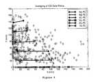
- FIG. 2is a plot in polar format of the acoustic window size data obtained from 22 male and 7 female subjects (29 subjects).
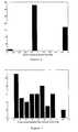
- FIG. 3is a histogram of the window areas of the same 29 subjects from which the FIG. 2 data was obtained.
- FIG. 4shows acoustic window size in polar format. Maximum, minimum and average window size for all of the same 29 subjects is depicted.
- FIG. 5illustrates in Cartesian coordinates variations of the ICS ultrasonic probe data points from the left ( ⁇ ) and right ( ⁇ ) ICS.
- the statistical averages for ICS's 1 to 5are shown in solid lines.
- a perimeter connecting the ends of the solid linesis a visualization of the average geometry of the six intercostal spaces.
- FIG. 6is a histogram that indicates ICS nearest to the centroids of the average window area (see FIG. 4 ).
- FIG. 7is a histogram indicating the distribution of the perpendicular distances from the centroid of the average window area (see FIG. 4) to the nearest ICS.
- FIG. 8is a histogram illustrating the distribution of the distance from the left side of the nearest ICS to the projection of the centroid of the average window area (see FIG. 4 ).
- FIG. 9depicts a prior art nine sensor array based on seismic accelerometers commercially available from Wilcoxon Research, 21 Firstfield Road, Gaithersburg, Md. 20878.
- the arraycomprises eight equally spaced sensors in two concentric circles having prime numbers of sensors in each circle and a ninth sensor in the common center of the concentric circles.
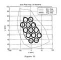
- FIG. 10depicts a 13 element array positioned over an acoustic window of average size (solid line, see FIG. 4 ). An acoustic window of maximum area is also shown (broken line).
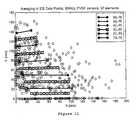
- FIG. 11illustrates a 57 element small PVDF sensor array based on averaging of ICS data points.
- the arraycomprises five linear subarrays positioned above intercostal spaces 2 to 6 .
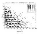
- FIG. 12illustrates a 32 element array of large PVDF sensors based on averaging of ICS data points.
- the arraycomprises five linear subarrays positioned above intercostal spaces 2 to 6 .
- FIG. 13illustrates a beam pattern in x for delay-and-sum (DS) (higher in value) and MVDR (lower in value) beamformers using prior art (HA) (dashed lines), bowling pin (BO) (dashed dotted lines), small PDVF (SP) (solid lines) and large PDVF (LP) (dotted lines) arrays.
- HAprior art
- BObowling pin
- SPsmall PDVF
- LPlarge PDVF
- FIG. 14illustrates a beam pattern in y (dB) for delay-and-sum (DS) (broken lines) and MVDR (solid lines) beamformers using prior art (HA), bowling pin (BO), small PDVF (SP) and large PDVF (LP) arrays.
- DSdelay-and-sum
- MVDRsolid lines
- FIG. 15illustrates a beam pattern in z (dB) for delay-and-sum (DS) (broken lines) and MVDR (solid lines) beamformers using Harris (HA), bowling pin (BO), small PDVF (SP) and large PDVF (LP) arrays.
- DSdelay-and-sum
- MVDRsolid lines
- FIG. 16is a proposed array based on the medical Wilcoxon accelerometer. Only 12 elements are used due to the limitation of the data collection system. The broken line indicates the perimeter of an acoustic window.
- FIG. 17illustrates a 45-element PVDF sensor array comprising five nine element linear subarrays positioned above intercostal spaces 2 to 6 . Large and small acoustic window perimeters with centroids near the fourth and fifth ICS's are shown.
- the acoustic windowmay include one or a combination of the small areas (intercostal space window areas) of the patient's chest surface directly above the intercostal spaces one through six. Determination of the size of an acoustic window may be accomplished by steps (i) to (v).
- step (iii)Repeat step (ii) for intercostal spaces two through six.
- Average or “generic” templatesmay be prepared from average data determined in the same way from a plurality of persons.
- a like proceduremay be used to determine an acoustic window of a person lying slanted on a bed.
- Table 1lists the window areas in cm-squared for the three window sizes in two bed positions. For the maximum and average window cases, lying slanted on the bed produces a slightly larger (5% and 16%, respectively) window size than lying flat on the bed. In the case of the minimum window, lying flat does produce a significant 37% larger window area than lying slanted on the bed.
- the flat positionis more advantageous since it does not significantly reduce the acoustics window for subjects with large to medium window sizes and at the same time significantly opens up the smaller acoustic window sizes.
- Another method for identifying an acoustic windowentails examination of which sensor channels receive the highest signal to noise ratio (SNR) as measured by the optimal weights for summing channels. See copending, commonly assigned Stearns U.S. application.
- SNRsignal to noise ratio
- the distribution of the window size data in polar formatwas tested for normality using the normal probability plot from the MATLAB Statistics Toolbox.
- the purpose of a normal probability plotis to graphically assess whether the data could come from a normal distribution. If the data are normal, the plot will be linear. Other distribution types will introduce curvature in the plot. As shown in FIG. 2, the data points are virtually in a straight line, indicating that the polar coordinate data is Gaussian.
- FIG. 3illustrates the histogram of the window areas.
- FIG. 4shows the acoustics window size in polar format.
- the outside perimeteris for maximum
- the inside perimeteris for the minimum
- the intermediate perimeteris for the average across all subjects.
- Computed correlation coefficients between the window area and the subject demographics dataare shown in Table 2. This analysis was carried out based on data broken down by male (22 subjects), female (7 subjects) and a combination of both sexes. In general, there exists no strong correlation between the window area and demographics data, with the exception of strong negative correlation of ⁇ 0.84 between the acoustics window area and the anterior/posterior (AP) diameter of the female subjects and a strong negative correlation between the acoustics window area and the sternum length in both male and female subjects.
- APanterior/posterior
- the XY coordinates of the left in ( ⁇ ) and right in ( ⁇ ) ICSare quite different across the subjects.
- the statistical averages of the left and right ICSare also shown.
- a visualization of the average geometry of the six intercostal spacesis provided.
- the x-y coordinates of the acoustics window centroidswere measured and correlated with the lines defined by the left and right ICS. The purpose of this correlation is to determine which of the six spaces the window centroid is near to and then to ascertain the best space(s) for location of the array.
- a histogram of the ICS to which the centroids of the window areas are nearestis plotted in FIG. 6 .
- the resultindicates that the fourth and fifth ICS are good candidates for positioning the array center, with the fourth ICS being more frequent than the fifth ICS.
- Constraints imposed on array geometryinclude:
- Sensor sizewhich limits the number of elements that can be put in the array aperture.
- the medical Wilcoxon sensor diameteris about one cm.
- the prefabricated thin film strip sizedictates how many sensors could be placed in the array aperture.
- the inter-element spacing of the sensorsis required to be less than half a wavelength at the highest operating frequency to avoid spatial aliasing in the plane wave case. This requirement is relaxed in the near field where source location is the objective.
- the use of irregularity in array geometrymay also alleviate the aliasing problem when there is an inter-element spacing of more than half wavelength.
- the 13-element array of FIG. 10was based on the average window size described with reference to FIG. 4 . Using the actual dimensions of the medical Wilcoxon accelerometer on graph paper, each accelerometer was placed on straight lines starting from the center and populating the perimeter until space is occupied. A total of 13 elements that were fitted into this average window size. Clinical data indicates that 13 elements may not give optimum array gain especially when element signal-to-noise ratio of turbulent flow is low.
- PVDF filmis available in linear strips of 9 and 16 elements per unit, each strip can be put on the intercostal space to maximize signal reception. These factors motivate the array geometries illustrated in FIGS. 11 and 12.
- FIGS. 9, 10 , 11 and 12The performance of the four sensor arrays depicted by FIGS. 9, 10 , 11 and 12 is presented in terms of beam width and array gain by FIGS. 13, 14 and 15 .
- the beam pattern plotsare for frequency at 250 Hz using Verberg propagation model and 10 db element SNR. These figures show the beam patterns in x, y and z direction for a source 3 cm directly below the array center.
- the beam pattern for the conventional delay and sum (DS) beamformeris shown in dashed line, and the beam pattern for the MVDR beamformer is in solid line.
- the figuresshow that for a conventional beamformer, the array gain is proportional to the number of elements.
- the effect of the number of elements on the array beamwidthis much more visible for the MVDR beamformer than for the DS beamformer.
- Also notableis the lack of array aperture in the z direction, as illustrated by the large beam width shown in FIG. 15 .
- the MVDR beamformerprovides an estimate of the signal power at the signal direction as can clearly be seen from FIGS. 13, 14 and 15 .
- the output of the MVDR beamformeris 10 dB regardless of the number of element in the array.
- the effect of an increase in the number of elementsis a narrower beam width, which is consistent with data showing that the beam width of an MVDR beamformer is inversely proportional to the number of elements (and the element SNR).
- the acoustics windowis the union of the two window areas for the 4 th and 5 th ICS. These two windows are the average of the XY data obtained from the acoustics window study. The merging of the two windows increases the area available for the array aperture which is an advantage to array performance.
- FIG. 16A design for a Wilcoxon commercial accelerometer array is shown in FIG. 16 .
- the composite window areaconsists of the two averaged windows with centroids near the 4 th and 5 th ICSs.
- the missing elementsare chosen such that the resulting array is as irregular as possible with at least one sensor pair very close to each other to prevent spatial aliasing.
- FIG. 17For the same composite acoustics window, a 45-element PVDF array is shown in FIG. 17 .
- This arrayessentially consists of 5 rows of 9-element large PVDF linear array arranged in such a way that conforms to the human chest curvature and, if possible, lies within the lower ICS to adapt to the patient anatomy.
- One reason for a 5 by 9 linear PVDF arrayis in the manufacturing and logistics of the thin film technology.
- An acceptable data collection schemeincludes estimation of the signal-to-noise ratio at each element, and weighting or eliminating the sensors that receive the noisiest signal. The use of this weighting technique enables the array to adapt to the differences in acoustic window size that are embodied in human anatomy.
Landscapes
- Health & Medical Sciences (AREA)
- Life Sciences & Earth Sciences (AREA)
- Engineering & Computer Science (AREA)
- Biomedical Technology (AREA)
- Heart & Thoracic Surgery (AREA)
- Medical Informatics (AREA)
- Molecular Biology (AREA)
- Surgery (AREA)
- Animal Behavior & Ethology (AREA)
- General Health & Medical Sciences (AREA)
- Public Health (AREA)
- Veterinary Medicine (AREA)
- Ultra Sonic Daignosis Equipment (AREA)
Abstract
Description
This application is a divisional of U.S. application Ser. No. 09/188,434 filed Nov. 9, 1998, the contents of which are hereby incorporated by reference as if recited in full herein.
This application is related to concurrently filed and co-assigned U.S. Patent application entitled “Non-Invasive Turbulent Blood Flow Imaging System” identified by U.S. Ser. No. 09/188,510 which corresponds to PCT/US97/20186 filed Nov. 10, 1997 (“the 20186 application”). This application is also related to co-pending and co-assigned patent application Ser. No. 09/136,933, entitled “Thin Film Piezoelectric Polymer Sensor,” and concurrently filed and co-assigned Provisional Patent Application identified by U.S. Ser. No. 60/107,616 entitled “Acoustic Window Identification.” The contents of the above-identified applications are hereby incorporated by reference as if recited in full herein.
This invention relates to arrays of acoustic sensors that facilitate the non-invasive detection of coronary artery disease (CAD).
The 20186 application describes an invention for the non-invasive in vivo detection and localization of abnormal blood flow. Embodiments of that invention display the spatial distribution of phase coherence in the shear eave component of blood flow signals generated by an acoustic sensor array. An essentially uniform display indicates normal blood flow. A non-uniform display may indicate the presence of an occlusion and the presence or extent of abnormal, turbulent blood flow. Poor correlation of signals from the array sensors may adversely affect the display uniformity.
Acoustic sensor arrays are conventionally positioned above a measurement area defined as the hairless human chest skin located vertically between the sternum and a parallel line passing through the left nipple and horizontally 10 cm above and 6 cm below the left and right nipples.
A prior art acoustic sensor array comprising eight equally spaced sensors in two concentric circles having prime numbers of sensors in each circle and a ninth sensor at the common center of the concentric circle is illustrated by FIG. 6 of the 20186 application.
To reach sensors in a conventionally positioned prior art array as described in the 20186 application, sound waves must travel either directly through lung tissue or first to the body surface and then laterally with consequent attenuation of correlation. A study of the correlation by that array of patient data signals generated by the quiet interval revealed that only four or five of the nine sensors are well correlated.
It is known that a notch (“cardiac notch”) in the human left lung allows the heart to be in contact with the chest wall. Well correlated blood flow signals may be generated by acoustic sensors positioned on a human chest in a small area (“acoustic window”) located above the cardiac notch. The bounds of the acoustic window have been approximated by ultrasonic probe means as described in this application and by locating the portions of sensor corresponding to channels achieve the highest apparent signal to noise ratio (SNR) as described in the Stearns application.
Acoustic Window
An area above the notch in the human left lung which allows the heart to be in contact with the chest wall. Well correlated acoustic blood flow signals of good quality may be generated by a sensor array positioned on a patient's chest within or substantially within the perimeter of an acoustic window.
Sensor or Accelerometer
Any current or voltage mode device which generates an electric signal from displacement or a derivative thereof upon detection of a sound wave.
Sensor Array
A pattern or spaced arrangement of a plurality of sensors on or to be placed on the body surface of a patient.
Sensor Array Aperture
The space or area within the perimeter of an array.
Sensor Array Geometry
The shape of the perimeter of a sensor array.
Channel
The path to a receiver followed by a signal from the sensor by which the signal is generated.
Pursuant to one embodiment of the invention, an acoustic window may be defined by ultrasonic probe means. The invention includes sensor arrays having an aperture locatable within or substantially within the bounds of an acoustic window when the array is positioned on the chest of a person.
An important aspect of the invention includes the identification of an acoustic window comprising the merged acoustic window sub-areas corresponding to two or more intercostal spaces (ICS's), and array designs to accommodate such acoustic windows.
FIG. 1 illustrates an ultrasonic probe acoustic window characterization method that provides a template for the positioning of sensors on a person's chest. All acoustic window data illustrated by FIGS. 2 through 8 and10 through12 was obtained by the FIG. 1 method.
FIG. 2 is a plot in polar format of the acoustic window size data obtained from 22 male and 7 female subjects (29 subjects).
FIG. 3 is a histogram of the window areas of the same 29 subjects from which the FIG. 2 data was obtained.
FIG. 4 shows acoustic window size in polar format. Maximum, minimum and average window size for all of the same 29 subjects is depicted.
FIG. 5 illustrates in Cartesian coordinates variations of the ICS ultrasonic probe data points from the left (□) and right (⋄) ICS. The statistical averages for ICS's1 to5 are shown in solid lines. A perimeter connecting the ends of the solid lines is a visualization of the average geometry of the six intercostal spaces.
FIG. 6 is a histogram that indicates ICS nearest to the centroids of the average window area (see FIG.4).
FIG. 7 is a histogram indicating the distribution of the perpendicular distances from the centroid of the average window area (see FIG. 4) to the nearest ICS.
FIG. 8 is a histogram illustrating the distribution of the distance from the left side of the nearest ICS to the projection of the centroid of the average window area (see FIG.4).
FIG. 9 depicts a prior art nine sensor array based on seismic accelerometers commercially available from Wilcoxon Research, 21 Firstfield Road, Gaithersburg, Md. 20878. The array comprises eight equally spaced sensors in two concentric circles having prime numbers of sensors in each circle and a ninth sensor in the common center of the concentric circles.
FIG. 10 depicts a 13 element array positioned over an acoustic window of average size (solid line, see FIG.4). An acoustic window of maximum area is also shown (broken line).
FIG. 11 illustrates a 57 element small PVDF sensor array based on averaging of ICS data points. The array comprises five linear subarrays positioned aboveintercostal spaces 2 to6.
FIG. 12 illustrates a 32 element array of large PVDF sensors based on averaging of ICS data points. The array comprises five linear subarrays positioned aboveintercostal spaces 2 to6.
FIG. 13 illustrates a beam pattern in x for delay-and-sum (DS) (higher in value) and MVDR (lower in value) beamformers using prior art (HA) (dashed lines), bowling pin (BO) (dashed dotted lines), small PDVF (SP) (solid lines) and large PDVF (LP) (dotted lines) arrays.
FIG. 14 illustrates a beam pattern in y (dB) for delay-and-sum (DS) (broken lines) and MVDR (solid lines) beamformers using prior art (HA), bowling pin (BO), small PDVF (SP) and large PDVF (LP) arrays.
FIG. 15 illustrates a beam pattern in z (dB) for delay-and-sum (DS) (broken lines) and MVDR (solid lines) beamformers using Harris (HA), bowling pin (BO), small PDVF (SP) and large PDVF (LP) arrays.
FIG. 16 is a proposed array based on the medical Wilcoxon accelerometer. Only 12 elements are used due to the limitation of the data collection system. The broken line indicates the perimeter of an acoustic window.
FIG. 17 illustrates a 45-element PVDF sensor array comprising five nine element linear subarrays positioned aboveintercostal spaces 2 to6. Large and small acoustic window perimeters with centroids near the fourth and fifth ICS's are shown.
The invention generally comprises the identification of an acoustic window and the design of arrays having geometry sized to fit within or substantially within and thus accommodate the perimeter of the window. The invention may include an average acoustic window and consolidated or merged window subareas and array geometry sized accordingly.
The acoustic window may include one or a combination of the small areas (intercostal space window areas) of the patient's chest surface directly above the intercostal spaces one through six. Determination of the size of an acoustic window may be accomplished by steps (i) to (v).
(i) With the patient supine, i.e., lying on his back or side, draw a series of dots along the left sternal border at the beginning of each intercostal space (ICS) for spaces one through six.
(ii) Place an ultrasound probe at the left sternal border of the first intercostal space (ICS). Then move the probe along the intercostal space until the lung tissue is encountered. Place a dot on the chest to mark where the lung tissue begins.
(iii) Repeat step (ii) for intercostal spaces two through six.
(iv) Wipe the ultrasound gel off the chest, and draw a line following each intercostal space, connecting the two previously drawn dots. The lines should be similar to FIG.1.
(v) After the chest has been marked as above, place a sheet of tracing paper on the chest, and transfer the markings onto the paper to provide a template for positioning of sensors.
Average or “generic” templates may be prepared from average data determined in the same way from a plurality of persons.
A like procedure may be used to determine an acoustic window of a person lying slanted on a bed. Table 1 lists the window areas in cm-squared for the three window sizes in two bed positions. For the maximum and average window cases, lying slanted on the bed produces a slightly larger (5% and 16%, respectively) window size than lying flat on the bed. In the case of the minimum window, lying flat does produce a significant 37% larger window area than lying slanted on the bed.
| TABLE 1 |
| Differences in the Flat and Slanted Bed Positions |
| Areas in cm | Average Area | Maximum Area | Minimum Area |
| Slant | 58.08 | 134.46 | 6.73 |
| Position | |||
| Flat Position | 48.82 | 127.37 | 9.22 |
| % Difference | 15.94 | 5.26 | −36.87 |
Based on these results, the flat position is more advantageous since it does not significantly reduce the acoustics window for subjects with large to medium window sizes and at the same time significantly opens up the smaller acoustic window sizes.
Another method for identifying an acoustic window entails examination of which sensor channels receive the highest signal to noise ratio (SNR) as measured by the optimal weights for summing channels. See copending, commonly assigned Stearns U.S. application.
Acoustic window size data, collected pursuant to the described ultrasonic probe methodology, was obtained from 22 male and 7 female subjects. There are two types of data.
1. Measurements based on a Cartesian coordinate with X axis on the 6thintercostal space (ICS) and Y axis along the left end of the ICS.
2. Data estimate in polar coordinate centered at the centroid of the acoustic window mass. This data is derived from measuring the distance from the centroid to the edge of the window at 30 degree angle increments. There is a total of twelve data points per subject.
The distribution of the window size data in polar format was tested for normality using the normal probability plot from the MATLAB Statistics Toolbox. The purpose of a normal probability plot is to graphically assess whether the data could come from a normal distribution. If the data are normal, the plot will be linear. Other distribution types will introduce curvature in the plot. As shown in FIG. 2, the data points are virtually in a straight line, indicating that the polar coordinate data is Gaussian.
When the area for each of the 22 male windows was computed using AutoCad software, the ratio of the maximum to the minimum area was found to be 15. FIG. 3 illustrates the histogram of the window areas.
FIG. 4 shows the acoustics window size in polar format. In this figure, the outside perimeter is for maximum, the inside perimeter is for the minimum and the intermediate perimeter is for the average across all subjects.
Computed correlation coefficients between the window area and the subject demographics data are shown in Table 2. This analysis was carried out based on data broken down by male (22 subjects), female (7 subjects) and a combination of both sexes. In general, there exists no strong correlation between the window area and demographics data, with the exception of strong negative correlation of −0.84 between the acoustics window area and the anterior/posterior (AP) diameter of the female subjects and a strong negative correlation between the acoustics window area and the sternum length in both male and female subjects.
| TABLE 2 | |||
| Correlation w/ | Correlation w/ | Correlation w/ | |
| Acoustics | Acoustics | Acoustics | |
| Demographic | Window | Window | Window |
| Feature | Area (M&F) | Area (Male) | Area (Female) |
| Age | 0.0043 | 0.0152 | −0.0095 |
| Height | −0.1846 | −0.4345 | 0.2794 |
| Weight | −0.3503 | −0.3579 | −0.6678 |
| Chest | −0.3578 | −0.3800 | −0.3623 |
| Circumference | |||
| AP Diameter | −0.5904 | −0.5027 | −0.8690 |
| Aortic Valve Depth | 0.1868 | −0.0043 | 0.5311 |
| ICS of Aortic | −0.0774 | −0.2081 | 0.1810 |
| Valve | |||
| Sternum Length | −0.6446 | −0.6959 | −0.6587 |
| Body Type | −0.0874 | −0.1637 | 0.0604 |
Examination of the data in Cartesian coordinate reveals the absence of common single reference point such as the centroid in the polar data case. The X-Y data was collected relative to the six intercostal spaces and was measured as left and right ICS. The only single common reference was made when the six ICS's were aligned on the x-axis so that the other spaces can be seen relative to this reference space.
As seen from FIG. 5, the XY coordinates of the left in (□) and right in (⋄) ICS are quite different across the subjects. The statistical averages of the left and right ICS are also shown. When connected, a visualization of the average geometry of the six intercostal spaces is provided.
The x-y coordinates of the acoustics window centroids were measured and correlated with the lines defined by the left and right ICS. The purpose of this correlation is to determine which of the six spaces the window centroid is near to and then to ascertain the best space(s) for location of the array.
A histogram of the ICS to which the centroids of the window areas are nearest is plotted in FIG.6. The result indicates that the fourth and fifth ICS are good candidates for positioning the array center, with the fourth ICS being more frequent than the fifth ICS. In practice, it is appropriate to consider these two ICS equally and pick one based on the best knowledge of which ICS has the best heartbeat sound.
The distribution of (a) the perpendicular distances from the centroid to the nearest ICS and (b) the distances from the left side of the nearest ICS to the projection of the centroid are histogrammed in FIGS. 7 and 8. These results provide guidelines as to the approximate location of the array center relative to the nearest intercostal space.
Constraints imposed on array geometry include:
1. Limitation on the array aperture by the size of the acoustics window which varies from person to person.
2. Sensor size which limits the number of elements that can be put in the array aperture. For example, the medical Wilcoxon sensor diameter is about one cm. In the case of PVDF sensor, the prefabricated thin film strip size dictates how many sensors could be placed in the array aperture.
3. The anti-aliasing requirement of the array design at different operating frequencies. In principle, the inter-element spacing of the sensors is required to be less than half a wavelength at the highest operating frequency to avoid spatial aliasing in the plane wave case. This requirement is relaxed in the near field where source location is the objective. The use of irregularity in array geometry may also alleviate the aliasing problem when there is an inter-element spacing of more than half wavelength.
Use of the acoustics window in array design based on the medical Wilcoxon accelerometer and the large and small PVDF sensors resulted in the three arrays depicted by FIGS. 10,11 and12.
The 13-element array of FIG. 10 was based on the average window size described with reference to FIG.4. Using the actual dimensions of the medical Wilcoxon accelerometer on graph paper, each accelerometer was placed on straight lines starting from the center and populating the perimeter until space is occupied. A total of 13 elements that were fitted into this average window size. Clinical data indicates that 13 elements may not give optimum array gain especially when element signal-to-noise ratio of turbulent flow is low.
More elements per unit area are possible with PVDF technology. Because PVDF film is available in linear strips of 9 and 16 elements per unit, each strip can be put on the intercostal space to maximize signal reception. These factors motivate the array geometries illustrated in FIGS. 11 and 12.
In these two arrangements, five lines of PVDF film strip are placed alongICSs 2 to6 at approximately the length of the average ICS as described with reference to FIG.5. The placement of these PVDF film strips as shown in FIGS. 11 and 12 are for illustration only and not necessarily the exact position and direction of the film strips. Also, because of the inherent variations in human anatomy, the actual placement of the PVDF sensor strips is expected to be different from person to person, in view of the effect of the ribs as a factor in signal reception.
At the end, 57 elements for the small PVDF and 32 elements for the large PVDF sensors were used in this array design.
The performance of the four sensor arrays depicted by FIGS. 9,10,11 and12 is presented in terms of beam width and array gain by FIGS. 13,14 and15. The beam pattern plots are for frequency at 250 Hz using Verberg propagation model and 10 db element SNR. These figures show the beam patterns in x, y and z direction for asource 3 cm directly below the array center. The beam pattern for the conventional delay and sum (DS) beamformer is shown in dashed line, and the beam pattern for the MVDR beamformer is in solid line.
The figures show that for a conventional beamformer, the array gain is proportional to the number of elements. The effect of the number of elements on the array beamwidth is much more visible for the MVDR beamformer than for the DS beamformer. Also notable is the lack of array aperture in the z direction, as illustrated by the large beam width shown in FIG.15.
It is known that the MVDR beamformer provides an estimate of the signal power at the signal direction as can clearly be seen from FIGS. 13,14 and15. At the source location, the output of the MVDR beamformer is 10 dB regardless of the number of element in the array. The effect of an increase in the number of elements is a narrower beam width, which is consistent with data showing that the beam width of an MVDR beamformer is inversely proportional to the number of elements (and the element SNR).
Increasing the number of elements in constrained by acoustics window size and the physical dimensions of the individual sensor.
It became apparent from the performance of the array designs of FIGS. 10 to14 that the use of the 4thand 5thintercostal spaces for centering purpose has merit in the array design process. For both designs, the acoustics window is the union of the two window areas for the 4thand 5thICS. These two windows are the average of the XY data obtained from the acoustics window study. The merging of the two windows increases the area available for the array aperture which is an advantage to array performance.
A design for a Wilcoxon commercial accelerometer array is shown in FIG.16. In this design, there are a total of 16 elements that will fit the composite window area. The composite window area consists of the two averaged windows with centroids near the 4thand 5thICSs. In the current data collection system, only 12 elements are used. The missing elements are chosen such that the resulting array is as irregular as possible with at least one sensor pair very close to each other to prevent spatial aliasing.
For the same composite acoustics window, a 45-element PVDF array is shown in FIG.17. This array essentially consists of 5 rows of 9-element large PVDF linear array arranged in such a way that conforms to the human chest curvature and, if possible, lies within the lower ICS to adapt to the patient anatomy. One reason for a 5 by 9 linear PVDF array is in the manufacturing and logistics of the thin film technology.
It may not be possible to use all 45 elements for beamforming, since some of the array elements may fall out the acoustics window and thus will not be able to receive the heart sound. An acceptable data collection scheme includes estimation of the signal-to-noise ratio at each element, and weighting or eliminating the sensors that receive the noisiest signal. The use of this weighting technique enables the array to adapt to the differences in acoustic window size that are embodied in human anatomy.
Claims (8)
1. A method for the spatial distribution of acoustic window which comprises the steps of:
(i) visualizing the perimeters of an acoustic window of an individual; and
(ii) providing an acoustic sensor array having an aperture sized to accommodate said acoustic window perimeter.
2. Theclaim 1 method further comprising the step of:
(iii) positioning within said aperture of said array a number of sensors as determined by sensor size and by the quality of the combined signal from all sensors.
3. A method for defining a merged acoustic window which comprises the steps of:
(i) determining the perimeter of a proximate acoustic window area separately for a plurality of adjacent intercostal spaces; and
(ii) merging two or more of said proximate intercostal space window areas,
wherein a merged acoustic window is defined.
4. Theclaim 3 method further comprising the step of:
(iii) providing a sensor array wherein said array comprises an aperture sized to accommodate said merged acoustic window of step (ii).
5. Theclaim 3 method wherein said merged window areas are the fourth and fifth intercostal space window areas.
6. A method for the spatial distribution of acoustic sensors which comprises the steps of:
(i) determining the average size of the acoustic window of a plurality of patients; and
(ii) providing an acoustic array geometry which accommodates a predetermined number of sensors within said average acoustic window size as determined in step (i)
wherein said number of sensors is predetermined by sensor size and by the quality of a combined signal from all sensors in said array.
7. A template for determining appropriate locations on the chest of a patient, wherein said template includes a perimeter corresponding to the average size of the acoustic window of a plurality of individuals, and
wherein said template includes indicia to indicate an appropriate position of said template on a person's chest.
8. A method of:
separately determining acoustic window areas proximate to a plurality of adjacent intercostal spaces, wherein each of said proximate acoustic window areas comprises an area of an intercostal space extending from the left sternal border to a point above the lung tissue.
Priority Applications (2)
| Application Number | Priority Date | Filing Date | Title |
|---|---|---|---|
| US09/734,448US6478746B2 (en) | 1998-11-09 | 2000-12-11 | Acoustic sensor array for non-invasive detection of coronary artery disease |
| US10/291,129US6939308B2 (en) | 1998-11-09 | 2002-11-08 | Acoustic sensor array for non-invasive detection of coronary artery disease |
Applications Claiming Priority (2)
| Application Number | Priority Date | Filing Date | Title |
|---|---|---|---|
| US09/188,434US6193668B1 (en) | 1997-11-10 | 1998-11-09 | Acoustic sensor array for non-invasive detection of coronary artery disease |
| US09/734,448US6478746B2 (en) | 1998-11-09 | 2000-12-11 | Acoustic sensor array for non-invasive detection of coronary artery disease |
Related Parent Applications (1)
| Application Number | Title | Priority Date | Filing Date |
|---|---|---|---|
| US09/188,434DivisionUS6193668B1 (en) | 1997-11-10 | 1998-11-09 | Acoustic sensor array for non-invasive detection of coronary artery disease |
Related Child Applications (2)
| Application Number | Title | Priority Date | Filing Date |
|---|---|---|---|
| US09/188,434DivisionUS6193668B1 (en) | 1997-11-10 | 1998-11-09 | Acoustic sensor array for non-invasive detection of coronary artery disease |
| US10/291,129DivisionUS6939308B2 (en) | 1998-11-09 | 2002-11-08 | Acoustic sensor array for non-invasive detection of coronary artery disease |
Publications (2)
| Publication Number | Publication Date |
|---|---|
| US20010001808A1 US20010001808A1 (en) | 2001-05-24 |
| US6478746B2true US6478746B2 (en) | 2002-11-12 |
Family
ID=22693133
Family Applications (3)
| Application Number | Title | Priority Date | Filing Date |
|---|---|---|---|
| US09/188,434Expired - Fee RelatedUS6193668B1 (en) | 1997-11-10 | 1998-11-09 | Acoustic sensor array for non-invasive detection of coronary artery disease |
| US09/734,448Expired - LifetimeUS6478746B2 (en) | 1998-11-09 | 2000-12-11 | Acoustic sensor array for non-invasive detection of coronary artery disease |
| US10/291,129Expired - Fee RelatedUS6939308B2 (en) | 1998-11-09 | 2002-11-08 | Acoustic sensor array for non-invasive detection of coronary artery disease |
Family Applications Before (1)
| Application Number | Title | Priority Date | Filing Date |
|---|---|---|---|
| US09/188,434Expired - Fee RelatedUS6193668B1 (en) | 1997-11-10 | 1998-11-09 | Acoustic sensor array for non-invasive detection of coronary artery disease |
Family Applications After (1)
| Application Number | Title | Priority Date | Filing Date |
|---|---|---|---|
| US10/291,129Expired - Fee RelatedUS6939308B2 (en) | 1998-11-09 | 2002-11-08 | Acoustic sensor array for non-invasive detection of coronary artery disease |
Country Status (1)
| Country | Link |
|---|---|
| US (3) | US6193668B1 (en) |
Cited By (37)
| Publication number | Priority date | Publication date | Assignee | Title |
|---|---|---|---|---|
| US20040106961A1 (en)* | 2002-12-02 | 2004-06-03 | Siejko Krzysztof Z. | Method and apparatus for phonocardiographic image acquisition and presentation |
| US20040106960A1 (en)* | 2002-12-02 | 2004-06-03 | Siejko Krzysztof Z. | Phonocardiographic image-based atrioventricular delay optimization |
| US20050004609A1 (en)* | 2003-07-02 | 2005-01-06 | Stahmann Jeffrey E. | Implantable devices and methods using frequency-domain analysis of thoracic signal |
| US20050038360A1 (en)* | 2003-04-23 | 2005-02-17 | Hemchandra Shertukde | Apparatus and method for non-invasive diagnosing of coronary artery disease |
| US20050148896A1 (en)* | 2003-12-24 | 2005-07-07 | Siejko Krzysztof Z. | Method and apparatus for third heart sound detection |
| US20050149136A1 (en)* | 2003-12-24 | 2005-07-07 | Siejko Krzysztof Z. | Third heart sound activity index for heart failure monitoring |
| US20060020295A1 (en)* | 2004-07-23 | 2006-01-26 | Cardiac Pacemakers, Inc. | Method and apparatus for monitoring heart failure patients with cardiopulmonary comorbidities |
| US20060184021A1 (en)* | 2005-01-24 | 2006-08-17 | Medison Co., Ltd. | Method of improving the quality of a three-dimensional ultrasound doppler image |
| US7101339B2 (en) | 2002-12-13 | 2006-09-05 | Cardiac Pacemakers, Inc. | Respiration signal measurement apparatus, systems, and methods |
| US7113825B2 (en) | 2002-05-03 | 2006-09-26 | Cardiac Pacemakers, Inc. | Method and apparatus for detecting acoustic oscillations in cardiac rhythm |
| US20060245597A1 (en)* | 2005-04-13 | 2006-11-02 | Regents Of The University Of Minnesota | Detection of coronary artery disease using an electronic stethoscope |
| US20060270939A1 (en)* | 2005-05-24 | 2006-11-30 | Cardiac Pacemakers, Inc. | Systems and methods for multi-axis cardiac vibration measurements |
| US20070036409A1 (en)* | 2005-08-02 | 2007-02-15 | Valadez Gerardo H | System and method for automatic segmentation of vessels in breast MR sequences |
| US20070055151A1 (en)* | 2005-01-20 | 2007-03-08 | Shertukde Hemchandra M | Apparatus and methods for acoustic diagnosis |
| US7209786B2 (en) | 2004-06-10 | 2007-04-24 | Cardiac Pacemakers, Inc. | Method and apparatus for optimization of cardiac resynchronization therapy using heart sounds |
| US7248923B2 (en) | 2003-11-06 | 2007-07-24 | Cardiac Pacemakers, Inc. | Dual-use sensor for rate responsive pacing and heart sound monitoring |
| US20070179392A1 (en)* | 2006-01-30 | 2007-08-02 | Yi Zhang | Rejection of noises caused by postural changes during acute myocardial infarction detection |
| US20080015653A1 (en)* | 2002-12-02 | 2008-01-17 | Cardiac Pacemakers, Inc. | Method and apparatus for phonocardiographic image acquisition and presentation |
| US7559901B2 (en) | 2004-07-28 | 2009-07-14 | Cardiac Pacemakers, Inc. | Determining a patient's posture from mechanical vibrations of the heart |
| US7662104B2 (en) | 2005-01-18 | 2010-02-16 | Cardiac Pacemakers, Inc. | Method for correction of posture dependence on heart sounds |
| US7736319B2 (en) | 2007-01-19 | 2010-06-15 | Cardiac Pacemakers, Inc. | Ischemia detection using heart sound timing |
| US7780606B2 (en) | 2006-03-29 | 2010-08-24 | Cardiac Pacemakers, Inc. | Hemodynamic stability assessment based on heart sounds |
| US7922669B2 (en) | 2005-06-08 | 2011-04-12 | Cardiac Pacemakers, Inc. | Ischemia detection using a heart sound sensor |
| US7972275B2 (en) | 2002-12-30 | 2011-07-05 | Cardiac Pacemakers, Inc. | Method and apparatus for monitoring of diastolic hemodynamics |
| US8000780B2 (en) | 2006-06-27 | 2011-08-16 | Cardiac Pacemakers, Inc. | Detection of myocardial ischemia from the time sequence of implanted sensor measurements |
| US8108034B2 (en) | 2005-11-28 | 2012-01-31 | Cardiac Pacemakers, Inc. | Systems and methods for valvular regurgitation detection |
| US8306621B2 (en) | 2003-07-02 | 2012-11-06 | Cardiac Pacemakers, Inc. | Cardiac cycle synchronized sampling of impedance signal |
| US8332034B2 (en) | 2007-04-17 | 2012-12-11 | Cardiac Pacemakers, Inc. | Heart sound tracking system and method |
| US8391989B2 (en) | 2002-12-18 | 2013-03-05 | Cardiac Pacemakers, Inc. | Advanced patient management for defining, identifying and using predetermined health-related events |
| US8483818B2 (en) | 2005-05-11 | 2013-07-09 | Cardiac Pacemakers, Inc. | Enhancements to the detection of pulmonary edema when using transthoracic impedance |
| US8597197B2 (en) | 2006-11-20 | 2013-12-03 | Cardiac Pacemakers, Inc. | Monitoring of heart sounds |
| US8951205B2 (en) | 2002-12-30 | 2015-02-10 | Cardiac Pacemakers, Inc. | Method and apparatus for detecting atrial filling pressure |
| US9101274B2 (en) | 2010-06-24 | 2015-08-11 | Cvr Global, Inc. | Sensor, sensor pad and sensor array for detecting infrasonic acoustic signals |
| US9226726B1 (en) | 2014-11-25 | 2016-01-05 | John L Semmlow | Method and system for detection of cardiac sounds |
| US9700726B2 (en) | 2006-11-29 | 2017-07-11 | Cardiac Pacemakers, Inc. | Adaptive sampling of heart sounds |
| US11284827B2 (en) | 2017-10-21 | 2022-03-29 | Ausculsciences, Inc. | Medical decision support system |
| US11413653B2 (en) | 2010-06-24 | 2022-08-16 | Cvr Global, Inc. | Sensor, sensor pad and sensor array for detecting infrasonic acoustic signals |
Families Citing this family (18)
| Publication number | Priority date | Publication date | Assignee | Title |
|---|---|---|---|---|
| US6371924B1 (en) | 1998-11-09 | 2002-04-16 | Medacoustics, Inc. | Acoustic window identification |
| AU2004201785B2 (en)* | 1999-03-01 | 2007-10-25 | Harris Corporation | Low Profile Acoustic sensor array and sensors with pleated transmission lines and related methods |
| US7037268B1 (en) | 1999-03-01 | 2006-05-02 | Medacoustics, Inc. | Low profile acoustic sensor arry and sensors with pleated transmission lines and related methods |
| US20060287590A1 (en)* | 2003-09-18 | 2006-12-21 | Mceowen Edwin L | Noninvasive vital sign measurement device |
| US7559894B2 (en)* | 2003-09-18 | 2009-07-14 | New Paradigm Concepts, LLC | Multiparameter whole blood monitor and method |
| EP1722686A4 (en)* | 2004-02-10 | 2009-07-22 | Cardiovascular Resonances Llc | Methods, systems, and computer program products for analyzing cardiovascular sounds using eigen functions |
| US7274328B2 (en)* | 2004-08-31 | 2007-09-25 | Raytheon Company | Transmitting and receiving radio frequency signals using an active electronically scanned array |
| US7044922B1 (en) | 2004-12-29 | 2006-05-16 | Leon Michael Dondysh | Non-invasive diagnostic apparatus and method comprising a cerebral stethoscope for detecting cerebrovascular disease |
| US8920343B2 (en) | 2006-03-23 | 2014-12-30 | Michael Edward Sabatino | Apparatus for acquiring and processing of physiological auditory signals |
| US8155734B2 (en)* | 2006-04-19 | 2012-04-10 | Cardiac Pacemakers, Inc. | Probabilistic fusion in arrhythmia diagnosis and therapy |
| US9968266B2 (en)* | 2006-12-27 | 2018-05-15 | Cardiac Pacemakers, Inc. | Risk stratification based heart failure detection algorithm |
| US20110137210A1 (en)* | 2009-12-08 | 2011-06-09 | Johnson Marie A | Systems and methods for detecting cardiovascular disease |
| US20220265219A1 (en)* | 2012-12-26 | 2022-08-25 | Cardiac Pacemakers, Inc. | Neural network based worsening heart failure detection |
| KR20180008914A (en)* | 2015-06-15 | 2018-01-24 | 씨브이알 글로발, 인크. | Non-Invasive Method for Measuring Acoustic Frequency Generated by Eddy Current in the Carotid Artery |
| EP3307144A4 (en)* | 2015-06-15 | 2019-02-20 | CVR Global, Inc. | Yoke for sensing carotid stenosis |
| EP3614908B1 (en) | 2017-04-29 | 2024-10-09 | Cardiac Pacemakers, Inc. | Heart failure event rate assessment |
| EP3701876A1 (en)* | 2019-02-27 | 2020-09-02 | Koninklijke Philips N.V. | Acoustic sensing apparatus and method |
| US20250288251A1 (en)* | 2022-04-25 | 2025-09-18 | Ohio State Innovation Foundation | Device for ensuring accurate and precise placement of a medical sensor on a patient's chest |
Citations (104)
| Publication number | Priority date | Publication date | Assignee | Title |
|---|---|---|---|---|
| US3442264A (en) | 1964-06-04 | 1969-05-06 | Joseph R Levitt | Data processing method and means |
| US3573394A (en) | 1967-09-14 | 1971-04-06 | Ind Scient Research Corp | Piezoelectric microphone with biasing means |
| US3799147A (en) | 1972-03-23 | 1974-03-26 | Directors University Cincinnat | Method and apparatus for diagnosing myocardial infarction in human heart |
| US3903733A (en) | 1972-11-20 | 1975-09-09 | Kureha Chemical Ind Co Ltd | Method of measuring vibrations by means of piezoelectric body and the apparatus therefor |
| US4008408A (en) | 1974-02-28 | 1977-02-15 | Pioneer Electronic Corporation | Piezoelectric electro-acoustic transducer |
| US4054808A (en) | 1974-08-19 | 1977-10-18 | Matsushita Electric Industrial Co., Ltd. | Vibration detecting device having a piezoelectric ceramic plate and a method for adapting the same for use in musical instruments |
| US4094308A (en) | 1976-08-19 | 1978-06-13 | Cormier Cardiac Systems, Inc. | Method and system for rapid non-invasive determination of the systolic time intervals |
| US4146955A (en) | 1977-08-15 | 1979-04-03 | Harris Corporation | Method of fabricating a stepped-array acousto-optic beam deflector |
| US4183249A (en) | 1975-03-07 | 1980-01-15 | Varian Associates, Inc. | Lens system for acoustical imaging |
| US4226248A (en) | 1978-10-26 | 1980-10-07 | Manoli Samir H | Phonocephalographic device |
| US4234813A (en) | 1978-04-10 | 1980-11-18 | Toray Industries, Inc. | Piezoelectric or pyroelectric polymer input element for use as a transducer in keyboards |
| US4255791A (en) | 1978-12-04 | 1981-03-10 | Harris Corporation | Signal processing system |
| US4268912A (en) | 1978-06-06 | 1981-05-19 | Magnavox Government And Industrial Electronics Co. | Directional hydrophone suitable for flush mounting |
| US4308870A (en) | 1980-06-04 | 1982-01-05 | The Kendall Company | Vital signs monitor |
| US4376302A (en) | 1978-04-13 | 1983-03-08 | The United States Of America As Represented By The Secretary Of The Navy | Piezoelectric polymer hydrophone |
| US4385255A (en) | 1979-11-02 | 1983-05-24 | Yokogawa Electric Works, Ltd. | Linear array ultrasonic transducer |
| US4387378A (en) | 1978-06-28 | 1983-06-07 | Harris Corporation | Antenna having electrically positionable phase center |
| US4406967A (en) | 1980-08-23 | 1983-09-27 | Kureha Kagaku Kogyo Kabushiki Kaisha | Ultrasonic probe |
| US4413630A (en) | 1976-04-05 | 1983-11-08 | Diasonics Cardio/Imaging, Inc. | Sector scanner display and recording system for ultrasonic diagnosis |
| US4424465A (en) | 1979-05-16 | 1984-01-03 | Toray Industries, Inc. | Piezoelectric vibration transducer |
| US4428380A (en) | 1980-09-11 | 1984-01-31 | Hughes Aircraft Company | Method and improved apparatus for analyzing activity |
| DE3234584A1 (en) | 1982-09-17 | 1984-03-22 | Reinhard 8206 Bruckmühl Fuchs | Structure-borne sound pick-up of piezo-electric flexion pick-ups |
| US4458693A (en) | 1981-03-13 | 1984-07-10 | Medtronic, Inc. | Monitoring system |
| US4491051A (en) | 1980-02-22 | 1985-01-01 | Barcus Lester M | String instrument pickup system |
| US4509527A (en) | 1983-04-08 | 1985-04-09 | Timex Medical Products Corporation | Cardio-respiration transducer |
| US4546777A (en) | 1981-03-06 | 1985-10-15 | Siemens Gammasonics, Inc. | Heart sound detector and synchronization for diagnostics |
| US4549551A (en) | 1982-11-24 | 1985-10-29 | Her Majesty The Queen In Right Of Canada, As Represented By The Minister Of National Defense | Heart rate detector |
| DE3531399A1 (en) | 1984-09-03 | 1986-03-13 | Vickers PLC, London | BREATHING CONTROL DEVICE |
| US4586514A (en) | 1983-08-10 | 1986-05-06 | Biotronics Instruments | Phonoangiographic spectral analysing apparatus |
| USRE32180E (en) | 1980-02-12 | 1986-06-10 | Composite sheets constituting electromechanical transducers and transducers equipped with such sheets | |
| US4628321A (en) | 1982-04-14 | 1986-12-09 | Harris Corporation | Aperture transformation sidelobe canceller |
| US4630203A (en) | 1983-12-27 | 1986-12-16 | Thomas Szirtes | Contour radiography: a system for determining 3-dimensional contours of an object from its 2-dimensional images |
| US4656385A (en) | 1984-04-17 | 1987-04-07 | Murata Manufacturing Co., Ltd. | Housing having grooves therein for mounting a piezoelectric ladder filter |
| US4697597A (en) | 1982-02-12 | 1987-10-06 | Ernst Sanz | Apparatus for cardiogoniometry |
| US4700712A (en) | 1985-08-30 | 1987-10-20 | Willi Studer Ag | Method for determining the starting point and the end point of closed spatial signal patterns |
| US4712565A (en) | 1986-10-27 | 1987-12-15 | International Acoustics Incorporated | Method and apparatus for evaluating of artificial heart valves |
| US4742458A (en) | 1985-10-29 | 1988-05-03 | Software Plus, Inc. | Method and apparatus for performing pattern recognition analysis |
| US4777961A (en) | 1985-10-15 | 1988-10-18 | Bruce Saltzman | High sensitivity stethoscopic system and method |
| US4781200A (en) | 1985-10-04 | 1988-11-01 | Baker Donald A | Ambulatory non-invasive automatic fetal monitoring system |
| US4784154A (en) | 1986-11-13 | 1988-11-15 | Colin Electronics Co., Ltd. | Interference resistant biomedical transducer |
| US4792145A (en) | 1985-11-05 | 1988-12-20 | Sound Enhancement Systems, Inc. | Electronic stethoscope system and method |
| US4803996A (en) | 1987-09-28 | 1989-02-14 | Nippon Colin Co., Ltd. | Cardiovascular monitor |
| US4803986A (en) | 1987-04-24 | 1989-02-14 | Minnesota Mining And Manufacturing Company | Ergonometric transcutaneous electrical nerve stimulator |
| US4805633A (en) | 1987-01-26 | 1989-02-21 | Tdk Corporation | Displacement sensor |
| EP0194823A3 (en) | 1985-03-12 | 1989-03-01 | BAXTER INTERNATIONAL INC. (a Delaware corporation) | Medical electrode |
| US4812976A (en) | 1983-07-22 | 1989-03-14 | Lundy Research Laboratories, Inc. | Method and apparatus for characterizing the unknown state of a physical system |
| US4821584A (en) | 1988-03-15 | 1989-04-18 | The United States Of America As Represented By The United States Department Of Energy | Piezoelectric film load cell robot collision detector |
| US4840183A (en) | 1987-08-13 | 1989-06-20 | Tdk Corporation | Electrocardiograph |
| US4842411A (en) | 1986-02-06 | 1989-06-27 | Vectron, Inc. | Method of automatically measuring the shape of a continuous surface |
| US4862361A (en) | 1985-06-05 | 1989-08-29 | Massachusetts Institute Of Technology | Methods and apparatus for monitoring cardiovascular regulation using heart rate power spectral analysis |
| US4862144A (en) | 1987-04-21 | 1989-08-29 | Tao Billy S K | Movement monitor |
| US4862897A (en) | 1985-11-05 | 1989-09-05 | Sound Enhancement Systems, Inc. | Electrocardiogram enhancement system and method |
| EP0325805A3 (en) | 1987-12-31 | 1989-09-06 | Jochen Heimann | Recording device for the non-invasive measurement of waves, pressure and vibrations inside a human body humain |
| US4905706A (en) | 1988-04-20 | 1990-03-06 | Nippon Colin Co., Ltd. | Method an apparatus for detection of heart disease |
| GB2188732B (en) | 1986-04-07 | 1990-04-04 | Micro Medical Ltd | Portable computer apparatus for the display of a phonocardiogram and an electrocardiogram of a person |
| US4924875A (en) | 1987-10-09 | 1990-05-15 | Biometrak Corporation | Cardiac biopotential analysis system and method |
| US4928705A (en) | 1987-12-08 | 1990-05-29 | University Of Pittsburgh Of The Commonwealth System Of Higher Education | Acoustic aneurysm detector and associated method |
| US4947859A (en) | 1989-01-25 | 1990-08-14 | Cherne Medical, Inc. | Bio-acoustic signal sensing device |
| US4957369A (en) | 1989-01-23 | 1990-09-18 | California Institute Of Technology | Apparatus for measuring three-dimensional surface geometries |
| US4967760A (en) | 1989-02-02 | 1990-11-06 | Bennett Jr William R | Dynamic spectral phonocardiograph |
| US4991581A (en) | 1988-03-04 | 1991-02-12 | Andries Tek R&D Limited Partnership | Acoustic processing apparatus |
| US5002060A (en) | 1988-06-16 | 1991-03-26 | Dror Nedivi | Medical monitoring system |
| US5003605A (en) | 1989-08-14 | 1991-03-26 | Cardiodyne, Inc. | Electronically augmented stethoscope with timing sound |
| US5002058A (en) | 1986-04-25 | 1991-03-26 | Intra-Sonix, Inc. | Ultrasonic transducer |
| US5010889A (en) | 1988-02-04 | 1991-04-30 | Bloodline Technology | Intelligent stethoscope |
| US5012815A (en) | 1989-02-02 | 1991-05-07 | Yale University | Dynamic spectral phonocardiograph |
| US5025809A (en) | 1989-11-28 | 1991-06-25 | Cardionics, Inc. | Recording, digital stethoscope for identifying PCG signatures |
| US5036857A (en) | 1989-10-26 | 1991-08-06 | Rutgers, The State University Of New Jersey | Noninvasive diagnostic system for coronary artery disease |
| US5056201A (en) | 1987-12-29 | 1991-10-15 | Seiko Instruments Inc. | Method of making a travelling-wave motor |
| US5086776A (en) | 1990-03-06 | 1992-02-11 | Precision Diagnostics, Inc. | Apparatus and method for sensing cardiac performance |
| US5109863A (en) | 1989-10-26 | 1992-05-05 | Rutgers, The State University Of New Jersey | Noninvasive diagnostic system for coronary artery disease |
| US5129403A (en) | 1988-04-14 | 1992-07-14 | The United States Of America As Represented By The Secretary Of The Navy | Method and apparatus for detecting and transducing intersaccular acoustic signals |
| US5140992A (en) | 1990-07-16 | 1992-08-25 | The United States Of America As Represented By The Administrator Of The National Aeronautics And Space Administration | Passive fetal monitoring sensor |
| US5164627A (en) | 1988-07-26 | 1992-11-17 | Harris Corporation | Phased array acoustic signal processor |
| US5176153A (en) | 1990-11-02 | 1993-01-05 | Eberhardt Allen C | Heart chamber simulator with electronic accelerated heart valve wear and fatigue test apparatus and method |
| US5213108A (en) | 1988-02-04 | 1993-05-25 | Blood Line Technology, Inc. | Visual display stethoscope |
| US5218969A (en) | 1988-02-04 | 1993-06-15 | Blood Line Technology, Inc. | Intelligent stethoscope |
| US5301679A (en) | 1991-05-31 | 1994-04-12 | Taylor Microtechnology, Inc. | Method and system for analysis of body sounds |
| US5315512A (en) | 1989-09-01 | 1994-05-24 | Montefiore Medical Center | Apparatus and method for generating image representations of a body utilizing an ultrasonic imaging subsystem and a three-dimensional digitizer subsystem |
| US5337752A (en) | 1992-05-21 | 1994-08-16 | Mcg International, Inc. | System for simultaneously producing and synchronizing spectral patterns of heart sounds and an ECG signal |
| US5363401A (en) | 1993-02-25 | 1994-11-08 | Harris Corporation | Mechanism for extracting hybrid (fh/ds) spread spectrum signals within multi-signal type environment |
| US5365937A (en) | 1992-09-09 | 1994-11-22 | Mcg International, Inc. | Disposable sensing device with contaneous conformance |
| US5381804A (en) | 1992-10-15 | 1995-01-17 | Aspect Medical Systems, Inc. | Monitor and method for acquiring and processing electrical signals relating to bodily functions |
| US5394876A (en) | 1994-06-30 | 1995-03-07 | Spacelabs Medical, Inc. | Method and apparatus for aiming a doppler flow sensing device |
| US5455385A (en) | 1993-06-28 | 1995-10-03 | Harris Corporation | Multilayer LTCC tub architecture for hermetically sealing semiconductor die, external electrical access for which is provided by way of sidewall recesses |
| US5501229A (en) | 1994-08-01 | 1996-03-26 | New England Medical Center Hospital | Continuous monitoring using a predictive instrument |
| US5553113A (en) | 1994-11-18 | 1996-09-03 | Analogic Corporation | Auxiliary data acquisition in a medical imaging system |
| US5551437A (en) | 1992-12-05 | 1996-09-03 | Avl Medical Instruments Ag | Sensor for measuring blood pressure |
| US5595188A (en) | 1995-07-26 | 1997-01-21 | Flowscan, Inc. | Assembly process for polymer-based acoustic differential-output sensor |
| US5598845A (en) | 1995-11-16 | 1997-02-04 | Stellartech Research Corporation | Ultrasound transducer device for continuous imaging of the heart and other body parts |
| US5617869A (en) | 1995-06-16 | 1997-04-08 | The United States Of America As Represented By The Secretary Of The Navy | Device and method for locating flow blockage in a three-dimensional object |
| US5638823A (en) | 1995-08-28 | 1997-06-17 | Rutgers University | System and method for noninvasive detection of arterial stenosis |
| US5673702A (en) | 1994-06-10 | 1997-10-07 | Cambridge Heart, Inc. | Method and apparatus for the improved electronic display of physiologic waveforms |
| US5680513A (en) | 1994-08-19 | 1997-10-21 | Hyland; David C. | Series parallel approach to identification of dynamic systems |
| US5686917A (en) | 1995-04-19 | 1997-11-11 | National Instruments Corporation | System and method for demultiplexing data in an instrumentation system |
| US5687738A (en) | 1995-07-03 | 1997-11-18 | The Regents Of The University Of Colorado | Apparatus and methods for analyzing heart sounds |
| US5704365A (en) | 1994-11-14 | 1998-01-06 | Cambridge Heart, Inc. | Using related signals to reduce ECG noise |
| US5724983A (en) | 1994-08-01 | 1998-03-10 | New England Center Hospitals, Inc. | Continuous monitoring using a predictive instrument |
| US5724967A (en) | 1995-11-21 | 1998-03-10 | Nellcor Puritan Bennett Incorporated | Noise reduction apparatus for low level analog signals |
| US5724968A (en) | 1993-12-29 | 1998-03-10 | First Opinion Corporation | Computerized medical diagnostic system including meta function |
| US5727561A (en) | 1996-04-23 | 1998-03-17 | The United States Of America As Represented By The Department Of The Navy | Method and apparatus for non-invasive detection and analysis of turbulent flow in a patient's blood vessels |
| US5785657A (en) | 1994-01-14 | 1998-07-28 | Pacesetter Ab | Blood flow measurement device |
| US5796920A (en) | 1994-08-19 | 1998-08-18 | Harris Corporation | Multiprocessor system and method for identification and adaptive control of dynamic systems |
| US5853005A (en) | 1996-05-02 | 1998-12-29 | The United States Of America As Represented By The Secretary Of The Army | Acoustic monitoring system |
Family Cites Families (6)
| Publication number | Priority date | Publication date | Assignee | Title |
|---|---|---|---|---|
| US32180A (en)* | 1861-04-30 | Halter-king | ||
| US390733A (en)* | 1888-10-09 | Velocipede | ||
| FR2507424A1 (en)* | 1981-06-05 | 1982-12-10 | Cgr | SELF-ADHESIVE PIEZOELECTRIC TRANSDUCER AND DEVICE FOR IMPLEMENTING THE TRANSDUCER |
| FR2581496B1 (en) | 1985-05-02 | 1987-06-26 | Silec Liaisons Elec | MICROPHONE CONTACT SENSOR WITH PIEZO POLYMER MEMBRANE |
| DE69215599T2 (en) | 1991-08-09 | 1997-06-19 | Kureha Chemical Ind Co Ltd | Flexible piezoelectric device |
| AU7715494A (en) | 1993-08-30 | 1995-03-22 | Mcg International, Inc. | Disposable acoustic pad sensors |
- 1998
- 1998-11-09USUS09/188,434patent/US6193668B1/ennot_activeExpired - Fee Related
- 2000
- 2000-12-11USUS09/734,448patent/US6478746B2/ennot_activeExpired - Lifetime
- 2002
- 2002-11-08USUS10/291,129patent/US6939308B2/ennot_activeExpired - Fee Related
Patent Citations (109)
| Publication number | Priority date | Publication date | Assignee | Title |
|---|---|---|---|---|
| US3442264A (en) | 1964-06-04 | 1969-05-06 | Joseph R Levitt | Data processing method and means |
| US3573394A (en) | 1967-09-14 | 1971-04-06 | Ind Scient Research Corp | Piezoelectric microphone with biasing means |
| US3799147A (en) | 1972-03-23 | 1974-03-26 | Directors University Cincinnat | Method and apparatus for diagnosing myocardial infarction in human heart |
| US3903733A (en) | 1972-11-20 | 1975-09-09 | Kureha Chemical Ind Co Ltd | Method of measuring vibrations by means of piezoelectric body and the apparatus therefor |
| US4008408A (en) | 1974-02-28 | 1977-02-15 | Pioneer Electronic Corporation | Piezoelectric electro-acoustic transducer |
| US4054808A (en) | 1974-08-19 | 1977-10-18 | Matsushita Electric Industrial Co., Ltd. | Vibration detecting device having a piezoelectric ceramic plate and a method for adapting the same for use in musical instruments |
| US4183249A (en) | 1975-03-07 | 1980-01-15 | Varian Associates, Inc. | Lens system for acoustical imaging |
| US4413630B1 (en) | 1976-04-05 | 1994-04-05 | Diasonics Delaware Inc | Sector scanner display and recording system for ultrasonic diagnosis |
| US4413630A (en) | 1976-04-05 | 1983-11-08 | Diasonics Cardio/Imaging, Inc. | Sector scanner display and recording system for ultrasonic diagnosis |
| US4094308A (en) | 1976-08-19 | 1978-06-13 | Cormier Cardiac Systems, Inc. | Method and system for rapid non-invasive determination of the systolic time intervals |
| US4146955A (en) | 1977-08-15 | 1979-04-03 | Harris Corporation | Method of fabricating a stepped-array acousto-optic beam deflector |
| US4234813A (en) | 1978-04-10 | 1980-11-18 | Toray Industries, Inc. | Piezoelectric or pyroelectric polymer input element for use as a transducer in keyboards |
| US4376302A (en) | 1978-04-13 | 1983-03-08 | The United States Of America As Represented By The Secretary Of The Navy | Piezoelectric polymer hydrophone |
| US4268912A (en) | 1978-06-06 | 1981-05-19 | Magnavox Government And Industrial Electronics Co. | Directional hydrophone suitable for flush mounting |
| US4387378A (en) | 1978-06-28 | 1983-06-07 | Harris Corporation | Antenna having electrically positionable phase center |
| US4226248A (en) | 1978-10-26 | 1980-10-07 | Manoli Samir H | Phonocephalographic device |
| US4255791A (en) | 1978-12-04 | 1981-03-10 | Harris Corporation | Signal processing system |
| US4424465A (en) | 1979-05-16 | 1984-01-03 | Toray Industries, Inc. | Piezoelectric vibration transducer |
| US4385255A (en) | 1979-11-02 | 1983-05-24 | Yokogawa Electric Works, Ltd. | Linear array ultrasonic transducer |
| USRE32180E (en) | 1980-02-12 | 1986-06-10 | Composite sheets constituting electromechanical transducers and transducers equipped with such sheets | |
| US4491051A (en) | 1980-02-22 | 1985-01-01 | Barcus Lester M | String instrument pickup system |
| US4308870A (en) | 1980-06-04 | 1982-01-05 | The Kendall Company | Vital signs monitor |
| US4406967A (en) | 1980-08-23 | 1983-09-27 | Kureha Kagaku Kogyo Kabushiki Kaisha | Ultrasonic probe |
| US4428380A (en) | 1980-09-11 | 1984-01-31 | Hughes Aircraft Company | Method and improved apparatus for analyzing activity |
| US4546777A (en) | 1981-03-06 | 1985-10-15 | Siemens Gammasonics, Inc. | Heart sound detector and synchronization for diagnostics |
| US4458693A (en) | 1981-03-13 | 1984-07-10 | Medtronic, Inc. | Monitoring system |
| US4697597A (en) | 1982-02-12 | 1987-10-06 | Ernst Sanz | Apparatus for cardiogoniometry |
| US4628321A (en) | 1982-04-14 | 1986-12-09 | Harris Corporation | Aperture transformation sidelobe canceller |
| DE3234584A1 (en) | 1982-09-17 | 1984-03-22 | Reinhard 8206 Bruckmühl Fuchs | Structure-borne sound pick-up of piezo-electric flexion pick-ups |
| US4549551A (en) | 1982-11-24 | 1985-10-29 | Her Majesty The Queen In Right Of Canada, As Represented By The Minister Of National Defense | Heart rate detector |
| US4509527A (en) | 1983-04-08 | 1985-04-09 | Timex Medical Products Corporation | Cardio-respiration transducer |
| US4812976A (en) | 1983-07-22 | 1989-03-14 | Lundy Research Laboratories, Inc. | Method and apparatus for characterizing the unknown state of a physical system |
| US4586514A (en) | 1983-08-10 | 1986-05-06 | Biotronics Instruments | Phonoangiographic spectral analysing apparatus |
| US4630203A (en) | 1983-12-27 | 1986-12-16 | Thomas Szirtes | Contour radiography: a system for determining 3-dimensional contours of an object from its 2-dimensional images |
| US4656385A (en) | 1984-04-17 | 1987-04-07 | Murata Manufacturing Co., Ltd. | Housing having grooves therein for mounting a piezoelectric ladder filter |
| DE3531399A1 (en) | 1984-09-03 | 1986-03-13 | Vickers PLC, London | BREATHING CONTROL DEVICE |
| GB2166871A (en) | 1984-09-03 | 1986-05-14 | Vickers Plc | Respiration monitor |
| EP0194823A3 (en) | 1985-03-12 | 1989-03-01 | BAXTER INTERNATIONAL INC. (a Delaware corporation) | Medical electrode |
| US4862361A (en) | 1985-06-05 | 1989-08-29 | Massachusetts Institute Of Technology | Methods and apparatus for monitoring cardiovascular regulation using heart rate power spectral analysis |
| US4700712A (en) | 1985-08-30 | 1987-10-20 | Willi Studer Ag | Method for determining the starting point and the end point of closed spatial signal patterns |
| US4781200A (en) | 1985-10-04 | 1988-11-01 | Baker Donald A | Ambulatory non-invasive automatic fetal monitoring system |
| US4777961A (en) | 1985-10-15 | 1988-10-18 | Bruce Saltzman | High sensitivity stethoscopic system and method |
| US4742458A (en) | 1985-10-29 | 1988-05-03 | Software Plus, Inc. | Method and apparatus for performing pattern recognition analysis |
| US4792145A (en) | 1985-11-05 | 1988-12-20 | Sound Enhancement Systems, Inc. | Electronic stethoscope system and method |
| US4862897A (en) | 1985-11-05 | 1989-09-05 | Sound Enhancement Systems, Inc. | Electrocardiogram enhancement system and method |
| US4842411A (en) | 1986-02-06 | 1989-06-27 | Vectron, Inc. | Method of automatically measuring the shape of a continuous surface |
| GB2188732B (en) | 1986-04-07 | 1990-04-04 | Micro Medical Ltd | Portable computer apparatus for the display of a phonocardiogram and an electrocardiogram of a person |
| US5002058A (en) | 1986-04-25 | 1991-03-26 | Intra-Sonix, Inc. | Ultrasonic transducer |
| US4712565A (en) | 1986-10-27 | 1987-12-15 | International Acoustics Incorporated | Method and apparatus for evaluating of artificial heart valves |
| US4784154A (en) | 1986-11-13 | 1988-11-15 | Colin Electronics Co., Ltd. | Interference resistant biomedical transducer |
| US4805633A (en) | 1987-01-26 | 1989-02-21 | Tdk Corporation | Displacement sensor |
| US4862144A (en) | 1987-04-21 | 1989-08-29 | Tao Billy S K | Movement monitor |
| US4803986A (en) | 1987-04-24 | 1989-02-14 | Minnesota Mining And Manufacturing Company | Ergonometric transcutaneous electrical nerve stimulator |
| US4840183A (en) | 1987-08-13 | 1989-06-20 | Tdk Corporation | Electrocardiograph |
| US4803996A (en) | 1987-09-28 | 1989-02-14 | Nippon Colin Co., Ltd. | Cardiovascular monitor |
| US4924875A (en) | 1987-10-09 | 1990-05-15 | Biometrak Corporation | Cardiac biopotential analysis system and method |
| US4928705A (en) | 1987-12-08 | 1990-05-29 | University Of Pittsburgh Of The Commonwealth System Of Higher Education | Acoustic aneurysm detector and associated method |
| US5056201A (en) | 1987-12-29 | 1991-10-15 | Seiko Instruments Inc. | Method of making a travelling-wave motor |
| EP0325805A3 (en) | 1987-12-31 | 1989-09-06 | Jochen Heimann | Recording device for the non-invasive measurement of waves, pressure and vibrations inside a human body humain |
| US5035247A (en) | 1987-12-31 | 1991-07-30 | Jochen Heimann | Sensor for non-invasive measurement of sound, pressure and vibration on the human body |
| US5213108A (en) | 1988-02-04 | 1993-05-25 | Blood Line Technology, Inc. | Visual display stethoscope |
| US5010889A (en) | 1988-02-04 | 1991-04-30 | Bloodline Technology | Intelligent stethoscope |
| US5218969A (en) | 1988-02-04 | 1993-06-15 | Blood Line Technology, Inc. | Intelligent stethoscope |
| US4991581A (en) | 1988-03-04 | 1991-02-12 | Andries Tek R&D Limited Partnership | Acoustic processing apparatus |
| US4821584A (en) | 1988-03-15 | 1989-04-18 | The United States Of America As Represented By The United States Department Of Energy | Piezoelectric film load cell robot collision detector |
| US5129403A (en) | 1988-04-14 | 1992-07-14 | The United States Of America As Represented By The Secretary Of The Navy | Method and apparatus for detecting and transducing intersaccular acoustic signals |
| US4905706A (en) | 1988-04-20 | 1990-03-06 | Nippon Colin Co., Ltd. | Method an apparatus for detection of heart disease |
| US5002060A (en) | 1988-06-16 | 1991-03-26 | Dror Nedivi | Medical monitoring system |
| US5164627A (en) | 1988-07-26 | 1992-11-17 | Harris Corporation | Phased array acoustic signal processor |
| US4957369A (en) | 1989-01-23 | 1990-09-18 | California Institute Of Technology | Apparatus for measuring three-dimensional surface geometries |
| US4947859A (en) | 1989-01-25 | 1990-08-14 | Cherne Medical, Inc. | Bio-acoustic signal sensing device |
| US4967760A (en) | 1989-02-02 | 1990-11-06 | Bennett Jr William R | Dynamic spectral phonocardiograph |
| US5012815A (en) | 1989-02-02 | 1991-05-07 | Yale University | Dynamic spectral phonocardiograph |
| US5003605A (en) | 1989-08-14 | 1991-03-26 | Cardiodyne, Inc. | Electronically augmented stethoscope with timing sound |
| US5315512A (en) | 1989-09-01 | 1994-05-24 | Montefiore Medical Center | Apparatus and method for generating image representations of a body utilizing an ultrasonic imaging subsystem and a three-dimensional digitizer subsystem |
| US5036857A (en) | 1989-10-26 | 1991-08-06 | Rutgers, The State University Of New Jersey | Noninvasive diagnostic system for coronary artery disease |
| US5109863A (en) | 1989-10-26 | 1992-05-05 | Rutgers, The State University Of New Jersey | Noninvasive diagnostic system for coronary artery disease |
| US5025809A (en) | 1989-11-28 | 1991-06-25 | Cardionics, Inc. | Recording, digital stethoscope for identifying PCG signatures |
| US5086776A (en) | 1990-03-06 | 1992-02-11 | Precision Diagnostics, Inc. | Apparatus and method for sensing cardiac performance |
| US5140992A (en) | 1990-07-16 | 1992-08-25 | The United States Of America As Represented By The Administrator Of The National Aeronautics And Space Administration | Passive fetal monitoring sensor |
| US5176153A (en) | 1990-11-02 | 1993-01-05 | Eberhardt Allen C | Heart chamber simulator with electronic accelerated heart valve wear and fatigue test apparatus and method |
| US5301679A (en) | 1991-05-31 | 1994-04-12 | Taylor Microtechnology, Inc. | Method and system for analysis of body sounds |
| US5337752A (en) | 1992-05-21 | 1994-08-16 | Mcg International, Inc. | System for simultaneously producing and synchronizing spectral patterns of heart sounds and an ECG signal |
| US5365937A (en) | 1992-09-09 | 1994-11-22 | Mcg International, Inc. | Disposable sensing device with contaneous conformance |
| US5807268A (en) | 1992-09-09 | 1998-09-15 | Medacoustics, Inc. | Disposable sensing device with contaneous conformance |
| US5381804A (en) | 1992-10-15 | 1995-01-17 | Aspect Medical Systems, Inc. | Monitor and method for acquiring and processing electrical signals relating to bodily functions |
| US5551437A (en) | 1992-12-05 | 1996-09-03 | Avl Medical Instruments Ag | Sensor for measuring blood pressure |
| US5363401A (en) | 1993-02-25 | 1994-11-08 | Harris Corporation | Mechanism for extracting hybrid (fh/ds) spread spectrum signals within multi-signal type environment |
| US5455385A (en) | 1993-06-28 | 1995-10-03 | Harris Corporation | Multilayer LTCC tub architecture for hermetically sealing semiconductor die, external electrical access for which is provided by way of sidewall recesses |
| US5724968A (en) | 1993-12-29 | 1998-03-10 | First Opinion Corporation | Computerized medical diagnostic system including meta function |
| US5785657A (en) | 1994-01-14 | 1998-07-28 | Pacesetter Ab | Blood flow measurement device |
| US5673702A (en) | 1994-06-10 | 1997-10-07 | Cambridge Heart, Inc. | Method and apparatus for the improved electronic display of physiologic waveforms |
| US5394876A (en) | 1994-06-30 | 1995-03-07 | Spacelabs Medical, Inc. | Method and apparatus for aiming a doppler flow sensing device |
| US5501229A (en) | 1994-08-01 | 1996-03-26 | New England Medical Center Hospital | Continuous monitoring using a predictive instrument |
| US5724983A (en) | 1994-08-01 | 1998-03-10 | New England Center Hospitals, Inc. | Continuous monitoring using a predictive instrument |
| US5718233A (en) | 1994-08-01 | 1998-02-17 | New England Medical Center Hospitals, Inc. | Continuous monitoring using a predictive instrument |
| US5680513A (en) | 1994-08-19 | 1997-10-21 | Hyland; David C. | Series parallel approach to identification of dynamic systems |
| US5796920A (en) | 1994-08-19 | 1998-08-18 | Harris Corporation | Multiprocessor system and method for identification and adaptive control of dynamic systems |
| US5704365A (en) | 1994-11-14 | 1998-01-06 | Cambridge Heart, Inc. | Using related signals to reduce ECG noise |
| US5553113A (en) | 1994-11-18 | 1996-09-03 | Analogic Corporation | Auxiliary data acquisition in a medical imaging system |
| US5686917A (en) | 1995-04-19 | 1997-11-11 | National Instruments Corporation | System and method for demultiplexing data in an instrumentation system |
| US5617869A (en) | 1995-06-16 | 1997-04-08 | The United States Of America As Represented By The Secretary Of The Navy | Device and method for locating flow blockage in a three-dimensional object |
| US5687738A (en) | 1995-07-03 | 1997-11-18 | The Regents Of The University Of Colorado | Apparatus and methods for analyzing heart sounds |
| US5595188A (en) | 1995-07-26 | 1997-01-21 | Flowscan, Inc. | Assembly process for polymer-based acoustic differential-output sensor |
| US5638823A (en) | 1995-08-28 | 1997-06-17 | Rutgers University | System and method for noninvasive detection of arterial stenosis |
| US5598845A (en) | 1995-11-16 | 1997-02-04 | Stellartech Research Corporation | Ultrasound transducer device for continuous imaging of the heart and other body parts |
| US5724967A (en) | 1995-11-21 | 1998-03-10 | Nellcor Puritan Bennett Incorporated | Noise reduction apparatus for low level analog signals |
| US5727561A (en) | 1996-04-23 | 1998-03-17 | The United States Of America As Represented By The Department Of The Navy | Method and apparatus for non-invasive detection and analysis of turbulent flow in a patient's blood vessels |
| US5853005A (en) | 1996-05-02 | 1998-12-29 | The United States Of America As Represented By The Secretary Of The Army | Acoustic monitoring system |
Non-Patent Citations (27)
| Title |
|---|
| "Gabor Spectrogram," DSP Software Development, National Instruments, Austin, TX (believed to be prior art). |
| Akay et al., "Application of Adaptive Filters to Noninvasive Acoustical Detection of Coronary Occlusions Before and After Angioplasty," IEEE Transactions on Biomedical Engineering, vol. 39, No. 2, pp. 176-183 (Feb. 1992). |
| Akay et al., "Application of Adaptive FTF/FAEST Zero Tracking Filters to Noninvasive Characterization of the Sound Pattern Caused by Coronary Artery Stenosis Before and After Angioplasty," Annals of Biomedical Engineering, vol. 21, pp. 9-17 (1993). |
| Akay et al., "Noninvasive acoustical detection of coronary artery disease using the adaptive line enhancer method," Medical & Biological Engineering & Computing, vol. 30, pp. 147-154 (Mar. 1992). |
| Akay et al., "Noninvasive Acoustical Detection of Coronary Artery Disease: A Comparataive Study of Signal Processing Methods," IEEE Transactions on Biomedical Engineering, vol. 40, No. 6, pp. 571-5784 (Jun. 1993). |
| Donnerstein, "Continuous Spectral Analysis of Heart Murmurs for Evaluating Stenotic Cardiac Lesions," The Am. Journ. Card., vol. 64, pp. 625-630 (Sep. 15, 1989). |
| Durand et al., "Evaluation of FFT-Based and Modern Parametric Methods for the Spectral Analysis of Bioprosthetic Valve Sounds," IEEE Trans. on Biomedical Eng., vol. BME-33, No. 6, pp. 572-578 (Jun. 1986). |
| Durand et al: "Spectral analysis and acoustic transmission of mitral and aortic valve closure sounds in dogs" Medical & Biological Engineering & Computing 28:4 269-277 (1990). |
| Foale et al., "Detection of aortic porcine valve dysfunction by maximum entrophy spectral analysis," Circulation, vol. 68, No. 1, pp. 42-49 (Jul. 1983). |
| Fraden, "Application of Piezo/Pyroelectric Films in Medical Transducers," Jour. of Clinical Eng., vol. 13, No. 3, pp. 133-138 (Mar.-Apr. 1988). |
| Haralick, R. et al., Computer and Robot Vision, Addison-Wesley Publ. Co., NY, NY, pp. 31-40 (believed to be prior art). |
| Iwata et al., "Algorithm for Detecting the First and Second Heart Sounds by Spectral Tracking," Medical & Biological Engineering and Computing, pp. 19-26 (Jan. 1980). |
| Johnson et al., "Estimation of the Severity of Aortic Valve Stenosis by Frequency Analysis of the Murmur," J. Am. Coll. Cardiol., 1(5):1315-23 (1983). |
| Johnson et al., "Evaluation of Aortic Stenosis by Spectral Analysis of the Murmur," JACC, vol. 6, No. 1, pp. 55-65 (Jul. 1985). |
| Joo et al. "Pole-Zero Modeling and Classification of Phonocardiograms," IEEE Trans. on Biomedical Eng., vol. BME-30, No. 2, pp. 110-118 (Feb. 1983). |
| Kagawa et al., "Real-time sound spectroanalysis for diagnosis of malfunctioning prosthetic valves," J. Thorac. Cardiovasc. Surg., vol. 79, pp. 671-679 (May 1980). |
| Lees et al., "Phonongiography: A New Noninvasive Diagnostic Method for Studying Arterial Disease," Proceedings of the National Academy of Sciences, vol. 67, No. 2, pp. 935-942 (Oct. 1970). |
| Nilsson et al., "A Combined Microphone for Simultaneous Recording of Pulse, Phono and Reference ECG," Electromedica, vol. 2, No. 76, pp. 64-68 (1976). |
| Oestreicher, "Field and Impedance of an Oscillating Sphere in a Viscoelastic Medium with an Application to Biophysics," J. Acoust. Soc. Am., vol. 23, No. 6, pp. 707-714 (Nov. 1951). |
| Qian et al., "Orthogonal-Like Discrete Gabor Expansion," 26th Conf. on Infor. Sci an Systems, Princeton University (Mar. 18, 1992). |
| Semmlow et al., "Coronary Artery Disease-Correlates Between Diastolic Auditory Characteristics and Coronary Artery Stenoses," IEEE Transactions on Biomedical Engineering, vol. BME-30, No. 2, pp. 136-139 (Feb. 1983). |
| Semmlow et al., "Non-Invasive Diagnosis of Coronary Artery Disease by Enhanced Coronary Phonocardiography," IEEE Frontiers of Eng. in Health Care, pp. 181-185 (1982). |
| Semmlow et al., Noninvasive Detection of Coronary Artery Disease Using Parametric Spectral Analysis Methods, IEEE Engineering in Medicine and Biology Magazine, pp. 33-36 (Mar. 1990). |
| Stein et al., "Frequency Spectra of the First Heart Sound and of the Aortic Component of the Second Heart Sound in Patients with Degenerated Porcine Bioprosthetic Valves," The Am. Journ. of Carad., vol. 53, pp. 557-581 (Feb. 1, 1984). |
| Verburg, "Transmission of Vibrations of the Heart to the Chestwall," Adv. Cardiovasc. Phys., vol. 5 (Part III), pp. 84-103 (1983). |
| von Gierke, H. et al., "Physics of Vibrations in Living Tissues," J. App. Physiology, vol. 4, pp. 886-900 (Jun. 1952). |
| Wang et al., Modeling Sound Generation in Stenosed Coronary Arteries, IEEE Transactions on Biomedical Engineering, vol. 37, No. 11, pp. 1087-1094 (Nov. 1990). |
Cited By (74)
| Publication number | Priority date | Publication date | Assignee | Title |
|---|---|---|---|---|
| US7113825B2 (en) | 2002-05-03 | 2006-09-26 | Cardiac Pacemakers, Inc. | Method and apparatus for detecting acoustic oscillations in cardiac rhythm |
| US7930019B2 (en) | 2002-12-02 | 2011-04-19 | Cardiac Pacemakers, Inc. | Method and apparatus for phonocardiographic image acquisition and presentation |
| US20080015653A1 (en)* | 2002-12-02 | 2008-01-17 | Cardiac Pacemakers, Inc. | Method and apparatus for phonocardiographic image acquisition and presentation |
| US20040106961A1 (en)* | 2002-12-02 | 2004-06-03 | Siejko Krzysztof Z. | Method and apparatus for phonocardiographic image acquisition and presentation |
| US20040106960A1 (en)* | 2002-12-02 | 2004-06-03 | Siejko Krzysztof Z. | Phonocardiographic image-based atrioventricular delay optimization |
| US7260429B2 (en) | 2002-12-02 | 2007-08-21 | Cardiac Pacemakers, Inc. | Method and apparatus for phonocardiographic image acquisition and presentation |
| US7123962B2 (en) | 2002-12-02 | 2006-10-17 | Cardiac Pacemakers, Inc. | Phonocardiographic image-based atrioventricular delay optimization |
| US7101339B2 (en) | 2002-12-13 | 2006-09-05 | Cardiac Pacemakers, Inc. | Respiration signal measurement apparatus, systems, and methods |
| US8391989B2 (en) | 2002-12-18 | 2013-03-05 | Cardiac Pacemakers, Inc. | Advanced patient management for defining, identifying and using predetermined health-related events |
| US8543215B2 (en) | 2002-12-18 | 2013-09-24 | Cardiac Pacemakers, Inc. | Advanced patient management for defining, identifying and using predetermined health-related events |
| US7972275B2 (en) | 2002-12-30 | 2011-07-05 | Cardiac Pacemakers, Inc. | Method and apparatus for monitoring of diastolic hemodynamics |
| US8636669B2 (en) | 2002-12-30 | 2014-01-28 | Cardiac Pacemakers, Inc. | Method and apparatus for monitoring of diastolic hemodynamics |
| US8951205B2 (en) | 2002-12-30 | 2015-02-10 | Cardiac Pacemakers, Inc. | Method and apparatus for detecting atrial filling pressure |
| US20050038360A1 (en)* | 2003-04-23 | 2005-02-17 | Hemchandra Shertukde | Apparatus and method for non-invasive diagnosing of coronary artery disease |
| US7291111B2 (en) | 2003-04-23 | 2007-11-06 | Medscansonics, Inc. | Apparatus and method for non-invasive diagnosing of coronary artery disease |
| US8688214B2 (en) | 2003-07-02 | 2014-04-01 | Cardiac Pacemakers. Inc. | Cardiac cycle synchronized sampling of impedance signal |
| US7186220B2 (en) | 2003-07-02 | 2007-03-06 | Cardiac Pacemakers, Inc. | Implantable devices and methods using frequency-domain analysis of thoracic signal |
| US20050004609A1 (en)* | 2003-07-02 | 2005-01-06 | Stahmann Jeffrey E. | Implantable devices and methods using frequency-domain analysis of thoracic signal |
| US8306621B2 (en) | 2003-07-02 | 2012-11-06 | Cardiac Pacemakers, Inc. | Cardiac cycle synchronized sampling of impedance signal |
| US8880171B2 (en) | 2003-07-02 | 2014-11-04 | Cardiac Pacemakers, Inc. | Cardiac cycle synchronized sampling of impedance signal |
| US8442633B2 (en) | 2003-07-02 | 2013-05-14 | Cardiac Pacemakers, Inc. | Cardiac cycle synchronized sampling of impedance signal |
| US7248923B2 (en) | 2003-11-06 | 2007-07-24 | Cardiac Pacemakers, Inc. | Dual-use sensor for rate responsive pacing and heart sound monitoring |
| US8500650B2 (en) | 2003-12-24 | 2013-08-06 | Cardiac Pacemakers, Inc. | Third heart sound activity index for heart failure monitoring |
| US20050149136A1 (en)* | 2003-12-24 | 2005-07-07 | Siejko Krzysztof Z. | Third heart sound activity index for heart failure monitoring |
| US9668713B2 (en) | 2003-12-24 | 2017-06-06 | Cardiac Pacemakers, Inc. | Third heart sound activity index for heart failure monitoring |
| US7431699B2 (en) | 2003-12-24 | 2008-10-07 | Cardiac Pacemakers, Inc. | Method and apparatus for third heart sound detection |
| US20090018461A1 (en)* | 2003-12-24 | 2009-01-15 | Cardiac Pacemakers, Inc. | Method and apparatus for third heart sound detection |
| US7115096B2 (en) | 2003-12-24 | 2006-10-03 | Cardiac Pacemakers, Inc. | Third heart sound activity index for heart failure monitoring |
| US20050148896A1 (en)* | 2003-12-24 | 2005-07-07 | Siejko Krzysztof Z. | Method and apparatus for third heart sound detection |
| US8317717B2 (en) | 2003-12-24 | 2012-11-27 | Cardiac Pacemakers, Inc. | Method and apparatus for third heart sound detection |
| US8840563B2 (en) | 2003-12-24 | 2014-09-23 | Cardiac Pacemakers, Inc. | Third heart sound activity index for heart failure monitoring |
| US8211033B2 (en) | 2003-12-24 | 2012-07-03 | Cardiac Pacemakers, Inc. | Third heart sound activity index for heart failure monitoring |
| US8827919B2 (en) | 2003-12-24 | 2014-09-09 | Cardiac Pacemakers, Inc. | Method and apparatus for third heart sound detection |
| US7209786B2 (en) | 2004-06-10 | 2007-04-24 | Cardiac Pacemakers, Inc. | Method and apparatus for optimization of cardiac resynchronization therapy using heart sounds |
| US20070162080A1 (en)* | 2004-06-10 | 2007-07-12 | Cardiac Pacemakers, Inc. | Method and apparatus for optimization of cardiac resynchronization therapy using heart sounds |
| US7953484B2 (en) | 2004-06-10 | 2011-05-31 | Cardiac Pacemakers, Inc. | Method and apparatus for optimization of cardiac resynchronization therapy using heart sounds |
| US8065010B2 (en) | 2004-07-23 | 2011-11-22 | Cardiac Pacemakers, Inc. | Method and apparatus for monitoring heart failure patients with cardiopulmonary comorbidities |
| US7480528B2 (en) | 2004-07-23 | 2009-01-20 | Cardiac Pacemakers, Inc. | Method and apparatus for monitoring heart failure patients with cardiopulmonary comorbidities |
| US20090132000A1 (en)* | 2004-07-23 | 2009-05-21 | Cardiac Pacemakers, Inc. | Method and apparatus for monitoring heart failure patients with cardiopulmonary comorbidities |
| US20060020295A1 (en)* | 2004-07-23 | 2006-01-26 | Cardiac Pacemakers, Inc. | Method and apparatus for monitoring heart failure patients with cardiopulmonary comorbidities |
| US7559901B2 (en) | 2004-07-28 | 2009-07-14 | Cardiac Pacemakers, Inc. | Determining a patient's posture from mechanical vibrations of the heart |
| US7951087B2 (en) | 2005-01-18 | 2011-05-31 | Cardiac Pacemakers, Inc. | Method for correction of posture dependence on heart sounds |
| US7662104B2 (en) | 2005-01-18 | 2010-02-16 | Cardiac Pacemakers, Inc. | Method for correction of posture dependence on heart sounds |
| US20070055151A1 (en)* | 2005-01-20 | 2007-03-08 | Shertukde Hemchandra M | Apparatus and methods for acoustic diagnosis |
| US20060184021A1 (en)* | 2005-01-24 | 2006-08-17 | Medison Co., Ltd. | Method of improving the quality of a three-dimensional ultrasound doppler image |
| US10039520B2 (en) | 2005-04-13 | 2018-08-07 | Aum Cardiovascular, Inc | Detection of coronary artery disease using an electronic stethoscope |
| US20060245597A1 (en)* | 2005-04-13 | 2006-11-02 | Regents Of The University Of Minnesota | Detection of coronary artery disease using an electronic stethoscope |
| US7520860B2 (en) | 2005-04-13 | 2009-04-21 | Marie G. Johnson | Detection of coronary artery disease using an electronic stethoscope |
| US8483818B2 (en) | 2005-05-11 | 2013-07-09 | Cardiac Pacemakers, Inc. | Enhancements to the detection of pulmonary edema when using transthoracic impedance |
| US20060270939A1 (en)* | 2005-05-24 | 2006-11-30 | Cardiac Pacemakers, Inc. | Systems and methods for multi-axis cardiac vibration measurements |
| US7424321B2 (en) | 2005-05-24 | 2008-09-09 | Cardiac Pacemakers, Inc. | Systems and methods for multi-axis cardiac vibration measurements |
| US8758260B2 (en) | 2005-06-08 | 2014-06-24 | Cardiac Pacemakers, Inc. | Ischemia detection using a heart sound sensor |
| US8034000B2 (en) | 2005-06-08 | 2011-10-11 | Cardiac Pacemakers, Inc. | Ischemia detection using a heart sound sensor |
| US7922669B2 (en) | 2005-06-08 | 2011-04-12 | Cardiac Pacemakers, Inc. | Ischemia detection using a heart sound sensor |
| US7711164B2 (en)* | 2005-08-02 | 2010-05-04 | Siemens Medical Solutiions Usa, Inc. | System and method for automatic segmentation of vessels in breast MR sequences |
| US20070036409A1 (en)* | 2005-08-02 | 2007-02-15 | Valadez Gerardo H | System and method for automatic segmentation of vessels in breast MR sequences |
| US8108034B2 (en) | 2005-11-28 | 2012-01-31 | Cardiac Pacemakers, Inc. | Systems and methods for valvular regurgitation detection |
| US20070179392A1 (en)* | 2006-01-30 | 2007-08-02 | Yi Zhang | Rejection of noises caused by postural changes during acute myocardial infarction detection |
| US7567836B2 (en) | 2006-01-30 | 2009-07-28 | Cardiac Pacemakers, Inc. | ECG signal power vector detection of ischemia or infarction |
| US7780606B2 (en) | 2006-03-29 | 2010-08-24 | Cardiac Pacemakers, Inc. | Hemodynamic stability assessment based on heart sounds |
| US7938781B2 (en) | 2006-03-29 | 2011-05-10 | Cardiac Pacemakers, Inc. | Hemodynamic stability assessment based on heart sounds |
| US8000780B2 (en) | 2006-06-27 | 2011-08-16 | Cardiac Pacemakers, Inc. | Detection of myocardial ischemia from the time sequence of implanted sensor measurements |
| US8597197B2 (en) | 2006-11-20 | 2013-12-03 | Cardiac Pacemakers, Inc. | Monitoring of heart sounds |
| US8801624B2 (en) | 2006-11-20 | 2014-08-12 | Cardiac Pacemakers, Inc. | Monitoring of heart sounds |
| US9700726B2 (en) | 2006-11-29 | 2017-07-11 | Cardiac Pacemakers, Inc. | Adaptive sampling of heart sounds |
| US10729909B2 (en) | 2006-11-29 | 2020-08-04 | Cardiac Pacemakers, Inc. | Adaptive sampling of heart sounds |
| US7736319B2 (en) | 2007-01-19 | 2010-06-15 | Cardiac Pacemakers, Inc. | Ischemia detection using heart sound timing |
| US9364193B2 (en) | 2007-04-17 | 2016-06-14 | Cardiac Pacemakers, Inc. | Heart sound tracking system and method |
| US9049981B2 (en) | 2007-04-17 | 2015-06-09 | Cardiac Pacemakers, Inc. | Heart sound tracking system and method |
| US8332034B2 (en) | 2007-04-17 | 2012-12-11 | Cardiac Pacemakers, Inc. | Heart sound tracking system and method |
| US9101274B2 (en) | 2010-06-24 | 2015-08-11 | Cvr Global, Inc. | Sensor, sensor pad and sensor array for detecting infrasonic acoustic signals |
| US11413653B2 (en) | 2010-06-24 | 2022-08-16 | Cvr Global, Inc. | Sensor, sensor pad and sensor array for detecting infrasonic acoustic signals |
| US9226726B1 (en) | 2014-11-25 | 2016-01-05 | John L Semmlow | Method and system for detection of cardiac sounds |
| US11284827B2 (en) | 2017-10-21 | 2022-03-29 | Ausculsciences, Inc. | Medical decision support system |
Also Published As
| Publication number | Publication date |
|---|---|
| US20010001808A1 (en) | 2001-05-24 |
| US6193668B1 (en) | 2001-02-27 |
| US6939308B2 (en) | 2005-09-06 |
| US20030069506A1 (en) | 2003-04-10 |
Similar Documents
| Publication | Publication Date | Title |
|---|---|---|
| US6478746B2 (en) | Acoustic sensor array for non-invasive detection of coronary artery disease | |
| US6699201B2 (en) | Acoustic window identification | |
| US10758146B2 (en) | System and method for fetal heartbeat sound monitoring and recording by propagation and spacial location analysis by a sensor matrix | |
| EP1876958B1 (en) | Method and dual-array transducer probe for real time mechanical imaging of prostate | |
| EP3823536B1 (en) | Ultrasound patch for detecting fluid flow | |
| US7409243B2 (en) | Breast cancer detection | |
| EP2386250B1 (en) | Cardiac parameter measurement | |
| US20050020921A1 (en) | Ultrasonic sensor garment for breast tumor | |
| US20080167566A1 (en) | Systems and methods for determining systolic time intervals | |
| US20060241459A1 (en) | Automatic signal-optimizing transducer assembly for blood flow measurement | |
| US20130178742A1 (en) | Method and an apparatus for recording bladder volume | |
| US20140052027A1 (en) | Method and device for real time mechanical imaging of prostate | |
| EP3930582B1 (en) | Acoustic sensing apparatus and method | |
| US20200289089A1 (en) | Ultrasound device including a detachable acoustic coupling pad | |
| CN111568468B (en) | Ultrasonic chip, ultrasonic detection device and method for detecting blood pressure | |
| US20250017569A1 (en) | Systems and methods for locating and monitoring a subcutaneous target site | |
| KR20190057344A (en) | Fetal monitoring electronic system | |
| KR101649074B1 (en) | Method for sensing intercellular potential difference and breastcancer diagnosis system thereof | |
| EP4125609B1 (en) | Medical sensing system and positioning method | |
| WO2000027287A1 (en) | Non-invasive acoustic detection of coronary artery disease | |
| WO2024019993A1 (en) | Systems and methods for monitoring and treatment of injuries in a patient population using one or more wearable devices | |
| CN119745421A (en) | Multichannel ultrasonic monitoring system and method based on data analysis | |
| RU16444U1 (en) | DEVICE FOR ULTRASONIC BRAIN STUDY | |
| CN119700199A (en) | Multi-section wearable ultrasound patch and detection alarm system for CICU intensive care |
Legal Events
| Date | Code | Title | Description |
|---|---|---|---|
| STCF | Information on status: patent grant | Free format text:PATENTED CASE | |
| AS | Assignment | Owner name:HARRIS CORPORATION, FLORIDA Free format text:ASSIGNMENT OF PATENTS;ASSIGNOR:MEDACOUSTICS, INC.;REEL/FRAME:017251/0857 Effective date:20051110 | |
| FPAY | Fee payment | Year of fee payment:4 | |
| FPAY | Fee payment | Year of fee payment:8 | |
| SULP | Surcharge for late payment | Year of fee payment:7 | |
| AS | Assignment | Owner name:MCG INTERNATIONAL INC., CONNECTICUT Free format text:ASSIGNMENT OF ASSIGNORS INTEREST;ASSIGNOR:HARRIS CORPORATION;REEL/FRAME:026825/0176 Effective date:20110601 | |
| REMI | Maintenance fee reminder mailed |