US6468297B1 - Cryogenically enhanced intravascular interventions - Google Patents
Cryogenically enhanced intravascular interventionsDownload PDFInfo
- Publication number
- US6468297B1 US6468297B1US09/511,191US51119100AUS6468297B1US 6468297 B1US6468297 B1US 6468297B1US 51119100 AUS51119100 AUS 51119100AUS 6468297 B1US6468297 B1US 6468297B1
- Authority
- US
- United States
- Prior art keywords
- cooling
- balloon
- cryogenic
- temperature
- angioplasty
- Prior art date
- Legal status (The legal status is an assumption and is not a legal conclusion. Google has not performed a legal analysis and makes no representation as to the accuracy of the status listed.)
- Expired - Lifetime
Links
Images


Classifications
- A—HUMAN NECESSITIES
- A61—MEDICAL OR VETERINARY SCIENCE; HYGIENE
- A61B—DIAGNOSIS; SURGERY; IDENTIFICATION
- A61B18/00—Surgical instruments, devices or methods for transferring non-mechanical forms of energy to or from the body
- A61B18/02—Surgical instruments, devices or methods for transferring non-mechanical forms of energy to or from the body by cooling, e.g. cryogenic techniques
- A—HUMAN NECESSITIES
- A61—MEDICAL OR VETERINARY SCIENCE; HYGIENE
- A61B—DIAGNOSIS; SURGERY; IDENTIFICATION
- A61B17/00—Surgical instruments, devices or methods
- A61B17/22—Implements for squeezing-off ulcers or the like on inner organs of the body; Implements for scraping-out cavities of body organs, e.g. bones; for invasive removal or destruction of calculus using mechanical vibrations; for removing obstructions in blood vessels, not otherwise provided for
- A61B2017/22001—Angioplasty, e.g. PCTA
- A61B2017/22002—Angioplasty, e.g. PCTA preventing restenosis
- A—HUMAN NECESSITIES
- A61—MEDICAL OR VETERINARY SCIENCE; HYGIENE
- A61B—DIAGNOSIS; SURGERY; IDENTIFICATION
- A61B17/00—Surgical instruments, devices or methods
- A61B17/22—Implements for squeezing-off ulcers or the like on inner organs of the body; Implements for scraping-out cavities of body organs, e.g. bones; for invasive removal or destruction of calculus using mechanical vibrations; for removing obstructions in blood vessels, not otherwise provided for
- A61B2017/22051—Implements for squeezing-off ulcers or the like on inner organs of the body; Implements for scraping-out cavities of body organs, e.g. bones; for invasive removal or destruction of calculus using mechanical vibrations; for removing obstructions in blood vessels, not otherwise provided for with an inflatable part, e.g. balloon, for positioning, blocking, or immobilisation
- A61B2017/22062—Implements for squeezing-off ulcers or the like on inner organs of the body; Implements for scraping-out cavities of body organs, e.g. bones; for invasive removal or destruction of calculus using mechanical vibrations; for removing obstructions in blood vessels, not otherwise provided for with an inflatable part, e.g. balloon, for positioning, blocking, or immobilisation to be filled with liquid
- A—HUMAN NECESSITIES
- A61—MEDICAL OR VETERINARY SCIENCE; HYGIENE
- A61B—DIAGNOSIS; SURGERY; IDENTIFICATION
- A61B18/00—Surgical instruments, devices or methods for transferring non-mechanical forms of energy to or from the body
- A61B2018/00053—Mechanical features of the instrument of device
- A61B2018/00214—Expandable means emitting energy, e.g. by elements carried thereon
- A61B2018/0022—Balloons
- A—HUMAN NECESSITIES
- A61—MEDICAL OR VETERINARY SCIENCE; HYGIENE
- A61B—DIAGNOSIS; SURGERY; IDENTIFICATION
- A61B18/00—Surgical instruments, devices or methods for transferring non-mechanical forms of energy to or from the body
- A61B18/02—Surgical instruments, devices or methods for transferring non-mechanical forms of energy to or from the body by cooling, e.g. cryogenic techniques
- A61B2018/0212—Surgical instruments, devices or methods for transferring non-mechanical forms of energy to or from the body by cooling, e.g. cryogenic techniques using an instrument inserted into a body lumen, e.g. catheter
- A—HUMAN NECESSITIES
- A61—MEDICAL OR VETERINARY SCIENCE; HYGIENE
- A61B—DIAGNOSIS; SURGERY; IDENTIFICATION
- A61B18/00—Surgical instruments, devices or methods for transferring non-mechanical forms of energy to or from the body
- A61B18/02—Surgical instruments, devices or methods for transferring non-mechanical forms of energy to or from the body by cooling, e.g. cryogenic techniques
- A61B2018/0231—Characteristics of handpieces or probes
- A61B2018/0262—Characteristics of handpieces or probes using a circulating cryogenic fluid
Definitions
- the present inventionrelates generally to apparatus and methods for treating atherosclerotic disease.
- the present inventionprovides a combination of controlled cryogenic cooling and balloon distention of a diseased vessel wall.
- PTApercutaneous transluminal angioplasty
- a catheter having an expansible distal endusually in the form of an inflatable balloon
- Other procedures for opening stenotic regionsinclude directional arthrectlomy, rotational arthrectomy, laser angioplasty, stenting, and the like. While these procedures have gained wide acceptance (either alone or in combination, particularly PTA in combination with stenting), they continue to suffer from significant disadvantages.
- a particularly common disadvantage with PTA and other known procedures for opening stenotic regionsis the subsequent occurrence of restenosis.
- Restenosisrefers to the re-narrowing of an artery following an initially successful angioplasty or other primary treatment. Restenosis typically occurs within weeks or months of the primary procedure, and may affect up to 50% of all angioplasty patients to some extent. Restenosis results at least in part from smooth muscle cell proliferation in response to the injury caused by the primary treatment. This cell proliferation is referred to as “hyperplasia.” Blood vessels in which significant restenosis occurs will typically require further treatment.
- a cryoplasty device and methodare described in WO 98/38934.
- Balloon catheters for intravascular cooling or heating of a patientare described in U.S. Pat. No. 5,486,208 and WO 91/05528.
- a cryosurgical probe with an inflatable bladder for performing intrauterine ablationis described in U.S. Pat. No. 5,501,681.
- Cryosurgical probes relying on Joule-Thomson coolingare described in U.S. Pat. Nos. 5,275,595; 5,190,539; 5,147,355; 5,078,713; and 3,901,241.
- Catheters with heated balloons for post-angioplasty and other treatmentsare described in U.S. Pat. Nos.
- the present inventionprovides new techniques for treating atherosclerotic disease using controlled cryogenic cooling.
- the inventionmay make use of a combination cryogenic/angioplasty catheter, eliminating any need for an exchange procedure to be preformed between dilation of a stenotic region within a vessel wall and the application of cryogenic cooling to inhibit hyperplasia.
- the cooling cathetermay be suitable for cooling the diseased blood vessel before, during, and/or after dilation.
- controlled cooling of the vessel wallchanges its mechanical properties so as to enhance the ease of concurrent and/or subsequent dilation. More specifically, the cooling process may weaken the vessel and allows it to be expanded with a much lower balloon pressure than with conventional uncooled angioplasty.
- Controlled cooling of the vessel wallhas been found to effectively reduce actual and/or observed hyperplasia as compared to conventional uncooled treatment of the blood vessel.
- Reductions in restenosismay be provided for primary treatments of the blood vessel including angioplasty, directional arthrectomy, rotational arthrectomy, laser angioplasty, stenting, and the like. Cooling of the vessel wall will often be performed through plaque, and the cooling process will preferably take the thermodynamic effects of the plaque into account so as to enhance efficacy while inhibiting morbidity.
- the present inventionprovides a method for treating hyperplasia or neoplasia of a blood vessel region.
- the methodcomprises cooling an inner surface of the blood vessel region to a temperature and for a time sufficient to remodel the blood vessel such that observed subsequent excessive cell growth-induced stenosis of the blood vessel is reduced as compared to a stenosis of an equivalently treated uncooled blood vessel region.
- the coolingwill reduce stenosis by a relative amount of at least about 5% of the stenosis which would otherwise occur in the vessel, preferably by at least about 10%, and more preferably by at least about 25%.
- the cooling stepeffects a relative reduction of the stenosis of it least about 50% of the equivalent vessel region stenosis, and may even be tailored to reduce stenosis by about 80% or more.
- Measured reductions in absolute stenosis percentagesoften measure more than 6%, preferably being more than 8%, and in experiments described herein, have been shown to be more than 15% and even better than 22%.
- cooling timesin a range from about 10 to about 30 seconds, and with the cooling temperature of the inner surface of the blood vessel being in a range from about 4° to about ⁇ 31° C. (preferably being in a range from about ⁇ 5° to about ⁇ 15° C.).
- the inventionprovides a method for inhibiting restenosis of a blood vessel region of a mammal.
- the blood vessel regionis subjected to a primary treatment effecting an initial reduction in stenosis and inducing the restenosis.
- Typical primary treatmentsinclude directional angioplasty, arthrectomy, rotational arthrectomy, laser angioplasty, stenting, and the like.
- the methodcomprises cooling an inner surface of the blood vessel region to a temperature and for a time sufficient to remodel the blood vessel region such that observed restenosis of the blood vessel is measurably inhibited.
- the cooling stepinduces at least one of apoptosis, cell membrane damage, and programmed cell death so as to provide these advantages.
- the inventionprovides a method for inhibiting restenosis of a blood vessel region.
- the blood vessel regionis subjected to a primary treatment effecting an initial reduction in stenosis and inducing the restenosis.
- the methodcomprises cooling an inner surface of the blood vessel region, and then reducing cooling so that the inner surface of the blood vessel warms.
- the warmed inner surfaceis re-cooled so as to define at least one cooling/warming/cooling cycle.
- the at least one cyclehas cooling temperatures and cooling times sufficient to remodel the blood vessel region such that the restenosis of the blood vessel is measurably inhibited
- the inventionprovides a method for treating a blood vessel.
- the blood vesselhas plaque disposed between a lumen and a vessel wall of tissue.
- the methodcomprises cooling the vessel wall tissue to a temperature sufficient to inhibit excessive subsequent cell growth-induced stenosis of the blood vessel. This cooling step is performed by engaging a surface of the plaque with a cooling surface, and cooling the plaque with the cooling surface so that the plaque cools the vessel wall tissue.
- the vessel wall tissuewill be cooled to a target temperature in the range from about ⁇ 4° C. to about ⁇ 15° C.
- the cooling surfacewill often cool the lesion to a temperature significantly below that of the target temperature, as a significant thermogradient may exist between an inner surface of the plaque and a plaque/endothelial tissue interface.
- the cooling surfacemay cool the plaque to a temperature below the ⁇ 5° C. to ⁇ 15° C. range.
- the vessel wallmay be cooled to the target temperature for less than about 20 seconds, typically being cooled for at lest about 10 seconds.
- a rate of change of temperature of the vessel wall tissuemay be significantly less than a rate of change of a plaque surface temperature, again in recognition of the thermodynamic effects of the plaque.
- the vessel wall tissuemay stay at a reduced temperature for a significant amount of time after cooling is terminated.
- at least one of the characteristics of the cooling processsuch as a temperature of the cooling surface and/or a cooling time, may be determined at least in part based on a thickness of the plaque, as the plaque may have a surprisingly large impact on the cooling regimen to provide the desired tissue temperature cycle.
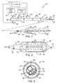
- FIG. 1schematically illustrates a combined cryogenic/angioplasty system including a catheter with an angioplasty balloon that is axially displaced from a cryogenic balloon.
- FIG. 2illustrates an alternative distal end of the cryogenic/angioplasty catheter for use in the system of FIG. 1, in which a cryogenic balloon is nested within an angioplasty balloon.
- FIG. 3is a cross-section taken along the catheter body of the cryogenic/angioplasty system of FIG. 1 .
- FIGS. 4A and 4Billustrate cryogenic cooling temperatures provided by expansion of N 2 O.
- FIGS. 5 and 5Aillustrate a particularly preferred controlled temperature cryogenic catheter in which a saline solution having a predetermined freezing temperature controls the cooling of tissues by thermally coupling an inexpansible heat exchanger with a surrounding angioplasty balloon.
- FIGS. 6A through 6Cschematically illustrate a method for using the controlled temperature cryogenic balloon of FIG. 5 .
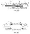
- FIGS. 6D and 6Eare partial cross-sections schematically illustrating methods for selectively cooling a tissue of the vessel wall to a desired temperature through a lesion along the vessel lumen such as plaque.
- FIGS. 7 and 7Aillustrate an alternative cryogenic treatment catheter.
- FIGS. 8A through 12Care graphical results of experiments showing an actual and observed reduction in restenosis and hyperplasia as described in the four Experimental sections provided hereinbelow.
- an exemplary system 10is capable of treating a diseased vessel wall of a blood vessel using a combination of both angioplasty dilation and cryogenic cooling.
- system 10includes a catheter 12 coupled to a cryogenic fluid supply system 14 and an angioplasty pressurization system 16 .
- cryogenic system 14 and pressurization system 16may be operatively coupled to a controller 18 for coordination of cooling and dilation, as will be described in more detail hereinbelow.
- Catheter 12generally includes a catheter body 20 having a proximal end 22 and a distal end 24 .
- a proximal housing 26includes a number of ports for coupling of cryogenic system 14 , pressurization system 16 , and the like to the proximal end of the catheter body.
- An angioplasty balloon 28 and a cryogenic balloon 30are mounted near the distal end 24 of catheter body 20 .
- the catheter bodywill generally be flexible and contain a plurality of lumens to provide fluid communication between the ports of proximal housing 26 and balloons 28 and 30 .
- Angioplasty balloon 28may be formed from a variety of materials conventionally used for dilating blood vessels. Angioplasty balloon 28 will typically comprise a non-dispensable material such as polyethylene terephthalate (PET). Such angioplasty balloons are formed in a variety of sizes depending on their intended use, typically having a length in a range from about 15 mm to about 50 mm and an expanded diameter in a range from about 2 mm to about 10 mm. Prior to inflation, angioplasty balloon 28 will generally remain in a low profile configuration suitable for insertion into and maneuvering through the vascular system. A guidewire lumen 32 extends through angioplasty balloon 28 and cryogenic balloon 30 from a proximal guidewire port 34 to facilitate accessing the target treatment site.
- PETpolyethylene terephthalate
- Angioplasty balloon 28is inflated by injecting fluid from pressurization system 16 into a pressurization lumen 36 through a pressurization port 38 .
- balloon 28will preferably be isolated from balloon 30 , so as to avoid inadvertent inflation of the cryogenic balloon during dilation.
- High contrast markersmay be provided within the balloon to enhance an image of the distal end of the catheter and facilitate positioning of the balloon fluoroscopically, sonographically, or under any other alternative image modality (with appropriate contrast structures). Such markers may be formed by winding a gold or platinum wire around the tubular structure defining pressurization lumen 36 , as illustrated.
- cryogenic balloon 30is disposed proximally of angioplasty balloon 28 .
- This arrangementis advantageous for first at least partially dilating the vessel wall and then treating the dilated vessel wall with cryogenic cooling, which can facilitate positioning of the cryogenic balloon within an occluded region of the vessel.
- the cryogenic balloonmay be disposed distally of the angioplasty balloon.
- cryogenic balloon 30The structure and operation of cryogenic balloon 30 may be understood with reference to FIGS. 1 and 2, and also with reference to U.S. patent application Ser. No. 09/203,011, previously incorporated herein by reference.
- Cryogenic fluidwill often be injected into a cryogenic supply port 42 and passed toward cryogenic balloon 30 through cryogenic supply lumen 44 within catheter body 20 .
- the cryogenic fluidmay comprise cryogenic liquids or liquid/gas mixtures, optionally including carbon dioxide, nitrous oxide, liquid nitrogen, or the like.
- As the cryogenic liquid passes from supply lumen 44 and into cryogenic balloon 30it is preferably distributed both radially and axially by a diffuser 46 .
- Diffuser 46will generally comprise a tubular structure with radially oriented openings.
- diffuser 46will direct the cooling fluid roughly perpendicularly against the wall of cryogenic balloon 30 , so that the heat transfer coefficient between the cooling vapor and balloon wall is quite even and quite high. This helps to reduce the temperature of the balloon wall and provides greater heat extraction for a given flow rate of coolant into the balloon. Additionally, as the ports of diffuser 46 are distributed both circumferentially and axially along the balloon, the diffuser can provide a substantially uniform cooling over a significant portion of (often over the majority of) the surface of the balloon.
- the cryogenic cooling fluidmay pass through a Joule-Thomson orifice between fluid supply lumen 44 and balloon 30 .
- at least a portion of the cryogenic cooling fluidmay exit one or more ports into the balloon as a liquid.
- the liquidwill vaporize within the balloon, and the enthalpy of vaporization can help cool the surrounding vessel wall.
- the liquidmay coat at least a portion of the balloon wall so as to enhance even cooling over at least a portion of the vessel wall.
- the ports of diffuser 46may have a total cross-section which is smaller than a cross-section of the fluid supply of lumen 44 , or which is at least as large as (or larger than) the cross-section of the fluid supply lumen.
- cryogenic cooling fluidAfter the cryogenic cooling fluid vaporizes within balloon 30 , it escapes the balloon proximally along an exhaust lumen 48 and is exhausted from catheter 12 through an exhaust port 50 .
- Inflation of cryogenic balloon 30may be controlled by the amount of cryogenic fluid injected into the balloon, and/or by the pressure head loss experience by the exhaust gases. Cooling is generally enhanced by minimizing the pressure within balloon 30 . To take advantage of this effect so as to control the amount of cooling a fixed or variable orifice may be provided at exhaust port 50 . Alternatively, a vacuum may be applied to the exhaust port to control cooling and enhance cooling efficiency.
- An exemplary structure for diffuser 46may comprise a polyimide tube having an inner diameter of about 0.032 inches and a wall thickness of 0.1001 inch. Each port will define a diameter of about 0.0025 inches. There will typically be between six and six hundred ports in diffuser 46 . In the exemplary embodiment, four axial rows of ports are separated by about 90° from each other. The rows are axially staggered so that the ports in a single row have central line separations of about 4 mm, while the ports of adjacent rows are separated by about 2 mm. The overall length of the porous diffuser tube will vary with the length of the balloon, and will typically be about 2 cm.
- Diffuser 46may be bonded concentrically about a central shaft defining guidewire lumen 32 .
- Adhesivesseal the proximal and distal ends of the diffuser, or the diffuser can be incorporated at the distal end of tube 64 with an adhesive seal at the distal end of the diffuser.
- High contrast markersmay again be provided to enhance an image of the catheter and facilitate positioning of cryogenic balloon 18 at the treatment site.
- the cryogenic cooling fluidwill generally be introduced through the annular space between the diffuser tube and the central shaft proximally of the balloon.
- the central shaftwill typically comprise a polyimide tube, but may alternatively include any of a wide variety of materials.
- a temperature sensormay be thermally coupled to balloon 30 to monitor and/or control cryogenic cooling of the arterial wall.
- Temperature sensor 52may optionally be disposed on an inner or outer surface of balloon 30 , and is coupled to controller 18 by thermocouple leads 54 .
- Temperature sensor 52may comprise a thermocouple, thermistor, or the like.
- controller 18will generally initiate, monitor, and/or control cooling of the tissue.
- Cryogenic supply 14will often inject sufficient cryogenic cooling fluid to effect a cooling rate of the tissue in a range from about 2° C. to about 30° C. per second.
- the systemwill maintain the temperature in a range from about 0° C. to about ⁇ 80° C., optionally at a temperature in range from ⁇ 5° C. to about ⁇ 40° C., for a time between about 1 and 60 seconds, ideally maintaining the tissue at a temperature in a range from about ⁇ 5° C. to about ⁇ 15° C. for a time from about 10 to about 20 seconds.
- the efficacy of the therapy at inhibiting restenosismay be enhanced by repeatedly cooling the tissue to such temperatures for between 1 and 5 cooling cycles, typically repeating between 1 and 6 cooling cycles every 60 seconds.
- Typical treatment cyclesmay cool a surface temperature of the endothelium down to about ⁇ 10° C., then allow the surface temperature to w arm to about 0° C. Five of these cooling cycles might be performed in about 40 seconds.
- a cryogenic liquid or liquid/gas mixturecomprising carbon dioxide, nitrous oxide, or the like may flow through the balloon at a rate in an average from about 100 to about 800 mg per second.
- Such cooling(and optional cooling cycles) may induce apoptosis and/or programmed cell death.
- proximal housing 26may include a cooling balloon pressure monitoring port 56 .
- the pressure monitoring portwill be in fluid communication with the cryogenic balloon 30 , preferably through a dedicated pressure monitoring lumen (not shown). Signals from pressure monitoring port 56 and a thermal couple connector 58 may be transmitted to the controller 18 .
- Thisallows the use of a feedback control system for initiating, regulating, and halting the supply of cryogenic fluid from fluid supply system 14 . More specifically, the controller will often provide a control signal to the fluid supply system in response to signals from pressure monitoring port 56 and/or thermal couple connector 58 .
- an alternative combination cryogenic/angioplasty catheteragain includes both a cryogenic balloon 30 and an angioplasty balloon 28 .
- cryogenic balloon 30is nested within angioplasty balloon 28 , so that if the low pressure cooling balloon were to break during the procedure, the higher pressure capability of the surrounding angioplasty balloon 28 would contain the exhaust gases until the flow of coolant was stopped.
- the structure of this nested embodimentis quite similar to that described above.
- the nested cryogenic/angioplasty balloon catheter of FIG. 2may allow pre-cooling of a diseased vessel wall prior to dilation, cooling of a vessel wall after dilation, interspersed cooling/dilation, and even concurrent dilation during cooling.
- the catheterneed not be repositioned between the application of dilation pressure and cryogenic cooling.
- this nested embodimentfacilitates the immediate sequential pre- and/or post-cooling of the stenosed vessel wall, thereby giving a wide flexibility in the treatment protocol.
- the interaction of cooling and dilationmay be precisely prescribed and effected by controller 18 (see FIG. 1) without having to wait for the surgeon to reposition the catheter.
- angioplasty balloon 28may be inflated first with contrast liquid 40 (as used in conventional angioplasty).
- the contrast liquidmay then be at least partially evacuated, allowing cooling balloon 30 to be inflated at a pressure that is lower than the angioplasty distention pressure.
- Inflation of cryogenic balloon 30pushes the angioplasty balloon against the diseased wall of the vessel, so that the cryogenic fluid 60 within the cryogenic balloon is thermally coupled to the diseased vessel wall by both the cryogenic balloon wall and the angioplasty balloon wall in series.
- a heat transfer enhancing materialmay be included in cryogenic balloon 30 and/or angioplasty balloon 28 , particularly where treatment temperatures of about ⁇ 50° C. and below are desired.
- the addition of between about 1% and 10% boron nitride in a polyethylene or other balloon polymercan significantly improve heat transfer of the entire system.
- a significant temperature differentialmay be found between an inner and outer surface of each balloon during cooling.
- improving the thermal conductivity of each balloon wall disposed between cryogenic fluid 60 and the targeted wall of the vesselmay provide significant benefits when cooling to low temperatures.
- coolingmay be initiated prior to complete dilation of the stenosed region of the vessel.
- the cooling processmay weaken the mechanical properties of the vessel and allow it to be expanded or dilated at a much lower pressure than is used with conventional angioplasty.
- dilation of a cryogenically cooled vesselmay require inflation of angioplasty balloon 28 with a fluid pressure of about 2 bar, as compared to about 10 bar for conventional uncooled angioplasty on the same vessel wall.
- Simultaneous cryogenic cooling and angioplastymay reduce and/or eliminate medial vessel fractures, thereby inhibiting proliferative response after angioplasty.
- Still further alternative treatment cyclesare possible, including inflating a balloon with a room temperature gas at normal angioplasty pressures to dilate the vessel, and then inflating the balloon with a cryogenic fluid or other coolant to treat the dilated area so as to inhibit hyperplasia.
- a balloonmay be inflated with a standard angioplasty contrast liquid at normal angioplasty pressures to dilate the vessel. The balloon may then be flushed with saline, and then flushed with a dry room temperature gas to dry the cooling fluid path. After the cryogenic fluid path is dry the balloon may be inflated with a coolant to treat the dilated area. Cooling cycles before angioplasty and/or before stenting may also provide the antiproliferative response described above.
- An outer sheath 62partially defines exhaust lumen 48 , the exhaust lumen here comprising an annular space disposed between the sheath and an inner jacket 64 .
- sheath 62comprises a polyethylene tube having an inner diameter of 0.058 inches and a wall thickness of about 0.003 inches.
- the exemplary jacket 64comprises a polyimide having an inner diameter of 0.035 inches and a wall thickness of 0.001 inches.
- a core shaft 66defines guidewire lumen 32
- a cooling inlet tube 68 and an angioplasty pressurization tube 70define supply lumen 44 and pressurization lumen 36 , respectively.
- the exemplary cooling inlet tubecomprises a polyester or polyimide
- the pressurization tube in the exemplary systemmay comprise a polyester or high density polyester.
- Thermocouple leads 54are insulated in a conventional manner.
- cryogenic coolingis capable of inducing temperatures well below the preferred antiproliferative treatment ranges of the present invention (typically in a range from about ⁇ 5° C. to: about ⁇ 15° C).
- expansion of N2O from an initial pressure of 500 psi and an initial temperature of 0° C. to final pressures in a range from atmospheric pressure to 100 psiresults in a cryogenic cooling fluid temperature significantly colder than our preferred target tissue temperatures.
- a layer of insulating material 72 between the cryogenic cooling fluid and the tissue engaging surface of the balloonis to add, a layer of insulating material 72 between the cryogenic cooling fluid and the tissue engaging surface of the balloon.
- a suitable insulation materialmight include a thin layer of expanded TeflonTM (ePTFE) on an inner or outer surface of cryogenic balloon 30 , on an inner or outer surface of angioplasty balloon 28 , or the like.
- the ePTFE layermay have a thickness in the range from about 0.00025 inches to about 0.001 inches.
- Alternative insulation materialsmight also be used.
- Alternative active temperature control techniquesmight be used with or without such an insulation layer, including the use of controller 18 as shown in FIG. 1 .
- a controlled temperature cryogenic balloon catheter 80again includes an angioplasty balloon 28 , which here contains an inexpansible heat exchanger 82 . Cooling of fluid inlet tube 68 releases the cryogenic cooling fluid within heat exchanger 82 , but does not expand the heat exchanger into direct thermal contact with the balloon wall of angioplasty balloon 28 . Instead, a saline solution 84 thermally couples the heat exchanger to the outer surface of angioplasty balloon 28 .
- Saline solution 84will generally have a predetermined freezing temperature, and sufficient cryogenic cooling fluid will generally be provided to heat exchanger 82 so that the saline solution is only partially frozen. As a result, the temperature of the saline solution within angioplasty balloon 28 will be maintained accurately at the freezing temperature. As the freezing temperature of saline may be varied by changing the salinity, this provides a convenient control mechanism to vary the treatment temperature. Specifically, a 6% saline solution will freeze at about ⁇ 3.5° C., while an 18% saline solution will freeze at about ⁇ 14° C.
- the temperature of the saline solution during cryogenic coolingcan be selected within a range from about 5° C. to about 15° C.
- a 12% saline solutionwill provide a freezing temperature of about 8° C., which is particularly advantageous for use with the controlled-temperature cryogenic catheter 80 illustrated in FIG. 5 .
- saline solution 84a variety of fluids, and possibly even solids, might be used in place of saline solution 84 .
- temperature controlwill be provided where a thermally coupling structure undergoes a change in phase involving a significant latent phase change energy.
- salineis particularly preferred as its range of freezing temperatures can be easily controlled within the desired range, and as it poses little risk in the event of release within the vasculature.
- contrastmay be included with the saline solution to improve imaging of the system within the patient body.
- heat exchanger 82extends proximally from angioplasty balloon 28 to at least in part define exhaust lumen 48 .
- This simple proximal tubular structure(which is referred to herein as an evaporator 84 ) may comprise a polyimide tube having an inner diameter in a range from about 0.036 inches to about 0.051 inches, ideally having an inner diameter of about 0.045 inches.
- Cooling inlet tube 68 within evaporator 84 and heat exchanger 82may comprise a polyimide tube having an inner diameter in a range from about 0.005 inches to about 0.012 inches, ideally having an inner diameter of about 0.009 inches.
- evaporator 84extending the entire length of the catheter may cause sufficient cooling of the saline proximally of the angioplasty balloon to induce freezing along catheter body 20 .
- an insulation jacketmay be provided around the evaporator proximally of heat exchanger 82 .
- the insulation jacketmay comprise a polyimide tube, preferably leaving a gap (as small as 0.001 inches) between the insulation jacket wall and evaporator 84 .
- the balloon inflation lumenmay be altered to prevent the saline from thermally coupling exhaust lumen 48 to outer sheath 64 .
- a polyimide tube having an inner diameter in a range from about 0.012 inches to about 0.025 inchesmay be disposed between evaporator 84 and outer sheath 62 , with this additional tubular structure providing fluid communication between inflation port 38 and angioplasty balloon 28 .
- an additional portmay be provided on the proximal housing in communication with the insulation gap (either between the insulation jacket and evaporator 84 or between evaporator 84 and outer sheath 62 ) such that at least some of the air could be evacuated from this gap to reduce heat transfer to in the blood surrounding catheter body 20 and the exhaust gases.
- FIGS. 6A through CA method for using controlled-temperature cryogenic catheter 80 is illustrated in FIGS. 6A through C.
- the catheteris introduced into the vasculature through an introducer sheath, most often using the widely known Seldinger technique.
- a guide wire GWis maneuvered through the vessel, and catheter 80 is advanced over the guide wire and positioned adjacent diseased portion DP of vessel wall VW.
- the balloonmay be inflated in a conventional manner through inflation port 38 to dilate the vessel, as illustrated in FIG. 6 B.
- the vesselmay be dilated using conventional contrast fluid to facilitate fluoroscopically directing dilation.
- the balloonWhen standard contrast has been used for dilation, the balloon may be evacuated and filled with a saline solution which freezes at the desired treatment temperature. Alternatively, dilation may be performed using this saline solution to avoid any delay between dilation and cryogenic treatment In still further alternative treatments, cryogenic cooling may be initiated prior to or during dilation.
- the saline solution having the predetermined freezing temperaturewill preferably be used to inflate angioplasty balloon 28 with sufficient pressure to provide good contact between the balloon and vessel wall VW.
- the angioplasty balloonwill be inflated by the saline solution to a pressure in a range from about 5 psi to about 30 psi, as illustrated in FIG. 6 B.
- a cryogenic fluid(usually in the form of a liquefied refrigerant or liquid/gas mixture) is injected into cryogenic supply port 42 .
- the cryogenic fluidflows through fluid supply lumen 44 and is transmitted into heat exchanger 82 , where it rapidly absorbs heat and vaporizes, thereby cooling saline 84 and angioplasty balloon 28 .
- Sufficient cryogenic cooling fluidis supplied to partially freeze saline 84 , so that the saline liquid/solid mixture remains at about freezing temperature.
- additional cryogenic cooling fluidmay be introduced, with the freezing of the saline providing a temporary plateau along the temperature excursion profile.
- the partially frozen salinemelts by absorbing heat from the surrounding body.
- the cooling of saline within angioplasty balloon 28results in treatment of a surface layer 88 of vessel wall VW engaged by the angioplasty balloon to an accurately controlled treatment temperature in a range from about ⁇ 5° C. to about 15° C.
- this treated tissue layerundergoes apoptosis, thereby avoiding and/or reducing the proliferative response of the luminal wall to dilation.
- a cooling cathetercan help to provide accurate treatment temperatures in a range from about ⁇ 5° C. to about ⁇ 15° C. In many embodiments, it will be desirable to maintain the target tissue within this range for a time between about 20 to about 60 seconds. As described in detail in the Experimental sections hereinbelow, accurately providing such treatment temperatures and times for treatment of the tissue can result in apoptosis without excessive immediate necrosis of the tissues of the vessel wall.
- many vessels targeted for a primary treatmentsuch as angioplasty, stenting, atherectomy, and the like, may have a significant amount of plaque P disposed between a tissue of the vessel wall VW and the vessel lumen L.
- Plaque Pcan have a surprisingly large thermodynamic effect on the treatment of the tissues of vessel wall VW, as a significant temperature gradient may exist between a surface of the plaque adjacent lumen L and a plaque/vessel wall interface.
- a cooling surfacesuch as a cooled balloon 28 (or optionally, a cooled liquid surface, or the like) engaging an inner surface of plaque P at a target temperature for cryotherapy may not reduce the temperature of the vessel wall tissue sufficiently to inhibit hyperplasia.
- the tissue of vessel wall VWis cooled accurately down to the desired treatment temperature, and if cooling is then maintained for the desired treatment time, the insulating effect of the plaque may maintain the vessel wall tissue at a reduced temperature for a significantly excessive amount of time.
- a thickness T of plaque Pmay be measured using an intravascular ultrasound system 90 as schematically illustrated in FIG. 6 D.
- An exemplary ultrasound systemis commercially available from Scimed of Maple Grove, Minn.
- Alternative methods for measuring plaque and other lesions within a lumen of a vesselinclude angiography, computer tomography, and a variety of other known medical sensing modalities
- a treatment time and/or temperature for cooling an outer surface of balloon 28can be determined.
- Treatment times and/or temperaturesmay be calculated using a mathematical model which accounts for the insulating effect of plaque P.
- Alternative methods for determining treatment parametersmay be based on dosimetry (based on prior measured treatments), or the like.
- Treatment parameters for maintaining cooling using balloon 28may be adjusted for a thickness, thermal conductivity, or other characteristic of plaque P. For example, it may take approximately 10 seconds to cool the tissue of vessel wall VW to a treatment temperature in a range from about ⁇ 5° C. to about ⁇ 15° C. due in part to the presence of plaque P.
- the total cooling treatmentmay be comparable to a treatment in which active cooling of a balloon in direct contact with the vessel wall tissue is maintained for a time and a range from about 20 to about 60 seconds after temperatures reach a target temperature between about ⁇ 5° C. and about ⁇ 15° C.
- treatment temperatures and timesare generally given for tissue in substantially direct contact with the cooling surface. Where significant amount of plaque is present within a lumen of the vessel wall, the thermodynamic effects of that plaque will preferably be taken into account in the treatment cycle of the cryotherapy device so as to effect the described tissue treatments.
- Cultured arterial endothelial cells and smooth muscle cellsare chilled or frozen under controlled thermal conditions. Consequences such as necrosis or apoptosis, as well as the impact on long term reproductive viability are measured with a variety of assays.
- CAECHuman coronary artery endothelial cells
- CASMCsmooth muscle cells
- Freezing or chilling of cellular suspensionsis carried out using a low temperature stage, which consists of a copper block machined to allow the circulation of liquid nitrogen through the block.
- a thermocouple mounted to the stageprovides feedback to a temperature controller. This controller regulates the power input to a thermoelectric heater fixed to the copper, block, thereby holding the temperature of the stage to within 1 degree Celsius of the desired, preset temperature.
- a 30 ⁇ l aliquot of cellular suspension stained with trypan blueis pipetted onto a precooled glass microslide positioned on the freezing stage, and immediately covered with a cover slip. After the allotted exposure time, the microslide is transferred to a 37° C. surface to thaw. For a second freezing cycle, the slide is moved back to the cold stage for the desired period of time and then thawed on the 37° C. surface. Trypan blue is excluded by intact plasma membranes, so a count of stained vs. unstained cells immediately following the freezing treatment provides a measure of acute necrosis as reflected by membrane integrity, in these experiments, cells are subjected to final freezing temperatures ranging from +10 to ⁇ 40° C. for 10, 20, and 60 second exposure times, and both single and double freeze/thaw cycles are considered.
- a 125 ⁇ l aliquot of cellular suspensionis placed in the well of a sterile, precooled chamber slide on the low temperature stage.
- the chamber slideis transferred to a 37° C. surface to thaw the suspension, and then moved back onto the freezing stage if a second freeze/thaw cycle is desired.
- the sampleis transferred to a culture flask in a sterile field at a seeding density of 1 ⁇ 10 4 cells/cm 2 , and incubated with growth medium mixed with 10% alamar blue.
- Alamar blueis an oxidation-reduction indicator, which yields a colorimetric change in response to metabolic activity.
- the reduction of the dyeis monitored hourly for 4 hours after the freeze, and then at 24 hour intervals for up to 6 days. Reduction of the dye provides a quantitative measure of the proliferation of the treated cells. Again, a temperature, range of +10 to ⁇ 40° C. is examined, and single and double freeze/thaw cycles are used with 60 second cold exposure times.
- Freezing experimentsare carried out with both cellular suspensions and adherent cells.
- Cellular suspensionsare frozen on chamber slides on the low temperature stage as described above.
- Adherent cellsare frozen or chilled by immersion of the culture flasks in saline-ice baths which maintain the desired temperatures. After the allotted exposure time, the flasks are transferred to a 37° C. bath for 1 to 2 hours.
- the cellsare removed from the flasks by trypsinization, and the cellular DNA is extracted using a lysing kit purchased from Boehringer-Mannheim. After elimination of RNA by incubation of the samples in RNase, the purified DNA is electrophoresed in a 1% agarose gel for 2.5 hours at 75 V.
- DNAis visualized with ethidium bromide using a UV transilluminator. DNA fragmentation indicative of apoptosis is detected by separation of the DNA into a characteristic ladder.
- cell suspensionsare exposed to final temperatures in the range of +5° to ⁇ 15° C. for 60 seconds and undergo 1 to 4 freeze/thaw sequences.
- Adherent cellsare cooled for longer periods of time, experiencing temperatures of +5 to ⁇ 15° C. for 0.5, 1, 2, and 4 hours. Post freeze incubation times of 1 to 2 hours at 37° C. are enforced to allow apoptosis to progress to the DNA fragmentation stage.
- 24, 72, and 120 hour culturesare used.
- FIGS. 8A through 8Dpresent necrosis data obtained with trypan blue. These plots include data for cells exposed to final freezing temperatures for 60 seconds (circles), 20 seconds (triangles), and 10 seconds, (squares). From these plots it is evident that cells suffer no acute membrane damage when chilled or frozen to temperature above ⁇ 5° C., and a substantial percentage of cells survive freezing to temperatures as low as ⁇ 15° C. A double freeze/thaw cycle increases cellular damage, however in the higher temperature range from +10 to ⁇ 5° C. the majority of cells still survive. The duration of cold exposure appears to have no effect on cell survival for the range of times considered in this study.
- the alamar blue reduction curves shown in FIGS. 9A through 9H and FIGS. 10A through 10Ggive a measure of the number of metabolically active cells in each treated culture, relative to a sham operated control.
- the percent reductions of alamar blue in CASMC culturesare plotted as a function of time for sham operated control cells (diamonds), cells exposed to a single freeze cycle (squares), and cells exposed to a double freeze cycle (circles).
- percent reduction of alamar blueare plotted for CAEC cultures as a function of time for sham operated control cells (diamonds), and for cells exposed to a single freeze cycle (squares).
- apoptosisor “programmed cell death”, which is characterized by internucleosomal cleavage of DNA, cell membrane blebbing, condensation of nuclear chromatin, and the fragmentation of the cell into smaller apoptotic bodies.
- Cold shockcan induce apoptosis.
- apoptosiscan be triggered by exposure to temperatures ranging from 0 to ⁇ 15° C., conditions which yield low levels of cellular necrosis.
- studieswere conducted to identify the role of apoptosis in the overall response to cold temperatures for the cells in this particular system.
- Apoptosisis especially interesting for this application because, unlike cellular necrosis, it does not cause the inflammation which marks the initiation of neointimal hyperplasia.
- the studies described in this sectionseek to assess whether the smooth muscle cells involved in the proliferative process of neointimal hyperplasia can be destroyed by inducing apoptosis rather than necrosis, thereby avoiding an inflammatory response.
- three assayshave been applied to study the occurrence of cold shock apoptosis: gel electrophoresis, a TUNEL assay, and an Annexin V assay.
- the gel electrophoresis assayhas certain limitations. For instance, the appearance of the characteristic ladder pattern is dependent on the presence of a minimum quantity of fragmented DNA, making the detection of a positive result dependent of the number of cells in the sample population and the efficacy of the DNA isolation process, as well as the percentage of cells in the appropriate stage of the apoptotic process. Therefore, it is possible that apoptotic events took place, but at a level too low to measure by this assay.
- fragmentation of the DNA into characteristic lengthsrepresents a specific stage in the apoptotic process. As that process continues, DNA fragments further and is sequestered in the smaller apoptotic bodies which are entirely digested by neighboring cells, so that the characteristic ladder is no longer produced.
- the assayis applied to a cell population too early (before fragmentation begins) or too late in the apoptotic process, it will fail to identify apoptosis.
- the time taken for a particular cell type to undergo each step of the apoptotic process in response to a particular driving mechanismis unknown and can vary from minutes to hours.
- the TUNEL (terminal deoxynucleotidyl transferase mediated dUTP nick end labeling) assayuses DNA fragmentation as the marker for apoptosis. This assay is based on the principle that cleavage of genomic DNA during apoptosis yields single strand breaks (“nicks”), which can be identified by labeling free 3′-OH termini with modified nucleotides in an enzymatic reaction. Incorporated fluorescein is detected by anti-fluorescein antibody Fab fragments from sheep, conjugated with horse-radish peroxidase (POD). After substrate reaction to produce a calorimetric precipitate, stained cells can be analyzed under light microscopy.
- nickssingle strand breaks
- the adherent sampleswere frozen by placing the coverslips on pre-cooled glass microslides positioned on the low temperature stage. Solidification was visually observed. To thaw, the coverslips were removed from the stage and dipped in growth medium at 37° C. Temperatures of +5 to ⁇ 15° C., an exposure time of 60 seconds, and a single freeze/thaw cycle were applied. After thawing, the cover slips were returned to the culture plates, and incubated with growth media at 37° C. Post freeze incubation times of 0, 0.5, 1, 2, 3, 4, and 24 hours were tested, since the time required for apoptotic cells to reach the DNA fragmentation stage was unknown.
- the adherent cellswere air dried, fixed in a 4% paraformaldehyde solution, and incubated with a 0.3% H 2 O 2 methanol solution to block endogenous peroxidase.
- the cellswere incubated in a permeabilisation solution to permit penetration of the TUNEL enzyme in a subsequent step.
- the cellswere incubated in converter horse-radish peroxidase (POD) which binds to TUNEL labeled DNA strand breaks, and then in a DAB/metal enhanced substrate, which produced a dark brown precipitate in the presence of bound POD.
- PODconverter horse-radish peroxidase
- the coverslipswere rinsed thoroughly with phosphate buffered saline between each of the incubations.
- the cover slipswere analyzed under light microscopy. Hemotoxylin was applied as a counterstain in order to facilitate the count of non-apoptotic cells.
- a positive controlincluded in each experiment., was prepared by incubating a fixed, permeabilized cell sample with DNase I to induce DNA strand breaks. Additionally, a sham operated control sample which was exposed the same handling but not to any cold shock, was included in each experiment. In examining the samples, care was taken to confirm that all positively stained cells included in the count of apoptotic cells exhibited a morphology characteristic of apoptosis. These cells appeared shrunken or condensed, in contrast to the swollen, distorted appearance of necrotic cells.
- An early event in the process of apoptosisis the flipping or inversion of the molecules of the plasma membrane, causing phospholipid phosphatidylserine (PS) to be translocated from the inner leaflet of the membrane to the outer cell surface.
- PSphospholipid phosphatidylserine
- the exposed PSserves as an identification tag utilized by Annexin V, a protein with a high affinity for phosphatidylserine.
- Annexin Va protein with a high affinity for phosphatidylserine.
- the freezing of adherent cells on coverslips using the low temperature stagewas performed as in the TUNEL experiments. Again, temperatures of +5 to ⁇ 15° C., and a single freeze/thaw cycle were applied.
- the correlations established by the TUNEL datademonstrated that the process of apoptosis is underway and has progressed to the DNA fragmentation stage between 1 to 2 hours after cold shock. Since membrane inversion is an earlier event in the apoptotic process, an incubation time of 1 hour was selected for this study. This timing allowed the membrane alterations to be detected before extensive degradation of the cells into smaller bodies could occur.
- the correlation between cold exposure time and apoptosiswas also investigated. Since only short exposure times were relevant to this particular application, cold exposure times of 30, 60, and 120 seconds were tested.
- the Annexin assaywas performed through the following series of steps. After the one hour incubation, the coverslips were removed from the culture plates and rinsed in phosphate buffered saline. They were then incubated in the Annexin-V-Biotin working solution. Subsequently, the cells were air dried, fixed in a methanol/ethanol solution, air dried again, and then incubated with Streptavidin conjugated with horse radish peroxidase (POD). The Streptavidin labeled the bound Annexin, and the POD provided a calorimetric reaction induced by exposure to a DAB/metal enhanced substrate. Following the substrate reaction, the coverslips were mounted on slides and examined under light microscopy.
- PODhorse radish peroxidase
- FIGS. 11A through 11Frepresent the findings of these assays.
- FIGS. 11A and 11Bshow the percentage of apoptotic cells found in samples of human coronary artery endothelial cells and rat arterial smooth muscle cells respectively, as a function of the final temperature to which they were frozen. TUNEL results are indicated with a dashed line, and Annexin results are indicated with a solid line. A minimum of three experiments were conducted for each thermal condition, and the data points represent the averaged result. These plots show that significant levels of apoptosis were found with both assays for the temperature range examined here. The two cell types experience very similar levels of apoptosis in response to the cold shock.
- apoptosiswas not found at hyperthermic conditions (+5 to 0° C.), but was induced at lower temperatures, with a maximum response occurring at ⁇ 10° C.
- a comparison between the Annexin results and TUNEL datareveals that the TUNEL assay produced somewhat higher levels of positive staining. It should be noted that, whereas positive apoptosis results are difficult to obtain with the electrophoresis assay, the TUNEL assay has a tendency to be biased towards positive outcomes.
- One possible factor contributing to falsely high apoptotic resultsis the positive staining of necrotic cells.
- the TUNEL reactionpreferentially labels DNA strand breaks generated during apoptosis, however extensive DNA fragmentation may occur in late stages of necrosis leading to some positive staining of necrotic cells.
- examination of the morphology of stained cellsgenerally allowed discrimination between necrotic and apoptotic cells, in some fraction of the stained cells the morphological characteristics were inconclusive and the designation of the cells was uncertain.
- FIG. 11Cshows the relationship between TUNEL detected levels of DNA fragmentation and post freeze incubation time for human coronary artery endothelial cells.
- the cold exposureconsisted of 60 second, single cycle freezes, and the results for three different final temperatures ( ⁇ 5, ⁇ 10, and ⁇ 15° C.) are presented in this plot. From the figure it is evident that for each temperature, the maximum response measured by the TUNEL assay was found within 1 to 2 hours after the cold shock. 24 hours after cold shock, little fragmentation was found. This indicates that DNA fragmentation reaches its peak at approximately 2 hours after the apoptotic process begins.
- FIGS. 11D and 11Eshow the relationship between the level of apoptosis detected with the Annexin assay and the time of cold exposure for human coronary artery endothelial cells and rat arterial smooth muscle cells, respectively.
- results for cold shock at ⁇ 5 and ⁇ 10° C., in a single freeze cycle with a one hour post freeze incubationare presented.
- the figuresreveal that the time of exposure does not significantly affect the percentage of apoptotic cells, within the 2 minute time range relevant to this application.
- the small variations in percentage of apoptosisare well within the range of accuracy of the Annexin assay.
- the varied results of the three assaysreflect the complex nature of the apoptotic phenomenon.
- the electrophoresis assayfailed to positively identify a substantial apoptotic fraction
- both the TUNEL and the Annexin assaysdemonstrated some contribution of apoptosis in the overall response of arterial cells to cold shock.
- the two assaysconsistently revealed an apoptotic peak at ⁇ 10° C., and no apoptosis above ⁇ 5° C. Notwithstanding the limitations in the assays themselves, exact percentages for apoptosis are difficult to establish because of the complexity of the mechanism.
- the proliferative and morphometric response in the swine coronary modelwere compared after balloon injury to balloon injury and intravascular cryogenic treatment.
- Two rabbits(total eight sites in the carotid arteries) were treated with a cooled or non-cooled angioplasty balloon. None of the rabbit carotids received balloon overstretch. The rabbits were sacrificed at 28 days.
- BrdU index in the shammeasured 16% to 43% medial fracture 0% to 67.6%, and percent stenosis 0% to 36.6%.
- BrdU index in the cryogenically treated arteriesBrdU index measured 18% to 36%, medial fracture 0% to 44.5% and percent stenosis 1.9% to 16.9%.
- percent stenosismeasured 3.5% to 10% in the shams and 3.1% to 7.4% in the cryogenically treated arteries.
- Medial fracturemeasured 0% in the shams and 0% to 2.4% in the cryogenically treated arteries.
- Intravascular cryogenic treatment in the swine and rabbit vascular modeleffect cell proliferation and present stenosis following a balloon injury.
- the intravascular cryogenic treatment systemincludes a cryogenic balloon catheter and a delivery system.
- the cryogenic balloon catheter(60-cm usable length) was mounted coaxially around a polyimide diffuser tube (0.034′′ OD. ⁇ 001′′ wall) and a 0.020′′ O.D. polyimide tube defining a guide wire lumen.
- a polyethylene balloon(4 cm in length) was mounted on a polyethylene catheter shaft (048′′ I.D. ⁇ 0.005′ wall).
- a manifoldis mounted to the proximal end of the balloon shaft and provides ports for guide wire insertion, refrigerant inlet and refrigerant exhaust.
- the polyimide diffuser tubeconnects the most distal end of the balloon to the manifold.
- the portion of the diffuser tube inside the balloonwas fenestrated (40 holes, 0.0028′′ dia. spaced evenly over 2 cm) to allow refrigerant to flow radially outward against the inside wail of the balloon.
- the polyimide refrigerant inlet tube(0.011′′ O.D. ⁇ 0.001′′ wall) resides in the coaxial space between the guide wire tube and the diffuser tube.
- the refrigerant inlet tube, guide wire tube, balloon shaft and diffuser tubeare potted into the manifold using a medical grade adhesive (Dymax 204-CTH).
- the refrigerant exhaust portconnects to an adjustable pressure relief valve and provides for control of the pressure inside the balloon.
- the valveis set between 60 and 120 psi.
- the delivery systemdispenses liquid nitrous oxide through the inlet port on the manifold.
- the nitrous oxideis contained in an 8-gram cylinder (675 psi at 23° C.) and is inverted so that the refrigerant flows through the inlet port of the manifold.
- a hand held housing surrounding the liquid nitrous oxide cylinderincorporates a puncture needle and seal on one end and a threaded plunger on the other end, enables the user to safely puncture the gas cylinder.
- a high-pressure stopcockcontaining a 20 ⁇ filter to trap particulates that may clog the inlet port, is mounted to the to the outlet of the delivery system.
- thermocoupletype ‘T’ and constructed from 0.0015′′ bifilar wire coated with 0.0001′′ polyurethane was mounted 2 cm from the distal tip of the guide wire, covered with 1 cm of polyester (0.00025′′ thick) and potted with adhesive to displace any air trapped adjacent to the thermocouple.
- the nitrous oxide cylinderwas punctured with the stopcock in the “off” position.
- the delivery systemwas then connected to the inlet port on the manifold and the stopcock opened.
- the nitrous oxidepassed through the inlet tube into the diffuser tube, spraying radially outward against the inner surface of the balloon.
- the nitrous oxideevaporated in the balloon and was exhausted as a gas through the coaxial space between the diffuser tube and the balloon shaft.
- arterieswere treated with balloon overstretch injury, 1.3:1 (3 inflations for 30 sec., separated by 1 min. deflation periods to restore coronary perfusion) following either cryogenic or sham treatment using the intravascular cryogenic treatment system with the temperature monitoring guide wire positioned between the balloon and the vessel wall.
- the sham arterieswere treated using treatment catheters filled with saline at pressures similar to cryogenic pressures used. All animals were recovered and survived for seven days. BrdU injections (50 mg/kg) were administered twenty-four hours and one hour prior to sacrifice. Prior to euthanasia all animals were systemically heparinized.
- the coronary arterieswere perfusion fixed at 75 mm Hg for 20 minutes with 10% neutral buffered formalin. The hearts were removed and immersion fixed for at least 24 hours.
- Two Rabbits(4.5 and 5 kg) prepared in a similar fashion were treated with the intravascular cryogenic treatment system or sham catheter in the carotid artery. Both femoral arteries were accessed to accommodate the 4.5F treatment catheter (or sham catheter) and the temperature monitoring guide wire. The rabbits were survived and sacrificed on day 28. The carotid arteries were perfusion fixed with 10% neutral buffered formalin.
- Endovascular cryothopathy applied to an injured arterywill result in similar or less neointimal formation than sham as judged by histologic and morphometric examination at 28 days.
- the left carotid arterywas repaired and the animal was recovered from anesthesia. Animals were housed on standard chow for 28 days following the experiments. For the duration of the recovery period the animals received Aspirin, and ticlopidine was given for the first two weeks. On day 28, each animal was sacrificed in an ethical manner, and the heart was perfusion fixed at approximately 75 mm Hg for 20 minutes in 10% buffered formalin. The arterial segments of interest were harvested and the tissue underwent routine histologic and morphometric evaluation.
- the intravascular cryogenic treatment systemincluded a cryogenic balloon catheter, as shown in FIGS. 7 and 7A, and a delivery system.
- the cryogenic balloon catheter 110(60 cm usable length) was mounted coaxially around a polyimide fluid delivery tube 112 (0.011′′ I.D. by 0.001′′ wall) and a 0.020′′ O.D. polyethylene tube defining a guide wire lumen 114 .
- a polyethylene balloon 116(3 cm in length) was mounted on a polyethylene catheter shaft (0.165′′ I.D. by 0.005′′ wall).
- a manifold 118was mounted to the proximal end of the balloon shaft and provided ports for guide wire insertion, refrigerant inlet, refrigerator exhaust, and temperature measurement inside the balloon.
- the guide wire tubeconnected the most distal end of the balloon to the manifold.
- the polyimide refrigerant inlet tube 123(0.011′′ O.D. by 0.001′′ wall) resided in the coaxial space 119 between the guide wire tube 114 and balloon catheter shaft 110 .
- thermocouple 120constructed from a 0.0015′′ by 0.003′′ bifilar wire was mounted inside the balloon.
- the thermocouple wire leads 117were positioned in the annular space between guide wire lumen 114 and balloon catheter shaft 110 .
- a connector 121was mounted on manifold 118 .
- the refrigerant inlet tube 123 , guide wire tube 114 , balloon shaft 110 , and thermocouple wires 120were ported into manifold 118 using a medical grade adhesive (Dymax 204-CTH).
- the refrigerant exhaust port 122connected to an adjustable pressure relief valve 124 and provided for control of the pressure inside the balloon. The valve was set at 15 psi.
- the delivery systemdispensed liquid AZ-50 126 (pentafluorothane and trifluoroethane) through the inlet port on the manifold.
- the AZ-50was contained a 25 cc cylinder (330 psi at 23° C.) and was inverted so that the refrigerant flowed through the inlet port of the manifold.
- a guide catheterwas advanced to the route of the aorta and the coronary arteries were selectively engaged under fluoroscopic visualization. Each coronary artery was interrogated by IVUS to determine cross-sectional diameter. Arterial injury was induced by percutaneous catheter implantation of intracorornary stents at a stent-to-artery ratio of 1.0:1 in one animal (#9108) and 1.3:1 in the other three animals (Nos. 9195, 9196, and 9197). Following stent placement, one artery in each animal underwent freezing treatment by insertion of the cryogenic catheter (described above), with the treatment zone centered in the stent.
- Refrigerantwas delivered in to the catheter for a period of 30 seconds, producing balloon inflation at a pressure of 15 psi, and a temperature of ⁇ 10° C. at the balloon/artery interface.
- Cryogenic catheter sizeswere chosen to provide good apposition between the balloon and the artery wall.
- the other artery in each animalwas sham treated at the stent location using a balloon catheter identical to the cryogenic catheter, but delivering no temperature change, inflated at 15 psi for 30 seconds.
- In-stent lesionswere treated with appropriately sized (one-to-one stent to artery ratio) balloon expandable stainless steel stents (DuetTM, 18 or 23 mm long, 3.0 to 24.0 mm diameter) to achieve a less than 10% angiographic residual stenosis.
- Angiographic and intravascular ultrasound analysiswere performed after about 28 days, and histological analysis was completed thereafter as described hereinbelow.
- the animalsgenerally had weights between about 20 and 50 kg, and were given pre-operative Aspirin (650 mg) and Procardia XL (30 mg) with a small amount of food. After sedation with about 20 mg/kg Ketamine HCL and Xylazine (2 mg/kg), the animals were intubated and mechanically ventilated with intravenous access established via an ear vein. Adjunctive atropine (.5 to 1.0 mg IV) was administered as indicated. Continuous anesthesia was maintained with 1% to 2% isoflurane. Sodium pentobarbital was administered as needed.
- a carotid artery cut-downwas performed to gain arterial access.
- Baseline coronary angiographywas performed with an 8F guiding catheter after the administration of 150 units/kg of intra-arterial Heparin. Additional Heparin was provided so as to maintain the activated clotting time above 300 seconds.
- nitroglycerinTwo hundred micrograms of nitroglycerin were administered intracoronary to prevent vasospasm following overstretch balloon angioplasty.
- the vessel targeted for treatmentwas sized by visual estimate using the guiding catheter as a reference.
- Overstretch balloon injurywas completed using a standard balloon angioplasty catheter having a size between about 1.2 and 1.4 times the baseline vessel diameter.
- Coronary angiographywas completed after balloon angioplasty.
- a 3.0 to 4.0 mm diameter stainless steel balloon expanded stent with a length between 13 and 18 mmwas implanted at the overstretch site using a single balloon inflation at 8 to 14 atm.
- Coronary angiographywas completed again after stent deployment.
- the catheters and sheathwere removed and the carotid artery was treated using standard techniques. The treated animals were allowed to recover from the procedure and received 325 mg of Aspirin daily while remaining on a normal diet.
- the treatment vesselsincluded the left anterior descending artery LAD, the left circumflex artery LCX, and the right coronary artery RCA.
- the timing of the initial stent injury, cryotherapy or sham treatment, and follow-upare shown in the animal log provided in FIG. 12 A.
- baseline coronary angiographywas performed with an 8F guiding catheter after administration of intra-arterial Heparin.
- Activated clotting timewas maintained at over 300 seconds, with 200 micrograms of nitroglycerin administered intracoronary.
- the vessels to be treatedwere sized by a visual estimate using the guiding catheter as a reference.
- the cryotherapywas administered using a cooled balloon as described hereinabove.
- a 3.0 to 4.0 mm diameter, 13 to 18 mm long stainless steel balloon expandable stentwas implanted at the treatment site (within the prior stent) using a single balloon inflation at between 8 and 14 atm. Coronary angiography was completed after stent deployment.
- intravascular ultrasoundwas performed by tracking a 30 MHz transducer (CVISTM, from Scimed, of Maple Grove, Minn.) over the guidewire within a 3.2F imaging catheter. Images were recorded using a motorized pullback rate of 0.5 mm/sec for off-line analysis. This process was completed in an additional artery for the placebo treatment. Following intravascular ultrasound, the catheters and sheath were removed and the carotid artery treated using standard techniques. The animals were recovered and monitored, receiving 250 mg of Ticlid for 14 days and 325 mg of Aspirin with a normal diet.
- CVISTM30 MHz transducer
- Each angiogramwas evaluated for evidence of intraluminal filling defects, side branch occlusion, lumen narrowing, and distal coronary flow characteristics.
- the baseline, balloon inflated stent, post injury, and follow-up coronary artery minimal lumen diameterswere measured from non-overlapped and non-foreshortened views using the guiding catheter for image calibration, and the data was recorded in mm.
- the acute balloon-to-artery ratio(minimal balloon inflated diameter divided by the baseline lumen diameter) was calculated from this data for each treated vessel.
- the percent stenosis at follow-updefined as: [(mean reference lumen diameter minus minimal lumen diameter at follow-up/mean reference lumen diameter)] X 100, was also calculated.
- FIG. 12 BQuantitative analysis of the treatment is provided in FIG. 12 B. Measurements are generally broken up into measurements for the entire group of test animals (Total), measurements for the control vessels which received only placebo/sham treatments (Control) and measurements for the arteries which received cryotherapy as described above (Cryo). As described, above baseline (BL) measurements were taken before treatment, while additional measurements were taken after the initial balloon injury (Balloon). The final two group of measurements were taken after cryotherapy or the associated placebo/sham (Post Ref), and at the follow-up date (FU). In addition to the basic reference diameter measurements, minimum lumen diameters (MLD) and percent diameter stenosis (%DS) are also set forth in FIG. 2 B.
- MLDminimum lumen diameters
- %DSpercent diameter stenosis
- FIG. 12 CStatistical analysis of the test group was performed using Statview 4.5 (from Abacus of Berkeley, Calif.), the summary of which is provided in FIG. 12 C. Morphological data are compared by analysis of variance (ANOVA) testing. Fischer's protected least significant difference (PLSD) is shown in FIG. 12C, in which a level of probability of statistical significance is indicated by p ⁇ 0.05 (at which the results could be considered to have established statistical significance).
- ANOVAanalysis of variance
- PLSDFischer's protected least significant difference
Landscapes
- Health & Medical Sciences (AREA)
- Surgery (AREA)
- Life Sciences & Earth Sciences (AREA)
- Nuclear Medicine, Radiotherapy & Molecular Imaging (AREA)
- Medical Informatics (AREA)
- Engineering & Computer Science (AREA)
- Biomedical Technology (AREA)
- Heart & Thoracic Surgery (AREA)
- Otolaryngology (AREA)
- Molecular Biology (AREA)
- Animal Behavior & Ethology (AREA)
- General Health & Medical Sciences (AREA)
- Public Health (AREA)
- Veterinary Medicine (AREA)
- Thermotherapy And Cooling Therapy Devices (AREA)
- Media Introduction/Drainage Providing Device (AREA)
Abstract
Description
| TABLE I |
| RESULTS |
| % Med- | ||||||
| ° C./ | % | ical | ||||
| An- | Animal | Time | Sten- | Frac- | BrdU% | |
| imal | # | Artery | (sec) | osis | ture | (neointimal) |
| Swine | 42 | LAD | −13/17 | 1.4 | 36.3 | 18 |
| Swine | 42 | LCX | sham | 8.3% | 27.5 | 43 |
| Swine | 24 | LAD | −31/24 | 10.6 | 44.5 | 24 |
| Swine | 24 | LCX | sham | 24.4 | 34.9 | 26 |
| Swine | 61 | LCX | −18/18 | 13.5 | 24.1 | 31 |
| Swine | 61 | RCA | sham | 36.6 | 67.6 | 24 |
| Swine | 9001 | LCX | −28/24 | 1.9 | 0.0 | not measured |
| Swine | 9001 | LAD | sham | 17.3 | 41.9 | 16 |
| Swine | 9002 | LAD | −21/30 | 9.36 | 38.1 | 36 |
| Swine | 9002 | LCX | sham | 0.0 | 0.0 | not measured |
| Swine | 9005 | LCX | −11/9 | 16.9 | 28.21 | 24 |
| Swine | 9005 | LAD | sham | 12.3 | 28.1 | 29 |
| Swine | 9006 | LAD | −12/6 | 4.6 | 30.6 | 31 |
| Swine | 9006 | LCX | sham | 15.3 | 51.9 | 26 |
| Rabbit | 2997K | Left-prox | sham | 10.0 | 0.0 | N/A |
| Rabbit | 2997K | Left-mid | −15/23 | 7.4 | 2.4 | N/A |
| Rabbit | 2997K | Right-prox | sham | 9.2 | 0.0 | N/A |
| Rabbit | 2997K | Right-mid | −21/25 | 5.4 | 0.0 | N/A |
| Rabbit | 3131K | Left-prox | sham | 5.7 | 0.0 | N/A |
| Rabbit | 3131K | Left-mid | −33/25 | 3.8 | 0.0 | N/A |
| Rabbit | 3131K | Right-prox | sham | 3.5 | 0.0 | N/A |
| Rabbit | 3131K | Right-mid | +4/0 | 3.1 | 0.0 | N/A |
| RESULTS TABLE II |
| Medical | Neointimal | Lumen | % | % Medical | Injury | |||
| Animal # | Vessel | Treatment | Area | Area | Area | Stenosis | Fracture | Score |
| 9108 | LAD | cryo | 1.43 | 2.03 | 3.42 | 37.25 | 0. | 0.88 |
| 9108 | LCX | sham | 1.25 | 3.45 | 2.25 | 60.53 | 0. | 0.55 |
| 9195 | LCX | cryo | 1.46 | 5.29 | 3.32 | 61.44 | 15.76 | 0.56 |
| 9195 | LAD | sham | 1.58 | 6.31 | 2.02 | 75.75 | 15.47 | 0.83 |
| 9196 | LAD | cryo | 1.57 | 2.92 | 3.17 | 47.95 | 0. | 0.08 |
| 9196 | LCX | sham | 1.52 | 2.16 | 3.61 | 37.44 | 0. | 0.17 |
| 9197 | RCA | cryo | 1.38 | 5.10 | 3.82 | 57.17 | 21.45 | 0.58 |
| 9197 | LAD | sham | 2.00 | 6.67 | 3.39 | 68.30 | 23.87 | 0.92 |
| Mean | CRYO | cryo | 1.46 | 3.83 | 3.43 | 50. | 9.84 | 0.525 |
| Mean | SHAM | sham | 1.59 | 4.65 | 2.82 | 60. | 9.3 | 0.617 |
| P-Value | 0.45 | 0.57 | 0.22 | 0.29 | 0.46 | 0.71 | ||
| (*areas are given in mm2) | ||||||||
| TABLE III |
| TREATMENT GROUPS |
| Volume and | ||||
| Animal | Vessel | Group | Temperature | Concentration |
| 526 | LAD | I | −5° C. | |
| 526 | LCX | II | 37° C. | 0 |
| 526 | RCA | I | −5° C. | |
| 527 | LAD | II | 7° C. | 0 |
| 527 | RCA | I | 5° C. | |
| 528 | LAD | I | −5° C. | |
| 528 | RCA | II | 37° C. | 0 |
| 529 | LAD | II | 37° C. | 0 |
| 529 | LCX | I | −5° C. | |
| 529 | RCA | I | −5° C. | |
| 530 | LAD | I | −5° C. | |
| 530 | RCA | II | 37° C. | 0 |
| 530 | LCX | I | −5° C. | |
| 531 | LAD | II | 37° C. | 0 |
| 531 | RCA | I | −5° C. | |
Claims (8)
Priority Applications (2)
| Application Number | Priority Date | Filing Date | Title |
|---|---|---|---|
| US09/511,191US6468297B1 (en) | 1999-02-24 | 2000-02-23 | Cryogenically enhanced intravascular interventions |
| US10/215,357US7081112B2 (en) | 1999-02-24 | 2002-08-07 | Cryogenically enhanced intravascular interventions |
Applications Claiming Priority (3)
| Application Number | Priority Date | Filing Date | Title |
|---|---|---|---|
| US12163799P | 1999-02-24 | 1999-02-24 | |
| US16910999P | 1999-12-06 | 1999-12-06 | |
| US09/511,191US6468297B1 (en) | 1999-02-24 | 2000-02-23 | Cryogenically enhanced intravascular interventions |
Related Child Applications (1)
| Application Number | Title | Priority Date | Filing Date |
|---|---|---|---|
| US10/215,357ContinuationUS7081112B2 (en) | 1999-02-24 | 2002-08-07 | Cryogenically enhanced intravascular interventions |
Publications (1)
| Publication Number | Publication Date |
|---|---|
| US6468297B1true US6468297B1 (en) | 2002-10-22 |
Family
ID=27382658
Family Applications (2)
| Application Number | Title | Priority Date | Filing Date |
|---|---|---|---|
| US09/511,191Expired - LifetimeUS6468297B1 (en) | 1999-02-24 | 2000-02-23 | Cryogenically enhanced intravascular interventions |
| US10/215,357Expired - LifetimeUS7081112B2 (en) | 1999-02-24 | 2002-08-07 | Cryogenically enhanced intravascular interventions |
Family Applications After (1)
| Application Number | Title | Priority Date | Filing Date |
|---|---|---|---|
| US10/215,357Expired - LifetimeUS7081112B2 (en) | 1999-02-24 | 2002-08-07 | Cryogenically enhanced intravascular interventions |
Country Status (1)
| Country | Link |
|---|---|
| US (2) | US6468297B1 (en) |
Cited By (188)
| Publication number | Priority date | Publication date | Assignee | Title |
|---|---|---|---|---|
| US20030149368A1 (en)* | 2000-10-24 | 2003-08-07 | Hennemann Willard W. | Method and apparatus for locating and detecting vascular plaque via impedence and conductivity measurements, and for cryogenically passivating vascular plaque and inhibiting vascular plaque progression and rupture |
| US20030226804A1 (en)* | 2002-02-28 | 2003-12-11 | Haley John W. | Wastewater trickle tower biomedia strands arrangement |
| US6736809B2 (en)* | 2001-09-26 | 2004-05-18 | Cryocath Technologies Inc. | Method and device for treatment of aneurysms |
| US20040104512A1 (en)* | 2002-12-03 | 2004-06-03 | Scimed Life Systems, Inc. | Method for forming catheter curves |
| US20040106841A1 (en)* | 2002-12-02 | 2004-06-03 | Shaw William J. | System for administering a combination of therapies to a body lumen |
| EP1428478A1 (en)* | 2002-12-11 | 2004-06-16 | Cryocor, Inc. | A system and method for performing a single step cryoablation |
| US20040176755A1 (en)* | 2001-12-18 | 2004-09-09 | Scimed Life Systems, Inc., A Minnesota Corporation | Cryo-temperature monitoring |
| US20040193204A1 (en)* | 2003-03-26 | 2004-09-30 | Scimed Life Systems, Inc. | Percutaneous transluminal endarterectomy |
| US20040215177A1 (en)* | 2003-04-24 | 2004-10-28 | Scimed Life Systems, Inc. | Therapeutic apparatus having insulated region at the insertion area |
| US20040236279A1 (en)* | 2003-05-22 | 2004-11-25 | Atrium Medical Corp. | Gaseous therapeutic agent delivery |
| US20040249372A1 (en)* | 2001-09-26 | 2004-12-09 | Leonilda Capuano | Method for treatment of aneurysms |
| WO2004108207A2 (en) | 2003-06-04 | 2004-12-16 | Cryovascular Systems, Inc. | Controllable pressure cryogenic balloon treatment system and method |
| US20050004584A1 (en)* | 2000-09-25 | 2005-01-06 | Cohesion Technologies, Inc. | Resorbable anastomosis stents and plugs and their use in patients |
| US20050065583A1 (en)* | 2003-08-25 | 2005-03-24 | Voorhees Marc E. | Active body cooling with vasodilation to reduce body temperature |
| US20050080374A1 (en)* | 1998-09-01 | 2005-04-14 | Brady Esch | Method and apparatus for treating acute myocardial infarction with selective hypothermic perfusion |
| US20050165428A1 (en)* | 2000-09-25 | 2005-07-28 | Franco Kenneth L. | Absorable surgical structure |
| US20050222649A1 (en)* | 2001-09-26 | 2005-10-06 | Leonilda Capuano | Method for treatment of aneurysms |
| US20050234545A1 (en)* | 2004-04-19 | 2005-10-20 | Yea-Yang Su | Amorphous oxide surface film for metallic implantable devices and method for production thereof |
| US20050261726A1 (en)* | 2004-05-24 | 2005-11-24 | John Pile-Spellman | Devices and methods for protecting against distal embolisms |
| US20060004286A1 (en)* | 2004-04-21 | 2006-01-05 | Acclarent, Inc. | Methods and devices for performing procedures within the ear, nose, throat and paranasal sinuses |
| US7022120B2 (en) | 1997-03-06 | 2006-04-04 | Scimed Life Systems, Inc. | Cryoplasty device and method |
| US20060084962A1 (en)* | 2004-06-02 | 2006-04-20 | Cryovascular Systems, Inc. | Controllable pressure cryogenic balloon treatment system and method |
| US20060106361A1 (en)* | 2004-04-21 | 2006-05-18 | Acclarent, Inc. | Devices and methods for delivering therapeutic substances for the treatment of sinusitis and other disorders |
| US20070299433A1 (en)* | 2006-06-27 | 2007-12-27 | C2 Therapeutics | Barrett's Esophagus Cryogenic Ablation System |
| US7361168B2 (en) | 2004-04-21 | 2008-04-22 | Acclarent, Inc. | Implantable device and methods for delivering drugs and other substances to treat sinusitis and other disorders |
| US20080097251A1 (en)* | 2006-06-15 | 2008-04-24 | Eilaz Babaev | Method and apparatus for treating vascular obstructions |
| US7449018B2 (en)* | 1998-03-31 | 2008-11-11 | Innercool Therapies, Inc. | Method and device for performing cooling- or cryo-therapies for, e.g., angioplasty with reduced restenosis or pulmonary vein cell necrosis to inhibit atrial fibrillation employing microporous balloon |
| US20080312644A1 (en)* | 2007-06-14 | 2008-12-18 | Boston Scientific Scimed, Inc. | Cryogenic balloon ablation instruments and systems |
| US20090182317A1 (en)* | 2008-01-11 | 2009-07-16 | Bencini Robert F | Ablation devices and methods of use |
| US20090209949A1 (en)* | 2008-02-19 | 2009-08-20 | Boston Scientific Scimed, Inc. | Apparatus and methods for uniformly distributing coolant within a cryo-ablation device |
| US20090281533A1 (en)* | 2008-05-12 | 2009-11-12 | Boston Scientific Scimed, Inc. | Apparatus and method for chilling cryo-ablation coolant and resulting cryo-ablation system |
| US20090287202A1 (en)* | 2008-05-15 | 2009-11-19 | Boston Scientific Scimed, Inc. | Apparatus and methods for cryogenically ablating tissue and adjusting cryogenic ablation regions |
| US20090299420A1 (en)* | 2008-06-02 | 2009-12-03 | Shuros Allan C | Method and apparatus for cryotherapy and pacing preconditioning |
| US20100016846A1 (en)* | 2002-10-31 | 2010-01-21 | Cooltouch Incorporated | Restless Leg Syndrome Treatment |
| US7674256B2 (en) | 2005-03-17 | 2010-03-09 | Boston Scientific Scimed, Inc. | Treating internal body tissue |
| US20100130970A1 (en)* | 2008-11-21 | 2010-05-27 | C2 Therapeutics | Cryogenic Ablation System and Method |
| US20100179527A1 (en)* | 2009-01-15 | 2010-07-15 | Boston Scientific Scimed, Inc. | Controlling Depth of Cryoablation |
| US20100241113A1 (en)* | 2009-03-20 | 2010-09-23 | Boston Scientific Scimed, Inc. | Protecting the phrenic nerve while ablating cardiac tissue |
| US20100241112A1 (en)* | 2009-03-23 | 2010-09-23 | Boston Scientific Scimed, Inc. | Systems apparatus and methods for distributing coolant within a cryo-ablation device |
| US7828790B2 (en) | 2004-12-03 | 2010-11-09 | Boston Scientific Scimed, Inc. | Selectively flexible catheter and method of use |
| US20100318075A1 (en)* | 2000-08-18 | 2010-12-16 | Boston Scientific Scimed, Inc. | Cryotherapy method for detecting and treating vulnerable plaque |
| US7918806B2 (en) | 2001-12-18 | 2011-04-05 | Boston Scientific Scimed, Inc. | Guide wire with adjustable flexibility |
| WO2011082279A2 (en) | 2009-12-31 | 2011-07-07 | Boston Scientific Scimed, Inc. | Patterned denervation therapy for innervated renal vasculature |
| WO2011082278A1 (en) | 2009-12-31 | 2011-07-07 | Boston Scientific Scimed,Inc. | Compliant cryoballon apparatus for denervating ostia of the renal arteries |
| WO2011130531A2 (en) | 2010-04-14 | 2011-10-20 | Boston Scientific Scimed,Inc. | Focused ultrasonic renal denervation |
| US8080000B2 (en) | 2004-04-21 | 2011-12-20 | Acclarent, Inc. | Methods and apparatus for treating disorders of the ear nose and throat |
| US8088101B2 (en) | 2004-04-21 | 2012-01-03 | Acclarent, Inc. | Devices, systems and methods for treating disorders of the ear, nose and throat |
| US8100933B2 (en) | 2002-09-30 | 2012-01-24 | Acclarent, Inc. | Method for treating obstructed paranasal frontal sinuses |
| WO2012016135A1 (en) | 2010-07-30 | 2012-02-02 | Boston Scientific Scimed, Inc. | Balloon with surface electrodes and integral cooling for renal nerve ablation |
| US8114113B2 (en) | 2005-09-23 | 2012-02-14 | Acclarent, Inc. | Multi-conduit balloon catheter |
| US8118757B2 (en) | 2007-04-30 | 2012-02-21 | Acclarent, Inc. | Methods and devices for ostium measurement |
| WO2012027641A2 (en) | 2010-08-26 | 2012-03-01 | Cryomedix, Llc | Cryoablation balloon catheter and related method |
| US8142422B2 (en) | 2004-04-21 | 2012-03-27 | Acclarent, Inc. | Devices, systems and methods for diagnosing and treating sinusitis and other disorders of the ears, nose and/or throat |
| US8146400B2 (en) | 2004-04-21 | 2012-04-03 | Acclarent, Inc. | Endoscopic methods and devices for transnasal procedures |
| WO2012058156A1 (en)* | 2010-10-26 | 2012-05-03 | Medtronic Ardian Luxembourg S.A.R.L. | Neuromodulation cryotherapeutic devices associated systems and methods |
| US8172828B2 (en) | 2004-04-21 | 2012-05-08 | Acclarent, Inc. | Apparatus and methods for dilating and modifying ostia of paranasal sinuses and other intranasal or paranasal structures |
| US8182432B2 (en) | 2008-03-10 | 2012-05-22 | Acclarent, Inc. | Corewire design and construction for medical devices |
| US8190389B2 (en) | 2006-05-17 | 2012-05-29 | Acclarent, Inc. | Adapter for attaching electromagnetic image guidance components to a medical device |
| US8388642B2 (en) | 2005-01-18 | 2013-03-05 | Acclarent, Inc. | Implantable devices and methods for treating sinusitis and other disorders |
| US8414473B2 (en) | 2004-04-21 | 2013-04-09 | Acclarent, Inc. | Methods and apparatus for treating disorders of the ear nose and throat |
| US8435290B2 (en) | 2009-03-31 | 2013-05-07 | Acclarent, Inc. | System and method for treatment of non-ventilating middle ear by providing a gas pathway through the nasopharynx |
| US8439687B1 (en) | 2006-12-29 | 2013-05-14 | Acclarent, Inc. | Apparatus and method for simulated insertion and positioning of guidewares and other interventional devices |
| US8485199B2 (en) | 2007-05-08 | 2013-07-16 | Acclarent, Inc. | Methods and devices for protecting nasal turbinate during surgery |
| US8597720B2 (en) | 2007-01-21 | 2013-12-03 | Hemoteq Ag | Medical product for treating stenosis of body passages and for preventing threatening restenosis |
| US8669360B2 (en) | 2011-08-05 | 2014-03-11 | Boston Scientific Scimed, Inc. | Methods of converting amorphous drug substance into crystalline form |
| US8702626B1 (en) | 2004-04-21 | 2014-04-22 | Acclarent, Inc. | Guidewires for performing image guided procedures |
| US8715169B2 (en) | 2004-04-21 | 2014-05-06 | Acclarent, Inc. | Devices, systems and methods useable for treating sinusitis |
| US8740929B2 (en) | 2001-02-06 | 2014-06-03 | Acclarent, Inc. | Spacing device for releasing active substances in the paranasal sinus |
| US8747389B2 (en) | 2004-04-21 | 2014-06-10 | Acclarent, Inc. | Systems for treating disorders of the ear, nose and throat |
| US8764729B2 (en) | 2004-04-21 | 2014-07-01 | Acclarent, Inc. | Frontal sinus spacer |
| US8864787B2 (en) | 2004-04-21 | 2014-10-21 | Acclarent, Inc. | Ethmoidotomy system and implantable spacer devices having therapeutic substance delivery capability for treatment of paranasal sinusitis |
| US8880185B2 (en) | 2010-06-11 | 2014-11-04 | Boston Scientific Scimed, Inc. | Renal denervation and stimulation employing wireless vascular energy transfer arrangement |
| US8889211B2 (en) | 2010-09-02 | 2014-11-18 | Boston Scientific Scimed, Inc. | Coating process for drug delivery balloons using heat-induced rewrap memory |
| US8894614B2 (en) | 2004-04-21 | 2014-11-25 | Acclarent, Inc. | Devices, systems and methods useable for treating frontal sinusitis |
| US8932276B1 (en) | 2004-04-21 | 2015-01-13 | Acclarent, Inc. | Shapeable guide catheters and related methods |
| US8939970B2 (en) | 2004-09-10 | 2015-01-27 | Vessix Vascular, Inc. | Tuned RF energy and electrical tissue characterization for selective treatment of target tissues |
| US8951225B2 (en) | 2005-06-10 | 2015-02-10 | Acclarent, Inc. | Catheters with non-removable guide members useable for treatment of sinusitis |
| US8951251B2 (en) | 2011-11-08 | 2015-02-10 | Boston Scientific Scimed, Inc. | Ostial renal nerve ablation |
| US8974451B2 (en) | 2010-10-25 | 2015-03-10 | Boston Scientific Scimed, Inc. | Renal nerve ablation using conductive fluid jet and RF energy |
| US8979888B2 (en) | 2008-07-30 | 2015-03-17 | Acclarent, Inc. | Paranasal ostium finder devices and methods |
| US9017317B2 (en) | 2012-12-06 | 2015-04-28 | Medtronic Ardian Luxembourg S.A.R.L. | Refrigerant supply system for cryotherapy including refrigerant recompression and associated devices, systems, and methods |
| US9023034B2 (en) | 2010-11-22 | 2015-05-05 | Boston Scientific Scimed, Inc. | Renal ablation electrode with force-activatable conduction apparatus |
| EP2757990A4 (en)* | 2011-09-20 | 2015-05-06 | Zoll Circulation Inc | Patient temperature control catheter with outer sleeve cooled by inner sleeve |
| US9028485B2 (en) | 2010-11-15 | 2015-05-12 | Boston Scientific Scimed, Inc. | Self-expanding cooling electrode for renal nerve ablation |
| US9028472B2 (en) | 2011-12-23 | 2015-05-12 | Vessix Vascular, Inc. | Methods and apparatuses for remodeling tissue of or adjacent to a body passage |
| US9050106B2 (en) | 2011-12-29 | 2015-06-09 | Boston Scientific Scimed, Inc. | Off-wall electrode device and methods for nerve modulation |
| US9056152B2 (en) | 2011-08-25 | 2015-06-16 | Boston Scientific Scimed, Inc. | Medical device with crystalline drug coating |
| US9060754B2 (en) | 2010-10-26 | 2015-06-23 | Medtronic Ardian Luxembourg S.A.R.L. | Neuromodulation cryotherapeutic devices and associated systems and methods |
| US9060761B2 (en) | 2010-11-18 | 2015-06-23 | Boston Scientific Scime, Inc. | Catheter-focused magnetic field induced renal nerve ablation |
| US9072626B2 (en) | 2009-03-31 | 2015-07-07 | Acclarent, Inc. | System and method for treatment of non-ventilating middle ear by providing a gas pathway through the nasopharynx |
| US9079000B2 (en) | 2011-10-18 | 2015-07-14 | Boston Scientific Scimed, Inc. | Integrated crossing balloon catheter |
| US9084592B2 (en) | 2011-07-11 | 2015-07-21 | C2 Therapeutics, Inc. | Focal ablation assembly |
| US9084609B2 (en) | 2010-07-30 | 2015-07-21 | Boston Scientific Scime, Inc. | Spiral balloon catheter for renal nerve ablation |
| US9089258B2 (en) | 2004-04-21 | 2015-07-28 | Acclarent, Inc. | Endoscopic methods and devices for transnasal procedures |
| US9089350B2 (en) | 2010-11-16 | 2015-07-28 | Boston Scientific Scimed, Inc. | Renal denervation catheter with RF electrode and integral contrast dye injection arrangement |
| US9095321B2 (en) | 2012-11-21 | 2015-08-04 | Medtronic Ardian Luxembourg S.A.R.L. | Cryotherapeutic devices having integral multi-helical balloons and methods of making the same |
| US9101384B2 (en) | 2004-04-21 | 2015-08-11 | Acclarent, Inc. | Devices, systems and methods for diagnosing and treating sinusitis and other disorders of the ears, Nose and/or throat |
| US9107574B2 (en) | 2004-04-21 | 2015-08-18 | Acclarent, Inc. | Endoscopic methods and devices for transnasal procedures |
| US9119632B2 (en) | 2011-11-21 | 2015-09-01 | Boston Scientific Scimed, Inc. | Deflectable renal nerve ablation catheter |
| US9119600B2 (en) | 2011-11-15 | 2015-09-01 | Boston Scientific Scimed, Inc. | Device and methods for renal nerve modulation monitoring |
| US9125667B2 (en) | 2004-09-10 | 2015-09-08 | Vessix Vascular, Inc. | System for inducing desirable temperature effects on body tissue |
| US9125666B2 (en) | 2003-09-12 | 2015-09-08 | Vessix Vascular, Inc. | Selectable eccentric remodeling and/or ablation of atherosclerotic material |
| US9155492B2 (en) | 2010-09-24 | 2015-10-13 | Acclarent, Inc. | Sinus illumination lightwire device |
| US9155589B2 (en) | 2010-07-30 | 2015-10-13 | Boston Scientific Scimed, Inc. | Sequential activation RF electrode set for renal nerve ablation |
| US9162046B2 (en) | 2011-10-18 | 2015-10-20 | Boston Scientific Scimed, Inc. | Deflectable medical devices |
| US9173696B2 (en) | 2012-09-17 | 2015-11-03 | Boston Scientific Scimed, Inc. | Self-positioning electrode system and method for renal nerve modulation |
| US9186209B2 (en) | 2011-07-22 | 2015-11-17 | Boston Scientific Scimed, Inc. | Nerve modulation system having helical guide |
| US9186210B2 (en) | 2011-10-10 | 2015-11-17 | Boston Scientific Scimed, Inc. | Medical devices including ablation electrodes |
| US9192435B2 (en) | 2010-11-22 | 2015-11-24 | Boston Scientific Scimed, Inc. | Renal denervation catheter with cooled RF electrode |
| US9192697B2 (en) | 2007-07-03 | 2015-11-24 | Hemoteq Ag | Balloon catheter for treating stenosis of body passages and for preventing threatening restenosis |
| US9220558B2 (en) | 2010-10-27 | 2015-12-29 | Boston Scientific Scimed, Inc. | RF renal denervation catheter with multiple independent electrodes |
| US9220561B2 (en) | 2011-01-19 | 2015-12-29 | Boston Scientific Scimed, Inc. | Guide-compatible large-electrode catheter for renal nerve ablation with reduced arterial injury |
| US9241752B2 (en) | 2012-04-27 | 2016-01-26 | Medtronic Ardian Luxembourg S.A.R.L. | Shafts with pressure relief in cryotherapeutic catheters and associated devices, systems, and methods |
| US9265969B2 (en) | 2011-12-21 | 2016-02-23 | Cardiac Pacemakers, Inc. | Methods for modulating cell function |
| US9265407B2 (en) | 2004-04-21 | 2016-02-23 | Acclarent, Inc. | Endoscopic methods and devices for transnasal procedures |
| US9277955B2 (en) | 2010-04-09 | 2016-03-08 | Vessix Vascular, Inc. | Power generating and control apparatus for the treatment of tissue |
| US9297845B2 (en) | 2013-03-15 | 2016-03-29 | Boston Scientific Scimed, Inc. | Medical devices and methods for treatment of hypertension that utilize impedance compensation |
| US9327100B2 (en) | 2008-11-14 | 2016-05-03 | Vessix Vascular, Inc. | Selective drug delivery in a lumen |
| US9326751B2 (en) | 2010-11-17 | 2016-05-03 | Boston Scientific Scimed, Inc. | Catheter guidance of external energy for renal denervation |
| US9351750B2 (en) | 2004-04-21 | 2016-05-31 | Acclarent, Inc. | Devices and methods for treating maxillary sinus disease |
| US9358365B2 (en) | 2010-07-30 | 2016-06-07 | Boston Scientific Scimed, Inc. | Precision electrode movement control for renal nerve ablation |
| US9364284B2 (en) | 2011-10-12 | 2016-06-14 | Boston Scientific Scimed, Inc. | Method of making an off-wall spacer cage |
| US9399121B2 (en) | 2004-04-21 | 2016-07-26 | Acclarent, Inc. | Systems and methods for transnasal dilation of passageways in the ear, nose or throat |
| US9408661B2 (en) | 2010-07-30 | 2016-08-09 | Patrick A. Haverkost | RF electrodes on multiple flexible wires for renal nerve ablation |
| US9420955B2 (en) | 2011-10-11 | 2016-08-23 | Boston Scientific Scimed, Inc. | Intravascular temperature monitoring system and method |
| US20160249969A1 (en)* | 2013-11-06 | 2016-09-01 | Cryotherapeutics Gmbh | Catheter for Plaque Stabilisation |
| US9433760B2 (en) | 2011-12-28 | 2016-09-06 | Boston Scientific Scimed, Inc. | Device and methods for nerve modulation using a novel ablation catheter with polymeric ablative elements |
| US9433437B2 (en) | 2013-03-15 | 2016-09-06 | Acclarent, Inc. | Apparatus and method for treatment of ethmoid sinusitis |
| US9463062B2 (en) | 2010-07-30 | 2016-10-11 | Boston Scientific Scimed, Inc. | Cooled conductive balloon RF catheter for renal nerve ablation |
| US9468362B2 (en) | 2004-04-21 | 2016-10-18 | Acclarent, Inc. | Endoscopic methods and devices for transnasal procedures |
| US9486355B2 (en) | 2005-05-03 | 2016-11-08 | Vessix Vascular, Inc. | Selective accumulation of energy with or without knowledge of tissue topography |
| US9579030B2 (en) | 2011-07-20 | 2017-02-28 | Boston Scientific Scimed, Inc. | Percutaneous devices and methods to visualize, target and ablate nerves |
| US9629684B2 (en) | 2013-03-15 | 2017-04-25 | Acclarent, Inc. | Apparatus and method for treatment of ethmoid sinusitis |
| US9649156B2 (en) | 2010-12-15 | 2017-05-16 | Boston Scientific Scimed, Inc. | Bipolar off-wall electrode device for renal nerve ablation |
| US9668811B2 (en) | 2010-11-16 | 2017-06-06 | Boston Scientific Scimed, Inc. | Minimally invasive access for renal nerve ablation |
| US9687166B2 (en) | 2013-10-14 | 2017-06-27 | Boston Scientific Scimed, Inc. | High resolution cardiac mapping electrode array catheter |
| US9693821B2 (en) | 2013-03-11 | 2017-07-04 | Boston Scientific Scimed, Inc. | Medical devices for modulating nerves |
| US9707036B2 (en) | 2013-06-25 | 2017-07-18 | Boston Scientific Scimed, Inc. | Devices and methods for nerve modulation using localized indifferent electrodes |
| US9713730B2 (en) | 2004-09-10 | 2017-07-25 | Boston Scientific Scimed, Inc. | Apparatus and method for treatment of in-stent restenosis |
| US9770606B2 (en) | 2013-10-15 | 2017-09-26 | Boston Scientific Scimed, Inc. | Ultrasound ablation catheter with cooling infusion and centering basket |
| US9808300B2 (en) | 2006-05-02 | 2017-11-07 | Boston Scientific Scimed, Inc. | Control of arterial smooth muscle tone |
| US9808311B2 (en) | 2013-03-13 | 2017-11-07 | Boston Scientific Scimed, Inc. | Deflectable medical devices |
| US9820688B2 (en) | 2006-09-15 | 2017-11-21 | Acclarent, Inc. | Sinus illumination lightwire device |
| US9827039B2 (en) | 2013-03-15 | 2017-11-28 | Boston Scientific Scimed, Inc. | Methods and apparatuses for remodeling tissue of or adjacent to a body passage |
| US9833283B2 (en) | 2013-07-01 | 2017-12-05 | Boston Scientific Scimed, Inc. | Medical devices for renal nerve ablation |
| US9895194B2 (en) | 2013-09-04 | 2018-02-20 | Boston Scientific Scimed, Inc. | Radio frequency (RF) balloon catheter having flushing and cooling capability |
| US9907609B2 (en) | 2014-02-04 | 2018-03-06 | Boston Scientific Scimed, Inc. | Alternative placement of thermal sensors on bipolar electrode |
| US9925001B2 (en) | 2013-07-19 | 2018-03-27 | Boston Scientific Scimed, Inc. | Spiral bipolar electrode renal denervation balloon |
| US9943365B2 (en) | 2013-06-21 | 2018-04-17 | Boston Scientific Scimed, Inc. | Renal denervation balloon catheter with ride along electrode support |
| US9956033B2 (en) | 2013-03-11 | 2018-05-01 | Boston Scientific Scimed, Inc. | Medical devices for modulating nerves |
| US9962223B2 (en) | 2013-10-15 | 2018-05-08 | Boston Scientific Scimed, Inc. | Medical device balloon |
| US9974607B2 (en) | 2006-10-18 | 2018-05-22 | Vessix Vascular, Inc. | Inducing desirable temperature effects on body tissue |
| US10004550B2 (en) | 2010-08-05 | 2018-06-26 | Medtronic Ardian Luxembourg S.A.R.L. | Cryoablation apparatuses, systems, and methods for renal neuromodulation |
| US10022182B2 (en) | 2013-06-21 | 2018-07-17 | Boston Scientific Scimed, Inc. | Medical devices for renal nerve ablation having rotatable shafts |
| US10080821B2 (en) | 2009-07-17 | 2018-09-25 | Boston Scientific Scimed, Inc. | Nucleation of drug delivery balloons to provide improved crystal size and density |
| US10085799B2 (en) | 2011-10-11 | 2018-10-02 | Boston Scientific Scimed, Inc. | Off-wall electrode device and methods for nerve modulation |
| RU2669052C1 (en)* | 2018-02-12 | 2018-10-05 | Государственное Бюджетное Учреждение Здравоохранения Города Москвы "Научно-Исследовательский Клинический Институт Оториноларингологии Им. Л.И. Свержевского" Департамента Здравоохранения Города Москвы (Гбуз Никио Им. Л.И. Свержевского Дзм) | Catheter for conducting electrophoresis and drug administration to drum cavity |
| US10098686B2 (en) | 2015-05-15 | 2018-10-16 | Pentax Of America, Inc. | Cryogenic balloon ablation system |
| US10188413B1 (en) | 2004-04-21 | 2019-01-29 | Acclarent, Inc. | Deflectable guide catheters and related methods |
| US10206821B2 (en) | 2007-12-20 | 2019-02-19 | Acclarent, Inc. | Eustachian tube dilation balloon with ventilation path |
| US10251693B2 (en) | 2016-05-20 | 2019-04-09 | Pentax Of America, Inc. | Cryogenic ablation system with rotatable and translatable catheter |
| US10265122B2 (en) | 2013-03-15 | 2019-04-23 | Boston Scientific Scimed, Inc. | Nerve ablation devices and related methods of use |
| US10271898B2 (en) | 2013-10-25 | 2019-04-30 | Boston Scientific Scimed, Inc. | Embedded thermocouple in denervation flex circuit |
| US10321946B2 (en) | 2012-08-24 | 2019-06-18 | Boston Scientific Scimed, Inc. | Renal nerve modulation devices with weeping RF ablation balloons |
| US10342609B2 (en) | 2013-07-22 | 2019-07-09 | Boston Scientific Scimed, Inc. | Medical devices for renal nerve ablation |
| US10369256B2 (en) | 2009-07-10 | 2019-08-06 | Boston Scientific Scimed, Inc. | Use of nanocrystals for drug delivery from a balloon |
| US10398464B2 (en) | 2012-09-21 | 2019-09-03 | Boston Scientific Scimed, Inc. | System for nerve modulation and innocuous thermal gradient nerve block |
| US10413357B2 (en) | 2013-07-11 | 2019-09-17 | Boston Scientific Scimed, Inc. | Medical device with stretchable electrode assemblies |
| US10492842B2 (en) | 2014-03-07 | 2019-12-03 | Medtronic Ardian Luxembourg S.A.R.L. | Monitoring and controlling internally administered cryotherapy |
| US10524814B2 (en) | 2009-03-20 | 2020-01-07 | Acclarent, Inc. | Guide system with suction |
| US10549127B2 (en) | 2012-09-21 | 2020-02-04 | Boston Scientific Scimed, Inc. | Self-cooling ultrasound ablation catheter |
| US10588682B2 (en) | 2011-04-25 | 2020-03-17 | Medtronic Ardian Luxembourg S.A.R.L. | Apparatus and methods related to constrained deployment of cryogenic balloons for limited cryogenic ablation of vessel walls |
| US10660703B2 (en) | 2012-05-08 | 2020-05-26 | Boston Scientific Scimed, Inc. | Renal nerve modulation devices |
| US10660698B2 (en) | 2013-07-11 | 2020-05-26 | Boston Scientific Scimed, Inc. | Devices and methods for nerve modulation |
| US10695124B2 (en) | 2013-07-22 | 2020-06-30 | Boston Scientific Scimed, Inc. | Renal nerve ablation catheter having twist balloon |
| US10722300B2 (en) | 2013-08-22 | 2020-07-28 | Boston Scientific Scimed, Inc. | Flexible circuit having improved adhesion to a renal nerve modulation balloon |
| US10835305B2 (en) | 2012-10-10 | 2020-11-17 | Boston Scientific Scimed, Inc. | Renal nerve modulation devices and methods |
| US10905490B2 (en) | 2012-04-27 | 2021-02-02 | Medtronic Ardian Luxembourg S.A.R.L. | Cryotherapeutic devices for renal neuromodulation and associated systems and methods |
| US10939947B2 (en)* | 2006-12-21 | 2021-03-09 | Pacira Cryotech, Inc. | Dermal and transdermal cryogenic microprobe systems |
| US10945786B2 (en) | 2013-10-18 | 2021-03-16 | Boston Scientific Scimed, Inc. | Balloon catheters with flexible conducting wires and related methods of use and manufacture |
| US10952790B2 (en) | 2013-09-13 | 2021-03-23 | Boston Scientific Scimed, Inc. | Ablation balloon with vapor deposited cover layer |
| US11000679B2 (en) | 2014-02-04 | 2021-05-11 | Boston Scientific Scimed, Inc. | Balloon protection and rewrapping devices and related methods of use |
| US20210153920A1 (en)* | 2019-11-22 | 2021-05-27 | SenoGuard, Inc. | Apparatus and method for ablation of soft tissue surrounding a breast cavity following lumpectomy |
| US20210196507A1 (en)* | 2016-05-13 | 2021-07-01 | Cryotherapeutics Gmbh | Balloon Catheter |
| US11065061B2 (en) | 2004-04-21 | 2021-07-20 | Acclarent, Inc. | Systems and methods for performing image guided procedures within the ear, nose, throat and paranasal sinuses |
| US11202671B2 (en) | 2014-01-06 | 2021-12-21 | Boston Scientific Scimed, Inc. | Tear resistant flex circuit assembly |
| US11246654B2 (en) | 2013-10-14 | 2022-02-15 | Boston Scientific Scimed, Inc. | Flexible renal nerve ablation devices and related methods of use and manufacture |
| US11529502B2 (en) | 2004-04-21 | 2022-12-20 | Acclarent, Inc. | Apparatus and methods for dilating and modifying ostia of paranasal sinuses and other intranasal or paranasal structures |
Families Citing this family (55)
| Publication number | Priority date | Publication date | Assignee | Title |
|---|---|---|---|---|
| US6306166B1 (en)* | 1997-08-13 | 2001-10-23 | Scimed Life Systems, Inc. | Loading and release of water-insoluble drugs |
| US6602276B2 (en)* | 1998-03-31 | 2003-08-05 | Innercool Therapies, Inc. | Method and device for performing cooling- or cryo-therapies for, e.g., angioplasty with reduced restenosis or pulmonary vein cell necrosis to inhibit atrial fibrillation |
| US6432102B2 (en) | 1999-03-15 | 2002-08-13 | Cryovascular Systems, Inc. | Cryosurgical fluid supply |
| US20040220559A1 (en)* | 2000-03-01 | 2004-11-04 | Kramer Hans W. | Preparation of working fluid for use in cryotherapies |
| US7713266B2 (en)* | 2005-05-20 | 2010-05-11 | Myoscience, Inc. | Subdermal cryogenic remodeling of muscles, nerves, connective tissue, and/or adipose tissue (fat) |
| US7850683B2 (en)* | 2005-05-20 | 2010-12-14 | Myoscience, Inc. | Subdermal cryogenic remodeling of muscles, nerves, connective tissue, and/or adipose tissue (fat) |
| JP5199118B2 (en) | 2005-12-22 | 2013-05-15 | ザ・トラスティーズ・オブ・コロンビア・ユニバーシティ・イン・ザ・シティ・オブ・ニューヨーク | Intravascular cooling system and method |
| KR101039758B1 (en) | 2006-04-28 | 2011-06-09 | 젤티크 애스세틱스, 인코포레이티드. | Cryoprotectants for use with therapeutic devices for improved cooling of subcutaneous lipid-rich cells |
| US20080077201A1 (en) | 2006-09-26 | 2008-03-27 | Juniper Medical, Inc. | Cooling devices with flexible sensors |
| US8192474B2 (en) | 2006-09-26 | 2012-06-05 | Zeltiq Aesthetics, Inc. | Tissue treatment methods |
| US9132031B2 (en) | 2006-09-26 | 2015-09-15 | Zeltiq Aesthetics, Inc. | Cooling device having a plurality of controllable cooling elements to provide a predetermined cooling profile |
| US8409185B2 (en)* | 2007-02-16 | 2013-04-02 | Myoscience, Inc. | Replaceable and/or easily removable needle systems for dermal and transdermal cryogenic remodeling |
| US8162929B2 (en)* | 2007-06-01 | 2012-04-24 | Boston Scientific Scimed, Inc. | Cryoablation segment for creating linear lesions |
| WO2009026471A1 (en) | 2007-08-21 | 2009-02-26 | Zeltiq Aesthetics, Inc. | Monitoring the cooling of subcutaneous lipid-rich cells, such as the cooling of adipose tissue |
| WO2009065061A1 (en) | 2007-11-14 | 2009-05-22 | Myoscience, Inc. | Pain management using cryogenic remodeling |
| US8672919B2 (en) | 2008-09-18 | 2014-03-18 | Cook Medical Technologies Llc | Dual balloon catheter assembly |
| US20100125266A1 (en)* | 2008-11-17 | 2010-05-20 | The Foundry, Llc | Methods and devices to treat compressive neuropathy and other diseases |
| JP5642087B2 (en) | 2008-12-22 | 2014-12-17 | ミオサイエンス インコーポレーティッド | Integrated cryosurgery system with refrigerant and power supply |
| CA2760610C (en) | 2009-04-30 | 2017-09-19 | Zeltiq Aesthetics, Inc. | Device, system and method of removing heat from subcutaneous lipid-rich cells |
| EP2670328B1 (en) | 2011-02-01 | 2019-10-16 | Channel Medsystems, Inc. | Apparatus for cyrogenic treatment of a body cavity or lumen |
| CN103781443B (en) | 2011-04-13 | 2016-07-06 | 低温疗法有限公司 | Plaque stabilization using cryogenic energy |
| WO2013106860A1 (en) | 2012-01-13 | 2013-07-18 | Myoscience, Inc. | Cryogenic probe filtration system |
| CN104159534B (en) | 2012-01-13 | 2017-02-22 | 肌肉科技股份有限公司 | Skin protection for subdermal cryogenic remodeling for cosmetic and other treatments |
| EP2802279B1 (en) | 2012-01-13 | 2017-08-16 | Myoscience, Inc. | Cryogenic needle with freeze zone regulation |
| US9017318B2 (en) | 2012-01-20 | 2015-04-28 | Myoscience, Inc. | Cryogenic probe system and method |
| US9883906B2 (en) | 2012-04-22 | 2018-02-06 | Newuro, B.V. | Bladder tissue modification for overactive bladder disorders |
| US10610294B2 (en) | 2012-04-22 | 2020-04-07 | Newuro, B.V. | Devices and methods for transurethral bladder partitioning |
| US9333035B2 (en) | 2012-09-19 | 2016-05-10 | Denervx LLC | Cooled microwave denervation |
| WO2014146126A1 (en) | 2013-03-15 | 2014-09-18 | Myoscience, Inc. | Cryogenic blunt dissection methods and devices |
| US9610112B2 (en) | 2013-03-15 | 2017-04-04 | Myoscience, Inc. | Cryogenic enhancement of joint function, alleviation of joint stiffness and/or alleviation of pain associated with osteoarthritis |
| WO2014146127A1 (en) | 2013-03-15 | 2014-09-18 | Myoscience, Inc. | Methods and systems for treatment of spasticity |
| US9295512B2 (en) | 2013-03-15 | 2016-03-29 | Myoscience, Inc. | Methods and devices for pain management |
| DE102013102929A1 (en)* | 2013-03-22 | 2014-09-25 | Acandis Gmbh & Co. Kg | Temperature measuring device for intravascular applications, cooling catheter with a temperature measuring device and method for temperature measurement |
| ES2990184T3 (en)* | 2013-05-23 | 2024-11-29 | Channel Medsystems Inc | Cryogenic treatment systems |
| WO2015048806A2 (en) | 2013-09-30 | 2015-04-02 | Nidus Medical, Llc | Apparatus and methods for treating rhinitis |
| US10390881B2 (en) | 2013-10-25 | 2019-08-27 | Denervx LLC | Cooled microwave denervation catheter with insertion feature |
| US10130409B2 (en) | 2013-11-05 | 2018-11-20 | Myoscience, Inc. | Secure cryosurgical treatment system |
| US20150209107A1 (en) | 2014-01-24 | 2015-07-30 | Denervx LLC | Cooled microwave denervation catheter configuration |
| US20150216719A1 (en) | 2014-01-31 | 2015-08-06 | Zeltiq Aesthetics, Inc | Treatment systems and methods for treating cellulite and for providing other treatments |
| US9763743B2 (en) | 2014-07-25 | 2017-09-19 | Arrinex, Inc. | Apparatus and method for treating rhinitis |
| US20160089550A1 (en)* | 2014-09-25 | 2016-03-31 | Zeltiq Aesthetics, Inc. | Treatment systems, methods, and apparatuses for altering the appearance of skin |
| US11154418B2 (en) | 2015-10-19 | 2021-10-26 | Zeltiq Aesthetics, Inc. | Vascular treatment systems, cooling devices, and methods for cooling vascular structures |
| US11517365B1 (en)* | 2016-02-04 | 2022-12-06 | Meital Mazor | Devices and methods for treatment of dermatological conditions |
| EP3413822B1 (en) | 2016-02-11 | 2023-08-30 | Arrinex, Inc. | Device for image guided post-nasal nerve ablation |
| US10765552B2 (en) | 2016-02-18 | 2020-09-08 | Zeltiq Aesthetics, Inc. | Cooling cup applicators with contoured heads and liner assemblies |
| EP4349396A3 (en) | 2016-05-13 | 2024-05-01 | Pacira CryoTech, Inc. | Systems for locating and treating with cold therapy |
| CN109600988B (en) | 2016-06-15 | 2021-08-31 | 阿里内克斯股份有限公司 | Devices and methods for treating the lateral surface of the nasal cavity |
| US10939965B1 (en) | 2016-07-20 | 2021-03-09 | Arrinex, Inc. | Devices and methods for treating a nerve of the nasal cavity using image guidance |
| US11253312B2 (en) | 2016-10-17 | 2022-02-22 | Arrinex, Inc. | Integrated nasal nerve detector ablation-apparatus, nasal nerve locator, and methods of use |
| US11076879B2 (en) | 2017-04-26 | 2021-08-03 | Zeltiq Aesthetics, Inc. | Shallow surface cryotherapy applicators and related technology |
| EP3614940B1 (en) | 2017-04-28 | 2024-11-20 | Arrinex, Inc. | Systems for locating blood vessels in the treatment of rhinitis |
| EP3709918B1 (en) | 2017-11-15 | 2025-06-18 | Pacira CryoTech, Inc. | Integrated cold therapy and electrical stimulation systems for locating and treating nerves |
| WO2020028472A1 (en) | 2018-07-31 | 2020-02-06 | Zeltiq Aesthetics, Inc. | Methods, devices, and systems for improving skin characteristics |
| US11957396B2 (en) | 2020-04-02 | 2024-04-16 | Medtronic Cryocath Lp | Control method for a one balloon fits all in automated mode |
| EP4541301A1 (en)* | 2023-10-17 | 2025-04-23 | Cryovasc GmbH | Cooling device for cryoablation catheter with enhanced performance |
Citations (13)
| Publication number | Priority date | Publication date | Assignee | Title |
|---|---|---|---|---|
| US3827436A (en)* | 1972-11-10 | 1974-08-06 | Frigitronics Of Conn Inc | Multipurpose cryosurgical probe |
| US5577387A (en)* | 1993-11-01 | 1996-11-26 | State Of Israel, Ministry Of Defence, Rafael-Armaments Development Authority | Controlled cryogenic contact system |
| US5779698A (en)* | 1989-01-18 | 1998-07-14 | Applied Medical Resources Corporation | Angioplasty catheter system and method for making same |
| US5800486A (en)* | 1996-06-17 | 1998-09-01 | Urologix, Inc. | Device for transurethral thermal therapy with cooling balloon |
| WO1998037822A1 (en) | 1997-02-27 | 1998-09-03 | Cryocath Technologies Inc. | Method and apparatus for linear ablation |
| US5868735A (en)* | 1997-03-06 | 1999-02-09 | Scimed Life Systems, Inc. | Cryoplasty device and method |
| US5899899A (en) | 1997-02-27 | 1999-05-04 | Cryocath Technologies Inc. | Cryosurgical linear ablation structure |
| US5899897A (en)* | 1996-09-26 | 1999-05-04 | Allegheny-Singer Research Institute | Method and apparatus for heating during cryosurgery |
| US5971979A (en) | 1997-12-02 | 1999-10-26 | Odyssey Technologies, Inc. | Method for cryogenic inhibition of hyperplasia |
| US5980563A (en)* | 1998-08-31 | 1999-11-09 | Tu; Lily Chen | Ablation apparatus and methods for treating atherosclerosis |
| US6036689A (en)* | 1998-09-24 | 2000-03-14 | Tu; Lily Chen | Ablation device for treating atherosclerotic tissues |
| US6129725A (en)* | 1998-12-04 | 2000-10-10 | Tu; Lily Chen | Methods for reduction of restenosis |
| US6241718B1 (en)* | 1998-11-30 | 2001-06-05 | Cryocath Technologies, Inc. | Method for inhibiting restenosis |
Family Cites Families (4)
| Publication number | Priority date | Publication date | Assignee | Title |
|---|---|---|---|---|
| US5897999A (en)* | 1996-03-22 | 1999-04-27 | The Johns Hopkins University | Cancer drug screen based on cell cycle uncoupling |
| US6283959B1 (en)* | 1999-08-23 | 2001-09-04 | Cyrocath Technologies, Inc. | Endovascular cryotreatment catheter |
| US6551274B2 (en)* | 2000-02-29 | 2003-04-22 | Biosense Webster, Inc. | Cryoablation catheter with an expandable cooling chamber |
| US6537271B1 (en)* | 2000-07-06 | 2003-03-25 | Cryogen, Inc. | Balloon cryogenic catheter |
- 2000
- 2000-02-23USUS09/511,191patent/US6468297B1/ennot_activeExpired - Lifetime
- 2002
- 2002-08-07USUS10/215,357patent/US7081112B2/ennot_activeExpired - Lifetime
Patent Citations (14)
| Publication number | Priority date | Publication date | Assignee | Title |
|---|---|---|---|---|
| US3827436A (en)* | 1972-11-10 | 1974-08-06 | Frigitronics Of Conn Inc | Multipurpose cryosurgical probe |
| US5779698A (en)* | 1989-01-18 | 1998-07-14 | Applied Medical Resources Corporation | Angioplasty catheter system and method for making same |
| US5577387A (en)* | 1993-11-01 | 1996-11-26 | State Of Israel, Ministry Of Defence, Rafael-Armaments Development Authority | Controlled cryogenic contact system |
| US5800486A (en)* | 1996-06-17 | 1998-09-01 | Urologix, Inc. | Device for transurethral thermal therapy with cooling balloon |
| US5899897A (en)* | 1996-09-26 | 1999-05-04 | Allegheny-Singer Research Institute | Method and apparatus for heating during cryosurgery |
| US5899899A (en) | 1997-02-27 | 1999-05-04 | Cryocath Technologies Inc. | Cryosurgical linear ablation structure |
| WO1998037822A1 (en) | 1997-02-27 | 1998-09-03 | Cryocath Technologies Inc. | Method and apparatus for linear ablation |
| US5899898A (en) | 1997-02-27 | 1999-05-04 | Cryocath Technologies Inc. | Cryosurgical linear ablation |
| US5868735A (en)* | 1997-03-06 | 1999-02-09 | Scimed Life Systems, Inc. | Cryoplasty device and method |
| US5971979A (en) | 1997-12-02 | 1999-10-26 | Odyssey Technologies, Inc. | Method for cryogenic inhibition of hyperplasia |
| US5980563A (en)* | 1998-08-31 | 1999-11-09 | Tu; Lily Chen | Ablation apparatus and methods for treating atherosclerosis |
| US6036689A (en)* | 1998-09-24 | 2000-03-14 | Tu; Lily Chen | Ablation device for treating atherosclerotic tissues |
| US6241718B1 (en)* | 1998-11-30 | 2001-06-05 | Cryocath Technologies, Inc. | Method for inhibiting restenosis |
| US6129725A (en)* | 1998-12-04 | 2000-10-10 | Tu; Lily Chen | Methods for reduction of restenosis |
Non-Patent Citations (1)
| Title |
|---|
| U.S. patent application Ser. No. 09/203,011 filed on Dec. 1, 1998 entitled: Apparatus and Method for Cryogenic Inhibition of Hyperplasia, Inventor(s): James Joye et al. |
Cited By (345)
| Publication number | Priority date | Publication date | Assignee | Title |
|---|---|---|---|---|
| US20110087204A1 (en)* | 1997-03-06 | 2011-04-14 | Boston Scientific Scimed, Inc. | Cryoplasty device and method |
| US20060178663A1 (en)* | 1997-03-06 | 2006-08-10 | Scimed Life Systems, Inc., A Delaware Corporation | Cryoplasty device and method |
| US7850681B2 (en) | 1997-03-06 | 2010-12-14 | Boston Scientific Scimed, Inc. | Cryoplasty device and method |
| US8088125B2 (en) | 1997-03-06 | 2012-01-03 | Boston Scientific Scimed, Inc. | Cryoplasty device and method |
| US7022120B2 (en) | 1997-03-06 | 2006-04-04 | Scimed Life Systems, Inc. | Cryoplasty device and method |
| US7449018B2 (en)* | 1998-03-31 | 2008-11-11 | Innercool Therapies, Inc. | Method and device for performing cooling- or cryo-therapies for, e.g., angioplasty with reduced restenosis or pulmonary vein cell necrosis to inhibit atrial fibrillation employing microporous balloon |
| US20050080374A1 (en)* | 1998-09-01 | 2005-04-14 | Brady Esch | Method and apparatus for treating acute myocardial infarction with selective hypothermic perfusion |
| US20100318075A1 (en)* | 2000-08-18 | 2010-12-16 | Boston Scientific Scimed, Inc. | Cryotherapy method for detecting and treating vulnerable plaque |
| US8029449B2 (en) | 2000-08-18 | 2011-10-04 | Boston Scientific Scimed, Inc. | Cryotherapy method for detecting and treating vulnerable plaque |
| US20050004584A1 (en)* | 2000-09-25 | 2005-01-06 | Cohesion Technologies, Inc. | Resorbable anastomosis stents and plugs and their use in patients |
| US20050165428A1 (en)* | 2000-09-25 | 2005-07-28 | Franco Kenneth L. | Absorable surgical structure |
| US20030149368A1 (en)* | 2000-10-24 | 2003-08-07 | Hennemann Willard W. | Method and apparatus for locating and detecting vascular plaque via impedence and conductivity measurements, and for cryogenically passivating vascular plaque and inhibiting vascular plaque progression and rupture |
| US8740929B2 (en) | 2001-02-06 | 2014-06-03 | Acclarent, Inc. | Spacing device for releasing active substances in the paranasal sinus |
| US20050222649A1 (en)* | 2001-09-26 | 2005-10-06 | Leonilda Capuano | Method for treatment of aneurysms |
| US7912554B2 (en) | 2001-09-26 | 2011-03-22 | Medtronic Cryocath Lp | Method for treatment of aneurysms |
| US20040249372A1 (en)* | 2001-09-26 | 2004-12-09 | Leonilda Capuano | Method for treatment of aneurysms |
| US6736809B2 (en)* | 2001-09-26 | 2004-05-18 | Cryocath Technologies Inc. | Method and device for treatment of aneurysms |
| US20040176755A1 (en)* | 2001-12-18 | 2004-09-09 | Scimed Life Systems, Inc., A Minnesota Corporation | Cryo-temperature monitoring |
| US8235976B2 (en)* | 2001-12-18 | 2012-08-07 | Boston Scientific Scimed, Inc. | Cryo-temperature monitoring |
| US7918806B2 (en) | 2001-12-18 | 2011-04-05 | Boston Scientific Scimed, Inc. | Guide wire with adjustable flexibility |
| US20030226804A1 (en)* | 2002-02-28 | 2003-12-11 | Haley John W. | Wastewater trickle tower biomedia strands arrangement |
| US9457175B2 (en) | 2002-09-30 | 2016-10-04 | Acclarent, Inc. | Balloon catheters and methods for treating paranasal sinuses |
| US8100933B2 (en) | 2002-09-30 | 2012-01-24 | Acclarent, Inc. | Method for treating obstructed paranasal frontal sinuses |
| US8764786B2 (en) | 2002-09-30 | 2014-07-01 | Acclarent, Inc. | Balloon catheters and methods for treating paranasal sinuses |
| US8317816B2 (en) | 2002-09-30 | 2012-11-27 | Acclarent, Inc. | Balloon catheters and methods for treating paranasal sinuses |
| US20100016846A1 (en)* | 2002-10-31 | 2010-01-21 | Cooltouch Incorporated | Restless Leg Syndrome Treatment |
| US7344490B2 (en) | 2002-12-02 | 2008-03-18 | Boston Scientific Scimed, Inc. | System for administering a combination of therapies to a body lumen |
| US20040106841A1 (en)* | 2002-12-02 | 2004-06-03 | Shaw William J. | System for administering a combination of therapies to a body lumen |
| US6918869B2 (en)* | 2002-12-02 | 2005-07-19 | Scimed Life Systems | System for administering a combination of therapies to a body lumen |
| US7378048B2 (en) | 2002-12-03 | 2008-05-27 | Boston Scientific Scimed, Inc. | Method for forming catheter curves |
| US20040104512A1 (en)* | 2002-12-03 | 2004-06-03 | Scimed Life Systems, Inc. | Method for forming catheter curves |
| EP1428478A1 (en)* | 2002-12-11 | 2004-06-16 | Cryocor, Inc. | A system and method for performing a single step cryoablation |
| US8142457B2 (en) | 2003-03-26 | 2012-03-27 | Boston Scientific Scimed, Inc. | Percutaneous transluminal endarterectomy |
| US20040193204A1 (en)* | 2003-03-26 | 2004-09-30 | Scimed Life Systems, Inc. | Percutaneous transluminal endarterectomy |
| US20060089631A1 (en)* | 2003-04-24 | 2006-04-27 | Swanson David K | Therapeutic apparatus having insulated region at the insertion area |
| US20060089630A1 (en)* | 2003-04-24 | 2006-04-27 | Swanson David K | Therapeutic apparatus having insulated region at the insertion area |
| US20080215042A1 (en)* | 2003-04-24 | 2008-09-04 | Swanson David K | Therapeutic Apparatus Having Insulated Region At The Insertion Area |
| US20040215177A1 (en)* | 2003-04-24 | 2004-10-28 | Scimed Life Systems, Inc. | Therapeutic apparatus having insulated region at the insertion area |
| US7306589B2 (en) | 2003-04-24 | 2007-12-11 | Boston Scientific Scimed, Inc. | Therapeutic apparatus having insulated region at the insertion area |
| US8535305B2 (en) | 2003-04-24 | 2013-09-17 | Boston Scientific Scimed, Inc. | Therapeutic apparatus having insulated region at the insertion area |
| US7306590B2 (en) | 2003-04-24 | 2007-12-11 | Boston Scientific Scimed, Inc. | Therapeutic apparatus having insulated region at the insertion area |
| US8870859B2 (en) | 2003-04-24 | 2014-10-28 | Boston Scientific Scimed, Inc. | Therapeutic apparatus having insulated region at the insertion area |
| US20040236279A1 (en)* | 2003-05-22 | 2004-11-25 | Atrium Medical Corp. | Gaseous therapeutic agent delivery |
| EP1628587A4 (en)* | 2003-06-04 | 2009-05-06 | Cryovascular Systems Inc | Controllable pressure cryogenic balloon treatment system and method |
| WO2004108207A2 (en) | 2003-06-04 | 2004-12-16 | Cryovascular Systems, Inc. | Controllable pressure cryogenic balloon treatment system and method |
| US20050065583A1 (en)* | 2003-08-25 | 2005-03-24 | Voorhees Marc E. | Active body cooling with vasodilation to reduce body temperature |
| US7361186B2 (en)* | 2003-08-25 | 2008-04-22 | Medivance, Incorporated | Active body cooling with vasodilation to reduce body temperature |
| US9510901B2 (en) | 2003-09-12 | 2016-12-06 | Vessix Vascular, Inc. | Selectable eccentric remodeling and/or ablation |
| US9125666B2 (en) | 2003-09-12 | 2015-09-08 | Vessix Vascular, Inc. | Selectable eccentric remodeling and/or ablation of atherosclerotic material |
| US10188457B2 (en) | 2003-09-12 | 2019-01-29 | Vessix Vascular, Inc. | Selectable eccentric remodeling and/or ablation |
| US20050234545A1 (en)* | 2004-04-19 | 2005-10-20 | Yea-Yang Su | Amorphous oxide surface film for metallic implantable devices and method for production thereof |
| US10631756B2 (en) | 2004-04-21 | 2020-04-28 | Acclarent, Inc. | Guidewires for performing image guided procedures |
| US9241834B2 (en) | 2004-04-21 | 2016-01-26 | Acclarent, Inc. | Devices, systems and methods for treating disorders of the ear, nose and throat |
| US10856727B2 (en) | 2004-04-21 | 2020-12-08 | Acclarent, Inc. | Endoscopic methods and devices for transnasal procedures |
| US9089258B2 (en) | 2004-04-21 | 2015-07-28 | Acclarent, Inc. | Endoscopic methods and devices for transnasal procedures |
| US11019989B2 (en) | 2004-04-21 | 2021-06-01 | Acclarent, Inc. | Methods and apparatus for treating disorders of the ear nose and throat |
| US11020136B2 (en) | 2004-04-21 | 2021-06-01 | Acclarent, Inc. | Deflectable guide catheters and related methods |
| US11065061B2 (en) | 2004-04-21 | 2021-07-20 | Acclarent, Inc. | Systems and methods for performing image guided procedures within the ear, nose, throat and paranasal sinuses |
| US7720521B2 (en) | 2004-04-21 | 2010-05-18 | Acclarent, Inc. | Methods and devices for performing procedures within the ear, nose, throat and paranasal sinuses |
| US9101384B2 (en) | 2004-04-21 | 2015-08-11 | Acclarent, Inc. | Devices, systems and methods for diagnosing and treating sinusitis and other disorders of the ears, Nose and/or throat |
| US9107574B2 (en) | 2004-04-21 | 2015-08-18 | Acclarent, Inc. | Endoscopic methods and devices for transnasal procedures |
| US10806477B2 (en) | 2004-04-21 | 2020-10-20 | Acclarent, Inc. | Systems and methods for transnasal dilation of passageways in the ear, nose or throat |
| US11202644B2 (en) | 2004-04-21 | 2021-12-21 | Acclarent, Inc. | Shapeable guide catheters and related methods |
| US10779752B2 (en) | 2004-04-21 | 2020-09-22 | Acclarent, Inc. | Guidewires for performing image guided procedures |
| US8080000B2 (en) | 2004-04-21 | 2011-12-20 | Acclarent, Inc. | Methods and apparatus for treating disorders of the ear nose and throat |
| US8088101B2 (en) | 2004-04-21 | 2012-01-03 | Acclarent, Inc. | Devices, systems and methods for treating disorders of the ear, nose and throat |
| US11511090B2 (en) | 2004-04-21 | 2022-11-29 | Acclarent, Inc. | Devices, systems and methods useable for treating sinusitis |
| US8090433B2 (en) | 2004-04-21 | 2012-01-03 | Acclarent, Inc. | Methods and apparatus for treating disorders of the ear nose and throat |
| US11529502B2 (en) | 2004-04-21 | 2022-12-20 | Acclarent, Inc. | Apparatus and methods for dilating and modifying ostia of paranasal sinuses and other intranasal or paranasal structures |
| US9055965B2 (en) | 2004-04-21 | 2015-06-16 | Acclarent, Inc. | Devices, systems and methods useable for treating sinusitis |
| US8114062B2 (en) | 2004-04-21 | 2012-02-14 | Acclarent, Inc. | Devices and methods for delivering therapeutic substances for the treatment of sinusitis and other disorders |
| US20060106361A1 (en)* | 2004-04-21 | 2006-05-18 | Acclarent, Inc. | Devices and methods for delivering therapeutic substances for the treatment of sinusitis and other disorders |
| US10702295B2 (en) | 2004-04-21 | 2020-07-07 | Acclarent, Inc. | Methods and apparatus for treating disorders of the ear nose and throat |
| US8123722B2 (en) | 2004-04-21 | 2012-02-28 | Acclarent, Inc. | Devices, systems and methods for treating disorders of the ear, nose and throat |
| US10695080B2 (en) | 2004-04-21 | 2020-06-30 | Acclarent, Inc. | Devices, systems and methods for diagnosing and treating sinusitis and other disorders of the ears, nose and/or throat |
| US11589742B2 (en) | 2004-04-21 | 2023-02-28 | Acclarent, Inc. | Methods and apparatus for treating disorders of the ear nose and throat |
| US8142422B2 (en) | 2004-04-21 | 2012-03-27 | Acclarent, Inc. | Devices, systems and methods for diagnosing and treating sinusitis and other disorders of the ears, nose and/or throat |
| US8146400B2 (en) | 2004-04-21 | 2012-04-03 | Acclarent, Inc. | Endoscopic methods and devices for transnasal procedures |
| US9167961B2 (en) | 2004-04-21 | 2015-10-27 | Acclarent, Inc. | Methods and apparatus for treating disorders of the ear nose and throat |
| US10874838B2 (en) | 2004-04-21 | 2020-12-29 | Acclarent, Inc. | Systems and methods for transnasal dilation of passageways in the ear, nose or throat |
| US10500380B2 (en) | 2004-04-21 | 2019-12-10 | Acclarent, Inc. | Devices, systems and methods useable for treating sinusitis |
| US8172828B2 (en) | 2004-04-21 | 2012-05-08 | Acclarent, Inc. | Apparatus and methods for dilating and modifying ostia of paranasal sinuses and other intranasal or paranasal structures |
| US8870893B2 (en) | 2004-04-21 | 2014-10-28 | Acclarent, Inc. | Devices, systems and methods for diagnosing and treating sinusitis and other disorders of the ears, nose and/or throat |
| US10492810B2 (en) | 2004-04-21 | 2019-12-03 | Acclarent, Inc. | Devices, systems and methods for diagnosing and treating sinusitis and other disorders of the ears, nose and/or throat |
| US9220879B2 (en) | 2004-04-21 | 2015-12-29 | Acclarent, Inc. | Devices, systems and methods useable for treating sinusitis |
| US11864725B2 (en) | 2004-04-21 | 2024-01-09 | Acclarent, Inc. | Devices, systems and methods for diagnosing and treating sinusitis and other disorders of the ears, nose and/or throat |
| US10441758B2 (en) | 2004-04-21 | 2019-10-15 | Acclarent, Inc. | Frontal sinus spacer |
| US10188413B1 (en) | 2004-04-21 | 2019-01-29 | Acclarent, Inc. | Deflectable guide catheters and related methods |
| US8864787B2 (en) | 2004-04-21 | 2014-10-21 | Acclarent, Inc. | Ethmoidotomy system and implantable spacer devices having therapeutic substance delivery capability for treatment of paranasal sinusitis |
| US8894614B2 (en) | 2004-04-21 | 2014-11-25 | Acclarent, Inc. | Devices, systems and methods useable for treating frontal sinusitis |
| US11957318B2 (en) | 2004-04-21 | 2024-04-16 | Acclarent, Inc. | Methods and apparatus for treating disorders of the ear nose and throat |
| US9265407B2 (en) | 2004-04-21 | 2016-02-23 | Acclarent, Inc. | Endoscopic methods and devices for transnasal procedures |
| US10098652B2 (en) | 2004-04-21 | 2018-10-16 | Acclarent, Inc. | Systems and methods for transnasal dilation of passageways in the ear, nose or throat |
| US10034682B2 (en) | 2004-04-21 | 2018-07-31 | Acclarent, Inc. | Devices, systems and methods useable for treating frontal sinusitis |
| US9351750B2 (en) | 2004-04-21 | 2016-05-31 | Acclarent, Inc. | Devices and methods for treating maxillary sinus disease |
| US8414473B2 (en) | 2004-04-21 | 2013-04-09 | Acclarent, Inc. | Methods and apparatus for treating disorders of the ear nose and throat |
| US8425457B2 (en) | 2004-04-21 | 2013-04-23 | Acclarent, Inc. | Devices, systems and methods for diagnosing and treating sinusitus and other disorder of the ears, nose and/or throat |
| US9370649B2 (en) | 2004-04-21 | 2016-06-21 | Acclarent, Inc. | Devices, systems and methods useable for treating sinusitis |
| US7410480B2 (en) | 2004-04-21 | 2008-08-12 | Acclarent, Inc. | Devices and methods for delivering therapeutic substances for the treatment of sinusitis and other disorders |
| US9826999B2 (en) | 2004-04-21 | 2017-11-28 | Acclarent, Inc. | Methods and apparatus for treating disorders of the ear nose and throat |
| US9399121B2 (en) | 2004-04-21 | 2016-07-26 | Acclarent, Inc. | Systems and methods for transnasal dilation of passageways in the ear, nose or throat |
| US8961495B2 (en) | 2004-04-21 | 2015-02-24 | Acclarent, Inc. | Devices, systems and methods for treating disorders of the ear, nose and throat |
| US8961398B2 (en) | 2004-04-21 | 2015-02-24 | Acclarent, Inc. | Methods and apparatus for treating disorders of the ear, nose and throat |
| US20060004286A1 (en)* | 2004-04-21 | 2006-01-05 | Acclarent, Inc. | Methods and devices for performing procedures within the ear, nose, throat and paranasal sinuses |
| US9649477B2 (en) | 2004-04-21 | 2017-05-16 | Acclarent, Inc. | Frontal sinus spacer |
| US9468362B2 (en) | 2004-04-21 | 2016-10-18 | Acclarent, Inc. | Endoscopic methods and devices for transnasal procedures |
| US9610428B2 (en) | 2004-04-21 | 2017-04-04 | Acclarent, Inc. | Devices, systems and methods useable for treating frontal sinusitis |
| US8945088B2 (en) | 2004-04-21 | 2015-02-03 | Acclarent, Inc. | Apparatus and methods for dilating and modifying ostia of paranasal sinuses and other intranasal or paranasal structures |
| US8932276B1 (en) | 2004-04-21 | 2015-01-13 | Acclarent, Inc. | Shapeable guide catheters and related methods |
| US8702626B1 (en) | 2004-04-21 | 2014-04-22 | Acclarent, Inc. | Guidewires for performing image guided procedures |
| US8715169B2 (en) | 2004-04-21 | 2014-05-06 | Acclarent, Inc. | Devices, systems and methods useable for treating sinusitis |
| US8721591B2 (en) | 2004-04-21 | 2014-05-13 | Acclarent, Inc. | Apparatus and methods for dilating and modifying ostia of paranasal sinuses and other intranasal or paranasal structures |
| US7361168B2 (en) | 2004-04-21 | 2008-04-22 | Acclarent, Inc. | Implantable device and methods for delivering drugs and other substances to treat sinusitis and other disorders |
| US8747389B2 (en) | 2004-04-21 | 2014-06-10 | Acclarent, Inc. | Systems for treating disorders of the ear, nose and throat |
| US8764709B2 (en) | 2004-04-21 | 2014-07-01 | Acclarent, Inc. | Devices, systems and methods for treating disorders of the ear, nose and throat |
| US9554691B2 (en) | 2004-04-21 | 2017-01-31 | Acclarent, Inc. | Endoscopic methods and devices for transnasal procedures |
| US8764729B2 (en) | 2004-04-21 | 2014-07-01 | Acclarent, Inc. | Frontal sinus spacer |
| US8764726B2 (en) | 2004-04-21 | 2014-07-01 | Acclarent, Inc. | Devices, systems and methods useable for treating sinusitis |
| US8905922B2 (en) | 2004-04-21 | 2014-12-09 | Acclarent, Inc. | Devices, systems and methods for diagnosing and treating sinusitis and other disorders of the ears, nose and/or throat |
| US8777926B2 (en) | 2004-04-21 | 2014-07-15 | Acclarent, Inc. | Apparatus and methods for dilating and modifying ostia of paranasal sinuses and other intranasel or paranasal structures |
| US8828041B2 (en) | 2004-04-21 | 2014-09-09 | Acclarent, Inc. | Devices, systems and methods useable for treating sinusitis |
| US8852143B2 (en) | 2004-04-21 | 2014-10-07 | Acclarent, Inc. | Devices, systems and methods for treating disorders of the ear, nose and throat |
| US8858586B2 (en) | 2004-04-21 | 2014-10-14 | Acclarent, Inc. | Methods for enlarging ostia of paranasal sinuses |
| US7534221B2 (en) | 2004-05-24 | 2009-05-19 | The Trustees Of Columbia University In The City Of New York | Devices and methods for protecting against distal embolisms |
| US20050261726A1 (en)* | 2004-05-24 | 2005-11-24 | John Pile-Spellman | Devices and methods for protecting against distal embolisms |
| US8177779B2 (en) | 2004-06-02 | 2012-05-15 | Boston Scientific Scimed, Inc. | Controllable pressure cryogenic balloon treatment system and method |
| US20060084962A1 (en)* | 2004-06-02 | 2006-04-20 | Cryovascular Systems, Inc. | Controllable pressure cryogenic balloon treatment system and method |
| US9084876B2 (en) | 2004-08-04 | 2015-07-21 | Acclarent, Inc. | Implantable devices and methods for delivering drugs and other substances to treat sinusitis and other disorders |
| US9039657B2 (en) | 2004-08-04 | 2015-05-26 | Acclarent, Inc. | Implantable devices and methods for delivering drugs and other substances to treat sinusitis and other disorders |
| US9039680B2 (en) | 2004-08-04 | 2015-05-26 | Acclarent, Inc. | Implantable devices and methods for delivering drugs and other substances to treat sinusitis and other disorders |
| US9713730B2 (en) | 2004-09-10 | 2017-07-25 | Boston Scientific Scimed, Inc. | Apparatus and method for treatment of in-stent restenosis |
| US9125667B2 (en) | 2004-09-10 | 2015-09-08 | Vessix Vascular, Inc. | System for inducing desirable temperature effects on body tissue |
| US8939970B2 (en) | 2004-09-10 | 2015-01-27 | Vessix Vascular, Inc. | Tuned RF energy and electrical tissue characterization for selective treatment of target tissues |
| US7828790B2 (en) | 2004-12-03 | 2010-11-09 | Boston Scientific Scimed, Inc. | Selectively flexible catheter and method of use |
| US8328791B2 (en) | 2004-12-03 | 2012-12-11 | Stryker Corporation | Selectively flexible catheter and method of use |
| US9308361B2 (en) | 2005-01-18 | 2016-04-12 | Acclarent, Inc. | Implantable devices and methods for treating sinusitis and other disorders |
| US8388642B2 (en) | 2005-01-18 | 2013-03-05 | Acclarent, Inc. | Implantable devices and methods for treating sinusitis and other disorders |
| US7674256B2 (en) | 2005-03-17 | 2010-03-09 | Boston Scientific Scimed, Inc. | Treating internal body tissue |
| US9486355B2 (en) | 2005-05-03 | 2016-11-08 | Vessix Vascular, Inc. | Selective accumulation of energy with or without knowledge of tissue topography |
| US10842978B2 (en) | 2005-06-10 | 2020-11-24 | Acclarent, Inc. | Catheters with non-removable guide members useable for treatment of sinusitis |
| US10124154B2 (en) | 2005-06-10 | 2018-11-13 | Acclarent, Inc. | Catheters with non-removable guide members useable for treatment of sinusitis |
| US8951225B2 (en) | 2005-06-10 | 2015-02-10 | Acclarent, Inc. | Catheters with non-removable guide members useable for treatment of sinusitis |
| US8114113B2 (en) | 2005-09-23 | 2012-02-14 | Acclarent, Inc. | Multi-conduit balloon catheter |
| US10639457B2 (en) | 2005-09-23 | 2020-05-05 | Acclarent, Inc. | Multi-conduit balloon catheter |
| US8968269B2 (en) | 2005-09-23 | 2015-03-03 | Acclarent, Inc. | Multi-conduit balloon catheter |
| US9999752B2 (en) | 2005-09-23 | 2018-06-19 | Acclarent, Inc. | Multi-conduit balloon catheter |
| US9050440B2 (en) | 2005-09-23 | 2015-06-09 | Acclarent, Inc. | Multi-conduit balloon catheter |
| US9808300B2 (en) | 2006-05-02 | 2017-11-07 | Boston Scientific Scimed, Inc. | Control of arterial smooth muscle tone |
| US9198736B2 (en) | 2006-05-17 | 2015-12-01 | Acclarent, Inc. | Adapter for attaching electromagnetic image guidance components to a medical device |
| US9629656B2 (en) | 2006-05-17 | 2017-04-25 | Acclarent, Inc. | Adapter for attaching electromagnetic image guidance components to a medical device |
| US8190389B2 (en) | 2006-05-17 | 2012-05-29 | Acclarent, Inc. | Adapter for attaching electromagnetic image guidance components to a medical device |
| US20080097251A1 (en)* | 2006-06-15 | 2008-04-24 | Eilaz Babaev | Method and apparatus for treating vascular obstructions |
| US20070299433A1 (en)* | 2006-06-27 | 2007-12-27 | C2 Therapeutics | Barrett's Esophagus Cryogenic Ablation System |
| US9603506B2 (en) | 2006-09-15 | 2017-03-28 | Acclarent, Inc. | Methods and devices for facilitating visualization in a surgical environment |
| US9179823B2 (en) | 2006-09-15 | 2015-11-10 | Acclarent, Inc. | Methods and devices for facilitating visualization in a surgical environment |
| US10716629B2 (en) | 2006-09-15 | 2020-07-21 | Acclarent, Inc. | Methods and devices for facilitating visualization in a surgical environment |
| US9572480B2 (en) | 2006-09-15 | 2017-02-21 | Acclarent, Inc. | Methods and devices for facilitating visualization in a surgical environment |
| US9820688B2 (en) | 2006-09-15 | 2017-11-21 | Acclarent, Inc. | Sinus illumination lightwire device |
| US9974607B2 (en) | 2006-10-18 | 2018-05-22 | Vessix Vascular, Inc. | Inducing desirable temperature effects on body tissue |
| US10213252B2 (en) | 2006-10-18 | 2019-02-26 | Vessix, Inc. | Inducing desirable temperature effects on body tissue |
| US12161392B2 (en) | 2006-10-18 | 2024-12-10 | Boston Scientific Scimed, Inc. | System for inducing desirable temperature effects on body tissue |
| US10413356B2 (en) | 2006-10-18 | 2019-09-17 | Boston Scientific Scimed, Inc. | System for inducing desirable temperature effects on body tissue |
| US10939947B2 (en)* | 2006-12-21 | 2021-03-09 | Pacira Cryotech, Inc. | Dermal and transdermal cryogenic microprobe systems |
| US8439687B1 (en) | 2006-12-29 | 2013-05-14 | Acclarent, Inc. | Apparatus and method for simulated insertion and positioning of guidewares and other interventional devices |
| US8597720B2 (en) | 2007-01-21 | 2013-12-03 | Hemoteq Ag | Medical product for treating stenosis of body passages and for preventing threatening restenosis |
| US8118757B2 (en) | 2007-04-30 | 2012-02-21 | Acclarent, Inc. | Methods and devices for ostium measurement |
| US9615775B2 (en) | 2007-04-30 | 2017-04-11 | Acclarent, Inc. | Methods and devices for ostium measurements |
| US8485199B2 (en) | 2007-05-08 | 2013-07-16 | Acclarent, Inc. | Methods and devices for protecting nasal turbinate during surgery |
| US9463068B2 (en) | 2007-05-08 | 2016-10-11 | Acclarent, Inc. | Methods and devices for protecting nasal turbinates |
| US20080312644A1 (en)* | 2007-06-14 | 2008-12-18 | Boston Scientific Scimed, Inc. | Cryogenic balloon ablation instruments and systems |
| US8663211B2 (en) | 2007-06-14 | 2014-03-04 | Boston Scientific Scimed, Inc. | Cryogenic balloon ablation instruments and systems |
| EP3009090A1 (en) | 2007-06-14 | 2016-04-20 | Boston Scientific Scimed, Inc. | Cryogenic balloon ablation instruments and systems |
| US9192697B2 (en) | 2007-07-03 | 2015-11-24 | Hemoteq Ag | Balloon catheter for treating stenosis of body passages and for preventing threatening restenosis |
| US10206821B2 (en) | 2007-12-20 | 2019-02-19 | Acclarent, Inc. | Eustachian tube dilation balloon with ventilation path |
| US11311419B2 (en) | 2007-12-20 | 2022-04-26 | Acclarent, Inc. | Eustachian tube dilation balloon with ventilation path |
| US11850120B2 (en) | 2007-12-20 | 2023-12-26 | Acclarent, Inc. | Eustachian tube dilation balloon with ventilation path |
| US20090182317A1 (en)* | 2008-01-11 | 2009-07-16 | Bencini Robert F | Ablation devices and methods of use |
| US8235977B2 (en)* | 2008-01-11 | 2012-08-07 | Boston Scientific Scimed, Inc. | Ablation devices and methods of use |
| US8632529B2 (en)* | 2008-01-11 | 2014-01-21 | Boston Scientific Scimed, Inc. | Ablation devices and methods of use |
| US20120283716A1 (en)* | 2008-01-11 | 2012-11-08 | Boston Scientific Scimed, Inc. | Ablation devices and methods of use |
| EP2249733B1 (en)* | 2008-01-11 | 2016-12-28 | Boston Scientific Scimed, Inc. | Ablation devices |
| US20090209949A1 (en)* | 2008-02-19 | 2009-08-20 | Boston Scientific Scimed, Inc. | Apparatus and methods for uniformly distributing coolant within a cryo-ablation device |
| US8469919B2 (en) | 2008-02-19 | 2013-06-25 | Boston Scientific Scimed, Inc. | Apparatus and methods for uniformly distributing coolant within a cryo-ablation device |
| US9861793B2 (en) | 2008-03-10 | 2018-01-09 | Acclarent, Inc. | Corewire design and construction for medical devices |
| US8182432B2 (en) | 2008-03-10 | 2012-05-22 | Acclarent, Inc. | Corewire design and construction for medical devices |
| US9028445B2 (en) | 2008-05-12 | 2015-05-12 | Frank W. Ingle | Apparatus and method for chilling cryo-ablation coolant and resulting cryo-ablation system |
| US20090281533A1 (en)* | 2008-05-12 | 2009-11-12 | Boston Scientific Scimed, Inc. | Apparatus and method for chilling cryo-ablation coolant and resulting cryo-ablation system |
| US9655668B2 (en) | 2008-05-12 | 2017-05-23 | Boston Scientific Scimed, Inc. | Apparatus and method for chilling cryo-ablation coolant and resulting cryo-ablation system |
| US20090287202A1 (en)* | 2008-05-15 | 2009-11-19 | Boston Scientific Scimed, Inc. | Apparatus and methods for cryogenically ablating tissue and adjusting cryogenic ablation regions |
| US20130289550A1 (en)* | 2008-05-15 | 2013-10-31 | Boston Scientific Scimed, Inc. | Apparatus and methods for cryogenically ablating tissue and adjusting cryogenic ablation regions |
| US8480663B2 (en) | 2008-05-15 | 2013-07-09 | Boston Scientific Scimed, Inc. | Apparatus and methods for cryogenically ablating tissue and adjusting cryogenic ablation regions |
| US10070910B2 (en)* | 2008-05-15 | 2018-09-11 | Boston Scientific Scimed, Inc. | Apparatus and methods for cryogenically ablating tissue and adjusting cryogenic ablation regions |
| US20090299420A1 (en)* | 2008-06-02 | 2009-12-03 | Shuros Allan C | Method and apparatus for cryotherapy and pacing preconditioning |
| US9750401B2 (en) | 2008-07-30 | 2017-09-05 | Acclarent, Inc. | Paranasal ostium finder devices and methods |
| US11116392B2 (en) | 2008-07-30 | 2021-09-14 | Acclarent, Inc. | Paranasal ostium finder devices and methods |
| US10271719B2 (en) | 2008-07-30 | 2019-04-30 | Acclarent, Inc. | Paranasal ostium finder devices and methods |
| US8979888B2 (en) | 2008-07-30 | 2015-03-17 | Acclarent, Inc. | Paranasal ostium finder devices and methods |
| US9327100B2 (en) | 2008-11-14 | 2016-05-03 | Vessix Vascular, Inc. | Selective drug delivery in a lumen |
| US10383676B2 (en) | 2008-11-21 | 2019-08-20 | Pentax Of America, Inc. | Cryogenic ablation system and method |
| US9168081B2 (en) | 2008-11-21 | 2015-10-27 | C2 Therapeutics, Inc. | Cryogenic ablation system and method |
| US8382746B2 (en) | 2008-11-21 | 2013-02-26 | C2 Therapeutics, Inc. | Cryogenic ablation system and method |
| US11298174B2 (en) | 2008-11-21 | 2022-04-12 | Pentax Of America, Inc. | Cryogenic ablation system and method |
| US20100130970A1 (en)* | 2008-11-21 | 2010-05-27 | C2 Therapeutics | Cryogenic Ablation System and Method |
| US20100179527A1 (en)* | 2009-01-15 | 2010-07-15 | Boston Scientific Scimed, Inc. | Controlling Depth of Cryoablation |
| WO2010083281A1 (en)* | 2009-01-15 | 2010-07-22 | Boston Scientific Scimed, Inc. | Controlling depth of cryoablation |
| US8480664B2 (en)* | 2009-01-15 | 2013-07-09 | Boston Scientific Scimed, Inc. | Controlling depth of cryoablation |
| US20100241113A1 (en)* | 2009-03-20 | 2010-09-23 | Boston Scientific Scimed, Inc. | Protecting the phrenic nerve while ablating cardiac tissue |
| US12303154B2 (en) | 2009-03-20 | 2025-05-20 | Acclarent, Inc. | Guide system with suction |
| US11207087B2 (en) | 2009-03-20 | 2021-12-28 | Acclarent, Inc. | Guide system with suction |
| US10524814B2 (en) | 2009-03-20 | 2020-01-07 | Acclarent, Inc. | Guide system with suction |
| US8764740B2 (en) | 2009-03-23 | 2014-07-01 | Boston Scientific Scimed, Inc. | Systems apparatus and methods for distributing coolant within a cryo-ablation device |
| US20100241112A1 (en)* | 2009-03-23 | 2010-09-23 | Boston Scientific Scimed, Inc. | Systems apparatus and methods for distributing coolant within a cryo-ablation device |
| US9072626B2 (en) | 2009-03-31 | 2015-07-07 | Acclarent, Inc. | System and method for treatment of non-ventilating middle ear by providing a gas pathway through the nasopharynx |
| US8435290B2 (en) | 2009-03-31 | 2013-05-07 | Acclarent, Inc. | System and method for treatment of non-ventilating middle ear by providing a gas pathway through the nasopharynx |
| US10376416B2 (en) | 2009-03-31 | 2019-08-13 | Acclarent, Inc. | System and method for treatment of non-ventilating middle ear by providing a gas pathway through the nasopharynx |
| US9636258B2 (en) | 2009-03-31 | 2017-05-02 | Acclarent, Inc. | System and method for treatment of non-ventilating middle ear by providing a gas pathway through the nasopharynx |
| US11278648B2 (en) | 2009-07-10 | 2022-03-22 | Boston Scientific Scimed, Inc. | Use of nanocrystals for drug delivery from a balloon |
| US10369256B2 (en) | 2009-07-10 | 2019-08-06 | Boston Scientific Scimed, Inc. | Use of nanocrystals for drug delivery from a balloon |
| US10080821B2 (en) | 2009-07-17 | 2018-09-25 | Boston Scientific Scimed, Inc. | Nucleation of drug delivery balloons to provide improved crystal size and density |
| WO2011082278A1 (en) | 2009-12-31 | 2011-07-07 | Boston Scientific Scimed,Inc. | Compliant cryoballon apparatus for denervating ostia of the renal arteries |
| WO2011082279A2 (en) | 2009-12-31 | 2011-07-07 | Boston Scientific Scimed, Inc. | Patterned denervation therapy for innervated renal vasculature |
| US9277955B2 (en) | 2010-04-09 | 2016-03-08 | Vessix Vascular, Inc. | Power generating and control apparatus for the treatment of tissue |
| US9192790B2 (en) | 2010-04-14 | 2015-11-24 | Boston Scientific Scimed, Inc. | Focused ultrasonic renal denervation |
| WO2011130531A2 (en) | 2010-04-14 | 2011-10-20 | Boston Scientific Scimed,Inc. | Focused ultrasonic renal denervation |
| US8880185B2 (en) | 2010-06-11 | 2014-11-04 | Boston Scientific Scimed, Inc. | Renal denervation and stimulation employing wireless vascular energy transfer arrangement |
| US9358365B2 (en) | 2010-07-30 | 2016-06-07 | Boston Scientific Scimed, Inc. | Precision electrode movement control for renal nerve ablation |
| US9408661B2 (en) | 2010-07-30 | 2016-08-09 | Patrick A. Haverkost | RF electrodes on multiple flexible wires for renal nerve ablation |
| WO2012016135A1 (en) | 2010-07-30 | 2012-02-02 | Boston Scientific Scimed, Inc. | Balloon with surface electrodes and integral cooling for renal nerve ablation |
| US9155589B2 (en) | 2010-07-30 | 2015-10-13 | Boston Scientific Scimed, Inc. | Sequential activation RF electrode set for renal nerve ablation |
| US9084609B2 (en) | 2010-07-30 | 2015-07-21 | Boston Scientific Scime, Inc. | Spiral balloon catheter for renal nerve ablation |
| US9463062B2 (en) | 2010-07-30 | 2016-10-11 | Boston Scientific Scimed, Inc. | Cooled conductive balloon RF catheter for renal nerve ablation |
| US10004550B2 (en) | 2010-08-05 | 2018-06-26 | Medtronic Ardian Luxembourg S.A.R.L. | Cryoablation apparatuses, systems, and methods for renal neuromodulation |
| WO2012027641A2 (en) | 2010-08-26 | 2012-03-01 | Cryomedix, Llc | Cryoablation balloon catheter and related method |
| EP2608837A4 (en)* | 2010-08-26 | 2015-04-01 | Cryomedix Llc | Cryoablation balloon catheter and related method |
| US8889211B2 (en) | 2010-09-02 | 2014-11-18 | Boston Scientific Scimed, Inc. | Coating process for drug delivery balloons using heat-induced rewrap memory |
| US9155492B2 (en) | 2010-09-24 | 2015-10-13 | Acclarent, Inc. | Sinus illumination lightwire device |
| US8974451B2 (en) | 2010-10-25 | 2015-03-10 | Boston Scientific Scimed, Inc. | Renal nerve ablation using conductive fluid jet and RF energy |
| WO2012058158A1 (en)* | 2010-10-26 | 2012-05-03 | Medtronic Ardian Luxembourg S.A.R.L. | Neuromodulation cryotherapeutic devices and associated systems and methods |
| WO2012058153A1 (en)* | 2010-10-26 | 2012-05-03 | Naomi Buckley | Neuromodulation cryotherapeutic devices and associated systems and methods |
| CN102935008A (en)* | 2010-10-26 | 2013-02-20 | 美敦力阿迪安卢森堡有限责任公司 | Neuromodulation cryotherapeutic devices and associated systems and methods |
| US10842547B2 (en) | 2010-10-26 | 2020-11-24 | Medtronic Ardian Luxembourg S.A.R.L. | Neuromodulation cryotherapeutic devices and associated systems and methods |
| US9060755B2 (en) | 2010-10-26 | 2015-06-23 | Medtronic Ardian Luxembourg S.A.R.L. | Neuromodulation cryotherapeutic devices and associated systems and methods |
| CN102727297A (en)* | 2010-10-26 | 2012-10-17 | 美敦力阿迪安卢森堡有限责任公司 | Neuromodulation cryotherapeutic devices and associated systems and methods |
| US10188445B2 (en) | 2010-10-26 | 2019-01-29 | Medtronic Ardian Luxembourg S.A.R.L. | Neuromodulation cryotherapeutic devices and associated systems and methods |
| CN102715949A (en)* | 2010-10-26 | 2012-10-10 | 美敦力阿迪安卢森堡有限责任公司 | Neuromodulation cryotherapeutic devices and associated systems and methods |
| US9439708B2 (en) | 2010-10-26 | 2016-09-13 | Medtronic Ardian Luxembourg S.A.R.L. | Neuromodulation cryotherapeutic devices and associated systems and methods |
| US9060754B2 (en) | 2010-10-26 | 2015-06-23 | Medtronic Ardian Luxembourg S.A.R.L. | Neuromodulation cryotherapeutic devices and associated systems and methods |
| US8945107B2 (en) | 2010-10-26 | 2015-02-03 | Medtronic Ardian Luxembourg S.A.R.L. | Neuromodulation cryotherapeutic devices and associated systems and methods |
| US9066713B2 (en) | 2010-10-26 | 2015-06-30 | Medtronic Ardian Luxembourg S.A.R.L. | Neuromodulation cryotherapeutic devices and associated systems and methods |
| WO2012058156A1 (en)* | 2010-10-26 | 2012-05-03 | Medtronic Ardian Luxembourg S.A.R.L. | Neuromodulation cryotherapeutic devices associated systems and methods |
| US12251150B2 (en) | 2010-10-26 | 2025-03-18 | Medtronic Ardian Luxembourg S.A.R.L. | Neuromodulation cryotherapeutic devices and associated systems and methods |
| US9220558B2 (en) | 2010-10-27 | 2015-12-29 | Boston Scientific Scimed, Inc. | RF renal denervation catheter with multiple independent electrodes |
| US9848946B2 (en) | 2010-11-15 | 2017-12-26 | Boston Scientific Scimed, Inc. | Self-expanding cooling electrode for renal nerve ablation |
| US9028485B2 (en) | 2010-11-15 | 2015-05-12 | Boston Scientific Scimed, Inc. | Self-expanding cooling electrode for renal nerve ablation |
| US9089350B2 (en) | 2010-11-16 | 2015-07-28 | Boston Scientific Scimed, Inc. | Renal denervation catheter with RF electrode and integral contrast dye injection arrangement |
| US9668811B2 (en) | 2010-11-16 | 2017-06-06 | Boston Scientific Scimed, Inc. | Minimally invasive access for renal nerve ablation |
| US9326751B2 (en) | 2010-11-17 | 2016-05-03 | Boston Scientific Scimed, Inc. | Catheter guidance of external energy for renal denervation |
| US9060761B2 (en) | 2010-11-18 | 2015-06-23 | Boston Scientific Scime, Inc. | Catheter-focused magnetic field induced renal nerve ablation |
| US9192435B2 (en) | 2010-11-22 | 2015-11-24 | Boston Scientific Scimed, Inc. | Renal denervation catheter with cooled RF electrode |
| US9023034B2 (en) | 2010-11-22 | 2015-05-05 | Boston Scientific Scimed, Inc. | Renal ablation electrode with force-activatable conduction apparatus |
| US9649156B2 (en) | 2010-12-15 | 2017-05-16 | Boston Scientific Scimed, Inc. | Bipolar off-wall electrode device for renal nerve ablation |
| US9220561B2 (en) | 2011-01-19 | 2015-12-29 | Boston Scientific Scimed, Inc. | Guide-compatible large-electrode catheter for renal nerve ablation with reduced arterial injury |
| US10588682B2 (en) | 2011-04-25 | 2020-03-17 | Medtronic Ardian Luxembourg S.A.R.L. | Apparatus and methods related to constrained deployment of cryogenic balloons for limited cryogenic ablation of vessel walls |
| US9084592B2 (en) | 2011-07-11 | 2015-07-21 | C2 Therapeutics, Inc. | Focal ablation assembly |
| US9579030B2 (en) | 2011-07-20 | 2017-02-28 | Boston Scientific Scimed, Inc. | Percutaneous devices and methods to visualize, target and ablate nerves |
| US9186209B2 (en) | 2011-07-22 | 2015-11-17 | Boston Scientific Scimed, Inc. | Nerve modulation system having helical guide |
| US8669360B2 (en) | 2011-08-05 | 2014-03-11 | Boston Scientific Scimed, Inc. | Methods of converting amorphous drug substance into crystalline form |
| US9056152B2 (en) | 2011-08-25 | 2015-06-16 | Boston Scientific Scimed, Inc. | Medical device with crystalline drug coating |
| EP2757990A4 (en)* | 2011-09-20 | 2015-05-06 | Zoll Circulation Inc | Patient temperature control catheter with outer sleeve cooled by inner sleeve |
| US9283110B2 (en) | 2011-09-20 | 2016-03-15 | Zoll Circulation, Inc. | Patient temperature control catheter with outer sleeve cooled by inner sleeve |
| US9283112B2 (en) | 2011-09-20 | 2016-03-15 | Zoll Circulation, Inc. | Patient temperature control catheter with outer sleeve cooled by inner sleeve |
| US9186210B2 (en) | 2011-10-10 | 2015-11-17 | Boston Scientific Scimed, Inc. | Medical devices including ablation electrodes |
| US9420955B2 (en) | 2011-10-11 | 2016-08-23 | Boston Scientific Scimed, Inc. | Intravascular temperature monitoring system and method |
| US10085799B2 (en) | 2011-10-11 | 2018-10-02 | Boston Scientific Scimed, Inc. | Off-wall electrode device and methods for nerve modulation |
| US9364284B2 (en) | 2011-10-12 | 2016-06-14 | Boston Scientific Scimed, Inc. | Method of making an off-wall spacer cage |
| US9079000B2 (en) | 2011-10-18 | 2015-07-14 | Boston Scientific Scimed, Inc. | Integrated crossing balloon catheter |
| US9162046B2 (en) | 2011-10-18 | 2015-10-20 | Boston Scientific Scimed, Inc. | Deflectable medical devices |
| US8951251B2 (en) | 2011-11-08 | 2015-02-10 | Boston Scientific Scimed, Inc. | Ostial renal nerve ablation |
| US9119600B2 (en) | 2011-11-15 | 2015-09-01 | Boston Scientific Scimed, Inc. | Device and methods for renal nerve modulation monitoring |
| US9119632B2 (en) | 2011-11-21 | 2015-09-01 | Boston Scientific Scimed, Inc. | Deflectable renal nerve ablation catheter |
| US9265969B2 (en) | 2011-12-21 | 2016-02-23 | Cardiac Pacemakers, Inc. | Methods for modulating cell function |
| US9028472B2 (en) | 2011-12-23 | 2015-05-12 | Vessix Vascular, Inc. | Methods and apparatuses for remodeling tissue of or adjacent to a body passage |
| US9592386B2 (en) | 2011-12-23 | 2017-03-14 | Vessix Vascular, Inc. | Methods and apparatuses for remodeling tissue of or adjacent to a body passage |
| US9037259B2 (en) | 2011-12-23 | 2015-05-19 | Vessix Vascular, Inc. | Methods and apparatuses for remodeling tissue of or adjacent to a body passage |
| US9186211B2 (en) | 2011-12-23 | 2015-11-17 | Boston Scientific Scimed, Inc. | Methods and apparatuses for remodeling tissue of or adjacent to a body passage |
| US9402684B2 (en) | 2011-12-23 | 2016-08-02 | Boston Scientific Scimed, Inc. | Methods and apparatuses for remodeling tissue of or adjacent to a body passage |
| US9174050B2 (en) | 2011-12-23 | 2015-11-03 | Vessix Vascular, Inc. | Methods and apparatuses for remodeling tissue of or adjacent to a body passage |
| US9072902B2 (en) | 2011-12-23 | 2015-07-07 | Vessix Vascular, Inc. | Methods and apparatuses for remodeling tissue of or adjacent to a body passage |
| US9433760B2 (en) | 2011-12-28 | 2016-09-06 | Boston Scientific Scimed, Inc. | Device and methods for nerve modulation using a novel ablation catheter with polymeric ablative elements |
| US9050106B2 (en) | 2011-12-29 | 2015-06-09 | Boston Scientific Scimed, Inc. | Off-wall electrode device and methods for nerve modulation |
| US9872718B2 (en) | 2012-04-27 | 2018-01-23 | Medtronic Adrian Luxembourg S.a.r.l. | Shafts with pressure relief in cryotherapeutic catheters and associated devices, systems, and methods |
| US10905490B2 (en) | 2012-04-27 | 2021-02-02 | Medtronic Ardian Luxembourg S.A.R.L. | Cryotherapeutic devices for renal neuromodulation and associated systems and methods |
| US9241752B2 (en) | 2012-04-27 | 2016-01-26 | Medtronic Ardian Luxembourg S.A.R.L. | Shafts with pressure relief in cryotherapeutic catheters and associated devices, systems, and methods |
| US11751931B2 (en) | 2012-04-27 | 2023-09-12 | Medtronic Ardian Luxembourg S.A.R.L. | Cryotherapeutic devices for renal neuromodulation and associated systems and methods |
| US10660703B2 (en) | 2012-05-08 | 2020-05-26 | Boston Scientific Scimed, Inc. | Renal nerve modulation devices |
| US10321946B2 (en) | 2012-08-24 | 2019-06-18 | Boston Scientific Scimed, Inc. | Renal nerve modulation devices with weeping RF ablation balloons |
| US9173696B2 (en) | 2012-09-17 | 2015-11-03 | Boston Scientific Scimed, Inc. | Self-positioning electrode system and method for renal nerve modulation |
| US10549127B2 (en) | 2012-09-21 | 2020-02-04 | Boston Scientific Scimed, Inc. | Self-cooling ultrasound ablation catheter |
| US10398464B2 (en) | 2012-09-21 | 2019-09-03 | Boston Scientific Scimed, Inc. | System for nerve modulation and innocuous thermal gradient nerve block |
| US10835305B2 (en) | 2012-10-10 | 2020-11-17 | Boston Scientific Scimed, Inc. | Renal nerve modulation devices and methods |
| US9095321B2 (en) | 2012-11-21 | 2015-08-04 | Medtronic Ardian Luxembourg S.A.R.L. | Cryotherapeutic devices having integral multi-helical balloons and methods of making the same |
| US9017317B2 (en) | 2012-12-06 | 2015-04-28 | Medtronic Ardian Luxembourg S.A.R.L. | Refrigerant supply system for cryotherapy including refrigerant recompression and associated devices, systems, and methods |
| US9956033B2 (en) | 2013-03-11 | 2018-05-01 | Boston Scientific Scimed, Inc. | Medical devices for modulating nerves |
| US9693821B2 (en) | 2013-03-11 | 2017-07-04 | Boston Scientific Scimed, Inc. | Medical devices for modulating nerves |
| US9808311B2 (en) | 2013-03-13 | 2017-11-07 | Boston Scientific Scimed, Inc. | Deflectable medical devices |
| US9827039B2 (en) | 2013-03-15 | 2017-11-28 | Boston Scientific Scimed, Inc. | Methods and apparatuses for remodeling tissue of or adjacent to a body passage |
| US9297845B2 (en) | 2013-03-15 | 2016-03-29 | Boston Scientific Scimed, Inc. | Medical devices and methods for treatment of hypertension that utilize impedance compensation |
| US9629684B2 (en) | 2013-03-15 | 2017-04-25 | Acclarent, Inc. | Apparatus and method for treatment of ethmoid sinusitis |
| US9433437B2 (en) | 2013-03-15 | 2016-09-06 | Acclarent, Inc. | Apparatus and method for treatment of ethmoid sinusitis |
| US10265122B2 (en) | 2013-03-15 | 2019-04-23 | Boston Scientific Scimed, Inc. | Nerve ablation devices and related methods of use |
| US10524869B2 (en) | 2013-03-15 | 2020-01-07 | Acclarent, Inc. | Apparatus and method for treatment of ethmoid sinusitis |
| US10022182B2 (en) | 2013-06-21 | 2018-07-17 | Boston Scientific Scimed, Inc. | Medical devices for renal nerve ablation having rotatable shafts |
| US9943365B2 (en) | 2013-06-21 | 2018-04-17 | Boston Scientific Scimed, Inc. | Renal denervation balloon catheter with ride along electrode support |
| US9707036B2 (en) | 2013-06-25 | 2017-07-18 | Boston Scientific Scimed, Inc. | Devices and methods for nerve modulation using localized indifferent electrodes |
| US9833283B2 (en) | 2013-07-01 | 2017-12-05 | Boston Scientific Scimed, Inc. | Medical devices for renal nerve ablation |
| US10413357B2 (en) | 2013-07-11 | 2019-09-17 | Boston Scientific Scimed, Inc. | Medical device with stretchable electrode assemblies |
| US10660698B2 (en) | 2013-07-11 | 2020-05-26 | Boston Scientific Scimed, Inc. | Devices and methods for nerve modulation |
| US9925001B2 (en) | 2013-07-19 | 2018-03-27 | Boston Scientific Scimed, Inc. | Spiral bipolar electrode renal denervation balloon |
| US10695124B2 (en) | 2013-07-22 | 2020-06-30 | Boston Scientific Scimed, Inc. | Renal nerve ablation catheter having twist balloon |
| US10342609B2 (en) | 2013-07-22 | 2019-07-09 | Boston Scientific Scimed, Inc. | Medical devices for renal nerve ablation |
| US12167889B2 (en) | 2013-08-22 | 2024-12-17 | Boston Scientific Scimed, Inc. | Flexible circuit having improved adhesion to a renal nerve modulation balloon |
| US10722300B2 (en) | 2013-08-22 | 2020-07-28 | Boston Scientific Scimed, Inc. | Flexible circuit having improved adhesion to a renal nerve modulation balloon |
| US9895194B2 (en) | 2013-09-04 | 2018-02-20 | Boston Scientific Scimed, Inc. | Radio frequency (RF) balloon catheter having flushing and cooling capability |
| US10952790B2 (en) | 2013-09-13 | 2021-03-23 | Boston Scientific Scimed, Inc. | Ablation balloon with vapor deposited cover layer |
| US9687166B2 (en) | 2013-10-14 | 2017-06-27 | Boston Scientific Scimed, Inc. | High resolution cardiac mapping electrode array catheter |
| US11246654B2 (en) | 2013-10-14 | 2022-02-15 | Boston Scientific Scimed, Inc. | Flexible renal nerve ablation devices and related methods of use and manufacture |
| US9770606B2 (en) | 2013-10-15 | 2017-09-26 | Boston Scientific Scimed, Inc. | Ultrasound ablation catheter with cooling infusion and centering basket |
| US9962223B2 (en) | 2013-10-15 | 2018-05-08 | Boston Scientific Scimed, Inc. | Medical device balloon |
| US10945786B2 (en) | 2013-10-18 | 2021-03-16 | Boston Scientific Scimed, Inc. | Balloon catheters with flexible conducting wires and related methods of use and manufacture |
| US10271898B2 (en) | 2013-10-25 | 2019-04-30 | Boston Scientific Scimed, Inc. | Embedded thermocouple in denervation flex circuit |
| US11974796B2 (en)* | 2013-11-06 | 2024-05-07 | Cryotherapeutics Gmbh | Catheter for plaque stabilisation |
| US20160249969A1 (en)* | 2013-11-06 | 2016-09-01 | Cryotherapeutics Gmbh | Catheter for Plaque Stabilisation |
| US11202671B2 (en) | 2014-01-06 | 2021-12-21 | Boston Scientific Scimed, Inc. | Tear resistant flex circuit assembly |
| US9907609B2 (en) | 2014-02-04 | 2018-03-06 | Boston Scientific Scimed, Inc. | Alternative placement of thermal sensors on bipolar electrode |
| US11000679B2 (en) | 2014-02-04 | 2021-05-11 | Boston Scientific Scimed, Inc. | Balloon protection and rewrapping devices and related methods of use |
| US11406437B2 (en) | 2014-03-07 | 2022-08-09 | Medtronic Ardian Luxembourg S.A.R.L. | Monitoring and controlling internally administered cryotherapy |
| US12274483B2 (en) | 2014-03-07 | 2025-04-15 | Medtronic Ardian Luxembourg S.A.R.L. | Monitoring and controlling internally administered cryotherapy |
| US10492842B2 (en) | 2014-03-07 | 2019-12-03 | Medtronic Ardian Luxembourg S.A.R.L. | Monitoring and controlling internally administered cryotherapy |
| US10098686B2 (en) | 2015-05-15 | 2018-10-16 | Pentax Of America, Inc. | Cryogenic balloon ablation system |
| US11819452B2 (en)* | 2016-05-13 | 2023-11-21 | Cryotherapeutics Gmbh | Balloon catheter |
| US20210196507A1 (en)* | 2016-05-13 | 2021-07-01 | Cryotherapeutics Gmbh | Balloon Catheter |
| US11266456B2 (en) | 2016-05-20 | 2022-03-08 | Pentax Of America, Inc. | Cryogenic ablation system with rotatable and translatable catheter |
| US10251693B2 (en) | 2016-05-20 | 2019-04-09 | Pentax Of America, Inc. | Cryogenic ablation system with rotatable and translatable catheter |
| RU2669052C1 (en)* | 2018-02-12 | 2018-10-05 | Государственное Бюджетное Учреждение Здравоохранения Города Москвы "Научно-Исследовательский Клинический Институт Оториноларингологии Им. Л.И. Свержевского" Департамента Здравоохранения Города Москвы (Гбуз Никио Им. Л.И. Свержевского Дзм) | Catheter for conducting electrophoresis and drug administration to drum cavity |
| US20210153920A1 (en)* | 2019-11-22 | 2021-05-27 | SenoGuard, Inc. | Apparatus and method for ablation of soft tissue surrounding a breast cavity following lumpectomy |
Also Published As
| Publication number | Publication date |
|---|---|
| US7081112B2 (en) | 2006-07-25 |
| US20030109912A1 (en) | 2003-06-12 |
Similar Documents
| Publication | Publication Date | Title |
|---|---|---|
| US6468297B1 (en) | Cryogenically enhanced intravascular interventions | |
| JP4833494B2 (en) | Cryotherapy apparatus and method | |
| US6786900B2 (en) | Cryotherapy methods for treating vessel dissections and side branch occlusion | |
| EP1039838B1 (en) | Apparatus for cryogenic inhibition of hyperplasia | |
| US6428534B1 (en) | Cryogenic angioplasty catheter | |
| US20020045894A1 (en) | Safety cryotherapy catheter | |
| JP4845330B2 (en) | Cryotherapy to detect and treat vulnerable plaque | |
| US6517533B1 (en) | Balloon catheter for controlling tissue remodeling and/or tissue proliferation | |
| US20040002749A1 (en) | Cryotherapy method for detecting and treating vulnerable plaque | |
| US20030149368A1 (en) | Method and apparatus for locating and detecting vascular plaque via impedence and conductivity measurements, and for cryogenically passivating vascular plaque and inhibiting vascular plaque progression and rupture | |
| JP2002536106A (en) | Cryotherapy balloon catheter and method of using cryotherapy balloon catheter | |
| WO2004000092A2 (en) | Cryotherapy methods for treating vessel dissections and side branch occlusion | |
| Verheye et al. | Cryotherapy increases features of plaque stability in atherosclerotic rabbits | |
| Shinohara et al. | Cryoablation of prostate cancer | |
| Gleason et al. | Thermal balloon angioplasty in a canine model: preliminary results | |
| Tsivian et al. | Cryotherapy of the prostate | |
| Yeung et al. | Thermal dose calculations in the treatment of symptomatic |
Legal Events
| Date | Code | Title | Description |
|---|---|---|---|
| AS | Assignment | Owner name:ODYSSEY TECHNOLOGIES, INC., CALIFORNIA Free format text:ASSIGNMENT OF ASSIGNORS INTEREST;ASSIGNORS:WILLIAMS, RONALD;JOYE, JAMES;WILLIAMS, RICHARD S.;REEL/FRAME:010833/0766 Effective date:20000530 | |
| AS | Assignment | Owner name:CRYOVASCULAR SYSTEMS, INC., CALIFORNIA Free format text:CHANGE OF NAME;ASSIGNOR:ODYSSEY TECHNOLOGIES, INC.;REEL/FRAME:011742/0533 Effective date:20001205 | |
| STCF | Information on status: patent grant | Free format text:PATENTED CASE | |
| AS | Assignment | Owner name:2180 ASSOCIATES FUND V, L.P., CALIFORNIA Free format text:SECURITY INTEREST;ASSIGNOR:CRYOVASCULAR SYSTEMS, INC.;REEL/FRAME:014097/0141 Effective date:20030516 Owner name:DENOVO (Q) VENTURES I, L.P., CALIFORNIA Free format text:SECURITY INTEREST;ASSIGNOR:CRYOVASCULAR SYSTEMS, INC.;REEL/FRAME:014097/0141 Effective date:20030516 Owner name:DEVOVO VENTURES, I, L.P., CALIFORNIA Free format text:SECURITY INTEREST;ASSIGNOR:CRYOVASCULAR SYSTEMS, INC.;REEL/FRAME:014097/0141 Effective date:20030516 Owner name:GUIDANT INVESTMENT CORPORATION, CALIFORNIA Free format text:SECURITY INTEREST;ASSIGNOR:CRYOVASCULAR SYSTEMS, INC.;REEL/FRAME:014097/0141 Effective date:20030516 Owner name:PEQOUT PRIVATE EQUITY FUND III, L.P., CONNECTICUT Free format text:SECURITY INTEREST;ASSIGNOR:CRYOVASCULAR SYSTEMS, INC.;REEL/FRAME:014097/0141 Effective date:20030516 Owner name:SAPIENT CAPITAL, L.P., WYOMING Free format text:SECURITY INTEREST;ASSIGNOR:CRYOVASCULAR SYSTEMS, INC.;REEL/FRAME:014097/0141 Effective date:20030516 Owner name:SARATOGA VENTURES III, L.P., CALIFORNIA Free format text:SECURITY INTEREST;ASSIGNOR:CRYOVASCULAR SYSTEMS, INC.;REEL/FRAME:014097/0141 Effective date:20030516 Owner name:U.S. VENTURE PARTNERS V, L.P., CALIFORNIA Free format text:SECURITY INTEREST;ASSIGNOR:CRYOVASCULAR SYSTEMS, INC.;REEL/FRAME:014097/0141 Effective date:20030516 Owner name:USVP V ENTREPRENEUR PARTNERS, L.P., CALIFORNIA Free format text:SECURITY INTEREST;ASSIGNOR:CRYOVASCULAR SYSTEMS, INC.;REEL/FRAME:014097/0141 Effective date:20030516 Owner name:USVP V. INTERNATIONAL L.P., CALIFORNIA Free format text:SECURITY INTEREST;ASSIGNOR:CRYOVASCULAR SYSTEMS, INC.;REEL/FRAME:014097/0141 Effective date:20030516 | |
| AS | Assignment | Owner name:CRYOVASCULAR SYSTEMS, INC., CALIFORNIA Free format text:TERMINATION OF PATENT SECURITY INTEREST;ASSIGNOR:PRESIDIO MANAGEMENT GROUP, INC., AS COLLATERAL AGENT;REEL/FRAME:015931/0804 Effective date:20050422 | |
| FEPP | Fee payment procedure | Free format text:PAT HOLDER NO LONGER CLAIMS SMALL ENTITY STATUS, ENTITY STATUS SET TO UNDISCOUNTED (ORIGINAL EVENT CODE: STOL); ENTITY STATUS OF PATENT OWNER: LARGE ENTITY | |
| REFU | Refund | Free format text:REFUND - SURCHARGE, PETITION TO ACCEPT PYMT AFTER EXP, UNINTENTIONAL (ORIGINAL EVENT CODE: R2551); ENTITY STATUS OF PATENT OWNER: LARGE ENTITY | |
| FPAY | Fee payment | Year of fee payment:4 | |
| FPAY | Fee payment | Year of fee payment:8 | |
| AS | Assignment | Owner name:BOSTON SCIENTIFIC SCIMED, INC.,MINNESOTA Free format text:ASSIGNMENT OF ASSIGNORS INTEREST;ASSIGNOR:CRYOVASCULAR SYSTEMS, INC.;REEL/FRAME:024626/0933 Effective date:20060101 Owner name:BOSTON SCIENTIFIC SCIMED, INC., MINNESOTA Free format text:ASSIGNMENT OF ASSIGNORS INTEREST;ASSIGNOR:CRYOVASCULAR SYSTEMS, INC.;REEL/FRAME:024626/0933 Effective date:20060101 | |
| FPAY | Fee payment | Year of fee payment:12 |