US6428502B1 - Punctal cannula - Google Patents
Punctal cannulaDownload PDFInfo
- Publication number
- US6428502B1 US6428502B1US09/567,175US56717500AUS6428502B1US 6428502 B1US6428502 B1US 6428502B1US 56717500 AUS56717500 AUS 56717500AUS 6428502 B1US6428502 B1US 6428502B1
- Authority
- US
- United States
- Prior art keywords
- punctum
- distal portion
- cannula
- external annulus
- lacrimal
- Prior art date
- Legal status (The legal status is an assumption and is not a legal conclusion. Google has not performed a legal analysis and makes no representation as to the accuracy of the status listed.)
- Expired - Fee Related
Links
Images


Classifications
- A—HUMAN NECESSITIES
- A61—MEDICAL OR VETERINARY SCIENCE; HYGIENE
- A61F—FILTERS IMPLANTABLE INTO BLOOD VESSELS; PROSTHESES; DEVICES PROVIDING PATENCY TO, OR PREVENTING COLLAPSING OF, TUBULAR STRUCTURES OF THE BODY, e.g. STENTS; ORTHOPAEDIC, NURSING OR CONTRACEPTIVE DEVICES; FOMENTATION; TREATMENT OR PROTECTION OF EYES OR EARS; BANDAGES, DRESSINGS OR ABSORBENT PADS; FIRST-AID KITS
- A61F9/00—Methods or devices for treatment of the eyes; Devices for putting in contact-lenses; Devices to correct squinting; Apparatus to guide the blind; Protective devices for the eyes, carried on the body or in the hand
- A61F9/007—Methods or devices for eye surgery
- A61F9/00772—Apparatus for restoration of tear ducts
- A—HUMAN NECESSITIES
- A61—MEDICAL OR VETERINARY SCIENCE; HYGIENE
- A61B—DIAGNOSIS; SURGERY; IDENTIFICATION
- A61B17/00—Surgical instruments, devices or methods
- A61B17/34—Trocars; Puncturing needles
- A61B17/3417—Details of tips or shafts, e.g. grooves, expandable, bendable; Multiple coaxial sliding cannulas, e.g. for dilating
- A61B2017/3419—Sealing means between cannula and body
- A—HUMAN NECESSITIES
- A61—MEDICAL OR VETERINARY SCIENCE; HYGIENE
- A61M—DEVICES FOR INTRODUCING MEDIA INTO, OR ONTO, THE BODY; DEVICES FOR TRANSDUCING BODY MEDIA OR FOR TAKING MEDIA FROM THE BODY; DEVICES FOR PRODUCING OR ENDING SLEEP OR STUPOR
- A61M25/00—Catheters; Hollow probes
- A61M25/10—Balloon catheters
- A61M2025/1043—Balloon catheters with special features or adapted for special applications
- A61M2025/1052—Balloon catheters with special features or adapted for special applications for temporarily occluding a vessel for isolating a sector
- A—HUMAN NECESSITIES
- A61—MEDICAL OR VETERINARY SCIENCE; HYGIENE
- A61M—DEVICES FOR INTRODUCING MEDIA INTO, OR ONTO, THE BODY; DEVICES FOR TRANSDUCING BODY MEDIA OR FOR TAKING MEDIA FROM THE BODY; DEVICES FOR PRODUCING OR ENDING SLEEP OR STUPOR
- A61M2210/00—Anatomical parts of the body
- A61M2210/06—Head
- A61M2210/0612—Eyes
Definitions
- the present inventiongenerally pertains to cannulas. More particularly, but not by way of limitation, the present invention pertains to cannulas for the removal of punctal plugs that are often used to treat dry eye syndrome, and to cannulas for the removal of obstructions from the lacrimal canaliculi.
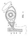
- FIG. 1illustrates the lacrimal duct system of a mammalian eye 10 .
- the systemincludes a lower punctum 12 connected to a lower lacrimal canaliculus 14 , and an upper punctum 16 connected to an upper lacrimal canaliculus 18 .
- Canaliculi 14 and 18are connected to a lacrimal sac 20 and a nasolacrimal duct 22 .
- a lacrimal gland 24is connected to eye 10 via a lacrimal duct 26 .
- tearsare produced by lacrimal gland 24 and are provided to eye 10 via lacrimal duct 26 , and tears are drained from eye 10 via punctum 12 and canaliculus 14 , punctum 16 and canaliculus 18 , and nasolacrimal duct 22 .
- tears produced by lacrimal gland 24are actually a complex composition in the form of a tear film.
- This tear filmincludes three basic layers: an outer lipid layer, an inner mucin layer, and an aqueous layer between the lipid and mucin layers. Each of the layers has a particular function.
- the lipid layerprevents evaporation of the tears from the surface of eye 10 .
- the aqueous layerprovides oxygen to the cornea and contains additional chemical components that are important to a healthy eye 10 .
- the mucin layerprovides for interaction between the lipid layer and the aqueous layer and prevents tears from “beading up” on the cornea.
- a “dry eye”is one that experiences insufficient lubrication of the cornea as a result a disturbance in the normal tear film.
- the conditionencompasses a wide variety of disease states ranging from mild, intermittent burning and/or scratchiness with foreign body sensation, to a severe lack of aqueous layer secretion accompanied by corneal and conjunctival disease (keratoconjunctivitis sicca (KCS)).
- Dry eyecan have a variety of specific causes and contributing factors, including arid environments, environmental airborne pollutants, certain systemic medications, auto-immune disorders, drug toxicity, hormone deficiency or changes, and even contact lens wear.
- lacrimal duct 26 from lacrimal gland 24may become clogged or may malfunction so that an insufficient amount of tears reach eye 10 .
- artificial tear productssuch as TEARS NATURALE® and BION® TEARS, sold by Alcon® Laboratories, Inc. of Fort Worth, Tex., were developed.
- puncta 12 and 16were sealed by stitching or by electrical or laser cauterization. Although such procedures can provide acceptable results, they are not reversible without reconstructive surgery. As it is sometimes difficult to determine whether dry eye is caused by too great of drainage or too little tear production, such procedures may expose the patient to unnecessary trauma. In addition, such procedures may result in epiphera, a condition where tears continually form on eye 10 , build up, and run down the face of the patient.
- Pre-formed collagen plugs for insertion into puncta 12 and 16 or the canaliculi 14 and 18were developed to provide a reversible sealing procedure.
- Collagen plugsare water-soluble and, when inserted into the puncta, typically dissolve within seven to fourteen days. Collagen plugs are thus effective as a test procedure to determine if it is desirable to more permanently seal the puncta.
- Pre-formed water-insoluble plugs for insertion into puncta 12 and 16 or canaliculi 14 and 18are described in a variety of United States Patents.
- U.S. Pat. No. 3,949,750 to Freemandescribes such a plug having a head portion that extends outside of the punctum and a barb portion that extends into the punctum and/or canaliculus.
- Such plugscan be seen in the corner of eye 10 , are sometimes uncomfortable, and are easily dislodged.
- such plugsare somewhat difficult to insert, and occasionally their size and shape causes tissue damage during insertion. If such plugs protrude too far from the puncta, they can cause irritation to the sclera.
- U.S. Pat. No. 5,283,063 to Freemandescribes a similar plug made from a hydrogel material having a hydrating port located in its barb portion that allows canalicular fluid to enter the barb and hydrate the plug to an expanded, relatively flexible state.
- U.S. Pat. Nos. 5,723,005 and 5,171,270 to Herrickdescribe water-insoluble punctal plugs that have collapsible flared sections for improved sealing and anchoring within the canaliculus. Some of these plugs also have a retaining portion that extends outside the punctum to further anchor the plug and prevent migration down the canaliculus.
- U.S. Pat. Nos. 3,949,750; 5,283,063; 5,723,005; and 5,171,270are each incorporated herein by reference.
- U.S. Pat. No. 5,469,867 to Schmittdescribes a method of occluding the lacrimal canaliculi and other mammalian channels or ducts by injecting a heated, flowable polymer or polymer composite of a specified composition through puncta 12 and 16 into canaliculi 14 and 18 , respectively.
- the specified polymer and polymer compositeare non-immunogenic, biocompatible materials that are solid and/or non-flowable at body temperature or lower and flowable when heated slightly above body temperature.
- the polymer and polymer compositeare capable of quickly changing from a flowable state to a non-flowable state by moving through only a few centigrade degrees of temperature.
- a punctal plugbe removable without the necessity of surgery.
- Various conventional techniqueshave been utilized to remove the above-described water insoluble plugs. For example, such plugs that have a portion extending outside the punctum are typically removed using forceps or a similar instrument.
- certain cannulashave been used to “flush” water-insoluble plugs not having a portion extending outside the punctum down nasolacrimal duct 22 . More specifically, it is known to use a cannula having an outer diameter equal to or slightly greater than the diameter of punctum 12 to seal the punctum and then inject saline into the punctum. The seal is created by dilating the punctum with the cannula and the sphincter action of the punctal muscle tightening around the cannula. Saline is then injected into canaliculus 14 so as to create enough water pressure to flush the plug down nasolacrimal duct 22 .
- An example of a conventional cannula that has been utilized to perform this techniqueis the E4404 lacrimal cannula sold by STORZ Ophthalmics of St. Louis, Mo., which has a 23 gauge outer diameter, 10 mm long tip.
- a cannula having a smaller outer diameter distal tipe.g. 26-27 gauge
- a larger outer diameter proximal portione.g. 23 gauge or greater
- a similar flushing proceduresuch as the cannula disclosed in U.S. Pat. No. 5,593,393 to Trudell et al.
- the larger outer diameter proximal portiondilates punctum 12
- the sphincter action of the punctal muscletightens around the proximal portion to complete the seal.
- Salineis then injected into canaliculus 14 so as to create enough water pressure to flush the plug down nasolacrimal duct 22 .
- a cannulawith an outer diameter less than the diameter of punctum 12 (e.g. 27 gauge) to flush such water-insoluble plugs down nasolacrimal duct 22 .
- Such a cannulacan be inserted into punctum 12 without dilation, but does not seal the punctum. Saline is then injected into canaliculus 14 in an attempt to create enough water pressure to flush the plug down nasolacrimal duct 22 .
- such a cannulatypically does not create enough water pressure to flush the plug due to the lack of a punctal seal, and excess saline is “backflushed” out of the eye and down the patient's face.
- U.S. Pat. No. 5,469,867discloses several techniques for removing its cast-in place thermoplastic plug from canaliculus 14 .
- the plugmay be physically extracted from the canaliculus by forceps or a similar instrument, or the plug may be heated by application of an electrical heating device that melts the polymer.
- a lipophilic compoundsuch as a naturally occurring oil or fatty acid ester that dissolves into the polymer and reduces the melting point of the polymer below body temperature may be introduced into canaliculus 14 for plug removal.
- This techniquetransforms the plug into a flowable fluid that is removable by irrigation with saline solution, which is typically performed using one of the above-described cannulas.
- the present inventionpertains to improved punctal cannula and methods of using such cannula to remove plugs or obstructions from mammalian canaliculi. More particularly, one aspect of the present invention comprises a cannula including a body having a distal portion, an external annulus disposed proximate the distal portion, and a hollow bore for fluid delivery from the distal portion.
- the distal portionis capable of insertion into a mammalian punctum without dilating the punctum.
- the external annulushas geometry capable of forming a seal with an external surface of the punctum without dilating the punctum.
- the present inventioncomprises a method of dislodging a plug or an obstruction from a mammalian canaliculus having a punctum.
- a cannulaincluding a body having a distal portion, an external annulus disposed proximate the distal portion, and a hollow bore for fluid delivery from the distal portion is provided.
- the distal portionis inserted into the punctum without dilating the punctum.
- the punctumis sealed by contacting an external surface of the punctum with the external annulus without dilating the punctum.
- An ophthalmic fluidis injected into the punctum with the cannula to pressurize a portion of the canaliculus between the plug or obstruction and the punctum.
- the present inventioncomprises a lacrimal cannula including a body having a distal portion, an external annulus, and a hollow bore for fluid delivery from the distal portion.
- the external annulushas a generally spherical geometry with the distal portion extending axially therefrom.
- the distal portionis capable of insertion into a mammalian lacrimal punctum without dilating the punctum.
- the external annulusis capable of forming a seal with an external surface of the punctum without dilating the punctum.
- the present inventioncomprises a lacrimal cannula including a body having a distal portion, an external annulus, and a hollow bore for fluid delivery from the distal portion.
- the external annulushas a generally inverted-cone shape geometry with the distal portion extending axially from a tip toward a base of the cone.
- the distal portionis capable of insertion into a mammalian lacrimal punctum without dilating the punctum.
- the external annulusis capable of forming a seal with an external surface of the punctum without dilating the punctum.
- the present inventioncomprises a lacrimal cannula including a body having a distal portion, an external annulus, and a hollow bore for fluid delivery from the distal portion.
- the external annulushas a generally cone shape geometry with the distal portion extending axially and outwardly from a tip of the cone.
- the distal portionis capable of insertion into a mammalian lacrimal punctum without dilating the punctum.
- the external annulusis capable of forming a seal with an external surface of the punctum without dilating the punctum.
- the present inventioncomprises a method of removing a cast-in place thermoplastic plug from a mammalian lacrimal canaliculus having a punctum.
- a cannulaincluding a body having a distal portion, an external annulus, and a hollow bore for fluid delivery from the distal portion is provided.
- the distal portionextends axially from the external annulus.
- the distal portionis inserted into the punctum without dilating the punctum.
- the punctumis sealed by contacting an external surface of the punctum with the external annulus without dilating the punctum.
- a warm ophthalmic fluidis injected into the punctum with the cannula to pressurize a portion of the canaliculus between the cast-in place thermoplastic plug and the punctum to dislodge the plug.
- FIG. 1is a schematic, front, partially sectional view of the lacrimal duct system of the mammalian eye
- FIG. 2is a schematic, front sectional view of the lacrimal canaliculi of FIG. 1 with the canaliculi blocked with conventional plugs;
- FIG. 3is a schematic side view of a cannula according to a first preferred embodiment of the present invention
- FIG. 4is a schematic side view of a cannula according to a second preferred embodiment of the present invention.
- FIG. 5is a schematic side view of a cannula according to a third preferred embodiment of the present invention.
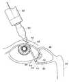
- FIG. 6is a schematic view of the cannula of FIG. 3 being used to remove a punctal plug from the lacrimal canaliculus.
- FIGS. 1-6 of the drawingslike numerals being used for like and corresponding parts of the various drawings. All wire gauges referred to in this document are preferably measured by the Brown & Sharpe technique.
- FIG. 2illustrates lacrimal canaliculi 14 and 18 blocked or occluded by plugs 30 and 32 , respectively.
- Plugs 30 and 32are preferably cast-in place thermoplastic plugs as disclosed in U.S. Pat. No. 5,469,867.
- plugs 30 and 32may be any of the conventional water-insoluble plugs that are typically flushed down nasolacrimal duct 22 .
- plugs 30 and 32 as shown in FIG. 2totally block canaliculi 14 and 18 , the plugs may be designed or formed so as to only partially block canaliculi 14 and 18 , if desired.
- Plugs 30 and 32may also be formed to extend into ampullae 19 and 21 , if desired.
- reference numerals 30 and 32may alternatively represent undesired obstructions in canaliculi 14 and 18 , respectively.
- Cannula 40generally comprises a body 42 having a distal portion 44 , an external annulus 46 , a proximal portion 48 , and a hollow bore 50 running within cannula 40 from distal portion 44 to proximal portion 48 .
- Proximal portion 48is coupled to a hub 52 .
- Hub 52is for removably coupling to a conventional syringe 53 , as shown in FIG. 6 .
- Cannula 40is preferably made of an inert metal such as surgical stainless steel, although other conventional metals, plastics, synthetics or composite materials may also be utilized.
- external annulus 46has a generally spherical geometry, with distal portion 44 of body 42 extending axially therefrom.
- External annulus 46has a shoulder 54 located proximate distal portion 44 .
- shoulder 54 of external annulus 46is located from about 0.5 mm to about 2.5 mm from a distal end 56 of distal portion 44 .
- External annulus 46has an outer diameter “D” substantially greater than an outer diameter “d” of distal portion 44 .
- the outer diametersare selected so that, when measured in inches, d ⁇ D ⁇ 6d.
- distal portion 44preferably has an outer diameter d from about 24 gauge to about 32 gauge, so that it can easily be inserted into puncta 12 and 16 without any dilation.
- external annulus 46preferably has an outer diameter D of about 18 gauge to about 8 gauge, so that shoulder 54 forms a smooth surface for contact with the external surface of puncta 12 and 16 .
- dis most preferably 27 gauge; and D is most preferably 16 gauge.
- proximal portion 48is shown with an outer diameter identical to the outer diameter d of distal portion 44 , proximal portion 48 may have a different diameter, if desired.
- cannula 40is especially suited for the removal of cast-in place thermoplastic plug 30 within canaliculus 14 in the following preferred manner.
- a warm, ophthalmic fluidsuch as saline solution is drawn into syringe 53 in the conventional manner.
- the ophthalmic fluidis preferably heated to above the melting point of the polymer or polymer composite comprising plug 30 .
- the ophthalmic fluidis heated to a temperature from about 3° C. to about 5° C. above the melting point of said polymer or polymer composite.
- distal portion 44is inserted into punctum 12 to the point where shoulder 54 of external annulus 46 is in contact with the external surface of punctum 12 .
- cannula 40thus effectively seals punctum 12 without any dilation of punctum 12 , and without the necessity of forcefully pressing cannula 40 against the punctal tissue to create a seal.
- distal portion 44due to the relatively short length of distal portion 44 (e.g. 0.5 mm to 2.5 mm), distal portion 44 can be easily inserted into canaliculus 14 without contacting plug 30 and thus interfering with the formation of the punctal seal.
- the warm ophthalmic fluidis injected into canaliculus 14 via hollow bore 50 by actuating syringe 53 .
- the warm ophthalmic fluidbegins to soften the polymer or polymer composite material comprising plug 30 .
- plug 30is quickly and easily dislodged from canaliculus 14 .
- warm, ophthalmic fluidis injected, further softening of plug 30 occurs, and plug 30 is eventually flushed down nasolacrimal duct 22 .
- plug 30remains a flowable, viscous mass, rather than becoming dissolved into the warm ophthalmic fluid.
- plug 30may be flushed at a lower pressure than conventional water-insoluble plugs.
- plug 30more easily negotiates turns 58 and surface irregularities 59 within canaliculus 14 or nasolacrimal duct 22 , as shown in FIG. 1 . Both of these advantages increase the chance of successfully flushing plug 30 and reduce the chance of over-pressurizing canaliculus 14 .
- a lipophilic compoundsuch as a mineral oil or fatty acid ester may be used as the ophthalmic fluid in the above-described process instead of saline solution.
- Such lipophilic materialsmigrate into and diffuse within the polymer or polymer composite comprising plug 30 causing the plug 30 to have a lower melting point. Therefore, such lipophilic compounds reduce the degree of heating required of the ophthalmic fluid.
- heating of the compoundmay not be required.
- Cannula 40may also be used to remove a conventional, water-insoluble plug 30 , or an obstruction 30 , within canaliculus 14 in the following preferred manner.
- An ophthalmic fluidsuch as saline solution is drawn into syringe 53 in the conventional manner.
- distal portion 44is inserted into punctum 12 to the point where shoulder 54 of external annulus 46 is in contact with the external surface of punctum 12 .
- the geometry of cannula 40thus effectively seals punctum 12 without any dilation of punctum 12 , and without the necessity of forcefully pressing cannula 40 against the punctal tissue to create a seal.
- the ophthalmic fluidis injected into canaliculus 14 via hollow bore 50 by actuating syringe 53 .
- Cannula 60generally comprises a body 62 having a distal portion 64 , an external annulus 66 , a proximal portion 68 , and a hollow bore 70 running within cannula 60 from distal portion 64 to proximal portion 68 .
- Proximal portion 68is coupled to a hub 72 .
- Hub 72is for removably coupling to a conventional syringe 53 .
- Cannula 60is preferably made of the same materials as cannula 40 .
- distal portion 64has a length of about 0.5 mm to about 2.5 mm.
- external annulus 66has a generally inverted-cone shaped geometry, with distal portion 64 of body 62 extending axially from the tip toward the base of the cone. More specifically, external annulus 66 has a rounded shoulder 74 proximate the base of its inverted-cone shaped geometry, and a slanted wall 76 that forms the wall of the inverted cone. For the human eye, slanted wall 76 preferably forms an angle ⁇ from about 10 degrees to about 80 degrees with a line 77 running perpendicular to the longitudinal axis of cannula 60 .
- distal portion 64preferably has an outer diameter d from about 24 gauge to about 32 gauge, so that it can easily be inserted into puncta 12 and 16 without any dilation.
- external annulus 66preferably has an outer diameter D of about 18 gauge to about 8 gauge, so that shoulder 74 and slanted wall 76 form a smooth surface for contact with the external surface of puncta 12 and 16 .
- dis most preferably 27 gauge
- Dis most preferably 16 gauge
- angle ⁇is most preferably about 30 to about 60 degrees.
- External annulus 66thus has a geometry specifically designed for engaging and sealing puncta 12 and 16 .
- Cannula 60is especially suited for the removal of cast-in place thermoplastic plug 30 within canaliculus 14 in the following preferred manner, which is substantially similar to, and has the same advantages of, the manner described above for cannula 40 .
- a warm, ophthalmic fluidsuch as saline solution is drawn into syringe 53 in the conventional manner.
- the ophthalmic fluidis preferably heated to above the melting point of the polymer or polymer composite comprising plug 30 .
- the ophthalmic fluidis heated to a temperature from about 3° C. to about 5° C. above the melting point of said polymer or polymer composite.
- distal portion 64is inserted into punctum 12 to the point where rounded shoulder 74 and slanted wall 76 of external annulus 66 are in contact with the external surface of punctum 12 .
- the geometry of cannula 60thus effectively seals punctum 12 without any dilation of punctum 12 , and without the necessity of forcefully pressing cannula 60 against the punctal tissue to create a seal.
- distal portion 64due to the relatively short length of distal portion 64 (e.g. 0.5 mm to 2.5 mm), distal portion 64 can be easily inserted into canaliculus 14 without contacting plug 30 and thus interfering with the formation of the punctal seal.
- the warm ophthalmic fluidis injected into canaliculus 14 via hollow bore 70 by actuating syringe 53 .
- the warm ophthalmic fluidbegins to soften the polymer or polymer composite material comprising plug 30 . Simultaneously, the pressure within canaliculus 14 between plug 30 and punctum 12 increases, and the canalicular tissue surrounding plug 30 gently expands away from plug 30 . In this manner, plug 30 is quickly and easily dislodged from canaliculus 14 . As additional, warm, ophthalmic fluid is injected, further softening of plug 30 occurs, and plug 30 is eventually flushed down nasolacrimal duct 22 . If it is desired, a lipophilic compound such as a mineral oil or fatty acid ester may be used as the ophthalmic fluid instead of saline solution, as described above in connection with cannula 40 .
- a lipophilic compoundsuch as a mineral oil or fatty acid ester may be used as the ophthalmic fluid instead of saline solution, as described above in connection with cannula 40 .
- Cannula 60may also be used to remove a conventional, water-insoluble plug 30 , or an obstruction 30 , within canaliculus 14 the following preferred manner, which is substantially similar to, and has the same advantages of, the manner described above for cannula 40 .
- An ophthalmic fluidsuch as saline solution is drawn into syringe 53 in the conventional manner.
- distal portion 64is placed within punctum 12 to the point where rounded shoulder 74 and slanted wall 76 of external annulus 66 are in contact with the external surface of punctum 12 .
- the geometry of cannula 60thus effectively seals punctum 12 without any dilation of punctum 12 , and without the necessity of forcefully pressing cannula 60 against the punctal tissue to create a seal.
- the ophthalmic fluidis injected into canaliculus 14 via hollow bore 70 by actuating syringe 53 .
- the pressure within canaliculus 14 between plug 30 and punctum 12increases until the means for anchoring plug 30 within canaliculus 14 is overcome.
- Additional salineis injected until plug 30 is flushed down nasolacrimal duct 22 .
- Cannula 80generally comprises a body 82 having a distal portion 84 , an external annulus 86 , a proximal portion 88 , and a hollow bore 90 running within cannula 80 from distal portion 84 to proximal portion 88 .
- Proximal portion 88is coupled to a hub 92 .
- Hub 92is for removably coupling to a conventional syringe 53 .
- Cannula 80is preferably made of the same materials as cannula 40 .
- distal portion 84has a length of about 0.5 mm to about 2.5 mm.
- external annulus 86has a generally cone shaped geometry, with distal portion 84 of body 82 extending axially and outwardly from the tip of the cone. More specifically, external annulus 86 has a slanted wall 94 that forms the wall of the cone. For the human eye, slanted wall 94 preferably forms an angle ⁇ from about 5 degrees to about 30 degrees from a line 96 running perpendicular to the longitudinal axis of cannula 80 . For the human eye, distal portion 84 preferably has an outer diameter d from about 24 gauge to about 32 gauge, so that it can easily be inserted into puncta 12 and 16 without any dilation.
- proximal portion 88preferably has an outer diameter D of about 18 gauge to about 8 gauge, so that slanted wall 94 forms a smooth surface for contact with the external surface of puncta 12 and 16 .
- dis most preferably 27 .gauge
- Dis most preferably 16 gauge
- angle ⁇ of slanted wall 94is most preferably about 5 to about 20 degrees.
- Cannula 80is especially suited for the removal of cast-in place thermoplastic plug 30 within canaliculus 14 in the following preferred manner, which is substantially similar to, and has the same advantages of, the manner described above for cannula 40 .
- a warm, ophthalmic fluidsuch as saline solution is drawn into syringe 53 in the conventional manner.
- the ophthalmic fluidis preferably heated to above the melting point of the polymer or polymer composite comprising plug 30 .
- the ophthalmic fluidis heated to a temperature from about 3° C. to about 5° C. above the melting point of said polymer or polymer composite.
- distal portion 84is inserted into punctum 12 to the point where slanted wall 94 of external annulus 46 contacts the external surface of punctum 12 . Due to the sharpness of angle ⁇ with respect to the longitudinal axis of cannula 80 , slanted wall 94 effectively seals punctum 12 without any dilation of punctum 12 , and without the necessity of forcefully pressing cannula 80 against the punctal tissue to create a seal. In addition, due to the relatively short length of distal portion 84 (e.g. 0.5 mm to 2.5 mm), distal portion 84 can be easily inserted into canaliculus 14 without contacting plug 30 and thus interfering with the formation of the punctal seal.
- the warm ophthalmic fluidis injected into canaliculus 14 via hollow bore 90 by actuating syringe 53 .
- the warm ophthalmic fluidbegins to soften the polymer or polymer composite material comprising plug 30 .
- the pressure within canaliculus 14 between plug 30 and slanted wall 94increases, and the canalicular tissue surrounding plug 30 gently expands away from plug 30 . In this manner, plug 30 is quickly and easily dislodged from canaliculus 14 .
- warm, ophthalmic fluidis injected, further softening of plug 30 occurs, and plug 30 is eventually flushed down nasolacrimal duct 22 .
- a lipophilic compoundsuch as a mineral oil or fatty acid ester may be used as the ophthalmic fluid instead of saline solution, as described above in connection with cannula 40 .
- Cannula 80may also be used to remove a conventional, water-insoluble plug 30 , or an obstruction 30 , within canaliculus 14 in the following preferred manner, which is substantially similar to, and has the same advantages of, the manner described above for cannula 40 .
- An ophthalmic fluidsuch as saline solution is drawn into syringe 53 in the conventional manner.
- distal portion 84is inserted into punctum 12 to the point slanted wall 94 of external annulus 86 contacts the external surface of punctum 12 .
- slanted wall 94effectively seals punctum 12 without any dilation of punctum 12 , and without the necessity of forcefully pressing cannula 80 against the punctal tissue to create a seal.
- the ophthalmic fluidis injected into canaliculus 14 via hollow bore 90 by actuating syringe 53 .
- the pressure within canaliculus 14 between plug 30 and slanted wall 94increases until the means for anchoring plug 30 within canaliculus 14 is overcome.
- Additional salineis injected until plug 30 is flushed down nasolacrimal duct 22 .
- the present inventionprovides an improved cannula for the removal of punctal plugs that are used to treat dry eye syndrome, and for the removal of obstructions in the lacrimal canaliculi.
- the improved cannulais easy to use, safe for the patient, and capable of economic manufacture.
- the cannulacan be used to remove such plugs or obstructions without any dilation, or with minimal dilation, of the punctum, and without the necessity of forcefully pressing the cannula against the punctal tissue to create a seal.
Landscapes
- Health & Medical Sciences (AREA)
- Ophthalmology & Optometry (AREA)
- Biomedical Technology (AREA)
- Nuclear Medicine, Radiotherapy & Molecular Imaging (AREA)
- Surgery (AREA)
- Engineering & Computer Science (AREA)
- Plastic & Reconstructive Surgery (AREA)
- Heart & Thoracic Surgery (AREA)
- Vascular Medicine (AREA)
- Life Sciences & Earth Sciences (AREA)
- Animal Behavior & Ethology (AREA)
- General Health & Medical Sciences (AREA)
- Public Health (AREA)
- Veterinary Medicine (AREA)
- Prostheses (AREA)
Abstract
Description
Claims (48)
Priority Applications (1)
| Application Number | Priority Date | Filing Date | Title |
|---|---|---|---|
| US09/567,175US6428502B1 (en) | 1999-06-25 | 2000-05-08 | Punctal cannula |
Applications Claiming Priority (2)
| Application Number | Priority Date | Filing Date | Title |
|---|---|---|---|
| US14119999P | 1999-06-25 | 1999-06-25 | |
| US09/567,175US6428502B1 (en) | 1999-06-25 | 2000-05-08 | Punctal cannula |
Publications (1)
| Publication Number | Publication Date |
|---|---|
| US6428502B1true US6428502B1 (en) | 2002-08-06 |
Family
ID=26838882
Family Applications (1)
| Application Number | Title | Priority Date | Filing Date |
|---|---|---|---|
| US09/567,175Expired - Fee RelatedUS6428502B1 (en) | 1999-06-25 | 2000-05-08 | Punctal cannula |
Country Status (1)
| Country | Link |
|---|---|
| US (1) | US6428502B1 (en) |
Cited By (46)
| Publication number | Priority date | Publication date | Assignee | Title |
|---|---|---|---|---|
| US20030208216A1 (en)* | 1999-09-08 | 2003-11-06 | Camp Matthew W. | Scleral plug system |
| US20040193102A1 (en)* | 2003-03-28 | 2004-09-30 | Kurt Haggstrom | Catheter with occlusion resistant tip |
| US20050033222A1 (en)* | 2003-03-28 | 2005-02-10 | Kurt Haggstrom | Triple lumen catheter with occlusion resistant tip |
| US20050045188A1 (en)* | 2003-05-22 | 2005-03-03 | Mendius Richard W. | Punctum plug |
| US20050070842A1 (en)* | 2003-03-28 | 2005-03-31 | Mark Lotito | Catheter with occlusion resistant tip |
| US20050165288A1 (en)* | 2004-01-27 | 2005-07-28 | Scimed Life Systems, Inc. | Systems and methods for treating breast tissue |
| US20050232972A1 (en)* | 2004-04-15 | 2005-10-20 | Steven Odrich | Drug delivery via punctal plug |
| US20050240152A1 (en)* | 2002-07-12 | 2005-10-27 | Scitec K.K. | Needle for medical use |
| US20060009522A1 (en)* | 2004-07-01 | 2006-01-12 | Reza Dana | Compositions and methods for treating eye disorders and conditions |
| US20060116629A1 (en)* | 2004-11-04 | 2006-06-01 | Tal Michael G | Catheter insertion apparatus |
| US20060173482A1 (en)* | 2003-04-28 | 2006-08-03 | Melker Jeremy S | Device for insertion of lacrimal stents |
| US20070243230A1 (en)* | 2006-03-31 | 2007-10-18 | Forsight Labs, Llc | Nasolacrimal Drainage System Implants for Drug Therapy |
| US20070265341A1 (en)* | 2004-07-01 | 2007-11-15 | The Schepens Eye Research Institute Inc. | Compositions and methods for treating eye disorders and conditions |
| US20090098584A1 (en)* | 2005-09-01 | 2009-04-16 | Bristol-Myers Squibb Company | Biomarkers and Methods for Determining Sensitivity to Vascular Endothelial growth factor Receptor-2 Modulators |
| US20090105749A1 (en)* | 2007-09-07 | 2009-04-23 | Qlt Plug Delivery, Inc. - Qpdi | Insertion and extraction tools for lacrimal implants |
| US20090104248A1 (en)* | 2007-09-07 | 2009-04-23 | Qlt Plug Delivery, Inc. -Qpdi | Lacrimal implants and related methods |
| US20090104243A1 (en)* | 2007-09-07 | 2009-04-23 | Qlt Plug Delivery, Inc. - Qpdi | Drug cores for sustained release of therapeutic agents |
| US20090118702A1 (en)* | 2004-07-02 | 2009-05-07 | Forsight Labs, Llc | Treatment Medium Delivery Device and Methods for Delivery of Such Treatment Mediums to the Eye Using such a Delivery Device |
| US20090137982A1 (en)* | 2005-04-21 | 2009-05-28 | Medtronic Vascular, Inc. | Guiding Catheter with Resiliently Compressible Occluder |
| WO2009064834A3 (en)* | 2007-11-13 | 2009-07-23 | Steven L Maskin | Meibomian gland intraductal diagnostic and treatment methods and related apparatus |
| US20090264861A1 (en)* | 2008-02-18 | 2009-10-22 | Qlt Plug Delivery, Inc. | Lacrimal implants and related methods |
| US20090280158A1 (en)* | 2008-05-09 | 2009-11-12 | Qlt Plug Delivery, Inc. | Sustained release delivery of active agents to treat glaucoma and ocular hypertension |
| US20100057011A1 (en)* | 2006-09-19 | 2010-03-04 | Charles Steven T | Trocar cannula |
| US20100100029A1 (en)* | 2007-11-13 | 2010-04-22 | Steven Maskin | Gland or duct diagnostic and treatment methods and related apparatus |
| US20100274204A1 (en)* | 2009-02-23 | 2010-10-28 | Qlt Plug Delivery, Inc. | Lacrimal implants and related methods |
| US20100331780A1 (en)* | 2009-06-26 | 2010-12-30 | Tyco Healthcare Group Lp | Catheterization System |
| RU2438626C2 (en)* | 2006-06-21 | 2012-01-10 | Джонсон Энд Джонсон Вижн Кэа, Инк. | Lachrymal opening bushings for active agent delivery |
| US20120143117A1 (en)* | 2010-09-16 | 2012-06-07 | The Cleveland Clinic Foundation | Lacrimal drainage manometer and method of use |
| US8277418B2 (en) | 2009-12-23 | 2012-10-02 | Alcon Research, Ltd. | Ophthalmic valved trocar cannula |
| US8343106B2 (en) | 2009-12-23 | 2013-01-01 | Alcon Research, Ltd. | Ophthalmic valved trocar vent |
| US8377001B2 (en) | 2010-10-01 | 2013-02-19 | Abbott Laboratories | Feeding set for a peristaltic pump system |
| US8377000B2 (en) | 2010-10-01 | 2013-02-19 | Abbott Laboratories | Enteral feeding apparatus having a feeding set |
| US20130096547A1 (en)* | 2011-10-17 | 2013-04-18 | Beaver-Visitec International (Us), Inc. | Method of, and device for, marking a patient's eye for reference during a toric lens implantation procedure |
| US20130204165A1 (en)* | 2010-09-16 | 2013-08-08 | The Cleveland Clinic Foundation | Lacrimal drainage manometer and method of use |
| US8689439B2 (en) | 2010-08-06 | 2014-04-08 | Abbott Laboratories | Method for forming a tube for use with a pump delivery system |
| US8747343B2 (en) | 2011-09-30 | 2014-06-10 | Covidien Lp | Hemodialysis catheter with improved side opening design |
| US9072867B2 (en) | 2011-09-30 | 2015-07-07 | Covidien Lp | Catheter with external flow channel |
| US9132088B2 (en) | 2008-04-30 | 2015-09-15 | Mati Therapeutics Inc. | Composite lacrimal insert and related methods |
| US9155862B2 (en) | 2012-09-28 | 2015-10-13 | Covidien Lp | Symmetrical tip acute catheter |
| US9610271B2 (en) | 2011-08-29 | 2017-04-04 | Mati Therapeutics Inc. | Sustained release delivery of active agents to treat glaucoma and ocular hypertension |
| US9974685B2 (en) | 2011-08-29 | 2018-05-22 | Mati Therapeutics | Drug delivery system and methods of treating open angle glaucoma and ocular hypertension |
| US10058676B2 (en) | 2009-09-30 | 2018-08-28 | Covidien Lp | Medical catheter having a design providing low recirculation and reversibility |
| US10603210B1 (en) | 2017-02-02 | 2020-03-31 | Mgd Innovations, Llc | Meibomian gland probing with blood product injection |
| US11141312B2 (en) | 2007-09-07 | 2021-10-12 | Mati Therapeutics Inc. | Lacrimal implant detection |
| US11273073B2 (en)* | 2014-12-17 | 2022-03-15 | Paul Gavaris | Canalicular plug, method and kit for treating dry eye |
| WO2023277887A1 (en)* | 2021-06-29 | 2023-01-05 | Visant Medical, Inc. | Method and device for irrigation into the lacrimal puncta |
Citations (10)
| Publication number | Priority date | Publication date | Assignee | Title |
|---|---|---|---|---|
| US3540447A (en) | 1967-09-29 | 1970-11-17 | Becton Dickinson Co | Spinal needle |
| US3949750A (en) | 1974-10-07 | 1976-04-13 | Freeman Jerre M | Punctum plug and method for treating keratoconjunctivitis sicca (dry eye) and other ophthalmic aliments using same |
| US4335718A (en) | 1980-10-02 | 1982-06-22 | Becton, Dickinson And Company | Needle cannula |
| US4915684A (en) | 1988-06-21 | 1990-04-10 | Mackeen Donald L | Method and apparatus for modulating the flow of lacrimal fluid through a punctum and associated canaliculus |
| US5171270A (en) | 1990-03-29 | 1992-12-15 | Herrick Robert S | Canalicular implant having a collapsible flared section and method |
| US5283063A (en) | 1992-01-31 | 1994-02-01 | Eagle Vision | Punctum plug method and apparatus |
| US5469867A (en)* | 1992-09-02 | 1995-11-28 | Landec Corporation | Cast-in place thermoplastic channel occluder |
| US5499065A (en) | 1995-06-20 | 1996-03-12 | Zimmerman; Thom J. | Device for preventing eye drops from entering the nasolacrimal duct system |
| US5593393A (en) | 1995-09-07 | 1997-01-14 | Trudell; Roger J. | Lacrimal irrigating cannula |
| US5723005A (en) | 1995-06-07 | 1998-03-03 | Herrick Family Limited Partnership | Punctum plug having a collapsible flared section and method |
- 2000
- 2000-05-08USUS09/567,175patent/US6428502B1/ennot_activeExpired - Fee Related
Patent Citations (11)
| Publication number | Priority date | Publication date | Assignee | Title |
|---|---|---|---|---|
| US3540447A (en) | 1967-09-29 | 1970-11-17 | Becton Dickinson Co | Spinal needle |
| US3949750A (en) | 1974-10-07 | 1976-04-13 | Freeman Jerre M | Punctum plug and method for treating keratoconjunctivitis sicca (dry eye) and other ophthalmic aliments using same |
| US4335718A (en) | 1980-10-02 | 1982-06-22 | Becton, Dickinson And Company | Needle cannula |
| US4915684A (en) | 1988-06-21 | 1990-04-10 | Mackeen Donald L | Method and apparatus for modulating the flow of lacrimal fluid through a punctum and associated canaliculus |
| US5171270A (en) | 1990-03-29 | 1992-12-15 | Herrick Robert S | Canalicular implant having a collapsible flared section and method |
| US5283063A (en) | 1992-01-31 | 1994-02-01 | Eagle Vision | Punctum plug method and apparatus |
| US5469867A (en)* | 1992-09-02 | 1995-11-28 | Landec Corporation | Cast-in place thermoplastic channel occluder |
| US5826584A (en)* | 1992-09-02 | 1998-10-27 | Schmitt; Edward E. | Devices for occluding channels in living mammals |
| US5723005A (en) | 1995-06-07 | 1998-03-03 | Herrick Family Limited Partnership | Punctum plug having a collapsible flared section and method |
| US5499065A (en) | 1995-06-20 | 1996-03-12 | Zimmerman; Thom J. | Device for preventing eye drops from entering the nasolacrimal duct system |
| US5593393A (en) | 1995-09-07 | 1997-01-14 | Trudell; Roger J. | Lacrimal irrigating cannula |
Non-Patent Citations (4)
| Title |
|---|
| Alcon 1997-1998 Catalog, "Cutting Instruments, Cannulas & Cystitomes, Ophthalmic Sponges, OPTEMP Cautery", pp. 11-17. |
| Grieshaber "Instruments for Irrigation Aspiration" Brochure, Feb. 1997, 1 page. |
| Katena Eye Instruments Catalog Supplement; 1997, pp. 87, 92. |
| Storz Instruments for Optometry Brochure, Sortz Ophthalmics, 1993, 11 pp. |
Cited By (101)
| Publication number | Priority date | Publication date | Assignee | Title |
|---|---|---|---|---|
| US20030208216A1 (en)* | 1999-09-08 | 2003-11-06 | Camp Matthew W. | Scleral plug system |
| US6846318B2 (en)* | 1999-09-08 | 2005-01-25 | Matthew W. Camp | Scleral plug system |
| US7022110B2 (en)* | 2002-07-12 | 2006-04-04 | Scitec K.K. | Needle for medical use |
| US20050240152A1 (en)* | 2002-07-12 | 2005-10-27 | Scitec K.K. | Needle for medical use |
| US20050070842A1 (en)* | 2003-03-28 | 2005-03-31 | Mark Lotito | Catheter with occlusion resistant tip |
| US7141035B2 (en)* | 2003-03-28 | 2006-11-28 | Sherwood Services Ag | Catheter with occlusion resistant tip |
| US7776005B2 (en) | 2003-03-28 | 2010-08-17 | Covidien Ag | Triple lumen catheter with occlusion resistant tip |
| US7090654B2 (en) | 2003-03-28 | 2006-08-15 | Sherwood Services Ag | Catheter with occlusion resistant tip |
| US20050033222A1 (en)* | 2003-03-28 | 2005-02-10 | Kurt Haggstrom | Triple lumen catheter with occlusion resistant tip |
| US20040193102A1 (en)* | 2003-03-28 | 2004-09-30 | Kurt Haggstrom | Catheter with occlusion resistant tip |
| US20060173482A1 (en)* | 2003-04-28 | 2006-08-03 | Melker Jeremy S | Device for insertion of lacrimal stents |
| US20050045188A1 (en)* | 2003-05-22 | 2005-03-03 | Mendius Richard W. | Punctum plug |
| US7204253B2 (en) | 2003-05-22 | 2007-04-17 | Clarity Corporation | Punctum plug |
| US20050165288A1 (en)* | 2004-01-27 | 2005-07-28 | Scimed Life Systems, Inc. | Systems and methods for treating breast tissue |
| US9463114B2 (en) | 2004-04-15 | 2016-10-11 | Mati Therapeutics Inc. | Punctal plug with active agent |
| US20050232972A1 (en)* | 2004-04-15 | 2005-10-20 | Steven Odrich | Drug delivery via punctal plug |
| US20100040670A1 (en)* | 2004-04-15 | 2010-02-18 | Qlt Plug Delivery, Inc. | Drug delivery via ocular implant |
| US20060009522A1 (en)* | 2004-07-01 | 2006-01-12 | Reza Dana | Compositions and methods for treating eye disorders and conditions |
| US20070265341A1 (en)* | 2004-07-01 | 2007-11-15 | The Schepens Eye Research Institute Inc. | Compositions and methods for treating eye disorders and conditions |
| US20080153909A1 (en)* | 2004-07-01 | 2008-06-26 | The Schepens Eye Research Institute, Inc. | Compositions and methods for treating eye disorders and conditions |
| US9180045B2 (en) | 2004-07-02 | 2015-11-10 | Mati Therapeutics Inc. | Treatment medium delivery device and methods for delivery of such treatment mediums to the eye using such a delivery device |
| US7922702B2 (en) | 2004-07-02 | 2011-04-12 | Qlt Inc. | Treatment medium delivery device and methods for delivery of such treatment mediums to the eye using such a delivery device |
| US9820884B2 (en) | 2004-07-02 | 2017-11-21 | Mati Therapeutics Inc. | Treatment medium delivery device and methods for delivery of such treatment mediums to the eye using such delivery device |
| US20090118702A1 (en)* | 2004-07-02 | 2009-05-07 | Forsight Labs, Llc | Treatment Medium Delivery Device and Methods for Delivery of Such Treatment Mediums to the Eye Using such a Delivery Device |
| US10610407B2 (en) | 2004-07-02 | 2020-04-07 | Mati Therapeutics Inc. | Treatment medium delivery device and methods for delivery of such treatment mediums to the eye using such delivery device |
| US10668248B2 (en) | 2004-11-04 | 2020-06-02 | Covidien Ag | Catheter insertion apparatus |
| US20060116629A1 (en)* | 2004-11-04 | 2006-06-01 | Tal Michael G | Catheter insertion apparatus |
| US9913962B2 (en) | 2004-11-04 | 2018-03-13 | Covidien Ag | Catheter insertion apparatus |
| US11504499B2 (en) | 2004-11-04 | 2022-11-22 | Covidien Ag | Catheter insertion apparatus |
| US20090137982A1 (en)* | 2005-04-21 | 2009-05-28 | Medtronic Vascular, Inc. | Guiding Catheter with Resiliently Compressible Occluder |
| US7909810B2 (en) | 2005-04-21 | 2011-03-22 | Medtronic Vascular, Inc | Guiding catheter with resiliently compressible occluder |
| US20090098584A1 (en)* | 2005-09-01 | 2009-04-16 | Bristol-Myers Squibb Company | Biomarkers and Methods for Determining Sensitivity to Vascular Endothelial growth factor Receptor-2 Modulators |
| US9168222B2 (en) | 2006-03-31 | 2015-10-27 | Mati Therapeutics Inc. | Nasolacrimal drainage system implants for drug therapy |
| US8747884B2 (en) | 2006-03-31 | 2014-06-10 | Mati Therapeutics Inc. | Nasolacrimal drainage system implants for drug therapy |
| US10874606B2 (en) | 2006-03-31 | 2020-12-29 | Mati Therapeutics Inc. | Nasolacrimal drainage system implants for drug therapy |
| US20070243230A1 (en)* | 2006-03-31 | 2007-10-18 | Forsight Labs, Llc | Nasolacrimal Drainage System Implants for Drug Therapy |
| US12226525B2 (en) | 2006-03-31 | 2025-02-18 | Mati Therapeutics, Inc. | Nasolacrimal drainage system implants for drug therapy |
| US7998497B2 (en) | 2006-03-31 | 2011-08-16 | Qlt Inc. | Nasolacrimal drainage system implants for drug therapy |
| US8691265B2 (en) | 2006-03-31 | 2014-04-08 | Mati Therapeutics, Inc. | Drug delivery methods, structures, and compositions for nasolacrimal system |
| US11406592B2 (en) | 2006-03-31 | 2022-08-09 | Mati Therapeutics Inc. | Drug delivery methods, structures, and compositions for nasolacrimal system |
| US8795711B2 (en) | 2006-03-31 | 2014-08-05 | Mati Therapeutics Inc. | Drug delivery methods, structures, and compositions for nasolacrimal system |
| US9610194B2 (en) | 2006-03-31 | 2017-04-04 | Mati Therapeutics Inc. | Drug delivery methods, structures, and compositions for nasolacrimal system |
| US9849082B2 (en) | 2006-03-31 | 2017-12-26 | Mati Therapeutics Inc. | Nasolacrimal drainage system implants for drug therapy |
| US10383817B2 (en) | 2006-03-31 | 2019-08-20 | Mati Therapeutics Inc. | Nasolacrimal drainage system implants for drug therapy |
| US10300014B2 (en) | 2006-03-31 | 2019-05-28 | Mati Therapeutics Inc. | Nasolacrimal drainage system implants for drug therapy |
| RU2438626C2 (en)* | 2006-06-21 | 2012-01-10 | Джонсон Энд Джонсон Вижн Кэа, Инк. | Lachrymal opening bushings for active agent delivery |
| US20100057011A1 (en)* | 2006-09-19 | 2010-03-04 | Charles Steven T | Trocar cannula |
| US8333726B2 (en) | 2007-09-07 | 2012-12-18 | Qlt Inc. | Lacrimal implants and related methods |
| US10434009B2 (en) | 2007-09-07 | 2019-10-08 | Mati Therapeutics Inc. | Lacrimal implants and related methods |
| US11141312B2 (en) | 2007-09-07 | 2021-10-12 | Mati Therapeutics Inc. | Lacrimal implant detection |
| US8628792B2 (en) | 2007-09-07 | 2014-01-14 | Mati Therapeutics, Inc. | Drug cores for sustained release of therapeutic agents |
| US9445944B2 (en) | 2007-09-07 | 2016-09-20 | Mati Therapeutics Inc. | Lacrimal implants and related methods |
| US11951038B2 (en) | 2007-09-07 | 2024-04-09 | Mati Therapeutics Inc. | Lacrimal implants and related methods |
| US20090104243A1 (en)* | 2007-09-07 | 2009-04-23 | Qlt Plug Delivery, Inc. - Qpdi | Drug cores for sustained release of therapeutic agents |
| US8702643B2 (en) | 2007-09-07 | 2014-04-22 | Mati Therapeutics, Inc. | Lacrimal implants and related methods |
| US20090104248A1 (en)* | 2007-09-07 | 2009-04-23 | Qlt Plug Delivery, Inc. -Qpdi | Lacrimal implants and related methods |
| US20090105749A1 (en)* | 2007-09-07 | 2009-04-23 | Qlt Plug Delivery, Inc. - Qpdi | Insertion and extraction tools for lacrimal implants |
| WO2009064834A3 (en)* | 2007-11-13 | 2009-07-23 | Steven L Maskin | Meibomian gland intraductal diagnostic and treatment methods and related apparatus |
| US11110003B2 (en) | 2007-11-13 | 2021-09-07 | Mgd Innovations, Inc. | Gland or duct diagnostic and treatment methods and related apparatus |
| US10159599B2 (en) | 2007-11-13 | 2018-12-25 | Mgd Innovations, Llc | Meibomian gland intraductal diagnostic and treatment methods |
| US9510844B2 (en) | 2007-11-13 | 2016-12-06 | Mgd Innovations, Llc | Gland or duct diagnostic and treatment methods and related apparatus |
| US12004997B2 (en) | 2007-11-13 | 2024-06-11 | Mgd Innovations, Inc. | Gland or duct diagnostic and treatment methods and related apparatus |
| US20100292630A1 (en)* | 2007-11-13 | 2010-11-18 | Maskin Steven L | Meibomian Gland Intraductal Diagnostic and Treatment Methods |
| US20100100029A1 (en)* | 2007-11-13 | 2010-04-22 | Steven Maskin | Gland or duct diagnostic and treatment methods and related apparatus |
| US9216108B2 (en) | 2008-02-18 | 2015-12-22 | Mati Therapeutics Inc. | Lacrimal implants and related methods |
| US20090264861A1 (en)* | 2008-02-18 | 2009-10-22 | Qlt Plug Delivery, Inc. | Lacrimal implants and related methods |
| US9764066B2 (en) | 2008-04-30 | 2017-09-19 | Mati Therapeutics Inc. | Composite lacrimal insert and related methods |
| US9132088B2 (en) | 2008-04-30 | 2015-09-15 | Mati Therapeutics Inc. | Composite lacrimal insert and related methods |
| US20090280158A1 (en)* | 2008-05-09 | 2009-11-12 | Qlt Plug Delivery, Inc. | Sustained release delivery of active agents to treat glaucoma and ocular hypertension |
| US9949942B2 (en) | 2008-05-09 | 2018-04-24 | Mati Therapeutics Inc. | Sustained release delivery of active agents to treat glaucoma and ocular hypertension |
| US20100274204A1 (en)* | 2009-02-23 | 2010-10-28 | Qlt Plug Delivery, Inc. | Lacrimal implants and related methods |
| US10238535B2 (en) | 2009-02-23 | 2019-03-26 | Mati Therapeutics Inc. | Lacrimal implants and related methods |
| US8187231B2 (en) | 2009-06-26 | 2012-05-29 | Tyco Healthcare Group Lp | Catheterization system |
| US20100331780A1 (en)* | 2009-06-26 | 2010-12-30 | Tyco Healthcare Group Lp | Catheterization System |
| US8496622B2 (en) | 2009-06-26 | 2013-07-30 | Covidien LLP | Catheterization system |
| US9468461B2 (en) | 2009-06-26 | 2016-10-18 | Covidien Lp | Catheterization system |
| US10058676B2 (en) | 2009-09-30 | 2018-08-28 | Covidien Lp | Medical catheter having a design providing low recirculation and reversibility |
| US8277418B2 (en) | 2009-12-23 | 2012-10-02 | Alcon Research, Ltd. | Ophthalmic valved trocar cannula |
| US8679064B2 (en) | 2009-12-23 | 2014-03-25 | Alcon Research, Ltd. | Ophthalmic valved trocar cannula |
| US8343106B2 (en) | 2009-12-23 | 2013-01-01 | Alcon Research, Ltd. | Ophthalmic valved trocar vent |
| US8689439B2 (en) | 2010-08-06 | 2014-04-08 | Abbott Laboratories | Method for forming a tube for use with a pump delivery system |
| US20130204165A1 (en)* | 2010-09-16 | 2013-08-08 | The Cleveland Clinic Foundation | Lacrimal drainage manometer and method of use |
| US8460230B2 (en)* | 2010-09-16 | 2013-06-11 | The Cleveland Clinic Foundation | Lacrimal drainage manometer and method of use |
| US8932231B2 (en)* | 2010-09-16 | 2015-01-13 | The Cleveland Clinic Foundation | Lacrimal drainage manometer and method of use |
| US20120143117A1 (en)* | 2010-09-16 | 2012-06-07 | The Cleveland Clinic Foundation | Lacrimal drainage manometer and method of use |
| US8377001B2 (en) | 2010-10-01 | 2013-02-19 | Abbott Laboratories | Feeding set for a peristaltic pump system |
| US8377000B2 (en) | 2010-10-01 | 2013-02-19 | Abbott Laboratories | Enteral feeding apparatus having a feeding set |
| US10632012B2 (en) | 2011-08-29 | 2020-04-28 | Mati Therapeutics Inc. | Sustained release delivery of active agents to treat glaucoma and ocular hypertension |
| US9610271B2 (en) | 2011-08-29 | 2017-04-04 | Mati Therapeutics Inc. | Sustained release delivery of active agents to treat glaucoma and ocular hypertension |
| US9974685B2 (en) | 2011-08-29 | 2018-05-22 | Mati Therapeutics | Drug delivery system and methods of treating open angle glaucoma and ocular hypertension |
| US9072867B2 (en) | 2011-09-30 | 2015-07-07 | Covidien Lp | Catheter with external flow channel |
| US8747343B2 (en) | 2011-09-30 | 2014-06-10 | Covidien Lp | Hemodialysis catheter with improved side opening design |
| US20130096547A1 (en)* | 2011-10-17 | 2013-04-18 | Beaver-Visitec International (Us), Inc. | Method of, and device for, marking a patient's eye for reference during a toric lens implantation procedure |
| US10117703B2 (en)* | 2011-10-17 | 2018-11-06 | Beaver-Visitec International (Us), Inc. | Method of, and device for, marking a patients eye for reference during a toric lens implantation procedure |
| US11413426B2 (en) | 2012-09-28 | 2022-08-16 | Covidien Lp | Symmetrical tip acute catheter |
| US11554247B2 (en) | 2012-09-28 | 2023-01-17 | Covidien Lp | Symmetrical tip acute catheter |
| US9155862B2 (en) | 2012-09-28 | 2015-10-13 | Covidien Lp | Symmetrical tip acute catheter |
| US9526861B2 (en) | 2012-09-28 | 2016-12-27 | Covidien Lp | Symmetrical tip acute catheter |
| US11273073B2 (en)* | 2014-12-17 | 2022-03-15 | Paul Gavaris | Canalicular plug, method and kit for treating dry eye |
| US10603210B1 (en) | 2017-02-02 | 2020-03-31 | Mgd Innovations, Llc | Meibomian gland probing with blood product injection |
| WO2023277887A1 (en)* | 2021-06-29 | 2023-01-05 | Visant Medical, Inc. | Method and device for irrigation into the lacrimal puncta |
Similar Documents
| Publication | Publication Date | Title |
|---|---|---|
| US6428502B1 (en) | Punctal cannula | |
| JP5290167B2 (en) | Puncture plug for active agent delivery | |
| US20190321225A1 (en) | Combined treatment for cataract and glaucoma treatment | |
| EP0988844A2 (en) | Apparatus for intubation of lacrimal duct | |
| TWI542338B (en) | Ophthalmic devices for the controlled release of active agents | |
| US5527356A (en) | Retinal plug | |
| US9789000B2 (en) | Glaucoma treatment device | |
| US9585789B2 (en) | Ocular implant with hydrogel expansion capabilities | |
| US6994684B2 (en) | Punctum plugs having fluid collecting recesses and methods of punctal occlusion | |
| US5334137A (en) | Lacrimal fluid control device | |
| US5840054A (en) | Method for obstructing lacrimal canaliculi with infusable solution or dispersion | |
| US20050277864A1 (en) | Injectable gel implant for glaucoma treatment | |
| US7862532B2 (en) | Punctum plugs having insertion guides and strengthening beams | |
| EP0506849A1 (en) | Method and apparatus for the treatment of glaucoma | |
| CA2655843A1 (en) | Punctal plugs for the delivery of active agents | |
| WO1991014406A1 (en) | Canalicular implant having collapsible section and method | |
| US5273751A (en) | Composition for preventing clouding of paosterior capsule after extracapsular cataract eye surgery and method of performing cataract surgery | |
| JP5259130B2 (en) | Puncture plug for administration of active agent | |
| US9504605B2 (en) | Lacrimal filler | |
| Vinod et al. | Vitreous occlusion of a glaucoma drainage implant—surgical management | |
| US5080647A (en) | Composition for preventing clouding of posterior capsule after extracapsular cataract eye surgery and method of performing cataract surgery | |
| JP5122187B2 (en) | Puncture plug for administration of active agent | |
| JPH1147183A (en) | Lacrimal punctum plug device | |
| Durukan et al. | Photodisruptive Neodymium Yttrium-Aluminum Garnet Laser in the Management of Premacular subhyaloid Hoemorrhage | |
| Mittl | Perfluorocarbon gases in the suprachoroidal space of rabbit eyes |
Legal Events
| Date | Code | Title | Description |
|---|---|---|---|
| AS | Assignment | Owner name:ALCON LABORATORIES, INC., TEXAS Free format text:ASSIGNMENT OF ASSIGNORS INTEREST;ASSIGNOR:LANG, JOHN C.;REEL/FRAME:010896/0378 Effective date:20000508 | |
| AS | Assignment | Owner name:ALCON MANUFACTURING, LTD., TEXAS Free format text:ASSIGNMENT OF ASSIGNORS INTEREST;ASSIGNOR:ALCON LABORATORIES, INC.;REEL/FRAME:011667/0559 Effective date:20010322 | |
| CC | Certificate of correction | ||
| FPAY | Fee payment | Year of fee payment:4 | |
| AS | Assignment | Owner name:ALCON RESEARCH, LTD., TEXAS Free format text:MERGER;ASSIGNOR:ALCON MANUFACTURING, LTD.;REEL/FRAME:021266/0729 Effective date:20080101 Owner name:ALCON RESEARCH, LTD.,TEXAS Free format text:MERGER;ASSIGNOR:ALCON MANUFACTURING, LTD.;REEL/FRAME:021266/0729 Effective date:20080101 | |
| FPAY | Fee payment | Year of fee payment:8 | |
| REMI | Maintenance fee reminder mailed | ||
| LAPS | Lapse for failure to pay maintenance fees | ||
| STCH | Information on status: patent discontinuation | Free format text:PATENT EXPIRED DUE TO NONPAYMENT OF MAINTENANCE FEES UNDER 37 CFR 1.362 | |
| FP | Lapsed due to failure to pay maintenance fee | Effective date:20140806 |