US6425867B1 - Noise-free real time ultrasonic imaging of a treatment site undergoing high intensity focused ultrasound therapy - Google Patents
Noise-free real time ultrasonic imaging of a treatment site undergoing high intensity focused ultrasound therapyDownload PDFInfo
- Publication number
- US6425867B1 US6425867B1US09/397,471US39747199AUS6425867B1US 6425867 B1US6425867 B1US 6425867B1US 39747199 AUS39747199 AUS 39747199AUS 6425867 B1US6425867 B1US 6425867B1
- Authority
- US
- United States
- Prior art keywords
- therapeutic
- hifu
- ultrasonic transducer
- treatment site
- target area
- Prior art date
- Legal status (The legal status is an assumption and is not a legal conclusion. Google has not performed a legal analysis and makes no representation as to the accuracy of the status listed.)
- Expired - Lifetime
Links
- 238000011282treatmentMethods0.000titleclaimsabstractdescription267
- 238000003384imaging methodMethods0.000titleclaimsabstractdescription248
- 238000002604ultrasonographyMethods0.000titleclaimsabstractdescription96
- 238000002560therapeutic procedureMethods0.000titleclaimsdescription116
- 230000001225therapeutic effectEffects0.000claimsabstractdescription239
- 239000000523sampleSubstances0.000claimsabstractdescription132
- 238000000034methodMethods0.000claimsabstractdescription107
- 201000010260leiomyomaDiseases0.000claimsabstractdescription49
- 206010046798Uterine leiomyomaDiseases0.000claimsabstractdescription46
- 210000004204blood vesselAnatomy0.000claimsabstractdescription29
- 230000003902lesionEffects0.000claimsabstractdescription28
- 230000000694effectsEffects0.000claimsabstractdescription26
- 230000008859changeEffects0.000claimsabstractdescription17
- 230000001965increasing effectEffects0.000claimsabstractdescription14
- QVGXLLKOCUKJST-UHFFFAOYSA-Natomic oxygenChemical compound[O]QVGXLLKOCUKJST-UHFFFAOYSA-N0.000claimsabstractdescription5
- 229910052760oxygenInorganic materials0.000claimsabstractdescription5
- 239000001301oxygenSubstances0.000claimsabstractdescription5
- 235000015097nutrientsNutrition0.000claimsabstractdescription4
- 238000012285ultrasound imagingMethods0.000claimsdescription34
- 238000012545processingMethods0.000claimsdescription26
- 230000000007visual effectEffects0.000claimsdescription20
- 239000008280bloodSubstances0.000claimsdescription19
- 210000004369bloodAnatomy0.000claimsdescription19
- 230000002829reductive effectEffects0.000claimsdescription17
- 208000010579uterine corpus leiomyomaDiseases0.000claimsdescription15
- 201000007954uterine fibroidDiseases0.000claimsdescription15
- 230000006378damageEffects0.000claimsdescription13
- 239000012530fluidSubstances0.000claimsdescription11
- 230000017074necrotic cell deathEffects0.000claimsdescription11
- 230000000740bleeding effectEffects0.000claimsdescription9
- 230000008569processEffects0.000claimsdescription9
- 210000000664rectumAnatomy0.000claimsdescription8
- 208000001154Dermoid CystDiseases0.000claimsdescription6
- 208000009774Follicular CystDiseases0.000claimsdescription6
- 206010036049Polycystic ovariesDiseases0.000claimsdescription6
- 238000002679ablationMethods0.000claimsdescription6
- 201000006381corpus luteum cystDiseases0.000claimsdescription6
- 208000016018endometrial polypDiseases0.000claimsdescription6
- 210000002540macrophageAnatomy0.000claimsdescription6
- 238000012544monitoring processMethods0.000claimsdescription6
- 208000025661ovarian cystDiseases0.000claimsdescription6
- 201000010065polycystic ovary syndromeDiseases0.000claimsdescription6
- 210000004994reproductive systemAnatomy0.000claimsdescription6
- 206010046811uterine polypDiseases0.000claimsdescription6
- 208000005641AdenomyosisDiseases0.000claimsdescription5
- 201000009273EndometriosisDiseases0.000claimsdescription5
- 208000005697cornual pregnancyDiseases0.000claimsdescription5
- 201000003511ectopic pregnancyDiseases0.000claimsdescription5
- 201000006828endometrial hyperplasiaDiseases0.000claimsdescription5
- 201000009274endometriosis of uterusDiseases0.000claimsdescription5
- 238000005111flow chemistry techniqueMethods0.000claimsdescription5
- 230000023597hemostasisEffects0.000claimsdescription5
- 230000036244malformationEffects0.000claimsdescription5
- 230000035935pregnancyEffects0.000claimsdescription5
- 238000010438heat treatmentMethods0.000claimsdescription4
- 230000004044responseEffects0.000claimsdescription4
- 238000006243chemical reactionMethods0.000claimsdescription3
- 230000001939inductive effectEffects0.000claimsdescription3
- 230000001360synchronised effectEffects0.000claimsdescription3
- 230000036770blood supplyEffects0.000claimsdescription2
- 239000002195soluble materialSubstances0.000claims6
- 230000035899viabilityEffects0.000claims6
- 230000017531blood circulationEffects0.000claims4
- 210000000683abdominal cavityAnatomy0.000claims3
- 230000003993interactionEffects0.000claims2
- 208000009206Abruptio PlacentaeDiseases0.000claims1
- 208000032170Congenital AbnormalitiesDiseases0.000claims1
- 230000002238attenuated effectEffects0.000claims1
- 230000002500effect on skinEffects0.000claims1
- 238000001802infusionMethods0.000claims1
- 201000008532placental abruptionDiseases0.000claims1
- 206010028980NeoplasmDiseases0.000abstractdescription79
- 210000004996female reproductive systemAnatomy0.000abstractdescription11
- 208000037265diseases, disorders, signs and symptomsDiseases0.000abstractdescription9
- 230000000451tissue damageEffects0.000abstract1
- 231100000827tissue damageToxicity0.000abstract1
- 210000001519tissueAnatomy0.000description52
- 230000008901benefitEffects0.000description16
- XLYOFNOQVPJJNP-UHFFFAOYSA-NwaterSubstancesOXLYOFNOQVPJJNP-UHFFFAOYSA-N0.000description11
- 238000013461designMethods0.000description10
- 210000004291uterusAnatomy0.000description9
- 206010028851NecrosisDiseases0.000description8
- 230000005284excitationEffects0.000description7
- 230000033001locomotionEffects0.000description7
- 206010004446Benign prostatic hyperplasiaDiseases0.000description6
- 208000004403Prostatic HyperplasiaDiseases0.000description6
- 208000035475disorderDiseases0.000description6
- 239000003814drugSubstances0.000description6
- 229940079593drugDrugs0.000description6
- 230000001338necrotic effectEffects0.000description6
- 238000013459approachMethods0.000description5
- 210000003679cervix uteriAnatomy0.000description5
- 239000003795chemical substances by applicationSubstances0.000description5
- 238000002651drug therapyMethods0.000description5
- 230000006870functionEffects0.000description5
- 230000029058respiratory gaseous exchangeEffects0.000description5
- 238000001356surgical procedureMethods0.000description5
- 206010016654FibrosisDiseases0.000description4
- 238000010586diagramMethods0.000description4
- 230000004761fibrosisEffects0.000description4
- 230000000977initiatory effectEffects0.000description4
- 238000005259measurementMethods0.000description4
- 210000002307prostateAnatomy0.000description4
- 210000001215vaginaAnatomy0.000description4
- 102000004127CytokinesHuman genes0.000description3
- 108090000695CytokinesProteins0.000description3
- 241001465754MetazoaSpecies0.000description3
- 238000004458analytical methodMethods0.000description3
- 230000003466anti-cipated effectEffects0.000description3
- 201000011510cancerDiseases0.000description3
- 230000003247decreasing effectEffects0.000description3
- 230000028993immune responseEffects0.000description3
- 208000015181infectious diseaseDiseases0.000description3
- 230000028709inflammatory responseEffects0.000description3
- 230000007246mechanismEffects0.000description3
- 238000012986modificationMethods0.000description3
- 230000004048modificationEffects0.000description3
- 230000009467reductionEffects0.000description3
- 238000012552reviewMethods0.000description3
- 208000024891symptomDiseases0.000description3
- 230000002195synergetic effectEffects0.000description3
- 239000011800void materialSubstances0.000description3
- 229910001369BrassInorganic materials0.000description2
- 206010060862Prostate cancerDiseases0.000description2
- 208000000236Prostatic NeoplasmsDiseases0.000description2
- 241000700159RattusSpecies0.000description2
- 230000003187abdominal effectEffects0.000description2
- 238000010521absorption reactionMethods0.000description2
- 230000000259anti-tumor effectEffects0.000description2
- 210000004556brainAnatomy0.000description2
- 239000010951brassSubstances0.000description2
- 238000004891communicationMethods0.000description2
- 150000001875compoundsChemical class0.000description2
- 238000001816coolingMethods0.000description2
- 230000008878couplingEffects0.000description2
- 238000010168coupling processMethods0.000description2
- 238000005859coupling reactionMethods0.000description2
- 230000016396cytokine productionEffects0.000description2
- 201000010099diseaseDiseases0.000description2
- 230000010102embolizationEffects0.000description2
- 239000003193general anesthetic agentSubstances0.000description2
- 230000036541healthEffects0.000description2
- 238000011065in-situ storageMethods0.000description2
- 238000004519manufacturing processMethods0.000description2
- 239000000463materialSubstances0.000description2
- 230000001766physiological effectEffects0.000description2
- 229920006395saturated elastomerPolymers0.000description2
- 230000008685targetingEffects0.000description2
- 210000004881tumor cellAnatomy0.000description2
- 208000003200AdenomaDiseases0.000description1
- 206010001233Adenoma benignDiseases0.000description1
- 206010002091AnaesthesiaDiseases0.000description1
- 208000003174Brain NeoplasmsDiseases0.000description1
- NMJREATYWWNIKX-UHFFFAOYSA-NGnRHChemical compoundC1CCC(C(=O)NCC(N)=O)N1C(=O)C(CC(C)C)NC(=O)C(CC=1C2=CC=CC=C2NC=1)NC(=O)CNC(=O)C(NC(=O)C(CO)NC(=O)C(CC=1C2=CC=CC=C2NC=1)NC(=O)C(CC=1NC=NC=1)NC(=O)C1NC(=O)CC1)CC1=CC=C(O)C=C1NMJREATYWWNIKX-UHFFFAOYSA-N0.000description1
- 241000699666Mus <mouse, genus>Species0.000description1
- 241000699670Mus sp.Species0.000description1
- 206010067482No adverse eventDiseases0.000description1
- 208000018737Parkinson diseaseDiseases0.000description1
- 241000405070PercophidaeSpecies0.000description1
- 241001632427RadiolaSpecies0.000description1
- 101000857870Squalus acanthias GonadoliberinProteins0.000description1
- 208000024313Testicular NeoplasmsDiseases0.000description1
- 206010057644Testis cancerDiseases0.000description1
- 206010054094Tumour necrosisDiseases0.000description1
- 230000009471actionEffects0.000description1
- 239000000853adhesiveSubstances0.000description1
- 230000001070adhesive effectEffects0.000description1
- 239000000556agonistSubstances0.000description1
- 230000037005anaesthesiaEffects0.000description1
- 210000003484anatomyAnatomy0.000description1
- 238000002583angiographyMethods0.000description1
- 239000000427antigenSubstances0.000description1
- 102000036639antigensHuman genes0.000description1
- 108091007433antigensProteins0.000description1
- 239000002246antineoplastic agentSubstances0.000description1
- 229940041181antineoplastic drugDrugs0.000description1
- 238000011717athymic nude mouseMethods0.000description1
- 230000009286beneficial effectEffects0.000description1
- 230000015572biosynthetic processEffects0.000description1
- 201000003163breast adenomaDiseases0.000description1
- 230000000747cardiac effectEffects0.000description1
- 210000004027cellAnatomy0.000description1
- 230000002490cerebral effectEffects0.000description1
- 238000002648combination therapyMethods0.000description1
- 239000012141concentrateSubstances0.000description1
- 239000002872contrast mediaSubstances0.000description1
- 238000005520cutting processMethods0.000description1
- 230000007123defenseEffects0.000description1
- 230000000593degrading effectEffects0.000description1
- 238000002405diagnostic procedureMethods0.000description1
- 230000003292diminished effectEffects0.000description1
- 230000001700effect on tissueEffects0.000description1
- 238000011846endoscopic investigationMethods0.000description1
- 238000001839endoscopyMethods0.000description1
- 238000005516engineering processMethods0.000description1
- 238000013467fragmentationMethods0.000description1
- 238000006062fragmentation reactionMethods0.000description1
- 230000002496gastric effectEffects0.000description1
- 238000002695general anesthesiaMethods0.000description1
- 210000001981hip boneAnatomy0.000description1
- 230000001660hyperkinetic effectEffects0.000description1
- 238000009802hysterectomyMethods0.000description1
- 238000001727in vivoMethods0.000description1
- 238000007689inspectionMethods0.000description1
- 230000002452interceptive effectEffects0.000description1
- 230000007794irritationEffects0.000description1
- 210000003734kidneyAnatomy0.000description1
- 230000000670limiting effectEffects0.000description1
- 210000004185liverAnatomy0.000description1
- 230000002979macrophagic effectEffects0.000description1
- 238000007726management methodMethods0.000description1
- 239000011159matrix materialSubstances0.000description1
- 238000013160medical therapyMethods0.000description1
- 239000012528membraneSubstances0.000description1
- 230000003387muscularEffects0.000description1
- 238000011580nude mouse modelMethods0.000description1
- 210000000056organAnatomy0.000description1
- 230000002611ovarianEffects0.000description1
- 230000036961partial effectEffects0.000description1
- 230000001575pathological effectEffects0.000description1
- 210000001539phagocyteAnatomy0.000description1
- 108090000623proteins and genesProteins0.000description1
- 102000004169proteins and genesHuman genes0.000description1
- 230000005855radiationEffects0.000description1
- 238000011552rat modelMethods0.000description1
- 238000011084recoveryMethods0.000description1
- 238000009877renderingMethods0.000description1
- 238000011160researchMethods0.000description1
- 238000012827research and developmentMethods0.000description1
- 230000007727signaling mechanismEffects0.000description1
- 210000003625skullAnatomy0.000description1
- 238000002693spinal anesthesiaMethods0.000description1
- 229910001220stainless steelInorganic materials0.000description1
- 239000010935stainless steelSubstances0.000description1
- 230000004936stimulating effectEffects0.000description1
- 238000003860storageMethods0.000description1
- 238000007920subcutaneous administrationMethods0.000description1
- 201000003120testicular cancerDiseases0.000description1
- 210000001550testisAnatomy0.000description1
- 238000012546transferMethods0.000description1
- 230000001052transient effectEffects0.000description1
- 230000005748tumor developmentEffects0.000description1
- 230000002485urinary effectEffects0.000description1
- 230000002792vascularEffects0.000description1
- 238000012795verificationMethods0.000description1
- 238000012800visualizationMethods0.000description1
Images


Classifications
- A—HUMAN NECESSITIES
- A61—MEDICAL OR VETERINARY SCIENCE; HYGIENE
- A61B—DIAGNOSIS; SURGERY; IDENTIFICATION
- A61B8/00—Diagnosis using ultrasonic, sonic or infrasonic waves
- A61B8/44—Constructional features of the ultrasonic, sonic or infrasonic diagnostic device
- A61B8/4483—Constructional features of the ultrasonic, sonic or infrasonic diagnostic device characterised by features of the ultrasound transducer
- A61B8/4488—Constructional features of the ultrasonic, sonic or infrasonic diagnostic device characterised by features of the ultrasound transducer the transducer being a phased array
- A—HUMAN NECESSITIES
- A61—MEDICAL OR VETERINARY SCIENCE; HYGIENE
- A61B—DIAGNOSIS; SURGERY; IDENTIFICATION
- A61B8/00—Diagnosis using ultrasonic, sonic or infrasonic waves
- A61B8/08—Clinical applications
- A61B8/0833—Clinical applications involving detecting or locating foreign bodies or organic structures
- A—HUMAN NECESSITIES
- A61—MEDICAL OR VETERINARY SCIENCE; HYGIENE
- A61B—DIAGNOSIS; SURGERY; IDENTIFICATION
- A61B8/00—Diagnosis using ultrasonic, sonic or infrasonic waves
- A61B8/12—Diagnosis using ultrasonic, sonic or infrasonic waves in body cavities or body tracts, e.g. by using catheters
- A—HUMAN NECESSITIES
- A61—MEDICAL OR VETERINARY SCIENCE; HYGIENE
- A61B—DIAGNOSIS; SURGERY; IDENTIFICATION
- A61B8/00—Diagnosis using ultrasonic, sonic or infrasonic waves
- A61B8/42—Details of probe positioning or probe attachment to the patient
- A61B8/4209—Details of probe positioning or probe attachment to the patient by using holders, e.g. positioning frames
- A—HUMAN NECESSITIES
- A61—MEDICAL OR VETERINARY SCIENCE; HYGIENE
- A61B—DIAGNOSIS; SURGERY; IDENTIFICATION
- A61B8/00—Diagnosis using ultrasonic, sonic or infrasonic waves
- A61B8/42—Details of probe positioning or probe attachment to the patient
- A61B8/4245—Details of probe positioning or probe attachment to the patient involving determining the position of the probe, e.g. with respect to an external reference frame or to the patient
- A61B8/4254—Details of probe positioning or probe attachment to the patient involving determining the position of the probe, e.g. with respect to an external reference frame or to the patient using sensors mounted on the probe
- A—HUMAN NECESSITIES
- A61—MEDICAL OR VETERINARY SCIENCE; HYGIENE
- A61B—DIAGNOSIS; SURGERY; IDENTIFICATION
- A61B8/00—Diagnosis using ultrasonic, sonic or infrasonic waves
- A61B8/44—Constructional features of the ultrasonic, sonic or infrasonic diagnostic device
- A61B8/4444—Constructional features of the ultrasonic, sonic or infrasonic diagnostic device related to the probe
- A61B8/445—Details of catheter construction
- A—HUMAN NECESSITIES
- A61—MEDICAL OR VETERINARY SCIENCE; HYGIENE
- A61B—DIAGNOSIS; SURGERY; IDENTIFICATION
- A61B8/00—Diagnosis using ultrasonic, sonic or infrasonic waves
- A61B8/44—Constructional features of the ultrasonic, sonic or infrasonic diagnostic device
- A61B8/4483—Constructional features of the ultrasonic, sonic or infrasonic diagnostic device characterised by features of the ultrasound transducer
- A—HUMAN NECESSITIES
- A61—MEDICAL OR VETERINARY SCIENCE; HYGIENE
- A61B—DIAGNOSIS; SURGERY; IDENTIFICATION
- A61B8/00—Diagnosis using ultrasonic, sonic or infrasonic waves
- A61B8/48—Diagnostic techniques
- A61B8/483—Diagnostic techniques involving the acquisition of a 3D volume of data
- A—HUMAN NECESSITIES
- A61—MEDICAL OR VETERINARY SCIENCE; HYGIENE
- A61N—ELECTROTHERAPY; MAGNETOTHERAPY; RADIATION THERAPY; ULTRASOUND THERAPY
- A61N7/00—Ultrasound therapy
- A61N7/02—Localised ultrasound hyperthermia
- A—HUMAN NECESSITIES
- A61—MEDICAL OR VETERINARY SCIENCE; HYGIENE
- A61B—DIAGNOSIS; SURGERY; IDENTIFICATION
- A61B17/00—Surgical instruments, devices or methods
- A61B17/42—Gynaecological or obstetrical instruments or methods
- A61B2017/4216—Operations on uterus, e.g. endometrium
- A—HUMAN NECESSITIES
- A61—MEDICAL OR VETERINARY SCIENCE; HYGIENE
- A61B—DIAGNOSIS; SURGERY; IDENTIFICATION
- A61B90/00—Instruments, implements or accessories specially adapted for surgery or diagnosis and not covered by any of the groups A61B1/00 - A61B50/00, e.g. for luxation treatment or for protecting wound edges
- A61B90/36—Image-producing devices or illumination devices not otherwise provided for
- A61B90/37—Surgical systems with images on a monitor during operation
- A61B2090/378—Surgical systems with images on a monitor during operation using ultrasound
- A—HUMAN NECESSITIES
- A61—MEDICAL OR VETERINARY SCIENCE; HYGIENE
- A61B—DIAGNOSIS; SURGERY; IDENTIFICATION
- A61B8/00—Diagnosis using ultrasonic, sonic or infrasonic waves
- A61B8/06—Measuring blood flow
- A—HUMAN NECESSITIES
- A61—MEDICAL OR VETERINARY SCIENCE; HYGIENE
- A61B—DIAGNOSIS; SURGERY; IDENTIFICATION
- A61B8/00—Diagnosis using ultrasonic, sonic or infrasonic waves
- A61B8/48—Diagnostic techniques
- A61B8/481—Diagnostic techniques involving the use of contrast agents, e.g. microbubbles introduced into the bloodstream
Definitions
- the present inventionrelates to ultrasonic imaging and therapy apparatus and method incorporating both ultrasonic observation and therapeutic waves, and more specifically to apparatus and method designed to allow real time, noise-free imaging of a treatment site to which high intensity focused ultrasound is directed.
- Ultrasoundhas gained acceptance as an imaging technique particularly well suited to providing information about a patient's internal structures without risk of exposure to potentially harmful radiation, as may occur when using X-ray imaging techniques.
- the first recorded use of ultrasound as an imaging techniquewas by Dr. Karl Dussik, a Psychiatrist at the hospital in Bad Ischl, Austria; who tried to locate brain tumors using ultrasound. He used two opposed probes, including one that transmitted ultrasound waves, while the other probe received them. With these probes, he transmitted an ultrasound beam through a patient's skull, and used the received signal to visualize the cerebral structure by measuring the ultrasound beam attenuation. He published his technique in 1942, in an article entitled, “Hyperphonography of the Brain.”
- ultrasound examinationis a safe diagnostic procedure that uses very high-frequency sound waves to produce an image of the internal structures of the body. Many studies have shown that these sound waves are harmless and may be used with complete safety, even on pregnant women, where the use of X-rays would be inappropriate. Furthermore, ultrasound examinations are sometimes quicker and typically less expensive than other imaging techniques.
- HIFU therapyemploys ultrasound transducers that are capable of delivering 1,000-10,000 W/cm 2 at a focal spot, in contrast to diagnostic ultrasound where intensity levels are usually below 0.1 W/cm 2 .
- a portion of the mechanical energy from these high intensity sound wavesis transferred to the targeted location as thermal energy.
- the amount of thermal energy thus transferredcan be sufficiently intense to cauterize tissue, or to cause tissue necrosis (by inducing a temperature rise to beyond 70° C.) without actual physical charring of the tissue. Tissue necrosis can also be achieved by mechanical action alone (i.e., by cavitation that results in mechanical disruption of the tissue structure).
- HIFUcan be used to induce hemostasis.
- the focal point of this energy transfercan be tightly controlled so as to obtain tissue necrosis in a small target area without damaging adjoining tissue.
- deep-seated tumorscan be destroyed with HIFU without surgical exposure of the tumor site.
- HIFUis less invasive.
- the current direction of medical therapyis progressively toward utilizing less-invasive and non-operative approaches, as will be evident from the increasing use of laparoscopic and endoscopic techniques.
- Advantagesinclude reduced blood loss, reduced risk of infection, shorter hospital stays, and lower health care costs.
- HIFUhas the potential to provide an additional treatment methodology consistent with this trend by offering a method of non-invasive surgery.
- HIFUenables transcutaneous tumor treatment without making a single incision, thus avoiding blood loss and the risk of infection.
- HIFU therapymay be performed without the need for anesthesia, thereby reducing surgical complications and cost. Most importantly, these treatments may be performed on an outpatient basis, further reducing health care cost, while increasing patient comfort.
- HIFUHIFU
- the use of HIFU for the destruction of tumorsis a relatively new technique.
- the first clinical trialswere performed on patients with hyperkinetic and hypertonic disorders (symptoms of Parkinson's disease).
- HIFUwas used to produce coagulation necrosis lesions in specific complexes of the brain. While the treatment was quite successful, monitoring and guidance of the HIFU lesion formation was not easily achieved (N. T. Sanghvi and R. H. Hawes, “High-intensity focused ultrasound,” Gastrointestinal Endoscopy Clinics of North America, vol. 4, pp. 383-95, 1994).
- the problemhas been that the high energy therapeutic wave introduces a significant amount of noise into an ultrasound imaging signal employed to monitor the treatment site, making simultaneous imaging and treatment difficult.
- HIFU-based tumor treatment methodsinvolve the use of discrete imaging and therapeutic steps, i.e., a treatment site is first imaged, therapy is applied, and the treatment site is again imaged.
- the therapeutic transduceris de-energized during the imaging process to eliminate the noise it would otherwise produce.
- the time required for carrying out each of these discrete stepshas prevented the significant potential of HIFU from being fully realized, since real-time guidance and monitoring of HIFU has not been achieved.
- HIFU-based systemshave been developed for the treatment of benign prostatic hyperplasia (BPH) in humans (E. D. Mulligan, T. H. Lynch, D. Mulvin, D. Greene, J. M. Smith, and J. M. Fitzpatrick, “High-intensity focused ultrasound in the treatment of benign prostatic hyperplasia,” Br J Urol, vol.70, pp.177-80, 1997). These systems are currently in clinical use in Europe and Japan, and are undergoing clinical trials in the United States. Both systems use a transrectal HIFU probe to deliver 1,000-2,000 W/cm 2 to the prostate tissue through the rectum wall. No evidence of damage to the rectal wall has been observed during a rectoscopy, performed immediately after HIFU treatment (S.
- HIFUultrasound imaging is employed to obtain pre- and post-treatment maps of the prostate and the treatment area.
- the noise induced in the imaging signal by the HIFUprevents real time imaging of the treatment site. Therefore, strict imaging requirements, such as no patient movement during the entire procedure (thus, the need for general or spinal anesthesia), limit the performance of these systems. It should be noted that respiration alone can result in sufficient patient movement so that the HIFU is no longer targeted as precisely as would be desired.
- the lack of real time imagingis a significant drawback to an otherwise potentially very useful treatment methodology.
- HIFUhas also been studied for the de-bulking of malignant tumors (C. R. Hill and G. R. ter Haar, “Review article: high intensity focused ultrasound—potential for cancer treatment,” Br J Radiol, vol. 68, pp. 1296-1303, 1995).
- Prostate cancerS. Madersbacher, M. Pedevilla, L. Vingers, M. Susani, and M. Marberger, “Effect of high-intensity focused ultrasound on human prostate cancer in vivo,” Cancer Research, vol.55, pp.3346-51, 1995
- testicular cancerS. Madersbacher, C. Kratzik, M. Susani, M. Pedevilla, and M.
- U.S. Pat. No. 5,471,988teaches the combination of a HIFU therapy transducer and an imaging transducer on the same probe.
- This patentpoints out that one of the problems with the prior art has been obtaining scanning data in conjunction with the therapeutic operation of the probe, due to the noise that the therapeutic wave introduces into the imaging signal.
- the referencenotes that a problem with non-simultaneous imaging is that in the time frame between when the image was last seen, and when the therapy transducer is energized, it is possible that the probe will move relative to a target area. Thus, the therapeutic energy may be applied to an area that is not the desired target.
- the patentteaches that it is desirable for the therapy and imaging transducers operate at different frequencies, e.g., 12 MHz for the imaging transducer and less than 2 MHz for the therapeutic transducer. It is also suggested that incorporating noise reduction circuitry in the imaging system can help to reduce the impact of the interfering noise. Unfortunately, it has been determined that this approach does not work as effectively as would be desired.
- U.S. Pat. No. 5,769,790describes another combination probe that includes transducers for both ultrasonic imaging and treatment.
- This patentteaches that prior to the delivery of therapy, verification of the focal point of the therapeutic wave is needed and advocates energizing the therapy transducer at a relatively low power level and using the imaging transducer to detect the low power ultrasound waves produced by the therapy transducer that are reflected from the target site.
- This techniqueprovides a B-mode image where the only area in the image to be significantly illuminated is the focus of the therapy transducer.
- the image framecan then be interleaved with or super imposed on a normal B-mode image frame where both transmit and receive functions are performed using the imaging transducer.
- the '790 patentdoes not teach the simultaneous scanning of the treatment area with the ultrasonic signal transmitted by the imaging transducer while the therapy transducer is operational, nor does the '790 patent discuss how the noise problem can be addressed.
- U.S. Pat. No. 5,895,356is directed to a method and apparatus optimized for the treatment of diseases of the prostate.
- This referenceteaches that the echogenicity of tissue heated to over 60° C. changes so that when imaged using an ultrasonic imaging transducer, a bright spot appears in the viewing field, and that this echogenicity is transient (it fades with time).
- the patentalso teaches storing the location of this region of higher echogenicity in a memory of an imaging system and superimposing the known focal point of the therapeutic transducer on the display of the imaging system, so that the therapeutic transducer can be focused on an area of interest prior to energizing the therapeutic transducer.
- the patentteaches imaging using low power ultrasound, focusing using the known focal point, ceasing the imaging, applying a higher power ultrasound therapy, ceasing the therapy, and then using low power ultrasound to generate an image of the area just treated. Significantly, the patent does not discuss how noise produced by the simultaneous operation of imaging and therapeutic ultrasound can be reduced.
- uterine fibroidswhich are benign tumors of the uterus and are found in more than half of all women, could be treated using an image-guided HIFU therapy system. Approximately 30% of all hysterectomies are related to these uterine fibroids.
- Current treatment methods for uterine fibroidsinclude both drug therapy and surgery. Drug therapy has virtually a 100% rate of tumor reoccurrence once the drug therapy has stopped, and the drug therapy itself includes numerous negative side effects.
- the rate of reoccurrenceis significantly less (about 15%) for the surgical therapy, though the surgical procedure is invasive, requiring a significant recovery period, and involves significant risks, such as blood loss, damage to related organs, and the ever present risk of infection. It is estimated that uterine fibroid procedures in the United States alone account for 1.2 to 3.6 billion dollars in annual medical costs.
- Such methods and apparatusmight be optimized for the treatment of uterine fibroids, and other gynecological and obstetrical disorders.
- Such treatmentis expected to compare favorably with the costs for the current drug related therapy for the treatment of uterine fibroids and should compare favorably with the higher success rate of the current surgical procedures, but without the attendant risks.
- a methodfor using ultrasound to simultaneously image a target area and to provide therapy to a treatment site within the target area in real time.
- the methodemploys a scanning ultrasonic transducer system adapted to scan a target area and to provide imaging data for the target area, a processor adapted to manipulate the imaging data, a display capable of providing a visual representation of the imaging data to a user to produce a displayed target area, and a therapeutic ultrasonic transducer system adapted to provide pulsed waves of HIFU to the treatment site within the target area.
- the methodincludes the steps of scanning the target area to generate the imaging data, and displaying a visual representation of the imaging data.
- a treatment siteis selected from within the displayed target area; and the therapeutic ultrasonic transducer system is energized to produce the therapeutic waves. Synchronization of the therapeutic ultrasonic transducer system relative to the scanning ultrasonic transducer system is adjusted such that any noise within the imaging data arising from the therapeutic waves is shifted away from the image of the treatment site. Thus, a noise-free image of the treatment site is provided.
- the therapeutic ultrasonic transducer systemis initially energized at a level that is not energetic enough to produce a therapeutic effect at the treatment site, but is sufficiently energetic to produce a change in the echogenicity of tissue at the treatment site.
- This change in echogenicityis detected by the scanning ultrasonic transducer system, so that the focal point of the therapeutic ultrasonic transducer system is clearly displayed, enabling the therapeutic ultrasonic transducer system to be focused at a desire position for the treatment site.
- the energy level of the pulsed therapeutic waveis increased to a therapeutic level that is sufficiently energetic to produce a desired therapeutic effect.
- the rest of the target areacan be monitored to detect any changes to any non-treatment site area due to the HIFU.
- the therapeutic ultrasonic transducer systemcan be de-energized to prevent further changes to non-treatment site areas, even if the desired therapeutic effect has not yet been achieved at the treatment site.
- the processoris capable of manipulating the imaging data by effecting scan conversion processing, color flow processing, Doppler processing, B-mode processing or M-mode processing.
- the three-dimensional (3D) location of the previously treated sitesare stored and appear on the display.
- the target areais the reproductive system of a mammalian female, and the desired therapeutic effect is applied to a uterine fibroid, an endometrial polyp, a follicular cyst, a polycystic ovary, a dermoid cyst, a corpus luteum cyst, an ectopic pregnancy, a cornual pregnancy, a multifetal pregnancy, a uterine malformation, an endometrial hyperplasia, an adenomyosis condition, and endometriosis condition, or an excessive bleeding condition.
- the treatment of disorders of the female reproductive system using the method of the present inventioncan be achieved by positioning the scanning ultrasonic transducer system and the therapeutic ultrasonic transducer system adjacent to the female reproductive system.
- Vaginal, rectal, abdominal, and laparoscopic approachesare contemplated.
- probes adapted for use in the vaginal canal and the rectumare also defined.
- these probesincorporate both the scanning ultrasonic transducer system and the therapeutic ultrasonic transducer system.
- the methodsinclude the step of inserting the probe into the appropriate cavity, and advancing the probe until the probe is adjacent to a target area before energizing the scanning ultrasonic transducer system.
- the focal point of the HIFUis positioned on the pathologic tissue.
- the step of energizing the therapeutic ultrasonic transducer systemgenerates frequencies within the range of 0.5 MHz to 10 MHz.
- An even more preferred range for the therapeutic ultrasonic transducer systemis 1 MHz to 3.5 MHz.
- the therapeutic ultrasonic transducer systemincludes a phased array that enables the focal point of the therapeutic ultrasonic transducer system to be effectively enlarged.
- the therapeutic ultrasonic transducer systemcomprises a vibrating element that is energized to cause a focal point of the therapeutic ultrasonic transducer system to be varied. The step of energizing the vibrating element causes the therapeutic ultrasonic transducer system to vibrate with a frequency in the range of 1 to 5 Hz, thereby avoiding the heating of tissue not associated with the treatment site in the unfocused regions of the HIFU beam.
- the vibrating elementcauses the therapeutic ultrasonic transducer system to vibrate with a frequency in the range of 10 to 50 Hz, thereby increasing an amount of energy applied to the treatment site, while avoiding undesired cavitational effects.
- the methodenables HIFU to be used to cause the cauterization of tissue at the treatment site for arresting bleeding, preventing bleeding, or causing tissue necrosis.
- the HIFUcan also be used to cause the necrosis of tissue at the treatment site by cavitation or thermal effects, or to ablate tissue at the treatment site.
- Another aspect of the present inventionis a method for treating a tumorous growth by damaging only selected regions within the tumorous growth.
- the steps of the methodinvolve using the scanning ultrasonic transducer system to produce an image of the target area on the display as a visual representation of the target area.
- a selected region from within the tumorous growthis then selected as a treatment site.
- the therapeutic ultrasonic transducer systemis focused on the selected region until the desired level of damage is obtained.
- a different region within the tumorous growthis selected; and the steps are repeated until a desired pattern of damaged areas has been formed in the tumorous growth.
- the treatmentis repeated, until the tumorous growth is substantially destroyed.
- Another aspect of the present inventionis directed to a system for simultaneously imaging and applying treatment using ultrasound.
- the systemincludes elements that carryout functions generally corresponding to the steps of the methods discussed above.
- FIGS. 1A-1Crespectively illustrate ultrasonic images generated during the simultaneous use of ultrasound for imaging and therapy according to the prior art, the pulsing of the HIFU in a conventional scanned image, and the synchronized pulsing of the HIFU and the scan image so as to shift the noise away from a displayed treatment site;
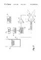
- FIG. 2is a block diagram illustrating the components of a system capable of the simultaneous use of ultrasound for imaging and therapy, in accord with the present invention
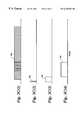
- FIGS. 3 A( 1 )- 3 D( 4 )illustrate timing and synchronization patterns that enable the simultaneous use of ultrasound for imaging and therapy
- FIG. 4illustrates yet another timing and synchronization pattern for synchronizing the HIFU and imaging scans
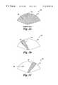
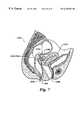
- FIG. 5Ais a schematic view of individual external imaging and therapeutic ultrasonic transducers being used for the simultaneous imaging and treatment of a tumor in a female reproductive system
- FIG. 5Billustrates an ultrasonic image that would be thus generated
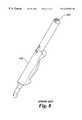
- FIG. 6is a schematic view of a vaginal probe that includes both imaging and therapeutic ultrasonic transducers being used for the simultaneous imaging and treatment of a tumor in a female reproductive system;
- FIG. 7is a schematic view of a rectal probe that includes both imaging and therapeutic ultrasonic transducers being used for the simultaneous imaging and treatment of a tumor in a female reproductive system;
- FIG. 8is a schematic view of a prior art vaginal probe that includes an imaging transducer
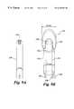
- FIG. 9Ais a schematic view of the distal end of a vaginal probe.
- FIG. 9Bis a HIFU module adapted to be used in conjunction with the vaginal probe
- FIG. 10is a schematic view of a HIFU module mounted onto the distal end of a prior art vaginal probe
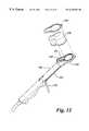
- FIG. 11is a schematic view of a combination HIFU module and prior art vaginal probe, and an ultrasonic image produced thereby in which the focal point of the HIFU module is displayed in a noise-free area of the image, in accord with the present invention
- FIG. 12is a schematic view of a second embodiment of a HIFU module combined with a prior art vaginal probe
- FIG. 13is a cross-sectional view of the second embodiment of the combination HIFU module and prior art vaginal probe, including a chamber in fluid communication with both the imaging and therapeutic transducers;
- FIG. 14is a schematic view of the second embodiment of the combination HIFU module and prior art vaginal probe, including the fluid filled chamber and the wave patterns of both the imaging and therapeutic transducers;
- FIG. 15is a schematic view of a third embodiment of a HIFU module combined with the prior art vaginal probe, including a chamber in fluid communication with only the therapeutic transducer;
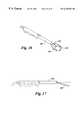
- FIG. 16is a schematic view of therapeutic and imaging transducers integrated into a vaginal probe that includes a paddle-shaped distal end;
- FIG. 17is a side elevational view of the integrated probe of FIG. 16 illustrating how a position of the paddle-shaped head can be varied around a pivot joint;
- FIG. 18is a schematic view of phased array therapeutic and imaging transducers of the integrated probe of FIG. 16, in which the imaging transducer is steerable, and the focal point of the therapeutic transducers is variable;
- FIG. 19is a schematic view of a different embodiment of a paddle-headed integrated probe similar to that of FIG. 16, in which the orientation of the imaging transducer has been shifted by 90°, thus shifting the scanning field by 90°;
- FIG. 20is a schematic view of a different embodiment of a paddle-headed integrated probe similar to that of FIG. 18, in which the therapy transducer is steerable in the same plane as the imaging transducer;
- FIG. 21is a schematic view of phased array therapeutic and imaging transducers of an integrated probe in which both the imaging transducer and the therapy transducer are steerable along both their longitudinal and latitudinal axes;
- FIG. 22is a schematic block diagram of a 3D imaging and HIFU therapy system that enables the HIFU therapy to be applied at selected treatment sites in a 3D image of a target area.
- the HIFU emitted by the therapy transducercompletely overwhelms the ultrasonic waves produced by the imaging transducer and any ultrasonic image generated is completely saturated with noise caused by the HIFU from the therapeutic transducer.
- FIG. 1Aillustrates an ultrasound image 10 in which a scanned field 12 is completely obscured by noise 14 , caused by the simultaneous operation of an imaging pulse and a HIFU wave (neither shown).
- ultrasound image 10a clinician may desire to focus the HIFU wave on a treatment site 18 .
- noise 14completely saturates scanned field 12 , it is impossible to accurately focus the HIFU wave onto treatment site 18 .
- the therapy transduceris completely de-energized, noise 14 is eliminated from the scanned field.
- the focal point of the HIFU wavewill not be seen, and thus, the HIFU wave cannot be accurately focused on treatment site 18 .
- Some prior art systemshave included a targeting icon in an ultrasound image to indicate where the known focal point of a specific HIFU transducer would be located in a scanned image. While this icon may be helpful in determining whether the HIFU was previously focused, it still did not enable a clinician to observe real-time results. Once the HIFU therapeutic transducer was energized, the scanned ultrasound image was completely saturated with noise and the clinician could not monitor the progress of the treatment without again de-energizing the HIFU therapeutic transducer.
- FIG. 1Billustrates one technique in which the amount of noise disrupting the ultrasound image is reduced.
- the HIFU wave generated by the therapeutic transducerhas been pulsed.
- This techniqueproduces an ultrasound image 20 , in which the location of noise 24 in a scanned field 22 is a function of the interference between the pulsed HIFU wave generated by the therapy transducer and the ultrasonic imaging pulses generated by the scanning transducer.
- noise 24substantially masks a treatment site 28 . This result would not occur in all cases, as to an observer noise 24 would move across scanned filed 22 as the interference between the HIFU waves and the imaging pulses varied in time.
- Pulsing of the HIFU wave alonewould thus allow the clinician to view a noise-free image of the treatment site only when noise 24 was randomly shifted to a different part of scanned field 22 , away from the treatment site.
- Such pulsing alonegenerates an image that is extremely distracting to a clinician, as noise 24 flickers across scanned field 22 , making it difficult to concentrate and difficult to consistently determine where the focal point of the HIFU wave is relative to the treatment site, in real time.
- FIG. 1Cillustrates an ultrasound image 30 in which a HIFU wave from a therapy transducer has been both pulsed and synchronized with respect to the ultrasonic imaging pulses from an imaging transducer, to ensure that noise 34 does not obscure a treatment site 38 .
- noise 34has been shifted to a location within a scanned field 32 that is spaced apart from treatment site 38 , by selectively adjusting both the pulsing and the synchronization of the HIFU wave.
- noise 34is shifted completely away from treatment site 38 , thus allowing the clinician a noise-free stable image of treatment site 38 that clearly shows the location of the focal point of the HIFU wave relative to the treatment site.
- the HIFU wavecan be focused in real time onto treatment site 38 , and a clinician can, in real time, view the therapeutic effects of the HIFU wave on treatment site 38 . It will be apparent that a clinician can de-energize the therapeutic transducer, thereby ceasing the generation of the HIFU wave, as soon as a desired therapeutic effect has been achieved at the treatment site. In this manner, undesired effects on non target tissue can be minimized.
- HIFU transducerHIFU transducer
- high intensity transducerrefers to a transducer that is capable of being energized to produce ultrasonic waves that are much more energetic than the ultrasonic pulses produced by an imaging transducer, and which can be focused or directed onto a discrete location, such as a treatment site in a target area.
- ultrasonic waves produced by such a transducerare at a high intensity in at least one embodiment of the present invention, as will be explained below.
- FIG. 2illustrates a block diagram of an embodiment of the present invention that synchronizes the image and HIFU waves required for the simultaneous imaging and therapy in real time.
- An ultrasound imaging machine 40is an ultrasound imaging system of the type that is well known to those of ordinary skill in the art and can be purchased from vendors such as ATL Inc., of Bothell, Wash.
- An imaging probe 44that is also of a type well known to those of ordinary skill in the art is connected to ultrasound imaging machine 40 via a cable 42 . Imaging probe 44 generates ultrasonic imaging pulses that propagate to the target area, are reflected from structure and tissue within the body, and are received by the imaging probe.
- the signal produced by the imaging probe in response to the reflected ultrasound wavesis communicated to the ultrasound imaging machine through cable 42 and processed to provide a visual representation of the structure and tissue that reflected the ultrasonic imaging pulses.
- An imaging beam sector 46 from imaging probe 44is identified in the Figure by dash lines.
- a therapeutic transducer 60When excited, this therapeutic transducer generates HIFU waves that are focused at a particular point of interest, i.e., a treatment site within a patient's body. In FIG. 2, the path of a HIFU beam 62 is indicated by dotted lines. HIFU beam 62 narrows to a focal point 64 .
- position of focal point 64 relative to therapeutic transducer 60is a function of the geometry of the therapeutic transducer and will normally depend upon the application.
- a therapeutic transducer that will be used to apply HIFIU therapy to the uterus of a patient from within the vaginal canalwill have a different optimum focal point than a therapeutic transducer used to apply treatment to the uterus from outside a patient's body (see FIG. 5 ).
- ultrasound imaging machine 40differs from prior art systems in several ways, including its inclusion of a synchronization output signal 48 .
- ultrasound imaging machine 40is modified to enable synchronization output signal 48 to be obtained. Because such a synchronization output signal has not been required for prior art ultrasonic imaging applications, provision of a synchronization output signal has generally not been made in prior art ultrasound imaging machines. If a prior art imaging machine that has not been modified to provide synchronization output signal 48 is used, the synchronization output signal can instead be derived from the ultrasonic imaging signal conveyed by cable 42 .
- Synchronization output signal 48is supplied to a synchronization delay circuit 50 .
- Synchronization delay circuit 50enables the user to selectively vary the initiation of each HIFU wave with respect to each sequence of ultrasonic imaging pulses that are generated to form an ultrasonic image. Referring to FIG. 1C, delay 50 enables a user to vary the position of noise 34 in scanned field 32 , so that the noise is moved away from treatment site 38 , to a different portion of scanned field 32 . The user is thus provided a noise-free image of treatment site 38 .
- a HIFU duration circuit 52is used to control the duration of the HIFU wave.
- a longer duration HIFU wavewill apply more energy to the treatment site. Generally, the more energy that is applied to a treatment site, the faster a desired therapeutic effect will be achieved.
- the duration of noise 34 as shown in ultrasound image 30will increase and can extend into the next ultrasound imaging pulse to obscure treatment site 28 , or may completely obscure ultrasound image 30 , generating a display very similar to ultrasound image 10 in FIG. 1 A.
- the userwill have to selectively adjust HIFU duration circuit 52 to obtain a noise-free image of treatment site 38 , while providing a sufficient level of energy to the treatment site to effect the desired therapeutic effect in an acceptable time.
- a HIFU excitation frequency generator 56is used to generate the desired frequency for the HIFU wave, and a power amplifier 58 is used to amplify the signal produced by the HIFU excitation frequency generator to achieve the desired energy level of the HIFU wave; power amplifier 58 is thus adjustable to obtain a desired energy level for the HIFU wave.
- a stable synchronization signal generator 66can be used to synchronize the HIFU wave to the imaging ultrasonic wave, instead of using synchronization output signal 48 from ultrasound imaging machine 40 .
- Stable synchronization signal generator 66can be used to provide a stable synchronizing pulse to initiate the HIFU wave, and the timing of this stable synchronizing pulse can be selectively varied until a noise-free image of the treatment site has been obtained.
- a drawback of using stable synchronization signal generator 66 instead of synchronization output signal 48is that any change in the timing of the ultrasound imaging pulses, such as is required to scan deeper within tissue, will require an adjustment to stable synchronization signal generator 66 that would not be required if synchronization output signal 48 were used.
- the processorwill be able to automatically find a stable synchronization signal using information from the movement of the noise.
- FIGS. 3 A( 1 )- 3 D( 4 ) and FIG. 4provide further detail for the synchronization and pulsing features of the present invention.
- FIG. 3 A( 1 )shows ultrasound imaging pulses 46 a produced by imaging machine 40 and imaging probe 44 that are used to acquire an ultrasound image of a target area (such as ultrasound image 30 of FIG. 1 C).
- a synchronization pulse 48 ais shown in FIG. 3 A( 2 ). It should be noted that synchronization pulse 48 a is illustrated as occurring before the generation of ultrasound imaging pulses 46 a ; however, the timing of synchronization pulse 48 a relative to the imaging pulses is not critical, so long as it is stable.
- Synchronization pulse 48 amerely establishes a timing reference point, from which a delay 50 a (shown in FIG. 3 A( 3 )), used for the initiation of the HIFU wave, is set such that noise from the HIFU wave in an ultrasonic image generated by imaging pulses 46 a is shifted away from the image of the treatment site.
- the delay 50 ais not fixed, and it is adjusted by the user until a noise-free image of the treatment site is obtained.
- a HIFU duration 52 adetermines the duration of the HIFU wave.
- HIFU duration 52 amay be very brief, as shown in FIG. 3 A( 4 ), or extended, as shown in FIGS. 3 B( 4 ) and 3 C( 4 ).
- An increase in the duration of the HIFU wavewill cause a greater portion of an ultrasound image to be obscured by noise, and may cause the HIFU wave to interfere with the image of the treatment site.
- delay 52 ais very short, and the resulting noisy region in the ultrasound image will be very small.
- a short duration HIFU wavemeans a correspondingly small amount of HIFU energy will be delivered to the treatment site, thus increasing the length of the treatment.
- HIFU duration 52 ato control the HIFU excitation frequency generator to variably set the duration of the HIFU wave
- the HIFU excitation frequency generatoritself could be adjusted to control the duration.
- FIGS. 3 B( 1 )- 3 C( 4 )similarly illustrate timing patterns that incorporate different settings for the delay relating to the initiation of the HIFU wave (setting 50 b in FIG. 3 B( 2 ) and setting 50 c in FIG. 3 C( 2 )) and delay relating to the duration of the HIFU wave (setting 52 b in FIG. 3 B( 3 ), and setting 52 c in FIG. 3 C( 3 )).
- FIGS. 3 D( 1 )- 3 D( 4 )illustrate a timing pattern that enables a longer duration HIFU wave (thus more energy applied to the treatment site) to be used, while still enabling a noise-free image of the treatment site to be generated.
- FIG. 3 D( 1 )- 3 D( 4 )illustrate a timing pattern that enables a longer duration HIFU wave (thus more energy applied to the treatment site) to be used, while still enabling a noise-free image of the treatment site to be generated.
- ultrasound imaging pulses 46 b and 46 cappear to be much shorter than in FIGS. 3 A( 1 ), 3 B( 1 ) and 3 C( 1 ), but actually are of the same duration, as the scales of FIGS. 3 D( 1 )- 3 D( 4 ) have been significantly increased.
- Synchronization pulse 48 a of FIG. 3 D( 2 )is obtained and used as described above.
- a delay 50 d in FIG. 3 D( 3 )is set to obtain a noise-free image of the treatment site, also as described above; however, as will be clarified below, not all of these synchronization pulses govern the image that is produced, as the delay 52 d dominates.
- FIGS. 3 A( 1 )- 3 C( 4 )is that delay 52 d has been significantly increased in FIG. 3D, such that a very long burst of HIFU energy is emitted, almost to the point of continuous emission.
- the noise-free imagingoccurs only every seventh image, during interrogation wave 46 c.
- delay 52By adjusting delay 52 , more or fewer images will be interfered with, and therefore, various duty cycle lengths for HIFU exposure can be accommodated. It should be noted as the number of images interfered with by the HIFU wave increases (here, 6 out of 7), the resulting image of the target area will arguably provide less real-time feedback.
- the actual time between visible images of the treatment sitemay be so short as to appear to occur in real time.
- the HIFU duration(such as to cause the HIFU wave to interfere with 99 out of 100 images of the treatment site)
- the advantages associated with real-time imaging of the treatment siteare diminished.
- the HIFU durationwill preferably not be set so high as to negate the benefits of real-time imaging of the treatment site and its ability to provide the clinician with immediate feedback concerning the effect of the therapy on the treatment site.
- FIG. 4illustrates another timing sequence that shows the relationships between ultrasound imaging pulses 46 d, a synchronization pulse 48 b, a delay 50 e, and a HIFU duration 52 e.
- synchronization pulse 48 boccurs during the ultrasound imaging pulses 46 d, rather than preceding the ultrasound imaging pulses, as shown in FIGS. 3A-3D.
- the position of each synchronization pulse 48 b relative to the ultrasound imaging pulsesis not critical, as delay 50 e is adjusted to shift the noise away from the image of the treatment sight.
- the duration of the HIFU wave(and thus, the energy applied to the treatment sight) is varied either by adjusting delay 52 e, as shown in FIG. 4, or by adjusting the HIFU excitation generator.
- a clinicianwill often be important for a clinician to be able to confirm that the focal point of a HIFU transducer is directed at a desired treatment site before initiating HIFU therapy. It has been determined that if the energy level of a HIFU transducer is reduced to a level less than a level that would cause any damage to tissue, the focal point of the HIFU transducer will still be evident within the target area displayed in the image developed from the reflected ultrasound signal produced and received by the ultrasound imaging transducer. The focal point will appear as a bright spot in the displayed image and will rapidly fade over time.
- a cliniciancan move the HIFU transducer as necessary to shift the focal point to a desired treatment site in the target area being imaged by the ultrasound imaging transducer and to see the focal point in the image as a bright spot that moves as the position of the HIFU transducer is changed. Only after the focal point is positioned on a desired treatment site will the clinician increase the energy of the ultrasound pulses produced by the HIFU transducer to a level sufficient to achieve the desired therapeutic effect, e.g., to a level sufficient to necrose tissue, or cause hemostasis.
- the ultrasound imaging transduceris not receiving the ultrasound signal produced by the HIFU transducer that is reflected by the tissue, but instead, is likely imaging the effect of the change in echogenicity of the tissue caused by the relatively low energy ultrasound burst produced by the HIFU transducer. This technique can be used with any of the HIFU transducers discussed below.
- a further advantage of the preceding technique for imaging the focal point of a HIFU transducercan be achieved by storing the image of each successive treatment site, which will appear as a bright area in the image produced by the ultrasound imaging transducer system.
- a storage type displayof the type readily available, can be used for this purpose.
- each previous treatment sitewill be visible in the image, it will be apparent that a desired pattern of treatment sites can readily be laid down over the tumor or other structure of interest.
- the change in echogenicity caused by a relatively high energy therapeutic HIFU wavewill be brighter and persist longer in the display, enabling the clinician to easily distinguish between a current prospective focus point for the next treatment site (produced using the low energy pulse) and the previous treatment sites to which the HIFU therapy has already been administered.
- FIG. 22a block diagram is illustrated for a system 200 that enables imaging of a target area in 3D and storing of the locations of treatment sites to which the HIFU therapy has been administered in the 3D image as a HIFU therapy session proceeds.
- the systemincludes a 3D image data processor and display 202 , an image acquisition section 204 , a magnetic field sensor 206 , a magnetic field generator 208 , and 6D electronic processing circuitry 210 .
- the latter three componentsare employed to track the imaging target area and the HIFU focal point as they are redirected in the 3D space and are part of a six-dimensional (6D) measurement system (i.e., three spatial coordinates for the 3D orthogonal axes and three angles of rotation around these three orthogonal axes).
- 6D measurement systemis commercially available from Ascension Technology, Burlington, Vt. This 6D measurement system uses 6D electronic processing circuitry 210 and magnetic field generator 206 to produce time sequential orthogonally oriented magnetic fields covering a general area as indicated in the Figure by the dash line that encompasses the region of magnetic field.
- Magnetic field sensor 206is mounted on a combined imaging and HIFU therapy probe 212 in a fixed manner relative to imaging and HIFU transducers 214 .
- the magnetic field sensordetects the magnetic field strength in 3D sequentially produced by the magnetic field generator.
- the 6D electronic processing circuitryuses the information from the magnetic field sensor and the known magnetic fields that were produced to compute the 3D position and the three angular orientations around the three orthogonal axes of the magnetic field sensor (and thus, of the combined imaging and HIFU therapy probe) with respect to the magnetic field generator, yielding the 6D information.
- the 6D informationis supplied to 3D image data processor and display 202 at a rate sufficient to enable movement of the magnetic field sensor to be tracked in the displayed 3D image of the target area.
- the position of the target area and the HIFU transducer focal pointcan be related to a 3D spatial point, so long as magnetic field sensor 206 is within the range of the magnetic field produced by magnetic field generator 208 .
- 3D image data processor and display 202also receive ultrasound image information from an ultrasound imaging machine 216 through image acquisition section 204 . It uses this information to develop and display 3D information.
- Ultrasound imaging machine 216provide the synchronization signal to a HIFU control and electrical energy generating system 218 , as discussed above. The remaining component in FIG.
- a physiological information acquisition section 220which enables synchronization of the imaging and HIFU therapy with physiological activity, such as respiration or cardiac activity (provided by an electrocardiogram system—not shown).
- physiological activitysuch as respiration or cardiac activity (provided by an electrocardiogram system—not shown).
- use of the physiological informationavoids problems associated with movement of the patient's body due to physiological activity.
- 3D imaging and HIFU therapycan be control so that they are implemented only at the end of expiration in the breathing cycle, since motion of the patient is more repeatable then than at mid inspiration.
- a physiological sensorsuch as a respiration detector (not shown), which is well known in the art, can provide the information for this section of the system.
- each of the treatment sites where HIFU therapy has been administered during a therapy sessiona clinician will be able to determine where each successive treatment site should be targeted to achieve a desired pattern of HIFU therapy within a tumor or other region of interest.
- the bright spot in the 3D image showing the location of each previous treatment sitegreatly facilitates this targeting process and enables a desired pattern of HIFU therapy to be achieved with considerable accuracy.
- the HIFU therapycan be employed to create a plurality of lesions on blood vessels supplying blood to a tumor, cutting off the supply of nutrients and oxygen to the tumor provided by the blood supply.
- the results of this techniqueare similar to those arising from the procedure referred to as “embolization,” in which a clot-inducing material is introduced into the vessel using a small catheter, but the HIFU therapy can achieve the same result non-invasively and can treat vessels that are too small or otherwise not accessible to render treatment through a catheter.
- Embolizationis a relatively new technique and has been used to treat a variety of conditions, including uterine fibroids. While the ability to store the location of previous treatment sites so that they are shown on a displayed 3D image of the target area is not essential to this use of HIFU therapy, it will be evident that the display of the treatment sites used to create lesions in the vessels will facilitate this procedure. Further, it should be noted that using HIFU to occlude (or induce hemostasis in) the blood vessels that supply oxygen and nourishment to a structure within the body can be used in conjunction with other imaging techniques, such as CT, MRI, or angiography.
- Doppler Flow imagingAnother imaging technique that is likely to be useful to a clinician when using HIFU to create lesions in blood vessels is Doppler Flow imaging, which can be used to represent blood vessels supplying a structure with blood in one color, and blood vessels that remove blood from a structure in a second color.
- Doppler Flow imagingcan be used to represent blood vessels supplying a structure with blood in one color, and blood vessels that remove blood from a structure in a second color.
- the circulation of blood within these blood vesselseither adds to or subtracts from the imaging wave, enabling blood vessels having blood flowing in opposite directions to be differentiated from one another.
- a final imaging technique that will likely be beneficially employed by a clinician using HIFU to create a lesion in a blood vesselis the use of conventional ultrasound imaging contrast agents. Not only will the use of such agents provide the clinician with a more useful ultrasonic image, but it is anticipated that such agents may actually increase the effectiveness of the HIFU in producing the desired lesion.
- a substantially brighter spot showing where the HIFU wave (at low power) was focusedcan be achieved if an anesthetic agent other blood soluble agent having a relatively high vapor pressure has previously been administered to the patient.
- the HIFU treatmentcan be stopped when a therapeutic produced lesion has grown to the point at which it begins to extend beyond the desired treatment site, and the HIFU focal point can then be repositioned to another treatment site and reactivated; (2) the focal point of the HIFU wave can be observed in the image due to changes in the echogenicity of the tissue at the focal point, which are apparent in the images of the target area, providing an instant feedback that can enable a clinician to adjust the focal point onto a desired treatment site; (3) the HIFU focal point can be adjusted during the administration of the HIFU therapy to compensate for tissue movement within the patient's body due to breathing or for other reasons; (4) real-time visualization of a treatment site is very reassuring to the medical therapist, in confirming that the HIFU energy is being applied to the correct position (and that healthy tissue is not being damaged); (5) the combined imaging and therapeutic treatment can be accomplished much faster than in the past, when it was necessary to render treatment, stop
- FIGS. 5-7illustrate how a variety of different configurations of HIFU transducers and imaging transducers can be used to simultaneously provide real-time imaging and therapy to an internal treatment site within a patient's body. It is expected that HIFU therapy with real-time imaging can be beneficially employed to treat a variety of disease conditions. In particular, it is envisioned that conditions relating to the reproductive system of a mammalian female will particularly benefit from HIFU therapy with real-time imaging, as ultrasonic imaging itself is widely used in association with the female reproductive system.
- HIFU therapycan be applied to a uterine fibroid, an endometrial polyp, a follicular cyst, a polycystic ovary, a dermoid cyst, a corpus luteum cyst, an ectopic pregnancy, a cornual pregnancy, a multifetal pregnancy, a uterine malformation, an endometrial hyperplasia, an adenomyosis condition, and endometriosis condition, or an excessive bleeding condition.
- the treatment of disorders of the female reproductive system using the method of the present inventioncan be achieved by positioning the scanning ultrasonic transducer system and the therapeutic ultrasonic transducer system adjacent to the female reproductive system. Vaginal, rectal, abdominal and laparoscopic approaches are contemplated.
- both a HIFU transducer 102 and an imaging transducer 104are disposed external to the patient's body.
- the reflected ultrasound waves received by imaging transducer 104are used to generate an ultrasound image 100 .
- the HIFUis being used to treat a tumor 110 on a uterus 111 of the patient.
- Imaging transducer 104is positioned so that tumor 110 is clearly displayed in ultrasound image 100 .
- Also visible in ultrasound image 100is a cross section of a rectum 106 .
- HIFU transducer 102is being used to destroy tissue in tumor 110 .
- the necrotic tissueis clearly visible as a lesion 112 in both the cross section of the body and in ultrasound image 100 .
- FIG. 6illustrates an embodiment in which a HIFU transducer 102 a and an imaging transducer 104 a have been combined on a vaginal probe 109 .
- Vaginal probe 109has been inserted into a vaginal canal 108 a and positioned to enable imaging transducer 104 a to be used in generating an ultrasonic image of a tumor 110 a.
- HIFU transducer 102 ais focused on a selected portion of tumor 110 a to which the clinician desires to administer the HIFU therapy to generate a lesion 112 a.
- the HIFU therapyis used to destroy the tumor by causing lesions of the blood vessels supplying oxygen and nutrients to the tumor, thereby generating a plurality of lesions similar to lesion 112 a, so that the tumor withers away, or by destroying spaced-apart portions of the tumor.
- the HIFUFU therapywill likely be repeated at intervals of several weeks. The time between successive therapy sessions enables macrophages in the patient's body to clear away or debride the necrotic tissue from the tumor so that it is reduced in size with each therapy session and eventually destroyed.
- FIG. 7illustrates a rectal probe 109 a, which incorporates a combination of a therapy transducer 102 b and an imaging transducer 104 b.
- Rectal probe 109 ahas been inserted into a rectum 106 b of the patient, and the imaging transducer is being used to locate a tumor 110 b.
- therapy transducer 102 bis focused on the desired portion of the tumor, and HIFU therapy is administered to a treatment site 112 b, until the desired therapeutic effect is achieved.
- FIGS. 9-19provide details of preferred embodiments of probes that can be used to simultaneously provide imaging and therapy to a treatment site. It should be noted that the method of simultaneously imaging and providing treatment using ultrasound can be applied to different locations in or on a patient's body to treat a variety of medical conditions.
- One application of a preferred embodiment of the present inventionis employed, by way of example, to provided simultaneous ultrasonic imaging and HIFU therapy of a uterine fibroid.
- the simultaneous imaging and therapy using the described methodis not at all limited to the treatment of uterine fibroids, or even the treatment of gynecological or obstetrical disorders.
- FIG. 8illustrates one such prior art ultrasonic vaginal imaging probe, which is a Model C9-5 transvaginal probe available from ATL Inc. of Bothell, Wash. It is envisioned that several different types of HIFU transducers can be mounted onto the standard transvaginal imaging transducer probe to allow the simultaneous imaging and therapy of treatment sites related to the female reproductive system, in accord with the present invention.
- an exemplary application of the present inventionis a vaginal probe incorporating both imaging and therapeutic transducers.
- HIFU transducer designis a function of the location of the desired treatment site, the following discussion is useful in determining preferred design parameters for a vaginal probe combining imaging and therapeutic transducers optimized for applying HIFU therapy to uterine fibroids.
- the ⁇ #(or relative aperture) is defined as being equal to the “focal length divided by the diameter of the aperture”.
- a low numberis preferred, as this insures a highly focused beam, thereby minimizing any undesired effects of the HIFU beam on adjacent tissue.
- a preferred numberis 1; but a number as high as 1.75 can be used, therefore a practical range is from 1-1.75. The lower the number, the tighter the focus, and the greater the intensity of the HIFU beam will be at the focus, with respect to the HIFU beam intensity at the surface of the transducer.
- HIFUcan also have a mechanical effect on tissue via cavitation. Therefore a low intensity at the surface of the transducer is desired to minimize the heating of the intermediate tissue, while for successful treatment, the tissue at the focus must be heated much more rapidly in order to achieve therapeutic temperatures at the focus without undue heat build up in the intermediate tissue. Therefore, it is very important to know the maximum distance from the transducer that one desires to treat, to determine the maximum focal length required. At the same time, one must have an understanding of the largest size aperture that one can physically introduce into the vaginal tract.
- the external opening of the vaginal canalgenerally has a circumferential dimension which ranges from of 10-12 cm. Since the vaginal entrance is longitudinal in nature, a probe having a distal end that is “paddle” or “spoon” shaped would be most beneficially employed. Practical transducer considerations require that the distal end of the probe (the logical point for mounting a therapeutic transducer) would be at least 0.5 cm thick, and that the widest portion of this “paddle” design would range from 4.5-5.5 cm. Since the vagina is a muscular structure that is quite strong, it can be stretched somewhat further, but this is uncomfortable for the patient. Therefore, it is preferred that the widest portion of the paddle range from 4.5-6 cm.
- the design of a preferred focal lengthnecessitated an analysis of where within the vaginal canal the probe will be positioned, and where uterine fibroids are likely to occur.
- the probewill be positionable in the vagina at the cervix.
- the vaginal fornicesare potential spaces formed by folds within the vaginal canal around the cervix, extending 5-6 cm along the uterus. Therefore, near the cervix, the vaginal cavity has a larger space, allowing either a larger probe, or the manipulation of a smaller probe, to achieve imaging and therapy of a desired site in the uterus.
- the size range of uterine fibroidsis from 1-15 cm, and the length of the uterus is approximately 5-6 cm. Therefore, a maximum focal length of about 20 cm would reach an entire fibroid located at the distal end of the uterus. Based on applicants' practical experience, it is anticipated that 20%, 35%, 50%, 75%, and 80% of all uterine fibroids will be within the respective distances of 4 cm, 6 cm, 8 cm, 10 cm and 12 cm respectively. From a cost/benefit point of view, a combination imaging and therapeutic vaginal probe needs to be able to treat at least 35% of uterine fibroids, and more preferably 50%. Therefore a preferred therapeutic transducer must be able to treat to a depth of at least 6 cm, and more preferably 8 cm.
- the design of different embodiments of a vaginal probe including both imaging and therapeutic transducersis preferably limited to designs in which the largest circumferential dimension of the combination probe (measured generally transverse to a longitudinal axis of the probe) is about 10.6 cm, which is a nominal limit in size to enable the combination imaging and therapy transducer to be readily inserted through the vaginal opening and into the vaginal canal.
- Table 1Based on the calculations for a circular aperture and practical relative apertures, Table 1 provides information of the relative percentages of uterine fibroids that can be treated with a specific circular aperture. For a circular aperture of 4.5 cm, just less than 35% of all uterine fibroids can be treated with a relative aperture of 1.25. This then defines the minimum diameter of circular applicator as 4.5 cm, based on the design parameter of being able to treat 35% of uterine fibroids encountered. An elliptic aperture may also be used, in which case the minor axis represents the transverse direction, and thus the limiting dimension of a probe that can readily be inserted into the vagina.
- An elliptical surface areais larger than a circular surface if the minor axis length equals the diameter of the circular surface, and the major axis is greater than the minor axis.
- the focal lengthis greater than for a circular surface for a given relative aperture number. Therefore, with a two to one ratio of major to minor axis for example, a 9/4.5 ratio would exist for the minor axis chosen. Referring to Table 2, this is equivalent to a 6.5 cm diameter circular aperture. Looking at Table 1, for the equivalent of a 6.5 cm diameter circular aperture, the percentage of fibroids that can be treated are 50%, ⁇ 75% , and 75%, respectively, for relative apertures of 1.25, 1.5, and 1.75.
- vaginal probe with a HIFU transducerthat has a distal end that is paddle or spoon shaped, with a minimum transverse dimension of 4.5 cm, and a maximum circumferential dimension (measured generally transverse to a longitudinal axis of the probe) of about 10.6 cm.
- No other devices cited in the prior art for use in other body orificesare suitable for a vaginal application of HIFU for the treatment of uterine fibroids.
- the size, shape, and configuration of such a combination imaging and therapy vaginal probeare believed to be specific to this particular access path through the vaginal canal, for rendering the HIFU therapy in treating obstetrical and gynecological medical conditions.
- FIGS. 9illustrates a first embodiment for mounting a HIFU module 123 onto a distal end of a prior art transvaginal imaging probe 120 .
- Transvaginal imaging probe 120includes an imaging transducer array 122 .
- HIFU module 123is sized and shaped to fit over the distal end of transvaginal imaging probe 120 and incorporates a cylindrical shaft 124 that has a cylindrical bore 126 .
- Cylindrical bore 126is sized to easily slide over the distal end of transvaginal imaging probe 120 and in its wall is disposed a plurality of fluid passages 130 .
- Fluid passages 130are used to circulate degassed water through a balloon 140 (FIG. 10) that surrounds HIFU module 123 . It is important that the water be degassed, as bubbles within the water increase cavitational effects, which will attenuate the ultrasonic waves.
- the water circulating through the balloonprovides cooling to the transducer elements to avoid an unwanted buildup of heat. It is currently common practice to use a condom for a balloon, although other inert and flexible elastomeric materials could be used.
- vaginal probe 120has been inserted into cylindrical bore 126 of cylinder 124 , and the imaging transducer array 122 is disposed within a void 138 .
- Shaft 124also includes electrical leads 128 , which connect the HIFU transducer to the power amplifier that drives the HIFU transducer.
- HIFU transducer mounting base 136Located atop cylinder 124 is a HIFU transducer mounting base 136 .
- HIFU transducer mounting base 136is also hollow, such that the transvaginal imaging probe 130 may pass completely through the center of HIFU transducer mounting base 136 so that imaging transducer array 122 is disposed within void 138 (see FIG. 9 B). This configuration enables imaging transducer array 122 to transmit an ultrasound imaging pulse to the target of interest.
- HIFU therapy transducer 132is pivotally mounted to HIFU transducer mounting base 136 at a pivot joint 134 .
- This pivotal mountingenables a clinician to selectively target a wider variety of treatment areas within the female reproductive system by rotating the HIFU transducer about pivot joint 134 . It is envisioned that the disposition of the HIFU transducer on pivotal mounting 134 will be adjusted prior to inserting the combination transvaginal probe and HIFU module into the vaginal canal. The angle of the HIFU transducer should be adjusted based on the relative position of the target area and treatment site.
- the combination transvaginal imaging probe and HIFU modulecan be moved to a position that enables an ultrasonic image of the target area to be observed on a display and then adjusted to focus the HIFU wave onto a desired treatment site within the target area.
- a mechanical linkage(not shown) connected to HIFU transducer 132 can be added to enable the HIFU transducer to be selectively rotated about pivot joint 134 while the combination transvaginal probe and HIFU module is in the vaginal canal. This capability would provide a clinician greater flexibility in focusing the HIFU transducer on a particular treatment site.
- a skilled cliniciancan initially select an angle for the HIFU transducer relative to the longitudinal axis of the transvaginal imaging probe, insert the combination vaginal probe and HIFU module into the vaginal canal, and then manipulate the combination imaging probe and HIFU module while in the vaginal canal to acquire the image of the target area and focus the HIFU beam on the desired treatment site.
- FIG. 11illustrates an ultrasonic image 142 superimposed on the combination transvaginal probe and HIFU transducer illustrated in FIG. 10 (but without balloon 140 ).
- the HIFU transducer 132has been focused on a treatment site 144 .
- the synchronization and pulsating elements described earlierhave been used to shift a noise section 146 a way from treatment site 144 , such that the clinician is provided with a noise-free real-time image of treatment site 144 .
- FIG. 12illustrates another embodiment of a combination imaging and therapy transducer based on a prior art transvaginal imaging probe.
- a therapy transducer module 150has been mounted onto transvaginal imaging probe 120 .
- the design of the moduleis constrained by the anatomy involved in the transvaginal application of HIFU to uterine fibroids, as discussed above.
- Module 150is mounted at the tip of the vaginal probe, and contains an opening through which the scan head (imaging transducer) transmits an ultrasound wave to obtain an image of the uterus, the fibroid, and any other structure of interest.
- the openingallows about one half of the scan head to transmit the imaging ultrasound wave.
- the other halfis covered by the module assembly, and does not have a window for imaging, and therefore does not contribute to the image.
- the main housing of the moduleis made of biocompatible, medical grade plastic.
- a chamber 158can be filled with a fluid, such as degassed water, for the purpose of coupling the HIFU to adjacent tissue.
- a water circulation systemis used to circulate degassed cold water through chamber 158 for both cooling of the HIFU transducer and carrying away any cavitation bubbles that may be generated during the HIFU excitation.
- the water conduitsextend through the plastic housing and two holes disposed on opposite sides of a central passage (not shown).
- Stainless steel needle stockcan be employed for tubes 155 carrying the water in and out of the chamber. Tubes 155 run along the shaft of the vaginal probe adjacent to a coaxial cable 153 employed for energizing the HIFU transducer.
- a cover 159attaches the membrane which forms chamber 158 to module 150 .
- a HIFU transducer 154is disposed on a rim cut inside a brass bowl 152 that is affixed with an appropriate adhesive to module 150 .
- An electrical connection to the HIFU transduceris made through coaxial cable 153 that runs along the shaft of the probe.
- the HIFU transduceris a concave, fixed focused transducer, operating at a center frequency of about 2.0 MHz.
- the radius of curvature of this embodiment of the HIFU transduceris about 55 mm, and its aperture diameter is about 35 mm.
- the focus of the HIFU transduceris within the imaging plane of the imaging probe (preferably an ATL, Inc., Model C9-5 transvaginal probe). In fact, the imaging plane intersects the HIFU beam envelope (cone shaped) through its center, placing both the focus and the HIFU beam longitudinal axis in the imaging plane, as can be seen in FIG. 14 .
- the HIFU transducer frequencywas selected based on several requirements, including: (1) the ability to administer HIFU therapy to uterine fibroids up to a maximum distance of about 6 cm from the cervix; and, (2) an intensity gain of about 20 dB from the transducer surface to the focal spot, providing about 1,000 W/cm 2 at the focus, and about 50 W/cm 2 at the transducer surface. These are all reasonable values for both treatment and transducer operation.
- Two different embodiments of chambers containing degassed waterare contemplated for the purpose of coupling the HIFU to adjacent tissue. These embodiments include a chamber containing just the HIFU transducer, and a chamber containing both the HIFU transducer and the imaging scan head.
- FIG. 14illustrates a combination transvaginal probe and HIFU transducer with both the imaging and HIFU transducer energized and chamber 158 filled with fluid.
- the HIFU transducerproduces a cone-shaped HIFU wave 162 that is focused at a focal point 166 .
- the imaging transducergenerates a scanning ultrasound wave 160 . It should be noted that HIFU wave 162 is within scanning wave 160 . Thus, focal point 166 can be readily seen in the image provided by scanning ultrasound wave 160 .
- FIG. 15illustrates an alternative design of a therapy transducer module 170 mounted on transvaginal imaging probe 120 .
- imaging transducer array 122is partially occluded by module 170 , and thus only a partial ultrasound image is generated.
- a HIFU therapy transducer 174is mounted on a brass ring 178 , in a manner similar to the configuration in the previous embodiment.
- a fluid filled chamber 158 aencloses HIFU therapy transducer 174 .
- fluid filled chamber 158 a of FIG. 15only encloses HIFU therapy transducer 174 , and not imaging transducer array 122 as well.
- FIGS. 16 and 17illustrate an embodiment of probe usable in the present invention in which the HIFU therapy transducer and imaging transducer have been integrated into a single device. It is anticipated that as the combination of real-time imaging and HIFU therapy gains acceptance, clinicians will desire an integrated device rather than a HIFU transducer and a imaging transducer configured as two separate components, mounted together on a single probe. An integrated imaging transducer and a therapy transducer are formed as a combination transvaginal probe 180 as shown in these Figures. FIG. 17 illustrates that the angle of a paddle head 186 containing both the therapy and imaging transducers is movable relative to the body of combination transvaginal probe 180 .
- paddle head 186will be placed in a desired position prior to placing combination transvaginal probe 180 into a patient's vaginal canal.
- a linkage mechanismcan be incorporated that will enable paddle head 186 to be moved relative to the handle portion of combination transvaginal probe 180 , while the paddle head is disposed in the vaginal canal.
- the paddle head configurationis particularly well suited for use in the vaginal canal.
- FIGS. 18 and 19illustrates the paddle head probe with imaging transducers 182 and 182 a positioned at two different angles relative to the longitudinal axis of the paddle head.
- imaging transducer 182is generally parallel to the axis
- an imaging transducer 182 aforms an angle of 90° relative to the axis.
- imaging transducers 182 a nd 182 acomprise a one-dimensional (1D) phased array of transducer elements, and a linear array concentric ring HIFIU transducer 184 is provided.
- the 1D phased array imaging transducersallow the ultrasound waves that are generated thereby to be steered and focused through a substantial range in a plane, while the concentric phased array of the HIFU transducer can only be electronically focused at different points along its central axis.
- the probecan thus remain stationary while the scanning ultrasonic wave are steered and/or focused at a target area and the HIFU waves are focused on a treatment site within the target area.
- vibrating element 190When activated, vibrating element 190 causes the focal point of the therapeutic ultrasonic wave to be varied, thus enlarging the size of the treated area within a limited portion of the target area without requiring the clinician to move the probe.
- the cliniciancan select from two vibrational patterns; a first pattern that involves a vibrational frequency in the range of 1 to 5 Hz (thereby avoiding the heating of tissue not associated with the treatment site), and a second pattern that involves a vibrational frequency in the range of 10 to 50 Hz (thereby increasing an amount of energy applied to the treatment site, while avoiding undesired cavitational effects).
- vibrating element 190could be beneficially incorporated into other probe embodiments as well, and is not limited to only the embodiment illustrated in FIG. 19 .
- FIG. 20illustrates a paddle head 186 ′ of a combination transvaginal probe 180 ′ that includes imaging transducer 182 a nd a HIFU transducer 184 a.
- Both imaging transducer 182 a nd HIFU transducer 184 acomprise 1D phased array elements oriented so that the respective imaging and therapeutic ultrasound signals can be steered and focused in substantially the same plane, i.e., in a plane aligned with a longitudinal axis of the combination transvaginal probe.
- the 1D phased array elements of HIFU transducer 184 aare generally semi-circular is shape, due to its slightly concave configuration.
- Paddle head 192 of a combination transvaginal probe 194is illustrated.
- Paddle head 192includes a two-dimensional (2D) phased array imaging transducer 196 and a 2D phased array HIFU transducer 198 .
- the 2D phased array elements of both the imaging transducer and HIFU transducercan thus be electronically steered and focused within a substantial conical volume, providing the clinician the capability to electronically control the location of the target area that is imaged and the location of the treatment site to which the HIFU waves are administered.
- the objective of the studywas to investigate the potential efficacy of high intensity focused ultrasound (HIFU) for the treatment of uterine fibroid tumors.
- HIFUhigh intensity focused ultrasound
- a total of 60 female athymic nude micewere inoculated subcutaneously with 3-5 ⁇ 10 6 ELT-3 cells, a uterine fibroid tumor cell line. Tumor development was monitored using subcutaneous caliper measurements of the tumors.
- the HIFU probewas a concave, single-element high-power transducer, operating at a frequency of 3.5 MHz, and an intensity of 2000 WM/cm 2 .
- the HIFU treatmentconsisted of scanning the tumors for 30-60 seconds, based on the tumor size.
- HIFU-treatmentresulted in a tumor reduction of 91% within one month of the treatment. Histological analysis of HIFU-treated tumors showed coagulation necrosis, and nuclear fragmentation of tumor cells. It is thus concluded that the HIFU effectively reduced uterine fibroid tumor size in a nude mouse model. Further studies are needed to assess the in-situ response of uterine fibroids to HIFU treatment.
- cytokinesnon-antibody proteins, released by macrophages (actively phagocytic cells), on contact with a specific antigen and which act as intercellular mediators, as in the generation of immune response
- therapeutic ultrasoundhas a stimulatory effect on the production of cytokines.
- the cytokine productionis a signaling mechanism that results in attraction of more macrophages, and an enhanced immune response.
- the inflammatory response of the body to HIFU treatmentmay provide an enhanced rate of tumor shrinkage due to enhanced cytokine production. This mechanism may provide grounds for a treatment procedure that involves the production of scattered HIFU lesions in the uterine fibroid tumor, and obtaining an optimal tumor shrinkage due to enhanced immune response.
- Synergistic effects of therapeutic ultrasound and anti-tumor compoundsmay provide a mechanism for treatment for uterine fibroids, involving a combination therapy using both anti-tumor drugs and HIFU.
- This managementhas two possibilities, including the use HIFU with the drugs currently available for uterine fibroids (GnRH agonists). These drugs can shrink the fibroids temporarily. As soon they are discontinued, the fibroids grow back to their original size. Since these drugs have side effects, they are primarily used for shrinking the fibroids before a myomectomy (surgical removal of the fibroids).
- HIFU treatmentmay be performed at the time when maximal shrinkage of the tumors due to drugs has occurred, thereby fixing the tumor size and preventing tumor re-growth.
- the second possibilityis to use HIFU with the new anti-tumor compounds which may offer further advantages.
- HIFU therapy of uterine fibroidsinclude the treatment of the entire tumor in a single procedure. It has been observed that a complete tumor treatment (100% shrinkage) may be obtained using this method. Alternatively, the tumor may be treated in several sessions, with sufficient time between each session for the macrophages in the patient's body to clear away the necrotic tissue resulting from the previous treatment session, effectively debriding the treatment side and exposing remaining tumor tissue for the next HIFU therapy session. In each session, a remaining part of the tumor is treated. A complete tumor treatment (100% shrinkage) in all of the tumors treated using this method has been observed. Scattered HIFU lesions in a tumor may also provide an optimal shrinkage, with minimal fibrosis.
- HIFU therapy sessionsmay be needed to completely eradicate the tumor. Further, as noted above, the treatment of a tumor using a combination of HIFU and drugs may yield synergistic results, particularly by beginning the HIFU therapy when the maximum benefit of the drug therapy on the tumor has been realized.
Landscapes
- Health & Medical Sciences (AREA)
- Life Sciences & Earth Sciences (AREA)
- Animal Behavior & Ethology (AREA)
- Veterinary Medicine (AREA)
- Nuclear Medicine, Radiotherapy & Molecular Imaging (AREA)
- Public Health (AREA)
- Radiology & Medical Imaging (AREA)
- Engineering & Computer Science (AREA)
- Biomedical Technology (AREA)
- General Health & Medical Sciences (AREA)
- Pathology (AREA)
- Molecular Biology (AREA)
- Surgery (AREA)
- Medical Informatics (AREA)
- Heart & Thoracic Surgery (AREA)
- Physics & Mathematics (AREA)
- Biophysics (AREA)
- Gynecology & Obstetrics (AREA)
- Surgical Instruments (AREA)
- Ultra Sonic Daignosis Equipment (AREA)
Abstract
Description
| TABLE 1 |
| Percentage of Fibroids Treatable for Relative Apertures |
| with an area equivalent to a Circular Aperture |
| Circular Aperture | Relative Apertures (ƒ#) |
| Diameter (cm) | 1.25 | 1.5 | 1.75 | ||
| 4.5 | <35% | >35% | 50% | ||
| 5 | 35% | <50% | <75% | ||
| 5.5 | <50% | 50% | <75% | ||
| 6 | <50% | <75% | 75% | ||
| 6.5 | 50% | <75% | <80% | ||
| 7 | <75% | 75% | 80% | ||
| 7.5 | <75% | <80% | |||
| 8 | <75% | 80% | |||
| 8.5 | 75% | ||||
| 9 | <80% | ||||
| 9.5 | 80% | ||||
| TABLE 2 |
| Major axis length (cm) for a given a minor axis length that gives an |
| ellipsoidal area equal to the area of a circular aperture of a specific |
| diameter. |
| Diameter | ||
| Circular | Ellipsoid Minor Axis Length (cm) |
| Aperture | 4.5 | 5 | 5.5 | 6 |
| 4.5 | 4.5 | 4.1 | 3.7 | 3.4 |
| 5 | 5.6 | 5.0 | 4.5 | 4.2 |
| 5.5 | 6.7 | 6.1 | 5.5 | 5.0 |
| 6 | 8.0 | 7.2 | 6.5 | 6.0 |
| 6.5 | 9.4 | 8.5 | 7.7 | 7.0 |
| 7 | 10.9 | 9.8 | 8.9 | 8.2 |
| 7.5 | 12.5 | 11.3 | 10.2 | 9.4 |
| 8 | 14.2 | 12.8 | 11.6 | 10.7 |
| 8.5 | 16.1 | 14.5 | 13.1 | 12.0 |
| 9 | 18.0 | 16.2 | 14.7 | 13.5 |
| 9.5 | 20.1 | 18.1 | 16.4 | 15.0 |
Claims (75)
Priority Applications (10)
| Application Number | Priority Date | Filing Date | Title |
|---|---|---|---|
| US09/397,471US6425867B1 (en) | 1998-09-18 | 1999-09-17 | Noise-free real time ultrasonic imaging of a treatment site undergoing high intensity focused ultrasound therapy |
| US10/166,795US6716184B2 (en) | 1998-09-18 | 2002-06-07 | Ultrasound therapy head configured to couple to an ultrasound imaging probe to facilitate contemporaneous imaging using low intensity ultrasound and treatment using high intensity focused ultrasound |
| US10/770,350US7686763B2 (en) | 1998-09-18 | 2004-02-02 | Use of contrast agents to increase the effectiveness of high intensity focused ultrasound therapy |
| US10/977,339US7520856B2 (en) | 1999-09-17 | 2004-10-29 | Image guided high intensity focused ultrasound device for therapy in obstetrics and gynecology |
| US11/016,701US7510536B2 (en) | 1999-09-17 | 2004-12-16 | Ultrasound guided high intensity focused ultrasound treatment of nerves |
| US11/207,554US7722539B2 (en) | 1998-09-18 | 2005-08-18 | Treatment of unwanted tissue by the selective destruction of vasculature providing nutrients to the tissue |
| US11/928,667US7850626B2 (en) | 1999-09-17 | 2007-10-30 | Method and probe for using high intensity focused ultrasound |
| US12/390,975US8197409B2 (en) | 1999-09-17 | 2009-02-23 | Ultrasound guided high intensity focused ultrasound treatment of nerves |
| US12/946,727US8337434B2 (en) | 1999-09-17 | 2010-11-15 | Methods for using high intensity focused ultrasound and associated systems and devices |
| US13/681,379US20130096431A1 (en) | 1999-09-17 | 2012-11-19 | Method for using high intensity focused ultrasound and associated systems and devices |
Applications Claiming Priority (2)
| Application Number | Priority Date | Filing Date | Title |
|---|---|---|---|
| US10081298P | 1998-09-18 | 1998-09-18 | |
| US09/397,471US6425867B1 (en) | 1998-09-18 | 1999-09-17 | Noise-free real time ultrasonic imaging of a treatment site undergoing high intensity focused ultrasound therapy |
Related Child Applications (1)
| Application Number | Title | Priority Date | Filing Date |
|---|---|---|---|
| US10/166,795DivisionUS6716184B2 (en) | 1998-09-18 | 2002-06-07 | Ultrasound therapy head configured to couple to an ultrasound imaging probe to facilitate contemporaneous imaging using low intensity ultrasound and treatment using high intensity focused ultrasound |
Publications (1)
| Publication Number | Publication Date |
|---|---|
| US6425867B1true US6425867B1 (en) | 2002-07-30 |
Family
ID=26797577
Family Applications (2)
| Application Number | Title | Priority Date | Filing Date |
|---|---|---|---|
| US09/397,471Expired - LifetimeUS6425867B1 (en) | 1998-09-18 | 1999-09-17 | Noise-free real time ultrasonic imaging of a treatment site undergoing high intensity focused ultrasound therapy |
| US10/166,795Expired - LifetimeUS6716184B2 (en) | 1998-09-18 | 2002-06-07 | Ultrasound therapy head configured to couple to an ultrasound imaging probe to facilitate contemporaneous imaging using low intensity ultrasound and treatment using high intensity focused ultrasound |
Family Applications After (1)
| Application Number | Title | Priority Date | Filing Date |
|---|---|---|---|
| US10/166,795Expired - LifetimeUS6716184B2 (en) | 1998-09-18 | 2002-06-07 | Ultrasound therapy head configured to couple to an ultrasound imaging probe to facilitate contemporaneous imaging using low intensity ultrasound and treatment using high intensity focused ultrasound |
Country Status (1)
| Country | Link |
|---|---|
| US (2) | US6425867B1 (en) |
Cited By (458)
| Publication number | Priority date | Publication date | Assignee | Title |
|---|---|---|---|---|
| US20020193682A1 (en)* | 2000-06-15 | 2002-12-19 | Torchia Mark G. | Hyperthermia treatment and probe therefor |
| US20030079753A1 (en)* | 1996-10-22 | 2003-05-01 | Epicor, Inc. | Apparatus and method for diagnosis and therapy of electrophysiological disease |
| US20030120286A1 (en)* | 2001-03-28 | 2003-06-26 | Vascular Control System | Luminal clip applicator with sensor |
| US20030120306A1 (en)* | 2000-04-21 | 2003-06-26 | Vascular Control System | Method and apparatus for the detection and occlusion of blood vessels |
| US6602251B2 (en)* | 1998-12-08 | 2003-08-05 | Vascular Control Systems, Inc. | Device and methods for occlusion of the uterine artieries |
| US20030149380A1 (en)* | 1998-10-26 | 2003-08-07 | Katsuhiko Fujimoto | Ultrasound treatment apparatus |
| US6612988B2 (en)* | 2000-08-29 | 2003-09-02 | Brigham And Women's Hospital, Inc. | Ultrasound therapy |
| US6638286B1 (en) | 2000-11-16 | 2003-10-28 | Vascular Control Systems, Inc. | Doppler directed suture ligation device and method |
| US6645202B1 (en) | 1996-10-22 | 2003-11-11 | Epicor Medical, Inc. | Apparatus and method for ablating tissue |
| US6679855B2 (en)* | 2000-11-07 | 2004-01-20 | Gerald Horn | Method and apparatus for the correction of presbyopia using high intensity focused ultrasound |
| US6685639B1 (en)* | 1998-01-25 | 2004-02-03 | Chongqing Hifu | High intensity focused ultrasound system for scanning and curing tumor |
| US6689128B2 (en) | 1996-10-22 | 2004-02-10 | Epicor Medical, Inc. | Methods and devices for ablation |
| US20040030268A1 (en)* | 1999-11-26 | 2004-02-12 | Therus Corporation (Legal) | Controlled high efficiency lesion formation using high intensity ultrasound |
| WO2002096502A3 (en)* | 2001-05-29 | 2004-03-18 | Ethicon Endo Surgery Inc | Ultrasound-based occlusive procedure for medical treatment |
| US20040059352A1 (en)* | 2000-11-16 | 2004-03-25 | Vascular Control Systems, Inc. | Doppler directed suture ligation device and method |
| US20040065647A1 (en)* | 2002-09-18 | 2004-04-08 | Tokyo Seimitsu Co., Ltd. | Die bonder |
| US20040092979A1 (en)* | 2001-03-28 | 2004-05-13 | Vascular Control System | Occlusion device with deployable paddles for detection and occlusion of blood vessels |
| US20040097961A1 (en)* | 2002-11-19 | 2004-05-20 | Vascular Control System | Tenaculum for use with occlusion devices |
| US20040097962A1 (en)* | 2002-11-19 | 2004-05-20 | Vascular Control System | Deployable constrictor for uterine artery occlusion |
| US20040102703A1 (en)* | 2002-11-26 | 2004-05-27 | Siemens Medical Solutions Usa, Inc. | High transmit power diagnostic ultrasound imaging |
| US20040114718A1 (en)* | 2002-11-28 | 2004-06-17 | Elekta Ab | Radiotherapy apparatus and operating method |
| WO2004056421A1 (en)* | 2002-12-23 | 2004-07-08 | Insightec - Txsonics Ltd | Tissue aberration corrections in ultrasound therapy |
| US20040153105A1 (en)* | 2003-01-30 | 2004-08-05 | Vascular Control Systems, Inc. | Uterine artery occlusion clamp |
| US20040158152A1 (en)* | 1999-08-09 | 2004-08-12 | Lizzi Frederic Louis | System and method for ultrasonic harmonic imaging for therapy guidance and monitoring |
| US20040187876A1 (en)* | 2003-07-02 | 2004-09-30 | Gunnar Myhr | Therapeutic probe, method and system |
| US6805128B1 (en) | 1996-10-22 | 2004-10-19 | Epicor Medical, Inc. | Apparatus and method for ablating tissue |
| US20040220476A1 (en)* | 2003-02-11 | 2004-11-04 | Jens Fehre | Method for generating a three-dimensional ultrasound image |
| US20040267243A1 (en)* | 2003-06-30 | 2004-12-30 | Klotz Conrad Lee | Surgical scalpel and system particularly for use in a transverse carpal ligament surgical procedure |
| US20040267120A1 (en)* | 2003-06-30 | 2004-12-30 | Ethicon, Inc. | Method and instrumentation to sense thermal lesion formation by ultrasound imaging |
| US6840936B2 (en) | 1996-10-22 | 2005-01-11 | Epicor Medical, Inc. | Methods and devices for ablation |
| US20050038340A1 (en)* | 1998-09-18 | 2005-02-17 | University Of Washington | Use of contrast agents to increase the effectiveness of high intensity focused ultrasound therapy |
| US20050096542A1 (en)* | 1999-12-23 | 2005-05-05 | Lee Weng | Ultrasound transducers for imaging and therapy |
| US20050101974A1 (en)* | 2003-02-05 | 2005-05-12 | Vascular Control Systems, Inc. | Vascular clamp for caesarian section |
| US20050113634A1 (en)* | 2003-11-25 | 2005-05-26 | Vascular Control Systems, Inc. | Occlusion device for asymmetrical uterine artery anatomy |
| US20050113852A1 (en)* | 2003-11-20 | 2005-05-26 | Vascular Control Systems, Inc. | Uterine artery occlusion device with cervical receptacle |
| US20050154431A1 (en)* | 2003-12-30 | 2005-07-14 | Liposonix, Inc. | Systems and methods for the destruction of adipose tissue |
| US20050154314A1 (en)* | 2003-12-30 | 2005-07-14 | Liposonix, Inc. | Component ultrasound transducer |
| US20050182326A1 (en)* | 2004-02-17 | 2005-08-18 | David Vilkomerson | Combined therapy and imaging ultrasound apparatus |
| US20050203399A1 (en)* | 1999-09-17 | 2005-09-15 | University Of Washington | Image guided high intensity focused ultrasound device for therapy in obstetrics and gynecology |
| US20050215892A1 (en)* | 2004-03-22 | 2005-09-29 | Siemens Medical Solutions Usa, Inc. | System and method for transducer array cooling through forced convection |
| US20050240123A1 (en)* | 2004-04-14 | 2005-10-27 | Mast T D | Ultrasound medical treatment system and method |
| US20050240127A1 (en)* | 2004-03-02 | 2005-10-27 | Ralf Seip | Ultrasound phased arrays |
| US20050240126A1 (en)* | 1999-09-17 | 2005-10-27 | University Of Washington | Ultrasound guided high intensity focused ultrasound treatment of nerves |
| US20050240170A1 (en)* | 1999-10-25 | 2005-10-27 | Therus Corporation | Insertable ultrasound probes, systems, and methods for thermal therapy |
| US20050261610A1 (en)* | 2004-05-21 | 2005-11-24 | Mast T D | Transmit apodization of an ultrasound transducer array |
| US20050261571A1 (en)* | 2004-05-21 | 2005-11-24 | Willis Nathaniel P | 3-D ultrasound navigation during radio-frequency ablation |
| US20050288783A1 (en)* | 2004-06-29 | 2005-12-29 | Emanuel Shaoulian | Methods for treating cardiac valves using magnetic fields |
| US20060015178A1 (en)* | 2004-07-15 | 2006-01-19 | Shahram Moaddeb | Implants and methods for reshaping heart valves |
| US20060015144A1 (en)* | 2004-07-19 | 2006-01-19 | Vascular Control Systems, Inc. | Uterine artery occlusion staple |
| US20060052701A1 (en)* | 1998-09-18 | 2006-03-09 | University Of Washington | Treatment of unwanted tissue by the selective destruction of vasculature providing nutrients to the tissue |
| US20060058709A1 (en)* | 2004-07-09 | 2006-03-16 | Martin Mason | Ultrasound systems and methods for treating ischemic limbs or tissue affected by peripheral arterial disease |
| US20060074328A1 (en)* | 2002-04-04 | 2006-04-06 | Vascular Control Systems, Inc. | Doppler directed suturing and compression device and method |
| US20060079816A1 (en)* | 2004-10-06 | 2006-04-13 | Guided Therapy Systems, L.L.C. | Method and system for treating stretch marks |
| US20060079868A1 (en)* | 2004-10-07 | 2006-04-13 | Guided Therapy Systems, L.L.C. | Method and system for treatment of blood vessel disorders |
| US20060106109A1 (en)* | 2004-10-27 | 2006-05-18 | Burbank Fred H | Short term treatment for uterine disorder |
| US20060116671A1 (en)* | 2004-10-06 | 2006-06-01 | Guided Therapy Systems, L.L.C. | Method and system for controlled thermal injury of human superficial tissue |
| US20060122508A1 (en)* | 2004-10-06 | 2006-06-08 | Guided Therapy Systems, L.L.C. | Method and system for noninvasive face lifts and deep tissue tightening |
| US7083620B2 (en) | 2002-10-30 | 2006-08-01 | Medtronic, Inc. | Electrosurgical hemostat |
| US20060173321A1 (en)* | 2003-01-31 | 2006-08-03 | Jun Kubota | Ultrasonic probe and ultrasonic device |
| US7094235B2 (en) | 2001-04-26 | 2006-08-22 | Medtronic, Inc. | Method and apparatus for tissue ablation |
| US20060189972A1 (en)* | 2005-02-02 | 2006-08-24 | Gynesonics, Inc. | Method and device for uterine fibroid treatment |
| US7118566B2 (en) | 2002-05-16 | 2006-10-10 | Medtronic, Inc. | Device and method for needle-less interstitial injection of fluid for ablation of cardiac tissue |
| US20060241337A1 (en)* | 2003-03-28 | 2006-10-26 | Vascular Control Systems, Inc. | Uterine tissue monitoring device and method |
| US7128740B2 (en) | 1996-05-03 | 2006-10-31 | Jacobs Clemens J | Method for interrupting conduction paths within the heart |
| US20060264748A1 (en)* | 2004-09-16 | 2006-11-23 | University Of Washington | Interference-free ultrasound imaging during HIFU therapy, using software tools |
| US20060287647A1 (en)* | 2001-06-15 | 2006-12-21 | Monteris Medical, Inc. | Hyperthermia treatment and probe therefor |
| US7156845B2 (en) | 1998-07-07 | 2007-01-02 | Medtronic, Inc. | Method and apparatus for creating a bi-polar virtual electrode used for the ablation of tissue |
| US20070004984A1 (en)* | 1997-10-31 | 2007-01-04 | University Of Washington | Method and apparatus for preparing organs and tissues for laparoscopic surgery |
| US20070010805A1 (en)* | 2005-07-08 | 2007-01-11 | Fedewa Russell J | Method and apparatus for the treatment of tissue |
| US7166105B2 (en) | 1995-02-22 | 2007-01-23 | Medtronic, Inc. | Pen-type electrosurgical instrument |
| US7169144B2 (en) | 1998-07-07 | 2007-01-30 | Medtronic, Inc. | Apparatus and method for creating, maintaining, and controlling a virtual electrode used for the ablation of tissue |
| US20070038096A1 (en)* | 2005-07-06 | 2007-02-15 | Ralf Seip | Method of optimizing an ultrasound transducer |
| US20070041961A1 (en)* | 2005-08-17 | 2007-02-22 | University Of Washington | Ultrasound target vessel occlusion using microbubbles |
| US20070049973A1 (en)* | 2005-08-29 | 2007-03-01 | Vascular Control Systems, Inc. | Method and device for treating adenomyosis and endometriosis |
| US20070049824A1 (en)* | 2005-05-12 | 2007-03-01 | Konofagou Elisa E | System and method for electromechanical wave imaging of body structures |
| US20070055155A1 (en)* | 2005-08-17 | 2007-03-08 | Neil Owen | Method and system to synchronize acoustic therapy with ultrasound imaging |
| US20070055156A1 (en)* | 2003-12-30 | 2007-03-08 | Liposonix, Inc. | Apparatus and methods for the destruction of adipose tissue |
| US20070055368A1 (en)* | 2005-09-07 | 2007-03-08 | Richard Rhee | Slotted annuloplasty ring |
| US20070073135A1 (en)* | 2005-09-13 | 2007-03-29 | Warren Lee | Integrated ultrasound imaging and ablation probe |
| US20070083117A1 (en)* | 2005-10-06 | 2007-04-12 | Georgios Sakas | Registering ultrasound image data and second image data of an object |
| US7211044B2 (en) | 2001-05-29 | 2007-05-01 | Ethicon Endo-Surgery, Inc. | Method for mapping temperature rise using pulse-echo ultrasound |
| US20070106157A1 (en)* | 2005-09-30 | 2007-05-10 | University Of Washington | Non-invasive temperature estimation technique for hifu therapy monitoring using backscattered ultrasound |
| US20070129633A1 (en)* | 2005-11-23 | 2007-06-07 | Warren Lee | Ablation array having independently activated ablation elements |
| US20070161905A1 (en)* | 2006-01-12 | 2007-07-12 | Gynesonics, Inc. | Intrauterine ultrasound and method for use |
| US7250048B2 (en) | 2001-04-26 | 2007-07-31 | Medtronic, Inc. | Ablation system and method of use |
| US20070179379A1 (en)* | 1999-10-25 | 2007-08-02 | Therus Corporation | Use of focused ultrasound for vascular sealing |
| US20070179380A1 (en)* | 2006-01-12 | 2007-08-02 | Gynesonics, Inc. | Interventional deployment and imaging system |
| US20070208331A1 (en)* | 2006-03-02 | 2007-09-06 | Chu Michael S | Systems and methods of treatment within a gastrointestinal lumen |
| US20070219448A1 (en)* | 2004-05-06 | 2007-09-20 | Focus Surgery, Inc. | Method and Apparatus for Selective Treatment of Tissue |
| US20070232913A1 (en)* | 2006-01-13 | 2007-10-04 | Mirabilis Medica Inc. | Methods and apparatus for the treatment of menometrorrhagia, endometrial pathology, and cervical neoplasia using high intensity focused ultrasound energy |
| US20070239011A1 (en)* | 2006-01-13 | 2007-10-11 | Mirabilis Medica, Inc. | Apparatus for delivering high intensity focused ultrasound energy to a treatment site internal to a patient's body |
| US20070249939A1 (en)* | 2006-04-20 | 2007-10-25 | Gynesonics, Inc. | Rigid delivery systems having inclined ultrasound and curved needle |
| US20070249936A1 (en)* | 2006-04-20 | 2007-10-25 | Gynesonics, Inc. | Devices and methods for treatment of tissue |
| US7294143B2 (en) | 2002-05-16 | 2007-11-13 | Medtronic, Inc. | Device and method for ablation of cardiac tissue |
| US20070276245A1 (en)* | 2004-10-15 | 2007-11-29 | Konofagou Elisa E | System And Method For Automated Boundary Detection Of Body Structures |
| US20070276242A1 (en)* | 2004-10-15 | 2007-11-29 | Konofagou Elisa E | System And Method For Localized Measurement And Imaging Of Viscosity Of Tissues |
| US7309325B2 (en) | 1998-07-07 | 2007-12-18 | Medtronic, Inc. | Helical needle apparatus for creating a virtual electrode used for the ablation of tissue |
| WO2007140331A3 (en)* | 2006-05-25 | 2008-01-17 | Medtronic Inc | Methods of using high intensity focused ultrasound to form an ablated tissue area containing a plurality of lesions |
| US20080039724A1 (en)* | 2006-08-10 | 2008-02-14 | Ralf Seip | Ultrasound transducer with improved imaging |
| US20080071165A1 (en)* | 2003-06-30 | 2008-03-20 | Depuy Products, Inc. | Imaging and therapeutic procedure for carpal tunnel syndrome |
| US20080071173A1 (en)* | 2006-09-18 | 2008-03-20 | Aldrich William N | Visualizing Formation of Ablation Lesions |
| US7347858B2 (en) | 2001-12-11 | 2008-03-25 | Medtronic, Inc. | Method and system for treatment of atrial tachyarrhythmias |
| US20080077056A1 (en)* | 2006-09-21 | 2008-03-27 | Shuhei Kagosaki | HIFU probe for treating tissue with in-line degassing of fluid |
| US7364578B2 (en) | 2002-01-25 | 2008-04-29 | Medtronic, Inc. | System and method of performing an electrosurgical procedure |
| US7367972B2 (en) | 2001-04-26 | 2008-05-06 | Medtronic, Inc. | Ablation system |
| US20080132809A1 (en)* | 2003-08-22 | 2008-06-05 | Boston Scientific Scimed, Inc. | Methods of delivering energy to body portions to produce a therapeutic response |
| WO2008065561A1 (en)* | 2006-11-28 | 2008-06-05 | Koninklijke Philips Electronics, N.V. | Apparatus for 3d ultrasound imaging and therapy |
| US7387126B2 (en) | 1996-10-22 | 2008-06-17 | St. Jude Medical, Atrial Fibrillation Division, Inc. | Surgical system and procedure for treatment of medically refractory atrial fibrillation |
| US20080154252A1 (en)* | 2001-06-15 | 2008-06-26 | Monteris Medical, Inc. | Hyperthermia Treatment and Probe Therefor |
| US20080183285A1 (en)* | 2004-06-29 | 2008-07-31 | Micardia Corporation | Adjustable cardiac valve implant with selective dimensional adjustment |
| WO2008097998A1 (en)* | 2007-02-05 | 2008-08-14 | Javid Ahmad E | Ultrasound method and apparatus for tumor ablation, clot lysis, and imaging |
| US20080200924A1 (en)* | 2000-04-21 | 2008-08-21 | Vascular Control Systems, Inc. | Methods for minimally invasive, non-permanent occlusion of a uterine artery |
| WO2008064645A3 (en)* | 2006-12-01 | 2008-08-21 | Kompetenzzentrum Medizintechni | Method and device for measuring perfusion using a contrast medium |
| US20080214966A1 (en)* | 2004-10-06 | 2008-09-04 | Slayton Michael H | Method and system for noninvasive face lifts and deep tissue tightening |
| US20080221491A1 (en)* | 2004-09-16 | 2008-09-11 | Guided Therapy Systems, Inc. | Method and system for combined energy therapy profile |
| US20080243035A1 (en)* | 2007-03-26 | 2008-10-02 | Liposonix, Inc. | Interchangeable high intensity focused ultrasound transducer |
| US7435250B2 (en) | 2000-04-27 | 2008-10-14 | Medtronic, Inc. | Method and apparatus for tissue ablation |
| US20080262350A1 (en)* | 2005-11-18 | 2008-10-23 | Imarx Therapeutics, Inc. | Ultrasound Apparatus and Method to Treat an Ischemic Stroke |
| US20080287793A1 (en)* | 2003-09-04 | 2008-11-20 | Andrew Kenneth Hoffmann | Low frequency vibration assisted blood perfusion emergency system |
| US20080285819A1 (en)* | 2006-08-30 | 2008-11-20 | The Trustees Of Columbia University In The City Of New York | Systems and method for composite elastography and wave imaging |
| US20080312562A1 (en)* | 2005-12-14 | 2008-12-18 | Koninklijke Philips Electronics, N.V. | Method and Apparatus for Guidance and Application of High Intensity Focused Ultrasound for Control of Bleeding Due to Severed Limbs |
| US7470272B2 (en) | 1997-07-18 | 2008-12-30 | Medtronic, Inc. | Device and method for ablating tissue |
| WO2009002492A1 (en)* | 2007-06-25 | 2008-12-31 | Dutch Cardio Llc | Image guided plaque ablation |
| US20090005711A1 (en)* | 2005-09-19 | 2009-01-01 | Konofagou Elisa E | Systems and methods for opening of the blood-brain barrier of a subject using ultrasound |
| US7473250B2 (en) | 2004-05-21 | 2009-01-06 | Ethicon Endo-Surgery, Inc. | Ultrasound medical system and method |
| US7494467B2 (en) | 2004-04-16 | 2009-02-24 | Ethicon Endo-Surgery, Inc. | Medical system having multiple ultrasound transducers or an ultrasound transducer and an RF electrode |
| US7497857B2 (en) | 2003-04-29 | 2009-03-03 | Medtronic, Inc. | Endocardial dispersive electrode for use with a monopolar RF ablation pen |
| US7507235B2 (en) | 2001-01-13 | 2009-03-24 | Medtronic, Inc. | Method and system for organ positioning and stabilization |
| US20090082703A1 (en)* | 2007-09-26 | 2009-03-26 | Robert Muratore | Method and apparatus for the treatment of tendon abnormalities |
| US20090093807A1 (en)* | 2007-10-03 | 2009-04-09 | Searete Llc, A Limited Liability Corporation Of The State Of Delaware | Vasculature and lymphatic system imaging and ablation |
| US20090093728A1 (en)* | 2007-10-05 | 2009-04-09 | Searete Llc, A Limited Liability Corporation Of The State Of Delaware | Vasculature and lymphatic system imaging and ablation associated with a reservoir |
| US20090099544A1 (en)* | 2007-10-12 | 2009-04-16 | Gynesonics, Inc. | Methods and systems for controlled deployment of needles in tissue |
| US20090112098A1 (en)* | 2005-09-16 | 2009-04-30 | Shahram Vaezy | Thin-profile therapeutic ultrasound applicators |
| US20090115859A1 (en)* | 2007-11-02 | 2009-05-07 | Young-Sin Lee | Apparatus for digital image stabilization using object tracking and method thereof |
| US20090118729A1 (en)* | 2007-11-07 | 2009-05-07 | Mirabilis Medica Inc. | Hemostatic spark erosion tissue tunnel generator with integral treatment providing variable volumetric necrotization of tissue |
| US20090118725A1 (en)* | 2007-11-07 | 2009-05-07 | Mirabilis Medica, Inc. | Hemostatic tissue tunnel generator for inserting treatment apparatus into tissue of a patient |
| US7536228B2 (en) | 2006-03-24 | 2009-05-19 | Micardia Corporation | Activation device for dynamic ring manipulation |
| US20090171266A1 (en)* | 2008-01-01 | 2009-07-02 | Dagan Harris | Combination therapy |
| US7566334B2 (en) | 2004-06-02 | 2009-07-28 | Medtronic, Inc. | Ablation device with jaws |
| US20090216122A1 (en)* | 2008-02-22 | 2009-08-27 | Yousry Faragalla | Method and apparatus to control therapy of moving objects in living body |
| US20090221916A1 (en)* | 2005-12-09 | 2009-09-03 | The Trustees Of Columbia University In The City Of New York | Systems and Methods for Elastography Imaging |
| US20090240146A1 (en)* | 2007-10-26 | 2009-09-24 | Liposonix, Inc. | Mechanical arm |
| US7615015B2 (en) | 2000-01-19 | 2009-11-10 | Medtronic, Inc. | Focused ultrasound ablation devices having selectively actuatable emitting elements and methods of using the same |
| US20090287081A1 (en)* | 2008-04-29 | 2009-11-19 | Gynesonics , Inc | Submucosal fibroid ablation for the treatment of menorrhagia |
| US7628780B2 (en) | 2001-01-13 | 2009-12-08 | Medtronic, Inc. | Devices and methods for interstitial injection of biologic agents into tissue |
| US20090318950A1 (en)* | 2006-07-24 | 2009-12-24 | Yossi Gross | Fibroid treatment apparatus and method |
| US20090326372A1 (en)* | 2008-06-30 | 2009-12-31 | Darlington Gregory | Compound Imaging with HIFU Transducer and Use of Pseudo 3D Imaging |
| US20100036292A1 (en)* | 2008-08-06 | 2010-02-11 | Mirabilis Medica Inc. | Optimization and feedback control of hifu power deposition through the analysis of detected signal characteristics |
| US20100036291A1 (en)* | 2008-08-06 | 2010-02-11 | Mirabilis Medica Inc. | Optimization and feedback control of hifu power deposition through the frequency analysis of backscattered hifu signals |
| US20100041989A1 (en)* | 2006-09-29 | 2010-02-18 | The Trustees Of The University Of Pennsylvania | Use of ultrasound as an antivascular agent |
| US20100056926A1 (en)* | 2008-08-26 | 2010-03-04 | Gynesonics, Inc. | Ablation device with articulated imaging transducer |
| US7678108B2 (en) | 2004-06-02 | 2010-03-16 | Medtronic, Inc. | Loop ablation apparatus and method |
| US7706894B2 (en) | 2000-10-10 | 2010-04-27 | Medtronic, Inc. | Heart wall ablation/mapping catheter and method |
| US7706882B2 (en) | 2000-01-19 | 2010-04-27 | Medtronic, Inc. | Methods of using high intensity focused ultrasound to form an ablated tissue area |
| US7740623B2 (en) | 2001-01-13 | 2010-06-22 | Medtronic, Inc. | Devices and methods for interstitial injection of biologic agents into tissue |
| US20100160781A1 (en)* | 2008-12-09 | 2010-06-24 | University Of Washington | Doppler and image guided device for negative feedback phased array hifu treatment of vascularized lesions |
| US7744562B2 (en) | 2003-01-14 | 2010-06-29 | Medtronics, Inc. | Devices and methods for interstitial injection of biologic agents into tissue |
| US7758580B2 (en) | 2004-06-02 | 2010-07-20 | Medtronic, Inc. | Compound bipolar ablation device and method |
| US7758576B2 (en) | 2004-06-02 | 2010-07-20 | Medtronic, Inc. | Clamping ablation tool and method |
| US20100185274A1 (en)* | 2005-12-16 | 2010-07-22 | Micardia Corporation | Adjustable prosthetic valve implant |
| US20100210976A1 (en)* | 2008-10-03 | 2010-08-19 | Mirabilis Medica, Inc. | Method and apparatus for treating tissues with hifu |
| US20100217173A1 (en)* | 2009-02-25 | 2010-08-26 | Searete Llc, A Limited Liability Corporation Of The State Of Delaware | Device, system, and method for controllably reducing inflammatory mediators in a subject |
| US20100228234A1 (en)* | 2009-02-25 | 2010-09-09 | Searete Llc, A Limited Liability Corporaiton Of The State Of Delaware | Device for actively removing a target cell from blood or lymph of a vertebrate subject |
| US20100228126A1 (en)* | 2009-03-06 | 2010-09-09 | Mirabilis Medica Inc. | Ultrasound treatment and imaging applicator |
| US20100241005A1 (en)* | 2008-10-03 | 2010-09-23 | Mirabilis Medica, Inc. | Office-based system for treating uterine fibroids or other tissues with hifu |
| US7806839B2 (en) | 2004-06-14 | 2010-10-05 | Ethicon Endo-Surgery, Inc. | System and method for ultrasound therapy using grating lobes |
| WO2010118307A1 (en)* | 2009-04-09 | 2010-10-14 | The Trustees Of The University Of Pennsylvania | Methods and systems for image-guided treatment of blood vessels |
| US7818039B2 (en) | 2000-04-27 | 2010-10-19 | Medtronic, Inc. | Suction stabilized epicardial ablation devices |
| US20100274130A1 (en)* | 2007-12-21 | 2010-10-28 | Koninklijke Philips Electronics N.V. | Systems and methods for tracking and guiding high intensity focused ultrasound beams |
| US7824399B2 (en) | 2001-04-26 | 2010-11-02 | Medtronic, Inc. | Ablation system and method of use |
| US7824403B2 (en) | 1996-10-22 | 2010-11-02 | St. Jude Medical, Atrial Fibrillation Division, Inc. | Methods and devices for ablation |
| US7846096B2 (en) | 2001-05-29 | 2010-12-07 | Ethicon Endo-Surgery, Inc. | Method for monitoring of medical treatment using pulse-echo ultrasound |
| US20110009734A1 (en)* | 2003-12-16 | 2011-01-13 | University Of Washington | Image guided high intensity focused ultrasound treatment of nerves |
| US20110022073A1 (en)* | 2009-07-27 | 2011-01-27 | Fibro Control, Inc. | Balloon with rigid tube for occluding the uterine artery |
| US7883468B2 (en) | 2004-05-18 | 2011-02-08 | Ethicon Endo-Surgery, Inc. | Medical system having an ultrasound source and an acoustic coupling medium |
| US20110034908A1 (en)* | 2009-02-25 | 2011-02-10 | Searete Llc, A Limited Liability Corporation Of The State Of Delaware | Device for actively removing a target component from blood or lymph of a vertebrate subject |
| US20110040172A1 (en)* | 2008-04-09 | 2011-02-17 | Alexandre Carpentier | Medical system comprising a percutaneous probe |
| US7951095B2 (en) | 2004-05-20 | 2011-05-31 | Ethicon Endo-Surgery, Inc. | Ultrasound medical system |
| US7959626B2 (en) | 2001-04-26 | 2011-06-14 | Medtronic, Inc. | Transmural ablation systems and methods |
| WO2011069985A1 (en) | 2009-12-10 | 2011-06-16 | Theraclion Sas | Ultrasound treatment device |
| US7967816B2 (en) | 2002-01-25 | 2011-06-28 | Medtronic, Inc. | Fluid-assisted electrosurgical instrument with shapeable electrode |
| US20110201976A1 (en)* | 2005-06-01 | 2011-08-18 | Focus Surgery, Inc. | Laparoscopic hifu probe |
| US8002706B2 (en) | 2003-05-22 | 2011-08-23 | Insightec Ltd. | Acoustic beam forming in phased arrays including large numbers of transducer elements |
| US20110230962A1 (en)* | 2006-12-04 | 2011-09-22 | Micardia Corporation | Dynamically adjustable suture and chordae tendinae |
| US8052604B2 (en) | 2007-07-31 | 2011-11-08 | Mirabilis Medica Inc. | Methods and apparatus for engagement and coupling of an intracavitory imaging and high intensity focused ultrasound probe |
| US20120010512A1 (en)* | 2010-07-12 | 2012-01-12 | O'laughlin Michael | Scanning Probe |
| US8166332B2 (en) | 2005-04-25 | 2012-04-24 | Ardent Sound, Inc. | Treatment system for enhancing safety of computer peripheral for use with medical devices by isolating host AC power |
| US8162933B2 (en) | 2000-04-27 | 2012-04-24 | Medtronic, Inc. | Vibration sensitive ablation device and method |
| US8167805B2 (en) | 2005-10-20 | 2012-05-01 | Kona Medical, Inc. | Systems and methods for ultrasound applicator station keeping |
| WO2012066472A1 (en)* | 2010-11-18 | 2012-05-24 | Koninklijke Philips Electronics N.V. | Interference reduction and signal to noise ratio improvement for ultrasound cardiac ablation monitoring |
| US8206300B2 (en) | 2008-08-26 | 2012-06-26 | Gynesonics, Inc. | Ablation device with articulated imaging transducer |
| US8221402B2 (en) | 2000-01-19 | 2012-07-17 | Medtronic, Inc. | Method for guiding a medical device |
| US8235909B2 (en) | 2004-05-12 | 2012-08-07 | Guided Therapy Systems, L.L.C. | Method and system for controlled scanning, imaging and/or therapy |
| US8235901B2 (en) | 2006-04-26 | 2012-08-07 | Insightec, Ltd. | Focused ultrasound system with far field tail suppression |
| US8235902B2 (en) | 2007-09-11 | 2012-08-07 | Focus Surgery, Inc. | System and method for tissue change monitoring during HIFU treatment |
| US8251908B2 (en) | 2007-10-01 | 2012-08-28 | Insightec Ltd. | Motion compensated image-guided focused ultrasound therapy system |
| US8262574B2 (en) | 2009-02-27 | 2012-09-11 | Gynesonics, Inc. | Needle and tine deployment mechanism |
| US8285366B2 (en) | 2007-10-04 | 2012-10-09 | The Invention Science Fund I, Llc | Vasculature and lymphatic system imaging and ablation associated with a local bypass |
| US8282554B2 (en) | 2004-10-06 | 2012-10-09 | Guided Therapy Systems, Llc | Methods for treatment of sweat glands |
| US8295912B2 (en) | 2009-10-12 | 2012-10-23 | Kona Medical, Inc. | Method and system to inhibit a function of a nerve traveling with an artery |
| US8308719B2 (en) | 1998-09-21 | 2012-11-13 | St. Jude Medical, Atrial Fibrillation Division, Inc. | Apparatus and method for ablating tissue |
| US8333764B2 (en) | 2004-05-12 | 2012-12-18 | Medtronic, Inc. | Device and method for determining tissue thickness and creating cardiac ablation lesions |
| USRE43901E1 (en) | 2000-11-28 | 2013-01-01 | Insightec Ltd. | Apparatus for controlling thermal dosing in a thermal treatment system |
| US8368401B2 (en) | 2009-11-10 | 2013-02-05 | Insightec Ltd. | Techniques for correcting measurement artifacts in magnetic resonance thermometry |
| US8374674B2 (en) | 2009-10-12 | 2013-02-12 | Kona Medical, Inc. | Nerve treatment system |
| US8409219B2 (en) | 2004-06-18 | 2013-04-02 | Medtronic, Inc. | Method and system for placement of electrical lead inside heart |
| US8409099B2 (en) | 2004-08-26 | 2013-04-02 | Insightec Ltd. | Focused ultrasound system for surrounding a body tissue mass and treatment method |
| US8409097B2 (en) | 2000-12-28 | 2013-04-02 | Ardent Sound, Inc | Visual imaging system for ultrasonic probe |
| WO2013047960A1 (en)* | 2011-09-29 | 2013-04-04 | 한국보건산업진흥원 | Ultrasonic imaging system for obtaining a hifu (high intensity focused ultrasound) focused image, and method for creating ultrasonic images by using same |
| US8428687B2 (en) | 2008-08-01 | 2013-04-23 | The Trustees Of Columbia University In The City Of New York | Systems and methods for matching and imaging tissue characteristics |
| US8425424B2 (en) | 2008-11-19 | 2013-04-23 | Inightee Ltd. | Closed-loop clot lysis |
| US8444562B2 (en) | 2004-10-06 | 2013-05-21 | Guided Therapy Systems, Llc | System and method for treating muscle, tendon, ligament and cartilage tissue |
| US8460193B2 (en) | 2004-10-06 | 2013-06-11 | Guided Therapy Systems Llc | System and method for ultra-high frequency ultrasound treatment |
| US8469904B2 (en) | 2009-10-12 | 2013-06-25 | Kona Medical, Inc. | Energetic modulation of nerves |
| US8480585B2 (en) | 1997-10-14 | 2013-07-09 | Guided Therapy Systems, Llc | Imaging, therapy and temperature monitoring ultrasonic system and method |
| US8512262B2 (en) | 2009-10-12 | 2013-08-20 | Kona Medical, Inc. | Energetic modulation of nerves |
| US8512337B2 (en) | 2001-04-26 | 2013-08-20 | Medtronic, Inc. | Method and system for treatment of atrial tachyarrhythmias |
| US8517962B2 (en) | 2009-10-12 | 2013-08-27 | Kona Medical, Inc. | Energetic modulation of nerves |
| US8568409B2 (en) | 2000-03-06 | 2013-10-29 | Medtronic Advanced Energy Llc | Fluid-assisted medical devices, systems and methods |
| US8608672B2 (en) | 2005-11-23 | 2013-12-17 | Insightec Ltd. | Hierarchical switching in ultra-high density ultrasound array |
| US8611189B2 (en) | 2004-09-16 | 2013-12-17 | University of Washington Center for Commercialization | Acoustic coupler using an independent water pillow with circulation for cooling a transducer |
| US8617073B2 (en) | 2009-04-17 | 2013-12-31 | Insightec Ltd. | Focusing ultrasound into the brain through the skull by utilizing both longitudinal and shear waves |
| US8632533B2 (en) | 2009-02-23 | 2014-01-21 | Medtronic Advanced Energy Llc | Fluid-assisted electrosurgical device |
| US8636665B2 (en) | 2004-10-06 | 2014-01-28 | Guided Therapy Systems, Llc | Method and system for ultrasound treatment of fat |
| US8661873B2 (en) | 2009-10-14 | 2014-03-04 | Insightec Ltd. | Mapping ultrasound transducers |
| US8663245B2 (en) | 2004-06-18 | 2014-03-04 | Medtronic, Inc. | Device for occlusion of a left atrial appendage |
| US8663112B2 (en) | 2004-10-06 | 2014-03-04 | Guided Therapy Systems, Llc | Methods and systems for fat reduction and/or cellulite treatment |
| US8690778B2 (en) | 2004-10-06 | 2014-04-08 | Guided Therapy Systems, Llc | Energy-based tissue tightening |
| US8708935B2 (en) | 2004-09-16 | 2014-04-29 | Guided Therapy Systems, Llc | System and method for variable depth ultrasound treatment |
| US8709007B2 (en) | 1997-10-15 | 2014-04-29 | St. Jude Medical, Atrial Fibrillation Division, Inc. | Devices and methods for ablating cardiac tissue |
| US8715186B2 (en) | 2009-11-24 | 2014-05-06 | Guided Therapy Systems, Llc | Methods and systems for generating thermal bubbles for improved ultrasound imaging and therapy |
| US20140125787A1 (en)* | 2005-01-19 | 2014-05-08 | II William T. Christiansen | Devices and methods for identifying and monitoring changes of a suspect area of a patient |
| US8721618B2 (en) | 2009-02-25 | 2014-05-13 | The Invention Science Fund I, Llc | Device for actively removing a target cell from blood or lymph of a vertebrate subject |
| US8728092B2 (en) | 2008-08-14 | 2014-05-20 | Monteris Medical Corporation | Stereotactic drive system |
| US8747418B2 (en) | 2008-08-15 | 2014-06-10 | Monteris Medical Corporation | Trajectory guide |
| US8764687B2 (en) | 2007-05-07 | 2014-07-01 | Guided Therapy Systems, Llc | Methods and systems for coupling and focusing acoustic energy using a coupler member |
| US8801707B2 (en) | 2004-05-14 | 2014-08-12 | Medtronic, Inc. | Method and devices for treating atrial fibrillation by mass ablation |
| US8821488B2 (en) | 2008-05-13 | 2014-09-02 | Medtronic, Inc. | Tissue lesion evaluation |
| CN104039392A (en)* | 2011-12-29 | 2014-09-10 | 爱飞纽医疗机械贸易有限公司 | Method using transmitted and received signals for forming ultrasonic images for ultrasonic diagnosis, and high intensity focused ultrasonic therapeutic device performing same |
| US8852103B2 (en) | 2011-10-17 | 2014-10-07 | Butterfly Network, Inc. | Transmissive imaging and related apparatus and methods |
| US8858471B2 (en) | 2011-07-10 | 2014-10-14 | Guided Therapy Systems, Llc | Methods and systems for ultrasound treatment |
| US8857438B2 (en) | 2010-11-08 | 2014-10-14 | Ulthera, Inc. | Devices and methods for acoustic shielding |
| US8870796B2 (en) | 2003-09-04 | 2014-10-28 | Ahof Biophysical Systems Inc. | Vibration method for clearing acute arterial thrombotic occlusions in the emergency treatment of heart attack and stroke |
| US8870864B2 (en) | 2011-10-28 | 2014-10-28 | Medtronic Advanced Energy Llc | Single instrument electrosurgery apparatus and its method of use |
| US8880185B2 (en) | 2010-06-11 | 2014-11-04 | Boston Scientific Scimed, Inc. | Renal denervation and stimulation employing wireless vascular energy transfer arrangement |
| US8882756B2 (en) | 2007-12-28 | 2014-11-11 | Medtronic Advanced Energy Llc | Fluid-assisted electrosurgical devices, methods and systems |
| US8906012B2 (en) | 2010-06-30 | 2014-12-09 | Medtronic Advanced Energy Llc | Electrosurgical devices with wire electrode |
| US8920417B2 (en) | 2010-06-30 | 2014-12-30 | Medtronic Advanced Energy Llc | Electrosurgical devices and methods of use thereof |
| US8926635B2 (en) | 2004-06-18 | 2015-01-06 | Medtronic, Inc. | Methods and devices for occlusion of an atrial appendage |
| US8932237B2 (en) | 2010-04-28 | 2015-01-13 | Insightec, Ltd. | Efficient ultrasound focusing |
| US8932208B2 (en) | 2005-05-26 | 2015-01-13 | Maquet Cardiovascular Llc | Apparatus and methods for performing minimally-invasive surgical procedures |
| US8939970B2 (en) | 2004-09-10 | 2015-01-27 | Vessix Vascular, Inc. | Tuned RF energy and electrical tissue characterization for selective treatment of target tissues |
| US8951251B2 (en) | 2011-11-08 | 2015-02-10 | Boston Scientific Scimed, Inc. | Ostial renal nerve ablation |
| WO2015021304A2 (en) | 2013-08-07 | 2015-02-12 | Cibiem, Inc. | Carotid body ablation via directed energy |
| US8974451B2 (en) | 2010-10-25 | 2015-03-10 | Boston Scientific Scimed, Inc. | Renal nerve ablation using conductive fluid jet and RF energy |
| US8979871B2 (en) | 2009-08-13 | 2015-03-17 | Monteris Medical Corporation | Image-guided therapy of a tissue |
| US8986231B2 (en) | 2009-10-12 | 2015-03-24 | Kona Medical, Inc. | Energetic modulation of nerves |
| US8986211B2 (en) | 2009-10-12 | 2015-03-24 | Kona Medical, Inc. | Energetic modulation of nerves |
| US8992447B2 (en) | 2009-10-12 | 2015-03-31 | Kona Medical, Inc. | Energetic modulation of nerves |
| US9005143B2 (en) | 2009-10-12 | 2015-04-14 | Kona Medical, Inc. | External autonomic modulation |
| US9011337B2 (en) | 2011-07-11 | 2015-04-21 | Guided Therapy Systems, Llc | Systems and methods for monitoring and controlling ultrasound power output and stability |
| US9023034B2 (en) | 2010-11-22 | 2015-05-05 | Boston Scientific Scimed, Inc. | Renal ablation electrode with force-activatable conduction apparatus |
| US9023040B2 (en) | 2010-10-26 | 2015-05-05 | Medtronic Advanced Energy Llc | Electrosurgical cutting devices |
| US9028485B2 (en) | 2010-11-15 | 2015-05-12 | Boston Scientific Scimed, Inc. | Self-expanding cooling electrode for renal nerve ablation |
| US9028472B2 (en) | 2011-12-23 | 2015-05-12 | Vessix Vascular, Inc. | Methods and apparatuses for remodeling tissue of or adjacent to a body passage |
| US9050106B2 (en) | 2011-12-29 | 2015-06-09 | Boston Scientific Scimed, Inc. | Off-wall electrode device and methods for nerve modulation |
| US20150157836A1 (en)* | 2008-01-28 | 2015-06-11 | Peter Mats Forsell | Implantable drainage device |
| US9060761B2 (en) | 2010-11-18 | 2015-06-23 | Boston Scientific Scime, Inc. | Catheter-focused magnetic field induced renal nerve ablation |
| US9066679B2 (en) | 2004-08-31 | 2015-06-30 | University Of Washington | Ultrasonic technique for assessing wall vibrations in stenosed blood vessels |
| US9079000B2 (en) | 2011-10-18 | 2015-07-14 | Boston Scientific Scimed, Inc. | Integrated crossing balloon catheter |
| US9084609B2 (en) | 2010-07-30 | 2015-07-21 | Boston Scientific Scime, Inc. | Spiral balloon catheter for renal nerve ablation |
| US9089350B2 (en) | 2010-11-16 | 2015-07-28 | Boston Scientific Scimed, Inc. | Renal denervation catheter with RF electrode and integral contrast dye injection arrangement |
| US9114247B2 (en) | 2004-09-16 | 2015-08-25 | Guided Therapy Systems, Llc | Method and system for ultrasound treatment with a multi-directional transducer |
| US9119632B2 (en) | 2011-11-21 | 2015-09-01 | Boston Scientific Scimed, Inc. | Deflectable renal nerve ablation catheter |
| US9119600B2 (en) | 2011-11-15 | 2015-09-01 | Boston Scientific Scimed, Inc. | Device and methods for renal nerve modulation monitoring |
| US9125667B2 (en) | 2004-09-10 | 2015-09-08 | Vessix Vascular, Inc. | System for inducing desirable temperature effects on body tissue |
| US9125666B2 (en) | 2003-09-12 | 2015-09-08 | Vessix Vascular, Inc. | Selectable eccentric remodeling and/or ablation of atherosclerotic material |
| US9138289B2 (en) | 2010-06-28 | 2015-09-22 | Medtronic Advanced Energy Llc | Electrode sheath for electrosurgical device |
| US9144421B1 (en) | 2008-12-17 | 2015-09-29 | Mirabilis Medica Inc. | Optimization of acoustic window and target depth for transabdominal ultrasound treatment or imaging of the uterus |
| US9149658B2 (en) | 2010-08-02 | 2015-10-06 | Guided Therapy Systems, Llc | Systems and methods for ultrasound treatment |
| US9155589B2 (en) | 2010-07-30 | 2015-10-13 | Boston Scientific Scimed, Inc. | Sequential activation RF electrode set for renal nerve ablation |
| US9162046B2 (en) | 2011-10-18 | 2015-10-20 | Boston Scientific Scimed, Inc. | Deflectable medical devices |
| US9177543B2 (en) | 2009-08-26 | 2015-11-03 | Insightec Ltd. | Asymmetric ultrasound phased-array transducer for dynamic beam steering to ablate tissues in MRI |
| US9173696B2 (en) | 2012-09-17 | 2015-11-03 | Boston Scientific Scimed, Inc. | Self-positioning electrode system and method for renal nerve modulation |
| US9186210B2 (en) | 2011-10-10 | 2015-11-17 | Boston Scientific Scimed, Inc. | Medical devices including ablation electrodes |
| US9186209B2 (en) | 2011-07-22 | 2015-11-17 | Boston Scientific Scimed, Inc. | Nerve modulation system having helical guide |
| US9192790B2 (en) | 2010-04-14 | 2015-11-24 | Boston Scientific Scimed, Inc. | Focused ultrasonic renal denervation |
| US9192435B2 (en) | 2010-11-22 | 2015-11-24 | Boston Scientific Scimed, Inc. | Renal denervation catheter with cooled RF electrode |
| US9216276B2 (en) | 2007-05-07 | 2015-12-22 | Guided Therapy Systems, Llc | Methods and systems for modulating medicants using acoustic energy |
| US9220561B2 (en) | 2011-01-19 | 2015-12-29 | Boston Scientific Scimed, Inc. | Guide-compatible large-electrode catheter for renal nerve ablation with reduced arterial injury |
| US9220558B2 (en) | 2010-10-27 | 2015-12-29 | Boston Scientific Scimed, Inc. | RF renal denervation catheter with multiple independent electrodes |
| US9226792B2 (en) | 2012-06-12 | 2016-01-05 | Medtronic Advanced Energy Llc | Debridement device and method |
| CN105251142A (en)* | 2015-11-13 | 2016-01-20 | 浙江大学 | Preventive maintenance method of high intensity focused ultrasound (HIFU) equipment |
| US9247921B2 (en) | 2013-06-07 | 2016-02-02 | The Trustees Of Columbia University In The City Of New York | Systems and methods of high frame rate streaming for treatment monitoring |
| US9254168B2 (en) | 2009-02-02 | 2016-02-09 | Medtronic Advanced Energy Llc | Electro-thermotherapy of tissue using penetrating microelectrode array |
| US9263663B2 (en) | 2012-04-13 | 2016-02-16 | Ardent Sound, Inc. | Method of making thick film transducer arrays |
| US9265969B2 (en) | 2011-12-21 | 2016-02-23 | Cardiac Pacemakers, Inc. | Methods for modulating cell function |
| US9277955B2 (en) | 2010-04-09 | 2016-03-08 | Vessix Vascular, Inc. | Power generating and control apparatus for the treatment of tissue |
| US20160074679A1 (en)* | 2008-10-30 | 2016-03-17 | Vytronus, Inc. | System and method for energy delivery to tissue while monitoring position, lesion depth, and wall motion |
| US9289154B2 (en) | 2009-08-19 | 2016-03-22 | Insightec Ltd. | Techniques for temperature measurement and corrections in long-term magnetic resonance thermometry |
| US9297845B2 (en) | 2013-03-15 | 2016-03-29 | Boston Scientific Scimed, Inc. | Medical devices and methods for treatment of hypertension that utilize impedance compensation |
| CN105435380A (en)* | 2015-11-13 | 2016-03-30 | 浙江大学 | Risk assessment method for HIFU treatment equipment based on inverse heat conduction |
| US9302124B2 (en) | 2008-09-10 | 2016-04-05 | The Trustees Of Columbia University In The City Of New York | Systems and methods for opening a tissue |
| US9320491B2 (en) | 2011-04-18 | 2016-04-26 | The Trustees Of Columbia University In The City Of New York | Ultrasound devices methods and systems |
| US9326751B2 (en) | 2010-11-17 | 2016-05-03 | Boston Scientific Scimed, Inc. | Catheter guidance of external energy for renal denervation |
| US9327100B2 (en) | 2008-11-14 | 2016-05-03 | Vessix Vascular, Inc. | Selective drug delivery in a lumen |
| US9333027B2 (en)* | 2010-05-28 | 2016-05-10 | Medtronic Advanced Energy Llc | Method of producing an electrosurgical device |
| US9345541B2 (en) | 2009-09-08 | 2016-05-24 | Medtronic Advanced Energy Llc | Cartridge assembly for electrosurgical devices, electrosurgical unit and methods of use thereof |
| US9358365B2 (en) | 2010-07-30 | 2016-06-07 | Boston Scientific Scimed, Inc. | Precision electrode movement control for renal nerve ablation |
| US9358023B2 (en) | 2008-03-19 | 2016-06-07 | The Trustees Of Columbia University In The City Of New York | Systems and methods for opening of a tissue barrier |
| US9364284B2 (en) | 2011-10-12 | 2016-06-14 | Boston Scientific Scimed, Inc. | Method of making an off-wall spacer cage |
| US20160184614A1 (en)* | 2013-08-27 | 2016-06-30 | University Of Washington Through Its Center For Commercialization | Systems and methods for treating abscesses and infected fluid collections |
| US9381061B2 (en) | 2000-03-06 | 2016-07-05 | Medtronic Advanced Energy Llc | Fluid-assisted medical devices, systems and methods |
| US9408661B2 (en) | 2010-07-30 | 2016-08-09 | Patrick A. Haverkost | RF electrodes on multiple flexible wires for renal nerve ablation |
| US9420955B2 (en) | 2011-10-11 | 2016-08-23 | Boston Scientific Scimed, Inc. | Intravascular temperature monitoring system and method |
| US9427281B2 (en) | 2011-03-11 | 2016-08-30 | Medtronic Advanced Energy Llc | Bronchoscope-compatible catheter provided with electrosurgical device |
| US9433760B2 (en) | 2011-12-28 | 2016-09-06 | Boston Scientific Scimed, Inc. | Device and methods for nerve modulation using a novel ablation catheter with polymeric ablative elements |
| US9433383B2 (en) | 2014-03-18 | 2016-09-06 | Monteris Medical Corporation | Image-guided therapy of a tissue |
| US9463062B2 (en) | 2010-07-30 | 2016-10-11 | Boston Scientific Scimed, Inc. | Cooled conductive balloon RF catheter for renal nerve ablation |
| US9486355B2 (en) | 2005-05-03 | 2016-11-08 | Vessix Vascular, Inc. | Selective accumulation of energy with or without knowledge of tissue topography |
| US9486270B2 (en) | 2002-04-08 | 2016-11-08 | Medtronic Ardian Luxembourg S.A.R.L. | Methods and apparatus for bilateral renal neuromodulation |
| US9504446B2 (en) | 2010-08-02 | 2016-11-29 | Guided Therapy Systems, Llc | Systems and methods for coupling an ultrasound source to tissue |
| US9504484B2 (en) | 2014-03-18 | 2016-11-29 | Monteris Medical Corporation | Image-guided therapy of a tissue |
| US9510802B2 (en) | 2012-09-21 | 2016-12-06 | Guided Therapy Systems, Llc | Reflective ultrasound technology for dermatological treatments |
| US9566454B2 (en) | 2006-09-18 | 2017-02-14 | Guided Therapy Systems, Llc | Method and sysem for non-ablative acne treatment and prevention |
| US9573000B2 (en) | 2010-08-18 | 2017-02-21 | Mirabilis Medica Inc. | HIFU applicator |
| US9579030B2 (en) | 2011-07-20 | 2017-02-28 | Boston Scientific Scimed, Inc. | Percutaneous devices and methods to visualize, target and ablate nerves |
| US9592090B2 (en) | 2010-03-11 | 2017-03-14 | Medtronic Advanced Energy Llc | Bipolar electrosurgical cutter with position insensitive return electrode contact |
| TWI577343B (en)* | 2014-06-12 | 2017-04-11 | 國立清華大學 | Imaging system of microbubble therapy and image evaluation method using the same |
| US9623266B2 (en) | 2009-08-04 | 2017-04-18 | Insightec Ltd. | Estimation of alignment parameters in magnetic-resonance-guided ultrasound focusing |
| US9649156B2 (en) | 2010-12-15 | 2017-05-16 | Boston Scientific Scimed, Inc. | Bipolar off-wall electrode device for renal nerve ablation |
| US9667889B2 (en) | 2013-04-03 | 2017-05-30 | Butterfly Network, Inc. | Portable electronic devices with integrated imaging capabilities |
| US9668811B2 (en) | 2010-11-16 | 2017-06-06 | Boston Scientific Scimed, Inc. | Minimally invasive access for renal nerve ablation |
| US9687166B2 (en) | 2013-10-14 | 2017-06-27 | Boston Scientific Scimed, Inc. | High resolution cardiac mapping electrode array catheter |
| US9694212B2 (en) | 2004-10-06 | 2017-07-04 | Guided Therapy Systems, Llc | Method and system for ultrasound treatment of skin |
| US9693821B2 (en) | 2013-03-11 | 2017-07-04 | Boston Scientific Scimed, Inc. | Medical devices for modulating nerves |
| US9707036B2 (en) | 2013-06-25 | 2017-07-18 | Boston Scientific Scimed, Inc. | Devices and methods for nerve modulation using localized indifferent electrodes |
| US9713730B2 (en) | 2004-09-10 | 2017-07-25 | Boston Scientific Scimed, Inc. | Apparatus and method for treatment of in-stent restenosis |
| US9750568B2 (en) | 2012-03-08 | 2017-09-05 | Medtronic Ardian Luxembourg S.A.R.L. | Ovarian neuromodulation and associated systems and methods |
| US9750565B2 (en) | 2011-09-30 | 2017-09-05 | Medtronic Advanced Energy Llc | Electrosurgical balloons |
| US9757193B2 (en) | 2002-04-08 | 2017-09-12 | Medtronic Ardian Luxembourg S.A.R.L. | Balloon catheter apparatus for renal neuromodulation |
| US9757180B2 (en) | 2012-04-24 | 2017-09-12 | Cibiem, Inc. | Endovascular catheters and methods for carotid body ablation |
| US9770606B2 (en) | 2013-10-15 | 2017-09-26 | Boston Scientific Scimed, Inc. | Ultrasound ablation catheter with cooling infusion and centering basket |
| US9782608B2 (en) | 2007-01-05 | 2017-10-10 | Angel Science & Technology (Canada) Inc. | High intensity focused ultrasound treatment head and system |
| US9795784B2 (en) | 2008-08-11 | 2017-10-24 | Cibiem, Inc. | Systems and methods for treating dyspnea, including via electrical afferent signal blocking |
| US9808311B2 (en) | 2013-03-13 | 2017-11-07 | Boston Scientific Scimed, Inc. | Deflectable medical devices |
| US9808300B2 (en) | 2006-05-02 | 2017-11-07 | Boston Scientific Scimed, Inc. | Control of arterial smooth muscle tone |
| US9808303B2 (en) | 2012-06-01 | 2017-11-07 | Cibiem, Inc. | Methods and devices for cryogenic carotid body ablation |
| US9820798B2 (en) | 2011-09-23 | 2017-11-21 | Alan N. Schwartz | System and method for providing targeted ablation of parathyroidal tissue |
| US9827039B2 (en) | 2013-03-15 | 2017-11-28 | Boston Scientific Scimed, Inc. | Methods and apparatuses for remodeling tissue of or adjacent to a body passage |
| US9827042B2 (en) | 2011-12-02 | 2017-11-28 | Medtronic Ardian Luxembourg S.A.R.L. | Renal neuromodulation methods and devices for treatment of polycystic kidney disease |
| US9827449B2 (en) | 2004-10-06 | 2017-11-28 | Guided Therapy Systems, L.L.C. | Systems for treating skin laxity |
| US9827040B2 (en) | 2002-04-08 | 2017-11-28 | Medtronic Adrian Luxembourg S.a.r.l. | Methods and apparatus for intravascularly-induced neuromodulation |
| US9833283B2 (en) | 2013-07-01 | 2017-12-05 | Boston Scientific Scimed, Inc. | Medical devices for renal nerve ablation |
| US9852727B2 (en) | 2010-04-28 | 2017-12-26 | Insightec, Ltd. | Multi-segment ultrasound transducers |
| US9870603B1 (en)* | 2016-08-03 | 2018-01-16 | Rockwell Collins, Inc. | Image integrity monitor system and method |
| US9895194B2 (en) | 2013-09-04 | 2018-02-20 | Boston Scientific Scimed, Inc. | Radio frequency (RF) balloon catheter having flushing and cooling capability |
| US9907609B2 (en) | 2014-02-04 | 2018-03-06 | Boston Scientific Scimed, Inc. | Alternative placement of thermal sensors on bipolar electrode |
| US20180064412A1 (en)* | 2015-04-02 | 2018-03-08 | Cardiawave | Method and apparatus for treating valvular disease |
| US9919144B2 (en) | 2011-04-08 | 2018-03-20 | Medtronic Adrian Luxembourg S.a.r.l. | Iontophoresis drug delivery system and method for denervation of the renal sympathetic nerve and iontophoretic drug delivery |
| US9925001B2 (en) | 2013-07-19 | 2018-03-27 | Boston Scientific Scimed, Inc. | Spiral bipolar electrode renal denervation balloon |
| US9943365B2 (en) | 2013-06-21 | 2018-04-17 | Boston Scientific Scimed, Inc. | Renal denervation balloon catheter with ride along electrode support |
| US9955946B2 (en) | 2014-03-12 | 2018-05-01 | Cibiem, Inc. | Carotid body ablation with a transvenous ultrasound imaging and ablation catheter |
| US9956033B2 (en) | 2013-03-11 | 2018-05-01 | Boston Scientific Scimed, Inc. | Medical devices for modulating nerves |
| US9962223B2 (en) | 2013-10-15 | 2018-05-08 | Boston Scientific Scimed, Inc. | Medical device balloon |
| US9974607B2 (en) | 2006-10-18 | 2018-05-22 | Vessix Vascular, Inc. | Inducing desirable temperature effects on body tissue |
| US9974599B2 (en) | 2014-08-15 | 2018-05-22 | Medtronic Ps Medical, Inc. | Multipurpose electrosurgical device |
| US9981148B2 (en) | 2010-10-22 | 2018-05-29 | Insightec, Ltd. | Adaptive active cooling during focused ultrasound treatment |
| US10022182B2 (en) | 2013-06-21 | 2018-07-17 | Boston Scientific Scimed, Inc. | Medical devices for renal nerve ablation having rotatable shafts |
| US10028723B2 (en) | 2013-09-03 | 2018-07-24 | The Trustees Of Columbia University In The City Of New York | Systems and methods for real-time, transcranial monitoring of blood-brain barrier opening |
| US10058380B2 (en) | 2007-10-05 | 2018-08-28 | Maquet Cordiovascular Llc | Devices and methods for minimally-invasive surgical procedures |
| US10058342B2 (en) | 2006-01-12 | 2018-08-28 | Gynesonics, Inc. | Devices and methods for treatment of tissue |
| US10085799B2 (en) | 2011-10-11 | 2018-10-02 | Boston Scientific Scimed, Inc. | Off-wall electrode device and methods for nerve modulation |
| CN108784739A (en)* | 2018-06-20 | 2018-11-13 | 深圳英美达医疗技术有限公司 | A kind of bimodulus probe of combination ultrasonic imaging and optical coherence tomography |
| US10130828B2 (en) | 2005-06-21 | 2018-11-20 | Insightec Ltd. | Controlled, non-linear focused ultrasound treatment |
| US10188456B2 (en) | 2015-02-18 | 2019-01-29 | Medtronic Xomed, Inc. | Electrode assembly for RF energy enabled tissue debridement device |
| US10265122B2 (en) | 2013-03-15 | 2019-04-23 | Boston Scientific Scimed, Inc. | Nerve ablation devices and related methods of use |
| US10271898B2 (en) | 2013-10-25 | 2019-04-30 | Boston Scientific Scimed, Inc. | Embedded thermocouple in denervation flex circuit |
| US10293190B2 (en) | 2002-04-08 | 2019-05-21 | Medtronic Ardian Luxembourg S.A.R.L. | Thermally-induced renal neuromodulation and associated systems and methods |
| US10314647B2 (en) | 2013-12-23 | 2019-06-11 | Medtronic Advanced Energy Llc | Electrosurgical cutting instrument |
| US10322178B2 (en) | 2013-08-09 | 2019-06-18 | The Trustees Of Columbia University In The City Of New York | Systems and methods for targeted drug delivery |
| US10321946B2 (en) | 2012-08-24 | 2019-06-18 | Boston Scientific Scimed, Inc. | Renal nerve modulation devices with weeping RF ablation balloons |
| US10327830B2 (en) | 2015-04-01 | 2019-06-25 | Monteris Medical Corporation | Cryotherapy, thermal therapy, temperature modulation therapy, and probe apparatus therefor |
| US10335280B2 (en) | 2000-01-19 | 2019-07-02 | Medtronic, Inc. | Method for ablating target tissue of a patient |
| US10342476B2 (en) | 2012-05-17 | 2019-07-09 | Alan N. Schwartz | Localization of the parathyroid |
| US10342609B2 (en) | 2013-07-22 | 2019-07-09 | Boston Scientific Scimed, Inc. | Medical devices for renal nerve ablation |
| WO2019145945A1 (en) | 2018-01-24 | 2019-08-01 | Nina Medical Ltd | Acoustic field mapping with ultrasonic particle velocity estimator |
| US10376302B2 (en) | 2015-02-18 | 2019-08-13 | Medtronic Xomed, Inc. | Rotating electrical connector for RF energy enabled tissue debridement device |
| US10398464B2 (en) | 2012-09-21 | 2019-09-03 | Boston Scientific Scimed, Inc. | System for nerve modulation and innocuous thermal gradient nerve block |
| US10413357B2 (en) | 2013-07-11 | 2019-09-17 | Boston Scientific Scimed, Inc. | Medical device with stretchable electrode assemblies |
| US10420960B2 (en) | 2013-03-08 | 2019-09-24 | Ulthera, Inc. | Devices and methods for multi-focus ultrasound therapy |
| US10441820B2 (en) | 2011-05-26 | 2019-10-15 | The Trustees Of Columbia University In The City Of New York | Systems and methods for opening of a tissue barrier in primates |
| US10517564B2 (en) | 2012-10-10 | 2019-12-31 | The Trustees Of Columbia University In The City Of New York | Systems and methods for mechanical mapping of cardiac rhythm |
| US10537304B2 (en) | 2008-06-06 | 2020-01-21 | Ulthera, Inc. | Hand wand for ultrasonic cosmetic treatment and imaging |
| US10543037B2 (en) | 2013-03-15 | 2020-01-28 | Medtronic Ardian Luxembourg S.A.R.L. | Controlled neuromodulation systems and methods of use |
| US10549127B2 (en) | 2012-09-21 | 2020-02-04 | Boston Scientific Scimed, Inc. | Self-cooling ultrasound ablation catheter |
| US10561862B2 (en) | 2013-03-15 | 2020-02-18 | Guided Therapy Systems, Llc | Ultrasound treatment device and methods of use |
| US10588682B2 (en) | 2011-04-25 | 2020-03-17 | Medtronic Ardian Luxembourg S.A.R.L. | Apparatus and methods related to constrained deployment of cryogenic balloons for limited cryogenic ablation of vessel walls |
| US10595819B2 (en) | 2006-04-20 | 2020-03-24 | Gynesonics, Inc. | Ablation device with articulated imaging transducer |
| US10595936B2 (en) | 2013-10-18 | 2020-03-24 | Ziva Medical, Inc. | Methods and systems for the treatment of polycystic ovary syndrome |
| US10603521B2 (en) | 2014-04-18 | 2020-03-31 | Ulthera, Inc. | Band transducer ultrasound therapy |
| US10660698B2 (en) | 2013-07-11 | 2020-05-26 | Boston Scientific Scimed, Inc. | Devices and methods for nerve modulation |
| US10660703B2 (en) | 2012-05-08 | 2020-05-26 | Boston Scientific Scimed, Inc. | Renal nerve modulation devices |
| US10675113B2 (en) | 2014-03-18 | 2020-06-09 | Monteris Medical Corporation | Automated therapy of a three-dimensional tissue region |
| US10687785B2 (en) | 2005-05-12 | 2020-06-23 | The Trustees Of Columbia Univeristy In The City Of New York | System and method for electromechanical activation of arrhythmias |
| US10695124B2 (en) | 2013-07-22 | 2020-06-30 | Boston Scientific Scimed, Inc. | Renal nerve ablation catheter having twist balloon |
| US10709490B2 (en) | 2014-05-07 | 2020-07-14 | Medtronic Ardian Luxembourg S.A.R.L. | Catheter assemblies comprising a direct heating element for renal neuromodulation and associated systems and methods |
| US10716612B2 (en) | 2015-12-18 | 2020-07-21 | Medtronic Advanced Energy Llc | Electrosurgical device with multiple monopolar electrode assembly |
| US10722300B2 (en) | 2013-08-22 | 2020-07-28 | Boston Scientific Scimed, Inc. | Flexible circuit having improved adhesion to a renal nerve modulation balloon |
| US10772681B2 (en) | 2009-10-12 | 2020-09-15 | Utsuka Medical Devices Co., Ltd. | Energy delivery to intraparenchymal regions of the kidney |
| US10813686B2 (en) | 2014-02-26 | 2020-10-27 | Medtronic Advanced Energy Llc | Electrosurgical cutting instrument |
| US10835305B2 (en) | 2012-10-10 | 2020-11-17 | Boston Scientific Scimed, Inc. | Renal nerve modulation devices and methods |
| US10864385B2 (en) | 2004-09-24 | 2020-12-15 | Guided Therapy Systems, Llc | Rejuvenating skin by heating tissue for cosmetic treatment of the face and body |
| US10925579B2 (en) | 2014-11-05 | 2021-02-23 | Otsuka Medical Devices Co., Ltd. | Systems and methods for real-time tracking of a target tissue using imaging before and during therapy delivery |
| US10945786B2 (en) | 2013-10-18 | 2021-03-16 | Boston Scientific Scimed, Inc. | Balloon catheters with flexible conducting wires and related methods of use and manufacture |
| US10952790B2 (en) | 2013-09-13 | 2021-03-23 | Boston Scientific Scimed, Inc. | Ablation balloon with vapor deposited cover layer |
| US10993770B2 (en) | 2016-11-11 | 2021-05-04 | Gynesonics, Inc. | Controlled treatment of tissue and dynamic interaction with, and comparison of, tissue and/or treatment data |
| US11000679B2 (en) | 2014-02-04 | 2021-05-11 | Boston Scientific Scimed, Inc. | Balloon protection and rewrapping devices and related methods of use |
| US11013549B2 (en) | 2012-03-08 | 2021-05-25 | Medtronic Ardian Luxembourg S.A.R.L. | Gastrointestinal neuromodulation and associated systems and methods |
| CN113017695A (en)* | 2019-12-25 | 2021-06-25 | 深圳迈瑞生物医疗电子股份有限公司 | Ultrasound imaging method, system and computer readable storage medium |
| US11045246B1 (en) | 2011-01-04 | 2021-06-29 | Alan N. Schwartz | Apparatus for effecting feedback of vaginal cavity physiology |
| US11045244B2 (en) | 2015-03-31 | 2021-06-29 | AblaCare, Inc. | Methods and systems for the manipulation of ovarian tissues |
| US11051875B2 (en) | 2015-08-24 | 2021-07-06 | Medtronic Advanced Energy Llc | Multipurpose electrosurgical device |
| US11160597B2 (en) | 2010-11-09 | 2021-11-02 | Aegea Medical Inc. | Positioning method and apparatus for delivering vapor to the uterus |
| US11202671B2 (en) | 2014-01-06 | 2021-12-21 | Boston Scientific Scimed, Inc. | Tear resistant flex circuit assembly |
| US11207130B2 (en) | 2015-02-18 | 2021-12-28 | Medtronic Xomed, Inc. | RF energy enabled tissue debridement device |
| US11207118B2 (en) | 2007-07-06 | 2021-12-28 | Tsunami Medtech, Llc | Medical system and method of use |
| US11207548B2 (en) | 2004-10-07 | 2021-12-28 | Guided Therapy Systems, L.L.C. | Ultrasound probe for treating skin laxity |
| US11213338B2 (en) | 2007-08-23 | 2022-01-04 | Aegea Medical Inc. | Uterine therapy device and method |
| US11219479B2 (en) | 2014-05-22 | 2022-01-11 | Aegea Medical Inc. | Integrity testing method and apparatus for delivering vapor to the uterus |
| US11224895B2 (en) | 2016-01-18 | 2022-01-18 | Ulthera, Inc. | Compact ultrasound device having annular ultrasound array peripherally electrically connected to flexible printed circuit board and method of assembly thereof |
| US11235179B2 (en) | 2004-10-06 | 2022-02-01 | Guided Therapy Systems, Llc | Energy based skin gland treatment |
| US11234760B2 (en) | 2012-10-05 | 2022-02-01 | Medtronic Advanced Energy Llc | Electrosurgical device for cutting and removing tissue |
| US11241218B2 (en) | 2016-08-16 | 2022-02-08 | Ulthera, Inc. | Systems and methods for cosmetic ultrasound treatment of skin |
| US11246654B2 (en) | 2013-10-14 | 2022-02-15 | Boston Scientific Scimed, Inc. | Flexible renal nerve ablation devices and related methods of use and manufacture |
| US11253189B2 (en) | 2018-01-24 | 2022-02-22 | Medtronic Ardian Luxembourg S.A.R.L. | Systems, devices, and methods for evaluating neuromodulation therapy via detection of magnetic fields |
| US11259825B2 (en) | 2006-01-12 | 2022-03-01 | Gynesonics, Inc. | Devices and methods for treatment of tissue |
| US11298568B2 (en) | 2008-10-30 | 2022-04-12 | Auris Health, Inc. | System and method for energy delivery to tissue while monitoring position, lesion depth, and wall motion |
| US11331037B2 (en) | 2016-02-19 | 2022-05-17 | Aegea Medical Inc. | Methods and apparatus for determining the integrity of a bodily cavity |
| US11337858B2 (en) | 2011-11-21 | 2022-05-24 | Alan N. Schwartz | Ostomy pouching system |
| US11389227B2 (en) | 2015-08-20 | 2022-07-19 | Medtronic Advanced Energy Llc | Electrosurgical device with multivariate control |
| US11497089B2 (en) | 2017-06-20 | 2022-11-08 | Aegea Medical Inc. | Induction coil assembly for uterine ablation and method |
| WO2023284390A1 (en)* | 2021-07-12 | 2023-01-19 | 重庆融海超声医学工程研究中心有限公司 | Ultrasonic medical device |
| US11564736B2 (en) | 2019-01-25 | 2023-01-31 | May Health Sas | Systems and methods for applying energy to ovarian tissue |
| CN116018096A (en)* | 2020-06-15 | 2023-04-25 | 艾西斯创新有限公司 | Mapping cavitation activity |
| US11717661B2 (en) | 2007-05-07 | 2023-08-08 | Guided Therapy Systems, Llc | Methods and systems for ultrasound assisted delivery of a medicant to tissue |
| US11724133B2 (en) | 2004-10-07 | 2023-08-15 | Guided Therapy Systems, Llc | Ultrasound probe for treatment of skin |
| US11806275B2 (en) | 2011-01-04 | 2023-11-07 | Alan N. Schwartz | Penile condom catheter for facilitating urine collection and egress of urinary fluids away from the body torso |
| US11849991B2 (en) | 2011-10-07 | 2023-12-26 | Aegea Medical Inc. | Integrity testing method and apparatus for delivering vapor to the uterus |
| US11883688B2 (en) | 2004-10-06 | 2024-01-30 | Guided Therapy Systems, Llc | Energy based fat reduction |
| US11944849B2 (en) | 2018-02-20 | 2024-04-02 | Ulthera, Inc. | Systems and methods for combined cosmetic treatment of cellulite with ultrasound |
| US11998266B2 (en) | 2009-10-12 | 2024-06-04 | Otsuka Medical Devices Co., Ltd | Intravascular energy delivery |
| US12023082B2 (en) | 2017-10-06 | 2024-07-02 | Medtronic Advanced Energy Llc | Hemostatic thermal sealer |
| US12076591B2 (en) | 2018-01-26 | 2024-09-03 | Ulthera, Inc. | Systems and methods for simultaneous multi-focus ultrasound therapy in multiple dimensions |
| US12102473B2 (en) | 2008-06-06 | 2024-10-01 | Ulthera, Inc. | Systems for ultrasound treatment |
| US12350526B2 (en) | 2016-07-17 | 2025-07-08 | Nina Medical Ltd | Doppler guided ultrasound therapy |
| US12377293B2 (en) | 2019-07-15 | 2025-08-05 | Ulthera, Inc. | Systems and methods for measuring elasticity with imaging of ultrasound multi-focus shearwaves in multiple dimensions |
| US12402802B2 (en) | 2011-08-31 | 2025-09-02 | Insightec Ltd. | Avoiding MRI-interference with co-existing systems |
Families Citing this family (85)
| Publication number | Priority date | Publication date | Assignee | Title |
|---|---|---|---|---|
| US6595934B1 (en)* | 2000-01-19 | 2003-07-22 | Medtronic Xomed, Inc. | Methods of skin rejuvenation using high intensity focused ultrasound to form an ablated tissue area containing a plurality of lesions |
| US20060079773A1 (en)* | 2000-11-28 | 2006-04-13 | Allez Physionix Limited | Systems and methods for making non-invasive physiological assessments by detecting induced acoustic emissions |
| US20100087728A1 (en)* | 2000-11-28 | 2010-04-08 | Physiosonics, Inc. | Acoustic palpation using non-invasive ultrasound techniques to identify and localize tissue eliciting biological responses |
| US7430449B2 (en) | 2003-03-14 | 2008-09-30 | Endovx, Inc. | Methods and apparatus for testing disruption of a vagal nerve |
| JP4283022B2 (en)* | 2003-03-31 | 2009-06-24 | 東芝医用システムエンジニアリング株式会社 | Ultrasonic probe for body cavity |
| US20090069728A1 (en)* | 2004-07-30 | 2009-03-12 | Andrew Kenneth Hoffmann | Randomic vibration for treatment of blood flow disorders |
| US8721573B2 (en) | 2003-09-04 | 2014-05-13 | Simon Fraser University | Automatically adjusting contact node for multiple rib space engagement |
| US8734368B2 (en) | 2003-09-04 | 2014-05-27 | Simon Fraser University | Percussion assisted angiogenesis |
| US20060025683A1 (en)* | 2004-07-30 | 2006-02-02 | Ahof Biophysical Systems Inc. | Hand-held imaging probe for treatment of states of low blood perfusion |
| US20090076391A1 (en)* | 2003-10-15 | 2009-03-19 | Der-Yang Tien | Method and system for leading macromolecule substances into living target cells |
| US20110071381A1 (en)* | 2003-10-15 | 2011-03-24 | Robert Tien | Method and system for leading macromolecule substances into living target cells |
| TWI243696B (en)* | 2003-10-15 | 2005-11-21 | Der-Yang Tien | System for introducing macromolecule substance into living target cell |
| US20050228286A1 (en)* | 2004-04-07 | 2005-10-13 | Messerly Jeffrey D | Medical system having a rotatable ultrasound source and a piercing tip |
| US20050240124A1 (en)* | 2004-04-15 | 2005-10-27 | Mast T D | Ultrasound medical treatment system and method |
| US20050256405A1 (en)* | 2004-05-17 | 2005-11-17 | Makin Inder Raj S | Ultrasound-based procedure for uterine medical treatment |
| US20050261587A1 (en)* | 2004-05-20 | 2005-11-24 | Makin Inder R S | Ultrasound medical system and method |
| US20050261588A1 (en)* | 2004-05-21 | 2005-11-24 | Makin Inder Raj S | Ultrasound medical system |
| CN101090670B (en)* | 2004-08-17 | 2010-05-26 | 特赫尼恩研究与发展基金有限公司 | Ultrasound imaging-guided tissue destruction systems and methods |
| US8391959B2 (en)* | 2004-09-29 | 2013-03-05 | Tel Hashomer Medical Research Infrastructure And Services Ltd. | Composition for improving efficiency of drug delivery |
| US7367944B2 (en)* | 2004-12-13 | 2008-05-06 | Tel Hashomer Medical Research Infrastructure And Services Ltd. | Method and system for monitoring ablation of tissues |
| US20080154132A1 (en)* | 2005-02-17 | 2008-06-26 | Koninklijke Philips Electronics, N.V. | Method and Apparatus for the Visualization of the Focus Generated Using Focused Ultrasound |
| FR2883190B1 (en)* | 2005-03-15 | 2007-08-10 | Edap S A | ENDO-CAVITARY THERAPEUTIC PROBE COMPRISING AN INTEGRATED IMAGING TRANSDUCER WITHIN THE ULTRASONIC THERAPY TRANSDUCER |
| WO2007009118A2 (en)* | 2005-07-13 | 2007-01-18 | Acoustx Corporation | Systems and methods for performing acoustic hemostasis of deep bleeding trauma in limbs |
| US20070016184A1 (en)* | 2005-07-14 | 2007-01-18 | Ethicon Endo-Surgery, Inc. | Medical-treatment electrode assembly and method for medical treatment |
| US7998095B2 (en)* | 2005-08-19 | 2011-08-16 | Boston Scientific Scimed, Inc. | Occlusion device |
| US7837619B2 (en)* | 2005-08-19 | 2010-11-23 | Boston Scientific Scimed, Inc. | Transeptal apparatus, system, and method |
| US7824397B2 (en)* | 2005-08-19 | 2010-11-02 | Boston Scientific Scimed, Inc. | Occlusion apparatus |
| US8062309B2 (en)* | 2005-08-19 | 2011-11-22 | Boston Scientific Scimed, Inc. | Defect occlusion apparatus, system, and method |
| US7766906B2 (en) | 2005-08-19 | 2010-08-03 | Boston Scientific Scimed, Inc. | Occlusion apparatus |
| US20070083121A1 (en)* | 2005-09-26 | 2007-04-12 | Hastings Harold M | Transesophageal ultrasound probe with reduced width |
| US20070088346A1 (en)* | 2005-10-14 | 2007-04-19 | Mirizzi Michael S | Method and apparatus for varicose vein treatment using acoustic hemostasis |
| US20070173798A1 (en)* | 2006-01-23 | 2007-07-26 | Adams Mark L | Minimally invasive methods for thermal treatment |
| WO2007092054A2 (en)* | 2006-02-06 | 2007-08-16 | Specht Donald F | Method and apparatus to visualize the coronary arteries using ultrasound |
| US10499937B2 (en) | 2006-05-19 | 2019-12-10 | Recor Medical, Inc. | Ablation device with optimized input power profile and method of using the same |
| US7431704B2 (en) | 2006-06-07 | 2008-10-07 | Bacoustics, Llc | Apparatus and method for the treatment of tissue with ultrasound energy by direct contact |
| US8562547B2 (en) | 2006-06-07 | 2013-10-22 | Eliaz Babaev | Method for debriding wounds |
| US8535250B2 (en) | 2006-10-13 | 2013-09-17 | University Of Washington Through Its Center For Commercialization | Method and apparatus to detect the fragmentation of kidney stones by measuring acoustic scatter |
| WO2008051639A2 (en) | 2006-10-25 | 2008-05-02 | Maui Imaging, Inc. | Method and apparatus to produce ultrasonic images using multiple apertures |
| KR100798480B1 (en)* | 2006-11-22 | 2008-01-28 | 한국표준과학연구원 | In Vitro Focusing Ultrasonic Necrosis Device by Electronic Signal Control |
| US7682360B2 (en)* | 2006-12-07 | 2010-03-23 | Tyco Healthcare Group Lp | Bipolar tissue debrider and method |
| US20080221448A1 (en)* | 2007-03-07 | 2008-09-11 | Khuri-Yakub Butrus T | Image-guided delivery of therapeutic tools duing minimally invasive surgeries and interventions |
| JP5301527B2 (en)* | 2007-03-30 | 2013-09-25 | コーニンクレッカ フィリップス エレクトロニクス エヌ ヴィ | MRI-guided HIFU marking to guide radiation therapy and other treatments |
| US20080281200A1 (en)* | 2007-05-10 | 2008-11-13 | Misonix, Incorporated | Elevated coupling liquid temperature during HIFU treatment method and hardware |
| TW200903724A (en)* | 2007-07-09 | 2009-01-16 | Ind Tech Res Inst | Phase change memory device and method of fabricating the same |
| US9282945B2 (en) | 2009-04-14 | 2016-03-15 | Maui Imaging, Inc. | Calibration of ultrasound probes |
| US9788813B2 (en) | 2010-10-13 | 2017-10-17 | Maui Imaging, Inc. | Multiple aperture probe internal apparatus and cable assemblies |
| US10226234B2 (en) | 2011-12-01 | 2019-03-12 | Maui Imaging, Inc. | Motion detection using ping-based and multiple aperture doppler ultrasound |
| WO2010017445A2 (en)* | 2008-08-08 | 2010-02-11 | Maui Imaging, Inc. | Imaging with multiple aperture medical ultrasound and synchronization of add-on systems |
| WO2010033875A1 (en)* | 2008-09-19 | 2010-03-25 | Physiosonics, Inc. | Acoustic palpation using non-invasive ultrasound techniques to identify and localize tissue eliciting biological responses |
| JP2012506736A (en)* | 2008-10-24 | 2012-03-22 | ミラビリス・メディカ・インコーポレイテッド | Method and apparatus for feedback control of HIFU therapy |
| JP5679988B2 (en)* | 2008-12-22 | 2015-03-04 | コーニンクレッカ フィリップス エヌ ヴェ | Ablation control device for real-time monitoring of tissue displacement against applied force |
| US20100191157A1 (en)* | 2009-01-27 | 2010-07-29 | Sanghvi Narendra T | Method for treating skin lesions |
| KR101659723B1 (en) | 2009-04-14 | 2016-09-26 | 마우이 이미징, 인코포레이티드 | Multiple aperture ultrasound array alignment fixture |
| EP2419023A4 (en)* | 2009-04-14 | 2013-01-16 | Maui Imaging Inc | Universal multiple aperture medical ultrasound probe |
| KR101673574B1 (en) | 2009-10-30 | 2016-11-07 | 레코 메디컬, 인코포레이티드 | Method and apparatus for treatment of hypertension through percutaneous ultrasound renal denervation |
| JP6274724B2 (en) | 2010-02-18 | 2018-02-07 | マウイ イマギング,インコーポレーテッド | Point source transmission and sound velocity correction using multi-aperture ultrasound imaging |
| WO2011133922A2 (en) | 2010-04-22 | 2011-10-27 | The University Of Washington Through Its Center For Commercialization | Ultrasound based method and apparatus for stone detection and to facilitate clearance thereof |
| US8776625B2 (en) | 2010-05-21 | 2014-07-15 | Focus-In-Time, LLC | Sonic resonator system for use in biomedical applications |
| KR101906838B1 (en) | 2010-10-13 | 2018-10-11 | 마우이 이미징, 인코포레이티드 | Concave ultrasound transducers and 3d arrays |
| JP2015503404A (en) | 2011-12-29 | 2015-02-02 | マウイ イマギング,インコーポレーテッド | Arbitrary path M-mode ultrasound imaging |
| JP6438769B2 (en) | 2012-02-21 | 2018-12-19 | マウイ イマギング,インコーポレーテッド | Determination of material hardness using multiple aperture ultrasound. |
| WO2013148673A1 (en) | 2012-03-26 | 2013-10-03 | Maui Imaging, Inc. | Systems and methods for improving ultrasound image quality by applying weighting factors |
| US10136835B1 (en) | 2012-05-02 | 2018-11-27 | University Of Washington Through Its Center For Commercialization | Determining a presence of an object |
| JP6270843B2 (en) | 2012-08-10 | 2018-01-31 | マウイ イマギング,インコーポレーテッド | Calibration of multiple aperture ultrasonic probes |
| WO2014031642A1 (en) | 2012-08-21 | 2014-02-27 | Maui Imaging, Inc. | Ultrasound imaging system memory architecture |
| US20140064513A1 (en) | 2012-09-06 | 2014-03-06 | MUSIC Group IP Ltd. | System and method for remotely controlling audio equipment |
| WO2014160291A1 (en) | 2013-03-13 | 2014-10-02 | Maui Imaging, Inc. | Alignment of ultrasound transducer arrays and multiple aperture probe assembly |
| WO2014159276A1 (en) | 2013-03-14 | 2014-10-02 | Recor Medical, Inc. | Ultrasound-based neuromodulation system |
| US10251657B1 (en) | 2013-05-02 | 2019-04-09 | University Of Washington Through Its Center For Commercialization | Noninvasive fragmentation of urinary tract stones with focused ultrasound |
| US9743909B1 (en) | 2013-05-15 | 2017-08-29 | University Of Washington Through Its Center For Commercialization | Imaging bubbles in a medium |
| US9883848B2 (en) | 2013-09-13 | 2018-02-06 | Maui Imaging, Inc. | Ultrasound imaging using apparent point-source transmit transducer |
| US20170027460A1 (en) | 2015-07-29 | 2017-02-02 | NeuroMedic, Inc. | Intraluminal microneurography probe |
| US12350050B2 (en) | 2014-04-14 | 2025-07-08 | Recor Medical, Inc. | Intraluminal microneurography probes and related systems and methods |
| US9999463B2 (en) | 2014-04-14 | 2018-06-19 | NeuroMedic, Inc. | Monitoring nerve activity |
| CN106794007B (en) | 2014-08-18 | 2021-03-09 | 毛伊图像公司 | Network-based ultrasound imaging system |
| KR102681141B1 (en) | 2015-03-30 | 2024-07-02 | 마우이 이미징, 인코포레이티드 | Ultrasonic imaging systems and methods for detecting object motion |
| CA2994309A1 (en)* | 2015-10-30 | 2017-05-04 | Georgia Tech Research Corporation | Foldable 2-d cmut-on-cmos arrays |
| CN108778530B (en) | 2016-01-27 | 2021-07-27 | 毛伊图像公司 | Ultrasound imaging with sparse array detectors |
| WO2019069113A1 (en)* | 2017-10-03 | 2019-04-11 | Profound Medical Inc. | Multi-channel real-time phase modulation for emi reduction in an ultrasound device |
| US11806554B2 (en) | 2017-10-03 | 2023-11-07 | Profound Medical Inc. | Multi-channel real-time phase modulation for EMI reduction in an ultrasound device |
| US12213832B2 (en)* | 2019-06-19 | 2025-02-04 | Theraclion Sa | Device and method for treatment of a patient by high-intensity focused ultrasound (HIFU) |
| FR3119091B1 (en) | 2021-01-26 | 2024-03-29 | Edap Tms France | device and method for characterizing the evolution of the fluid flow speed profile at the level of a treatment zone by energy emission |
| US12161888B2 (en)* | 2021-01-28 | 2024-12-10 | General Electric Company | Ultrasonic beam path determination and targeting |
| US12419662B2 (en) | 2021-02-19 | 2025-09-23 | Otsuka Medical Devices Co., Ltd. | Selectively insulated ultrasound transducers |
| KR102536179B1 (en)* | 2022-06-02 | 2023-05-26 | 이상구 | Ultrasound device, method for controlling ultrasound transducer module |
Citations (23)
| Publication number | Priority date | Publication date | Assignee | Title |
|---|---|---|---|---|
| USRE33590E (en) | 1983-12-14 | 1991-05-21 | Edap International, S.A. | Method for examining, localizing and treating with ultrasound |
| US5080102A (en) | 1983-12-14 | 1992-01-14 | Edap International, S.A. | Examining, localizing and treatment with ultrasound |
| US5150712A (en) | 1983-12-14 | 1992-09-29 | Edap International, S.A. | Apparatus for examining and localizing tumors using ultra sounds, comprising a device for localized hyperthermia treatment |
| US5219401A (en) | 1989-02-21 | 1993-06-15 | Technomed Int'l | Apparatus for selective destruction of cells by implosion of gas bubbles |
| US5311869A (en) | 1990-03-24 | 1994-05-17 | Kabushiki Kaisha Toshiba | Method and apparatus for ultrasonic wave treatment in which medical progress may be evaluated |
| US5391140A (en)* | 1993-01-29 | 1995-02-21 | Siemens Aktiengesellschaft | Therapy apparatus for locating and treating a zone in the body of a life form with acoustic waves |
| US5471988A (en) | 1993-12-24 | 1995-12-05 | Olympus Optical Co., Ltd. | Ultrasonic diagnosis and therapy system in which focusing point of therapeutic ultrasonic wave is locked at predetermined position within observation ultrasonic scanning range |
| US5492126A (en)* | 1994-05-02 | 1996-02-20 | Focal Surgery | Probe for medical imaging and therapy using ultrasound |
| US5526815A (en) | 1993-01-29 | 1996-06-18 | Siemens Aktiengesellschat | Therapy apparatus for locating and treating a zone located in the body of a life form with acoustic waves |
| US5558092A (en) | 1995-06-06 | 1996-09-24 | Imarx Pharmaceutical Corp. | Methods and apparatus for performing diagnostic and therapeutic ultrasound simultaneously |
| US5573497A (en) | 1994-11-30 | 1996-11-12 | Technomed Medical Systems And Institut National | High-intensity ultrasound therapy method and apparatus with controlled cavitation effect and reduced side lobes |
| US5666954A (en)* | 1991-03-05 | 1997-09-16 | Technomed Medical Systems Inserm-Institut National De La Sante Et De La Recherche Medicale | Therapeutic endo-rectal probe, and apparatus constituting an application thereof for destroying cancer tissue, in particular of the prostate, and preferably in combination with an imaging endo-cavitary-probe |
| US5720287A (en)* | 1993-07-26 | 1998-02-24 | Technomed Medical Systems | Therapy and imaging probe and therapeutic treatment apparatus utilizing it |
| US5720286A (en)* | 1994-05-30 | 1998-02-24 | Technomed Medical Systems | Use of A-mode echography for monitoring the position of a patient during ultrasound therapy |
| US5769790A (en) | 1996-10-25 | 1998-06-23 | General Electric Company | Focused ultrasound surgery system guided by ultrasound imaging |
| US5817021A (en)* | 1993-04-15 | 1998-10-06 | Siemens Aktiengesellschaft | Therapy apparatus for treating conditions of the heart and heart-proximate vessels |
| US5823962A (en)* | 1996-09-02 | 1998-10-20 | Siemens Aktiengesellschaft | Ultrasound transducer for diagnostic and therapeutic use |
| US5873828A (en)* | 1994-02-18 | 1999-02-23 | Olympus Optical Co., Ltd. | Ultrasonic diagnosis and treatment system |
| US5895356A (en) | 1995-11-15 | 1999-04-20 | American Medical Systems, Inc. | Apparatus and method for transurethral focussed ultrasound therapy |
| US5993389A (en)* | 1995-05-22 | 1999-11-30 | Ths International, Inc. | Devices for providing acoustic hemostasis |
| US6007499A (en)* | 1997-10-31 | 1999-12-28 | University Of Washington | Method and apparatus for medical procedures using high-intensity focused ultrasound |
| US6050943A (en)* | 1997-10-14 | 2000-04-18 | Guided Therapy Systems, Inc. | Imaging, therapy, and temperature monitoring ultrasonic system |
| US6221015B1 (en)* | 1986-02-28 | 2001-04-24 | Cardiovascular Imaging Systems, Inc. | Method and apparatus for intravascular two-dimensional ultrasonography |
- 1999
- 1999-09-17USUS09/397,471patent/US6425867B1/ennot_activeExpired - Lifetime
- 2002
- 2002-06-07USUS10/166,795patent/US6716184B2/ennot_activeExpired - Lifetime
Patent Citations (24)
| Publication number | Priority date | Publication date | Assignee | Title |
|---|---|---|---|---|
| US5080102A (en) | 1983-12-14 | 1992-01-14 | Edap International, S.A. | Examining, localizing and treatment with ultrasound |
| US5080101A (en) | 1983-12-14 | 1992-01-14 | Edap International, S.A. | Method for examining and aiming treatment with untrasound |
| US5150712A (en) | 1983-12-14 | 1992-09-29 | Edap International, S.A. | Apparatus for examining and localizing tumors using ultra sounds, comprising a device for localized hyperthermia treatment |
| USRE33590E (en) | 1983-12-14 | 1991-05-21 | Edap International, S.A. | Method for examining, localizing and treating with ultrasound |
| US6221015B1 (en)* | 1986-02-28 | 2001-04-24 | Cardiovascular Imaging Systems, Inc. | Method and apparatus for intravascular two-dimensional ultrasonography |
| US5219401A (en) | 1989-02-21 | 1993-06-15 | Technomed Int'l | Apparatus for selective destruction of cells by implosion of gas bubbles |
| US5311869A (en) | 1990-03-24 | 1994-05-17 | Kabushiki Kaisha Toshiba | Method and apparatus for ultrasonic wave treatment in which medical progress may be evaluated |
| US5666954A (en)* | 1991-03-05 | 1997-09-16 | Technomed Medical Systems Inserm-Institut National De La Sante Et De La Recherche Medicale | Therapeutic endo-rectal probe, and apparatus constituting an application thereof for destroying cancer tissue, in particular of the prostate, and preferably in combination with an imaging endo-cavitary-probe |
| US5391140A (en)* | 1993-01-29 | 1995-02-21 | Siemens Aktiengesellschaft | Therapy apparatus for locating and treating a zone in the body of a life form with acoustic waves |
| US5526815A (en) | 1993-01-29 | 1996-06-18 | Siemens Aktiengesellschat | Therapy apparatus for locating and treating a zone located in the body of a life form with acoustic waves |
| US5817021A (en)* | 1993-04-15 | 1998-10-06 | Siemens Aktiengesellschaft | Therapy apparatus for treating conditions of the heart and heart-proximate vessels |
| US5720287A (en)* | 1993-07-26 | 1998-02-24 | Technomed Medical Systems | Therapy and imaging probe and therapeutic treatment apparatus utilizing it |
| US5471988A (en) | 1993-12-24 | 1995-12-05 | Olympus Optical Co., Ltd. | Ultrasonic diagnosis and therapy system in which focusing point of therapeutic ultrasonic wave is locked at predetermined position within observation ultrasonic scanning range |
| US5873828A (en)* | 1994-02-18 | 1999-02-23 | Olympus Optical Co., Ltd. | Ultrasonic diagnosis and treatment system |
| US5492126A (en)* | 1994-05-02 | 1996-02-20 | Focal Surgery | Probe for medical imaging and therapy using ultrasound |
| US5720286A (en)* | 1994-05-30 | 1998-02-24 | Technomed Medical Systems | Use of A-mode echography for monitoring the position of a patient during ultrasound therapy |
| US5573497A (en) | 1994-11-30 | 1996-11-12 | Technomed Medical Systems And Institut National | High-intensity ultrasound therapy method and apparatus with controlled cavitation effect and reduced side lobes |
| US5993389A (en)* | 1995-05-22 | 1999-11-30 | Ths International, Inc. | Devices for providing acoustic hemostasis |
| US5558092A (en) | 1995-06-06 | 1996-09-24 | Imarx Pharmaceutical Corp. | Methods and apparatus for performing diagnostic and therapeutic ultrasound simultaneously |
| US5895356A (en) | 1995-11-15 | 1999-04-20 | American Medical Systems, Inc. | Apparatus and method for transurethral focussed ultrasound therapy |
| US5823962A (en)* | 1996-09-02 | 1998-10-20 | Siemens Aktiengesellschaft | Ultrasound transducer for diagnostic and therapeutic use |
| US5769790A (en) | 1996-10-25 | 1998-06-23 | General Electric Company | Focused ultrasound surgery system guided by ultrasound imaging |
| US6050943A (en)* | 1997-10-14 | 2000-04-18 | Guided Therapy Systems, Inc. | Imaging, therapy, and temperature monitoring ultrasonic system |
| US6007499A (en)* | 1997-10-31 | 1999-12-28 | University Of Washington | Method and apparatus for medical procedures using high-intensity focused ultrasound |
Cited By (875)
| Publication number | Priority date | Publication date | Assignee | Title |
|---|---|---|---|---|
| US9770282B2 (en) | 1995-02-22 | 2017-09-26 | Medtronic, Inc. | Apparatus and method for creating, maintaining, and controlling a virtual electrode used for the ablation of tissue |
| US7794460B2 (en) | 1995-02-22 | 2010-09-14 | Medtronic, Inc. | Method of ablating tissue |
| US7422588B2 (en) | 1995-02-22 | 2008-09-09 | Medtronic, Inc. | Pen-type electrosurgical instrument |
| US7166105B2 (en) | 1995-02-22 | 2007-01-23 | Medtronic, Inc. | Pen-type electrosurgical instrument |
| US7247155B2 (en) | 1995-02-22 | 2007-07-24 | Medtronic, Inc. | Apparatus and method for creating, maintaining, and controlling a virtual electrode used for the ablation of tissue |
| US7128740B2 (en) | 1996-05-03 | 2006-10-31 | Jacobs Clemens J | Method for interrupting conduction paths within the heart |
| US6701931B2 (en) | 1996-10-22 | 2004-03-09 | Epicor Medical, Inc. | Methods and devices for ablation |
| US7674257B2 (en) | 1996-10-22 | 2010-03-09 | St. Jude Medical, Atrial Fibrillation Division, Inc. | Apparatus and method for ablating tissue |
| US6645202B1 (en) | 1996-10-22 | 2003-11-11 | Epicor Medical, Inc. | Apparatus and method for ablating tissue |
| US8002771B2 (en) | 1996-10-22 | 2011-08-23 | St. Jude Medical, Atrial Fibrillation Division, Inc. | Surgical system and procedure for treatment of medically refractory atrial fibrillation |
| US8721636B2 (en) | 1996-10-22 | 2014-05-13 | St. Jude Medical, Atrial Fibrillation Division, Inc. | Apparatus and method for diagnosis and therapy of electrophysiological disease |
| US8057465B2 (en) | 1996-10-22 | 2011-11-15 | St. Jude Medical, Atrial Fibrillation Division, Inc. | Methods and devices for ablation |
| US6689128B2 (en) | 1996-10-22 | 2004-02-10 | Epicor Medical, Inc. | Methods and devices for ablation |
| US8114069B2 (en) | 1996-10-22 | 2012-02-14 | St. Jude Medical, Atrial Fibrillation Division, Inc. | Methods and devices for ablation |
| US6805128B1 (en) | 1996-10-22 | 2004-10-19 | Epicor Medical, Inc. | Apparatus and method for ablating tissue |
| US8535301B2 (en) | 1996-10-22 | 2013-09-17 | St. Jude Medical, Atrial Fibrillation Division, Inc. | Surgical system and procedure for treatment of medically refractory atrial fibrillation |
| US7387126B2 (en) | 1996-10-22 | 2008-06-17 | St. Jude Medical, Atrial Fibrillation Division, Inc. | Surgical system and procedure for treatment of medically refractory atrial fibrillation |
| US7338486B2 (en) | 1996-10-22 | 2008-03-04 | St. Jude Medical, Atrial Fibrillation Division, Inc. | Methods and devices for ablation |
| US6719755B2 (en) | 1996-10-22 | 2004-04-13 | Epicor Medical, Inc. | Methods and devices for ablation |
| US6805129B1 (en)* | 1996-10-22 | 2004-10-19 | Epicor Medical, Inc. | Apparatus and method for ablating tissue |
| US7824403B2 (en) | 1996-10-22 | 2010-11-02 | St. Jude Medical, Atrial Fibrillation Division, Inc. | Methods and devices for ablation |
| US6840936B2 (en) | 1996-10-22 | 2005-01-11 | Epicor Medical, Inc. | Methods and devices for ablation |
| US6971394B2 (en) | 1996-10-22 | 2005-12-06 | Epicor Medical, Inc. | Methods and devices for ablation |
| US20030079753A1 (en)* | 1996-10-22 | 2003-05-01 | Epicor, Inc. | Apparatus and method for diagnosis and therapy of electrophysiological disease |
| US6949095B2 (en) | 1996-10-22 | 2005-09-27 | Epicor Medical, Inc. | Apparatus and method for diagnosis and therapy of electrophysiological disease |
| US6858026B2 (en) | 1996-10-22 | 2005-02-22 | Epicor Medical, Inc. | Methods and devices for ablation |
| US7678111B2 (en) | 1997-07-18 | 2010-03-16 | Medtronic, Inc. | Device and method for ablating tissue |
| US7470272B2 (en) | 1997-07-18 | 2008-12-30 | Medtronic, Inc. | Device and method for ablating tissue |
| US9272162B2 (en) | 1997-10-14 | 2016-03-01 | Guided Therapy Systems, Llc | Imaging, therapy, and temperature monitoring ultrasonic method |
| US8480585B2 (en) | 1997-10-14 | 2013-07-09 | Guided Therapy Systems, Llc | Imaging, therapy and temperature monitoring ultrasonic system and method |
| US8709007B2 (en) | 1997-10-15 | 2014-04-29 | St. Jude Medical, Atrial Fibrillation Division, Inc. | Devices and methods for ablating cardiac tissue |
| US20070004984A1 (en)* | 1997-10-31 | 2007-01-04 | University Of Washington | Method and apparatus for preparing organs and tissues for laparoscopic surgery |
| US9198635B2 (en) | 1997-10-31 | 2015-12-01 | University Of Washington | Method and apparatus for preparing organs and tissues for laparoscopic surgery |
| US6685639B1 (en)* | 1998-01-25 | 2004-02-03 | Chongqing Hifu | High intensity focused ultrasound system for scanning and curing tumor |
| US7699805B2 (en) | 1998-07-07 | 2010-04-20 | Medtronic, Inc. | Helical coil apparatus for ablation of tissue |
| US7309325B2 (en) | 1998-07-07 | 2007-12-18 | Medtronic, Inc. | Helical needle apparatus for creating a virtual electrode used for the ablation of tissue |
| US7169144B2 (en) | 1998-07-07 | 2007-01-30 | Medtronic, Inc. | Apparatus and method for creating, maintaining, and controlling a virtual electrode used for the ablation of tissue |
| US9113896B2 (en) | 1998-07-07 | 2015-08-25 | Medtronic, Inc. | Method and apparatus for creating a bi-polar virtual electrode used for the ablation of tissue |
| US7156845B2 (en) | 1998-07-07 | 2007-01-02 | Medtronic, Inc. | Method and apparatus for creating a bi-polar virtual electrode used for the ablation of tissue |
| US7686763B2 (en) | 1998-09-18 | 2010-03-30 | University Of Washington | Use of contrast agents to increase the effectiveness of high intensity focused ultrasound therapy |
| US20050038340A1 (en)* | 1998-09-18 | 2005-02-17 | University Of Washington | Use of contrast agents to increase the effectiveness of high intensity focused ultrasound therapy |
| US20060052701A1 (en)* | 1998-09-18 | 2006-03-09 | University Of Washington | Treatment of unwanted tissue by the selective destruction of vasculature providing nutrients to the tissue |
| US7722539B2 (en) | 1998-09-18 | 2010-05-25 | University Of Washington | Treatment of unwanted tissue by the selective destruction of vasculature providing nutrients to the tissue |
| US8308719B2 (en) | 1998-09-21 | 2012-11-13 | St. Jude Medical, Atrial Fibrillation Division, Inc. | Apparatus and method for ablating tissue |
| US20030149380A1 (en)* | 1998-10-26 | 2003-08-07 | Katsuhiko Fujimoto | Ultrasound treatment apparatus |
| US20030216759A1 (en)* | 1998-12-08 | 2003-11-20 | Vascular Control Systems, Inc. | Devices and methods for occlusion of the uterine arteries |
| US6602251B2 (en)* | 1998-12-08 | 2003-08-05 | Vascular Control Systems, Inc. | Device and methods for occlusion of the uterine artieries |
| US7771357B2 (en)* | 1998-12-08 | 2010-08-10 | Vascular Control Systems, Inc. | Devices and methods for occlusion of the uterine arteries |
| US9055959B2 (en) | 1999-07-19 | 2015-06-16 | St. Jude Medical, Atrial Fibrillation Division, Inc. | Methods and devices for ablation |
| US20040158152A1 (en)* | 1999-08-09 | 2004-08-12 | Lizzi Frederic Louis | System and method for ultrasonic harmonic imaging for therapy guidance and monitoring |
| US7520856B2 (en) | 1999-09-17 | 2009-04-21 | University Of Washington | Image guided high intensity focused ultrasound device for therapy in obstetrics and gynecology |
| US20050203399A1 (en)* | 1999-09-17 | 2005-09-15 | University Of Washington | Image guided high intensity focused ultrasound device for therapy in obstetrics and gynecology |
| US20050240126A1 (en)* | 1999-09-17 | 2005-10-27 | University Of Washington | Ultrasound guided high intensity focused ultrasound treatment of nerves |
| US20110201929A1 (en)* | 1999-09-17 | 2011-08-18 | University Of Washington | Method for using high intensity focused ultrasound |
| US7510536B2 (en) | 1999-09-17 | 2009-03-31 | University Of Washington | Ultrasound guided high intensity focused ultrasound treatment of nerves |
| US8197409B2 (en) | 1999-09-17 | 2012-06-12 | University Of Washington | Ultrasound guided high intensity focused ultrasound treatment of nerves |
| US20080051656A1 (en)* | 1999-09-17 | 2008-02-28 | University Of Washington | Method for using high intensity focused ultrasound |
| US7850626B2 (en) | 1999-09-17 | 2010-12-14 | University Of Washington | Method and probe for using high intensity focused ultrasound |
| US20100234728A1 (en)* | 1999-09-17 | 2010-09-16 | University Of Washington | Ultrasound guided high intensity focused ultrasound treatment of nerves |
| US8337434B2 (en) | 1999-09-17 | 2012-12-25 | University Of Washington | Methods for using high intensity focused ultrasound and associated systems and devices |
| US8277398B2 (en) | 1999-10-25 | 2012-10-02 | Kona Medical, Inc. | Methods and devices to target vascular targets with high intensity focused ultrasound |
| US8137274B2 (en) | 1999-10-25 | 2012-03-20 | Kona Medical, Inc. | Methods to deliver high intensity focused ultrasound to target regions proximate blood vessels |
| US20050240170A1 (en)* | 1999-10-25 | 2005-10-27 | Therus Corporation | Insertable ultrasound probes, systems, and methods for thermal therapy |
| US8388535B2 (en) | 1999-10-25 | 2013-03-05 | Kona Medical, Inc. | Methods and apparatus for focused ultrasound application |
| US20070179379A1 (en)* | 1999-10-25 | 2007-08-02 | Therus Corporation | Use of focused ultrasound for vascular sealing |
| US20040030268A1 (en)* | 1999-11-26 | 2004-02-12 | Therus Corporation (Legal) | Controlled high efficiency lesion formation using high intensity ultrasound |
| US8622937B2 (en) | 1999-11-26 | 2014-01-07 | Kona Medical, Inc. | Controlled high efficiency lesion formation using high intensity ultrasound |
| US20110066085A1 (en)* | 1999-11-26 | 2011-03-17 | Kona Medical, Inc. | Formation of ultrasound based heating regions adjacent blood vessels |
| US7470241B2 (en)* | 1999-11-26 | 2008-12-30 | Therus Corporation | Controlled high efficiency lesion formation using high intensity ultrasound |
| US7063666B2 (en) | 1999-12-23 | 2006-06-20 | Therus Corporation | Ultrasound transducers for imaging and therapy |
| US20060235300A1 (en)* | 1999-12-23 | 2006-10-19 | Lee Weng | Ultrasound transducers for imaging and therapy |
| US20050096542A1 (en)* | 1999-12-23 | 2005-05-05 | Lee Weng | Ultrasound transducers for imaging and therapy |
| US7615015B2 (en) | 2000-01-19 | 2009-11-10 | Medtronic, Inc. | Focused ultrasound ablation devices having selectively actuatable emitting elements and methods of using the same |
| US10335280B2 (en) | 2000-01-19 | 2019-07-02 | Medtronic, Inc. | Method for ablating target tissue of a patient |
| US7706882B2 (en) | 2000-01-19 | 2010-04-27 | Medtronic, Inc. | Methods of using high intensity focused ultrasound to form an ablated tissue area |
| US8221402B2 (en) | 2000-01-19 | 2012-07-17 | Medtronic, Inc. | Method for guiding a medical device |
| US8568409B2 (en) | 2000-03-06 | 2013-10-29 | Medtronic Advanced Energy Llc | Fluid-assisted medical devices, systems and methods |
| US9381061B2 (en) | 2000-03-06 | 2016-07-05 | Medtronic Advanced Energy Llc | Fluid-assisted medical devices, systems and methods |
| US20030120306A1 (en)* | 2000-04-21 | 2003-06-26 | Vascular Control System | Method and apparatus for the detection and occlusion of blood vessels |
| US20080200924A1 (en)* | 2000-04-21 | 2008-08-21 | Vascular Control Systems, Inc. | Methods for minimally invasive, non-permanent occlusion of a uterine artery |
| US7435250B2 (en) | 2000-04-27 | 2008-10-14 | Medtronic, Inc. | Method and apparatus for tissue ablation |
| US8162933B2 (en) | 2000-04-27 | 2012-04-24 | Medtronic, Inc. | Vibration sensitive ablation device and method |
| US7818039B2 (en) | 2000-04-27 | 2010-10-19 | Medtronic, Inc. | Suction stabilized epicardial ablation devices |
| US9693819B2 (en) | 2000-04-27 | 2017-07-04 | Medtronic, Inc. | Vibration sensitive ablation device and method |
| US20020193682A1 (en)* | 2000-06-15 | 2002-12-19 | Torchia Mark G. | Hyperthermia treatment and probe therefor |
| US7167741B2 (en)* | 2000-06-15 | 2007-01-23 | Monteris Medical, Inc. | Hyperthermia treatment and probe therefor |
| US9387042B2 (en) | 2000-06-15 | 2016-07-12 | Monteris Medical Corporation | Hyperthermia treatment and probe therefor |
| US9333038B2 (en) | 2000-06-15 | 2016-05-10 | Monteris Medical Corporation | Hyperthermia treatment and probe therefore |
| US6612988B2 (en)* | 2000-08-29 | 2003-09-02 | Brigham And Women's Hospital, Inc. | Ultrasound therapy |
| US8706260B2 (en) | 2000-10-10 | 2014-04-22 | Medtronic, Inc. | Heart wall ablation/mapping catheter and method |
| US7706894B2 (en) | 2000-10-10 | 2010-04-27 | Medtronic, Inc. | Heart wall ablation/mapping catheter and method |
| US6679855B2 (en)* | 2000-11-07 | 2004-01-20 | Gerald Horn | Method and apparatus for the correction of presbyopia using high intensity focused ultrasound |
| US20040059352A1 (en)* | 2000-11-16 | 2004-03-25 | Vascular Control Systems, Inc. | Doppler directed suture ligation device and method |
| US6638286B1 (en) | 2000-11-16 | 2003-10-28 | Vascular Control Systems, Inc. | Doppler directed suture ligation device and method |
| US7141057B2 (en) | 2000-11-16 | 2006-11-28 | Vascular Control Systems, Inc. | Doppler directed suture ligation device and method |
| USRE43901E1 (en) | 2000-11-28 | 2013-01-01 | Insightec Ltd. | Apparatus for controlling thermal dosing in a thermal treatment system |
| US9907535B2 (en) | 2000-12-28 | 2018-03-06 | Ardent Sound, Inc. | Visual imaging system for ultrasonic probe |
| US8409097B2 (en) | 2000-12-28 | 2013-04-02 | Ardent Sound, Inc | Visual imaging system for ultrasonic probe |
| US7507235B2 (en) | 2001-01-13 | 2009-03-24 | Medtronic, Inc. | Method and system for organ positioning and stabilization |
| US7740623B2 (en) | 2001-01-13 | 2010-06-22 | Medtronic, Inc. | Devices and methods for interstitial injection of biologic agents into tissue |
| US7628780B2 (en) | 2001-01-13 | 2009-12-08 | Medtronic, Inc. | Devices and methods for interstitial injection of biologic agents into tissue |
| US7354444B2 (en) | 2001-03-28 | 2008-04-08 | Vascular Control Systems, Inc. | Occlusion device with deployable paddles for detection and occlusion of blood vessels |
| US20090287088A1 (en)* | 2001-03-28 | 2009-11-19 | Vascular Control Systems, Inc. | Luminal clip applicator with sensor and methods for occluding body lumens |
| US20040092979A1 (en)* | 2001-03-28 | 2004-05-13 | Vascular Control System | Occlusion device with deployable paddles for detection and occlusion of blood vessels |
| US20030120286A1 (en)* | 2001-03-28 | 2003-06-26 | Vascular Control System | Luminal clip applicator with sensor |
| US7959626B2 (en) | 2001-04-26 | 2011-06-14 | Medtronic, Inc. | Transmural ablation systems and methods |
| US7094235B2 (en) | 2001-04-26 | 2006-08-22 | Medtronic, Inc. | Method and apparatus for tissue ablation |
| US8512337B2 (en) | 2001-04-26 | 2013-08-20 | Medtronic, Inc. | Method and system for treatment of atrial tachyarrhythmias |
| US7824399B2 (en) | 2001-04-26 | 2010-11-02 | Medtronic, Inc. | Ablation system and method of use |
| US8221415B2 (en) | 2001-04-26 | 2012-07-17 | Medtronic, Inc. | Method and apparatus for tissue ablation |
| US7250048B2 (en) | 2001-04-26 | 2007-07-31 | Medtronic, Inc. | Ablation system and method of use |
| US7367972B2 (en) | 2001-04-26 | 2008-05-06 | Medtronic, Inc. | Ablation system |
| US8262649B2 (en) | 2001-04-26 | 2012-09-11 | Medtronic, Inc. | Method and apparatus for tissue ablation |
| US7250051B2 (en) | 2001-04-26 | 2007-07-31 | Medtronic, Inc. | Method and apparatus for tissue ablation |
| US7211044B2 (en) | 2001-05-29 | 2007-05-01 | Ethicon Endo-Surgery, Inc. | Method for mapping temperature rise using pulse-echo ultrasound |
| US7473224B2 (en) | 2001-05-29 | 2009-01-06 | Ethicon Endo-Surgery, Inc. | Deployable ultrasound medical transducers |
| US7806892B2 (en) | 2001-05-29 | 2010-10-05 | Ethicon Endo-Surgery, Inc. | Tissue-retaining system for ultrasound medical treatment |
| US7846096B2 (en) | 2001-05-29 | 2010-12-07 | Ethicon Endo-Surgery, Inc. | Method for monitoring of medical treatment using pulse-echo ultrasound |
| US9261596B2 (en) | 2001-05-29 | 2016-02-16 | T. Douglas Mast | Method for monitoring of medical treatment using pulse-echo ultrasound |
| US9005144B2 (en) | 2001-05-29 | 2015-04-14 | Michael H. Slayton | Tissue-retaining systems for ultrasound medical treatment |
| WO2002096502A3 (en)* | 2001-05-29 | 2004-03-18 | Ethicon Endo Surgery Inc | Ultrasound-based occlusive procedure for medical treatment |
| US8256430B2 (en) | 2001-06-15 | 2012-09-04 | Monteris Medical, Inc. | Hyperthermia treatment and probe therefor |
| US7691100B2 (en) | 2001-06-15 | 2010-04-06 | Monteris Medical, Inc. | Hyperthermia treatment and probe therefor |
| US20080154252A1 (en)* | 2001-06-15 | 2008-06-26 | Monteris Medical, Inc. | Hyperthermia Treatment and Probe Therefor |
| US20060287647A1 (en)* | 2001-06-15 | 2006-12-21 | Monteris Medical, Inc. | Hyperthermia treatment and probe therefor |
| US7347858B2 (en) | 2001-12-11 | 2008-03-25 | Medtronic, Inc. | Method and system for treatment of atrial tachyarrhythmias |
| US8623010B2 (en) | 2002-01-25 | 2014-01-07 | Medtronic, Inc. | Cardiac mapping instrument with shapeable electrode |
| US7967816B2 (en) | 2002-01-25 | 2011-06-28 | Medtronic, Inc. | Fluid-assisted electrosurgical instrument with shapeable electrode |
| US7364578B2 (en) | 2002-01-25 | 2008-04-29 | Medtronic, Inc. | System and method of performing an electrosurgical procedure |
| US7645284B2 (en) | 2002-04-04 | 2010-01-12 | Vascular Control Systems, Inc. | Doppler directed suturing and compression device and method |
| US20060074328A1 (en)* | 2002-04-04 | 2006-04-06 | Vascular Control Systems, Inc. | Doppler directed suturing and compression device and method |
| US9757193B2 (en) | 2002-04-08 | 2017-09-12 | Medtronic Ardian Luxembourg S.A.R.L. | Balloon catheter apparatus for renal neuromodulation |
| US10376311B2 (en) | 2002-04-08 | 2019-08-13 | Medtronic Ardian Luxembourg S.A.R.L. | Methods and apparatus for intravascularly-induced neuromodulation |
| US10293190B2 (en) | 2002-04-08 | 2019-05-21 | Medtronic Ardian Luxembourg S.A.R.L. | Thermally-induced renal neuromodulation and associated systems and methods |
| US10420606B2 (en) | 2002-04-08 | 2019-09-24 | Medtronic Ardian Luxembourg S.A.R.L. | Methods and apparatus for performing a non-continuous circumferential treatment of a body lumen |
| US9827040B2 (en) | 2002-04-08 | 2017-11-28 | Medtronic Adrian Luxembourg S.a.r.l. | Methods and apparatus for intravascularly-induced neuromodulation |
| US10105180B2 (en) | 2002-04-08 | 2018-10-23 | Medtronic Ardian Luxembourg S.A.R.L. | Methods and apparatus for intravascularly-induced neuromodulation |
| US9827041B2 (en) | 2002-04-08 | 2017-11-28 | Medtronic Ardian Luxembourg S.A.R.L. | Balloon catheter apparatuses for renal denervation |
| US9486270B2 (en) | 2002-04-08 | 2016-11-08 | Medtronic Ardian Luxembourg S.A.R.L. | Methods and apparatus for bilateral renal neuromodulation |
| US7294143B2 (en) | 2002-05-16 | 2007-11-13 | Medtronic, Inc. | Device and method for ablation of cardiac tissue |
| US7975703B2 (en) | 2002-05-16 | 2011-07-12 | Medtronic, Inc. | Device and method for needle-less interstitial injection of fluid for ablation of cardiac tissue |
| US8414573B2 (en) | 2002-05-16 | 2013-04-09 | Medtronic, Inc. | Device and method for ablation of cardiac tissue |
| US7118566B2 (en) | 2002-05-16 | 2006-10-10 | Medtronic, Inc. | Device and method for needle-less interstitial injection of fluid for ablation of cardiac tissue |
| US20040065647A1 (en)* | 2002-09-18 | 2004-04-08 | Tokyo Seimitsu Co., Ltd. | Die bonder |
| US7083620B2 (en) | 2002-10-30 | 2006-08-01 | Medtronic, Inc. | Electrosurgical hemostat |
| US7963963B2 (en) | 2002-10-30 | 2011-06-21 | Medtronic, Inc. | Electrosurgical hemostat |
| US7479145B2 (en) | 2002-11-19 | 2009-01-20 | Vascular Control Systems, Inc. | Tenaculum-like device for intravaginal instrument delivery |
| US20070173863A1 (en)* | 2002-11-19 | 2007-07-26 | Vascular Control Systems, Inc. | Deployable constrictor for uterine artery occlusion |
| US7172603B2 (en) | 2002-11-19 | 2007-02-06 | Vascular Control Systems, Inc. | Deployable constrictor for uterine artery occlusion |
| US20040158262A1 (en)* | 2002-11-19 | 2004-08-12 | Vascular Control Systems, Inc. | Tenaculum-like device for intravaginal instrument delivery |
| US20040097962A1 (en)* | 2002-11-19 | 2004-05-20 | Vascular Control System | Deployable constrictor for uterine artery occlusion |
| US20040097961A1 (en)* | 2002-11-19 | 2004-05-20 | Vascular Control System | Tenaculum for use with occlusion devices |
| US20040102703A1 (en)* | 2002-11-26 | 2004-05-27 | Siemens Medical Solutions Usa, Inc. | High transmit power diagnostic ultrasound imaging |
| US6824518B2 (en)* | 2002-11-26 | 2004-11-30 | Siemens Medical Solutions Usa, Inc. | High transmit power diagnostic ultrasound imaging |
| US20040114718A1 (en)* | 2002-11-28 | 2004-06-17 | Elekta Ab | Radiotherapy apparatus and operating method |
| WO2004056421A1 (en)* | 2002-12-23 | 2004-07-08 | Insightec - Txsonics Ltd | Tissue aberration corrections in ultrasound therapy |
| CN100479889C (en)* | 2002-12-23 | 2009-04-22 | 因赛泰克-特克斯索尼克斯公司 | Tissue aberration correction in ultrasound therapy |
| US7744562B2 (en) | 2003-01-14 | 2010-06-29 | Medtronics, Inc. | Devices and methods for interstitial injection of biologic agents into tissue |
| US8273072B2 (en) | 2003-01-14 | 2012-09-25 | Medtronic, Inc. | Devices and methods for interstitial injection of biologic agents into tissue |
| US20080097473A1 (en)* | 2003-01-30 | 2008-04-24 | Vascular Control Systems, Inc. | Treatment for post partum hemorrhage |
| US7404821B2 (en) | 2003-01-30 | 2008-07-29 | Vascular Control Systems, Inc. | Treatment for post partum hemorrhage |
| US20040153105A1 (en)* | 2003-01-30 | 2004-08-05 | Vascular Control Systems, Inc. | Uterine artery occlusion clamp |
| US7329265B2 (en) | 2003-01-30 | 2008-02-12 | Vascular Control Systems, Inc. | Uterine artery occlusion clamp |
| US7662098B2 (en)* | 2003-01-31 | 2010-02-16 | Hitachi Medical Corporation | Ultrasonic probe and ultrasonic device |
| US20060173321A1 (en)* | 2003-01-31 | 2006-08-03 | Jun Kubota | Ultrasonic probe and ultrasonic device |
| US7651511B2 (en) | 2003-02-05 | 2010-01-26 | Vascular Control Systems, Inc. | Vascular clamp for caesarian section |
| US20050101974A1 (en)* | 2003-02-05 | 2005-05-12 | Vascular Control Systems, Inc. | Vascular clamp for caesarian section |
| US20040220476A1 (en)* | 2003-02-11 | 2004-11-04 | Jens Fehre | Method for generating a three-dimensional ultrasound image |
| US7530951B2 (en)* | 2003-02-11 | 2009-05-12 | Siemens Aktiengesellschaft | Method for generating a three-dimensional ultrasound image |
| US20060241337A1 (en)* | 2003-03-28 | 2006-10-26 | Vascular Control Systems, Inc. | Uterine tissue monitoring device and method |
| US7333844B2 (en) | 2003-03-28 | 2008-02-19 | Vascular Control Systems, Inc. | Uterine tissue monitoring device and method |
| US7616979B2 (en) | 2003-03-28 | 2009-11-10 | Vascular Control Systems, Inc. | Uterine tissue monitoring device and method |
| US7871409B2 (en) | 2003-04-29 | 2011-01-18 | Medtronic, Inc. | Endocardial dispersive electrode for use with a monopolar RF ablation pen |
| US7497857B2 (en) | 2003-04-29 | 2009-03-03 | Medtronic, Inc. | Endocardial dispersive electrode for use with a monopolar RF ablation pen |
| US8002706B2 (en) | 2003-05-22 | 2011-08-23 | Insightec Ltd. | Acoustic beam forming in phased arrays including large numbers of transducer elements |
| US20040267120A1 (en)* | 2003-06-30 | 2004-12-30 | Ethicon, Inc. | Method and instrumentation to sense thermal lesion formation by ultrasound imaging |
| US10398467B2 (en) | 2003-06-30 | 2019-09-03 | DePuy Synthes Products, Inc. | Surgical scalpel and system particularly for use in a transverse carpal ligament surgical procedure |
| US9636140B2 (en) | 2003-06-30 | 2017-05-02 | DePuy Synthes Products, Inc. | Surgical scalpel and system particularly for use in a transverse carpal ligament surgical procedure |
| US20040267243A1 (en)* | 2003-06-30 | 2004-12-30 | Klotz Conrad Lee | Surgical scalpel and system particularly for use in a transverse carpal ligament surgical procedure |
| US8419728B2 (en) | 2003-06-30 | 2013-04-16 | Depuy Products, Inc. | Surgical scalpel and system particularly for use in a transverse carpal ligament surgical procedure |
| US20080071165A1 (en)* | 2003-06-30 | 2008-03-20 | Depuy Products, Inc. | Imaging and therapeutic procedure for carpal tunnel syndrome |
| US7662099B2 (en)* | 2003-06-30 | 2010-02-16 | Ethicon, Inc. | Method and instrumentation to sense thermal lesion formation by ultrasound imaging |
| US7220258B2 (en) | 2003-07-02 | 2007-05-22 | Cancercure As | Therapeutic probe, method and system |
| US20040187876A1 (en)* | 2003-07-02 | 2004-09-30 | Gunnar Myhr | Therapeutic probe, method and system |
| WO2005002671A1 (en)* | 2003-07-02 | 2005-01-13 | Cancercure As | Therapeutic probe, method and system |
| US20080132809A1 (en)* | 2003-08-22 | 2008-06-05 | Boston Scientific Scimed, Inc. | Methods of delivering energy to body portions to produce a therapeutic response |
| US8870796B2 (en) | 2003-09-04 | 2014-10-28 | Ahof Biophysical Systems Inc. | Vibration method for clearing acute arterial thrombotic occlusions in the emergency treatment of heart attack and stroke |
| US20080287793A1 (en)* | 2003-09-04 | 2008-11-20 | Andrew Kenneth Hoffmann | Low frequency vibration assisted blood perfusion emergency system |
| US10188457B2 (en) | 2003-09-12 | 2019-01-29 | Vessix Vascular, Inc. | Selectable eccentric remodeling and/or ablation |
| US9125666B2 (en) | 2003-09-12 | 2015-09-08 | Vessix Vascular, Inc. | Selectable eccentric remodeling and/or ablation of atherosclerotic material |
| US9510901B2 (en) | 2003-09-12 | 2016-12-06 | Vessix Vascular, Inc. | Selectable eccentric remodeling and/or ablation |
| US7325546B2 (en) | 2003-11-20 | 2008-02-05 | Vascular Control Systems, Inc. | Uterine artery occlusion device with cervical receptacle |
| US20050113852A1 (en)* | 2003-11-20 | 2005-05-26 | Vascular Control Systems, Inc. | Uterine artery occlusion device with cervical receptacle |
| US7686817B2 (en) | 2003-11-25 | 2010-03-30 | Vascular Control Systems, Inc. | Occlusion device for asymmetrical uterine artery anatomy |
| US20050113634A1 (en)* | 2003-11-25 | 2005-05-26 | Vascular Control Systems, Inc. | Occlusion device for asymmetrical uterine artery anatomy |
| US20110040171A1 (en)* | 2003-12-16 | 2011-02-17 | University Of Washington | Image guided high intensity focused ultrasound treatment of nerves |
| US8206299B2 (en) | 2003-12-16 | 2012-06-26 | University Of Washington | Image guided high intensity focused ultrasound treatment of nerves |
| US20110009734A1 (en)* | 2003-12-16 | 2011-01-13 | University Of Washington | Image guided high intensity focused ultrasound treatment of nerves |
| US8211017B2 (en) | 2003-12-16 | 2012-07-03 | University Of Washington | Image guided high intensity focused ultrasound treatment of nerves |
| US20050154314A1 (en)* | 2003-12-30 | 2005-07-14 | Liposonix, Inc. | Component ultrasound transducer |
| US7857773B2 (en) | 2003-12-30 | 2010-12-28 | Medicis Technologies Corporation | Apparatus and methods for the destruction of adipose tissue |
| US20110066084A1 (en)* | 2003-12-30 | 2011-03-17 | Medicis Technologies Corporation | Apparatus and methods for the destruction of adipose tissue |
| US8512250B2 (en) | 2003-12-30 | 2013-08-20 | Liposonix, Inc. | Component ultrasound transducer |
| US7993289B2 (en)* | 2003-12-30 | 2011-08-09 | Medicis Technologies Corporation | Systems and methods for the destruction of adipose tissue |
| US20050154431A1 (en)* | 2003-12-30 | 2005-07-14 | Liposonix, Inc. | Systems and methods for the destruction of adipose tissue |
| US20070055156A1 (en)* | 2003-12-30 | 2007-03-08 | Liposonix, Inc. | Apparatus and methods for the destruction of adipose tissue |
| US20080200813A1 (en)* | 2003-12-30 | 2008-08-21 | Liposonix, Inc. | Component ultrasound transducer |
| US20050182326A1 (en)* | 2004-02-17 | 2005-08-18 | David Vilkomerson | Combined therapy and imaging ultrasound apparatus |
| US20050240127A1 (en)* | 2004-03-02 | 2005-10-27 | Ralf Seip | Ultrasound phased arrays |
| US7662114B2 (en) | 2004-03-02 | 2010-02-16 | Focus Surgery, Inc. | Ultrasound phased arrays |
| US20100022921A1 (en)* | 2004-03-02 | 2010-01-28 | Ralf Seip | Ultrasound phased arrays |
| US20050215892A1 (en)* | 2004-03-22 | 2005-09-29 | Siemens Medical Solutions Usa, Inc. | System and method for transducer array cooling through forced convection |
| US20050240123A1 (en)* | 2004-04-14 | 2005-10-27 | Mast T D | Ultrasound medical treatment system and method |
| US7494467B2 (en) | 2004-04-16 | 2009-02-24 | Ethicon Endo-Surgery, Inc. | Medical system having multiple ultrasound transducers or an ultrasound transducer and an RF electrode |
| US20070219448A1 (en)* | 2004-05-06 | 2007-09-20 | Focus Surgery, Inc. | Method and Apparatus for Selective Treatment of Tissue |
| US8333764B2 (en) | 2004-05-12 | 2012-12-18 | Medtronic, Inc. | Device and method for determining tissue thickness and creating cardiac ablation lesions |
| US8235909B2 (en) | 2004-05-12 | 2012-08-07 | Guided Therapy Systems, L.L.C. | Method and system for controlled scanning, imaging and/or therapy |
| US8801707B2 (en) | 2004-05-14 | 2014-08-12 | Medtronic, Inc. | Method and devices for treating atrial fibrillation by mass ablation |
| US7883468B2 (en) | 2004-05-18 | 2011-02-08 | Ethicon Endo-Surgery, Inc. | Medical system having an ultrasound source and an acoustic coupling medium |
| US7951095B2 (en) | 2004-05-20 | 2011-05-31 | Ethicon Endo-Surgery, Inc. | Ultrasound medical system |
| US20050261571A1 (en)* | 2004-05-21 | 2005-11-24 | Willis Nathaniel P | 3-D ultrasound navigation during radio-frequency ablation |
| US7695436B2 (en) | 2004-05-21 | 2010-04-13 | Ethicon Endo-Surgery, Inc. | Transmit apodization of an ultrasound transducer array |
| US20050261610A1 (en)* | 2004-05-21 | 2005-11-24 | Mast T D | Transmit apodization of an ultrasound transducer array |
| US7473250B2 (en) | 2004-05-21 | 2009-01-06 | Ethicon Endo-Surgery, Inc. | Ultrasound medical system and method |
| US7875028B2 (en) | 2004-06-02 | 2011-01-25 | Medtronic, Inc. | Ablation device with jaws |
| US7758576B2 (en) | 2004-06-02 | 2010-07-20 | Medtronic, Inc. | Clamping ablation tool and method |
| US7678108B2 (en) | 2004-06-02 | 2010-03-16 | Medtronic, Inc. | Loop ablation apparatus and method |
| US8162941B2 (en) | 2004-06-02 | 2012-04-24 | Medtronic, Inc. | Ablation device with jaws |
| US7566334B2 (en) | 2004-06-02 | 2009-07-28 | Medtronic, Inc. | Ablation device with jaws |
| US7758580B2 (en) | 2004-06-02 | 2010-07-20 | Medtronic, Inc. | Compound bipolar ablation device and method |
| US8172837B2 (en) | 2004-06-02 | 2012-05-08 | Medtronic, Inc. | Clamping ablation tool and method |
| US9132287B2 (en) | 2004-06-14 | 2015-09-15 | T. Douglas Mast | System and method for ultrasound treatment using grating lobes |
| US7806839B2 (en) | 2004-06-14 | 2010-10-05 | Ethicon Endo-Surgery, Inc. | System and method for ultrasound therapy using grating lobes |
| US8926635B2 (en) | 2004-06-18 | 2015-01-06 | Medtronic, Inc. | Methods and devices for occlusion of an atrial appendage |
| US9656063B2 (en) | 2004-06-18 | 2017-05-23 | Medtronic, Inc. | Method and system for placement of electrical lead inside heart |
| US8409219B2 (en) | 2004-06-18 | 2013-04-02 | Medtronic, Inc. | Method and system for placement of electrical lead inside heart |
| US8663245B2 (en) | 2004-06-18 | 2014-03-04 | Medtronic, Inc. | Device for occlusion of a left atrial appendage |
| US7722668B2 (en) | 2004-06-29 | 2010-05-25 | Micardia Corporation | Cardiac valve implant with energy absorbing material |
| US7396364B2 (en) | 2004-06-29 | 2008-07-08 | Micardia Corporation | Cardiac valve implant with energy absorbing material |
| US20050288783A1 (en)* | 2004-06-29 | 2005-12-29 | Emanuel Shaoulian | Methods for treating cardiac valves using magnetic fields |
| US20050288782A1 (en)* | 2004-06-29 | 2005-12-29 | Shahram Moaddeb | Cardiac valve implant with energy absorbing material |
| US7377941B2 (en) | 2004-06-29 | 2008-05-27 | Micardia Corporation | Adjustable cardiac valve implant with selective dimensional adjustment |
| US7713298B2 (en) | 2004-06-29 | 2010-05-11 | Micardia Corporation | Methods for treating cardiac valves with adjustable implants |
| US7510577B2 (en) | 2004-06-29 | 2009-03-31 | Micardia Corporation | Adjustable cardiac valve implant with ferromagnetic material |
| US20080215145A1 (en)* | 2004-06-29 | 2008-09-04 | Micardia Corporation | Cardiac valve implant with energy absorbing material |
| US20080200981A1 (en)* | 2004-06-29 | 2008-08-21 | Micardia Corporation | Adjustable cardiac valve implant with coupling mechanism |
| US20050288777A1 (en)* | 2004-06-29 | 2005-12-29 | Rhee Richard S | Thermal conductor for adjustable cardiac valve implant |
| US20080183285A1 (en)* | 2004-06-29 | 2008-07-31 | Micardia Corporation | Adjustable cardiac valve implant with selective dimensional adjustment |
| US20100100015A1 (en)* | 2004-07-09 | 2010-04-22 | Boston Scientific Scimed, Inc. | Ultrasound systems and methods for treatng ischemic limbs or tissue affected by peripheral arterial disease |
| US20060058709A1 (en)* | 2004-07-09 | 2006-03-16 | Martin Mason | Ultrasound systems and methods for treating ischemic limbs or tissue affected by peripheral arterial disease |
| US7645244B2 (en)* | 2004-07-09 | 2010-01-12 | Boston Scientific Scimed, Inc. | Ultrasound systems and methods for treating ischemic limbs or tissue affected by peripheral arterial disease |
| US20060015178A1 (en)* | 2004-07-15 | 2006-01-19 | Shahram Moaddeb | Implants and methods for reshaping heart valves |
| US20060015144A1 (en)* | 2004-07-19 | 2006-01-19 | Vascular Control Systems, Inc. | Uterine artery occlusion staple |
| US8409099B2 (en) | 2004-08-26 | 2013-04-02 | Insightec Ltd. | Focused ultrasound system for surrounding a body tissue mass and treatment method |
| US9066679B2 (en) | 2004-08-31 | 2015-06-30 | University Of Washington | Ultrasonic technique for assessing wall vibrations in stenosed blood vessels |
| US9713730B2 (en) | 2004-09-10 | 2017-07-25 | Boston Scientific Scimed, Inc. | Apparatus and method for treatment of in-stent restenosis |
| US9125667B2 (en) | 2004-09-10 | 2015-09-08 | Vessix Vascular, Inc. | System for inducing desirable temperature effects on body tissue |
| US8939970B2 (en) | 2004-09-10 | 2015-01-27 | Vessix Vascular, Inc. | Tuned RF energy and electrical tissue characterization for selective treatment of target tissues |
| US9114247B2 (en) | 2004-09-16 | 2015-08-25 | Guided Therapy Systems, Llc | Method and system for ultrasound treatment with a multi-directional transducer |
| WO2006032058A3 (en)* | 2004-09-16 | 2007-11-08 | Univ Washington | Interference-free ultrasound imaging during hifu therapy, using software tools |
| US8708935B2 (en) | 2004-09-16 | 2014-04-29 | Guided Therapy Systems, Llc | System and method for variable depth ultrasound treatment |
| US8611189B2 (en) | 2004-09-16 | 2013-12-17 | University of Washington Center for Commercialization | Acoustic coupler using an independent water pillow with circulation for cooling a transducer |
| US20060264748A1 (en)* | 2004-09-16 | 2006-11-23 | University Of Washington | Interference-free ultrasound imaging during HIFU therapy, using software tools |
| AU2005284746B2 (en)* | 2004-09-16 | 2011-09-22 | University Of Washington | Interference-free ultrasound imaging during HIFU therapy, using software tools |
| AU2005284746B9 (en)* | 2004-09-16 | 2011-10-27 | University Of Washington | Interference-free ultrasound imaging during HIFU therapy, using software tools |
| US10039938B2 (en) | 2004-09-16 | 2018-08-07 | Guided Therapy Systems, Llc | System and method for variable depth ultrasound treatment |
| US9011336B2 (en)* | 2004-09-16 | 2015-04-21 | Guided Therapy Systems, Llc | Method and system for combined energy therapy profile |
| US7670291B2 (en) | 2004-09-16 | 2010-03-02 | University Of Washington | Interference-free ultrasound imaging during HIFU therapy, using software tools |
| US20080221491A1 (en)* | 2004-09-16 | 2008-09-11 | Guided Therapy Systems, Inc. | Method and system for combined energy therapy profile |
| US11590370B2 (en) | 2004-09-24 | 2023-02-28 | Guided Therapy Systems, Llc | Rejuvenating skin by heating tissue for cosmetic treatment of the face and body |
| US9895560B2 (en) | 2004-09-24 | 2018-02-20 | Guided Therapy Systems, Llc | Methods for rejuvenating skin by heating tissue for cosmetic treatment of the face and body |
| US10864385B2 (en) | 2004-09-24 | 2020-12-15 | Guided Therapy Systems, Llc | Rejuvenating skin by heating tissue for cosmetic treatment of the face and body |
| US10328289B2 (en) | 2004-09-24 | 2019-06-25 | Guided Therapy Systems, Llc | Rejuvenating skin by heating tissue for cosmetic treatment of the face and body |
| US9095697B2 (en) | 2004-09-24 | 2015-08-04 | Guided Therapy Systems, Llc | Methods for preheating tissue for cosmetic treatment of the face and body |
| JP2008515557A (en)* | 2004-10-06 | 2008-05-15 | ガイデッド セラピー システムズ, エル.エル.シー. | Method and system for controlled heat treatment of human surface tissue |
| US9833639B2 (en) | 2004-10-06 | 2017-12-05 | Guided Therapy Systems, L.L.C. | Energy based fat reduction |
| US9427600B2 (en) | 2004-10-06 | 2016-08-30 | Guided Therapy Systems, L.L.C. | Systems for treating skin laxity |
| US9440096B2 (en) | 2004-10-06 | 2016-09-13 | Guided Therapy Systems, Llc | Method and system for treating stretch marks |
| US8460193B2 (en) | 2004-10-06 | 2013-06-11 | Guided Therapy Systems Llc | System and method for ultra-high frequency ultrasound treatment |
| US9522290B2 (en) | 2004-10-06 | 2016-12-20 | Guided Therapy Systems, Llc | System and method for fat and cellulite reduction |
| US20060079816A1 (en)* | 2004-10-06 | 2006-04-13 | Guided Therapy Systems, L.L.C. | Method and system for treating stretch marks |
| US9533175B2 (en) | 2004-10-06 | 2017-01-03 | Guided Therapy Systems, Llc | Energy based fat reduction |
| US8506486B2 (en) | 2004-10-06 | 2013-08-13 | Guided Therapy Systems, Llc | Ultrasound treatment of sub-dermal tissue for cosmetic effects |
| US9694212B2 (en) | 2004-10-06 | 2017-07-04 | Guided Therapy Systems, Llc | Method and system for ultrasound treatment of skin |
| US9694211B2 (en) | 2004-10-06 | 2017-07-04 | Guided Therapy Systems, L.L.C. | Systems for treating skin laxity |
| US9700340B2 (en) | 2004-10-06 | 2017-07-11 | Guided Therapy Systems, Llc | System and method for ultra-high frequency ultrasound treatment |
| US9707412B2 (en) | 2004-10-06 | 2017-07-18 | Guided Therapy Systems, Llc | System and method for fat and cellulite reduction |
| US9713731B2 (en) | 2004-10-06 | 2017-07-25 | Guided Therapy Systems, Llc | Energy based fat reduction |
| US20060116671A1 (en)* | 2004-10-06 | 2006-06-01 | Guided Therapy Systems, L.L.C. | Method and system for controlled thermal injury of human superficial tissue |
| US20060122508A1 (en)* | 2004-10-06 | 2006-06-08 | Guided Therapy Systems, L.L.C. | Method and system for noninvasive face lifts and deep tissue tightening |
| US9827450B2 (en) | 2004-10-06 | 2017-11-28 | Guided Therapy Systems, L.L.C. | System and method for fat and cellulite reduction |
| US7615016B2 (en)* | 2004-10-06 | 2009-11-10 | Guided Therapy Systems, L.L.C. | Method and system for treating stretch marks |
| US9827449B2 (en) | 2004-10-06 | 2017-11-28 | Guided Therapy Systems, L.L.C. | Systems for treating skin laxity |
| US9320537B2 (en) | 2004-10-06 | 2016-04-26 | Guided Therapy Systems, Llc | Methods for noninvasive skin tightening |
| US9833640B2 (en) | 2004-10-06 | 2017-12-05 | Guided Therapy Systems, L.L.C. | Method and system for ultrasound treatment of skin |
| US9421029B2 (en) | 2004-10-06 | 2016-08-23 | Guided Therapy Systems, Llc | Energy based hyperhidrosis treatment |
| US9974982B2 (en) | 2004-10-06 | 2018-05-22 | Guided Therapy Systems, Llc | System and method for noninvasive skin tightening |
| US10603519B2 (en) | 2004-10-06 | 2020-03-31 | Guided Therapy Systems, Llc | Energy based fat reduction |
| US9283410B2 (en) | 2004-10-06 | 2016-03-15 | Guided Therapy Systems, L.L.C. | System and method for fat and cellulite reduction |
| US10010725B2 (en) | 2004-10-06 | 2018-07-03 | Guided Therapy Systems, Llc | Ultrasound probe for fat and cellulite reduction |
| US10010726B2 (en) | 2004-10-06 | 2018-07-03 | Guided Therapy Systems, Llc | Ultrasound probe for treatment of skin |
| US10010721B2 (en) | 2004-10-06 | 2018-07-03 | Guided Therapy Systems, L.L.C. | Energy based fat reduction |
| US10010724B2 (en) | 2004-10-06 | 2018-07-03 | Guided Therapy Systems, L.L.C. | Ultrasound probe for treating skin laxity |
| US9039619B2 (en) | 2004-10-06 | 2015-05-26 | Guided Therapy Systems, L.L.C. | Methods for treating skin laxity |
| US10046182B2 (en) | 2004-10-06 | 2018-08-14 | Guided Therapy Systems, Llc | Methods for face and neck lifts |
| US10046181B2 (en) | 2004-10-06 | 2018-08-14 | Guided Therapy Systems, Llc | Energy based hyperhidrosis treatment |
| US11883688B2 (en) | 2004-10-06 | 2024-01-30 | Guided Therapy Systems, Llc | Energy based fat reduction |
| US10238894B2 (en) | 2004-10-06 | 2019-03-26 | Guided Therapy Systems, L.L.C. | Energy based fat reduction |
| US10245450B2 (en) | 2004-10-06 | 2019-04-02 | Guided Therapy Systems, Llc | Ultrasound probe for fat and cellulite reduction |
| US8932224B2 (en) | 2004-10-06 | 2015-01-13 | Guided Therapy Systems, Llc | Energy based hyperhidrosis treatment |
| US10532230B2 (en) | 2004-10-06 | 2020-01-14 | Guided Therapy Systems, Llc | Methods for face and neck lifts |
| US10252086B2 (en) | 2004-10-06 | 2019-04-09 | Guided Therapy Systems, Llc | Ultrasound probe for treatment of skin |
| US8523775B2 (en) | 2004-10-06 | 2013-09-03 | Guided Therapy Systems, Llc | Energy based hyperhidrosis treatment |
| US10265550B2 (en) | 2004-10-06 | 2019-04-23 | Guided Therapy Systems, L.L.C. | Ultrasound probe for treating skin laxity |
| US8535228B2 (en)* | 2004-10-06 | 2013-09-17 | Guided Therapy Systems, Llc | Method and system for noninvasive face lifts and deep tissue tightening |
| US8920324B2 (en) | 2004-10-06 | 2014-12-30 | Guided Therapy Systems, Llc | Energy based fat reduction |
| US8915854B2 (en) | 2004-10-06 | 2014-12-23 | Guided Therapy Systems, Llc | Method for fat and cellulite reduction |
| US8915853B2 (en) | 2004-10-06 | 2014-12-23 | Guided Therapy Systems, Llc | Methods for face and neck lifts |
| US20080214966A1 (en)* | 2004-10-06 | 2008-09-04 | Slayton Michael H | Method and system for noninvasive face lifts and deep tissue tightening |
| US8915870B2 (en) | 2004-10-06 | 2014-12-23 | Guided Therapy Systems, Llc | Method and system for treating stretch marks |
| US10525288B2 (en) | 2004-10-06 | 2020-01-07 | Guided Therapy Systems, Llc | System and method for noninvasive skin tightening |
| US8366622B2 (en) | 2004-10-06 | 2013-02-05 | Guided Therapy Systems, Llc | Treatment of sub-dermal regions for cosmetic effects |
| JP2012075940A (en)* | 2004-10-06 | 2012-04-19 | Guided Therapy Systems Llc | Method and system for controlled thermal treatment of human superficial tissue |
| US9427601B2 (en) | 2004-10-06 | 2016-08-30 | Guided Therapy Systems, Llc | Methods for face and neck lifts |
| US11717707B2 (en) | 2004-10-06 | 2023-08-08 | Guided Therapy Systems, Llc | System and method for noninvasive skin tightening |
| US10603523B2 (en) | 2004-10-06 | 2020-03-31 | Guided Therapy Systems, Llc | Ultrasound probe for tissue treatment |
| US10610706B2 (en) | 2004-10-06 | 2020-04-07 | Guided Therapy Systems, Llc | Ultrasound probe for treatment of skin |
| US20140187944A1 (en)* | 2004-10-06 | 2014-07-03 | Guided Therapy Systems, Llc | Noninvasive tissue tightening system |
| US10610705B2 (en) | 2004-10-06 | 2020-04-07 | Guided Therapy Systems, L.L.C. | Ultrasound probe for treating skin laxity |
| US9283409B2 (en) | 2004-10-06 | 2016-03-15 | Guided Therapy Systems, Llc | Energy based fat reduction |
| US10888716B2 (en) | 2004-10-06 | 2021-01-12 | Guided Therapy Systems, Llc | Energy based fat reduction |
| US11697033B2 (en) | 2004-10-06 | 2023-07-11 | Guided Therapy Systems, Llc | Methods for lifting skin tissue |
| US8444562B2 (en) | 2004-10-06 | 2013-05-21 | Guided Therapy Systems, Llc | System and method for treating muscle, tendon, ligament and cartilage tissue |
| US8333700B1 (en) | 2004-10-06 | 2012-12-18 | Guided Therapy Systems, L.L.C. | Methods for treatment of hyperhidrosis |
| US10888717B2 (en) | 2004-10-06 | 2021-01-12 | Guided Therapy Systems, Llc | Probe for ultrasound tissue treatment |
| US10888718B2 (en) | 2004-10-06 | 2021-01-12 | Guided Therapy Systems, L.L.C. | Ultrasound probe for treating skin laxity |
| US10960236B2 (en) | 2004-10-06 | 2021-03-30 | Guided Therapy Systems, Llc | System and method for noninvasive skin tightening |
| US11167155B2 (en) | 2004-10-06 | 2021-11-09 | Guided Therapy Systems, Llc | Ultrasound probe for treatment of skin |
| US8690779B2 (en) | 2004-10-06 | 2014-04-08 | Guided Therapy Systems, Llc | Noninvasive aesthetic treatment for tightening tissue |
| US11179580B2 (en) | 2004-10-06 | 2021-11-23 | Guided Therapy Systems, Llc | Energy based fat reduction |
| US8690778B2 (en) | 2004-10-06 | 2014-04-08 | Guided Therapy Systems, Llc | Energy-based tissue tightening |
| US11207547B2 (en) | 2004-10-06 | 2021-12-28 | Guided Therapy Systems, Llc | Probe for ultrasound tissue treatment |
| US8690780B2 (en) | 2004-10-06 | 2014-04-08 | Guided Therapy Systems, Llc | Noninvasive tissue tightening for cosmetic effects |
| US8282554B2 (en) | 2004-10-06 | 2012-10-09 | Guided Therapy Systems, Llc | Methods for treatment of sweat glands |
| US8672848B2 (en) | 2004-10-06 | 2014-03-18 | Guided Therapy Systems, Llc | Method and system for treating cellulite |
| US8663112B2 (en) | 2004-10-06 | 2014-03-04 | Guided Therapy Systems, Llc | Methods and systems for fat reduction and/or cellulite treatment |
| US11400319B2 (en) | 2004-10-06 | 2022-08-02 | Guided Therapy Systems, Llc | Methods for lifting skin tissue |
| US8641622B2 (en) | 2004-10-06 | 2014-02-04 | Guided Therapy Systems, Llc | Method and system for treating photoaged tissue |
| US8636665B2 (en) | 2004-10-06 | 2014-01-28 | Guided Therapy Systems, Llc | Method and system for ultrasound treatment of fat |
| US11235179B2 (en) | 2004-10-06 | 2022-02-01 | Guided Therapy Systems, Llc | Energy based skin gland treatment |
| US11235180B2 (en) | 2004-10-06 | 2022-02-01 | Guided Therapy Systems, Llc | System and method for noninvasive skin tightening |
| US11338156B2 (en) | 2004-10-06 | 2022-05-24 | Guided Therapy Systems, Llc | Noninvasive tissue tightening system |
| US20060079868A1 (en)* | 2004-10-07 | 2006-04-13 | Guided Therapy Systems, L.L.C. | Method and system for treatment of blood vessel disorders |
| US20120330197A1 (en)* | 2004-10-07 | 2012-12-27 | Guided Therapy Systems, Llc | Noninvasive treatment of blood vessels |
| US11724133B2 (en) | 2004-10-07 | 2023-08-15 | Guided Therapy Systems, Llc | Ultrasound probe for treatment of skin |
| US11207548B2 (en) | 2004-10-07 | 2021-12-28 | Guided Therapy Systems, L.L.C. | Ultrasound probe for treating skin laxity |
| US20070276245A1 (en)* | 2004-10-15 | 2007-11-29 | Konofagou Elisa E | System And Method For Automated Boundary Detection Of Body Structures |
| US20070276242A1 (en)* | 2004-10-15 | 2007-11-29 | Konofagou Elisa E | System And Method For Localized Measurement And Imaging Of Viscosity Of Tissues |
| US7875036B2 (en) | 2004-10-27 | 2011-01-25 | Vascular Control Systems, Inc. | Short term treatment for uterine disorder |
| US20060106109A1 (en)* | 2004-10-27 | 2006-05-18 | Burbank Fred H | Short term treatment for uterine disorder |
| US20140125787A1 (en)* | 2005-01-19 | 2014-05-08 | II William T. Christiansen | Devices and methods for identifying and monitoring changes of a suspect area of a patient |
| US9723270B2 (en)* | 2005-01-19 | 2017-08-01 | Dermaspect Llc | Devices and methods for identifying and monitoring changes of a suspect area of a patient |
| US9808310B2 (en) | 2005-02-02 | 2017-11-07 | Gynesonics, Inc. | Method and device for uterine fibroid treatment |
| US20060189972A1 (en)* | 2005-02-02 | 2006-08-24 | Gynesonics, Inc. | Method and device for uterine fibroid treatment |
| US11950837B2 (en) | 2005-02-02 | 2024-04-09 | Gynesonics, Inc. | Method and device for uterine fibroid treatment |
| US12414813B2 (en) | 2005-02-02 | 2025-09-16 | Gynesonics, Inc. | Method and device for uterine fibroid treatment |
| US9987080B2 (en) | 2005-02-02 | 2018-06-05 | Gynesonics, Inc. | Method and device for uterine fibroid treatment |
| US20110087100A1 (en)* | 2005-02-02 | 2011-04-14 | Gynesonics, Inc. | Method and device for uterine fibroid treatment |
| US10182862B2 (en) | 2005-02-02 | 2019-01-22 | Gynesonics, Inc. | Method and device for uterine fibroid treatment |
| US11419668B2 (en) | 2005-02-02 | 2022-08-23 | Gynesonics, Inc. | Method and device for uterine fibroid treatment |
| US7918795B2 (en) | 2005-02-02 | 2011-04-05 | Gynesonics, Inc. | Method and device for uterine fibroid treatment |
| US8868958B2 (en) | 2005-04-25 | 2014-10-21 | Ardent Sound, Inc | Method and system for enhancing computer peripheral safety |
| US8166332B2 (en) | 2005-04-25 | 2012-04-24 | Ardent Sound, Inc. | Treatment system for enhancing safety of computer peripheral for use with medical devices by isolating host AC power |
| US9486355B2 (en) | 2005-05-03 | 2016-11-08 | Vessix Vascular, Inc. | Selective accumulation of energy with or without knowledge of tissue topography |
| US8858441B2 (en) | 2005-05-12 | 2014-10-14 | The Trustees Of Columbia University In The City Of New York | System and method for electromechanical wave imaging of body structures |
| US10687785B2 (en) | 2005-05-12 | 2020-06-23 | The Trustees Of Columbia Univeristy In The City Of New York | System and method for electromechanical activation of arrhythmias |
| US20070049824A1 (en)* | 2005-05-12 | 2007-03-01 | Konofagou Elisa E | System and method for electromechanical wave imaging of body structures |
| US8932208B2 (en) | 2005-05-26 | 2015-01-13 | Maquet Cardiovascular Llc | Apparatus and methods for performing minimally-invasive surgical procedures |
| US20110201976A1 (en)* | 2005-06-01 | 2011-08-18 | Focus Surgery, Inc. | Laparoscopic hifu probe |
| US8038631B1 (en) | 2005-06-01 | 2011-10-18 | Sanghvi Narendra T | Laparoscopic HIFU probe |
| US10130828B2 (en) | 2005-06-21 | 2018-11-20 | Insightec Ltd. | Controlled, non-linear focused ultrasound treatment |
| US20070038096A1 (en)* | 2005-07-06 | 2007-02-15 | Ralf Seip | Method of optimizing an ultrasound transducer |
| US9095695B2 (en) | 2005-07-08 | 2015-08-04 | Focus Surgery, Inc. | Method and apparatus for treatment of tissue |
| US10293188B2 (en)* | 2005-07-08 | 2019-05-21 | Focus Surgery, Inc. | Method and apparatus for the treatment of tissue |
| US20080091123A1 (en)* | 2005-07-08 | 2008-04-17 | Focus Surgery, Inc. | Method and apparatus for treatment of tissue |
| US20150321027A1 (en)* | 2005-07-08 | 2015-11-12 | Focus Surgery, Inc. | Method and apparatus for the treatment of tissue |
| US20080091124A1 (en)* | 2005-07-08 | 2008-04-17 | Focus Surgery, Inc. | Method and apparatus for treatment of tissue |
| US20070010805A1 (en)* | 2005-07-08 | 2007-01-11 | Fedewa Russell J | Method and apparatus for the treatment of tissue |
| US20070041961A1 (en)* | 2005-08-17 | 2007-02-22 | University Of Washington | Ultrasound target vessel occlusion using microbubbles |
| US7621873B2 (en) | 2005-08-17 | 2009-11-24 | University Of Washington | Method and system to synchronize acoustic therapy with ultrasound imaging |
| US20070055155A1 (en)* | 2005-08-17 | 2007-03-08 | Neil Owen | Method and system to synchronize acoustic therapy with ultrasound imaging |
| US7591996B2 (en) | 2005-08-17 | 2009-09-22 | University Of Washington | Ultrasound target vessel occlusion using microbubbles |
| US20070049973A1 (en)* | 2005-08-29 | 2007-03-01 | Vascular Control Systems, Inc. | Method and device for treating adenomyosis and endometriosis |
| US20070055368A1 (en)* | 2005-09-07 | 2007-03-08 | Richard Rhee | Slotted annuloplasty ring |
| US20070073135A1 (en)* | 2005-09-13 | 2007-03-29 | Warren Lee | Integrated ultrasound imaging and ablation probe |
| US20090112098A1 (en)* | 2005-09-16 | 2009-04-30 | Shahram Vaezy | Thin-profile therapeutic ultrasound applicators |
| US8414494B2 (en) | 2005-09-16 | 2013-04-09 | University Of Washington | Thin-profile therapeutic ultrasound applicators |
| US20090005711A1 (en)* | 2005-09-19 | 2009-01-01 | Konofagou Elisa E | Systems and methods for opening of the blood-brain barrier of a subject using ultrasound |
| US20070106157A1 (en)* | 2005-09-30 | 2007-05-10 | University Of Washington | Non-invasive temperature estimation technique for hifu therapy monitoring using backscattered ultrasound |
| US8016757B2 (en) | 2005-09-30 | 2011-09-13 | University Of Washington | Non-invasive temperature estimation technique for HIFU therapy monitoring using backscattered ultrasound |
| US20070083117A1 (en)* | 2005-10-06 | 2007-04-12 | Georgios Sakas | Registering ultrasound image data and second image data of an object |
| US7639895B2 (en)* | 2005-10-06 | 2009-12-29 | Medcom Gesellschaft Fur Medizinische Bildverarbeitung Mbh | Registering ultrasound image data and second image data of an object |
| US9220488B2 (en) | 2005-10-20 | 2015-12-29 | Kona Medical, Inc. | System and method for treating a therapeutic site |
| US8167805B2 (en) | 2005-10-20 | 2012-05-01 | Kona Medical, Inc. | Systems and methods for ultrasound applicator station keeping |
| US8372009B2 (en) | 2005-10-20 | 2013-02-12 | Kona Medical, Inc. | System and method for treating a therapeutic site |
| US20080262350A1 (en)* | 2005-11-18 | 2008-10-23 | Imarx Therapeutics, Inc. | Ultrasound Apparatus and Method to Treat an Ischemic Stroke |
| US7766833B2 (en) | 2005-11-23 | 2010-08-03 | General Electric Company | Ablation array having independently activated ablation elements |
| US20070129633A1 (en)* | 2005-11-23 | 2007-06-07 | Warren Lee | Ablation array having independently activated ablation elements |
| US8608672B2 (en) | 2005-11-23 | 2013-12-17 | Insightec Ltd. | Hierarchical switching in ultra-high density ultrasound array |
| US20090076390A1 (en)* | 2005-11-23 | 2009-03-19 | Warren Lee | Integrated ultrasound imaging and ablation probe |
| US20090221916A1 (en)* | 2005-12-09 | 2009-09-03 | The Trustees Of Columbia University In The City Of New York | Systems and Methods for Elastography Imaging |
| US8353834B2 (en)* | 2005-12-14 | 2013-01-15 | Koninklijke Philips Electronics N.V. | Method and apparatus for guidance and application of high intensity focused ultrasound for control of bleeding due to severed limbs |
| US20080312562A1 (en)* | 2005-12-14 | 2008-12-18 | Koninklijke Philips Electronics, N.V. | Method and Apparatus for Guidance and Application of High Intensity Focused Ultrasound for Control of Bleeding Due to Severed Limbs |
| US8449604B2 (en) | 2005-12-16 | 2013-05-28 | Micardia Corporation | Adjustable prosthetic valve implant |
| US20100185274A1 (en)* | 2005-12-16 | 2010-07-22 | Micardia Corporation | Adjustable prosthetic valve implant |
| US11259825B2 (en) | 2006-01-12 | 2022-03-01 | Gynesonics, Inc. | Devices and methods for treatment of tissue |
| US20070161905A1 (en)* | 2006-01-12 | 2007-07-12 | Gynesonics, Inc. | Intrauterine ultrasound and method for use |
| US20070179380A1 (en)* | 2006-01-12 | 2007-08-02 | Gynesonics, Inc. | Interventional deployment and imaging system |
| US9517047B2 (en) | 2006-01-12 | 2016-12-13 | Gynesonics, Inc. | Interventional deployment and imaging system |
| US10058342B2 (en) | 2006-01-12 | 2018-08-28 | Gynesonics, Inc. | Devices and methods for treatment of tissue |
| US9357977B2 (en) | 2006-01-12 | 2016-06-07 | Gynesonics, Inc. | Interventional deployment and imaging system |
| WO2007084508A3 (en)* | 2006-01-13 | 2007-11-22 | Mirabilis Medica Inc | Apparatus for delivering high intensity focused ultrasound energy to a treatment site internal to a patient's body |
| US20090088636A1 (en)* | 2006-01-13 | 2009-04-02 | Mirabilis Medica, Inc. | Apparatus for delivering high intensity focused ultrasound energy to a treatment site internal to a patient's body |
| US8057391B2 (en)* | 2006-01-13 | 2011-11-15 | Mirabilis Medica, Inc. | Apparatus for delivering high intensity focused ultrasound energy to a treatment site internal to a patient's body |
| US20070232913A1 (en)* | 2006-01-13 | 2007-10-04 | Mirabilis Medica Inc. | Methods and apparatus for the treatment of menometrorrhagia, endometrial pathology, and cervical neoplasia using high intensity focused ultrasound energy |
| US20070239011A1 (en)* | 2006-01-13 | 2007-10-11 | Mirabilis Medica, Inc. | Apparatus for delivering high intensity focused ultrasound energy to a treatment site internal to a patient's body |
| WO2007143281A3 (en)* | 2006-01-13 | 2008-10-16 | Mirabilis Medica Inc | Methods and apparatus for the treatment of menometrorrhagia, endometrial pathology, and cervical neoplasia using high intensity focused ultrasound energy |
| US8277379B2 (en)* | 2006-01-13 | 2012-10-02 | Mirabilis Medica Inc. | Methods and apparatus for the treatment of menometrorrhagia, endometrial pathology, and cervical neoplasia using high intensity focused ultrasound energy |
| US20070208331A1 (en)* | 2006-03-02 | 2007-09-06 | Chu Michael S | Systems and methods of treatment within a gastrointestinal lumen |
| EP1991316A1 (en)* | 2006-03-02 | 2008-11-19 | Boston Scientific Limited | Systems and methods of treatment within a gastrointestinal lumen |
| US7536228B2 (en) | 2006-03-24 | 2009-05-19 | Micardia Corporation | Activation device for dynamic ring manipulation |
| JP2009533188A (en)* | 2006-04-13 | 2009-09-17 | ミラビリス メディカ インコーポレイテッド | Methods and apparatus for the treatment of functional uterine bleeding, endometrial pathologies, and cervical neoplasia by use of high intensity focused ultrasound energy |
| US20070249936A1 (en)* | 2006-04-20 | 2007-10-25 | Gynesonics, Inc. | Devices and methods for treatment of tissue |
| US7815571B2 (en) | 2006-04-20 | 2010-10-19 | Gynesonics, Inc. | Rigid delivery systems having inclined ultrasound and needle |
| US10610197B2 (en) | 2006-04-20 | 2020-04-07 | Gynesonics, Inc. | Ablation device with articulated imaging transducer |
| US8506485B2 (en) | 2006-04-20 | 2013-08-13 | Gynesonics, Inc | Devices and methods for treatment of tissue |
| US12048583B2 (en) | 2006-04-20 | 2024-07-30 | Gynesonics, Inc. | Ablation device with articulated imaging transducer |
| US20070249939A1 (en)* | 2006-04-20 | 2007-10-25 | Gynesonics, Inc. | Rigid delivery systems having inclined ultrasound and curved needle |
| US10595819B2 (en) | 2006-04-20 | 2020-03-24 | Gynesonics, Inc. | Ablation device with articulated imaging transducer |
| US7874986B2 (en) | 2006-04-20 | 2011-01-25 | Gynesonics, Inc. | Methods and devices for visualization and ablation of tissue |
| US8235901B2 (en) | 2006-04-26 | 2012-08-07 | Insightec, Ltd. | Focused ultrasound system with far field tail suppression |
| US9808300B2 (en) | 2006-05-02 | 2017-11-07 | Boston Scientific Scimed, Inc. | Control of arterial smooth muscle tone |
| US9227088B2 (en) | 2006-05-25 | 2016-01-05 | Medtronic, Inc. | Methods of using high intensity focused ultrasound to form an ablated tissue area containing a plurality of lesions |
| US20180263651A1 (en)* | 2006-05-25 | 2018-09-20 | Medtronic, Inc. | Methods of Using High Intensity Focused Ultrasound to Form an Ablated Tissue Area Containing a Plurality of Lesions |
| WO2007140331A3 (en)* | 2006-05-25 | 2008-01-17 | Medtronic Inc | Methods of using high intensity focused ultrasound to form an ablated tissue area containing a plurality of lesions |
| US10589130B2 (en)* | 2006-05-25 | 2020-03-17 | Medtronic, Inc. | Methods of using high intensity focused ultrasound to form an ablated tissue area containing a plurality of lesions |
| US9931134B2 (en) | 2006-05-25 | 2018-04-03 | Medtronic, Inc. | Methods of using high intensity focused ultrasound to form an ablated tissue area containing a plurality of lesions |
| US9724119B2 (en) | 2006-05-25 | 2017-08-08 | Medtronic, Inc. | Methods of using high intensity focused ultrasound to form an ablated tissue area containing a plurality of lesions |
| US20090318950A1 (en)* | 2006-07-24 | 2009-12-24 | Yossi Gross | Fibroid treatment apparatus and method |
| US8357176B2 (en) | 2006-07-24 | 2013-01-22 | Fibro Control, Inc. | Fibroid treatment apparatus and method |
| US20080039724A1 (en)* | 2006-08-10 | 2008-02-14 | Ralf Seip | Ultrasound transducer with improved imaging |
| US8150128B2 (en) | 2006-08-30 | 2012-04-03 | The Trustees Of Columbia University In The City Of New York | Systems and method for composite elastography and wave imaging |
| US20080285819A1 (en)* | 2006-08-30 | 2008-11-20 | The Trustees Of Columbia University In The City Of New York | Systems and method for composite elastography and wave imaging |
| US20080071173A1 (en)* | 2006-09-18 | 2008-03-20 | Aldrich William N | Visualizing Formation of Ablation Lesions |
| US9566454B2 (en) | 2006-09-18 | 2017-02-14 | Guided Therapy Systems, Llc | Method and sysem for non-ablative acne treatment and prevention |
| US20080077056A1 (en)* | 2006-09-21 | 2008-03-27 | Shuhei Kagosaki | HIFU probe for treating tissue with in-line degassing of fluid |
| US7559905B2 (en) | 2006-09-21 | 2009-07-14 | Focus Surgery, Inc. | HIFU probe for treating tissue with in-line degassing of fluid |
| US20100041989A1 (en)* | 2006-09-29 | 2010-02-18 | The Trustees Of The University Of Pennsylvania | Use of ultrasound as an antivascular agent |
| US12161392B2 (en) | 2006-10-18 | 2024-12-10 | Boston Scientific Scimed, Inc. | System for inducing desirable temperature effects on body tissue |
| US10213252B2 (en) | 2006-10-18 | 2019-02-26 | Vessix, Inc. | Inducing desirable temperature effects on body tissue |
| US10413356B2 (en) | 2006-10-18 | 2019-09-17 | Boston Scientific Scimed, Inc. | System for inducing desirable temperature effects on body tissue |
| US9974607B2 (en) | 2006-10-18 | 2018-05-22 | Vessix Vascular, Inc. | Inducing desirable temperature effects on body tissue |
| US8500641B2 (en) | 2006-11-28 | 2013-08-06 | Koninklijke Philips N.V. | Apparatus and method for 3D ultrasound imaging and therapy |
| WO2008065561A1 (en)* | 2006-11-28 | 2008-06-05 | Koninklijke Philips Electronics, N.V. | Apparatus for 3d ultrasound imaging and therapy |
| RU2462997C2 (en)* | 2006-11-28 | 2012-10-10 | Конинклейке Филипс Электроникс, Н.В. | Device for ultrasonic three-dimensional visualisation and therapy |
| US20100069754A1 (en)* | 2006-11-28 | 2010-03-18 | Koninklijke Philips Electronics N.V. | Apparatus for 3d ultrasound imaging and therapy |
| CN101553173B (en)* | 2006-11-28 | 2011-08-10 | 皇家飞利浦电子股份有限公司 | Apparatus for 3D ultrasound imaging and therapy |
| WO2008064645A3 (en)* | 2006-12-01 | 2008-08-21 | Kompetenzzentrum Medizintechni | Method and device for measuring perfusion using a contrast medium |
| US20110230962A1 (en)* | 2006-12-04 | 2011-09-22 | Micardia Corporation | Dynamically adjustable suture and chordae tendinae |
| US9782608B2 (en) | 2007-01-05 | 2017-10-10 | Angel Science & Technology (Canada) Inc. | High intensity focused ultrasound treatment head and system |
| WO2008097998A1 (en)* | 2007-02-05 | 2008-08-14 | Javid Ahmad E | Ultrasound method and apparatus for tumor ablation, clot lysis, and imaging |
| US20080243035A1 (en)* | 2007-03-26 | 2008-10-02 | Liposonix, Inc. | Interchangeable high intensity focused ultrasound transducer |
| US20080243003A1 (en)* | 2007-03-26 | 2008-10-02 | Liposonix, Inc. | Slip ring space and method for its use |
| US8142200B2 (en) | 2007-03-26 | 2012-03-27 | Liposonix, Inc. | Slip ring spacer and method for its use |
| US11717661B2 (en) | 2007-05-07 | 2023-08-08 | Guided Therapy Systems, Llc | Methods and systems for ultrasound assisted delivery of a medicant to tissue |
| US9216276B2 (en) | 2007-05-07 | 2015-12-22 | Guided Therapy Systems, Llc | Methods and systems for modulating medicants using acoustic energy |
| US8764687B2 (en) | 2007-05-07 | 2014-07-01 | Guided Therapy Systems, Llc | Methods and systems for coupling and focusing acoustic energy using a coupler member |
| GB2463617B (en)* | 2007-06-25 | 2012-04-04 | Internat Cardio Corp | Image guided plaque ablation |
| AT507375A3 (en)* | 2007-06-25 | 2012-10-15 | Internat Cardio Corp | IMAGE-LEADING PLAQUE DISTANCE |
| CN101754784B (en)* | 2007-06-25 | 2013-11-20 | 国际心脏公司 | Image guided plaque ablation |
| WO2009002492A1 (en)* | 2007-06-25 | 2008-12-31 | Dutch Cardio Llc | Image guided plaque ablation |
| GB2463617A (en)* | 2007-06-25 | 2010-03-24 | Internat Cardio Corp | Image guided plaque ablation |
| AU2008269151B2 (en)* | 2007-06-25 | 2012-11-01 | International Cardio Corporation | Image guided plaque ablation |
| US20090048546A1 (en)* | 2007-06-25 | 2009-02-19 | Yolande Appelman | Image guided plaque ablation |
| US9144693B2 (en) | 2007-06-25 | 2015-09-29 | International Cardio Corporation | Image guided plaque ablation |
| US9630030B2 (en) | 2007-06-25 | 2017-04-25 | International Cardio Corporation | Image guided plaque ablation |
| ES2398416A1 (en)* | 2007-06-25 | 2013-03-19 | International Cardio Corporation | System to non-invasively raise tissue temperature and to reduce vascular plaques |
| US11207118B2 (en) | 2007-07-06 | 2021-12-28 | Tsunami Medtech, Llc | Medical system and method of use |
| US8052604B2 (en) | 2007-07-31 | 2011-11-08 | Mirabilis Medica Inc. | Methods and apparatus for engagement and coupling of an intracavitory imaging and high intensity focused ultrasound probe |
| US11213338B2 (en) | 2007-08-23 | 2022-01-04 | Aegea Medical Inc. | Uterine therapy device and method |
| US8235902B2 (en) | 2007-09-11 | 2012-08-07 | Focus Surgery, Inc. | System and method for tissue change monitoring during HIFU treatment |
| US20090082703A1 (en)* | 2007-09-26 | 2009-03-26 | Robert Muratore | Method and apparatus for the treatment of tendon abnormalities |
| US8548561B2 (en) | 2007-10-01 | 2013-10-01 | Insightec Ltd. | Motion compensated image-guided focused ultrasound therapy system |
| US8251908B2 (en) | 2007-10-01 | 2012-08-28 | Insightec Ltd. | Motion compensated image-guided focused ultrasound therapy system |
| US8165663B2 (en) | 2007-10-03 | 2012-04-24 | The Invention Science Fund I, Llc | Vasculature and lymphatic system imaging and ablation |
| US20090093807A1 (en)* | 2007-10-03 | 2009-04-09 | Searete Llc, A Limited Liability Corporation Of The State Of Delaware | Vasculature and lymphatic system imaging and ablation |
| US8285366B2 (en) | 2007-10-04 | 2012-10-09 | The Invention Science Fund I, Llc | Vasculature and lymphatic system imaging and ablation associated with a local bypass |
| US20090093728A1 (en)* | 2007-10-05 | 2009-04-09 | Searete Llc, A Limited Liability Corporation Of The State Of Delaware | Vasculature and lymphatic system imaging and ablation associated with a reservoir |
| US8285367B2 (en) | 2007-10-05 | 2012-10-09 | The Invention Science Fund I, Llc | Vasculature and lymphatic system imaging and ablation associated with a reservoir |
| US10993766B2 (en) | 2007-10-05 | 2021-05-04 | Maquet Cardiovascular Llc | Devices and methods for minimally-invasive surgical procedures |
| US10058380B2 (en) | 2007-10-05 | 2018-08-28 | Maquet Cordiovascular Llc | Devices and methods for minimally-invasive surgical procedures |
| US20170245891A1 (en)* | 2007-10-12 | 2017-08-31 | Gynesonics, Inc. | Methods and systems for controlled deployment of needles in tissue |
| US8088072B2 (en) | 2007-10-12 | 2012-01-03 | Gynesonics, Inc. | Methods and systems for controlled deployment of needles in tissue |
| US11925512B2 (en) | 2007-10-12 | 2024-03-12 | Gynesonics, Inc. | Methods and systems for controlled deployment of needles in tissue |
| US20090099544A1 (en)* | 2007-10-12 | 2009-04-16 | Gynesonics, Inc. | Methods and systems for controlled deployment of needles in tissue |
| US11826207B2 (en) | 2007-10-12 | 2023-11-28 | Gynesonics, Inc | Methods and systems for controlled deployment of needles in tissue |
| US11096760B2 (en) | 2007-10-12 | 2021-08-24 | Gynesonics, Inc. | Methods and systems for controlled deployment of needles in tissue |
| US8262577B2 (en) | 2007-10-12 | 2012-09-11 | Gynesonics, Inc. | Methods and systems for controlled deployment of needles in tissue |
| US11096761B2 (en) | 2007-10-12 | 2021-08-24 | Gynesonics, Inc. | Methods and systems for controlled deployment of needles in tissue |
| US20090240146A1 (en)* | 2007-10-26 | 2009-09-24 | Liposonix, Inc. | Mechanical arm |
| US20090115859A1 (en)* | 2007-11-02 | 2009-05-07 | Young-Sin Lee | Apparatus for digital image stabilization using object tracking and method thereof |
| US20090118725A1 (en)* | 2007-11-07 | 2009-05-07 | Mirabilis Medica, Inc. | Hemostatic tissue tunnel generator for inserting treatment apparatus into tissue of a patient |
| US20090118729A1 (en)* | 2007-11-07 | 2009-05-07 | Mirabilis Medica Inc. | Hemostatic spark erosion tissue tunnel generator with integral treatment providing variable volumetric necrotization of tissue |
| US8439907B2 (en) | 2007-11-07 | 2013-05-14 | Mirabilis Medica Inc. | Hemostatic tissue tunnel generator for inserting treatment apparatus into tissue of a patient |
| US8187270B2 (en) | 2007-11-07 | 2012-05-29 | Mirabilis Medica Inc. | Hemostatic spark erosion tissue tunnel generator with integral treatment providing variable volumetric necrotization of tissue |
| US9420988B2 (en)* | 2007-12-21 | 2016-08-23 | Koninklijke Philips Electronics N.V. | Systems and methods for tracking and guiding high intensity focused ultrasound beams |
| US20100274130A1 (en)* | 2007-12-21 | 2010-10-28 | Koninklijke Philips Electronics N.V. | Systems and methods for tracking and guiding high intensity focused ultrasound beams |
| US8882756B2 (en) | 2007-12-28 | 2014-11-11 | Medtronic Advanced Energy Llc | Fluid-assisted electrosurgical devices, methods and systems |
| US20090171266A1 (en)* | 2008-01-01 | 2009-07-02 | Dagan Harris | Combination therapy |
| US20150157836A1 (en)* | 2008-01-28 | 2015-06-11 | Peter Mats Forsell | Implantable drainage device |
| US9694165B2 (en)* | 2008-01-28 | 2017-07-04 | Peter Mats Forsell | Implantable drainage device |
| US20090216122A1 (en)* | 2008-02-22 | 2009-08-27 | Yousry Faragalla | Method and apparatus to control therapy of moving objects in living body |
| US10166379B2 (en) | 2008-03-19 | 2019-01-01 | The Trustees Of Columbia University In The City Of New York | Systems and methods for opening of a tissue barrier |
| US9358023B2 (en) | 2008-03-19 | 2016-06-07 | The Trustees Of Columbia University In The City Of New York | Systems and methods for opening of a tissue barrier |
| US8942781B2 (en)* | 2008-04-09 | 2015-01-27 | Universite Pierre Et Marie Curie (Paris 6) | Medical system comprising a percutaneous probe |
| US20110040172A1 (en)* | 2008-04-09 | 2011-02-17 | Alexandre Carpentier | Medical system comprising a percutaneous probe |
| US20090287081A1 (en)* | 2008-04-29 | 2009-11-19 | Gynesonics , Inc | Submucosal fibroid ablation for the treatment of menorrhagia |
| US8821488B2 (en) | 2008-05-13 | 2014-09-02 | Medtronic, Inc. | Tissue lesion evaluation |
| US11123039B2 (en) | 2008-06-06 | 2021-09-21 | Ulthera, Inc. | System and method for ultrasound treatment |
| US11723622B2 (en) | 2008-06-06 | 2023-08-15 | Ulthera, Inc. | Systems for ultrasound treatment |
| US12102473B2 (en) | 2008-06-06 | 2024-10-01 | Ulthera, Inc. | Systems for ultrasound treatment |
| US10537304B2 (en) | 2008-06-06 | 2020-01-21 | Ulthera, Inc. | Hand wand for ultrasonic cosmetic treatment and imaging |
| US20090326372A1 (en)* | 2008-06-30 | 2009-12-31 | Darlington Gregory | Compound Imaging with HIFU Transducer and Use of Pseudo 3D Imaging |
| US8428687B2 (en) | 2008-08-01 | 2013-04-23 | The Trustees Of Columbia University In The City Of New York | Systems and methods for matching and imaging tissue characteristics |
| US9514358B2 (en) | 2008-08-01 | 2016-12-06 | The Trustees Of Columbia University In The City Of New York | Systems and methods for matching and imaging tissue characteristics |
| US9248318B2 (en) | 2008-08-06 | 2016-02-02 | Mirabilis Medica Inc. | Optimization and feedback control of HIFU power deposition through the analysis of detected signal characteristics |
| US20100036292A1 (en)* | 2008-08-06 | 2010-02-11 | Mirabilis Medica Inc. | Optimization and feedback control of hifu power deposition through the analysis of detected signal characteristics |
| US20100036291A1 (en)* | 2008-08-06 | 2010-02-11 | Mirabilis Medica Inc. | Optimization and feedback control of hifu power deposition through the frequency analysis of backscattered hifu signals |
| US8216161B2 (en) | 2008-08-06 | 2012-07-10 | Mirabilis Medica Inc. | Optimization and feedback control of HIFU power deposition through the frequency analysis of backscattered HIFU signals |
| US10226646B2 (en) | 2008-08-06 | 2019-03-12 | Mirabillis Medica, Inc. | Optimization and feedback control of HIFU power deposition through the analysis of detected signal characteristics |
| US9795784B2 (en) | 2008-08-11 | 2017-10-24 | Cibiem, Inc. | Systems and methods for treating dyspnea, including via electrical afferent signal blocking |
| USRE47469E1 (en) | 2008-08-14 | 2019-07-02 | Monteris Medical Corporation | Stereotactic drive system |
| US8728092B2 (en) | 2008-08-14 | 2014-05-20 | Monteris Medical Corporation | Stereotactic drive system |
| US8747418B2 (en) | 2008-08-15 | 2014-06-10 | Monteris Medical Corporation | Trajectory guide |
| US8206300B2 (en) | 2008-08-26 | 2012-06-26 | Gynesonics, Inc. | Ablation device with articulated imaging transducer |
| US20100056926A1 (en)* | 2008-08-26 | 2010-03-04 | Gynesonics, Inc. | Ablation device with articulated imaging transducer |
| US9302124B2 (en) | 2008-09-10 | 2016-04-05 | The Trustees Of Columbia University In The City Of New York | Systems and methods for opening a tissue |
| US20100210976A1 (en)* | 2008-10-03 | 2010-08-19 | Mirabilis Medica, Inc. | Method and apparatus for treating tissues with hifu |
| US20100241005A1 (en)* | 2008-10-03 | 2010-09-23 | Mirabilis Medica, Inc. | Office-based system for treating uterine fibroids or other tissues with hifu |
| US9050449B2 (en)* | 2008-10-03 | 2015-06-09 | Mirabilis Medica, Inc. | System for treating a volume of tissue with high intensity focused ultrasound |
| US9770605B2 (en) | 2008-10-03 | 2017-09-26 | Mirabilis Medica, Inc. | System for treating a volume of tissue with high intensity focused ultrasound |
| US8845559B2 (en) | 2008-10-03 | 2014-09-30 | Mirabilis Medica Inc. | Method and apparatus for treating tissues with HIFU |
| US20160074679A1 (en)* | 2008-10-30 | 2016-03-17 | Vytronus, Inc. | System and method for energy delivery to tissue while monitoring position, lesion depth, and wall motion |
| US11298568B2 (en) | 2008-10-30 | 2022-04-12 | Auris Health, Inc. | System and method for energy delivery to tissue while monitoring position, lesion depth, and wall motion |
| US9833641B2 (en)* | 2008-10-30 | 2017-12-05 | Vytronus, Inc. | System and method for energy delivery to tissue while monitoring position, lesion depth, and wall motion |
| US9327100B2 (en) | 2008-11-14 | 2016-05-03 | Vessix Vascular, Inc. | Selective drug delivery in a lumen |
| US8425424B2 (en) | 2008-11-19 | 2013-04-23 | Inightee Ltd. | Closed-loop clot lysis |
| US20100160781A1 (en)* | 2008-12-09 | 2010-06-24 | University Of Washington | Doppler and image guided device for negative feedback phased array hifu treatment of vascularized lesions |
| US9144421B1 (en) | 2008-12-17 | 2015-09-29 | Mirabilis Medica Inc. | Optimization of acoustic window and target depth for transabdominal ultrasound treatment or imaging of the uterus |
| US9254168B2 (en) | 2009-02-02 | 2016-02-09 | Medtronic Advanced Energy Llc | Electro-thermotherapy of tissue using penetrating microelectrode array |
| US8632533B2 (en) | 2009-02-23 | 2014-01-21 | Medtronic Advanced Energy Llc | Fluid-assisted electrosurgical device |
| US9486283B2 (en) | 2009-02-23 | 2016-11-08 | Medtronic Advanced Energy Llc | Fluid-assisted electrosurgical device |
| US8308672B2 (en) | 2009-02-25 | 2012-11-13 | The Invention Science Fund I, Llc | Device for passively removing a target component from blood or lymph of a vertebrate subject |
| US20100255057A1 (en)* | 2009-02-25 | 2010-10-07 | Searete Llc, A Limited Liability Corporation Of The State Of Delaware | Device for actively removing a target cell from blood or lymph of a vertebrate subject |
| US8246565B2 (en) | 2009-02-25 | 2012-08-21 | The Invention Science Fund I, Llc | Device for passively removing a target component from blood or lymph of a vertebrate subject |
| US20100217172A1 (en)* | 2009-02-25 | 2010-08-26 | Searete Llc, A Limited Liability Corporation Of The State Of Delaware | Device, system, and method for controllably reducing inflammatory mediators in a subject |
| US20100216226A1 (en)* | 2009-02-25 | 2010-08-26 | Searete Llc, A Limited Liability Corporation Of The State Of Delaware | Device, system, and method for controllably reducing inflammatory mediators in a subject |
| US20110034908A1 (en)* | 2009-02-25 | 2011-02-10 | Searete Llc, A Limited Liability Corporation Of The State Of Delaware | Device for actively removing a target component from blood or lymph of a vertebrate subject |
| US9125974B2 (en) | 2009-02-25 | 2015-09-08 | The Invention Science Fund I, Llc | Device for passively removing a target component from blood or lymph of a vertebrate subject |
| US8430831B2 (en) | 2009-02-25 | 2013-04-30 | The Invention Science Fund I, Llc | Device, system, and method for controllably reducing inflammatory mediators in a subject |
| US8206330B2 (en) | 2009-02-25 | 2012-06-26 | The Invention Science Fund I, Llc | Device, system, and method for controllably reducing inflammatory mediators in a subject |
| US8721618B2 (en) | 2009-02-25 | 2014-05-13 | The Invention Science Fund I, Llc | Device for actively removing a target cell from blood or lymph of a vertebrate subject |
| US8172826B2 (en) | 2009-02-25 | 2012-05-08 | The Invention Science Fund I, Llc | Device for actively removing a target cell from blood or lymph of a vertebrate subject |
| US20100268199A1 (en)* | 2009-02-25 | 2010-10-21 | Searete Llc, A Limited Liability Corporation Of The State Of Delaware | Device for passively removing a target component from blood or lymph of a vertebrate subject |
| US8317737B2 (en) | 2009-02-25 | 2012-11-27 | The Invention Science Fund I, Llc | Device for actively removing a target component from blood or lymph of a vertebrate subject |
| US20100217173A1 (en)* | 2009-02-25 | 2010-08-26 | Searete Llc, A Limited Liability Corporation Of The State Of Delaware | Device, system, and method for controllably reducing inflammatory mediators in a subject |
| US20110082412A1 (en)* | 2009-02-25 | 2011-04-07 | Searete Llc, A Limited Liability Corporation Of The State Of Delaware | Device for actively removing a target component from blood or lymph of a vertebrate subject |
| US8454547B2 (en)* | 2009-02-25 | 2013-06-04 | The Invention Science Fund I, Llc | Device, system, and method for controllably reducing inflammatory mediators in a subject |
| US8192385B2 (en) | 2009-02-25 | 2012-06-05 | The Invention Science Fund I, Llc | Device, system, and method for controllably reducing inflammatory mediators in a subject |
| US8167871B2 (en) | 2009-02-25 | 2012-05-01 | The Invention Science Fund I, Llc | Device for actively removing a target cell from blood or lymph of a vertebrate subject |
| US9061094B2 (en) | 2009-02-25 | 2015-06-23 | The Invention Science Fund I, Llc | Device for passively removing a target component from blood or lymph of a vertebrate subject |
| US20100298766A1 (en)* | 2009-02-25 | 2010-11-25 | Searete Llc, A Limited Liability Corporation Of The State Of Delaware | Device for passively removing a target component from blood or lymph of a vertebrate subject |
| US20100228234A1 (en)* | 2009-02-25 | 2010-09-09 | Searete Llc, A Limited Liability Corporaiton Of The State Of Delaware | Device for actively removing a target cell from blood or lymph of a vertebrate subject |
| US11564735B2 (en) | 2009-02-27 | 2023-01-31 | Gynesonics, Inc. | Needle and fine deployment mechanism |
| US10321951B2 (en) | 2009-02-27 | 2019-06-18 | Gynesonics, Inc. | Needle and tine deployment mechanism |
| US8262574B2 (en) | 2009-02-27 | 2012-09-11 | Gynesonics, Inc. | Needle and tine deployment mechanism |
| US11992258B2 (en) | 2009-02-27 | 2024-05-28 | Gynesonics, Inc. | Needle and tine deployment mechanism |
| US20100228126A1 (en)* | 2009-03-06 | 2010-09-09 | Mirabilis Medica Inc. | Ultrasound treatment and imaging applicator |
| WO2010118307A1 (en)* | 2009-04-09 | 2010-10-14 | The Trustees Of The University Of Pennsylvania | Methods and systems for image-guided treatment of blood vessels |
| US8617073B2 (en) | 2009-04-17 | 2013-12-31 | Insightec Ltd. | Focusing ultrasound into the brain through the skull by utilizing both longitudinal and shear waves |
| US8403953B2 (en) | 2009-07-27 | 2013-03-26 | Fibro Control, Inc. | Balloon with rigid tube for occluding the uterine artery |
| US20110022073A1 (en)* | 2009-07-27 | 2011-01-27 | Fibro Control, Inc. | Balloon with rigid tube for occluding the uterine artery |
| US9623266B2 (en) | 2009-08-04 | 2017-04-18 | Insightec Ltd. | Estimation of alignment parameters in magnetic-resonance-guided ultrasound focusing |
| US9510909B2 (en) | 2009-08-13 | 2016-12-06 | Monteris Medical Corporation | Image-guide therapy of a tissue |
| US9211157B2 (en) | 2009-08-13 | 2015-12-15 | Monteris Medical Corporation | Probe driver |
| US10188462B2 (en) | 2009-08-13 | 2019-01-29 | Monteris Medical Corporation | Image-guided therapy of a tissue |
| US8979871B2 (en) | 2009-08-13 | 2015-03-17 | Monteris Medical Corporation | Image-guided therapy of a tissue |
| US10610317B2 (en) | 2009-08-13 | 2020-04-07 | Monteris Medical Corporation | Image-guided therapy of a tissue |
| US9271794B2 (en) | 2009-08-13 | 2016-03-01 | Monteris Medical Corporation | Monitoring and noise masking of thermal therapy |
| US9289154B2 (en) | 2009-08-19 | 2016-03-22 | Insightec Ltd. | Techniques for temperature measurement and corrections in long-term magnetic resonance thermometry |
| US9177543B2 (en) | 2009-08-26 | 2015-11-03 | Insightec Ltd. | Asymmetric ultrasound phased-array transducer for dynamic beam steering to ablate tissues in MRI |
| US11751942B2 (en) | 2009-09-08 | 2023-09-12 | Medtronic Advanced Energy Llc | Surgical device |
| US9345541B2 (en) | 2009-09-08 | 2016-05-24 | Medtronic Advanced Energy Llc | Cartridge assembly for electrosurgical devices, electrosurgical unit and methods of use thereof |
| US8986211B2 (en) | 2009-10-12 | 2015-03-24 | Kona Medical, Inc. | Energetic modulation of nerves |
| US8512262B2 (en) | 2009-10-12 | 2013-08-20 | Kona Medical, Inc. | Energetic modulation of nerves |
| US9005143B2 (en) | 2009-10-12 | 2015-04-14 | Kona Medical, Inc. | External autonomic modulation |
| US8295912B2 (en) | 2009-10-12 | 2012-10-23 | Kona Medical, Inc. | Method and system to inhibit a function of a nerve traveling with an artery |
| US8992447B2 (en) | 2009-10-12 | 2015-03-31 | Kona Medical, Inc. | Energetic modulation of nerves |
| US8715209B2 (en) | 2009-10-12 | 2014-05-06 | Kona Medical, Inc. | Methods and devices to modulate the autonomic nervous system with ultrasound |
| US8556834B2 (en) | 2009-10-12 | 2013-10-15 | Kona Medical, Inc. | Flow directed heating of nervous structures |
| US9174065B2 (en) | 2009-10-12 | 2015-11-03 | Kona Medical, Inc. | Energetic modulation of nerves |
| US9352171B2 (en) | 2009-10-12 | 2016-05-31 | Kona Medical, Inc. | Nerve treatment system |
| US9579518B2 (en) | 2009-10-12 | 2017-02-28 | Kona Medical, Inc. | Nerve treatment system |
| US9119952B2 (en) | 2009-10-12 | 2015-09-01 | Kona Medical, Inc. | Methods and devices to modulate the autonomic nervous system via the carotid body or carotid sinus |
| US9358401B2 (en) | 2009-10-12 | 2016-06-07 | Kona Medical, Inc. | Intravascular catheter to deliver unfocused energy to nerves surrounding a blood vessel |
| US11998266B2 (en) | 2009-10-12 | 2024-06-04 | Otsuka Medical Devices Co., Ltd | Intravascular energy delivery |
| US10772681B2 (en) | 2009-10-12 | 2020-09-15 | Utsuka Medical Devices Co., Ltd. | Energy delivery to intraparenchymal regions of the kidney |
| US9199097B2 (en) | 2009-10-12 | 2015-12-01 | Kona Medical, Inc. | Energetic modulation of nerves |
| US9119951B2 (en) | 2009-10-12 | 2015-09-01 | Kona Medical, Inc. | Energetic modulation of nerves |
| US8374674B2 (en) | 2009-10-12 | 2013-02-12 | Kona Medical, Inc. | Nerve treatment system |
| US11154356B2 (en) | 2009-10-12 | 2021-10-26 | Otsuka Medical Devices Co., Ltd. | Intravascular energy delivery |
| US8986231B2 (en) | 2009-10-12 | 2015-03-24 | Kona Medical, Inc. | Energetic modulation of nerves |
| US9125642B2 (en) | 2009-10-12 | 2015-09-08 | Kona Medical, Inc. | External autonomic modulation |
| US8469904B2 (en) | 2009-10-12 | 2013-06-25 | Kona Medical, Inc. | Energetic modulation of nerves |
| US8517962B2 (en) | 2009-10-12 | 2013-08-27 | Kona Medical, Inc. | Energetic modulation of nerves |
| US8661873B2 (en) | 2009-10-14 | 2014-03-04 | Insightec Ltd. | Mapping ultrasound transducers |
| US9412357B2 (en) | 2009-10-14 | 2016-08-09 | Insightec Ltd. | Mapping ultrasound transducers |
| US8368401B2 (en) | 2009-11-10 | 2013-02-05 | Insightec Ltd. | Techniques for correcting measurement artifacts in magnetic resonance thermometry |
| US9541621B2 (en) | 2009-11-10 | 2017-01-10 | Insightec, Ltd. | Techniques for correcting measurement artifacts in magnetic resonance thermometry |
| US9345910B2 (en) | 2009-11-24 | 2016-05-24 | Guided Therapy Systems Llc | Methods and systems for generating thermal bubbles for improved ultrasound imaging and therapy |
| US8715186B2 (en) | 2009-11-24 | 2014-05-06 | Guided Therapy Systems, Llc | Methods and systems for generating thermal bubbles for improved ultrasound imaging and therapy |
| US9039617B2 (en) | 2009-11-24 | 2015-05-26 | Guided Therapy Systems, Llc | Methods and systems for generating thermal bubbles for improved ultrasound imaging and therapy |
| WO2011069985A1 (en) | 2009-12-10 | 2011-06-16 | Theraclion Sas | Ultrasound treatment device |
| US9033886B2 (en) | 2009-12-10 | 2015-05-19 | Theraclion Sas | Ultrasound treatment device |
| US8758330B2 (en) | 2010-03-05 | 2014-06-24 | The Invention Science Fund I, Llc | Device for actively removing a target cell from blood or lymph of a vertebrate subject |
| US8758324B2 (en) | 2010-03-05 | 2014-06-24 | The Invention Science Fund I, Llc | Device for actively removing a target cell from blood or lymph of a vertebrate subject |
| US10085796B2 (en) | 2010-03-11 | 2018-10-02 | Medtronic Advanced Energy Llc | Bipolar electrosurgical cutter with position insensitive return electrode contact |
| US9592090B2 (en) | 2010-03-11 | 2017-03-14 | Medtronic Advanced Energy Llc | Bipolar electrosurgical cutter with position insensitive return electrode contact |
| US9277955B2 (en) | 2010-04-09 | 2016-03-08 | Vessix Vascular, Inc. | Power generating and control apparatus for the treatment of tissue |
| US9192790B2 (en) | 2010-04-14 | 2015-11-24 | Boston Scientific Scimed, Inc. | Focused ultrasonic renal denervation |
| US9852727B2 (en) | 2010-04-28 | 2017-12-26 | Insightec, Ltd. | Multi-segment ultrasound transducers |
| US8932237B2 (en) | 2010-04-28 | 2015-01-13 | Insightec, Ltd. | Efficient ultrasound focusing |
| US9333027B2 (en)* | 2010-05-28 | 2016-05-10 | Medtronic Advanced Energy Llc | Method of producing an electrosurgical device |
| US8880185B2 (en) | 2010-06-11 | 2014-11-04 | Boston Scientific Scimed, Inc. | Renal denervation and stimulation employing wireless vascular energy transfer arrangement |
| US9138289B2 (en) | 2010-06-28 | 2015-09-22 | Medtronic Advanced Energy Llc | Electrode sheath for electrosurgical device |
| US9895191B2 (en) | 2010-06-28 | 2018-02-20 | Medtronic Advanced Energy Llc | Electrode sheath for electrosurgical device |
| US9445858B2 (en) | 2010-06-30 | 2016-09-20 | Medtronic Advanced Energy Llc | Bipolar electrosurgical device |
| US8920417B2 (en) | 2010-06-30 | 2014-12-30 | Medtronic Advanced Energy Llc | Electrosurgical devices and methods of use thereof |
| US8906012B2 (en) | 2010-06-30 | 2014-12-09 | Medtronic Advanced Energy Llc | Electrosurgical devices with wire electrode |
| US20120010512A1 (en)* | 2010-07-12 | 2012-01-12 | O'laughlin Michael | Scanning Probe |
| US8758256B2 (en)* | 2010-07-12 | 2014-06-24 | Best Medical International, Inc. | Apparatus for brachytherapy that uses a scanning probe for treatment of malignant tissue |
| US9463062B2 (en) | 2010-07-30 | 2016-10-11 | Boston Scientific Scimed, Inc. | Cooled conductive balloon RF catheter for renal nerve ablation |
| US9358365B2 (en) | 2010-07-30 | 2016-06-07 | Boston Scientific Scimed, Inc. | Precision electrode movement control for renal nerve ablation |
| US9084609B2 (en) | 2010-07-30 | 2015-07-21 | Boston Scientific Scime, Inc. | Spiral balloon catheter for renal nerve ablation |
| US9155589B2 (en) | 2010-07-30 | 2015-10-13 | Boston Scientific Scimed, Inc. | Sequential activation RF electrode set for renal nerve ablation |
| US9408661B2 (en) | 2010-07-30 | 2016-08-09 | Patrick A. Haverkost | RF electrodes on multiple flexible wires for renal nerve ablation |
| US10183182B2 (en) | 2010-08-02 | 2019-01-22 | Guided Therapy Systems, Llc | Methods and systems for treating plantar fascia |
| US9504446B2 (en) | 2010-08-02 | 2016-11-29 | Guided Therapy Systems, Llc | Systems and methods for coupling an ultrasound source to tissue |
| US9149658B2 (en) | 2010-08-02 | 2015-10-06 | Guided Therapy Systems, Llc | Systems and methods for ultrasound treatment |
| US9573000B2 (en) | 2010-08-18 | 2017-02-21 | Mirabilis Medica Inc. | HIFU applicator |
| US9981148B2 (en) | 2010-10-22 | 2018-05-29 | Insightec, Ltd. | Adaptive active cooling during focused ultrasound treatment |
| US8974451B2 (en) | 2010-10-25 | 2015-03-10 | Boston Scientific Scimed, Inc. | Renal nerve ablation using conductive fluid jet and RF energy |
| US9023040B2 (en) | 2010-10-26 | 2015-05-05 | Medtronic Advanced Energy Llc | Electrosurgical cutting devices |
| US9220558B2 (en) | 2010-10-27 | 2015-12-29 | Boston Scientific Scimed, Inc. | RF renal denervation catheter with multiple independent electrodes |
| US8857438B2 (en) | 2010-11-08 | 2014-10-14 | Ulthera, Inc. | Devices and methods for acoustic shielding |
| US12279802B2 (en) | 2010-11-09 | 2025-04-22 | Coopersurgical, Inc. | Positioning method and apparatus for delivering vapor to the uterus |
| US11160597B2 (en) | 2010-11-09 | 2021-11-02 | Aegea Medical Inc. | Positioning method and apparatus for delivering vapor to the uterus |
| US9848946B2 (en) | 2010-11-15 | 2017-12-26 | Boston Scientific Scimed, Inc. | Self-expanding cooling electrode for renal nerve ablation |
| US9028485B2 (en) | 2010-11-15 | 2015-05-12 | Boston Scientific Scimed, Inc. | Self-expanding cooling electrode for renal nerve ablation |
| US9089350B2 (en) | 2010-11-16 | 2015-07-28 | Boston Scientific Scimed, Inc. | Renal denervation catheter with RF electrode and integral contrast dye injection arrangement |
| US9668811B2 (en) | 2010-11-16 | 2017-06-06 | Boston Scientific Scimed, Inc. | Minimally invasive access for renal nerve ablation |
| US9326751B2 (en) | 2010-11-17 | 2016-05-03 | Boston Scientific Scimed, Inc. | Catheter guidance of external energy for renal denervation |
| WO2012066472A1 (en)* | 2010-11-18 | 2012-05-24 | Koninklijke Philips Electronics N.V. | Interference reduction and signal to noise ratio improvement for ultrasound cardiac ablation monitoring |
| US9060761B2 (en) | 2010-11-18 | 2015-06-23 | Boston Scientific Scime, Inc. | Catheter-focused magnetic field induced renal nerve ablation |
| US9192435B2 (en) | 2010-11-22 | 2015-11-24 | Boston Scientific Scimed, Inc. | Renal denervation catheter with cooled RF electrode |
| US9023034B2 (en) | 2010-11-22 | 2015-05-05 | Boston Scientific Scimed, Inc. | Renal ablation electrode with force-activatable conduction apparatus |
| US9649156B2 (en) | 2010-12-15 | 2017-05-16 | Boston Scientific Scimed, Inc. | Bipolar off-wall electrode device for renal nerve ablation |
| US11045246B1 (en) | 2011-01-04 | 2021-06-29 | Alan N. Schwartz | Apparatus for effecting feedback of vaginal cavity physiology |
| US11806275B2 (en) | 2011-01-04 | 2023-11-07 | Alan N. Schwartz | Penile condom catheter for facilitating urine collection and egress of urinary fluids away from the body torso |
| US9220561B2 (en) | 2011-01-19 | 2015-12-29 | Boston Scientific Scimed, Inc. | Guide-compatible large-electrode catheter for renal nerve ablation with reduced arterial injury |
| US9427281B2 (en) | 2011-03-11 | 2016-08-30 | Medtronic Advanced Energy Llc | Bronchoscope-compatible catheter provided with electrosurgical device |
| US10517671B2 (en) | 2011-03-11 | 2019-12-31 | Medtronic Advanced Engery LLC | Broncoscope-compatible catheter provided with electrosurgical device |
| US9919144B2 (en) | 2011-04-08 | 2018-03-20 | Medtronic Adrian Luxembourg S.a.r.l. | Iontophoresis drug delivery system and method for denervation of the renal sympathetic nerve and iontophoretic drug delivery |
| US9320491B2 (en) | 2011-04-18 | 2016-04-26 | The Trustees Of Columbia University In The City Of New York | Ultrasound devices methods and systems |
| US11096660B2 (en) | 2011-04-18 | 2021-08-24 | The Trustees Of Columbia University In The City Of New York | Ultrasound devices methods and systems |
| US10588682B2 (en) | 2011-04-25 | 2020-03-17 | Medtronic Ardian Luxembourg S.A.R.L. | Apparatus and methods related to constrained deployment of cryogenic balloons for limited cryogenic ablation of vessel walls |
| US11273329B2 (en) | 2011-05-26 | 2022-03-15 | The Trustees Of Columbia University In The City Of New York | Systems and methods for opening of a tissue barrier in primates |
| US12076590B2 (en) | 2011-05-26 | 2024-09-03 | The Trustees Of Columbia University In The City Of New York | Systems and methods for opening of a tissue barrier in primates |
| US10441820B2 (en) | 2011-05-26 | 2019-10-15 | The Trustees Of Columbia University In The City Of New York | Systems and methods for opening of a tissue barrier in primates |
| US9452302B2 (en) | 2011-07-10 | 2016-09-27 | Guided Therapy Systems, Llc | Systems and methods for accelerating healing of implanted material and/or native tissue |
| US8858471B2 (en) | 2011-07-10 | 2014-10-14 | Guided Therapy Systems, Llc | Methods and systems for ultrasound treatment |
| US9011337B2 (en) | 2011-07-11 | 2015-04-21 | Guided Therapy Systems, Llc | Systems and methods for monitoring and controlling ultrasound power output and stability |
| US9579030B2 (en) | 2011-07-20 | 2017-02-28 | Boston Scientific Scimed, Inc. | Percutaneous devices and methods to visualize, target and ablate nerves |
| US9186209B2 (en) | 2011-07-22 | 2015-11-17 | Boston Scientific Scimed, Inc. | Nerve modulation system having helical guide |
| US12402802B2 (en) | 2011-08-31 | 2025-09-02 | Insightec Ltd. | Avoiding MRI-interference with co-existing systems |
| US9820798B2 (en) | 2011-09-23 | 2017-11-21 | Alan N. Schwartz | System and method for providing targeted ablation of parathyroidal tissue |
| US11406438B2 (en) | 2011-09-23 | 2022-08-09 | Alan N. Schwartz | Instrument for therapeutically cytotoxically ablating parathyroidal tissue within a parathyroid gland |
| KR101317359B1 (en)* | 2011-09-29 | 2013-10-11 | 서강대학교산학협력단 | Ultrasound imaging system of generating a HIFU focus image and method of generating it |
| WO2013047960A1 (en)* | 2011-09-29 | 2013-04-04 | 한국보건산업진흥원 | Ultrasonic imaging system for obtaining a hifu (high intensity focused ultrasound) focused image, and method for creating ultrasonic images by using same |
| CN104093452A (en)* | 2011-09-29 | 2014-10-08 | 西江大学校产学协力团 | Ultrasonic imaging system for obtaining hifu (high intensity focused ultrasound) focused image, and method for creating ultrasonic images by using same |
| CN104093452B (en)* | 2011-09-29 | 2017-10-20 | 西江大学校产学协力团 | Ultrasonic wave is as system and ultrasonic wave are as generation method |
| US10154878B2 (en) | 2011-09-30 | 2018-12-18 | Medtronic Advanced Energy Llc | Electrosurgical balloons |
| US9750565B2 (en) | 2011-09-30 | 2017-09-05 | Medtronic Advanced Energy Llc | Electrosurgical balloons |
| US11849991B2 (en) | 2011-10-07 | 2023-12-26 | Aegea Medical Inc. | Integrity testing method and apparatus for delivering vapor to the uterus |
| US9186210B2 (en) | 2011-10-10 | 2015-11-17 | Boston Scientific Scimed, Inc. | Medical devices including ablation electrodes |
| US10085799B2 (en) | 2011-10-11 | 2018-10-02 | Boston Scientific Scimed, Inc. | Off-wall electrode device and methods for nerve modulation |
| US9420955B2 (en) | 2011-10-11 | 2016-08-23 | Boston Scientific Scimed, Inc. | Intravascular temperature monitoring system and method |
| US9364284B2 (en) | 2011-10-12 | 2016-06-14 | Boston Scientific Scimed, Inc. | Method of making an off-wall spacer cage |
| US9028412B2 (en) | 2011-10-17 | 2015-05-12 | Butterfly Network, Inc. | Transmissive imaging and related apparatus and methods |
| US9198637B2 (en) | 2011-10-17 | 2015-12-01 | Butterfly Network, Inc. | Transmissive imaging and related apparatus and methods |
| US9268015B2 (en) | 2011-10-17 | 2016-02-23 | Butterfly Network, Inc. | Image-guided high intensity focused ultrasound and related apparatus and methods |
| US9247924B2 (en) | 2011-10-17 | 2016-02-02 | Butterfly Networks, Inc. | Transmissive imaging and related apparatus and methods |
| US8852103B2 (en) | 2011-10-17 | 2014-10-07 | Butterfly Network, Inc. | Transmissive imaging and related apparatus and methods |
| US9155521B2 (en) | 2011-10-17 | 2015-10-13 | Butterfly Network, Inc. | Transmissive imaging and related apparatus and methods |
| US9022936B2 (en) | 2011-10-17 | 2015-05-05 | Butterfly Network, Inc. | Transmissive imaging and related apparatus and methods |
| US9268014B2 (en) | 2011-10-17 | 2016-02-23 | Butterfly Network, Inc. | Transmissive imaging and related apparatus and methods |
| US9033884B2 (en) | 2011-10-17 | 2015-05-19 | Butterfly Network, Inc. | Transmissive imaging and related apparatus and methods |
| US9149255B2 (en) | 2011-10-17 | 2015-10-06 | Butterfly Network, Inc. | Image-guided high intensity focused ultrasound and related apparatus and methods |
| US9162046B2 (en) | 2011-10-18 | 2015-10-20 | Boston Scientific Scimed, Inc. | Deflectable medical devices |
| US9079000B2 (en) | 2011-10-18 | 2015-07-14 | Boston Scientific Scimed, Inc. | Integrated crossing balloon catheter |
| US8870864B2 (en) | 2011-10-28 | 2014-10-28 | Medtronic Advanced Energy Llc | Single instrument electrosurgery apparatus and its method of use |
| US8951251B2 (en) | 2011-11-08 | 2015-02-10 | Boston Scientific Scimed, Inc. | Ostial renal nerve ablation |
| US9119600B2 (en) | 2011-11-15 | 2015-09-01 | Boston Scientific Scimed, Inc. | Device and methods for renal nerve modulation monitoring |
| US11974951B2 (en) | 2011-11-21 | 2024-05-07 | Alan N. Schwartz | Pair of eye goggles |
| US11337858B2 (en) | 2011-11-21 | 2022-05-24 | Alan N. Schwartz | Ostomy pouching system |
| US9119632B2 (en) | 2011-11-21 | 2015-09-01 | Boston Scientific Scimed, Inc. | Deflectable renal nerve ablation catheter |
| US11179195B2 (en) | 2011-12-02 | 2021-11-23 | Medtronic Ardian Luxembourg S.A.R.L. | Renal neuromodulation methods and devices for treatment of polycystic kidney disease |
| US9827042B2 (en) | 2011-12-02 | 2017-11-28 | Medtronic Ardian Luxembourg S.A.R.L. | Renal neuromodulation methods and devices for treatment of polycystic kidney disease |
| US9265969B2 (en) | 2011-12-21 | 2016-02-23 | Cardiac Pacemakers, Inc. | Methods for modulating cell function |
| US9037259B2 (en) | 2011-12-23 | 2015-05-19 | Vessix Vascular, Inc. | Methods and apparatuses for remodeling tissue of or adjacent to a body passage |
| US9592386B2 (en) | 2011-12-23 | 2017-03-14 | Vessix Vascular, Inc. | Methods and apparatuses for remodeling tissue of or adjacent to a body passage |
| US9028472B2 (en) | 2011-12-23 | 2015-05-12 | Vessix Vascular, Inc. | Methods and apparatuses for remodeling tissue of or adjacent to a body passage |
| US9402684B2 (en) | 2011-12-23 | 2016-08-02 | Boston Scientific Scimed, Inc. | Methods and apparatuses for remodeling tissue of or adjacent to a body passage |
| US9174050B2 (en) | 2011-12-23 | 2015-11-03 | Vessix Vascular, Inc. | Methods and apparatuses for remodeling tissue of or adjacent to a body passage |
| US9186211B2 (en) | 2011-12-23 | 2015-11-17 | Boston Scientific Scimed, Inc. | Methods and apparatuses for remodeling tissue of or adjacent to a body passage |
| US9072902B2 (en) | 2011-12-23 | 2015-07-07 | Vessix Vascular, Inc. | Methods and apparatuses for remodeling tissue of or adjacent to a body passage |
| US9433760B2 (en) | 2011-12-28 | 2016-09-06 | Boston Scientific Scimed, Inc. | Device and methods for nerve modulation using a novel ablation catheter with polymeric ablative elements |
| EP2799113A4 (en)* | 2011-12-29 | 2015-11-11 | Alpinion Medical Systems Co | Method using transmitted and received signals for forming ultrasonic images for ultrasonic diagnosis, and high intensity focused ultrasonic therapeutic device performing same |
| CN104039392A (en)* | 2011-12-29 | 2014-09-10 | 爱飞纽医疗机械贸易有限公司 | Method using transmitted and received signals for forming ultrasonic images for ultrasonic diagnosis, and high intensity focused ultrasonic therapeutic device performing same |
| US9050106B2 (en) | 2011-12-29 | 2015-06-09 | Boston Scientific Scimed, Inc. | Off-wall electrode device and methods for nerve modulation |
| US11013549B2 (en) | 2012-03-08 | 2021-05-25 | Medtronic Ardian Luxembourg S.A.R.L. | Gastrointestinal neuromodulation and associated systems and methods |
| US10874455B2 (en) | 2012-03-08 | 2020-12-29 | Medtronic Ardian Luxembourg S.A.R.L. | Ovarian neuromodulation and associated systems and methods |
| US9750568B2 (en) | 2012-03-08 | 2017-09-05 | Medtronic Ardian Luxembourg S.A.R.L. | Ovarian neuromodulation and associated systems and methods |
| US9263663B2 (en) | 2012-04-13 | 2016-02-16 | Ardent Sound, Inc. | Method of making thick film transducer arrays |
| US10219855B2 (en) | 2012-04-24 | 2019-03-05 | Cibiem, Inc. | Endovascular catheters and methods for carotid body ablation |
| US9757180B2 (en) | 2012-04-24 | 2017-09-12 | Cibiem, Inc. | Endovascular catheters and methods for carotid body ablation |
| US10660703B2 (en) | 2012-05-08 | 2020-05-26 | Boston Scientific Scimed, Inc. | Renal nerve modulation devices |
| US10342476B2 (en) | 2012-05-17 | 2019-07-09 | Alan N. Schwartz | Localization of the parathyroid |
| US12127846B2 (en) | 2012-05-17 | 2024-10-29 | Alan N. Schwartz | Localization of the parathyroid |
| US9808303B2 (en) | 2012-06-01 | 2017-11-07 | Cibiem, Inc. | Methods and devices for cryogenic carotid body ablation |
| US11737812B2 (en) | 2012-06-12 | 2023-08-29 | Medtronic Advanced Energy Llc | Debridement device and method |
| US10653478B2 (en) | 2012-06-12 | 2020-05-19 | Medtronic Advanced Energy, Llc | Debridement device and method |
| US9226792B2 (en) | 2012-06-12 | 2016-01-05 | Medtronic Advanced Energy Llc | Debridement device and method |
| US12329443B2 (en) | 2012-06-12 | 2025-06-17 | Medtronic Advanced Energy Llc | Debridement device and method |
| US10548678B2 (en) | 2012-06-27 | 2020-02-04 | Monteris Medical Corporation | Method and device for effecting thermal therapy of a tissue |
| US10321946B2 (en) | 2012-08-24 | 2019-06-18 | Boston Scientific Scimed, Inc. | Renal nerve modulation devices with weeping RF ablation balloons |
| US9173696B2 (en) | 2012-09-17 | 2015-11-03 | Boston Scientific Scimed, Inc. | Self-positioning electrode system and method for renal nerve modulation |
| US9510802B2 (en) | 2012-09-21 | 2016-12-06 | Guided Therapy Systems, Llc | Reflective ultrasound technology for dermatological treatments |
| US10398464B2 (en) | 2012-09-21 | 2019-09-03 | Boston Scientific Scimed, Inc. | System for nerve modulation and innocuous thermal gradient nerve block |
| US9802063B2 (en) | 2012-09-21 | 2017-10-31 | Guided Therapy Systems, Llc | Reflective ultrasound technology for dermatological treatments |
| US10549127B2 (en) | 2012-09-21 | 2020-02-04 | Boston Scientific Scimed, Inc. | Self-cooling ultrasound ablation catheter |
| US11234760B2 (en) | 2012-10-05 | 2022-02-01 | Medtronic Advanced Energy Llc | Electrosurgical device for cutting and removing tissue |
| US10517564B2 (en) | 2012-10-10 | 2019-12-31 | The Trustees Of Columbia University In The City Of New York | Systems and methods for mechanical mapping of cardiac rhythm |
| US10835305B2 (en) | 2012-10-10 | 2020-11-17 | Boston Scientific Scimed, Inc. | Renal nerve modulation devices and methods |
| US10420960B2 (en) | 2013-03-08 | 2019-09-24 | Ulthera, Inc. | Devices and methods for multi-focus ultrasound therapy |
| US11517772B2 (en) | 2013-03-08 | 2022-12-06 | Ulthera, Inc. | Devices and methods for multi-focus ultrasound therapy |
| US11969609B2 (en) | 2013-03-08 | 2024-04-30 | Ulthera, Inc. | Devices and methods for multi-focus ultrasound therapy |
| US9693821B2 (en) | 2013-03-11 | 2017-07-04 | Boston Scientific Scimed, Inc. | Medical devices for modulating nerves |
| US9956033B2 (en) | 2013-03-11 | 2018-05-01 | Boston Scientific Scimed, Inc. | Medical devices for modulating nerves |
| US9808311B2 (en) | 2013-03-13 | 2017-11-07 | Boston Scientific Scimed, Inc. | Deflectable medical devices |
| US9827039B2 (en) | 2013-03-15 | 2017-11-28 | Boston Scientific Scimed, Inc. | Methods and apparatuses for remodeling tissue of or adjacent to a body passage |
| US9297845B2 (en) | 2013-03-15 | 2016-03-29 | Boston Scientific Scimed, Inc. | Medical devices and methods for treatment of hypertension that utilize impedance compensation |
| US10543037B2 (en) | 2013-03-15 | 2020-01-28 | Medtronic Ardian Luxembourg S.A.R.L. | Controlled neuromodulation systems and methods of use |
| US10561862B2 (en) | 2013-03-15 | 2020-02-18 | Guided Therapy Systems, Llc | Ultrasound treatment device and methods of use |
| US10265122B2 (en) | 2013-03-15 | 2019-04-23 | Boston Scientific Scimed, Inc. | Nerve ablation devices and related methods of use |
| US9667889B2 (en) | 2013-04-03 | 2017-05-30 | Butterfly Network, Inc. | Portable electronic devices with integrated imaging capabilities |
| US9247921B2 (en) | 2013-06-07 | 2016-02-02 | The Trustees Of Columbia University In The City Of New York | Systems and methods of high frame rate streaming for treatment monitoring |
| US10022182B2 (en) | 2013-06-21 | 2018-07-17 | Boston Scientific Scimed, Inc. | Medical devices for renal nerve ablation having rotatable shafts |
| US9943365B2 (en) | 2013-06-21 | 2018-04-17 | Boston Scientific Scimed, Inc. | Renal denervation balloon catheter with ride along electrode support |
| US9707036B2 (en) | 2013-06-25 | 2017-07-18 | Boston Scientific Scimed, Inc. | Devices and methods for nerve modulation using localized indifferent electrodes |
| US9833283B2 (en) | 2013-07-01 | 2017-12-05 | Boston Scientific Scimed, Inc. | Medical devices for renal nerve ablation |
| US10413357B2 (en) | 2013-07-11 | 2019-09-17 | Boston Scientific Scimed, Inc. | Medical device with stretchable electrode assemblies |
| US10660698B2 (en) | 2013-07-11 | 2020-05-26 | Boston Scientific Scimed, Inc. | Devices and methods for nerve modulation |
| US9925001B2 (en) | 2013-07-19 | 2018-03-27 | Boston Scientific Scimed, Inc. | Spiral bipolar electrode renal denervation balloon |
| US10695124B2 (en) | 2013-07-22 | 2020-06-30 | Boston Scientific Scimed, Inc. | Renal nerve ablation catheter having twist balloon |
| US10342609B2 (en) | 2013-07-22 | 2019-07-09 | Boston Scientific Scimed, Inc. | Medical devices for renal nerve ablation |
| EP3030167A4 (en)* | 2013-08-07 | 2017-03-22 | Cibiem, Inc. | Carotid body ablation via directed energy |
| WO2015021304A2 (en) | 2013-08-07 | 2015-02-12 | Cibiem, Inc. | Carotid body ablation via directed energy |
| US10322178B2 (en) | 2013-08-09 | 2019-06-18 | The Trustees Of Columbia University In The City Of New York | Systems and methods for targeted drug delivery |
| US12167889B2 (en) | 2013-08-22 | 2024-12-17 | Boston Scientific Scimed, Inc. | Flexible circuit having improved adhesion to a renal nerve modulation balloon |
| US10722300B2 (en) | 2013-08-22 | 2020-07-28 | Boston Scientific Scimed, Inc. | Flexible circuit having improved adhesion to a renal nerve modulation balloon |
| US20160184614A1 (en)* | 2013-08-27 | 2016-06-30 | University Of Washington Through Its Center For Commercialization | Systems and methods for treating abscesses and infected fluid collections |
| US10028723B2 (en) | 2013-09-03 | 2018-07-24 | The Trustees Of Columbia University In The City Of New York | Systems and methods for real-time, transcranial monitoring of blood-brain barrier opening |
| US9895194B2 (en) | 2013-09-04 | 2018-02-20 | Boston Scientific Scimed, Inc. | Radio frequency (RF) balloon catheter having flushing and cooling capability |
| US10952790B2 (en) | 2013-09-13 | 2021-03-23 | Boston Scientific Scimed, Inc. | Ablation balloon with vapor deposited cover layer |
| US11246654B2 (en) | 2013-10-14 | 2022-02-15 | Boston Scientific Scimed, Inc. | Flexible renal nerve ablation devices and related methods of use and manufacture |
| US9687166B2 (en) | 2013-10-14 | 2017-06-27 | Boston Scientific Scimed, Inc. | High resolution cardiac mapping electrode array catheter |
| US9962223B2 (en) | 2013-10-15 | 2018-05-08 | Boston Scientific Scimed, Inc. | Medical device balloon |
| US9770606B2 (en) | 2013-10-15 | 2017-09-26 | Boston Scientific Scimed, Inc. | Ultrasound ablation catheter with cooling infusion and centering basket |
| US10595936B2 (en) | 2013-10-18 | 2020-03-24 | Ziva Medical, Inc. | Methods and systems for the treatment of polycystic ovary syndrome |
| US10945786B2 (en) | 2013-10-18 | 2021-03-16 | Boston Scientific Scimed, Inc. | Balloon catheters with flexible conducting wires and related methods of use and manufacture |
| US11793564B2 (en) | 2013-10-18 | 2023-10-24 | May Health Us Inc. | Methods and systems for the treatment of polycystic ovary syndrome |
| US12114915B2 (en) | 2013-10-18 | 2024-10-15 | May Health Us Inc. | Methods and systems for the treatment of polycystic ovary syndrome |
| US10939955B2 (en) | 2013-10-18 | 2021-03-09 | AblaCare, Inc. | Methods and systems for the treatment of polycystic ovary syndrome |
| US11937870B2 (en) | 2013-10-18 | 2024-03-26 | May Health Us Inc. | Methods and systems for the treatment of polycystic ovary syndrome |
| US10271898B2 (en) | 2013-10-25 | 2019-04-30 | Boston Scientific Scimed, Inc. | Embedded thermocouple in denervation flex circuit |
| US10314647B2 (en) | 2013-12-23 | 2019-06-11 | Medtronic Advanced Energy Llc | Electrosurgical cutting instrument |
| US11202671B2 (en) | 2014-01-06 | 2021-12-21 | Boston Scientific Scimed, Inc. | Tear resistant flex circuit assembly |
| US11000679B2 (en) | 2014-02-04 | 2021-05-11 | Boston Scientific Scimed, Inc. | Balloon protection and rewrapping devices and related methods of use |
| US9907609B2 (en) | 2014-02-04 | 2018-03-06 | Boston Scientific Scimed, Inc. | Alternative placement of thermal sensors on bipolar electrode |
| US12343070B2 (en) | 2014-02-26 | 2025-07-01 | Medtronic Advanced Energy Llc | Electrosurgical cutting instrument |
| US11864824B2 (en) | 2014-02-26 | 2024-01-09 | Medtronic Advanced Energy Llc | Electrosurgical cutting instrument |
| US10813686B2 (en) | 2014-02-26 | 2020-10-27 | Medtronic Advanced Energy Llc | Electrosurgical cutting instrument |
| US9955946B2 (en) | 2014-03-12 | 2018-05-01 | Cibiem, Inc. | Carotid body ablation with a transvenous ultrasound imaging and ablation catheter |
| US10342632B2 (en) | 2014-03-18 | 2019-07-09 | Monteris Medical Corporation | Image-guided therapy of a tissue |
| US9486170B2 (en) | 2014-03-18 | 2016-11-08 | Monteris Medical Corporation | Image-guided therapy of a tissue |
| US9433383B2 (en) | 2014-03-18 | 2016-09-06 | Monteris Medical Corporation | Image-guided therapy of a tissue |
| US9504484B2 (en) | 2014-03-18 | 2016-11-29 | Monteris Medical Corporation | Image-guided therapy of a tissue |
| US9492121B2 (en) | 2014-03-18 | 2016-11-15 | Monteris Medical Corporation | Image-guided therapy of a tissue |
| US10092367B2 (en) | 2014-03-18 | 2018-10-09 | Monteris Medical Corporation | Image-guided therapy of a tissue |
| US9700342B2 (en) | 2014-03-18 | 2017-07-11 | Monteris Medical Corporation | Image-guided therapy of a tissue |
| US10675113B2 (en) | 2014-03-18 | 2020-06-09 | Monteris Medical Corporation | Automated therapy of a three-dimensional tissue region |
| US11351401B2 (en) | 2014-04-18 | 2022-06-07 | Ulthera, Inc. | Band transducer ultrasound therapy |
| US10603521B2 (en) | 2014-04-18 | 2020-03-31 | Ulthera, Inc. | Band transducer ultrasound therapy |
| US10709490B2 (en) | 2014-05-07 | 2020-07-14 | Medtronic Ardian Luxembourg S.A.R.L. | Catheter assemblies comprising a direct heating element for renal neuromodulation and associated systems and methods |
| US11219479B2 (en) | 2014-05-22 | 2022-01-11 | Aegea Medical Inc. | Integrity testing method and apparatus for delivering vapor to the uterus |
| TWI577343B (en)* | 2014-06-12 | 2017-04-11 | 國立清華大學 | Imaging system of microbubble therapy and image evaluation method using the same |
| US9974599B2 (en) | 2014-08-15 | 2018-05-22 | Medtronic Ps Medical, Inc. | Multipurpose electrosurgical device |
| US12133765B2 (en) | 2014-11-05 | 2024-11-05 | Otsuka Medical Devices Co., Ltd. | Systems and methods for real-time tracking of a target tissue using imaging before and during therapy delivery |
| US10925579B2 (en) | 2014-11-05 | 2021-02-23 | Otsuka Medical Devices Co., Ltd. | Systems and methods for real-time tracking of a target tissue using imaging before and during therapy delivery |
| US11197714B2 (en) | 2015-02-18 | 2021-12-14 | Medtronic Xomed, Inc. | Electrode assembly for RF energy enabled tissue debridement device |
| US11207130B2 (en) | 2015-02-18 | 2021-12-28 | Medtronic Xomed, Inc. | RF energy enabled tissue debridement device |
| US10188456B2 (en) | 2015-02-18 | 2019-01-29 | Medtronic Xomed, Inc. | Electrode assembly for RF energy enabled tissue debridement device |
| US12108978B2 (en) | 2015-02-18 | 2024-10-08 | Medtronic Xomed, Inc. | Rotating electrical connector for RF energy enabled tissue debridement device |
| US10376302B2 (en) | 2015-02-18 | 2019-08-13 | Medtronic Xomed, Inc. | Rotating electrical connector for RF energy enabled tissue debridement device |
| US11045244B2 (en) | 2015-03-31 | 2021-06-29 | AblaCare, Inc. | Methods and systems for the manipulation of ovarian tissues |
| US12048467B2 (en) | 2015-03-31 | 2024-07-30 | May Health Us Inc. | Methods and systems for the manipulation of ovarian tissues |
| US11672583B2 (en) | 2015-04-01 | 2023-06-13 | Monteris Medical Corporation | Cryotherapy, thermal therapy, temperature modulation therapy, and probe apparatus therefor |
| US10327830B2 (en) | 2015-04-01 | 2019-06-25 | Monteris Medical Corporation | Cryotherapy, thermal therapy, temperature modulation therapy, and probe apparatus therefor |
| US20180064412A1 (en)* | 2015-04-02 | 2018-03-08 | Cardiawave | Method and apparatus for treating valvular disease |
| US10736603B2 (en)* | 2015-04-02 | 2020-08-11 | Cardiawave | Method and apparatus for treating valvular disease |
| US11389227B2 (en) | 2015-08-20 | 2022-07-19 | Medtronic Advanced Energy Llc | Electrosurgical device with multivariate control |
| US12082871B2 (en) | 2015-08-24 | 2024-09-10 | Medtronic Advanced Energy Llc | Multipurpose electrosurgical device |
| US11051875B2 (en) | 2015-08-24 | 2021-07-06 | Medtronic Advanced Energy Llc | Multipurpose electrosurgical device |
| CN105251142A (en)* | 2015-11-13 | 2016-01-20 | 浙江大学 | Preventive maintenance method of high intensity focused ultrasound (HIFU) equipment |
| CN105435380A (en)* | 2015-11-13 | 2016-03-30 | 浙江大学 | Risk assessment method for HIFU treatment equipment based on inverse heat conduction |
| CN105251142B (en)* | 2015-11-13 | 2019-01-29 | 浙江大学 | The preventive maintenance method of high-intensity focus supersonic tumor therapeutic equipment |
| CN105435380B (en)* | 2015-11-13 | 2018-05-01 | 浙江大学 | The methods of risk assessment of HIFU Treatment equipment based on reverse heat transfer |
| US10716612B2 (en) | 2015-12-18 | 2020-07-21 | Medtronic Advanced Energy Llc | Electrosurgical device with multiple monopolar electrode assembly |
| US11224895B2 (en) | 2016-01-18 | 2022-01-18 | Ulthera, Inc. | Compact ultrasound device having annular ultrasound array peripherally electrically connected to flexible printed circuit board and method of assembly thereof |
| US11331037B2 (en) | 2016-02-19 | 2022-05-17 | Aegea Medical Inc. | Methods and apparatus for determining the integrity of a bodily cavity |
| US12011283B2 (en) | 2016-02-19 | 2024-06-18 | Aegea Medical Inc. | Methods and apparatus for determining the integrity of a bodily cavity |
| US12350526B2 (en) | 2016-07-17 | 2025-07-08 | Nina Medical Ltd | Doppler guided ultrasound therapy |
| US9870603B1 (en)* | 2016-08-03 | 2018-01-16 | Rockwell Collins, Inc. | Image integrity monitor system and method |
| US11241218B2 (en) | 2016-08-16 | 2022-02-08 | Ulthera, Inc. | Systems and methods for cosmetic ultrasound treatment of skin |
| US11419682B2 (en) | 2016-11-11 | 2022-08-23 | Gynesonics, Inc. | Controlled treatment of tissue and dynamic interaction with, and comparison of, tissue and/or treatment data |
| US12239382B2 (en) | 2016-11-11 | 2025-03-04 | Gynesonics, Inc. | Controlled treatment of tissue and dynamic interaction with, and comparison of, tissue and/or treatment data |
| US10993770B2 (en) | 2016-11-11 | 2021-05-04 | Gynesonics, Inc. | Controlled treatment of tissue and dynamic interaction with, and comparison of, tissue and/or treatment data |
| US11950348B2 (en) | 2017-06-20 | 2024-04-02 | Aegea Medical Inc. | Induction coil assembly for uterine ablation and method |
| US11497089B2 (en) | 2017-06-20 | 2022-11-08 | Aegea Medical Inc. | Induction coil assembly for uterine ablation and method |
| US11576236B2 (en) | 2017-06-20 | 2023-02-07 | Aegea Medical Inc. | Induction coil assembly for uterine ablation and method |
| US12023082B2 (en) | 2017-10-06 | 2024-07-02 | Medtronic Advanced Energy Llc | Hemostatic thermal sealer |
| WO2019145945A1 (en) | 2018-01-24 | 2019-08-01 | Nina Medical Ltd | Acoustic field mapping with ultrasonic particle velocity estimator |
| CN111970972A (en)* | 2018-01-24 | 2020-11-20 | 尼娜医疗有限公司 | Acoustic field mapped with ultrasonic particle velocity estimator |
| US11253189B2 (en) | 2018-01-24 | 2022-02-22 | Medtronic Ardian Luxembourg S.A.R.L. | Systems, devices, and methods for evaluating neuromodulation therapy via detection of magnetic fields |
| EP3742976A4 (en)* | 2018-01-24 | 2021-10-20 | Nina Medical Ltd. | SOUND FIELD IMAGING WITH ULTRASONIC PARTICLE VELOCITY ESTIMATE |
| US12076591B2 (en) | 2018-01-26 | 2024-09-03 | Ulthera, Inc. | Systems and methods for simultaneous multi-focus ultrasound therapy in multiple dimensions |
| US11944849B2 (en) | 2018-02-20 | 2024-04-02 | Ulthera, Inc. | Systems and methods for combined cosmetic treatment of cellulite with ultrasound |
| CN108784739A (en)* | 2018-06-20 | 2018-11-13 | 深圳英美达医疗技术有限公司 | A kind of bimodulus probe of combination ultrasonic imaging and optical coherence tomography |
| CN108784739B (en)* | 2018-06-20 | 2024-02-20 | 深圳英美达医疗技术有限公司 | Dual-mode probe combining ultrasonic imaging and optical coherence tomography |
| US11564736B2 (en) | 2019-01-25 | 2023-01-31 | May Health Sas | Systems and methods for applying energy to ovarian tissue |
| US12377293B2 (en) | 2019-07-15 | 2025-08-05 | Ulthera, Inc. | Systems and methods for measuring elasticity with imaging of ultrasound multi-focus shearwaves in multiple dimensions |
| CN113017695A (en)* | 2019-12-25 | 2021-06-25 | 深圳迈瑞生物医疗电子股份有限公司 | Ultrasound imaging method, system and computer readable storage medium |
| CN116018096A (en)* | 2020-06-15 | 2023-04-25 | 艾西斯创新有限公司 | Mapping cavitation activity |
| WO2023284390A1 (en)* | 2021-07-12 | 2023-01-19 | 重庆融海超声医学工程研究中心有限公司 | Ultrasonic medical device |
Also Published As
| Publication number | Publication date |
|---|---|
| US20030028111A1 (en) | 2003-02-06 |
| US6716184B2 (en) | 2004-04-06 |
Similar Documents
| Publication | Publication Date | Title |
|---|---|---|
| US6425867B1 (en) | Noise-free real time ultrasonic imaging of a treatment site undergoing high intensity focused ultrasound therapy | |
| US7686763B2 (en) | Use of contrast agents to increase the effectiveness of high intensity focused ultrasound therapy | |
| US7722539B2 (en) | Treatment of unwanted tissue by the selective destruction of vasculature providing nutrients to the tissue | |
| US6626855B1 (en) | Controlled high efficiency lesion formation using high intensity ultrasound | |
| US6106517A (en) | Surgical instrument with ultrasound pulse generator | |
| US8277379B2 (en) | Methods and apparatus for the treatment of menometrorrhagia, endometrial pathology, and cervical neoplasia using high intensity focused ultrasound energy | |
| US5895356A (en) | Apparatus and method for transurethral focussed ultrasound therapy | |
| US5762066A (en) | Multifaceted ultrasound transducer probe system and methods for its use | |
| Chapelon et al. | Treatment of localised prostate cancer with transrectal high intensity focused ultrasound | |
| US5993389A (en) | Devices for providing acoustic hemostasis | |
| CA2253664C (en) | Methods and devices for providing acoustic hemostasis | |
| CN101754784B (en) | Image guided plaque ablation | |
| WO1996000037A9 (en) | Surgical instrument with ultrasound pulse generator | |
| US20090326372A1 (en) | Compound Imaging with HIFU Transducer and Use of Pseudo 3D Imaging | |
| Tavakkoli et al. | Laparoscopic high intensity focused ultrasound: application to kidney ablation | |
| Witzsch | Cryotherapy of Renal Cell Carcinomas | |
| Ahmed et al. | High‐Intensity Focused Ultrasound | |
| HK1145072B (en) | A system for non-invasively and a controller | |
| MXPA96005629A (en) | Apparatus and procedure for transfertral therapy centrated in ultrasoni |
Legal Events
| Date | Code | Title | Description |
|---|---|---|---|
| AS | Assignment | Owner name:UNIVERSITY OF WASHINGTON, WASHINGTON Free format text:ASSIGNMENT OF ASSIGNORS INTEREST;ASSIGNOR:VAEZY, SHAHRAM;REEL/FRAME:010436/0056 Effective date:19991129 Owner name:UNIVERSITY OF WASHINGTON, WASHINGTON Free format text:ASSIGNMENT OF ASSIGNORS INTEREST;ASSIGNOR:MARTIN, ROY W.;REEL/FRAME:010436/0060 Effective date:19991207 Owner name:UNIVERSITY OF WASHINGTON, WASHINGTON Free format text:ASSIGNMENT OF ASSIGNORS INTEREST;ASSIGNOR:CARTER, STEPHEN J.;REEL/FRAME:010436/0064 Effective date:19991123 Owner name:WASHINGTON, UNIVERSITY OF, WASHINGTON Free format text:ASSIGNMENT OF ASSIGNORS INTEREST;ASSIGNOR:FUJIMOTO, VICTOR Y.;REEL/FRAME:010436/0066 Effective date:19991203 Owner name:WASHINGTON UNIVERSITY OF, WASHINGTON Free format text:ASSIGNMENT OF ASSIGNORS INTEREST;ASSIGNOR:CRUM, LAWRENCE A.;REEL/FRAME:010436/0087 Effective date:19991122 | |
| STCF | Information on status: patent grant | Free format text:PATENTED CASE | |
| CC | Certificate of correction | ||
| FPAY | Fee payment | Year of fee payment:4 | |
| AS | Assignment | Owner name:SONIC CONCEPTS, INC.,WASHINGTON Free format text:ASSIGNMENT OF ASSIGNORS INTEREST;ASSIGNOR:KEILMAN, GEORGE;REEL/FRAME:017230/0197 Effective date:20060223 Owner name:SONIC CONCEPTS, INC., WASHINGTON Free format text:ASSIGNMENT OF ASSIGNORS INTEREST;ASSIGNOR:KEILMAN, GEORGE;REEL/FRAME:017230/0197 Effective date:20060223 | |
| AS | Assignment | Owner name:MIRABILIS MEDICA, INC., WASHINGTON Free format text:ASSIGNMENT OF ASSIGNORS INTEREST;ASSIGNOR:SONIC CONCEPTS, INC.;REEL/FRAME:020119/0173 Effective date:20060724 Owner name:MIRABILIS MEDICA, INC.,WASHINGTON Free format text:ASSIGNMENT OF ASSIGNORS INTEREST;ASSIGNOR:SONIC CONCEPTS, INC.;REEL/FRAME:020119/0173 Effective date:20060724 | |
| FPAY | Fee payment | Year of fee payment:8 | |
| FPAY | Fee payment | Year of fee payment:12 |