US6425859B1 - Cannula and a retractor for percutaneous surgery - Google Patents
Cannula and a retractor for percutaneous surgeryDownload PDFInfo
- Publication number
- US6425859B1 US6425859B1US09/436,946US43694699AUS6425859B1US 6425859 B1US6425859 B1US 6425859B1US 43694699 AUS43694699 AUS 43694699AUS 6425859 B1US6425859 B1US 6425859B1
- Authority
- US
- United States
- Prior art keywords
- cannula
- retractor
- working
- tissue
- opposite edges
- Prior art date
- Legal status (The legal status is an assumption and is not a legal conclusion. Google has not performed a legal analysis and makes no representation as to the accuracy of the status listed.)
- Expired - Lifetime
Links
Images


Classifications
- A—HUMAN NECESSITIES
- A61—MEDICAL OR VETERINARY SCIENCE; HYGIENE
- A61M—DEVICES FOR INTRODUCING MEDIA INTO, OR ONTO, THE BODY; DEVICES FOR TRANSDUCING BODY MEDIA OR FOR TAKING MEDIA FROM THE BODY; DEVICES FOR PRODUCING OR ENDING SLEEP OR STUPOR
- A61M29/00—Dilators with or without means for introducing media, e.g. remedies
- A61M29/02—Dilators made of swellable material
- A—HUMAN NECESSITIES
- A61—MEDICAL OR VETERINARY SCIENCE; HYGIENE
- A61B—DIAGNOSIS; SURGERY; IDENTIFICATION
- A61B17/00—Surgical instruments, devices or methods
- A61B17/02—Surgical instruments, devices or methods for holding wounds open, e.g. retractors; Tractors
- A—HUMAN NECESSITIES
- A61—MEDICAL OR VETERINARY SCIENCE; HYGIENE
- A61B—DIAGNOSIS; SURGERY; IDENTIFICATION
- A61B17/00—Surgical instruments, devices or methods
- A61B17/34—Trocars; Puncturing needles
- A61B17/3417—Details of tips or shafts, e.g. grooves, expandable, bendable; Multiple coaxial sliding cannulas, e.g. for dilating
- A—HUMAN NECESSITIES
- A61—MEDICAL OR VETERINARY SCIENCE; HYGIENE
- A61M—DEVICES FOR INTRODUCING MEDIA INTO, OR ONTO, THE BODY; DEVICES FOR TRANSDUCING BODY MEDIA OR FOR TAKING MEDIA FROM THE BODY; DEVICES FOR PRODUCING OR ENDING SLEEP OR STUPOR
- A61M29/00—Dilators with or without means for introducing media, e.g. remedies
- A—HUMAN NECESSITIES
- A61—MEDICAL OR VETERINARY SCIENCE; HYGIENE
- A61B—DIAGNOSIS; SURGERY; IDENTIFICATION
- A61B17/00—Surgical instruments, devices or methods
- A61B17/16—Instruments for performing osteoclasis; Drills or chisels for bones; Trepans
- A61B17/1662—Instruments for performing osteoclasis; Drills or chisels for bones; Trepans for particular parts of the body
- A61B17/1671—Instruments for performing osteoclasis; Drills or chisels for bones; Trepans for particular parts of the body for the spine
- A—HUMAN NECESSITIES
- A61—MEDICAL OR VETERINARY SCIENCE; HYGIENE
- A61B—DIAGNOSIS; SURGERY; IDENTIFICATION
- A61B17/00—Surgical instruments, devices or methods
- A61B17/34—Trocars; Puncturing needles
- A61B17/3417—Details of tips or shafts, e.g. grooves, expandable, bendable; Multiple coaxial sliding cannulas, e.g. for dilating
- A61B17/3421—Cannulas
- A—HUMAN NECESSITIES
- A61—MEDICAL OR VETERINARY SCIENCE; HYGIENE
- A61B—DIAGNOSIS; SURGERY; IDENTIFICATION
- A61B17/00—Surgical instruments, devices or methods
- A61B17/00234—Surgical instruments, devices or methods for minimally invasive surgery
- A61B2017/00238—Type of minimally invasive operation
- A61B2017/00261—Discectomy
- A—HUMAN NECESSITIES
- A61—MEDICAL OR VETERINARY SCIENCE; HYGIENE
- A61B—DIAGNOSIS; SURGERY; IDENTIFICATION
- A61B17/00—Surgical instruments, devices or methods
- A61B17/00234—Surgical instruments, devices or methods for minimally invasive surgery
- A61B2017/00292—Surgical instruments, devices or methods for minimally invasive surgery mounted on or guided by flexible, e.g. catheter-like, means
- A61B2017/00296—Surgical instruments, devices or methods for minimally invasive surgery mounted on or guided by flexible, e.g. catheter-like, means mounted on an endoscope
- A—HUMAN NECESSITIES
- A61—MEDICAL OR VETERINARY SCIENCE; HYGIENE
- A61B—DIAGNOSIS; SURGERY; IDENTIFICATION
- A61B17/00—Surgical instruments, devices or methods
- A61B2017/0046—Surgical instruments, devices or methods with a releasable handle; with handle and operating part separable
- A—HUMAN NECESSITIES
- A61—MEDICAL OR VETERINARY SCIENCE; HYGIENE
- A61B—DIAGNOSIS; SURGERY; IDENTIFICATION
- A61B17/00—Surgical instruments, devices or methods
- A61B2017/0046—Surgical instruments, devices or methods with a releasable handle; with handle and operating part separable
- A61B2017/00469—Surgical instruments, devices or methods with a releasable handle; with handle and operating part separable for insertion of instruments, e.g. guide wire, optical fibre
- A—HUMAN NECESSITIES
- A61—MEDICAL OR VETERINARY SCIENCE; HYGIENE
- A61B—DIAGNOSIS; SURGERY; IDENTIFICATION
- A61B17/00—Surgical instruments, devices or methods
- A61B17/02—Surgical instruments, devices or methods for holding wounds open, e.g. retractors; Tractors
- A61B17/025—Joint distractors
- A61B2017/0256—Joint distractors for the spine
- A61B2017/0262—Joint distractors for the spine with a provision for protecting nerves
- A—HUMAN NECESSITIES
- A61—MEDICAL OR VETERINARY SCIENCE; HYGIENE
- A61B—DIAGNOSIS; SURGERY; IDENTIFICATION
- A61B17/00—Surgical instruments, devices or methods
- A61B17/34—Trocars; Puncturing needles
- A61B17/3417—Details of tips or shafts, e.g. grooves, expandable, bendable; Multiple coaxial sliding cannulas, e.g. for dilating
- A61B17/3421—Cannulas
- A61B2017/3445—Cannulas used as instrument channel for multiple instruments
- A—HUMAN NECESSITIES
- A61—MEDICAL OR VETERINARY SCIENCE; HYGIENE
- A61B—DIAGNOSIS; SURGERY; IDENTIFICATION
- A61B17/00—Surgical instruments, devices or methods
- A61B17/34—Trocars; Puncturing needles
- A61B2017/347—Locking means, e.g. for locking instrument in cannula
- A—HUMAN NECESSITIES
- A61—MEDICAL OR VETERINARY SCIENCE; HYGIENE
- A61B—DIAGNOSIS; SURGERY; IDENTIFICATION
- A61B90/00—Instruments, implements or accessories specially adapted for surgery or diagnosis and not covered by any of the groups A61B1/00 - A61B50/00, e.g. for luxation treatment or for protecting wound edges
- A61B90/30—Devices for illuminating a surgical field, the devices having an interrelation with other surgical devices or with a surgical procedure
- A61B2090/306—Devices for illuminating a surgical field, the devices having an interrelation with other surgical devices or with a surgical procedure using optical fibres
- A—HUMAN NECESSITIES
- A61—MEDICAL OR VETERINARY SCIENCE; HYGIENE
- A61B—DIAGNOSIS; SURGERY; IDENTIFICATION
- A61B90/00—Instruments, implements or accessories specially adapted for surgery or diagnosis and not covered by any of the groups A61B1/00 - A61B50/00, e.g. for luxation treatment or for protecting wound edges
- A61B90/36—Image-producing devices or illumination devices not otherwise provided for
- A61B90/361—Image-producing devices, e.g. surgical cameras
- A61B2090/3614—Image-producing devices, e.g. surgical cameras using optical fibre
- A—HUMAN NECESSITIES
- A61—MEDICAL OR VETERINARY SCIENCE; HYGIENE
- A61B—DIAGNOSIS; SURGERY; IDENTIFICATION
- A61B90/00—Instruments, implements or accessories specially adapted for surgery or diagnosis and not covered by any of the groups A61B1/00 - A61B50/00, e.g. for luxation treatment or for protecting wound edges
- A61B90/36—Image-producing devices or illumination devices not otherwise provided for
- A61B90/37—Surgical systems with images on a monitor during operation
- A61B2090/373—Surgical systems with images on a monitor during operation using light, e.g. by using optical scanners
- A—HUMAN NECESSITIES
- A61—MEDICAL OR VETERINARY SCIENCE; HYGIENE
- A61B—DIAGNOSIS; SURGERY; IDENTIFICATION
- A61B90/00—Instruments, implements or accessories specially adapted for surgery or diagnosis and not covered by any of the groups A61B1/00 - A61B50/00, e.g. for luxation treatment or for protecting wound edges
- A61B90/50—Supports for surgical instruments, e.g. articulated arms
Definitions
- the present inventionrelates to devices, instruments and methods for performing percutaneous surgeries, particularly at locations deep within the body.
- One specific application of the inventionconcern devices, instruments and techniques for percutaneous, minimally invasive spinal surgery.
- the percutaneous surgeryis performed under direct vision at any location in the body.
- Minimally invasive alternativessuch as arthroscopic techniques reduce pain, post-operative recovery time and the destruction of healthy tissue.
- Orthopedic surgical patientshave particularly benefitted from minimally invasive surgical techniques.
- the site of pathologyis accessed through portals rather than through a significant incision thus preserving the integrity of the intervening tissues.
- These minimally invasive techniquesalso often require only local anesthesia. The avoidance of general anesthesia reduces post-operative recovery time and the risk of complications.
- Minimally invasive surgical techniquesare particularly desirable for spinal and neurosurgical applications because of the need for access to locations deep within the body and the danger of damage to vital intervening tissues.
- a common open procedure for disc herniation, laminectomy followed by discectomyrequires stripping or dissection of the major muscles of the back to expose the spine.
- tissue including spinal nerves and blood vessels around the dural sac, ligaments and musclemust be retracted to clear a channel from the skin to the disc.
- These proceduresnormally take at least one-two hours to perform under general anesthesia and require post-operative recovery periods of at least several weeks.
- the destruction of tissueis a major disadvantage of open spinal procedures.
- micro-surgical techniqueshave been developed. For example, in micro-surgical discectomies, the disc is accessed by cutting a channel from the surface of the patient's back to the disc through a small incision. An operating microscope or loupes is used to visualize the surgical field. Small diameter micro-surgical instruments are passed through the small incision and between two laminae and into the disc. The intervening tissues are disrupted less because the incision is smaller. Although these micro-surgical procedures are less invasive, they still involve some of the same complications associated with open procedures, such as injury to the nerve root and dural sac, perineural scar formation, reherniation at the surgical site and instability due to excess bone removal.
- chemonucleolysiswhich involved the injection of an enzyme into the disc to partially dissolve the nucleus to alleviate disc herniation.
- the enzyme, chymopapainhas been plagued by concerns about both its effectiveness and complications such as severe spasms, post-operative pain and sensitivity reactions including anaphylactic shock.
- U.S. Pat. No. 5,439,464 to Shapirodiscloses a method and instruments for performing arthroscopic spinal surgeries such as laminectomies and fusions with a mid-line or medial posterior approach using three cannulas. Each of the cannulas requires a separate incision. While Shapiro discloses an improvement over prior procedures which were limited to a posterolateral or lateral approach for disc work, Shapiro's procedure still suffers from many of the disadvantages of known prior percutaneous spinal surgery techniques and tools. One disadvantage of the Shapiro procedure is its requirement of a fluid working space. Another significant detriment is that the procedure requires multiple portals into the patient.
- Fluidis required in these prior procedures to maintain the working space for proper function of optics fixed within a prior art cannula and inserted percutaneously.
- Irrigation, or the introduction of fluid into the working spacecan often be logistically disadvantageous and even dangerous to the patient for several reasons.
- the introduction of fluid into the working spacemakes hemostasis more difficult and may damage surrounding tissue. Excess fluid may dangerously dilute the sodium concentration of the patient's blood supply which can cause seizures or worse.
- the fluid environmentcan also make drilling difficult due to cavitation.
- the requirement for a fluid environmentgenerally increases expenses associated with the surgery and adds to the complexity of the surgery, due in part to the relatively high volume of fluid required.
- a signficant needis present in this field for techniques and instruments that permit surgical procedures in the working space under direct vision. Procedures that reduce the number of entries into the patient are also highly desirable.
- the fields of spinal and neuro surgeryhave particularly sought devices and techniques that minimize the invasion into the patient and that are streamlined and concise in their application.
- a device for use in percutaneous surgeryincludes an,elongated cannula having a first inner diameter and an outer diameter sized for percutaneous introduction into a patient.
- the cannulafurther includes a distal working end and an opposite proximal end and defines a working channel between the ends having a second diameter which is equal to the first inner diameter.
- the working channelis sized to receive a tool therethrough.
- the devicealso includes an elongated viewing element mounted inside the cannula adjacent the working channel. The viewing element has a first end connectable to a viewing apparatus and an opposite second end disposed adjacent the distal working end of the cannula.
- a fixturefor mounting the elongated viewing element to the cannula.
- the fixtureincludes a housing attachable to the proximal end of the cannula.
- the housingdefines a working channel opening therethrough in communication with the working channel.
- the working channel openingis sized to substantially correspond to the second diameter of the working channel.
- the housingalso defines an optics bore adjacent the working channel opening. The optics bore is sized to receive the elongated viewing element therethrough.
- the fixturesupports the viewing device for movement within the optics bore along the longitudinal axis of the bore to extend or retract the lens relative to the distal working end of the cannula. In other embodiments, the fixture supports the viewing device for rotation within the optics bore about the longitudinal axis of the bore. In some embodiments, the housing is rotatable relative to the cannula so that the longitudinal axis of the optics bore is rotatable about the longitudinal axis of the working channel.
- a tissue retractorin one embodiment includes a body and an integral working tip configured to atraumatically displace tissue as the retractor is manipulated through tissue.
- the bodyhas a convex surface configured to conform to the inner cylindrical surface of the cannula and an opposite concave surface which does not obstruct the working channel or visualization of the working space.
- Cannulated tissue dilatorsare also provided which are insertable over a guidewire or another dilator as well as insertable into the working channel.
- the tissue dilatorsinclude a tapered working end to displace tissue and a gripping portion having a number of circumferential grooves to enhance gripping and manipulation of the dilator.
- spinal and other surgeriescan be performed percutaneously with direct visualization without the requirement for a fluid-maintained working space.
- all steps of a surgical procedureare conducted under direct vision through a single working channel cannula.
- An optical scope or viewing deviceis moved within the working channel and throughout the working space from a variety of angles and orientations to provide a clear view of the operative steps.
- the techniques of the present inventionalso encompass passing multiple tools and instruments through the single working channel cannula and manipulating the instruments and tools within the working space.
- a tissue retractoris provided that extends through the working channel without significantly reducing the dimensions of the channel.
- One advantage of this inventionis that percutaneous procedures can be accomplished in a dry. environment because a fluid working space is not required for the proper function of the optics.
- One benefit of this inventionis that it provides instruments and methods which reduce the cost, risk, pain and recovery time associated with surgery.
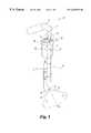
- FIG. 1is a side elevational view of a device according to this invention.
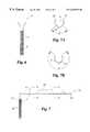
- FIG. 2is a top elevational view of a fixture for supporting a viewing device within a cannula according to this invention.
- FIG. 3is a side cross-sectional view of the fixture shown in FIG. 2 .
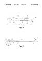
- FIG. 4is a side elevational view of a retractor according to one embodiment of this invention.
- FIG. 4Ais an end cross-sectional view of the retractor of FIG. 4 taken along lines A—A.
- FIG. 5is a top elevational view of the retractor shown in FIG. 4 .
- FIG. 6is an end elevational view of the retractor shown in FIGS. 4 and 5.
- FIG. 7is a side elevational view of a retractor according to another embodiment of this invention.
- FIG. 7Ais an end cross-sectional view of the retractor of FIG. 7 taken along lines A—A.
- FIG. 7Bis an end cross-sectional view of the retractor of FIG. 7 taken along lines B—B.
- FIG. 8is a top elevational view of the retractor shown in FIG. 7 .
- FIG. 9is a side elevational view of a dilator according to this invention.
- FIG. 10 ( a )-( i )depicts the steps of a method according to this invention.
- FIG. 11is a side cross-sectional view of a device according to one embodiment of this invention.
- FIG. 12is a side cross-sectional view of an aspiration cap as shown in FIG. 11 .
- the present inventionprovides instruments and methods for performing percutaneous surgery, including spinal applications such as laminotomy, laminectomy, foramenotomy, facetectomy or discectomy, with a single working channel endoscope.
- spinal applicationssuch as laminotomy, laminectomy, foramenotomy, facetectomy or discectomy
- the present inventorshave discovered that many percutaneous surgeries may be performed without a fluid workspace through the use of optics which move independently of the cannula.
- the present inventioncontemplates techniques and instruments that can be implemented with or without a fluid environment.
- This inventionalso brings the advantages of percutaneous procedures to applications that previously required open surgery.
- One advantageis based upon the further discovery that bone work can be performed percutaneously through a large working channel.
- Another advantageis realized in the use of a single portal within the patient to perform a wide range of simultaneous procedures.
- a device 10for use in percutaneous surgery which includes an elongated cannula 20 having a first inner diameter D I and an outer diameter D O sized for percutaneous introduction into a patient.
- the cannula 20also includes a distal working end 21 and an opposite proximal end 22 .
- the cannuladefines a working channel 25 between the ends 21 , 22 having a second diameter d 2 equal to the first inner diameter D I sized for receiving a tool therethrough.
- the cannulahas a length along its longitudinal axis L that is sized to pass through the patient from the skin to an operative site or working space. In some cases, the working space may be adjacent a vertebra or disc, or in the spinal canal.
- An elongated viewing element 50is mountable inside cannula 20 adjacent the working channel 25 .
- the viewing element 50has a first end 51 connectable to a viewing apparatus, such as an eyepiece or camera, and an opposite second end 52 disposed or positionable adjacent the distal working end 21 of the cannula 20 .
- a viewing apparatussuch as an eyepiece or camera
- the particular elongated viewing element 50is not critical to the invention. Any suitable viewing element is contemplated that creates an optical or image transmission channel.
- the elongated viewing element 50includes a fiber optic scope 54 and a lens 55 at the second end 52 .
- the fiber optic scopeincludes illumination fibers and image transmission fibers (not shown).
- the viewing elementmay be a rigid endoscope or an endoscope having a steerable or bendable tip.
- One advantage of this inventionis that it provides optics which are movable relative to the cannula 20 . Because the optics are movable, it is not necessary to provide a fluid-maintained work space. The optics can be removed, cleaned and replaced while the cannula is percutaneously positioned within the patient over the working space. Any configuration which allows the optics to be movably supported adjacent the working channel 25 is contemplated.
- a fixture 30is provided for mounting the elongated viewing element 50 to the cannula 20 .
- the fixture 30includes a housing 31 attachable to the proximal end 22 of the cannula 20 .
- the working channel opening 35is sized to substantially correspond to the second diameter d 2 of the working channel 25 to receive tools.
- the fixture 30includes a housing 31 which defines a working channel opening 35 arranged to communicate with the working channel 25 when the fixture 30 is mounted to the cannula 20 .
- the working channel opening 35is sized to receive tools therethrough for passage through the working channel 25 .
- the fixture 30is configured to mount the viewing element 50 within the working channel 25 .
- the housing 31also defines an optics bore 60 adjacent the working channel opening 35 .
- the optics bore 60has a longitudinal axis l that is preferably substantially parallel to the axis L of the cannula and working channel.
- the optics bore 60is preferably sized to removably receive the elongated viewing element 50 therethrough.
- the fixture 30preferably supports the viewing element 50 for movement within the optics bore 60 along the longitudinal axis l of the bore 60 to extend or retract the lens 55 relative to the distal working end 21 of the cannula 20 .
- the retractable/extendable feature of the optics of this inventionprovides an advantage over prior endoscopes because it eliminates the requirement for a fluid workspace.
- the device 10 and its viewing element 50can be easily used in a fluid environment, the fluid is not essential for the system to operate, contrary to prior systems. Furthermore, many of the prior endoscopes were not suited to access certain areas because of their large diameters. For example, prior endoscopes could not access the spinal canal. However, with this invention, access to the spinal canal is not limited by the diameter of the channel or cannula.
- the cannula 20can be left behind in the soft tissue or supported by the lamina while the-second end 52 of the elongated viewing element 50 can be advanced into the spinal canal along with any spinal instruments which have been inserted into the working channel 25 .
- the fixture 30also supports the viewing element 50 for rotation within the optics bore 60 about the longitudinal axis l of the bore 60 .
- the lens 55 of the viewing element 50defines an optical axis A O .
- the optical axis A Ocan be offset at an angle relative to the longitudinal axis l of the optics bore 60 . This feature allows the optical axis A o of the lens to be swept through a conical field of view F for greater visibility of the working space.
- the fixture 30can further be configured so that the viewing element 50 is rotatable relative to the cannula 20 .
- the housing 31is rotatable relative to the cannula 20 so that the second longitudinal axis l of the optics bore 60 rotates about the longitudinal axis L of the working channel 25 .
- the rotatable features of this inventionallows visualization of the entire working space. This feature also aids in simplifying the surgical procedure because the optics 50 and accompanying fittings can be moved out of the way of the surgeon's hands and tools passing through the working channel.
- the housing 31defines a receiver bore 40 having an inner diameter d 1 slightly larger than the outer diameter D O of the cannula 20 .
- the proximal end 22 of the cannula 20can be received within the receiver bore 40 so that the housing 31 can rotate about the proximal end 22 of the cannula 20 .
- the housing 31also includes an upper bore 41 which is contiguous with the working channel opening 35 and the receiver bore 40 .
- the optics bore 60is disposed within the upper bore 41 of the housing 31 .
- the optics bore 60is defined by a C-shaped clip 61 disposed within the upper bore 41 .
- the C-shaped clip 61is formed of a resilient material and the optics bore 60 defined by the clip 61 has an inner diameter D i that is slightly less than the outer diameter of the elongated viewing element 50 .
- D ithe inner diameter of the elongated viewing element 50 .
- the optics bore 60can have an inner diameter larger than the outer diameter of the viewing element.
- the viewing element 50can be supported outside the device 20 , either manually or by a separate support fixture.
- the device 10provides engagement means for securely yet rotatably engaging the fixture 30 to the cannula 20 .
- the fixture 30is configured to engage a standard cannula 20 .
- Engagement meanscan be disposed between the housing 31 and the cannula 20 when the fixture 30 is mounted to the proximal end 22 of the cannula 20 for providing gripping engagement between the housing 31 and the cannula 20 .
- the engagement meansincludes a number of grooves 32 within the receiver bore 40 and a resilient sealing member, such as an O-ring (see FIG. 11) disposed in each groove 32 .
- the O-ringsprovide sufficient resistance to movement to hold the fixture 30 in a selectable position on the cannula.
- the housing 31defines a receiver bore 40 which has an inner diameter d I which is only slightly larger than the outer diameter D O of the cannula 20 so that the housing 31 can rotate freely about the cannula 20 .
- the working channel 25 and the working channel opening 35are both sized to receive a tool or instrument therethrough.
- the working channel opening 35 of the housing 31has a diameter Dw which is substantially equal to the inner diameter d 2 of the working channel 25 so that the effective diameter of the working channel is not reduced by the fixture 30 .
- This configurationprovides a maximum amount of space for the insertion of tools into the working channel 25 .
- the present inventionis advantageous because standard micro-surgical spinal tools can be inserted into the working channel and manipulated to perform a surgical procedure.
- the present inventionis particularly advantageous because the working channel 25 will simultaneously accept a plurality of movable instruments. No other known prior art device has a working channel that accepts more than one movable instrument at a time through a single port. Therefore, according to this invention, an entire percutaneous surgical procedure can be performed through the working channel 25 of the device 10 under direct visualization using the viewing element 50 disposed within the optics bore 60 .
- a tissue retractor 70is provided as depicted in FIGS. 4-6.
- the retractor 70is removably and rotatably insertable through the working channel 25 and the working channel opening 35 of the device 10 .
- the tissue retractor 70includes a working tip 75 configured to atraumatically displace tissue as the retractor 70 is manipulated through the tissue and a body 76 having a proximal first end 77 and a distal second end 78 .
- the second end 78can be integral with the working tip 75 which preferably has a blunt curved end 82 .
- the working tip 75is also preferably bent or curved away from the body 76 , as shown in FIG. 7 .
- the body 76is sized to be rotatably received within the cannula 20 and has a length B from the first end 77 to the second end 78 sufficient so that the first end 77 and the working tip 75 can both extend outside the cannula 20 when the body 76 is within the cannula- 20 .
- the body 76includes a curved plate 84 that is configured to conform to the inner cylindrical surface 26 of the cannula without substantially blocking the working channel 25 .
- the curved plate 84has a convex surface 80 and an opposite concave surface 81 .
- the curved plate 84includes a first plate portion 85 defining a first convex surface 80 and an opposite first concave surface 81 .
- a second plate portion 86is integral with the first plate portion 85 and is disposed between the first plate portion 85 and the working tip 75 .
- the second plate portion 86defines a second convex surface (not shown) and an opposite second concave surface 81 ′. Both the first plate portion 85 and the second plate portion 86 include opposite edges 90 extending substantially parallel to the length B of the body 76 .
- the curved plate 84subtends an arc A 1 between the opposite edges 90 of at least 200 degrees, and most preferably 270 degrees.
- the second plate portion 86 and specifically the second concave surface 81 ′subtends an angle that decreases along the length of the retractor.
- the second concave surface 81 ′subtends an angle of about 200 degrees adjacent the first plate portion 85 , decreasing to an angle of less than about 10 degrees at end 78 .
- FIGS. 7-8An alternate embodiment of a tissue retractor according to this invention is depicted in FIGS. 7-8.
- This retractor 100has a body 106 which includes a first plate portion 115 defining a first convex surface 110 , and an opposite first concave surface 111 and includes first opposite edges 120 extending substantially parallel to the length B of the body 106 .
- the first plate portion 115subtends a first arc A 2 between the first opposite edges 120 .
- the retractor body 106also includes a second plate portionl 116 which is integral with the first plate portion 115 and is disposed between the first plate portion 115 and a working tip 105 .
- the second plate portion 116defines a second convex surface 110 ′ and an opposite second concave surface 111 ′ and includes second opposite edges 120 ′ extending substantially parallel to the length B.
- the second plate portion 116subtends a second arc A 3 between the second opposite edges 120 ′ that is different from the first arc A 2 in this embodiment.
- the first arc A 2subtends an angle of less than 180 degrees and the second arc A 3 subtends an angle of more than 180 degrees.
- the first arc A 2subtends an angle of about 90 degrees and the second arc A 3 subtends an angle of about 270 degrees.
- the retractors of this inventionmay be provided with means for engaging the retractors 70 , 100 within the working channel 25 of the cannula 20 .
- the convex surfaces 80 , 110can be configured to have a diameter that is greater than the diameter D I of the inner cylindrical surface 26 of the cannula 20 .
- the body 76 , 106may be formed of a resilient material that is deformable to be insertable into the cannula 20 so that the convex surface 80 , 110 is in contact with the inner cylindrical surface 26 of the cannula 20 . When the body 76 , 106 is deformed, it exerts an outward force against the surface 26 to frictionally hold the retractor in its selected position.
- the preferred components provided by this inventionare configured so that multiple tools and instruments can be accepted and manipulated within the working channel 25 of the cannula 20 .
- the componentsare also configured so that more than one surgeon may manipulate instruments through the working channel 25 of the cannula 20 at one time. For example, one surgeon may be manipulating the retractor while another surgeon is drilling into a bone.
- the curvature of the body 76 , 106 of the retractors 70 , 100provides more working space and increases visibility.
- Another featureis that the long axis of the component can be placed in the working channel 25 while a bend in the handle portion keeps hands away from the channel 25 so that more than one surgeon can work in the channel 25 and more tools can be placed in the channel 25 .
- the arm 71 , 101is at an angle a which is less than 180 degrees from the longitudinal axis of the length L of the body 76 .
- the angle oxis about 90 degrees so that the arm 71 , 101 is substantially perpendicular to the length L of the body 76 , 106 .
- the arm 71 , 101has a gripping surface 72 , 102 to facilitate manipulation of the retractor 70 , 100 .
- a dilator 130preferably includes a hollow sleeve 135 defining a channel 131 .
- the channel 131allows the dilator 130 to be placed over a guidewire (not shown) or other dilators.
- the hollow sleeve 135has a working end 136 defining a first opening 132 in communication with the channel 131 and an opposite end 137 defining a second opening 133 .
- the working end 136is tapered to a tapered tip 138 to atraumatically displace tissue.
- a gripping portion 140is provided on the outer surface 141 of the sleeve 135 adjacent the opposite end 137 .
- the gripping portion 140is defined by a plurality of circumferential grooves 142 defined in the outer surface 141 .
- the grooves 142are configured for manual gripping of the dilator 130 to manipulate the dilator 130 through tissue.
- the grooves 142are partially cylindrical.
- the gripping portion 140includes a number of circumferential flats 143 each of the circumferential grooves 142 .
- the grooves 142have a first width W 1 along the length of the sleeve 135 and the flats 143 have a second width W 2 146 along the length.
- the first and second widths W 1 and W 2are substantially equal.
- the present inventionhas application to a wide range of surgical procedures, and particularly spinal procedures such as laminotomy, laminectomy, foramenotomy, facetectomy and discectomy.
- Prior surgical techniques for each of these procedureshas evolved from a grossly invasive open surgeries to the minimally invasive techniques represented by the patents of Kambin and Shapiro.
- the minimally invasive techniquesrepresented by the patents of Kambin and Shapiro.
- multiple entries into the patientis required.
- most of the prior minimally invasive techniquesare readily adapted only for a posterolateral approach to the spine.
- the devices and instruments of the present inventionhave application in an inventive surgical technique that permits each of these several types of surgical procedures to be performed via a single working channel. This invention can also be used from any approach and in other regions besides the spine.
- a guidewire 150can be advanced through the skin and tissue into the laminae M of a vertebral body V.
- a small incisionis made in the skin to facilitate penetration of the guidewire through the skin.
- the guidewirewhich may be a K-wire, is inserted under radiographic or image guided control to verify its proper positioning within the laminae L of the vertebra V.
- the guidewire 150can be positioned at virtually any location in the spine and in any portion of a vertebra V. The positioning of the guidewire is dependent upon the surgical procedure to be conducted through the working channel cannula of the present invention.
- the guidewire 150is solidly anchored into the vertebral bone, being tapped by a mallet if necessary.
- a series of tissue dilatorsare advanced over the guidewire 150 , as depicted in steps ( b )-( d ) in FIG. 10 .
- the dilatorscan be advanced through the incision without the aid of a guidewire, followed by blunt dissection of the underlying tissues.
- a series of successively larger dilators 151 , 152 and 153are concentrically disposed over each other and over the guidewire 150 and advanced into the body to sequentially dilate the perispinous soft tissues.
- the tissue dilatorsare of the type shown in FIG. 9 of the present application.
- the dilatorshave successively larger diameters, ranging from 5 mm, to 9 mm to 12.5 mm for the largest dilator.
- Other dilator sizesare contemplated depending upon the anatomical approach and upon the desired size of the working channel.
- the working channel cannula 20is advanced over the largest dilator 153 , as shown in step (e), and the dilators and guidewire 150 are removed, as shown in step (f).
- the working channel cannula 20has an inner diameter D I of 12.7 mm so that it can be easily advanced over the 12.5 mm outer diameter of the large dilator 153 . Larger working channel cannulas are contemplated depending upon the anatomical region and surgical procedure.
- the cannula 20With the cannula 20 in position, a working channel is formed between the skin of the patient to a working space adjacent the spine. It is understood that the length of the cannula 20 is determined by the particular surgical operation being performed and the anatomy surrounding the working space. For instance, in the lumbar spine the distance between the laminae M of a vertebra V to the skin of the patient requires a longer cannula 20 than a similar procedure performed in the cervical spine where the vertebral body is closer to the skin. In one specific embodiment in which the cannula 20 is used in a lumbar discectomy procedure, the cannula has a length of 87 mm, although generally only about half of the length of the cannula will be situated within the patient during the procedure.
- the working channel cannula 20is at least initially only supported by the soft tissue and skin of the patient.
- the cannula 20can include a mounting bracket 27 affixed to the outer surface of the cannula (FIG. 10 ( f ), FIG. 11 ).
- This mounting bracket 27can be fastened to a flexible support arm 160 , which can be of known design.
- the flexible support arm 160is engaged to the bracket 27 by way of a bolt and wing nut 161 , as shown in FIG. 10 ( i ) and in more detail in FIG. 11, although other fasteners are also contemplated.
- This flexible arm 160can be mounted on the surgical table and can be readily adjusted into a fixed position to provide firm support for the cannula 20 .
- the flexible arm 160is preferred so that it can be contoured as required to stay clear of the surgical site and to allow the surgeons adequate room to manipulate the variety of tools that would be used throughout the procedure.
- the fixture 30can be engaged over the proximal end of the cannula 20 .
- the fixture 30provides an optics bore 60 for supporting an elongated viewing element, such as element 50 shown in step h.
- the viewing element 50is advanced into the fixture 30 and supported by the optics bore 60 (FIG. 2 ).
- the element 50is most preferably a fiber optic scope, although a rod lens scope or other viewing scopes may be utilized.
- the flexible arm 160is mounted to the bracket 27 to support the cannula 20 which in turn supports the optical viewing element 50 .
- This final position of step (i) in FIG. 10is shown in more detail in FIG. 11 .
- the viewing element 50can be of a variety of types, including a rigid endoscope or a flexible and steerable scope.
- the surgeoncan directly visualize the area beneath the working channel 25 of the cannula 20 .
- the surgeoncan freely manipulate the viewing element 50 within the working channel 25 or beyond the distal end of the cannula into the working space.
- the second end 52 of the viewing element 50which carries the lens 55 , can be manipulated to different positions, such as shown in FIG. 11 .
- the manipulation and positioning of the scopeis not limited by the working channel 25 , in contrast to prior systems.
- the positioning capability provided by the fixture 30is utilized to allow extension of the lens 55 into the working space or retraction back within the cannula 20 , as depicted by the arrows T in FIG. 1 .
- the fixturepreferably accommodates rotation of the element 50 about its own axis (arrows R in FIG. 1) to vary the viewing angle provided by the angled lens 55 , or rotation of the entire viewing element 50 about the cannula 20 and around the circumference of the working channel 25 , as shown by the arrows N in FIG. 1 .
- the surgeonis provided with a complete and unrestricted view of the entire working space beneath the working channel 25 .
- the viewing orientation of the opticsi.e., left-right and up-down
- the cannula 20 of the present inventioncan be readily positioned over an appropriate target tissue or bone, to thereby move the working space as necessary for the surgical procedure.
- the working channel cannula 20since the working channel cannula 20 is freely situated within the patient's skin and tissue, it can be manipulated so that the working space beneath the cannula 20 is more appropriately centered over the target region of the spine.
- Repositioning of the cannula 20can be performed under fluoroscopic guidance.
- the cannulamay be fitted with position sensing devices, such as LEDs, to be guided stereotactically. As the cannula is being repositioned, the surgeon can also directly visualize the spine through the viewing element 50 .
- a variety of tools and instrumentscan be extended through the working channel 25 to accomplish the particular surgical procedure to be performed. For instance, in the case of a laminotomy, laminectomy, foramenotomy or facetectomy, a variety of rongeurs, curettes, and trephines can be extended through the working channel opening 35 (see FIG. 2) and through the working channel 25 of the cannula 20 (see FIG. 11) into the working space. It is understood that these various tools and instruments are designed to fit through the working channel.
- the working channel 25 through the cannula 20can have a maximum diameter d 2 of 12.7 mm.
- the effective diameteris about 8 mm in the specific illustrated embodiment, although adequate space is provided within the working channel 25 around the viewing element 50 to allow a wide range of movement of the tool or instrument within the working channel.
- the present inventionis not limited to particular sizes for the working channel and effective diameter, since the dimensions of the components will depend upon the anatomy of the surgical site and the type of procedure being performed.
- each of the tools and instruments used with the working channel cannula 20are designed to minimize obstruction of the surgeon's visualization of and access to the working space at the distal end of the working channel cannula.
- the instruments and toolsare designed so that their actuating ends which are manipulated by the surgeon are displaced from the working channel cannula 20 .
- One such exampleis the tissue retractor shown in FIGS. 4-8. With these retractors, the handles that are manually gripped by the surgeon are offset at about a 90 degree angle relative to the longitudinal axis of the tool itself.
- the surgical procedures conducted through the working channel cannula 20 and within the working space at the distal end of the cannulaare performed “dry” —that is, without the use of irrigation fluid.
- the working space at the surgical siteis fluid filled to maintain the working space and to assist in the use of the visualization optics.
- the visualization opticswere fixed within the endoscope.
- the device 10 of the present inventionallows a wide range of movement for the viewing element 50 so that the lens 55 can be retracted completely within the working channel 25 of the cannula 20 to protect it from contact with the perispinous tissue or blood that may be generated at the surgical site.
- the viewing element 50since the viewing element 50 is removable and replaceable, the element 50 can be completely removed from the fixture 30 so that the lens 55 can be cleaned, after which the viewing element 50 can be reinserted into the fixture and advanced back to the working space. Under these circumstances, then, the need for irrigation is less critical.
- This featurecan be of particular value when cutting operations are being performed by a power drill. It has been found in prior surgical procedures that the use of a power drill in a fluid environment can cause turbulence or cavitation of the fluid. This turbulence can completely shroud the surgeon's view of the surgical site at least while the drill is being operated. With the present invention, the dry environment allows continuous viewing of the operation of the power drill so that the surgeon can quickly and efficiently perform the necessary cutting procedures.
- irrigationmay be provided separately through the working channel 25 .
- the viewing device 50itself may include a tube 54 supported by the fitting 53 through which modest amounts of fluid can be provided to keep the visualization space clear.
- aspiration of the excised tissueis preferred, and irrigation will frequently assist in rapid removal of this tissue.
- separate irrigation and aspiration elementscan also be inserted through the working channel 25 as required by the procedure.
- an aspiration cap 165is provided as shown in FIGS. 11 and 12.
- the cap 165includes a body 166 which defines a mating bore 167 having an inner diameter d b larger than the outer diameter D h of the housing 31 of fitting 30 .
- a tool opening 168is provided in communication with the mating bore 167 .
- the aspiration cap 165is also provided with a tube receiver bore 169 which intersects the mating bore 167 .
- the receiver bore 169is configured to receive an aspiration tube through which a vacuum or suction is applied.
- the tool opening 168may be covered while suction is applied through the tool receiver bore 169 and mating bore 167 , and ultimately through the working channel 25 . Covering the opening 168 can optimize the aspiration effect through the working channel.
- the paraspinous tissuecan be reflected using instruments as described above, and a laminectomy performed using various rongeurs, curettes and drills.
- the cannula 20can be angled to allow a greater region of bone removal, which may be necessary for access to other portions of the spinal anatomy.
- access to the spinal canal and the posterior medial aspects of the disc annulusmay require cutting a portion of the vertebral bone that is greater than the inner diameter of the working channel 25 .
- some manipulation of the cannula 20may be necessary to permit removal of a greater portion of bone.
- multi-level laminectomies or foramenotomiesmay be necessary.
- these multi-level procedurescan be conducted by sequentially inserting the working channel cannula 20 through several small cutaneous incisions along the spinal mid-line.
- several working channel cannulas 20can be placed at each of the small cutaneous incisions to perform the multi-level bone removal procedures.
- an openingis cut into the laminae M of the vertebra V providing direct visual access to the spinal canal itself.
- tissue surrounding the spinal nerve rootcan be removed utilizing micro surgical knives and curettes.
- a retractorsuch as the retractors shown in FIGS. 4-8, can be used to gently move and hold the nerve root outside the working space.
- the portion of the retractor passing through the working channel 25generally conforms to the inner surface of the cannula 20 so that the working channel 25 is not disrupted by the retractor tool.
- the effective diameter within the working channel 25is reduced only by the thickness of the curved plates 84 , 114 of the retractors 70 , 100 .
- this thicknessis about 0.3 mm, so it can be seen that the tissue retractors do not significantly reduce the space available in the working channel 25 for insertion of other tools and instruments.
- bone within the spinal canalcan be removed with a curette or a high speed drill.
- the fractured bonemay be impacted back into the vertebral body with a bone impactor.
- the spinal procedure to be performedis the removal of epidural spinal tumors
- the tumorscan be resected utilizing various micro-surgical instruments.
- the duramay be opened and the intradural pathology may be approached with micro-surgical instruments passing through the working channel cannula 20 .
- the nerve root retracted posterior medial disc herniationscan be readily excised directly at the site of the herniation.
- the large diameter of the working channel 25 in the cannula 20allows the surgeon or surgeons conducting the surgical procedure to introduce a plurality of instruments or tools into the working space.
- a tissue retractor and discectomy instrumentscan be simultaneously extended through the working channel.
- the discectomy instrumentscould include a trephine for boring a hole through the disc annulus and a powered tissue cutter for excising the herniated disc nucleus.
- the present inventioncontemplates the simultaneous introduction of other types of instruments or tools as may be dictated by the particular surgical procedure to be performed. For example, an appropriately sized curette and a rongeur may be simultaneously extended through the working channel into the working space.
- the surgeoncan readily manipulate each of the instruments to perform tissue removal and bone cutting operations, without having to remove one tool and insert the other.
- the surgical procedurescan be conducted without the necessity of irrigation fluid, the surgeon has a clear view through the working space of the target tissue.
- aspects of the inventionwhich permit a wide range of motion to the viewing element 50 allow the surgeon to clearly visualize the target tissue and clearly observe the surgical procedures being conducted in the working space.
- surgeoncan capitalize on the same advantages in conducting a wide range of procedures at a wide range of locations in the human body.
- facetectomiescould be conducted through the working channel by simply orienting the working channel cannula 20 over the particular facet joints.
- the insertion of vertebral fixation elementscan also be accomplished through the device 10 .
- an incisioncan be made in the skin posterior to the location of the vertebra at which the fixation element is to be implanted.
- the cannula 20can be positioned through the incision and tissue directly above the particular location on the vertebra to be instrumented.
- an insertion tool holding the vertebral fixation elementcan be projected through the cannula 20 and manipulated at the vertebra.
- the fixation elementcan be a bone screw.
- the working channel 25has a diameter that is large enough to accept most bone screws and their associated insertion tools.
- the location of the bone screw within the vertebrais critical, so identification of the position of the cannula 20 over the bony site is necessary. As mentioned above, this position can be verified fluoroscopically or using stereotactic technology.
- cannulated bone screwsare driven into the vertebra along K-wires.
- the present inventioneliminates the need for the K-wire and for a cannulated screw.
- the working channelitself can effectively operate as a positioning guide, once the cannula 20 is properly oriented with respect to the vertebra.
- the device 10allows insertion of the bone screw into the vertebra to be conducted under direct vision. The surgeon can then readily verify that the screw is passing into the vertebra properly. This can be particularly important for bone screws being threaded into the pedicle of a vertebra.
- the working channel cannula 20can be used to directly insert a self-tapping bone screw into the pedicle, or can accept a variety of tools to prepare a threaded bore within the pedicle to receive a bone screw.
- the device 10can also be used to prepare a site for fusion of two adjacent vertebrae, and for implantation of a fusion device or material.
- a fusion device or materialFor example, in one surgical technique, an incision can be made in the skin posterior to a particular disc space to be fused. The incision can be made anteriorly, posteriorly or posterior laterally. If the incision is made anteriorly for anterior insertion of the working channel, it is anticipated that care will be taken to retract tissues, muscle and organs that may follow the path of the incision to the disc space.
- the device 10 of the present inventionallows this tissue retraction to occur under direct vision so that the surgeon can easily and accurately guide the cannula 20 to the disc space without fear of injury to the surrounding tissue.
- the working channel cannula 20can be progressively advanced toward the anticipated working space adjacent the vertebral disc.
- the disc spacecan be prepared for implantation of fusion materials or a fusion device.
- this preparationincludes preparing an opening in the disc annulus, and excising all or part of the disc nucleus through this opening.
- a boreis cut through the disc annulus and into the endplates of the adjacent vertebrae.
- a fusion devicesuch as a bone dowel, a push-in implant or a threaded implant can then be advanced through the working channel of device 10 and into the prepared bore at the subject disc space.
- the preparatory stepsinvolve preparing the vertebral endplates by reducing the endplates to bleeding bone. In this instance, some aspiration and irrigation may be beneficial. All of these procedures can be conducted by tools and instruments extending through the working channel cannula 20 and under direct vision from the viewing element 50 .
- graft materialis simply placed within the prepared bore. This graft material can also be passed through the working channel cannula 20 into the disc space location. In other procedures, graft material or bone chips are positioned across posterior aspects of the spine. Again, this procedure can be conducted through the working channel cannula particularly given the capability of the cannula to be moved to different angles from a single incision site in the skin.
- the present inventionprovides instruments and techniques for conducting a variety of surgical procedures. In the illustrated embodiments, these procedures are conducted on the spine. However, the same devices and techniques can be used at other places in the body. For example, an appropriately sized working channel device 10 can be used to remove lesions in the brain.
- the present inventionhas particular value for percutaneous procedures where minimal invasion into the patient is desirable and where accurate manipulation of tools and instruments at the surgical site is required. While the preferred embodiments illustrated above concern spinal procedures, the present invention and techniques can be used throughout the body, such as in the cranial cavity, the pituitary regions, the gastrointestinal tract, etc.
- the ability to reposition the viewing optics as required to visualize the surgical siteallows for much greater accuracy and control of the surgical procedure.
- the present inventionallows the use of but a single entry into the patient which greatly reduces the risk associated with open surgery or multiple invasions through the patierit's skin.
Landscapes
- Health & Medical Sciences (AREA)
- Life Sciences & Earth Sciences (AREA)
- Animal Behavior & Ethology (AREA)
- General Health & Medical Sciences (AREA)
- Veterinary Medicine (AREA)
- Engineering & Computer Science (AREA)
- Biomedical Technology (AREA)
- Heart & Thoracic Surgery (AREA)
- Public Health (AREA)
- Surgery (AREA)
- Molecular Biology (AREA)
- Medical Informatics (AREA)
- Nuclear Medicine, Radiotherapy & Molecular Imaging (AREA)
- Anesthesiology (AREA)
- Hematology (AREA)
- Pathology (AREA)
- Vascular Medicine (AREA)
- Surgical Instruments (AREA)
- Medicinal Preparation (AREA)
- Acyclic And Carbocyclic Compounds In Medicinal Compositions (AREA)
- Pharmaceuticals Containing Other Organic And Inorganic Compounds (AREA)
Abstract
Description
This application is a continuation of U.S. patent application Ser. No. 08/920,991 filed on Aug. 29, 1997, now U.S. Pat. No. 6,007,487, a divisional of U.S. Application Ser. No. 08/620,933, filed on Mar. 22, 1996, now issued as U.S. Pat. No. 5,792,044.
The present invention relates to devices, instruments and methods for performing percutaneous surgeries, particularly at locations deep within the body. One specific application of the invention concern devices, instruments and techniques for percutaneous, minimally invasive spinal surgery. In another aspect of the invention, the percutaneous surgery is performed under direct vision at any location in the body.
Traditional surgical procedures for pathologies located, deep within the body can cause significant trauma to the intervening tissues. These open procedures often require a long incision, extensive muscle stripping, prolonged retraction of tissues, denervation and devascularization of tissue. Most of these surgeries require a recovery room time of several hours and several weeks of post-operative recovery time due to the use of general anesthesia and the destruction of tissue during the surgical procedure. In some cases, these invasive procedures lead to permanent scarring and pain that can be more severe than the pain leading to the surgical intervention.
Minimally invasive alternatives such as arthroscopic techniques reduce pain, post-operative recovery time and the destruction of healthy tissue. Orthopedic surgical patients have particularly benefitted from minimally invasive surgical techniques. The site of pathology is accessed through portals rather than through a significant incision thus preserving the integrity of the intervening tissues. These minimally invasive techniques also often require only local anesthesia. The avoidance of general anesthesia reduces post-operative recovery time and the risk of complications.
Minimally invasive surgical techniques are particularly desirable for spinal and neurosurgical applications because of the need for access to locations deep within the body and the danger of damage to vital intervening tissues. For example, a common open procedure for disc herniation, laminectomy followed by discectomy requires stripping or dissection of the major muscles of the back to expose the spine. In a posterior approach, tissue including spinal nerves and blood vessels around the dural sac, ligaments and muscle must be retracted to clear a channel from the skin to the disc. These procedures normally take at least one-two hours to perform under general anesthesia and require post-operative recovery periods of at least several weeks. In addition to the long recovery time, the destruction of tissue is a major disadvantage of open spinal procedures. This aspect of open procedures is even more invasive when the discectomy is accompanied by fusion of the adjacent vertebrae. Many patients are reluctant to seek surgery as a solution to pain caused by herniated discs and other spinal conditions because of the severe pain sometimes associated with the muscle. dissection.
In order to reduce the post-operative recovery time and pain associated with spinal and other procedures, micro-surgical techniques have been developed. For example, in micro-surgical discectomies, the disc is accessed by cutting a channel from the surface of the patient's back to the disc through a small incision. An operating microscope or loupes is used to visualize the surgical field. Small diameter micro-surgical instruments are passed through the small incision and between two laminae and into the disc. The intervening tissues are disrupted less because the incision is smaller. Although these micro-surgical procedures are less invasive, they still involve some of the same complications associated with open procedures, such as injury to the nerve root and dural sac, perineural scar formation, reherniation at the surgical site and instability due to excess bone removal.
Other attempts have been made for minimally invasive procedures to correct symptomatic spinal conditions. One example is chemonucleolysis which involved the injection of an enzyme into the disc to partially dissolve the nucleus to alleviate disc herniation. Unfortunately, the enzyme, chymopapain, has been plagued by concerns about both its effectiveness and complications such as severe spasms, post-operative pain and sensitivity reactions including anaphylactic shock.
The development of percutaneous spinal procedures has yielded a major improvement in reducing recovery time and post-operative pain because they require minimal, if any, muscle dissection and they can be performed under local anesthesia. For example, U.S. Pat. No. 4,545,374 to Jacobson discloses a percutaneous lumbar discectomy using a lateral approach, preferably under fluoroscopic X-ray. This procedure is limited because it does not provide direct visualization of the discectomy site.
Other procedures have been developed which include arthroscopic visualization of the spine and intervening structures. U.S. Pat. Nos. 4,573,448 and 5,395,317 to Kambin disclose percutaneous decompression of herniated discs with a posterolateral approach. Fragments of the herniated disc are evacuated through a cannula positioned against the annulus. The '317 Kambin patent discloses a biportal procedure which involves percutaneously placing both a working cannula and a visualization cannula for an endoscope. This procedure allows simultaneous visualization and suction, irrigation and resection in disc procedures.
Unfortunately, disadvantages remain with these procedures and the accompanying tools because they are limited to a specific application or approach. For example, Jacobson, Kambin and other references require a lateral or a posterolateral approach for percutaneous discectomy. These approaches seek to avoid damage to soft tissue structures and the need for bone removal because it was thought to be impractical to cut and remove bone through a channel. However, these approaches do not address other spinal conditions which may require a mid-line approach, removal of bone or implants.
U.S. Pat. No. 5,439,464 to Shapiro discloses a method and instruments for performing arthroscopic spinal surgeries such as laminectomies and fusions with a mid-line or medial posterior approach using three cannulas. Each of the cannulas requires a separate incision. While Shapiro discloses an improvement over prior procedures which were limited to a posterolateral or lateral approach for disc work, Shapiro's procedure still suffers from many of the disadvantages of known prior percutaneous spinal surgery techniques and tools. One disadvantage of the Shapiro procedure is its requirement of a fluid working space. Another significant detriment is that the procedure requires multiple portals into the patient.
Fluid is required in these prior procedures to maintain the working space for proper function of optics fixed within a prior art cannula and inserted percutaneously. Irrigation, or the introduction of fluid into the working space, can often be logistically disadvantageous and even dangerous to the patient for several reasons. The introduction of fluid into the working space makes hemostasis more difficult and may damage surrounding tissue. Excess fluid may dangerously dilute the sodium concentration of the patient's blood supply which can cause seizures or worse. The fluid environment can also make drilling difficult due to cavitation. The requirement for a fluid environment generally increases expenses associated with the surgery and adds to the complexity of the surgery, due in part to the relatively high volume of fluid required.
A need has remained for devices and methods that provide for percutaneous minimally invasive surgery for all applications and approaches. A need has also remained for percutaneous methods and devices which do not require a fluid-filled working space, but that can be adapted to a fluid environment if necessary.
A signficant need is present in this field for techniques and instruments that permit surgical procedures in the working space under direct vision. Procedures that reduce the number of entries into the patient are also highly desirable. The fields of spinal and neuro surgery have particularly sought devices and techniques that minimize the invasion into the patient and that are streamlined and concise in their application.
Briefly describing one aspect of the invention, there is provided devices and method for performing percutaneous, procedures under direct visualization, even at locations deep within a patient. In one embodiment, a device for use in percutaneous surgery includes an,elongated cannula having a first inner diameter and an outer diameter sized for percutaneous introduction into a patient. The cannula further includes a distal working end and an opposite proximal end and defines a working channel between the ends having a second diameter which is equal to the first inner diameter. The working channel is sized to receive a tool therethrough. The device also includes an elongated viewing element mounted inside the cannula adjacent the working channel. The viewing element has a first end connectable to a viewing apparatus and an opposite second end disposed adjacent the distal working end of the cannula.
In another aspect, a fixture is provided for mounting the elongated viewing element to the cannula. The fixture includes a housing attachable to the proximal end of the cannula. The housing defines a working channel opening therethrough in communication with the working channel. The working channel opening is sized to substantially correspond to the second diameter of the working channel. The housing also defines an optics bore adjacent the working channel opening. The optics bore is sized to receive the elongated viewing element therethrough.
In some embodiments, the fixture supports the viewing device for movement within the optics bore along the longitudinal axis of the bore to extend or retract the lens relative to the distal working end of the cannula. In other embodiments, the fixture supports the viewing device for rotation within the optics bore about the longitudinal axis of the bore. In some embodiments, the housing is rotatable relative to the cannula so that the longitudinal axis of the optics bore is rotatable about the longitudinal axis of the working channel.
Novel tools are also provided which are insertable into the working channel of the cannula. A tissue retractor in one embodiment includes a body and an integral working tip configured to atraumatically displace tissue as the retractor is manipulated through tissue. The body has a convex surface configured to conform to the inner cylindrical surface of the cannula and an opposite concave surface which does not obstruct the working channel or visualization of the working space. Cannulated tissue dilators are also provided which are insertable over a guidewire or another dilator as well as insertable into the working channel. In some embodiments, the tissue dilators include a tapered working end to displace tissue and a gripping portion having a number of circumferential grooves to enhance gripping and manipulation of the dilator.
According to the methods of this invention, spinal and other surgeries can be performed percutaneously with direct visualization without the requirement for a fluid-maintained working space. In another aspect of the inventive surgical techniques, all steps of a surgical procedure are conducted under direct vision through a single working channel cannula. An optical scope or viewing device is moved within the working channel and throughout the working space from a variety of angles and orientations to provide a clear view of the operative steps.
The techniques of the present invention also encompass passing multiple tools and instruments through the single working channel cannula and manipulating the instruments and tools within the working space. In one specific embodiment, a tissue retractor is provided that extends through the working channel without significantly reducing the dimensions of the channel.
It is an object of the invention to provide devices and methods for percutaneous spinal surgery for all applications and approaches. One advantage of this invention is that percutaneous procedures can be accomplished in a dry. environment because a fluid working space is not required for the proper function of the optics. One benefit of this invention is that it provides instruments and methods which reduce the cost, risk, pain and recovery time associated with surgery. These and other objects, advantages and features are accomplished according to the devices and methods of the present invention.
FIG. 1 is a side elevational view of a device according to this invention.
FIG. 2 is a top elevational view of a fixture for supporting a viewing device within a cannula according to this invention.
FIG. 3 is a side cross-sectional view of the fixture shown in FIG.2.
FIG. 4 is a side elevational view of a retractor according to one embodiment of this invention.
FIG. 4A is an end cross-sectional view of the retractor of FIG. 4 taken along lines A—A.
FIG. 5 is a top elevational view of the retractor shown in FIG.4.
FIG. 6 is an end elevational view of the retractor shown in FIGS. 4 and 5.
FIG. 7 is a side elevational view of a retractor according to another embodiment of this invention.
FIG. 7A is an end cross-sectional view of the retractor of FIG. 7 taken along lines A—A.
FIG. 7B is an end cross-sectional view of the retractor of FIG. 7 taken along lines B—B.
FIG. 8 is a top elevational view of the retractor shown in FIG.7.
FIG. 9 is a side elevational view of a dilator according to this invention.
FIG.10(a)-(i) depicts the steps of a method according to this invention.
FIG. 11 is a side cross-sectional view of a device according to one embodiment of this invention.
FIG. 12 is a side cross-sectional view of an aspiration cap as shown in FIG.11.
For the purposes of promoting an understanding of the principles of the invention, reference will now be made to the embodiments illustrated in the drawings and specific language will be used to describe the same. It will nevertheless be understood that no limitation of the scope of the invention is thereby intended, such alterations and further modifications in the illustrated devices and described methods, and such further applications of the principles of the invention as illustrated therein being contemplated as would normally occur to one skilled in the art to which the invention relates.
The present invention provides instruments and methods for performing percutaneous surgery, including spinal applications such as laminotomy, laminectomy, foramenotomy, facetectomy or discectomy, with a single working channel endoscope. The present inventors have discovered that many percutaneous surgeries may be performed without a fluid workspace through the use of optics which move independently of the cannula. The present invention contemplates techniques and instruments that can be implemented with or without a fluid environment.
This invention also brings the advantages of percutaneous procedures to applications that previously required open surgery. One advantage is based upon the further discovery that bone work can be performed percutaneously through a large working channel. Another advantage is realized in the use of a single portal within the patient to perform a wide range of simultaneous procedures.
According to one embodiment of the present invention, as depicted in FIG. 1, adevice 10 is provided for use in percutaneous surgery which includes anelongated cannula 20 having a first inner diameter DIand an outer diameter DOsized for percutaneous introduction into a patient. Thecannula 20 also includes a distal workingend 21 and an oppositeproximal end 22. The cannula defines a workingchannel 25 between theends
Anelongated viewing element 50 is mountableinside cannula 20 adjacent the workingchannel 25. Theviewing element 50 has afirst end 51 connectable to a viewing apparatus, such as an eyepiece or camera, and an oppositesecond end 52 disposed or positionable adjacent the distal workingend 21 of thecannula 20. The particularelongated viewing element 50 is not critical to the invention. Any suitable viewing element is contemplated that creates an optical or image transmission channel. In one embodiment, theelongated viewing element 50 includes afiber optic scope 54 and alens 55 at thesecond end 52. Preferably, the fiber optic scope includes illumination fibers and image transmission fibers (not shown). Alternatively, the viewing element may be a rigid endoscope or an endoscope having a steerable or bendable tip.
One advantage of this invention is that it provides optics which are movable relative to thecannula 20. Because the optics are movable, it is not necessary to provide a fluid-maintained work space. The optics can be removed, cleaned and replaced while the cannula is percutaneously positioned within the patient over the working space. Any configuration which allows the optics to be movably supported adjacent the workingchannel 25 is contemplated. In one embodiment, shown in FIGS. 1-3, afixture 30 is provided for mounting theelongated viewing element 50 to thecannula 20. Preferably, thefixture 30 includes ahousing 31 attachable to theproximal end 22 of thecannula 20. The workingchannel opening 35 is sized to substantially correspond to the second diameter d2of the workingchannel 25 to receive tools. Thefixture 30. includes ahousing 31 which defines a workingchannel opening 35 arranged to communicate with the workingchannel 25 when thefixture 30 is mounted to thecannula 20. The workingchannel opening 35 is sized to receive tools therethrough for passage through the workingchannel 25. In the embodiments shown in FIGS. 1-3, thefixture 30 is configured to mount theviewing element 50 within the workingchannel 25.
Thehousing 31 also defines an optics bore60 adjacent the workingchannel opening 35. The optics bore60 has a longitudinal axis l that is preferably substantially parallel to the axis L of the cannula and working channel. The optics bore60 is preferably sized to removably receive theelongated viewing element 50 therethrough. Thefixture 30 preferably supports theviewing element 50 for movement within the optics bore60 along the longitudinal axis l of thebore 60 to extend or retract thelens 55 relative to the distal workingend 21 of thecannula 20. The retractable/extendable feature of the optics of this invention provides an advantage over prior endoscopes because it eliminates the requirement for a fluid workspace. While thedevice 10 and itsviewing element 50 can be easily used in a fluid environment, the fluid is not essential for the system to operate, contrary to prior systems. Furthermore, many of the prior endoscopes were not suited to access certain areas because of their large diameters. For example, prior endoscopes could not access the spinal canal. However, with this invention, access to the spinal canal is not limited by the diameter of the channel or cannula. Thecannula 20 can be left behind in the soft tissue or supported by the lamina while the-second end 52 of theelongated viewing element 50 can be advanced into the spinal canal along with any spinal instruments which have been inserted into the workingchannel 25.
Preferably thefixture 30 also supports theviewing element 50 for rotation within the optics bore60 about the longitudinal axis l of thebore 60. Thelens 55 of theviewing element 50 defines an optical axis AO. As in many endoscopes, the optical axis AOcan be offset at an angle relative to the longitudinal axis l of the optics bore60. This feature allows the optical axis Aoof the lens to be swept through a conical field of view F for greater visibility of the working space. Thefixture 30 can further be configured so that theviewing element 50 is rotatable relative to thecannula 20. In this embodiment, thehousing 31 is rotatable relative to thecannula 20 so that the second longitudinal axis l of the optics bore60 rotates about the longitudinal axis L of the workingchannel 25. The rotatable features of this invention allows visualization of the entire working space. This feature also aids in simplifying the surgical procedure because theoptics 50 and accompanying fittings can be moved out of the way of the surgeon's hands and tools passing through the working channel.
In one embodiment depicted in FIG. 3, thehousing 31 defines a receiver bore40 having an inner diameter d1slightly larger than the outer diameter DOof thecannula 20. In this configuration, theproximal end 22 of thecannula 20 can be received within the receiver bore40 so that thehousing 31 can rotate about theproximal end 22 of thecannula 20. As shown in FIG. 3, thehousing 31 also includes anupper bore 41 which is contiguous with the workingchannel opening 35 and the receiver bore40. In one embodiment, the optics bore60 is disposed within theupper bore 41 of thehousing 31.
In a preferred embodiment depicted in FIG. 2, the optics bore60 is defined by a C-shapedclip 61 disposed within theupper bore 41. Preferably, the C-shapedclip 61 is formed of a resilient material and the optics bore60 defined by theclip 61 has an inner diameter Dithat is slightly less than the outer diameter of theelongated viewing element 50. When theviewing element 50 is pushed into the optics bore60 it resiliently deflects the C-shapedclip 61. The resilience of theclip 61 provides a gripping force on theelement 50 to hold it in the desired position, while still allowing theelement 50 to be repositioned.
Alternatively, the optics bore60 can have an inner diameter larger than the outer diameter of the viewing element. In this instance, theviewing element 50 can be supported outside thedevice 20, either manually or by a separate support fixture.
Preferably thedevice 10 provides engagement means for securely yet rotatably engaging thefixture 30 to thecannula 20. Most preferably, thefixture 30 is configured to engage astandard cannula 20. Engagement means can be disposed between thehousing 31 and thecannula 20 when thefixture 30 is mounted to theproximal end 22 of thecannula 20 for providing gripping engagement between thehousing 31 and thecannula 20. In one embodiment depicted in FIG. 3 the engagement means includes a number ofgrooves 32 within the receiver bore40 and a resilient sealing member, such as an O-ring (see FIG. 11) disposed in eachgroove 32. The sealing members, or O-rings, disposed between thehousing 31 and the outer diameter DOof thecannula 20 rotatably secure thefixture 30 to thecannula 20. The O-rings provide sufficient resistance to movement to hold thefixture 30 in a selectable position on the cannula. In another embodiment, thehousing 31 defines a receiver bore40 which has an inner diameter dIwhich is only slightly larger than the outer diameter DOof thecannula 20 so that thehousing 31 can rotate freely about thecannula 20.
The workingchannel 25 and the workingchannel opening 35 are both sized to receive a tool or instrument therethrough. Preferably, the working channel opening35 of thehousing 31 has a diameter Dw which is substantially equal to the inner diameter d2of the workingchannel 25 so that the effective diameter of the working channel is not reduced by thefixture 30. This configuration provides a maximum amount of space for the insertion of tools into the workingchannel 25. The present invention is advantageous because standard micro-surgical spinal tools can be inserted into the working channel and manipulated to perform a surgical procedure. The present invention is particularly advantageous because the workingchannel 25 will simultaneously accept a plurality of movable instruments. No other known prior art device has a working channel that accepts more than one movable instrument at a time through a single port. Therefore, according to this invention, an entire percutaneous surgical procedure can be performed through the workingchannel 25 of thedevice 10 under direct visualization using theviewing element 50 disposed within the optics bore60.
Although standard micro-surgical instruments may be used with the present invention, this invention also contemplates certain novel tools which capitalize on and enhance the advantages of this invention.
According to one preferred embodiment of the invention, atissue retractor 70 is provided as depicted in FIGS. 4-6. Theretractor 70 is removably and rotatably insertable through the workingchannel 25 and the working channel opening35 of thedevice 10. Thetissue retractor 70 includes a workingtip 75 configured to atraumatically displace tissue as theretractor 70 is manipulated through the tissue and abody 76 having a proximalfirst end 77 and a distalsecond end 78. Thesecond end 78 can be integral with the workingtip 75 which preferably has a bluntcurved end 82. In addition, the workingtip 75 is also preferably bent or curved away from thebody 76, as shown in FIG.7. Thebody 76 is sized to be rotatably received within thecannula 20 and has a length B from thefirst end 77 to thesecond end 78 sufficient so that thefirst end 77 and the workingtip 75 can both extend outside thecannula 20 when thebody 76 is within the cannula-20.
This invention contemplates any suitable retractor for use through the workingchannel 25. However, retractors such as theretractor 70 depicted in FIGS. 4-6 are preferred in which thebody 76 includes acurved plate 84 that is configured to conform to the inner cylindrical surface26 of the cannula without substantially blocking the workingchannel 25. Thecurved plate 84 has aconvex surface 80 and an oppositeconcave surface 81. In one embodiment, thecurved plate 84 includes afirst plate portion 85 defining a firstconvex surface 80 and an opposite firstconcave surface 81. Asecond plate portion 86 is integral with thefirst plate portion 85 and is disposed between thefirst plate portion 85 and the workingtip 75. Thesecond plate portion 86 defines a second convex surface (not shown) and an opposite secondconcave surface 81′. Both thefirst plate portion 85 and thesecond plate portion 86 includeopposite edges 90 extending substantially parallel to the length B of thebody 76.
Preferably, thecurved plate 84 subtends an arc A1between theopposite edges 90 of at least 200 degrees, and most preferably 270 degrees. In a specific embodiment, thesecond plate portion 86 and specifically the secondconcave surface 81′ subtends an angle that decreases along the length of the retractor. Thus, in an embodiment, the secondconcave surface 81′ subtends an angle of about 200 degrees adjacent thefirst plate portion 85, decreasing to an angle of less than about 10 degrees atend 78.
An alternate embodiment of a tissue retractor according to this invention is depicted in FIGS. 7-8. Thisretractor 100 has abody 106 which includes afirst plate portion 115 defining a firstconvex surface 110, and an opposite firstconcave surface 111 and includes firstopposite edges 120 extending substantially parallel to the length B of thebody 106. Thefirst plate portion 115 subtends a first arc A2between the firstopposite edges 120. Theretractor body 106 also includes a second plate portionl116 which is integral with thefirst plate portion 115 and is disposed between thefirst plate portion 115 and a workingtip 105. Thesecond plate portion 116 defines a secondconvex surface 110′ and an opposite secondconcave surface 111′ and includes secondopposite edges 120′ extending substantially parallel to the length B. Thesecond plate portion 116 subtends a second arc A3between the secondopposite edges 120′ that is different from the first arc A2in this embodiment. Preferably, the first arc A2subtends an angle of less than 180 degrees and the second arc A3subtends an angle of more than 180 degrees. Most preferably, the first arc A2subtends an angle of about 90 degrees and the second arc A3subtends an angle of about 270 degrees.
The retractors of this invention may be provided with means for engaging theretractors channel 25 of thecannula 20. For example, theconvex surfaces cannula 20. In that case, thebody cannula 20 so that theconvex surface cannula 20. When thebody
The preferred components provided by this invention are configured so that multiple tools and instruments can be accepted and manipulated within the workingchannel 25 of thecannula 20. The components are also configured so that more than one surgeon may manipulate instruments through the workingchannel 25 of thecannula 20 at one time. For example, one surgeon may be manipulating the retractor while another surgeon is drilling into a bone. The curvature of thebody retractors channel 25 while a bend in the handle portion keeps hands away from thechannel 25 so that more than one surgeon can work in thechannel 25 and more tools can be placed in thechannel 25. The retractors shown in FIGS. 4-8 each comprise anarm first end 77,107 of thebody arm body 76. Most preferably, the angle ox is about 90 degrees so that thearm body arm gripping surface retractor
The present invention also provides tissue dilators usable with thedevice 10. Any dilator which is insertable into the workingchannel 25 of thecannula 20 is contemplated; however, a preferred dilator provided by this invention is depicted in FIG. 9. Adilator 130 preferably includes ahollow sleeve 135 defining achannel 131. Thechannel 131 allows thedilator 130 to be placed over a guidewire (not shown) or other dilators. Thehollow sleeve 135 has a workingend 136 defining afirst opening 132 in communication with thechannel 131 and anopposite end 137 defining asecond opening 133. The workingend 136 is tapered to a taperedtip 138 to atraumatically displace tissue. Preferably, a grippingportion 140 is provided on theouter surface 141 of thesleeve 135 adjacent theopposite end 137. In one embodiment, the grippingportion 140 is defined by a plurality ofcircumferential grooves 142 defined in theouter surface 141. Thegrooves 142 are configured for manual gripping of thedilator 130 to manipulate thedilator 130 through tissue. Preferably, thegrooves 142 are partially cylindrical. In the embodiment shown in FIG. 9, the grippingportion 140 includes a number ofcircumferential flats 143 each of thecircumferential grooves 142. Thegrooves 142 have a first width W1along the length of thesleeve 135 and theflats 143 have a second width W2146 along the length. Preferably, the first and second widths W1and W2are substantially equal.
The present invention has application to a wide range of surgical procedures, and particularly spinal procedures such as laminotomy, laminectomy, foramenotomy, facetectomy and discectomy. Prior surgical techniques for each of these procedures has evolved from a grossly invasive open surgeries to the minimally invasive techniques represented by the patents of Kambin and Shapiro. However, in each of these minimally invasive techniques, multiple entries into the patient is required. Moreover, most of the prior minimally invasive techniques are readily adapted only for a posterolateral approach to the spine. The devices and instruments of the present invention have application in an inventive surgical technique that permits each of these several types of surgical procedures to be performed via a single working channel. This invention can also be used from any approach and in other regions besides the spine.
The steps of a spinal surgical procedure in accordance with one aspect of the present invention are depicted in FIG.10. As can be readily seen from each of the depicted steps (a)-(i), the present embodiment of the invention permits a substantially mid-line or medial posterior approach to the spine. Of course, it is understood that many of the following surgical steps can be performed from other approaches to the spine, such as posterolateral and anterior. In a first step of the technique, aguidewire 150 can be advanced through the skin and tissue into the laminae M of a vertebral body V. Preferably, a small incision is made in the skin to facilitate penetration of the guidewire through the skin. In addition, most preferably the guidewire, which may be a K-wire, is inserted under radiographic or image guided control to verify its proper positioning within the laminae L of the vertebra V. It is, of course, understood that theguidewire 150 can be positioned at virtually any location in the spine and in any portion of a vertebra V. The positioning of the guidewire is dependent upon the surgical procedure to be conducted through the working channel cannula of the present invention. Preferably, theguidewire 150 is solidly anchored into the vertebral bone, being tapped by a mallet if necessary.
In subsequent steps of the preferred method, a series of tissue dilators are advanced over theguidewire 150, as depicted in steps (b)-(d) in FIG.10. Alternatively, the dilators can be advanced through the incision without the aid of a guidewire, followed by blunt dissection of the underlying tissues. In the specific illustrated embodiment, a series of successivelylarger dilators guidewire 150 and advanced into the body to sequentially dilate the perispinous soft tissues. Most preferably, the tissue dilators are of the type shown in FIG. 9 of the present application. In a specific embodiment, the dilators have successively larger diameters, ranging from 5 mm, to 9 mm to 12.5 mm for the largest dilator. Other dilator sizes are contemplated depending upon the anatomical approach and upon the desired size of the working channel.
In the next step of the illustrated technique, the workingchannel cannula 20 is advanced over thelargest dilator 153, as shown in step (e), and the dilators and guidewire150 are removed, as shown in step (f). Preferably, the workingchannel cannula 20 has an inner diameter DIof 12.7 mm so that it can be easily advanced over the 12.5 mm outer diameter of thelarge dilator 153. Larger working channel cannulas are contemplated depending upon the anatomical region and surgical procedure.
With thecannula 20 in position, a working channel is formed between the skin of the patient to a working space adjacent the spine. It is understood that the length of thecannula 20 is determined by the particular surgical operation being performed and the anatomy surrounding the working space. For instance, in the lumbar spine the distance between the laminae M of a vertebra V to the skin of the patient requires alonger cannula 20 than a similar procedure performed in the cervical spine where the vertebral body is closer to the skin. In one specific embodiment in which thecannula 20 is used in a lumbar discectomy procedure, the cannula has a length of 87 mm, although generally only about half of the length of the cannula will be situated within the patient during the procedure.
In accordance with the present surgical technique, the workingchannel cannula 20 is at least initially only supported by the soft tissue and skin of the patient. Thus, in one aspect of the preferred embodiment, thecannula 20 can include a mountingbracket 27 affixed to the outer surface of the cannula (FIG.10(f), FIG.11). This mountingbracket 27 can be fastened to aflexible support arm 160, which can be of known design. Preferably, theflexible support arm 160 is engaged to thebracket 27 by way of a bolt andwing nut 161, as shown in FIG.10(i) and in more detail in FIG. 11, although other fasteners are also contemplated. Thisflexible arm 160 can be mounted on the surgical table and can be readily adjusted into a fixed position to provide firm support for thecannula 20. Theflexible arm 160 is preferred so that it can be contoured as required to stay clear of the surgical site and to allow the surgeons adequate room to manipulate the variety of tools that would be used throughout the procedure.
Returning to FIG. 10, once thecannula 20 is seated within the patient, thefixture 30 can be engaged over the proximal end of thecannula 20. Thefixture 30, as shown in FIGS. 2 and 3 and as described above, provides an optics bore60 for supporting an elongated viewing element, such aselement 50 shown in step h. In accordance with the invention, theviewing element 50 is advanced into thefixture 30 and supported by the optics bore60 (FIG.2). In one specific embodiment, theelement 50 is most preferably a fiber optic scope, although a rod lens scope or other viewing scopes may be utilized. In the final step (i) of the procedure shown in FIG. 10, theflexible arm 160 is mounted to thebracket 27 to support thecannula 20 which in turn supports theoptical viewing element 50. This final position of step (i) in FIG. 10 is shown in more detail in FIG.11. Theviewing element 50 can be of a variety of types, including a rigid endoscope or a flexible and steerable scope.
With the viewing element orscope 50 supported by thefixture 30 the surgeon can directly visualize the area beneath the workingchannel 25 of thecannula 20. The surgeon can freely manipulate theviewing element 50 within the workingchannel 25 or beyond the distal end of the cannula into the working space. In the case of a steerable tip scope, thesecond end 52 of theviewing element 50, which carries thelens 55, can be manipulated to different positions, such as shown in FIG.11. With virtually any type of viewing element, the manipulation and positioning of the scope is not limited by the workingchannel 25, in contrast to prior systems.
Preferably, the positioning capability provided by thefixture 30 is utilized to allow extension of thelens 55 into the working space or retraction back within thecannula 20, as depicted by the arrows T in FIG.1. Also the fixture preferably accommodates rotation of theelement 50 about its own axis (arrows R in FIG. 1) to vary the viewing angle provided by theangled lens 55, or rotation of theentire viewing element 50 about thecannula 20 and around the circumference of the workingchannel 25, as shown by the arrows N in FIG.1. In this manner, the surgeon is provided with a complete and unrestricted view of the entire working space beneath the workingchannel 25. In instances when thefixture 30 is rotated about thecannula 20, the viewing orientation of the optics (i.e., left-right and up-down) is not altered so the surgeon's view of the procedure and surrounding anatomy is not disturbed.
Another advantage provided by the single workingchannel cannula 20 of the present invention, is that the cannula can be readily positioned over an appropriate target tissue or bone, to thereby move the working space as necessary for the surgical procedure. In other words, since the workingchannel cannula 20 is freely situated within the patient's skin and tissue, it can be manipulated so that the working space beneath thecannula 20 is more appropriately centered over the target region of the spine. Repositioning of thecannula 20 can be performed under fluoroscopic guidance. Alternatively, the cannula may be fitted with position sensing devices, such as LEDs, to be guided stereotactically. As the cannula is being repositioned, the surgeon can also directly visualize the spine through theviewing element 50.
Once the position of thecannula 20 is established and a working space is oriented over the proper target tissue, a variety of tools and instruments can be extended through the workingchannel 25 to accomplish the particular surgical procedure to be performed. For instance, in the case of a laminotomy, laminectomy, foramenotomy or facetectomy, a variety of rongeurs, curettes, and trephines can be extended through the working channel opening35 (see FIG. 2) and through the workingchannel 25 of the cannula20 (see FIG. 11) into the working space. It is understood that these various tools and instruments are designed to fit through the working channel. For instance, in one specific embodiment, the workingchannel 25 through thecannula 20 can have a maximum diameter d2of 12.7 mm. However, with theviewing element 50 extending into the workingchannel 25, the effective diameter is about 8 mm in the specific illustrated embodiment, although adequate space is provided within the workingchannel 25 around theviewing element 50 to allow a wide range of movement of the tool or instrument within the working channel. The present invention is not limited to particular sizes for the working channel and effective diameter, since the dimensions of the components will depend upon the anatomy of the surgical site and the type of procedure being performed.
Preferably, each of the tools and instruments used with the workingchannel cannula 20 are designed to minimize obstruction of the surgeon's visualization of and access to the working space at the distal end of the working channel cannula. Likewise, the instruments and tools are designed so that their actuating ends which are manipulated by the surgeon are displaced from the workingchannel cannula 20. One such example is the tissue retractor shown in FIGS. 4-8. With these retractors, the handles that are manually gripped by the surgeon are offset at about a 90 degree angle relative to the longitudinal axis of the tool itself.
In accordance with once aspect of the present invention, the surgical procedures conducted through the workingchannel cannula 20 and within the working space at the distal end of the cannula are performed “dry” —that is, without the use of irrigation fluid. In prior surgical techniques, the working space at the surgical site is fluid filled to maintain the working space and to assist in the use of the visualization optics. However, in these prior systems the visualization optics were fixed within the endoscope. In contrast, thedevice 10 of the present invention allows a wide range of movement for theviewing element 50 so that thelens 55 can be retracted completely within the workingchannel 25 of thecannula 20 to protect it from contact with the perispinous tissue or blood that may be generated at the surgical site.
Moreover, since theviewing element 50 is removable and replaceable, theelement 50 can be completely removed from thefixture 30 so that thelens 55 can be cleaned, after which theviewing element 50 can be reinserted into the fixture and advanced back to the working space. Under these circumstances, then, the need for irrigation is less critical. This feature can be of particular value when cutting operations are being performed by a power drill. It has been found in prior surgical procedures that the use of a power drill in a fluid environment can cause turbulence or cavitation of the fluid. This turbulence can completely shroud the surgeon's view of the surgical site at least while the drill is being operated. With the present invention, the dry environment allows continuous viewing of the operation of the power drill so that the surgeon can quickly and efficiently perform the necessary cutting procedures.
While the present invention permits the surgeon to conduct surgical procedures in the working space under a dry environment, irrigation may be provided separately through the workingchannel 25. Alternatively, theviewing device 50 itself may include atube 54 supported by the fitting53 through which modest amounts of fluid can be provided to keep the visualization space clear. In addition, during a discectomy, aspiration of the excised tissue is preferred, and irrigation will frequently assist in rapid removal of this tissue. Thus, separate irrigation and aspiration elements can also be inserted through the workingchannel 25 as required by the procedure.
As necessary, aspiration can be conducted directly through the workingchannel 25 of thecannula 20. In one specific embodiment, anaspiration cap 165 is provided as shown in FIGS. 11 and 12. Thecap 165 includes abody 166 which defines amating bore 167 having an inner diameter dblarger than the outer diameter Dhof thehousing 31 of fitting30. Atool opening 168 is provided in communication with the mating bore167. When theaspiration cap 165 is mounted over thehousing 31, as shown in FIG. 11, thetool opening 168 communicates directly with theupper bore 41 and provides the same entry capabilities as the working channel opening35 of the housing a31. Theaspiration cap 165 is also provided with a tube receiver bore169 which intersects the mating bore167. The receiver bore169 is configured to receive an aspiration tube through which a vacuum or suction is applied. In certain instances, thetool opening 168 may be covered while suction is applied through the tool receiver bore169 and mating bore167, and ultimately through the workingchannel 25. Covering theopening 168 can optimize the aspiration effect through the working channel.
Returning again to the surgical technique of one embodiment of the present invention, once the workingchannel cannula 20 and theoptics 50 are in position, as depicted in FIG. 10 step (i) and FIG. 11, the paraspinous tissue can be reflected using instruments as described above, and a laminectomy performed using various rongeurs, curettes and drills. As necessary, thecannula 20 can be angled to allow a greater region of bone removal, which may be necessary for access to other portions of the spinal anatomy. In some instances, access to the spinal canal and the posterior medial aspects of the disc annulus may require cutting a portion of the vertebral bone that is greater than the inner diameter of the workingchannel 25. Thus, some manipulation of thecannula 20 may be necessary to permit removal of a greater portion of bone. In other operations, multi-level laminectomies or foramenotomies may be necessary. In this instance, these multi-level procedures can be conducted by sequentially inserting the workingchannel cannula 20 through several small cutaneous incisions along the spinal mid-line. Alternatively, several workingchannel cannulas 20 can be placed at each of the small cutaneous incisions to perform the multi-level bone removal procedures.
Again, in accordance with the preferred illustrated surgical technique, an opening is cut into the laminae M of the vertebra V providing direct visual access to the spinal canal itself. As necessary, tissue surrounding the spinal nerve root can be removed utilizing micro surgical knives and curettes. Once the spinal nerve root is exposed, a retractor, such as the retractors shown in FIGS. 4-8, can be used to gently move and hold the nerve root outside the working space. In one important aspect of the tworetractors channel 25 generally conforms to the inner surface of thecannula 20 so that the workingchannel 25 is not disrupted by the retractor tool. Specifically, the effective diameter within the workingchannel 25 is reduced only by the thickness of thecurved plates 84,114 of theretractors channel 25 for insertion of other tools and instruments.
With the tissue retractor in place within the workingchannel 25, bone within the spinal canal, 'such as may occur in a burst fracture, can be removed with a curette or a high speed drill. Alternatively, the fractured bone may be impacted back into the vertebral body with a bone impactor. At this point, if the spinal procedure to be performed is the removal of epidural spinal tumors, the tumors can be resected utilizing various micro-surgical instruments. In other procedures, the dura may be opened and the intradural pathology may be approached with micro-surgical instruments passing through the workingchannel cannula 20. In accordance with the specific illustrated technique, with the nerve root retracted posterior medial disc herniations can be readily excised directly at the site of the herniation.
One important feature of the present invention is achieved by the large diameter of the workingchannel 25 in thecannula 20. This large diameter allows the surgeon or surgeons conducting the surgical procedure to introduce a plurality of instruments or tools into the working space. For example, as described above, a tissue retractor and discectomy instruments can be simultaneously extended through the working channel. In that illustrated embodiment, the discectomy instruments could include a trephine for boring a hole through the disc annulus and a powered tissue cutter for excising the herniated disc nucleus. Likewise, the present invention contemplates the simultaneous introduction of other types of instruments or tools as may be dictated by the particular surgical procedure to be performed. For example, an appropriately sized curette and a rongeur may be simultaneously extended through the working channel into the working space. Since all operations being conducted in the working space are under direct visualization through theviewing element 50, the surgeon can readily manipulate each of the instruments to perform tissue removal and bone cutting operations, without having to remove one tool and insert the other. In addition, since the surgical procedures can be conducted without the necessity of irrigation fluid, the surgeon has a clear view through the working space of the target tissue. Furthermore, aspects of the invention which permit a wide range of motion to theviewing element 50 allow the surgeon to clearly visualize the target tissue and clearly observe the surgical procedures being conducted in the working space.
The surgeon can capitalize on the same advantages in conducting a wide range of procedures at a wide range of locations in the human body. For example, facetectomies could be conducted through the working channel by simply orienting the workingchannel cannula 20 over the particular facet joints. The insertion of vertebral fixation elements can also be accomplished through thedevice 10. In this type of procedure, an incision can be made in the skin posterior to the location of the vertebra at which the fixation element is to be implanted. Implementing the steps shown in FIG. 10, thecannula 20 can be positioned through the incision and tissue directly above the particular location on the vertebra to be instrumented. With the optics extending through the working channel, an insertion tool holding the vertebral fixation element can be projected through thecannula 20 and manipulated at the vertebra. In one specific embodiment, the fixation element can be a bone screw. The workingchannel 25 has a diameter that is large enough to accept most bone screws and their associated insertion tools. In many instances, the location of the bone screw within the vertebra is critical, so identification of the position of thecannula 20 over the bony site is necessary. As mentioned above, this position can be verified fluoroscopically or using stereotactic technology.
In many prior procedures, cannulated bone screws are driven into the vertebra along K-wires. The present invention eliminates the need for the K-wire and for a cannulated screw. The working channel itself can effectively operate as a positioning guide, once thecannula 20 is properly oriented with respect to the vertebra. Moreover, thedevice 10 allows insertion of the bone screw into the vertebra to be conducted under direct vision. The surgeon can then readily verify that the screw is passing into the vertebra properly. This can be particularly important for bone screws being threaded into the pedicle of a vertebra. The workingchannel cannula 20 can be used to directly insert a self-tapping bone screw into the pedicle, or can accept a variety of tools to prepare a threaded bore within the pedicle to receive a bone screw.
Thedevice 10 can also be used to prepare a site for fusion of two adjacent vertebrae, and for implantation of a fusion device or material. For example, in one surgical technique, an incision can be made in the skin posterior to a particular disc space to be fused. The incision can be made anteriorly, posteriorly or posterior laterally. If the incision is made anteriorly for anterior insertion of the working channel, it is anticipated that care will be taken to retract tissues, muscle and organs that may follow the path of the incision to the disc space. However, thedevice 10 of the present invention allows this tissue retraction to occur under direct vision so that the surgeon can easily and accurately guide thecannula 20 to the disc space without fear of injury to the surrounding tissue. As the tissue beneath the skin is successively excised or retracted, the workingchannel cannula 20 can be progressively advanced toward the anticipated working space adjacent the vertebral disc. Again under direct vision, the disc space can be prepared for implantation of fusion materials or a fusion device. Typically, this preparation includes preparing an opening in the disc annulus, and excising all or part of the disc nucleus through this opening.
In subsequent steps, a bore is cut through the disc annulus and into the endplates of the adjacent vertebrae. A fusion device, such as a bone dowel, a push-in implant or a threaded implant can then be advanced through the working channel ofdevice 10 and into the prepared bore at the subject disc space. In some instances, the preparatory steps involve preparing the vertebral endplates by reducing the endplates to bleeding bone. In this instance, some aspiration and irrigation may be beneficial. All of these procedures can be conducted by tools and instruments extending through the workingchannel cannula 20 and under direct vision from theviewing element 50.
In some instances, graft material is simply placed within the prepared bore. This graft material can also be passed through the workingchannel cannula 20 into the disc space location. In other procedures, graft material or bone chips are positioned across posterior aspects of the spine. Again, this procedure can be conducted through the working channel cannula particularly given the capability of the cannula to be moved to different angles from a single incision site in the skin.
The present invention provides instruments and techniques for conducting a variety of surgical procedures. In the illustrated embodiments, these procedures are conducted on the spine. However, the same devices and techniques can be used at other places in the body. For example, an appropriately sized workingchannel device 10 can be used to remove lesions in the brain. The present invention has particular value for percutaneous procedures where minimal invasion into the patient is desirable and where accurate manipulation of tools and instruments at the surgical site is required. While the preferred embodiments illustrated above concern spinal procedures, the present invention and techniques can be used throughout the body, such as in the cranial cavity, the pituitary regions, the gastrointestinal tract, etc. The ability to reposition the viewing optics as required to visualize the surgical site allows for much greater accuracy and control of the surgical procedure. The present invention allows the use of but a single entry into the patient which greatly reduces the risk associated with open surgery or multiple invasions through the patierit's skin.
While the invention has been illustrated and described in detail in the drawings and foregoing description, the same is to be considered as illustrative and not restrictive in character, it being understood that only the preferred embodiment has been shown and described and that all changes and modifications that come within the spirit of the invention are desired to be protected.
Claims (38)
1. A device for retracting tissue, comprising:
a cannula having an inner cylindrical surface defining a working channel extending between a working end and a proximal end;
a retractor positionable in said cannula, said retractor including:
a working tip configured to atraumatically displace tissue as the retractor is manipulated through the tissue; and
a body having a proximal first end and a distal second end, said second end being integral with said working tip, and having a length from said first end to said second end sufficient so that said first end and said working tip can be outside said cannula when said body is in said working channel of said cannula, wherein said body includes a convex surface having a diameter that is greater than the diameter of the inner cylindrical surface of said cannula, said body being resiliently deformable to be insertable into said cannula with said convex surface in contact with said inner cylindrical surface of said cannula.
2. The device according toclaim 1 , wherein said convex surface of said body is configured to conform to said inner cylindrical surface of said cannula when said tissue retractor is disposed within said cannula.
3. The device according toclaim 1 , wherein said body is sized to be rotatably received within said cannula.
4. The device ofclaim 1 , wherein said body includes a curved plate defining said convex surface and an opposite concave surface.
5. The device ofclaim 4 , wherein said curved plate includes opposite edges extending substantially parallel to said length of said body, said curved plate defining a circular cross-section extending between said opposite edges subtending an arc of at least 200 degrees.
6. A device for retracting tissue, comprising:
a cannula having an inner cylindrical surface defining a working channel between a working end and a proximal end;
a retractor positionable in said working channel of said cannula, said retractor including:
a working tip configured to atraumatically displace tissue as the retractor is manipulated through the tissue; and
a body having a proximal first end and a distal second end adjacent said working tip, said body sized to be rotatably received within the cannula and having a length from said first end to said second end sufficient so that said first end and said working tip can be outside the cannula when said body is within the cannula, said body having a convex surface extending along at least a portion of said length configured to conform to the inner surface of the cannula when said body and said working tip are disposed within the cannula, wherein said convex surface of said body is at a diameter that is greater than the diameter of said inner cylindrical surface of said cannula, said body being resiliently deformable to be insertable into the cannula with said convex surface in contact with said inner cylindrical surface of said cannula.
7. The device ofclaim 6 , wherein said working tip has a blunt curved end.
8. The device ofclaim 6 , wherein said body includes a curved plate defining said convex surface and an opposite concave surface.
9. The device ofclaim 8 , wherein said curved plate includes opposite edges extending substantially parallel to said length of said body, said curved plate subtending an arc between said opposite edges of at least 200 degrees.
10. The device ofclaim 9 , wherein said curved plate subtends an arc between said opposite edges of about 270 degrees.
11. The device ofclaim 6 , further comprising an arm attached to said proximal first end of said body, said arm having a gripping surface to facilitate manipulation of said tissue retractor.
12. The device ofclaim 11 , wherein said arm is substantially perpendicular to said length of said body.
13. A device for retracting tissue, comprising:
a cannula having an inner cylindrical surface defining a working channel between a working end and a proximal end;
a retractor positionable in said working channel of said cannula, said retractor including:
a working tip configured to atraumatically displace tissue as the retractor is manipulated through the tissue;
a body having a proximal first end and a distal second end, said second end being integral with said working tip, said body sized to be rotatably received within the cannula and having a length from said first end to said second end sufficient so that said first end and said working tip can be outside the cannula when said body is within the cannula, said body having a convex surface extending along at least a portion of said length configured to conform to the inner surface of the cannula when said body and said working tip are disposed within the cannula; and
an arm to facilitate manipulation of said tissue retractor attached to said proximal end of said body and extending substantially perpendicular to said length of said body.
14. The device ofclaim 13 , wherein said working tip has a blunt curved end.
15. The device ofclaim 13 , wherein said body includes a curved plate defining said convex surface and an opposite concave surface.
16. The device ofclaim 15 , wherein said curved plate includes opposite edges extending substantially parallel to said length of said body, said curved plate subtending an arc between said opposite edges of at least 200 degrees.
17. The device ofclaim 16 , wherein said curved plate subtends an arc between said opposite edges of about 270 degrees.
18. The device ofclaim 13 , wherein said convex surface of said body is at a diameter that is greater than the diameter of said inner cylindrical surface of said cannula, said body being resiliently deformable to be insertable into the cannula with said convex surface in contact with said inner cylindrical surface of said cannula.
19. The device ofclaim 13 , wherein said arm includes a gripping surface.
20. A device for retracting tissue, comprising:
a cannula having an inner cylindrical surface extending between a working end and a proximal end;
a retractor positionable in said cannula, said retractor including:
a working tip configured to atraumatically displace tissue as the retractor is manipulated through the tissue;
a body having a proximal first end and a distal second end, said second end being adjacent said working tip, said body having a convex surface defining a working channel between said first end and said second end configured to receive at least one surgical tool therethrough when said retractor is disposed within said cannula, said body having a length from said first end to said second end sufficient so that said first end and said working tip can be outside said cannula when said body is within said cannula; and
an arm to facilitate manipulation of said tissue retractor attached to said proximal end of said body and extending substantially perpendicular to said length of said body.
21. The device ofclaim 20 , wherein said working tip has a blunt curved end.
22. The tissue retractor ofclaim 20 , wherein said body includes a curved plate defining said convex surface and an opposite concave surface.
23. The device ofclaim 22 , wherein said curved plate includes opposite edges extending substantially parallel to said length of said body, said curved plate subtending an arc between said opposite edges of at least 200 degrees.
24. The device ofclaim 23 , wherein said curved plate subtends an arc between said opposite edges of about 270 degrees.
25. The device ofclaim 20 , wherein said arm has a gripping surface.
26. The device ofclaim 20 , wherein said body is configured to conform to said inner surface of said cannula.
27. The device ofclaim 26 , wherein said body is rotatably received within said cannula.
28. The device ofclaim 26 , wherein said body includes a curved plate defining said convex surface and an opposite concave surface.
29. The device ofclaim 28 , wherein said curved plate includes opposite edges extending substantially parallel to said length of said body, said curved plate defining a circular cross-section extending between said opposite edges subtending an arc of at least 200 degrees.
30. A device for retracting tissue, comprising:
a cannula having an inner cylindrical surface extending between a working end and a proximal end;
a retractor positionable in said cannula, said retractor including:
a working tip configured to atraumatically displace tissue as the retractor is manipulated through the tissue; and
a body having a proximal first end and a distal second end, said second end adjacent said working tip, said body having plate with a convex surface and opposite concave surface extending between opposite edges and configured to allow at least one surgical tool to extend through said cannula when said retractor is disposed within said cannula, said plate having a portion subtending an arc between said opposite edges that is less than 360 degrees, said body having a length from said first end to said second end sufficient so that said first end and said working tipcan be outside said cannula adjacent said working end when said portion of said plate is within said cannula.
31. The device ofclaim 30 , wherein said portion extends between said first end and said second end.
32. The device ofclaim 30 , wherein said plate subtends an arc between said opposite edges of at least 200 degrees.
33. The device ofclaim 30 , wherein said plate subtends an arc between said opposite edges of about 270 degrees.
34. The device ofclaim 30 , wherein said plate is configured to exert an outward force against said inner surface of said cannula to frictionally hold said retractor in a selected position relative to said cannula.
35. The device ofclaim 30 , wherein said subtended angle between said opposite edges decreases along said length of said body.
36. The device ofclaim 30 , wherein said plate includes a second portion subtending a second arc that is different from said arc subtended by said portion.
37. A device for retracting tissue, comprising:
a cannula having an inner cylindrical surface extending between a working end and a proximal end;
a retractor positionable in said cannula, said retractor including:
a working tip configured to atraumatically displace tissue as the retractor is manipulated through the tissue; and
a body having a proximal first end and a distal second end, said second end adjacent said working tip, said body configured to receive at least one surgical tool extending through said cannula when said retractor is disposed within said cannula, said body having a length from said first end to said second end sufficient so that said first end and said working tip can be outside said cannula when said body is within said cannula, wherein said body is configured to exert an outward force against said inner surface of said cannula to frictionally hold said retractor in a selected position relative to said cannula.
38. The device ofclaim 37 , wherein said body includes an inner concave surface and an outer convex surface, said convex surface being at a diameter that is greater than the diameter of said inner cylindrical surface of said cannula, said body being resiliently deformable for insertion into the cannula with said convex surface in contact with said inner cylindrical surface of said cannula.
Priority Applications (1)
| Application Number | Priority Date | Filing Date | Title |
|---|---|---|---|
| US09/436,946US6425859B1 (en) | 1996-03-22 | 1999-11-09 | Cannula and a retractor for percutaneous surgery |
Applications Claiming Priority (3)
| Application Number | Priority Date | Filing Date | Title |
|---|---|---|---|
| US08/620,933US5792044A (en) | 1996-03-22 | 1996-03-22 | Devices and methods for percutaneous surgery |
| US08/920,991US6007487A (en) | 1996-03-22 | 1997-08-29 | Tissue retractor for use through a cannula |
| US09/436,946US6425859B1 (en) | 1996-03-22 | 1999-11-09 | Cannula and a retractor for percutaneous surgery |
Related Parent Applications (1)
| Application Number | Title | Priority Date | Filing Date |
|---|---|---|---|
| US08/920,991ContinuationUS6007487A (en) | 1996-03-22 | 1997-08-29 | Tissue retractor for use through a cannula |
Publications (1)
| Publication Number | Publication Date |
|---|---|
| US6425859B1true US6425859B1 (en) | 2002-07-30 |
Family
ID=24488004
Family Applications (9)
| Application Number | Title | Priority Date | Filing Date |
|---|---|---|---|
| US08/620,933Expired - LifetimeUS5792044A (en) | 1996-03-22 | 1996-03-22 | Devices and methods for percutaneous surgery |
| US08/736,626Expired - LifetimeUS5902231A (en) | 1996-03-22 | 1996-10-24 | Devices and methods for percutaneous surgery |
| US08/920,990Expired - LifetimeUS5954635A (en) | 1996-03-22 | 1997-08-29 | Devices and methods for percutaneous surgery |
| US08/920,991Expired - Fee RelatedUS6007487A (en) | 1996-03-22 | 1997-08-29 | Tissue retractor for use through a cannula |
| US09/347,200Expired - LifetimeUS6206822B1 (en) | 1996-03-22 | 1999-07-02 | Devices and methods for percutaneous surgery |
| US09/436,946Expired - LifetimeUS6425859B1 (en) | 1996-03-22 | 1999-11-09 | Cannula and a retractor for percutaneous surgery |
| US09/449,647Expired - LifetimeUS6520907B1 (en) | 1996-03-22 | 1999-11-30 | Methods for accessing the spinal column |
| US10/359,996AbandonedUS20030139648A1 (en) | 1996-03-22 | 2003-02-06 | Devices and methods for percutaneous surgery |
| US11/714,578Expired - Fee RelatedUS7993378B2 (en) | 1996-03-22 | 2007-03-06 | Methods for percutaneous spinal surgery |
Family Applications Before (5)
| Application Number | Title | Priority Date | Filing Date |
|---|---|---|---|
| US08/620,933Expired - LifetimeUS5792044A (en) | 1996-03-22 | 1996-03-22 | Devices and methods for percutaneous surgery |
| US08/736,626Expired - LifetimeUS5902231A (en) | 1996-03-22 | 1996-10-24 | Devices and methods for percutaneous surgery |
| US08/920,990Expired - LifetimeUS5954635A (en) | 1996-03-22 | 1997-08-29 | Devices and methods for percutaneous surgery |
| US08/920,991Expired - Fee RelatedUS6007487A (en) | 1996-03-22 | 1997-08-29 | Tissue retractor for use through a cannula |
| US09/347,200Expired - LifetimeUS6206822B1 (en) | 1996-03-22 | 1999-07-02 | Devices and methods for percutaneous surgery |
Family Applications After (3)
| Application Number | Title | Priority Date | Filing Date |
|---|---|---|---|
| US09/449,647Expired - LifetimeUS6520907B1 (en) | 1996-03-22 | 1999-11-30 | Methods for accessing the spinal column |
| US10/359,996AbandonedUS20030139648A1 (en) | 1996-03-22 | 2003-02-06 | Devices and methods for percutaneous surgery |
| US11/714,578Expired - Fee RelatedUS7993378B2 (en) | 1996-03-22 | 2007-03-06 | Methods for percutaneous spinal surgery |
Country Status (5)
| Country | Link |
|---|---|
| US (9) | US5792044A (en) |
| AT (1) | ATE485008T1 (en) |
| DE (1) | DE69740029D1 (en) |
| ES (1) | ES2352726T3 (en) |
| ZA (1) | ZA972394B (en) |
Cited By (189)
| Publication number | Priority date | Publication date | Assignee | Title |
|---|---|---|---|---|
| US20030013936A1 (en)* | 2001-07-11 | 2003-01-16 | Jackson Avery M. | Endoscopic pedicle probe |
| US20030120439A1 (en)* | 2001-12-21 | 2003-06-26 | Kozo Iijima | Electronic tide gauge, method for calculating time of high tide and low tide, and storage medium of program for executing the method |
| US20040122473A1 (en)* | 2002-12-11 | 2004-06-24 | Ewers Richard C. | Delivery systems and methods for gastric reduction |
| US20040138525A1 (en)* | 2003-01-15 | 2004-07-15 | Usgi Medical Corp. | Endoluminal tool deployment system |
| US20040176763A1 (en)* | 1996-03-22 | 2004-09-09 | Foley Kevin T. | Methods for percutaneous surgery |
| US20040199084A1 (en)* | 1999-11-24 | 2004-10-07 | Nuvasive, Inc. | Electromyography system |
| US20040225228A1 (en)* | 2003-05-08 | 2004-11-11 | Ferree Bret A. | Neurophysiological apparatus and procedures |
| US20050004623A1 (en)* | 2002-10-30 | 2005-01-06 | Patrick Miles | System and methods for performing percutaneous pedicle integrity assessments |
| US20050049623A1 (en)* | 2003-09-02 | 2005-03-03 | Moore Jeffrey D. | Devices and techniques for a minimally invasive disc space preparation and implant insertion |
| US20050075578A1 (en)* | 2001-09-25 | 2005-04-07 | James Gharib | System and methods for performing surgical procedures and assessments |
| US20050192486A1 (en)* | 2000-12-20 | 2005-09-01 | Hamel Ross J. | Surgical retractor system |
| US20060004398A1 (en)* | 2004-07-02 | 2006-01-05 | Binder Lawrence J Jr | Sequential dilator system |
| US20060025703A1 (en)* | 2003-08-05 | 2006-02-02 | Nuvasive, Inc. | System and methods for performing dynamic pedicle integrity assessments |
| US20060036271A1 (en)* | 2004-07-29 | 2006-02-16 | X-Sten, Inc. | Spinal ligament modification devices |
| US7056321B2 (en) | 2000-08-01 | 2006-06-06 | Endius, Incorporated | Method of securing vertebrae |
| US20060241350A1 (en)* | 2005-04-22 | 2006-10-26 | Sdgi Holdings, Inc. | Instruments and methods for selective tissue retraction through a retractor sleeve |
| US20060264962A1 (en)* | 2003-09-24 | 2006-11-23 | Chin Kingsley R | System and method for spinal implant placement |
| US20060287583A1 (en)* | 2005-06-17 | 2006-12-21 | Pool Cover Corporation | Surgical access instruments for use with delicate tissues |
| US20060287584A1 (en)* | 2005-06-16 | 2006-12-21 | Javier Garcia-Bengochia | Surgical retractor extensions |
| US20070010824A1 (en)* | 2005-07-11 | 2007-01-11 | Hugues Malandain | Products, systems and methods for delivering material to bone and other internal body parts |
| US20070010716A1 (en)* | 2005-07-11 | 2007-01-11 | Malandain Hugues F | Surgical access device, system, and methods of use |
| US20070010844A1 (en)* | 2005-07-08 | 2007-01-11 | Gorman Gong | Radiopaque expandable body and methods |
| US20070010845A1 (en)* | 2005-07-08 | 2007-01-11 | Gorman Gong | Directionally controlled expandable device and methods for use |
| US20070006692A1 (en)* | 2005-07-11 | 2007-01-11 | Phan Christopher U | Torque limiting device |
| US20070016223A1 (en)* | 2002-10-25 | 2007-01-18 | Pagliuca James J | Apparatus and methods for shielding body structures during surgery |
| US20070060939A1 (en)* | 2005-09-02 | 2007-03-15 | Zimmer Spine, Inc. | Expandable and retractable cannula |
| US20070123890A1 (en)* | 2005-11-04 | 2007-05-31 | X-Sten, Corp. | Tissue retrieval devices and methods |
| US20070129608A1 (en)* | 2005-12-07 | 2007-06-07 | Sandhu Faheem A | Access system for minimally invasive spinal surgery |
| US20070129747A1 (en)* | 2005-11-14 | 2007-06-07 | Scapa Flow, Llc | Medical dilator system or dilator device |
| US20070198062A1 (en)* | 2003-09-25 | 2007-08-23 | Nuvasive, Inc. | Surgical access system and related methods |
| US20070260227A1 (en)* | 2005-07-11 | 2007-11-08 | Phan Christopher U | Axial load limiting system and methods |
| US20070276491A1 (en)* | 2006-05-24 | 2007-11-29 | Disc Dynamics, Inc. | Mold assembly for intervertebral prosthesis |
| US20070299459A1 (en)* | 2006-06-26 | 2007-12-27 | X-Sten Corp. | Percutaneous Tissue Access Device |
| US20080033249A1 (en)* | 2006-08-04 | 2008-02-07 | Carl Kaoru Sakamoto | Laryngoscope Blade |
| US20080086142A1 (en)* | 2006-10-06 | 2008-04-10 | Kohm Andrew C | Products and Methods for Delivery of Material to Bone and Other Internal Body Parts |
| US20080097164A1 (en)* | 2003-01-16 | 2008-04-24 | Nuvasive, Inc. | Surgical access system and related methods |
| US20080221394A1 (en)* | 2007-03-08 | 2008-09-11 | Warsaw Orthopedic, Inc. | Tissue retractor |
| US20080243249A1 (en)* | 2007-03-30 | 2008-10-02 | Kohm Andrew C | Devices for multipoint emplacement in a body part and methods of use of such devices |
| US20090099605A1 (en)* | 2006-02-06 | 2009-04-16 | Stryker Spine | Rod contouring apparatus for percutaneous pedicle screw extension |
| US7520888B2 (en) | 2006-02-14 | 2009-04-21 | Warsaw Orthopedic, Inc. | Treatment of the vertebral column |
| US7553307B2 (en) | 2004-10-15 | 2009-06-30 | Baxano, Inc. | Devices and methods for tissue modification |
| US20090177112A1 (en)* | 2005-02-02 | 2009-07-09 | James Gharib | System and Methods for Performing Neurophysiologic Assessments During Spine Surgery |
| US20090182478A1 (en)* | 2008-01-15 | 2009-07-16 | Gm Global Technology Operations, Inc. | Axle torque based cruise control |
| US7569061B2 (en) | 2004-11-16 | 2009-08-04 | Innovative Spinal Technologies, Inc. | Off-axis anchor guidance system |
| USD598549S1 (en) | 2008-09-16 | 2009-08-18 | Vertos Medical, Inc. | Surgical trocar |
| US7578819B2 (en) | 2005-05-16 | 2009-08-25 | Baxano, Inc. | Spinal access and neural localization |
| US7582058B1 (en) | 2002-06-26 | 2009-09-01 | Nuvasive, Inc. | Surgical access system and related methods |
| US20090270686A1 (en)* | 2008-04-29 | 2009-10-29 | Ethicon Endo-Surgery, Inc. | Methods and devices for maintaining visibility during surgical procedures |
| US7615053B2 (en) | 2004-12-06 | 2009-11-10 | Aeolin, Llc | Surgical rongeur |
| US20090287165A1 (en)* | 2008-05-15 | 2009-11-19 | Warsaw Orthopedic, Inc. | Cannulated Device with Atraumatic Tip for Delivering Local Depot to a Joint Space and Methods of Use Thereof |
| USD606654S1 (en) | 2006-07-31 | 2009-12-22 | Vertos Medical, Inc. | Tissue excision device |
| US20100022844A1 (en)* | 2005-06-22 | 2010-01-28 | Mangiardi John R | Surgical Access Instruments for Use with Spinal or Orthopedic Surgery |
| USD610259S1 (en) | 2008-10-23 | 2010-02-16 | Vertos Medical, Inc. | Tissue modification device |
| US20100049081A1 (en)* | 2000-05-18 | 2010-02-25 | Nuvasive, Inc. | Tissue Discrimination and Applications in Medical Procedures |
| USD611146S1 (en) | 2008-10-23 | 2010-03-02 | Vertos Medical, Inc. | Tissue modification device |
| US20100081885A1 (en)* | 2008-09-30 | 2010-04-01 | Aesculap Implant Systems, Inc. | Tissue retractor system |
| US7693562B2 (en) | 1998-12-23 | 2010-04-06 | Nuvasive, Inc. | Nerve surveillance cannulae systems |
| US7713301B2 (en) | 1994-05-06 | 2010-05-11 | Disc Dynamics, Inc. | Intervertebral disc prosthesis |
| US7738969B2 (en) | 2004-10-15 | 2010-06-15 | Baxano, Inc. | Devices and methods for selective surgical removal of tissue |
| US7736380B2 (en) | 2004-12-21 | 2010-06-15 | Rhausler, Inc. | Cervical plate system |
| USD619253S1 (en) | 2008-10-23 | 2010-07-06 | Vertos Medical, Inc. | Tissue modification device |
| USD619252S1 (en) | 2008-10-23 | 2010-07-06 | Vertos Medical, Inc. | Tissue modification device |
| USD620593S1 (en) | 2006-07-31 | 2010-07-27 | Vertos Medical, Inc. | Tissue excision device |
| USD621939S1 (en) | 2008-10-23 | 2010-08-17 | Vertos Medical, Inc. | Tissue modification device |
| US7819801B2 (en) | 2003-02-27 | 2010-10-26 | Nuvasive, Inc. | Surgical access system and related methods |
| US7846171B2 (en) | 2004-05-27 | 2010-12-07 | C.R. Bard, Inc. | Method and apparatus for delivering a prosthetic fabric into a patient |
| US7857813B2 (en) | 2006-08-29 | 2010-12-28 | Baxano, Inc. | Tissue access guidewire system and method |
| USD631962S1 (en) | 2006-11-14 | 2011-02-01 | Scapa Flow, Llc | Medical dilator |
| US20110028789A1 (en)* | 2007-10-15 | 2011-02-03 | Jeffery Thramann | Surgical access apparatus and methods |
| US7887538B2 (en) | 2005-10-15 | 2011-02-15 | Baxano, Inc. | Methods and apparatus for tissue modification |
| US7905907B2 (en) | 2003-10-21 | 2011-03-15 | Theken Spine, Llc | Internal structure stabilization system for spanning three or more structures |
| US7905840B2 (en) | 2003-10-17 | 2011-03-15 | Nuvasive, Inc. | Surgical access system and related methods |
| US7920922B2 (en) | 2001-07-11 | 2011-04-05 | Nuvasive, Inc. | System and methods for determining nerve proximity, direction, and pathology during surgery |
| US7918849B2 (en) | 2004-10-15 | 2011-04-05 | Baxano, Inc. | Devices and methods for tissue access |
| USD635671S1 (en) | 2008-10-23 | 2011-04-05 | Vertos Medical, Inc. | Tissue modification device |
| US20110087074A1 (en)* | 2009-04-03 | 2011-04-14 | Hardenbrook Mitchell A | Surgical retractor system |
| US7938830B2 (en) | 2004-10-15 | 2011-05-10 | Baxano, Inc. | Powered tissue modification devices and methods |
| US7942830B2 (en) | 2006-05-09 | 2011-05-17 | Vertos Medical, Inc. | Ipsilateral approach to minimally invasive ligament decompression procedure |
| US7942884B2 (en) | 2002-12-11 | 2011-05-17 | Usgi Medical, Inc. | Methods for reduction of a gastric lumen |
| US7955340B2 (en) | 1999-06-25 | 2011-06-07 | Usgi Medical, Inc. | Apparatus and methods for forming and securing gastrointestinal tissue folds |
| US7955355B2 (en) | 2003-09-24 | 2011-06-07 | Stryker Spine | Methods and devices for improving percutaneous access in minimally invasive surgeries |
| US7959577B2 (en) | 2007-09-06 | 2011-06-14 | Baxano, Inc. | Method, system, and apparatus for neural localization |
| US7976501B2 (en) | 2007-12-07 | 2011-07-12 | Ethicon Endo-Surgery, Inc. | Trocar seal with reduced contact area |
| US7981092B2 (en) | 2008-05-08 | 2011-07-19 | Ethicon Endo-Surgery, Inc. | Vibratory trocar |
| US7985247B2 (en) | 2000-08-01 | 2011-07-26 | Zimmer Spine, Inc. | Methods and apparatuses for treating the spine through an access device |
| US8048080B2 (en) | 2004-10-15 | 2011-11-01 | Baxano, Inc. | Flexible tissue rasp |
| US8062300B2 (en) | 2006-05-04 | 2011-11-22 | Baxano, Inc. | Tissue removal with at least partially flexible devices |
| US8062298B2 (en) | 2005-10-15 | 2011-11-22 | Baxano, Inc. | Flexible tissue removal devices and methods |
| US8092536B2 (en) | 2006-05-24 | 2012-01-10 | Disc Dynamics, Inc. | Retention structure for in situ formation of an intervertebral prosthesis |
| US8092456B2 (en) | 2005-10-15 | 2012-01-10 | Baxano, Inc. | Multiple pathways for spinal nerve root decompression from a single access point |
| US8100929B2 (en) | 2007-06-29 | 2012-01-24 | Ethicon Endo-Surgery, Inc. | Duckbill seal with fluid drainage feature |
| US8137284B2 (en) | 2002-10-08 | 2012-03-20 | Nuvasive, Inc. | Surgical access system and related methods |
| US8147421B2 (en) | 2003-01-15 | 2012-04-03 | Nuvasive, Inc. | System and methods for determining nerve direction to a surgical instrument |
| US8192436B2 (en) | 2007-12-07 | 2012-06-05 | Baxano, Inc. | Tissue modification devices |
| US8206312B2 (en) | 2005-09-22 | 2012-06-26 | Nuvasive, Inc. | Multi-channel stimulation threshold detection algorithm for use in neurophysiology monitoring |
| US8216260B2 (en) | 2002-12-11 | 2012-07-10 | Usgi Medical, Inc. | Apparatus and methods for forming and securing gastrointestinal tissue folds |
| US8221397B2 (en) | 2004-10-15 | 2012-07-17 | Baxano, Inc. | Devices and methods for tissue modification |
| US8255045B2 (en)* | 2007-04-03 | 2012-08-28 | Nuvasive, Inc. | Neurophysiologic monitoring system |
| US8257356B2 (en) | 2004-10-15 | 2012-09-04 | Baxano, Inc. | Guidewire exchange systems to treat spinal stenosis |
| US8273060B2 (en) | 2008-04-28 | 2012-09-25 | Ethicon Endo-Surgery, Inc. | Fluid removal in a surgical access device |
| US8287597B1 (en) | 2009-04-16 | 2012-10-16 | Nuvasive, Inc. | Method and apparatus for performing spine surgery |
| US8313430B1 (en) | 2006-01-11 | 2012-11-20 | Nuvasive, Inc. | Surgical access system and related methods |
| US8328851B2 (en) | 2005-07-28 | 2012-12-11 | Nuvasive, Inc. | Total disc replacement system and related methods |
| US8366712B2 (en) | 2005-10-15 | 2013-02-05 | Baxano, Inc. | Multiple pathways for spinal nerve root decompression from a single access point |
| US8394102B2 (en) | 2009-06-25 | 2013-03-12 | Baxano, Inc. | Surgical tools for treatment of spinal stenosis |
| US8398641B2 (en) | 2008-07-01 | 2013-03-19 | Baxano, Inc. | Tissue modification devices and methods |
| US8409206B2 (en) | 2008-07-01 | 2013-04-02 | Baxano, Inc. | Tissue modification devices and methods |
| US8430881B2 (en) | 2004-10-15 | 2013-04-30 | Baxano, Inc. | Mechanical tissue modification devices and methods |
| US8538539B2 (en) | 2004-10-07 | 2013-09-17 | Nu Vasive, Inc. | System and methods for assessing the neuromuscular pathway prior to nerve testing |
| US8540746B2 (en) | 1998-08-20 | 2013-09-24 | Zimmer Spine, Inc. | Cannula for receiving surgical instruments |
| US8568331B2 (en) | 2005-02-02 | 2013-10-29 | Nuvasive, Inc. | System and methods for monitoring during anterior surgery |
| US8568317B1 (en) | 2005-09-27 | 2013-10-29 | Nuvasive, Inc. | System and methods for nerve monitoring |
| US8568362B2 (en) | 2008-04-28 | 2013-10-29 | Ethicon Endo-Surgery, Inc. | Surgical access device with sorbents |
| US8579807B2 (en) | 2008-04-28 | 2013-11-12 | Ethicon Endo-Surgery, Inc. | Absorbing fluids in a surgical access device |
| US8613745B2 (en) | 2004-10-15 | 2013-12-24 | Baxano Surgical, Inc. | Methods, systems and devices for carpal tunnel release |
| US8636686B2 (en) | 2008-04-28 | 2014-01-28 | Ethicon Endo-Surgery, Inc. | Surgical access device |
| USD700326S1 (en) | 2008-04-28 | 2014-02-25 | Ethicon Endo-Surgery, Inc. | Trocar housing |
| US8690831B2 (en) | 2008-04-25 | 2014-04-08 | Ethicon Endo-Surgery, Inc. | Gas jet fluid removal in a trocar |
| US8696671B2 (en) | 2005-07-29 | 2014-04-15 | Vertos Medical Inc. | Percutaneous tissue excision devices |
| US8721538B2 (en) | 2010-05-10 | 2014-05-13 | St. Louis University | Distractor |
| US8740783B2 (en) | 2005-07-20 | 2014-06-03 | Nuvasive, Inc. | System and methods for performing neurophysiologic assessments with pressure monitoring |
| US8790406B1 (en) | 2011-04-01 | 2014-07-29 | William D. Smith | Systems and methods for performing spine surgery |
| US8795167B2 (en) | 2011-11-15 | 2014-08-05 | Baxano Surgical, Inc. | Spinal therapy lateral approach access instruments |
| US8801626B2 (en) | 2004-10-15 | 2014-08-12 | Baxano Surgical, Inc. | Flexible neural localization devices and methods |
| US8845639B2 (en) | 2008-07-14 | 2014-09-30 | Baxano Surgical, Inc. | Tissue modification devices |
| US20140316206A1 (en)* | 2010-07-30 | 2014-10-23 | Nilesh R. Vasan | Disposable, self-contained laryngoscope and method of using same |
| US8870747B2 (en) | 2008-04-28 | 2014-10-28 | Ethicon Endo-Surgery, Inc. | Scraping fluid removal in a surgical access device |
| US8876851B1 (en) | 2008-10-15 | 2014-11-04 | Nuvasive, Inc. | Systems and methods for performing spinal fusion surgery |
| US20150080755A1 (en)* | 2012-05-16 | 2015-03-19 | Avery M. Jackson, III | Illuminated Endoscopic Pedicle Probe With Electromyographic Monitoring |
| US9028522B1 (en) | 2011-11-15 | 2015-05-12 | Seaspine, Inc. | Tissue dilator and retractor system and method of use |
| US9066701B1 (en) | 2012-02-06 | 2015-06-30 | Nuvasive, Inc. | Systems and methods for performing neurophysiologic monitoring during spine surgery |
| US9101386B2 (en) | 2004-10-15 | 2015-08-11 | Amendia, Inc. | Devices and methods for treating tissue |
| US9155503B2 (en) | 2010-10-27 | 2015-10-13 | Cadwell Labs | Apparatus, system, and method for mapping the location of a nerve |
| US9198765B1 (en) | 2011-10-31 | 2015-12-01 | Nuvasive, Inc. | Expandable spinal fusion implants and related methods |
| US20150342621A1 (en)* | 2014-05-29 | 2015-12-03 | Avery M. Jackson, III | Illuminated endoscopic pedicle probe with dynamic real time monitoring for proximity to nerves |
| US9216015B2 (en) | 2004-10-28 | 2015-12-22 | Vycor Medical, Inc. | Apparatus and methods for performing brain surgery |
| US9247952B2 (en) | 2004-10-15 | 2016-02-02 | Amendia, Inc. | Devices and methods for tissue access |
| US9265514B2 (en) | 2012-04-17 | 2016-02-23 | Miteas Ltd. | Manipulator for grasping tissue |
| US9295401B2 (en) | 2012-11-27 | 2016-03-29 | Cadwell Laboratories, Inc. | Neuromonitoring systems and methods |
| US9351845B1 (en) | 2009-04-16 | 2016-05-31 | Nuvasive, Inc. | Method and apparatus for performing spine surgery |
| US9358041B2 (en) | 2008-04-28 | 2016-06-07 | Ethicon Endo-Surgery, Llc | Wicking fluid management in a surgical access device |
| US9392953B1 (en) | 2010-09-17 | 2016-07-19 | Nuvasive, Inc. | Neurophysiologic monitoring |
| US9408716B1 (en) | 2013-12-06 | 2016-08-09 | Stryker European Holdings I, Llc | Percutaneous posterior spinal fusion implant construction and method |
| US9510875B2 (en) | 2013-03-14 | 2016-12-06 | Stryker European Holdings I, Llc | Systems and methods for percutaneous spinal fusion |
| US9579131B1 (en) | 2012-03-08 | 2017-02-28 | Nuvasive, Inc. | Systems and methods for performing spine surgery |
| US9622732B2 (en) | 2004-10-08 | 2017-04-18 | Nuvasive, Inc. | Surgical access system and related methods |
| US9655505B1 (en) | 2012-02-06 | 2017-05-23 | Nuvasive, Inc. | Systems and methods for performing neurophysiologic monitoring during spine surgery |
| US9737287B2 (en) | 2014-05-13 | 2017-08-22 | Vycor Medical, Inc. | Guidance system mounts for surgical introducers |
| US9744050B1 (en) | 2013-12-06 | 2017-08-29 | Stryker European Holdings I, Llc | Compression and distraction system for percutaneous posterior spinal fusion |
| US9757072B1 (en) | 2013-02-11 | 2017-09-12 | Nuvasive, Inc. | Waveform marker placement algorithm for use in neurophysiologic monitoring |
| US9757067B1 (en) | 2012-11-09 | 2017-09-12 | Nuvasive, Inc. | Systems and methods for performing neurophysiologic monitoring during spine surgery |
| US9827020B2 (en) | 2013-03-14 | 2017-11-28 | Stryker European Holdings I, Llc | Percutaneous spinal cross link system and method |
| US9827109B2 (en) | 1999-03-07 | 2017-11-28 | Nuvasive, Inc. | Methods and apparatus for performing spine surgery |
| US9848864B2 (en) | 2014-05-27 | 2017-12-26 | Kyphon SÀRL | Adjustable cannula and methods of use |
| US9888859B1 (en) | 2013-03-14 | 2018-02-13 | Nuvasive, Inc. | Directional dilator for intraoperative monitoring |
| US10034690B2 (en) | 2014-12-09 | 2018-07-31 | John A. Heflin | Spine alignment system |
| US10098585B2 (en) | 2013-03-15 | 2018-10-16 | Cadwell Laboratories, Inc. | Neuromonitoring systems and methods |
| US10159579B1 (en) | 2013-12-06 | 2018-12-25 | Stryker European Holdings I, Llc | Tubular instruments for percutaneous posterior spinal fusion systems and methods |
| US10182808B2 (en) | 2015-04-23 | 2019-01-22 | DePuy Synthes Products, Inc. | Knotless suture anchor guide |
| US20190223901A1 (en)* | 2018-01-25 | 2019-07-25 | Medtronic Holding Company Sàrl | Vertebral body access cannula with enhanced bending stiffness |
| US10376258B2 (en) | 2016-11-07 | 2019-08-13 | Vycor Medical, Inc. | Surgical introducer with guidance system receptacle |
| US10398451B2 (en) | 2012-05-16 | 2019-09-03 | Optical Spine, Llc | Illuminated endoscopic pedicle probe with replaceable tip |
| US10420480B1 (en) | 2014-09-16 | 2019-09-24 | Nuvasive, Inc. | Systems and methods for performing neurophysiologic monitoring |
| US10426539B2 (en) | 2013-03-15 | 2019-10-01 | Orthocision Inc. | Method and implant system for sacroiliac joint fixation and fusion |
| US10433793B1 (en) | 2015-03-27 | 2019-10-08 | Cadwell Laboratories, Inc. | Methods and systems for simultaneous review of brain activity and physical manifestations of users |
| US10543016B2 (en) | 2016-11-07 | 2020-01-28 | Vycor Medical, Inc. | Surgical introducer with guidance system receptacle |
| USD876625S1 (en) | 2018-08-07 | 2020-02-25 | Adroit Surgical, Llc | Laryngoscope |
| US10898175B2 (en) | 2016-10-04 | 2021-01-26 | Jgmg Bengochea, Llc | Retractor extension clip systems |
| US10959860B2 (en) | 2008-12-26 | 2021-03-30 | Pantheon Spinal, Llc | Method of retroperitoneal lateral insertion of spinal implants |
| US11128076B2 (en) | 2019-01-21 | 2021-09-21 | Cadwell Laboratories, Inc. | Connector receptacle |
| US11177610B2 (en) | 2017-01-23 | 2021-11-16 | Cadwell Laboratories, ino. | Neuromonitoring connection system |
| US11185684B2 (en) | 2018-09-18 | 2021-11-30 | Cadwell Laboratories, Inc. | Minimally invasive two-dimensional grid electrode |
| US11235111B2 (en) | 2008-04-28 | 2022-02-01 | Ethicon Llc | Surgical access device |
| US11253182B2 (en) | 2018-05-04 | 2022-02-22 | Cadwell Laboratories, Inc. | Apparatus and method for polyphasic multi-output constant-current and constant-voltage neurophysiological stimulation |
| US11259737B2 (en) | 2012-11-06 | 2022-03-01 | Nuvasive, Inc. | Systems and methods for performing neurophysiologic monitoring during spine surgery |
| US11317841B2 (en) | 2018-11-14 | 2022-05-03 | Cadwell Laboratories, Inc. | Method and system for electrode verification |
| US11443649B2 (en) | 2018-06-29 | 2022-09-13 | Cadwell Laboratories, Inc. | Neurophysiological monitoring training simulator |
| US11471087B2 (en) | 2018-11-09 | 2022-10-18 | Cadwell Laboratories, Inc. | Integrity verification system for testing high channel count neuromonitoring recording equipment |
| US11517239B2 (en) | 2018-04-05 | 2022-12-06 | Cadwell Laboratories, Inc. | Systems and methods for processing and displaying electromyographic signals |
| US11517245B2 (en) | 2018-10-30 | 2022-12-06 | Cadwell Laboratories, Inc. | Method and system for data synchronization |
| US11529107B2 (en) | 2018-11-27 | 2022-12-20 | Cadwell Laboratories, Inc. | Methods for automatic generation of EEG montages |
| US11596337B2 (en) | 2018-04-24 | 2023-03-07 | Cadwell Laboratories, Inc | Methods and systems for operating an intraoperative neurophysiological monitoring system in conjunction with electrocautery procedures |
| US11793504B2 (en) | 2011-08-19 | 2023-10-24 | Nuvasive, Inc. | Surgical retractor system and methods of use |
| US11877860B2 (en) | 2012-11-06 | 2024-01-23 | Nuvasive, Inc. | Systems and methods for performing neurophysiologic monitoring during spine surgery |
| US11950972B2 (en) | 2016-12-12 | 2024-04-09 | Cadwell Laboratories, Inc. | Controller, adapter and connector systems for high density electrode management |
| US11992339B2 (en) | 2018-05-04 | 2024-05-28 | Cadwell Laboratories, Inc. | Systems and methods for dynamic neurophysiological stimulation |
| US12102348B2 (en) | 2016-09-07 | 2024-10-01 | Vertos Medical, Inc. | Percutaneous lateral recess resection methods and instruments |
| US12178469B2 (en) | 2016-11-07 | 2024-12-31 | Vycor Medical Inc. | Surgical introducer with guidance system receptacle |
| US12324572B2 (en) | 2022-06-16 | 2025-06-10 | Vertos Medical, Inc. | Integrated instrument assembly |
Families Citing this family (589)
| Publication number | Priority date | Publication date | Assignee | Title |
|---|---|---|---|---|
| US7491205B1 (en) | 1988-06-13 | 2009-02-17 | Warsaw Orthopedic, Inc. | Instrumentation for the surgical correction of human thoracic and lumbar spinal disease from the lateral aspect of the spine |
| US6248110B1 (en)* | 1994-01-26 | 2001-06-19 | Kyphon, Inc. | Systems and methods for treating fractured or diseased bone using expandable bodies |
| US5792044A (en) | 1996-03-22 | 1998-08-11 | Danek Medical, Inc. | Devices and methods for percutaneous surgery |
| US7198598B2 (en)* | 1996-03-22 | 2007-04-03 | Warsaw Orthopedic, Inc. | Devices and methods for percutaneous surgery |
| DE19780707C2 (en)* | 1996-03-22 | 2002-09-12 | Sdgi Holdings Inc | Percutaneous surgery device |
| US6679833B2 (en) | 1996-03-22 | 2004-01-20 | Sdgi Holdings, Inc. | Devices and methods for percutaneous surgery |
| US5954713A (en) | 1996-07-12 | 1999-09-21 | Newman; Fredric A. | Endarterectomy surgical instruments and procedure |
| TW375522B (en)* | 1996-10-24 | 1999-12-01 | Danek Medical Inc | Devices for percutaneous surgery under direct visualization and through an elongated cannula |
| US8182469B2 (en)* | 1997-11-21 | 2012-05-22 | Intuitive Surgical Operations, Inc. | Surgical accessory clamp and method |
| US8529582B2 (en) | 1996-12-12 | 2013-09-10 | Intuitive Surgical Operations, Inc. | Instrument interface of a robotic surgical system |
| US6331181B1 (en) | 1998-12-08 | 2001-12-18 | Intuitive Surgical, Inc. | Surgical robotic tools, data architecture, and use |
| US8206406B2 (en) | 1996-12-12 | 2012-06-26 | Intuitive Surgical Operations, Inc. | Disposable sterile surgical adaptor |
| US7727244B2 (en) | 1997-11-21 | 2010-06-01 | Intuitive Surgical Operation, Inc. | Sterile surgical drape |
| US7666191B2 (en) | 1996-12-12 | 2010-02-23 | Intuitive Surgical, Inc. | Robotic surgical system with sterile surgical adaptor |
| US6132368A (en) | 1996-12-12 | 2000-10-17 | Intuitive Surgical, Inc. | Multi-component telepresence system and method |
| US6068630A (en)* | 1997-01-02 | 2000-05-30 | St. Francis Medical Technologies, Inc. | Spine distraction implant |
| DE19708703C2 (en)* | 1997-02-24 | 2002-01-24 | Co Don Ag | Surgical cutlery |
| US6739744B2 (en)* | 1997-07-02 | 2004-05-25 | Lumitex, Inc. | Light delivery systems and applications thereof |
| US6175758B1 (en) | 1997-07-15 | 2001-01-16 | Parviz Kambin | Method for percutaneous arthroscopic disc removal, bone biopsy and fixation of the vertebrae |
| US6086595A (en)* | 1997-08-29 | 2000-07-11 | Sulzer Spine-Tech Inc. | Apparatus and method for spinal stabilization |
| US6228025B1 (en)* | 1998-05-01 | 2001-05-08 | Genzyme Corporation | Illuminated saphenous vein retractor |
| US7641670B2 (en) | 1998-08-20 | 2010-01-05 | Zimmer Spine, Inc. | Cannula for receiving surgical instruments |
| US7682370B2 (en)* | 1998-08-20 | 2010-03-23 | Zimmer Spine, Inc. | Surgical tool for use in expanding a cannula |
| US7799036B2 (en)* | 1998-08-20 | 2010-09-21 | Zimmer Spine, Inc. | Method and apparatus for securing vertebrae |
| US6210325B1 (en)* | 1998-10-02 | 2001-04-03 | Minnesota Scientific, Inc. | Cam-activated adjustable arm and illuminated tubular retractor |
| US20060063977A1 (en)* | 1998-10-02 | 2006-03-23 | The LeVahn Intellectual Property Holding Company, LLC | Adjustable retractor support |
| US6527704B1 (en)* | 1999-03-10 | 2003-03-04 | Stryker Corporation | Endoscopic camera system integrated with a trocar sleeve |
| US6159179A (en)* | 1999-03-12 | 2000-12-12 | Simonson; Robert E. | Cannula and sizing and insertion method |
| US6689142B1 (en) | 1999-04-26 | 2004-02-10 | Scimed Life Systems, Inc. | Apparatus and methods for guiding a needle |
| IL131197A (en) | 1999-08-01 | 2009-12-24 | Assaf Dekel | Apparatus for spinal procedures |
| US20030187460A1 (en)* | 1999-08-10 | 2003-10-02 | Chin Albert K. | Methods and apparatus for endoscopic cardiac surgery |
| US6200322B1 (en)* | 1999-08-13 | 2001-03-13 | Sdgi Holdings, Inc. | Minimal exposure posterior spinal interbody instrumentation and technique |
| US6537205B1 (en) | 1999-10-14 | 2003-03-25 | Scimed Life Systems, Inc. | Endoscopic instrument system having reduced backlash control wire action |
| US6454702B1 (en) | 1999-10-14 | 2002-09-24 | Scimed Life Systems, Inc. | Endoscope and endoscopic instrument system having reduced backlash when moving the endoscopic instrument within a working channel of the endoscope |
| US6409727B1 (en) | 1999-10-15 | 2002-06-25 | Scimed Life Systems, Inc. | Multifilar flexible rotary shaft and medical instruments incorporating the same |
| US6575899B1 (en)* | 1999-10-20 | 2003-06-10 | Sdgi Holdings, Inc. | Methods and instruments for endoscopic interbody surgical techniques |
| US6530929B1 (en)* | 1999-10-20 | 2003-03-11 | Sdgi Holdings, Inc. | Instruments for stabilization of bony structures |
| US6666846B1 (en)* | 1999-11-12 | 2003-12-23 | Edwards Lifesciences Corporation | Medical device introducer and obturator and methods of use |
| US7641657B2 (en) | 2003-06-10 | 2010-01-05 | Trans1, Inc. | Method and apparatus for providing posterior or anterior trans-sacral access to spinal vertebrae |
| US6322499B1 (en)* | 2000-01-20 | 2001-11-27 | Genzyme Corporation | Pivotal and illuminated saphenous vein retractor |
| US6684886B1 (en)* | 2000-01-21 | 2004-02-03 | Prospine, Inc. | Intervertebral disc repair methods and apparatus |
| US6361488B1 (en)* | 2000-01-28 | 2002-03-26 | Endius Incorporated | Support apparatus for endoscopic surgery |
| US6740090B1 (en) | 2000-02-16 | 2004-05-25 | Trans1 Inc. | Methods and apparatus for forming shaped axial bores through spinal vertebrae |
| US6575979B1 (en) | 2000-02-16 | 2003-06-10 | Axiamed, Inc. | Method and apparatus for providing posterior or anterior trans-sacral access to spinal vertebrae |
| US20030191474A1 (en)* | 2000-02-16 | 2003-10-09 | Cragg Andrew H. | Apparatus for performing a discectomy through a trans-sacral axial bore within the vertebrae of the spine |
| US6899716B2 (en) | 2000-02-16 | 2005-05-31 | Trans1, Inc. | Method and apparatus for spinal augmentation |
| US7744599B2 (en) | 2000-02-16 | 2010-06-29 | Trans1 Inc. | Articulating spinal implant |
| US6558390B2 (en) | 2000-02-16 | 2003-05-06 | Axiamed, Inc. | Methods and apparatus for performing therapeutic procedures in the spine |
| US6558386B1 (en)* | 2000-02-16 | 2003-05-06 | Trans1 Inc. | Axial spinal implant and method and apparatus for implanting an axial spinal implant within the vertebrae of the spine |
| US7717958B2 (en) | 2000-02-16 | 2010-05-18 | Trans1, Inc. | Prosthetic nucleus apparatus |
| DK1578315T3 (en) | 2000-02-16 | 2008-10-06 | Trans1 Inc | Device for vertebral column distribution and fusion |
| US7014633B2 (en) | 2000-02-16 | 2006-03-21 | Trans1, Inc. | Methods of performing procedures in the spine |
| US6514260B1 (en)* | 2000-03-15 | 2003-02-04 | Sdgi Holdings, Inc. | Methods and instruments for laparoscopic spinal surgery |
| US6440061B1 (en)* | 2000-03-24 | 2002-08-27 | Donald E. Wenner | Laparoscopic instrument system for real-time biliary exploration and stone removal |
| AU5326701A (en)* | 2000-04-05 | 2001-10-23 | Kyphon Inc | Methods and devices for treating fractured and/or diseased bone |
| US7815649B2 (en)* | 2000-04-07 | 2010-10-19 | Kyphon SÀRL | Insertion devices and method of use |
| CN101543422B (en)* | 2000-04-07 | 2012-07-04 | 科丰有限公司 | System for determining the expansion direction of an expandable structure within a bone |
| US6851430B2 (en) | 2000-05-01 | 2005-02-08 | Paul M. Tsou | Method and apparatus for endoscopic spinal surgery |
| US6605095B2 (en)* | 2000-06-13 | 2003-08-12 | Sdgi Holdings, Inc. | Percutaneous needle alignment system and associated method |
| EP1292239B1 (en) | 2000-06-23 | 2013-02-13 | University Of Southern California | Percutaneous vertebral fusion system |
| US6899713B2 (en)* | 2000-06-23 | 2005-05-31 | Vertelink Corporation | Formable orthopedic fixation system |
| US6875212B2 (en)* | 2000-06-23 | 2005-04-05 | Vertelink Corporation | Curable media for implantable medical device |
| US6749614B2 (en) | 2000-06-23 | 2004-06-15 | Vertelink Corporation | Formable orthopedic fixation system with cross linking |
| US6964667B2 (en) | 2000-06-23 | 2005-11-15 | Sdgi Holdings, Inc. | Formed in place fixation system with thermal acceleration |
| AU2001280476B2 (en) | 2000-06-30 | 2005-11-24 | Stephen Ritland | Polyaxial connection device and method |
| US6692434B2 (en) | 2000-09-29 | 2004-02-17 | Stephen Ritland | Method and device for retractor for microsurgical intermuscular lumbar arthrodesis |
| US7166073B2 (en) | 2000-09-29 | 2007-01-23 | Stephen Ritland | Method and device for microsurgical intermuscular spinal surgery |
| US6500170B2 (en)* | 2000-10-11 | 2002-12-31 | Popcab, Llc | Instrument stabilizer for through-the-port surgery |
| WO2002034148A2 (en)* | 2000-10-25 | 2002-05-02 | Kyphon Inc. | Systems and methods for reducing fractured bone using a fracture reduction cannula |
| US7033373B2 (en) | 2000-11-03 | 2006-04-25 | Satiety, Inc. | Method and device for use in minimally invasive placement of space-occupying intragastric devices |
| CA2428546A1 (en)* | 2000-11-13 | 2002-05-16 | Frank H. Boehm, Jr. | Device and method for lumbar interbody fusion |
| WO2002060330A1 (en)* | 2001-01-29 | 2002-08-08 | Stephen Ritland | Retractor and method for spinal pedicle screw placement |
| US6929606B2 (en)* | 2001-01-29 | 2005-08-16 | Depuy Spine, Inc. | Retractor and method for spinal pedicle screw placement |
| US6613065B2 (en)* | 2001-03-01 | 2003-09-02 | Karl Storz Gmbh & Co. Kg | Device for guiding a medical instrument |
| US6896680B2 (en)* | 2001-03-01 | 2005-05-24 | Gary K. Michelson | Arcuate dynamic lordotic guard with movable extensions for creating an implantation space posteriorly in the lumbar spine |
| AU2002247230B9 (en) | 2001-03-01 | 2007-05-10 | Warsaw Orthopedic, Inc. | Dynamic lordotic guard with movable extensions for creating an implantation space posteriorly in the lumbar spine and method for use thereof |
| US7686807B2 (en)* | 2001-03-22 | 2010-03-30 | Interventional Spine, Inc. | Tool for bone fixation device |
| US6821243B2 (en) | 2001-08-27 | 2004-11-23 | Endius Incorporated | Apparatus for adjustably supporting an endoscope |
| US6530880B2 (en) | 2001-03-29 | 2003-03-11 | Endius Incorporated | Apparatus for supporting an endoscope |
| AU2002250471A1 (en)* | 2001-03-29 | 2002-10-15 | Endius Incorporated | Apparatus for adjustably supporting an endoscope |
| US6511481B2 (en) | 2001-03-30 | 2003-01-28 | Triage Medical, Inc. | Method and apparatus for fixation of proximal femoral fractures |
| US6887243B2 (en) | 2001-03-30 | 2005-05-03 | Triage Medical, Inc. | Method and apparatus for bone fixation with secondary compression |
| US7341564B2 (en) | 2001-05-03 | 2008-03-11 | Boston Scientific Scimed, Inc. | Biopsy forceps device with transparent outer sheath |
| US10258382B2 (en) | 2007-01-18 | 2019-04-16 | Roger P. Jackson | Rod-cord dynamic connection assemblies with slidable bone anchor attachment members along the cord |
| US8292926B2 (en) | 2005-09-30 | 2012-10-23 | Jackson Roger P | Dynamic stabilization connecting member with elastic core and outer sleeve |
| US7862587B2 (en) | 2004-02-27 | 2011-01-04 | Jackson Roger P | Dynamic stabilization assemblies, tool set and method |
| US10729469B2 (en) | 2006-01-09 | 2020-08-04 | Roger P. Jackson | Flexible spinal stabilization assembly with spacer having off-axis core member |
| US8353932B2 (en) | 2005-09-30 | 2013-01-15 | Jackson Roger P | Polyaxial bone anchor assembly with one-piece closure, pressure insert and plastic elongate member |
| US6524320B2 (en) | 2001-05-15 | 2003-02-25 | Endius Incorporated | Cannula for receiving surgical instruments |
| US7144393B2 (en)* | 2001-05-15 | 2006-12-05 | Dipoto Gene P | Structure for receiving surgical instruments |
| US7083629B2 (en)* | 2001-05-30 | 2006-08-01 | Satiety, Inc. | Overtube apparatus for insertion into a body |
| US6558400B2 (en) | 2001-05-30 | 2003-05-06 | Satiety, Inc. | Obesity treatment tools and methods |
| US6682534B2 (en) | 2001-08-02 | 2004-01-27 | Depuy Acromed, Inc. | Endplate preparation instrument and associated method |
| US20030055316A1 (en)* | 2001-09-19 | 2003-03-20 | Brannon James Kevin | Endoscopic bone debridement |
| DE60238997D1 (en) | 2001-09-28 | 2011-03-03 | Stephen Ritland | CHROME OR HOOKS |
| US7008431B2 (en)* | 2001-10-30 | 2006-03-07 | Depuy Spine, Inc. | Configured and sized cannula |
| US7824410B2 (en) | 2001-10-30 | 2010-11-02 | Depuy Spine, Inc. | Instruments and methods for minimally invasive spine surgery |
| US6916330B2 (en)* | 2001-10-30 | 2005-07-12 | Depuy Spine, Inc. | Non cannulated dilators |
| DE10154163A1 (en) | 2001-11-03 | 2003-05-22 | Advanced Med Tech | Device for straightening and stabilizing the spine |
| US6695772B1 (en) | 2001-11-26 | 2004-02-24 | Visionary Biomedical, Inc. | Small diameter cannula devices, systems and methods |
| US7169155B2 (en)* | 2001-12-14 | 2007-01-30 | Scimed Life Systems, Inc. | Methods and apparatus for guiding a needle |
| US6723095B2 (en)* | 2001-12-28 | 2004-04-20 | Hemodynamics, Inc. | Method of spinal fixation using adhesive media |
| WO2003057051A1 (en)* | 2002-01-09 | 2003-07-17 | Synthes Ag Chur | Device for drilling or for inserting implants |
| US6817978B2 (en) | 2002-01-23 | 2004-11-16 | Teleflex-Ct Devices Incorporated | Illuminated retractor for use in connection with harvesting a blood vessel from the arm |
| ATE476930T1 (en) | 2002-02-20 | 2010-08-15 | Stephen Ritland | DEVICE FOR CONNECTING HAND SCREWS |
| US9155544B2 (en)* | 2002-03-20 | 2015-10-13 | P Tech, Llc | Robotic systems and methods |
| US20030187431A1 (en)* | 2002-03-29 | 2003-10-02 | Simonson Robert E. | Apparatus and method for targeting for surgical procedures |
| US6966910B2 (en) | 2002-04-05 | 2005-11-22 | Stephen Ritland | Dynamic fixation device and method of use |
| US7261688B2 (en) | 2002-04-05 | 2007-08-28 | Warsaw Orthopedic, Inc. | Devices and methods for percutaneous tissue retraction and surgery |
| ATE552789T1 (en) | 2002-05-08 | 2012-04-15 | Stephen Ritland | DYNAMIC FIXATION DEVICE |
| US6805666B2 (en) | 2002-05-23 | 2004-10-19 | Donna D. Holland | Pivotal and illuminated saphenous vein retractor with tapered design |
| US8328716B2 (en)* | 2002-05-23 | 2012-12-11 | Arthrex, Inc. | Retracting cannula |
| US20030225432A1 (en)* | 2002-05-31 | 2003-12-04 | Baptiste Reginald C. | Soft tissue retraction device for an endoscopic instrument |
| US7004947B2 (en)* | 2002-06-24 | 2006-02-28 | Endius Incorporated | Surgical instrument for moving vertebrae |
| US6945933B2 (en)* | 2002-06-26 | 2005-09-20 | Sdgi Holdings, Inc. | Instruments and methods for minimally invasive tissue retraction and surgery |
| US7473222B2 (en)* | 2002-06-26 | 2009-01-06 | Warsaw Orthopedic, Inc. | Instruments and methods for minimally invasive tissue retraction and surgery |
| US6793678B2 (en) | 2002-06-27 | 2004-09-21 | Depuy Acromed, Inc. | Prosthetic intervertebral motion disc having dampening |
| US6773440B2 (en)* | 2002-07-02 | 2004-08-10 | Satiety, Inc. | Method and device for use in tissue approximation and fixation |
| JP4988203B2 (en) | 2002-07-19 | 2012-08-01 | インターヴェンショナル スパイン、インコーポレイテッド | Spinal fixation method and spinal fixation device |
| US20040024291A1 (en)* | 2002-08-01 | 2004-02-05 | Zinkel John L. | Method and apparatus for spinal surgery |
| US6746460B2 (en)* | 2002-08-07 | 2004-06-08 | Satiety, Inc. | Intra-gastric fastening devices |
| US7033384B2 (en) | 2002-08-30 | 2006-04-25 | Satiety, Inc. | Stented anchoring of gastric space-occupying devices |
| US7214233B2 (en)* | 2002-08-30 | 2007-05-08 | Satiety, Inc. | Methods and devices for maintaining a space occupying device in a relatively fixed location within a stomach |
| US6648888B1 (en) | 2002-09-06 | 2003-11-18 | Endius Incorporated | Surgical instrument for moving a vertebra |
| US7074226B2 (en)* | 2002-09-19 | 2006-07-11 | Sdgi Holdings, Inc. | Oval dilator and retractor set and method |
| US7229428B2 (en) | 2002-10-23 | 2007-06-12 | Satiety, Inc. | Method and device for use in endoscopic organ procedures |
| US7220237B2 (en) | 2002-10-23 | 2007-05-22 | Satiety, Inc. | Method and device for use in endoscopic organ procedures |
| AU2003287273C1 (en) | 2002-10-30 | 2010-01-07 | Zimmer Spine, Inc. | Spinal stabilization system insertion and methods |
| US9539012B2 (en) | 2002-10-30 | 2017-01-10 | Zimmer Spine, Inc. | Spinal stabilization systems with quick-connect sleeve assemblies for use in surgical procedures |
| US20040147928A1 (en)* | 2002-10-30 | 2004-07-29 | Landry Michael E. | Spinal stabilization system using flexible members |
| US20040106997A1 (en)* | 2002-11-01 | 2004-06-03 | Lieberson Robert E. | Apparatus and method for creating a surgical channel |
| US6656194B1 (en)* | 2002-11-05 | 2003-12-02 | Satiety, Inc. | Magnetic anchoring devices |
| EP2305812A3 (en)* | 2002-11-14 | 2012-06-06 | Dharmacon, Inc. | Fuctional and hyperfunctional sirna |
| US7014608B2 (en)* | 2002-12-13 | 2006-03-21 | Synthes Spine Company, Lp | Guided retractor and methods of use |
| US20040116777A1 (en)* | 2002-12-13 | 2004-06-17 | Jeffrey Larson | Guided retractor and methods of use |
| US6869398B2 (en)* | 2003-01-06 | 2005-03-22 | Theodore G. Obenchain | Four-blade surgical speculum |
| EP1596738A4 (en) | 2003-02-25 | 2010-01-20 | Stephen Ritland | Adjustable rod and connector device and method of use |
| JP2006519088A (en)* | 2003-02-28 | 2006-08-24 | トリアージ メディカル、 インコーポレイテッド | Distal bone anchor attachment tool with secondary compression |
| US7641659B2 (en)* | 2003-03-13 | 2010-01-05 | Zimmer Spine, Inc. | Spinal access instrument |
| ES2545328T3 (en) | 2003-03-14 | 2015-09-10 | Depuy Spine, Inc. | Bone cement hydraulic injection device in percutaneous vertebroplasty |
| US8066713B2 (en) | 2003-03-31 | 2011-11-29 | Depuy Spine, Inc. | Remotely-activated vertebroplasty injection device |
| TWI315010B (en)* | 2003-03-31 | 2009-09-21 | Sharp Corporatio | Liquid crystal display device and method of manufacturing the same |
| US20060200128A1 (en)* | 2003-04-04 | 2006-09-07 | Richard Mueller | Bone anchor |
| US7621918B2 (en)* | 2004-11-23 | 2009-11-24 | Jackson Roger P | Spinal fixation tool set and method |
| US7175638B2 (en) | 2003-04-16 | 2007-02-13 | Satiety, Inc. | Method and devices for modifying the function of a body organ |
| US7645232B2 (en)* | 2003-05-16 | 2010-01-12 | Zimmer Spine, Inc. | Access device for minimally invasive surgery |
| WO2004110247A2 (en) | 2003-05-22 | 2004-12-23 | Stephen Ritland | Intermuscular guide for retractor insertion and method of use |
| US8715303B2 (en) | 2003-06-06 | 2014-05-06 | Abbott Laboratories | Sizing and positioning adapter for medical instruments |
| US8415407B2 (en) | 2004-03-21 | 2013-04-09 | Depuy Spine, Inc. | Methods, materials, and apparatus for treating bone and other tissue |
| US7776067B2 (en) | 2005-05-27 | 2010-08-17 | Jackson Roger P | Polyaxial bone screw with shank articulation pressure insert and method |
| US7766915B2 (en) | 2004-02-27 | 2010-08-03 | Jackson Roger P | Dynamic fixation assemblies with inner core and outer coil-like member |
| DE20309481U1 (en)* | 2003-06-20 | 2003-09-04 | stryker Trauma GmbH, 24232 Schönkirchen | Device for correctly inserting a guide wire for a drilling tool into a bone |
| US6945974B2 (en) | 2003-07-07 | 2005-09-20 | Aesculap Inc. | Spinal stabilization implant and method of application |
| US6945975B2 (en)* | 2003-07-07 | 2005-09-20 | Aesculap, Inc. | Bone fixation assembly and method of securement |
| US7131074B2 (en)* | 2003-07-08 | 2006-10-31 | International Business Machines Corporation | Nested voltage island architecture |
| US20050033393A1 (en)* | 2003-08-08 | 2005-02-10 | Advanced Neuromodulation Systems, Inc. | Apparatus and method for implanting an electrical stimulation system and a paddle style electrical stimulation lead |
| US7359755B2 (en) | 2003-08-08 | 2008-04-15 | Advanced Neuromodulation Systems, Inc. | Method and apparatus for implanting an electrical stimulation lead using a flexible introducer |
| US20050288758A1 (en)* | 2003-08-08 | 2005-12-29 | Jones Timothy S | Methods and apparatuses for implanting and removing an electrical stimulation lead |
| US7226451B2 (en)* | 2003-08-26 | 2007-06-05 | Shluzas Alan E | Minimally invasive access device and method |
| US7811303B2 (en)* | 2003-08-26 | 2010-10-12 | Medicine Lodge Inc | Bodily tissue dilation systems and methods |
| DE602004018342D1 (en)* | 2003-08-26 | 2009-01-22 | Zimmer Spine Inc | ACCESS SYSTEMS FOR MINIMALLY INVASIVE SURGERY |
| US8340779B2 (en) | 2003-08-29 | 2012-12-25 | Medtronic, Inc. | Percutaneous flat lead introducer |
| US7182729B2 (en)* | 2003-09-18 | 2007-02-27 | Stryker Spine | Surgical retractor with removable scissor arms |
| US8579908B2 (en) | 2003-09-26 | 2013-11-12 | DePuy Synthes Products, LLC. | Device for delivering viscous material |
| US20050070919A1 (en)* | 2003-09-26 | 2005-03-31 | Lieberman Daniel M. | Device for guiding insertion of a medical device into the spinal epidural space and method therefor |
| US7731737B2 (en)* | 2003-10-24 | 2010-06-08 | Zimmer Spine, Inc. | Methods and apparatuses for fixation of the spine through an access device |
| US7655012B2 (en)* | 2003-10-02 | 2010-02-02 | Zimmer Spine, Inc. | Methods and apparatuses for minimally invasive replacement of intervertebral discs |
| US20050090899A1 (en)* | 2003-10-24 | 2005-04-28 | Dipoto Gene | Methods and apparatuses for treating the spine through an access device |
| US20050090822A1 (en)* | 2003-10-24 | 2005-04-28 | Dipoto Gene | Methods and apparatus for stabilizing the spine through an access device |
| US7914543B2 (en)* | 2003-10-14 | 2011-03-29 | Satiety, Inc. | Single fold device for tissue fixation |
| US7097650B2 (en)* | 2003-10-14 | 2006-08-29 | Satiety, Inc. | System for tissue approximation and fixation |
| US7967826B2 (en) | 2003-10-21 | 2011-06-28 | Theken Spine, Llc | Connector transfer tool for internal structure stabilization systems |
| AU2004283727A1 (en)* | 2003-10-23 | 2005-05-06 | Trans1 Inc. | Tools and tool kits for performing minimally invasive procedures on the spine |
| US7699852B2 (en)* | 2003-11-19 | 2010-04-20 | Zimmer Spine, Inc. | Fenestrated bone tap and method |
| US7144368B2 (en)* | 2003-11-26 | 2006-12-05 | Synthes Spine Company, Lp | Guided retractor and methods of use |
| US7666188B2 (en)* | 2003-12-16 | 2010-02-23 | Depuy Spine, Inc. | Methods and devices for spinal fixation element placement |
| US11419642B2 (en) | 2003-12-16 | 2022-08-23 | Medos International Sarl | Percutaneous access devices and bone anchor assemblies |
| US7527638B2 (en) | 2003-12-16 | 2009-05-05 | Depuy Spine, Inc. | Methods and devices for minimally invasive spinal fixation element placement |
| US7179261B2 (en) | 2003-12-16 | 2007-02-20 | Depuy Spine, Inc. | Percutaneous access devices and bone anchor assemblies |
| US8038611B2 (en) | 2003-12-18 | 2011-10-18 | Depuy Spine, Inc. | Surgical methods and surgical kits |
| US20050216087A1 (en)* | 2004-01-05 | 2005-09-29 | St. Francis Medical Technologies, Inc. | Disk repair structures for positioning disk repair material |
| US20050177176A1 (en)* | 2004-02-05 | 2005-08-11 | Craig Gerbi | Single-fold system for tissue approximation and fixation |
| CA2556228C (en)* | 2004-02-13 | 2014-05-13 | Satiety, Inc. | Methods for reducing hollow organ volume |
| US7311712B2 (en)* | 2004-02-26 | 2007-12-25 | Aesculap Implant Systems, Inc. | Polyaxial locking screw plate assembly |
| JP2007525274A (en)* | 2004-02-27 | 2007-09-06 | ロジャー・ピー・ジャクソン | Orthopedic implant rod reduction instrument set and method |
| US7470279B2 (en)* | 2004-02-27 | 2008-12-30 | Jackson Roger P | Orthopedic implant rod reduction tool set and method |
| US7160300B2 (en) | 2004-02-27 | 2007-01-09 | Jackson Roger P | Orthopedic implant rod reduction tool set and method |
| US11241261B2 (en) | 2005-09-30 | 2022-02-08 | Roger P Jackson | Apparatus and method for soft spinal stabilization using a tensionable cord and releasable end structure |
| US8152810B2 (en) | 2004-11-23 | 2012-04-10 | Jackson Roger P | Spinal fixation tool set and method |
| MXPA06009971A (en)* | 2004-02-27 | 2007-08-08 | Satiety Inc | Methods and devices for reducing hollow organ volume. |
| US8252009B2 (en)* | 2004-03-09 | 2012-08-28 | Ethicon Endo-Surgery, Inc. | Devices and methods for placement of partitions within a hollow body organ |
| US8449560B2 (en) | 2004-03-09 | 2013-05-28 | Satiety, Inc. | Devices and methods for placement of partitions within a hollow body organ |
| US8628547B2 (en) | 2004-03-09 | 2014-01-14 | Ethicon Endo-Surgery, Inc. | Devices and methods for placement of partitions within a hollow body organ |
| US9028511B2 (en) | 2004-03-09 | 2015-05-12 | Ethicon Endo-Surgery, Inc. | Devices and methods for placement of partitions within a hollow body organ |
| US7547318B2 (en)* | 2004-03-19 | 2009-06-16 | Depuy Spine, Inc. | Spinal fixation element and methods |
| US7435219B2 (en)* | 2004-03-25 | 2008-10-14 | Depuy Spine, Inc. | Surgical retractor positioning device |
| AU2005231323B2 (en) | 2004-03-26 | 2011-03-31 | Ethicon Endo-Surgery, Inc | Systems and methods for treating obesity |
| US20050251192A1 (en)* | 2004-03-31 | 2005-11-10 | Shluzas Alan E | Access device having discrete visualization locations |
| US7452351B2 (en)* | 2004-04-16 | 2008-11-18 | Kyphon Sarl | Spinal diagnostic methods and apparatus |
| US7824390B2 (en) | 2004-04-16 | 2010-11-02 | Kyphon SÀRL | Spinal diagnostic methods and apparatus |
| US20060004409A1 (en)* | 2004-05-14 | 2006-01-05 | Nobis Rudolph H | Devices for locking and/or cutting a suture |
| WO2005112786A2 (en)* | 2004-05-14 | 2005-12-01 | Ethicon Endo-Surgery, Inc. | T-type suture anchoring devices and methods of using same |
| US7909843B2 (en)* | 2004-06-30 | 2011-03-22 | Thompson Surgical Instruments, Inc. | Elongateable surgical port and dilator |
| US7651496B2 (en)* | 2004-07-23 | 2010-01-26 | Zimmer Spine, Inc. | Methods and apparatuses for percutaneous implant delivery |
| CN101065080B (en) | 2004-07-30 | 2021-10-29 | 德普伊新特斯产品有限责任公司 | Materials and Instruments for Manipulating Bone and Other Tissues |
| WO2006017507A2 (en)* | 2004-08-03 | 2006-02-16 | Triage Medical | Telescopic percutaneous tissue dilation systems and related methods |
| US9387313B2 (en) | 2004-08-03 | 2016-07-12 | Interventional Spine, Inc. | Telescopic percutaneous tissue dilation systems and related methods |
| US20060030872A1 (en)* | 2004-08-03 | 2006-02-09 | Brad Culbert | Dilation introducer for orthopedic surgery |
| US20060047296A1 (en)* | 2004-08-31 | 2006-03-02 | Sdg Holdings, Inc. | Annulus replacement system and technique |
| US20060052812A1 (en)* | 2004-09-07 | 2006-03-09 | Michael Winer | Tool for preparing a surgical site for an access device |
| US7455639B2 (en) | 2004-09-20 | 2008-11-25 | Stephen Ritland | Opposing parallel bladed retractor and method of use |
| US7651502B2 (en) | 2004-09-24 | 2010-01-26 | Jackson Roger P | Spinal fixation tool set and method for rod reduction and fastener insertion |
| US7666189B2 (en)* | 2004-09-29 | 2010-02-23 | Synthes Usa, Llc | Less invasive surgical system and methods |
| US20090018507A1 (en)* | 2007-07-09 | 2009-01-15 | Baxano, Inc. | Spinal access system and method |
| US20110190772A1 (en) | 2004-10-15 | 2011-08-04 | Vahid Saadat | Powered tissue modification devices and methods |
| US20100331883A1 (en) | 2004-10-15 | 2010-12-30 | Schmitz Gregory P | Access and tissue modification systems and methods |
| US20100004654A1 (en)* | 2008-07-01 | 2010-01-07 | Schmitz Gregory P | Access and tissue modification systems and methods |
| US20060122458A1 (en)* | 2004-10-15 | 2006-06-08 | Baxano, Inc. | Devices and methods for tissue access |
| US20080161809A1 (en)* | 2006-10-03 | 2008-07-03 | Baxano, Inc. | Articulating Tissue Cutting Device |
| US7452369B2 (en)* | 2004-10-18 | 2008-11-18 | Barry Richard J | Spine microsurgery techniques, training aids and implants |
| EP1807012B1 (en)* | 2004-10-25 | 2016-07-06 | Lanx, LLC | Nterspinous distraction devices |
| US9055981B2 (en) | 2004-10-25 | 2015-06-16 | Lanx, Inc. | Spinal implants and methods |
| US7918875B2 (en)* | 2004-10-25 | 2011-04-05 | Lanx, Inc. | Interspinous distraction devices and associated methods of insertion |
| US8241330B2 (en) | 2007-01-11 | 2012-08-14 | Lanx, Inc. | Spinous process implants and associated methods |
| US7651499B2 (en) | 2004-10-26 | 2010-01-26 | Concept Matrix, Llc | Working channel for minimally invasive spine surgery |
| US7594888B2 (en)* | 2004-10-29 | 2009-09-29 | Depuy Spine, Inc. | Expandable ports and methods for minimally invasive surgery |
| US20060106288A1 (en)* | 2004-11-17 | 2006-05-18 | Roth Alex T | Remote tissue retraction device |
| US7621916B2 (en)* | 2004-11-18 | 2009-11-24 | Depuy Spine, Inc. | Cervical bone preparation tool and implant guide systems |
| US20060111779A1 (en)* | 2004-11-22 | 2006-05-25 | Orthopedic Development Corporation, A Florida Corporation | Minimally invasive facet joint fusion |
| WO2006058079A2 (en)* | 2004-11-22 | 2006-06-01 | Endius, Inc. | Expandable device for providing access to the spine |
| US8021392B2 (en)* | 2004-11-22 | 2011-09-20 | Minsurg International, Inc. | Methods and surgical kits for minimally-invasive facet joint fusion |
| WO2006057837A1 (en)* | 2004-11-23 | 2006-06-01 | Jackson Roger P | Spinal fixation tool attachment structure |
| US9216041B2 (en) | 2009-06-15 | 2015-12-22 | Roger P. Jackson | Spinal connecting members with tensioned cords and rigid sleeves for engaging compression inserts |
| WO2006058221A2 (en) | 2004-11-24 | 2006-06-01 | Abdou Samy M | Devices and methods for inter-vertebral orthopedic device placement |
| US7648508B2 (en)* | 2004-11-30 | 2010-01-19 | Stryker Trauma S.A. | Bone plating implants, instruments and methods |
| WO2006065271A2 (en)* | 2004-12-15 | 2006-06-22 | Embo-Optics, Llc | Point of infusion lighting device |
| WO2006066228A2 (en)* | 2004-12-16 | 2006-06-22 | Innovative Spinal Technologies | Expandable implants for spinal disc replacement |
| WO2006074237A2 (en)* | 2005-01-07 | 2006-07-13 | Stryker Spine | Three-prong retractor with elastomeric sheath |
| US7785253B1 (en) | 2005-01-31 | 2010-08-31 | Nuvasive, Inc. | Surgical access system and related methods |
| US8628534B2 (en)* | 2005-02-02 | 2014-01-14 | DePuy Synthes Products, LLC | Ultrasonic cutting device |
| US8137664B2 (en)* | 2005-02-02 | 2012-03-20 | Sdgi Holdings, Inc. | Method and kit for repairing a defect in bone |
| US20060195091A1 (en)* | 2005-02-15 | 2006-08-31 | Mcgraw J K | Percutaneous spinal stabilization device and method |
| US7901437B2 (en) | 2007-01-26 | 2011-03-08 | Jackson Roger P | Dynamic stabilization member with molded connection |
| WO2008024937A2 (en) | 2006-08-23 | 2008-02-28 | Pioneer Surgical Technology, Inc. | Minimally invasive surgical system |
| EP1858422A4 (en)* | 2005-02-23 | 2011-12-28 | Pioneer Surgical Technology Inc | Minimally invasive surgical system |
| US20060235279A1 (en)* | 2005-03-18 | 2006-10-19 | Hawkes David T | Less invasive access port system and method for using the same |
| US7749269B2 (en)* | 2005-03-28 | 2010-07-06 | Warsaw Orthopedic, Inc. | Spinal system and method including lateral approach |
| US7763078B2 (en)* | 2005-03-28 | 2010-07-27 | Warsaw Orthopedic, Inc. | Spinal device including lateral approach |
| US20060224044A1 (en)* | 2005-03-31 | 2006-10-05 | Depuy Spine, Inc. | Surgical retractors and methods of use |
| WO2006108067A2 (en)* | 2005-04-05 | 2006-10-12 | Triage Medical, Inc. | Tissue dilation systems and related methods |
| US20060253198A1 (en)* | 2005-05-03 | 2006-11-09 | Disc Dynamics, Inc. | Multi-lumen mold for intervertebral prosthesis and method of using same |
| US20060253199A1 (en)* | 2005-05-03 | 2006-11-09 | Disc Dynamics, Inc. | Lordosis creating nucleus replacement method and apparatus |
| US20060271055A1 (en)* | 2005-05-12 | 2006-11-30 | Jeffery Thramann | Spinal stabilization |
| US20080234550A1 (en)* | 2005-05-26 | 2008-09-25 | Hawkes David T | Minimally Traumatic Portal |
| US8753394B2 (en)* | 2005-06-03 | 2014-06-17 | Arthrodisc, L.L.C. | Minimally invasive apparatus to manipulate and revitalize spinal column disc |
| US7879099B2 (en)* | 2005-06-03 | 2011-02-01 | Zipnick Richard I | Minimally invasive apparatus to manipulate and revitalize spinal column disc |
| US7628800B2 (en)* | 2005-06-03 | 2009-12-08 | Warsaw Orthopedic, Inc. | Formed in place corpectomy device |
| WO2006135753A1 (en) | 2005-06-09 | 2006-12-21 | Medtronic, Inc. | Introducer for therapy delivery elements |
| EP1898771A2 (en)* | 2005-06-24 | 2008-03-19 | Power Ten, LLC | Expandable surgical site access system |
| JP4907908B2 (en)* | 2005-06-29 | 2012-04-04 | ルネサスエレクトロニクス株式会社 | Driving circuit and display device |
| US20070000501A1 (en)* | 2005-07-01 | 2007-01-04 | Wert Lindsay T | Surgical procedure supplemental accessory controller and method utilizing turn-on and turn-off time delays |
| US20070032703A1 (en)* | 2005-07-11 | 2007-02-08 | Sankaran Meera L | Radially expansive surgical instruments for tissue retraction and methods for using the same |
| JP4988735B2 (en) | 2005-07-19 | 2012-08-01 | リットランド、ステファン | Rod extension for elongating fusion structures |
| US7566302B2 (en)* | 2005-07-28 | 2009-07-28 | Synthes Usa, Llc | Expandable access device |
| WO2007016684A2 (en)* | 2005-07-29 | 2007-02-08 | X-Sten, Corp. | Tools for percutaneous spinal ligament decompression and device for supporting same |
| US9381024B2 (en) | 2005-07-31 | 2016-07-05 | DePuy Synthes Products, Inc. | Marked tools |
| US9918767B2 (en) | 2005-08-01 | 2018-03-20 | DePuy Synthes Products, Inc. | Temperature control system |
| WO2007021772A2 (en)* | 2005-08-09 | 2007-02-22 | Trans1, Inc. | Exchange system for axial spinal procedures |
| US7909830B2 (en)* | 2005-08-25 | 2011-03-22 | Synthes Usa, Llc | Methods of spinal fixation and instrumentation |
| US20070073397A1 (en)* | 2005-09-15 | 2007-03-29 | Mckinley Laurence M | Disc nucleus prosthesis and its method of insertion and revision |
| US20070083210A1 (en)* | 2005-09-16 | 2007-04-12 | Zimmer Spine, Inc. | Apparatus and method for minimally invasive spine surgery |
| EP1767161A1 (en)* | 2005-09-22 | 2007-03-28 | Zimmer Spine, Inc. | Spinal fixation rod contouring system |
| US7846093B2 (en)* | 2005-09-26 | 2010-12-07 | K2M, Inc. | Minimally invasive retractor and methods of use |
| WO2007038429A1 (en) | 2005-09-27 | 2007-04-05 | Endius, Inc. | Methods and apparatuses for stabilizing the spine through an access device |
| US8105368B2 (en) | 2005-09-30 | 2012-01-31 | Jackson Roger P | Dynamic stabilization connecting member with slitted core and outer sleeve |
| US20070106316A1 (en)* | 2005-10-10 | 2007-05-10 | University Of South Florida | Dural Knife with Foot Plate |
| US20080091227A1 (en)* | 2006-08-25 | 2008-04-17 | Baxano, Inc. | Surgical probe and method of making |
| US20080033465A1 (en)* | 2006-08-01 | 2008-02-07 | Baxano, Inc. | Multi-Wire Tissue Cutter |
| US8360629B2 (en) | 2005-11-22 | 2013-01-29 | Depuy Spine, Inc. | Mixing apparatus having central and planetary mixing elements |
| US7704271B2 (en) | 2005-12-19 | 2010-04-27 | Abdou M Samy | Devices and methods for inter-vertebral orthopedic device placement |
| US7930065B2 (en) | 2005-12-30 | 2011-04-19 | Intuitive Surgical Operations, Inc. | Robotic surgery system including position sensors using fiber bragg gratings |
| US9962066B2 (en) | 2005-12-30 | 2018-05-08 | Intuitive Surgical Operations, Inc. | Methods and apparatus to shape flexible entry guides for minimally invasive surgery |
| US7981031B2 (en) | 2006-01-04 | 2011-07-19 | Depuy Spine, Inc. | Surgical access devices and methods of minimally invasive surgery |
| US7758501B2 (en) | 2006-01-04 | 2010-07-20 | Depuy Spine, Inc. | Surgical reactors and methods of minimally invasive surgery |
| US7918792B2 (en) | 2006-01-04 | 2011-04-05 | Depuy Spine, Inc. | Surgical retractor for use with minimally invasive spinal stabilization systems and methods of minimally invasive surgery |
| US7955257B2 (en) | 2006-01-05 | 2011-06-07 | Depuy Spine, Inc. | Non-rigid surgical retractor |
| US7985179B2 (en) | 2006-01-23 | 2011-07-26 | Pioneer Surgical Technology | Retraction apparatus and method of use |
| US20070185490A1 (en)* | 2006-01-31 | 2007-08-09 | Dante Implicito | Percutaneous interspinous distraction device and method |
| US20070227547A1 (en)* | 2006-02-14 | 2007-10-04 | Sdgi Holdings, Inc. | Treatment of the vertebral column |
| US20070213718A1 (en)* | 2006-02-14 | 2007-09-13 | Sdgi Holdings, Inc. | Treatment of the vertebral column |
| US20070213717A1 (en)* | 2006-02-14 | 2007-09-13 | Sdgi Holdings, Inc. | Biological fusion in the vertebral column |
| US20070233089A1 (en)* | 2006-02-17 | 2007-10-04 | Endius, Inc. | Systems and methods for reducing adjacent level disc disease |
| US7918783B2 (en) | 2006-03-22 | 2011-04-05 | Boston Scientific Scimed, Inc. | Endoscope working channel with multiple functionality |
| WO2007121271A2 (en)* | 2006-04-11 | 2007-10-25 | Synthes (U.S.A) | Minimally invasive fixation system |
| US7766922B1 (en)* | 2006-04-21 | 2010-08-03 | Advanced Neuromodulation Systems, Inc. | Burr hole caps and methods of use |
| KR20090015073A (en)* | 2006-04-21 | 2009-02-11 | 인터벤셔널 스파인, 인코포레이티드 | Methods and instruments for spinal fixation |
| US8696560B2 (en) | 2006-05-02 | 2014-04-15 | K2M, Inc. | Minimally open retraction device |
| US7842038B2 (en) | 2006-05-04 | 2010-11-30 | Warsaw Orthopedic, Inc. | Method for using retractable stylet and cannula combination to form an opening in bone |
| US8167899B2 (en)* | 2006-05-04 | 2012-05-01 | Warsaw Orthopedic, Inc. | Retractable stylet and cannula combination |
| US8109958B1 (en) | 2006-06-01 | 2012-02-07 | Neville Alleyne | Method and apparatus for spinal osteoligamentous resection |
| KR101477133B1 (en) | 2006-06-13 | 2014-12-29 | 인튜어티브 서지컬 인코포레이티드 | Minimally invasive surgical system |
| US7670343B2 (en)* | 2006-06-14 | 2010-03-02 | Biomet Manufacturing Corp. | Method and apparatus for reaming an acetabulum |
| US7959564B2 (en) | 2006-07-08 | 2011-06-14 | Stephen Ritland | Pedicle seeker and retractor, and methods of use |
| WO2008008840A2 (en)* | 2006-07-11 | 2008-01-17 | Alpinespine Llc | Selectively locking minimally traumatic access port |
| US8262569B2 (en)* | 2006-07-19 | 2012-09-11 | Zimmer Spine, Inc. | Surgical access system and method of using the same |
| US7892174B2 (en)* | 2006-07-19 | 2011-02-22 | Zimmer Spine, Inc. | Surgical access system and method of using the same |
| US8506636B2 (en) | 2006-09-08 | 2013-08-13 | Theken Spine, Llc | Offset radius lordosis |
| AU2007297097A1 (en) | 2006-09-14 | 2008-03-20 | Depuy Spine, Inc. | Bone cement and methods of use thereof |
| WO2008035384A1 (en)* | 2006-09-21 | 2008-03-27 | Thd S.P.A. | A device for examinig and surgically operating on body cavities, in particular the anal and vaginal cavities |
| US7918857B2 (en) | 2006-09-26 | 2011-04-05 | Depuy Spine, Inc. | Minimally invasive bone anchor extensions |
| US8950929B2 (en) | 2006-10-19 | 2015-02-10 | DePuy Synthes Products, LLC | Fluid delivery system |
| ES2279733B1 (en)* | 2006-11-27 | 2008-08-16 | Rudolf Morgenstern Lopez | DEVICE FOR ELIMINATION OF FABRIC IN ENDOSCOPIC OPERATIONS. |
| WO2008066543A1 (en) | 2006-11-27 | 2008-06-05 | Vycor Medical Llc | Surgical access instruments for use with delicate tissues |
| US20080132766A1 (en)* | 2006-12-05 | 2008-06-05 | Zimmer Spine, Inc. | Surgical Access System And Method Of Using Same |
| EP2241274B1 (en)* | 2006-12-07 | 2012-02-01 | Baxano, Inc. | Tissue removal devices |
| WO2008070863A2 (en) | 2006-12-07 | 2008-06-12 | Interventional Spine, Inc. | Intervertebral implant |
| US8979931B2 (en) | 2006-12-08 | 2015-03-17 | DePuy Synthes Products, LLC | Nucleus replacement device and method |
| CA2670988C (en) | 2006-12-08 | 2014-03-25 | Roger P. Jackson | Tool system for dynamic spinal implants |
| US8663328B2 (en) | 2006-12-21 | 2014-03-04 | Warsaw Orthopedic, Inc. | Methods for positioning a load-bearing component of an orthopedic implant device by inserting a malleable device that hardens in vivo |
| US8480718B2 (en) | 2006-12-21 | 2013-07-09 | Warsaw Orthopedic, Inc. | Curable orthopedic implant devices configured to be hardened after placement in vivo |
| US7771476B2 (en) | 2006-12-21 | 2010-08-10 | Warsaw Orthopedic Inc. | Curable orthopedic implant devices configured to harden after placement in vivo by application of a cure-initiating energy before insertion |
| US8758407B2 (en) | 2006-12-21 | 2014-06-24 | Warsaw Orthopedic, Inc. | Methods for positioning a load-bearing orthopedic implant device in vivo |
| US20080161929A1 (en)* | 2006-12-29 | 2008-07-03 | Mccormack Bruce | Cervical distraction device |
| US9265532B2 (en) | 2007-01-11 | 2016-02-23 | Lanx, Inc. | Interspinous implants and methods |
| US9247968B2 (en) | 2007-01-11 | 2016-02-02 | Lanx, Inc. | Spinous process implants and associated methods |
| US8475498B2 (en) | 2007-01-18 | 2013-07-02 | Roger P. Jackson | Dynamic stabilization connecting member with cord connection |
| US8366745B2 (en) | 2007-05-01 | 2013-02-05 | Jackson Roger P | Dynamic stabilization assembly having pre-compressed spacers with differential displacements |
| US8062217B2 (en) | 2007-01-26 | 2011-11-22 | Theken Spine, Llc | Surgical retractor with removable blades and method of use |
| EP2114268A4 (en)* | 2007-02-12 | 2010-03-03 | Vertos Medical Inc | DEVICES AND METHODS FOR TISSUE EXCISION |
| US7648521B2 (en)* | 2007-03-15 | 2010-01-19 | Zimmer Spine, Inc. | System and method for minimally invasive spinal surgery |
| US7901439B2 (en)* | 2007-04-13 | 2011-03-08 | Horton Kenneth L | Allograft spinal facet fusion system |
| WO2008131084A2 (en) | 2007-04-17 | 2008-10-30 | K2M, Inc. | Minimally open interbody access retraction device and surgical method |
| US8979904B2 (en) | 2007-05-01 | 2015-03-17 | Roger P Jackson | Connecting member with tensioned cord, low profile rigid sleeve and spacer with torsion control |
| US10383660B2 (en) | 2007-05-01 | 2019-08-20 | Roger P. Jackson | Soft stabilization assemblies with pretensioned cords |
| WO2008141202A2 (en)* | 2007-05-11 | 2008-11-20 | Board Of Regents, The University Of Texas System | Medical scope carrier and scope as system and method |
| US20080287891A1 (en)* | 2007-05-17 | 2008-11-20 | Foster Kevin L | Device for irrigating and inspecting a wound |
| US9096033B2 (en) | 2007-06-13 | 2015-08-04 | Intuitive Surgical Operations, Inc. | Surgical system instrument sterile adapter |
| WO2008157513A1 (en)* | 2007-06-15 | 2008-12-24 | Baxano, Inc. | Devices and methods for measuring the space around a nerve root |
| US8900307B2 (en) | 2007-06-26 | 2014-12-02 | DePuy Synthes Products, LLC | Highly lordosed fusion cage |
| US8043343B2 (en) | 2007-06-28 | 2011-10-25 | Zimmer Spine, Inc. | Stabilization system and method |
| US8623019B2 (en) | 2007-07-03 | 2014-01-07 | Pioneer Surgical Technology, Inc. | Bone plate system |
| US8361126B2 (en) | 2007-07-03 | 2013-01-29 | Pioneer Surgical Technology, Inc. | Bone plate system |
| US7922767B2 (en)* | 2007-07-07 | 2011-04-12 | Jmea Corporation | Disk fusion implant |
| US8372131B2 (en) | 2007-07-16 | 2013-02-12 | Power Ten , LLC | Surgical site access system and deployment device for same |
| US8852089B2 (en)* | 2007-08-01 | 2014-10-07 | Warsaw Orthopedic, Inc. | Instrumentation for tissue retraction |
| US20090088789A1 (en) | 2007-09-28 | 2009-04-02 | O'neil Michael J | Balloon With Shape Control For Spinal Procedures |
| BRPI0818608A2 (en) | 2007-10-05 | 2015-04-22 | Synthes Gmbh | Sequential directional dilatation system for dilating from a nerve of a patient's anatomy, and method for forming an access opening through a psoas muscle to a patient's spine using a dilatation system |
| US8641609B2 (en)* | 2007-10-23 | 2014-02-04 | Zimmer Spine, Inc. | Surgical access system and method of using the same |
| US20090118709A1 (en)* | 2007-11-06 | 2009-05-07 | Vertos Medical, Inc. A Delaware Corporation | Tissue Excision Tool, Kits and Methods of Using the Same |
| JP5004771B2 (en)* | 2007-11-22 | 2012-08-22 | 株式会社リコー | Image forming apparatus |
| JP6017759B2 (en)* | 2007-12-21 | 2016-11-02 | スミス アンド ネフュー インコーポレーテッドSmith & Nephew,Inc. | Cannula |
| WO2009089367A2 (en) | 2008-01-09 | 2009-07-16 | Providence Medical Technology, Inc. | Methods and apparatus for accessing and treating the facet joint |
| EP2237748B1 (en) | 2008-01-17 | 2012-09-05 | Synthes GmbH | An expandable intervertebral implant |
| US8932210B2 (en)* | 2008-02-28 | 2015-01-13 | K2M, Inc. | Minimally invasive retraction device having detachable blades |
| US8097026B2 (en)* | 2008-02-28 | 2012-01-17 | K2M, Inc. | Minimally invasive retraction device having removable blades |
| US8246538B2 (en)* | 2008-02-28 | 2012-08-21 | K2M, Inc. | Minimally invasive retractor with separable blades and methods of use |
| US20090221879A1 (en)* | 2008-02-28 | 2009-09-03 | K2M, Inc. | Minimally Invasive Retractor Having Separable Blades |
| US20090222044A1 (en)* | 2008-02-28 | 2009-09-03 | K2M, Inc. | Minimally Invasive Retractor Screw and Methods of Use |
| US8747407B2 (en)* | 2008-02-28 | 2014-06-10 | K2M, Inc. | Minimally invasive retractor and methods of use |
| US8936641B2 (en) | 2008-04-05 | 2015-01-20 | DePuy Synthes Products, LLC | Expandable intervertebral implant |
| US20090275993A1 (en)* | 2008-04-30 | 2009-11-05 | Phan Christopher U | Apparatus and methods for inserting facet screws |
| DK2271933T3 (en)* | 2008-05-07 | 2013-04-02 | Univ Strathclyde | Cell characterization system and method |
| US8932332B2 (en)* | 2008-05-08 | 2015-01-13 | Aesculap Implant Systems, Llc | Minimally invasive spinal stabilization system |
| WO2009151926A2 (en)* | 2008-05-23 | 2009-12-17 | Spine View, Inc. | Method and devices for treating spinal stenosis |
| US8262570B2 (en)* | 2008-05-30 | 2012-09-11 | Pioneer Surgical Technology, Inc. | Retraction apparatus and method of use |
| US8267966B2 (en) | 2008-06-06 | 2012-09-18 | Providence Medical Technology, Inc. | Facet joint implants and delivery tools |
| US8361152B2 (en) | 2008-06-06 | 2013-01-29 | Providence Medical Technology, Inc. | Facet joint implants and delivery tools |
| US11224521B2 (en) | 2008-06-06 | 2022-01-18 | Providence Medical Technology, Inc. | Cervical distraction/implant delivery device |
| US9333086B2 (en) | 2008-06-06 | 2016-05-10 | Providence Medical Technology, Inc. | Spinal facet cage implant |
| CA2725811A1 (en) | 2008-06-06 | 2009-12-10 | Providence Medical Technology, Inc. | Facet joint implants and delivery tools |
| US9381049B2 (en) | 2008-06-06 | 2016-07-05 | Providence Medical Technology, Inc. | Composite spinal facet implant with textured surfaces |
| EP2361046B1 (en) | 2008-06-06 | 2019-04-24 | Providence Medical Technology, Inc. | Cervical distraction/implant delivery device |
| US9314253B2 (en) | 2008-07-01 | 2016-04-19 | Amendia, Inc. | Tissue modification devices and methods |
| US7836967B2 (en)* | 2008-07-28 | 2010-11-23 | Caterpillar Inc | Cooling system packaging arrangement for a machine |
| RU2384305C1 (en)* | 2008-08-04 | 2010-03-20 | Михаил Алексеевич Некрасов | Method of twin-screw fixation of c2 vertebral dens fracture and related apparatus for introducing one pin for twin-screw fixation of c2 vertebral dens fracture |
| USD608001S1 (en) | 2008-12-09 | 2010-01-12 | Reardon Joshua J | Surgical implant inserter |
| USD605289S1 (en) | 2008-12-17 | 2009-12-01 | Horton Kenneth L | Surgical drill guide |
| USD610257S1 (en) | 2008-12-17 | 2010-02-16 | Horton Kenneth L | Surgical drill guide |
| US20100160921A1 (en)* | 2008-12-19 | 2010-06-24 | Arthrocare Corporation | Cancellous bone displacement system and methods of use |
| WO2010091309A1 (en) | 2009-02-06 | 2010-08-12 | Endoclear, Llc | Methods for cleaning endotracheal tubes |
| US8468637B2 (en) | 2009-02-06 | 2013-06-25 | Endoclear Llc | Mechanically-actuated endotracheal tube cleaning device |
| WO2010091368A1 (en)* | 2009-02-09 | 2010-08-12 | Spine View, Inc. | Helical groove dilating device and related methods |
| US9526620B2 (en) | 2009-03-30 | 2016-12-27 | DePuy Synthes Products, Inc. | Zero profile spinal fusion cage |
| US9168047B2 (en)* | 2009-04-02 | 2015-10-27 | John T. To | Minimally invasive discectomy |
| US8343035B2 (en)* | 2009-04-20 | 2013-01-01 | Spine View, Inc. | Dilator with direct visualization |
| CN102497828B (en) | 2009-05-20 | 2015-09-09 | 斯恩蒂斯有限公司 | What patient installed retracts part |
| USD629904S1 (en) | 2009-06-05 | 2010-12-28 | Horton Kenneth L | Spinous process implant |
| USD615653S1 (en) | 2009-06-05 | 2010-05-11 | Horton Kenneth L | Spinal implant |
| US9668771B2 (en) | 2009-06-15 | 2017-06-06 | Roger P Jackson | Soft stabilization assemblies with off-set connector |
| US11547275B2 (en) | 2009-06-18 | 2023-01-10 | Endochoice, Inc. | Compact multi-viewing element endoscope system |
| US9101287B2 (en) | 2011-03-07 | 2015-08-11 | Endochoice Innovation Center Ltd. | Multi camera endoscope assembly having multiple working channels |
| US12137873B2 (en) | 2009-06-18 | 2024-11-12 | Endochoice, Inc. | Compact multi-viewing element endoscope system |
| US9402533B2 (en) | 2011-03-07 | 2016-08-02 | Endochoice Innovation Center Ltd. | Endoscope circuit board assembly |
| US9492063B2 (en) | 2009-06-18 | 2016-11-15 | Endochoice Innovation Center Ltd. | Multi-viewing element endoscope |
| US9706903B2 (en) | 2009-06-18 | 2017-07-18 | Endochoice, Inc. | Multiple viewing elements endoscope system with modular imaging units |
| US8926502B2 (en) | 2011-03-07 | 2015-01-06 | Endochoice, Inc. | Multi camera endoscope having a side service channel |
| WO2010146587A1 (en) | 2009-06-18 | 2010-12-23 | Peer Medical Ltd. | Multi-camera endoscope |
| US9713417B2 (en) | 2009-06-18 | 2017-07-25 | Endochoice, Inc. | Image capture assembly for use in a multi-viewing elements endoscope |
| US10165929B2 (en) | 2009-06-18 | 2019-01-01 | Endochoice, Inc. | Compact multi-viewing element endoscope system |
| US9901244B2 (en) | 2009-06-18 | 2018-02-27 | Endochoice, Inc. | Circuit board assembly of a multiple viewing elements endoscope |
| US11864734B2 (en) | 2009-06-18 | 2024-01-09 | Endochoice, Inc. | Multi-camera endoscope |
| US9642513B2 (en) | 2009-06-18 | 2017-05-09 | Endochoice Inc. | Compact multi-viewing element endoscope system |
| US9872609B2 (en) | 2009-06-18 | 2018-01-23 | Endochoice Innovation Center Ltd. | Multi-camera endoscope |
| US9101268B2 (en) | 2009-06-18 | 2015-08-11 | Endochoice Innovation Center Ltd. | Multi-camera endoscope |
| US11278190B2 (en) | 2009-06-18 | 2022-03-22 | Endochoice, Inc. | Multi-viewing element endoscope |
| US20100331891A1 (en)* | 2009-06-24 | 2010-12-30 | Interventional Spine, Inc. | System and method for spinal fixation |
| USD623747S1 (en) | 2009-06-30 | 2010-09-14 | Horton Kenneth L | Spinal implant |
| US20110028795A1 (en)* | 2009-07-29 | 2011-02-03 | Tyco Healthcare Group Lp | Surgical portal device including textured surface |
| US8152720B2 (en)* | 2009-08-05 | 2012-04-10 | Thomas Stuart Loftus | Retracto component system and method of using same |
| USD629519S1 (en) | 2009-08-10 | 2010-12-21 | Horton Kenneth L | Dowel implant |
| USD629105S1 (en) | 2009-08-10 | 2010-12-14 | Horton Kenneth L | Spinous process implant |
| USD629106S1 (en) | 2009-08-28 | 2010-12-14 | Horton Kenneth L | Facet fusion wedge |
| USD629905S1 (en) | 2009-08-28 | 2010-12-28 | Horton Kenneth L | Facet fusion wedge |
| USD623748S1 (en) | 2009-09-24 | 2010-09-14 | Horton Kenneth L | Cervical spinal implant with lock |
| EP2485654B1 (en) | 2009-10-05 | 2021-05-05 | Jackson P. Roger | Polyaxial bone anchor with non-pivotable retainer and pop-on shank, some with friction fit |
| USD623749S1 (en) | 2009-10-23 | 2010-09-14 | Horton Kenneth L | Cervical spinal implant |
| US8979748B2 (en)* | 2009-10-23 | 2015-03-17 | James L. Chappuis | Devices and methods for temporarily retaining spinal rootlets within dural sac |
| US20110112436A1 (en)* | 2009-11-06 | 2011-05-12 | SpineSmith Partners, LP | Distraction pins for fluid aspiration |
| US20110112373A1 (en)* | 2009-11-10 | 2011-05-12 | Trans1 Inc. | Soft tissue access apparatus and methods for spinal surgery |
| US8764806B2 (en) | 2009-12-07 | 2014-07-01 | Samy Abdou | Devices and methods for minimally invasive spinal stabilization and instrumentation |
| USD627460S1 (en) | 2009-12-08 | 2010-11-16 | Horton Kenneth L | Spinous process sizer distractor |
| USD622843S1 (en) | 2009-12-08 | 2010-08-31 | Horton Kenneth L | Spinous process inserter |
| USD629107S1 (en) | 2009-12-08 | 2010-12-14 | Horton Kenneth L | Spinous process implant |
| USD622851S1 (en) | 2009-12-08 | 2010-08-31 | Horton Kenneth L | Spinous process reamer |
| US9393129B2 (en) | 2009-12-10 | 2016-07-19 | DePuy Synthes Products, Inc. | Bellows-like expandable interbody fusion cage |
| USD629896S1 (en) | 2009-12-10 | 2010-12-28 | Horton Kenneth L | Spinous process tapered dilator |
| US20120149984A1 (en)* | 2010-01-19 | 2012-06-14 | Brian Kip Reilly | Heat-and Fire-Resistant Container for Holding Endoscope During Surgical Operation |
| US8540719B2 (en) | 2010-02-09 | 2013-09-24 | Aesculap Implant Systems, Llc | Percutaneous rod insertion system and method |
| US8323286B2 (en) | 2010-03-02 | 2012-12-04 | Warsaw Orthopedic, Inc. | Systems and methods for minimally invasive surgical procedures |
| USD629520S1 (en) | 2010-03-18 | 2010-12-21 | Horton Kenneth L | Spinous process implant |
| EP2552293B1 (en) | 2010-03-29 | 2015-01-07 | Endoclear LLC | Airway cleaning and visualization |
| US9445714B2 (en) | 2010-03-29 | 2016-09-20 | Endoclear Llc | Endotracheal tube coupling adapters |
| US8535318B2 (en) | 2010-04-23 | 2013-09-17 | DePuy Synthes Products, LLC | Minimally invasive instrument set, devices and related methods |
| US12220105B2 (en) | 2010-06-16 | 2025-02-11 | Endochoice, Inc. | Circuit board assembly of a multiple viewing elements endoscope |
| US8206395B2 (en) | 2010-06-18 | 2012-06-26 | Spine Wave, Inc. | Surgical instrument and method for the distraction or compression of bones |
| US8512383B2 (en) | 2010-06-18 | 2013-08-20 | Spine Wave, Inc. | Method of percutaneously fixing a connecting rod to a spine |
| US8454664B2 (en) | 2010-06-18 | 2013-06-04 | Spine Wave, Inc. | Method for fixing a connecting rod to a thoracic spine |
| US8394108B2 (en) | 2010-06-18 | 2013-03-12 | Spine Wave, Inc. | Screw driver for a multiaxial bone screw |
| US8777954B2 (en) | 2010-06-18 | 2014-07-15 | Spine Wave, Inc. | Pedicle screw extension for use in percutaneous spinal fixation |
| US8979860B2 (en) | 2010-06-24 | 2015-03-17 | DePuy Synthes Products. LLC | Enhanced cage insertion device |
| US9907560B2 (en) | 2010-06-24 | 2018-03-06 | DePuy Synthes Products, Inc. | Flexible vertebral body shavers |
| US8623091B2 (en) | 2010-06-29 | 2014-01-07 | DePuy Synthes Products, LLC | Distractible intervertebral implant |
| US8617062B2 (en) | 2010-07-08 | 2013-12-31 | Warsaw Orthopedic, Inc. | Over dilation |
| CN103096843A (en) | 2010-07-21 | 2013-05-08 | Nlt脊椎有限公司 | Spinal surgery implants and delivery system |
| AU2011299558A1 (en) | 2010-09-08 | 2013-05-02 | Roger P. Jackson | Dynamic stabilization members with elastic and inelastic sections |
| US8298185B2 (en) | 2010-09-14 | 2012-10-30 | Suremka Medical, Llc | Retractable cannula for surgical procedures |
| WO2012040206A1 (en) | 2010-09-20 | 2012-03-29 | Synthes Usa, Llc | Spinal access retractor |
| EP2618718B1 (en) | 2010-09-20 | 2020-04-15 | EndoChoice Innovation Center Ltd. | Multi-camera endoscope having fluid channels |
| US9560953B2 (en) | 2010-09-20 | 2017-02-07 | Endochoice, Inc. | Operational interface in a multi-viewing element endoscope |
| US9402732B2 (en) | 2010-10-11 | 2016-08-02 | DePuy Synthes Products, Inc. | Expandable interspinous process spacer implant |
| US9795771B2 (en) | 2010-10-19 | 2017-10-24 | Warsaw Orthopedic, Inc. | Expandable spinal access instruments and methods of use |
| US12204087B2 (en) | 2010-10-28 | 2025-01-21 | Endochoice, Inc. | Optical systems for multi-sensor endoscopes |
| CN103403605A (en) | 2010-10-28 | 2013-11-20 | 恩多巧爱思创新中心有限公司 | Optical systems for multi-sensor endoscopes |
| US8409257B2 (en) | 2010-11-10 | 2013-04-02 | Warsaw Othopedic, Inc. | Systems and methods for facet joint stabilization |
| US9211140B2 (en) | 2010-11-24 | 2015-12-15 | Kyphon Sarl | Dynamically expandable cannulae and systems and methods for performing percutaneous surgical procedures employing same |
| US11889986B2 (en) | 2010-12-09 | 2024-02-06 | Endochoice, Inc. | Flexible electronic circuit board for a multi-camera endoscope |
| CN107361721B (en) | 2010-12-09 | 2019-06-18 | 恩多巧爱思创新中心有限公司 | Flexible electronic circuit boards for multi-camera endoscopes |
| US9320419B2 (en) | 2010-12-09 | 2016-04-26 | Endochoice Innovation Center Ltd. | Fluid channeling component of a multi-camera endoscope |
| US8956284B2 (en) | 2011-01-20 | 2015-02-17 | K2M, Inc. | Minimally invasive retractor and posted screw |
| US20120203290A1 (en)* | 2011-02-04 | 2012-08-09 | Interventional Spine, Inc. | Method and apparatus for spinal fixation |
| EP2672878B1 (en) | 2011-02-07 | 2017-11-22 | Endochoice Innovation Center Ltd. | Multi-element cover for a multi-camera endoscope |
| US8702600B2 (en) | 2011-03-08 | 2014-04-22 | Pioneer Surgical Technology, Inc. | Apparatus and method for enlarging an incision |
| US9579095B2 (en) | 2011-03-08 | 2017-02-28 | Pioneer Surgical Technology, Inc. | Apparatus and method for enlarging an incision |
| US10357239B2 (en) | 2011-03-08 | 2019-07-23 | Pioneer Surgical Technology, Inc. | Apparatus and method for enlarging an incision |
| US8518087B2 (en) | 2011-03-10 | 2013-08-27 | Interventional Spine, Inc. | Method and apparatus for minimally invasive insertion of intervertebral implants |
| US8394129B2 (en) | 2011-03-10 | 2013-03-12 | Interventional Spine, Inc. | Method and apparatus for minimally invasive insertion of intervertebral implants |
| US8834507B2 (en) | 2011-05-17 | 2014-09-16 | Warsaw Orthopedic, Inc. | Dilation instruments and methods |
| CN103717159B (en) | 2011-05-27 | 2016-08-17 | 新特斯有限责任公司 | Minimally Invasive Spinal Fixation System Including Vertebral Alignment Features |
| US9283019B2 (en) | 2011-06-08 | 2016-03-15 | Warsaw Orthopedic, Inc. | Flexible guide wire |
| US8845728B1 (en) | 2011-09-23 | 2014-09-30 | Samy Abdou | Spinal fixation devices and methods of use |
| US9861349B2 (en) | 2011-09-29 | 2018-01-09 | Proa Medical, Inc. | Speculum for obstetrical and gynecological exams and related procedures |
| US11812923B2 (en) | 2011-10-07 | 2023-11-14 | Alan Villavicencio | Spinal fixation device |
| US20130303944A1 (en) | 2012-05-14 | 2013-11-14 | Intuitive Surgical Operations, Inc. | Off-axis electromagnetic sensor |
| US9387048B2 (en) | 2011-10-14 | 2016-07-12 | Intuitive Surgical Operations, Inc. | Catheter sensor systems |
| US9452276B2 (en) | 2011-10-14 | 2016-09-27 | Intuitive Surgical Operations, Inc. | Catheter with removable vision probe |
| US10238837B2 (en) | 2011-10-14 | 2019-03-26 | Intuitive Surgical Operations, Inc. | Catheters with control modes for interchangeable probes |
| US9622779B2 (en) | 2011-10-27 | 2017-04-18 | DePuy Synthes Products, Inc. | Method and devices for a sub-splenius / supra-levator scapulae surgical access technique |
| US9808232B2 (en) | 2011-11-01 | 2017-11-07 | DePuy Synthes Products, Inc. | Dilation system |
| US9561059B1 (en) | 2011-11-23 | 2017-02-07 | Nuvasive, Inc. | Minimally invasive facet release |
| HK1197964A2 (en) | 2011-12-03 | 2015-02-27 | DePuy Synthes Products, Inc. | Safe cutting heads and systems for fast removal of a target tissue |
| CA2798716A1 (en) | 2011-12-13 | 2013-06-13 | Peermedical Ltd. | Removable tip endoscope |
| EP2604172B1 (en) | 2011-12-13 | 2015-08-12 | EndoChoice Innovation Center Ltd. | Rotatable connector for an endoscope |
| US9198769B2 (en) | 2011-12-23 | 2015-12-01 | Pioneer Surgical Technology, Inc. | Bone anchor assembly, bone plate system, and method |
| US20130226240A1 (en) | 2012-02-22 | 2013-08-29 | Samy Abdou | Spinous process fixation devices and methods of use |
| US9138265B2 (en) | 2012-03-19 | 2015-09-22 | Aesculap Implant Systems, Llc | Apparatus and method for visualizing insertion of a fixation element into an implant |
| US9265490B2 (en) | 2012-04-16 | 2016-02-23 | DePuy Synthes Products, Inc. | Detachable dilator blade |
| AU2013266290B2 (en) | 2012-05-25 | 2016-03-10 | Boston Scientific Neuromodulation Corporation | Percutaneous implantation of an electrical stimulation lead for stimulating dorsal root ganglion |
| US9919148B2 (en) | 2012-05-25 | 2018-03-20 | Boston Scientific Neuromodulation Corporation | Distally curved electrical stimulation lead and methods of making and using |
| WO2014011836A1 (en) | 2012-07-12 | 2014-01-16 | Boston Scientific Neuromodulation Corporation | Electrical stimulation system with a tissue-penetrating electrode |
| US9560954B2 (en) | 2012-07-24 | 2017-02-07 | Endochoice, Inc. | Connector for use with endoscope |
| EP2877127B1 (en) | 2012-07-26 | 2019-08-21 | Synthes GmbH | Expandable implant |
| US9198767B2 (en) | 2012-08-28 | 2015-12-01 | Samy Abdou | Devices and methods for spinal stabilization and instrumentation |
| US20140067069A1 (en) | 2012-08-30 | 2014-03-06 | Interventional Spine, Inc. | Artificial disc |
| US9480855B2 (en) | 2012-09-26 | 2016-11-01 | DePuy Synthes Products, Inc. | NIR/red light for lateral neuroprotection |
| US9320617B2 (en) | 2012-10-22 | 2016-04-26 | Cogent Spine, LLC | Devices and methods for spinal stabilization and instrumentation |
| USD745156S1 (en) | 2012-10-23 | 2015-12-08 | Providence Medical Technology, Inc. | Spinal implant |
| USD732667S1 (en) | 2012-10-23 | 2015-06-23 | Providence Medical Technology, Inc. | Cage spinal implant |
| US9084591B2 (en) | 2012-10-23 | 2015-07-21 | Neurostructures, Inc. | Retractor |
| US10004863B2 (en) | 2012-12-04 | 2018-06-26 | Endoclear Llc | Closed suction cleaning devices, systems and methods |
| US9717601B2 (en) | 2013-02-28 | 2017-08-01 | DePuy Synthes Products, Inc. | Expandable intervertebral implant, system, kit and method |
| US9522070B2 (en) | 2013-03-07 | 2016-12-20 | Interventional Spine, Inc. | Intervertebral implant |
| WO2014159225A2 (en) | 2013-03-14 | 2014-10-02 | Baxano Surgical, Inc. | Spinal implants and implantation system |
| US9993142B2 (en) | 2013-03-28 | 2018-06-12 | Endochoice, Inc. | Fluid distribution device for a multiple viewing elements endoscope |
| US9986899B2 (en) | 2013-03-28 | 2018-06-05 | Endochoice, Inc. | Manifold for a multiple viewing elements endoscope |
| US10499794B2 (en) | 2013-05-09 | 2019-12-10 | Endochoice, Inc. | Operational interface in a multi-viewing element endoscope |
| US9295500B2 (en) | 2013-06-12 | 2016-03-29 | Spine Wave, Inc. | Screw driver with release for a multiaxial bone screw |
| US9522028B2 (en) | 2013-07-03 | 2016-12-20 | Interventional Spine, Inc. | Method and apparatus for sacroiliac joint fixation |
| US10463354B2 (en) | 2013-07-09 | 2019-11-05 | Globus Medical, Inc | Surgical access system and methods |
| US10709434B2 (en) | 2013-07-09 | 2020-07-14 | Globus Medical, Inc. | Surgical access systems and methods |
| EP3021768B1 (en) | 2013-07-19 | 2020-08-19 | DePuy Synthes Products, Inc. | An anti-clogging device for a vacuum-assisted, tissue removal system |
| US20160199072A1 (en)* | 2013-08-19 | 2016-07-14 | Smith & Nephew, Inc. | Bone removal under direct visualization |
| CN105636635B (en) | 2013-09-22 | 2019-10-22 | 马佐尔机器人有限公司 | Automatic expander |
| US9788856B2 (en) | 2014-03-11 | 2017-10-17 | Stryker European Holdings I, Llc | Endoscopic surgical systems and methods |
| AU2015267055B2 (en) | 2014-05-27 | 2020-04-02 | Christopher U. Phan | Lateral mass fixation implant |
| JP2017520357A (en) | 2014-05-28 | 2017-07-27 | プロビデンス メディカル テクノロジー インコーポレイテッド | Outer mass fixing system |
| WO2015187583A1 (en) | 2014-06-03 | 2015-12-10 | Endoclear Llc | Cleaning devices, systems and methods |
| EP3164080A4 (en) | 2014-07-06 | 2018-06-27 | Garcia-Bengochea, Javier | Methods and devices for surgical access |
| WO2017008087A1 (en) | 2015-07-06 | 2017-01-12 | Javier Garcia-Bengochea | Methods and devices for surgical access |
| US9662002B2 (en)* | 2014-07-10 | 2017-05-30 | Amendia, Inc. | Endoscopic portal protective shield assembly |
| US9980737B2 (en) | 2014-08-04 | 2018-05-29 | Medos International Sarl | Flexible transport auger |
| US10258228B2 (en) | 2014-08-08 | 2019-04-16 | K2M, Inc. | Retraction devices, systems, and methods for minimally invasive spinal surgery |
| US10264959B2 (en) | 2014-09-09 | 2019-04-23 | Medos International Sarl | Proximal-end securement of a minimally invasive working channel |
| US10111712B2 (en) | 2014-09-09 | 2018-10-30 | Medos International Sarl | Proximal-end securement of a minimally invasive working channel |
| US9924979B2 (en) | 2014-09-09 | 2018-03-27 | Medos International Sarl | Proximal-end securement of a minimally invasive working channel |
| US9901457B2 (en) | 2014-10-16 | 2018-02-27 | Jmea Corporation | Coiling implantable prostheses |
| US10265046B2 (en) | 2014-11-26 | 2019-04-23 | Visura Technologies, Inc. | Apparatus, system and methods for proper transesophageal echocardiography probe positioning by using camera for ultrasound imaging |
| US10045758B2 (en)* | 2014-11-26 | 2018-08-14 | Visura Technologies, LLC | Apparatus, systems and methods for proper transesophageal echocardiography probe positioning by using camera for ultrasound imaging |
| US9808598B2 (en) | 2015-02-04 | 2017-11-07 | Teleflex Medical Incorporated | Flexible tip dilator |
| US11426290B2 (en) | 2015-03-06 | 2022-08-30 | DePuy Synthes Products, Inc. | Expandable intervertebral implant, system, kit and method |
| KR200481475Y1 (en)* | 2015-03-10 | 2016-10-05 | 주식회사 메드릭스 | Retractor assembly for minimal invasive surgery |
| US10786264B2 (en) | 2015-03-31 | 2020-09-29 | Medos International Sarl | Percutaneous disc clearing device |
| US9913727B2 (en) | 2015-07-02 | 2018-03-13 | Medos International Sarl | Expandable implant |
| US10149674B2 (en) | 2015-08-12 | 2018-12-11 | K2M, Inc. | Orthopedic surgical system including surgical access systems, distraction systems, and methods of using same |
| US10499894B2 (en) | 2015-08-12 | 2019-12-10 | K2M, Inc. | Orthopedic surgical system including surgical access systems, distraction systems, and methods of using same |
| US10987129B2 (en) | 2015-09-04 | 2021-04-27 | Medos International Sarl | Multi-shield spinal access system |
| US11744447B2 (en) | 2015-09-04 | 2023-09-05 | Medos International | Surgical visualization systems and related methods |
| US12150636B2 (en) | 2015-09-04 | 2024-11-26 | Medos International Sárl | Surgical instrument connectors and related methods |
| US11439380B2 (en) | 2015-09-04 | 2022-09-13 | Medos International Sarl | Surgical instrument connectors and related methods |
| US11672562B2 (en) | 2015-09-04 | 2023-06-13 | Medos International Sarl | Multi-shield spinal access system |
| CN113143355A (en) | 2015-09-04 | 2021-07-23 | 美多斯国际有限公司 | Multi-shield spinal access system |
| CN108289689A (en) | 2015-10-13 | 2018-07-17 | 普罗维登斯医疗技术公司 | Joint of vertebral column implantation material conveying device and system |
| USD841165S1 (en) | 2015-10-13 | 2019-02-19 | Providence Medical Technology, Inc. | Cervical cage |
| US10857003B1 (en) | 2015-10-14 | 2020-12-08 | Samy Abdou | Devices and methods for vertebral stabilization |
| US10299838B2 (en)* | 2016-02-05 | 2019-05-28 | Medos International Sarl | Method and instruments for interbody fusion and posterior fixation through a single incision |
| WO2017201058A1 (en) | 2016-05-17 | 2017-11-23 | Boston Scientific Neuromodulation Corporation | Systems and methods for anchoring a lead for neurostimulation of a target anatomy |
| TW201806562A (en) | 2016-06-28 | 2018-03-01 | 普羅維登斯醫療科技公司 | Spinal implant and methods of using the same |
| US11510788B2 (en) | 2016-06-28 | 2022-11-29 | Eit Emerging Implant Technologies Gmbh | Expandable, angularly adjustable intervertebral cages |
| EP3474784A2 (en) | 2016-06-28 | 2019-05-01 | Eit Emerging Implant Technologies GmbH | Expandable and angularly adjustable intervertebral cages with articulating joint |
| USD887552S1 (en) | 2016-07-01 | 2020-06-16 | Providence Medical Technology, Inc. | Cervical cage |
| US10744000B1 (en) | 2016-10-25 | 2020-08-18 | Samy Abdou | Devices and methods for vertebral bone realignment |
| US10973648B1 (en) | 2016-10-25 | 2021-04-13 | Samy Abdou | Devices and methods for vertebral bone realignment |
| US10537436B2 (en) | 2016-11-01 | 2020-01-21 | DePuy Synthes Products, Inc. | Curved expandable cage |
| US10716935B2 (en) | 2016-11-04 | 2020-07-21 | Boston Scientific Neuromodulation Corporation | Electrical stimulation leads, systems and methods for stimulation of dorsal root ganglia |
| US10888433B2 (en) | 2016-12-14 | 2021-01-12 | DePuy Synthes Products, Inc. | Intervertebral implant inserter and related methods |
| US10709886B2 (en) | 2017-02-28 | 2020-07-14 | Boston Scientific Neuromodulation Corporation | Electrical stimulation leads and systems with elongate anchoring elements and methods of making and using |
| US10835739B2 (en) | 2017-03-24 | 2020-11-17 | Boston Scientific Neuromodulation Corporation | Electrical stimulation leads and systems with elongate anchoring elements and methods of making and using |
| US10398563B2 (en) | 2017-05-08 | 2019-09-03 | Medos International Sarl | Expandable cage |
| US11871968B2 (en) | 2017-05-19 | 2024-01-16 | Providence Medical Technology, Inc. | Spinal fixation access and delivery system |
| US11344424B2 (en) | 2017-06-14 | 2022-05-31 | Medos International Sarl | Expandable intervertebral implant and related methods |
| US10940016B2 (en) | 2017-07-05 | 2021-03-09 | Medos International Sarl | Expandable intervertebral fusion cage |
| US11648128B2 (en) | 2018-01-04 | 2023-05-16 | Providence Medical Technology, Inc. | Facet screw and delivery device |
| US11076920B2 (en) | 2018-01-16 | 2021-08-03 | Microendoscopic Spine Institute, LLC | Instrumentation and surgical method for image-guided microendoscopic decompression |
| US12390349B2 (en)* | 2018-06-25 | 2025-08-19 | Conmed Corporation | Ligament revision system |
| RU2707904C1 (en)* | 2018-09-13 | 2019-12-02 | Михаил Алексеевич Некрасов | Nekrasov method of screw fixation of c1-c2 vertebras |
| WO2020061464A1 (en) | 2018-09-21 | 2020-03-26 | Providence Medical Technology, Inc. | Vertebral joint access and decortication devices and methods of using |
| US11179248B2 (en) | 2018-10-02 | 2021-11-23 | Samy Abdou | Devices and methods for spinal implantation |
| RU2697992C1 (en)* | 2018-10-12 | 2019-08-21 | Михаил Алексеевич Некрасов | Method of screw fixation of c1-c2 vertebras (versions) |
| US11446156B2 (en) | 2018-10-25 | 2022-09-20 | Medos International Sarl | Expandable intervertebral implant, inserter instrument, and related methods |
| US10660631B1 (en)* | 2018-12-13 | 2020-05-26 | Invictus Development & Engineering, Inc. | Pedicle screw mounted retractor system |
| US11013530B2 (en) | 2019-03-08 | 2021-05-25 | Medos International Sarl | Surface features for device retention |
| US11241252B2 (en) | 2019-03-22 | 2022-02-08 | Medos International Sarl | Skin foundation access portal |
| US11129727B2 (en) | 2019-03-29 | 2021-09-28 | Medos International Sari | Inflatable non-distracting intervertebral implants and related methods |
| US11813026B2 (en) | 2019-04-05 | 2023-11-14 | Medos International Sarl | Systems, devices, and methods for providing surgical trajectory guidance |
| USD933230S1 (en) | 2019-04-15 | 2021-10-12 | Providence Medical Technology, Inc. | Cervical cage |
| USD911525S1 (en) | 2019-06-21 | 2021-02-23 | Providence Medical Technology, Inc. | Spinal cage |
| MY206061A (en)* | 2019-07-11 | 2024-11-27 | Kok Kheng Teh | An endoscopic-release surgical system |
| RU2720709C1 (en)* | 2019-08-20 | 2020-05-12 | федеральное государственное бюджетное образовательное учреждение высшего образования "Приволжский исследовательский медицинский университет" Министерства здравоохранения Российской Федерации (ФГБОУ ВО "ПИМУ" Минздрава России) | Method of accessing structures of different spinal regions and a device for its implementation |
| US11759192B2 (en) | 2020-01-24 | 2023-09-19 | Snj Patents, Llc | Nerve retractor tool |
| USD945621S1 (en) | 2020-02-27 | 2022-03-08 | Providence Medical Technology, Inc. | Spinal cage |
| US11426286B2 (en) | 2020-03-06 | 2022-08-30 | Eit Emerging Implant Technologies Gmbh | Expandable intervertebral implant |
| US11877779B2 (en) | 2020-03-26 | 2024-01-23 | Xtant Medical Holdings, Inc. | Bone plate system |
| US11627952B2 (en) | 2020-06-29 | 2023-04-18 | Surgalign Spine Technologies, Inc. | Surgical retractor |
| AU2021338193A1 (en) | 2020-09-02 | 2023-03-02 | Nuvasive, Inc. | Surgical systems |
| US12429678B1 (en) | 2021-01-13 | 2025-09-30 | Advance Research System, Llc | Handheld microscope for cannulated surgical systems |
| CN112754416A (en)* | 2021-02-10 | 2021-05-07 | 贺新宁 | Micro endoscope |
| US11771517B2 (en) | 2021-03-12 | 2023-10-03 | Medos International Sarl | Camera position indication systems and methods |
| US11850160B2 (en) | 2021-03-26 | 2023-12-26 | Medos International Sarl | Expandable lordotic intervertebral fusion cage |
| US11752009B2 (en) | 2021-04-06 | 2023-09-12 | Medos International Sarl | Expandable intervertebral fusion cage |
| US12090064B2 (en) | 2022-03-01 | 2024-09-17 | Medos International Sarl | Stabilization members for expandable intervertebral implants, and related systems and methods |
Citations (84)
| Publication number | Priority date | Publication date | Assignee | Title |
|---|---|---|---|---|
| US465161A (en) | 1891-12-15 | Surgical instrument | ||
| US2235979A (en) | 1940-06-03 | 1941-03-25 | Albert L Brown | Surgical and diagnostic instrument |
| US2255647A (en) | 1940-05-21 | 1941-09-09 | William R Brown | Convertible seat |
| US2482116A (en) | 1948-01-07 | 1949-09-20 | Charles R Lanahan | Tongue depressor |
| US2575253A (en) | 1949-05-16 | 1951-11-13 | Joseph F Bicek | Vaginal speculum |
| US2666428A (en) | 1952-07-03 | 1954-01-19 | Robert J Glenner | Self-retaining cul-de-sac retractor |
| US2756742A (en) | 1953-08-18 | 1956-07-31 | Barton Foundation | Endotracheal tongue blade with tube guide |
| US2829649A (en) | 1956-01-17 | 1958-04-08 | Robert J Glenner | Hemostat-retractor |
| US2886004A (en) | 1957-05-27 | 1959-05-12 | Ingersoll Rand Co | Fluid actuated percussive tool |
| US3486505A (en) | 1967-05-22 | 1969-12-30 | Gordon M Morrison | Orthopedic surgical instrument |
| DE1566116A1 (en) | 1966-08-23 | 1970-01-08 | Medical Products Corp | Sterilizable lighting for surgical instruments |
| US3570498A (en) | 1970-03-09 | 1971-03-16 | Charles Weighton | Trocar and cannula for veterinary use |
| US3626471A (en) | 1969-10-13 | 1971-12-07 | Robert E Florin | Illuminated suction brain retractor |
| US3651800A (en) | 1970-05-15 | 1972-03-28 | James L Wilbanks | Surgical instrument |
| DE2222979A1 (en) | 1972-05-10 | 1973-11-22 | Technology Transfer Ltd | MEDICAL DEVICE |
| US3941127A (en) | 1974-10-03 | 1976-03-02 | Froning Edward C | Apparatus and method for stereotaxic lateral extradural disc puncture |
| US3964480A (en) | 1974-10-03 | 1976-06-22 | Froning Edward C | Apparatus for sterotaxic lateral extradural disc puncture |
| US4049000A (en) | 1975-08-01 | 1977-09-20 | Williams Robert W | Suction retraction instrument |
| US4232660A (en) | 1979-03-26 | 1980-11-11 | Coles Robert L | Winged irrigating surgical retractor |
| US4344419A (en) | 1979-12-20 | 1982-08-17 | Kermit Burgin | Acrylooptic tongue depressor and handle therefor |
| US4350151A (en) | 1981-03-12 | 1982-09-21 | Lone Star Medical Products, Inc. | Expanding dilator |
| US4461281A (en) | 1977-06-15 | 1984-07-24 | Carson Robert W | Arthroscopic surgical apparatus and method |
| US4498902A (en) | 1982-11-13 | 1985-02-12 | Purdue Research Foundation | Catheter guide |
| US4545374A (en) | 1982-09-03 | 1985-10-08 | Jacobson Robert E | Method and instruments for performing a percutaneous lumbar diskectomy |
| US4562832A (en) | 1984-01-21 | 1986-01-07 | Wilder Joseph R | Medical instrument and light pipe illumination assembly |
| US4573448A (en) | 1983-10-05 | 1986-03-04 | Pilling Co. | Method for decompressing herniated intervertebral discs |
| US4586491A (en) | 1984-12-14 | 1986-05-06 | Warner-Lambert Technologies, Inc. | Bronchoscope with small gauge viewing attachment |
| US4638799A (en) | 1985-06-13 | 1987-01-27 | Moore Robert R | Needle guide apparatus for discolysis procedures |
| US4655216A (en) | 1985-07-23 | 1987-04-07 | Alfred Tischer | Combination instrument for laparoscopical tube sterilization |
| US4674501A (en) | 1986-04-14 | 1987-06-23 | Greenberg I Melbourne | Surgical instrument |
| US4678459A (en) | 1984-07-23 | 1987-07-07 | E-Z-Em, Inc. | Irrigating, cutting and aspirating system for percutaneous surgery |
| US4700694A (en) | 1984-02-20 | 1987-10-20 | Olympus Optical Co., Ltd. | Endoscope means and ovum picker employed by inserting through endoscope means |
| US4736738A (en) | 1984-07-09 | 1988-04-12 | Matej Lipovsek | Instrument kit and procedure for performing posterior lumbar interbody fusion |
| US4750475A (en) | 1985-08-14 | 1988-06-14 | Kabushiki Kaisha Machida Seisakusho | Operating instrument guide mechanism for endoscope apparatus |
| US4750487A (en) | 1986-11-24 | 1988-06-14 | Zanetti Paul H | Stereotactic frame |
| US4762120A (en) | 1983-11-08 | 1988-08-09 | Laserscope, Inc. | Endoscopic device having handle assembly and catheter assembly |
| EP0303824A2 (en) | 1987-07-28 | 1989-02-22 | Antonio Espejo Martinez | A device for locating the epidural space |
| US4875897A (en) | 1981-06-12 | 1989-10-24 | Regents Of University Of California | Catheter assembly |
| US4899729A (en) | 1985-05-30 | 1990-02-13 | Gill Steven S | Expansible cannula |
| US4905082A (en) | 1987-05-06 | 1990-02-27 | Olympus Optical Co., Ltd. | Rigid video endoscope having a detachable imaging unit |
| US4947896A (en) | 1988-11-04 | 1990-08-14 | Bartlett Robert L | Laryngoscope |
| DE3936811A1 (en) | 1989-03-25 | 1990-09-27 | Storz Karl | Endoscope for removal of gall stones - has dilator formed from several telescopic tubes |
| US4972827A (en) | 1989-02-06 | 1990-11-27 | Fuji Photo Optical Co., Ltd. | Guide device for percutaneous insertion of endoscope |
| GB2234906A (en) | 1989-08-02 | 1991-02-20 | Aesculap Skidmore Holdings | Nuclectomy instrument |
| US5004457A (en) | 1988-12-02 | 1991-04-02 | The United States Of Americas As Represented By The Secretary Of The Department Of Health And Human Services | Tissue transplantation system |
| US5071410A (en) | 1991-03-14 | 1991-12-10 | Pazell John A | Arthroscopic surgery system |
| US5125396A (en) | 1990-10-05 | 1992-06-30 | Ray R Charles | Surgical retractor |
| US5139487A (en)* | 1990-11-28 | 1992-08-18 | Baber Bloomfield W | Laparoscopic surgical instrument apparatus |
| US5158543A (en) | 1990-10-30 | 1992-10-27 | Lazarus Harrison M | Laparoscopic surgical system and method |
| WO1992019146A1 (en) | 1991-04-26 | 1992-11-12 | Andrew Michael Wild | Surgical instrument |
| US5171279A (en)* | 1992-03-17 | 1992-12-15 | Danek Medical | Method for subcutaneous suprafascial pedicular internal fixation |
| EP0528562A2 (en) | 1991-08-15 | 1993-02-24 | Smith & Nephew Richards Inc | Pedicle screw and percutaneous fixation of vertebrae |
| US5195507A (en)* | 1990-11-06 | 1993-03-23 | Ethicon, Inc. | Endoscopic surgical instrument for displacing tissue or organs |
| US5195541A (en) | 1991-10-18 | 1993-03-23 | Obenchain Theodore G | Method of performing laparoscopic lumbar discectomy |
| US5201729A (en) | 1990-01-12 | 1993-04-13 | Laserscope | Method for performing percutaneous diskectomy using a laser |
| WO1993014801A1 (en) | 1992-01-24 | 1993-08-05 | Wilk Peter J | Laparoscopic cannula assembly and associated method |
| WO1993015647A1 (en) | 1992-02-06 | 1993-08-19 | Linvatec Corporation | Disposable endoscope |
| US5242444A (en) | 1991-11-04 | 1993-09-07 | University Of Florida | Lumbosacral fixation and fusion method and device |
| US5334150A (en) | 1992-11-17 | 1994-08-02 | Kaali Steven G | Visually directed trocar for laparoscopic surgical procedures and method of using same |
| FR2701379A1 (en) | 1993-02-15 | 1994-08-19 | Destandau Jean | Endoscopic device for the intracanal surgical treatment of disc hernias |
| US5339802A (en)* | 1992-10-19 | 1994-08-23 | Cook Richard G | Endoscopic retractor |
| US5354302A (en) | 1992-11-06 | 1994-10-11 | Ko Sung Tao | Medical device and method for facilitating intra-tissue visual observation and manipulation of distensible tissues |
| US5392766A (en) | 1993-10-06 | 1995-02-28 | Innerdyne Medical, Inc. | System and method for cleaning viewing scope lenses |
| US5395317A (en) | 1991-10-30 | 1995-03-07 | Smith & Nephew Dyonics, Inc. | Unilateral biportal percutaneous surgical procedure |
| US5396880A (en) | 1992-04-08 | 1995-03-14 | Danek Medical, Inc. | Endoscope for direct visualization of the spine and epidural space |
| FR2714285A1 (en) | 1993-12-24 | 1995-06-30 | Impact | Percutaneous nucleotomy instrument |
| US5437637A (en) | 1991-11-08 | 1995-08-01 | Baxter International Inc. | Transport catheter |
| US5439449A (en) | 1994-04-22 | 1995-08-08 | E-Z-Em, Inc. | Flow visualization needle system |
| US5439464A (en) | 1993-03-09 | 1995-08-08 | Shapiro Partners Limited | Method and instruments for performing arthroscopic spinal surgery |
| US5441041A (en) | 1993-09-13 | 1995-08-15 | United States Surgical Corporation | Optical trocar |
| US5443058A (en) | 1989-05-26 | 1995-08-22 | Ough; Yon D. | Blade for telescopic laryngoscope |
| WO1995022285A1 (en) | 1994-02-18 | 1995-08-24 | Smith & Nephew Richards, Inc. | Apparatus for treating herniated discs |
| US5445142A (en) | 1994-03-15 | 1995-08-29 | Ethicon Endo-Surgery, Inc. | Surgical trocars having optical tips defining one or more viewing ports |
| US5472426A (en) | 1991-09-12 | 1995-12-05 | B.E.I. Medical | Cervical discectomy instruments |
| US5512034A (en) | 1992-11-25 | 1996-04-30 | Finn; Miles A. | Surgical instrument including viewing optics and a ball probe |
| US5534009A (en) | 1987-10-13 | 1996-07-09 | United States Surgical Corporation | Trocar assembly with rotatable tip |
| US5562696A (en) | 1992-11-12 | 1996-10-08 | Cordis Innovasive Systems, Inc. | Visualization trocar |
| US5569205A (en) | 1994-07-14 | 1996-10-29 | Hart; Charles C. | Multiport trocar |
| US5603688A (en) | 1995-04-24 | 1997-02-18 | Upsher Laryngoscope Corporation | Laryngoscope including an upwardly curved blade having a downwardly directed tip portion |
| US5667478A (en) | 1992-11-06 | 1997-09-16 | Clarus Medical Systems, Inc. | Surgical instrument with stick-on fiber-optic viewing system and method of using |
| US5667473A (en) | 1994-03-18 | 1997-09-16 | Clarus Medical Systems, Inc. | Surgical instrument and method for use with a viewing system |
| US5728046A (en) | 1995-06-23 | 1998-03-17 | Aesculap Ag | Surgical retractor |
| US5735792A (en) | 1992-11-25 | 1998-04-07 | Clarus Medical Systems, Inc. | Surgical instrument including viewing optics and an atraumatic probe |
| US5772661A (en) | 1988-06-13 | 1998-06-30 | Michelson; Gary Karlin | Methods and instrumentation for the surgical correction of human thoracic and lumbar spinal disease from the antero-lateral aspect of the spine |
Family Cites Families (42)
| Publication number | Priority date | Publication date | Assignee | Title |
|---|---|---|---|---|
| US2255657A (en)* | 1941-05-03 | 1941-09-09 | Freedman Hyman | Dental apparatus |
| US3044461A (en) | 1960-01-21 | 1962-07-17 | Murdock Barbara | Procto-sigmoidoscope |
| US3515137A (en)* | 1966-10-26 | 1970-06-02 | Deseret Pharma | Intravenous catheter unit with inserter means for sequential feeding of catheter |
| US3515317A (en)* | 1968-08-05 | 1970-06-02 | Clarence T Bickner | Apparatus for making ornamental bows |
| US3696812A (en)* | 1970-08-13 | 1972-10-10 | Tampax Inc | Tampon applicator |
| US4409968A (en)* | 1980-02-04 | 1983-10-18 | Drummond Denis S | Method and apparatus for engaging a hook assembly to a spinal column |
| GB2130889B (en) | 1982-11-26 | 1986-06-18 | Wolf Gmbh Richard | Rectoscope |
| GB8424436D0 (en) | 1984-09-27 | 1984-10-31 | Pratt Int Ltd Burnerd | Surgical appliance |
| US4767408A (en)* | 1986-03-24 | 1988-08-30 | Taut, Inc. | Flashback structure |
| US4862891A (en) | 1988-03-14 | 1989-09-05 | Canyon Medical Products | Device for sequential percutaneous dilation |
| US4875879A (en)* | 1988-05-11 | 1989-10-24 | Black & Decker Inc. | Cord retainer for a portable electric tool |
| EP0703757B1 (en)* | 1988-06-13 | 2003-08-27 | Karlin Technology, Inc. | Apparatus for inserting spinal implants |
| US5015247A (en)* | 1988-06-13 | 1991-05-14 | Michelson Gary K | Threaded spinal implant |
| US6120437A (en) | 1988-07-22 | 2000-09-19 | Inbae Yoon | Methods for creating spaces at obstructed sites endoscopically and methods therefor |
| USRE36221E (en)* | 1989-02-03 | 1999-06-01 | Breard; Francis Henri | Flexible inter-vertebral stabilizer as well as process and apparatus for determining or verifying its tension before installation on the spinal column |
| GB2240926A (en) | 1990-02-14 | 1991-08-21 | Steven Streatfield Gill | An expansible cannula |
| US5370647A (en) | 1991-01-23 | 1994-12-06 | Surgical Innovations, Inc. | Tissue and organ extractor |
| US5217451A (en) | 1991-05-24 | 1993-06-08 | Dexide, Inc. | Gear activated trocar assembly |
| US5865728A (en)* | 1991-05-29 | 1999-02-02 | Origin Medsystems, Inc. | Method of using an endoscopic inflatable lifting apparatus to create an anatomic working space |
| US5199419A (en)* | 1991-08-05 | 1993-04-06 | United States Surgical Corporation | Surgical retractor |
| US5762629A (en)* | 1991-10-30 | 1998-06-09 | Smith & Nephew, Inc. | Oval cannula assembly and method of use |
| DE4137426C1 (en)* | 1991-11-14 | 1993-02-11 | Richard Wolf Gmbh, 7134 Knittlingen, De | |
| US5171218A (en) | 1992-01-02 | 1992-12-15 | Trustees Of Boston University | Bidirectional femoral arterial cannula |
| US5672158A (en)* | 1992-01-07 | 1997-09-30 | Sherwood Medical Company | Catheter introducer |
| US5336191A (en) | 1992-08-13 | 1994-08-09 | Dlp, Incorporated | Surgical needle assembly |
| US5320611A (en) | 1993-02-04 | 1994-06-14 | Peter M. Bonutti | Expandable cannula having longitudinal wire and method of use |
| US5431676A (en) | 1993-03-05 | 1995-07-11 | Innerdyne Medical, Inc. | Trocar system having expandable port |
| US5353784A (en) | 1993-04-02 | 1994-10-11 | The Research Foundation Of Suny | Endoscopic device and method of use |
| US5339803A (en) | 1993-04-13 | 1994-08-23 | Ilya Mayzels | Self-hinging disposable retractor instrument for endoscopic surgery |
| US5588949A (en)* | 1993-10-08 | 1996-12-31 | Heartport, Inc. | Stereoscopic percutaneous visualization system |
| US5447473A (en)* | 1994-07-11 | 1995-09-05 | William J. Scripps | Carpet cover fabric for bowling pinsetter |
| US6162236A (en) | 1994-07-11 | 2000-12-19 | Terumo Kabushiki Kaisha | Trocar needle and expandable trocar tube |
| JP2802244B2 (en)* | 1994-08-29 | 1998-09-24 | オリンパス光学工業株式会社 | Endoscope sheath |
| US5562695A (en)* | 1995-01-10 | 1996-10-08 | Obenchain; Theodore G. | Nerve deflecting conduit needle and method |
| US5569290A (en)* | 1995-01-30 | 1996-10-29 | Paul C. McAfee | Method of and apparatus for laparoscopic or endoscopic spinal surgery using an unsealed anteriorly inserted transparent trochar |
| GB9518888D0 (en) | 1995-09-15 | 1995-11-15 | Byrne Phillip O | Device and method for transcutaneous surgery |
| US5707359A (en) | 1995-11-14 | 1998-01-13 | Bufalini; Bruno | Expanding trocar assembly |
| DE19780707C2 (en) | 1996-03-22 | 2002-09-12 | Sdgi Holdings Inc | Percutaneous surgery device |
| US5792044A (en)* | 1996-03-22 | 1998-08-11 | Danek Medical, Inc. | Devices and methods for percutaneous surgery |
| TW375522B (en) | 1996-10-24 | 1999-12-01 | Danek Medical Inc | Devices for percutaneous surgery under direct visualization and through an elongated cannula |
| EA199900519A1 (en)* | 1996-12-05 | 2000-02-28 | Е.И. Дюпон Де Немур Энд Компани | POLYIMIDES HAVING HIGH TG, HIGH TERM-OXIDATIVE STABILITY AND LOW MOISTURE ABSORPTION |
| US6159179A (en) | 1999-03-12 | 2000-12-12 | Simonson; Robert E. | Cannula and sizing and insertion method |
- 1996
- 1996-03-22USUS08/620,933patent/US5792044A/ennot_activeExpired - Lifetime
- 1996-10-24USUS08/736,626patent/US5902231A/ennot_activeExpired - Lifetime
- 1997
- 1997-03-14DEDE69740029Tpatent/DE69740029D1/ennot_activeExpired - Lifetime
- 1997-03-14ESES04076959Tpatent/ES2352726T3/ennot_activeExpired - Lifetime
- 1997-03-14ATAT04076959Tpatent/ATE485008T1/ennot_activeIP Right Cessation
- 1997-03-19ZAZA9702394Apatent/ZA972394B/enunknown
- 1997-08-29USUS08/920,990patent/US5954635A/ennot_activeExpired - Lifetime
- 1997-08-29USUS08/920,991patent/US6007487A/ennot_activeExpired - Fee Related
- 1999
- 1999-07-02USUS09/347,200patent/US6206822B1/ennot_activeExpired - Lifetime
- 1999-11-09USUS09/436,946patent/US6425859B1/ennot_activeExpired - Lifetime
- 1999-11-30USUS09/449,647patent/US6520907B1/ennot_activeExpired - Lifetime
- 2003
- 2003-02-06USUS10/359,996patent/US20030139648A1/ennot_activeAbandoned
- 2007
- 2007-03-06USUS11/714,578patent/US7993378B2/ennot_activeExpired - Fee Related
Patent Citations (89)
| Publication number | Priority date | Publication date | Assignee | Title |
|---|---|---|---|---|
| US465161A (en) | 1891-12-15 | Surgical instrument | ||
| US2255647A (en) | 1940-05-21 | 1941-09-09 | William R Brown | Convertible seat |
| US2235979A (en) | 1940-06-03 | 1941-03-25 | Albert L Brown | Surgical and diagnostic instrument |
| US2482116A (en) | 1948-01-07 | 1949-09-20 | Charles R Lanahan | Tongue depressor |
| US2575253A (en) | 1949-05-16 | 1951-11-13 | Joseph F Bicek | Vaginal speculum |
| US2666428A (en) | 1952-07-03 | 1954-01-19 | Robert J Glenner | Self-retaining cul-de-sac retractor |
| US2756742A (en) | 1953-08-18 | 1956-07-31 | Barton Foundation | Endotracheal tongue blade with tube guide |
| US2829649A (en) | 1956-01-17 | 1958-04-08 | Robert J Glenner | Hemostat-retractor |
| US2886004A (en) | 1957-05-27 | 1959-05-12 | Ingersoll Rand Co | Fluid actuated percussive tool |
| DE1566116A1 (en) | 1966-08-23 | 1970-01-08 | Medical Products Corp | Sterilizable lighting for surgical instruments |
| US3486505A (en) | 1967-05-22 | 1969-12-30 | Gordon M Morrison | Orthopedic surgical instrument |
| US3626471A (en) | 1969-10-13 | 1971-12-07 | Robert E Florin | Illuminated suction brain retractor |
| US3570498A (en) | 1970-03-09 | 1971-03-16 | Charles Weighton | Trocar and cannula for veterinary use |
| US3651800A (en) | 1970-05-15 | 1972-03-28 | James L Wilbanks | Surgical instrument |
| DE2222979A1 (en) | 1972-05-10 | 1973-11-22 | Technology Transfer Ltd | MEDICAL DEVICE |
| US3941127A (en) | 1974-10-03 | 1976-03-02 | Froning Edward C | Apparatus and method for stereotaxic lateral extradural disc puncture |
| US3964480A (en) | 1974-10-03 | 1976-06-22 | Froning Edward C | Apparatus for sterotaxic lateral extradural disc puncture |
| US4049000A (en) | 1975-08-01 | 1977-09-20 | Williams Robert W | Suction retraction instrument |
| US4461281A (en) | 1977-06-15 | 1984-07-24 | Carson Robert W | Arthroscopic surgical apparatus and method |
| US4232660A (en) | 1979-03-26 | 1980-11-11 | Coles Robert L | Winged irrigating surgical retractor |
| US4344419A (en) | 1979-12-20 | 1982-08-17 | Kermit Burgin | Acrylooptic tongue depressor and handle therefor |
| US4350151A (en) | 1981-03-12 | 1982-09-21 | Lone Star Medical Products, Inc. | Expanding dilator |
| US4875897A (en) | 1981-06-12 | 1989-10-24 | Regents Of University Of California | Catheter assembly |
| US4545374A (en) | 1982-09-03 | 1985-10-08 | Jacobson Robert E | Method and instruments for performing a percutaneous lumbar diskectomy |
| US4498902A (en) | 1982-11-13 | 1985-02-12 | Purdue Research Foundation | Catheter guide |
| US4573448A (en) | 1983-10-05 | 1986-03-04 | Pilling Co. | Method for decompressing herniated intervertebral discs |
| US4762120A (en) | 1983-11-08 | 1988-08-09 | Laserscope, Inc. | Endoscopic device having handle assembly and catheter assembly |
| US4562832A (en) | 1984-01-21 | 1986-01-07 | Wilder Joseph R | Medical instrument and light pipe illumination assembly |
| US4700694A (en) | 1984-02-20 | 1987-10-20 | Olympus Optical Co., Ltd. | Endoscope means and ovum picker employed by inserting through endoscope means |
| US4736738A (en) | 1984-07-09 | 1988-04-12 | Matej Lipovsek | Instrument kit and procedure for performing posterior lumbar interbody fusion |
| US4678459A (en) | 1984-07-23 | 1987-07-07 | E-Z-Em, Inc. | Irrigating, cutting and aspirating system for percutaneous surgery |
| US4586491A (en) | 1984-12-14 | 1986-05-06 | Warner-Lambert Technologies, Inc. | Bronchoscope with small gauge viewing attachment |
| US4899729A (en) | 1985-05-30 | 1990-02-13 | Gill Steven S | Expansible cannula |
| US4638799A (en) | 1985-06-13 | 1987-01-27 | Moore Robert R | Needle guide apparatus for discolysis procedures |
| US4655216A (en) | 1985-07-23 | 1987-04-07 | Alfred Tischer | Combination instrument for laparoscopical tube sterilization |
| US4750475A (en) | 1985-08-14 | 1988-06-14 | Kabushiki Kaisha Machida Seisakusho | Operating instrument guide mechanism for endoscope apparatus |
| US4674501A (en) | 1986-04-14 | 1987-06-23 | Greenberg I Melbourne | Surgical instrument |
| US4750487A (en) | 1986-11-24 | 1988-06-14 | Zanetti Paul H | Stereotactic frame |
| US4905082A (en) | 1987-05-06 | 1990-02-27 | Olympus Optical Co., Ltd. | Rigid video endoscope having a detachable imaging unit |
| EP0303824A2 (en) | 1987-07-28 | 1989-02-22 | Antonio Espejo Martinez | A device for locating the epidural space |
| US5534009A (en) | 1987-10-13 | 1996-07-09 | United States Surgical Corporation | Trocar assembly with rotatable tip |
| US5772661A (en) | 1988-06-13 | 1998-06-30 | Michelson; Gary Karlin | Methods and instrumentation for the surgical correction of human thoracic and lumbar spinal disease from the antero-lateral aspect of the spine |
| US4947896A (en) | 1988-11-04 | 1990-08-14 | Bartlett Robert L | Laryngoscope |
| US5004457A (en) | 1988-12-02 | 1991-04-02 | The United States Of Americas As Represented By The Secretary Of The Department Of Health And Human Services | Tissue transplantation system |
| US4972827A (en) | 1989-02-06 | 1990-11-27 | Fuji Photo Optical Co., Ltd. | Guide device for percutaneous insertion of endoscope |
| DE3936811A1 (en) | 1989-03-25 | 1990-09-27 | Storz Karl | Endoscope for removal of gall stones - has dilator formed from several telescopic tubes |
| US5443058A (en) | 1989-05-26 | 1995-08-22 | Ough; Yon D. | Blade for telescopic laryngoscope |
| GB2234906A (en) | 1989-08-02 | 1991-02-20 | Aesculap Skidmore Holdings | Nuclectomy instrument |
| US5201729A (en) | 1990-01-12 | 1993-04-13 | Laserscope | Method for performing percutaneous diskectomy using a laser |
| US5125396A (en) | 1990-10-05 | 1992-06-30 | Ray R Charles | Surgical retractor |
| US5158543A (en) | 1990-10-30 | 1992-10-27 | Lazarus Harrison M | Laparoscopic surgical system and method |
| US5195507A (en)* | 1990-11-06 | 1993-03-23 | Ethicon, Inc. | Endoscopic surgical instrument for displacing tissue or organs |
| US5139487A (en)* | 1990-11-28 | 1992-08-18 | Baber Bloomfield W | Laparoscopic surgical instrument apparatus |
| US5071410A (en) | 1991-03-14 | 1991-12-10 | Pazell John A | Arthroscopic surgery system |
| WO1992019146A1 (en) | 1991-04-26 | 1992-11-12 | Andrew Michael Wild | Surgical instrument |
| EP0528562A2 (en) | 1991-08-15 | 1993-02-24 | Smith & Nephew Richards Inc | Pedicle screw and percutaneous fixation of vertebrae |
| US5472426A (en) | 1991-09-12 | 1995-12-05 | B.E.I. Medical | Cervical discectomy instruments |
| US5195541A (en) | 1991-10-18 | 1993-03-23 | Obenchain Theodore G | Method of performing laparoscopic lumbar discectomy |
| US5395317A (en) | 1991-10-30 | 1995-03-07 | Smith & Nephew Dyonics, Inc. | Unilateral biportal percutaneous surgical procedure |
| US5242444A (en) | 1991-11-04 | 1993-09-07 | University Of Florida | Lumbosacral fixation and fusion method and device |
| US5437637A (en) | 1991-11-08 | 1995-08-01 | Baxter International Inc. | Transport catheter |
| WO1993014801A1 (en) | 1992-01-24 | 1993-08-05 | Wilk Peter J | Laparoscopic cannula assembly and associated method |
| WO1993015647A1 (en) | 1992-02-06 | 1993-08-19 | Linvatec Corporation | Disposable endoscope |
| US5357983A (en) | 1992-03-17 | 1994-10-25 | Danek Medical, Inc. | Method for subcutaneous suprafascial pedicular internal fixation |
| US5171279A (en)* | 1992-03-17 | 1992-12-15 | Danek Medical | Method for subcutaneous suprafascial pedicular internal fixation |
| US5396880A (en) | 1992-04-08 | 1995-03-14 | Danek Medical, Inc. | Endoscope for direct visualization of the spine and epidural space |
| US5339802A (en)* | 1992-10-19 | 1994-08-23 | Cook Richard G | Endoscopic retractor |
| US5354302A (en) | 1992-11-06 | 1994-10-11 | Ko Sung Tao | Medical device and method for facilitating intra-tissue visual observation and manipulation of distensible tissues |
| US5667478A (en) | 1992-11-06 | 1997-09-16 | Clarus Medical Systems, Inc. | Surgical instrument with stick-on fiber-optic viewing system and method of using |
| US5562696A (en) | 1992-11-12 | 1996-10-08 | Cordis Innovasive Systems, Inc. | Visualization trocar |
| US5334150A (en) | 1992-11-17 | 1994-08-02 | Kaali Steven G | Visually directed trocar for laparoscopic surgical procedures and method of using same |
| US5376076A (en) | 1992-11-17 | 1994-12-27 | Kaali; Steven G. | Visually directed trocar for laparoscopic surgical procedures and method of using same |
| US5380291A (en) | 1992-11-17 | 1995-01-10 | Kaali; Steven G. | Visually directed trocar for laparoscopic surgical procedures and method of using same |
| US5551947A (en) | 1992-11-17 | 1996-09-03 | Worldwide Optical Trocar Licensing Corporation | Visually directed trocar for laparoscopic surgical procedures and method of using same |
| US5512034A (en) | 1992-11-25 | 1996-04-30 | Finn; Miles A. | Surgical instrument including viewing optics and a ball probe |
| US5735792A (en) | 1992-11-25 | 1998-04-07 | Clarus Medical Systems, Inc. | Surgical instrument including viewing optics and an atraumatic probe |
| FR2701379A1 (en) | 1993-02-15 | 1994-08-19 | Destandau Jean | Endoscopic device for the intracanal surgical treatment of disc hernias |
| US5439464A (en) | 1993-03-09 | 1995-08-08 | Shapiro Partners Limited | Method and instruments for performing arthroscopic spinal surgery |
| US5441041A (en) | 1993-09-13 | 1995-08-15 | United States Surgical Corporation | Optical trocar |
| US5392766A (en) | 1993-10-06 | 1995-02-28 | Innerdyne Medical, Inc. | System and method for cleaning viewing scope lenses |
| FR2714285A1 (en) | 1993-12-24 | 1995-06-30 | Impact | Percutaneous nucleotomy instrument |
| WO1995022285A1 (en) | 1994-02-18 | 1995-08-24 | Smith & Nephew Richards, Inc. | Apparatus for treating herniated discs |
| US5445142A (en) | 1994-03-15 | 1995-08-29 | Ethicon Endo-Surgery, Inc. | Surgical trocars having optical tips defining one or more viewing ports |
| US5667473A (en) | 1994-03-18 | 1997-09-16 | Clarus Medical Systems, Inc. | Surgical instrument and method for use with a viewing system |
| US5667472A (en) | 1994-03-18 | 1997-09-16 | Clarus Medical Systems, Inc. | Surgical instrument and method for use with a viewing system |
| US5439449A (en) | 1994-04-22 | 1995-08-08 | E-Z-Em, Inc. | Flow visualization needle system |
| US5569205A (en) | 1994-07-14 | 1996-10-29 | Hart; Charles C. | Multiport trocar |
| US5603688A (en) | 1995-04-24 | 1997-02-18 | Upsher Laryngoscope Corporation | Laryngoscope including an upwardly curved blade having a downwardly directed tip portion |
| US5728046A (en) | 1995-06-23 | 1998-03-17 | Aesculap Ag | Surgical retractor |
Non-Patent Citations (4)
| Title |
|---|
| Laparoscopic Bone Dowel Instruments Brochure, Sofamor Danek 1995. |
| Laparoscopic Bone Dowel Surgical Technique, Sofamor Danek The Spine Specialist. |
| Micro Endo Systems Brochure, Sofamor Danek 1994. |
| Spinal Endoscopy, Evolution, Applications & Foundations, Hallet M. Mathews, M.D. |
Cited By (472)
| Publication number | Priority date | Publication date | Assignee | Title |
|---|---|---|---|---|
| US7766965B2 (en) | 1994-05-06 | 2010-08-03 | Disc Dynamics, Inc. | Method of making an intervertebral disc prosthesis |
| US7713301B2 (en) | 1994-05-06 | 2010-05-11 | Disc Dynamics, Inc. | Intervertebral disc prosthesis |
| US20040176763A1 (en)* | 1996-03-22 | 2004-09-09 | Foley Kevin T. | Methods for percutaneous surgery |
| US8540746B2 (en) | 1998-08-20 | 2013-09-24 | Zimmer Spine, Inc. | Cannula for receiving surgical instruments |
| US9014776B2 (en) | 1998-12-23 | 2015-04-21 | Nuvasive, Inc. | Surgical access and nerve surveillance |
| US7962191B2 (en) | 1998-12-23 | 2011-06-14 | Nuvasive, Inc. | Nerve surveillance cannulae systems |
| US8165653B2 (en) | 1998-12-23 | 2012-04-24 | Nuvasive, Inc. | Surgical access and nerve surveillance |
| US7693562B2 (en) | 1998-12-23 | 2010-04-06 | Nuvasive, Inc. | Nerve surveillance cannulae systems |
| US9827109B2 (en) | 1999-03-07 | 2017-11-28 | Nuvasive, Inc. | Methods and apparatus for performing spine surgery |
| US7955340B2 (en) | 1999-06-25 | 2011-06-07 | Usgi Medical, Inc. | Apparatus and methods for forming and securing gastrointestinal tissue folds |
| US7963927B2 (en) | 1999-11-24 | 2011-06-21 | Nuvasive, Inc. | Electromyography system |
| US20040199084A1 (en)* | 1999-11-24 | 2004-10-07 | Nuvasive, Inc. | Electromyography system |
| US9743853B2 (en) | 1999-11-24 | 2017-08-29 | Nuvasive, Inc. | Electromyography system |
| US7470236B1 (en) | 1999-11-24 | 2008-12-30 | Nuvasive, Inc. | Electromyography system |
| US8090436B2 (en) | 2000-05-18 | 2012-01-03 | Nuvasive, Inc. | Tissue discrimination and applications in medical procedures |
| US20100049081A1 (en)* | 2000-05-18 | 2010-02-25 | Nuvasive, Inc. | Tissue Discrimination and Applications in Medical Procedures |
| US7056321B2 (en) | 2000-08-01 | 2006-06-06 | Endius, Incorporated | Method of securing vertebrae |
| US9622735B2 (en) | 2000-08-01 | 2017-04-18 | Zimmer Spine, Inc. | Method for securing vertebrae |
| US7850695B2 (en) | 2000-08-01 | 2010-12-14 | Zimmer Spine, Inc. | Method of securing vertebrae |
| US7722530B2 (en) | 2000-08-01 | 2010-05-25 | Zimmer Spine, Inc. | Method of securing vertebrae |
| US8864785B2 (en) | 2000-08-01 | 2014-10-21 | Zimmer Spine, Inc. | Method for securing vertebrae |
| US7985247B2 (en) | 2000-08-01 | 2011-07-26 | Zimmer Spine, Inc. | Methods and apparatuses for treating the spine through an access device |
| US8277486B2 (en) | 2000-08-01 | 2012-10-02 | Zimmer Spine, Inc. | System for performing a procedure at a spinal location |
| US9101353B2 (en) | 2000-08-01 | 2015-08-11 | Zimmer Spine, Inc. | Method of securing vertebrae |
| US8777997B2 (en) | 2000-08-01 | 2014-07-15 | Zimmer Spine, Inc. | Method for securing vertebrae |
| US7699877B2 (en) | 2000-08-01 | 2010-04-20 | Zimmer Spine, Inc. | Method of securing vertebrae |
| US7001333B2 (en) | 2000-12-20 | 2006-02-21 | Hamel Ross J | Surgical retractor system |
| US20050192486A1 (en)* | 2000-12-20 | 2005-09-01 | Hamel Ross J. | Surgical retractor system |
| US9931077B2 (en) | 2001-07-11 | 2018-04-03 | Nuvasive, Inc. | System and methods for determining nerve proximity, direction and pathology during surgery |
| US10716509B2 (en) | 2001-07-11 | 2020-07-21 | Nuvasive, Inc. | System and methods for determining nerve proximity, direction and pathology during surgery |
| US7920922B2 (en) | 2001-07-11 | 2011-04-05 | Nuvasive, Inc. | System and methods for determining nerve proximity, direction, and pathology during surgery |
| US8634904B2 (en) | 2001-07-11 | 2014-01-21 | Nuvasive, Inc. | System and methods for determining nerve proximity, direction, and pathology during surgery |
| US6855105B2 (en)* | 2001-07-11 | 2005-02-15 | Jackson, Iii Avery M. | Endoscopic pedicle probe |
| US9456783B2 (en) | 2001-07-11 | 2016-10-04 | Nuvasive, Inc. | System and methods for determining nerve proximity, direction and pathology during surgery |
| US20030013936A1 (en)* | 2001-07-11 | 2003-01-16 | Jackson Avery M. | Endoscopic pedicle probe |
| US9037250B2 (en) | 2001-07-11 | 2015-05-19 | Nuvasive, Inc. | System and methods for determining nerve proximity, direction and pathology during surgery |
| US8050769B2 (en) | 2001-07-11 | 2011-11-01 | Nuvasive, Inc. | System and methods for determining nerve proximity, direction, and pathology during surgery |
| US8812116B2 (en) | 2001-07-11 | 2014-08-19 | Nuvasive, Inc. | System and methods for determining nerve proximity, direction, and pathology during surgery |
| US8068912B2 (en) | 2001-07-11 | 2011-11-29 | Nuvasive, Inc. | System and methods for determining nerve proximity, direction, and pathology during surgery |
| US8265744B2 (en) | 2001-09-25 | 2012-09-11 | Nuvasive, Inc. | Systems and methods for performing surgical procedures and assessments |
| US8005535B2 (en) | 2001-09-25 | 2011-08-23 | Nuvasive, Inc. | System and methods for performing surgical procedures and assessments |
| US8244343B2 (en) | 2001-09-25 | 2012-08-14 | Nuvasive, Inc. | System and methods for performing surgical procedures and assessments |
| US20050075578A1 (en)* | 2001-09-25 | 2005-04-07 | James Gharib | System and methods for performing surgical procedures and assessments |
| US8738123B2 (en) | 2001-09-25 | 2014-05-27 | Nuvasive, Inc. | System and methods for performing surgical procedures and assessments |
| US10507120B2 (en) | 2001-09-25 | 2019-12-17 | Nuvasive, Inc. | Systems and methods for performing surgical procedures and assessments |
| US8768450B2 (en) | 2001-09-25 | 2014-07-01 | Nuvasive, Inc. | System and methods for performing surgical procedures and assessments |
| US8977352B2 (en) | 2001-09-25 | 2015-03-10 | Nuvasive, Inc. | Systems and methods for performing surgical procedures and assessments |
| US8548579B2 (en) | 2001-09-25 | 2013-10-01 | Nuvasive, Inc. | System and methods for performing surgical procedures and assessments |
| US7522953B2 (en)* | 2001-09-25 | 2009-04-21 | Nuvasive, Inc. | System and methods for performing surgical procedures and assessments |
| US8000782B2 (en) | 2001-09-25 | 2011-08-16 | Nuvasive, Inc. | System and methods for performing surgical procedures and assessments |
| US8027716B2 (en) | 2001-09-25 | 2011-09-27 | Nuvasive, Inc. | System and methods for performing surgical procedures and assessments |
| US10470707B2 (en) | 2001-10-30 | 2019-11-12 | Nuvasive, Inc. | System and methods for performing percutaneous pedicle integrity assessments |
| US20030120439A1 (en)* | 2001-12-21 | 2003-06-26 | Kozo Iijima | Electronic tide gauge, method for calculating time of high tide and low tide, and storage medium of program for executing the method |
| USD652921S1 (en) | 2002-06-26 | 2012-01-24 | Nuvasive, Inc. | Dilator |
| US9826968B2 (en) | 2002-06-26 | 2017-11-28 | Nuvasive, Inc. | Surgical access system and related methods |
| US9833227B2 (en) | 2002-06-26 | 2017-12-05 | Nuvasive, Inc. | Surgical access system and related methods |
| US8915846B2 (en) | 2002-06-26 | 2014-12-23 | Nuvasive, Inc. | Surgical access system and related methods |
| US9848861B2 (en) | 2002-06-26 | 2017-12-26 | Nuvasive, Inc. | Surgical access system and related methods |
| US7582058B1 (en) | 2002-06-26 | 2009-09-01 | Nuvasive, Inc. | Surgical access system and related methods |
| US10251633B2 (en) | 2002-06-26 | 2019-04-09 | Nuvasive, Inc. | Surgical access system and related methods |
| US9750490B2 (en) | 2002-06-26 | 2017-09-05 | Nuvasive, Inc. | Surgical access system and related methods |
| US8708899B2 (en) | 2002-06-26 | 2014-04-29 | Nuvasive, Inc. | Surgical access system and related methods |
| USD652519S1 (en) | 2002-06-26 | 2012-01-17 | Nuvasive, Inc. | Dilator |
| USD652922S1 (en) | 2002-06-26 | 2012-01-24 | Nuvasive, Inc. | Dilator |
| USD666294S1 (en) | 2002-06-26 | 2012-08-28 | Nuvasive, Inc. | Dilator |
| USD666293S1 (en) | 2002-06-26 | 2012-08-28 | Nuvasive, Inc. | Dilator |
| USD666292S1 (en) | 2002-06-26 | 2012-08-28 | Nuvasive, Inc. | Dilator |
| US10980524B2 (en) | 2002-06-26 | 2021-04-20 | Nuvasive, Inc. | Surgical access system and related methods |
| US8672840B2 (en) | 2002-06-26 | 2014-03-18 | Nuvasive, Inc. | Surgical access system and related methods |
| US7935051B2 (en) | 2002-06-26 | 2011-05-03 | Nuvasive, Inc. | Surgical access system and related methods |
| US8192356B2 (en) | 2002-06-26 | 2012-06-05 | Nuvasive, Inc. | Surgical access system and related methods |
| US8182423B2 (en) | 2002-06-26 | 2012-05-22 | Nuvasive, Inc. | Surgical access system and related methods |
| US8187179B2 (en) | 2002-06-26 | 2012-05-29 | Nuvasive, Inc. | Surgical access system and related methods |
| US9572562B2 (en) | 2002-10-08 | 2017-02-21 | Nuvasive, Inc. | Surgical access system and related methods |
| US8137284B2 (en) | 2002-10-08 | 2012-03-20 | Nuvasive, Inc. | Surgical access system and related methods |
| US8663100B2 (en) | 2002-10-08 | 2014-03-04 | Nuvasive, Inc. | Surgical access system and related methods |
| US8192357B2 (en) | 2002-10-08 | 2012-06-05 | Nuvasive, Inc. | Surgical access system and related methods |
| US9204871B2 (en) | 2002-10-08 | 2015-12-08 | Nuvasive, Inc. | Surgical access system and related methods |
| US9820729B2 (en) | 2002-10-08 | 2017-11-21 | Nuvasive, Inc. | Surgical access system and related methods |
| US8679006B2 (en) | 2002-10-08 | 2014-03-25 | Nuvasive, Inc. | Surgical access system and related methods |
| US8512235B2 (en) | 2002-10-08 | 2013-08-20 | Nuvasive, Inc. | Surgical access system and related methods |
| US10695044B2 (en) | 2002-10-08 | 2020-06-30 | Nuvasive, Inc. | Surgical access system and related methods |
| US8956283B2 (en) | 2002-10-08 | 2015-02-17 | Nuvasive, Inc. | Surgical access system and related methods |
| US7988623B2 (en) | 2002-10-25 | 2011-08-02 | Zimmer Spine, Inc. | Apparatus and methods for shielding body structures during surgery |
| US20070016223A1 (en)* | 2002-10-25 | 2007-01-18 | Pagliuca James J | Apparatus and methods for shielding body structures during surgery |
| US7664544B2 (en) | 2002-10-30 | 2010-02-16 | Nuvasive, Inc. | System and methods for performing percutaneous pedicle integrity assessments |
| US20050004623A1 (en)* | 2002-10-30 | 2005-01-06 | Patrick Miles | System and methods for performing percutaneous pedicle integrity assessments |
| US7942884B2 (en) | 2002-12-11 | 2011-05-17 | Usgi Medical, Inc. | Methods for reduction of a gastric lumen |
| US8216260B2 (en) | 2002-12-11 | 2012-07-10 | Usgi Medical, Inc. | Apparatus and methods for forming and securing gastrointestinal tissue folds |
| US7942898B2 (en) | 2002-12-11 | 2011-05-17 | Usgi Medical, Inc. | Delivery systems and methods for gastric reduction |
| US20040122473A1 (en)* | 2002-12-11 | 2004-06-24 | Ewers Richard C. | Delivery systems and methods for gastric reduction |
| US8147421B2 (en) | 2003-01-15 | 2012-04-03 | Nuvasive, Inc. | System and methods for determining nerve direction to a surgical instrument |
| US10993650B2 (en) | 2003-01-15 | 2021-05-04 | Nuvasive, Inc. | System for determining nerve direction to a surgical instrument |
| US7918845B2 (en) | 2003-01-15 | 2011-04-05 | Usgi Medical, Inc. | Endoluminal tool deployment system |
| US7637905B2 (en) | 2003-01-15 | 2009-12-29 | Usgi Medical, Inc. | Endoluminal tool deployment system |
| US20040138529A1 (en)* | 2003-01-15 | 2004-07-15 | Usgi Medical Corp. | Endoluminal tool deployment system |
| US20040138525A1 (en)* | 2003-01-15 | 2004-07-15 | Usgi Medical Corp. | Endoluminal tool deployment system |
| US8562521B2 (en) | 2003-01-16 | 2013-10-22 | Nuvasive, Inc. | Surgical access system and related methods |
| US8602982B2 (en) | 2003-01-16 | 2013-12-10 | Nuvasive, Inc. | Surgical access system and related methods |
| US8523768B2 (en) | 2003-01-16 | 2013-09-03 | Nuvasive, Inc. | Surgical access system and related methods |
| US11219440B2 (en) | 2003-01-16 | 2022-01-11 | Nuvasive, Inc. | Surgical access system and related methods |
| US8172750B2 (en) | 2003-01-16 | 2012-05-08 | Nuvasive, Inc. | Surgical access system and related methods |
| US8747307B2 (en) | 2003-01-16 | 2014-06-10 | Nuvasive, Inc. | Surgical access system and related methods |
| US8439832B2 (en) | 2003-01-16 | 2013-05-14 | Nuvasive, Inc. | Surgical access system and related methods |
| US8133173B2 (en) | 2003-01-16 | 2012-03-13 | Nuvasive, Inc. | Surgical access system and related methods |
| US8114019B2 (en) | 2003-01-16 | 2012-02-14 | Nuvasive, Inc. | Surgical access system and related methods |
| US8753270B2 (en) | 2003-01-16 | 2014-06-17 | Nuvasive, Inc. | Surgical access system and related methods |
| US9301743B2 (en) | 2003-01-16 | 2016-04-05 | Nuvasive, Inc. | Surgical access system and related methods |
| US20100160738A1 (en)* | 2003-01-16 | 2010-06-24 | Nuvasive, Inc. | Surgical access system and related methods |
| US10357238B2 (en) | 2003-01-16 | 2019-07-23 | Nuvasive, Inc. | Surgical access system and related methods |
| US8403841B2 (en)* | 2003-01-16 | 2013-03-26 | Nuvasive, Inc. | Surgical access system and related methods |
| US20080097164A1 (en)* | 2003-01-16 | 2008-04-24 | Nuvasive, Inc. | Surgical access system and related methods |
| US8343046B2 (en) | 2003-01-16 | 2013-01-01 | Nuvasive, Inc. | Surgical access system and related methods |
| US7691057B2 (en) | 2003-01-16 | 2010-04-06 | Nuvasive, Inc. | Surgical access system and related methods |
| US9795371B2 (en) | 2003-01-16 | 2017-10-24 | Nuvasive, Inc. | Surgical access system and related methods |
| US8696559B2 (en) | 2003-02-27 | 2014-04-15 | Nuvasive, Inc. | Surgical access system and related methods |
| US7819801B2 (en) | 2003-02-27 | 2010-10-26 | Nuvasive, Inc. | Surgical access system and related methods |
| US8303498B2 (en) | 2003-02-27 | 2012-11-06 | Nuvasive, Inc. | Surgical access system and related methods |
| US7892173B2 (en) | 2003-02-27 | 2011-02-22 | Nuvasive, Inc. | Surgical access system and related methods |
| US8550994B2 (en) | 2003-02-27 | 2013-10-08 | Nuvasive, Inc. | Surgical access system and related methods |
| US9468405B2 (en) | 2003-02-27 | 2016-10-18 | Nuvasive, Inc. | Surgical access system and related methods |
| US10695108B1 (en) | 2003-05-08 | 2020-06-30 | Nuvasive, Inc. | Neurophysiological apparatus and procedures |
| US20040225228A1 (en)* | 2003-05-08 | 2004-11-11 | Ferree Bret A. | Neurophysiological apparatus and procedures |
| US9131947B2 (en) | 2003-05-08 | 2015-09-15 | Nuvasive, Inc. | Neurophysiological apparatus and procedures |
| US8255044B2 (en) | 2003-08-05 | 2012-08-28 | Nuvasive, Inc. | System and methods for performing dynamic pedicle integrity assessments |
| US20100249644A1 (en)* | 2003-08-05 | 2010-09-30 | Patrick Miles | System and Methods for Performing Dynamic Pedicle Integrity Assessements |
| US20060025703A1 (en)* | 2003-08-05 | 2006-02-02 | Nuvasive, Inc. | System and methods for performing dynamic pedicle integrity assessments |
| US7657308B2 (en) | 2003-08-05 | 2010-02-02 | Nuvasive, Inc. | System and methods for performing dynamic pedicle integrity assessments |
| US7578820B2 (en) | 2003-09-02 | 2009-08-25 | Moore Jeffrey D | Devices and techniques for a minimally invasive disc space preparation and implant insertion |
| US20050049623A1 (en)* | 2003-09-02 | 2005-03-03 | Moore Jeffrey D. | Devices and techniques for a minimally invasive disc space preparation and implant insertion |
| US7955355B2 (en) | 2003-09-24 | 2011-06-07 | Stryker Spine | Methods and devices for improving percutaneous access in minimally invasive surgeries |
| US20060264962A1 (en)* | 2003-09-24 | 2006-11-23 | Chin Kingsley R | System and method for spinal implant placement |
| USRE45676E1 (en) | 2003-09-24 | 2015-09-29 | Stryker Spine | System and method for spinal implant placement |
| USRE45338E1 (en) | 2003-09-24 | 2015-01-13 | Stryker Spine | System and method for spinal implant placement |
| US8002798B2 (en) | 2003-09-24 | 2011-08-23 | Stryker Spine | System and method for spinal implant placement |
| US9700357B2 (en) | 2003-09-24 | 2017-07-11 | Stryker European Holdings I, Llc | Methods and devices for improving percutaneous access in minimally invasive surgeries |
| USRE46432E1 (en) | 2003-09-24 | 2017-06-13 | Stryker European Holdings I, Llc | System and method for spinal implant placement |
| US8685063B2 (en) | 2003-09-24 | 2014-04-01 | Stryker Spine | Methods and devices for improving percutaneous access in minimally invasive surgeries |
| US8016767B2 (en) | 2003-09-25 | 2011-09-13 | Nuvasive, Inc. | Surgical access system and related methods |
| US8821396B1 (en) | 2003-09-25 | 2014-09-02 | Nuvasive, Inc. | Surgical access system and related methods |
| US20070198062A1 (en)* | 2003-09-25 | 2007-08-23 | Nuvasive, Inc. | Surgical access system and related methods |
| US8556808B2 (en) | 2003-09-25 | 2013-10-15 | Nuvasive, Inc. | Surgical access system and related methods |
| US9610071B2 (en) | 2003-09-25 | 2017-04-04 | Nuvasive, Inc. | Surgical access system and related methods |
| US8753271B1 (en) | 2003-09-25 | 2014-06-17 | Nuvasive, Inc. | Surgical access system and related methods |
| US10357233B2 (en) | 2003-09-25 | 2019-07-23 | Nuvasive, Inc. | Surgical access system and related methods |
| US9265493B2 (en) | 2003-09-25 | 2016-02-23 | Nuvasive, Inc. | Surgical access system and related methods |
| US8303515B2 (en) | 2003-09-25 | 2012-11-06 | Nuvasive, Inc. | Surgical access system and related methods |
| US9314152B2 (en) | 2003-09-25 | 2016-04-19 | Nuvasive, Inc. | Surgical access system and related methods |
| US8628469B2 (en) | 2003-09-25 | 2014-01-14 | Nuvasive, Inc. | Surgical access system and related methods |
| US8764649B2 (en) | 2003-09-25 | 2014-07-01 | Nuvasive, Inc. | Surgical access system and related methods |
| US11064934B2 (en) | 2003-09-25 | 2021-07-20 | Nuvasive, Inc. | Surgical access system and related methods |
| US8591432B2 (en) | 2003-09-25 | 2013-11-26 | Nuvasive, Inc. | Surgical access system and related methods |
| US8355780B2 (en) | 2003-09-25 | 2013-01-15 | Nuvasive, Inc. | Surgical access system and related methods |
| US9974531B2 (en) | 2003-09-25 | 2018-05-22 | Nuvasive, Inc. | Surgical access system and related methods |
| US8388527B2 (en) | 2003-09-25 | 2013-03-05 | Nuvasive, Inc. | Surgical access system and related method |
| US8500634B2 (en) | 2003-09-25 | 2013-08-06 | Nuvasive, Inc. | Surgical access system and related methods |
| US9788822B2 (en) | 2003-09-25 | 2017-10-17 | Nuvasive, Inc. | Surgical access system and related methods |
| US8942801B2 (en) | 2003-09-25 | 2015-01-27 | Nuvasive, Inc. | Surgical access system and related methods |
| US8945004B2 (en) | 2003-09-25 | 2015-02-03 | Nuvasive, Inc. | Surgical access system and related methods |
| US7905840B2 (en) | 2003-10-17 | 2011-03-15 | Nuvasive, Inc. | Surgical access system and related methods |
| US10653308B2 (en) | 2003-10-17 | 2020-05-19 | Nuvasive, Inc. | Surgical access system and related methods |
| US7905907B2 (en) | 2003-10-21 | 2011-03-15 | Theken Spine, Llc | Internal structure stabilization system for spanning three or more structures |
| USRE48376E1 (en) | 2003-11-08 | 2021-01-05 | Stryker European Operations Holdings Llc | System and method for spinal implant placement |
| US10143502B2 (en) | 2003-11-08 | 2018-12-04 | Stryker European Holdings I, Llc | Methods and devices for improving percutaneous access in minimally invasive surgeries |
| USRE49432E1 (en) | 2003-11-08 | 2023-02-28 | Stryker European Operations Holdings Llc | System and method for spinal implant placement |
| USRE47348E1 (en) | 2003-11-08 | 2019-04-16 | Stryker European Holdings I, Llc | System and method for spinal implant placement |
| US10993747B2 (en) | 2003-11-08 | 2021-05-04 | Stryker European Operations Holdings Llc | Methods and devices for improving percutaneous access in minimally invasive surgeries |
| US8221440B2 (en) | 2004-05-27 | 2012-07-17 | C.R. Bard, Inc. | Method and apparatus for delivering a prosthetic fabric into a patient |
| US7846171B2 (en) | 2004-05-27 | 2010-12-07 | C.R. Bard, Inc. | Method and apparatus for delivering a prosthetic fabric into a patient |
| US20060004398A1 (en)* | 2004-07-02 | 2006-01-05 | Binder Lawrence J Jr | Sequential dilator system |
| US20070198019A1 (en)* | 2004-07-29 | 2007-08-23 | X-Sten Corp. | Spinal ligament modification devices |
| US7896879B2 (en) | 2004-07-29 | 2011-03-01 | Vertos Medical, Inc. | Spinal ligament modification |
| US20060036271A1 (en)* | 2004-07-29 | 2006-02-16 | X-Sten, Inc. | Spinal ligament modification devices |
| US8538539B2 (en) | 2004-10-07 | 2013-09-17 | Nu Vasive, Inc. | System and methods for assessing the neuromuscular pathway prior to nerve testing |
| US8989866B2 (en) | 2004-10-07 | 2015-03-24 | Nuvasive, Inc. | System and methods for assessing the neuromuscular pathway prior to nerve testing |
| US9622732B2 (en) | 2004-10-08 | 2017-04-18 | Nuvasive, Inc. | Surgical access system and related methods |
| US11723644B2 (en) | 2004-10-08 | 2023-08-15 | Nuvasive, Inc. | Surgical access system and related methods |
| US7738969B2 (en) | 2004-10-15 | 2010-06-15 | Baxano, Inc. | Devices and methods for selective surgical removal of tissue |
| US10052116B2 (en) | 2004-10-15 | 2018-08-21 | Amendia, Inc. | Devices and methods for treating tissue |
| US8647346B2 (en) | 2004-10-15 | 2014-02-11 | Baxano Surgical, Inc. | Devices and methods for tissue modification |
| US8192435B2 (en) | 2004-10-15 | 2012-06-05 | Baxano, Inc. | Devices and methods for tissue modification |
| US7738968B2 (en) | 2004-10-15 | 2010-06-15 | Baxano, Inc. | Devices and methods for selective surgical removal of tissue |
| US9463041B2 (en) | 2004-10-15 | 2016-10-11 | Amendia, Inc. | Devices and methods for tissue access |
| US7740631B2 (en) | 2004-10-15 | 2010-06-22 | Baxano, Inc. | Devices and methods for tissue modification |
| US8617163B2 (en) | 2004-10-15 | 2013-12-31 | Baxano Surgical, Inc. | Methods, systems and devices for carpal tunnel release |
| US8613745B2 (en) | 2004-10-15 | 2013-12-24 | Baxano Surgical, Inc. | Methods, systems and devices for carpal tunnel release |
| US7555343B2 (en) | 2004-10-15 | 2009-06-30 | Baxano, Inc. | Devices and methods for selective surgical removal of tissue |
| US7553307B2 (en) | 2004-10-15 | 2009-06-30 | Baxano, Inc. | Devices and methods for tissue modification |
| US8652138B2 (en) | 2004-10-15 | 2014-02-18 | Baxano Surgical, Inc. | Flexible tissue rasp |
| US8579902B2 (en) | 2004-10-15 | 2013-11-12 | Baxano Signal, Inc. | Devices and methods for tissue modification |
| US9247952B2 (en) | 2004-10-15 | 2016-02-02 | Amendia, Inc. | Devices and methods for tissue access |
| US9101386B2 (en) | 2004-10-15 | 2015-08-11 | Amendia, Inc. | Devices and methods for treating tissue |
| US8257356B2 (en) | 2004-10-15 | 2012-09-04 | Baxano, Inc. | Guidewire exchange systems to treat spinal stenosis |
| US7918849B2 (en) | 2004-10-15 | 2011-04-05 | Baxano, Inc. | Devices and methods for tissue access |
| US8430881B2 (en) | 2004-10-15 | 2013-04-30 | Baxano, Inc. | Mechanical tissue modification devices and methods |
| US7938830B2 (en) | 2004-10-15 | 2011-05-10 | Baxano, Inc. | Powered tissue modification devices and methods |
| US11382647B2 (en) | 2004-10-15 | 2022-07-12 | Spinal Elements, Inc. | Devices and methods for treating tissue |
| US7963915B2 (en) | 2004-10-15 | 2011-06-21 | Baxano, Inc. | Devices and methods for tissue access |
| US8048080B2 (en) | 2004-10-15 | 2011-11-01 | Baxano, Inc. | Flexible tissue rasp |
| US8801626B2 (en) | 2004-10-15 | 2014-08-12 | Baxano Surgical, Inc. | Flexible neural localization devices and methods |
| US8221397B2 (en) | 2004-10-15 | 2012-07-17 | Baxano, Inc. | Devices and methods for tissue modification |
| US9968414B2 (en) | 2004-10-28 | 2018-05-15 | Vycor Medical, Inc. | Apparatus and methods for performing brain surgery |
| US9386974B2 (en) | 2004-10-28 | 2016-07-12 | Vycor Medical, Inc. | Apparatus and methods for performing brain surgery |
| US9216015B2 (en) | 2004-10-28 | 2015-12-22 | Vycor Medical, Inc. | Apparatus and methods for performing brain surgery |
| US9968415B2 (en) | 2004-10-28 | 2018-05-15 | Vycor Medical, Inc. | Apparatus and methods for performing brain surgery |
| US7569061B2 (en) | 2004-11-16 | 2009-08-04 | Innovative Spinal Technologies, Inc. | Off-axis anchor guidance system |
| US7615053B2 (en) | 2004-12-06 | 2009-11-10 | Aeolin, Llc | Surgical rongeur |
| US7736380B2 (en) | 2004-12-21 | 2010-06-15 | Rhausler, Inc. | Cervical plate system |
| US9700228B2 (en) | 2005-02-02 | 2017-07-11 | Nuvasive, Inc. | System and methods for performing neurophysiologic assessments during spine surgery |
| US8568331B2 (en) | 2005-02-02 | 2013-10-29 | Nuvasive, Inc. | System and methods for monitoring during anterior surgery |
| US10362957B2 (en) | 2005-02-02 | 2019-07-30 | Nuvasive, Inc. | System and methods for performing neurophysiologic assessments during spine surgery |
| US11033218B2 (en) | 2005-02-02 | 2021-06-15 | Nuvasive, Inc. | System and methods for performing neurophysiologic assessments during spine surgery |
| US20090177112A1 (en)* | 2005-02-02 | 2009-07-09 | James Gharib | System and Methods for Performing Neurophysiologic Assessments During Spine Surgery |
| US11793447B2 (en) | 2005-02-02 | 2023-10-24 | Nuvasive, Inc. | System and methods for performing neurophysiologic assessments during spine surgery |
| WO2006116315A1 (en)* | 2005-04-22 | 2006-11-02 | Warsaw Orthopedic, Inc. | Instruments and methods for selective tissue retraction through a retractor sleeve |
| US20060241350A1 (en)* | 2005-04-22 | 2006-10-26 | Sdgi Holdings, Inc. | Instruments and methods for selective tissue retraction through a retractor sleeve |
| US7427264B2 (en) | 2005-04-22 | 2008-09-23 | Warsaw Orthopedic, Inc. | Instruments and methods for selective tissue retraction through a retractor sleeve |
| US8419653B2 (en) | 2005-05-16 | 2013-04-16 | Baxano, Inc. | Spinal access and neural localization |
| US7578819B2 (en) | 2005-05-16 | 2009-08-25 | Baxano, Inc. | Spinal access and neural localization |
| US20100105985A1 (en)* | 2005-06-16 | 2010-04-29 | Javier Garcia-Bengochea | Surgical retractor extensions |
| US20060287584A1 (en)* | 2005-06-16 | 2006-12-21 | Javier Garcia-Bengochia | Surgical retractor extensions |
| US20100113881A1 (en)* | 2005-06-16 | 2010-05-06 | Javier Garcia-Bengochea | Surgical retractor extensions |
| US8048109B2 (en) | 2005-06-16 | 2011-11-01 | Javier Garcia-Bengochea | Surgical retractor extensions |
| US8409083B2 (en) | 2005-06-17 | 2013-04-02 | Vycor Medical, Inc. | Surgical access methods for use with delicate tissues |
| US20100010315A1 (en)* | 2005-06-17 | 2010-01-14 | Vycor Medical Llc | Surgical Access Instruments For Use With Delicate Tissues |
| US20090312611A1 (en)* | 2005-06-17 | 2009-12-17 | Vycor Medical Llc | Surgical Access Methods For Use With Delicate Tissues |
| US20060287583A1 (en)* | 2005-06-17 | 2006-12-21 | Pool Cover Corporation | Surgical access instruments for use with delicate tissues |
| US9675331B2 (en) | 2005-06-17 | 2017-06-13 | Vycor Medical, Inc. | Tissue retractor apparatus and methods |
| US9307969B2 (en) | 2005-06-17 | 2016-04-12 | Vycor Medical, Inc. | Tissue retractor apparatus and methods |
| US9782157B2 (en) | 2005-06-17 | 2017-10-10 | Vycor Medical, Inc. | Tissue retractor apparatus and methods |
| US8608650B2 (en) | 2005-06-17 | 2013-12-17 | Vycor Medical, Llc | Surgical access instruments for use with delicate tissues |
| US9566052B2 (en) | 2005-06-17 | 2017-02-14 | Vycor Medical, Inc. | Tissue retractor apparatus and methods |
| US8360970B2 (en) | 2005-06-22 | 2013-01-29 | Vycor Medical, Inc. | Surgical access instruments for use with spinal or orthopedic surgery |
| US20100022844A1 (en)* | 2005-06-22 | 2010-01-28 | Mangiardi John R | Surgical Access Instruments for Use with Spinal or Orthopedic Surgery |
| US20070010844A1 (en)* | 2005-07-08 | 2007-01-11 | Gorman Gong | Radiopaque expandable body and methods |
| US20070010845A1 (en)* | 2005-07-08 | 2007-01-11 | Gorman Gong | Directionally controlled expandable device and methods for use |
| US8021366B2 (en) | 2005-07-11 | 2011-09-20 | Kyphon Sarl | Axial load limiting system and methods |
| US8317791B2 (en) | 2005-07-11 | 2012-11-27 | Kyphon Sarl | Torque limiting device and methods |
| US20070010716A1 (en)* | 2005-07-11 | 2007-01-11 | Malandain Hugues F | Surgical access device, system, and methods of use |
| US20070010824A1 (en)* | 2005-07-11 | 2007-01-11 | Hugues Malandain | Products, systems and methods for delivering material to bone and other internal body parts |
| US20070256527A1 (en)* | 2005-07-11 | 2007-11-08 | Phan Christopher U | Torque limiting device and methods |
| US20070260257A1 (en)* | 2005-07-11 | 2007-11-08 | Phan Christopher U | Surgical device having interchangeable components and methods of use |
| US8105236B2 (en) | 2005-07-11 | 2012-01-31 | Kyphon Sarl | Surgical access device, system, and methods of use |
| US20070260227A1 (en)* | 2005-07-11 | 2007-11-08 | Phan Christopher U | Axial load limiting system and methods |
| US8021365B2 (en) | 2005-07-11 | 2011-09-20 | Kyphon Sarl | Surgical device having interchangeable components and methods of use |
| US20070006692A1 (en)* | 2005-07-11 | 2007-01-11 | Phan Christopher U | Torque limiting device |
| US8740783B2 (en) | 2005-07-20 | 2014-06-03 | Nuvasive, Inc. | System and methods for performing neurophysiologic assessments with pressure monitoring |
| US8328851B2 (en) | 2005-07-28 | 2012-12-11 | Nuvasive, Inc. | Total disc replacement system and related methods |
| US9610171B2 (en) | 2005-07-28 | 2017-04-04 | Nuvasive, Inc. | Total disc replacement system and related methods |
| US9168149B2 (en) | 2005-07-28 | 2015-10-27 | NaVasive, Inc. | Total disc replacement system and related methods |
| US8870960B2 (en) | 2005-07-28 | 2014-10-28 | Nuvasive, Inc. | Total disc replacement system and related methods |
| US8696671B2 (en) | 2005-07-29 | 2014-04-15 | Vertos Medical Inc. | Percutaneous tissue excision devices |
| US8894653B2 (en) | 2005-07-29 | 2014-11-25 | Vertos Medical, Inc. | Percutaneous tissue excision devices and methods |
| US8882772B2 (en) | 2005-07-29 | 2014-11-11 | Vertos Medical, Inc. | Percutaneous tissue excision devices and methods |
| US20070060939A1 (en)* | 2005-09-02 | 2007-03-15 | Zimmer Spine, Inc. | Expandable and retractable cannula |
| US10441183B2 (en) | 2005-09-22 | 2019-10-15 | Nuvasive, Inc. | Multi-channel stimulation threshold detection algorithm for use with neurophysiology monitoring systems |
| US11457857B2 (en) | 2005-09-22 | 2022-10-04 | Nuvasive, Inc. | Multi-channel stimulation threshold detection algorithm for use with neurophysiology monitoring systems |
| US8206312B2 (en) | 2005-09-22 | 2012-06-26 | Nuvasive, Inc. | Multi-channel stimulation threshold detection algorithm for use in neurophysiology monitoring |
| US8500653B2 (en) | 2005-09-22 | 2013-08-06 | Nuvasive, Inc. | Neurophysiology monitoring system configured for rapid stimulation threshold acquisition |
| US12127877B2 (en) | 2005-09-27 | 2024-10-29 | Nuvasive Inc. | System and methods for nerve monitoring |
| US8568317B1 (en) | 2005-09-27 | 2013-10-29 | Nuvasive, Inc. | System and methods for nerve monitoring |
| US10299756B1 (en) | 2005-09-27 | 2019-05-28 | Nuvasive, Inc. | System and methods for nerve monitoring |
| US11540804B2 (en) | 2005-09-27 | 2023-01-03 | Nuvasive, Inc. | System and methods for nerve monitoring |
| US11617562B2 (en) | 2005-09-27 | 2023-04-04 | Nuvasive, Inc. | System and methods for nerve monitoring |
| US11653894B2 (en) | 2005-09-27 | 2023-05-23 | Nuvasive, Inc. | System and methods for nerve monitoring |
| US11712218B2 (en) | 2005-09-27 | 2023-08-01 | Nuvasive, Inc. | System and methods for nerve monitoring |
| US8366712B2 (en) | 2005-10-15 | 2013-02-05 | Baxano, Inc. | Multiple pathways for spinal nerve root decompression from a single access point |
| US8062298B2 (en) | 2005-10-15 | 2011-11-22 | Baxano, Inc. | Flexible tissue removal devices and methods |
| US8092456B2 (en) | 2005-10-15 | 2012-01-10 | Baxano, Inc. | Multiple pathways for spinal nerve root decompression from a single access point |
| US7887538B2 (en) | 2005-10-15 | 2011-02-15 | Baxano, Inc. | Methods and apparatus for tissue modification |
| US20070123890A1 (en)* | 2005-11-04 | 2007-05-31 | X-Sten, Corp. | Tissue retrieval devices and methods |
| US20070129747A1 (en)* | 2005-11-14 | 2007-06-07 | Scapa Flow, Llc | Medical dilator system or dilator device |
| US8066730B2 (en)* | 2005-11-14 | 2011-11-29 | Scapa Flow, Llc | Medical dilator system or dilator device |
| US9918709B2 (en) | 2005-12-07 | 2018-03-20 | Spinal Usa, Inc. | Stand-alone access system for minimally invasive spinal surgery |
| US8480576B2 (en) | 2005-12-07 | 2013-07-09 | Faheem A. Sandhu | Access system for minimally invasive spinal surgery |
| US9414831B2 (en) | 2005-12-07 | 2016-08-16 | Spinal Usa, Inc. | Stand-alone access system for minimally invasive spinal surgery |
| US20070129608A1 (en)* | 2005-12-07 | 2007-06-07 | Sandhu Faheem A | Access system for minimally invasive spinal surgery |
| US8313430B1 (en) | 2006-01-11 | 2012-11-20 | Nuvasive, Inc. | Surgical access system and related methods |
| US8827900B1 (en) | 2006-01-11 | 2014-09-09 | Nuvasive, Inc. | Surgical access system and related methods |
| US10765488B2 (en) | 2006-02-06 | 2020-09-08 | Stryker European Holdings I, Llc | Rod contouring apparatus for percutaneous pedicle screw extension |
| US10070936B2 (en) | 2006-02-06 | 2018-09-11 | Stryker European Holdings I, Llc | Rod contouring apparatus for percutaneous pedicle screw extension |
| US8894655B2 (en) | 2006-02-06 | 2014-11-25 | Stryker Spine | Rod contouring apparatus and method for percutaneous pedicle screw extension |
| US8979851B2 (en) | 2006-02-06 | 2015-03-17 | Stryker Spine | Rod contouring apparatus for percutaneous pedicle screw extension |
| US9119684B2 (en) | 2006-02-06 | 2015-09-01 | Stryker Spine | Rod contouring method for percutaneous pedicle screw extension |
| US9247977B2 (en) | 2006-02-06 | 2016-02-02 | Stryker European Holdings I, Llc | Rod contouring apparatus for percutaneous pedicle screw extension |
| US9655685B2 (en) | 2006-02-06 | 2017-05-23 | Stryker European Holdings I, Llc | Rod contouring apparatus for percutaneous pedicle screw extension |
| US20090099605A1 (en)* | 2006-02-06 | 2009-04-16 | Stryker Spine | Rod contouring apparatus for percutaneous pedicle screw extension |
| US7520888B2 (en) | 2006-02-14 | 2009-04-21 | Warsaw Orthopedic, Inc. | Treatment of the vertebral column |
| US8163018B2 (en) | 2006-02-14 | 2012-04-24 | Warsaw Orthopedic, Inc. | Treatment of the vertebral column |
| US8585704B2 (en) | 2006-05-04 | 2013-11-19 | Baxano Surgical, Inc. | Flexible tissue removal devices and methods |
| US8062300B2 (en) | 2006-05-04 | 2011-11-22 | Baxano, Inc. | Tissue removal with at least partially flexible devices |
| US7942830B2 (en) | 2006-05-09 | 2011-05-17 | Vertos Medical, Inc. | Ipsilateral approach to minimally invasive ligament decompression procedure |
| US8734477B2 (en) | 2006-05-09 | 2014-05-27 | Vertos Medical, Inc. | Translaminar approach to minimally invasive ligament decompression procedure |
| US8608762B2 (en) | 2006-05-09 | 2013-12-17 | Vertos Medical, Inc. | Translaminar approach to minimally invasive ligament decompression procedure |
| US20070276491A1 (en)* | 2006-05-24 | 2007-11-29 | Disc Dynamics, Inc. | Mold assembly for intervertebral prosthesis |
| US8092536B2 (en) | 2006-05-24 | 2012-01-10 | Disc Dynamics, Inc. | Retention structure for in situ formation of an intervertebral prosthesis |
| US20070299459A1 (en)* | 2006-06-26 | 2007-12-27 | X-Sten Corp. | Percutaneous Tissue Access Device |
| USD620593S1 (en) | 2006-07-31 | 2010-07-27 | Vertos Medical, Inc. | Tissue excision device |
| USD606654S1 (en) | 2006-07-31 | 2009-12-22 | Vertos Medical, Inc. | Tissue excision device |
| US7955256B2 (en)* | 2006-08-04 | 2011-06-07 | Carl Kaoru Sakamoto | Laryngoscope blade |
| US20080033249A1 (en)* | 2006-08-04 | 2008-02-07 | Carl Kaoru Sakamoto | Laryngoscope Blade |
| US7857813B2 (en) | 2006-08-29 | 2010-12-28 | Baxano, Inc. | Tissue access guidewire system and method |
| US8551097B2 (en) | 2006-08-29 | 2013-10-08 | Baxano Surgical, Inc. | Tissue access guidewire system and method |
| US20080086142A1 (en)* | 2006-10-06 | 2008-04-10 | Kohm Andrew C | Products and Methods for Delivery of Material to Bone and Other Internal Body Parts |
| USD648021S1 (en) | 2006-11-14 | 2011-11-01 | Scapa Flow, Llc | Medical dilator |
| USD631962S1 (en) | 2006-11-14 | 2011-02-01 | Scapa Flow, Llc | Medical dilator |
| US8202216B2 (en) | 2007-03-08 | 2012-06-19 | Warsaw Orthopedic, Inc. | Tissue retractor |
| US20080221394A1 (en)* | 2007-03-08 | 2008-09-11 | Warsaw Orthopedic, Inc. | Tissue retractor |
| US20080243249A1 (en)* | 2007-03-30 | 2008-10-02 | Kohm Andrew C | Devices for multipoint emplacement in a body part and methods of use of such devices |
| US8255045B2 (en)* | 2007-04-03 | 2012-08-28 | Nuvasive, Inc. | Neurophysiologic monitoring system |
| US9295396B2 (en) | 2007-04-03 | 2016-03-29 | Nuvasive, Inc. | Neurophysiologic monitoring system |
| US8100929B2 (en) | 2007-06-29 | 2012-01-24 | Ethicon Endo-Surgery, Inc. | Duckbill seal with fluid drainage feature |
| US8771307B2 (en) | 2007-06-29 | 2014-07-08 | Ethicon Endo-Surgery, Inc. | Duckbill seal with fluid drainage feature |
| US7959577B2 (en) | 2007-09-06 | 2011-06-14 | Baxano, Inc. | Method, system, and apparatus for neural localization |
| US8303516B2 (en) | 2007-09-06 | 2012-11-06 | Baxano, Inc. | Method, system and apparatus for neural localization |
| US20110028789A1 (en)* | 2007-10-15 | 2011-02-03 | Jeffery Thramann | Surgical access apparatus and methods |
| US7976501B2 (en) | 2007-12-07 | 2011-07-12 | Ethicon Endo-Surgery, Inc. | Trocar seal with reduced contact area |
| US8672890B2 (en) | 2007-12-07 | 2014-03-18 | Ethicon Endo-Surgery, Inc. | Trocar seal with reduced contact area |
| US8192436B2 (en) | 2007-12-07 | 2012-06-05 | Baxano, Inc. | Tissue modification devices |
| US8663228B2 (en) | 2007-12-07 | 2014-03-04 | Baxano Surgical, Inc. | Tissue modification devices |
| US20090182478A1 (en)* | 2008-01-15 | 2009-07-16 | Gm Global Technology Operations, Inc. | Axle torque based cruise control |
| US8690831B2 (en) | 2008-04-25 | 2014-04-08 | Ethicon Endo-Surgery, Inc. | Gas jet fluid removal in a trocar |
| USD700326S1 (en) | 2008-04-28 | 2014-02-25 | Ethicon Endo-Surgery, Inc. | Trocar housing |
| USD878606S1 (en) | 2008-04-28 | 2020-03-17 | Ethicon Llc | Fluid remover |
| US9033929B2 (en) | 2008-04-28 | 2015-05-19 | Ethicon Endo-Surgery, Inc. | Fluid removal in a surgical access device |
| US9358041B2 (en) | 2008-04-28 | 2016-06-07 | Ethicon Endo-Surgery, Llc | Wicking fluid management in a surgical access device |
| US11235111B2 (en) | 2008-04-28 | 2022-02-01 | Ethicon Llc | Surgical access device |
| US9827383B2 (en) | 2008-04-28 | 2017-11-28 | Ethicon Llc | Surgical access device |
| USD735852S1 (en) | 2008-04-28 | 2015-08-04 | Ethicon Endo-Surgery, Inc. | Fluid remover |
| US8273060B2 (en) | 2008-04-28 | 2012-09-25 | Ethicon Endo-Surgery, Inc. | Fluid removal in a surgical access device |
| USD736926S1 (en) | 2008-04-28 | 2015-08-18 | Ethicon Endo-Sugery, Inc. | Trocar housing |
| US8870747B2 (en) | 2008-04-28 | 2014-10-28 | Ethicon Endo-Surgery, Inc. | Scraping fluid removal in a surgical access device |
| US8568362B2 (en) | 2008-04-28 | 2013-10-29 | Ethicon Endo-Surgery, Inc. | Surgical access device with sorbents |
| US8636686B2 (en) | 2008-04-28 | 2014-01-28 | Ethicon Endo-Surgery, Inc. | Surgical access device |
| US8579807B2 (en) | 2008-04-28 | 2013-11-12 | Ethicon Endo-Surgery, Inc. | Absorbing fluids in a surgical access device |
| US20090270686A1 (en)* | 2008-04-29 | 2009-10-29 | Ethicon Endo-Surgery, Inc. | Methods and devices for maintaining visibility during surgical procedures |
| US7981092B2 (en) | 2008-05-08 | 2011-07-19 | Ethicon Endo-Surgery, Inc. | Vibratory trocar |
| US20090287165A1 (en)* | 2008-05-15 | 2009-11-19 | Warsaw Orthopedic, Inc. | Cannulated Device with Atraumatic Tip for Delivering Local Depot to a Joint Space and Methods of Use Thereof |
| US8398641B2 (en) | 2008-07-01 | 2013-03-19 | Baxano, Inc. | Tissue modification devices and methods |
| US8409206B2 (en) | 2008-07-01 | 2013-04-02 | Baxano, Inc. | Tissue modification devices and methods |
| US8845639B2 (en) | 2008-07-14 | 2014-09-30 | Baxano Surgical, Inc. | Tissue modification devices |
| USD598549S1 (en) | 2008-09-16 | 2009-08-18 | Vertos Medical, Inc. | Surgical trocar |
| US8211012B2 (en) | 2008-09-30 | 2012-07-03 | Aesculap Implant Systems, Llc | Tissue retractor system |
| US20100081885A1 (en)* | 2008-09-30 | 2010-04-01 | Aesculap Implant Systems, Inc. | Tissue retractor system |
| US9604040B1 (en) | 2008-10-15 | 2017-03-28 | Nuvasive, Inc. | System and methods for performing spinal fusion surgery |
| US8876851B1 (en) | 2008-10-15 | 2014-11-04 | Nuvasive, Inc. | Systems and methods for performing spinal fusion surgery |
| US9924933B2 (en) | 2008-10-15 | 2018-03-27 | Nuvasive, Inc. | System and methods for performing spinal fusion surgery |
| USD676964S1 (en) | 2008-10-23 | 2013-02-26 | Vertos Medical, Inc. | Tissue modification device |
| USD621939S1 (en) | 2008-10-23 | 2010-08-17 | Vertos Medical, Inc. | Tissue modification device |
| USD611146S1 (en) | 2008-10-23 | 2010-03-02 | Vertos Medical, Inc. | Tissue modification device |
| USD619252S1 (en) | 2008-10-23 | 2010-07-06 | Vertos Medical, Inc. | Tissue modification device |
| USD619253S1 (en) | 2008-10-23 | 2010-07-06 | Vertos Medical, Inc. | Tissue modification device |
| USD610259S1 (en) | 2008-10-23 | 2010-02-16 | Vertos Medical, Inc. | Tissue modification device |
| USD635671S1 (en) | 2008-10-23 | 2011-04-05 | Vertos Medical, Inc. | Tissue modification device |
| US10959860B2 (en) | 2008-12-26 | 2021-03-30 | Pantheon Spinal, Llc | Method of retroperitoneal lateral insertion of spinal implants |
| US12383411B2 (en) | 2008-12-26 | 2025-08-12 | Pantheon Spinal, Llc | Spinal surgery methods and devices |
| US11969359B2 (en) | 2008-12-26 | 2024-04-30 | Pantheon Spinal, Llc | Method of retroperitoneal lateral insertion of spinal implants |
| US8568306B2 (en) | 2009-04-03 | 2013-10-29 | Mitchell A Hardenbrook | Surgical retractor system |
| US20110087074A1 (en)* | 2009-04-03 | 2011-04-14 | Hardenbrook Mitchell A | Surgical retractor system |
| US10390812B2 (en) | 2009-04-03 | 2019-08-27 | Mh Brook, Inc. | Two-stage spinal access assembly |
| US8801608B2 (en) | 2009-04-03 | 2014-08-12 | Mitchell A. Hardenbrook | Two-stage spinal access channel with psoas docking |
| US9192482B1 (en) | 2009-04-16 | 2015-11-24 | Nuvasive, Inc. | Methods and apparatus for performing spine surgery |
| US8920500B1 (en) | 2009-04-16 | 2014-12-30 | Nuvasive, Inc. | Methods and apparatus for performing spine surgery |
| US12396863B2 (en) | 2009-04-16 | 2025-08-26 | Globus Medical Inc. | Methods and apparatus for performing spine surgery |
| US11647999B1 (en) | 2009-04-16 | 2023-05-16 | Nuvasive, Inc. | Method and apparatus for performing spine surgery |
| US9351845B1 (en) | 2009-04-16 | 2016-05-31 | Nuvasive, Inc. | Method and apparatus for performing spine surgery |
| US11246713B2 (en) | 2009-04-16 | 2022-02-15 | Nuvasive, Inc. | Methods and apparatus for performing spine surgery |
| US9757246B1 (en) | 2009-04-16 | 2017-09-12 | Nuvasive, Inc. | Methods and apparatus for performing spine surgery |
| US10327750B1 (en) | 2009-04-16 | 2019-06-25 | Nuvasive, Inc. | Method and apparatus for performing spine surgery |
| US10426627B2 (en) | 2009-04-16 | 2019-10-01 | Nuvasive, Inc. | Methods and apparatus for performing spine surgery |
| US8287597B1 (en) | 2009-04-16 | 2012-10-16 | Nuvasive, Inc. | Method and apparatus for performing spine surgery |
| US8394102B2 (en) | 2009-06-25 | 2013-03-12 | Baxano, Inc. | Surgical tools for treatment of spinal stenosis |
| US8721538B2 (en) | 2010-05-10 | 2014-05-13 | St. Louis University | Distractor |
| US9289114B2 (en)* | 2010-07-30 | 2016-03-22 | Nilesh R. Vasan | Disposable, self-contained laryngoscope and method of using same |
| US20140316206A1 (en)* | 2010-07-30 | 2014-10-23 | Nilesh R. Vasan | Disposable, self-contained laryngoscope and method of using same |
| US9392953B1 (en) | 2010-09-17 | 2016-07-19 | Nuvasive, Inc. | Neurophysiologic monitoring |
| US9155503B2 (en) | 2010-10-27 | 2015-10-13 | Cadwell Labs | Apparatus, system, and method for mapping the location of a nerve |
| US9730634B2 (en) | 2010-10-27 | 2017-08-15 | Cadwell Labs | Apparatus, system, and method for mapping the location of a nerve |
| US8790406B1 (en) | 2011-04-01 | 2014-07-29 | William D. Smith | Systems and methods for performing spine surgery |
| US9949840B1 (en) | 2011-04-01 | 2018-04-24 | William D. Smith | Systems and methods for performing spine surgery |
| US12256916B2 (en) | 2011-08-19 | 2025-03-25 | Nuvasive, Inc. | Surgical retractor system and methods of use |
| US11793504B2 (en) | 2011-08-19 | 2023-10-24 | Nuvasive, Inc. | Surgical retractor system and methods of use |
| US9198765B1 (en) | 2011-10-31 | 2015-12-01 | Nuvasive, Inc. | Expandable spinal fusion implants and related methods |
| US9655744B1 (en) | 2011-10-31 | 2017-05-23 | Nuvasive, Inc. | Expandable spinal fusion implants and related methods |
| US8795167B2 (en) | 2011-11-15 | 2014-08-05 | Baxano Surgical, Inc. | Spinal therapy lateral approach access instruments |
| US9028522B1 (en) | 2011-11-15 | 2015-05-12 | Seaspine, Inc. | Tissue dilator and retractor system and method of use |
| US9066701B1 (en) | 2012-02-06 | 2015-06-30 | Nuvasive, Inc. | Systems and methods for performing neurophysiologic monitoring during spine surgery |
| US9655505B1 (en) | 2012-02-06 | 2017-05-23 | Nuvasive, Inc. | Systems and methods for performing neurophysiologic monitoring during spine surgery |
| US9579131B1 (en) | 2012-03-08 | 2017-02-28 | Nuvasive, Inc. | Systems and methods for performing spine surgery |
| US11337630B2 (en) | 2012-03-16 | 2022-05-24 | Nuvasive, Inc. | Directional dilator for intraoperative monitoring |
| US11633203B2 (en) | 2012-04-17 | 2023-04-25 | A-Base Korlatolt Felelossegu Tarsasag | Manipulator for grasping tissue |
| US10441302B2 (en) | 2012-04-17 | 2019-10-15 | A-Base Korlatolt Felelossegu Tarsasag | Manipulator for grasping tissue |
| US9610088B2 (en) | 2012-04-17 | 2017-04-04 | A-Base Korlatolt Felelossegu Tarsasag | Manipulator for grasping tissue |
| US12207831B2 (en) | 2012-04-17 | 2025-01-28 | A-Base Korlatolt Felelossegu Tarsasag | Manipulator for grasping tissue |
| US9265514B2 (en) | 2012-04-17 | 2016-02-23 | Miteas Ltd. | Manipulator for grasping tissue |
| US11000291B2 (en) | 2012-05-16 | 2021-05-11 | Optical Spine, Llc | Illuminated probe with replaceable tip |
| US20150080755A1 (en)* | 2012-05-16 | 2015-03-19 | Avery M. Jackson, III | Illuminated Endoscopic Pedicle Probe With Electromyographic Monitoring |
| US10939925B2 (en) | 2012-05-16 | 2021-03-09 | Avery M. Jackson, III | Illuminated endoscopic pedicle probe with replaceable tip |
| US10398451B2 (en) | 2012-05-16 | 2019-09-03 | Optical Spine, Llc | Illuminated endoscopic pedicle probe with replaceable tip |
| US11877860B2 (en) | 2012-11-06 | 2024-01-23 | Nuvasive, Inc. | Systems and methods for performing neurophysiologic monitoring during spine surgery |
| US11259737B2 (en) | 2012-11-06 | 2022-03-01 | Nuvasive, Inc. | Systems and methods for performing neurophysiologic monitoring during spine surgery |
| US9757067B1 (en) | 2012-11-09 | 2017-09-12 | Nuvasive, Inc. | Systems and methods for performing neurophysiologic monitoring during spine surgery |
| US10039461B2 (en) | 2012-11-27 | 2018-08-07 | Cadwell Laboratories, Inc. | Neuromonitoring systems and methods |
| US9295401B2 (en) | 2012-11-27 | 2016-03-29 | Cadwell Laboratories, Inc. | Neuromonitoring systems and methods |
| US10945621B2 (en) | 2012-11-27 | 2021-03-16 | Cadwell Laboratories, Inc. | Neuromonitoring systems and methods |
| US9757072B1 (en) | 2013-02-11 | 2017-09-12 | Nuvasive, Inc. | Waveform marker placement algorithm for use in neurophysiologic monitoring |
| US9827020B2 (en) | 2013-03-14 | 2017-11-28 | Stryker European Holdings I, Llc | Percutaneous spinal cross link system and method |
| US12059178B2 (en) | 2013-03-14 | 2024-08-13 | Stryker European Operations Holdings Llc | Percutaneous spinal cross link system and method |
| US9888859B1 (en) | 2013-03-14 | 2018-02-13 | Nuvasive, Inc. | Directional dilator for intraoperative monitoring |
| US9510875B2 (en) | 2013-03-14 | 2016-12-06 | Stryker European Holdings I, Llc | Systems and methods for percutaneous spinal fusion |
| US10568669B2 (en) | 2013-03-14 | 2020-02-25 | Stryker European Holdings I, Llc | Systems and methods for percutaneous spinal fusion |
| US11779377B2 (en) | 2013-03-14 | 2023-10-10 | Stryker European Operations Holdings Llc | Systems and methods for percutaneous spinal fusion |
| US10912590B2 (en) | 2013-03-14 | 2021-02-09 | Stryker European Operations Holdings Llc | Percutaneous spinal cross link system and method |
| US11026627B2 (en) | 2013-03-15 | 2021-06-08 | Cadwell Laboratories, Inc. | Surgical instruments for determining a location of a nerve during a procedure |
| US10993757B2 (en) | 2013-03-15 | 2021-05-04 | Orthocision Inc. | Method and implant system for sacroiliac joint fixation and fusion |
| US10426539B2 (en) | 2013-03-15 | 2019-10-01 | Orthocision Inc. | Method and implant system for sacroiliac joint fixation and fusion |
| US11083511B2 (en) | 2013-03-15 | 2021-08-10 | Orthocision Inc. | Method and implant system for sacroiliac joint fixation and fusion |
| US12178606B2 (en) | 2013-03-15 | 2024-12-31 | Cadwell Laboratories, Inc. | Neuromonitoring systems and methods |
| US10098585B2 (en) | 2013-03-15 | 2018-10-16 | Cadwell Laboratories, Inc. | Neuromonitoring systems and methods |
| US10159579B1 (en) | 2013-12-06 | 2018-12-25 | Stryker European Holdings I, Llc | Tubular instruments for percutaneous posterior spinal fusion systems and methods |
| US9744050B1 (en) | 2013-12-06 | 2017-08-29 | Stryker European Holdings I, Llc | Compression and distraction system for percutaneous posterior spinal fusion |
| US9408716B1 (en) | 2013-12-06 | 2016-08-09 | Stryker European Holdings I, Llc | Percutaneous posterior spinal fusion implant construction and method |
| US12127949B1 (en) | 2013-12-06 | 2024-10-29 | Stryker European Operations Holdings Llc | Percutaneous posterior spinal fusion implant construction and method |
| US11116487B2 (en) | 2014-05-13 | 2021-09-14 | Vycor Medical, Inc. | Guidance system mounts for surgical introducers |
| US12059144B2 (en) | 2014-05-13 | 2024-08-13 | Vycor Medical, Inc. | Guidance system mounts for surgical introducers |
| US10327748B2 (en) | 2014-05-13 | 2019-06-25 | Vycor Medical, Inc. | Guidance system mounts for surgical introducers |
| US9737287B2 (en) | 2014-05-13 | 2017-08-22 | Vycor Medical, Inc. | Guidance system mounts for surgical introducers |
| US9848864B2 (en) | 2014-05-27 | 2017-12-26 | Kyphon SÀRL | Adjustable cannula and methods of use |
| US10080561B2 (en) | 2014-05-27 | 2018-09-25 | Medtronic Holding Company Sárl | Adjustable cannula and methods of use |
| US20150342621A1 (en)* | 2014-05-29 | 2015-12-03 | Avery M. Jackson, III | Illuminated endoscopic pedicle probe with dynamic real time monitoring for proximity to nerves |
| US11471086B2 (en) | 2014-09-16 | 2022-10-18 | Nuvasive, Inc. | Systems and methods for performing neurophysiologic monitoring |
| US10420480B1 (en) | 2014-09-16 | 2019-09-24 | Nuvasive, Inc. | Systems and methods for performing neurophysiologic monitoring |
| US10736668B2 (en) | 2014-12-09 | 2020-08-11 | John A. Heflin | Spine alignment system |
| US10034690B2 (en) | 2014-12-09 | 2018-07-31 | John A. Heflin | Spine alignment system |
| US11419637B2 (en) | 2014-12-09 | 2022-08-23 | John A. Heflin | Spine alignment system |
| US10433793B1 (en) | 2015-03-27 | 2019-10-08 | Cadwell Laboratories, Inc. | Methods and systems for simultaneous review of brain activity and physical manifestations of users |
| US10973508B2 (en) | 2015-04-23 | 2021-04-13 | DePuy Synthes Products, Inc. | Knotless suture anchor guide |
| US11864748B2 (en) | 2015-04-23 | 2024-01-09 | Depuy Synthes Products, Inc | Knotless suture anchor guide |
| US10182808B2 (en) | 2015-04-23 | 2019-01-22 | DePuy Synthes Products, Inc. | Knotless suture anchor guide |
| US12102348B2 (en) | 2016-09-07 | 2024-10-01 | Vertos Medical, Inc. | Percutaneous lateral recess resection methods and instruments |
| US10898175B2 (en) | 2016-10-04 | 2021-01-26 | Jgmg Bengochea, Llc | Retractor extension clip systems |
| US11045182B2 (en) | 2016-11-07 | 2021-06-29 | Vycor Medical, Inc. | Surgical introducer with guidance system receptacle |
| US10543016B2 (en) | 2016-11-07 | 2020-01-28 | Vycor Medical, Inc. | Surgical introducer with guidance system receptacle |
| US10376258B2 (en) | 2016-11-07 | 2019-08-13 | Vycor Medical, Inc. | Surgical introducer with guidance system receptacle |
| US12178469B2 (en) | 2016-11-07 | 2024-12-31 | Vycor Medical Inc. | Surgical introducer with guidance system receptacle |
| US11517347B2 (en) | 2016-11-07 | 2022-12-06 | Vycor Medical, Inc. | Surgical introducer with guidance system receptacle |
| US11950972B2 (en) | 2016-12-12 | 2024-04-09 | Cadwell Laboratories, Inc. | Controller, adapter and connector systems for high density electrode management |
| US12364574B2 (en) | 2016-12-12 | 2025-07-22 | Cadwell Laboratories, Inc. | System and method for high density electrode management |
| US11949188B2 (en) | 2017-01-23 | 2024-04-02 | Cadwell Laboratories, Inc. | Methods for concurrently forming multiple electrical connections in a neuro-monitoring system |
| US11177610B2 (en) | 2017-01-23 | 2021-11-16 | Cadwell Laboratories, ino. | Neuromonitoring connection system |
| US11844505B2 (en) | 2017-02-21 | 2023-12-19 | Jgmg Bengochea, Llc | Retractor extension clip systems and methods |
| US11109890B2 (en)* | 2018-01-25 | 2021-09-07 | Kyphon Sarl | Vertebral body access cannula with enhanced bending stiffness |
| US20190223901A1 (en)* | 2018-01-25 | 2019-07-25 | Medtronic Holding Company Sàrl | Vertebral body access cannula with enhanced bending stiffness |
| US11517239B2 (en) | 2018-04-05 | 2022-12-06 | Cadwell Laboratories, Inc. | Systems and methods for processing and displaying electromyographic signals |
| US11596337B2 (en) | 2018-04-24 | 2023-03-07 | Cadwell Laboratories, Inc | Methods and systems for operating an intraoperative neurophysiological monitoring system in conjunction with electrocautery procedures |
| US11253182B2 (en) | 2018-05-04 | 2022-02-22 | Cadwell Laboratories, Inc. | Apparatus and method for polyphasic multi-output constant-current and constant-voltage neurophysiological stimulation |
| US11992339B2 (en) | 2018-05-04 | 2024-05-28 | Cadwell Laboratories, Inc. | Systems and methods for dynamic neurophysiological stimulation |
| US11998338B2 (en) | 2018-05-04 | 2024-06-04 | Cadwell Laboratories, Inc. | Systems and methods for dynamically switching output port cathode and anode designations |
| US11443649B2 (en) | 2018-06-29 | 2022-09-13 | Cadwell Laboratories, Inc. | Neurophysiological monitoring training simulator |
| US11978360B2 (en) | 2018-06-29 | 2024-05-07 | Cadwell Laboratories, Inc. | Systems and methods for neurophysiological simulation |
| USD876625S1 (en) | 2018-08-07 | 2020-02-25 | Adroit Surgical, Llc | Laryngoscope |
| US11938313B2 (en) | 2018-09-18 | 2024-03-26 | Cadwell Laboratories, Inc. | Methods and systems for deploying an electrode array at a target location and verifying the location thereof |
| US11185684B2 (en) | 2018-09-18 | 2021-11-30 | Cadwell Laboratories, Inc. | Minimally invasive two-dimensional grid electrode |
| US11517245B2 (en) | 2018-10-30 | 2022-12-06 | Cadwell Laboratories, Inc. | Method and system for data synchronization |
| US11896378B2 (en) | 2018-11-09 | 2024-02-13 | Cadwell Laboratories, Inc. | Integrity verification system for testing high channel count neuromonitoring recording equipment |
| US11471087B2 (en) | 2018-11-09 | 2022-10-18 | Cadwell Laboratories, Inc. | Integrity verification system for testing high channel count neuromonitoring recording equipment |
| US11317841B2 (en) | 2018-11-14 | 2022-05-03 | Cadwell Laboratories, Inc. | Method and system for electrode verification |
| US11529107B2 (en) | 2018-11-27 | 2022-12-20 | Cadwell Laboratories, Inc. | Methods for automatic generation of EEG montages |
| US11128076B2 (en) | 2019-01-21 | 2021-09-21 | Cadwell Laboratories, Inc. | Connector receptacle |
| US11777243B2 (en) | 2019-01-21 | 2023-10-03 | Cadwell Laboratories, Inc. | Connector receptacle with improved mating retention and release |
| US12324572B2 (en) | 2022-06-16 | 2025-06-10 | Vertos Medical, Inc. | Integrated instrument assembly |
| US12342999B2 (en) | 2022-06-16 | 2025-07-01 | Vertos Medical, Inc. | Integrated instrument assembly |
Also Published As
| Publication number | Publication date |
|---|---|
| DE69740029D1 (en) | 2010-12-02 |
| US20070156020A1 (en) | 2007-07-05 |
| US7993378B2 (en) | 2011-08-09 |
| ATE485008T1 (en) | 2010-11-15 |
| ZA972394B (en) | 1997-09-25 |
| US20030139648A1 (en) | 2003-07-24 |
| US5792044A (en) | 1998-08-11 |
| US6007487A (en) | 1999-12-28 |
| ES2352726T3 (en) | 2011-02-22 |
| US6206822B1 (en) | 2001-03-27 |
| US6520907B1 (en) | 2003-02-18 |
| US5902231A (en) | 1999-05-11 |
| US5954635A (en) | 1999-09-21 |
Similar Documents
| Publication | Publication Date | Title |
|---|---|---|
| US6425859B1 (en) | Cannula and a retractor for percutaneous surgery | |
| US6217509B1 (en) | Devices and methods for percutaneous surgery | |
| US6152871A (en) | Apparatus for percutaneous surgery | |
| US7198598B2 (en) | Devices and methods for percutaneous surgery | |
| US6679833B2 (en) | Devices and methods for percutaneous surgery | |
| US20210298797A1 (en) | Methods And Devices For Improving Percutaneous Access In Minimally Invasive Surgeries | |
| US20040176763A1 (en) | Methods for percutaneous surgery | |
| AU2002247350A1 (en) | Devices and methods for percutaneous surgery | |
| US20090216234A1 (en) | Spinal Access Systems and Methods | |
| HK1036207B (en) | Devices for percutaneous surgery |
Legal Events
| Date | Code | Title | Description |
|---|---|---|---|
| STCF | Information on status: patent grant | Free format text:PATENTED CASE | |
| CC | Certificate of correction | ||
| FPAY | Fee payment | Year of fee payment:4 | |
| FPAY | Fee payment | Year of fee payment:8 | |
| FPAY | Fee payment | Year of fee payment:12 |