US6419627B1 - Ophthalmic endoscope and endoscope attachments for use therewith - Google Patents
Ophthalmic endoscope and endoscope attachments for use therewithDownload PDFInfo
- Publication number
- US6419627B1 US6419627B1US09/463,711US46371100AUS6419627B1US 6419627 B1US6419627 B1US 6419627B1US 46371100 AUS46371100 AUS 46371100AUS 6419627 B1US6419627 B1US 6419627B1
- Authority
- US
- United States
- Prior art keywords
- endoscope
- attachment
- probe
- tip
- ophthalmic
- Prior art date
- Legal status (The legal status is an assumption and is not a legal conclusion. Google has not performed a legal analysis and makes no representation as to the accuracy of the status listed.)
- Expired - Lifetime
Links
- 239000000523sampleSubstances0.000claimsabstractdescription54
- 230000003287optical effectEffects0.000claimsdescription23
- 238000001356surgical procedureMethods0.000claimsdescription14
- 238000006073displacement reactionMethods0.000claimsdescription7
- 238000005286illuminationMethods0.000claimsdescription7
- 239000000835fiberSubstances0.000claimsdescription6
- 238000011477surgical interventionMethods0.000claimsdescription5
- 230000008878couplingEffects0.000claimsdescription3
- 238000010168coupling processMethods0.000claimsdescription3
- 238000005859coupling reactionMethods0.000claimsdescription3
- 238000003384imaging methodMethods0.000description3
- 238000003780insertionMethods0.000description2
- 230000037431insertionEffects0.000description2
- 238000012986modificationMethods0.000description2
- 230000004048modificationEffects0.000description2
- 230000000881depressing effectEffects0.000description1
- 238000000034methodMethods0.000description1
Images


Classifications
- A—HUMAN NECESSITIES
- A61—MEDICAL OR VETERINARY SCIENCE; HYGIENE
- A61B—DIAGNOSIS; SURGERY; IDENTIFICATION
- A61B1/00—Instruments for performing medical examinations of the interior of cavities or tubes of the body by visual or photographical inspection, e.g. endoscopes; Illuminating arrangements therefor
- A61B1/00147—Holding or positioning arrangements
- A—HUMAN NECESSITIES
- A61—MEDICAL OR VETERINARY SCIENCE; HYGIENE
- A61B—DIAGNOSIS; SURGERY; IDENTIFICATION
- A61B1/00—Instruments for performing medical examinations of the interior of cavities or tubes of the body by visual or photographical inspection, e.g. endoscopes; Illuminating arrangements therefor
- A61B1/00064—Constructional details of the endoscope body
- A61B1/00071—Insertion part of the endoscope body
- A61B1/0008—Insertion part of the endoscope body characterised by distal tip features
- A61B1/00087—Tools
- A—HUMAN NECESSITIES
- A61—MEDICAL OR VETERINARY SCIENCE; HYGIENE
- A61B—DIAGNOSIS; SURGERY; IDENTIFICATION
- A61B1/00—Instruments for performing medical examinations of the interior of cavities or tubes of the body by visual or photographical inspection, e.g. endoscopes; Illuminating arrangements therefor
- A61B1/00131—Accessories for endoscopes
- A61B1/00135—Oversleeves mounted on the endoscope prior to insertion
- A—HUMAN NECESSITIES
- A61—MEDICAL OR VETERINARY SCIENCE; HYGIENE
- A61B—DIAGNOSIS; SURGERY; IDENTIFICATION
- A61B1/00—Instruments for performing medical examinations of the interior of cavities or tubes of the body by visual or photographical inspection, e.g. endoscopes; Illuminating arrangements therefor
- A61B1/04—Instruments for performing medical examinations of the interior of cavities or tubes of the body by visual or photographical inspection, e.g. endoscopes; Illuminating arrangements therefor combined with photographic or television appliances
- A61B1/042—Instruments for performing medical examinations of the interior of cavities or tubes of the body by visual or photographical inspection, e.g. endoscopes; Illuminating arrangements therefor combined with photographic or television appliances characterised by a proximal camera, e.g. a CCD camera
- A—HUMAN NECESSITIES
- A61—MEDICAL OR VETERINARY SCIENCE; HYGIENE
- A61B—DIAGNOSIS; SURGERY; IDENTIFICATION
- A61B1/00—Instruments for performing medical examinations of the interior of cavities or tubes of the body by visual or photographical inspection, e.g. endoscopes; Illuminating arrangements therefor
- A61B1/313—Instruments for performing medical examinations of the interior of cavities or tubes of the body by visual or photographical inspection, e.g. endoscopes; Illuminating arrangements therefor for introducing through surgical openings, e.g. laparoscopes
- A—HUMAN NECESSITIES
- A61—MEDICAL OR VETERINARY SCIENCE; HYGIENE
- A61F—FILTERS IMPLANTABLE INTO BLOOD VESSELS; PROSTHESES; DEVICES PROVIDING PATENCY TO, OR PREVENTING COLLAPSING OF, TUBULAR STRUCTURES OF THE BODY, e.g. STENTS; ORTHOPAEDIC, NURSING OR CONTRACEPTIVE DEVICES; FOMENTATION; TREATMENT OR PROTECTION OF EYES OR EARS; BANDAGES, DRESSINGS OR ABSORBENT PADS; FIRST-AID KITS
- A61F9/00—Methods or devices for treatment of the eyes; Devices for putting in contact-lenses; Devices to correct squinting; Apparatus to guide the blind; Protective devices for the eyes, carried on the body or in the hand
- A61F9/007—Methods or devices for eye surgery
- A61F9/008—Methods or devices for eye surgery using laser
Definitions
- the inventiongenerally relates to ophthalmic surgical equipment in general and ophthalmic endoscopes in particular.
- Ophthalmic surgical procedurestypically require that two or even three surgical instruments are simultaneously deployed within the vicinity of a surgical site.
- the recommended diameter of an ocular surgical openingis about 1 mm which approximately corresponds to the diameter of each instrument and therefore normally two or three ocular surgical openings are required for an ophthalmic surgical procedure, each surgical opening enabling the insertion of a single instrument therethrough.
- an ophthalmic endoscopefor use with an endoscope attachment having a generally tubular body member, the endoscope comprising a rigid probe with an optical axis and a tip, and an attachment interface for releasably engaging the body member of an endoscope attachment slidingly mounted on said probe.
- an endoscope attachmentfor use with an ophthalmic endoscope having a rigid probe with an optical axis and a tip, the endoscope attachment comprising a tubular body member for slidingly mounting on the probe.
- Endoscope attachments of the present inventioncan be adapted for mounting on either a purpose built ophthalmic endoscope with an attachment interface or a conventional ophthalmic endoscope whereby a hitherto considered passive device is converted into a multi-purpose surgical device useful for a wide range of conventional and newly envisaged intraocular surgical procedures in various tissues of the eye.
- Mounting of an endoscope attachment on an ophthalmic endoscope's probeenables the imaging of a surgical procedure at high magnifications of up to 50 ⁇ with an image resolution better than that obtained with an hitherto employed external imaging system i.e. an operating microscope.
- the endoscope attachmentspreferably have a tubular body member coextensive with the endoscope probe when mounted thereon, the body member having longitudinally directed optic fibers for transmitting illumination light therealong for illumination of a surgical site.
- the overall width of the probe together with an endoscope attachment mounted thereonis about 1 mm for insertion through a conventional sized ocular surgical opening.
- an endoscope attachmentcan include surgery associated means typically disposed beyond the probe tip when mounted on the probe, the surgery associated means being implemented as an optical element for ocular examination, a surgical tip for ocular surgical intervention or a cauterization tip for ocular cauterization intervention.
- the ophthalmic endoscope of the present inventioncan be implemented for selectively displacing the surgical associated means relative to the probe along its optical axis whereby surgical procedures can be performed whilst maintaining the same field of view and image features.
- the ophthalmic endoscope of the present inventioncan be implemented for use with a computer controlled XYZ manipulator for facilitating magnifications greater than ⁇ 100.
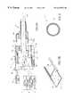
- FIG. 1is a pictorial view of an ophthalmic endoscope with an endoscope attachment for use therewith in accordance with the present invention
- FIG. 2is a front view of the endoscope of FIG. 1;
- FIG. 3Ais a longitudinal cross section of the ophthalmic endoscope of FIG. 1 along line A—A;
- FIG. 3Bis a close-up view up of an interchangeable dichroic mirror of the ophthalmic endoscope of FIG. 1;
- FIG. 4is a transverse cross section of the endoscope attachment of FIG. 1 along line B—B;
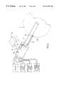
- FIG. 5is a pictorial view of the ophthalmic endoscope of FIG. 1 held and manipulated by a computer controlled XYZ manipulator;
- FIGS. 6-9are partially cut-away side views of the ophthalmic endoscope of FIG. 1 with different endoscope attachments;
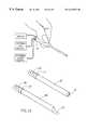
- FIG. 10is a perspective view of a conventional ophthalmic endoscope with an endoscope attachment in accordance with the present invention.
- FIG. 11is a close-up view showing the mode of attachment of an endoscope attachment to a conventional endoscope attachment.
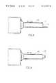
- FIGS. 12A-12Eare close-up views of various endoscope attachments of the present invention.
- an ophthalmic endoscope 1is adapted for use with an endoscope attachment 2 and for connection to a display 3 , an external light source 4 , an external laser source 6 and an external DC power source 7 .
- the endoscope 1has a handpiece 8 and a rigid probe 9 having a tip 11 and defining an optical axis 12 on which a CCD 13 is disposed for imaging a surgical site on the display 3 .
- the handpiece 8has an attachment interface 14 constituted by a major socket 16 from whose center the probe 9 extends and a minor socket 17 , the major socket 16 being adapted for securely engaging a proximal end 18 A of a tubular sleeve-like body member 18 of the endoscope attachment 2 and the minor socket 17 being adapted to receive a rearwardly directed tang 19 of the endoscope attachment 2 .
- the endoscope attachment's body member 18is shaped and dimensioned for tight sliding along the probe 9 and contains a fiber optic annulus 21 (see FIG. 4) which is coupled via a pin 22 received in a socket 23 connected to the external light source 4 for transmitting illumination light therealong for illuminating an eye's interior.
- the handpiece 8has a dichroic flip flop mirror 24 under the control of a hand operated external lever 26 for selectively enabling laser light from the external laser source 6 to be transmitted along the probe 9 when disposed along the optical axis 12 .
- the flip flop mirror 24is releasably held in a carriage 27 slidably mounted on tracks 28 such that the flip flop mirror 24 is interchangeable whereby different lasers can be employed
- the handpiece 8has a cog 29 driven by a motor 31 under the control of a hand operated rocker switch 32 for engaging a rack 33 formed on the tang's underside for selectively displacing the endoscope attachment 2 relative to the probe 9 along a stroke of about 3 mm whereby surgical procedures can be performed whilst maintaining the same field of view and image features.
- the ophthalmic endoscope 1has a sensing unit 34 for use with a computer controlled XYZ manipulator 35 (see FIG. 5) supporting the handpiece 8 by means of a support rod 36 .
- the sensing unit 34has a sensor 37 reciprocatingly extendable in a direction parallel to the optical axis 12 between a fully retracted position when it is substantially flush with the handpiece's front surface and a fully protruding position when it is substantially coextensive with the probe tip 11 .
- the sensor 37is initially fully extended by a motor 38 on depressing a pushbutton 39 and thereafter its tip 37 A is continuously urging against a portion of an eye wall A adjacent an ocular surgical opening B through which a surgical procedure is performed.
- the XYZ manipulator 35utilizes the distance of the sensor's tip 37 A from its fully retracted position as sensed by the sensing unit 34 for manipulating the endoscope 1 relative to the ocular surgical opening B in a manner similar to a ball and socket like universal socket.
- the ophthalmic endoscope 1is adapted for use with a number of different endoscope attachments 2 , the simplest attachment 40 (see FIG. 6) thereof being similar to the endoscope attachment 2 except without the tang 19 suitable for ocular examination and ocular laser intervention.
- Other attachments with a tang enabling selective displacement relative to the and including surgical associated means at their distal endsare as follows: An endoscope attachment 41 thereto (see FIG. 7) with an optical element 42 disposed along the optical axis 12 on its mounting suitable for ocular examination and ocular laser intervention.
- An endoscope attachment 43(see FIG. 8) with a surgical tip 44 extending beyond the probe tip 11 in a bayonet-like fashion on its mounting suitable for ocular surgical intervention.
- An endoscope attachment 46(see FIG. 9) with a cauterization tip 47 coupled to an electrical coupling 48 (see FIG. 2) connected to the external DC power source 8 extending beyond the probe tip 11 in a bayonet-like fashion suitable for ocular cauterization intervention.
- an endoscope attachment 49 with a surgical blade 51 or an endoscope attachment 52 with an optical element 53can be slidably mounted on aconventional ophthalmic endoscope 54 , each endoscope attachment 49 and 52 having a slit 56 transverse to its longitudinal axis whereby it secured to the endoscope's probe by means of an elastic member 57 circumscribing the probe surface exposed through the slit 56 and the probe's outer surface (see FIG. 11 ).
- the probe 9may include the fiber optic annulus 21 .
- a surgical tipcan include a pin point shaped blade (see FIG. 12 A), a crescent shaped blade (see FIG. 12B) and a round point shaped blade (see FIG. 12C) whilst an optical element can include a convex lens 16 for focusing purposes (see FIG. 12D) or a prism 18 for refracting purposes (see FIG. 12 E).
Landscapes
- Health & Medical Sciences (AREA)
- Life Sciences & Earth Sciences (AREA)
- Surgery (AREA)
- Public Health (AREA)
- Nuclear Medicine, Radiotherapy & Molecular Imaging (AREA)
- Optics & Photonics (AREA)
- Animal Behavior & Ethology (AREA)
- General Health & Medical Sciences (AREA)
- Veterinary Medicine (AREA)
- Engineering & Computer Science (AREA)
- Biomedical Technology (AREA)
- Heart & Thoracic Surgery (AREA)
- Physics & Mathematics (AREA)
- Medical Informatics (AREA)
- Molecular Biology (AREA)
- Radiology & Medical Imaging (AREA)
- Pathology (AREA)
- Biophysics (AREA)
- Ophthalmology & Optometry (AREA)
- Vascular Medicine (AREA)
- Endoscopes (AREA)
- Materials For Medical Uses (AREA)
- Instruments For Viewing The Inside Of Hollow Bodies (AREA)
- Prostheses (AREA)
Abstract
Description
The invention generally relates to ophthalmic surgical equipment in general and ophthalmic endoscopes in particular.
Ophthalmic surgical procedures typically require that two or even three surgical instruments are simultaneously deployed within the vicinity of a surgical site. The recommended diameter of an ocular surgical opening is about 1 mm which approximately corresponds to the diameter of each instrument and therefore normally two or three ocular surgical openings are required for an ophthalmic surgical procedure, each surgical opening enabling the insertion of a single instrument therethrough.
In U.S. Pat. No. 4,607,622 to Fritch, there is illustrated and described an ophthalmic endoscope provided with a probe formed with a conduit for accepting instruments, for example, a cutting device- for taking a sample (see Col. 3, lines 16-19) or fiber optics connected to an external laser source for ocular laser intervention However, in order to accommodate the conduit whilst maintaining the same endoscopic ability, the dimensions of the probe are necessarily greater tan those recommended for a surgical opening.
In accordance with a first aspect of the present invention, there is provided an ophthalmic endoscope for use with an endoscope attachment having a generally tubular body member, the endoscope comprising a rigid probe with an optical axis and a tip, and an attachment interface for releasably engaging the body member of an endoscope attachment slidingly mounted on said probe.
In accordance with a second aspect of the present invention, there is provided an endoscope attachment for use with an ophthalmic endoscope having a rigid probe with an optical axis and a tip, the endoscope attachment comprising a tubular body member for slidingly mounting on the probe.
Endoscope attachments of the present invention can be adapted for mounting on either a purpose built ophthalmic endoscope with an attachment interface or a conventional ophthalmic endoscope whereby a hitherto considered passive device is converted into a multi-purpose surgical device useful for a wide range of conventional and newly envisaged intraocular surgical procedures in various tissues of the eye. Mounting of an endoscope attachment on an ophthalmic endoscope's probe enables the imaging of a surgical procedure at high magnifications of up to 50× with an image resolution better than that obtained with an hitherto employed external imaging system i.e. an operating microscope.
For use with the purpose built ophthalmic endoscope of the present invention, the endoscope attachments preferably have a tubular body member coextensive with the endoscope probe when mounted thereon, the body member having longitudinally directed optic fibers for transmitting illumination light therealong for illumination of a surgical site. The overall width of the probe together with an endoscope attachment mounted thereon is about 1 mm for insertion through a conventional sized ocular surgical opening. In addition, an endoscope attachment can include surgery associated means typically disposed beyond the probe tip when mounted on the probe, the surgery associated means being implemented as an optical element for ocular examination, a surgical tip for ocular surgical intervention or a cauterization tip for ocular cauterization intervention. The ophthalmic endoscope of the present invention can be implemented for selectively displacing the surgical associated means relative to the probe along its optical axis whereby surgical procedures can be performed whilst maintaining the same field of view and image features. The ophthalmic endoscope of the present invention can be implemented for use with a computer controlled XYZ manipulator for facilitating magnifications greater than ×100.
In order to understand the invention and how it is used, preferred embodiments will now be described by way of non-limiting examples only, with reference to the accompanying drawing in which:
FIG. 1 is a pictorial view of an ophthalmic endoscope with an endoscope attachment for use therewith in accordance with the present invention;
FIG. 2 is a front view of the endoscope of FIG. 1;
FIG. 3A is a longitudinal cross section of the ophthalmic endoscope of FIG. 1 along line A—A;
FIG. 3B is a close-up view up of an interchangeable dichroic mirror of the ophthalmic endoscope of FIG. 1;
FIG. 4 is a transverse cross section of the endoscope attachment of FIG. 1 along line B—B;
FIG. 5 is a pictorial view of the ophthalmic endoscope of FIG. 1 held and manipulated by a computer controlled XYZ manipulator;
FIGS. 6-9 are partially cut-away side views of the ophthalmic endoscope of FIG. 1 with different endoscope attachments;
FIG. 10 is a perspective view of a conventional ophthalmic endoscope with an endoscope attachment in accordance with the present invention;
FIG. 11 is a close-up view showing the mode of attachment of an endoscope attachment to a conventional endoscope attachment; and
FIGS. 12A-12E are close-up views of various endoscope attachments of the present invention.
In FIGS. 1-3, an ophthalmic endoscope1 is adapted for use with anendoscope attachment 2 and for connection to adisplay 3, anexternal light source 4, anexternal laser source 6 and an external DC power source7. The endoscope1 has ahandpiece 8 and arigid probe 9 having atip 11 and defining anoptical axis 12 on which aCCD 13 is disposed for imaging a surgical site on thedisplay 3.
Thehandpiece 8 has anattachment interface 14 constituted by amajor socket 16 from whose center theprobe 9 extends and aminor socket 17, themajor socket 16 being adapted for securely engaging a proximal end18A of a tubular sleeve-like body member 18 of theendoscope attachment 2 and theminor socket 17 being adapted to receive a rearwardly directedtang 19 of theendoscope attachment 2. The endoscope attachment'sbody member 18 is shaped and dimensioned for tight sliding along theprobe 9 and contains a fiber optic annulus21 (see FIG. 4) which is coupled via apin 22 received in asocket 23 connected to theexternal light source 4 for transmitting illumination light therealong for illuminating an eye's interior.
Thehandpiece 8 has a dichroicflip flop mirror 24 under the control of a hand operatedexternal lever 26 for selectively enabling laser light from theexternal laser source 6 to be transmitted along theprobe 9 when disposed along theoptical axis 12. Theflip flop mirror 24 is releasably held in acarriage 27 slidably mounted ontracks 28 such that theflip flop mirror 24 is interchangeable whereby different lasers can be employed Thehandpiece 8 has acog 29 driven by amotor 31 under the control of a hand operatedrocker switch 32 for engaging arack 33 formed on the tang's underside for selectively displacing theendoscope attachment 2 relative to theprobe 9 along a stroke of about 3 mm whereby surgical procedures can be performed whilst maintaining the same field of view and image features.
The ophthalmic endoscope1 has asensing unit 34 for use with a computer controlled XYZ manipulator35 (see FIG. 5) supporting thehandpiece 8 by means of asupport rod 36. Thesensing unit 34 has asensor 37 reciprocatingly extendable in a direction parallel to theoptical axis 12 between a fully retracted position when it is substantially flush with the handpiece's front surface and a fully protruding position when it is substantially coextensive with theprobe tip 11. Thesensor 37 is initially fully extended by amotor 38 on depressing apushbutton 39 and thereafter itstip 37A is continuously urging against a portion of an eye wall A adjacent an ocular surgical opening B through which a surgical procedure is performed. During such a procedure, theXYZ manipulator 35 utilizes the distance of the sensor'stip 37A from its fully retracted position as sensed by thesensing unit 34 for manipulating the endoscope1 relative to the ocular surgical opening B in a manner similar to a ball and socket like universal socket.
The ophthalmic endoscope1 is adapted for use with a number ofdifferent endoscope attachments 2, the simplest attachment40 (see FIG. 6) thereof being similar to theendoscope attachment 2 except without thetang 19 suitable for ocular examination and ocular laser intervention. Other attachments with a tang enabling selective displacement relative to the and including surgical associated means at their distal ends are as follows: Anendoscope attachment 41 thereto (see FIG. 7) with anoptical element 42 disposed along theoptical axis 12 on its mounting suitable for ocular examination and ocular laser intervention. An endoscope attachment43 (see FIG. 8) with a surgical tip44 extending beyond theprobe tip 11 in a bayonet-like fashion on its mounting suitable for ocular surgical intervention. An endoscope attachment46 (see FIG. 9) with acauterization tip 47 coupled to an electrical coupling48 (see FIG. 2) connected to the externalDC power source 8 extending beyond theprobe tip 11 in a bayonet-like fashion suitable for ocular cauterization intervention.
In FIG. 10, anendoscope attachment 49 with asurgical blade 51 or anendoscope attachment 52 with anoptical element 53 can be slidably mounted on aconventionalophthalmic endoscope 54, eachendoscope attachment slit 56 transverse to its longitudinal axis whereby it secured to the endoscope's probe by means of anelastic member 57 circumscribing the probe surface exposed through theslit 56 and the probe's outer surface (see FIG.11).
Various modifications and changes may be made in the configuration described above that come within the spirit of the invention. The invention embraces all such changes and modifications coming within the scope of the appended claims. For example, different modes of attachment between the endoscope attachments and the endoscopes can be employed. In addition, theprobe 9 may include the fiberoptic annulus 21. Also, a surgical tip can include a pin point shaped blade (see FIG.12A), a crescent shaped blade (see FIG. 12B) and a round point shaped blade (see FIG. 12C) whilst an optical element can include aconvex lens 16 for focusing purposes (see FIG. 12D) or aprism 18 for refracting purposes (see FIG.12E).
Claims (26)
1. Ophthalmic endoscope comprising a rigid probe with an optical axis and a tip, for use with an endoscope attachment having generally tubular body member, said endoscope having engaging means adapted for slidingly mounting said attachment on said rigid probe so that, when so mounted, said body is co-extensive with said probe, and for releasably engaging the body member to provide selective displacement of said attachment on said probe.
2. An endoscope according toclaim 1 further comprising a dichroic optical element for selectively directing laser light along said optical axis.
3. An endoscope according toclaim 2 wherein said dichroic optical element is interchangeable.
4. An endoscope according toclaim 1 further comprising a light coupling for transmitting illumination light along an endoscope attachment for illumination of an eye's interior.
5. An endoscope according toclaim 1 , wherein said engaging means is motorized and is capable of providing said displacement within a distance enabling the use of said attachment whilst maintaining the endoscope's field of view.
6. An endoscope according toclaim 1 further comprising an electrical coupling for energizing a cauterization tip of an endoscope attachment for ocular cauterization intervention.
7. An endoscope according toclaim 1 further comprising a sensor selectively urgeable against an eye wall during an ophthalmic surgical procedure.
8. An endoscope according toclaim 7 wherein said sensor is extendable in a direction substantially parallel to said optical axis.
9. An endoscope according toclaim 1 wherein said rigid probe includes an optic core and a fiber optic annulus in a cross section thereof perpendicular to said probe axis.
10. An endoscope attachment for use with an ophthalmic endoscope having a rigid probe with an optical axis and a tip and having an attachment interface, the endoscope attachment comprising a generally tubular body member shaped and dimensioned for mounting on and tight sliding along said probe so as to be co-extensive with said probe when mounted thereon, and engaging means for engaging said attachment interface so as to enable selective displacement of said attachment along said probe.
11. An endoscope attachment according toclaim 10 wherein said body member has longitudinally directed fiber optics for transmitting illumination light therealong for illumination of an eye's interior, when the attachment is mounted on said probe.
12. An endoscope attachment according toclaim 10 further comprising surgery associated means disposed beyond the probe tip when said attachment is mounted on the probe.
13. An endoscope attachment according toclaim 12 wherein said surgery associated means includes an optical element for ocular examination.
14. An endoscope attachment according toclaim 12 wherein said surgery associated means includes a surgical tip for ocular surgical intervention.
15. An endoscope attachment according toclaim 12 wherein said surgery associated means includes a cauterization tip for ocular cauterization intervention.
16. A multi-purpose ophthalmic endoscope system comprising an ophthalmic endoscope having a rigid probe with an optical axis and a tip, and a plurality of different endoscope attachments each having a generally tubular body member slidingly mountable on said rigid probe so as to be co-extensive therewith, said endoscope and said attachments having engaging means for releasably interacting with each other in such a manner as to enable selective displacement of each attachment on said probe.
17. A multi-purpose ophthalmic endoscope system according toclaim 16 , wherein said engaging means is motorized.
18. A multi-purpose ophthalmic endoscope system according toclaim 16 , wherein said engaging means is adapted to provide said displacement within a distance enabling the use of said attachment whilst maintaining the endoscope's field of view.
19. A multi-purpose ophthalmic endoscope system according toclaim 16 , wherein at least one of said attachments has an operative tip at a distal end of said body member, selected from a group consisting of an optical element for ocular examination and ocular, laser intervention, a surgical tip for ocular surgical intervention, and a cauterization tip for ocular cauterization intervention.
20. A multi-purpose ophthalmic endoscope system according toclaim 19 , wherein said attachments are adapted for mounting on said probe so as to dispose their operative tips in the vicinity of the tip of said probe.
21. A multi-purpose ophthalmic endoscope system according toclaim 20 , wherein at least one of said attachments is adapted for mounting on said probe so as to dispose its operative tip beyond the tip of said probe.
22. An ophthalmic endoscope comprising a rigid probe with an optical axis and a tip, for use with an endoscope attachment having generally tubular body member adapted to be slidingly mounted on said rigid probe, said endoscope having engaging means for releasably engaging said body member and for selectively displacing said attachment on said probe within a distance enabling the use of said attachment whilst maintaining the endoscope's field of view.
23. An ophthalmic endoscope system comprising an ophthalmic endoscope having a rigid probe with an optical axis and a tip, and at least one endoscope attachment having a generally tubular body member adapted to be slidingly mounted on said rigid probe, said endoscope and said attachment having engaging means adapted to interact with each other so as to provide for selective displacement of said attachment on said probe within a distance enabling the use of said attachment whilst maintaining the endoscope's field of view.
24. An ophthalmic endoscope according toclaim 23 , comprising a plurality of the endoscope attachments.
25. An ophthalmic endoscope system according toclaim 23 , wherein said attachment has an operative tip at a distal end thereof, selected from a group consisting of an optical element for ocular examination and ocular laser intervention, a surgical tip for ocular surgical intervention, and a cauterization tip for ocular cauterization intervention.
26. An ophthalmic endoscope system according toclaim 23 , wherein said attachment is adapted for mounting on said probe so as to dispose an operative tip thereof beyond the tip of said probe.
Applications Claiming Priority (3)
| Application Number | Priority Date | Filing Date | Title |
|---|---|---|---|
| IL12145097AIL121450A0 (en) | 1997-08-01 | 1997-08-01 | Ophthalmic surgical equipment |
| IL121450 | 1997-08-01 | ||
| PCT/IL1998/000362WO1999005997A1 (en) | 1997-08-01 | 1998-07-31 | Ophthalmic endoscope and endoscope attachments for use therewith |
Publications (1)
| Publication Number | Publication Date |
|---|---|
| US6419627B1true US6419627B1 (en) | 2002-07-16 |
Family
ID=11070457
Family Applications (1)
| Application Number | Title | Priority Date | Filing Date |
|---|---|---|---|
| US09/463,711Expired - LifetimeUS6419627B1 (en) | 1997-08-01 | 1998-07-31 | Ophthalmic endoscope and endoscope attachments for use therewith |
Country Status (9)
| Country | Link |
|---|---|
| US (1) | US6419627B1 (en) |
| EP (1) | EP0999809B1 (en) |
| JP (1) | JP4328464B2 (en) |
| AT (1) | ATE287250T1 (en) |
| AU (1) | AU729666B2 (en) |
| CA (1) | CA2298657A1 (en) |
| DE (1) | DE69828695T2 (en) |
| IL (1) | IL121450A0 (en) |
| WO (1) | WO1999005997A1 (en) |
Cited By (34)
| Publication number | Priority date | Publication date | Assignee | Title |
|---|---|---|---|---|
| DE10315134A1 (en)* | 2003-04-03 | 2004-10-14 | Schölly Fiberoptic GmbH | Auxiliary instrument for carrying out examinations/treatment on an eye includes an endoscope to be inserted into the eye's interior |
| EP1512366A1 (en)* | 2003-09-05 | 2005-03-09 | Knit Ventures Llc | Ophthalmic endoscope |
| US20050209618A1 (en)* | 2004-03-05 | 2005-09-22 | Auld Michael D | Rigid shafted instrumentation for vitreoretinal surgery |
| NL1026121C2 (en)* | 2004-05-06 | 2005-11-08 | Petrus Marinus Johanne Heyboer | Apparatus for optical observation and / or investigation of more difficult places and / or to search at more difficult places with the possibility of carrying out work in those places, in particular for household use and personal care. |
| US20060135957A1 (en)* | 2004-12-21 | 2006-06-22 | Dorin Panescu | Method and apparatus to align a probe with a cornea |
| US20060167340A1 (en)* | 2005-01-10 | 2006-07-27 | Pease Alfred A | Optical snake |
| US20070179430A1 (en)* | 2005-12-16 | 2007-08-02 | Smith Ronald T | Illuminated infusion cannula |
| US20090225159A1 (en)* | 2008-03-07 | 2009-09-10 | Scott Schneider | Visual inspection device |
| US20090229842A1 (en)* | 2008-03-07 | 2009-09-17 | Rick Gray | Battery pack for use with a power tool and a non-motorized sensing tool |
| WO2011035125A1 (en)* | 2009-09-17 | 2011-03-24 | Wesley Carlos K | Hair restoration surgery |
| US8277418B2 (en) | 2009-12-23 | 2012-10-02 | Alcon Research, Ltd. | Ophthalmic valved trocar cannula |
| US8343106B2 (en) | 2009-12-23 | 2013-01-01 | Alcon Research, Ltd. | Ophthalmic valved trocar vent |
| US20130110003A1 (en)* | 2011-10-27 | 2013-05-02 | Cook Medical Technologies Llc | Visualization catheter periscope |
| WO2013177611A1 (en)* | 2012-05-30 | 2013-12-05 | Ellex R&D Pty Ltd | Reflex coaxial illuminator |
| US8998931B2 (en) | 2011-10-17 | 2015-04-07 | Pilofocus, Inc. | Hair restoration |
| US9314082B2 (en) | 2009-09-17 | 2016-04-19 | Pilofocus, Inc. | System and method for extraction of hair follicle |
| US9343489B2 (en) | 2011-05-12 | 2016-05-17 | DePuy Synthes Products, Inc. | Image sensor for endoscopic use |
| US9370295B2 (en) | 2014-01-13 | 2016-06-21 | Trice Medical, Inc. | Fully integrated, disposable tissue visualization device |
| US9462234B2 (en)* | 2012-07-26 | 2016-10-04 | DePuy Synthes Products, Inc. | Camera system with minimal area monolithic CMOS image sensor |
| WO2017062759A1 (en) | 2015-10-09 | 2017-04-13 | Vasoptic Medical, Inc. | System and method for rapid examination of vasculature and particulate flow using laser speckle contrast imaging |
| US9693799B2 (en) | 2009-09-17 | 2017-07-04 | Pilofocus, Inc. | System and method for aligning hair follicle |
| US10045686B2 (en) | 2008-11-12 | 2018-08-14 | Trice Medical, Inc. | Tissue visualization and modification device |
| US10226167B2 (en) | 2010-05-13 | 2019-03-12 | Beaver-Visitec International, Inc. | Laser video endoscope |
| US10342579B2 (en) | 2014-01-13 | 2019-07-09 | Trice Medical, Inc. | Fully integrated, disposable tissue visualization device |
| US10405886B2 (en) | 2015-08-11 | 2019-09-10 | Trice Medical, Inc. | Fully integrated, disposable tissue visualization device |
| CN110367914A (en)* | 2019-08-14 | 2019-10-25 | 上海懿熹医疗科技有限公司 | A kind of endoscope of suitable eye Intraorbital operation |
| US10517469B2 (en) | 2013-03-15 | 2019-12-31 | DePuy Synthes Products, Inc. | Image sensor synchronization without input clock and data transmission clock |
| US10750933B2 (en) | 2013-03-15 | 2020-08-25 | DePuy Synthes Products, Inc. | Minimize image sensor I/O and conductor counts in endoscope applications |
| US20200352433A1 (en)* | 2016-06-30 | 2020-11-12 | Iridex Corporation | Handheld Ophthalmic Laser System With Replaceable Contact Tips and Treatment Guide |
| US11337598B2 (en) | 2010-05-13 | 2022-05-24 | Beaver-Visitec International, Inc. | Laser video endoscope |
| US11547446B2 (en) | 2014-01-13 | 2023-01-10 | Trice Medical, Inc. | Fully integrated, disposable tissue visualization device |
| US11622753B2 (en) | 2018-03-29 | 2023-04-11 | Trice Medical, Inc. | Fully integrated endoscope with biopsy capabilities and methods of use |
| CN116687664A (en)* | 2023-08-03 | 2023-09-05 | 北京同仁医学科技有限责任公司 | Meibomian gland probe |
| US12035889B2 (en) | 2008-07-22 | 2024-07-16 | Trice Medical, Inc. | Tissue modification devices and methods of using the same |
Families Citing this family (5)
| Publication number | Priority date | Publication date | Assignee | Title |
|---|---|---|---|---|
| US6278685B1 (en)* | 1999-08-19 | 2001-08-21 | Intellon Corporation | Robust transmission mode |
| US8038602B2 (en)* | 2001-10-19 | 2011-10-18 | Visionscope Llc | Portable imaging system employing a miniature endoscope |
| WO2007053590A1 (en)* | 2005-10-31 | 2007-05-10 | Alcon, Inc. | Extending small-gauge illuminator |
| EP1992277A1 (en)* | 2007-05-14 | 2008-11-19 | Institut National De La Sante Et De La Recherche Medicale (Inserm) | Optical device and method for acquiring images of eye structures |
| JP2018538069A (en)* | 2015-12-11 | 2018-12-27 | ビーバー−ビジテック インターナショナル インコーポレイテッド | Laser video endoscope |
Citations (20)
| Publication number | Priority date | Publication date | Assignee | Title |
|---|---|---|---|---|
| DE2810879A1 (en)* | 1977-03-14 | 1978-10-05 | Spectra Med | DEVICE AND METHOD FOR CAUTERIZING BLOOD VESSELS AND OTHER BIOLOGICAL TISSUE |
| US4607622A (en)* | 1985-04-11 | 1986-08-26 | Charles D. Fritch | Fiber optic ocular endoscope |
| US4825259A (en)* | 1988-02-03 | 1989-04-25 | Berry Jr Robert F | Adapter tip for remote measuring device |
| US4905082A (en)* | 1987-05-06 | 1990-02-27 | Olympus Optical Co., Ltd. | Rigid video endoscope having a detachable imaging unit |
| US5121740A (en)* | 1991-05-06 | 1992-06-16 | Martin Uram | Laser video endoscope |
| US5334183A (en)* | 1985-08-28 | 1994-08-02 | Valleylab, Inc. | Endoscopic electrosurgical apparatus |
| US5335648A (en)* | 1992-04-30 | 1994-08-09 | Hoya Corporation | Entoptoscopic instrument for observing the interior of an eye |
| US5396366A (en)* | 1993-03-04 | 1995-03-07 | Sigma Dynamics Corporation | Endoscope apparatus |
| US5402768A (en)* | 1992-09-01 | 1995-04-04 | Adair; Edwin L. | Endoscope with reusable core and disposable sheath with passageways |
| DE29507925U1 (en)* | 1994-06-17 | 1995-07-20 | Fa. Carl Zeiss, 89518 Heidenheim | Applicator for the treatment of increased intraocular pressure using laser radiation |
| US5441496A (en)* | 1993-04-15 | 1995-08-15 | Infinitech, Inc. | Laser delivery system with soft tip |
| US5573493A (en)* | 1993-10-08 | 1996-11-12 | United States Surgical Corporation | Endoscope attachment for changing angle of view |
| US5575754A (en)* | 1995-02-24 | 1996-11-19 | Olympus Optical Co., Ltd. | Endoscopic apparatus for three dimensional instrumentation |
| US5632740A (en)* | 1991-01-30 | 1997-05-27 | Ceram Optec Industries, Inc. | Illuminated leading probe device |
| US5651783A (en)* | 1995-12-20 | 1997-07-29 | Reynard; Michael | Fiber optic sleeve for surgical instruments |
| US5879289A (en)* | 1996-07-15 | 1999-03-09 | Universal Technologies International, Inc. | Hand-held portable endoscopic camera |
| US5935141A (en)* | 1997-10-30 | 1999-08-10 | Partisan Management Group | Interventional cardiology instrument controlled from an intracoronary reference |
| US6004263A (en)* | 1996-03-13 | 1999-12-21 | Hihon Kohden Corporation | Endoscope with detachable operation unit and insertion unit |
| US6086530A (en)* | 1998-10-30 | 2000-07-11 | Mack; Michael | Adjustable sleeve for endoscopes |
| US6203493B1 (en)* | 1996-02-15 | 2001-03-20 | Biosense, Inc. | Attachment with one or more sensors for precise position determination of endoscopes |
- 1997
- 1997-08-01ILIL12145097Apatent/IL121450A0/enunknown
- 1998
- 1998-07-31CACA002298657Apatent/CA2298657A1/ennot_activeAbandoned
- 1998-07-31DEDE69828695Tpatent/DE69828695T2/ennot_activeExpired - Lifetime
- 1998-07-31WOPCT/IL1998/000362patent/WO1999005997A1/enactiveIP Right Grant
- 1998-07-31AUAU85588/98Apatent/AU729666B2/ennot_activeCeased
- 1998-07-31EPEP98936653Apatent/EP0999809B1/ennot_activeExpired - Lifetime
- 1998-07-31USUS09/463,711patent/US6419627B1/ennot_activeExpired - Lifetime
- 1998-07-31ATAT98936653Tpatent/ATE287250T1/ennot_activeIP Right Cessation
- 1998-07-31JPJP2000504818Apatent/JP4328464B2/ennot_activeExpired - Fee Related
Patent Citations (21)
| Publication number | Priority date | Publication date | Assignee | Title |
|---|---|---|---|---|
| DE2810879A1 (en)* | 1977-03-14 | 1978-10-05 | Spectra Med | DEVICE AND METHOD FOR CAUTERIZING BLOOD VESSELS AND OTHER BIOLOGICAL TISSUE |
| US4607622A (en)* | 1985-04-11 | 1986-08-26 | Charles D. Fritch | Fiber optic ocular endoscope |
| US5334183A (en)* | 1985-08-28 | 1994-08-02 | Valleylab, Inc. | Endoscopic electrosurgical apparatus |
| US4905082A (en)* | 1987-05-06 | 1990-02-27 | Olympus Optical Co., Ltd. | Rigid video endoscope having a detachable imaging unit |
| US4825259A (en)* | 1988-02-03 | 1989-04-25 | Berry Jr Robert F | Adapter tip for remote measuring device |
| US5632740A (en)* | 1991-01-30 | 1997-05-27 | Ceram Optec Industries, Inc. | Illuminated leading probe device |
| US5121740A (en)* | 1991-05-06 | 1992-06-16 | Martin Uram | Laser video endoscope |
| US5335648A (en)* | 1992-04-30 | 1994-08-09 | Hoya Corporation | Entoptoscopic instrument for observing the interior of an eye |
| US5402768A (en)* | 1992-09-01 | 1995-04-04 | Adair; Edwin L. | Endoscope with reusable core and disposable sheath with passageways |
| US5396366A (en)* | 1993-03-04 | 1995-03-07 | Sigma Dynamics Corporation | Endoscope apparatus |
| US5441496A (en)* | 1993-04-15 | 1995-08-15 | Infinitech, Inc. | Laser delivery system with soft tip |
| US5573493A (en)* | 1993-10-08 | 1996-11-12 | United States Surgical Corporation | Endoscope attachment for changing angle of view |
| US5514125A (en)* | 1994-06-17 | 1996-05-07 | Carl-Zeiss-Stiftung | Applicator for the treatment of an elevated internal ocular pressure by means of laser radiation |
| DE29507925U1 (en)* | 1994-06-17 | 1995-07-20 | Fa. Carl Zeiss, 89518 Heidenheim | Applicator for the treatment of increased intraocular pressure using laser radiation |
| US5575754A (en)* | 1995-02-24 | 1996-11-19 | Olympus Optical Co., Ltd. | Endoscopic apparatus for three dimensional instrumentation |
| US5651783A (en)* | 1995-12-20 | 1997-07-29 | Reynard; Michael | Fiber optic sleeve for surgical instruments |
| US6203493B1 (en)* | 1996-02-15 | 2001-03-20 | Biosense, Inc. | Attachment with one or more sensors for precise position determination of endoscopes |
| US6004263A (en)* | 1996-03-13 | 1999-12-21 | Hihon Kohden Corporation | Endoscope with detachable operation unit and insertion unit |
| US5879289A (en)* | 1996-07-15 | 1999-03-09 | Universal Technologies International, Inc. | Hand-held portable endoscopic camera |
| US5935141A (en)* | 1997-10-30 | 1999-08-10 | Partisan Management Group | Interventional cardiology instrument controlled from an intracoronary reference |
| US6086530A (en)* | 1998-10-30 | 2000-07-11 | Mack; Michael | Adjustable sleeve for endoscopes |
Cited By (93)
| Publication number | Priority date | Publication date | Assignee | Title |
|---|---|---|---|---|
| DE10315134A1 (en)* | 2003-04-03 | 2004-10-14 | Schölly Fiberoptic GmbH | Auxiliary instrument for carrying out examinations/treatment on an eye includes an endoscope to be inserted into the eye's interior |
| EP1512366A1 (en)* | 2003-09-05 | 2005-03-09 | Knit Ventures Llc | Ophthalmic endoscope |
| US20050209618A1 (en)* | 2004-03-05 | 2005-09-22 | Auld Michael D | Rigid shafted instrumentation for vitreoretinal surgery |
| WO2005086772A3 (en)* | 2004-03-05 | 2009-04-09 | Michael D Auld | Rigid shafted instrumentation for vitreoretinal surgery |
| NL1026121C2 (en)* | 2004-05-06 | 2005-11-08 | Petrus Marinus Johanne Heyboer | Apparatus for optical observation and / or investigation of more difficult places and / or to search at more difficult places with the possibility of carrying out work in those places, in particular for household use and personal care. |
| US20060135957A1 (en)* | 2004-12-21 | 2006-06-22 | Dorin Panescu | Method and apparatus to align a probe with a cornea |
| WO2006069004A3 (en)* | 2004-12-21 | 2006-10-26 | Refractec Inc | Method and apparatus to align a probe with a cornea |
| US7758495B2 (en)* | 2005-01-10 | 2010-07-20 | Perceptron, Inc. | Remote inspection device |
| US20060167340A1 (en)* | 2005-01-10 | 2006-07-27 | Pease Alfred A | Optical snake |
| US20070179430A1 (en)* | 2005-12-16 | 2007-08-02 | Smith Ronald T | Illuminated infusion cannula |
| US7783346B2 (en)* | 2005-12-16 | 2010-08-24 | Alcon, Inc. | Illuminated infusion cannula |
| US9196881B2 (en) | 2008-03-07 | 2015-11-24 | Milwaukee Electric Tool Corporation | Battery pack for use with a power tool and a non-motorized sensing tool |
| US8988522B2 (en)* | 2008-03-07 | 2015-03-24 | Milwaukee Electric Tool Corporation | Visual inspection device |
| US20090225159A1 (en)* | 2008-03-07 | 2009-09-10 | Scott Schneider | Visual inspection device |
| US8189043B2 (en)* | 2008-03-07 | 2012-05-29 | Milwaukee Electric Tool Corporation | Hand-held visual inspection device for viewing confined or difficult to access locations |
| US8251157B2 (en) | 2008-03-07 | 2012-08-28 | Milwaukee Electric Tool Corporation | Battery pack for use with a power tool and a non-motorized sensing tool |
| US20120224047A1 (en)* | 2008-03-07 | 2012-09-06 | Scott Schneider | Visual inspection device |
| US9986212B2 (en)* | 2008-03-07 | 2018-05-29 | Milwaukee Electric Tool Corporation | Visual inspection device |
| US20170295347A1 (en)* | 2008-03-07 | 2017-10-12 | Milwaukee Electric Tool Corporation | Visual inspection device |
| US9693024B2 (en)* | 2008-03-07 | 2017-06-27 | Milwaukee Electric Tool Corporation | Visual inspection device |
| US20090229842A1 (en)* | 2008-03-07 | 2009-09-17 | Rick Gray | Battery pack for use with a power tool and a non-motorized sensing tool |
| US8659652B2 (en)* | 2008-03-07 | 2014-02-25 | Milwaukee Electric Tool Corporation | Visual inspection device |
| US20150201170A1 (en)* | 2008-03-07 | 2015-07-16 | Milwaukee Electric Tool Corporation | Visual inspection device |
| US20140160268A1 (en)* | 2008-03-07 | 2014-06-12 | Milwaukee Electric Tool Corporation | Visual inspection device |
| US8851200B2 (en) | 2008-03-07 | 2014-10-07 | Milwaukee Electric Tool Corporation | Battery pack for use with a power tool and a non-motorized sensing tool |
| US12035889B2 (en) | 2008-07-22 | 2024-07-16 | Trice Medical, Inc. | Tissue modification devices and methods of using the same |
| US10045686B2 (en) | 2008-11-12 | 2018-08-14 | Trice Medical, Inc. | Tissue visualization and modification device |
| US9693799B2 (en) | 2009-09-17 | 2017-07-04 | Pilofocus, Inc. | System and method for aligning hair follicle |
| US9314082B2 (en) | 2009-09-17 | 2016-04-19 | Pilofocus, Inc. | System and method for extraction of hair follicle |
| WO2011035125A1 (en)* | 2009-09-17 | 2011-03-24 | Wesley Carlos K | Hair restoration surgery |
| US9364252B2 (en) | 2009-09-17 | 2016-06-14 | Pilofocus, Inc. | Hair restoration surgery |
| US8679064B2 (en) | 2009-12-23 | 2014-03-25 | Alcon Research, Ltd. | Ophthalmic valved trocar cannula |
| US8277418B2 (en) | 2009-12-23 | 2012-10-02 | Alcon Research, Ltd. | Ophthalmic valved trocar cannula |
| US8343106B2 (en) | 2009-12-23 | 2013-01-01 | Alcon Research, Ltd. | Ophthalmic valved trocar vent |
| US10226167B2 (en) | 2010-05-13 | 2019-03-12 | Beaver-Visitec International, Inc. | Laser video endoscope |
| US11337598B2 (en) | 2010-05-13 | 2022-05-24 | Beaver-Visitec International, Inc. | Laser video endoscope |
| US11179029B2 (en) | 2011-05-12 | 2021-11-23 | DePuy Synthes Products, Inc. | Image sensor with tolerance optimizing interconnects |
| US9343489B2 (en) | 2011-05-12 | 2016-05-17 | DePuy Synthes Products, Inc. | Image sensor for endoscopic use |
| US11682682B2 (en) | 2011-05-12 | 2023-06-20 | DePuy Synthes Products, Inc. | Pixel array area optimization using stacking scheme for hybrid image sensor with minimal vertical interconnects |
| US9763566B2 (en) | 2011-05-12 | 2017-09-19 | DePuy Synthes Products, Inc. | Pixel array area optimization using stacking scheme for hybrid image sensor with minimal vertical interconnects |
| US11848337B2 (en) | 2011-05-12 | 2023-12-19 | DePuy Synthes Products, Inc. | Image sensor |
| US10537234B2 (en) | 2011-05-12 | 2020-01-21 | DePuy Synthes Products, Inc. | Image sensor with tolerance optimizing interconnects |
| US9907459B2 (en) | 2011-05-12 | 2018-03-06 | DePuy Synthes Products, Inc. | Image sensor with tolerance optimizing interconnects |
| US9980633B2 (en) | 2011-05-12 | 2018-05-29 | DePuy Synthes Products, Inc. | Image sensor for endoscopic use |
| US10863894B2 (en) | 2011-05-12 | 2020-12-15 | DePuy Synthes Products, Inc. | System and method for sub-column parallel digitizers for hybrid stacked image sensor using vertical interconnects |
| US12100716B2 (en) | 2011-05-12 | 2024-09-24 | DePuy Synthes Products, Inc. | Image sensor with tolerance optimizing interconnects |
| US11432715B2 (en) | 2011-05-12 | 2022-09-06 | DePuy Synthes Products, Inc. | System and method for sub-column parallel digitizers for hybrid stacked image sensor using vertical interconnects |
| US10517471B2 (en) | 2011-05-12 | 2019-12-31 | DePuy Synthes Products, Inc. | Pixel array area optimization using stacking scheme for hybrid image sensor with minimal vertical interconnects |
| US10709319B2 (en) | 2011-05-12 | 2020-07-14 | DePuy Synthes Products, Inc. | System and method for sub-column parallel digitizers for hybrid stacked image sensor using vertical interconnects |
| US11026565B2 (en) | 2011-05-12 | 2021-06-08 | DePuy Synthes Products, Inc. | Image sensor for endoscopic use |
| US11109750B2 (en) | 2011-05-12 | 2021-09-07 | DePuy Synthes Products, Inc. | Pixel array area optimization using stacking scheme for hybrid image sensor with minimal vertical interconnects |
| US8998931B2 (en) | 2011-10-17 | 2015-04-07 | Pilofocus, Inc. | Hair restoration |
| US9861386B2 (en) | 2011-10-17 | 2018-01-09 | Pilofocus, Inc. | Hair restoration |
| US9119531B2 (en)* | 2011-10-27 | 2015-09-01 | Cook Medical Technologies Llc | Visualization catheter periscope |
| US20130110003A1 (en)* | 2011-10-27 | 2013-05-02 | Cook Medical Technologies Llc | Visualization catheter periscope |
| US10369051B2 (en) | 2012-05-30 | 2019-08-06 | Ellex R&D Pty Ltd | Reflex coaxial illuminator |
| US10898378B2 (en) | 2012-05-30 | 2021-01-26 | Ellex Medical Pty Ltd | Reflex coaxial illuminator |
| WO2013177611A1 (en)* | 2012-05-30 | 2013-12-05 | Ellex R&D Pty Ltd | Reflex coaxial illuminator |
| US10075626B2 (en) | 2012-07-26 | 2018-09-11 | DePuy Synthes Products, Inc. | Camera system with minimal area monolithic CMOS image sensor |
| US11089192B2 (en) | 2012-07-26 | 2021-08-10 | DePuy Synthes Products, Inc. | Camera system with minimal area monolithic CMOS image sensor |
| US9462234B2 (en)* | 2012-07-26 | 2016-10-04 | DePuy Synthes Products, Inc. | Camera system with minimal area monolithic CMOS image sensor |
| US11766175B2 (en) | 2012-07-26 | 2023-09-26 | DePuy Synthes Products, Inc. | Camera system with minimal area monolithic CMOS image sensor |
| US10701254B2 (en) | 2012-07-26 | 2020-06-30 | DePuy Synthes Products, Inc. | Camera system with minimal area monolithic CMOS image sensor |
| US11344189B2 (en) | 2013-03-15 | 2022-05-31 | DePuy Synthes Products, Inc. | Image sensor synchronization without input clock and data transmission clock |
| US10517469B2 (en) | 2013-03-15 | 2019-12-31 | DePuy Synthes Products, Inc. | Image sensor synchronization without input clock and data transmission clock |
| US12150620B2 (en) | 2013-03-15 | 2024-11-26 | DePuy Synthes Products, Inc. | Minimize image sensor I/O and conductor counts in endoscope applications |
| US11903564B2 (en) | 2013-03-15 | 2024-02-20 | DePuy Synthes Products, Inc. | Image sensor synchronization without input clock and data transmission clock |
| US10980406B2 (en) | 2013-03-15 | 2021-04-20 | DePuy Synthes Products, Inc. | Image sensor synchronization without input clock and data transmission clock |
| US10750933B2 (en) | 2013-03-15 | 2020-08-25 | DePuy Synthes Products, Inc. | Minimize image sensor I/O and conductor counts in endoscope applications |
| US11253139B2 (en) | 2013-03-15 | 2022-02-22 | DePuy Synthes Products, Inc. | Minimize image sensor I/O and conductor counts in endoscope applications |
| US10881272B2 (en) | 2013-03-15 | 2021-01-05 | DePuy Synthes Products, Inc. | Minimize image sensor I/O and conductor counts in endoscope applications |
| US10398298B2 (en) | 2014-01-13 | 2019-09-03 | Trice Medical, Inc. | Fully integrated, disposable tissue visualization device |
| US10342579B2 (en) | 2014-01-13 | 2019-07-09 | Trice Medical, Inc. | Fully integrated, disposable tissue visualization device |
| US10092176B2 (en) | 2014-01-13 | 2018-10-09 | Trice Medical, Inc. | Fully integrated, disposable tissue visualization device |
| US9370295B2 (en) | 2014-01-13 | 2016-06-21 | Trice Medical, Inc. | Fully integrated, disposable tissue visualization device |
| US11547446B2 (en) | 2014-01-13 | 2023-01-10 | Trice Medical, Inc. | Fully integrated, disposable tissue visualization device |
| US9610007B2 (en) | 2014-01-13 | 2017-04-04 | Trice Medical, Inc. | Fully integrated, disposable tissue visualization device |
| US10405886B2 (en) | 2015-08-11 | 2019-09-10 | Trice Medical, Inc. | Fully integrated, disposable tissue visualization device |
| US10945588B2 (en) | 2015-08-11 | 2021-03-16 | Trice Medical, Inc. | Fully integrated, disposable tissue visualization device |
| EP4235268A3 (en)* | 2015-10-09 | 2023-10-04 | Vasoptic Medical, Inc. | System and method for rapid examination of vasculature and particulate flow using laser speckle contrast imaging |
| CN108430306A (en)* | 2015-10-09 | 2018-08-21 | 瓦索普蒂奇医疗公司 | System and method for using laser speckle contrast Imaging fast to check vascular system and particle stream |
| US11666238B2 (en) | 2015-10-09 | 2023-06-06 | Vasoptic Medical Inc. | System and method for rapid examination of vasculature and particulate flow using laser speckle contrast imaging |
| WO2017062759A1 (en) | 2015-10-09 | 2017-04-13 | Vasoptic Medical, Inc. | System and method for rapid examination of vasculature and particulate flow using laser speckle contrast imaging |
| US11020015B2 (en) | 2015-10-09 | 2021-06-01 | Vasoptic Medical, Inc. | System and method for rapid examination of vasculature and particulate flow using laser speckle contrast imaging |
| AU2016335761B2 (en)* | 2015-10-09 | 2020-12-03 | Vasoptic Medical, Inc. | System and method for rapid examination of vasculature and particulate flow using laser speckle contrast imaging |
| US11576569B2 (en)* | 2016-06-30 | 2023-02-14 | Iridex Corporation | Handheld ophthalmic laser system with replaceable contact tips and treatment guide |
| US20230141548A1 (en)* | 2016-06-30 | 2023-05-11 | Iridex Corporation | Handheld Ophthalmic Laser System With Replaceable Contact Tips and Treatment Guide |
| US20200352433A1 (en)* | 2016-06-30 | 2020-11-12 | Iridex Corporation | Handheld Ophthalmic Laser System With Replaceable Contact Tips and Treatment Guide |
| US12167891B2 (en)* | 2016-06-30 | 2024-12-17 | Iridex Corporation | Handheld ophthalmic laser system with replaceable contact tips and treatment guide |
| US11622753B2 (en) | 2018-03-29 | 2023-04-11 | Trice Medical, Inc. | Fully integrated endoscope with biopsy capabilities and methods of use |
| CN110367914A (en)* | 2019-08-14 | 2019-10-25 | 上海懿熹医疗科技有限公司 | A kind of endoscope of suitable eye Intraorbital operation |
| CN116687664B (en)* | 2023-08-03 | 2023-12-19 | 北京同仁医学科技有限责任公司 | A meibomian gland probe needle |
| CN116687664A (en)* | 2023-08-03 | 2023-09-05 | 北京同仁医学科技有限责任公司 | Meibomian gland probe |
Also Published As
| Publication number | Publication date |
|---|---|
| CA2298657A1 (en) | 1999-02-11 |
| DE69828695T2 (en) | 2006-01-05 |
| DE69828695D1 (en) | 2005-02-24 |
| IL121450A0 (en) | 1998-02-08 |
| AU729666B2 (en) | 2001-02-08 |
| WO1999005997A1 (en) | 1999-02-11 |
| EP0999809A1 (en) | 2000-05-17 |
| JP4328464B2 (en) | 2009-09-09 |
| ATE287250T1 (en) | 2005-02-15 |
| JP2001511393A (en) | 2001-08-14 |
| EP0999809B1 (en) | 2005-01-19 |
| AU8558898A (en) | 1999-02-22 |
Similar Documents
| Publication | Publication Date | Title |
|---|---|---|
| US6419627B1 (en) | Ophthalmic endoscope and endoscope attachments for use therewith | |
| US9867531B2 (en) | Video retractor | |
| AU643889B2 (en) | Laser video endoscope | |
| US6743168B2 (en) | Endoscope system and method of use | |
| US5512034A (en) | Surgical instrument including viewing optics and a ball probe | |
| US5121740A (en) | Laser video endoscope | |
| US5156604A (en) | Small probing hook for arthroscopy | |
| JPS58501500A (en) | Endoscopes and surgical instruments for use with endoscopes | |
| EP2449953B1 (en) | Mirrored arthroscope | |
| DE69730426D1 (en) | SURGICAL INSTRUMENT WITH VIEWING OPTICS AND ATRAUMATIC PROBE | |
| WO2001072200A3 (en) | Resectoscope | |
| US7060028B2 (en) | Endoilluminator | |
| US20050222493A1 (en) | Endoscope | |
| US20090088601A1 (en) | Endoscope and endoscopy method | |
| WO2005048817A3 (en) | Illuminated laser probe with adjustble area of illumination | |
| US20080275435A1 (en) | Illuminated Laser Probe with Multiplied Area of Illumination | |
| US5961441A (en) | Surgical otoscope and method | |
| WO1995020341A1 (en) | Endoscope with insertable tool | |
| JP2000245740A (en) | Treating tool for enoscope | |
| EP0638279A1 (en) | Endoscope with retractable tool | |
| JP2005537901A (en) | Ablation mirror with removable outer shaft | |
| JP4222799B2 (en) | Surgical site observation system | |
| JPH0751217A (en) | Hard endoscope | |
| CA2121385A1 (en) | Endoscope with retractable tool | |
| JP2001161628A (en) | Endoscope used with microscopic operation |
Legal Events
| Date | Code | Title | Description |
|---|---|---|---|
| AS | Assignment | Owner name:ISI LOGIC CORPORATION, CALIFORNIA Free format text:ASSIGNMENT OF ASSIGNORS INTEREST;ASSIGNOR:DESAI, CHINTAN;REEL/FRAME:010520/0311 Effective date:19991216 | |
| AS | Assignment | Owner name:ONE WAY OCULAR TECHNOLOGY, LTD., ISRAEL Free format text:ASSIGNMENT OF ASSIGNORS INTEREST;ASSIGNOR:BEN NUN, YEHOSHUA;REEL/FRAME:010666/0949 Effective date:20000201 | |
| STCF | Information on status: patent grant | Free format text:PATENTED CASE | |
| FPAY | Fee payment | Year of fee payment:4 | |
| FPAY | Fee payment | Year of fee payment:8 | |
| REMI | Maintenance fee reminder mailed | ||
| FPAY | Fee payment | Year of fee payment:12 | |
| SULP | Surcharge for late payment | Year of fee payment:11 |