US6390973B1 - Endoscope for ultrasonic examination and surgical treatment associated thereto - Google Patents
Endoscope for ultrasonic examination and surgical treatment associated theretoDownload PDFInfo
- Publication number
- US6390973B1 US6390973B1US09/337,266US33726699AUS6390973B1US 6390973 B1US6390973 B1US 6390973B1US 33726699 AUS33726699 AUS 33726699AUS 6390973 B1US6390973 B1US 6390973B1
- Authority
- US
- United States
- Prior art keywords
- ultrasonic
- probe
- distal end
- insertion portion
- endoscope
- Prior art date
- Legal status (The legal status is an assumption and is not a legal conclusion. Google has not performed a legal analysis and makes no representation as to the accuracy of the status listed.)
- Expired - Lifetime
Links
Images


Classifications
- A—HUMAN NECESSITIES
- A61—MEDICAL OR VETERINARY SCIENCE; HYGIENE
- A61B—DIAGNOSIS; SURGERY; IDENTIFICATION
- A61B8/00—Diagnosis using ultrasonic, sonic or infrasonic waves
- A61B8/12—Diagnosis using ultrasonic, sonic or infrasonic waves in body cavities or body tracts, e.g. by using catheters
- A—HUMAN NECESSITIES
- A61—MEDICAL OR VETERINARY SCIENCE; HYGIENE
- A61B—DIAGNOSIS; SURGERY; IDENTIFICATION
- A61B1/00—Instruments for performing medical examinations of the interior of cavities or tubes of the body by visual or photographical inspection, e.g. endoscopes; Illuminating arrangements therefor
- A61B1/00064—Constructional details of the endoscope body
- A61B1/00071—Insertion part of the endoscope body
- A61B1/0008—Insertion part of the endoscope body characterised by distal tip features
- A61B1/00098—Deflecting means for inserted tools
- A—HUMAN NECESSITIES
- A61—MEDICAL OR VETERINARY SCIENCE; HYGIENE
- A61B—DIAGNOSIS; SURGERY; IDENTIFICATION
- A61B1/00—Instruments for performing medical examinations of the interior of cavities or tubes of the body by visual or photographical inspection, e.g. endoscopes; Illuminating arrangements therefor
- A61B1/00163—Optical arrangements
- A61B1/00174—Optical arrangements characterised by the viewing angles
- A61B1/00177—Optical arrangements characterised by the viewing angles for 90 degrees side-viewing
- A—HUMAN NECESSITIES
- A61—MEDICAL OR VETERINARY SCIENCE; HYGIENE
- A61B—DIAGNOSIS; SURGERY; IDENTIFICATION
- A61B1/00—Instruments for performing medical examinations of the interior of cavities or tubes of the body by visual or photographical inspection, e.g. endoscopes; Illuminating arrangements therefor
- A61B1/04—Instruments for performing medical examinations of the interior of cavities or tubes of the body by visual or photographical inspection, e.g. endoscopes; Illuminating arrangements therefor combined with photographic or television appliances
- A61B1/042—Instruments for performing medical examinations of the interior of cavities or tubes of the body by visual or photographical inspection, e.g. endoscopes; Illuminating arrangements therefor combined with photographic or television appliances characterised by a proximal camera, e.g. a CCD camera
- A—HUMAN NECESSITIES
- A61—MEDICAL OR VETERINARY SCIENCE; HYGIENE
- A61B—DIAGNOSIS; SURGERY; IDENTIFICATION
- A61B1/00—Instruments for performing medical examinations of the interior of cavities or tubes of the body by visual or photographical inspection, e.g. endoscopes; Illuminating arrangements therefor
- A61B1/12—Instruments for performing medical examinations of the interior of cavities or tubes of the body by visual or photographical inspection, e.g. endoscopes; Illuminating arrangements therefor with cooling or rinsing arrangements
- A61B1/127—Instruments for performing medical examinations of the interior of cavities or tubes of the body by visual or photographical inspection, e.g. endoscopes; Illuminating arrangements therefor with cooling or rinsing arrangements with means for preventing fogging
- A—HUMAN NECESSITIES
- A61—MEDICAL OR VETERINARY SCIENCE; HYGIENE
- A61B—DIAGNOSIS; SURGERY; IDENTIFICATION
- A61B8/00—Diagnosis using ultrasonic, sonic or infrasonic waves
- A61B8/44—Constructional features of the ultrasonic, sonic or infrasonic diagnostic device
- A61B8/4444—Constructional features of the ultrasonic, sonic or infrasonic diagnostic device related to the probe
- A61B8/445—Details of catheter construction
- A—HUMAN NECESSITIES
- A61—MEDICAL OR VETERINARY SCIENCE; HYGIENE
- A61B—DIAGNOSIS; SURGERY; IDENTIFICATION
- A61B8/00—Diagnosis using ultrasonic, sonic or infrasonic waves
- A61B8/44—Constructional features of the ultrasonic, sonic or infrasonic diagnostic device
- A61B8/4483—Constructional features of the ultrasonic, sonic or infrasonic diagnostic device characterised by features of the ultrasound transducer
- A61B8/4488—Constructional features of the ultrasonic, sonic or infrasonic diagnostic device characterised by features of the ultrasound transducer the transducer being a phased array
- A—HUMAN NECESSITIES
- A61—MEDICAL OR VETERINARY SCIENCE; HYGIENE
- A61B—DIAGNOSIS; SURGERY; IDENTIFICATION
- A61B1/00—Instruments for performing medical examinations of the interior of cavities or tubes of the body by visual or photographical inspection, e.g. endoscopes; Illuminating arrangements therefor
- A61B1/012—Instruments for performing medical examinations of the interior of cavities or tubes of the body by visual or photographical inspection, e.g. endoscopes; Illuminating arrangements therefor characterised by internal passages or accessories therefor
- A61B1/018—Instruments for performing medical examinations of the interior of cavities or tubes of the body by visual or photographical inspection, e.g. endoscopes; Illuminating arrangements therefor characterised by internal passages or accessories therefor for receiving instruments
Definitions
- the present inventionrelates to an endoscope for ultrasonic examination and surgical treatment associated thereto.
- an ultrasonic probecapable of producing an ultrasonic cross-sectional image and a treatment tool such as an endoscopic injection tool have to be guided to an area near the diseased part via an endoscope.
- a two-channel endoscopeis adapted to be such that the tips of both an ultrasonic probe and a treatment tool project in the direction in which examination is done with an optical viewing system.
- the ultrasonic probeIn order to realize ultrasonic scan on the site being examined with the endoscope under a condition that the tip of the ultrasonic probe projects in the viewing field, the ultrasonic probe must be designed to transmit ultrasonic waves forward and receiving the reflected waves for scan.
- the ultrasonic probemust be capable of passing through the channel in the endoscope, and therefore cannot be thicker than 2 to 3 mm.
- the probe for creating an ultrasonic cross-sectional image in a forward directionachieves only a very low resolution.
- the present inventionhas been accomplished under these circumstances and has as an object providing an endoscope for ultrasonic examination, which is adapted for use with an ultrasonic probe capable of scanning by transmitting and receiving ultrasonic waves laterally of its tip and which can obtain a sharp ultrasonic cross-sectional image within a range of optical viewing area ahead of the foremost end of the insertion portion of the endoscope.
- Another object of the present inventionis to provide an endoscope for surgical treatment associated with the ultrasonic examination, which is adapted for use with an ultrasonic probe capable of scanning by transmitting and receiving ultrasonic waves laterally of its tip and which can obtain a sharp ultrasonic cross-sectional image of a diseased part under the membrane being optically examined while performing an endoscopic treatment on the diseased part.
- an endoscopewhich comprises: an elongated insertion portion; a tip housing on a longitudinal end of the insertion portion; an optical imaging system provided in the tip housing and directed in a first direction; and a probe insertion channel extending along the insertion portion, and communicating with a probe projecting port disposed in the tip housing and opened in a second direction substantially lateral with respect to the first direction.
- the endoscopefurther comprises a movable guide disposed within the probe projecting port.
- the endoscopefurther comprises a treatment tool insertion channel extending along the insertion portion, and communicating with a treatment tool projecting port disposed in the tip housing and opened substantially in the first direction.
- an endoscopefurther comprises a movable guide disposed within the treatment tool insertion channel.
- the endoscopefurther comprises a second movable guide disposed within the probe projecting port.
- the endoscopefurther comprises an ultrasonic probe removably passed through the probe insertion channel so that its tip end projects from the probe projecting port.
- the endoscopefurther comprises: an adjusting mechanism which adjusts the direction in which the tip end of the ultrasonic probe projects from the probe projecting port.
- the endoscopefurther comprises a wire extending along the insertion section and connected to the movable guide.
- the endoscopefurther comprises a wire extending along the insertion section and connected to the movable guide.
- the present inventionprovides an endoscope for ultrasonic examination, which comprises: an insertion portion; an optical examination mechanism provided on a distal end of the insertion portion for endoscopic examination forwardly of the insertion portion; a probe insertion channel disposed along the insertion portion so that a ultrasonic probe is passed therethrough; and a probe guiding mechanism disposed in the insertion portion to guide a distal end of the ultrasonic probe to project from the distal end of the insertion portion laterally of the distal end of the insertion portion.
- the ultrasonic probetransmits and receives ultrasonic waves laterally of its distal end.
- the probe guiding mechanismadjusts, through a remote operation, a projecting direction in which the distal end of the ultrasonic probe projects from the distal end of the insertion portion.
- the present inventionprovides an endoscope for surgical treatment associated with ultrasonic examination, which comprises: an insertion portion; an optical examination mechanism provided on a distal end of the insertion portion for endoscopic examination forwardly of the insertion portion; a treatment tool insertion channel disposed along the insertion portion so that a treatment tool is passed therethrough; a probe insertion channel disposed along the insertion portion so that a ultrasonic probe is passed therethrough; a treatment tool projecting port by which a distal end of the treatment tool passed through the treatment tool insertion channel projects from the distal end of the insertion portion toward and within an examination viewing field of the optical examination mechanism; and a probe projecting port by which a distal end of the ultrasonic probe passed through the probe insertion channel projects from the distal end of the insertion portion in a direction away from an examination direction of the optical examination mechanism.
- the ultrasonic probetransmits and receives ultrasonic waves laterally of its distal end.
- the endoscope for surgical treatment associated with ultrasonic examinationfurther comprises a probe projecting direction adjusting mechanism which adjusts, through a remote operation, a projecting direction in which the distal end of the ultrasonic probe projects from the distal end of the insertion portion.
- the distal end of the ultrasonic probe projecting from the distal end of the insertion portionis located within a peripheral portion of the examination viewing field of the optical examination mechanism.
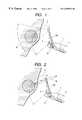
- FIG. 1is a sketch showing how a sector scanning ultrasonic probe is used with an endoscope for ultrasonic examination according to an embodiment of the invention
- FIG. 2is a sketch showing how a radial scanning ultrasonic probe is used with the same endoscope
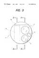
- FIG. 3is a front view of the same endoscope
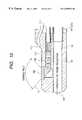
- FIG. 4is section 4 — 4 of FIG. 3;
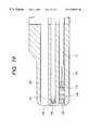
- FIG. 5is section 5 — 5 of FIG. 3;
- FIG. 6is a simplified side view of the sector scanning ultrasonic probe shown in FIG. 1;
- FIG. 7is a simplified side view of the radial scanning ultrasonic probe shown in FIG. 2;
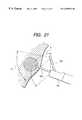
- FIG. 8is a perspective view of the tip of a forward viewing endoscope, as seen from the front, according to another embodiment of the invention.
- FIG. 9is a plan view of the tip of the same endoscope.
- FIG. 10is side section 10 — 10 of FIG. 8;
- FIG. 11is another example of side section 10 — 10 of FIG. 8;
- FIG. 12is side section 12 — 12 of FIG. 8;
- FIG. 13is a sketch showing how the forward viewing endoscope according to the second embodiment of the invention is used in practice
- FIG. 14is a front view of the tip of an oblique viewing endoscope according to a third embodiment of the invention.
- FIG. 15is side section 15 — 15 of FIG. 14;
- FIG. 16is side section 16 — 16 of FIG. 14;
- FIG. 17is a sketch showing how the oblique viewing endoscope according to the third embodiment of the invention is used in practice
- FIG. 18is a front view of the tip of a lateral viewing endoscope according to a fourth embodiment of the invention.
- FIG. 19is side section 19 — 19 of FIG. 18;
- FIG. 20is side section 20 — 20 of FIG. 18;
- FIG. 21is a sketch showing how the lateral viewing endoscope according to the fourth embodiment of the invention is used in practice.
- FIG. 3shows the end face of the tip housing 1 forming the foremost end of the insertion portion of an endoscope and it has two juxtaposed windows, a viewing window 2 for optical examination and an illumination window 3 for illuminating the scope of optical examination. Shown by 4 is a nozzle for spraying the surface of the viewing window 2 with air and water.
- FIG. 4is section 4 — 4 of FIG. 3 .
- objective optics 5is provided rearward of the viewing window 2 to ensure that an object lying ahead of the housing tip 1 is focused at the imaging plane of a solid-state imaging device 6 .
- the focused optical imageis sent over a signal cable 7 through the insertion portion to a video processor (not shown) so that an endoscopically examined image is displayed on a TV monitor.
- a video processornot shown
- the combination of the solid-state imaging device 6 and the signal cable 7may be replaced by an image guide fiber bundle.
- the exit end of an illuminating lightguide fiber bundle 8is provided rearward of the illumination window 3 and the illuminating light issued from the bundle 8 is projected to the object ahead of the tip housing 1 .
- a probe projecting groove or probe projecting port 9is formed in the tip housing 1 in a position juxtaposed to the viewing window 2 and the illumination window 3 .
- a probe guide plate 10is provided in the groove 9 .
- FIG. 5is section 5 — 5 of FIG. 3 .
- the exit of a probe insertion channel 11communicates with and connects to an area rearward of the probe projecting groove 9 .
- the channelis typically formed of a PTFE (polytetrafluoroethylene) tube, and inserted into and arranged along the insertion portion of the endoscope. With this arrangement, the tip of an ultrasonic probe 100 inserted into the channel 11 is guided along the plate 10 so that it projects from the groove 9 to be directed laterally of the tip housing 1 .
- laterallydoes not have the strict sense of being “perpendicular” to the longitudinal axis of the tip housing 1 but simply means “approximately lateral”.
- the probe guide plate 10 in the probe projecting groove 9is pivotal about a shaft 12 supported on the tip housing 1 .
- a handling section(not shown) is coupled to the end of the insertion portion which is the closer to the operator, and a maneuvering wire 13 that can be moved back and forth by remote operation of the handling section is coupled to the probe guide plate 10 .
- the probe guide plate 10pivots within the probe projecting groove 9 such that the direction in which the tip of the ultrasonic probe 100 projects is adjustable over a broad range from a obliquely forward position with respect to the tip housing 1 to a lateral position substantially perpendicular to the longitudinal axis of the tip housing 1 .
- FIGS. 6 and 7are illustrations of the ultrasonic probe 100 to be used with the endoscope for ultrasonic examination.
- the ultrasonic probe 100 shown in FIG. 6is of a so-called “sector scanning” type which has an ultrasonic oscillator array 101 provided on a lateral side near the tip; the array 101 is made up of a multiple of ultrasonic oscillators arranged parallel to its longitudinal axis.
- ultrasonic wavesare transmitted and received in a sector manner to scan a sectional range in both forward and backward directions in an area lateral to the longitudinal axis of the probe.
- the respective ultrasonic oscillatorsare arranged in directions perpendicular to single opposite point 102 .
- the ultrasonic probe 100 shown in FIG. 7is of a so-called “radial scanning” type, in which a single ultrasonic oscillator 103 is driven to rotate about the longitudinal axis of the probe to scan in directions perpendicular to said axis through 360°.
- FIG. 1shows how the endoscope is used with the sector scanning ultrasonic probe 100 of FIG. 6 being inserted into the probe insertion channel 11 . Since the tip of the probe 100 projects laterally of the tip housing 1 , the ultrasonic scan range B substantially overlaps with the viewing field A ahead of the tip housing 1 so that a cross-section of a living tissue ahead of the viewing field A can be imaged by ultrasonic examination.
- the ultrasonic scan range Bcan be adjusted by pivoting the probe guide plate 10 to change the direction in which the ultrasonic probe 100 projects. If the tip of the ultrasonic probe 100 is set such that its movement is at all times visible within the viewing field A (does not go beyond it), the operator can handle the endoscope with safety.
- FIG. 2shows how the endoscope is used with the radial scanning ultrasonic probe 100 of FIG. 2 being inserted into the prove insertion channel 11 .
- a living tissue located ahead of the viewing field A which is forward of the tip housing 1is scanned with ultrasonic waves through a section cut perpendicular to the longitudinal axis of the probe, thereby producing a cross-sectional image of the tissue.
- FIG. 9is a plan view of the tip of the insertion portion of a lateral viewing endoscope according to a second embodiment of the invention. As shown, a viewing window 102 for optical examination and an illumination window 103 for illuminating the scope of optical examination are provided on a lateral side of tip housing 101 .
- Shown by 104is a treatment tool projecting port from which is projected the tip portion of a treatment tool inserted into a treatment tool insertion channel to be described below.
- the port 104is formed as an elongated groove extending in a direction parallel to the longitudinal axis of the tip housing 101 . It is open to the same lateral side of the tip housing 101 where the viewing window 102 is made; thus, the treatment tool projecting port 104 and the viewing window 102 lie side by side.
- a treatment tool erecting plate 105 for adjusting the direction in which the treatment tool projectsis provided within the port 104 .
- FIG. 8is a perspective view of the tip of the insertion portion as seen from the front.
- Objective optics 106is provided rearward of the viewing window 102 and further rearward is provided a probe insertion channel 107 through which an ultrasonic probe is to be inserted.
- the treatment tool projecting port 104communicates with and connects to a treatment tool insertion channel 108 through which a treatment tool is to be inserted.
- FIG. 10is section 10 — 10 of FIG. 8 .
- the objective optics 106 provided rearward of the viewing window 102ensures that an object lying lateral to the tip housing 101 is focused at the imaging plane of a solid-state imaging device 110 .
- Shown by 112is a nozzle for spraying the surface of the viewing window 102 with air and water.
- the focused optical imageis sent over a signal cable 111 through the insertion portion to a video processor (not shown) so that an endoscopically examined image is displayed on a TV monitor.
- a video processornot shown
- the combination of the solid-state imaging device 110 and the signal cable 111may be replaced by an image guide fiber bundle.
- the probe insertion channel 107is coupled to the tip housing 101 ; it is a flexible tube 107 a that is typically formed of PTFE (polytetrafluoroethylene) and which is passed through substantially the entire length of the insertion portion.
- An ultrasonic probe projecting port 109is open in the front end face of the tip housing 101 to be directed forward of the insertion portion.
- the tip of the ultrasonic probe inserted into the channel 107projects from the port 109 in a direction perpendicular to the optical axis of illuminating light so that it does not interfere with the viewing field.
- a probe erecting plate 115 for adjusting the direction in which the tip portion of the ultrasonic probe projectsmay be provided within the ultrasonic probe projecting port 109 as similarly to the first embodiment.
- the probe erecting plate 115is pivotal about a shaft 116 supported on the tip housing 101 .
- a handling section(not shown) is coupled to the end of the insertion portion which is the closer to the operator and a maneuvering wire 117 that can be moved back and forth by remote operation of the handling section is coupled to the probe erecting plate 115 .
- the probe erecting plate 115pivots such that the direction in which the tip of the ultrasonic probe projects from the port 109 is adjustable to any angle.
- FIG. 12is section 12 — 12 of FIG. 8 . As shown, the exit end of an illuminating lightguide fiber bundle 119 is provided rearward of the illumination window 103 and the illuminating light issued from the bundle 119 is projected to the object lying lateral to the tip housing 101 .
- the treatment tool insertion channel 108is coupled to the tip housing 101 ; it is a flexible tube 108 a that is typically formed of PTFE (polytetrafluoroethylene) and which is passed through substantially the entire length of the insertion portion.
- a treatment tool inserted into the channel 108can have its tip guided along the treatment tool erecting plate 105 to project from the port 104 laterally of the tip housing 101 .
- the treatment tool erecting plate 105is pivotal about a shaft 121 supported on the tip housing 101 .
- a maneuvering wire 122 coupled to the plate 105can be moved back and forth by remote operation of the handling section coupled to the end of the insertion portion which is the closer to the operator.
- the treatment tool erecting plate 5pivots within the port 104 such that the direction in which the tip of the treatment tool projects is adjustable over a broad range from a obliquely forward position with respect to the tip housing 101 to a lateral position substantially perpendicular to the longitudinal axis of the tip housing 101 .
- the treatment tool projecting port 104is inclined to the optical axis A of the viewing light so that the direction B in which the treatment tool projects is toward the center of the viewing field (see FIG. 8 ).
- FIG. 13shows how the endoscope according to the second embodiment is used with the sector scanning ultrasonic probe 100 of FIG. 6 being inserted into the probe insertion channel 107 and an endoscopic injection tool 200 into the treatment tool insertion channel 108 . Since the tip of the ultrasonic probe 100 projects forward of the tip housing 101 , the ultrasonic scan range D substantially overlaps with the viewing field C lateral to the tip housing 1 so that an ultrasonic cross-sectional image ahead of the viewing field C can be taken.
- the tip of the injection tool 200projects laterally from the tip housing 101 , so it can be pierced toward the diseased part under the membrane within the viewing field C and the ultrasonic scan range D and at a sufficiently large angle (close to 90°) with respect to the membrane surface to enable the necessary treatment to be performed.
- FIG. 14shows a third embodiment of the invention by a front view of the tip of the insertion portion of an oblique viewing endoscope to which the concept of the invention is applied.
- the components having the same functions as in the second embodimentare identified by like numerals and will not be described in detail.
- FIG. 15is section 15 — 15 of FIG. 14 . As shown, the upper half of the end face of the tip housing 101 is cut obliquely and the viewing window 102 and the illumination window 103 are arranged side by side on the oblique face so that an object situated obliquely forward of the tip housing 1 can be examined optically.
- FIG. 16is section 16 — 16 of FIG. 14 .
- the ultrasonic probe projecting port 109is formed as an opening in the end face of the tip housing 101 so that the tip of the ultrasonic probe projects forward (parallel to the longitudinal axis of the tip housing 101 ).
- the treatment tool projecting port 104is formed as an opening in the oblique face of the tip housing 101 so that the tip of a treatment tool inserted into the treatment tool insertion channel 108 projects in a direction toward the viewing field obliquely ahead of the tip portion 101 .
- the direction of its projectioncan be adjusted by the mechanism of the treatment tool erecting plate 105 positioned within the treatment tool projecting port 104 .
- FIG. 17shows how the oblique viewing endoscope according to the third embodiment is used with the radial scanning ultrasonic probe 100 of FIG. 7 being inserted into the probe insertion channel 107 and an endoscopic injection tool 200 into the treatment tool insertion channel 108 .
- the tip of the ultrasonic probe 100projects ahead of the tip housing 101 , so a desired ultrasonic cross-sectional image can be obtained by scanning through the viewing field C lying obliquely ahead of the tip portion 101 .
- the tip of the injection tool 200is allowed to project from the tip housing 101 to an obliquely forward position until it is pierced into the diseased part under the membrane within the viewing field C and the ultrasonic scan range D to enable the necessary treatment to be performed. If the tip of the ultrasonic probe 100 is set such that its movement is at all times visible within the viewing field C (does not go beyond it), the operator can handle the endoscope with safety.
- FIG. 18shows a fourth embodiment of the invention by a front view of the tip of the insertion portion of a forward viewing endoscope to which the concept of the invention is applied.
- the components having the same functions as in the second and third embodimentsare identified by like numerals and will not be described in detail.
- the viewing window 2 and the illumination window 3are arranged side by side on the end face of the tip housing 101 in the fourth embodiment and this allows for optical viewing of an object lying ahead of the tip housing 101 .
- the treatment tool insertion channel 108 formed parallel to the longitudinal axis of the tip housing 101extends straight to make an opening (treatment tool projecting port 104 ) in the end face of the tip housing 101 , through which the tip of the treatment tool inserted into the channel 108 projects in a forward viewing direction.
- the ultrasonic probe projecting port 109is formed as an opening in a lateral side of the tip housing 1 so that the tip of the ultrasonic probe 100 inserted into the probe insertion channel 107 can be projected in a direction lateral to the tip housing 101 .
- the direction in which the ultrasonic probe 100 projectscan be adjusted to any angle by remote operation of the probe erecting plate 115 provided within the probe projecting port 109 .
- Shown by 116is a shaft supported on the tip housing 101 , and 117 is a maneuvering wire that is moved back and forth by operation of the handling section.
- FIG. 21shows how the forward viewing endoscope according to the fourth embodiment of the invention is used with the sector scanning ultrasonic probe 100 of FIG. 6 being inserted into the probe insertion channel 107 and an endoscopic injection tool 200 into the treatment tool insertion channel 108 .
- the tip of the ultrasonic probe 100projects laterally of the tip housing 101 so that the viewing field C lying ahead of the tip portion 101 can be scanned to produce an ultrasonic cross-sectional image.
- the tip of the injection tool 200is allowed to project forward from the tip housing 101 until it is pierced into the diseased part under the membrane within the viewing field C and the ultrasonic scan range D to enable the necessary treatment to be performed.
- the tip of the ultrasonic probe 100is set such that its movement is at all times visible within the viewing field C (does not go beyond it), the operator can handle the endoscope with safety.
Landscapes
- Health & Medical Sciences (AREA)
- Life Sciences & Earth Sciences (AREA)
- Surgery (AREA)
- General Health & Medical Sciences (AREA)
- Medical Informatics (AREA)
- Veterinary Medicine (AREA)
- Pathology (AREA)
- Radiology & Medical Imaging (AREA)
- Biophysics (AREA)
- Engineering & Computer Science (AREA)
- Biomedical Technology (AREA)
- Heart & Thoracic Surgery (AREA)
- Nuclear Medicine, Radiotherapy & Molecular Imaging (AREA)
- Molecular Biology (AREA)
- Animal Behavior & Ethology (AREA)
- Physics & Mathematics (AREA)
- Public Health (AREA)
- Optics & Photonics (AREA)
- Gynecology & Obstetrics (AREA)
- Endoscopes (AREA)
- Ultra Sonic Daignosis Equipment (AREA)
Abstract
Description
The present invention relates to an endoscope for ultrasonic examination and surgical treatment associated thereto.
In order to diagnose and treat a diseased part under the membrane in a body cavity, an ultrasonic probe capable of producing an ultrasonic cross-sectional image and a treatment tool such as an endoscopic injection tool have to be guided to an area near the diseased part via an endoscope.
To meet this need, one may think of using an endoscope for surgical treatment having two insertion channels. A two-channel endoscope is adapted to be such that the tips of both an ultrasonic probe and a treatment tool project in the direction in which examination is done with an optical viewing system.
In order to realize ultrasonic scan on the site being examined with the endoscope under a condition that the tip of the ultrasonic probe projects in the viewing field, the ultrasonic probe must be designed to transmit ultrasonic waves forward and receiving the reflected waves for scan.
On the other hand, the ultrasonic probe must be capable of passing through the channel in the endoscope, and therefore cannot be thicker than 2 to 3 mm. However, with such a small diameter, the probe for creating an ultrasonic cross-sectional image in a forward direction achieves only a very low resolution.
The present invention has been accomplished under these circumstances and has as an object providing an endoscope for ultrasonic examination, which is adapted for use with an ultrasonic probe capable of scanning by transmitting and receiving ultrasonic waves laterally of its tip and which can obtain a sharp ultrasonic cross-sectional image within a range of optical viewing area ahead of the foremost end of the insertion portion of the endoscope.
Another object of the present invention is to provide an endoscope for surgical treatment associated with the ultrasonic examination, which is adapted for use with an ultrasonic probe capable of scanning by transmitting and receiving ultrasonic waves laterally of its tip and which can obtain a sharp ultrasonic cross-sectional image of a diseased part under the membrane being optically examined while performing an endoscopic treatment on the diseased part.
To achieve the above-noted and other objects, the present invention provides an endoscope which comprises: an elongated insertion portion; a tip housing on a longitudinal end of the insertion portion; an optical imaging system provided in the tip housing and directed in a first direction; and a probe insertion channel extending along the insertion portion, and communicating with a probe projecting port disposed in the tip housing and opened in a second direction substantially lateral with respect to the first direction.
It is preferable that the endoscope further comprises a movable guide disposed within the probe projecting port.
It is preferable that the endoscope further comprises a treatment tool insertion channel extending along the insertion portion, and communicating with a treatment tool projecting port disposed in the tip housing and opened substantially in the first direction.
It is preferable that an endoscope further comprises a movable guide disposed within the treatment tool insertion channel.
It is preferable that the endoscope further comprises a second movable guide disposed within the probe projecting port.
It is preferable that the endoscope further comprises an ultrasonic probe removably passed through the probe insertion channel so that its tip end projects from the probe projecting port.
It is preferable that the endoscope further comprises: an adjusting mechanism which adjusts the direction in which the tip end of the ultrasonic probe projects from the probe projecting port.
It is preferable that the endoscope further comprises a wire extending along the insertion section and connected to the movable guide.
It is preferable that the endoscope further comprises a wire extending along the insertion section and connected to the movable guide.
The present invention provides an endoscope for ultrasonic examination, which comprises: an insertion portion; an optical examination mechanism provided on a distal end of the insertion portion for endoscopic examination forwardly of the insertion portion; a probe insertion channel disposed along the insertion portion so that a ultrasonic probe is passed therethrough; and a probe guiding mechanism disposed in the insertion portion to guide a distal end of the ultrasonic probe to project from the distal end of the insertion portion laterally of the distal end of the insertion portion.
It is preferable that the ultrasonic probe transmits and receives ultrasonic waves laterally of its distal end.
It is preferable that the probe guiding mechanism adjusts, through a remote operation, a projecting direction in which the distal end of the ultrasonic probe projects from the distal end of the insertion portion.
The present invention provides an endoscope for surgical treatment associated with ultrasonic examination, which comprises: an insertion portion; an optical examination mechanism provided on a distal end of the insertion portion for endoscopic examination forwardly of the insertion portion; a treatment tool insertion channel disposed along the insertion portion so that a treatment tool is passed therethrough; a probe insertion channel disposed along the insertion portion so that a ultrasonic probe is passed therethrough; a treatment tool projecting port by which a distal end of the treatment tool passed through the treatment tool insertion channel projects from the distal end of the insertion portion toward and within an examination viewing field of the optical examination mechanism; and a probe projecting port by which a distal end of the ultrasonic probe passed through the probe insertion channel projects from the distal end of the insertion portion in a direction away from an examination direction of the optical examination mechanism.
It is preferable that the ultrasonic probe transmits and receives ultrasonic waves laterally of its distal end.
It is preferable that the endoscope for surgical treatment associated with ultrasonic examination further comprises a probe projecting direction adjusting mechanism which adjusts, through a remote operation, a projecting direction in which the distal end of the ultrasonic probe projects from the distal end of the insertion portion.
It is preferable that the distal end of the ultrasonic probe projecting from the distal end of the insertion portion is located within a peripheral portion of the examination viewing field of the optical examination mechanism.
The present disclosure relates to the subject matter contained in Japanese patent application Nos. Hei. 10-178505 and Hei. 10-178506 (both filed on Jun. 25, 1998), which are expressly incorporated herein by reference in their entireties.
FIG. 1 is a sketch showing how a sector scanning ultrasonic probe is used with an endoscope for ultrasonic examination according to an embodiment of the invention;
FIG. 2 is a sketch showing how a radial scanning ultrasonic probe is used with the same endoscope;
FIG. 3 is a front view of the same endoscope;
FIG. 4 issection 4—4 of FIG. 3;
FIG. 5 issection 5—5 of FIG. 3;
FIG. 6 is a simplified side view of the sector scanning ultrasonic probe shown in FIG. 1;
FIG. 7 is a simplified side view of the radial scanning ultrasonic probe shown in FIG. 2;
FIG. 8 is a perspective view of the tip of a forward viewing endoscope, as seen from the front, according to another embodiment of the invention;
FIG. 9 is a plan view of the tip of the same endoscope;
FIG. 10 isside section 10—10 of FIG. 8;
FIG. 11 is another example ofside section 10—10 of FIG. 8;
FIG. 12 isside section 12—12 of FIG. 8;
FIG. 13 is a sketch showing how the forward viewing endoscope according to the second embodiment of the invention is used in practice;
FIG. 14 is a front view of the tip of an oblique viewing endoscope according to a third embodiment of the invention;
FIG. 15 isside section 15—15 of FIG. 14;
FIG. 16 isside section 16—16 of FIG. 14;
FIG. 17 is a sketch showing how the oblique viewing endoscope according to the third embodiment of the invention is used in practice;
FIG. 18 is a front view of the tip of a lateral viewing endoscope according to a fourth embodiment of the invention;
FIG. 19 isside section 19—19 of FIG. 18;
FIG. 20 isside section 20—20 of FIG. 18; and
FIG. 21 is a sketch showing how the lateral viewing endoscope according to the fourth embodiment of the invention is used in practice.
An embodiment of the present invention is described below with reference to the accompanying drawings.
FIG. 3 shows the end face of thetip housing 1 forming the foremost end of the insertion portion of an endoscope and it has two juxtaposed windows, aviewing window 2 for optical examination and anillumination window 3 for illuminating the scope of optical examination. Shown by4 is a nozzle for spraying the surface of theviewing window 2 with air and water.
FIG. 4 issection 4—4 of FIG.3. As shown,objective optics 5 is provided rearward of theviewing window 2 to ensure that an object lying ahead of thehousing tip 1 is focused at the imaging plane of a solid-state imaging device6.
The focused optical image is sent over asignal cable 7 through the insertion portion to a video processor (not shown) so that an endoscopically examined image is displayed on a TV monitor. If desired, the combination of the solid-state imaging device6 and thesignal cable 7 may be replaced by an image guide fiber bundle.
The exit end of an illuminatinglightguide fiber bundle 8 is provided rearward of theillumination window 3 and the illuminating light issued from thebundle 8 is projected to the object ahead of thetip housing 1.
Turning back to the FIG. 3, a probe projecting groove orprobe projecting port 9 is formed in thetip housing 1 in a position juxtaposed to theviewing window 2 and theillumination window 3. Aprobe guide plate 10 is provided in thegroove 9.
FIG. 5 issection 5—5 of FIG.3. As shown, the exit of aprobe insertion channel 11 communicates with and connects to an area rearward of theprobe projecting groove 9. The channel is typically formed of a PTFE (polytetrafluoroethylene) tube, and inserted into and arranged along the insertion portion of the endoscope. With this arrangement, the tip of anultrasonic probe 100 inserted into thechannel 11 is guided along theplate 10 so that it projects from thegroove 9 to be directed laterally of thetip housing 1. It should be noted that the term “laterally” does not have the strict sense of being “perpendicular” to the longitudinal axis of thetip housing 1 but simply means “approximately lateral”.
Theprobe guide plate 10 in theprobe projecting groove 9 is pivotal about ashaft 12 supported on thetip housing 1. A handling section (not shown) is coupled to the end of the insertion portion which is the closer to the operator, and amaneuvering wire 13 that can be moved back and forth by remote operation of the handling section is coupled to theprobe guide plate 10.
If the handling section is operated to move themaneuvering wire 13 back and forth, theprobe guide plate 10 pivots within theprobe projecting groove 9 such that the direction in which the tip of theultrasonic probe 100 projects is adjustable over a broad range from a obliquely forward position with respect to thetip housing 1 to a lateral position substantially perpendicular to the longitudinal axis of thetip housing 1.
FIGS. 6 and 7 are illustrations of theultrasonic probe 100 to be used with the endoscope for ultrasonic examination.
Theultrasonic probe 100 shown in FIG. 6 is of a so-called “sector scanning” type which has anultrasonic oscillator array 101 provided on a lateral side near the tip; thearray 101 is made up of a multiple of ultrasonic oscillators arranged parallel to its longitudinal axis.
As in a so-called “convex” type, ultrasonic waves are transmitted and received in a sector manner to scan a sectional range in both forward and backward directions in an area lateral to the longitudinal axis of the probe. To this end, the respective ultrasonic oscillators are arranged in directions perpendicular to singleopposite point 102.
Theultrasonic probe 100 shown in FIG. 7 is of a so-called “radial scanning” type, in which a singleultrasonic oscillator 103 is driven to rotate about the longitudinal axis of the probe to scan in directions perpendicular to said axis through 360°.
FIG. 1 shows how the endoscope is used with the sector scanningultrasonic probe 100 of FIG. 6 being inserted into theprobe insertion channel 11. Since the tip of theprobe 100 projects laterally of thetip housing 1, the ultrasonic scan range B substantially overlaps with the viewing field A ahead of thetip housing 1 so that a cross-section of a living tissue ahead of the viewing field A can be imaged by ultrasonic examination.
The ultrasonic scan range B can be adjusted by pivoting theprobe guide plate 10 to change the direction in which theultrasonic probe 100 projects. If the tip of theultrasonic probe 100 is set such that its movement is at all times visible within the viewing field A (does not go beyond it), the operator can handle the endoscope with safety.
FIG. 2 shows how the endoscope is used with the radial scanningultrasonic probe 100 of FIG. 2 being inserted into the proveinsertion channel 11. Compared to the case shown in FIG. 1, a living tissue located ahead of the viewing field A which is forward of thetip housing 1 is scanned with ultrasonic waves through a section cut perpendicular to the longitudinal axis of the probe, thereby producing a cross-sectional image of the tissue.
FIG. 9 is a plan view of the tip of the insertion portion of a lateral viewing endoscope according to a second embodiment of the invention. As shown, aviewing window 102 for optical examination and anillumination window 103 for illuminating the scope of optical examination are provided on a lateral side oftip housing 101.
Shown by104 is a treatment tool projecting port from which is projected the tip portion of a treatment tool inserted into a treatment tool insertion channel to be described below. Theport 104 is formed as an elongated groove extending in a direction parallel to the longitudinal axis of thetip housing 101. It is open to the same lateral side of thetip housing 101 where theviewing window 102 is made; thus, the treatmenttool projecting port 104 and theviewing window 102 lie side by side. A treatmenttool erecting plate 105 for adjusting the direction in which the treatment tool projects is provided within theport 104.
FIG. 8 is a perspective view of the tip of the insertion portion as seen from the front.Objective optics 106 is provided rearward of theviewing window 102 and further rearward is provided aprobe insertion channel 107 through which an ultrasonic probe is to be inserted. The treatmenttool projecting port 104 communicates with and connects to a treatmenttool insertion channel 108 through which a treatment tool is to be inserted.
FIG. 10 issection 10—10 of FIG.8. As shown, theobjective optics 106 provided rearward of theviewing window 102 ensures that an object lying lateral to thetip housing 101 is focused at the imaging plane of a solid-state imaging device 110. Shown by112 is a nozzle for spraying the surface of theviewing window 102 with air and water.
The focused optical image is sent over asignal cable 111 through the insertion portion to a video processor (not shown) so that an endoscopically examined image is displayed on a TV monitor. If desired, the combination of the solid-state imaging device 110 and thesignal cable 111 may be replaced by an image guide fiber bundle.
Theprobe insertion channel 107 is coupled to thetip housing 101; it is aflexible tube 107athat is typically formed of PTFE (polytetrafluoroethylene) and which is passed through substantially the entire length of the insertion portion. An ultrasonicprobe projecting port 109 is open in the front end face of thetip housing 101 to be directed forward of the insertion portion.
With this arrangement, the tip of the ultrasonic probe inserted into thechannel 107 projects from theport 109 in a direction perpendicular to the optical axis of illuminating light so that it does not interfere with the viewing field.
As shown in FIG. 11, aprobe erecting plate 115 for adjusting the direction in which the tip portion of the ultrasonic probe projects may be provided within the ultrasonicprobe projecting port 109 as similarly to the first embodiment.
Further referring to FIG. 11, theprobe erecting plate 115 is pivotal about ashaft 116 supported on thetip housing 101. A handling section (not shown) is coupled to the end of the insertion portion which is the closer to the operator and amaneuvering wire 117 that can be moved back and forth by remote operation of the handling section is coupled to theprobe erecting plate 115.
If the handling section is operated to move themaneuvering wire 117 back and forth, theprobe erecting plate 115 pivots such that the direction in which the tip of the ultrasonic probe projects from theport 109 is adjustable to any angle.
FIG. 12 issection 12—12 of FIG.8. As shown, the exit end of an illuminatinglightguide fiber bundle 119 is provided rearward of theillumination window 103 and the illuminating light issued from thebundle 119 is projected to the object lying lateral to thetip housing 101.
The treatmenttool insertion channel 108 is coupled to thetip housing 101; it is aflexible tube 108athat is typically formed of PTFE (polytetrafluoroethylene) and which is passed through substantially the entire length of the insertion portion. A treatment tool inserted into thechannel 108 can have its tip guided along the treatmenttool erecting plate 105 to project from theport 104 laterally of thetip housing 101.
The treatmenttool erecting plate 105 is pivotal about ashaft 121 supported on thetip housing 101. Amaneuvering wire 122 coupled to theplate 105 can be moved back and forth by remote operation of the handling section coupled to the end of the insertion portion which is the closer to the operator.
If the handling section is operated to move themaneuvering wire 122 back and forth, the treatmenttool erecting plate 5 pivots within theport 104 such that the direction in which the tip of the treatment tool projects is adjustable over a broad range from a obliquely forward position with respect to thetip housing 101 to a lateral position substantially perpendicular to the longitudinal axis of thetip housing 101.
Note that the treatmenttool projecting port 104 is inclined to the optical axis A of the viewing light so that the direction B in which the treatment tool projects is toward the center of the viewing field (see FIG.8).
FIG. 13 shows how the endoscope according to the second embodiment is used with the sector scanningultrasonic probe 100 of FIG. 6 being inserted into theprobe insertion channel 107 and anendoscopic injection tool 200 into the treatmenttool insertion channel 108. Since the tip of theultrasonic probe 100 projects forward of thetip housing 101, the ultrasonic scan range D substantially overlaps with the viewing field C lateral to thetip housing 1 so that an ultrasonic cross-sectional image ahead of the viewing field C can be taken.
On the other hand, the tip of theinjection tool 200 projects laterally from thetip housing 101, so it can be pierced toward the diseased part under the membrane within the viewing field C and the ultrasonic scan range D and at a sufficiently large angle (close to 90°) with respect to the membrane surface to enable the necessary treatment to be performed.
FIG. 14 shows a third embodiment of the invention by a front view of the tip of the insertion portion of an oblique viewing endoscope to which the concept of the invention is applied. The components having the same functions as in the second embodiment are identified by like numerals and will not be described in detail.
FIG. 15 issection 15—15 of FIG.14. As shown, the upper half of the end face of thetip housing 101 is cut obliquely and theviewing window 102 and theillumination window 103 are arranged side by side on the oblique face so that an object situated obliquely forward of thetip housing 1 can be examined optically.
FIG. 16 issection 16—16 of FIG.14. As in the second embodiment, the ultrasonicprobe projecting port 109 is formed as an opening in the end face of thetip housing 101 so that the tip of the ultrasonic probe projects forward (parallel to the longitudinal axis of the tip housing101).
The treatmenttool projecting port 104 is formed as an opening in the oblique face of thetip housing 101 so that the tip of a treatment tool inserted into the treatmenttool insertion channel 108 projects in a direction toward the viewing field obliquely ahead of thetip portion 101. The direction of its projection can be adjusted by the mechanism of the treatmenttool erecting plate 105 positioned within the treatmenttool projecting port 104.
FIG. 17 shows how the oblique viewing endoscope according to the third embodiment is used with the radial scanningultrasonic probe 100 of FIG. 7 being inserted into theprobe insertion channel 107 and anendoscopic injection tool 200 into the treatmenttool insertion channel 108.
Again, the tip of theultrasonic probe 100 projects ahead of thetip housing 101, so a desired ultrasonic cross-sectional image can be obtained by scanning through the viewing field C lying obliquely ahead of thetip portion 101.
Subsequently, the tip of theinjection tool 200 is allowed to project from thetip housing 101 to an obliquely forward position until it is pierced into the diseased part under the membrane within the viewing field C and the ultrasonic scan range D to enable the necessary treatment to be performed. If the tip of theultrasonic probe 100 is set such that its movement is at all times visible within the viewing field C (does not go beyond it), the operator can handle the endoscope with safety.
FIG. 18 shows a fourth embodiment of the invention by a front view of the tip of the insertion portion of a forward viewing endoscope to which the concept of the invention is applied. The components having the same functions as in the second and third embodiments are identified by like numerals and will not be described in detail.
As also shown in FIG. 19 which issection 19—19 of FIG. 18, theviewing window 2 and theillumination window 3 are arranged side by side on the end face of thetip housing 101 in the fourth embodiment and this allows for optical viewing of an object lying ahead of thetip housing 101.
The treatmenttool insertion channel 108 formed parallel to the longitudinal axis of thetip housing 101 extends straight to make an opening (treatment tool projecting port104) in the end face of thetip housing 101, through which the tip of the treatment tool inserted into thechannel 108 projects in a forward viewing direction.
As shown in FIG. 20 which issection 20—20 of FIG. 18, the ultrasonicprobe projecting port 109 is formed as an opening in a lateral side of thetip housing 1 so that the tip of theultrasonic probe 100 inserted into theprobe insertion channel 107 can be projected in a direction lateral to thetip housing 101.
The direction in which theultrasonic probe 100 projects can be adjusted to any angle by remote operation of theprobe erecting plate 115 provided within theprobe projecting port 109. Shown by116 is a shaft supported on thetip housing
FIG. 21 shows how the forward viewing endoscope according to the fourth embodiment of the invention is used with the sector scanningultrasonic probe 100 of FIG. 6 being inserted into theprobe insertion channel 107 and anendoscopic injection tool 200 into the treatmenttool insertion channel 108.
In the case under consideration, the tip of theultrasonic probe 100 projects laterally of thetip housing 101 so that the viewing field C lying ahead of thetip portion 101 can be scanned to produce an ultrasonic cross-sectional image.
Subsequently, the tip of theinjection tool 200 is allowed to project forward from thetip housing 101 until it is pierced into the diseased part under the membrane within the viewing field C and the ultrasonic scan range D to enable the necessary treatment to be performed. As in the case shown in FIG. 17, if the tip of theultrasonic probe 100 is set such that its movement is at all times visible within the viewing field C (does not go beyond it), the operator can handle the endoscope with safety.
Claims (5)
1. An endoscope for surgical treatment associated with ultrasonic examination, comprising:
an insertion portion;
an optical examination mechanism with a viewing field facing in a first direction relative to the distal end of the insertion portion;
a treatment tool insertion channel disposed along the insertion portion so that a treatment tool is passable therethrough;
a probe insertion channel disposed along the insertion portion so that an ultrasonic probe is passable therethrough;
a treatment tool projecting port by which a distal end of the treatment tool passing through the treatment tool insertion channel projects from the distal end of the insertion portion; and
a probe projecting port by which a distal end of the ultrasonic probe passing through the probe insertion channel projects from the distal end of the insertion portion in a direction different than the first direction, the distal end of the ultrasonic probe having an ultrasonic scan range substantially lateral to said insertion portion and at least partly overlapping the examination viewing field;
wherein a distal end of said treatment tool extends into the overlapping portion of the viewing field and the ultrasonic scan range.
2. An endoscope for surgical treatment associated with ultrasonic examination according toclaim 1 , wherein the ultrasonic probe transmits and receives ultrasonic waves laterally of its distal end.
3. An endoscope for surgical treatment associated with ultrasonic examination according toclaim 1 , further comprising:
a probe projecting direction adjusting mechanism which adjusts, through a remote operation, a projecting direction in which the distal end of the ultrasonic probe projects from the distal end of the insertion portion.
4. An endoscope for surgical treatment associated with ultrasonic examination according toclaim 1 , wherein the distal end of the ultrasonic probe projecting from the distal end of the insertion portion is located within a peripheral portion of the examination viewing field of the optical examination mechanism.
5. The endoscope for ultrasonic examination ofclaim 1 , an oblique face provided on a distal end of said insertion portion, said treatment tool projecting from the oblique face and into the viewing field of said optical examination mechanism.
Applications Claiming Priority (4)
| Application Number | Priority Date | Filing Date | Title |
|---|---|---|---|
| JP10178506AJP2000005183A (en) | 1998-06-25 | 1998-06-25 | Endoscope for ultrasonic observation treatment |
| JP10-178505 | 1998-06-25 | ||
| JP10178505AJP2000005182A (en) | 1998-06-25 | 1998-06-25 | Endoscope for ultrasonic observation |
| JP10-178506 | 1998-06-25 |
Publications (1)
| Publication Number | Publication Date |
|---|---|
| US6390973B1true US6390973B1 (en) | 2002-05-21 |
Family
ID=26498670
Family Applications (1)
| Application Number | Title | Priority Date | Filing Date |
|---|---|---|---|
| US09/337,266Expired - LifetimeUS6390973B1 (en) | 1998-06-25 | 1999-06-22 | Endoscope for ultrasonic examination and surgical treatment associated thereto |
Country Status (2)
| Country | Link |
|---|---|
| US (1) | US6390973B1 (en) |
| DE (1) | DE19929314A1 (en) |
Cited By (110)
| Publication number | Priority date | Publication date | Assignee | Title |
|---|---|---|---|---|
| US6645195B1 (en)* | 2001-01-05 | 2003-11-11 | Advanced Cardiovascular Systems, Inc. | Intraventricularly guided agent delivery system and method of use |
| US6682477B2 (en)* | 2000-02-25 | 2004-01-27 | Richard Wolf Gmbh | Hysteroscope |
| US20040082883A1 (en)* | 2002-10-18 | 2004-04-29 | Fuji Photo Optical Co., Ltd. | Ultrasound endoscope |
| US20050222493A1 (en)* | 2004-03-31 | 2005-10-06 | Shinichi Kohno | Endoscope |
| US20050228289A1 (en)* | 2004-03-31 | 2005-10-13 | Fujinon Corporation | Ultrasonic endoscope |
| US20050228213A1 (en)* | 2004-04-09 | 2005-10-13 | Schneider Robert E | Implantable hearing aid transducer system |
| US20060155169A1 (en)* | 2003-01-21 | 2006-07-13 | Filippo Bastia | Retractor for operations on the arteria haemorroidalis |
| US20070038112A1 (en)* | 2001-10-16 | 2007-02-15 | Taylor James D | Scanning probe with integrated electronics |
| US20070232913A1 (en)* | 2006-01-13 | 2007-10-04 | Mirabilis Medica Inc. | Methods and apparatus for the treatment of menometrorrhagia, endometrial pathology, and cervical neoplasia using high intensity focused ultrasound energy |
| US20070293787A1 (en)* | 2003-08-13 | 2007-12-20 | Taylor James D | Targeted biopsy delivery system |
| US20080051655A1 (en)* | 2006-08-28 | 2008-02-28 | Masatoshi Sato | Ultrasonic endoscope, therapeutic system, treatment method using therapeutic system, endoscopic system, treatment method using ultrasonic endoscope, t-bar, and t-bar suturing device |
| US20090036773A1 (en)* | 2007-07-31 | 2009-02-05 | Mirabilis Medica Inc. | Methods and apparatus for engagement and coupling of an intracavitory imaging and high intensity focused ultrasound probe |
| US20090088636A1 (en)* | 2006-01-13 | 2009-04-02 | Mirabilis Medica, Inc. | Apparatus for delivering high intensity focused ultrasound energy to a treatment site internal to a patient's body |
| US20090118725A1 (en)* | 2007-11-07 | 2009-05-07 | Mirabilis Medica, Inc. | Hemostatic tissue tunnel generator for inserting treatment apparatus into tissue of a patient |
| US20090118729A1 (en)* | 2007-11-07 | 2009-05-07 | Mirabilis Medica Inc. | Hemostatic spark erosion tissue tunnel generator with integral treatment providing variable volumetric necrotization of tissue |
| EP1769719A4 (en)* | 2004-07-05 | 2009-10-21 | Olympus Medical Systems Corp | ELECTRONIC ENDOSCOPE |
| US20100036292A1 (en)* | 2008-08-06 | 2010-02-11 | Mirabilis Medica Inc. | Optimization and feedback control of hifu power deposition through the analysis of detected signal characteristics |
| US20100036291A1 (en)* | 2008-08-06 | 2010-02-11 | Mirabilis Medica Inc. | Optimization and feedback control of hifu power deposition through the frequency analysis of backscattered hifu signals |
| US20100210976A1 (en)* | 2008-10-03 | 2010-08-19 | Mirabilis Medica, Inc. | Method and apparatus for treating tissues with hifu |
| US20100228086A1 (en)* | 2009-03-09 | 2010-09-09 | Tomohiro Ohki | Side viewing endoscope system |
| US20100241005A1 (en)* | 2008-10-03 | 2010-09-23 | Mirabilis Medica, Inc. | Office-based system for treating uterine fibroids or other tissues with hifu |
| US8758256B2 (en) | 2010-07-12 | 2014-06-24 | Best Medical International, Inc. | Apparatus for brachytherapy that uses a scanning probe for treatment of malignant tissue |
| US9044216B2 (en) | 2010-07-12 | 2015-06-02 | Best Medical International, Inc. | Biopsy needle assembly |
| US20150238180A1 (en)* | 2006-12-01 | 2015-08-27 | Boston Scientific Scimed, Inc. | Guide tube systems and methods |
| US9380996B2 (en) | 2012-09-05 | 2016-07-05 | Olympus Corporation | Ultrasound endoscope |
| US20160374712A1 (en)* | 2012-06-29 | 2016-12-29 | Ethicon Endo-Surgery, Llc | Surgical instruments with articulating shafts |
| US10251664B2 (en) | 2016-01-15 | 2019-04-09 | Ethicon Llc | Modular battery powered handheld surgical instrument with multi-function motor via shifting gear assembly |
| US10278721B2 (en) | 2010-07-22 | 2019-05-07 | Ethicon Llc | Electrosurgical instrument with separate closure and cutting members |
| US10285724B2 (en) | 2014-07-31 | 2019-05-14 | Ethicon Llc | Actuation mechanisms and load adjustment assemblies for surgical instruments |
| US10299810B2 (en) | 2010-02-11 | 2019-05-28 | Ethicon Llc | Rotatable cutting implements with friction reducing material for ultrasonic surgical instruments |
| US10335614B2 (en) | 2008-08-06 | 2019-07-02 | Ethicon Llc | Devices and techniques for cutting and coagulating tissue |
| US10335183B2 (en) | 2012-06-29 | 2019-07-02 | Ethicon Llc | Feedback devices for surgical control systems |
| US10342602B2 (en) | 2015-03-17 | 2019-07-09 | Ethicon Llc | Managing tissue treatment |
| US10349999B2 (en) | 2014-03-31 | 2019-07-16 | Ethicon Llc | Controlling impedance rise in electrosurgical medical devices |
| US10376305B2 (en) | 2016-08-05 | 2019-08-13 | Ethicon Llc | Methods and systems for advanced harmonic energy |
| US10433900B2 (en) | 2011-07-22 | 2019-10-08 | Ethicon Llc | Surgical instruments for tensioning tissue |
| US10441345B2 (en) | 2009-10-09 | 2019-10-15 | Ethicon Llc | Surgical generator for ultrasonic and electrosurgical devices |
| US10441310B2 (en) | 2012-06-29 | 2019-10-15 | Ethicon Llc | Surgical instruments with curved section |
| US10456193B2 (en) | 2016-05-03 | 2019-10-29 | Ethicon Llc | Medical device with a bilateral jaw configuration for nerve stimulation |
| US10463421B2 (en) | 2014-03-27 | 2019-11-05 | Ethicon Llc | Two stage trigger, clamp and cut bipolar vessel sealer |
| US10485607B2 (en) | 2016-04-29 | 2019-11-26 | Ethicon Llc | Jaw structure with distal closure for electrosurgical instruments |
| US10517627B2 (en) | 2012-04-09 | 2019-12-31 | Ethicon Llc | Switch arrangements for ultrasonic surgical instruments |
| US10524854B2 (en) | 2010-07-23 | 2020-01-07 | Ethicon Llc | Surgical instrument |
| US10524872B2 (en) | 2012-06-29 | 2020-01-07 | Ethicon Llc | Closed feedback control for electrosurgical device |
| US10543008B2 (en) | 2012-06-29 | 2020-01-28 | Ethicon Llc | Ultrasonic surgical instruments with distally positioned jaw assemblies |
| US10555769B2 (en) | 2016-02-22 | 2020-02-11 | Ethicon Llc | Flexible circuits for electrosurgical instrument |
| US10575892B2 (en) | 2015-12-31 | 2020-03-03 | Ethicon Llc | Adapter for electrical surgical instruments |
| US10595930B2 (en) | 2015-10-16 | 2020-03-24 | Ethicon Llc | Electrode wiping surgical device |
| US10595929B2 (en) | 2015-03-24 | 2020-03-24 | Ethicon Llc | Surgical instruments with firing system overload protection mechanisms |
| US10610286B2 (en) | 2015-09-30 | 2020-04-07 | Ethicon Llc | Techniques for circuit topologies for combined generator |
| US10639092B2 (en) | 2014-12-08 | 2020-05-05 | Ethicon Llc | Electrode configurations for surgical instruments |
| US10646269B2 (en) | 2016-04-29 | 2020-05-12 | Ethicon Llc | Non-linear jaw gap for electrosurgical instruments |
| US10688321B2 (en) | 2009-07-15 | 2020-06-23 | Ethicon Llc | Ultrasonic surgical instruments |
| US10702329B2 (en) | 2016-04-29 | 2020-07-07 | Ethicon Llc | Jaw structure with distal post for electrosurgical instruments |
| US10716615B2 (en) | 2016-01-15 | 2020-07-21 | Ethicon Llc | Modular battery powered handheld surgical instrument with curved end effectors having asymmetric engagement between jaw and blade |
| US10729494B2 (en) | 2012-02-10 | 2020-08-04 | Ethicon Llc | Robotically controlled surgical instrument |
| US10765470B2 (en) | 2015-06-30 | 2020-09-08 | Ethicon Llc | Surgical system with user adaptable techniques employing simultaneous energy modalities based on tissue parameters |
| US10779845B2 (en) | 2012-06-29 | 2020-09-22 | Ethicon Llc | Ultrasonic surgical instruments with distally positioned transducers |
| US10779879B2 (en) | 2014-03-18 | 2020-09-22 | Ethicon Llc | Detecting short circuits in electrosurgical medical devices |
| US10835307B2 (en) | 2001-06-12 | 2020-11-17 | Ethicon Llc | Modular battery powered handheld surgical instrument containing elongated multi-layered shaft |
| US10856929B2 (en) | 2014-01-07 | 2020-12-08 | Ethicon Llc | Harvesting energy from a surgical generator |
| US10881449B2 (en) | 2012-09-28 | 2021-01-05 | Ethicon Llc | Multi-function bi-polar forceps |
| US10898256B2 (en) | 2015-06-30 | 2021-01-26 | Ethicon Llc | Surgical system with user adaptable techniques based on tissue impedance |
| US10912603B2 (en) | 2013-11-08 | 2021-02-09 | Ethicon Llc | Electrosurgical devices |
| US10912580B2 (en) | 2013-12-16 | 2021-02-09 | Ethicon Llc | Medical device |
| US10925659B2 (en) | 2013-09-13 | 2021-02-23 | Ethicon Llc | Electrosurgical (RF) medical instruments for cutting and coagulating tissue |
| US10952788B2 (en) | 2015-06-30 | 2021-03-23 | Ethicon Llc | Surgical instrument with user adaptable algorithms |
| US10987123B2 (en) | 2012-06-28 | 2021-04-27 | Ethicon Llc | Surgical instruments with articulating shafts |
| US10993763B2 (en) | 2012-06-29 | 2021-05-04 | Ethicon Llc | Lockout mechanism for use with robotic electrosurgical device |
| US20210169312A1 (en)* | 2018-09-10 | 2021-06-10 | Fujifilm Corporation | Endoscope |
| US11051873B2 (en) | 2015-06-30 | 2021-07-06 | Cilag Gmbh International | Surgical system with user adaptable techniques employing multiple energy modalities based on tissue parameters |
| US11090104B2 (en) | 2009-10-09 | 2021-08-17 | Cilag Gmbh International | Surgical generator for ultrasonic and electrosurgical devices |
| US11129669B2 (en) | 2015-06-30 | 2021-09-28 | Cilag Gmbh International | Surgical system with user adaptable techniques based on tissue type |
| US11129670B2 (en) | 2016-01-15 | 2021-09-28 | Cilag Gmbh International | Modular battery powered handheld surgical instrument with selective application of energy based on button displacement, intensity, or local tissue characterization |
| US11179173B2 (en) | 2012-10-22 | 2021-11-23 | Cilag Gmbh International | Surgical instrument |
| US11229471B2 (en) | 2016-01-15 | 2022-01-25 | Cilag Gmbh International | Modular battery powered handheld surgical instrument with selective application of energy based on tissue characterization |
| US11234581B2 (en)* | 2014-05-02 | 2022-02-01 | Endochoice, Inc. | Elevator for directing medical tool |
| US11266430B2 (en) | 2016-11-29 | 2022-03-08 | Cilag Gmbh International | End effector control and calibration |
| US11311326B2 (en) | 2015-02-06 | 2022-04-26 | Cilag Gmbh International | Electrosurgical instrument with rotation and articulation mechanisms |
| US11324527B2 (en) | 2012-11-15 | 2022-05-10 | Cilag Gmbh International | Ultrasonic and electrosurgical devices |
| US11337747B2 (en) | 2014-04-15 | 2022-05-24 | Cilag Gmbh International | Software algorithms for electrosurgical instruments |
| US11399855B2 (en) | 2014-03-27 | 2022-08-02 | Cilag Gmbh International | Electrosurgical devices |
| US11452525B2 (en) | 2019-12-30 | 2022-09-27 | Cilag Gmbh International | Surgical instrument comprising an adjustment system |
| US11589916B2 (en) | 2019-12-30 | 2023-02-28 | Cilag Gmbh International | Electrosurgical instruments with electrodes having variable energy densities |
| US11627870B2 (en)* | 2018-01-05 | 2023-04-18 | Boston Scientific Scimed, Inc. | Fluorophore imaging devices, systems, and methods for an endoscopic procedure |
| US11660089B2 (en) | 2019-12-30 | 2023-05-30 | Cilag Gmbh International | Surgical instrument comprising a sensing system |
| US11684412B2 (en) | 2019-12-30 | 2023-06-27 | Cilag Gmbh International | Surgical instrument with rotatable and articulatable surgical end effector |
| US11696776B2 (en) | 2019-12-30 | 2023-07-11 | Cilag Gmbh International | Articulatable surgical instrument |
| US11723716B2 (en) | 2019-12-30 | 2023-08-15 | Cilag Gmbh International | Electrosurgical instrument with variable control mechanisms |
| US11759251B2 (en) | 2019-12-30 | 2023-09-19 | Cilag Gmbh International | Control program adaptation based on device status and user input |
| US11779387B2 (en) | 2019-12-30 | 2023-10-10 | Cilag Gmbh International | Clamp arm jaw to minimize tissue sticking and improve tissue control |
| US11779329B2 (en) | 2019-12-30 | 2023-10-10 | Cilag Gmbh International | Surgical instrument comprising a flex circuit including a sensor system |
| US11786291B2 (en) | 2019-12-30 | 2023-10-17 | Cilag Gmbh International | Deflectable support of RF energy electrode with respect to opposing ultrasonic blade |
| US11812957B2 (en) | 2019-12-30 | 2023-11-14 | Cilag Gmbh International | Surgical instrument comprising a signal interference resolution system |
| US11911063B2 (en) | 2019-12-30 | 2024-02-27 | Cilag Gmbh International | Techniques for detecting ultrasonic blade to electrode contact and reducing power to ultrasonic blade |
| US11937866B2 (en) | 2019-12-30 | 2024-03-26 | Cilag Gmbh International | Method for an electrosurgical procedure |
| US11937863B2 (en) | 2019-12-30 | 2024-03-26 | Cilag Gmbh International | Deflectable electrode with variable compression bias along the length of the deflectable electrode |
| US11944366B2 (en) | 2019-12-30 | 2024-04-02 | Cilag Gmbh International | Asymmetric segmented ultrasonic support pad for cooperative engagement with a movable RF electrode |
| US11950797B2 (en) | 2019-12-30 | 2024-04-09 | Cilag Gmbh International | Deflectable electrode with higher distal bias relative to proximal bias |
| US11986201B2 (en) | 2019-12-30 | 2024-05-21 | Cilag Gmbh International | Method for operating a surgical instrument |
| US12023086B2 (en) | 2019-12-30 | 2024-07-02 | Cilag Gmbh International | Electrosurgical instrument for delivering blended energy modalities to tissue |
| US12053224B2 (en) | 2019-12-30 | 2024-08-06 | Cilag Gmbh International | Variation in electrode parameters and deflectable electrode to modify energy density and tissue interaction |
| US12064109B2 (en) | 2019-12-30 | 2024-08-20 | Cilag Gmbh International | Surgical instrument comprising a feedback control circuit |
| US12076006B2 (en) | 2019-12-30 | 2024-09-03 | Cilag Gmbh International | Surgical instrument comprising an orientation detection system |
| US12082808B2 (en) | 2019-12-30 | 2024-09-10 | Cilag Gmbh International | Surgical instrument comprising a control system responsive to software configurations |
| US12114912B2 (en) | 2019-12-30 | 2024-10-15 | Cilag Gmbh International | Non-biased deflectable electrode to minimize contact between ultrasonic blade and electrode |
| US12193698B2 (en) | 2016-01-15 | 2025-01-14 | Cilag Gmbh International | Method for self-diagnosing operation of a control switch in a surgical instrument system |
| US12262937B2 (en) | 2019-12-30 | 2025-04-01 | Cilag Gmbh International | User interface for surgical instrument with combination energy modality end-effector |
| US12336747B2 (en) | 2019-12-30 | 2025-06-24 | Cilag Gmbh International | Method of operating a combination ultrasonic / bipolar RF surgical device with a combination energy modality end-effector |
| US12343063B2 (en) | 2019-12-30 | 2025-07-01 | Cilag Gmbh International | Multi-layer clamp arm pad for enhanced versatility and performance of a surgical device |
Families Citing this family (2)
| Publication number | Priority date | Publication date | Assignee | Title |
|---|---|---|---|---|
| ITBO20010502A1 (en)* | 2001-08-02 | 2003-02-02 | Anthea S R L | DISPOSABLE DEVICE FOR SURGICAL INTERVENTIONS ON THE HEMORRHOID ARTERY |
| JP5153476B2 (en)* | 2008-06-24 | 2013-02-27 | オリンパスメディカルシステムズ株式会社 | Endoscope device |
Citations (15)
| Publication number | Priority date | Publication date | Assignee | Title |
|---|---|---|---|---|
| US4224929A (en)* | 1977-11-08 | 1980-09-30 | Olympus Optical Co., Ltd. | Endoscope with expansible cuff member and operation section |
| US4279247A (en)* | 1978-07-27 | 1981-07-21 | Olympus Optical Co., Ltd. | Endoscope having a plurality of optical systems each provided with an identification mark element |
| US4407273A (en)* | 1981-02-25 | 1983-10-04 | Kabushiki Kaisha Medos Kenkyusho | Raising means for guiding an implement of an endoscope |
| US4436087A (en)* | 1977-12-11 | 1984-03-13 | Kabushiki Kaisha Medos Kenkyusho | Bioptic instrument |
| US4582067A (en)* | 1983-02-14 | 1986-04-15 | Washington Research Foundation | Method for endoscopic blood flow detection by the use of ultrasonic energy |
| JPS6258255A (en) | 1985-08-09 | 1987-03-13 | Konishiroku Photo Ind Co Ltd | Method for processing photosensitive lithographic plate |
| JPH0630937A (en) | 1992-07-15 | 1994-02-08 | Olympus Optical Co Ltd | Ultrasonic diagnosing apparatus |
| JPH0655605A (en) | 1991-03-08 | 1994-03-01 | Elf Atochem Sa | Extruder device for coating at least two core materials with material of the same kind and coating method |
| JPH06105847A (en) | 1992-09-28 | 1994-04-19 | Fuji Photo Optical Co Ltd | Endoscope |
| JPH074374A (en) | 1993-06-21 | 1995-01-10 | Nippon Soken Inc | Motor-driven compressor |
| JPH07143985A (en) | 1993-11-22 | 1995-06-06 | Toshiba Corp | Ultrasound system inserted into the body |
| JPH07171150A (en) | 1993-12-20 | 1995-07-11 | Fuji Photo Optical Co Ltd | Ultrasound system inserted into the body |
| JPH08131442A (en) | 1994-11-04 | 1996-05-28 | Olympus Optical Co Ltd | Ultrasonic endoscope |
| JPH08140976A (en) | 1994-11-14 | 1996-06-04 | Fuji Photo Optical Co Ltd | Ultrasonic endoscope |
| US5596991A (en)* | 1994-04-07 | 1997-01-28 | Fuji Photo Optical Co., Ltd. | Catheter type ultrasound probe |
- 1999
- 1999-06-22USUS09/337,266patent/US6390973B1/ennot_activeExpired - Lifetime
- 1999-06-25DEDE19929314Apatent/DE19929314A1/ennot_activeWithdrawn
Patent Citations (17)
| Publication number | Priority date | Publication date | Assignee | Title |
|---|---|---|---|---|
| US4224929A (en)* | 1977-11-08 | 1980-09-30 | Olympus Optical Co., Ltd. | Endoscope with expansible cuff member and operation section |
| US4436087A (en)* | 1977-12-11 | 1984-03-13 | Kabushiki Kaisha Medos Kenkyusho | Bioptic instrument |
| US4279247A (en)* | 1978-07-27 | 1981-07-21 | Olympus Optical Co., Ltd. | Endoscope having a plurality of optical systems each provided with an identification mark element |
| US4407273A (en)* | 1981-02-25 | 1983-10-04 | Kabushiki Kaisha Medos Kenkyusho | Raising means for guiding an implement of an endoscope |
| US4582067A (en)* | 1983-02-14 | 1986-04-15 | Washington Research Foundation | Method for endoscopic blood flow detection by the use of ultrasonic energy |
| JPS6258255A (en) | 1985-08-09 | 1987-03-13 | Konishiroku Photo Ind Co Ltd | Method for processing photosensitive lithographic plate |
| JPH0655605A (en) | 1991-03-08 | 1994-03-01 | Elf Atochem Sa | Extruder device for coating at least two core materials with material of the same kind and coating method |
| JPH0630937A (en) | 1992-07-15 | 1994-02-08 | Olympus Optical Co Ltd | Ultrasonic diagnosing apparatus |
| JPH06105847A (en) | 1992-09-28 | 1994-04-19 | Fuji Photo Optical Co Ltd | Endoscope |
| JPH074374A (en) | 1993-06-21 | 1995-01-10 | Nippon Soken Inc | Motor-driven compressor |
| JPH07143985A (en) | 1993-11-22 | 1995-06-06 | Toshiba Corp | Ultrasound system inserted into the body |
| US5499630A (en) | 1993-11-22 | 1996-03-19 | Kabushiki Kaisha Toshiba | Catheter type ultrasound probe |
| JPH07171150A (en) | 1993-12-20 | 1995-07-11 | Fuji Photo Optical Co Ltd | Ultrasound system inserted into the body |
| US5456258A (en) | 1993-12-20 | 1995-10-10 | Fuji Photo Optical Co., Ltd. | Catheter type ultrasound probe |
| US5596991A (en)* | 1994-04-07 | 1997-01-28 | Fuji Photo Optical Co., Ltd. | Catheter type ultrasound probe |
| JPH08131442A (en) | 1994-11-04 | 1996-05-28 | Olympus Optical Co Ltd | Ultrasonic endoscope |
| JPH08140976A (en) | 1994-11-14 | 1996-06-04 | Fuji Photo Optical Co Ltd | Ultrasonic endoscope |
Cited By (205)
| Publication number | Priority date | Publication date | Assignee | Title |
|---|---|---|---|---|
| US6682477B2 (en)* | 2000-02-25 | 2004-01-27 | Richard Wolf Gmbh | Hysteroscope |
| US6645195B1 (en)* | 2001-01-05 | 2003-11-11 | Advanced Cardiovascular Systems, Inc. | Intraventricularly guided agent delivery system and method of use |
| US11229472B2 (en) | 2001-06-12 | 2022-01-25 | Cilag Gmbh International | Modular battery powered handheld surgical instrument with multiple magnetic position sensors |
| US10835307B2 (en) | 2001-06-12 | 2020-11-17 | Ethicon Llc | Modular battery powered handheld surgical instrument containing elongated multi-layered shaft |
| US8137279B2 (en) | 2001-10-16 | 2012-03-20 | Envisioneering, Llc | Scanning probe |
| US20070038112A1 (en)* | 2001-10-16 | 2007-02-15 | Taylor James D | Scanning probe with integrated electronics |
| US20040082883A1 (en)* | 2002-10-18 | 2004-04-29 | Fuji Photo Optical Co., Ltd. | Ultrasound endoscope |
| US7318806B2 (en)* | 2002-10-18 | 2008-01-15 | Fujinon Corporation | Ultrasound endoscope |
| US7452329B2 (en) | 2003-01-21 | 2008-11-18 | Thd S.P.A. | Retractor for operations on the arteria haemorroidalis |
| US20060155169A1 (en)* | 2003-01-21 | 2006-07-13 | Filippo Bastia | Retractor for operations on the arteria haemorroidalis |
| CN100553552C (en)* | 2003-01-21 | 2009-10-28 | Thd股份公司 | Be used for operating retractor on arteria haemorrhoidalis |
| US20110144492A1 (en)* | 2003-08-13 | 2011-06-16 | Taylor James D | Targeted Treatment Delivery System |
| US8317724B2 (en) | 2003-08-13 | 2012-11-27 | Envisioneering, Llc | Targeted treatment delivery system |
| US20090054807A1 (en)* | 2003-08-13 | 2009-02-26 | Taylor James D | Targeted biopsy delivery system |
| US20070293787A1 (en)* | 2003-08-13 | 2007-12-20 | Taylor James D | Targeted biopsy delivery system |
| US7833168B2 (en) | 2003-08-13 | 2010-11-16 | Envisioneering Medical Technologies, Llc | Targeted biopsy delivery system |
| US20050222493A1 (en)* | 2004-03-31 | 2005-10-06 | Shinichi Kohno | Endoscope |
| US7946993B2 (en)* | 2004-03-31 | 2011-05-24 | Fujinon Corporation | Ultrasonic endoscope |
| US20050228289A1 (en)* | 2004-03-31 | 2005-10-13 | Fujinon Corporation | Ultrasonic endoscope |
| US7771349B2 (en)* | 2004-03-31 | 2010-08-10 | Fujinon Corporation | Endoscope |
| US20050228213A1 (en)* | 2004-04-09 | 2005-10-13 | Schneider Robert E | Implantable hearing aid transducer system |
| US9007449B2 (en) | 2004-07-05 | 2015-04-14 | Olympus Medical Systems Corp. | Electronic endoscope |
| EP1769719A4 (en)* | 2004-07-05 | 2009-10-21 | Olympus Medical Systems Corp | ELECTRONIC ENDOSCOPE |
| US20070232913A1 (en)* | 2006-01-13 | 2007-10-04 | Mirabilis Medica Inc. | Methods and apparatus for the treatment of menometrorrhagia, endometrial pathology, and cervical neoplasia using high intensity focused ultrasound energy |
| US8277379B2 (en) | 2006-01-13 | 2012-10-02 | Mirabilis Medica Inc. | Methods and apparatus for the treatment of menometrorrhagia, endometrial pathology, and cervical neoplasia using high intensity focused ultrasound energy |
| US20090088636A1 (en)* | 2006-01-13 | 2009-04-02 | Mirabilis Medica, Inc. | Apparatus for delivering high intensity focused ultrasound energy to a treatment site internal to a patient's body |
| US8057391B2 (en)* | 2006-01-13 | 2011-11-15 | Mirabilis Medica, Inc. | Apparatus for delivering high intensity focused ultrasound energy to a treatment site internal to a patient's body |
| US20080051655A1 (en)* | 2006-08-28 | 2008-02-28 | Masatoshi Sato | Ultrasonic endoscope, therapeutic system, treatment method using therapeutic system, endoscopic system, treatment method using ultrasonic endoscope, t-bar, and t-bar suturing device |
| US11712150B2 (en) | 2006-12-01 | 2023-08-01 | Boston Scientific Scimed, Inc. | Medical systems comprising tool members |
| US10939807B2 (en) | 2006-12-01 | 2021-03-09 | Boston Scientific Scimed, Inc. | Medical systems comprising articulating devices |
| US10588707B2 (en) | 2006-12-01 | 2020-03-17 | Boston Scientific Scimed, Inc. | Medical systems comprising tool members |
| US10299874B2 (en)* | 2006-12-01 | 2019-05-28 | Boston Scientific Scimed, Inc. | Guide tube systems and methods |
| US10993606B2 (en) | 2006-12-01 | 2021-05-04 | Boston Scientific Scimed, Inc. | Medical systems comprising optical devices |
| US11974718B2 (en) | 2006-12-01 | 2024-05-07 | Boston Scientific Scimed, Inc. | Guide tube systems and methods |
| CN106943164A (en)* | 2006-12-01 | 2017-07-14 | 波士顿科学股份有限公司 | Direct drive endoscopy systems and method |
| US11344185B2 (en)* | 2006-12-01 | 2022-05-31 | Boston Scientific Scimed, Inc. | Guide tube systems and methods |
| US12127730B2 (en) | 2006-12-01 | 2024-10-29 | Boston Scientific Scimed, Inc. | Direct drive endoscopy systems and methods |
| US20150238180A1 (en)* | 2006-12-01 | 2015-08-27 | Boston Scientific Scimed, Inc. | Guide tube systems and methods |
| US8052604B2 (en) | 2007-07-31 | 2011-11-08 | Mirabilis Medica Inc. | Methods and apparatus for engagement and coupling of an intracavitory imaging and high intensity focused ultrasound probe |
| US20090036773A1 (en)* | 2007-07-31 | 2009-02-05 | Mirabilis Medica Inc. | Methods and apparatus for engagement and coupling of an intracavitory imaging and high intensity focused ultrasound probe |
| US20090118725A1 (en)* | 2007-11-07 | 2009-05-07 | Mirabilis Medica, Inc. | Hemostatic tissue tunnel generator for inserting treatment apparatus into tissue of a patient |
| US8187270B2 (en) | 2007-11-07 | 2012-05-29 | Mirabilis Medica Inc. | Hemostatic spark erosion tissue tunnel generator with integral treatment providing variable volumetric necrotization of tissue |
| US20090118729A1 (en)* | 2007-11-07 | 2009-05-07 | Mirabilis Medica Inc. | Hemostatic spark erosion tissue tunnel generator with integral treatment providing variable volumetric necrotization of tissue |
| US8439907B2 (en) | 2007-11-07 | 2013-05-14 | Mirabilis Medica Inc. | Hemostatic tissue tunnel generator for inserting treatment apparatus into tissue of a patient |
| US20100036292A1 (en)* | 2008-08-06 | 2010-02-11 | Mirabilis Medica Inc. | Optimization and feedback control of hifu power deposition through the analysis of detected signal characteristics |
| US20100036291A1 (en)* | 2008-08-06 | 2010-02-11 | Mirabilis Medica Inc. | Optimization and feedback control of hifu power deposition through the frequency analysis of backscattered hifu signals |
| US9248318B2 (en) | 2008-08-06 | 2016-02-02 | Mirabilis Medica Inc. | Optimization and feedback control of HIFU power deposition through the analysis of detected signal characteristics |
| US10335614B2 (en) | 2008-08-06 | 2019-07-02 | Ethicon Llc | Devices and techniques for cutting and coagulating tissue |
| US8216161B2 (en) | 2008-08-06 | 2012-07-10 | Mirabilis Medica Inc. | Optimization and feedback control of HIFU power deposition through the frequency analysis of backscattered HIFU signals |
| US11890491B2 (en) | 2008-08-06 | 2024-02-06 | Cilag Gmbh International | Devices and techniques for cutting and coagulating tissue |
| US10226646B2 (en) | 2008-08-06 | 2019-03-12 | Mirabillis Medica, Inc. | Optimization and feedback control of HIFU power deposition through the analysis of detected signal characteristics |
| US20100210976A1 (en)* | 2008-10-03 | 2010-08-19 | Mirabilis Medica, Inc. | Method and apparatus for treating tissues with hifu |
| US9770605B2 (en) | 2008-10-03 | 2017-09-26 | Mirabilis Medica, Inc. | System for treating a volume of tissue with high intensity focused ultrasound |
| US9050449B2 (en) | 2008-10-03 | 2015-06-09 | Mirabilis Medica, Inc. | System for treating a volume of tissue with high intensity focused ultrasound |
| US20100241005A1 (en)* | 2008-10-03 | 2010-09-23 | Mirabilis Medica, Inc. | Office-based system for treating uterine fibroids or other tissues with hifu |
| US8845559B2 (en) | 2008-10-03 | 2014-09-30 | Mirabilis Medica Inc. | Method and apparatus for treating tissues with HIFU |
| US20100228086A1 (en)* | 2009-03-09 | 2010-09-09 | Tomohiro Ohki | Side viewing endoscope system |
| JP2010253234A (en)* | 2009-03-09 | 2010-11-11 | Fujifilm Corp | Endoscopic endoscope device |
| EP2228002A1 (en)* | 2009-03-09 | 2010-09-15 | FUJIFILM Corporation | Side viewing endoscope system |
| US10688321B2 (en) | 2009-07-15 | 2020-06-23 | Ethicon Llc | Ultrasonic surgical instruments |
| US11717706B2 (en) | 2009-07-15 | 2023-08-08 | Cilag Gmbh International | Ultrasonic surgical instruments |
| US10441345B2 (en) | 2009-10-09 | 2019-10-15 | Ethicon Llc | Surgical generator for ultrasonic and electrosurgical devices |
| US11090104B2 (en) | 2009-10-09 | 2021-08-17 | Cilag Gmbh International | Surgical generator for ultrasonic and electrosurgical devices |
| US12408967B2 (en) | 2009-10-09 | 2025-09-09 | Cilag Gmbh International | Surgical generator for ultrasonic and electrosurgical devices |
| US11871982B2 (en) | 2009-10-09 | 2024-01-16 | Cilag Gmbh International | Surgical generator for ultrasonic and electrosurgical devices |
| US11382642B2 (en) | 2010-02-11 | 2022-07-12 | Cilag Gmbh International | Rotatable cutting implements with friction reducing material for ultrasonic surgical instruments |
| US10299810B2 (en) | 2010-02-11 | 2019-05-28 | Ethicon Llc | Rotatable cutting implements with friction reducing material for ultrasonic surgical instruments |
| US8758256B2 (en) | 2010-07-12 | 2014-06-24 | Best Medical International, Inc. | Apparatus for brachytherapy that uses a scanning probe for treatment of malignant tissue |
| US9044216B2 (en) | 2010-07-12 | 2015-06-02 | Best Medical International, Inc. | Biopsy needle assembly |
| US10278721B2 (en) | 2010-07-22 | 2019-05-07 | Ethicon Llc | Electrosurgical instrument with separate closure and cutting members |
| US10524854B2 (en) | 2010-07-23 | 2020-01-07 | Ethicon Llc | Surgical instrument |
| US10433900B2 (en) | 2011-07-22 | 2019-10-08 | Ethicon Llc | Surgical instruments for tensioning tissue |
| US10729494B2 (en) | 2012-02-10 | 2020-08-04 | Ethicon Llc | Robotically controlled surgical instrument |
| US10517627B2 (en) | 2012-04-09 | 2019-12-31 | Ethicon Llc | Switch arrangements for ultrasonic surgical instruments |
| US12167866B2 (en) | 2012-04-09 | 2024-12-17 | Cilag Gmbh International | Switch arrangements for ultrasonic surgical instruments |
| US11419626B2 (en) | 2012-04-09 | 2022-08-23 | Cilag Gmbh International | Switch arrangements for ultrasonic surgical instruments |
| US10987123B2 (en) | 2012-06-28 | 2021-04-27 | Ethicon Llc | Surgical instruments with articulating shafts |
| US11583306B2 (en) | 2012-06-29 | 2023-02-21 | Cilag Gmbh International | Surgical instruments with articulating shafts |
| US11426191B2 (en) | 2012-06-29 | 2022-08-30 | Cilag Gmbh International | Ultrasonic surgical instruments with distally positioned jaw assemblies |
| US10993763B2 (en) | 2012-06-29 | 2021-05-04 | Ethicon Llc | Lockout mechanism for use with robotic electrosurgical device |
| US11871955B2 (en) | 2012-06-29 | 2024-01-16 | Cilag Gmbh International | Surgical instruments with articulating shafts |
| US11717311B2 (en) | 2012-06-29 | 2023-08-08 | Cilag Gmbh International | Surgical instruments with articulating shafts |
| US10335182B2 (en)* | 2012-06-29 | 2019-07-02 | Ethicon Llc | Surgical instruments with articulating shafts |
| US10335183B2 (en) | 2012-06-29 | 2019-07-02 | Ethicon Llc | Feedback devices for surgical control systems |
| US10441310B2 (en) | 2012-06-29 | 2019-10-15 | Ethicon Llc | Surgical instruments with curved section |
| US11096752B2 (en) | 2012-06-29 | 2021-08-24 | Cilag Gmbh International | Closed feedback control for electrosurgical device |
| US20160374712A1 (en)* | 2012-06-29 | 2016-12-29 | Ethicon Endo-Surgery, Llc | Surgical instruments with articulating shafts |
| US12268408B2 (en) | 2012-06-29 | 2025-04-08 | Cilag Gmbh International | Haptic feedback devices for surgical robot |
| US10543008B2 (en) | 2012-06-29 | 2020-01-28 | Ethicon Llc | Ultrasonic surgical instruments with distally positioned jaw assemblies |
| US12426913B2 (en) | 2012-06-29 | 2025-09-30 | Cilag Gmbh International | Surgical instruments with articulating shafts |
| US10966747B2 (en) | 2012-06-29 | 2021-04-06 | Ethicon Llc | Haptic feedback devices for surgical robot |
| US10524872B2 (en) | 2012-06-29 | 2020-01-07 | Ethicon Llc | Closed feedback control for electrosurgical device |
| US10779845B2 (en) | 2012-06-29 | 2020-09-22 | Ethicon Llc | Ultrasonic surgical instruments with distally positioned transducers |
| US9380996B2 (en) | 2012-09-05 | 2016-07-05 | Olympus Corporation | Ultrasound endoscope |
| US10881449B2 (en) | 2012-09-28 | 2021-01-05 | Ethicon Llc | Multi-function bi-polar forceps |
| US11179173B2 (en) | 2012-10-22 | 2021-11-23 | Cilag Gmbh International | Surgical instrument |
| US11324527B2 (en) | 2012-11-15 | 2022-05-10 | Cilag Gmbh International | Ultrasonic and electrosurgical devices |
| US10925659B2 (en) | 2013-09-13 | 2021-02-23 | Ethicon Llc | Electrosurgical (RF) medical instruments for cutting and coagulating tissue |
| US10912603B2 (en) | 2013-11-08 | 2021-02-09 | Ethicon Llc | Electrosurgical devices |
| US10912580B2 (en) | 2013-12-16 | 2021-02-09 | Ethicon Llc | Medical device |
| US10856929B2 (en) | 2014-01-07 | 2020-12-08 | Ethicon Llc | Harvesting energy from a surgical generator |
| US10779879B2 (en) | 2014-03-18 | 2020-09-22 | Ethicon Llc | Detecting short circuits in electrosurgical medical devices |
| US10932847B2 (en) | 2014-03-18 | 2021-03-02 | Ethicon Llc | Detecting short circuits in electrosurgical medical devices |
| US10463421B2 (en) | 2014-03-27 | 2019-11-05 | Ethicon Llc | Two stage trigger, clamp and cut bipolar vessel sealer |
| US11399855B2 (en) | 2014-03-27 | 2022-08-02 | Cilag Gmbh International | Electrosurgical devices |
| US10349999B2 (en) | 2014-03-31 | 2019-07-16 | Ethicon Llc | Controlling impedance rise in electrosurgical medical devices |
| US11471209B2 (en) | 2014-03-31 | 2022-10-18 | Cilag Gmbh International | Controlling impedance rise in electrosurgical medical devices |
| US11337747B2 (en) | 2014-04-15 | 2022-05-24 | Cilag Gmbh International | Software algorithms for electrosurgical instruments |
| US11234581B2 (en)* | 2014-05-02 | 2022-02-01 | Endochoice, Inc. | Elevator for directing medical tool |
| US12053155B2 (en)* | 2014-05-02 | 2024-08-06 | Endochoice, Inc. | Elevator for directing medical tool |
| US11413060B2 (en) | 2014-07-31 | 2022-08-16 | Cilag Gmbh International | Actuation mechanisms and load adjustment assemblies for surgical instruments |
| US10285724B2 (en) | 2014-07-31 | 2019-05-14 | Ethicon Llc | Actuation mechanisms and load adjustment assemblies for surgical instruments |
| US10639092B2 (en) | 2014-12-08 | 2020-05-05 | Ethicon Llc | Electrode configurations for surgical instruments |
| US11311326B2 (en) | 2015-02-06 | 2022-04-26 | Cilag Gmbh International | Electrosurgical instrument with rotation and articulation mechanisms |
| US10342602B2 (en) | 2015-03-17 | 2019-07-09 | Ethicon Llc | Managing tissue treatment |
| US10595929B2 (en) | 2015-03-24 | 2020-03-24 | Ethicon Llc | Surgical instruments with firing system overload protection mechanisms |
| US10898256B2 (en) | 2015-06-30 | 2021-01-26 | Ethicon Llc | Surgical system with user adaptable techniques based on tissue impedance |
| US11903634B2 (en) | 2015-06-30 | 2024-02-20 | Cilag Gmbh International | Surgical instrument with user adaptable techniques |
| US10765470B2 (en) | 2015-06-30 | 2020-09-08 | Ethicon Llc | Surgical system with user adaptable techniques employing simultaneous energy modalities based on tissue parameters |
| US11129669B2 (en) | 2015-06-30 | 2021-09-28 | Cilag Gmbh International | Surgical system with user adaptable techniques based on tissue type |
| US11051873B2 (en) | 2015-06-30 | 2021-07-06 | Cilag Gmbh International | Surgical system with user adaptable techniques employing multiple energy modalities based on tissue parameters |
| US10952788B2 (en) | 2015-06-30 | 2021-03-23 | Ethicon Llc | Surgical instrument with user adaptable algorithms |
| US11141213B2 (en) | 2015-06-30 | 2021-10-12 | Cilag Gmbh International | Surgical instrument with user adaptable techniques |
| US11766287B2 (en) | 2015-09-30 | 2023-09-26 | Cilag Gmbh International | Methods for operating generator for digitally generating electrical signal waveforms and surgical instruments |
| US11058475B2 (en) | 2015-09-30 | 2021-07-13 | Cilag Gmbh International | Method and apparatus for selecting operations of a surgical instrument based on user intention |
| US10610286B2 (en) | 2015-09-30 | 2020-04-07 | Ethicon Llc | Techniques for circuit topologies for combined generator |
| US10687884B2 (en) | 2015-09-30 | 2020-06-23 | Ethicon Llc | Circuits for supplying isolated direct current (DC) voltage to surgical instruments |
| US11033322B2 (en) | 2015-09-30 | 2021-06-15 | Ethicon Llc | Circuit topologies for combined generator |
| US11559347B2 (en) | 2015-09-30 | 2023-01-24 | Cilag Gmbh International | Techniques for circuit topologies for combined generator |
| US10736685B2 (en) | 2015-09-30 | 2020-08-11 | Ethicon Llc | Generator for digitally generating combined electrical signal waveforms for ultrasonic surgical instruments |
| US10624691B2 (en) | 2015-09-30 | 2020-04-21 | Ethicon Llc | Techniques for operating generator for digitally generating electrical signal waveforms and surgical instruments |
| US10751108B2 (en) | 2015-09-30 | 2020-08-25 | Ethicon Llc | Protection techniques for generator for digitally generating electrosurgical and ultrasonic electrical signal waveforms |
| US10595930B2 (en) | 2015-10-16 | 2020-03-24 | Ethicon Llc | Electrode wiping surgical device |
| US11666375B2 (en) | 2015-10-16 | 2023-06-06 | Cilag Gmbh International | Electrode wiping surgical device |
| US10575892B2 (en) | 2015-12-31 | 2020-03-03 | Ethicon Llc | Adapter for electrical surgical instruments |
| US10537351B2 (en) | 2016-01-15 | 2020-01-21 | Ethicon Llc | Modular battery powered handheld surgical instrument with variable motor control limits |
| US11751929B2 (en) | 2016-01-15 | 2023-09-12 | Cilag Gmbh International | Modular battery powered handheld surgical instrument with selective application of energy based on tissue characterization |
| US10779849B2 (en) | 2016-01-15 | 2020-09-22 | Ethicon Llc | Modular battery powered handheld surgical instrument with voltage sag resistant battery pack |
| US10716615B2 (en) | 2016-01-15 | 2020-07-21 | Ethicon Llc | Modular battery powered handheld surgical instrument with curved end effectors having asymmetric engagement between jaw and blade |
| US10709469B2 (en) | 2016-01-15 | 2020-07-14 | Ethicon Llc | Modular battery powered handheld surgical instrument with energy conservation techniques |
| US11974772B2 (en) | 2016-01-15 | 2024-05-07 | Cilag GmbH Intemational | Modular battery powered handheld surgical instrument with variable motor control limits |
| US11134978B2 (en) | 2016-01-15 | 2021-10-05 | Cilag Gmbh International | Modular battery powered handheld surgical instrument with self-diagnosing control switches for reusable handle assembly |
| US10828058B2 (en) | 2016-01-15 | 2020-11-10 | Ethicon Llc | Modular battery powered handheld surgical instrument with motor control limits based on tissue characterization |
| US12193698B2 (en) | 2016-01-15 | 2025-01-14 | Cilag Gmbh International | Method for self-diagnosing operation of a control switch in a surgical instrument system |
| US11229450B2 (en) | 2016-01-15 | 2022-01-25 | Cilag Gmbh International | Modular battery powered handheld surgical instrument with motor drive |
| US10251664B2 (en) | 2016-01-15 | 2019-04-09 | Ethicon Llc | Modular battery powered handheld surgical instrument with multi-function motor via shifting gear assembly |
| US12402906B2 (en) | 2016-01-15 | 2025-09-02 | Cilag Gmbh International | Modular battery powered handheld surgical instrument and methods therefor |
| US11051840B2 (en) | 2016-01-15 | 2021-07-06 | Ethicon Llc | Modular battery powered handheld surgical instrument with reusable asymmetric handle housing |
| US11896280B2 (en) | 2016-01-15 | 2024-02-13 | Cilag Gmbh International | Clamp arm comprising a circuit |
| US11684402B2 (en) | 2016-01-15 | 2023-06-27 | Cilag Gmbh International | Modular battery powered handheld surgical instrument with selective application of energy based on tissue characterization |
| US11058448B2 (en) | 2016-01-15 | 2021-07-13 | Cilag Gmbh International | Modular battery powered handheld surgical instrument with multistage generator circuits |
| US10842523B2 (en) | 2016-01-15 | 2020-11-24 | Ethicon Llc | Modular battery powered handheld surgical instrument and methods therefor |
| US11229471B2 (en) | 2016-01-15 | 2022-01-25 | Cilag Gmbh International | Modular battery powered handheld surgical instrument with selective application of energy based on tissue characterization |
| US12201339B2 (en) | 2016-01-15 | 2025-01-21 | Cilag Gmbh International | Modular battery powered handheld surgical instrument with selective application of energy based on tissue characterization |
| US10299821B2 (en) | 2016-01-15 | 2019-05-28 | Ethicon Llc | Modular battery powered handheld surgical instrument with motor control limit profile |
| US12239360B2 (en) | 2016-01-15 | 2025-03-04 | Cilag Gmbh International | Modular battery powered handheld surgical instrument with selective application of energy based on button displacement, intensity, or local tissue characterization |
| US11129670B2 (en) | 2016-01-15 | 2021-09-28 | Cilag Gmbh International | Modular battery powered handheld surgical instrument with selective application of energy based on button displacement, intensity, or local tissue characterization |
| US10555769B2 (en) | 2016-02-22 | 2020-02-11 | Ethicon Llc | Flexible circuits for electrosurgical instrument |
| US11202670B2 (en) | 2016-02-22 | 2021-12-21 | Cilag Gmbh International | Method of manufacturing a flexible circuit electrode for electrosurgical instrument |
| US10646269B2 (en) | 2016-04-29 | 2020-05-12 | Ethicon Llc | Non-linear jaw gap for electrosurgical instruments |
| US10485607B2 (en) | 2016-04-29 | 2019-11-26 | Ethicon Llc | Jaw structure with distal closure for electrosurgical instruments |
| US10702329B2 (en) | 2016-04-29 | 2020-07-07 | Ethicon Llc | Jaw structure with distal post for electrosurgical instruments |
| US10456193B2 (en) | 2016-05-03 | 2019-10-29 | Ethicon Llc | Medical device with a bilateral jaw configuration for nerve stimulation |
| US11864820B2 (en) | 2016-05-03 | 2024-01-09 | Cilag Gmbh International | Medical device with a bilateral jaw configuration for nerve stimulation |
| US12114914B2 (en) | 2016-08-05 | 2024-10-15 | Cilag Gmbh International | Methods and systems for advanced harmonic energy |
| US11344362B2 (en) | 2016-08-05 | 2022-05-31 | Cilag Gmbh International | Methods and systems for advanced harmonic energy |
| US10376305B2 (en) | 2016-08-05 | 2019-08-13 | Ethicon Llc | Methods and systems for advanced harmonic energy |
| US11266430B2 (en) | 2016-11-29 | 2022-03-08 | Cilag Gmbh International | End effector control and calibration |
| US11998230B2 (en) | 2016-11-29 | 2024-06-04 | Cilag Gmbh International | End effector control and calibration |
| US12310562B2 (en) | 2018-01-05 | 2025-05-27 | Boston Scientific Scimed, Inc. | Attachment for duodenoscope |
| US11627870B2 (en)* | 2018-01-05 | 2023-04-18 | Boston Scientific Scimed, Inc. | Fluorophore imaging devices, systems, and methods for an endoscopic procedure |
| US20210169312A1 (en)* | 2018-09-10 | 2021-06-10 | Fujifilm Corporation | Endoscope |
| US12310554B2 (en)* | 2018-09-10 | 2025-05-27 | Fujifilm Corporation | Endoscope having elevator support member in distal end portion body for heat dissipation |
| US11744636B2 (en) | 2019-12-30 | 2023-09-05 | Cilag Gmbh International | Electrosurgical systems with integrated and external power sources |
| US11944366B2 (en) | 2019-12-30 | 2024-04-02 | Cilag Gmbh International | Asymmetric segmented ultrasonic support pad for cooperative engagement with a movable RF electrode |
| US11950797B2 (en) | 2019-12-30 | 2024-04-09 | Cilag Gmbh International | Deflectable electrode with higher distal bias relative to proximal bias |
| US11937863B2 (en) | 2019-12-30 | 2024-03-26 | Cilag Gmbh International | Deflectable electrode with variable compression bias along the length of the deflectable electrode |
| US11937866B2 (en) | 2019-12-30 | 2024-03-26 | Cilag Gmbh International | Method for an electrosurgical procedure |
| US11974801B2 (en) | 2019-12-30 | 2024-05-07 | Cilag Gmbh International | Electrosurgical instrument with flexible wiring assemblies |
| US11986201B2 (en) | 2019-12-30 | 2024-05-21 | Cilag Gmbh International | Method for operating a surgical instrument |
| US11986234B2 (en) | 2019-12-30 | 2024-05-21 | Cilag Gmbh International | Surgical system communication pathways |
| US11911063B2 (en) | 2019-12-30 | 2024-02-27 | Cilag Gmbh International | Techniques for detecting ultrasonic blade to electrode contact and reducing power to ultrasonic blade |
| US12023086B2 (en) | 2019-12-30 | 2024-07-02 | Cilag Gmbh International | Electrosurgical instrument for delivering blended energy modalities to tissue |
| US12053224B2 (en) | 2019-12-30 | 2024-08-06 | Cilag Gmbh International | Variation in electrode parameters and deflectable electrode to modify energy density and tissue interaction |
| US11812957B2 (en) | 2019-12-30 | 2023-11-14 | Cilag Gmbh International | Surgical instrument comprising a signal interference resolution system |
| US12064109B2 (en) | 2019-12-30 | 2024-08-20 | Cilag Gmbh International | Surgical instrument comprising a feedback control circuit |
| US12076006B2 (en) | 2019-12-30 | 2024-09-03 | Cilag Gmbh International | Surgical instrument comprising an orientation detection system |
| US12082808B2 (en) | 2019-12-30 | 2024-09-10 | Cilag Gmbh International | Surgical instrument comprising a control system responsive to software configurations |
| US11786294B2 (en) | 2019-12-30 | 2023-10-17 | Cilag Gmbh International | Control program for modular combination energy device |
| US12114912B2 (en) | 2019-12-30 | 2024-10-15 | Cilag Gmbh International | Non-biased deflectable electrode to minimize contact between ultrasonic blade and electrode |
| US11786291B2 (en) | 2019-12-30 | 2023-10-17 | Cilag Gmbh International | Deflectable support of RF energy electrode with respect to opposing ultrasonic blade |
| US11779329B2 (en) | 2019-12-30 | 2023-10-10 | Cilag Gmbh International | Surgical instrument comprising a flex circuit including a sensor system |
| US11779387B2 (en) | 2019-12-30 | 2023-10-10 | Cilag Gmbh International | Clamp arm jaw to minimize tissue sticking and improve tissue control |
| US11759251B2 (en) | 2019-12-30 | 2023-09-19 | Cilag Gmbh International | Control program adaptation based on device status and user input |
| US11723716B2 (en) | 2019-12-30 | 2023-08-15 | Cilag Gmbh International | Electrosurgical instrument with variable control mechanisms |
| US12262937B2 (en) | 2019-12-30 | 2025-04-01 | Cilag Gmbh International | User interface for surgical instrument with combination energy modality end-effector |
| US11707318B2 (en) | 2019-12-30 | 2023-07-25 | Cilag Gmbh International | Surgical instrument with jaw alignment features |
| US11696776B2 (en) | 2019-12-30 | 2023-07-11 | Cilag Gmbh International | Articulatable surgical instrument |
| US11684412B2 (en) | 2019-12-30 | 2023-06-27 | Cilag Gmbh International | Surgical instrument with rotatable and articulatable surgical end effector |
| US12336747B2 (en) | 2019-12-30 | 2025-06-24 | Cilag Gmbh International | Method of operating a combination ultrasonic / bipolar RF surgical device with a combination energy modality end-effector |
| US12343063B2 (en) | 2019-12-30 | 2025-07-01 | Cilag Gmbh International | Multi-layer clamp arm pad for enhanced versatility and performance of a surgical device |
| US12349961B2 (en) | 2019-12-30 | 2025-07-08 | Cilag Gmbh International | Electrosurgical instrument with electrodes operable in bipolar and monopolar modes |
| US11660089B2 (en) | 2019-12-30 | 2023-05-30 | Cilag Gmbh International | Surgical instrument comprising a sensing system |
| US11589916B2 (en) | 2019-12-30 | 2023-02-28 | Cilag Gmbh International | Electrosurgical instruments with electrodes having variable energy densities |
| US11452525B2 (en) | 2019-12-30 | 2022-09-27 | Cilag Gmbh International | Surgical instrument comprising an adjustment system |
Also Published As
| Publication number | Publication date |
|---|---|
| DE19929314A1 (en) | 1999-12-30 |
Similar Documents
| Publication | Publication Date | Title |
|---|---|---|
| US6390973B1 (en) | Endoscope for ultrasonic examination and surgical treatment associated thereto | |
| US6338717B1 (en) | Tip of ultrasonic endoscope | |
| US6238336B1 (en) | Ultrasonic endoscope including radial scanning and linear scanning ultrasonic transducers | |
| US6149598A (en) | Ultrasound endoscope | |
| EP0659387B1 (en) | Ultrasonic diagnosis and therapy system in which focusing point of therapeutic ultrasonic wave is locked at predetermined position within observation ultrasonic scanning range | |
| US6888119B2 (en) | Light scanning probe apparatus using light of low coherence | |
| US6461304B1 (en) | Ultrasound inspection apparatus detachably connected to endoscope | |
| JP6594151B2 (en) | Ultrasound endoscope | |
| US6409666B1 (en) | Tip end of ultrasonic endoscope | |
| US6193666B1 (en) | Tip of ultrasonic endoscope | |
| US6224555B1 (en) | Ultrasonic detector insertable into body cavity | |
| JPH05344973A (en) | Ultrasonic wave endoscope | |
| JPS6230013B2 (en) | ||
| CN219645761U (en) | Ultrasonic endoscope and head end thereof | |
| JP4142403B2 (en) | Ultrasound endoscope tip | |
| JP4864603B2 (en) | Ultrasound endoscope | |
| JP3462776B2 (en) | The tip of the ultrasonic treatment endoscope | |
| JPH07194594A (en) | Instrument insertion passage for in-vivo inspection device | |
| JP2001008895A (en) | Body cavity lumen securing device | |
| JP3003961B2 (en) | Endoscope | |
| JP2000005183A (en) | Endoscope for ultrasonic observation treatment | |
| JP2007209449A (en) | Confocal endoscope device | |
| JP2000060856A (en) | Treatment ultrasound endoscope | |
| JP2591763B2 (en) | Rigid endoscope device | |
| JP3735239B2 (en) | Ultrasound endoscope |
Legal Events
| Date | Code | Title | Description |
|---|---|---|---|
| AS | Assignment | Owner name:ASAHI KOGAKU KOGYO KABUSHIKI KAISHA, JAPAN Free format text:ASSIGNMENT OF ASSIGNORS INTEREST;ASSIGNOR:OUCHI, TERUO;REEL/FRAME:010076/0747 Effective date:19990614 | |
| STCF | Information on status: patent grant | Free format text:PATENTED CASE | |
| FPAY | Fee payment | Year of fee payment:4 | |
| FPAY | Fee payment | Year of fee payment:8 | |
| FPAY | Fee payment | Year of fee payment:12 | |
| AS | Assignment | Owner name:PENTAX CORPORATION, JAPAN Free format text:CHANGE OF NAME;ASSIGNOR:ASAHI KOGAKU KOGYO KABUSHIKI KAISHA;REEL/FRAME:041758/0285 Effective date:20021001 | |
| AS | Assignment | Owner name:HOYA CORPORATION, JAPAN Free format text:MERGER;ASSIGNOR:PENTAX CORPORATION;REEL/FRAME:042114/0463 Effective date:20080407 | |
| AS | Assignment | Owner name:HOYA CORPORATION, JAPAN Free format text:CHANGE OF ADDRESS OF ASSIGNEE;ASSIGNOR:HOYA CORPORATION;REEL/FRAME:042424/0318 Effective date:20160401 |