US6379367B1 - Method instruments and kit for autologous transplantation - Google Patents
Method instruments and kit for autologous transplantationDownload PDFInfo
- Publication number
- US6379367B1 US6379367B1US09/690,252US69025200AUS6379367B1US 6379367 B1US6379367 B1US 6379367B1US 69025200 AUS69025200 AUS 69025200AUS 6379367 B1US6379367 B1US 6379367B1
- Authority
- US
- United States
- Prior art keywords
- cartilage
- chondrocytes
- defect
- cells
- membrane
- Prior art date
- Legal status (The legal status is an assumption and is not a legal conclusion. Google has not performed a legal analysis and makes no representation as to the accuracy of the status listed.)
- Expired - Lifetime
Links
- 238000000034methodMethods0.000titleabstractdescription33
- 238000002054transplantationMethods0.000titleabstractdescription13
- 210000000845cartilageAnatomy0.000claimsabstractdescription82
- 210000001612chondrocyteAnatomy0.000claimsabstractdescription60
- 230000007547defectEffects0.000claimsabstractdescription36
- 108010035532CollagenProteins0.000claimsdescription29
- 102000008186CollagenHuman genes0.000claimsdescription29
- 229920001436collagenPolymers0.000claimsdescription29
- 230000008439repair processEffects0.000claimsdescription29
- 239000012528membraneSubstances0.000claimsdescription15
- 239000000853adhesiveSubstances0.000claimsdescription8
- 230000001070adhesive effectEffects0.000claimsdescription8
- 102000000503Collagen Type IIHuman genes0.000claimsdescription4
- 108010041390Collagen Type IIProteins0.000claimsdescription4
- 210000001188articular cartilageAnatomy0.000claimsdescription4
- 102000001187Collagen Type IIIHuman genes0.000claimsdescription2
- 108010069502Collagen Type IIIProteins0.000claimsdescription2
- 206010061762ChondropathyDiseases0.000claims4
- 210000004027cellAnatomy0.000description56
- 230000004888barrier functionEffects0.000description47
- 230000002439hemostatic effectEffects0.000description41
- 108010080379Fibrin Tissue AdhesiveProteins0.000description37
- 239000000463materialSubstances0.000description33
- 229940033618tisseelDrugs0.000description25
- 239000003292glueSubstances0.000description23
- 108010009565Bio-GideProteins0.000description22
- 210000000988bone and boneAnatomy0.000description19
- SXRSQZLOMIGNAQ-UHFFFAOYSA-NGlutaraldehydeChemical compoundO=CCCCC=OSXRSQZLOMIGNAQ-UHFFFAOYSA-N0.000description13
- 238000005520cutting processMethods0.000description13
- 210000002966serumAnatomy0.000description11
- 239000002609mediumSubstances0.000description10
- 239000011159matrix materialSubstances0.000description9
- 210000001519tissueAnatomy0.000description9
- 239000001963growth mediumSubstances0.000description8
- 238000002360preparation methodMethods0.000description7
- 239000003104tissue culture mediaSubstances0.000description7
- 230000002792vascularEffects0.000description7
- 108010039627AprotininProteins0.000description6
- 229960004405aprotininDrugs0.000description6
- OGGXGZAMXPVRFZ-UHFFFAOYSA-Ndimethylarsinic acidChemical compoundC[As](C)(O)=OOGGXGZAMXPVRFZ-UHFFFAOYSA-N0.000description6
- ZPNFWUPYTFPOJU-LPYSRVMUSA-NiniprolChemical compoundC([C@H]1C(=O)NCC(=O)NCC(=O)N[C@H]2CSSC[C@H]3C(=O)N[C@@H](CCCCN)C(=O)N[C@@H](C)C(=O)N[C@@H](CCCNC(N)=N)C(=O)N[C@H](C(N[C@H](C(=O)N[C@@H](CCCNC(N)=N)C(=O)N[C@@H](CC=4C=CC(O)=CC=4)C(=O)N[C@@H](CC=4C=CC=CC=4)C(=O)N[C@@H](CC=4C=CC(O)=CC=4)C(=O)N[C@@H](CC(N)=O)C(=O)N[C@@H](C)C(=O)N[C@@H](CCCCN)C(=O)N[C@@H](C)C(=O)NCC(=O)N[C@@H](CC(C)C)C(=O)N[C@@H](CSSC[C@H](NC(=O)[C@H](CC(O)=O)NC(=O)[C@H](CCC(O)=O)NC(=O)[C@H](C)NC(=O)[C@H](CO)NC(=O)[C@H](CCCCN)NC(=O)[C@H](CC=4C=CC=CC=4)NC(=O)[C@H](CC(N)=O)NC(=O)[C@H](CC(N)=O)NC(=O)[C@H](CCCNC(N)=N)NC(=O)[C@H](CCCCN)NC(=O)[C@H](C)NC(=O)[C@H](CCCNC(N)=N)NC2=O)C(=O)N[C@@H](CCSC)C(=O)N[C@@H](CCCNC(N)=N)C(=O)N[C@@H]([C@@H](C)O)C(=O)N[C@@H](CSSC[C@H](NC(=O)[C@H](CC=2C=CC=CC=2)NC(=O)[C@H](CC(O)=O)NC(=O)[C@H]2N(CCC2)C(=O)[C@@H](N)CCCNC(N)=N)C(=O)N[C@@H](CC(C)C)C(=O)N[C@@H](CCC(O)=O)C(=O)N2[C@@H](CCC2)C(=O)N2[C@@H](CCC2)C(=O)N[C@@H](CC=2C=CC(O)=CC=2)C(=O)N[C@@H]([C@@H](C)O)C(=O)NCC(=O)N2[C@@H](CCC2)C(=O)N3)C(=O)NCC(=O)NCC(=O)N[C@@H](C)C(O)=O)C(=O)N[C@@H](CCC(N)=O)C(=O)N[C@H](C(=O)N[C@@H](CC=2C=CC=CC=2)C(=O)N[C@H](C(=O)N1)C(C)C)[C@@H](C)O)[C@@H](C)CC)=O)[C@@H](C)CC)C1=CC=C(O)C=C1ZPNFWUPYTFPOJU-LPYSRVMUSA-N0.000description6
- 210000003127kneeAnatomy0.000description6
- 239000010410layerSubstances0.000description6
- 230000003902lesionEffects0.000description6
- JKMHFZQWWAIEOD-UHFFFAOYSA-N2-[4-(2-hydroxyethyl)piperazin-1-yl]ethanesulfonic acidChemical compoundOCC[NH+]1CCN(CCS([O-])(=O)=O)CC1JKMHFZQWWAIEOD-UHFFFAOYSA-N0.000description5
- 102000016611ProteoglycansHuman genes0.000description5
- 108010067787ProteoglycansProteins0.000description5
- 230000008859changeEffects0.000description5
- 230000006835compressionEffects0.000description5
- 238000007906compressionMethods0.000description5
- 230000006378damageEffects0.000description5
- 239000000834fixativeSubstances0.000description5
- 238000000338in vitroMethods0.000description5
- 238000001356surgical procedureMethods0.000description5
- KCXVZYZYPLLWCC-UHFFFAOYSA-NEDTAChemical compoundOC(=O)CN(CC(O)=O)CCN(CC(O)=O)CC(O)=OKCXVZYZYPLLWCC-UHFFFAOYSA-N0.000description4
- 241000985282SymbionSpecies0.000description4
- GLNADSQYFUSGOU-GPTZEZBUSA-JTrypan blueChemical compound[Na+].[Na+].[Na+].[Na+].C1=C(S([O-])(=O)=O)C=C2C=C(S([O-])(=O)=O)C(/N=N/C3=CC=C(C=C3C)C=3C=C(C(=CC=3)\N=N\C=3C(=CC4=CC(=CC(N)=C4C=3O)S([O-])(=O)=O)S([O-])(=O)=O)C)=C(O)C2=C1NGLNADSQYFUSGOU-GPTZEZBUSA-J0.000description4
- 108090000631TrypsinProteins0.000description4
- 102000004142TrypsinHuman genes0.000description4
- 239000003012bilayer membraneSubstances0.000description4
- 230000015572biosynthetic processEffects0.000description4
- 239000003795chemical substances by applicationSubstances0.000description4
- 238000012258culturingMethods0.000description4
- 230000002950deficientEffects0.000description4
- 238000011161developmentMethods0.000description4
- 230000018109developmental processEffects0.000description4
- 238000001493electron microscopyMethods0.000description4
- 239000012530fluidSubstances0.000description4
- 210000000629knee jointAnatomy0.000description4
- 229910052751metalInorganic materials0.000description4
- 239000002184metalSubstances0.000description4
- 238000011160researchMethods0.000description4
- 238000010008shearingMethods0.000description4
- 238000010186stainingMethods0.000description4
- 239000000126substanceSubstances0.000description4
- 238000012360testing methodMethods0.000description4
- 239000012588trypsinSubstances0.000description4
- 230000035899viabilityEffects0.000description4
- 241000283690Bos taurusSpecies0.000description3
- 108010073385FibrinProteins0.000description3
- 102000009123FibrinHuman genes0.000description3
- BWGVNKXGVNDBDI-UHFFFAOYSA-NFibrin monomerChemical compoundCNC(=O)CNC(=O)CNBWGVNKXGVNDBDI-UHFFFAOYSA-N0.000description3
- FAPWRFPIFSIZLT-UHFFFAOYSA-MSodium chlorideChemical compound[Na+].[Cl-]FAPWRFPIFSIZLT-UHFFFAOYSA-M0.000description3
- HEMHJVSKTPXQMS-UHFFFAOYSA-MSodium hydroxideChemical compound[OH-].[Na+]HEMHJVSKTPXQMS-UHFFFAOYSA-M0.000description3
- 210000004204blood vesselAnatomy0.000description3
- 229950004243cacodylic acidDrugs0.000description3
- 230000001413cellular effectEffects0.000description3
- 229950003499fibrinDrugs0.000description3
- 239000002874hemostatic agentSubstances0.000description3
- VDEGQTCMQUFPFH-UHFFFAOYSA-Nhydroxy-dimethyl-arsineNatural productsC[As](C)OVDEGQTCMQUFPFH-UHFFFAOYSA-N0.000description3
- 238000011534incubationMethods0.000description3
- 230000008595infiltrationEffects0.000description3
- 238000001764infiltrationMethods0.000description3
- 230000002401inhibitory effectEffects0.000description3
- 210000001503jointAnatomy0.000description3
- 238000004519manufacturing processMethods0.000description3
- 201000008482osteoarthritisDiseases0.000description3
- 102000004169proteins and genesHuman genes0.000description3
- 108090000623proteins and genesProteins0.000description3
- IHQKEDIOMGYHEB-UHFFFAOYSA-Msodium dimethylarsinateChemical compound[Na+].C[As](C)([O-])=OIHQKEDIOMGYHEB-UHFFFAOYSA-M0.000description3
- 159000000000sodium saltsChemical class0.000description3
- 239000007787solidSubstances0.000description3
- 102000012422Collagen Type IHuman genes0.000description2
- 108010022452Collagen Type IProteins0.000description2
- 206010018852HaematomaDiseases0.000description2
- 208000032843HemorrhageDiseases0.000description2
- 108010072582Matrilin ProteinsProteins0.000description2
- 102000055008Matrilin ProteinsHuman genes0.000description2
- 241000906034OrthopsSpecies0.000description2
- 108090000190ThrombinProteins0.000description2
- 208000007536ThrombosisDiseases0.000description2
- 208000027418Wounds and injuryDiseases0.000description2
- 210000001015abdomenAnatomy0.000description2
- 210000003484anatomyAnatomy0.000description2
- 230000000740bleeding effectEffects0.000description2
- 206010061592cardiac fibrillationDiseases0.000description2
- 239000006285cell suspensionSubstances0.000description2
- 238000006243chemical reactionMethods0.000description2
- 239000011248coating agentSubstances0.000description2
- 238000000576coating methodMethods0.000description2
- 210000002808connective tissueAnatomy0.000description2
- 238000010586diagramMethods0.000description2
- 230000004069differentiationEffects0.000description2
- 239000000835fiberSubstances0.000description2
- 230000002600fibrillogenic effectEffects0.000description2
- 210000002950fibroblastAnatomy0.000description2
- 238000002695general anesthesiaMethods0.000description2
- 230000035876healingEffects0.000description2
- 238000005286illuminationMethods0.000description2
- 239000007943implantSubstances0.000description2
- 238000001727in vivoMethods0.000description2
- 230000005764inhibitory processEffects0.000description2
- 230000009545invasionEffects0.000description2
- 210000003141lower extremityAnatomy0.000description2
- 239000007758minimum essential mediumSubstances0.000description2
- 239000000203mixtureSubstances0.000description2
- 230000003349osteoarthritic effectEffects0.000description2
- 210000004417patellaAnatomy0.000description2
- 230000007170pathologyEffects0.000description2
- 230000008569processEffects0.000description2
- 230000004044responseEffects0.000description2
- 239000011780sodium chlorideSubstances0.000description2
- 239000007779soft materialSubstances0.000description2
- 239000000243solutionSubstances0.000description2
- 210000005065subchondral bone plateAnatomy0.000description2
- 229960004072thrombinDrugs0.000description2
- 230000036346tooth eruptionEffects0.000description2
- 238000012800visualizationMethods0.000description2
- BMUDPLZKKRQECS-UHFFFAOYSA-K3-[18-(2-carboxyethyl)-8,13-bis(ethenyl)-3,7,12,17-tetramethylporphyrin-21,24-diid-2-yl]propanoic acid iron(3+) hydroxideChemical compound[OH-].[Fe+3].[N-]1C2=C(C)C(CCC(O)=O)=C1C=C([N-]1)C(CCC(O)=O)=C(C)C1=CC(C(C)=C1C=C)=NC1=CC(C(C)=C1C=C)=NC1=C2BMUDPLZKKRQECS-UHFFFAOYSA-K0.000description1
- FHVDTGUDJYJELY-UHFFFAOYSA-N6-{[2-carboxy-4,5-dihydroxy-6-(phosphanyloxy)oxan-3-yl]oxy}-4,5-dihydroxy-3-phosphanyloxane-2-carboxylic acidChemical compoundO1C(C(O)=O)C(P)C(O)C(O)C1OC1C(C(O)=O)OC(OP)C(O)C1OFHVDTGUDJYJELY-UHFFFAOYSA-N0.000description1
- ZCYVEMRRCGMTRW-UHFFFAOYSA-N7553-56-2Chemical compound[I]ZCYVEMRRCGMTRW-UHFFFAOYSA-N0.000description1
- 108010067219AggrecansProteins0.000description1
- 102000016284AggrecansHuman genes0.000description1
- -1Austria)Substances0.000description1
- UXVMQQNJUSDDNG-UHFFFAOYSA-LCalcium chlorideChemical compound[Cl-].[Cl-].[Ca+2]UXVMQQNJUSDDNG-UHFFFAOYSA-L0.000description1
- 102000030746Collagen Type XHuman genes0.000description1
- 108010022510Collagen Type XProteins0.000description1
- 241000557626Corvus coraxSpecies0.000description1
- 230000006820DNA synthesisEffects0.000description1
- 102000010834Extracellular Matrix ProteinsHuman genes0.000description1
- 108010037362Extracellular Matrix ProteinsProteins0.000description1
- 108010071289Factor XIIIProteins0.000description1
- 108010049003FibrinogenProteins0.000description1
- 102000008946FibrinogenHuman genes0.000description1
- 102100037362FibronectinHuman genes0.000description1
- 108010067306FibronectinsProteins0.000description1
- 102000016359FibronectinsHuman genes0.000description1
- 206010020880HypertrophyDiseases0.000description1
- 241001465754MetazoaSpecies0.000description1
- 208000012868OvergrowthDiseases0.000description1
- 241000282320Panthera leoSpecies0.000description1
- 102000013566PlasminogenHuman genes0.000description1
- 108010051456PlasminogenProteins0.000description1
- 241000282887SuidaeSpecies0.000description1
- 238000005299abrasionMethods0.000description1
- 238000010521absorption reactionMethods0.000description1
- 239000011543agarose gelSubstances0.000description1
- 230000004931aggregating effectEffects0.000description1
- 229940072056alginateDrugs0.000description1
- 235000010443alginic acidNutrition0.000description1
- 229920000615alginic acidPolymers0.000description1
- 238000004458analytical methodMethods0.000description1
- 230000000890antigenic effectEffects0.000description1
- 230000002917arthritic effectEffects0.000description1
- 239000011324beadSubstances0.000description1
- 210000000601blood cellAnatomy0.000description1
- 230000022159cartilage developmentEffects0.000description1
- 239000006143cell culture mediumSubstances0.000description1
- 230000032823cell divisionEffects0.000description1
- 230000036978cell physiologyEffects0.000description1
- 239000001913celluloseSubstances0.000description1
- 229920002678cellulosePolymers0.000description1
- 210000003837chick embryoAnatomy0.000description1
- 229940096422collagen type iDrugs0.000description1
- 238000004132cross linkingMethods0.000description1
- 230000001934delayEffects0.000description1
- 230000003111delayed effectEffects0.000description1
- 208000037265diseases, disorders, signs and symptomsDiseases0.000description1
- 239000012153distilled waterSubstances0.000description1
- 238000005553drillingMethods0.000description1
- 230000035194endochondral ossificationEffects0.000description1
- 210000003743erythrocyteAnatomy0.000description1
- 210000002744extracellular matrixAnatomy0.000description1
- 229940012444factor xiiiDrugs0.000description1
- 229940012952fibrinogenDrugs0.000description1
- 229940109738hematinDrugs0.000description1
- 208000014674injuryDiseases0.000description1
- 238000003780insertionMethods0.000description1
- 230000037431insertionEffects0.000description1
- 230000010354integrationEffects0.000description1
- 229910052740iodineInorganic materials0.000description1
- 239000011630iodineSubstances0.000description1
- 238000013150knee replacementMethods0.000description1
- 210000003041ligamentAnatomy0.000description1
- 108700041430linkProteins0.000description1
- 230000033001locomotionEffects0.000description1
- 230000035800maturationEffects0.000description1
- 238000012986modificationMethods0.000description1
- 230000004048modificationEffects0.000description1
- 238000004264monolayer cultureMethods0.000description1
- 230000004660morphological changeEffects0.000description1
- 210000003205muscleAnatomy0.000description1
- 235000015097nutrientsNutrition0.000description1
- 235000016709nutritionNutrition0.000description1
- 230000035764nutritionEffects0.000description1
- 239000011368organic materialSubstances0.000description1
- 210000004409osteocyteAnatomy0.000description1
- 206010033675panniculitisDiseases0.000description1
- 230000035515penetrationEffects0.000description1
- 230000000704physical effectEffects0.000description1
- 239000002504physiological saline solutionSubstances0.000description1
- 230000002980postoperative effectEffects0.000description1
- 230000002265preventionEffects0.000description1
- 230000002035prolonged effectEffects0.000description1
- 230000001737promoting effectEffects0.000description1
- 238000011084recoveryMethods0.000description1
- 230000001172regenerating effectEffects0.000description1
- 230000008929regenerationEffects0.000description1
- 238000011069regeneration methodMethods0.000description1
- 230000000250revascularizationEffects0.000description1
- 239000000565sealantSubstances0.000description1
- 238000007711solidificationMethods0.000description1
- 230000008023solidificationEffects0.000description1
- 210000002023somiteAnatomy0.000description1
- 238000005507sprayingMethods0.000description1
- 238000004114suspension cultureMethods0.000description1
- 230000002194synthesizing effectEffects0.000description1
- 231100000331toxicToxicity0.000description1
- 230000002588toxic effectEffects0.000description1
- 230000001988toxicityEffects0.000description1
- 231100000419toxicityToxicity0.000description1
- 210000000689upper legAnatomy0.000description1
- 230000006444vascular growthEffects0.000description1
- 230000009790vascular invasionEffects0.000description1
- 238000005406washingMethods0.000description1
- XLYOFNOQVPJJNP-UHFFFAOYSA-NwaterChemical compoundOXLYOFNOQVPJJNP-UHFFFAOYSA-N0.000description1
Images


Classifications
- A—HUMAN NECESSITIES
- A61—MEDICAL OR VETERINARY SCIENCE; HYGIENE
- A61F—FILTERS IMPLANTABLE INTO BLOOD VESSELS; PROSTHESES; DEVICES PROVIDING PATENCY TO, OR PREVENTING COLLAPSING OF, TUBULAR STRUCTURES OF THE BODY, e.g. STENTS; ORTHOPAEDIC, NURSING OR CONTRACEPTIVE DEVICES; FOMENTATION; TREATMENT OR PROTECTION OF EYES OR EARS; BANDAGES, DRESSINGS OR ABSORBENT PADS; FIRST-AID KITS
- A61F2/00—Filters implantable into blood vessels; Prostheses, i.e. artificial substitutes or replacements for parts of the body; Appliances for connecting them with the body; Devices providing patency to, or preventing collapsing of, tubular structures of the body, e.g. stents
- A61F2/02—Prostheses implantable into the body
- A61F2/30—Joints
- A—HUMAN NECESSITIES
- A61—MEDICAL OR VETERINARY SCIENCE; HYGIENE
- A61F—FILTERS IMPLANTABLE INTO BLOOD VESSELS; PROSTHESES; DEVICES PROVIDING PATENCY TO, OR PREVENTING COLLAPSING OF, TUBULAR STRUCTURES OF THE BODY, e.g. STENTS; ORTHOPAEDIC, NURSING OR CONTRACEPTIVE DEVICES; FOMENTATION; TREATMENT OR PROTECTION OF EYES OR EARS; BANDAGES, DRESSINGS OR ABSORBENT PADS; FIRST-AID KITS
- A61F2/00—Filters implantable into blood vessels; Prostheses, i.e. artificial substitutes or replacements for parts of the body; Appliances for connecting them with the body; Devices providing patency to, or preventing collapsing of, tubular structures of the body, e.g. stents
- A61F2/02—Prostheses implantable into the body
- A61F2/28—Bones
- A61F2/2846—Support means for bone substitute or for bone graft implants, e.g. membranes or plates for covering bone defects
- A—HUMAN NECESSITIES
- A61—MEDICAL OR VETERINARY SCIENCE; HYGIENE
- A61F—FILTERS IMPLANTABLE INTO BLOOD VESSELS; PROSTHESES; DEVICES PROVIDING PATENCY TO, OR PREVENTING COLLAPSING OF, TUBULAR STRUCTURES OF THE BODY, e.g. STENTS; ORTHOPAEDIC, NURSING OR CONTRACEPTIVE DEVICES; FOMENTATION; TREATMENT OR PROTECTION OF EYES OR EARS; BANDAGES, DRESSINGS OR ABSORBENT PADS; FIRST-AID KITS
- A61F2/00—Filters implantable into blood vessels; Prostheses, i.e. artificial substitutes or replacements for parts of the body; Appliances for connecting them with the body; Devices providing patency to, or preventing collapsing of, tubular structures of the body, e.g. stents
- A61F2/02—Prostheses implantable into the body
- A61F2/30—Joints
- A61F2/30756—Cartilage endoprostheses
- A—HUMAN NECESSITIES
- A61—MEDICAL OR VETERINARY SCIENCE; HYGIENE
- A61B—DIAGNOSIS; SURGERY; IDENTIFICATION
- A61B17/00—Surgical instruments, devices or methods
- A61B17/00491—Surgical glue applicators
- A—HUMAN NECESSITIES
- A61—MEDICAL OR VETERINARY SCIENCE; HYGIENE
- A61F—FILTERS IMPLANTABLE INTO BLOOD VESSELS; PROSTHESES; DEVICES PROVIDING PATENCY TO, OR PREVENTING COLLAPSING OF, TUBULAR STRUCTURES OF THE BODY, e.g. STENTS; ORTHOPAEDIC, NURSING OR CONTRACEPTIVE DEVICES; FOMENTATION; TREATMENT OR PROTECTION OF EYES OR EARS; BANDAGES, DRESSINGS OR ABSORBENT PADS; FIRST-AID KITS
- A61F2/00—Filters implantable into blood vessels; Prostheses, i.e. artificial substitutes or replacements for parts of the body; Appliances for connecting them with the body; Devices providing patency to, or preventing collapsing of, tubular structures of the body, e.g. stents
- A61F2/02—Prostheses implantable into the body
- A61F2/30—Joints
- A61F2/38—Joints for elbows or knees
- A—HUMAN NECESSITIES
- A61—MEDICAL OR VETERINARY SCIENCE; HYGIENE
- A61F—FILTERS IMPLANTABLE INTO BLOOD VESSELS; PROSTHESES; DEVICES PROVIDING PATENCY TO, OR PREVENTING COLLAPSING OF, TUBULAR STRUCTURES OF THE BODY, e.g. STENTS; ORTHOPAEDIC, NURSING OR CONTRACEPTIVE DEVICES; FOMENTATION; TREATMENT OR PROTECTION OF EYES OR EARS; BANDAGES, DRESSINGS OR ABSORBENT PADS; FIRST-AID KITS
- A61F2/00—Filters implantable into blood vessels; Prostheses, i.e. artificial substitutes or replacements for parts of the body; Appliances for connecting them with the body; Devices providing patency to, or preventing collapsing of, tubular structures of the body, e.g. stents
- A61F2/02—Prostheses implantable into the body
- A61F2/28—Bones
- A61F2002/2835—Bone graft implants for filling a bony defect or an endoprosthesis cavity, e.g. by synthetic material or biological material
- A—HUMAN NECESSITIES
- A61—MEDICAL OR VETERINARY SCIENCE; HYGIENE
- A61F—FILTERS IMPLANTABLE INTO BLOOD VESSELS; PROSTHESES; DEVICES PROVIDING PATENCY TO, OR PREVENTING COLLAPSING OF, TUBULAR STRUCTURES OF THE BODY, e.g. STENTS; ORTHOPAEDIC, NURSING OR CONTRACEPTIVE DEVICES; FOMENTATION; TREATMENT OR PROTECTION OF EYES OR EARS; BANDAGES, DRESSINGS OR ABSORBENT PADS; FIRST-AID KITS
- A61F2/00—Filters implantable into blood vessels; Prostheses, i.e. artificial substitutes or replacements for parts of the body; Appliances for connecting them with the body; Devices providing patency to, or preventing collapsing of, tubular structures of the body, e.g. stents
- A61F2/02—Prostheses implantable into the body
- A61F2/30—Joints
- A61F2002/30001—Additional features of subject-matter classified in A61F2/28, A61F2/30 and subgroups thereof
- A61F2002/30003—Material related properties of the prosthesis or of a coating on the prosthesis
- A61F2002/30004—Material related properties of the prosthesis or of a coating on the prosthesis the prosthesis being made from materials having different values of a given property at different locations within the same prosthesis
- A61F2002/30016—Material related properties of the prosthesis or of a coating on the prosthesis the prosthesis being made from materials having different values of a given property at different locations within the same prosthesis differing in hardness, e.g. Vickers, Shore, Brinell
- A—HUMAN NECESSITIES
- A61—MEDICAL OR VETERINARY SCIENCE; HYGIENE
- A61F—FILTERS IMPLANTABLE INTO BLOOD VESSELS; PROSTHESES; DEVICES PROVIDING PATENCY TO, OR PREVENTING COLLAPSING OF, TUBULAR STRUCTURES OF THE BODY, e.g. STENTS; ORTHOPAEDIC, NURSING OR CONTRACEPTIVE DEVICES; FOMENTATION; TREATMENT OR PROTECTION OF EYES OR EARS; BANDAGES, DRESSINGS OR ABSORBENT PADS; FIRST-AID KITS
- A61F2/00—Filters implantable into blood vessels; Prostheses, i.e. artificial substitutes or replacements for parts of the body; Appliances for connecting them with the body; Devices providing patency to, or preventing collapsing of, tubular structures of the body, e.g. stents
- A61F2/02—Prostheses implantable into the body
- A61F2/30—Joints
- A61F2002/30001—Additional features of subject-matter classified in A61F2/28, A61F2/30 and subgroups thereof
- A61F2002/30003—Material related properties of the prosthesis or of a coating on the prosthesis
- A61F2002/3006—Properties of materials and coating materials
- A61F2002/30062—(bio)absorbable, biodegradable, bioerodable, (bio)resorbable, resorptive
- A—HUMAN NECESSITIES
- A61—MEDICAL OR VETERINARY SCIENCE; HYGIENE
- A61F—FILTERS IMPLANTABLE INTO BLOOD VESSELS; PROSTHESES; DEVICES PROVIDING PATENCY TO, OR PREVENTING COLLAPSING OF, TUBULAR STRUCTURES OF THE BODY, e.g. STENTS; ORTHOPAEDIC, NURSING OR CONTRACEPTIVE DEVICES; FOMENTATION; TREATMENT OR PROTECTION OF EYES OR EARS; BANDAGES, DRESSINGS OR ABSORBENT PADS; FIRST-AID KITS
- A61F2/00—Filters implantable into blood vessels; Prostheses, i.e. artificial substitutes or replacements for parts of the body; Appliances for connecting them with the body; Devices providing patency to, or preventing collapsing of, tubular structures of the body, e.g. stents
- A61F2/02—Prostheses implantable into the body
- A61F2/30—Joints
- A61F2002/30001—Additional features of subject-matter classified in A61F2/28, A61F2/30 and subgroups thereof
- A61F2002/30316—The prosthesis having different structural features at different locations within the same prosthesis; Connections between prosthetic parts; Special structural features of bone or joint prostheses not otherwise provided for
- A61F2002/30535—Special structural features of bone or joint prostheses not otherwise provided for
- A—HUMAN NECESSITIES
- A61—MEDICAL OR VETERINARY SCIENCE; HYGIENE
- A61F—FILTERS IMPLANTABLE INTO BLOOD VESSELS; PROSTHESES; DEVICES PROVIDING PATENCY TO, OR PREVENTING COLLAPSING OF, TUBULAR STRUCTURES OF THE BODY, e.g. STENTS; ORTHOPAEDIC, NURSING OR CONTRACEPTIVE DEVICES; FOMENTATION; TREATMENT OR PROTECTION OF EYES OR EARS; BANDAGES, DRESSINGS OR ABSORBENT PADS; FIRST-AID KITS
- A61F2/00—Filters implantable into blood vessels; Prostheses, i.e. artificial substitutes or replacements for parts of the body; Appliances for connecting them with the body; Devices providing patency to, or preventing collapsing of, tubular structures of the body, e.g. stents
- A61F2/02—Prostheses implantable into the body
- A61F2/30—Joints
- A61F2/30756—Cartilage endoprostheses
- A61F2002/30761—Support means for artificial cartilage, e.g. cartilage defect covering membranes
- A—HUMAN NECESSITIES
- A61—MEDICAL OR VETERINARY SCIENCE; HYGIENE
- A61F—FILTERS IMPLANTABLE INTO BLOOD VESSELS; PROSTHESES; DEVICES PROVIDING PATENCY TO, OR PREVENTING COLLAPSING OF, TUBULAR STRUCTURES OF THE BODY, e.g. STENTS; ORTHOPAEDIC, NURSING OR CONTRACEPTIVE DEVICES; FOMENTATION; TREATMENT OR PROTECTION OF EYES OR EARS; BANDAGES, DRESSINGS OR ABSORBENT PADS; FIRST-AID KITS
- A61F2/00—Filters implantable into blood vessels; Prostheses, i.e. artificial substitutes or replacements for parts of the body; Appliances for connecting them with the body; Devices providing patency to, or preventing collapsing of, tubular structures of the body, e.g. stents
- A61F2/02—Prostheses implantable into the body
- A61F2/30—Joints
- A61F2/30756—Cartilage endoprostheses
- A61F2002/30762—Means for culturing cartilage
- A—HUMAN NECESSITIES
- A61—MEDICAL OR VETERINARY SCIENCE; HYGIENE
- A61F—FILTERS IMPLANTABLE INTO BLOOD VESSELS; PROSTHESES; DEVICES PROVIDING PATENCY TO, OR PREVENTING COLLAPSING OF, TUBULAR STRUCTURES OF THE BODY, e.g. STENTS; ORTHOPAEDIC, NURSING OR CONTRACEPTIVE DEVICES; FOMENTATION; TREATMENT OR PROTECTION OF EYES OR EARS; BANDAGES, DRESSINGS OR ABSORBENT PADS; FIRST-AID KITS
- A61F2210/00—Particular material properties of prostheses classified in groups A61F2/00 - A61F2/26 or A61F2/82 or A61F9/00 or A61F11/00 or subgroups thereof
- A61F2210/0004—Particular material properties of prostheses classified in groups A61F2/00 - A61F2/26 or A61F2/82 or A61F9/00 or A61F11/00 or subgroups thereof bioabsorbable
- A—HUMAN NECESSITIES
- A61—MEDICAL OR VETERINARY SCIENCE; HYGIENE
- A61F—FILTERS IMPLANTABLE INTO BLOOD VESSELS; PROSTHESES; DEVICES PROVIDING PATENCY TO, OR PREVENTING COLLAPSING OF, TUBULAR STRUCTURES OF THE BODY, e.g. STENTS; ORTHOPAEDIC, NURSING OR CONTRACEPTIVE DEVICES; FOMENTATION; TREATMENT OR PROTECTION OF EYES OR EARS; BANDAGES, DRESSINGS OR ABSORBENT PADS; FIRST-AID KITS
- A61F2250/00—Special features of prostheses classified in groups A61F2/00 - A61F2/26 or A61F2/82 or A61F9/00 or A61F11/00 or subgroups thereof
- A61F2250/0014—Special features of prostheses classified in groups A61F2/00 - A61F2/26 or A61F2/82 or A61F9/00 or A61F11/00 or subgroups thereof having different values of a given property or geometrical feature, e.g. mechanical property or material property, at different locations within the same prosthesis
- A61F2250/0019—Special features of prostheses classified in groups A61F2/00 - A61F2/26 or A61F2/82 or A61F9/00 or A61F11/00 or subgroups thereof having different values of a given property or geometrical feature, e.g. mechanical property or material property, at different locations within the same prosthesis differing in hardness, e.g. Vickers, Shore, Brinell
- A—HUMAN NECESSITIES
- A61—MEDICAL OR VETERINARY SCIENCE; HYGIENE
- A61F—FILTERS IMPLANTABLE INTO BLOOD VESSELS; PROSTHESES; DEVICES PROVIDING PATENCY TO, OR PREVENTING COLLAPSING OF, TUBULAR STRUCTURES OF THE BODY, e.g. STENTS; ORTHOPAEDIC, NURSING OR CONTRACEPTIVE DEVICES; FOMENTATION; TREATMENT OR PROTECTION OF EYES OR EARS; BANDAGES, DRESSINGS OR ABSORBENT PADS; FIRST-AID KITS
- A61F2250/00—Special features of prostheses classified in groups A61F2/00 - A61F2/26 or A61F2/82 or A61F9/00 or A61F11/00 or subgroups thereof
- A61F2250/0058—Additional features; Implant or prostheses properties not otherwise provided for
- A—HUMAN NECESSITIES
- A61—MEDICAL OR VETERINARY SCIENCE; HYGIENE
- A61F—FILTERS IMPLANTABLE INTO BLOOD VESSELS; PROSTHESES; DEVICES PROVIDING PATENCY TO, OR PREVENTING COLLAPSING OF, TUBULAR STRUCTURES OF THE BODY, e.g. STENTS; ORTHOPAEDIC, NURSING OR CONTRACEPTIVE DEVICES; FOMENTATION; TREATMENT OR PROTECTION OF EYES OR EARS; BANDAGES, DRESSINGS OR ABSORBENT PADS; FIRST-AID KITS
- A61F2310/00—Prostheses classified in A61F2/28 or A61F2/30 - A61F2/44 being constructed from or coated with a particular material
- A61F2310/00005—The prosthesis being constructed from a particular material
- A61F2310/00365—Proteins; Polypeptides; Degradation products thereof
- A—HUMAN NECESSITIES
- A61—MEDICAL OR VETERINARY SCIENCE; HYGIENE
- A61L—METHODS OR APPARATUS FOR STERILISING MATERIALS OR OBJECTS IN GENERAL; DISINFECTION, STERILISATION OR DEODORISATION OF AIR; CHEMICAL ASPECTS OF BANDAGES, DRESSINGS, ABSORBENT PADS OR SURGICAL ARTICLES; MATERIALS FOR BANDAGES, DRESSINGS, ABSORBENT PADS OR SURGICAL ARTICLES
- A61L2430/00—Materials or treatment for tissue regeneration
- A61L2430/06—Materials or treatment for tissue regeneration for cartilage reconstruction, e.g. meniscus
Definitions
- the instant inventionconcerns the field of chondrocyte transplantation, bone and cartilage grafting, healing, joint repair and the prevention of arthritic pathologies.
- methods for the preparation of the graft siteinstruments for such preparation and for the autologous transplantation of cells to the prepared graft site.
- the articular chondrocytesare specialized mesenchymal derived cells found exclusively in cartilage.
- Cartilageis an avascular tissue whose physical properties depend on the extracellular matrix produced by the chondrocytes.
- endochondral ossification chondrocytesundergo a maturation leading to cellular hypertrophy, characterized by the onset of expression of type X collagen (Upholt, W. B. and Olsen, R. R., In: Cartilage Molecular Aspects (Hall, B & Newman, S, Eds.) CRC Boca Raton 1991, 43; Reichenberger, E. et al., Dev. Biol. 1991, 148, 562; Kirsch, T. et al., Differentiation, 1992, 52, 89; Stephens, M. et al., J. Cell Sci. 1993, 103, 1111).
- the teachings of the instant inventionprovide for effective, and efficient means of promoting the transplantation of cartilage and/or chondrocytes into a defect in an articulating joint or other cartilage covered bone surface, whereby cartilage is regenerated to fix the defect.
- the instant inventionalso provides for surgical instruments which are designed prepare the graft site so as to facilitate the efficient integration of grafted material to the graft site.
- the instant inventionprovides a method for the effective treatment of articulating joint surface cartilage by the transplantation of chondrocytes in a suitable matrix, to a surface to be treated, with a hemostatic barrier and a cell-free covering-patch comprising; first placing a hemostatic barrier proximal to the surface to be treated, placing chondrocytes in a suitable matrix upon the surface to be treated distal to the hemostatic barrier, covering the surface to be treated with a cell-free covering-patch.
- a hemostatic barrieris a barrier which inhibits the penetration of vascularizing cells and tissue into the grafted material.
- the instant methodprovides for a hemostatic barrier that is a resorbable, semi-permeable material which inhibits or prohibits vascular infiltration through the barrier.
- the hemostatic barriercontains collagen.
- Cell-freeis used herein as in the art, and means a material that is substantially free from intact cells which are capable of further cell division, promulgation or biological activty.
- a cell-free materialis free from all intact nucleated cells.
- the instant methodencompasses the use of a cell-free covering patch which contains a semi-permeable collagen matrix.
- the porous surface of the cell-free covering-patchis directed towards the implant material.
- the instant inventionfurther provides for the autologous transplantation of collagen or chondrocytes to a graft site, wherein the graft site has first been prepared by surgical manipulation to better accept the grafted material.
- the graft siteis sculpted such that the walls of the graft site are contoured in an undulating pattern such that the grafted material, when placed within the graft site and expanded to contact the graft site wall, there will be resistance against removal or expulsion of the entire graft from the graft site.
- the instant inventionfurther provides for surgical instruments designed to sculpt the graft site as taught by the method of the invention.
- the inventionfurther provides for a kit for cartilage and/or chondrocyte transplantation onto the surface of an articular joint wherein said kit comprises a hemostatic barrier, cell-free semi-permeable covering-patch, and organic glue.
- the kitcan optionally further provide one or more surgical instruments which can be used to sculpt the graft site in accordance with the methods of the instant invention.
- FIG. 1Ais a drawing showing a typical articulating end of a bone.
- the bone materialis covered on the articulating surface with a cartilaginous cap (shown by cross-hatching).
- FIG. 1Bshows an example of where a defect or injury to the cartilaginous cap occurs (gap in the cross-hatching), and such a defect can be treated directly, enlarged slightly, or sculpted to accept the grafted material by surgical procedures prior to treatment.
- FIG. 1Cshows how the hemostatic barrier (solid black, numbered 1 ) is placed within the defect in the cartilage cap to inhibit or prevent vascularization into regenerating cartilage, from the underlying bone.
- the chondrocytes to be implanted into the defect cavityare then layered on top of the hemostatic barrier.
- FIG. 2is a drawing showing the treated defect (gap in cross-hatched area) in the cartilaginous cap (cross-hatched area) covered by a cell-free semi-permeable material (solid black, numbered 2 ) which is used to form a cap/patch or bandage over the defect site.
- This capis fixed in place, either sutured to the edge of the cavity into healthy cartilage, or otherwise attached.
- This capis covering the defective area of the joint into which the cultured chondrocytes/cartilage transplant has been placed.
- FIG. 3Ais a diagram illustrating the differential response to compression and shearing forces by harder and softer cartilage with subsequent zone of demarcation.
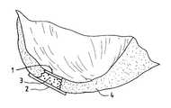
- FIG. 3Billustrates the graft site, after the defect has been sculpted to have undulating walls.
- FIG. 3Cillustrates the sculpted graft site with the hemostatic barrier ( 1 ), transplanted material ( 3 ), and cell-free covering-patch ( 2 ) in place within the articular surface cartilage ( 4 ).
- FIG. 4Aillustrates one embodiment of the surgical device of the instant invention showing cutting teeth ( 5 ) and protruding placement pin ( 6 ).
- FIG. 4Billustrates a second embodiment of the surgical device of the instant invention.
- FIG. 5is a diagram illustrating the modified differential response to compression and shearing forces by harder cartilage and softer cartilage after sculpting the graft site.
- FIG. 6Ais an MRI image of a pig knee showing cartilage defect in left (medial) condyle.
- FIG. 6Bis an MRI image of the same pig knee three months after treatment.
- This inventionconcerns the use of certain products that inhibit the formation of vascular tissue, for instance such as capillary loops projecting into the cartilage being established, during the process of autologous transplantation of chondrocytes into defects in the cartilage.
- vascular tissuefor instance such as capillary loops projecting into the cartilage being established, during the process of autologous transplantation of chondrocytes into defects in the cartilage.
- the formation of vascular tissue from the underlying bonewill tend to project into the new cartilage to be formed leading to appearance of cells other than the mesenchymal specialized chondrocytes desired.
- the contaminating cells introduced by the vascularizationmay give rise to encroachment and over-growth into the cartilage to be formed by the implanted chondrocytes.
- One of the types of commercial products which can be used in this inventionis Surgicel® (Ethicon Ltd., UK) which is absorbable after a period of 7-14 days.
- Surgicel®Ethicon Ltd., UK
- the use of this material in the method of the instant inventionis contrary to the normal use of a hemostatic device, such as Surgicel® as it is described in the package insert from Ethicon Ltd.
- a hemostatic material materialwill act like a gel-like artificial coagulate. If red blood cells should be present within the full-thickness defect of articular cartilage that is capped by such a hemostatic barrier, these blood cells will be chemically changed to hematin, and thus rendered unable to induce vascular growth.
- a hemostatic product used as a re-vascularization inhibitory barrier with or without fibrin adhesivessuch as for example the Surgicel®, is effective for the envisioned method as taught by the instant invention.
- Another part of this inventionis the use of a cell-free component, that is used as a patch covering the defective area of the joint into which the cultured chondrocytes/cartilage are being transplanted, using autologous chondrocytes for the transplantation.
- the method of the inventionalso contemplates the use of suitable allogenic chondrocytes or xenogenic chondrocytes for the repair of a cartilage defect.
- the instant inventionteaches methods for effective repair or treatment of cartilage defects in articular joint bone surfaces which comprises administering an agent or device to block vascular invasion into the cartilage site to be repaired, and also providing for a cell-free barrier which will isolate the repair site and keep transplanted cells in place.
- the instant inventionalso provides for a kit comprising a hemostatic barrier component for insertion into the site to be repaired, such that there is effective inhibition of vascularization into the site to be repaired; and once the chondrocytes to be transplanted are placed into the site to be repaired, a cell-free semi-permeable barrier is capped over the repair site such that the transplanted chondrocytes are held in place, but are still able to gain access to nutrients.
- Suitable hemostatic productswill be characterized by having the ability to inhibit the growth, or invasion of vascular tissue, osteocytes, fibroblasts etc. into the developing cartilage.
- a suitable hemostatic materialwill achieve the goal of the method of the instant invention in that vascular and cellular invasion into developing cartilage should be prevented in order to optimize the formation of cartilage and achieve repair of the full-thickness of any defects in the articular cartilage.
- the hemostatic barrierwill be stable for an extended period of time sufficient to allow for full cartilage repair, and then be able to be resorbed or otherwise broken down over time.
- Surgicel® W1912an absorbable hemostat containing oxidized regenerated sterile cellulose; Lot GG3DH, Ethicon Ltd. UK.
- BioGide®a commercially available type I collagen matrix pad; Geistlich Söhne, Switzerland.
- Suitable organic glue materialcan be found commercially, such as for example Tisseel® or Tissucol® (fibrin based adhesive; Immuno AG, Austria), Adhesive Protein (Cat. #A-2707, Sigma Chemical, USA), and Dow Coming Medical Adhesive B (Cat. #895-3, Dow Coming, USA).
- the surgical instruments contemplated by the instant inventioncan be manufactured from metal and/or plastic suitable for making single-use disposable, or multi-use reusable surgical instruments.
- the cutting instrumentmay contain cutting teeth that are fully circular or flat, or anything in between.
- cartilageis a relatively soft material it may be advantageous to manufacture hardened plastic cutting edges which will be able to sculpt cartilage without being able to damage bone.
- Such cutting instrumentscan be manufactured to incorporate openings for administration of fluid, suction removal of cutting debris and fluid, and fiber optic threads for illumination and visualization of the defect site.
- Surgicel®was first treated with a fixative, such as glutaric aldehyde. Briefly, Surgicel® was treated with 0.6% glutaric aldehyde for 1 minute, followed by several washings to eliminate glutaric aldehyde residues that may otherwise be toxic to tissue. Alternatively, the Surgicel® was treated with the fibrin adhesive called Tisseel® prior to treatment with glutaric aldehyde as described in Example 2.
- a fixativesuch as glutaric aldehyde.
- Tisseel®as the fibrin adhesive to coat the Surgicel®.
- fibrin adhesive or gluemay also be applied directly on the bottom of the lesion towards the bone, on which the Surgicel® is glued.
- the in vitro system usedin lieu of in vivo testing, consisted of a NUNCLONTM Delta 6-well sterile disposable plate for cell research work (NUNC, InterMed, Roskilde, Denmark). Each well measures approximately 4 cm in diameter.
- the fibrin adhesivecan be any adhesive which together with the fibrin component will produce a glue that can be tolerated in humans (Ihara, N, et al., Burns Incl. Therm. Inj., 1984, 10, 396).
- the inventionalso anticipates any other glue component that can be used in lieu of the fibrin adhesive.
- Tisseel® or Tissucol®Immuno AG, Vienna, Austria.
- the Tisseel® kitconsists of the following components:
- Tisseel®a lyophilized, virus-inactivated Sealer, containing clottable protein, thereof: fibrinogen, Plasma fibronectin (CIG) and Factor XIII, and Plasminogen.
- the Tisseel® kitcontains a DUPLOJECT® Application System.
- the fibrin adhesive or the two-component sealant using Tisseel® Kitis combined in the following manner according to the Immuno AG product insert sheet:
- Chondrocyteswere grown in minimal essential culture medium containing HAM F12 and 15 mM Hepes buffer and 5 to 7.5% autologous serum in a CO 2 incubator at 37° C. and handled in a Class 100 laboratory at Verigen Europe A/S, Symbion Science Park, Copenhagen, Denmark. Other compositions of culture medium may be used for culturing the chondrocytes.
- the cellswere trypsinized using trypsin EDTA for 5 to 10 minutes and counted using Trypan Blue viability staining in a Burker-Turk chamber. The cell count was adjusted to 7.5 ⁇ 10 5 cells per ml.
- One NUNCLONTMplatewas uncovered in the Class 100 laboratory.
- the “coated” hemostatic barrierwas placed on the bottom of the well in a NUNCLONTM Delta 6-well sterile disposable plate for cell research work.
- a small amount of tissue culture medium containing serumwas applied to be absorbed into the hemostatic barrier.
- tissue culture medium containing serumwas placed directly on top of the Hemostat, dispersed over the surface of the hemostatic barrier.
- the platewas then incubated in a CO 2 incubator at 37° C. for 60 minutes.
- An amount of 2 to 5 ml of tissue culture medium containing 5 to 7.5% serumwas carefully added to the well containing the cells avoiding splashing the cells by holding the pipette tip tangential to the side of the well when expelling the medium.
- Chondrocyteswere grown in minimal essential culture medium containing HAM F12 and 15 mM Hepes buffer and 5 to 7.5% autologous serum in a CO 2 incubator at 37° C. and handled in a Class 100 laboratory at Verigen Europe A/S, Symbion Science Park, Copenhagen, Denmark.
- the cellswere trypsinized using trypsin EDTA for 5 to 10 minutes and counted using Trypan Blue viability staining in a Burker-Turk chamber. The cell count was adjusted to 7.5 ⁇ 10 5 to 2 ⁇ 10 6 cells per ml.
- One NUNCLONTM platewas uncovered in the Class 100 laboratory.
- the Bio-Gide®can be used as a resorbable bilayer membrane which will be used as the patch or bandage covering the defective area of the joint into which the cultured chondrocytes are being transplanted as well as the hemostatic barrier.
- the Bio-Gide®is a pure collagen membrane obtained by standardized, controlled manufacturing processes (by E. D. Geistlich Söhne AG, CH-6110 Wolhusen). The collagen is extracted from veterinary certified pigs and is carefully purified to avoid antigenic reactions, and sterilized in double blisters by y-irradiation.
- the bilayer membranehas a porous surface and a dense surface. The membrane is made of collagen type I and type III without further cross-linking or chemical treatment.
- the collagenis resorbed within 24 weeks.
- the membraneretains its structural integrity even when wet and it can be fixed by sutures or nails.
- the membranemay also be “glued” using fibrin adhesive such as Tisseel®to the neighboring cartilage or tissue either instead of sutures or together with sutures.
- the Bio-Gide®was un-covered in a class 100 laboratory and placed under aseptic conditions on the bottom of the wells in a NUNCLONTM Delta 6-well sterile disposable plate for cell research work,—either with the porous surface of the bilayer membrane facing up or with the dense surface facing up.
- tissue culture medium containing serumwas placed directly on top of the Bio-Gide®, dispersed either over the porous or the dense surface of the Bio-Gide®.
- the platewas then incubated in a CO 2 incubator at 37° C. for 60 minutes. An amount of 2 to 5 ml of tissue culture medium containing 5 to 7.5% serum was carefully added to the well containing the cells avoiding splashing the cells by holding the pipette tip tangential to the side of the well when expelling the medium.
- Chondrocyteswere grown in minimal essential culture medium containing HAM F12 and 15 mM Hepes buffer and 5 to 7.5% autologous serum in a CO 2 incubator at 37° C. and handled in a Class 100 laboratory at Verigen Europe A/S, Symbion Science Park, Copenhagen, Denmark.
- the cellswere trypsinized using trypsin EDTA for 5 to 10 minutes and counted using Trypan Blue viability staining in a Burker-Turk chamber. The cell count was adjusted to 7.5 ⁇ 10 5 to 2 ⁇ 10 6 cells per ml.
- One NUNCLONTM platewas uncovered in the Class 100 laboratory.
- the Bio-Gide® used as a resorbable bilayer membranemay also be used together with an organic glue such as Tisseel® with additional, significantly higher content of Aprotinin than normally found in Tisseel®, as described in the product insert.
- an organic gluesuch as Tisseel® with additional, significantly higher content of Aprotinin than normally found in Tisseel®, as described in the product insert.
- the instant inventionprovides for a hybrid collagen patch where said patch is a collagen matrix with elevated levels of aprotinin component, preferably about 25,000 KIU/ml, in association with an organic matrix glue, where the collagen component is similar to the Bio-Gide® resorbable bilayer material or Type II collagen, and the organic glue is similar to the Tisseel® material.
- the hybrid collagen patchdoes not use any organic glue to adhere to the site of the repair.
- the method of the inventionteaches the use of surgical instruments to sculpt the walls of the graft site such that the walls are non-linear, and thus provide for undulated surfaces. It is also possible to shape the graft site such that the diameter of the site proximal to the bone surface is of a greater dimension then the opening distal to the bone, and at the surface of the cartilage.
- the preferred embodimentdescribes the sculpting of the walls of the graft site in an fashion similar to a threaded opening for receiving a bolt or screw (as illustrated in FIG. 3 B), thus providing mechanical resistance to the compression and or ejection of the grafted material from the graft site which can be described as “male” and “female” threading.
- the surgical instruments contemplated by the instant inventioncan be manufactured from metal and/or plastic suitable for making single-use disposable, or multi-use reusable surgical instruments.
- cartilageis a relatively soft material it may be advantageous to manufacture hardened plastic cutting edges which will be able to sculpt cartilage without being able to damage bone.
- Such cutting instrumentscan be manufactured to incorporate openings for administration of fluid, suction removal of cutting debris and fluid, and fiber optic threads for illumination and visualization of the defect site.
- the base of the instrumentmay have protruding point or pin-like structure which will assist in guiding and placing the instrument in the graft site. Of course such a pin would be designed to minimized damage to the underlying bone.
- the cutting surface of the instrumentmay be single toothed, or multi-toothed, or describe a screw-like pattern such as that in a metal tap used to generate threaded holes in metal parts
- the characteristic required of the cutting instrumentis that the resulting sculpted sides of the graft site is undulated, and non-linear.
- the cutting edge of the instrumentcan be shaped similar to that shown in FIG. 4A, or as in FIG. 4 B.
- the cutting edgemaybe flat, or circular in that it wraps around the diameter of the cutting instrument. Many other shapes can be designed to accomplish the purpose of the method of the invention to create an interface which provides for mechanical resistance to differential reaction to compression and shearing forces on the transplanted material and the surrounding material.
- a four month old mixed Yorkshire breed pigwas subjected to general anesthesia and placed on its back. The pig was washed and draped in a surgical suite at Harrington Arthritis Research Center, Phoenix, Ariz. The entire surgical procedure was performed aseptically. The left hind-leg and adjacent abdomen and inguinal area was cleaned with iodine. The knee joint was localized, and the patella localized. A medial incision was performed approximately 3 cm from the posterior part of the patella and the several subcutis, muscle layers and ligaments was cut approximately in order to get access to the medial femoral condyle.
- Adhesive ProteinA-2707, Sigma Chemical, USA
- Adhesive ProteinA-2707, Sigma Chemical, USA
- a second BioGide®was cut somewhat bigger in circumference than the lesion and was placed with dense side up (thus the porous side down towards the graft) as described above.
- the chondrocyte cell suspension(about 0.6 ml) was drawn up into the barrel of the syringe.
- a 23 gauge short needlewas switched for the 16 gauge needle, and the cell suspension was injected under the sutured covering-patch into the graft site (about 10 ⁇ 10 6 cells).
- the open edge of the capwas then glued prior to removal of the needle, and the needle carefully withdrawn. No leakage of cells was seen.
- the woundwas sutured and as above, no tourniquet was used, no bleeding was observed.
- the final skin layerswere sutured. No protrusion of the skin occurred after suturing, which indicates that there was no hematoma. Postoperative recovery was uneventful.
- kitscomprising the components useful for practicing the method of the invention, will allow for the convenient practice of the method of the invention in a surgical setting.
- a kit of the inventionwill provide sterile components suitable for easy use in the surgical environment, and will provide a suitable hemostatic barrier, suitable covering patch, and if needed organic glue.
- a kit of the inventionmay also provide sterile, cell-free matrix material suitable for supporting autologous chondrocytes that are to be implanted into an articular joint surface defect.
Landscapes
- Health & Medical Sciences (AREA)
- Transplantation (AREA)
- Life Sciences & Earth Sciences (AREA)
- Animal Behavior & Ethology (AREA)
- Oral & Maxillofacial Surgery (AREA)
- Orthopedic Medicine & Surgery (AREA)
- Engineering & Computer Science (AREA)
- Biomedical Technology (AREA)
- Heart & Thoracic Surgery (AREA)
- Vascular Medicine (AREA)
- Veterinary Medicine (AREA)
- Cardiology (AREA)
- General Health & Medical Sciences (AREA)
- Public Health (AREA)
- Rheumatology (AREA)
- Materials For Medical Uses (AREA)
- Prostheses (AREA)
- Medicines Containing Material From Animals Or Micro-Organisms (AREA)
- Surgical Instruments (AREA)
- Pharmaceuticals Containing Other Organic And Inorganic Compounds (AREA)
- Acyclic And Carbocyclic Compounds In Medicinal Compositions (AREA)
- Agricultural Chemicals And Associated Chemicals (AREA)
Abstract
Description
Claims (13)
Priority Applications (12)
| Application Number | Priority Date | Filing Date | Title |
|---|---|---|---|
| US09/690,252US6379367B1 (en) | 1996-08-30 | 2000-10-17 | Method instruments and kit for autologous transplantation |
| US09/970,323US6569172B2 (en) | 1996-08-30 | 2001-10-03 | Method, instruments, and kit for autologous transplantation |
| US10/055,105US6592598B2 (en) | 1996-08-30 | 2002-01-23 | Method, instruments, and kit for autologous transplantation |
| US10/091,006US6599300B2 (en) | 1996-08-30 | 2002-03-04 | Method, instruments, and kit for autologous transplantation |
| US10/090,922US6592599B2 (en) | 1996-08-30 | 2002-03-05 | Method, instruments, and kit for autologous transplantation |
| US10/093,129US6599301B2 (en) | 1996-08-30 | 2002-03-06 | Method, instruments, and kit for autologous transplantation |
| US10/121,249US20020173806A1 (en) | 1996-08-30 | 2002-04-12 | Method for autologous transplantation |
| US10/397,404US7137989B2 (en) | 1996-08-30 | 2003-03-26 | Method, instruments, and kit for autologous transplantation |
| US10/444,391US7048750B2 (en) | 1996-08-30 | 2003-05-23 | Method, instruments, and kits for autologous transplantation |
| US10/962,868US20060025786A1 (en) | 1996-08-30 | 2004-10-12 | Method for autologous transplantation |
| US11/375,183US20060167483A1 (en) | 1996-08-30 | 2006-03-14 | Method, instruments, and kit for autologous transplantation |
| US11/375,180US20060195122A1 (en) | 1996-08-30 | 2006-03-14 | Method, instruments, and kit for autologous transplantation |
Applications Claiming Priority (4)
| Application Number | Priority Date | Filing Date | Title |
|---|---|---|---|
| US08/704,891US5759190A (en) | 1996-08-30 | 1996-08-30 | Method and kit for autologous transplantation |
| US08/857,090US5989269A (en) | 1996-08-30 | 1997-05-15 | Method, instruments and kit for autologous transplantation |
| US09/320,246US6283980B1 (en) | 1996-08-30 | 1999-05-26 | Method, instruments, and kit for autologous transplantation |
| US09/690,252US6379367B1 (en) | 1996-08-30 | 2000-10-17 | Method instruments and kit for autologous transplantation |
Related Parent Applications (1)
| Application Number | Title | Priority Date | Filing Date |
|---|---|---|---|
| US09/320,246ContinuationUS6283980B1 (en) | 1996-08-30 | 1999-05-26 | Method, instruments, and kit for autologous transplantation |
Related Child Applications (2)
| Application Number | Title | Priority Date | Filing Date |
|---|---|---|---|
| US09/970,323Continuation-In-PartUS6569172B2 (en) | 1996-08-30 | 2001-10-03 | Method, instruments, and kit for autologous transplantation |
| US10/055,105ContinuationUS6592598B2 (en) | 1996-08-30 | 2002-01-23 | Method, instruments, and kit for autologous transplantation |
Publications (1)
| Publication Number | Publication Date |
|---|---|
| US6379367B1true US6379367B1 (en) | 2002-04-30 |
Family
ID=27107401
Family Applications (9)
| Application Number | Title | Priority Date | Filing Date |
|---|---|---|---|
| US08/857,090Expired - LifetimeUS5989269A (en) | 1996-08-30 | 1997-05-15 | Method, instruments and kit for autologous transplantation |
| US09/320,246Expired - LifetimeUS6283980B1 (en) | 1996-08-30 | 1999-05-26 | Method, instruments, and kit for autologous transplantation |
| US09/690,252Expired - LifetimeUS6379367B1 (en) | 1996-08-30 | 2000-10-17 | Method instruments and kit for autologous transplantation |
| US10/055,105Expired - LifetimeUS6592598B2 (en) | 1996-08-30 | 2002-01-23 | Method, instruments, and kit for autologous transplantation |
| US10/091,006Expired - LifetimeUS6599300B2 (en) | 1996-08-30 | 2002-03-04 | Method, instruments, and kit for autologous transplantation |
| US10/090,922Expired - LifetimeUS6592599B2 (en) | 1996-08-30 | 2002-03-05 | Method, instruments, and kit for autologous transplantation |
| US10/093,129Expired - LifetimeUS6599301B2 (en) | 1996-08-30 | 2002-03-06 | Method, instruments, and kit for autologous transplantation |
| US10/444,391Expired - Fee RelatedUS7048750B2 (en) | 1996-08-30 | 2003-05-23 | Method, instruments, and kits for autologous transplantation |
| US11/375,180AbandonedUS20060195122A1 (en) | 1996-08-30 | 2006-03-14 | Method, instruments, and kit for autologous transplantation |
Family Applications Before (2)
| Application Number | Title | Priority Date | Filing Date |
|---|---|---|---|
| US08/857,090Expired - LifetimeUS5989269A (en) | 1996-08-30 | 1997-05-15 | Method, instruments and kit for autologous transplantation |
| US09/320,246Expired - LifetimeUS6283980B1 (en) | 1996-08-30 | 1999-05-26 | Method, instruments, and kit for autologous transplantation |
Family Applications After (6)
| Application Number | Title | Priority Date | Filing Date |
|---|---|---|---|
| US10/055,105Expired - LifetimeUS6592598B2 (en) | 1996-08-30 | 2002-01-23 | Method, instruments, and kit for autologous transplantation |
| US10/091,006Expired - LifetimeUS6599300B2 (en) | 1996-08-30 | 2002-03-04 | Method, instruments, and kit for autologous transplantation |
| US10/090,922Expired - LifetimeUS6592599B2 (en) | 1996-08-30 | 2002-03-05 | Method, instruments, and kit for autologous transplantation |
| US10/093,129Expired - LifetimeUS6599301B2 (en) | 1996-08-30 | 2002-03-06 | Method, instruments, and kit for autologous transplantation |
| US10/444,391Expired - Fee RelatedUS7048750B2 (en) | 1996-08-30 | 2003-05-23 | Method, instruments, and kits for autologous transplantation |
| US11/375,180AbandonedUS20060195122A1 (en) | 1996-08-30 | 2006-03-14 | Method, instruments, and kit for autologous transplantation |
Country Status (26)
| Country | Link |
|---|---|
| US (9) | US5989269A (en) |
| EP (4) | EP1181908B1 (en) |
| JP (3) | JP2002502272A (en) |
| KR (2) | KR20030097603A (en) |
| CN (2) | CN1200656C (en) |
| AT (3) | ATE256443T1 (en) |
| AU (1) | AU731162B2 (en) |
| BR (1) | BR9711967A (en) |
| CA (2) | CA2264138C (en) |
| CZ (1) | CZ297248B6 (en) |
| DE (4) | DE69726896T2 (en) |
| DK (5) | DK1384452T3 (en) |
| ES (3) | ES2218697T3 (en) |
| HU (2) | HU225952B1 (en) |
| IL (1) | IL128580A (en) |
| MX (1) | MXPA99001794A (en) |
| NO (1) | NO990933D0 (en) |
| NZ (3) | NZ508145A (en) |
| PL (1) | PL331834A1 (en) |
| PT (3) | PT1006950E (en) |
| RU (2) | RU2214197C2 (en) |
| SG (2) | SG119137A1 (en) |
| SI (2) | SI1006950T1 (en) |
| SK (1) | SK285075B6 (en) |
| TR (3) | TR199900437T2 (en) |
| WO (1) | WO1998008469A2 (en) |
Cited By (101)
| Publication number | Priority date | Publication date | Assignee | Title |
|---|---|---|---|---|
| US20020116063A1 (en)* | 1999-08-02 | 2002-08-22 | Bruno Giannetti | Kit for chondrocyte cell transplantation |
| US20020119177A1 (en)* | 2000-12-21 | 2002-08-29 | Bowman Steven M. | Reinforced foam implants with enhanced integrity for soft tissue repair and regeneration |
| US20020127265A1 (en)* | 2000-12-21 | 2002-09-12 | Bowman Steven M. | Use of reinforced foam implants with enhanced integrity for soft tissue repair and regeneration |
| US6482209B1 (en) | 2001-06-14 | 2002-11-19 | Gerard A. Engh | Apparatus and method for sculpting the surface of a joint |
| US20030021827A1 (en)* | 2001-07-16 | 2003-01-30 | Prasanna Malaviya | Hybrid biologic/synthetic porous extracellular matrix scaffolds |
| US20030049299A1 (en)* | 2001-07-16 | 2003-03-13 | Prasanna Malaviya | Porous delivery scaffold and method |
| US20030078617A1 (en)* | 2001-07-16 | 2003-04-24 | Schwartz Herbert E. | Unitary surgical device and method |
| US20030147935A1 (en)* | 2000-12-21 | 2003-08-07 | Ethicon, Inc. | Use of reinforced foam implants with enhanced integrity for soft tissue repair and regeneration |
| US20030193104A1 (en)* | 2000-12-21 | 2003-10-16 | Melican Mora Carolynne | Reinforced tissue implants and methods of manufacture and use |
| US20030216669A1 (en)* | 2001-05-25 | 2003-11-20 | Imaging Therapeutics, Inc. | Methods and compositions for articular repair |
| US20030236523A1 (en)* | 2001-06-14 | 2003-12-25 | Johnson Wesley D. | Apparatus and method for minimally invasive total joint replacement |
| US20040078090A1 (en)* | 2002-10-18 | 2004-04-22 | Francois Binette | Biocompatible scaffolds with tissue fragments |
| US20040078077A1 (en)* | 2002-10-18 | 2004-04-22 | Francois Binette | Biocompatible scaffold for ligament or tendon repair |
| US20040133276A1 (en)* | 2002-10-07 | 2004-07-08 | Imaging Therapeutics, Inc. | Minimally invasive joint implant with 3-Dimensional geometry matching the articular surfaces |
| US20040136968A1 (en)* | 2002-09-27 | 2004-07-15 | Verigen Ag | Autologous cells on a support matrix for tissue repair |
| US20040143344A1 (en)* | 2001-07-16 | 2004-07-22 | Prasanna Malaviya | Implantable tissue repair device and method |
| US20040147927A1 (en)* | 2002-11-07 | 2004-07-29 | Imaging Therapeutics, Inc. | Methods for determining meniscal size and shape and for devising treatment |
| US20040166169A1 (en)* | 2002-07-15 | 2004-08-26 | Prasanna Malaviya | Porous extracellular matrix scaffold and method |
| US20040219182A1 (en)* | 2003-04-29 | 2004-11-04 | Gomes Katherine A. | Novel glue for cartilage repair |
| US20040236424A1 (en)* | 2001-05-25 | 2004-11-25 | Imaging Therapeutics, Inc. | Patient selectable joint arthroplasty devices and surgical tools facilitating increased accuracy, speed and simplicity in performing total and partial joint arthroplasty |
| US20040267362A1 (en)* | 2003-06-30 | 2004-12-30 | Julia Hwang | Scaffold for connective tissue repair |
| US20050064042A1 (en)* | 2003-04-29 | 2005-03-24 | Musculoskeletal Transplant Foundation | Cartilage implant plug with fibrin glue and method for implantation |
| WO2005039528A2 (en) | 2003-10-23 | 2005-05-06 | Katharina Beschorner | Composition for the treatment of arthrosis/arthritis, especially for treating joints |
| US20050113937A1 (en)* | 2003-11-26 | 2005-05-26 | Francois Binette | Conformable tissue repair implant capable of injection delivery |
| US20050177249A1 (en)* | 2004-02-09 | 2005-08-11 | Kladakis Stephanie M. | Scaffolds with viable tissue |
| US20050234461A1 (en)* | 2001-05-25 | 2005-10-20 | Burdulis Albert G Jr | Surgical tools facilitating increased accuracy, speed and simplicity in performing joint arthroplasty |
| US20050234549A1 (en)* | 2004-04-20 | 2005-10-20 | Kladakis Stephanie M | Meniscal repair scaffold |
| US20050232967A1 (en)* | 2004-04-20 | 2005-10-20 | Kladakis Stephanie M | Nonwoven tissue scaffold |
| US20050251268A1 (en)* | 2003-05-16 | 2005-11-10 | Musculoskeletal Transplant Foundation | Cartilage allograft plug |
| US20050249771A1 (en)* | 2004-05-04 | 2005-11-10 | Prasanna Malaviya | Hybrid biologic-synthetic bioabsorbable scaffolds |
| US20060111778A1 (en)* | 2004-10-29 | 2006-05-25 | Michalow Alexander E | Methods of promoting healing of cartilage defects and method of causing stem cells to differentiate by the articular chondrocyte pathway |
| US20060128296A1 (en)* | 2004-10-29 | 2006-06-15 | Schwan Wade E | Intestine processing device and associated method |
| US20060135638A1 (en)* | 2004-12-22 | 2006-06-22 | Pedrozo Hugo A | Method for organizing the assembly of collagen fibers and compositions formed therefrom |
| US7160333B2 (en) | 2000-08-04 | 2007-01-09 | Depuy Orthopaedics, Inc. | Reinforced small intestinal submucosa |
| US20070031470A1 (en)* | 2004-04-20 | 2007-02-08 | Depuy Mitek, Inc. | Nonwoven tissue scaffold |
| US20070073394A1 (en)* | 2003-10-28 | 2007-03-29 | Xiros Plc | Repair of damaged tissue on a bone site |
| US20070198022A1 (en)* | 2001-05-25 | 2007-08-23 | Conformis, Inc. | Patient Selectable Joint Arthroplasty Devices and Surgical Tools |
| US20080077251A1 (en)* | 1999-06-07 | 2008-03-27 | Chen Silvia S | Cleaning and devitalization of cartilage |
| US7361195B2 (en) | 2001-07-16 | 2008-04-22 | Depuy Products, Inc. | Cartilage repair apparatus and method |
| US20080281426A1 (en)* | 2001-05-25 | 2008-11-13 | Conformis, Inc. | Patient Selectable Joint Arthroplasty Devices and Surgical Tools |
| US7476250B1 (en)* | 1999-04-06 | 2009-01-13 | Mansmann Kevin A | Semi-permeable membranes to assist in cartilage repair |
| US20090024223A1 (en)* | 2007-07-16 | 2009-01-22 | Chen Silvia S | Crafting of cartilage |
| US20090024229A1 (en)* | 2007-07-16 | 2009-01-22 | Chen Silvia S | Devitalization and recellularization of cartilage |
| US20090214614A1 (en)* | 2005-09-02 | 2009-08-27 | Interface Biotech A/S | Method for Cell Implantation |
| US7595062B2 (en) | 2005-07-28 | 2009-09-29 | Depuy Products, Inc. | Joint resurfacing orthopaedic implant and associated method |
| US20100030340A1 (en)* | 1998-06-30 | 2010-02-04 | Wolfinbarger Jr Lloyd | Plasticized Grafts and Methods of Making and Using Same |
| US7799084B2 (en) | 2002-10-23 | 2010-09-21 | Mako Surgical Corp. | Modular femoral component for a total knee joint replacement for minimally invasive implantation |
| US7815926B2 (en) | 2005-07-11 | 2010-10-19 | Musculoskeletal Transplant Foundation | Implant for articular cartilage repair |
| US7837740B2 (en) | 2007-01-24 | 2010-11-23 | Musculoskeletal Transplant Foundation | Two piece cancellous construct for cartilage repair |
| US20100298894A1 (en)* | 2006-02-06 | 2010-11-25 | Conformis, Inc. | Patient-Specific Joint Arthroplasty Devices for Ligament Repair |
| US20100322908A1 (en)* | 2008-02-29 | 2010-12-23 | Hanne Everland | Compositions and methods for augmentation and regeneration of living tissue in a subject |
| US7871440B2 (en) | 2006-12-11 | 2011-01-18 | Depuy Products, Inc. | Unitary surgical device and method |
| US20110014267A1 (en)* | 2008-02-29 | 2011-01-20 | Hanne Everland | Biosynthetic cartilaginous matrix and methods for their production |
| US7881768B2 (en) | 1998-09-14 | 2011-02-01 | The Board Of Trustees Of The Leland Stanford Junior University | Assessing the condition of a joint and devising treatment |
| US7901461B2 (en) | 2003-12-05 | 2011-03-08 | Ethicon, Inc. | Viable tissue repair implants and methods of use |
| US7901457B2 (en) | 2003-05-16 | 2011-03-08 | Musculoskeletal Transplant Foundation | Cartilage allograft plug |
| US8012205B2 (en) | 2001-07-16 | 2011-09-06 | Depuy Products, Inc. | Cartilage repair and regeneration device |
| US8016867B2 (en) | 1999-07-23 | 2011-09-13 | Depuy Mitek, Inc. | Graft fixation device and method |
| US8025896B2 (en) | 2001-07-16 | 2011-09-27 | Depuy Products, Inc. | Porous extracellular matrix scaffold and method |
| US8036729B2 (en) | 1998-09-14 | 2011-10-11 | The Board Of Trustees Of The Leland Stanford Junior University | Assessing the condition of a joint and devising treatment |
| US8092529B2 (en) | 2001-07-16 | 2012-01-10 | Depuy Products, Inc. | Meniscus regeneration device |
| US8234097B2 (en) | 2001-05-25 | 2012-07-31 | Conformis, Inc. | Automated systems for manufacturing patient-specific orthopedic implants and instrumentation |
| US8265730B2 (en) | 1998-09-14 | 2012-09-11 | The Board Of Trustees Of The Leland Stanford Junior University | Assessing the condition of a joint and preventing damage |
| US8292968B2 (en) | 2004-10-12 | 2012-10-23 | Musculoskeletal Transplant Foundation | Cancellous constructs, cartilage particles and combinations of cancellous constructs and cartilage particles |
| US8337537B2 (en) | 2001-07-16 | 2012-12-25 | Depuy Products, Inc. | Device from naturally occurring biologically derived materials |
| US8337507B2 (en) | 2001-05-25 | 2012-12-25 | Conformis, Inc. | Methods and compositions for articular repair |
| US8366787B2 (en) | 2000-08-04 | 2013-02-05 | Depuy Products, Inc. | Hybrid biologic-synthetic bioabsorbable scaffolds |
| US8435551B2 (en) | 2007-03-06 | 2013-05-07 | Musculoskeletal Transplant Foundation | Cancellous construct with support ring for repair of osteochondral defects |
| US8439926B2 (en) | 2001-05-25 | 2013-05-14 | Conformis, Inc. | Patient selectable joint arthroplasty devices and surgical tools |
| US8449561B2 (en) | 1999-07-23 | 2013-05-28 | Depuy Mitek, Llc | Graft fixation device combination |
| US8480754B2 (en) | 2001-05-25 | 2013-07-09 | Conformis, Inc. | Patient-adapted and improved articular implants, designs and related guide tools |
| US8545569B2 (en) | 2001-05-25 | 2013-10-01 | Conformis, Inc. | Patient selectable knee arthroplasty devices |
| US8556983B2 (en) | 2001-05-25 | 2013-10-15 | Conformis, Inc. | Patient-adapted and improved orthopedic implants, designs and related tools |
| US8617242B2 (en) | 2001-05-25 | 2013-12-31 | Conformis, Inc. | Implant device and method for manufacture |
| US8623026B2 (en) | 2006-02-06 | 2014-01-07 | Conformis, Inc. | Patient selectable joint arthroplasty devices and surgical tools incorporating anatomical relief |
| US8682052B2 (en) | 2008-03-05 | 2014-03-25 | Conformis, Inc. | Implants for altering wear patterns of articular surfaces |
| US8697139B2 (en) | 2004-09-21 | 2014-04-15 | Frank M. Phillips | Method of intervertebral disc treatment using articular chondrocyte cells |
| US8735773B2 (en) | 2007-02-14 | 2014-05-27 | Conformis, Inc. | Implant device and method for manufacture |
| US8771365B2 (en) | 2009-02-25 | 2014-07-08 | Conformis, Inc. | Patient-adapted and improved orthopedic implants, designs, and related tools |
| US8808303B2 (en) | 2009-02-24 | 2014-08-19 | Microport Orthopedics Holdings Inc. | Orthopedic surgical guide |
| US8882847B2 (en) | 2001-05-25 | 2014-11-11 | Conformis, Inc. | Patient selectable knee joint arthroplasty devices |
| US8895045B2 (en) | 2003-03-07 | 2014-11-25 | Depuy Mitek, Llc | Method of preparation of bioabsorbable porous reinforced tissue implants and implants thereof |
| US8951260B2 (en) | 2001-05-25 | 2015-02-10 | Conformis, Inc. | Surgical cutting guide |
| US9017334B2 (en) | 2009-02-24 | 2015-04-28 | Microport Orthopedics Holdings Inc. | Patient specific surgical guide locator and mount |
| US9020788B2 (en) | 1997-01-08 | 2015-04-28 | Conformis, Inc. | Patient-adapted and improved articular implants, designs and related guide tools |
| US9125971B2 (en) | 1998-06-30 | 2015-09-08 | Lifenet Health | Plasticized bone and soft tissue grafts and methods of making and using same |
| US9286686B2 (en) | 1998-09-14 | 2016-03-15 | The Board Of Trustees Of The Leland Stanford Junior University | Assessing the condition of a joint and assessing cartilage loss |
| US9308091B2 (en) | 2001-05-25 | 2016-04-12 | Conformis, Inc. | Devices and methods for treatment of facet and other joints |
| US9486226B2 (en) | 2012-04-18 | 2016-11-08 | Conformis, Inc. | Tibial guides, tools, and techniques for resecting the tibial plateau |
| US9603711B2 (en) | 2001-05-25 | 2017-03-28 | Conformis, Inc. | Patient-adapted and improved articular implants, designs and related guide tools |
| US9649117B2 (en) | 2009-02-24 | 2017-05-16 | Microport Orthopedics Holdings, Inc. | Orthopedic surgical guide |
| US9675471B2 (en) | 2012-06-11 | 2017-06-13 | Conformis, Inc. | Devices, techniques and methods for assessing joint spacing, balancing soft tissues and obtaining desired kinematics for joint implant components |
| US9701940B2 (en) | 2005-09-19 | 2017-07-11 | Histogenics Corporation | Cell-support matrix having narrowly defined uniformly vertically and non-randomly organized porosity and pore density and a method for preparation thereof |
| US9936688B2 (en) | 2000-09-12 | 2018-04-10 | Lifenet Health | Process for devitalizing soft-tissue engineered medical implants, and devitalized soft-tissue medical implants produced |
| US10077420B2 (en) | 2014-12-02 | 2018-09-18 | Histogenics Corporation | Cell and tissue culture container |
| US10085839B2 (en) | 2004-01-05 | 2018-10-02 | Conformis, Inc. | Patient-specific and patient-engineered orthopedic implants |
| US10583220B2 (en) | 2003-08-11 | 2020-03-10 | DePuy Synthes Products, Inc. | Method and apparatus for resurfacing an articular surface |
| US12108959B2 (en) | 2019-05-29 | 2024-10-08 | Wright Medical Technology, Inc. | Preparing a tibia for receiving tibial implant component of a replacement ankle |
| US12383287B2 (en) | 2009-02-24 | 2025-08-12 | Microport Orthopedics Holdings, Inc. | Systems and methods for installing an orthopedic implant |
| US12396739B2 (en) | 2020-01-17 | 2025-08-26 | Wright Medical Technology, Inc. | Guidance tools, systems, and methods |
| US12440227B2 (en) | 2022-01-05 | 2025-10-14 | Wright Medical Technology, Inc. | Preparing a tibia for receiving tibial implant component of a replacement ankle |
Families Citing this family (126)
| Publication number | Priority date | Publication date | Assignee | Title |
|---|---|---|---|---|
| US20050186673A1 (en)* | 1995-02-22 | 2005-08-25 | Ed. Geistlich Soehne Ag Fuer Chemistrie Industrie | Collagen carrier of therapeutic genetic material, and method |
| US5940807A (en)* | 1996-05-24 | 1999-08-17 | Purcell; Daniel S. | Automated and independently accessible inventory information exchange system |
| US20020173806A1 (en)* | 1996-08-30 | 2002-11-21 | Verigen Transplantation Service International (Vtsi) Ag | Method for autologous transplantation |
| US20060025786A1 (en)* | 1996-08-30 | 2006-02-02 | Verigen Transplantation Service International (Vtsi) Ag | Method for autologous transplantation |
| US6569172B2 (en)* | 1996-08-30 | 2003-05-27 | Verigen Transplantation Service International (Vtsi) | Method, instruments, and kit for autologous transplantation |
| US5989269A (en)* | 1996-08-30 | 1999-11-23 | Vts Holdings L.L.C. | Method, instruments and kit for autologous transplantation |
| US20070100462A1 (en) | 2001-05-25 | 2007-05-03 | Conformis, Inc | Joint Arthroplasty Devices |
| US20030180263A1 (en)* | 2002-02-21 | 2003-09-25 | Peter Geistlich | Resorbable extracellular matrix for reconstruction of bone |
| US20050186283A1 (en)* | 1997-10-10 | 2005-08-25 | Ed. Geistlich Soehne Ag Fuer Chemistrie Industrie | Collagen carrier of therapeutic genetic material, and method |
| US9034315B2 (en)* | 1997-10-10 | 2015-05-19 | Ed. Geistlich Soehne Ag Fuer Chemische Industrie | Cell-charged multi-layer collagen membrane |
| US8858981B2 (en)* | 1997-10-10 | 2014-10-14 | Ed. Geistlich Soehne Fuer Chemistrie Industrie | Bone healing material comprising matrix carrying bone-forming cells |
| EP1656960A1 (en)* | 1998-08-14 | 2006-05-17 | Verigen AG | Methods, instruments and materials for chondrocyte cell transplantation |
| WO2000009179A2 (en)* | 1998-08-14 | 2000-02-24 | Verigen Transplantation Service International (Vtsi) Ag | Methods, instruments and materials for chondrocyte cell transplantation |
| US20020081732A1 (en) | 2000-10-18 | 2002-06-27 | Bowlin Gary L. | Electroprocessing in drug delivery and cell encapsulation |
| US7615373B2 (en) | 1999-02-25 | 2009-11-10 | Virginia Commonwealth University Intellectual Property Foundation | Electroprocessed collagen and tissue engineering |
| ES2239608T3 (en)* | 1999-07-28 | 2005-10-01 | Interface Biotech A/S | IN VITRO REPAIR OF BONE AND / OR CARTILAGO DEFECTS. |
| JP2003505198A (en)* | 1999-08-02 | 2003-02-12 | フェリゲン トランスプランタツィオン セルヴィス インターナツィオナール アーゲー | Kit for cell transplantation in cartilage matrix |
| ATE423517T1 (en)* | 1999-12-03 | 2009-03-15 | Univ Leeds | RESTORATION OF DAMAGED TISSUE |
| US6626945B2 (en)* | 2000-03-14 | 2003-09-30 | Chondrosite, Llc | Cartilage repair plug |
| US6610067B2 (en) | 2000-05-01 | 2003-08-26 | Arthrosurface, Incorporated | System and method for joint resurface repair |
| US6520964B2 (en) | 2000-05-01 | 2003-02-18 | Std Manufacturing, Inc. | System and method for joint resurface repair |
| US7678151B2 (en) | 2000-05-01 | 2010-03-16 | Ek Steven W | System and method for joint resurface repair |
| US8177841B2 (en) | 2000-05-01 | 2012-05-15 | Arthrosurface Inc. | System and method for joint resurface repair |
| US7163541B2 (en) | 2002-12-03 | 2007-01-16 | Arthrosurface Incorporated | Tibial resurfacing system |
| US7713305B2 (en)* | 2000-05-01 | 2010-05-11 | Arthrosurface, Inc. | Articular surface implant |
| US7618462B2 (en) | 2000-05-01 | 2009-11-17 | Arthrosurface Incorporated | System and method for joint resurface repair |
| ES2324282T3 (en)* | 2000-05-01 | 2009-08-04 | Arthro Surface, Inc. | SYSTEM OF REPAIR OF THE SURFACE OF AN ARTICULATION. |
| MXPA03001914A (en) | 2000-09-01 | 2004-05-24 | Univ Virginia Commonwealth | Electroprocessed fibrin-based matrices and tissues. |
| US20040151705A1 (en) | 2002-03-22 | 2004-08-05 | Shuichi Mizuno | Neo-cartilage constructs and a method for preparation thereof |
| US6713085B2 (en) | 2001-04-27 | 2004-03-30 | Ed. Geistlich Soehne Ag Fuer Chemische Industrie | Method and membrane for mucosa regeneration |
| IL160883A0 (en)* | 2001-09-24 | 2004-08-31 | Verigen Ag | Autologous growth factor cocktail composition, method of production and use |
| US6712822B2 (en)* | 2001-10-01 | 2004-03-30 | Scandius Biomedical, Inc. | Apparatus and method for the repair of articular cartilage defects |
| CA2412012C (en)* | 2001-11-20 | 2011-08-02 | Ed. Geistlich Soehne Ag Fuer Chemische Industrie | Resorbable extracellular matrix containing collagen i and collagen ii for reconstruction of cartilage |
| CA2472239A1 (en)* | 2002-01-22 | 2003-07-31 | Pfizer Inc. | 3-(imidazolyl)-2-aminopropanoic acids for use as tafi-a inhibitors for the treatment of thrombotic diseases |
| US7537780B2 (en)* | 2002-03-22 | 2009-05-26 | Histogenics Corporation | Method for preparing and implanting a cartilage construct to treat cartilage lesions |
| JP2005523770A (en)* | 2002-05-01 | 2005-08-11 | ベリーゲン アーゲー | Injectable chondrocyte implant |
| SE0201479D0 (en)* | 2002-05-16 | 2002-05-16 | Pharmacia Groningen Bv | Kit and method in eye surgery |
| AU2003261497B2 (en) | 2002-11-08 | 2009-02-26 | Howmedica Osteonics Corp. | Laser-produced porous surface |
| US20060147332A1 (en) | 2004-12-30 | 2006-07-06 | Howmedica Osteonics Corp. | Laser-produced porous structure |
| US7901408B2 (en) | 2002-12-03 | 2011-03-08 | Arthrosurface, Inc. | System and method for retrograde procedure |
| US7914545B2 (en) | 2002-12-03 | 2011-03-29 | Arthrosurface, Inc | System and method for retrograde procedure |
| AU2003900620A0 (en)* | 2003-02-12 | 2003-02-27 | Australian Surgical Design And Manufacture Pty Limited | Arthroscopic chondrocyte implantation method and device |
| US8388624B2 (en) | 2003-02-24 | 2013-03-05 | Arthrosurface Incorporated | Trochlear resurfacing system and method |
| AU2004246749A1 (en)* | 2003-06-12 | 2004-12-23 | Interface Biotech A/S | A method for cell implantation |
| US20050046915A1 (en)* | 2003-08-22 | 2005-03-03 | Fuji Photo Film Co., Ltd. | Hologram recording material composition, hologram recording material and hologram recording method |
| RU2242981C1 (en)* | 2003-10-31 | 2004-12-27 | ЗАО "РеМеТэкс" | Biotransplant and method for treating degenerative and traumatic diseases of articular cartilage |
| RU2242980C1 (en)* | 2003-10-31 | 2004-12-27 | ЗАО "РеМеТэкс" | Culture of genetically non-modified human chondroprogenitory cells and method for their preparing |
| AU2004293042A1 (en) | 2003-11-20 | 2005-06-09 | Arthrosurface, Inc. | Retrograde delivery of resurfacing devices |
| US7951163B2 (en) | 2003-11-20 | 2011-05-31 | Arthrosurface, Inc. | Retrograde excision system and apparatus |
| RU2259852C1 (en)* | 2003-11-24 | 2005-09-10 | Государственное учреждение "Нижегородский научно-исследовательский институт травматологии и ортопедии Министерства здравоохранения Российской Федерации" | Method for preparing cell-free dermal transplantation matrix in treating wounds of various etiology including burns in particular |
| RU2260379C2 (en)* | 2003-11-26 | 2005-09-20 | Юшков Борис Германович | Method for obtaining autoprostheses for plasty of tissues and organs |
| WO2005058207A1 (en) | 2003-12-11 | 2005-06-30 | Isto Technologies, Inc. | Particulate cartilage system |
| RU2283050C2 (en)* | 2004-02-09 | 2006-09-10 | Федеральное государственное учреждение "Российский научно-исследовательский институт травматологии и ортопедии им.Р.Р.Вредена Федерального агентства по здравоохранению и социальному развитию" (ФГУ "РНИИТО им.Р.Р.Вредена Росздрава") | Method for treating knee joint cartilage for local injuries |
| EP1576957A1 (en)* | 2004-03-18 | 2005-09-21 | Universiteit Twente | Tissue repair using pluripotent cells |
| RU2265442C1 (en)* | 2004-05-14 | 2005-12-10 | ЗАО "РеМеТэкс" | Biotransplant and method for treating the cases of osteoporosis |
| CA2470463C (en)* | 2004-06-09 | 2012-09-18 | Lornamead Brands, Inc. | Tooth whitening products and methods of making the same |
| RU2268738C1 (en)* | 2004-06-18 | 2006-01-27 | Владимир Никитич Ярыгин | Method for reconstructing hyaline articular cartilage |
| WO2006004885A2 (en) | 2004-06-28 | 2006-01-12 | Arthrosurface, Inc. | System for articular surface replacement |
| RU2270015C1 (en)* | 2004-07-23 | 2006-02-20 | Владимир Александрович Макаров | Hemostatic glue |
| RU2262937C1 (en)* | 2004-07-23 | 2005-10-27 | Макаров Владимир Александрович | Hemostatic glue |
| WO2006022671A1 (en)* | 2004-07-30 | 2006-03-02 | Histogenics Corporation | Method for in situ repair of injured, damaged, diseased or aged articular cartilage using neo-cartilage constructs and a method for preparation thereof |
| US7828853B2 (en) | 2004-11-22 | 2010-11-09 | Arthrosurface, Inc. | Articular surface implant and delivery system |
| CN100384390C (en)* | 2005-01-13 | 2008-04-30 | 中国人民解放军第三军医大学第一附属医院 | Tissue engineered bone and cartilage composite tissue and its in vitro construction method |
| DE102005030614B4 (en)* | 2005-06-30 | 2014-05-08 | Biotissue Ag | Cell-free graft, its use, process for its preparation, matrix thus produced with gel and process for the preparation of this matrix with gel |
| WO2007025290A2 (en) | 2005-08-26 | 2007-03-01 | Isto Technologies, Inc. | Implants and methods for repair, replacement and treatment of joint disease |
| RU2295980C1 (en)* | 2005-09-08 | 2007-03-27 | Григорий Федорович Назаренко | Implant for repairing bone and/or cartilage tissue and method for producing it |
| WO2007062079A2 (en) | 2005-11-21 | 2007-05-31 | Philipp Lang | Devices and methods for treating facet joints, uncovertebral joints, costovertebral joints and other joints |
| US8728387B2 (en) | 2005-12-06 | 2014-05-20 | Howmedica Osteonics Corp. | Laser-produced porous surface |
| CA2572095C (en)* | 2005-12-30 | 2009-12-08 | Howmedica Osteonics Corp. | Laser-produced implants |
| US20070288021A1 (en)* | 2006-06-07 | 2007-12-13 | Howmedica Osteonics Corp. | Flexible joint implant |
| US8147861B2 (en)* | 2006-08-15 | 2012-04-03 | Howmedica Osteonics Corp. | Antimicrobial implant |
| WO2008029264A2 (en)* | 2006-09-07 | 2008-03-13 | Ed. Geistlich Soehne Ag Fuer Chemische Industrie | Method of treating bone cancer |
| KR20080037883A (en)* | 2006-10-27 | 2008-05-02 | 세원셀론텍(주) | Medical kits and how to use them |
| US9358029B2 (en) | 2006-12-11 | 2016-06-07 | Arthrosurface Incorporated | Retrograde resection apparatus and method |
| US8163549B2 (en) | 2006-12-20 | 2012-04-24 | Zimmer Orthobiologics, Inc. | Method of obtaining viable small tissue particles and use for tissue repair |
| US7758643B2 (en)* | 2007-02-26 | 2010-07-20 | Biomet Sports Medicine, Llc | Stable cartilage defect repair plug |
| US20090012629A1 (en) | 2007-04-12 | 2009-01-08 | Isto Technologies, Inc. | Compositions and methods for tissue repair |
| US20090117087A1 (en)* | 2007-04-13 | 2009-05-07 | Wake Forest University | Methods and compositions for printing biologically compatible nanotube composites of autologous tissue |
| US9421304B2 (en) | 2007-07-03 | 2016-08-23 | Histogenics Corporation | Method for improvement of differentiation of mesenchymal stem cells using a double-structured tissue implant |
| US20090054984A1 (en) | 2007-08-20 | 2009-02-26 | Histogenics Corporation | Method For Use Of A Double-Structured Tissue Implant For Treatment Of Tissue Defects |
| US8685107B2 (en) | 2007-07-03 | 2014-04-01 | Histogenics Corporation | Double-structured tissue implant and a method for preparation and use thereof |
| WO2009014718A1 (en) | 2007-07-24 | 2009-01-29 | Porex Corporation | Porous laser sintered articles |
| EP2262448A4 (en) | 2008-03-03 | 2014-03-26 | Arthrosurface Inc | Bone resurfacing system and method |
| GB0812631D0 (en)* | 2008-07-10 | 2008-08-20 | Imp Innovations Ltd | Modular knee implants |
| US10945743B2 (en) | 2009-04-17 | 2021-03-16 | Arthrosurface Incorporated | Glenoid repair system and methods of use thereof |
| AU2010236182A1 (en) | 2009-04-17 | 2011-11-24 | Arthrosurface Incorporated | Glenoid resurfacing system and method |
| WO2010121250A1 (en) | 2009-04-17 | 2010-10-21 | Arthrosurface Incorporated | Glenoid resurfacing system and method |
| EP2542165A4 (en) | 2010-03-05 | 2015-10-07 | Arthrosurface Inc | Tibial resurfacing system and method |
| RU2428128C1 (en)* | 2010-03-23 | 2011-09-10 | Государственное образовательное учреждение высшего профессионального образования "Санкт-Петербургская государственная медицинская академия им. И.И. Мечникова Федерального агентства по здравоохранению и социальному развитию" | Method of fixation of cartilage plate in treatment of defects of cartilage surface of femur condyles |
| EP2625263B1 (en) | 2010-10-08 | 2020-03-11 | Terumo BCT, Inc. | Configurable methods and systems of growing and harvesting cells in a hollow fiber bioreactor system |
| US9066716B2 (en) | 2011-03-30 | 2015-06-30 | Arthrosurface Incorporated | Suture coil and suture sheath for tissue repair |
| WO2013081071A1 (en)* | 2011-12-02 | 2013-06-06 | テルモ株式会社 | Device for cell transplantation |
| EP2804565B1 (en) | 2011-12-22 | 2018-03-07 | Arthrosurface Incorporated | System for bone fixation |
| US9364896B2 (en) | 2012-02-07 | 2016-06-14 | Medical Modeling Inc. | Fabrication of hybrid solid-porous medical implantable devices with electron beam melting technology |
| US9135374B2 (en) | 2012-04-06 | 2015-09-15 | Howmedica Osteonics Corp. | Surface modified unit cell lattice structures for optimized secure freeform fabrication |
| US9180010B2 (en) | 2012-04-06 | 2015-11-10 | Howmedica Osteonics Corp. | Surface modified unit cell lattice structures for optimized secure freeform fabrication |
| US9186053B2 (en)* | 2012-05-03 | 2015-11-17 | Covidien Lp | Methods of using light to repair hernia defects |
| WO2014008126A1 (en) | 2012-07-03 | 2014-01-09 | Arthrosurface Incorporated | System and method for joint resurfacing and repair |
| US10016527B2 (en) | 2012-10-23 | 2018-07-10 | Orthovita, Inc. | Materials and methods for repair of cartilage defects |
| US20140178343A1 (en) | 2012-12-21 | 2014-06-26 | Jian Q. Yao | Supports and methods for promoting integration of cartilage tissue explants |
| US9492200B2 (en) | 2013-04-16 | 2016-11-15 | Arthrosurface Incorporated | Suture system and method |
| DK2853384T3 (en)* | 2013-09-27 | 2017-02-20 | Skulle Implants Oy | METHOD OF COATING AND COATING SURFACE |
| WO2015073913A1 (en) | 2013-11-16 | 2015-05-21 | Terumo Bct, Inc. | Expanding cells in a bioreactor |
| US10624748B2 (en) | 2014-03-07 | 2020-04-21 | Arthrosurface Incorporated | System and method for repairing articular surfaces |
| US11607319B2 (en) | 2014-03-07 | 2023-03-21 | Arthrosurface Incorporated | System and method for repairing articular surfaces |
| US9931219B2 (en) | 2014-03-07 | 2018-04-03 | Arthrosurface Incorporated | Implant and anchor assembly |
| CN106232800B (en) | 2014-03-25 | 2020-07-03 | 泰尔茂比司特公司 | Passive replacement of media |
| JP6830059B2 (en) | 2014-09-26 | 2021-02-17 | テルモ ビーシーティー、インコーポレーテッド | Scheduled cell feeding |
| CA2985398C (en)* | 2015-05-08 | 2021-08-03 | Reoss Gmbh | Device for covering and/or reconstructing a bone defect site; method for producing a cap of a covering device for a bone defect site |
| US20160331539A1 (en)* | 2015-05-12 | 2016-11-17 | Elwha Llc | Modifiable implants |
| WO2017004592A1 (en) | 2015-07-02 | 2017-01-05 | Terumo Bct, Inc. | Cell growth with mechanical stimuli |
| WO2017205667A1 (en) | 2016-05-25 | 2017-11-30 | Terumo Bct, Inc. | Cell expansion |
| US11104874B2 (en) | 2016-06-07 | 2021-08-31 | Terumo Bct, Inc. | Coating a bioreactor |
| US11685883B2 (en) | 2016-06-07 | 2023-06-27 | Terumo Bct, Inc. | Methods and systems for coating a cell growth surface |
| US12234441B2 (en) | 2017-03-31 | 2025-02-25 | Terumo Bct, Inc. | Cell expansion |
| US11624046B2 (en) | 2017-03-31 | 2023-04-11 | Terumo Bct, Inc. | Cell expansion |
| EP3601521A2 (en) | 2017-03-31 | 2020-02-05 | Terumo BCT, Inc. | Cell expansion |
| US11298747B2 (en) | 2017-05-18 | 2022-04-12 | Howmedica Osteonics Corp. | High fatigue strength porous structure |
| US11160663B2 (en) | 2017-08-04 | 2021-11-02 | Arthrosurface Incorporated | Multicomponent articular surface implant |
| GB2567173A (en)* | 2017-10-04 | 2019-04-10 | Univ Oxford Innovation Ltd | Cartilage plug |
| DE102018113580A1 (en)* | 2018-06-07 | 2019-12-12 | Christoph Karl | METHOD AND DEVICE FOR PRODUCING AN IMPLANT |
| WO2020186099A1 (en) | 2019-03-12 | 2020-09-17 | Arthrosurface Incorporated | Humeral and glenoid articular surface implant systems and methods |
| EP4314244B1 (en) | 2021-03-23 | 2025-07-23 | Terumo BCT, Inc. | Cell capture and expansion |
| US12152699B2 (en) | 2022-02-28 | 2024-11-26 | Terumo Bct, Inc. | Multiple-tube pinch valve assembly |
| US11523834B1 (en) | 2022-06-20 | 2022-12-13 | University Of Utah Research Foundation | Cartilage and bone harvest and delivery system and methods |
| US11660194B1 (en) | 2022-06-20 | 2023-05-30 | University Of Utah Research Foundation | Cartilage and bone harvest and delivery system and methods |
Citations (44)
| Publication number | Priority date | Publication date | Assignee | Title |
|---|---|---|---|---|
| US4642120A (en) | 1983-03-23 | 1987-02-10 | Ramot University Authority For Applied Research And Industrial Development Ltd. | Repair of cartilage and bones |
| US4642117A (en) | 1985-03-22 | 1987-02-10 | Collagen Corporation | Mechanically sheared collagen implant material and method |
| US4681588A (en) | 1983-10-20 | 1987-07-21 | Vettivetpillai Ketharanathan | Biomaterial |
| WO1988006894A1 (en) | 1987-03-13 | 1988-09-22 | Ed Geistlich Söhne A.G. Für Chemische Industrie | Dressings |
| US4789663A (en) | 1984-07-06 | 1988-12-06 | Collagen Corporation | Methods of bone repair using collagen |
| US4846835A (en) | 1987-06-15 | 1989-07-11 | Grande Daniel A | Technique for healing lesions in cartilage |
| EP0339607A2 (en) | 1988-04-29 | 1989-11-02 | Samuel Dr. Itay | Composition for repair of cartilage and bone and method for their preparation as skeletal tissue implant |
| WO1990005755A1 (en) | 1988-11-21 | 1990-05-31 | Collagen Corporation | Collagen-polymer conjugates |
| WO1990010018A1 (en) | 1989-02-23 | 1990-09-07 | Creative Biomolecules, Inc. | Bone collagen matrix for implants |
| WO1990013302A1 (en) | 1989-04-28 | 1990-11-15 | Brigham And Women's Hospital | Novel materials and methods for guided tissue regeneration |
| US5041138A (en) | 1986-11-20 | 1991-08-20 | Massachusetts Institute Of Technology | Neomorphogenesis of cartilage in vivo from cell culture |
| US5067964A (en) | 1989-12-13 | 1991-11-26 | Stryker Corporation | Articular surface repair |
| US5092883A (en) | 1988-12-28 | 1992-03-03 | Eppley Barry L | Method for promoting soft connective tissue growth and repair in mammals |
| US5116374A (en) | 1989-03-02 | 1992-05-26 | Regen Corporation | Prosthetic meniscus |
| WO1992013565A1 (en) | 1991-01-31 | 1992-08-20 | Shaw, Robert, Francis | Growth factor containing matrix for the treatment of cartilage lesions |
| US5158574A (en) | 1987-07-20 | 1992-10-27 | Regen Corporation | Prosthetic meniscus |
| FR2679778A1 (en) | 1991-08-02 | 1993-02-05 | Coletica | USE OF CROLAGEN CROSSLINKED BY A CROSSLINKING AGENT FOR THE MANUFACTURE OF A SLOW RESORPTION, BIOCOMPATIBLE, SUTURABLE MEMBRANE, AS WELL AS SUCH A MEMBRANE. |
| EP0530804A1 (en) | 1991-09-06 | 1993-03-10 | Shaw, Robert Francis | Kits and compositions for the treatment and repair of defects or lesions in cartilage or bone |
| US5201745A (en) | 1988-03-15 | 1993-04-13 | Imedex | Visceral surgery patch |
| US5206028A (en) | 1991-02-11 | 1993-04-27 | Li Shu Tung | Dense collagen membrane matrices for medical uses |
| WO1993011723A1 (en) | 1991-12-17 | 1993-06-24 | Regen Biologics, Inc. | Prosthetic articular cartilage |
| WO1993019168A1 (en) | 1992-03-18 | 1993-09-30 | Mount Sinai Hospital Corporation | Reconstituted cartilage tissue |
| US5258043A (en) | 1987-07-20 | 1993-11-02 | Regen Corporation | Method for making a prosthetic intervertebral disc |
| US5259835A (en) | 1991-08-29 | 1993-11-09 | Tri-Point Medical L.P. | Wound closure means and method using flowable adhesive |
| WO1994009722A1 (en) | 1988-03-14 | 1994-05-11 | Thm Biomedical, Inc. | Method and device for reconstruction of articular cartilage |
| EP0640647A2 (en) | 1993-08-26 | 1995-03-01 | Collagen Corporation | Ionically crosslinked glycosaminoglycan gels for soft tissue augmentation and drug delivery |
| WO1995018638A1 (en) | 1994-01-06 | 1995-07-13 | Ed Geistlich Sohne Ag Fur Chemische Industrie | Membrane |
| EP0674908A1 (en) | 1994-03-29 | 1995-10-04 | Collagen Corporation | Collagen implants having improved tensile properties |
| WO1995030383A1 (en) | 1994-05-05 | 1995-11-16 | Genzyme Corporation | Methods and compositions for the repair of articular cartilage defects in mammals |
| US5470911A (en) | 1988-11-21 | 1995-11-28 | Collagen Corporation | Glycosaminoglycan-synthetic polymer conjugates |
| WO1996003093A1 (en) | 1994-07-28 | 1996-02-08 | Ivan Vesely | Bioprosthetic implants and method of making and using same |
| WO1996024310A1 (en) | 1995-02-10 | 1996-08-15 | The Hospital For Joint Diseases, Orthopaedic Institute | Multi-stage collagen-based template or implant for use in the repair of cartilage lesions |
| WO1996025961A1 (en) | 1995-02-22 | 1996-08-29 | Ed Geistlich Söhne Ag Für Chemische Industrie | Resorbable extracellular matrix for reconstruction of cartilage tissue |
| WO1996039196A1 (en) | 1995-06-06 | 1996-12-12 | University Of Pittsburgh Of The Commonwealth System Of Higher Education | Gene transfer for treating a connective tissue of a mammalian host |
| WO1997030662A1 (en) | 1996-02-21 | 1997-08-28 | Advanced Tissue Sciences, Inc. | Method for making and/or repairing cartilage |
| DE19654884A1 (en) | 1996-03-04 | 1997-09-11 | Kirsch Axel | Membrane for tissue or bone regeneration |
| US5713374A (en) | 1995-02-10 | 1998-02-03 | The Hospital For Joint Diseases Orthopaedic Institute | Fixation method for the attachment of wound repair materials to cartilage defects |
| US5736372A (en) | 1986-11-20 | 1998-04-07 | Massachusetts Institute Of Technology | Biodegradable synthetic polymeric fibrous matrix containing chondrocyte for in vivo production of a cartilaginous structure |
| US5853746A (en) | 1991-01-31 | 1998-12-29 | Robert Francis Shaw | Methods and compositions for the treatment and repair of defects or lesions in cartilage or bone using functional barrier |
| WO1999019005A1 (en) | 1997-10-10 | 1999-04-22 | Ed Geistlich Söhne Ag Für Chemische Industrie | Membrane for use in guided tissue regeneration |
| US5902741A (en) | 1986-04-18 | 1999-05-11 | Advanced Tissue Sciences, Inc. | Three-dimensional cartilage cultures |
| US5932459A (en) | 1995-10-20 | 1999-08-03 | Sittinger; Michael | Artificial tissues, methods for the production and the use thereof |
| US5989269A (en) | 1996-08-30 | 1999-11-23 | Vts Holdings L.L.C. | Method, instruments and kit for autologous transplantation |
| US6132463A (en) | 1995-05-19 | 2000-10-17 | Etex Corporation | Cell seeding of ceramic compositions |
Family Cites Families (53)
| Publication number | Priority date | Publication date | Assignee | Title |
|---|---|---|---|---|
| CH632923A5 (en) | 1978-10-06 | 1982-11-15 | Sulzer Ag | Implant for partial replacement of a sliding surface of a human joint |
| SU770479A1 (en)* | 1978-12-21 | 1980-10-15 | Tsnii Travmatologii Ortopedii | Method of recovering articular cartilage |
| DE3160759D1 (en) | 1980-02-21 | 1983-09-22 | Coats Ltd J & P | Device for use in the treatment of damaged articular surfaces of human joints |
| US4649918A (en)* | 1980-09-03 | 1987-03-17 | Custom Medical Devices, Inc. | Bone core removing tool |
| US4553272A (en) | 1981-02-26 | 1985-11-19 | University Of Pittsburgh | Regeneration of living tissues by growth of isolated cells in porous implant and product thereof |
| SU1090362A1 (en)* | 1982-04-19 | 1984-05-07 | Центральный научно-исследовательский институт травматологии и ортопедии им.Н.Н.Приорова | Method of recovery of articular cartilage |
| US4393874A (en) | 1982-04-26 | 1983-07-19 | Telectronics Pty. Ltd. | Bradycardia event counting and reporting pacer |
| US4559936A (en)* | 1983-09-29 | 1985-12-24 | Hill Edward B | Bone plugging apparatus |
| US4620327A (en) | 1984-07-05 | 1986-11-04 | Caplan Arnold I | Process of adapting soluble bone protein for use in stimulating osteoinduction |
| US4877020A (en)* | 1984-11-30 | 1989-10-31 | Vich Jose M O | Apparatus for bone graft |
| NL8700113A (en)* | 1987-01-19 | 1988-08-16 | Groningen Science Park | INK, SUITABLE FOR TREATMENT BY RECONSTRUCTIVE SURGERY, WITH TISSUE-SPECIFIC POROSITY, AND METHOD FOR MANUFACTURING THE ENTAGMENT. |
| CA1329089C (en) | 1987-09-02 | 1994-05-03 | Russell Warren | Surgical fastener |
| NZ226170A (en) | 1987-09-18 | 1990-07-26 | Ethicon Inc | Stable freeze-dried pharmaceutical composition containing epidermal growth factor |
| WO1989004646A1 (en) | 1987-11-13 | 1989-06-01 | Jefferies Steven R | Bone repair material and delayed drug delivery |
| JP2645098B2 (en)* | 1988-09-06 | 1997-08-25 | テルモ株式会社 | Antibacterial agent-containing artificial skin and method for producing the same |
| US5053050A (en)* | 1988-04-29 | 1991-10-01 | Samuel Itay | Compositions for repair of cartilage and bone |
| US5108436A (en) | 1988-09-29 | 1992-04-28 | Collagen Corporation | Implant fixation |
| IE61346B1 (en) | 1988-11-02 | 1994-11-02 | Genentech Inc | A permeable material to fit around the teeth or gums of a mammal |
| WO1990009783A1 (en) | 1989-02-22 | 1990-09-07 | Massachusetts Institute Of Technology | Delivery system for controlled release of bioactive factors |
| US5062845A (en)* | 1989-05-10 | 1991-11-05 | Spine-Tech, Inc. | Method of making an intervertebral reamer |
| US5129906A (en) | 1989-09-08 | 1992-07-14 | Linvatec Corporation | Bioabsorbable tack for joining bodily tissue and in vivo method and apparatus for deploying same |
| US5019108A (en)* | 1990-02-02 | 1991-05-28 | Bertin Kim C | Modular implant |
| SE9101853D0 (en)* | 1991-06-17 | 1991-06-17 | Jonas Wadstroem | IMPROVED TISSUE ASHESIVE |
| IL100721A (en)* | 1992-01-21 | 1996-12-05 | Milo Simcha | Punch for opening passages between two compartments |
| AU667877B2 (en)* | 1992-02-14 | 1996-04-18 | Board Of Regents, The University Of Texas System | Multi-phase bioerodible implant/carrier and method of manufacturing and using same |
| AU4543193A (en)* | 1992-06-22 | 1994-01-24 | Henry E. Young | Scar inhibitory factor and use thereof |
| WO1994009118A1 (en) | 1992-10-13 | 1994-04-28 | The General Hospital Corporation | Immortalized human chondrocytes |
| US5455597A (en)* | 1992-12-29 | 1995-10-03 | Canon Kabushiki Kaisha | Image-forming apparatus, and designation of electron beam diameter at image-forming member in image-forming apparatus |
| DE4300039C1 (en) | 1993-01-04 | 1994-06-09 | Imz Fertigung Vertrieb | Fixture nail for attachment of cover membrane to bone surrounding defect point filled with bone structural material - has nail head of large dia. and shaft of smaller dia. which has cylindrical retention part and conical point part |
| US5514378A (en)* | 1993-02-01 | 1996-05-07 | Massachusetts Institute Of Technology | Biocompatible polymer membranes and methods of preparation of three dimensional membrane structures |
| DE4306661C2 (en) | 1993-03-03 | 1995-04-20 | Michael Dipl Biol Sittinger | Process for producing an implant from cell cultures |
| DE4431598C2 (en) | 1993-03-03 | 1997-03-20 | Michael Sittinger | Process for producing an implant from cell cultures |
| US5709854A (en)* | 1993-04-30 | 1998-01-20 | Massachusetts Institute Of Technology | Tissue formation by injecting a cell-polymeric solution that gels in vivo |
| DE4317448A1 (en)* | 1993-05-19 | 1994-11-24 | Gross Ulrich | Joint replacement |
| US5423858A (en) | 1993-09-30 | 1995-06-13 | United States Surgical Corporation | Septoplasty fasteners and device for applying same |
| US5354283A (en) | 1994-01-07 | 1994-10-11 | Little Rapids Corporation | Trocar retention apparatus |
| US5858781A (en)* | 1994-05-13 | 1999-01-12 | Matyas; John R. | Method of tissue transfer and retrieval |
| EP0692227A1 (en) | 1994-07-11 | 1996-01-17 | SULZER Medizinaltechnik AG | Sheet implant |
| US5769899A (en) | 1994-08-12 | 1998-06-23 | Matrix Biotechnologies, Inc. | Cartilage repair unit |
| DE4425456A1 (en)* | 1994-09-07 | 1996-03-21 | Matthias Dr Med Honl | Bone saw for concentric cylindrical or part spherical cuts |
| US5569252A (en) | 1994-09-27 | 1996-10-29 | Justin; Daniel F. | Device for repairing a meniscal tear in a knee and method |
| WO1997004665A1 (en) | 1995-07-31 | 1997-02-13 | Alta Spinner Australia Pty. Limited | Surface moisture removal from food products |
| US5659252A (en)* | 1995-08-25 | 1997-08-19 | Sony Corporation | Apparatus and method for detecting arcing in a CRT |
| DE19540487A1 (en) | 1995-10-20 | 1997-04-24 | Olaf Schultz | Cell interaction system for induction of artificial 3-dimensional tissue |
| DE19601477C2 (en) | 1996-01-17 | 1999-12-16 | Axel Kirsch | Fastening nail |
| DE59710789D1 (en) | 1996-06-04 | 2003-10-30 | Sulzer Orthopedics Ltd | METHOD FOR PRODUCING CARTILAGE TISSUE AND IMPLANTS |
| EP0842670A1 (en) | 1996-11-13 | 1998-05-20 | Katsunari Nishihara | Biomedical materials |
| US6187053B1 (en) | 1996-11-16 | 2001-02-13 | Will Minuth | Process for producing a natural implant |
| DE19648876C2 (en) | 1996-11-16 | 1999-10-07 | Will Minuth | Method of making a natural implant |
| AU6654598A (en) | 1997-02-13 | 1998-09-08 | Benedict, James A. | Implantable collagen-containing putty material |
| RU2142285C1 (en)* | 1997-04-14 | 1999-12-10 | Чайлахян Рубен Карпович | Method for restoring hyalin cartilage integrity |
| DE19957388A1 (en) | 1999-11-24 | 2001-06-13 | Michael Sittinger | Chondroinductive and implantable substrates for cartilage healing and protection |
| DE10006822A1 (en) | 2000-02-08 | 2001-08-23 | Michael Sittinger | Artificial bone chips, processes for their production and their use |
- 1997
- 1997-05-15USUS08/857,090patent/US5989269A/ennot_activeExpired - Lifetime
- 1997-08-29DEDE69726896Tpatent/DE69726896T2/ennot_activeExpired - Lifetime
- 1997-08-29NZNZ508145Apatent/NZ508145A/ennot_activeIP Right Cessation
- 1997-08-29ILIL128580Apatent/IL128580A/ennot_activeIP Right Cessation
- 1997-08-29CACA002264138Apatent/CA2264138C/ennot_activeExpired - Lifetime
- 1997-08-29EPEP01126359Apatent/EP1181908B1/ennot_activeRevoked
- 1997-08-29PTPT97939677Tpatent/PT1006950E/enunknown
- 1997-08-29DKDK03023272Tpatent/DK1384452T3/enactive
- 1997-08-29KRKR1020027011281Apatent/KR20030097603A/ennot_activeCeased
- 1997-08-29DEDE29724585Upatent/DE29724585U1/ennot_activeExpired - Lifetime
- 1997-08-29RURU99106519/14Apatent/RU2214197C2/ennot_activeIP Right Cessation
- 1997-08-29WOPCT/US1997/015258patent/WO1998008469A2/enactiveIP Right Grant
- 1997-08-29AUAU41710/97Apatent/AU731162B2/ennot_activeExpired
- 1997-08-29CZCZ0058799Apatent/CZ297248B6/ennot_activeIP Right Cessation
- 1997-08-29ATAT01126359Tpatent/ATE256443T1/enactive
- 1997-08-29ESES97939677Tpatent/ES2218697T3/ennot_activeExpired - Lifetime
- 1997-08-29DEDE69728569Tpatent/DE69728569T2/ennot_activeExpired - Lifetime
- 1997-08-29CNCNB97199207XApatent/CN1200656C/ennot_activeExpired - Fee Related
- 1997-08-29ESES03023272Tpatent/ES2325206T3/ennot_activeExpired - Lifetime
- 1997-08-29HUHU0400358Apatent/HU225952B1/enunknown
- 1997-08-29PTPT03023272Tpatent/PT1384452E/enunknown
- 1997-08-29ATAT97939677Tpatent/ATE263524T1/enactive
- 1997-08-29SGSG200101255Apatent/SG119137A1/enunknown
- 1997-08-29MXMXPA99001794Apatent/MXPA99001794A/ennot_activeIP Right Cessation
- 1997-08-29BRBR9711967-9Apatent/BR9711967A/ennot_activeApplication Discontinuation
- 1997-08-29CACA002419644Apatent/CA2419644C/ennot_activeExpired - Lifetime
- 1997-08-29TRTR1999/00437Tpatent/TR199900437T2/enunknown
- 1997-08-29DKDK97939677Tpatent/DK1006950T3/enactive
- 1997-08-29KRKR1019997001597Apatent/KR100686412B1/ennot_activeExpired - Fee Related
- 1997-08-29CNCNA031553281Apatent/CN1500447A/enactivePending
- 1997-08-29TRTR2001/02871Tpatent/TR200102871T2/enunknown
- 1997-08-29EPEP03023272Apatent/EP1384452B1/ennot_activeRevoked
- 1997-08-29TRTR2001/02883Tpatent/TR200102883T2/enunknown
- 1997-08-29SISI9730655Tpatent/SI1006950T1/enunknown
- 1997-08-29JPJP51196798Apatent/JP2002502272A/enactivePending
- 1997-08-29PTPT01126359Tpatent/PT1181908E/enunknown
- 1997-08-29DKDK01126359Tpatent/DK1181908T3/enactive
- 1997-08-29SKSK240-99Apatent/SK285075B6/enunknown
- 1997-08-29SGSG200101256Apatent/SG120854A1/enunknown
- 1997-08-29ESES01126359Tpatent/ES2211722T3/ennot_activeExpired - Lifetime
- 1997-08-29EPEP97939677Apatent/EP1006950B1/ennot_activeExpired - Lifetime
- 1997-08-29NZNZ334400Apatent/NZ334400A/ennot_activeIP Right Cessation
- 1997-08-29HUHU0002980Apatent/HUP0002980A3/enunknown
- 1997-08-29ATAT03023272Tpatent/ATE428376T1/enactive
- 1997-08-29DEDE69739363Tpatent/DE69739363D1/ennot_activeExpired - Lifetime
- 1997-08-29EPEP04008116Apatent/EP1459709A1/ennot_activeWithdrawn
- 1997-08-29SISI9730789Tpatent/SI1384452T1/enunknown
- 1997-08-29NZNZ518474Apatent/NZ518474A/ennot_activeIP Right Cessation
- 1997-08-29PLPL97331834Apatent/PL331834A1/enunknown
- 1999
- 1999-02-26NONO990933Apatent/NO990933D0/ennot_activeApplication Discontinuation
- 1999-05-26USUS09/320,246patent/US6283980B1/ennot_activeExpired - Lifetime
- 2000
- 2000-10-17USUS09/690,252patent/US6379367B1/ennot_activeExpired - Lifetime
- 2002
- 2002-01-23USUS10/055,105patent/US6592598B2/ennot_activeExpired - Lifetime
- 2002-02-08RURU2002103570/14Apatent/RU2002103570A/ennot_activeApplication Discontinuation
- 2002-03-04USUS10/091,006patent/US6599300B2/ennot_activeExpired - Lifetime
- 2002-03-05USUS10/090,922patent/US6592599B2/ennot_activeExpired - Lifetime
- 2002-03-06USUS10/093,129patent/US6599301B2/ennot_activeExpired - Lifetime
- 2002-06-04DKDK200200178Upatent/DK200200178U1/enunknown
- 2002-06-04DKDK200200177Upatent/DK200200177U1/enunknown
- 2002-10-30JPJP2002316043Apatent/JP2003159266A/enactivePending
- 2003
- 2003-05-23USUS10/444,391patent/US7048750B2/ennot_activeExpired - Fee Related
- 2006
- 2006-03-14USUS11/375,180patent/US20060195122A1/ennot_activeAbandoned
- 2007
- 2007-06-13JPJP2007155993Apatent/JP2007296371A/enactivePending
Patent Citations (54)
| Publication number | Priority date | Publication date | Assignee | Title |
|---|---|---|---|---|
| US4642120A (en) | 1983-03-23 | 1987-02-10 | Ramot University Authority For Applied Research And Industrial Development Ltd. | Repair of cartilage and bones |
| US4681588A (en) | 1983-10-20 | 1987-07-21 | Vettivetpillai Ketharanathan | Biomaterial |
| US4789663A (en) | 1984-07-06 | 1988-12-06 | Collagen Corporation | Methods of bone repair using collagen |
| US4642117A (en) | 1985-03-22 | 1987-02-10 | Collagen Corporation | Mechanically sheared collagen implant material and method |
| US5902741A (en) | 1986-04-18 | 1999-05-11 | Advanced Tissue Sciences, Inc. | Three-dimensional cartilage cultures |
| US5041138A (en) | 1986-11-20 | 1991-08-20 | Massachusetts Institute Of Technology | Neomorphogenesis of cartilage in vivo from cell culture |
| US5736372A (en) | 1986-11-20 | 1998-04-07 | Massachusetts Institute Of Technology | Biodegradable synthetic polymeric fibrous matrix containing chondrocyte for in vivo production of a cartilaginous structure |
| WO1988006894A1 (en) | 1987-03-13 | 1988-09-22 | Ed Geistlich Söhne A.G. Für Chemische Industrie | Dressings |
| GB2226247A (en) | 1987-03-13 | 1990-06-27 | Geistlich Soehne Ag | Dressings |
| US4846835A (en) | 1987-06-15 | 1989-07-11 | Grande Daniel A | Technique for healing lesions in cartilage |
| US5258043A (en) | 1987-07-20 | 1993-11-02 | Regen Corporation | Method for making a prosthetic intervertebral disc |
| US5158574A (en) | 1987-07-20 | 1992-10-27 | Regen Corporation | Prosthetic meniscus |
| US5306311A (en) | 1987-07-20 | 1994-04-26 | Regen Corporation | Prosthetic articular cartilage |
| WO1994009722A1 (en) | 1988-03-14 | 1994-05-11 | Thm Biomedical, Inc. | Method and device for reconstruction of articular cartilage |
| US5201745A (en) | 1988-03-15 | 1993-04-13 | Imedex | Visceral surgery patch |
| EP0339607A2 (en) | 1988-04-29 | 1989-11-02 | Samuel Dr. Itay | Composition for repair of cartilage and bone and method for their preparation as skeletal tissue implant |
| US4904259A (en) | 1988-04-29 | 1990-02-27 | Samuel Itay | Compositions and methods for repair of cartilage and bone |
| US5470911A (en) | 1988-11-21 | 1995-11-28 | Collagen Corporation | Glycosaminoglycan-synthetic polymer conjugates |
| WO1990005755A1 (en) | 1988-11-21 | 1990-05-31 | Collagen Corporation | Collagen-polymer conjugates |
| US5092883A (en) | 1988-12-28 | 1992-03-03 | Eppley Barry L | Method for promoting soft connective tissue growth and repair in mammals |
| WO1990010018A1 (en) | 1989-02-23 | 1990-09-07 | Creative Biomolecules, Inc. | Bone collagen matrix for implants |
| US5116374A (en) | 1989-03-02 | 1992-05-26 | Regen Corporation | Prosthetic meniscus |
| WO1990013302A1 (en) | 1989-04-28 | 1990-11-15 | Brigham And Women's Hospital | Novel materials and methods for guided tissue regeneration |
| US5067964A (en) | 1989-12-13 | 1991-11-26 | Stryker Corporation | Articular surface repair |
| WO1992013565A1 (en) | 1991-01-31 | 1992-08-20 | Shaw, Robert, Francis | Growth factor containing matrix for the treatment of cartilage lesions |
| US5206023A (en) | 1991-01-31 | 1993-04-27 | Robert F. Shaw | Method and compositions for the treatment and repair of defects or lesions in cartilage |
| US5853746A (en) | 1991-01-31 | 1998-12-29 | Robert Francis Shaw | Methods and compositions for the treatment and repair of defects or lesions in cartilage or bone using functional barrier |
| US5206028A (en) | 1991-02-11 | 1993-04-27 | Li Shu Tung | Dense collagen membrane matrices for medical uses |
| FR2679778A1 (en) | 1991-08-02 | 1993-02-05 | Coletica | USE OF CROLAGEN CROSSLINKED BY A CROSSLINKING AGENT FOR THE MANUFACTURE OF A SLOW RESORPTION, BIOCOMPATIBLE, SUTURABLE MEMBRANE, AS WELL AS SUCH A MEMBRANE. |
| US5567806A (en) | 1991-08-02 | 1996-10-22 | Abdul-Malak; Nabil | Collagen crosslinked with a crosslinking agent for the manufacture of a suturable, biocompatible slowresorbing membrane, and such a membrane |
| US5259835A (en) | 1991-08-29 | 1993-11-09 | Tri-Point Medical L.P. | Wound closure means and method using flowable adhesive |
| US5445597A (en) | 1991-08-29 | 1995-08-29 | Tri-Point Medical L.P. | Wound closure means using flowable adhesive |
| EP0530804A1 (en) | 1991-09-06 | 1993-03-10 | Shaw, Robert Francis | Kits and compositions for the treatment and repair of defects or lesions in cartilage or bone |
| WO1993011723A1 (en) | 1991-12-17 | 1993-06-24 | Regen Biologics, Inc. | Prosthetic articular cartilage |
| WO1993019168A1 (en) | 1992-03-18 | 1993-09-30 | Mount Sinai Hospital Corporation | Reconstituted cartilage tissue |
| EP0640647A2 (en) | 1993-08-26 | 1995-03-01 | Collagen Corporation | Ionically crosslinked glycosaminoglycan gels for soft tissue augmentation and drug delivery |
| WO1995018638A1 (en) | 1994-01-06 | 1995-07-13 | Ed Geistlich Sohne Ag Fur Chemische Industrie | Membrane |
| US5837278A (en) | 1994-01-06 | 1998-11-17 | Ed Geistlich Sohne Ag Fur Chemische Industrie | Resorbable collagen membrane for use in guided tissue regeneration |
| EP0674908A1 (en) | 1994-03-29 | 1995-10-04 | Collagen Corporation | Collagen implants having improved tensile properties |
| WO1995030383A1 (en) | 1994-05-05 | 1995-11-16 | Genzyme Corporation | Methods and compositions for the repair of articular cartilage defects in mammals |
| WO1996003093A1 (en) | 1994-07-28 | 1996-02-08 | Ivan Vesely | Bioprosthetic implants and method of making and using same |
| US6080194A (en) | 1995-02-10 | 2000-06-27 | The Hospital For Joint Disease Orthopaedic Institute | Multi-stage collagen-based template or implant for use in the repair of cartilage lesions |
| US5713374A (en) | 1995-02-10 | 1998-02-03 | The Hospital For Joint Diseases Orthopaedic Institute | Fixation method for the attachment of wound repair materials to cartilage defects |
| WO1996024310A1 (en) | 1995-02-10 | 1996-08-15 | The Hospital For Joint Diseases, Orthopaedic Institute | Multi-stage collagen-based template or implant for use in the repair of cartilage lesions |
| WO1996025961A1 (en) | 1995-02-22 | 1996-08-29 | Ed Geistlich Söhne Ag Für Chemische Industrie | Resorbable extracellular matrix for reconstruction of cartilage tissue |
| US6132463A (en) | 1995-05-19 | 2000-10-17 | Etex Corporation | Cell seeding of ceramic compositions |
| WO1996039196A1 (en) | 1995-06-06 | 1996-12-12 | University Of Pittsburgh Of The Commonwealth System Of Higher Education | Gene transfer for treating a connective tissue of a mammalian host |
| US5932459A (en) | 1995-10-20 | 1999-08-03 | Sittinger; Michael | Artificial tissues, methods for the production and the use thereof |
| US6143501A (en) | 1995-10-20 | 2000-11-07 | Sittinger; Michael | Artificial tissues, methods for the production and the use thereof |
| WO1997030662A1 (en) | 1996-02-21 | 1997-08-28 | Advanced Tissue Sciences, Inc. | Method for making and/or repairing cartilage |
| WO1997032616A1 (en) | 1996-03-04 | 1997-09-12 | Axel Kirsch | Covering membrane, moulded bodies produced therefrom and process for the production thereof |
| DE19654884A1 (en) | 1996-03-04 | 1997-09-11 | Kirsch Axel | Membrane for tissue or bone regeneration |
| US5989269A (en) | 1996-08-30 | 1999-11-23 | Vts Holdings L.L.C. | Method, instruments and kit for autologous transplantation |
| WO1999019005A1 (en) | 1997-10-10 | 1999-04-22 | Ed Geistlich Söhne Ag Für Chemische Industrie | Membrane for use in guided tissue regeneration |
Non-Patent Citations (72)
| Title |
|---|
| "Connective Tissue", pp 52-63, 1981. |
| Archer et al., Phenotypic modulation in sub-populations of human articular Science, vol. 97, 1990, pp 361-371. |
| Aulthouse et al., "Expression of the Human Chondrocyte Phenotype in Vitro", In Vitro vol. 25, No. 7, Jul. 1989, pp 659-668. |
| Benya, "Independent Regulation of Collagen Types by Chondrocytes during the Loss of in Culture", Cell, vol. 15, Dec. 1978, pp 1313-1321. |
| Bonaventure et al., "Reexpression of Cartilage-Specific Genes by Differentiated Chondrocytes Cultured in Alginate Beads", Experimental Cell Research, vol. 212, 1994. |
| Brittberg, M., M.D.; Lindahl, A., M.D., Ph.D.; Nilsson, A., M.D., Ph.D.; Ohlsson, C., M.D., Ph.D.; Isaksson, O., M.D., Ph.D.; and Peterson, L., M.D., Ph.D., "Treatment of Deep Cartilage Defects in the Knee with Autologous Chondrocyte Transplantation," New England Journal of Medicine, vol. 331, No. 14, Oct. 6, 1994, pp. 889-895. |
| Brittberg, M., M.D.; Nilsson, Anders, M.D., Ph.D.; Lindahl, A., M.D., Ph.D.; Ohlsson, C., M.D., Ph.D.; and Peterson, L., M.D., Ph.D., "Rabbit Articular Cartilage Defects Treated With Autologous Cultured Chondrocytes," Clinical Orthopaedics and Related Research, No. 326, 1996, pp. 270-283. |
| Bujia, J., "In vitro Engineering of Autologous Cartilage Tissue for Reconstructive Surgery: Possibilities and Limitations," Laryngo-Rhino-Otol., vol. 75, 1995, pp. 205-210. |
| Bujia, J.; Sittinger, M.; Hammer, C.; and Burmester, G., "Engineering Human Cartilage Tissue Using a Perfusion Chamber," Laryngo-Rhino-Otol., vol. 73, 1994, pp. 577-580. |
| Cahn, R.D. and Lasher, R., "Simultaneous Synthesis of DNA and Specialized Cellular Products by Differentiating Cartilage Cellis in vitro," Proc. N.A.S., vol. 58, 1967, pp. 1131-1138. |
| Coon, H.G. and Cahn, R.D., "Differentiation in vitro: Effects of Sephadex Fractions of Chick Embryo Extract," Science, vol. 153, Sep. 2, 1966, pp. 1116-1119. |
| Ficat, R.P., M.D.; Ficat, C., M.D.; Gedeon, P., M.D.; and Toussaint, J.B., M.D., "Spongialization: A New Treatment for Diseased Patellae," Clinical Orthopaedics and Related Research, No. 144, Oct. 1979, pp. 74-83. |
| Fujisato et al., "Effect of basic fibroblast growth factor on cartilage regeneration in collagen sponge scaffold", Biomaterials, vol. 17, No. 2, pp. 155-162, 1996. |
| Fujisato et al., Effect of Basic Fibroblast Growth Factor on Cartilage Regeneration in Chondrocyte-seeded collagen Sponge Scaffold, Biomaterials, 1996, 17:155-162. |
| Gospodarowicz et al., "A Comparison of the Responses on Cultured Myoblasts and and Epidermal Growth Factors", J. Cell Physiol., 1993, pp 117-128. |
| Grande, D. et al., Joint Resurfacing Using Allograft Chondrocytes and Synthetic Biodegradable Scaffolds, J. Biomed. Mater. Res. 1994, 28(8):891-899. |
| Hall et al., "Cartilage: Molecular Aspects", Biomed, 1991, pp 42-57. |
| Helbing, G.; Burri, C.; Heit, W.; Neugebauer, R.; and Ruter, A., "In vivo synthesis of cartilage after transplantation of chondrocytes in animal experiments," Chir Forum Exp Klin Forsch, 1980, pp. 47-51. (English Abstract Only). |
| Hendrickson et al., "Chondrocyte-fibrin matrix transplants for resurfacing extensive defects", J. Orthop Res., Jul. 1994, 12(4), 485-97. |
| Hendrickson, D., et al., Chondrocyte-Fibrin Matrix Transplants for Resurfacing Extensive Articular Cartilage Defects, J. of Orth. Res., 1994, 12:485-497. |
| Hendrickson, D.A.; Nixon, A.J.; Grande, D.A.; Todhunter, R.J.; Minor, R.M.; Erb, H.; and Lust, G., "Chondorcyte-Fibrin Matrix Transplants for Resurfacing Extensive Articular Cartilage Defects," Journal of Orthopaedic Research, vol. 12, No. 4, 1994, pp. 485-497. |
| Hinek, A.; Kawiak, J.; Czarnowska, E.; and Barcew, B., "The Effect of Agarose and Dexamethasone on the Nature and Production of Extracellular Matrix Components by Elastic Cartilage Chondrocytes," Acta Biologica Hungarica, vol. 35, 1984, pp. 245-258. |
| Insall, J., M.D., "The Bridie Debridement Operation for Osteoarthritis of the Knee," Clinical Orthopaedics and Related Research, No. 101, Jun. 1974, pp. 61-67. |
| Ishizeki, K.; Nagano, H.; Fujiwara, N.; and Nawa T., "Morphological changes during survival, cellular transformation, and calcification of the embryonic mouse: Meckel's cartilage transplanted into heterotopic sites," J Carniofac Genet Dev Biol, vol. 14, 1994, pp. 33-42. |
| Johnson, L.L., "Arthroscopic Abrasion Arthroplasty," Operative Arthroscopy, edited by J.B. McGinty et al., Raven Press, New York, 1991, Chapter 24, pp. 341-360. |
| Kandel, R.A., M.D.; Chen, H., M.Sc.; Clark, J.; and Renlund, R., D.V.M., M.Sc., "Transplantation of Cartilagenous Tissue Generated In Vitro Into Articular Joint Defects," Art. Cells, Blood Subs., and Immob. Biotech., vol. 23(5), 1995, pp. 565-577. |
| Kawabe, N., M.D.; Ehrlich, M.G., M.D.; and Mankin, H.J., M.D., "Growth Plate Reconstruction Using Chondrocyte Allograft Transplants," Journal of Pediatric Orthopaedics, vol. 7, 1987, pp. 381-388. |
| Kawiak, J.; Moskalewski, S.; and Hinek, J., "Reconstruction of the elastic cartilage by isolated chondrocytes in autogeneic transplants," Acta anat., vol. 76, 1970, pp. 530-544. |
| Kempson et al., "The Effects of Proteolytic Enzymes on the Mechanical Properties of Cartilage", Biochimica et Biophysica Acta, 1976, pp 741-760. |
| Kirsch, T.; Swoboda, Bernd; and von der Mark, K., "Ascorbate independent differentiation of human chondrocytes in vitro: simultaneous expression of types I and X collagen and mitrix minerlization," Differentiation, vol. 52, 1992, pp. 89-100. |
| Kolettas, E.; Buluwela, L.; Bayliss, M.T.; and Muir, H.I., "Expression of cartilage-specific molecules is retained on long-term culture of human articular chondrocytes," Journal of Cell Science, vol. 108, 1995, pp. 1991-1999. |
| Ksiazek, Tomasz, Ph.D. and Moskalewski, S., Ph.D., Studies on Bone Formation by Cartilage Reconstructed by Isolated Epiphyseal Chondrocytes, Transplanted Syngeneically or across Known Histocompatibility Barriers in Mice, Clinical Orthopaedics and Related Research, No. 172, Jan.-Feb. 1983, pp. 233-242. |
| Miller, M.D., "Treatment of Chondral Injuries", Operative Techniques in Orthopaedics, vol. 7, No. 4, Oct., 1997. |
| Minas, et al., "Chondrocyte Transplantation", Operative Techniques in Orthopaedics, vol. 7, pp. 323-333 (1997). |
| Moskalewski, S. and Malejczyk, J., "Bone formation following intrarenal transplantation of isolated murine honcrocytes: chondrocyte-bone cell transdifferentiation?", Development, vol. 107, 1989, pp. 473-480. |
| Moskalewski, S. and Rybicka, E., "The influence of the degree of maturation of donor tissue on the reconstruction of elastic cartilage by isolated chondrocytes," Acta anat., vol. 97, 1977, pp. 231-240. |
| Moskalewski, S., "The Elastogenetic Process in Transplants and Cultures of Isolated Auricular Chondrocytes," Connective Tissue Research, vol. 8, 1981, pp. 171-174. |
| Moskalewski, S.; Hyc, A.; Grzela, T.; and Malejczyk, J., "Differences in Cartilage Formed Intramuscularly or in Joint Surface Defects by Syngeneic Rat Chondrocytes Isolated from the Articular-Epiphyseal Cartilage Complex," Cell Transplantation, vol. 2, 1993, pp. 467-473. |
| Moskalewski, S.; Malejczyk, J.; and Osiecka, A., "Structural differences between bone formed intramuscularly following the transplantation of isolated calvarial bone cells or chondrocytes," Anat Ambrol, vol. 175, 1986, pp. 271-277. |
| Moskalewski, The elastogenetic process in transplants and cultures of isolated Connect Tissue Res., 1981, 8(3-4); 171-4. |
| Mutter, D., et al., Biomaterial Supports for Colonic Wall Defect Healing, Biomaterials, 1995, 17:1411-1415. |
| Nehrer et al., "Canine Chondrocytes Seeded in Type I and Type II Collagen Implants Investigated In Vitro", Journal of Biomedical Materials Research, vol. 38, pp. 95-104, 1997. |
| Nixon et al., Temporal Matrix Synthesis and Histologic Features of a Chondrocyte-Laden Porous Collagen Cartilage Analogue, Am. J. Vet. Res. 1993, 54:349-356. |
| Nixon, A.J., BVSc, MS; Sames, A.E., DVM; Lust, G., PhD; Grande, D., PhD; and Mohammed, H.O., BVSc, PhD, "Temporal matrix synthesis and histologic features of chondorcyte-laden porous collagen cartilage analogue," American Jornal of Veterminary Research, 54, No. 2, Feb. 1993, pp. 349-356. |
| Osiecka, A.; Malejczyk, J.; and Moskalewski, S., "Cartilage Transplants in Normal and Preimmunized Mice," Arch Immunol Ther Exp (Warsz), 1990, vol. 38(5-6), pp. 461-473. |
| Pachence, J., Collagen Based Devices for Soft-Tissue Repair, 1996, J. Biomed. Mater. Res., 33(1):35-40. |
| Perka et al., Experimentelle Untersuchungen Mechanish Induzierter Gelenkknorpeldefekte nach Implantation allogener embryonaler Chondrozyten in einem kollagen-Fibrin-Gel beim Huhn, Z. Orthop., 1996, 134:562-572. Abstract in English. No translation provided. |
| Perka, C.; Lindenhayn, K.; Heilmann, H.-H.; Sittinger, M.; and Muschlik, M., "Implantation of Allogenic Embryonal Chondrocytes in a Collagen-Fibrogen Matrix into Mechanical Induced Full-Thickness Defects of Chick Articular Cartilage," Z. Orthop., vol. 134, 1996, pp. 562-572. |
| Praemer et al., "Musculoskeletal Conditions in the United States", American Academy of 1992, pp 127-141. |
| Quatela et al., "The Human Auricular Chondrocyte, Responses to Growth Factors", Arch Neck Surg, vol. 119, Jan. 1993, pp. 32-37. |
| Reichenberger, E.; Aigner, T., von der Mark, K.; St�beta, H.; and Bertling, W., "In Situ Hybridization Studies on the Expression of Type X Collagen in Fetal Human Cartilage," Developmental Biology, vol. 148, 1991, pp. 562-572. |
| Reichenberger, E.; Aigner, T., von der Mark, K.; Stöβ, H.; and Bertling, W., "In Situ Hybridization Studies on the Expression of Type X Collagen in Fetal Human Cartilage," Developmental Biology, vol. 148, 1991, pp. 562-572. |
| R�nning, O. and Peltom�ki, "Growth Potential of the Rat Mandibular Condyle as an Isogeneic Transplant Traversing the Interparietal Suture," Arcs oral Biol., vol. 36, No. 3, 1991, pp. 203-210. |
| Rönning, O. and Peltomäki, "Growth Potential of the Rat Mandibular Condyle as an Isogeneic Transplant Traversing the Interparietal Suture," Arcs oral Biol., vol. 36, No. 3, 1991, pp. 203-210. |
| Roth et al., "The Intrinsic Tensile Behavior of the Matrix of Bovine Articular Cartilage and Age", Journal of Bone and Joint Surgery, vol. 62, No. 7, Oct. 1980, pp 1102-1117. |
| Sams, A., et al., Chondrocyte-laden Collagen Scaffolds for Resurfacing Extensive Articular Cartilage Defects, Osteoarthritis and Cartilage, 1995, 3:47-59. |
| Sams, A., et al., Local and Remote Matrix Responses to Chondrocyte-laden Collagen Scaffold Implantation in Extensive Articular Cartilage Defects, Osteoarthritis and Cartilage, 1995, 3:61-70. |
| Sittinger et al., Engineering of cartilage tissue using bioresorbable polymer carriers in Biomaterials, vol. 15, No. 6, 1994, pp 451-456. |
| Sittinger et al., Engineering of Cartilage Tissue Using Bioresorbable Polymer Carriers in Perfusion Culture, Biomaterials, 1994, 12:451-456. |
| Stephens et al., "Human articular surface chondrocytes initiate alkaline phosphatase and synthesis in suspension culture", Journal of Cell Science, vol. 103, 1992, pp 1111-1116. |
| Stone, et al., "Regeneration of Meniscal Cartilage with Use of a Collagen Scaffold", The The Journal of Bone and Joint Surgery, vol. 79-A, pp. 1770-1777 (1997). |
| Stone, et al., "Surgical Technique for Articular Cartilate Transplantation to Full-Thickness Cartilage Defects in the Knee Joint", Articular Cartilage Transplantation, pp. 305-311 (1997). |
| Stone, K., et al., Future Directions: Collagen-Based Prosthesis for Meniscal Regeneration, Clinical Orthopaedics and Related Research, 1990, 252:129-135. |
| Stone, K., et al., Meniscal Regeneration with Copolymeric Collagen Scaffolds, Am. J. Sports Med., 1992, 20(2):104-111. |
| Suh, et al., "Injury and Repair of Articular Cartilage: Related Scientific Issues", Operative Techniques in Orthopaedics, vol. 7, No. 4, Oct., 1997, pp. 270-278. |
| Toolan et al., "Effects of growth-factor-enhanced culture on a chondrocyte-collagen repair", Journal of Biomedical Materials Research, vol. 31, pp. 273-280, 1996. |
| Toolan, et al., Effects of Growth Factor-Enhanced Culture on a Chondrocyte-collagen Implant for Cartilage Repair, J. Biomed. Mater Res., 1996, 31:273-280. |
| van Susante et al., "Culture of chondrocytes in alginate and collagen carrier gels", Acta vol. 66, No. 6, pp. 549-556, 1995. |
| Wakitani et al., Repair of Rabbit Articular Surfaces with Allograft Chondrocytes The Journal of Bone and Joint Surgery, vol. 71-B, No. 1, pp. 74-80, 1989. |
| Wheater et al., "Skeletal system", Basic Histopathology, Churchill Livingstone, 1985, pp. |
| Woo et al., "Biomechanical Properties of Articular Cartilage", McGraw-Hill Book. |
| Yoshinao, Masatoshi, "Immune Responses to Articular Cartilage Reconstruction using Chondrocytes Allograft Transplant," J. Jpn. Orthop. Assoc., vol. 64, 1990, pp. 835-846. |
Cited By (287)
| Publication number | Priority date | Publication date | Assignee | Title |
|---|---|---|---|---|
| US9020788B2 (en) | 1997-01-08 | 2015-04-28 | Conformis, Inc. | Patient-adapted and improved articular implants, designs and related guide tools |
| US9579420B2 (en) | 1998-06-30 | 2017-02-28 | Lifenet Health | Plasticized bone and soft tissue grafts and methods of making and using same |
| US9125971B2 (en) | 1998-06-30 | 2015-09-08 | Lifenet Health | Plasticized bone and soft tissue grafts and methods of making and using same |
| US20100030340A1 (en)* | 1998-06-30 | 2010-02-04 | Wolfinbarger Jr Lloyd | Plasticized Grafts and Methods of Making and Using Same |
| US9585986B2 (en) | 1998-06-30 | 2017-03-07 | Lifenet Health | Plasticized bone and soft tissue grafts and methods of making and using same |
| US7881768B2 (en) | 1998-09-14 | 2011-02-01 | The Board Of Trustees Of The Leland Stanford Junior University | Assessing the condition of a joint and devising treatment |
| US8112142B2 (en) | 1998-09-14 | 2012-02-07 | The Board Of Trustees Of The Leland Stanford Junior University | Assessing the condition of a joint and devising treatment |
| US8265730B2 (en) | 1998-09-14 | 2012-09-11 | The Board Of Trustees Of The Leland Stanford Junior University | Assessing the condition of a joint and preventing damage |
| USRE43282E1 (en) | 1998-09-14 | 2012-03-27 | The Board Of Trustees Of The Leland Stanford Junior University | Assessing the condition of a joint and devising treatment |
| US8306601B2 (en) | 1998-09-14 | 2012-11-06 | The Board Of Trustees Of The Leland Stanford Junior University | Assessing the condition of a joint and devising treatment |
| US8369926B2 (en) | 1998-09-14 | 2013-02-05 | The Board Of Trustees Of The Leland Stanford Junior University | Assessing the condition of a joint and devising treatment |
| US9286686B2 (en) | 1998-09-14 | 2016-03-15 | The Board Of Trustees Of The Leland Stanford Junior University | Assessing the condition of a joint and assessing cartilage loss |
| US8862202B2 (en) | 1998-09-14 | 2014-10-14 | The Board Of Trustees Of The Leland Stanford Junior University | Assessing the condition of a joint and preventing damage |
| US8036729B2 (en) | 1998-09-14 | 2011-10-11 | The Board Of Trustees Of The Leland Stanford Junior University | Assessing the condition of a joint and devising treatment |
| US7476250B1 (en)* | 1999-04-06 | 2009-01-13 | Mansmann Kevin A | Semi-permeable membranes to assist in cartilage repair |
| US20080077251A1 (en)* | 1999-06-07 | 2008-03-27 | Chen Silvia S | Cleaning and devitalization of cartilage |
| US8449561B2 (en) | 1999-07-23 | 2013-05-28 | Depuy Mitek, Llc | Graft fixation device combination |
| US8016867B2 (en) | 1999-07-23 | 2011-09-13 | Depuy Mitek, Inc. | Graft fixation device and method |
| US20020116063A1 (en)* | 1999-08-02 | 2002-08-22 | Bruno Giannetti | Kit for chondrocyte cell transplantation |
| US7799089B2 (en) | 2000-08-04 | 2010-09-21 | Depuy Orthopaedics, Inc. | Reinforced small intestinal submucosa |
| US8366787B2 (en) | 2000-08-04 | 2013-02-05 | Depuy Products, Inc. | Hybrid biologic-synthetic bioabsorbable scaffolds |
| US20070129811A1 (en)* | 2000-08-04 | 2007-06-07 | Plouhar Pamela L | Reinforced small intestinal submucosa |
| US7160333B2 (en) | 2000-08-04 | 2007-01-09 | Depuy Orthopaedics, Inc. | Reinforced small intestinal submucosa |
| US9936688B2 (en) | 2000-09-12 | 2018-04-10 | Lifenet Health | Process for devitalizing soft-tissue engineered medical implants, and devitalized soft-tissue medical implants produced |
| US8691259B2 (en) | 2000-12-21 | 2014-04-08 | Depuy Mitek, Llc | Reinforced foam implants with enhanced integrity for soft tissue repair and regeneration |
| US20030193104A1 (en)* | 2000-12-21 | 2003-10-16 | Melican Mora Carolynne | Reinforced tissue implants and methods of manufacture and use |
| US20030147935A1 (en)* | 2000-12-21 | 2003-08-07 | Ethicon, Inc. | Use of reinforced foam implants with enhanced integrity for soft tissue repair and regeneration |
| US20060067967A1 (en)* | 2000-12-21 | 2006-03-30 | Depuy Mitek, Inc. | Reinforced foam implants with enhanced integrity for soft tissue repair and regeneration |
| US6852330B2 (en) | 2000-12-21 | 2005-02-08 | Depuy Mitek, Inc. | Reinforced foam implants with enhanced integrity for soft tissue repair and regeneration |
| US6884428B2 (en) | 2000-12-21 | 2005-04-26 | Depuy Mitek, Inc. | Use of reinforced foam implants with enhanced integrity for soft tissue repair and regeneration |
| US20020127265A1 (en)* | 2000-12-21 | 2002-09-12 | Bowman Steven M. | Use of reinforced foam implants with enhanced integrity for soft tissue repair and regeneration |
| US20020119177A1 (en)* | 2000-12-21 | 2002-08-29 | Bowman Steven M. | Reinforced foam implants with enhanced integrity for soft tissue repair and regeneration |
| US9186161B2 (en) | 2001-05-25 | 2015-11-17 | Conformis, Inc. | Surgical tools for arthroplasty |
| US7981158B2 (en) | 2001-05-25 | 2011-07-19 | Conformis, Inc. | Patient selectable joint arthroplasty devices and surgical tools |
| US8657827B2 (en) | 2001-05-25 | 2014-02-25 | Conformis, Inc. | Surgical tools for arthroplasty |
| US8641716B2 (en) | 2001-05-25 | 2014-02-04 | Conformis, Inc. | Joint arthroplasty devices and surgical tools |
| US8768028B2 (en) | 2001-05-25 | 2014-07-01 | Conformis, Inc. | Methods and compositions for articular repair |
| US9877790B2 (en) | 2001-05-25 | 2018-01-30 | Conformis, Inc. | Tibial implant and systems with variable slope |
| US9775680B2 (en) | 2001-05-25 | 2017-10-03 | Conformis, Inc. | Patient-adapted and improved articular implants, designs and related guide tools |
| US8617172B2 (en) | 2001-05-25 | 2013-12-31 | Conformis, Inc. | Joint arthroplasty devices and surgical tools |
| US8556906B2 (en) | 2001-05-25 | 2013-10-15 | Conformis, Inc. | Joint arthroplasty devices and surgical tools |
| US8617242B2 (en) | 2001-05-25 | 2013-12-31 | Conformis, Inc. | Implant device and method for manufacture |
| US20050234461A1 (en)* | 2001-05-25 | 2005-10-20 | Burdulis Albert G Jr | Surgical tools facilitating increased accuracy, speed and simplicity in performing joint arthroplasty |
| US8585708B2 (en) | 2001-05-25 | 2013-11-19 | Conformis, Inc. | Patient selectable joint arthroplasty devices and surgical tools |
| US9700971B2 (en) | 2001-05-25 | 2017-07-11 | Conformis, Inc. | Implant device and method for manufacture |
| US9603711B2 (en) | 2001-05-25 | 2017-03-28 | Conformis, Inc. | Patient-adapted and improved articular implants, designs and related guide tools |
| US8568480B2 (en) | 2001-05-25 | 2013-10-29 | Conformis, Inc. | Joint arthroplasty devices and surgical tools |
| US8568479B2 (en) | 2001-05-25 | 2013-10-29 | Conformis, Inc. | Joint arthroplasty devices and surgical tools |
| US8562611B2 (en) | 2001-05-25 | 2013-10-22 | Conformis, Inc. | Joint arthroplasty devices and surgical tools |
| US20070198022A1 (en)* | 2001-05-25 | 2007-08-23 | Conformis, Inc. | Patient Selectable Joint Arthroplasty Devices and Surgical Tools |
| US8562618B2 (en) | 2001-05-25 | 2013-10-22 | Conformis, Inc. | Joint arthroplasty devices and surgical tools |
| US20070250169A1 (en)* | 2001-05-25 | 2007-10-25 | Philipp Lang | Joint arthroplasty devices formed in situ |
| US9579110B2 (en) | 2001-05-25 | 2017-02-28 | Conformis, Inc. | Patient selectable joint arthroplasty devices and surgical tools |
| US8556907B2 (en) | 2001-05-25 | 2013-10-15 | Conformis, Inc. | Joint arthroplasty devices and surgical tools |
| US8556983B2 (en) | 2001-05-25 | 2013-10-15 | Conformis, Inc. | Patient-adapted and improved orthopedic implants, designs and related tools |
| US8551169B2 (en) | 2001-05-25 | 2013-10-08 | Conformis, Inc. | Joint arthroplasty devices and surgical tools |
| US8551102B2 (en) | 2001-05-25 | 2013-10-08 | Conformis, Inc. | Joint arthroplasty devices and surgical tools |
| US20080281426A1 (en)* | 2001-05-25 | 2008-11-13 | Conformis, Inc. | Patient Selectable Joint Arthroplasty Devices and Surgical Tools |
| US20080281329A1 (en)* | 2001-05-25 | 2008-11-13 | Conformis, Inc. | Patient Selectable Joint Arthroplasty Devices and Surgical Tools |
| US20080281328A1 (en)* | 2001-05-25 | 2008-11-13 | Conformis, Inc. | Surgical Tools for Arthroplasty |
| US7468075B2 (en) | 2001-05-25 | 2008-12-23 | Conformis, Inc. | Methods and compositions for articular repair |
| US9495483B2 (en) | 2001-05-25 | 2016-11-15 | Conformis, Inc. | Automated Systems for manufacturing patient-specific orthopedic implants and instrumentation |
| US9439767B2 (en) | 2001-05-25 | 2016-09-13 | Conformis, Inc. | Patient-adapted and improved articular implants, designs and related guide tools |
| US9387079B2 (en) | 2001-05-25 | 2016-07-12 | Conformis, Inc. | Patient-adapted and improved articular implants, designs and related guide tools |
| US9358018B2 (en) | 2001-05-25 | 2016-06-07 | Conformis, Inc. | Joint arthroplasty devices and surgical tools |
| US8551103B2 (en) | 2001-05-25 | 2013-10-08 | Conformis, Inc. | Joint arthroplasty devices and surgical tools |
| US9333085B2 (en) | 2001-05-25 | 2016-05-10 | Conformis, Inc. | Patient selectable knee arthroplasty devices |
| US8551099B2 (en) | 2001-05-25 | 2013-10-08 | Conformis, Inc. | Surgical tools for arthroplasty |
| US7534263B2 (en) | 2001-05-25 | 2009-05-19 | Conformis, Inc. | Surgical tools facilitating increased accuracy, speed and simplicity in performing joint arthroplasty |
| US9308091B2 (en) | 2001-05-25 | 2016-04-12 | Conformis, Inc. | Devices and methods for treatment of facet and other joints |
| US9295482B2 (en) | 2001-05-25 | 2016-03-29 | Conformis, Inc. | Patient selectable joint arthroplasty devices and surgical tools |
| US8545569B2 (en) | 2001-05-25 | 2013-10-01 | Conformis, Inc. | Patient selectable knee arthroplasty devices |
| US8529630B2 (en) | 2001-05-25 | 2013-09-10 | Conformis, Inc. | Patient selectable joint arthroplasty devices and surgical tools |
| US8882847B2 (en) | 2001-05-25 | 2014-11-11 | Conformis, Inc. | Patient selectable knee joint arthroplasty devices |
| US7618451B2 (en) | 2001-05-25 | 2009-11-17 | Conformis, Inc. | Patient selectable joint arthroplasty devices and surgical tools facilitating increased accuracy, speed and simplicity in performing total and partial joint arthroplasty |
| US9216025B2 (en) | 2001-05-25 | 2015-12-22 | Conformis, Inc. | Joint arthroplasty devices and surgical tools |
| US8480754B2 (en) | 2001-05-25 | 2013-07-09 | Conformis, Inc. | Patient-adapted and improved articular implants, designs and related guide tools |
| US7717956B2 (en) | 2001-05-25 | 2010-05-18 | Conformis, Inc. | Joint arthroplasty devices formed in situ |
| US20100160917A1 (en)* | 2001-05-25 | 2010-06-24 | Conformis, Inc. | Joint Arthroplasty Devices and Surgical Tools |
| US8460304B2 (en) | 2001-05-25 | 2013-06-11 | Conformis, Inc. | Joint arthroplasty devices and surgical tools |
| US8439926B2 (en) | 2001-05-25 | 2013-05-14 | Conformis, Inc. | Patient selectable joint arthroplasty devices and surgical tools |
| US8377129B2 (en) | 2001-05-25 | 2013-02-19 | Conformis, Inc. | Joint arthroplasty devices and surgical tools |
| US8366771B2 (en) | 2001-05-25 | 2013-02-05 | Conformis, Inc. | Surgical tools facilitating increased accuracy, speed and simplicity in performing joint arthroplasty |
| US9186254B2 (en) | 2001-05-25 | 2015-11-17 | Conformis, Inc. | Patient selectable knee arthroplasty devices |
| US8343218B2 (en) | 2001-05-25 | 2013-01-01 | Conformis, Inc. | Methods and compositions for articular repair |
| US8337507B2 (en) | 2001-05-25 | 2012-12-25 | Conformis, Inc. | Methods and compositions for articular repair |
| US20100281678A1 (en)* | 2001-05-25 | 2010-11-11 | Conformis, Inc. | Surgical Tools Facilitating Increased Accuracy, Speed and Simplicity in Performing Joint Arthroplasty |
| US8337501B2 (en) | 2001-05-25 | 2012-12-25 | Conformis, Inc. | Patient selectable joint arthroplasty devices and surgical tools |
| US9125673B2 (en) | 2001-05-25 | 2015-09-08 | Conformis, Inc. | Joint arthroplasty devices and surgical tools |
| US20100305573A1 (en)* | 2001-05-25 | 2010-12-02 | Conformis, Inc. | Patient Selectable Joint Arthroplasty Devices and Surgical Tools |
| US9125672B2 (en) | 2001-05-25 | 2015-09-08 | Conformis, Inc. | Joint arthroplasty devices and surgical tools |
| US8906107B2 (en) | 2001-05-25 | 2014-12-09 | Conformis, Inc. | Patient-adapted and improved orthopedic implants, designs and related tools |
| US20030216669A1 (en)* | 2001-05-25 | 2003-11-20 | Imaging Therapeutics, Inc. | Methods and compositions for articular repair |
| US9107679B2 (en) | 2001-05-25 | 2015-08-18 | Conformis, Inc. | Patient selectable joint arthroplasty devices and surgical tools |
| US9107680B2 (en) | 2001-05-25 | 2015-08-18 | Conformis, Inc. | Patient selectable joint arthroplasty devices and surgical tools |
| US9084617B2 (en) | 2001-05-25 | 2015-07-21 | Conformis, Inc. | Patient selectable joint arthroplasty devices and surgical tools |
| US8234097B2 (en) | 2001-05-25 | 2012-07-31 | Conformis, Inc. | Automated systems for manufacturing patient-specific orthopedic implants and instrumentation |
| US9072531B2 (en) | 2001-05-25 | 2015-07-07 | Conformis, Inc. | Patient selectable joint arthroplasty devices and surgical tools |
| US8561278B2 (en) | 2001-05-25 | 2013-10-22 | Conformis, Inc. | Joint arthroplasty devices and surgical tools |
| US8926706B2 (en) | 2001-05-25 | 2015-01-06 | Conformis, Inc. | Patient-adapted and improved articular implants, designs and related guide tools |
| US20110071581A1 (en)* | 2001-05-25 | 2011-03-24 | Conformis, Inc. | Surgical Tools for Arthroplasty |
| US8945230B2 (en) | 2001-05-25 | 2015-02-03 | Conformis, Inc. | Patient selectable knee joint arthroplasty devices |
| US8951259B2 (en) | 2001-05-25 | 2015-02-10 | Conformis, Inc. | Patient selectable joint arthroplasty devices and surgical tools |
| US9066728B2 (en) | 2001-05-25 | 2015-06-30 | Conformis, Inc. | Surgical tools facilitating increased accuracy, speed and simplicity in performing joint arthroplasty |
| US8690945B2 (en) | 2001-05-25 | 2014-04-08 | Conformis, Inc. | Patient selectable knee arthroplasty devices |
| US9055953B2 (en) | 2001-05-25 | 2015-06-16 | Conformis, Inc. | Methods and compositions for articular repair |
| US9023050B2 (en) | 2001-05-25 | 2015-05-05 | Conformis, Inc. | Surgical tools for arthroplasty |
| US8122582B2 (en) | 2001-05-25 | 2012-02-28 | Conformis, Inc. | Surgical tools facilitating increased accuracy, speed and simplicity in performing joint arthroplasty |
| US8998915B2 (en) | 2001-05-25 | 2015-04-07 | Conformis, Inc. | Joint arthroplasty devices and surgical tools |
| US8974539B2 (en) | 2001-05-25 | 2015-03-10 | Conformis, Inc. | Patient-adapted and improved articular implants, designs and related guide tools |
| US8105330B2 (en) | 2001-05-25 | 2012-01-31 | Conformis, Inc. | Patient selectable joint arthroplasty devices and surgical tools |
| US8083745B2 (en) | 2001-05-25 | 2011-12-27 | Conformis, Inc. | Surgical tools for arthroplasty |
| US20040236424A1 (en)* | 2001-05-25 | 2004-11-25 | Imaging Therapeutics, Inc. | Patient selectable joint arthroplasty devices and surgical tools facilitating increased accuracy, speed and simplicity in performing total and partial joint arthroplasty |
| US8066708B2 (en) | 2001-05-25 | 2011-11-29 | Conformis, Inc. | Patient selectable joint arthroplasty devices and surgical tools |
| US8951260B2 (en) | 2001-05-25 | 2015-02-10 | Conformis, Inc. | Surgical cutting guide |
| US8062302B2 (en) | 2001-05-25 | 2011-11-22 | Conformis, Inc. | Surgical tools for arthroplasty |
| US7115131B2 (en) | 2001-06-14 | 2006-10-03 | Alexandria Research Technologies, Llc | Apparatus and method for sculpting the surface of a joint |
| US7520901B2 (en) | 2001-06-14 | 2009-04-21 | Alexandria Research Technologies, Inc. | Bicompartmental implants and method of use |
| US6723102B2 (en) | 2001-06-14 | 2004-04-20 | Alexandria Research Technologies, Llc | Apparatus and method for minimally invasive total joint replacement |
| US6482209B1 (en) | 2001-06-14 | 2002-11-19 | Gerard A. Engh | Apparatus and method for sculpting the surface of a joint |
| US7604637B2 (en) | 2001-06-14 | 2009-10-20 | Alexandria Research Technologies, Llc | Apparatus and method for minimally invasive total joint replacement |
| US7896922B2 (en) | 2001-06-14 | 2011-03-01 | Alexandria Research Technologies, Llc | Implants for partial knee arthroplasty |
| US20020198528A1 (en)* | 2001-06-14 | 2002-12-26 | Engh Gerard A. | Apparatus and method for sculpting the surface of a joint |
| US20030236523A1 (en)* | 2001-06-14 | 2003-12-25 | Johnson Wesley D. | Apparatus and method for minimally invasive total joint replacement |
| US20090265012A1 (en)* | 2001-06-14 | 2009-10-22 | Alexandria Research Technologies, Inc. | Implants for partial knee arthroplasty |
| US20070078517A1 (en)* | 2001-06-14 | 2007-04-05 | Alexandria Research Technologies, Llc | Bicompartmental Implants and Method of Use |
| US20040143344A1 (en)* | 2001-07-16 | 2004-07-22 | Prasanna Malaviya | Implantable tissue repair device and method |
| US20030078617A1 (en)* | 2001-07-16 | 2003-04-24 | Schwartz Herbert E. | Unitary surgical device and method |
| US7914808B2 (en) | 2001-07-16 | 2011-03-29 | Depuy Products, Inc. | Hybrid biologic/synthetic porous extracellular matrix scaffolds |
| US7201917B2 (en) | 2001-07-16 | 2007-04-10 | Depuy Products, Inc. | Porous delivery scaffold and method |
| US8025896B2 (en) | 2001-07-16 | 2011-09-27 | Depuy Products, Inc. | Porous extracellular matrix scaffold and method |
| US8012205B2 (en) | 2001-07-16 | 2011-09-06 | Depuy Products, Inc. | Cartilage repair and regeneration device |
| US8092529B2 (en) | 2001-07-16 | 2012-01-10 | Depuy Products, Inc. | Meniscus regeneration device |
| US20030021827A1 (en)* | 2001-07-16 | 2003-01-30 | Prasanna Malaviya | Hybrid biologic/synthetic porous extracellular matrix scaffolds |
| US20050027307A1 (en)* | 2001-07-16 | 2005-02-03 | Schwartz Herbert Eugene | Unitary surgical device and method |
| US8337537B2 (en) | 2001-07-16 | 2012-12-25 | Depuy Products, Inc. | Device from naturally occurring biologically derived materials |
| US7163563B2 (en) | 2001-07-16 | 2007-01-16 | Depuy Products, Inc. | Unitary surgical device and method |
| US7361195B2 (en) | 2001-07-16 | 2008-04-22 | Depuy Products, Inc. | Cartilage repair apparatus and method |
| US7819918B2 (en) | 2001-07-16 | 2010-10-26 | Depuy Products, Inc. | Implantable tissue repair device |
| US20030049299A1 (en)* | 2001-07-16 | 2003-03-13 | Prasanna Malaviya | Porous delivery scaffold and method |
| US20080167716A1 (en)* | 2001-07-16 | 2008-07-10 | Schwartz Hebert E | Cartilage repair apparatus and method |
| US20040166169A1 (en)* | 2002-07-15 | 2004-08-26 | Prasanna Malaviya | Porous extracellular matrix scaffold and method |
| EP3213778A1 (en) | 2002-09-27 | 2017-09-06 | Verigen AG | Cells on a support matrix for tissue repair |
| US20040136968A1 (en)* | 2002-09-27 | 2004-07-15 | Verigen Ag | Autologous cells on a support matrix for tissue repair |
| US20070212396A1 (en)* | 2002-09-27 | 2007-09-13 | Verigen Ag | Autologous cells on a support matrix for tissue repair |
| US20040133276A1 (en)* | 2002-10-07 | 2004-07-08 | Imaging Therapeutics, Inc. | Minimally invasive joint implant with 3-Dimensional geometry matching the articular surfaces |
| US7799077B2 (en) | 2002-10-07 | 2010-09-21 | Conformis, Inc. | Minimally invasive joint implant with 3-dimensional geometry matching the articular surfaces |
| US8709089B2 (en) | 2002-10-07 | 2014-04-29 | Conformis, Inc. | Minimally invasive joint implant with 3-dimensional geometry matching the articular surfaces |
| US20040078077A1 (en)* | 2002-10-18 | 2004-04-22 | Francois Binette | Biocompatible scaffold for ligament or tendon repair |
| US20110009963A1 (en)* | 2002-10-18 | 2011-01-13 | Depuy Mitek, Inc. | Biocompatible scaffold for ligament or tendon repair |
| US20040078090A1 (en)* | 2002-10-18 | 2004-04-22 | Francois Binette | Biocompatible scaffolds with tissue fragments |
| US10603408B2 (en) | 2002-10-18 | 2020-03-31 | DePuy Synthes Products, Inc. | Biocompatible scaffolds with tissue fragments |
| US8637066B2 (en) | 2002-10-18 | 2014-01-28 | Depuy Mitek, Llc | Biocompatible scaffold for ligament or tendon repair |
| US20110091517A1 (en)* | 2002-10-18 | 2011-04-21 | Depuy Mitek, Inc. | Biocompatible scaffolds with tissue fragments |
| US7824701B2 (en) | 2002-10-18 | 2010-11-02 | Ethicon, Inc. | Biocompatible scaffold for ligament or tendon repair |
| US9511171B2 (en) | 2002-10-18 | 2016-12-06 | Depuy Mitek, Llc | Biocompatible scaffolds with tissue fragments |
| US7799084B2 (en) | 2002-10-23 | 2010-09-21 | Mako Surgical Corp. | Modular femoral component for a total knee joint replacement for minimally invasive implantation |
| US8965088B2 (en) | 2002-11-07 | 2015-02-24 | Conformis, Inc. | Methods for determining meniscal size and shape and for devising treatment |
| US7796791B2 (en) | 2002-11-07 | 2010-09-14 | Conformis, Inc. | Methods for determining meniscal size and shape and for devising treatment |
| US20040147927A1 (en)* | 2002-11-07 | 2004-07-29 | Imaging Therapeutics, Inc. | Methods for determining meniscal size and shape and for devising treatment |
| US8932363B2 (en) | 2002-11-07 | 2015-01-13 | Conformis, Inc. | Methods for determining meniscal size and shape and for devising treatment |
| US20040153079A1 (en)* | 2002-11-07 | 2004-08-05 | Imaging Therapeutics, Inc. | Methods for determining meniscal size and shape and for devising treatment |
| US8077950B2 (en) | 2002-11-07 | 2011-12-13 | Conformis, Inc. | Methods for determining meniscal size and shape and for devising treatment |
| US8634617B2 (en) | 2002-11-07 | 2014-01-21 | Conformis, Inc. | Methods for determining meniscal size and shape and for devising treatment |
| US8895045B2 (en) | 2003-03-07 | 2014-11-25 | Depuy Mitek, Llc | Method of preparation of bioabsorbable porous reinforced tissue implants and implants thereof |
| US20040219182A1 (en)* | 2003-04-29 | 2004-11-04 | Gomes Katherine A. | Novel glue for cartilage repair |
| US7067123B2 (en) | 2003-04-29 | 2006-06-27 | Musculoskeletal Transplant Foundation | Glue for cartilage repair |
| USRE42208E1 (en) | 2003-04-29 | 2011-03-08 | Musculoskeletal Transplant Foundation | Glue for cartilage repair |
| USRE43258E1 (en) | 2003-04-29 | 2012-03-20 | Musculoskeletal Transplant Foundation | Glue for cartilage repair |
| US20050064042A1 (en)* | 2003-04-29 | 2005-03-24 | Musculoskeletal Transplant Foundation | Cartilage implant plug with fibrin glue and method for implantation |
| US8221500B2 (en) | 2003-05-16 | 2012-07-17 | Musculoskeletal Transplant Foundation | Cartilage allograft plug |
| US7488348B2 (en) | 2003-05-16 | 2009-02-10 | Musculoskeletal Transplant Foundation | Cartilage allograft plug |
| US20050251268A1 (en)* | 2003-05-16 | 2005-11-10 | Musculoskeletal Transplant Foundation | Cartilage allograft plug |
| US7901457B2 (en) | 2003-05-16 | 2011-03-08 | Musculoskeletal Transplant Foundation | Cartilage allograft plug |
| US20040267362A1 (en)* | 2003-06-30 | 2004-12-30 | Julia Hwang | Scaffold for connective tissue repair |
| US8226715B2 (en) | 2003-06-30 | 2012-07-24 | Depuy Mitek, Inc. | Scaffold for connective tissue repair |
| US9211362B2 (en) | 2003-06-30 | 2015-12-15 | Depuy Mitek, Llc | Scaffold for connective tissue repair |
| US10583220B2 (en) | 2003-08-11 | 2020-03-10 | DePuy Synthes Products, Inc. | Method and apparatus for resurfacing an articular surface |
| WO2005039528A2 (en) | 2003-10-23 | 2005-05-06 | Katharina Beschorner | Composition for the treatment of arthrosis/arthritis, especially for treating joints |
| US20070073394A1 (en)* | 2003-10-28 | 2007-03-29 | Xiros Plc | Repair of damaged tissue on a bone site |
| US7632311B2 (en)* | 2003-10-28 | 2009-12-15 | Xiros Plc | Repair of damaged tissue on a bone site |
| US9408615B2 (en) | 2003-11-25 | 2016-08-09 | Conformis, Inc. | Patient selectable joint arthroplasty devices and surgical tools |
| US9295481B2 (en) | 2003-11-25 | 2016-03-29 | Conformis, Inc. | Patient selectable joint arthroplasty devices and surgical tools |
| US9241724B2 (en) | 2003-11-25 | 2016-01-26 | Conformis, Inc. | Patient selectable joint arthroplasty devices and surgical tools |
| US9241725B2 (en) | 2003-11-25 | 2016-01-26 | Conformis, Inc. | Patient selectable joint arthroplasty devices and surgical tools |
| US9314256B2 (en) | 2003-11-25 | 2016-04-19 | Conformis, Inc. | Patient selectable joint arthroplasty devices and surgical tools |
| US9381025B2 (en) | 2003-11-25 | 2016-07-05 | Conformis, Inc. | Patient selectable joint arthroplasty devices and surgical tools |
| US9375222B2 (en) | 2003-11-25 | 2016-06-28 | Conformis, Inc. | Patient selectable joint arthroplasty devices and surgical tools |
| US20110213429A1 (en)* | 2003-11-25 | 2011-09-01 | Conformis, Inc. | Patient Selectable Joint Arthroplasty Devices and Surgical Tools |
| US9308005B2 (en) | 2003-11-25 | 2016-04-12 | Conformis, Inc. | Patient selectable joint arthroplasty devices and surgical tools |
| US20110213427A1 (en)* | 2003-11-25 | 2011-09-01 | Conformis, Inc. | Patient Selectable Joint Arthroplasty Devices and Surgical Tools |
| US20110213377A1 (en)* | 2003-11-25 | 2011-09-01 | Conformis, Inc. | Patient Selectable Joint Arthroplasty Devices and Surgical Tools |
| US9113921B2 (en) | 2003-11-25 | 2015-08-25 | Conformis, Inc. | Patient selectable joint arthroplasty devices and surgical tools |
| US20110238073A1 (en)* | 2003-11-25 | 2011-09-29 | Conformis, Inc. | Patient Selectable Joint Arthroplasty Devices and Surgical Tools |
| US20110218584A1 (en)* | 2003-11-25 | 2011-09-08 | Conformis, Inc. | Patient Selectable Joint Arthroplasty Devices and Surgical Tools |
| US20110213428A1 (en)* | 2003-11-25 | 2011-09-01 | Conformis, Inc. | Patient Selectable Joint Arthroplasty Devices and Surgical Tools |
| US7875296B2 (en) | 2003-11-26 | 2011-01-25 | Depuy Mitek, Inc. | Conformable tissue repair implant capable of injection delivery |
| US8137702B2 (en) | 2003-11-26 | 2012-03-20 | Depuy Mitek, Inc. | Conformable tissue repair implant capable of injection delivery |
| US7316822B2 (en) | 2003-11-26 | 2008-01-08 | Ethicon, Inc. | Conformable tissue repair implant capable of injection delivery |
| US20050113937A1 (en)* | 2003-11-26 | 2005-05-26 | Francois Binette | Conformable tissue repair implant capable of injection delivery |
| US8496970B2 (en) | 2003-11-26 | 2013-07-30 | Depuy Mitek, Llc | Conformable tissue repair implant capable of injection delivery |
| US20110097381A1 (en)* | 2003-11-26 | 2011-04-28 | Depuy Mitek, Inc. | Conformable tissue repair implant capable of injection delivery |
| US20110177134A1 (en)* | 2003-12-05 | 2011-07-21 | Depuy Mitek, Inc. | Viable Tissue Repair Implants and Methods of Use |
| US8641775B2 (en) | 2003-12-05 | 2014-02-04 | Depuy Mitek, Llc | Viable tissue repair implants and methods of use |
| US7901461B2 (en) | 2003-12-05 | 2011-03-08 | Ethicon, Inc. | Viable tissue repair implants and methods of use |
| US10085839B2 (en) | 2004-01-05 | 2018-10-02 | Conformis, Inc. | Patient-specific and patient-engineered orthopedic implants |
| US20050177249A1 (en)* | 2004-02-09 | 2005-08-11 | Kladakis Stephanie M. | Scaffolds with viable tissue |
| US11395865B2 (en) | 2004-02-09 | 2022-07-26 | DePuy Synthes Products, Inc. | Scaffolds with viable tissue |
| US8137686B2 (en) | 2004-04-20 | 2012-03-20 | Depuy Mitek, Inc. | Nonwoven tissue scaffold |
| US20050234549A1 (en)* | 2004-04-20 | 2005-10-20 | Kladakis Stephanie M | Meniscal repair scaffold |
| US8221780B2 (en) | 2004-04-20 | 2012-07-17 | Depuy Mitek, Inc. | Nonwoven tissue scaffold |
| US8657881B2 (en) | 2004-04-20 | 2014-02-25 | Depuy Mitek, Llc | Meniscal repair scaffold |
| US20070031470A1 (en)* | 2004-04-20 | 2007-02-08 | Depuy Mitek, Inc. | Nonwoven tissue scaffold |
| US20050232967A1 (en)* | 2004-04-20 | 2005-10-20 | Kladakis Stephanie M | Nonwoven tissue scaffold |
| US7569233B2 (en) | 2004-05-04 | 2009-08-04 | Depuy Products, Inc. | Hybrid biologic-synthetic bioabsorbable scaffolds |
| US20050249771A1 (en)* | 2004-05-04 | 2005-11-10 | Prasanna Malaviya | Hybrid biologic-synthetic bioabsorbable scaffolds |
| US8697139B2 (en) | 2004-09-21 | 2014-04-15 | Frank M. Phillips | Method of intervertebral disc treatment using articular chondrocyte cells |
| US8292968B2 (en) | 2004-10-12 | 2012-10-23 | Musculoskeletal Transplant Foundation | Cancellous constructs, cartilage particles and combinations of cancellous constructs and cartilage particles |
| US20060111778A1 (en)* | 2004-10-29 | 2006-05-25 | Michalow Alexander E | Methods of promoting healing of cartilage defects and method of causing stem cells to differentiate by the articular chondrocyte pathway |
| US20060128296A1 (en)* | 2004-10-29 | 2006-06-15 | Schwan Wade E | Intestine processing device and associated method |
| US7513866B2 (en) | 2004-10-29 | 2009-04-07 | Depuy Products, Inc. | Intestine processing device and associated method |
| US20060135638A1 (en)* | 2004-12-22 | 2006-06-22 | Pedrozo Hugo A | Method for organizing the assembly of collagen fibers and compositions formed therefrom |
| US7354627B2 (en) | 2004-12-22 | 2008-04-08 | Depuy Products, Inc. | Method for organizing the assembly of collagen fibers and compositions formed therefrom |
| US7815926B2 (en) | 2005-07-11 | 2010-10-19 | Musculoskeletal Transplant Foundation | Implant for articular cartilage repair |
| US7595062B2 (en) | 2005-07-28 | 2009-09-29 | Depuy Products, Inc. | Joint resurfacing orthopaedic implant and associated method |
| US20090214614A1 (en)* | 2005-09-02 | 2009-08-27 | Interface Biotech A/S | Method for Cell Implantation |
| US9701940B2 (en) | 2005-09-19 | 2017-07-11 | Histogenics Corporation | Cell-support matrix having narrowly defined uniformly vertically and non-randomly organized porosity and pore density and a method for preparation thereof |
| US9326780B2 (en) | 2006-02-06 | 2016-05-03 | Conformis, Inc. | Patient selectable joint arthroplasty devices and surgical tools incorporating anatomical relief |
| US20100298894A1 (en)* | 2006-02-06 | 2010-11-25 | Conformis, Inc. | Patient-Specific Joint Arthroplasty Devices for Ligament Repair |
| US9308053B2 (en) | 2006-02-06 | 2016-04-12 | Conformis, Inc. | Patient-specific joint arthroplasty devices for ligament repair |
| US9220516B2 (en) | 2006-02-06 | 2015-12-29 | Conformis, Inc. | Patient selectable joint arthroplasty devices and surgical tools |
| US8623026B2 (en) | 2006-02-06 | 2014-01-07 | Conformis, Inc. | Patient selectable joint arthroplasty devices and surgical tools incorporating anatomical relief |
| US9220517B2 (en) | 2006-02-06 | 2015-12-29 | Conformis, Inc. | Patient selectable joint arthroplasty devices and surgical tools |
| US8500740B2 (en) | 2006-02-06 | 2013-08-06 | Conformis, Inc. | Patient-specific joint arthroplasty devices for ligament repair |
| US7871440B2 (en) | 2006-12-11 | 2011-01-18 | Depuy Products, Inc. | Unitary surgical device and method |
| US7837740B2 (en) | 2007-01-24 | 2010-11-23 | Musculoskeletal Transplant Foundation | Two piece cancellous construct for cartilage repair |
| US8906110B2 (en) | 2007-01-24 | 2014-12-09 | Musculoskeletal Transplant Foundation | Two piece cancellous construct for cartilage repair |
| US8735773B2 (en) | 2007-02-14 | 2014-05-27 | Conformis, Inc. | Implant device and method for manufacture |
| US8435551B2 (en) | 2007-03-06 | 2013-05-07 | Musculoskeletal Transplant Foundation | Cancellous construct with support ring for repair of osteochondral defects |
| US11147674B2 (en) | 2007-07-16 | 2021-10-19 | Lifenet Health | Crafting of cartilage |
| US9125743B2 (en) | 2007-07-16 | 2015-09-08 | Lifenet Health | Devitalization and recellularization of cartilage |
| US10881515B2 (en) | 2007-07-16 | 2021-01-05 | Lifenet Health | Implantation of cartilage |
| US20090024229A1 (en)* | 2007-07-16 | 2009-01-22 | Chen Silvia S | Devitalization and recellularization of cartilage |
| US20090024224A1 (en)* | 2007-07-16 | 2009-01-22 | Chen Silvia S | Implantation of cartilage |
| US20090024223A1 (en)* | 2007-07-16 | 2009-01-22 | Chen Silvia S | Crafting of cartilage |
| US9744043B2 (en) | 2007-07-16 | 2017-08-29 | Lifenet Health | Crafting of cartilage |
| US20100322908A1 (en)* | 2008-02-29 | 2010-12-23 | Hanne Everland | Compositions and methods for augmentation and regeneration of living tissue in a subject |
| US20110014267A1 (en)* | 2008-02-29 | 2011-01-20 | Hanne Everland | Biosynthetic cartilaginous matrix and methods for their production |
| US8877246B2 (en) | 2008-02-29 | 2014-11-04 | Coloplast A/S | Compositions and methods for augmentation and regeneration of living tissue in a subject |
| US9700420B2 (en) | 2008-03-05 | 2017-07-11 | Conformis, Inc. | Implants for altering wear patterns of articular surfaces |
| US8682052B2 (en) | 2008-03-05 | 2014-03-25 | Conformis, Inc. | Implants for altering wear patterns of articular surfaces |
| US9180015B2 (en) | 2008-03-05 | 2015-11-10 | Conformis, Inc. | Implants for altering wear patterns of articular surfaces |
| US11389177B2 (en) | 2009-02-24 | 2022-07-19 | Microport Orthopedics Holdings Inc. | Method for forming a patient specific surgical guide mount |
| US9113914B2 (en) | 2009-02-24 | 2015-08-25 | Microport Orthopedics Holdings Inc. | Method for forming a patient specific surgical guide mount |
| US9089342B2 (en) | 2009-02-24 | 2015-07-28 | Microport Orthopedics Holdings Inc. | Patient specific surgical guide locator and mount |
| US9675365B2 (en) | 2009-02-24 | 2017-06-13 | Microport Orthopedics Holdings Inc. | System and method for anterior approach for installing tibial stem |
| US12383287B2 (en) | 2009-02-24 | 2025-08-12 | Microport Orthopedics Holdings, Inc. | Systems and methods for installing an orthopedic implant |
| US9883870B2 (en) | 2009-02-24 | 2018-02-06 | Microport Orthopedics Holdings Inc. | Method for forming a patient specific surgical guide mount |
| US9901353B2 (en) | 2009-02-24 | 2018-02-27 | Microport Holdings Inc. | Patient specific surgical guide locator and mount |
| US9649117B2 (en) | 2009-02-24 | 2017-05-16 | Microport Orthopedics Holdings, Inc. | Orthopedic surgical guide |
| US9949747B2 (en) | 2009-02-24 | 2018-04-24 | Microport Orthopedics Holdings, Inc. | Systems and methods for installing an orthopedic implant |
| US10039557B2 (en) | 2009-02-24 | 2018-08-07 | Micorport Orthopedics Holdings, Inc. | Orthopedic surgical guide |
| US12256944B2 (en) | 2009-02-24 | 2025-03-25 | MicroPort Orthopedic Holdings, Inc. | Patient specific surgical guide locator and mount |
| US8808303B2 (en) | 2009-02-24 | 2014-08-19 | Microport Orthopedics Holdings Inc. | Orthopedic surgical guide |
| US10512476B2 (en) | 2009-02-24 | 2019-12-24 | Microport Orthopedics Holdings, Inc. | Orthopedic surgical guide |
| US12220134B2 (en) | 2009-02-24 | 2025-02-11 | Microport Orthopedics Holdings Inc. | System for forming a patient specific surgical guide mount |
| US9642632B2 (en) | 2009-02-24 | 2017-05-09 | Microport Orthopedics Holdings Inc. | Orthopedic surgical guide |
| US10646238B2 (en) | 2009-02-24 | 2020-05-12 | Microport Orthopedics Holdings, Inc. | Systems and methods for installing an orthopedic implant |
| US10660654B2 (en) | 2009-02-24 | 2020-05-26 | Microport Orthopedics Holdings Inc. | Method for forming a patient specific surgical guide mount |
| US9017334B2 (en) | 2009-02-24 | 2015-04-28 | Microport Orthopedics Holdings Inc. | Patient specific surgical guide locator and mount |
| US10973536B2 (en) | 2009-02-24 | 2021-04-13 | Microport Orthopedics Holdings, Inc. | Orthopedic surgical guide |
| US11911046B2 (en) | 2009-02-24 | 2024-02-27 | Microport Orthopedics Holdings, Inc. | Patient specific surgical guide locator and mount |
| US11154305B2 (en) | 2009-02-24 | 2021-10-26 | Microport Orthopedics Holdings Inc. | Patient specific surgical guide locator and mount |
| US9566075B2 (en) | 2009-02-24 | 2017-02-14 | Microport Orthopedics Holdings Inc. | Patient specific surgical guide locator and mount |
| US9320620B2 (en) | 2009-02-24 | 2016-04-26 | Conformis, Inc. | Patient-adapted and improved articular implants, designs and related guide tools |
| US11464527B2 (en) | 2009-02-24 | 2022-10-11 | Microport Orthopedics Holdings Inc. | Systems and methods for installing an orthopedic implant |
| US11534186B2 (en) | 2009-02-24 | 2022-12-27 | Microport Orthopedics Holdings Inc. | Orthopedic surgical guide |
| US11779347B2 (en) | 2009-02-24 | 2023-10-10 | Microport Orthopedics Holdings Inc. | System for forming a patient specific surgical guide mount |
| US11779356B2 (en) | 2009-02-24 | 2023-10-10 | Microport Orthopedics Holdings, Inc. | Orthopedic surgical guide |
| US8771365B2 (en) | 2009-02-25 | 2014-07-08 | Conformis, Inc. | Patient-adapted and improved orthopedic implants, designs, and related tools |
| US9486226B2 (en) | 2012-04-18 | 2016-11-08 | Conformis, Inc. | Tibial guides, tools, and techniques for resecting the tibial plateau |
| US9675471B2 (en) | 2012-06-11 | 2017-06-13 | Conformis, Inc. | Devices, techniques and methods for assessing joint spacing, balancing soft tissues and obtaining desired kinematics for joint implant components |
| US11555172B2 (en) | 2014-12-02 | 2023-01-17 | Ocugen, Inc. | Cell and tissue culture container |
| US10077420B2 (en) | 2014-12-02 | 2018-09-18 | Histogenics Corporation | Cell and tissue culture container |
| US12108959B2 (en) | 2019-05-29 | 2024-10-08 | Wright Medical Technology, Inc. | Preparing a tibia for receiving tibial implant component of a replacement ankle |
| US12396739B2 (en) | 2020-01-17 | 2025-08-26 | Wright Medical Technology, Inc. | Guidance tools, systems, and methods |
| US12440227B2 (en) | 2022-01-05 | 2025-10-14 | Wright Medical Technology, Inc. | Preparing a tibia for receiving tibial implant component of a replacement ankle |
Also Published As
Similar Documents
| Publication | Publication Date | Title |
|---|---|---|
| US6379367B1 (en) | Method instruments and kit for autologous transplantation | |
| US6569172B2 (en) | Method, instruments, and kit for autologous transplantation | |
| AU2002334852A1 (en) | Method, instruments, and kit for autologous transplantation | |
| AU775219B2 (en) | Method, instruments and kit for autologous transplantation | |
| AU781216B2 (en) | Method, instruments and kit for autologous transplantation | |
| HK1069969A (en) | Method, instruments and kit for autologous transplantation | |
| HK1063003B (en) | Method for providing a kit for autologous transplantation | |
| AU2007234527A1 (en) | Method, instruments and kit for autologous transplantation | |
| HK1064021B (en) | Instruments for autologous transplantation |
Legal Events
| Date | Code | Title | Description |
|---|---|---|---|
| STCF | Information on status: patent grant | Free format text:PATENTED CASE | |
| AS | Assignment | Owner name:VERIGEN AG, GERMANY Free format text:ASSIGNMENT OF ASSIGNORS INTEREST;ASSIGNOR:VERIGEN, INC.;REEL/FRAME:015629/0442 Effective date:20040122 Owner name:VERIGEN AG, GERMANY Free format text:ASSIGNMENT OF ASSIGNORS INTEREST;ASSIGNOR:VTS HOLDINGS, LLC;REEL/FRAME:015629/0451 Effective date:20040122 Owner name:VERIGEN AG, GERMANY Free format text:ASSIGNMENT OF ASSIGNORS INTEREST;ASSIGNOR:VTS HOLDINGS, LTD.;REEL/FRAME:015629/0458 Effective date:20040122 | |
| AS | Assignment | Owner name:VERIGEN AG, GERMANY Free format text:CHANGE OF NAME;ASSIGNOR:VERIGEN TRANSPLANTATION SERVICE INTERNATIONAL (VTSI) AG;REEL/FRAME:015962/0899 Effective date:20020320 | |
| FPAY | Fee payment | Year of fee payment:4 | |
| FPAY | Fee payment | Year of fee payment:8 | |
| FEPP | Fee payment procedure | Free format text:PAYOR NUMBER ASSIGNED (ORIGINAL EVENT CODE: ASPN); ENTITY STATUS OF PATENT OWNER: LARGE ENTITY | |
| FEPP | Fee payment procedure | Free format text:PAYER NUMBER DE-ASSIGNED (ORIGINAL EVENT CODE: RMPN); ENTITY STATUS OF PATENT OWNER: LARGE ENTITY | |
| FPAY | Fee payment | Year of fee payment:12 | |
| AS | Assignment | Owner name:GENZYME CORPORATION, MASSACHUSETTS Free format text:ASSIGNMENT OF ASSIGNORS INTEREST;ASSIGNOR:VERIGEN AG;REEL/FRAME:035726/0918 Effective date:20090722 | |
| AS | Assignment | Owner name:AASTROM BIOSCIENCES, INC., MICHIGAN Free format text:ASSIGNMENT OF ASSIGNORS INTEREST;ASSIGNOR:GENZYME CORPORATION;REEL/FRAME:035779/0881 Effective date:20140530 | |
| AS | Assignment | Owner name:VERICEL CORPORATION, MASSACHUSETTS Free format text:CHANGE OF NAME;ASSIGNOR:AASTROM BIOSCIENCES, INC.;REEL/FRAME:035790/0587 Effective date:20141121 |