US6378526B1 - Methods of ophthalmic administration - Google Patents
Methods of ophthalmic administrationDownload PDFInfo
- Publication number
- US6378526B1 US6378526B1US09/127,920US12792098AUS6378526B1US 6378526 B1US6378526 B1US 6378526B1US 12792098 AUS12792098 AUS 12792098AUS 6378526 B1US6378526 B1US 6378526B1
- Authority
- US
- United States
- Prior art keywords
- sclera
- eye
- cannula
- agents
- agent
- Prior art date
- Legal status (The legal status is an assumption and is not a legal conclusion. Google has not performed a legal analysis and makes no representation as to the accuracy of the status listed.)
- Expired - Lifetime
Links
Images
Classifications
- A—HUMAN NECESSITIES
- A61—MEDICAL OR VETERINARY SCIENCE; HYGIENE
- A61K—PREPARATIONS FOR MEDICAL, DENTAL OR TOILETRY PURPOSES
- A61K31/00—Medicinal preparations containing organic active ingredients
- A—HUMAN NECESSITIES
- A61—MEDICAL OR VETERINARY SCIENCE; HYGIENE
- A61K—PREPARATIONS FOR MEDICAL, DENTAL OR TOILETRY PURPOSES
- A61K38/00—Medicinal preparations containing peptides
- A61K38/16—Peptides having more than 20 amino acids; Gastrins; Somatostatins; Melanotropins; Derivatives thereof
- A61K38/17—Peptides having more than 20 amino acids; Gastrins; Somatostatins; Melanotropins; Derivatives thereof from animals; from humans
- A61K38/18—Growth factors; Growth regulators
- A61K38/1858—Platelet-derived growth factor [PDGF]
- A61K38/1866—Vascular endothelial growth factor [VEGF]
- A—HUMAN NECESSITIES
- A61—MEDICAL OR VETERINARY SCIENCE; HYGIENE
- A61K—PREPARATIONS FOR MEDICAL, DENTAL OR TOILETRY PURPOSES
- A61K48/00—Medicinal preparations containing genetic material which is inserted into cells of the living body to treat genetic diseases; Gene therapy
- A—HUMAN NECESSITIES
- A61—MEDICAL OR VETERINARY SCIENCE; HYGIENE
- A61K—PREPARATIONS FOR MEDICAL, DENTAL OR TOILETRY PURPOSES
- A61K9/00—Medicinal preparations characterised by special physical form
- A61K9/0012—Galenical forms characterised by the site of application
- A61K9/0048—Eye, e.g. artificial tears
- A—HUMAN NECESSITIES
- A61—MEDICAL OR VETERINARY SCIENCE; HYGIENE
- A61P—SPECIFIC THERAPEUTIC ACTIVITY OF CHEMICAL COMPOUNDS OR MEDICINAL PREPARATIONS
- A61P21/00—Drugs for disorders of the muscular or neuromuscular system
- A61P21/02—Muscle relaxants, e.g. for tetanus or cramps
- A—HUMAN NECESSITIES
- A61—MEDICAL OR VETERINARY SCIENCE; HYGIENE
- A61P—SPECIFIC THERAPEUTIC ACTIVITY OF CHEMICAL COMPOUNDS OR MEDICINAL PREPARATIONS
- A61P27/00—Drugs for disorders of the senses
- A61P27/02—Ophthalmic agents
- A—HUMAN NECESSITIES
- A61—MEDICAL OR VETERINARY SCIENCE; HYGIENE
- A61P—SPECIFIC THERAPEUTIC ACTIVITY OF CHEMICAL COMPOUNDS OR MEDICINAL PREPARATIONS
- A61P27/00—Drugs for disorders of the senses
- A61P27/02—Ophthalmic agents
- A61P27/06—Antiglaucoma agents or miotics
- A—HUMAN NECESSITIES
- A61—MEDICAL OR VETERINARY SCIENCE; HYGIENE
- A61P—SPECIFIC THERAPEUTIC ACTIVITY OF CHEMICAL COMPOUNDS OR MEDICINAL PREPARATIONS
- A61P27/00—Drugs for disorders of the senses
- A61P27/02—Ophthalmic agents
- A61P27/12—Ophthalmic agents for cataracts
- A—HUMAN NECESSITIES
- A61—MEDICAL OR VETERINARY SCIENCE; HYGIENE
- A61P—SPECIFIC THERAPEUTIC ACTIVITY OF CHEMICAL COMPOUNDS OR MEDICINAL PREPARATIONS
- A61P9/00—Drugs for disorders of the cardiovascular system
- A61P9/10—Drugs for disorders of the cardiovascular system for treating ischaemic or atherosclerotic diseases, e.g. antianginal drugs, coronary vasodilators, drugs for myocardial infarction, retinopathy, cerebrovascula insufficiency, renal arteriosclerosis
- Y—GENERAL TAGGING OF NEW TECHNOLOGICAL DEVELOPMENTS; GENERAL TAGGING OF CROSS-SECTIONAL TECHNOLOGIES SPANNING OVER SEVERAL SECTIONS OF THE IPC; TECHNICAL SUBJECTS COVERED BY FORMER USPC CROSS-REFERENCE ART COLLECTIONS [XRACs] AND DIGESTS
- Y10—TECHNICAL SUBJECTS COVERED BY FORMER USPC
- Y10S—TECHNICAL SUBJECTS COVERED BY FORMER USPC CROSS-REFERENCE ART COLLECTIONS [XRACs] AND DIGESTS
- Y10S977/00—Nanotechnology
- Y10S977/70—Nanostructure
- Y10S977/773—Nanoparticle, i.e. structure having three dimensions of 100 nm or less
- Y—GENERAL TAGGING OF NEW TECHNOLOGICAL DEVELOPMENTS; GENERAL TAGGING OF CROSS-SECTIONAL TECHNOLOGIES SPANNING OVER SEVERAL SECTIONS OF THE IPC; TECHNICAL SUBJECTS COVERED BY FORMER USPC CROSS-REFERENCE ART COLLECTIONS [XRACs] AND DIGESTS
- Y10—TECHNICAL SUBJECTS COVERED BY FORMER USPC
- Y10S—TECHNICAL SUBJECTS COVERED BY FORMER USPC CROSS-REFERENCE ART COLLECTIONS [XRACs] AND DIGESTS
- Y10S977/00—Nanotechnology
- Y10S977/902—Specified use of nanostructure
- Y10S977/904—Specified use of nanostructure for medical, immunological, body treatment, or diagnosis
- Y10S977/906—Drug delivery
- Y—GENERAL TAGGING OF NEW TECHNOLOGICAL DEVELOPMENTS; GENERAL TAGGING OF CROSS-SECTIONAL TECHNOLOGIES SPANNING OVER SEVERAL SECTIONS OF THE IPC; TECHNICAL SUBJECTS COVERED BY FORMER USPC CROSS-REFERENCE ART COLLECTIONS [XRACs] AND DIGESTS
- Y10—TECHNICAL SUBJECTS COVERED BY FORMER USPC
- Y10S—TECHNICAL SUBJECTS COVERED BY FORMER USPC CROSS-REFERENCE ART COLLECTIONS [XRACs] AND DIGESTS
- Y10S977/00—Nanotechnology
- Y10S977/902—Specified use of nanostructure
- Y10S977/904—Specified use of nanostructure for medical, immunological, body treatment, or diagnosis
- Y10S977/908—Mechanical repair performed/surgical
Definitions
- the present inventionrelates to methods of ophthalmic administration. Specifically, the methods relate to intrascleral injection of therapeutic or diagnostic materials.
- Topical instillation of an agent to the front of the eyesuch as by eye drops, generally provides low amounts of the agent (including none) to the posterior portion of the eye, due in part to poor diffusion through the various layers as well as the natural clearing processes encountered.
- Providing effective amounts of an agent to, for example, the retina via topical instillationis generally not possible given the distance and number of layers between the deposit site of the agent and the site to be treated.
- Another potential shortcoming with topical instillationis that the composition tends to be quickly removed from the eye by tears and other natural clearing processes. The resulting short duration of contact can further limit the likelihood of an appreciable amount of the agent reaching the posterior segment.
- systemic delivery of an agent to the posterior segment of the eyeis limited by the blood-retinal barrier.
- the barrierlimits the size and amount of agents that can reach the choroid and retina.
- the agentis systemically delivered, the dosage is limited so as not to provide a toxic dose of the agent to other parts of the body. This is especially a concern in treating chronic disorders where a long term dosing regimen is typically required. For this reason, overcoming the barrier by administering higher doses of the agent is usually not a practical alternative. Likewise, the risk of side effects is increased with systemic delivery.
- An insertis a device inserted over the eye, such as on the conjunctival layer, and generally comprises a polymer matrix containing an active agent.
- the agent that is released from the insertcan diffuse through the sclera and into the eye. While sustained or long term agent contact with the eye can be achieved by this method, little if any of the agent reaches the posterior segment of the eye for much the same reasons as topical instillation.
- Implantsare devices similar to inserts but they are surgically placed within the eye. Accordingly, implants bring the risk of infection and other problems due to its more invasive nature.
- U.S. Pat. No. 4,863,457 to Leerelates to an implant having a stem and base wherein the stem releases a drug and is positioned to extend into a canal, passageway, or orifice of the eye.
- the implantis taught to serve two functions: internal delivery of drug and mechanical prevention of passageway closure.
- the drawingsillustrate placing the base of the implant in the subconjunctival space, or within the sclera itself, with the stem extending into the anterior chamber.
- the implantis taught to be especially useful in post-operative glaucoma patients as the drugs released can suppress scar tissue around the stem while the stem structure helps to maintain a passageway from the anterior chamber to Schlemm's canal. In this way, the implant is taught to ensure continued drainage of the aqueous humor from the anterior chamber and prevent a recurrence of the pressure buildup caused by glaucoma.
- this implantis directed to treating the anterior chamber and not the posterior segment of the eye.
- the option of inserting the implant into the sclerais problematic if attempted in the posterior segment of the eye.
- partially cutting the sclera where it overlies the retina and inserting the base and stem of the implant thereinraises the risk of retinal detachment and choroidal hemorrhage.
- the partial thickness sclera flap techniquecan be practically performed. Accordingly, the design and placement of this implant is not effective for delivering an agent to the posterior portion of the eye.
- U.S. Pat. No. 5,707,643 to Ogura et al.relates to a biodegradable scleral plug that is inserted through an incision in the sclera into the vitreous body.
- the plugreleases a drug into the vitreous body for treating the retina.
- the path of the plugis not, however, indicated.
- the plugwould extend through the avascular region of the pars plana so as not to rupture any significant blood vessels or the retina.
- the drugwill be applied to the entire retina by diffusion through the vitreous body, thus precluding the ability to provide a more concentrated application of the drug to one portion of the retina.
- the invasive nature of the plugbrings various risks including the risk of infection.
- U.S. Pat. No. 5,443,505 to Wong et al.relates to implants that are taught to deliver drug to a localized site.
- the implantsare typically placed in the suprachoroidal space over an avascular region of the eye such as the pars plana or a surgically induced avascular region.
- Another embodimentinvolves forming a partial thickness scleral flap over an avascular region, inserting the implant onto the remaining scleral bed, optionally with holes therein, and suturing closed the flap.
- the drugdiffuses into the vitreous region and the intraocular structure. Locating the implant close to the back of the eye is apparently not possible as the region would not be avascular, unless surgery is performed to make an avascular region. Such removal is normally undesirable since vision loss will be induced.
- U.S. Pat. No. 5,632,984 to Wong et al.relates to the treatment of macular degeneration with various drugs by intraocular injection.
- the drugsare preferably injected as microcapsules.
- the intraocular injection into the posterior segmentis taught to allow diffusion of the drug throughout the vitreous, the entire retina, the choroid and the opposing sclera.
- U.S. Pat. No. 5,770,589 to Billison et al.relates to treating macular degeneration by intravitreally injecting an anti-inflammatory into the vitreous humor.
- U.S. Pat. No. 5,767,079 to Glaser et al.relates to the treatment of ophthalmic disorders including macular holes and macular degeneration, by administration of TGF- ⁇ .
- the method of administrationvaries depending upon the nature and location of the pathology.
- the patentcontemplates placing an effective amount of the growth factor on the ophthalmic abnormality.
- the examplesteach that a surgical procedure involving a core vitrectomy or a complete pars plana vitrectomy is performed before the growth factor can be directly applied.
- the patentdoes mention the possible use of, inter alia, an intrascleral injection. However, no specifics are given about such a procedure, nor is such a procedure well known in the art.
- the patenteeintended either administration to the sclera on the anterior segment of the eye at an avascular region or administration to the sclera behind the retina via a surgical procedure through the vitreous body, retina, and choroid.
- the former methodwill not provide a large amount of drug to the posterior segment, as discussed above with regard to topical instillation and implants.
- the latter methodis a dramatic, highly invasive, technique that would be suitable only where partial vision loss has already occurred or was imminently threatened. Such a procedure carries a high risk of infection or retinal detachment as well as loss of vision and clearly is problematic for chronic administration.
- U.S. Pat. No. 5,273,530relates to the intraretinal delivery and withdrawal of samples and a device therefor. Unlike direct intraocular injection techniques, the method disclosed in this patent avoids the use of a pars plana incision and instead uses an insertion path around the exterior of the orbit.
- the devicehaving a curved handle and tip with a collar, allows a cannula to be inserted through the posterior sclera and down into the subretinal space without passing through the vitreous body.
- the collaris stated to regulate the penetration to the desired depth.
- the methodis basically directed to supplying cells to and/or withdrawing samples from the subretinal space.
- the deviceis taught to be adjustable to any part of the eye including the scleral area, the choroidal area, the subretinal area, the retinal area and the vitreous area.
- the disclosed subretinal delivery methodpresents a significant risk of causing choroidal hemorrhaging. It should be noted that although the approximate location of the cannula can be observed through a slit lamp by tinting, the penetration of the cannula through the sclera and choroid can not be seen until the tip of the cannula penetrates the retinal surface.
- the above methodsshow that delivering agents to the posterior segment of the eye, especially the back of the eye at the retina, macula, etc., is difficult.
- This region of the eyeis isolated by both the anterior segment and the blood-retinal barrier.
- the techniques which are relatively easy to applytopical instillation, oral administration
- techniques that deliver effective amountsare complicated, invasive procedures that subject the patient to the risk of infection, retinal detachment, and further vision or eye damage.
- a minimally invasive method for delivering agents to the posterior segment of the eyewould be of great benefit.
- One embodiment of the inventionis a method of intrascleral injection, which comprises injecting into the scleral layer of an eye through a location on the exterior surface of the sclera which overlies retinal tissue an effective amount of a therapeutic or diagnostic material. The material or a component thereof will diffuse into the underlying tissue layers including the choroid and retina.
- the methodavoids the invasiveness of the intravitreal injection technique, thereby reducing the risk of infection and allowing a regimen of treatments to be given throughout the year, if needed.
- the site of deposit on the sclerawill map to the corresponding point on the underlying retina, even as the eye moves within the eye socket. This means that site specific delivery can be achieved and maintained.
- site specific deliverycan be achieved and maintained.
- a cannulais inserted into the sclera in a rotational direction relative to the eye and not orthogonal to the surface of the sclera.
- the present inventionallows the delivery of a variety of agents to the posterior segment of the eye whenever such delivery would be desirable, including treating conditions of the posterior or anterior segments and diagnosing various conditions.
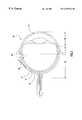
- FIG. 1illustrates an eye having a cannula inserted into the scleral in accordance with one embodiment of the present invention.
- the present inventioninvolves injecting a material into the sclera of an eye.
- the eyecan be of any vertebrate animal including human and non-human mammals.
- the sclerais a thin, avascular layer, comprised of a highly ordered collagen network, that surrounds most of the vertebrate eye.
- the corneareplaces the sclera in the front of the eye, with the transition from sclera to cornea occurring at the limbus. Because the sclera is avascular, there is essentially no risk of hemorrhaging after an injection therein and the injected material is not rapidly removed or “cleared” from the eye. Thus the sclera can be effectively utilized in the present invention as a natural storage depot.
- the materialcan be placed within the sclera by any suitable injection technique.
- a cannulais inserted into the sclera and the therapeutic or diagnostic material is then injected through the cannula and into the scleral layer.
- this type of injectionuses a syringe or other piston-type mechanism, which can be operated either by hand or by an actuator.
- the materialcan be injected through the cannula by a pump, such as a metering pump.
- Other techniquesinclude non-cannular injection methods, or so-called “needle-less injection” techniques, where gas pressure is used to force solutions or particles, or both, of material into the underlying tissue.
- processes similar to the commercially available needle-less injectors for solutions, or for powders like the POWDERJECTTM technologycan be used.
- processesinvolve injecting particles larger than 20 to 40 microns directly or indirectly into the tissue by the use of high pressure gas. Particles may be larger or smaller depending upon the suitable application.
- Eye 10(not drawn to scale) can be divided into an anterior segment 20 and posterior segment 30 with an equator 40 .
- Sclera 11covers the outside of the eye around the posterior segment and part of the anterior segment while cornea 21 covers the outer part of the remainder of the anterior segment.
- Underlying the sclerais choroid 12 and retina 13 .
- a cannula 50(shown here as a needle with a beveled terminal end) has been inserted into the sclera from a location on the external (or outer) surface of the sclera that overlies the retina.
- the cannulahas been inserted in a substantially rotational direction meaning that the insertion path into the sclera generally points around the eye and not into the center of the eye. This is a preferred embodiment because it decreases the risk of accidentally penetrating through the sclera and into the choroid or retina. Obviously, inserting a cannula, especially a sharpened or beveled cannula, into the vascular choroid or light sensitive retina can cause serious injury to these layers with resulting vision impairment.
- insertion of the cannula in a “substantially rotational direction”will be performed at an insertion angle of less than about 60 degrees; the “insertion angle” being defined by the angle formed between the tangent to the sclera at the external point of entry and the insertion path of the cannula into the sclera (or in the case of a curved cannula, the tangent to the curved cannula insertion path at the entry point into the sclera).
- the insertion angleis less than 50, more preferably from about 20 to about 40 degrees. In one embodiment the insertion angle is about 30 degrees.
- the cannulais inserted in an orientation to the sclera such that it must exit the sclera, if at all, through another location on the exterior surface of the sclera: hereinafter a “fail safe orientation.” This can be accomplished, for example, by inserting a straight cannula at a sufficiently low insertion angle. Because the sclera is curved, the cannula can be angled so as to travel on a path that is tangent to a point on the curving inner surface layer of the sclera. At all lower angles, the closest the cannula will come to the inner surface of the sclera is above the tangent point.
- Inserting the cannula in a substantially rotational directionalso allows for increased insertion distances of the cannula into the sclera which can increase the hydrodynamic seal between the cannula and the scleral tissue.
- the cannulais preferably inserted into the sclera a distance that is greater than or equal to, the thickness of the sclera, preferably at least one and a half times the thickness, measured at the entry point on the exterior surface. This is particularly useful near the equator where the sclera is quite thin, but is not limited to such a region.
- a better hydrodynamic sealcan be formed which allows for larger and/or faster injections.
- the cannulais not particularly limited and need only fit within the thickness of the sclera at the point of entry.
- the cannulais sufficiently small in diameter that no hole is visible in the sclera upon macroscopic observation of the entry site after the injection.
- at least a 25 gauge, preferably at least a 28 gauge, more preferably about 30-33 gauge cannulais employed, but such is not required.
- the size of the cannuladepends in part on the viscosity of the material to be injected, the amount of material to be injected, and the time of injection.
- a very fine gauge cannulawhile causing little if any trauma to the eye, may not be able to allow sufficient flow of a particular material into the sclera and thus would not serve as a useful conduit.
- the above gauge sizesare, especially for humans, a size that typically accommodates these competing features.
- the cannulais sharp on its leading end (i.e. a needle) such as with a bevel or a hollow ground point.
- the bevelengages the sclera in an upside down orientation whereby the leading edge of the bevel is adjacent to and makes first contact with the exterior surface of the sclera.
- the desirability of such an upside down bevel approachdepends upon the insertion angle, the thickness of the sclera, the shape of the cannula and the size of the cannula.
- the entry point on the exterior surface of the scleraoverlies the retina and thus is in the posterior segment of the eye. It should be noted, however, that the injection of material into the sclera may occur at a location within the sclera that does not overlie the retina, depending upon the angle and direction of injection; e.g. in the case of a cannular injection in a substantially rotational direction, the injection site within the sclera may be anterior to the retina. Nonetheless, generally the injection site of the material within the sclera is also over the retina.
- the entry point and injection siteare posterior to the area of eye muscle insertion, more preferably posterior to the equator of the eye, and more preferably more than 45 degrees posterior to the equator.
- a depot of the injected materialis formed within the sclera that is near the site to be treated and preferably a portion of the injected material at least partially overlies the localized area to be treated.
- Suchcan allow for more effective treatment and/or reduced amounts of material needed to be injected.
- the materialis stored within the sclera, the material remains in proximity with the affected area regardless of eye movement.
- the posterior segmentcan be reached in a number of ways.
- the eyecan be rotated in order to expose the posterior segment. This is typically accomplished by holding the conjunctiva with forceps and rotating the eye so that the front of the eye moves downwardly (i.e. “rotated forwardly”).
- the eyecan be rotated in other directions as appropriate and other techniques for rotation can be used as desired.
- Another techniqueinvolves using a curved handled device that can be inserted around the eye to position the cannula or other injection apparatus over the desired posterior location. The concept of such a device is shown in U.S. Pat. No. 5,273,530. While the device could be used as shown therein, it should preferably be modified so that the cannula is retractable and more preferably modified so that the cannula will be inserted in a substantially rotational direction.
- Accessing the posterior of an eyealso normally entails penetrating the conjunctiva.
- One wayis to make an incision in the conjunctiva and insert the cannula or other injection apparatus through the incision to the sclera.
- Such a methodworks with both the eye rotating technique and the curved handle device technique discussed above for accessing the posterior segment of the eye.
- Such an incisionis relatively non-invasive and is similar to conjunctival incisions (peritomy) that ophthalmologists make in carrying out other procedures.
- Another approachis to rotate the eye into the desired position and then inject through the conjunctiva and into the sclera.
- physical restrainte.g., friction or pins
- a guided injection devicehas a mechanism for providing a needle at a predetermined angle of insertion and preferably a predetermined depth of insertion.
- a device having a guide platformwhich comprises a support surface that conforms to the shape of the sclera and a channel extending through it for guiding the needle can be advantageously employed.
- the angle of the channel relative to the sclerais fixed.
- the needle transmitted through the channelwill thus be inserted into the sclera at the predetermined insertion angle.
- Various mechanical meanssuch as a stop or collar, can be used to limit the insertion distance of the needle.
- the needleitself is connected to the material to be injected, such as by directly attaching to a reservoir on the device or to a remote reservoir, so as to facilitate the injection step.
- the needleis preferably retractable such that after injection of the material, the needle can be withdrawn back within the device, behind the support surface. Actuators for achieving the back and forth movement of a needle are well known in the art.
- Such a guide platformcan be placed on the distal end of the device shown in U.S. Pat. No. 5,273,530, and thus, with an appropriate predetermined angle of insertion, make the blind insertion of a needle into the sclera at the back of the eye a safe procedure.
- the material to be injectedcan be any material having a therapeutic or diagnostic utility or purpose.
- the materialcan be a gas, a liquid, a suspension, a powder, etc., so long as it is injectable.
- the materialis injectable through a cannula.
- the materials injected in the present inventionare similar to intravitreal and intramuscular injection formulations in terms of concentrations, viscosities, adjuvants, etc., although such is not required.
- diagnostic and therapeutic materialsare well known in the art for treating various ocular diseases and conditions, as is their preparation and formulation, and all such materials are specifically contemplated for use in the present invention.
- a “therapeutic material”means a material that provides a healing, restraining or prophylactic effect to a disease or condition or which suppresses, ameliorates or prevents the symptoms associated with a disease or condition.
- the materialcan be a single substance or a combination of substances.
- a therapeutic materialis a composition containing a pharmaceutically active agent and an ophthalmologically acceptable carrier or diluent.
- the active agent useful in the present inventioninclude all ophthalmologically effective agents, examples of which include anti-angiogenesis agents such as metalloproteinase inhibitors, vascular endothelium growth factor (VEGF) regulating agents, fibroblast growth factor (FGF) regulating agents, integrin blockers, protein kinase C inhibitors, and endogenous angiogenesis inhibitors (e.g., angiostatin); ischemic/reperfusion preventing agents such as NMDA receptor antagonists, AMPA receptor antagonists, antioxidants, peroxidation inhibitors, apoptosis inhibitors, adenosine or adenosine regulating agents, calcium channel blockers, and nitric oxide regulating agents; anti-inflammatory agents such as steroidal and non-steroidal anti-inflammatory agents; antiviral agents; antioxidants; antibiotics; antitumor agents such as tumor necrosis factors; anti-cataract agents; anti-glaucoma agents; anesthetics; gene therapy compositions such as triplex
- useful active agentsinclude, but are not limited to, pilocarpine, timolol, atenolol, betaxolol, levobunolol, tetracycline, hydrocortisone, prednisolone, prednisone, dexamethasone, progesterone, fluorometholone, lazaroids and 21-aminosteroid compounds as disclosed in U.S. Pat. No.
- One advantage of the present inventionis the ability to use enzyme-unstable agents. Because the sclera is avascular, enzymes that would normally attack and degrade certain proteins and other agents if placed intraocularly, will not generally reach the intrascleral depot formed by the present invention.
- the active agentcan be combined with a suitable carrier or diluent, if needed, as is well known in the art and includes aqueous as well as non-aqueous systems.
- a suitable carrier or diluentif needed, as is well known in the art and includes aqueous as well as non-aqueous systems.
- the composition used in the present inventioncontains no physiologically or ophthalmologically harmful constituents. Typically purified or deionized water is used. The pH is adjusted as needed by adding any physiologically and ophthalmologically acceptable pH adjusting acids, bases or buffers.
- acidsinclude acetic, boric, citric, lactic, phosphoric, hydrochloric, and the like
- basesinclude sodium hydroxide, sodium phosphate, sodium borate, sodium citrate, sodium acetate, sodium lactate, tromethamine, THAM (trishydroxymethylamino-methane), and the like.
- Salts and bufferswould include citrate/dextrose, sodium bicarbonate, ammonium chloride and mixtures of the aforementioned acids and bases.
- the pHis typically in the neutral range such as from about 6 to about 8, but is not limited thereto.
- Non-aqueous systemsinclude the use of known ophthalmologically acceptable oils such as polyethylene glycols and silicone oils.
- the active agentcan be in solution, in suspension, or both. If the active agent is in solid form, its particle size should be sufficiently limited to permit injection (e.g., the agent is able to pass through a cannula) and so as not to cause irritation to the eye once injected.
- the compositioncontains a component that facilitates or improves the sustained release of the active agent as is known in the art.
- a component that facilitates or improves the sustained release of the active agentas is known in the art.
- incorporating a polymeric suspending agentcan provide sustained release.
- the polymershould be biodegradable or biocompatible such that it can be cleared from the eye by natural transport effects.
- the active ingredientcan be incorporated into the polymer matrix, adsorbed on the polymer surface, encapsulated within a polymer coating, etc. as are well known in the art.
- suitable polymersinclude water-soluble polymers such as dextran, polyethylene glycols, polyvinylpyrolidones, polysaccaride gels, Gelrite®, cellulosic polymers like hydroxypropyl methylcellulose and carboxymethylcellulose, hyaluronic acid polymers, and poly(lactic acid) and copolymers of lactic acid and one or more of glycolic acid, malic acid, glyceric acid, and tartaric acid.
- Carboxy-containing polymerssuch as uncrosslinked polyacrylic acids and copolymers thereof are also useful as suspending agents for insuring sustained release.
- Crosslinkingis permissible only to the extent that the polymer can clear; i.e., crosslinking generally prevents biodegredation and thus the entire polymer must be susceptible of being cleared from the eye.
- Other formsinclude liposomes and lipid emulsions.
- the compositionshould contain a sufficient amount of active ingredient to achieve the desired effect as can be readily determined by workers skilled in the art.
- the solubility of the active ingredient in water and the concentration of the active ingredient needed in the tissueguide the amount and rate of release of the agent.
- the sclerais a depot of limited size and the concentration of the agent may need to reflect this.
- the amount of material to be injectedis at least 0.1 ⁇ l, typically from around 0. 1 to 25 ⁇ l, more typically from about 1 to 25 ⁇ l, such as from about 3 to about 25 ⁇ l or from about 3 to 10 ⁇ l. If more material is needed then can be practically delivered in a single injection, then multiple injections can be performed; i.e., injecting 6 ⁇ l of therapeutic material in three different sites within the sclera during a single office visit.
- compositionsinclude solubilizers, stabilizers, preservatives, and other ingredients as are well known in the ophthalmology art. If the composition is supplied as a ready to inject single dose, then a preservative is typically omitted.
- the compositioncan be provided as a frozen liquid or as a lyophilized powder for reconstituting.
- Diagnostic materialsinclude a gas and dye solutions.
- a gassuch as nitrogen, air, or other inert gas, can be supplied in order to inflate the area and aid in some types of diagnostic procedures; i.e., improving the image in an ophthalmoscope.
- a dyecan be injected in order to aid in diagnosing various conditions by providing higher contrast and/or a staining pattern.
- the rate of injection of the material via a cannula into the sclerais dependent on several factors including the viscosity of the material and the duration of the injection and can be readily determined by workers of ordinary skill in the art.
- the injection rateis from about 0.1 to about 3.0 ⁇ l/s, preferably from about 0.5 to 1.0 ⁇ l/s.
- Injectionstypically will last for up to 10 seconds, although longer injection times are possible, especially with the use of a hand rest/support, and are contemplated by the present invention. Another consideration is the degree of hydrodynamic seal around the cannula.
- Too high of an injection rate or pressuremay cause the material to flow out of the sclera along the sides of the cannula with very little of the material being lodged within the sclera.
- One way to improve the hydrodynamic sealis to increase the insertion length of the cannula into the sclera, as has been discussed above.
- the present inventioncan be used to treat a variety of ocular diseases or conditions including, but not limited to, cystoid macular edema, age-related macular degeneration, diabetic retinopathy, diabetic maculopathy, central retinal artery occlusion, central retinal vein occlusion, branch retinal artery occlusion, branch retinal vein occlusion, retinopathy of prematurity, sickel cell retinopathy, photic retinopathy, radiation retinopathy, retinal detachment, retinitis pigmentosa, macular hole, cataract, and glaucoma as well as accidental or surgically induced trauma to the eye.
- Suitable therapeutic materialsknown for treatment of an ocular disease or condition, especially a retinal disease or condition, can be injected into the sclera in close proximity to the affected site by the present invention to thereby provide effective treatment and enhanced delivery.
- one embodiment of the inventionrelates to treating neovascular diseases of the eye, such as diabetic retinopathy, macular degeneration, and neovascularization of the retina or choroid, by injecting into the sclera, through a location on the exterior surface of the sclera that overlies retinal tissue, an effective neovascularization reducing or preventing amount of an anti-angiogenesis agent.
- neovascular diseases of the eyesuch as diabetic retinopathy, macular degeneration, and neovascularization of the retina or choroid
- agentsare described above and are generally well known in the art, including metalloproteinase inhibitors, vascular endothelium growth factor regulating agents, FGF regulating agents, integrin blockers, and protein kinase C inhibitors.
- the VEGF regulating agentsinclude, without limitation, VEGF, antisense compounds thereof, antibodies thereof, and antibody fragments thereof having anti-angiogenesis activity.
- Antioxidantsare also a
- the insertion siteis over the macula or in its immediate vicinity (e.g., more than 45 degrees posterior to the equator). More preferably, the insertion and injection steps provide at least a portion of the intrasclerally injected therapeutic material overlying the macula.
- the material to be injectedcan be any macular degeneration treating material.
- active agentsinclude the above anti-angiogenesis compounds of VEGF, an antisense compound of VEGF, an antibody of VEGF, a fragment of an antibody of VEGF, triplex nucleic acids of VEGF, a receptor blocker for VEGF, and ribozymes for VEGF as well as antioxidants.
- Another embodimentis treating cataracts. Although the disease manifests in the anterior segment, its root cause may lie in the posterior segment. Providing an antioxidant into the posterior segment can prevent or reduce cataracts.
- a preferred antioxidantis a 21-aminosteroid such as a lazaroid.
- An injectable therapeutic material containing the PAF antagonist Lexipafant(BB-882) is prepared as follows. In a 250-ml beaker, about 50 g of DI water is added and heated up to 80-90° C. on a hot plate while stirring with a magnetic stir bar. HPMC is dispersed into the hot water and stirred for 15 min. followed by cooling to RT while stirring. 10 g of room temperature water is then added to the polymer and stirred for 10 min. In a separate container, Pluronic F-127 and sorbitol are dissolved in 20 g of DI water. Glycerin is then added to the Pluronic F-127 solution and stirred until dissolve completely.
- the Pluronic F-127 solutionis then added to the polymer suspension and stirred for 10 min.
- BB-882dissolved in 1N HCl solution, is then added to the polymer mixture with stirring for 10 min.
- the pH of the resulting polymer mixtureis adjusted to about 7.4 with 2N NaOH, stirred for 10 min., and then brought to 100% with q.s. of DI water.
- the formulationmay be made sterile by heating the formulation to 123° C. for 30 minutes and sterile filtering the drug, NaOH, and residual water after heating.
- the 100 grams of materialis summarized in the following table:
- COMPOSITION % (Wt/Wt) BB-8821.0 Hydroxylpropyl Methylcellulose, Type 2910, USP 2.5 Sorbitol, USP 1.5 Glycerin, USP 1.0 Pluronic F-127, NF 1.0 Hydrochloric Acid, (1N solution) 5.0 Sodium Hydroxide, NF, 2N for pH adjustment q.s. to pH 7.4 Purified Water, USP q.s.
- COMPOSITION % (Wt/Wt) BB-8820.1 Hydroxylpropyl Methylcellulose, Type 2910, USP 2.5 Sorbitol, USP 1.5 Glycerin, USP 0.2 Edetate Disodium, USP 0.10 Sodium Chloride, USP 0.32 Sodium Hydroxide, NF, 2N for pH adjustment q.s. to pH 6 Purified Water, USP q.s.
- COMPOSITION % (Wt/Wt) Diclofenac NaUSP 0.1-1.0 Hydroxylpropyl Methylcellulose, Type 2910, USP 2.5 Mannitol, USP 1.5 Sodium Chioride, USP 0.21 Poloxamer 407, NF 0.05 Boric Acid, USP 0.5 Magnesium Chioride, USP 0.05 Sodium Hydroxide, NF, 2N for pH adjustment q.s. to pH 6 Purified Water, USP q.s.
- Lazaroidsare known to be potentially useful in treating a variety of ocular ischemic diseases such as glaucoma and diabetic retinopathy.
- Suitable formulationsmay be generally formulated as follows. 0.005 grams of the aminosteroid is dissolved into a saline solution formed of 0.9 grams of sodium chloride dissolved in intravenous grade water. The pH is then adjusted to 7.4 with NaOH and the total weight adjusted with water to 100 grams. The mixture is then sterilized.
- the aminosteroidis not a powder, but is a lipid emulsion or in a liposome, it can be dispersed in the saline solution.
- a suspensioncan be made by adding hyaluronic acid such as sodium hyaluronate, or other suitable polymer, typically about 1.0 grams and with an increase in the amount of the agent, such as to 0.05 grams.
- hyaluronic acidsuch as sodium hyaluronate, or other suitable polymer
- the suspensionneed only remain sufficiently viscous to allow injection.
- Suitable aninosteroidsinclude U-74006F, U-74500A, and U-75412A.
- Another formulationis to slowly add 10 grams of U-74006F to 950 ml of pure water having 20 millimoles of citric acid under an inert atmosphere and with stirring. Three millimoles of sodium citrate and 8 millimoles of sodium chloride are added with stirring until a clear solution is obtained. The solution can then be sterilized.
- An injectable therapeutic material containing Batimastat(BB-94) is prepared as follows. In a 250-ml beaker, about 50 g of DI water is added and heated up to 80-90° C. on a hot plate while stirring with a magnetic stir bar. HPMC is dispersed into the hot water and stirred for 15 min. followed by cooling to RT while stirring. 10 g of room temperature water is then added to the polymer and stirred for 10 min. In a separate container, Pluronic F-127 and sorbitol are dissolved in 20 g of DI water. Glycerin is then added to the Pluronic F-127 solution and stirred until dissolve completely. The Pluronic F-127 solution is then added to the polymer suspension and stirred for 10 min.
- BB-94is then added to the polymer mixture with stirring for 10 min.
- the pH of the resulting polymer mixtureis adjusted to about 6.0 with 2N NaOH, stirred for 10 min., and then brought to 100% with q.s. of DI water.
- the formulationmay be made sterile by heating the formulation to 123° C. for 30 minutes and sterile filtering the drug, NaOH, and residual water after heating.
- the 100 grams of materialis summarized in the following table:
- COMPOSITION % (Wt/Wt) BB-940.3 Hydroxylpropyl Methylcellulose, Type 2910, USP 2.5 Sorbitol, USP 1.5 Glycerin, USP 1.0 Pluronic F-127, NF 1.0 Sodium Hydroxide, NF, 2N for pH adjustment q.s. to pH 6.0 Purified Water, USP q.s.
Landscapes
- Health & Medical Sciences (AREA)
- Life Sciences & Earth Sciences (AREA)
- Public Health (AREA)
- Veterinary Medicine (AREA)
- Pharmacology & Pharmacy (AREA)
- Chemical & Material Sciences (AREA)
- General Health & Medical Sciences (AREA)
- Medicinal Chemistry (AREA)
- Animal Behavior & Ethology (AREA)
- Engineering & Computer Science (AREA)
- Epidemiology (AREA)
- Bioinformatics & Cheminformatics (AREA)
- Ophthalmology & Optometry (AREA)
- General Chemical & Material Sciences (AREA)
- Nuclear Medicine, Radiotherapy & Molecular Imaging (AREA)
- Chemical Kinetics & Catalysis (AREA)
- Organic Chemistry (AREA)
- Vascular Medicine (AREA)
- Proteomics, Peptides & Aminoacids (AREA)
- Molecular Biology (AREA)
- Genetics & Genomics (AREA)
- Zoology (AREA)
- Gastroenterology & Hepatology (AREA)
- Immunology (AREA)
- Biotechnology (AREA)
- Pain & Pain Management (AREA)
- Neurology (AREA)
- Orthopedic Medicine & Surgery (AREA)
- Physical Education & Sports Medicine (AREA)
- Heart & Thoracic Surgery (AREA)
- Urology & Nephrology (AREA)
- Cardiology (AREA)
- Medicines That Contain Protein Lipid Enzymes And Other Medicines (AREA)
- Infusion, Injection, And Reservoir Apparatuses (AREA)
- Medicinal Preparation (AREA)
- Medicines Containing Antibodies Or Antigens For Use As Internal Diagnostic Agents (AREA)
Abstract
Description
| COMPOSITION | % (Wt/Wt) |
| BB-882 | 1.0 |
| Hydroxylpropyl Methylcellulose, Type 2910, USP | 2.5 |
| Sorbitol, USP | 1.5 |
| Glycerin, USP | 1.0 |
| Pluronic F-127, NF | 1.0 |
| Hydrochloric Acid, (1N solution) | 5.0 |
| Sodium Hydroxide, NF, 2N for pH adjustment | q.s. to pH 7.4 |
| Purified Water, USP | q.s. |
| COMPOSITION | % (Wt/Wt) |
| BB-882 | 0.1 |
| Hydroxylpropyl Methylcellulose, Type 2910, USP | 2.5 |
| Sorbitol, USP | 1.5 |
| Glycerin, USP | 0.2 |
| Edetate Disodium, USP | 0.10 |
| Sodium Chloride, USP | 0.32 |
| Sodium Hydroxide, NF, 2N for pH adjustment | q.s. to pH 6 |
| Purified Water, USP | q.s. |
| COMPOSITION | % (Wt/Wt) |
| Diclofenac Na, USP | 0.1-1.0 |
| Hydroxylpropyl Methylcellulose, Type 2910, USP | 2.5 |
| Mannitol, USP | 1.5 |
| Sodium Chioride, USP | 0.21 |
| Poloxamer 407, NF | 0.05 |
| Boric Acid, USP | 0.5 |
| Magnesium Chioride, USP | 0.05 |
| Sodium Hydroxide, NF, 2N for pH adjustment | q.s. to pH 6 |
| Purified Water, USP | q.s. |
| COMPOSITION | % (Wt/Wt) |
| BB-94 | 0.3 |
| Hydroxylpropyl Methylcellulose, Type 2910, USP | 2.5 |
| Sorbitol, USP | 1.5 |
| Glycerin, USP | 1.0 |
| Pluronic F-127, NF | 1.0 |
| Sodium Hydroxide, NF, 2N for pH adjustment | q.s. to pH 6.0 |
| Purified Water, USP | q.s. |
Claims (29)
Priority Applications (8)
| Application Number | Priority Date | Filing Date | Title |
|---|---|---|---|
| US09/127,920US6378526B1 (en) | 1998-08-03 | 1998-08-03 | Methods of ophthalmic administration |
| US09/366,072US6397849B1 (en) | 1998-08-03 | 1999-08-02 | Methods of ophthalmic administration |
| CA002339244ACA2339244C (en) | 1998-08-03 | 1999-08-02 | Methods of ophthalmic administration |
| JP2000563251AJP2002522373A (en) | 1998-08-03 | 1999-08-02 | Eye administration method |
| AU53327/99AAU761830B2 (en) | 1998-08-03 | 1999-08-02 | Methods of ophthalmic administration |
| EP99938953AEP1100462A2 (en) | 1998-08-03 | 1999-08-02 | Methods of ophthalmic administration |
| PCT/US1999/017543WO2000007565A2 (en) | 1998-08-03 | 1999-08-02 | Methods of ophthalmic administration |
| NO20010556ANO20010556L (en) | 1998-08-03 | 2001-02-01 | Procedure for ophthalmic administration |
Applications Claiming Priority (1)
| Application Number | Priority Date | Filing Date | Title |
|---|---|---|---|
| US09/127,920US6378526B1 (en) | 1998-08-03 | 1998-08-03 | Methods of ophthalmic administration |
Related Child Applications (1)
| Application Number | Title | Priority Date | Filing Date |
|---|---|---|---|
| US09/366,072Continuation-In-PartUS6397849B1 (en) | 1998-08-03 | 1999-08-02 | Methods of ophthalmic administration |
Publications (1)
| Publication Number | Publication Date |
|---|---|
| US6378526B1true US6378526B1 (en) | 2002-04-30 |
Family
ID=22432636
Family Applications (2)
| Application Number | Title | Priority Date | Filing Date |
|---|---|---|---|
| US09/127,920Expired - LifetimeUS6378526B1 (en) | 1998-08-03 | 1998-08-03 | Methods of ophthalmic administration |
| US09/366,072Expired - LifetimeUS6397849B1 (en) | 1998-08-03 | 1999-08-02 | Methods of ophthalmic administration |
Family Applications After (1)
| Application Number | Title | Priority Date | Filing Date |
|---|---|---|---|
| US09/366,072Expired - LifetimeUS6397849B1 (en) | 1998-08-03 | 1999-08-02 | Methods of ophthalmic administration |
Country Status (7)
| Country | Link |
|---|---|
| US (2) | US6378526B1 (en) |
| EP (1) | EP1100462A2 (en) |
| JP (1) | JP2002522373A (en) |
| AU (1) | AU761830B2 (en) |
| CA (1) | CA2339244C (en) |
| NO (1) | NO20010556L (en) |
| WO (1) | WO2000007565A2 (en) |
Cited By (130)
| Publication number | Priority date | Publication date | Assignee | Title |
|---|---|---|---|---|
| US20020198511A1 (en)* | 2001-06-22 | 2002-12-26 | Varner Signe Erickson | Method and device for subretinal drug delivery |
| US20030206956A1 (en)* | 1999-03-31 | 2003-11-06 | Insite Vision Incorporated | Topical treatment of prevention of ocular infections |
| US6685958B2 (en) | 2001-04-25 | 2004-02-03 | Insite Vision Incorporated | Quinolone carboxylic acid compositions and related methods of treatment |
| US20040029788A1 (en)* | 2000-11-01 | 2004-02-12 | Hans-Markus Bender | Methods and compositions for the treatment of diseases of the eye |
| US6699492B2 (en) | 1999-03-31 | 2004-03-02 | Insite Vision Incorporated | Quinolone carboxylic acid compositions and related methods of treatment |
| US20040092911A1 (en)* | 2001-07-23 | 2004-05-13 | Yoseph Yaacobi | Ophthalmic drug delivery device |
| US20040198644A1 (en)* | 2001-08-01 | 2004-10-07 | Hans-Markus Bender | Integrin inhibitors for the treatment of eye diseases |
| US20040249333A1 (en)* | 2000-04-14 | 2004-12-09 | Bergheim Olav B. | Glaucoma implant with bi-directional flow |
| US20040253293A1 (en)* | 2003-06-16 | 2004-12-16 | Afshin Shafiee | Rate controlled release of a pharmaceutical agent in a biodegradable device |
| US20050009910A1 (en)* | 2003-07-10 | 2005-01-13 | Allergan, Inc. | Delivery of an active drug to the posterior part of the eye via subconjunctival or periocular delivery of a prodrug |
| US20050065136A1 (en)* | 2003-08-13 | 2005-03-24 | Roby Russell R. | Methods and compositions for the treatment of infertility using dilute hormone solutions |
| US20050064010A1 (en)* | 2003-09-18 | 2005-03-24 | Cooper Eugene R. | Transscleral delivery |
| US20050089545A1 (en)* | 2002-02-22 | 2005-04-28 | Mitsuaki Kuwano | Drug delivery system for the subconjunctival administration of fine grains |
| US20050143363A1 (en)* | 2002-09-29 | 2005-06-30 | Innorx, Inc. | Method for subretinal administration of therapeutics including steroids; method for localizing pharmacodynamic action at the choroid of the retina; and related methods for treatment and/or prevention of retinal diseases |
| US20050239758A1 (en)* | 2004-04-21 | 2005-10-27 | Roby Russell R | Hormone treatment of multiple sclerosis |
| US20050239757A1 (en)* | 2004-04-21 | 2005-10-27 | Roby Russell R | Hormone treatment of macular degeneration |
| US20050245524A1 (en)* | 2004-04-08 | 2005-11-03 | Targegen, Inc. | Benzotriazine inhibitors of kinases |
| WO2005107845A1 (en)* | 2004-04-29 | 2005-11-17 | Iscience Interventional Corporation | Apparatus and method for ocular treatment |
| US20050277802A1 (en)* | 2004-02-12 | 2005-12-15 | Larsen Charles E | Method and apparatus for intraocular brachytherapy |
| US20060013859A1 (en)* | 2002-12-04 | 2006-01-19 | Santen Pharmaceutical Co., Ltd. | Drug delivery system using subconjunctival depot |
| US20060025390A1 (en)* | 2004-07-28 | 2006-02-02 | Roby Russell R | Treatment of hormone allergy and related symptoms and disorders |
| US20060039952A1 (en)* | 2003-07-10 | 2006-02-23 | Yoseph Yaacobi | Ophthalmic drug delivery device |
| US20060046970A1 (en)* | 2004-08-31 | 2006-03-02 | Insite Vision Incorporated | Topical otic compositions and methods of topical treatment of prevention of otic infections |
| US20060110428A1 (en)* | 2004-07-02 | 2006-05-25 | Eugene Dejuan | Methods and devices for the treatment of ocular conditions |
| US20060116626A1 (en)* | 2002-03-07 | 2006-06-01 | Gregory Smedley | Fluid infusion methods for glaucoma treatment |
| US7056893B2 (en) | 1999-03-31 | 2006-06-06 | Insite Vision, Inc. | Topical treatment for prevention of ocular infections |
| US7077848B1 (en)* | 2000-03-11 | 2006-07-18 | John Hopkins University | Sutureless occular surgical methods and instruments for use in such methods |
| US20060182771A1 (en)* | 2005-02-09 | 2006-08-17 | Dor Philippe J | Formulations for ocular treatment |
| US7094226B2 (en) | 2001-07-23 | 2006-08-22 | Alcon, Inc. | Ophthalmic drug delivery device |
| US20060257451A1 (en)* | 2005-04-08 | 2006-11-16 | Varner Signe E | Sustained release implants and methods for subretinal delivery of bioactive agents to treat or prevent retinal disease |
| US7141607B1 (en)* | 2000-03-10 | 2006-11-28 | Insite Vision Incorporated | Methods and compositions for treating and inhibiting retinal neovascularization |
| US20060275367A1 (en)* | 2005-04-25 | 2006-12-07 | Shubha Chungi | Extended release formulations |
| WO2006133411A1 (en)* | 2005-06-08 | 2006-12-14 | Targegen, Inc. | Methods and compositions for the treatment of ocular disorders |
| US20060287285A1 (en)* | 2001-11-16 | 2006-12-21 | Roby Russell R | Methods and Compositions for the Treatment of Pain and Other Hormone-Allergy-Related Symptoms Using Dilute Hormone Solutions |
| US20060286173A1 (en)* | 2003-08-20 | 2006-12-21 | Kazuhito Yamada | Drug delivery system for sub-tenon s capsule adminstration of fine grains |
| US20070092570A1 (en)* | 1999-10-21 | 2007-04-26 | Missel Paul J | Drug delivery device |
| US20070118010A1 (en)* | 2005-02-11 | 2007-05-24 | Hillstead Richard A | Methods and apparatus for intraocular brachytherapy |
| US20070166382A1 (en)* | 2004-03-26 | 2007-07-19 | Kiser Patrick F | Bioresponsive polymer system for delivery of microbicides |
| US20070202186A1 (en)* | 2006-02-22 | 2007-08-30 | Iscience Interventional Corporation | Apparatus and formulations for suprachoroidal drug delivery |
| US20070224178A1 (en)* | 2004-09-13 | 2007-09-27 | Abraham Scaria | Multimeric constructs |
| US20070259904A1 (en)* | 2005-11-01 | 2007-11-08 | Targegen, Inc. | Bi-aryl meta-pyrimidine inhibitors of kinases |
| US20070265294A1 (en)* | 2006-03-23 | 2007-11-15 | Kleinman David M | Formulations and methods for vascular permeability-related diseases or conditions |
| US20070265485A1 (en)* | 2001-02-22 | 2007-11-15 | Dejuan Eugene Jr | Beta radiotherapy emitting surgical device and methods of use thereof |
| US20070287756A1 (en)* | 2004-04-23 | 2007-12-13 | Toru Nakazawa | Methods and Compositions for Preserving the Viability of Photoreceptor Cells |
| US20080058704A1 (en)* | 2004-04-29 | 2008-03-06 | Michael Hee | Apparatus and Method for Ocular Treatment |
| US20080112952A1 (en)* | 2006-11-13 | 2008-05-15 | Paul Theodone Finger | Anti-VEGF treatment for radiation-induced vasculopathy |
| US20080265343A1 (en)* | 2007-04-26 | 2008-10-30 | International Business Machines Corporation | Field effect transistor with inverted t shaped gate electrode and methods for fabrication thereof |
| US20080268051A1 (en)* | 2007-04-30 | 2008-10-30 | Allergan, Inc. | High viscosity macromolecular compositions for treating ocular conditions |
| US20090036819A1 (en)* | 2001-04-07 | 2009-02-05 | Glaukos Corporation | Drug eluting ocular implant with anchor and methods thereof |
| US20090043321A1 (en)* | 2004-04-29 | 2009-02-12 | Iscience Interventional Corporation | Apparatus And Method For Surgical Enhancement Of Aqueous Humor Drainage |
| US20090074786A1 (en)* | 2005-02-09 | 2009-03-19 | Macusight, Inc. | Formulations for treating ocular diseases and conditions |
| US7563222B2 (en) | 2004-02-12 | 2009-07-21 | Neovista, Inc. | Methods and apparatus for intraocular brachytherapy |
| US20090286789A1 (en)* | 2005-11-01 | 2009-11-19 | Targegen, Inc. | Bi-Aryl Meta-Pyrimidine Inhibitors of Kinases |
| WO2009105690A3 (en)* | 2008-02-21 | 2010-01-14 | Targeted Genetics Corporation | Devices and methods for delivering polynucleotides into retinal cells of the macula and fovea |
| US7648959B2 (en) | 2000-11-01 | 2010-01-19 | Merck Patent Gmbh | Methods and compositions for the treatment of diseases of the eye |
| US20100034808A1 (en)* | 2006-11-21 | 2010-02-11 | Toru Nakazawa | Compositions and methods for preserving cells of the eye |
| US20100087774A1 (en)* | 2002-09-21 | 2010-04-08 | Glaukos Corporation | Ocular implant with anchoring mechanism and multiple outlets |
| US20100106073A1 (en)* | 2001-05-02 | 2010-04-29 | Glaukos Corporation | Method of monitoring intraocular pressure and treating an ocular disorder |
| US20100152646A1 (en)* | 2008-02-29 | 2010-06-17 | Reshma Girijavallabhan | Intravitreal injection device and method |
| US20100191103A1 (en)* | 2002-09-17 | 2010-07-29 | Iscience Interventional Corporation | Apparatus and method for surgical bypass of aqueous humor |
| US20100226971A1 (en)* | 2009-03-06 | 2010-09-09 | Ying Chau | Ultrasound-enhanced intrascleral delivery of macromolecules |
| US20100234790A1 (en)* | 2000-04-14 | 2010-09-16 | Glaukos Corporation | Ocular implant with therapeutic agents and methods thereof |
| US20100242949A1 (en)* | 2009-03-30 | 2010-09-30 | Mitsubishi Heavy Industries, Ltd. | Sunlight collecting heat receiver |
| US20100256597A1 (en)* | 2006-05-02 | 2010-10-07 | Emory University | Methods and Devices for Drug Delivery to Ocular Tissue Using Microneedle |
| US20100286632A1 (en)* | 2009-05-06 | 2010-11-11 | Cesario Pereira Dos Santos | Multi-Layer Heat Assembly For A Drug Delivery Device |
| US20100330030A1 (en)* | 2002-10-03 | 2010-12-30 | Targegen, Inc. | Vasculostatic Agents and Methods of Use Thereof |
| US20110152767A1 (en)* | 2009-12-22 | 2011-06-23 | Pinedjian Raffi S | Method and Apparatus for Drug Delivery |
| US20110212077A1 (en)* | 2005-11-01 | 2011-09-01 | Targegen, Inc. | Bi-aryl meta-pyrimidine inhibitors of kinases |
| AU2010207761B2 (en)* | 2000-03-11 | 2011-09-15 | Johns Hopkins University | Sutureless ocular surgical methods and instruments for use in such methods |
| US8030487B2 (en) | 2006-07-07 | 2011-10-04 | Targegen, Inc. | 2-amino—5-substituted pyrimidine inhibitors |
| US20120213841A1 (en)* | 2008-11-13 | 2012-08-23 | Peyman Gholam A | Ophthalmic drug delivery system and method |
| US8277418B2 (en) | 2009-12-23 | 2012-10-02 | Alcon Research, Ltd. | Ophthalmic valved trocar cannula |
| US8343106B2 (en) | 2009-12-23 | 2013-01-01 | Alcon Research, Ltd. | Ophthalmic valved trocar vent |
| US8353812B2 (en) | 2008-06-04 | 2013-01-15 | Neovista, Inc. | Handheld radiation delivery system |
| WO2013007766A1 (en) | 2011-07-13 | 2013-01-17 | Vib Vzw | Means and methods for the treatment of pathological angiogenesis |
| US8372971B2 (en) | 2004-08-25 | 2013-02-12 | Targegen, Inc. | Heterocyclic compounds and methods of use |
| US8425473B2 (en) | 2009-01-23 | 2013-04-23 | Iscience Interventional Corporation | Subretinal access device |
| US8492400B2 (en) | 2006-02-09 | 2013-07-23 | Santen Pharmaceutical Co., Ltd. | Stable formulations, and methods of their preparation and use |
| US20130202613A1 (en)* | 2002-12-06 | 2013-08-08 | Thrombogenics Nv | Pharmacological vitreolysis |
| EP2662089A2 (en) | 2005-10-08 | 2013-11-13 | Potentia Pharmaceuticals, Inc. | Compstatin and analogs thereof for eye disorders |
| WO2014150067A1 (en) | 2013-03-15 | 2014-09-25 | Insite Vision Incorporated | Concentrated aqueous azalide formulations |
| US9056076B2 (en) | 2005-10-08 | 2015-06-16 | Potentia Pharmaceuticals, Inc. | Method of treating age-related macular degeneration comprising administering a compstatin analog |
| US9180047B2 (en) | 2013-05-03 | 2015-11-10 | Clearside Biomedical, Inc. | Apparatus and methods for ocular injection |
| USD750223S1 (en) | 2014-10-14 | 2016-02-23 | Clearside Biomedical, Inc. | Medical injector for ocular injection |
| US9441029B2 (en) | 2010-08-06 | 2016-09-13 | Genzyme Corporation | VEGF antagonist compositions and uses thereof |
| US9572800B2 (en) | 2012-11-08 | 2017-02-21 | Clearside Biomedical, Inc. | Methods and devices for the treatment of ocular diseases in human subjects |
| US9682144B2 (en) | 2011-06-30 | 2017-06-20 | Gene Signal International, Sa | Composition comprising inhibitors of IRS-1 and of VEGF |
| US9730638B2 (en) | 2013-03-13 | 2017-08-15 | Glaukos Corporation | Intraocular physiological sensor |
| US9956114B2 (en) | 2014-06-20 | 2018-05-01 | Clearside Biomedical, Inc. | Variable diameter cannula and methods for controlling insertion depth for medicament delivery |
| EP3338743A1 (en)* | 2006-01-17 | 2018-06-27 | Novartis Ag | Drug delivery treatment device |
| US10010447B2 (en) | 2013-12-18 | 2018-07-03 | Novartis Ag | Systems and methods for subretinal delivery of therapeutic agents |
| US10138485B2 (en) | 2008-09-22 | 2018-11-27 | Rxi Pharmaceuticals Corporation | Neutral nanotransporters |
| US10184124B2 (en) | 2010-03-24 | 2019-01-22 | Phio Pharmaceuticals Corp. | RNA interference in ocular indications |
| US10188550B2 (en) | 2013-06-03 | 2019-01-29 | Clearside Biomedical, Inc. | Apparatus and methods for drug delivery using multiple reservoirs |
| US10206813B2 (en) | 2009-05-18 | 2019-02-19 | Dose Medical Corporation | Implants with controlled drug delivery features and methods of using same |
| US10240149B2 (en) | 2010-03-24 | 2019-03-26 | Phio Pharmaceuticals Corp. | Reduced size self-delivering RNAi compounds |
| US10245178B1 (en) | 2011-06-07 | 2019-04-02 | Glaukos Corporation | Anterior chamber drug-eluting ocular implant |
| US10285852B2 (en)* | 2010-12-02 | 2019-05-14 | Tel Hashomer Medical Research Infrastructure And Services Ltd., The Chaim Sheba Medical Center | Subretinal delivery of therapeutic compositions |
| US10308687B2 (en) | 2013-03-15 | 2019-06-04 | Apellis Pharmaceuticals, Inc. | Cell-penetrating compstatin analogs and uses thereof |
| US10390901B2 (en) | 2016-02-10 | 2019-08-27 | Clearside Biomedical, Inc. | Ocular injection kit, packaging, and methods of use |
| US10391094B2 (en) | 2010-11-07 | 2019-08-27 | Impact Biomedicines, Inc. | Compositions and methods for treating myelofibrosis |
| WO2019195712A2 (en) | 2018-04-06 | 2019-10-10 | The Trustees Of The University Of Pennsylvania | Compstatin analogs with increased solubility and improved pharmacokinetic properties |
| US10479992B2 (en) | 2009-02-04 | 2019-11-19 | Phio Pharmaceuticals Corp. | RNA duplexes with single stranded phosphorothioate nucleotide regions for additional functionality |
| US10485701B2 (en) | 2002-04-08 | 2019-11-26 | Glaukos Corporation | Devices and methods for glaucoma treatment |
| CN111249034A (en)* | 2020-03-31 | 2020-06-09 | 许寅聪 | Posterior sclera reinforcing structure capable of developing under nuclear magnetic resonance |
| US10828195B2 (en) | 2006-11-10 | 2020-11-10 | Glaukos Corporation | Uveoscleral shunt and methods for implanting same |
| US10828473B2 (en) | 2001-04-07 | 2020-11-10 | Glaukos Corporation | Ocular implant delivery system and methods thereof |
| USD901683S1 (en) | 2017-10-27 | 2020-11-10 | Glaukos Corporation | Implant delivery apparatus |
| US10875893B2 (en) | 2012-11-15 | 2020-12-29 | Apellis Pharmaceuticals, Inc. | Cell-reactive, long-acting, or targeted compstatin analogs and related compositions and methods |
| US10952894B2 (en) | 2010-10-15 | 2021-03-23 | Clearside Biomedical, Inc. | Device for ocular access |
| US10959941B2 (en) | 2014-05-29 | 2021-03-30 | Glaukos Corporation | Implants with controlled drug delivery features and methods of using same |
| US10973681B2 (en) | 2016-08-12 | 2021-04-13 | Clearside Biomedical, Inc. | Devices and methods for adjusting the insertion depth of a needle for medicament delivery |
| US11040107B2 (en) | 2017-04-07 | 2021-06-22 | Apellis Pharmaceuticals, Inc. | Dosing regimens and related compositions and methods |
| US11116625B2 (en) | 2017-09-28 | 2021-09-14 | Glaukos Corporation | Apparatus and method for controlling placement of intraocular implants |
| US11197780B2 (en) | 2012-03-26 | 2021-12-14 | Glaukos Corporation | System and method for delivering multiple ocular implants |
| US11279934B2 (en) | 2014-04-28 | 2022-03-22 | Phio Pharmaceuticals Corp. | Methods for treating cancer using nucleic acids targeting MDM2 or MYCN |
| US11318043B2 (en) | 2016-04-20 | 2022-05-03 | Dose Medical Corporation | Bioresorbable ocular drug delivery device |
| US11363951B2 (en) | 2011-09-13 | 2022-06-21 | Glaukos Corporation | Intraocular physiological sensor |
| US11376040B2 (en) | 2017-10-06 | 2022-07-05 | Glaukos Corporation | Systems and methods for delivering multiple ocular implants |
| US11564833B2 (en) | 2015-09-25 | 2023-01-31 | Glaukos Corporation | Punctal implants with controlled drug delivery features and methods of using same |
| US11596545B2 (en) | 2016-05-02 | 2023-03-07 | Clearside Biomedical, Inc. | Systems and methods for ocular drug delivery |
| US11903994B2 (en) | 2015-10-07 | 2024-02-20 | Apellis Pharmaceuticals, Inc. | Dosing regimens |
| US11925578B2 (en) | 2015-09-02 | 2024-03-12 | Glaukos Corporation | Drug delivery implants with bi-directional delivery capacity |
| WO2024110878A1 (en) | 2022-11-24 | 2024-05-30 | Amyndas Pharmaceuticals Us Llc | Compstatin analogs for vector-based therapy |
| US11998482B2 (en) | 2014-02-12 | 2024-06-04 | Gyroscope Therapeutics Limited | Method and apparatus for subretinal administration of therapeutic agent |
| US12083042B2 (en) | 2016-06-17 | 2024-09-10 | Gyroscope Therapeutics Limited | Guide apparatus for tangential entry into suprachoroidal space |
| US12090294B2 (en) | 2017-05-02 | 2024-09-17 | Georgia Tech Research Corporation | Targeted drug delivery methods using a microneedle |
| US12201555B2 (en) | 2009-05-18 | 2025-01-21 | Dose Medical Corporation | Drug eluting ocular implant |
| US12290566B2 (en) | 2017-12-15 | 2025-05-06 | Apellis Pharmaceuticals, Inc. | Dosing regimens and related compositions and methods |
| US12419783B2 (en) | 2010-11-24 | 2025-09-23 | Glaukos Corporation | Drug eluting ocular implant |
Families Citing this family (36)
| Publication number | Priority date | Publication date | Assignee | Title |
|---|---|---|---|---|
| CA2358296A1 (en)* | 1999-01-05 | 2000-07-13 | Anthony P. Adamis | Targeted transscleral controlled release drug delivery to the retina and choroid |
| HK1049438B (en) | 1999-10-21 | 2005-08-26 | Alcon, Inc. | Sub-tenon drug delivery |
| BR0108802A (en)* | 2000-02-29 | 2003-12-23 | Alcon Lab Inc | Diagnostic and therapeutic methods for glaucoma |
| JP2003529567A (en)* | 2000-03-10 | 2003-10-07 | インサイト・ビジョン・インコーポレイテッド | Methods and compositions for treating and preventing posterior segment eye diseases |
| DE60135352D1 (en)* | 2000-08-30 | 2008-09-25 | Univ Johns Hopkins | DEVICE FOR INTRA-OCCULAR ACTIVE AGGREGATION |
| US7795218B2 (en) | 2001-04-12 | 2010-09-14 | Bioaxone Therapeutique Inc. | ADP-ribosyl transferase fusion variant proteins |
| WO2002102241A2 (en)* | 2001-06-15 | 2002-12-27 | The University Of Houston System | Thin film optical detectors for retinal implantation and methods for making and using same |
| AU2003209297A1 (en)* | 2002-01-18 | 2003-09-02 | Massachusetts Eye And Ear Infirmary | Methods and compositions for preserving the viability of photoreceptor cells |
| CN102178692A (en)* | 2002-07-03 | 2011-09-14 | 派瑞克科学公司 | Compositions of hyaluronic acid and methods of use |
| SE0401182D0 (en)* | 2004-05-05 | 2004-05-05 | Q Med Ab | Novel use of a viscoelastic composition |
| US7553496B2 (en)* | 2004-12-21 | 2009-06-30 | University Of Kentucky Research Foundation | VEGF-A as an inhibitor of angiogenesis and methods of using same |
| WO2006091666A2 (en)* | 2005-02-23 | 2006-08-31 | Massachusetts Eye And Ear Infirmary | Methods and compositions for treating conditions of the eye |
| US7918814B2 (en) | 2006-05-02 | 2011-04-05 | Georgia Tech Research Corporation | Method for drug delivery to ocular tissue using microneedle |
| US8231892B2 (en)* | 2007-05-24 | 2012-07-31 | Allergan, Inc. | Biodegradable drug delivery system |
| US20090036827A1 (en)* | 2007-07-31 | 2009-02-05 | Karl Cazzini | Juxtascleral Drug Delivery and Ocular Implant System |
| US7585291B1 (en)* | 2008-07-03 | 2009-09-08 | Michael Campion | Eye coloring systems |
| US20100121441A1 (en)* | 2008-11-10 | 2010-05-13 | Nabil Jabbour | Surgical implant and method |
| US20100191177A1 (en)* | 2009-01-23 | 2010-07-29 | Iscience Interventional Corporation | Device for aspirating fluids |
| US8287494B2 (en)* | 2009-03-23 | 2012-10-16 | Colin Ma | Intravitreal injection devices and methods of injecting a substance into the vitreous chamber of the eye |
| US8217163B2 (en)* | 2010-09-20 | 2012-07-10 | Biomics Biotechnologies Co., Ltd. | Application of highly conserved domain sequences from viral genome as template to design therapeutic slirnas |
| US8349005B2 (en)* | 2011-01-03 | 2013-01-08 | Masatoshi Murata | Method for burying implant to choroid |
| WO2013167299A1 (en) | 2012-05-11 | 2013-11-14 | Kael-Gemvax Co., Ltd. | Anti-inflammatory peptides and composition comprising the same |
| US10967000B2 (en) | 2012-07-11 | 2021-04-06 | Gemvax & Kael Co., Ltd. | Cell-penetrating peptide, conjugate comprising same and composition comprising same |
| RU2677277C2 (en) | 2013-06-07 | 2019-01-16 | Джемвакс Энд Каэл Ко., Лтд. | Biological markers that can be used in cancer immunotherapy |
| CN105324123B (en) | 2013-06-21 | 2022-02-08 | 珍白斯凯尔有限公司 | Hormone secretion regulator, composition comprising the same, and method for controlling hormone secretion using the same |
| US20160279055A1 (en) | 2013-07-22 | 2016-09-29 | Imprimis Pharmaceuticals, Inc. | Pharmaceutical ophthalmic compositions for intraocular administration and methods for fabricating thereof |
| BR112016001544A8 (en)* | 2013-07-22 | 2021-08-03 | Imprimis Pharmaceuticals Inc | pharmaceutical compositions for intraocular administration comprising an antibacterial agent and an anti-inflammatory agent |
| US20150164882A1 (en) | 2013-07-22 | 2015-06-18 | Imprimis Pharmaceuticals, Inc. | Pharmaceutical compositions for intraocular administration and methods for fabricating thereof |
| KR102494803B1 (en) | 2013-11-22 | 2023-02-06 | 주식회사 젬백스앤카엘 | Peptide having angiogenesis inhibitory activity and composition containing same |
| ES2809251T3 (en) | 2013-12-17 | 2021-03-03 | Gemvax & Kael Co Ltd | Composition to treat prostate cancer |
| JP6420459B2 (en) | 2014-04-11 | 2018-11-07 | ジェムバックス アンド カエル カンパニー,リミティド | Peptide having fibrosis inhibitory activity and composition containing the same |
| JP6466971B2 (en) | 2014-04-30 | 2019-02-06 | ジェムバックス アンド カエル カンパニー,リミティド | Organ, tissue or cell transplant composition, kit and transplant method |
| KR102413243B1 (en)* | 2014-12-23 | 2022-06-27 | 주식회사 젬백스앤카엘 | Peptides for treating ophthalmopathy and the Composition Comprising the Same |
| US10835582B2 (en) | 2015-02-27 | 2020-11-17 | Gemvax & Kael Co. Ltd. | Peptide for preventing hearing loss, and composition comprising same |
| CN107847551B (en) | 2015-07-02 | 2022-02-08 | 珍白斯凯尔有限公司 | Peptide with antiviral effect and composition comprising the same |
| WO2017176087A1 (en) | 2016-04-07 | 2017-10-12 | 주식회사 젬백스앤카엘 | Peptide having effects of increasing telomerase activity and extending telomere, and composition containing same |
Citations (63)
| Publication number | Priority date | Publication date | Assignee | Title |
|---|---|---|---|---|
| US2033397A (en) | 1935-05-25 | 1936-03-10 | Richman Frances | Method of and apparatus for the intracapsular extraction of the crystalline lens of an eye |
| US2591457A (en) | 1948-09-15 | 1952-04-01 | Emma C Maynes | Syringe operating device |
| US2760483A (en) | 1953-10-29 | 1956-08-28 | Tassicker Graham Edward | Retinal stimulator |
| US3626940A (en) | 1969-05-02 | 1971-12-14 | Alza Corp | Ocular insert |
| DE2148700A1 (en) | 1969-05-31 | 1973-04-05 | Siemens Ag | DEVICE FOR PRECISE AND QUICK LOCATION OF BLOOD VESSELS OD. DGL. AND TO INSERT AN INJECTION CANULE INTO THESE VESSELS SAFELY |
| US3890970A (en) | 1974-01-21 | 1975-06-24 | Robert L Gullen | Retention cannula or catheter and applicator |
| US3961628A (en) | 1974-04-10 | 1976-06-08 | Alza Corporation | Ocular drug dispensing system |
| US4186448A (en) | 1976-04-16 | 1980-02-05 | Brekke John H | Device and method for treating and healing a newly created bone void |
| EP0022977A1 (en) | 1979-07-20 | 1981-01-28 | Lothar Kling | Appliance for hypodermic syringes |
| US4454151A (en) | 1982-03-22 | 1984-06-12 | Syntex (U.S.A.) Inc. | Use of pyrrolo pyrroles in treatment of ophthalmic diseases |
| US4499898A (en) | 1982-08-23 | 1985-02-19 | Koi Associates | Surgical knife with controllably extendable blade and gauge therefor |
| US4534348A (en) | 1982-01-12 | 1985-08-13 | Moskovsky Nauchno-Issledovatelsky Institut Mirrokhirurgii Glaza | Instrument for making corneal incisions |
| US4549529A (en) | 1985-10-11 | 1985-10-29 | White Thomas C | Myopia alleviation prosthesis |
| US4552146A (en) | 1982-05-18 | 1985-11-12 | Myocur, Inc. | Disposable ophthalmic instrument for performing radial keratotomy on the cornea |
| US4578061A (en) | 1980-10-28 | 1986-03-25 | Lemelson Jerome H | Injection catheter and method |
| US4580561A (en) | 1984-05-04 | 1986-04-08 | Williamson Theodore J | Interstitial implant system |
| US4688570A (en) | 1981-03-09 | 1987-08-25 | The Regents Of The University Of California | Ophthalmologic surgical instrument |
| US4759746A (en) | 1987-05-14 | 1988-07-26 | Straus Jeffrey G | Retro-bulbar needle |
| US4853224A (en) | 1987-12-22 | 1989-08-01 | Visionex | Biodegradable ocular implants |
| US4863457A (en) | 1986-11-24 | 1989-09-05 | Lee David A | Drug delivery device |
| US4957117A (en) | 1988-11-03 | 1990-09-18 | Ramsey Foundation | One-handed percutaneous transluminal angioplasty steering device and method |
| US4968296A (en) | 1989-12-20 | 1990-11-06 | Robert Ritch | Transscleral drainage implant device for the treatment of glaucoma |
| US4990135A (en) | 1989-08-29 | 1991-02-05 | Truesdale Jr R Grant | Inoculator and needle therefor |
| US4997652A (en) | 1987-12-22 | 1991-03-05 | Visionex | Biodegradable ocular implants |
| US5049142A (en) | 1984-11-07 | 1991-09-17 | Herrick Robert S | Intracanalicular implant for horizontal canalicular blockade treatment of the eye |
| WO1992008406A1 (en) | 1990-11-14 | 1992-05-29 | The University Of Rochester | Intraretinal delivery and withdrawal instruments |
| US5152769A (en) | 1991-11-04 | 1992-10-06 | Will Baber | Apparatus for laparoscopic suturing with improved suture needle |
| WO1992019296A2 (en) | 1991-04-29 | 1992-11-12 | Kramer George C | System and method for rapid vascular drug delivery |
| US5164188A (en) | 1989-11-22 | 1992-11-17 | Visionex, Inc. | Biodegradable ocular implants |
| US5167641A (en) | 1991-05-29 | 1992-12-01 | Arnis, Inc. | Auto-retracting needle injector system |
| US5207660A (en) | 1991-04-26 | 1993-05-04 | Cornell Research Foundation, Inc. | Method for the delivery of compositions to the ocular tissues |
| US5279265A (en) | 1991-07-26 | 1994-01-18 | Nissan Motor Co., Ltd. | V-type internal combustion engine with improved water pump arrangement |
| US5284476A (en) | 1992-03-20 | 1994-02-08 | Koch Paul S | Nuclear hydrolysis cannula |
| US5294604A (en) | 1989-12-20 | 1994-03-15 | The Government Of The United States Of America As Represented By The Secretary Of The Department Of Health And Human Services | Method of treating ocular diseases by periocular administration of cyclosporine A or G |
| US5314419A (en) | 1992-10-30 | 1994-05-24 | Pelling George E | Method for dispensing ophthalmic drugs to the eye |
| US5336178A (en) | 1992-11-02 | 1994-08-09 | Localmed, Inc. | Intravascular catheter with infusion array |
| US5342377A (en) | 1992-12-18 | 1994-08-30 | Lazerson Howard E | Rotating blade capsulotomy instrument and method of performing a capsulotomy |
| US5370607A (en) | 1992-10-28 | 1994-12-06 | Annuit Coeptis, Inc. | Glaucoma implant device and method for implanting same |
| US5391174A (en) | 1991-11-29 | 1995-02-21 | Weston; Peter V. | Endoscopic needle holders |
| WO1995007722A1 (en) | 1993-09-14 | 1995-03-23 | North Shore Laboratories Pty. Ltd. | Injection device |
| US5419777A (en) | 1994-03-10 | 1995-05-30 | Bavaria Medizin Technologie Gmbh | Catheter for injecting a fluid or medicine |
| US5437640A (en) | 1994-01-31 | 1995-08-01 | Schwab; Louis | Apparatus and method for inserting hypodermic, tuberculin and other needles and for administering Mantoux tuberculin tests |
| US5443505A (en) | 1993-11-15 | 1995-08-22 | Oculex Pharmaceuticals, Inc. | Biocompatible ocular implants |
| US5454796A (en) | 1991-04-09 | 1995-10-03 | Hood Laboratories | Device and method for controlling intraocular fluid pressure |
| WO1995032756A1 (en) | 1994-05-27 | 1995-12-07 | Medtronic, Inc. | Balloon catheter |
| US5496329A (en) | 1993-09-08 | 1996-03-05 | Alpha Surgical, Inc. | Method and apparatus for implanting a medical ventilation tube |
| US5499991A (en) | 1994-12-19 | 1996-03-19 | Linvatec Corporation | Endoscopic needle with suture retriever |
| WO1996009838A1 (en) | 1994-09-28 | 1996-04-04 | Celtrix Pharmaceuticals, Inc. | METHOD OF TREATING MACULAR DEGENERATION USING TGF-$g(b) |
| US5538054A (en) | 1993-02-27 | 1996-07-23 | Apv Ortmann & Herbst Gmbh | Method and apparatus for filling transparent beverage receptacles |
| US5562693A (en) | 1995-08-11 | 1996-10-08 | Alcon Laboratories, Inc. | Cutting blade assembly for a surgical scissors |
| US5562691A (en) | 1993-09-30 | 1996-10-08 | Nidek Co., Ltd. | Ophthalmic surgical apparatus |
| US5630827A (en) | 1995-06-19 | 1997-05-20 | Dutch Ophthalmic Research Center International Bv | Vitreous removing apparatus |
| US5632984A (en) | 1993-07-22 | 1997-05-27 | Oculex Pharmaceuticals, Inc. | Method of treatment of macular degeneration |
| US5643292A (en) | 1995-01-10 | 1997-07-01 | Applied Medical Resources Corporation | Percutaneous suturing device |
| US5674240A (en) | 1993-02-04 | 1997-10-07 | Peter M. Bonutti | Expandable cannula |
| WO1997041844A1 (en) | 1996-05-09 | 1997-11-13 | Alcon Laboratories, Inc. | Combinations of angiostatic compounds |
| US5702414A (en) | 1995-05-14 | 1997-12-30 | Optonol Ltd | Method of implanting an intraocular implant |
| US5707643A (en) | 1993-02-26 | 1998-01-13 | Santen Pharmaceutical Co., Ltd. | Biodegradable scleral plug |
| US5746716A (en) | 1995-07-10 | 1998-05-05 | Interventional Technologies Inc. | Catheter for injecting fluid medication into an arterial wall |
| US5767079A (en) | 1992-07-08 | 1998-06-16 | Celtrix Pharmaceuticals, Inc. | Method of treating ophthalmic disorders using TGF -β |
| US5770589A (en) | 1993-07-27 | 1998-06-23 | The University Of Sydney | Treatment of macular degeneration |
| US5837680A (en)* | 1996-02-16 | 1998-11-17 | Children's Medical Center Corporation | Pharmaceutical compositions comprising troponin subunits, fragments and analogs thereof and methods of their use to inhibit angiogenesis |
| US5911707A (en) | 1997-04-09 | 1999-06-15 | Datascope Investment Corp. | Needle guide |
Family Cites Families (2)
| Publication number | Priority date | Publication date | Assignee | Title |
|---|---|---|---|---|
| US5538504A (en) | 1992-07-14 | 1996-07-23 | Scimed Life Systems, Inc. | Intra-extravascular drug delivery catheter and method |
| US5279565A (en) | 1993-02-03 | 1994-01-18 | Localmed, Inc. | Intravascular treatment apparatus and method |
- 1998
- 1998-08-03USUS09/127,920patent/US6378526B1/ennot_activeExpired - Lifetime
- 1999
- 1999-08-02WOPCT/US1999/017543patent/WO2000007565A2/ennot_activeApplication Discontinuation
- 1999-08-02CACA002339244Apatent/CA2339244C/ennot_activeExpired - Fee Related
- 1999-08-02AUAU53327/99Apatent/AU761830B2/ennot_activeCeased
- 1999-08-02USUS09/366,072patent/US6397849B1/ennot_activeExpired - Lifetime
- 1999-08-02EPEP99938953Apatent/EP1100462A2/ennot_activeCeased
- 1999-08-02JPJP2000563251Apatent/JP2002522373A/enactivePending
- 2001
- 2001-02-01NONO20010556Apatent/NO20010556L/ennot_activeApplication Discontinuation
Patent Citations (67)
| Publication number | Priority date | Publication date | Assignee | Title |
|---|---|---|---|---|
| US2033397A (en) | 1935-05-25 | 1936-03-10 | Richman Frances | Method of and apparatus for the intracapsular extraction of the crystalline lens of an eye |
| US2591457A (en) | 1948-09-15 | 1952-04-01 | Emma C Maynes | Syringe operating device |
| US2760483A (en) | 1953-10-29 | 1956-08-28 | Tassicker Graham Edward | Retinal stimulator |
| US3626940A (en) | 1969-05-02 | 1971-12-14 | Alza Corp | Ocular insert |
| DE2148700A1 (en) | 1969-05-31 | 1973-04-05 | Siemens Ag | DEVICE FOR PRECISE AND QUICK LOCATION OF BLOOD VESSELS OD. DGL. AND TO INSERT AN INJECTION CANULE INTO THESE VESSELS SAFELY |
| US3890970A (en) | 1974-01-21 | 1975-06-24 | Robert L Gullen | Retention cannula or catheter and applicator |
| US3961628A (en) | 1974-04-10 | 1976-06-08 | Alza Corporation | Ocular drug dispensing system |
| US4186448A (en) | 1976-04-16 | 1980-02-05 | Brekke John H | Device and method for treating and healing a newly created bone void |
| EP0022977A1 (en) | 1979-07-20 | 1981-01-28 | Lothar Kling | Appliance for hypodermic syringes |
| US4578061A (en) | 1980-10-28 | 1986-03-25 | Lemelson Jerome H | Injection catheter and method |
| US4688570A (en) | 1981-03-09 | 1987-08-25 | The Regents Of The University Of California | Ophthalmologic surgical instrument |
| US4534348A (en) | 1982-01-12 | 1985-08-13 | Moskovsky Nauchno-Issledovatelsky Institut Mirrokhirurgii Glaza | Instrument for making corneal incisions |
| US4454151A (en) | 1982-03-22 | 1984-06-12 | Syntex (U.S.A.) Inc. | Use of pyrrolo pyrroles in treatment of ophthalmic diseases |
| US4552146A (en) | 1982-05-18 | 1985-11-12 | Myocur, Inc. | Disposable ophthalmic instrument for performing radial keratotomy on the cornea |
| US4499898A (en) | 1982-08-23 | 1985-02-19 | Koi Associates | Surgical knife with controllably extendable blade and gauge therefor |
| US4580561A (en) | 1984-05-04 | 1986-04-08 | Williamson Theodore J | Interstitial implant system |
| US5049142A (en) | 1984-11-07 | 1991-09-17 | Herrick Robert S | Intracanalicular implant for horizontal canalicular blockade treatment of the eye |
| US4549529A (en) | 1985-10-11 | 1985-10-29 | White Thomas C | Myopia alleviation prosthesis |
| US4863457A (en) | 1986-11-24 | 1989-09-05 | Lee David A | Drug delivery device |
| US4759746A (en) | 1987-05-14 | 1988-07-26 | Straus Jeffrey G | Retro-bulbar needle |
| US4853224A (en) | 1987-12-22 | 1989-08-01 | Visionex | Biodegradable ocular implants |
| US4997652A (en) | 1987-12-22 | 1991-03-05 | Visionex | Biodegradable ocular implants |
| US4957117A (en) | 1988-11-03 | 1990-09-18 | Ramsey Foundation | One-handed percutaneous transluminal angioplasty steering device and method |
| US4990135A (en) | 1989-08-29 | 1991-02-05 | Truesdale Jr R Grant | Inoculator and needle therefor |
| US5164188A (en) | 1989-11-22 | 1992-11-17 | Visionex, Inc. | Biodegradable ocular implants |
| US4968296A (en) | 1989-12-20 | 1990-11-06 | Robert Ritch | Transscleral drainage implant device for the treatment of glaucoma |
| US5294604A (en) | 1989-12-20 | 1994-03-15 | The Government Of The United States Of America As Represented By The Secretary Of The Department Of Health And Human Services | Method of treating ocular diseases by periocular administration of cyclosporine A or G |
| WO1992008406A1 (en) | 1990-11-14 | 1992-05-29 | The University Of Rochester | Intraretinal delivery and withdrawal instruments |
| US5273530A (en) | 1990-11-14 | 1993-12-28 | The University Of Rochester | Intraretinal delivery and withdrawal instruments |
| US5409457A (en) | 1990-11-14 | 1995-04-25 | The University Of Rochester | Intraretinal delivery and withdrawal instruments |
| US5454796A (en) | 1991-04-09 | 1995-10-03 | Hood Laboratories | Device and method for controlling intraocular fluid pressure |
| US5207660A (en) | 1991-04-26 | 1993-05-04 | Cornell Research Foundation, Inc. | Method for the delivery of compositions to the ocular tissues |
| WO1992019296A2 (en) | 1991-04-29 | 1992-11-12 | Kramer George C | System and method for rapid vascular drug delivery |
| US5167641A (en) | 1991-05-29 | 1992-12-01 | Arnis, Inc. | Auto-retracting needle injector system |
| US5279265A (en) | 1991-07-26 | 1994-01-18 | Nissan Motor Co., Ltd. | V-type internal combustion engine with improved water pump arrangement |
| US5152769A (en) | 1991-11-04 | 1992-10-06 | Will Baber | Apparatus for laparoscopic suturing with improved suture needle |
| US5391174A (en) | 1991-11-29 | 1995-02-21 | Weston; Peter V. | Endoscopic needle holders |
| US5284476A (en) | 1992-03-20 | 1994-02-08 | Koch Paul S | Nuclear hydrolysis cannula |
| US5767079A (en) | 1992-07-08 | 1998-06-16 | Celtrix Pharmaceuticals, Inc. | Method of treating ophthalmic disorders using TGF -β |
| US5370607A (en) | 1992-10-28 | 1994-12-06 | Annuit Coeptis, Inc. | Glaucoma implant device and method for implanting same |
| US5314419A (en) | 1992-10-30 | 1994-05-24 | Pelling George E | Method for dispensing ophthalmic drugs to the eye |
| US5609574A (en) | 1992-11-02 | 1997-03-11 | Localmed, Inc. | Intravascular catheter with infusion array |
| US5336178A (en) | 1992-11-02 | 1994-08-09 | Localmed, Inc. | Intravascular catheter with infusion array |
| US5713860A (en) | 1992-11-02 | 1998-02-03 | Localmed, Inc. | Intravascular catheter with infusion array |
| US5342377A (en) | 1992-12-18 | 1994-08-30 | Lazerson Howard E | Rotating blade capsulotomy instrument and method of performing a capsulotomy |
| US5674240A (en) | 1993-02-04 | 1997-10-07 | Peter M. Bonutti | Expandable cannula |
| US5707643A (en) | 1993-02-26 | 1998-01-13 | Santen Pharmaceutical Co., Ltd. | Biodegradable scleral plug |
| US5538054A (en) | 1993-02-27 | 1996-07-23 | Apv Ortmann & Herbst Gmbh | Method and apparatus for filling transparent beverage receptacles |
| US5632984A (en) | 1993-07-22 | 1997-05-27 | Oculex Pharmaceuticals, Inc. | Method of treatment of macular degeneration |
| US5770589A (en) | 1993-07-27 | 1998-06-23 | The University Of Sydney | Treatment of macular degeneration |
| US5496329A (en) | 1993-09-08 | 1996-03-05 | Alpha Surgical, Inc. | Method and apparatus for implanting a medical ventilation tube |
| WO1995007722A1 (en) | 1993-09-14 | 1995-03-23 | North Shore Laboratories Pty. Ltd. | Injection device |
| US5562691A (en) | 1993-09-30 | 1996-10-08 | Nidek Co., Ltd. | Ophthalmic surgical apparatus |
| US5443505A (en) | 1993-11-15 | 1995-08-22 | Oculex Pharmaceuticals, Inc. | Biocompatible ocular implants |
| US5437640A (en) | 1994-01-31 | 1995-08-01 | Schwab; Louis | Apparatus and method for inserting hypodermic, tuberculin and other needles and for administering Mantoux tuberculin tests |
| US5419777A (en) | 1994-03-10 | 1995-05-30 | Bavaria Medizin Technologie Gmbh | Catheter for injecting a fluid or medicine |
| WO1995032756A1 (en) | 1994-05-27 | 1995-12-07 | Medtronic, Inc. | Balloon catheter |
| WO1996009838A1 (en) | 1994-09-28 | 1996-04-04 | Celtrix Pharmaceuticals, Inc. | METHOD OF TREATING MACULAR DEGENERATION USING TGF-$g(b) |
| US5499991A (en) | 1994-12-19 | 1996-03-19 | Linvatec Corporation | Endoscopic needle with suture retriever |
| US5643292A (en) | 1995-01-10 | 1997-07-01 | Applied Medical Resources Corporation | Percutaneous suturing device |
| US5702414A (en) | 1995-05-14 | 1997-12-30 | Optonol Ltd | Method of implanting an intraocular implant |
| US5630827A (en) | 1995-06-19 | 1997-05-20 | Dutch Ophthalmic Research Center International Bv | Vitreous removing apparatus |
| US5746716A (en) | 1995-07-10 | 1998-05-05 | Interventional Technologies Inc. | Catheter for injecting fluid medication into an arterial wall |
| US5562693A (en) | 1995-08-11 | 1996-10-08 | Alcon Laboratories, Inc. | Cutting blade assembly for a surgical scissors |
| US5837680A (en)* | 1996-02-16 | 1998-11-17 | Children's Medical Center Corporation | Pharmaceutical compositions comprising troponin subunits, fragments and analogs thereof and methods of their use to inhibit angiogenesis |
| WO1997041844A1 (en) | 1996-05-09 | 1997-11-13 | Alcon Laboratories, Inc. | Combinations of angiostatic compounds |
| US5911707A (en) | 1997-04-09 | 1999-06-15 | Datascope Investment Corp. | Needle guide |
Non-Patent Citations (1)
| Title |
|---|
| Verbeek, A.M., et al., "Recurrent intrascleral cyst after strabismus surgery," Graef's Arch Clin Exp Ophthalmol, 234:S 229-S 231 (1996). (Abstract previously submitted). |
Cited By (267)
| Publication number | Priority date | Publication date | Assignee | Title |
|---|---|---|---|---|
| US20030206956A1 (en)* | 1999-03-31 | 2003-11-06 | Insite Vision Incorporated | Topical treatment of prevention of ocular infections |
| US6699492B2 (en) | 1999-03-31 | 2004-03-02 | Insite Vision Incorporated | Quinolone carboxylic acid compositions and related methods of treatment |
| US7056893B2 (en) | 1999-03-31 | 2006-06-06 | Insite Vision, Inc. | Topical treatment for prevention of ocular infections |
| US7732415B2 (en)* | 1999-03-31 | 2010-06-08 | Insite Vision Incorporated | Topical treatment or prevention of ocular infections |
| US7749970B2 (en) | 1999-03-31 | 2010-07-06 | Insite Vision Incorporated | Topical treatment of prevention of ocular infections |
| US20040266702A1 (en)* | 1999-03-31 | 2004-12-30 | Insite Vision Incorporated | Topical treatment or prevention of ocular infections |
| US20070092570A1 (en)* | 1999-10-21 | 2007-04-26 | Missel Paul J | Drug delivery device |
| US7943162B2 (en) | 1999-10-21 | 2011-05-17 | Alcon, Inc. | Drug delivery device |
| US7141607B1 (en)* | 2000-03-10 | 2006-11-28 | Insite Vision Incorporated | Methods and compositions for treating and inhibiting retinal neovascularization |
| AU2010207761B2 (en)* | 2000-03-11 | 2011-09-15 | Johns Hopkins University | Sutureless ocular surgical methods and instruments for use in such methods |
| US7077848B1 (en)* | 2000-03-11 | 2006-07-18 | John Hopkins University | Sutureless occular surgical methods and instruments for use in such methods |
| US20100234790A1 (en)* | 2000-04-14 | 2010-09-16 | Glaukos Corporation | Ocular implant with therapeutic agents and methods thereof |
| US20090137983A1 (en)* | 2000-04-14 | 2009-05-28 | Glaukos Corporation | Implant delivery device and methods thereof for treatment of ocular disorders |
| US8273050B2 (en) | 2000-04-14 | 2012-09-25 | Glaukos Corporation | Ocular implant with anchor and therapeutic agent |
| US8808219B2 (en) | 2000-04-14 | 2014-08-19 | Glaukos Corporation | Implant delivery device and methods thereof for treatment of ocular disorders |
| US8814820B2 (en) | 2000-04-14 | 2014-08-26 | Glaukos Corporation | Ocular implant with therapeutic agent and methods thereof |
| US20040249333A1 (en)* | 2000-04-14 | 2004-12-09 | Bergheim Olav B. | Glaucoma implant with bi-directional flow |
| US20080234624A2 (en)* | 2000-04-14 | 2008-09-25 | Glaukos Corporation | Ocular implant with anchor and therapeutic agent |
| US9066782B2 (en) | 2000-04-14 | 2015-06-30 | Dose Medical Corporation | Ocular implant with therapeutic agents and methods thereof |
| US9789001B2 (en) | 2000-04-14 | 2017-10-17 | Dose Medical Corporation | Ocular implant with therapeutic agents and methods thereof |
| US8348877B2 (en) | 2000-04-14 | 2013-01-08 | Dose Medical Corporation | Ocular implant with therapeutic agents and methods thereof |
| US7648959B2 (en) | 2000-11-01 | 2010-01-19 | Merck Patent Gmbh | Methods and compositions for the treatment of diseases of the eye |
| US20040029788A1 (en)* | 2000-11-01 | 2004-02-12 | Hans-Markus Bender | Methods and compositions for the treatment of diseases of the eye |
| US8100818B2 (en) | 2001-02-22 | 2012-01-24 | TDH Partners, Inc. | Beta radiotherapy emitting surgical device and methods of use thereof |
| US20070265485A1 (en)* | 2001-02-22 | 2007-11-15 | Dejuan Eugene Jr | Beta radiotherapy emitting surgical device and methods of use thereof |
| US9155654B2 (en) | 2001-04-07 | 2015-10-13 | Glaukos Corporation | Ocular system with anchoring implant and therapeutic agent |
| US20090036819A1 (en)* | 2001-04-07 | 2009-02-05 | Glaukos Corporation | Drug eluting ocular implant with anchor and methods thereof |
| US8118768B2 (en) | 2001-04-07 | 2012-02-21 | Dose Medical Corporation | Drug eluting ocular implant with anchor and methods thereof |
| US10406029B2 (en) | 2001-04-07 | 2019-09-10 | Glaukos Corporation | Ocular system with anchoring implant and therapeutic agent |
| US10828473B2 (en) | 2001-04-07 | 2020-11-10 | Glaukos Corporation | Ocular implant delivery system and methods thereof |
| US20040157825A1 (en)* | 2001-04-25 | 2004-08-12 | Samir Roy | Quinolone carboxylic acid compositions and related methods of treatment |
| US6685958B2 (en) | 2001-04-25 | 2004-02-03 | Insite Vision Incorporated | Quinolone carboxylic acid compositions and related methods of treatment |
| US20100106073A1 (en)* | 2001-05-02 | 2010-04-29 | Glaukos Corporation | Method of monitoring intraocular pressure and treating an ocular disorder |
| US8142364B2 (en) | 2001-05-02 | 2012-03-27 | Dose Medical Corporation | Method of monitoring intraocular pressure and treating an ocular disorder |
| US7485113B2 (en)* | 2001-06-22 | 2009-02-03 | Johns Hopkins University | Method for drug delivery through the vitreous humor |
| US20080154204A1 (en)* | 2001-06-22 | 2008-06-26 | The Johns Hopkins University | Method and device for subretinal drug delivery |
| US20020198511A1 (en)* | 2001-06-22 | 2002-12-26 | Varner Signe Erickson | Method and device for subretinal drug delivery |
| US6986900B2 (en) | 2001-07-23 | 2006-01-17 | Alcon, Inc. | Ophthalmic drug delivery device |
| US20040092911A1 (en)* | 2001-07-23 | 2004-05-13 | Yoseph Yaacobi | Ophthalmic drug delivery device |
| US7094226B2 (en) | 2001-07-23 | 2006-08-22 | Alcon, Inc. | Ophthalmic drug delivery device |
| US7645736B2 (en)* | 2001-08-01 | 2010-01-12 | Merck Patent Gmbh | Integrin inhibitors for the treatment of eye diseases |
| US20060287225A1 (en)* | 2001-08-01 | 2006-12-21 | Hans-Markus Bender | Integrin inhibitors for the treatment of eye diseases |
| US20040198644A1 (en)* | 2001-08-01 | 2004-10-07 | Hans-Markus Bender | Integrin inhibitors for the treatment of eye diseases |
| US20060287285A1 (en)* | 2001-11-16 | 2006-12-21 | Roby Russell R | Methods and Compositions for the Treatment of Pain and Other Hormone-Allergy-Related Symptoms Using Dilute Hormone Solutions |
| US20050089545A1 (en)* | 2002-02-22 | 2005-04-28 | Mitsuaki Kuwano | Drug delivery system for the subconjunctival administration of fine grains |
| US8617094B2 (en)* | 2002-03-07 | 2013-12-31 | Glaukos Corporation | Fluid infusion methods for glaucoma treatment |
| US9220632B2 (en) | 2002-03-07 | 2015-12-29 | Glaukos Corporation | Fluid infusion methods for ocular disorder treatment |
| US20060116626A1 (en)* | 2002-03-07 | 2006-06-01 | Gregory Smedley | Fluid infusion methods for glaucoma treatment |
| US20080183121A2 (en)* | 2002-03-07 | 2008-07-31 | Glaukos Corporation | Fluid infusion methods for ocular disorder treatment |
| US10485701B2 (en) | 2002-04-08 | 2019-11-26 | Glaukos Corporation | Devices and methods for glaucoma treatment |
| US20100191103A1 (en)* | 2002-09-17 | 2010-07-29 | Iscience Interventional Corporation | Apparatus and method for surgical bypass of aqueous humor |
| US20100087774A1 (en)* | 2002-09-21 | 2010-04-08 | Glaukos Corporation | Ocular implant with anchoring mechanism and multiple outlets |
| US8007459B2 (en) | 2002-09-21 | 2011-08-30 | Glaukos Corporation | Ocular implant with anchoring mechanism and multiple outlets |
| US20050143363A1 (en)* | 2002-09-29 | 2005-06-30 | Innorx, Inc. | Method for subretinal administration of therapeutics including steroids; method for localizing pharmacodynamic action at the choroid of the retina; and related methods for treatment and/or prevention of retinal diseases |
| US20100330030A1 (en)* | 2002-10-03 | 2010-12-30 | Targegen, Inc. | Vasculostatic Agents and Methods of Use Thereof |
| US20060013859A1 (en)* | 2002-12-04 | 2006-01-19 | Santen Pharmaceutical Co., Ltd. | Drug delivery system using subconjunctival depot |
| US20130202613A1 (en)* | 2002-12-06 | 2013-08-08 | Thrombogenics Nv | Pharmacological vitreolysis |
| US8834869B2 (en) | 2002-12-06 | 2014-09-16 | Thrombogenics Nv | Pharmacological vitreolysis |
| US20150064161A1 (en)* | 2002-12-06 | 2015-03-05 | Thrombogenics Nv | Pharmacological vitreolysis |
| US8747842B2 (en) | 2002-12-06 | 2014-06-10 | Thrombogenics Nv | Pharmacological vitreolysis |
| US9770494B2 (en)* | 2002-12-06 | 2017-09-26 | Thrombogenics Nv | Pharmacological vitreolysis |
| US9186394B2 (en) | 2002-12-06 | 2015-11-17 | Thrombogenics Nv | Pharmacological vitreolysis |
| US20040253293A1 (en)* | 2003-06-16 | 2004-12-16 | Afshin Shafiee | Rate controlled release of a pharmaceutical agent in a biodegradable device |
| US20060039952A1 (en)* | 2003-07-10 | 2006-02-23 | Yoseph Yaacobi | Ophthalmic drug delivery device |
| US20050009910A1 (en)* | 2003-07-10 | 2005-01-13 | Allergan, Inc. | Delivery of an active drug to the posterior part of the eye via subconjunctival or periocular delivery of a prodrug |
| US20050065136A1 (en)* | 2003-08-13 | 2005-03-24 | Roby Russell R. | Methods and compositions for the treatment of infertility using dilute hormone solutions |
| US20060286173A1 (en)* | 2003-08-20 | 2006-12-21 | Kazuhito Yamada | Drug delivery system for sub-tenon s capsule adminstration of fine grains |
| US20050064010A1 (en)* | 2003-09-18 | 2005-03-24 | Cooper Eugene R. | Transscleral delivery |
| US7585517B2 (en) | 2003-09-18 | 2009-09-08 | Macusight, Inc. | Transscleral delivery |
| US20050277802A1 (en)* | 2004-02-12 | 2005-12-15 | Larsen Charles E | Method and apparatus for intraocular brachytherapy |
| US7744520B2 (en) | 2004-02-12 | 2010-06-29 | Neovista, Inc. | Method and apparatus for intraocular brachytherapy |
| US8365721B2 (en) | 2004-02-12 | 2013-02-05 | Neovista Inc. | Methods and apparatus for intraocular brachytherapy |
| US7563222B2 (en) | 2004-02-12 | 2009-07-21 | Neovista, Inc. | Methods and apparatus for intraocular brachytherapy |
| US7951060B2 (en) | 2004-02-12 | 2011-05-31 | Neovista, Inc. | Methods and apparatus for intraocular brachytherapy |
| US20110004045A1 (en)* | 2004-02-12 | 2011-01-06 | Larsen Charles E | Methods And Apparatus for Intraocular Brachytherapy |
| US20100268013A1 (en)* | 2004-02-12 | 2010-10-21 | Larsen Charles E | Methods and apparatus for intraocular brachytherapy |
| US7803102B2 (en) | 2004-02-12 | 2010-09-28 | Neovista, Inc. | Methods and apparatus for intraocular brachytherapy |
| US20070166382A1 (en)* | 2004-03-26 | 2007-07-19 | Kiser Patrick F | Bioresponsive polymer system for delivery of microbicides |
| US20050245524A1 (en)* | 2004-04-08 | 2005-11-03 | Targegen, Inc. | Benzotriazine inhibitors of kinases |
| US20090275569A1 (en)* | 2004-04-08 | 2009-11-05 | Xianchang Gong | Benzotriazine Inhibitors of Kinases |
| US8481536B2 (en) | 2004-04-08 | 2013-07-09 | Targegen, Inc. | Benzotriazine inhibitors of kinases |
| US7456176B2 (en) | 2004-04-08 | 2008-11-25 | Targegen, Inc. | Benzotriazine inhibitors of kinases |
| US20050239757A1 (en)* | 2004-04-21 | 2005-10-27 | Roby Russell R | Hormone treatment of macular degeneration |
| US20050239758A1 (en)* | 2004-04-21 | 2005-10-27 | Roby Russell R | Hormone treatment of multiple sclerosis |
| US9549895B2 (en) | 2004-04-23 | 2017-01-24 | Massachusetts Eye And Ear Infirmary | Methods and compositions for preserving the viability of photoreceptor cells |
| US20070287756A1 (en)* | 2004-04-23 | 2007-12-13 | Toru Nakazawa | Methods and Compositions for Preserving the Viability of Photoreceptor Cells |
| US20090043321A1 (en)* | 2004-04-29 | 2009-02-12 | Iscience Interventional Corporation | Apparatus And Method For Surgical Enhancement Of Aqueous Humor Drainage |
| WO2005107845A1 (en)* | 2004-04-29 | 2005-11-17 | Iscience Interventional Corporation | Apparatus and method for ocular treatment |
| US20080058704A1 (en)* | 2004-04-29 | 2008-03-06 | Michael Hee | Apparatus and Method for Ocular Treatment |
| US8454582B2 (en) | 2004-07-02 | 2013-06-04 | Surmodics, Inc. | Methods and devices for the treatment of ocular conditions |
| US20060110428A1 (en)* | 2004-07-02 | 2006-05-25 | Eugene Dejuan | Methods and devices for the treatment of ocular conditions |
| US20060025390A1 (en)* | 2004-07-28 | 2006-02-02 | Roby Russell R | Treatment of hormone allergy and related symptoms and disorders |
| US8372971B2 (en) | 2004-08-25 | 2013-02-12 | Targegen, Inc. | Heterocyclic compounds and methods of use |
| US20060046970A1 (en)* | 2004-08-31 | 2006-03-02 | Insite Vision Incorporated | Topical otic compositions and methods of topical treatment of prevention of otic infections |
| US20070224178A1 (en)* | 2004-09-13 | 2007-09-27 | Abraham Scaria | Multimeric constructs |
| US9815892B2 (en) | 2004-09-13 | 2017-11-14 | Genzyme Corporation | Multimeric constructs |
| US8658602B2 (en) | 2004-09-13 | 2014-02-25 | GenzymeCorporation | Multimeric constructs |
| US7928072B2 (en) | 2004-09-13 | 2011-04-19 | Genzyme Corporation | Multimeric constructs |
| US20100227879A1 (en)* | 2005-02-09 | 2010-09-09 | Macusight, Inc. | Liquid formulations for treatment of diseases or conditions |
| US20060264453A1 (en)* | 2005-02-09 | 2006-11-23 | Macusight, Inc. | Rapamycin formulations and methods of their use |
| US8663639B2 (en) | 2005-02-09 | 2014-03-04 | Santen Pharmaceutical Co., Ltd. | Formulations for treating ocular diseases and conditions |
| US9381153B2 (en) | 2005-02-09 | 2016-07-05 | Santen Pharmaceutical Co., Ltd. | Liquid formulations for treatment of diseases or conditions |
| US20060182771A1 (en)* | 2005-02-09 | 2006-08-17 | Dor Philippe J | Formulations for ocular treatment |
| US8637070B2 (en) | 2005-02-09 | 2014-01-28 | Santen Pharmaceutical Co., Ltd. | Rapamycin formulations and methods of their use |
| US8927005B2 (en) | 2005-02-09 | 2015-01-06 | Santen Pharmaceutical Co., Ltd. | Liquid formulations for treatment of diseases or conditions |
| US8367097B2 (en) | 2005-02-09 | 2013-02-05 | Santen Pharmaceutical Co., Ltd. | Liquid formulations for treatment of diseases or conditions |
| US9387165B2 (en) | 2005-02-09 | 2016-07-12 | Santen Pharmaceutical Co., Ltd. | Rapamycin formulations and methods of their use |
| US20090074786A1 (en)* | 2005-02-09 | 2009-03-19 | Macusight, Inc. | Formulations for treating ocular diseases and conditions |
| US20070118010A1 (en)* | 2005-02-11 | 2007-05-24 | Hillstead Richard A | Methods and apparatus for intraocular brachytherapy |
| US7803103B2 (en) | 2005-02-11 | 2010-09-28 | Neovista Inc. | Methods and apparatus for intraocular brachytherapy |
| US20110021906A1 (en)* | 2005-02-11 | 2011-01-27 | Hillstead Richard A | Methods and Apparatus for Intraocular Brachytherapy |
| US8292795B2 (en) | 2005-02-11 | 2012-10-23 | Neovista, Inc. | Methods and apparatus for intraocular brachytherapy |
| US8003124B2 (en) | 2005-04-08 | 2011-08-23 | Surmodics, Inc. | Sustained release implants and methods for subretinal delivery of bioactive agents to treat or prevent retinal disease |
| US20060257451A1 (en)* | 2005-04-08 | 2006-11-16 | Varner Signe E | Sustained release implants and methods for subretinal delivery of bioactive agents to treat or prevent retinal disease |
| US20060275367A1 (en)* | 2005-04-25 | 2006-12-07 | Shubha Chungi | Extended release formulations |
| US20060292203A1 (en)* | 2005-06-08 | 2006-12-28 | Targegen, Inc. | Methods and compositions for the treatment of ocular disorders |
| WO2006133411A1 (en)* | 2005-06-08 | 2006-12-14 | Targegen, Inc. | Methods and compositions for the treatment of ocular disorders |
| EP2662089A2 (en) | 2005-10-08 | 2013-11-13 | Potentia Pharmaceuticals, Inc. | Compstatin and analogs thereof for eye disorders |
| US9056076B2 (en) | 2005-10-08 | 2015-06-16 | Potentia Pharmaceuticals, Inc. | Method of treating age-related macular degeneration comprising administering a compstatin analog |
| US10407466B2 (en) | 2005-10-08 | 2019-09-10 | Apellis Pharmaceuticals, Inc. | Methods of selecting compstatin mimetics |
| US8138199B2 (en) | 2005-11-01 | 2012-03-20 | Targegen, Inc. | Use of bi-aryl meta-pyrimidine inhibitors of kinases |
| US7528143B2 (en) | 2005-11-01 | 2009-05-05 | Targegen, Inc. | Bi-aryl meta-pyrimidine inhibitors of kinases |
| US20090275582A1 (en)* | 2005-11-01 | 2009-11-05 | Glenn Noronha | Bi-Aryl Meta-Pyrimidine Inhibitors of Kinases |
| US20090286789A1 (en)* | 2005-11-01 | 2009-11-19 | Targegen, Inc. | Bi-Aryl Meta-Pyrimidine Inhibitors of Kinases |
| US7825246B2 (en) | 2005-11-01 | 2010-11-02 | Targegen, Inc. | Bi-aryl meta-pyrimidine inhibitors of kinases |
| US20110212077A1 (en)* | 2005-11-01 | 2011-09-01 | Targegen, Inc. | Bi-aryl meta-pyrimidine inhibitors of kinases |
| US20070259904A1 (en)* | 2005-11-01 | 2007-11-08 | Targegen, Inc. | Bi-aryl meta-pyrimidine inhibitors of kinases |
| US8133900B2 (en) | 2005-11-01 | 2012-03-13 | Targegen, Inc. | Use of bi-aryl meta-pyrimidine inhibitors of kinases |
| US8604042B2 (en) | 2005-11-01 | 2013-12-10 | Targegen, Inc. | Bi-aryl meta-pyrimidine inhibitors of kinases |
| EP3338743A1 (en)* | 2006-01-17 | 2018-06-27 | Novartis Ag | Drug delivery treatment device |
| US8492400B2 (en) | 2006-02-09 | 2013-07-23 | Santen Pharmaceutical Co., Ltd. | Stable formulations, and methods of their preparation and use |
| US8658667B2 (en) | 2006-02-09 | 2014-02-25 | Santen Pharmaceutical Co., Ltd. | Stable formulations, and methods of their preparation and use |
| US11944703B2 (en) | 2006-02-22 | 2024-04-02 | Clearside Biomedical, Inc. | Ocular injector and methods for accessing suprachoroidal space of the eye |
| US11752101B2 (en) | 2006-02-22 | 2023-09-12 | Clearside Biomedical, Inc. | Ocular injector and methods for accessing suprachoroidal space of the eye |
| US20070202186A1 (en)* | 2006-02-22 | 2007-08-30 | Iscience Interventional Corporation | Apparatus and formulations for suprachoroidal drug delivery |
| US20070265294A1 (en)* | 2006-03-23 | 2007-11-15 | Kleinman David M | Formulations and methods for vascular permeability-related diseases or conditions |
| US8222271B2 (en) | 2006-03-23 | 2012-07-17 | Santen Pharmaceutical Co., Ltd. | Formulations and methods for vascular permeability-related diseases or conditions |
| US9452156B2 (en) | 2006-03-23 | 2016-09-27 | Santen Pharmaceutical Co., Ltd. | Formulations and methods for vascular permeability-related diseases or conditions |
| US8486960B2 (en) | 2006-03-23 | 2013-07-16 | Santen Pharmaceutical Co., Ltd. | Formulations and methods for vascular permeability-related diseases or conditions |
| US8808225B2 (en) | 2006-05-02 | 2014-08-19 | Emory University | Methods and devices for drug delivery to ocular tissue using microneedle |
| US20140107566A1 (en)* | 2006-05-02 | 2014-04-17 | Georgia Tech Research Corporation | Methods and devices for drug delivery to ocular tissue using microneedle |
| US8636713B2 (en) | 2006-05-02 | 2014-01-28 | Emory University | Methods and devices for drug delivery to ocular tissue using microneedle |
| US10632013B2 (en) | 2006-05-02 | 2020-04-28 | Georgia Tech Research Corporation | Methods and devices for drug delivery to ocular tissue using microneedle |
| US20100256597A1 (en)* | 2006-05-02 | 2010-10-07 | Emory University | Methods and Devices for Drug Delivery to Ocular Tissue Using Microneedle |
| US8197435B2 (en) | 2006-05-02 | 2012-06-12 | Emory University | Methods and devices for drug delivery to ocular tissue using microneedle |
| US9788995B2 (en)* | 2006-05-02 | 2017-10-17 | Georgia Tech Research Corporation | Methods and devices for drug delivery to ocular tissue using microneedle |
| US10905586B2 (en)* | 2006-05-02 | 2021-02-02 | Georgia Tech Research Corporation | Methods and devices for drug delivery to ocular tissue using microneedle |
| US8030487B2 (en) | 2006-07-07 | 2011-10-04 | Targegen, Inc. | 2-amino—5-substituted pyrimidine inhibitors |
| US10828195B2 (en) | 2006-11-10 | 2020-11-10 | Glaukos Corporation | Uveoscleral shunt and methods for implanting same |
| US12186237B2 (en) | 2006-11-10 | 2025-01-07 | Glaukos Corporation | Uveoscleral shunt and methods for implanting same |
| US7553486B2 (en)* | 2006-11-13 | 2009-06-30 | Paul Theodore Finger | Anti-VEGF treatment for radiation-induced vasculopathy |
| US20080112952A1 (en)* | 2006-11-13 | 2008-05-15 | Paul Theodone Finger | Anti-VEGF treatment for radiation-induced vasculopathy |
| US20100034808A1 (en)* | 2006-11-21 | 2010-02-11 | Toru Nakazawa | Compositions and methods for preserving cells of the eye |
| US9914782B2 (en) | 2006-11-21 | 2018-03-13 | Massachusetts Eye And Ear Infirmary | Methods for preserving the viability of retinal ganglion cells in patients with glaucoma by an anti-TNF receptor 2 (anti-TNFR2) antibody |
| US20080265343A1 (en)* | 2007-04-26 | 2008-10-30 | International Business Machines Corporation | Field effect transistor with inverted t shaped gate electrode and methods for fabrication thereof |
| US11078262B2 (en)* | 2007-04-30 | 2021-08-03 | Allergan, Inc. | High viscosity macromolecular compositions for treating ocular conditions |
| US20080268051A1 (en)* | 2007-04-30 | 2008-10-30 | Allergan, Inc. | High viscosity macromolecular compositions for treating ocular conditions |
| WO2009105690A3 (en)* | 2008-02-21 | 2010-01-14 | Targeted Genetics Corporation | Devices and methods for delivering polynucleotides into retinal cells of the macula and fovea |
| US20100152646A1 (en)* | 2008-02-29 | 2010-06-17 | Reshma Girijavallabhan | Intravitreal injection device and method |
| US8353812B2 (en) | 2008-06-04 | 2013-01-15 | Neovista, Inc. | Handheld radiation delivery system |
| US10138485B2 (en) | 2008-09-22 | 2018-11-27 | Rxi Pharmaceuticals Corporation | Neutral nanotransporters |
| US11396654B2 (en) | 2008-09-22 | 2022-07-26 | Phio Pharmaceuticals Corp. | Neutral nanotransporters |
| US9486357B2 (en)* | 2008-11-13 | 2016-11-08 | Gholam A. Peyman | Ophthalmic drug delivery system and method |
| US20120213841A1 (en)* | 2008-11-13 | 2012-08-23 | Peyman Gholam A | Ophthalmic drug delivery system and method |
| US8425473B2 (en) | 2009-01-23 | 2013-04-23 | Iscience Interventional Corporation | Subretinal access device |
| US10479992B2 (en) | 2009-02-04 | 2019-11-19 | Phio Pharmaceuticals Corp. | RNA duplexes with single stranded phosphorothioate nucleotide regions for additional functionality |
| US11667915B2 (en) | 2009-02-04 | 2023-06-06 | Phio Pharmaceuticals Corp. | RNA duplexes with single stranded phosphorothioate nucleotide regions for additional functionality |
| US9314421B2 (en)* | 2009-03-06 | 2016-04-19 | The Hong Kong University Of Science And Technology | Ultrasound-enhanced intrascleral delivery of macromolecules |
| US20100226971A1 (en)* | 2009-03-06 | 2010-09-09 | Ying Chau | Ultrasound-enhanced intrascleral delivery of macromolecules |
| US20100242949A1 (en)* | 2009-03-30 | 2010-09-30 | Mitsubishi Heavy Industries, Ltd. | Sunlight collecting heat receiver |
| US20100286654A1 (en)* | 2009-05-06 | 2010-11-11 | Cesario Pereira Dos Santos | Multiple Thermal Sensors in a Multiple Processor Environment for Temperature Control in a Drug Delivery Device |
| US8372036B2 (en) | 2009-05-06 | 2013-02-12 | Alcon Research, Ltd. | Multi-layer heat assembly for a drug delivery device |
| US8632511B2 (en) | 2009-05-06 | 2014-01-21 | Alcon Research, Ltd. | Multiple thermal sensors in a multiple processor environment for temperature control in a drug delivery device |
| US20100286632A1 (en)* | 2009-05-06 | 2010-11-11 | Cesario Pereira Dos Santos | Multi-Layer Heat Assembly For A Drug Delivery Device |
| US11426306B2 (en) | 2009-05-18 | 2022-08-30 | Dose Medical Corporation | Implants with controlled drug delivery features and methods of using same |
| US12201555B2 (en) | 2009-05-18 | 2025-01-21 | Dose Medical Corporation | Drug eluting ocular implant |
| US10206813B2 (en) | 2009-05-18 | 2019-02-19 | Dose Medical Corporation | Implants with controlled drug delivery features and methods of using same |
| US12201557B2 (en) | 2009-05-18 | 2025-01-21 | Dose Medical Corporation | Drug eluting ocular implant and method of treating an ocular disorder |
| US12376989B2 (en) | 2009-05-18 | 2025-08-05 | Glaukos Corporation | Drug eluting ocular implants and methods of treating an ocular disorder |
| US20110152767A1 (en)* | 2009-12-22 | 2011-06-23 | Pinedjian Raffi S | Method and Apparatus for Drug Delivery |
| US8177747B2 (en) | 2009-12-22 | 2012-05-15 | Alcon Research, Ltd. | Method and apparatus for drug delivery |
| US8343106B2 (en) | 2009-12-23 | 2013-01-01 | Alcon Research, Ltd. | Ophthalmic valved trocar vent |
| US8277418B2 (en) | 2009-12-23 | 2012-10-02 | Alcon Research, Ltd. | Ophthalmic valved trocar cannula |
| US8679064B2 (en) | 2009-12-23 | 2014-03-25 | Alcon Research, Ltd. | Ophthalmic valved trocar cannula |
| US11584933B2 (en) | 2010-03-24 | 2023-02-21 | Phio Pharmaceuticals Corp. | RNA interference in ocular indications |
| US10240149B2 (en) | 2010-03-24 | 2019-03-26 | Phio Pharmaceuticals Corp. | Reduced size self-delivering RNAi compounds |
| US10662430B2 (en) | 2010-03-24 | 2020-05-26 | Phio Pharmaceuticals Corp. | RNA interference in ocular indications |
| US11118178B2 (en) | 2010-03-24 | 2021-09-14 | Phio Pharmaceuticals Corp. | Reduced size self-delivering RNAI compounds |
| US10184124B2 (en) | 2010-03-24 | 2019-01-22 | Phio Pharmaceuticals Corp. | RNA interference in ocular indications |
| CN102971030A (en)* | 2010-04-26 | 2013-03-13 | 爱默蕾大学 | Methods and devices for drug delivery to ocular tissue using microneedles |
| CN104921868A (en)* | 2010-04-26 | 2015-09-23 | 爱默蕾大学 | Methods and Devices for Drug Delivery to Ocular Tissue Using Microneedle |
| WO2011139713A3 (en)* | 2010-04-26 | 2012-04-05 | Emory University | Methods and devices for drug delivery to ocular tissue using microneedle |
| CN102971030B (en)* | 2010-04-26 | 2017-05-03 | 爱默蕾大学 | Methods and devices for drug delivery to ocular tissue using microneedles |
| US9441029B2 (en) | 2010-08-06 | 2016-09-13 | Genzyme Corporation | VEGF antagonist compositions and uses thereof |
| US12090088B2 (en) | 2010-10-15 | 2024-09-17 | Clearside Biomedical, Inc. | Device for ocular access |
| US10952894B2 (en) | 2010-10-15 | 2021-03-23 | Clearside Biomedical, Inc. | Device for ocular access |
| US10391094B2 (en) | 2010-11-07 | 2019-08-27 | Impact Biomedicines, Inc. | Compositions and methods for treating myelofibrosis |
| US12419783B2 (en) | 2010-11-24 | 2025-09-23 | Glaukos Corporation | Drug eluting ocular implant |
| US10285852B2 (en)* | 2010-12-02 | 2019-05-14 | Tel Hashomer Medical Research Infrastructure And Services Ltd., The Chaim Sheba Medical Center | Subretinal delivery of therapeutic compositions |
| US10245178B1 (en) | 2011-06-07 | 2019-04-02 | Glaukos Corporation | Anterior chamber drug-eluting ocular implant |
| US9682144B2 (en) | 2011-06-30 | 2017-06-20 | Gene Signal International, Sa | Composition comprising inhibitors of IRS-1 and of VEGF |
| WO2013007766A1 (en) | 2011-07-13 | 2013-01-17 | Vib Vzw | Means and methods for the treatment of pathological angiogenesis |
| US11363951B2 (en) | 2011-09-13 | 2022-06-21 | Glaukos Corporation | Intraocular physiological sensor |
| US12343288B2 (en) | 2012-03-26 | 2025-07-01 | Glaukos Corporation | System and method for delivering multiple ocular implants |
| US11197780B2 (en) | 2012-03-26 | 2021-12-14 | Glaukos Corporation | System and method for delivering multiple ocular implants |
| US11944573B2 (en) | 2012-03-26 | 2024-04-02 | Glaukos Corporation | System and method for delivering multiple ocular implants |
| US9931330B2 (en) | 2012-11-08 | 2018-04-03 | Clearside Biomedical, Inc. | Methods and devices for the treatment of ocular diseases in human subjects |
| US9572800B2 (en) | 2012-11-08 | 2017-02-21 | Clearside Biomedical, Inc. | Methods and devices for the treatment of ocular diseases in human subjects |
| US9636332B2 (en) | 2012-11-08 | 2017-05-02 | Clearside Biomedical, Inc. | Methods and devices for the treatment of ocular diseases in human subjects |
| US12350261B2 (en) | 2012-11-08 | 2025-07-08 | Clearside Biomedical, Inc. | Methods and devices for the treatment of ocular diseases in human subjects |
| US11292815B2 (en) | 2012-11-15 | 2022-04-05 | Apellis Pharmaceuticals, Inc. | Cell-reactive, long-acting, or targeted compstatin analogs and related compositions and methods |
| US10875893B2 (en) | 2012-11-15 | 2020-12-29 | Apellis Pharmaceuticals, Inc. | Cell-reactive, long-acting, or targeted compstatin analogs and related compositions and methods |
| US9730638B2 (en) | 2013-03-13 | 2017-08-15 | Glaukos Corporation | Intraocular physiological sensor |
| US10849558B2 (en) | 2013-03-13 | 2020-12-01 | Glaukos Corporation | Intraocular physiological sensor |
| US10308687B2 (en) | 2013-03-15 | 2019-06-04 | Apellis Pharmaceuticals, Inc. | Cell-penetrating compstatin analogs and uses thereof |
| US11407789B2 (en) | 2013-03-15 | 2022-08-09 | Apellis Pharmaceuticals, Inc. | Cell-penetrating compstatin analogs and uses thereof |
| US12208034B2 (en) | 2013-03-15 | 2025-01-28 | Dose Medical Corporation | Implants with controlled drug delivery features and methods of using same |
| US12427057B2 (en) | 2013-03-15 | 2025-09-30 | Glaukos Corporation | Controlled drug delivery ocular implants and methods of using same |
| US10941184B2 (en) | 2013-03-15 | 2021-03-09 | Apellis Pharmaceuticals, Inc. | Cell-penetrating compstatin analogs and uses thereof |
| WO2014150067A1 (en) | 2013-03-15 | 2014-09-25 | Insite Vision Incorporated | Concentrated aqueous azalide formulations |
| US11253394B2 (en) | 2013-03-15 | 2022-02-22 | Dose Medical Corporation | Controlled drug delivery ocular implants and methods of using same |
| US9044508B2 (en) | 2013-03-15 | 2015-06-02 | Insite Vision Corporation | Concentrated aqueous azalide formulations |
| US9636253B1 (en) | 2013-05-03 | 2017-05-02 | Clearside Biomedical, Inc. | Apparatus and methods for ocular injection |
| US9180047B2 (en) | 2013-05-03 | 2015-11-10 | Clearside Biomedical, Inc. | Apparatus and methods for ocular injection |
| US9937075B2 (en) | 2013-05-03 | 2018-04-10 | Clearside Biomedical, Inc. | Apparatus and methods for ocular injection |
| US11559428B2 (en) | 2013-05-03 | 2023-01-24 | Clearside Biomedical, Inc. | Apparatus and methods for ocular injection |
| US10722396B2 (en) | 2013-05-03 | 2020-07-28 | Clearside Biomedical., Inc. | Apparatus and methods for ocular injection |
| US10517756B2 (en) | 2013-05-03 | 2019-12-31 | Clearside Biomedical, Inc | Apparatus and methods for ocular injection |
| US9770361B2 (en) | 2013-05-03 | 2017-09-26 | Clearside Biomedical, Inc. | Apparatus and methods for ocular injection |
| US12201558B2 (en) | 2013-05-03 | 2025-01-21 | Clearside Biomedical, Inc. | Apparatus and methods for ocular injection |
| US9539139B2 (en) | 2013-05-03 | 2017-01-10 | Clearside Biomedical, Inc. | Apparatus and methods for ocular injection |
| US10555833B2 (en) | 2013-05-03 | 2020-02-11 | Clearside Biomedical, Inc. | Apparatus and methods for ocular injection |
| US10188550B2 (en) | 2013-06-03 | 2019-01-29 | Clearside Biomedical, Inc. | Apparatus and methods for drug delivery using multiple reservoirs |
| US10010447B2 (en) | 2013-12-18 | 2018-07-03 | Novartis Ag | Systems and methods for subretinal delivery of therapeutic agents |
| US11998482B2 (en) | 2014-02-12 | 2024-06-04 | Gyroscope Therapeutics Limited | Method and apparatus for subretinal administration of therapeutic agent |
| US12409064B2 (en) | 2014-02-12 | 2025-09-09 | Genentech, Inc. | Method and apparatus for suprachoroidal administration of therapeutic agent |
| US11279934B2 (en) | 2014-04-28 | 2022-03-22 | Phio Pharmaceuticals Corp. | Methods for treating cancer using nucleic acids targeting MDM2 or MYCN |
| US10959941B2 (en) | 2014-05-29 | 2021-03-30 | Glaukos Corporation | Implants with controlled drug delivery features and methods of using same |
| US11992551B2 (en) | 2014-05-29 | 2024-05-28 | Glaukos Corporation | Implants with controlled drug delivery features and methods of using same |
| US9956114B2 (en) | 2014-06-20 | 2018-05-01 | Clearside Biomedical, Inc. | Variable diameter cannula and methods for controlling insertion depth for medicament delivery |
| USD750223S1 (en) | 2014-10-14 | 2016-02-23 | Clearside Biomedical, Inc. | Medical injector for ocular injection |
| US11925578B2 (en) | 2015-09-02 | 2024-03-12 | Glaukos Corporation | Drug delivery implants with bi-directional delivery capacity |
| US11564833B2 (en) | 2015-09-25 | 2023-01-31 | Glaukos Corporation | Punctal implants with controlled drug delivery features and methods of using same |
| US11903994B2 (en) | 2015-10-07 | 2024-02-20 | Apellis Pharmaceuticals, Inc. | Dosing regimens |
| US10390901B2 (en) | 2016-02-10 | 2019-08-27 | Clearside Biomedical, Inc. | Ocular injection kit, packaging, and methods of use |
| US11318043B2 (en) | 2016-04-20 | 2022-05-03 | Dose Medical Corporation | Bioresorbable ocular drug delivery device |
| US11596545B2 (en) | 2016-05-02 | 2023-03-07 | Clearside Biomedical, Inc. | Systems and methods for ocular drug delivery |
| US12083042B2 (en) | 2016-06-17 | 2024-09-10 | Gyroscope Therapeutics Limited | Guide apparatus for tangential entry into suprachoroidal space |
| US12127975B2 (en) | 2016-08-12 | 2024-10-29 | Clearside Biomedical, Inc. | Devices and methods for adjusting the insertion depth of a needle for medicament delivery |
| US10973681B2 (en) | 2016-08-12 | 2021-04-13 | Clearside Biomedical, Inc. | Devices and methods for adjusting the insertion depth of a needle for medicament delivery |
| US11844841B2 (en) | 2017-04-07 | 2023-12-19 | Apellis Pharmaceuticals, Inc. | Dosing regimens and related compositions and methods |
| US11040107B2 (en) | 2017-04-07 | 2021-06-22 | Apellis Pharmaceuticals, Inc. | Dosing regimens and related compositions and methods |
| US12090294B2 (en) | 2017-05-02 | 2024-09-17 | Georgia Tech Research Corporation | Targeted drug delivery methods using a microneedle |
| US11116625B2 (en) | 2017-09-28 | 2021-09-14 | Glaukos Corporation | Apparatus and method for controlling placement of intraocular implants |
| US12226308B2 (en) | 2017-09-28 | 2025-02-18 | Glaukos Corporation | Method for controlling placement of intraocular implants |
| US11376040B2 (en) | 2017-10-06 | 2022-07-05 | Glaukos Corporation | Systems and methods for delivering multiple ocular implants |
| US12414798B2 (en) | 2017-10-06 | 2025-09-16 | Glaukos Corporation | Systems and methods for delivering multiple ocular implants |
| USD938585S1 (en) | 2017-10-27 | 2021-12-14 | Glaukos Corporation | Implant delivery apparatus |
| USD901683S1 (en) | 2017-10-27 | 2020-11-10 | Glaukos Corporation | Implant delivery apparatus |
| US12290566B2 (en) | 2017-12-15 | 2025-05-06 | Apellis Pharmaceuticals, Inc. | Dosing regimens and related compositions and methods |
| EP4011905A2 (en) | 2018-04-06 | 2022-06-15 | The Trustees Of The University Of Pennsylvania | Compstatin analogs with increased solubility and improved pharmacokinetic properties |
| WO2019195712A2 (en) | 2018-04-06 | 2019-10-10 | The Trustees Of The University Of Pennsylvania | Compstatin analogs with increased solubility and improved pharmacokinetic properties |
| US10800812B2 (en) | 2018-04-06 | 2020-10-13 | The Trustees Of The University Of Pennsylvania | Compstatin analogs with increased solubility and improved pharmacokinetic properties |
| US11884747B2 (en) | 2018-04-06 | 2024-01-30 | The Trustees Of The University Of Pennsylvania | Compstatin analogs with increased solubility and improved pharmacokinetic properties |
| CN111249034A (en)* | 2020-03-31 | 2020-06-09 | 许寅聪 | Posterior sclera reinforcing structure capable of developing under nuclear magnetic resonance |
| CN111249034B (en)* | 2020-03-31 | 2021-11-16 | 许寅聪 | Posterior sclera reinforcing structure capable of developing under nuclear magnetic resonance |
| WO2024110878A1 (en) | 2022-11-24 | 2024-05-30 | Amyndas Pharmaceuticals Us Llc | Compstatin analogs for vector-based therapy |
Also Published As
| Publication number | Publication date |
|---|---|
| NO20010556L (en) | 2001-04-03 |
| WO2000007565A2 (en) | 2000-02-17 |
| CA2339244C (en) | 2008-10-14 |
| AU5332799A (en) | 2000-02-28 |
| US6397849B1 (en) | 2002-06-04 |
| NO20010556D0 (en) | 2001-02-01 |
| CA2339244A1 (en) | 2000-02-17 |
| EP1100462A2 (en) | 2001-05-23 |
| JP2002522373A (en) | 2002-07-23 |
| WO2000007565A3 (en) | 2000-05-11 |
| AU761830B2 (en) | 2003-06-12 |
Similar Documents
| Publication | Publication Date | Title |
|---|---|---|
| US6378526B1 (en) | Methods of ophthalmic administration | |
| US6413245B1 (en) | Sub-tenon drug delivery | |
| Raghava et al. | Periocular routes for retinal drug delivery | |
| Cardillo et al. | Single intraoperative application versus postoperative mitomycin C eye drops in pterygium surgery | |
| El Sayyad et al. | One-site versus 2-site phacotrabeculectomy: a randomized study | |
| AU2011328102B2 (en) | Liquid pharmaceutical composition for the treatment of a posterior eye disease | |
| JP4261343B2 (en) | Ophthalmic drug administration device | |
| EP2187980B1 (en) | Hydrogel polymeric compositions and methods | |
| JP2003511205A (en) | Eye drop delivery device | |
| Boone et al. | THE USE OF INTRAVTTREAL TISSUE PLASMINOGEN ACTIVATOR IN THE TREATMENT OF EXPERIMENTAL SUBRETINAL HEMORRHAGE IN THE PIG MODEL | |
| İnan et al. | Prevention of posterior capsule opacification by retinoic acid and mitomycin | |
| Park et al. | Topical cyclosporine and glaucoma drainage implant surgery in rabbits | |
| Ophir et al. | Needling with intra-bleb 5-fluorouracil for intractable neovascular glaucoma | |
| Ophir et al. | 5-Fluorouracil-needling and paracentesis through the failing filtering bleb | |
| Waite et al. | Long-acting drug delivery systems for ocular therapies | |
| Manners et al. | Phacotrabeculectomy without peripheral iridectomy | |
| Wang et al. | Accessing the suprachoroidal space for therapeutic delivery | |
| Tam et al. | The SOLX gold shunt device for glaucoma | |
| Regnier | Barriers to Ocular Drug Delivery | |
| RU2734050C1 (en) | Method for prolonging the needling results of the scar-modified filtration pad | |
| Kozobolis et al. | Combined cataract-glaucoma surgery | |
| Ascaso et al. | Combination of Nd: Yag laser-induced subconjunctival bleeding and intracameral viscoelastic injection to treat hypotony maculopathy | |
| Mealy et al. | Lerdelimumab | |
| Tam et al. | New glaucoma surgical devices | |
| HK1235304B (en) | Liquid pharmaceutical composition for the treatment of a posterior eye disease |
Legal Events
| Date | Code | Title | Description |
|---|---|---|---|
| AS | Assignment | Owner name:INSITE VISION, INCORPORATED, CALIFORNIA Free format text:ASSIGNMENT OF ASSIGNORS INTEREST;ASSIGNORS:BOWMAN, LYLE M.;PFEIFFER, JAMES F.;CLARK, LESLIE A.;AND OTHERS;REEL/FRAME:009505/0097;SIGNING DATES FROM 19980803 TO 19980804 | |
| STCF | Information on status: patent grant | Free format text:PATENTED CASE | |
| FPAY | Fee payment | Year of fee payment:4 | |
| FEPP | Fee payment procedure | Free format text:PAYER NUMBER DE-ASSIGNED (ORIGINAL EVENT CODE: RMPN); ENTITY STATUS OF PATENT OWNER: SMALL ENTITY Free format text:PAYOR NUMBER ASSIGNED (ORIGINAL EVENT CODE: ASPN); ENTITY STATUS OF PATENT OWNER: SMALL ENTITY | |
| AS | Assignment | Owner name:THE BANK OF NEW YORK, AS COLLATERAL AGENT, TEXAS Free format text:GRANT OF PATENT SECURITY INTEREST;ASSIGNOR:INSITE VISION INCORPORATED;REEL/FRAME:016958/0124 Effective date:20051230 | |
| AS | Assignment | Owner name:INSITE VISION INCORPORATED, CALIFORNIA Free format text:RELEASE OF PATENT SECURITY INTEREST;ASSIGNOR:THE BANK OF NEW YORK, AS COLLATERAL AGENT;REEL/FRAME:019146/0986 Effective date:20070404 | |
| FPAY | Fee payment | Year of fee payment:8 | |
| FPAY | Fee payment | Year of fee payment:12 | |
| SULP | Surcharge for late payment | Year of fee payment:11 | |
| FEPP | Fee payment procedure | Free format text:PAT HOLDER CLAIMS SMALL ENTITY STATUS, ENTITY STATUS SET TO SMALL (ORIGINAL EVENT CODE: LTOS); ENTITY STATUS OF PATENT OWNER: SMALL ENTITY | |
| REFU | Refund | Free format text:REFUND - 11.5 YR SURCHARGE - LATE PMT W/IN 6 MO, LARGE ENTITY (ORIGINAL EVENT CODE: R1556); ENTITY STATUS OF PATENT OWNER: SMALL ENTITY Free format text:REFUND - PAYMENT OF MAINTENANCE FEE, 12TH YEAR, LARGE ENTITY (ORIGINAL EVENT CODE: R1553); ENTITY STATUS OF PATENT OWNER: SMALL ENTITY | |
| AS | Assignment | Owner name:U.S. BANK NATIONAL ASSOCIATION, AS COLLATERAL AGEN Free format text:SECURITY INTEREST;ASSIGNOR:INSITE VISION INCORPORATED;REEL/FRAME:033975/0433 Effective date:20141009 | |
| AS | Assignment | Owner name:INSITE VISION INCORPORATED, CALIFORNIA Free format text:INTELLECTUAL PROPERTY SECURITY AGREEMENT;ASSIGNOR:QLT INC.;REEL/FRAME:035889/0478 Effective date:20150608 | |
| AS | Assignment | Owner name:INSITE VISION, INC., CALIFORNIA Free format text:RELEASE BY SECURED PARTY;ASSIGNOR:QLT INC.;REEL/FRAME:036578/0154 Effective date:20150915 | |
| AS | Assignment | Owner name:RANBAXY, INC., INDIA Free format text:SECURITY INTEREST;ASSIGNOR:INSITE VISION INCORPORATED;REEL/FRAME:036584/0229 Effective date:20150915 | |
| AS | Assignment | Owner name:INSITE VISION INCORPORATED, CALIFORNIA Free format text:RELEASE BY SECURED PARTY;ASSIGNOR:RANBAXY, INC.;REEL/FRAME:037064/0929 Effective date:20151102 | |
| AS | Assignment | Owner name:INSITE VISION INCORPORATED, CALIFORNIA Free format text:RELEASE BY SECURED PARTY;ASSIGNOR:U.S. BANK NATIONAL ASSOCIATION;REEL/FRAME:037065/0458 Effective date:20151103 | |
| AS | Assignment | Owner name:SUN PHARMA GLOBAL FZE, UNITED ARAB EMIRATES Free format text:ASSIGNMENT OF ASSIGNORS INTEREST;ASSIGNOR:INSITE VISION INCORPORATED;REEL/FRAME:044883/0912 Effective date:20160301 |