US6375642B1 - Method of and device for improving a drainage of aqueous humor within the eye - Google Patents
Method of and device for improving a drainage of aqueous humor within the eyeDownload PDFInfo
- Publication number
- US6375642B1 US6375642B1US09/503,884US50388400AUS6375642B1US 6375642 B1US6375642 B1US 6375642B1US 50388400 AUS50388400 AUS 50388400AUS 6375642 B1US6375642 B1US 6375642B1
- Authority
- US
- United States
- Prior art keywords
- schlemm
- canal
- support element
- aqueous humor
- scleral flap
- Prior art date
- Legal status (The legal status is an assumption and is not a legal conclusion. Google has not performed a legal analysis and makes no representation as to the accuracy of the status listed.)
- Expired - Lifetime
Links
- 210000001742aqueous humorAnatomy0.000titleclaimsabstractdescription40
- 238000000034methodMethods0.000titleclaimsabstractdescription21
- 210000003786scleraAnatomy0.000claimsabstractdescription31
- 239000000463materialSubstances0.000claimsabstractdescription23
- 210000002555descemet membraneAnatomy0.000claimsabstractdescription14
- 210000004087corneaAnatomy0.000claimsabstractdescription10
- 210000001519tissueAnatomy0.000claimsabstractdescription7
- 238000002513implantationMethods0.000claimsdescription24
- 210000001585trabecular meshworkAnatomy0.000claimsdescription9
- 210000002159anterior chamberAnatomy0.000claimsdescription6
- 229920002385Sodium hyaluronatePolymers0.000claimsdescription5
- 229940010747sodium hyaluronateDrugs0.000claimsdescription5
- YWIVKILSMZOHHF-QJZPQSOGSA-Nsodium;(2s,3s,4s,5r,6r)-6-[(2s,3r,4r,5s,6r)-3-acetamido-2-[(2s,3s,4r,5r,6r)-6-[(2r,3r,4r,5s,6r)-3-acetamido-2,5-dihydroxy-6-(hydroxymethyl)oxan-4-yl]oxy-2-carboxy-4,5-dihydroxyoxan-3-yl]oxy-5-hydroxy-6-(hydroxymethyl)oxan-4-yl]oxy-3,4,5-trihydroxyoxane-2-Chemical compound[Na+].CC(=O)N[C@H]1[C@H](O)O[C@H](CO)[C@@H](O)[C@@H]1O[C@H]1[C@H](O)[C@@H](O)[C@H](O[C@H]2[C@@H]([C@@H](O[C@H]3[C@@H]([C@@H](O)[C@H](O)[C@H](O3)C(O)=O)O)[C@H](O)[C@@H](CO)O2)NC(C)=O)[C@@H](C(O)=O)O1YWIVKILSMZOHHF-QJZPQSOGSA-N0.000claimsdescription5
- 238000002347injectionMethods0.000abstractdescription10
- 239000007924injectionSubstances0.000abstractdescription10
- 230000015572biosynthetic processEffects0.000abstractdescription2
- 239000000523sampleSubstances0.000description6
- 238000004891communicationMethods0.000description3
- 239000007943implantSubstances0.000description3
- 201000004569BlindnessDiseases0.000description2
- 208000010412GlaucomaDiseases0.000description2
- 229910000831SteelInorganic materials0.000description2
- 230000017531blood circulationEffects0.000description2
- 230000004087circulationEffects0.000description2
- 201000010099diseaseDiseases0.000description2
- 208000037265diseases, disorders, signs and symptomsDiseases0.000description2
- 238000003780insertionMethods0.000description2
- 230000037431insertionEffects0.000description2
- 231100000915pathological changeToxicity0.000description2
- 230000036285pathological changeEffects0.000description2
- BASFCYQUMIYNBI-UHFFFAOYSA-NplatinumChemical compound[Pt]BASFCYQUMIYNBI-UHFFFAOYSA-N0.000description2
- 239000000243solutionSubstances0.000description2
- 239000010959steelSubstances0.000description2
- 241000949648AngulusSpecies0.000description1
- BQCADISMDOOEFD-UHFFFAOYSA-NSilverChemical compound[Ag]BQCADISMDOOEFD-UHFFFAOYSA-N0.000description1
- 239000007864aqueous solutionSubstances0.000description1
- 230000004323axial lengthEffects0.000description1
- 239000000560biocompatible materialSubstances0.000description1
- 210000000795conjunctivaAnatomy0.000description1
- 239000000835fiberSubstances0.000description1
- 239000012530fluidSubstances0.000description1
- PCHJSUWPFVWCPO-UHFFFAOYSA-NgoldChemical compound[Au]PCHJSUWPFVWCPO-UHFFFAOYSA-N0.000description1
- 229910052737goldInorganic materials0.000description1
- 239000010931goldSubstances0.000description1
- 238000012986modificationMethods0.000description1
- 230000004048modificationEffects0.000description1
- 210000005036nerveAnatomy0.000description1
- 210000001328optic nerveAnatomy0.000description1
- 230000037361pathwayEffects0.000description1
- 229910052697platinumInorganic materials0.000description1
- 230000001105regulatory effectEffects0.000description1
- 238000000926separation methodMethods0.000description1
- 229910052709silverInorganic materials0.000description1
- 239000004332silverSubstances0.000description1
- 238000001356surgical procedureMethods0.000description1
- 238000011144upstream manufacturingMethods0.000description1
- 230000000007visual effectEffects0.000description1
- 238000004804windingMethods0.000description1
Images


Classifications
- A—HUMAN NECESSITIES
- A61—MEDICAL OR VETERINARY SCIENCE; HYGIENE
- A61F—FILTERS IMPLANTABLE INTO BLOOD VESSELS; PROSTHESES; DEVICES PROVIDING PATENCY TO, OR PREVENTING COLLAPSING OF, TUBULAR STRUCTURES OF THE BODY, e.g. STENTS; ORTHOPAEDIC, NURSING OR CONTRACEPTIVE DEVICES; FOMENTATION; TREATMENT OR PROTECTION OF EYES OR EARS; BANDAGES, DRESSINGS OR ABSORBENT PADS; FIRST-AID KITS
- A61F9/00—Methods or devices for treatment of the eyes; Devices for putting in contact-lenses; Devices to correct squinting; Apparatus to guide the blind; Protective devices for the eyes, carried on the body or in the hand
- A61F9/007—Methods or devices for eye surgery
- A61F9/00781—Apparatus for modifying intraocular pressure, e.g. for glaucoma treatment
- A—HUMAN NECESSITIES
- A61—MEDICAL OR VETERINARY SCIENCE; HYGIENE
- A61F—FILTERS IMPLANTABLE INTO BLOOD VESSELS; PROSTHESES; DEVICES PROVIDING PATENCY TO, OR PREVENTING COLLAPSING OF, TUBULAR STRUCTURES OF THE BODY, e.g. STENTS; ORTHOPAEDIC, NURSING OR CONTRACEPTIVE DEVICES; FOMENTATION; TREATMENT OR PROTECTION OF EYES OR EARS; BANDAGES, DRESSINGS OR ABSORBENT PADS; FIRST-AID KITS
- A61F2210/00—Particular material properties of prostheses classified in groups A61F2/00 - A61F2/26 or A61F2/82 or A61F9/00 or A61F11/00 or subgroups thereof
- A61F2210/0004—Particular material properties of prostheses classified in groups A61F2/00 - A61F2/26 or A61F2/82 or A61F9/00 or A61F11/00 or subgroups thereof bioabsorbable
Definitions
- the present inventionrelates, in general, to a method for improving a drainage of aqueous humor within the eye of a living being, and to a device for maintaining the improved drainage of aqueous humor.
- trabecular meshworkIf the trabecular meshwork is either partially or completely non-functional due to an obstruction or back-up, or pathological changes, natural flow of the aqueous humor becomes limited, thereby raising the pressure inside the eye which negatively impacts on the blood circulation and the function of the visual nerve.
- the resulting diseaseis commonly known under the name “glaucoma” which may lead to blindness in the eye.
- U.S. Pat. No. 5,360,399describes a method and apparatus, by which the trabecular meshwork, which is located upstream of the Schlemm's canal and which due to pathological changes, may either partially or completely obstruct the outflow of aqueous humor, is slightly widened by the hydraulic pressure of a highly viscous aqueous solution, which when injected into the Schlemm's canal opens it at several location points, so that an outflow of the aqueous humor can be realized.
- a method in accordance with the present inventionwhich includes the following steps: incising a first lamellar section of the sclera to form a first scleral flap; lifting the first scleral flap upwards in the direction of the cornea, thereby creating a recess in the sclera; incising a second lamellar section in the area of the recess to thereby form a second scleral flap and a support surface bounding the scleral flap; lifting the second scleral flap upwards in the direction of the first sclera flap, thereby creating a second recess and exposing a portion of the Schlemm's canal; implanting in the lumen of the Schlemm's canal on each of both sides of the second recess, and, optionally, in the exposed portion of the Schlemm's canal, at least one support element, with each support element being made of a material that is decomposable by the tissue of the
- the support elementis suitably made of biolytically decomposable material and may have a ring-shaped or spherically-shaped configuration or may have an elongate, tubular configuration to hold the lumen in expanded position.
- the support elementmay also be formed as a braided mesh of elongate configuration, or as an elongate helical spring.
- the second scleral flapcan be detached along a portion thereof in the area of the Schwalbe's line from the partially aqueous humor permeable Descemet's membrane by applying a slight pressure force to thereby form a gap-shaped opening (window).
- the detached portion of the second scleral flapcan thereby be held in open disposition by protrusions projecting out from the support element, so that the anterior chamber is fluidly connected in the area of the iridocorneal angle via the Descemet's membrane and the gap-shaped opening with the subscleral space.
- aqueous humor flowing naturally via the trabecular meshwork into the Schlemm's canalis additionally conducted from the anterior chamber through the partially aqueous humor-permeable Descemet's membrane and through the gap-shaped opening into the scleral space in fluid communication with the Schlemm's canal.
- This objectis attained in accordance with the present invention by providing a support element for implantation into the lumen of the Schlemm's canal, with the support element having a ring shaped configuration, spherical shaped configuration or tubular shaped configuration, and being made of a decomposable material, in particular of a material that can be decomposed by the tissue of the Schlemm's canal and/or the aqueous humor.
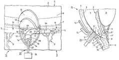
- FIG. 1is a schematic illustration of a portion of a eye, on an enlarged scale, showing a first parabolic incision in the sclera for forming a first scleral flap which is folded upwards;
- FIG. 2is a schematic view of the portion of the eye of FIG. 1, taken along the line II—II in FIG. 1;
- FIG. 3is a schematic illustration of the portion of the eye of FIG. 1, showing a second parabolic incision within the area of first incision for formation of a second scleral flap which is folded upwards;
- FIG. 4is a schematic view of the portion of the eye of FIG. 3, illustrating both upwardly folded scleral flaps and taken along the line IV—IV in FIG. 3;
- FIG. 5is a schematic illustration of the portion of the eye of FIG. 3, depicting a probe for insertion into the exposed Schlemm's canal;
- FIG. 6is a schematic view of the portion of the eye of FIG. 4, illustrating the application of a swab for applying a small pressure force against the Schwalbe's Line in the area of the two upwardly folded scleral flaps;
- FIG. 7is a schematic view of the portion of the eye of FIG. 6, illustrating a detachment of the Descemet's membrane from the cornea and the severed second scleral flap;
- FIG. 8is a schematic view of the portion of the eye of FIG. 4, illustrating exemplified support elements according to the present invention for implantation in the lumen of the Schlemm's canal;
- FIG. 9is a schematic view of the portion of the eye of FIG. 4, illustrating further exemplified support elements according to the present invention for implantation in the lumen of the Schlemm's canal;
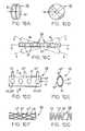
- FIG. 10Ais a detailed perspective view, on an enlarged scale, of a first variation of a support element according to the present invention.
- FIG. 10Bis a detailed perspective view, on an enlarged scale, of a second variation of a support element according to the present invention.
- FIG. 10Cis a detailed view, on an enlarged scale, of a third variation of a support element according to the present invention.
- FIG. 10Dis a detailed view, on an enlarged scale, of a fourth variation of a support element according to the present invention, for exemplified implantation in the exposed portion of the Schlemm's canal;
- FIG. 10Eis a detailed view, on an enlarged scale, of the support element of FIG. 10D, taken along the line E—E in FIG. 10D;
- FIG. 10Fis a detailed view, on an enlarged scale, of a fifth variation of a support element according to the present invention.
- FIG. 10Gis a detailed view, on an enlarged scale, of a sixth variation of a support element according to the present invention.
- FIG. 11is a schematic view of the portion of the eye of FIG. 8, illustrating the exemplified implantation of support elements of FIG. 10A in the Schlemm's canal and the exemplified implantation of support elements of FIG. 10B in the exposed portion of the Schlemm's canal;
- FIG. 12is a schematic view of the portion of the eye of FIG. 11, taken along the line XII—XII in FIG. 11, illustrating the exemplified implantation of a support element of FIG. 10 E and folding back of the first scleral flap;
- FIG. 13is a schematic view of the portion of the eye of FIG. 8, illustrating the exemplified implantation of support elements of FIG. 10A in the Schlemm's canal and the exemplified implantation of a support element of FIG. 10F in the exposed portion of the Schlemm's canal;
- FIG. 14is a schematic view of the portion of the eye of FIG. 8, illustrating the exemplified implantation of support elements of FIG. 10 A and FIG. 10B in the Schlemm's canal and the exemplified implantation of a support element of FIG. 10G in the exposed portion of the Schlemm's canal.
- FIG. 1there is shown a first process step for improving a drainage of aqueous humor in an eye 15 which is shown only schematically by way of a portion of an iris 2 , a cornea 4 , a sclera 3 , a partial section of the circular Schlemm's canal 5 (sinus venosus sclerae) and a channel system 3 ′ which is comprised of a multitude of channels for conducting the aqueous humor.
- a first process step for improving a drainage of aqueous humor in an eye 15which is shown only schematically by way of a portion of an iris 2 , a cornea 4 , a sclera 3 , a partial section of the circular Schlemm's canal 5 (sinus venosus sclerae) and a channel system 3 ′ which is comprised of a multitude of channels for conducting the aqueous humor.
- a first incision of approximately parabolic shapeis made in the sclera 3 to form a scleral flap 10 which is lifted upwards in the direction towards the cornea 4 to thereby expose a corresponding recess 11 which is bounded by a circumferentially extending side wall 11 ′.
- the scleral flap 10is held in upwardly folded position by a tool or other means which are not shown for the sake of simplicity but are generally known by the artisan.
- FIG. 2which is a sectional view of the portion of the eye 15 , taken along the line II—II in FIG. 1, shows a portion of the sclera 3 , a portion of the cornea 4 with the Descemet's membrane 6 and the Schwalbe's line 7 , a portion of the iris 2 and a portion of the lens 9 connected to the sclera 3 by means of the zonular fibers 9 ′. Further shown is the first scleral flap 10 , which has been lifted upwards in the direction of arrow 16 and the corresponding recess 11 , in addition to the Schlemm's canal 5 with the trabecular meshwork 8 disposed anteriorly thereof.
- Arrows 1 and 1 ′ in FIG. 2designate essentially the circulation of aqueous humor and the natural drainage thereof.
- Aqueous humorwhich in a healthy eye, regenerates continually, flows according to arrow 1 from the posterior chamber H to the anterior chamber V and is conducted at the iridocorneal angle V′ (angulus irido-cornealis) in the direction of arrow 1 ′ via the trabecular meshwork 8 into the Schlemm's canal 5 and from there, via the natural channel system 3 ′ (FIG. 1) to a natural venous system (not shown).
- the trabecular meshwork 8When the trabecular meshwork 8 is partially or completely non-functional due to back-up or like blockage, the natural drainage of aqueous humor is limited to such an extent that the pressure inside the eye 15 rises to thereby restrict the blood circulation and thus the functionality of the optic nerve (not shown).
- the resulting diseaseis commonly known under the name “glaucoma” and may lead to blindness of the affected eye.
- a micro-surgical procedureis carried by which the conjunctiva (not shown) is retracted with a suitable tool for exposing a sufficient portion of the sclera 3 .
- the formed scleral flap 10is folded upwards in the direction towards the cornea 4 , thereby exposing the first recess 11 with its circumferential side wall 11 ′.
- the first incisionmay cover an area of, for example, 3 mm ⁇ 3 mm with a depth which is so selected that the thickness 10′ of the first sclera flap 10 is approximately 1 ⁇ 3 of the natural thickness of the sclera 3 in this zone, as depicted in FIG. 2 .
- the Schlemm's canal 5is not yet exposed.
- a second incisionis made within the area of the first incision to form a second parabolic scleral flap 12 which is then lifted upwards in the direction of the cornea 4 in a direction of arrow 16 ′ (FIG. 4 ), so that a second recess 13 is defined in correspondence with the second scleral flap 12 and bounded by a support surface 14 .
- the depth of the second incisioncan, for example, be selected such that the Schlemm's canal 5 is exposed by a portion, denoted in its entirety by reference numeral 18 .
- two inlets 17 and 17 ′ of the Schlemm's canal 5 in opposite disposition in the recess 13are accessible for injection of an expanding medium by means of a probe, shown in FIG. 5 .
- the second scleral flap 12is so formed that the Schlemm's canal 5 includes a depression 5 ′ which exhibits a substantially grooved configuration and extends across the entire width of the second scleral flap 12 .
- FIG. 4shows the portion of eye 15 , taken along the line IV—IV in FIG. 3, with the two scleral flaps 10 , 12 folded upwards in the direction of arrows 16 , 16 ′ and held in place by suitable means (not shown).
- the second incisionresults in a thickness 12 ′ of the second scleral flap 12 that allows sufficient exposure and accessibility of the Schlemm's canal 5 via the inlets 17 , 17 ′ and the exposed portion 18 .
- Thisis essentially realized by so selecting the depth of second incision that the groove-shaped depression 5 ′ of the Schlemm's canal 5 remains at the inside 12 ′′ of the second scleral flap 12 .
- FIGS. 3 and 4also show the recesses 11 , 13 in the sclera 3 with the side wall 11 ′ and the support surface 14 .
- FIG. 5which is an enlarged illustration of the eye 15 of FIG. 3 and shows a portion of the sclera 3 and the two upwardly folded sclera flaps 10 , 12 as well as the second recess 13 and the lateral support surface 14 of the sclera 3 with the channel system 3 ′, depicts a next process phase, in which a suitable medium, preferably a high viscosity sodium hyaluronate solution is injected into the two lateral inlets 17 and 17 ′ of the Schlemm's canal 5 by an injection unit, generally designated by reference numeral 25 to expand the lumen 19 of the Schlemm's canal 5 .
- the injection unit 25includes a probe 24 which, in the nonlimiting example of FIG.
- the injection unit 25 with the probe 24is withdrawn from the inlet 17 and can be turned for insertion in the opposite inlet 17 ′ of the Schlemm's canal 5 for injection of the expanding medium and expansion of the lumen 19 .
- the injection unit 25is connected via a supply conduit 28 to a pressure source 26 , indicated schematically only, which may be formed by a single-chamber syringe or like device.
- the injected mediumis forced into the lumen 19 of Schlemm's canal 5 in the direction of arrow 27 , by means of the manually or electrically operated pressure source 26 via the supply conduit 28 , adapter 29 , and probe 24 , for expansion of the lumen 19 .
- the injection unit 25is removed. It will be appreciated by persons skilled in the art that operation and structure of such injection unit 25 are generally known by the artisan and do not form part of the present invention so that a detailed description thereof has been omitted for the sake of simplicity.
- FIG. 7there is shown a further, optional, process step, after expansion of the Schlemm's canal 5 and withdrawal of the injection unit 25 , representing a detachment the Descemet's membrane 6 from the inner surface of the cornea 4 in the area of the Schwalbe's line 7 .
- the detachment of the Descemet's membrane 6is realized by using a swab 20 or like device to create an opening (window) 21 between the second scleral flap 12 and the Descemet's membrane 6 , as shown schematically on an enlarged scale in FIG. 7 .
- the opening 21is substantially gap-shaped and extends in a manner not shown here across the entire width of the exposed portion 18 or second recess 13 , as best seen in FIG. 8 .
- the opening 21provides a further connection between the anterior chamber V of the eye 15 and the second recess 13 so that aqueous humor can drain, apart from the natural outflow via the trabecular meshwork 8 in the direction of arrow 1 ′, also via the substantially transparent Descemet's membrane 6 , which is partly permeable for aqueous humor, in the direction of arrow 1 ′′, as shown in FIG. 7, and via the opening 21 to the recess 13 which is fluidly connected with the Schlemm's canal 5 .
- the second recess 13which essentially conforms to the scleral flap 12 , forms a reservoir (FIG. 12) for aqueous humor which is drained from the reservoir-forming recess 13 through both lateral inlets 17 , 17 ′ into the Schlemm's canal 5 , and from there via the channel system 3 ′.
- the second scleral flap 12is then, preferably, severed with a suitable surgical instrument (not shown), as shown in FIG. 8 . It will be appreciated by persons skilled in the art, that the separation of the second sclera flap 12 may be carried out also before detachment of the Descemet's membrane 6 by means of the swab 20 to form the gap-shaped opening 21 .
- FIG. 8there is shown a schematic view of the eye 15 , after severance of the second scleral flap 12 , and illustration of the exposed portion 18 of the Schlemm's canal 5 and both confronting inlets 17 , 17 ′.
- Implanted into the lumen 19 of the Schlemm's canal 5 on each of both sides of the exposed portion 18are two support elements in spaced-apart side-by-side disposition.
- two substantially ring-shaped support elements 30are arranged on one side of the exposed portion 18
- two spherical support elements 33are arranged on the other side of the exposed portion 18 .
- FIG. 9shows further exemplified support elements for implantation, that is a support element 45 in the form of a braided mesh is arranged on one side of the exposed portion 18 , and a support element 50 in the form of a helical spring is arranged on the other side of the exposed portion 18 .
- FIG. 10Adepicts a detailed perspective view, on an enlarged scale, of the support element 30 .
- the support element 30has a throughbore 32 and exhibits, preferably, an outer circular ring shape or elliptical ring shape.
- the support element 30is made of a material that will automatically conform to the lumen 19 of the Schlemm's canal 5 .
- the support element 30has a width 31 which is selected such that the support element 30 can be implanted in stable position in the lumen 19 and is prevented from toppling over in axial direction of the lumen 19 .
- FIG. 10Bdepicts a detailed view, on an enlarged scale, of the spherical support element 33 .
- the support element 33has at least one throughbore 34 , preferably several bores 34 which are spaced in circumferential direction.
- FIGS. 10C to 10 Eshow further examples of support elements in accordance with the present invention. It will be appreciated by persons skilled in the art that the various configurations of support elements, described here, can be combined in any desired manner for implantation in the lumen 19 of the Schlemm's canal 5 . Thus, the types of support elements implanted in FIGS. 8 and 9 are shown only for illustrative purposes.
- FIG. 10Cshows a support element 35 which is made from a flexible tube and has an outer configuration of circular ring shape or elliptic shape.
- the support element 35is traversed in axial direction by a throughbore 36 which is in communication with a plurality of inlet openings 37 spaced in axial direction.
- the support element 35is freely movable in its disposition and orientation, as shown schematically by broken lines, and thus can easily conform, as a consequence of its flexibility, to the inner configuration of the lumen 19 when inserted in the Schlemm's canal 5 .
- the flexibilityis however so limited that a kinking is eliminated.
- FIG. 10Dshows a support element 40 which is also made from an elongate flexible tube 41 and is traversed in axial direction by a throughbore 41 ′ which is communication with a plurality of apertures 42 , 42 ′ spaced from one another in axial direction.
- the support element 40has further arranged, preferably formed, thereon, a plurality of protrusions 43 , 43 ′, 43 ′′ which are spaced from one another in axial direction.
- the apertures 42 , 42 ′, which communicate with the throughbore 41 ′,are preferably arranged in diametrically confronting disposition on the longer side of the support element 40 . As shown in FIG.
- the support element 40has an outer elliptic configuration which substantially conforms to the configuration of the Schlemm's canal 5 .
- This type of support element 40is used, primarily, for implantation in the exposed portion 18 of the Schlemm's canal 5 in the area of the second scleral recess 13 of the sclera 3 .
- the function of the support element 40will be described in more detail with reference to FIGS. 11 and 12.
- FIG. 10Fshows in more detail the support element 45 in the form of a braided mesh.
- the support element 45is made from a plurality of threads 46 (filaments) which are helically intertwined to form the braided mesh, which has spacings 47 , 47 ′, 47 ′′ formed between the threads 46 for drainage of aqueous humor.
- the support element 45is capable to conform by itself to the outer configuration of the lumen 19 of the Schlemm's canal 5 .
- FIG. 10Gshows in more detail the support element 50 , which, for example, is formed from a single, helically twined thread 51 (filament). Aqueous humor is drained between spacings 52 , 52 ′ of the individual windings of the support element 50 .
- the support element 50is capable to conform by itself to the outer configuration of the lumen 19 of the Schlemm's canal 5 .
- the elongate support elements 35 , 40 , 45 or 50may be implanted in the portion 18 of the Schlemm's canal 5 , exposed in the area of the second recess 13 of the sclera 3 , as well as in the lumen 19 of the Schlemm's canal 5 . Implantation of the elongate support element 35 , 45 , 50 in the exposed portion of the Schlemm's canal 5 is also feasible, as will be described in more detail with reference with FIGS. 13 and 14.
- FIG. 11shows implantation of spaced apart, ring-shaped support elements 30 on both sides of the second recess 13 in the lumen 19 of the Schlemm's canal 5 , whereas a support element 40 is placed in the area of the second recess 13 of the sclera 3 in the exposed portion 18 of the Schlemm's canal 5 .
- the support element 40is so disposed in the exposed portion 18 of the Schlemm's canal 5 that the residual portion 12 . 1 , which has been left from the second scleral flap 12 and extends across the entire width of the second recess 13 , rests on the protrusions 43 , 43 ′, 43 ′′ of the support element 40 .
- the first sclera flap 10is folded back and, as shown in FIG. 12, placed on the parabolic support surface 14 . Subsequently, the first scleral flap 10 is sutured partially, in a manner known per se, to the sclera 3 .
- a subscleral space 13 ′is created in the form of the flat recess 13 behind the first scleral flap 10 and preferably filled by means of a syringe (not shown) with high viscosity medium, such as sodium hyaluronate, before completely rejoining the first scleral flap 10 . This prevents an inside surface 10 ′′ of the repositioned first scleral flap 10 to come into contact with the inside surface 13 ′′ of the recess 13 , as shown in FIG. 12 .
- FIG. 12shows the implanted support element 40 according to FIG. 11 along the section line XII—XII on an enlarged scale, with the scleral flap 10 being folded back, and the residual portion 12 . 1 of the second scleral flap 12 , separated from the Descemet's membrane 6 and placed on the protrusions 43 , 43 ′, 43 ′′.
- the provision of the protrusions 43 , 43 ′, 43 ′′prevents a closing of the gap-shaped opening 21 across the entire width of the exposed portion 18 of the Schlemm's canal 5 .
- the gap-shaped opening 21provides an additional connection between the iridocorneal angle V′ of the anterior chamber V and the second recess 13 .
- Aqueous humorcan thus drain in addition to the natural drainage in the direction of arrow 1 ′ via the trabecular meshwork 8 also in the direction of arrow 1 ′′ via the substantially transparent and partially permeable Descemet's membrane 6 and via the gap-shaped opening 21 to the recess 13 .
- the flat recess 13which has a configuration that approximates the second scleral flap 12 , forms the subscleral space 13 ′ or a reservoir for aqueous humor, when the scleral flap 10 is folded back.
- aqueous humoris drained via the two inlets 17 , 17 ′, fluidly connected to the subscleral space 13 ′, into the lumen 19 of the Schlemm's canal 5 , and from there into the channel system 3 ′.
- FIG. 13shows implantation of spaced apart, ring-shaped support elements 30 , according to FIG. 10A, on both sides of the second recess 13 in the lumen 19 of the Schlemm's canal 5 .
- support elements 30or in combination with the support elements 30 , several spaced-apart spherical support elements 33 may be used for implantation in the lumen 19 .
- FIG. 14shows implantation of two spaced-apart, ring-shaped support elements 30 , according to FIG.10A on one side in the lumen 19 , whereas two spaced-apart, spherical support elements 33 , according to FIG. 10B, are positioned on the other, opposite side in the lumen 19 .
- This non-limiting exampleincludes also the implantation of the support element 50 according to FIG. 10G in the exposed portion 18 of the Schlemm's canal 5 .
- the support elements 30 , 33 , 35 , 45 , 50are made from decomposable material, in particular material that is biolytically decomposable by the tissue of the Schlemm's canal 5 and/or the aqueous humor. Especially suitable are materials that biolytically decompose within 2 to 12 months after implantation. Examples for materials used for support elements 30 , 33 , 35 , 45 , 50 include a cross-linked sodium hyaluronate. There is, however, also the option, to make the support element, in particular the elongate support element 35 , 40 , 45 , 50 that is implantable in the exposed portion 18 of the Schlemm's canal 5 , from biocompatible material, e.g. plastic material, rust-free steel or special steel such as silver, gold or platinum.
- biocompatible materiale.g. plastic material, rust-free steel or special steel such as silver, gold or platinum.
Landscapes
- Health & Medical Sciences (AREA)
- Ophthalmology & Optometry (AREA)
- Heart & Thoracic Surgery (AREA)
- Surgery (AREA)
- Engineering & Computer Science (AREA)
- Biomedical Technology (AREA)
- Nuclear Medicine, Radiotherapy & Molecular Imaging (AREA)
- Vascular Medicine (AREA)
- Life Sciences & Earth Sciences (AREA)
- Animal Behavior & Ethology (AREA)
- General Health & Medical Sciences (AREA)
- Public Health (AREA)
- Veterinary Medicine (AREA)
- Prostheses (AREA)
- External Artificial Organs (AREA)
Abstract
Description
Claims (20)
Priority Applications (5)
| Application Number | Priority Date | Filing Date | Title |
|---|---|---|---|
| US09/503,884US6375642B1 (en) | 2000-02-15 | 2000-02-15 | Method of and device for improving a drainage of aqueous humor within the eye |
| AU10027/01AAU1002701A (en) | 2000-02-15 | 2001-01-03 | A method of and device for improving a drainage of aqueous humor within the eye |
| CA002331868ACA2331868A1 (en) | 2000-02-15 | 2001-01-22 | A method of and device for improving a drainage of aqueous humor within the eye |
| EP01810106AEP1125568A3 (en) | 2000-02-15 | 2001-02-02 | Method and device to improve as well as to maintain the outflow of the aqueous humor of an eye |
| JP2001032379AJP2001245916A (en) | 2000-02-15 | 2001-02-08 | Method and apparatus for improving discharge of aqueous humor of eye |
Applications Claiming Priority (1)
| Application Number | Priority Date | Filing Date | Title |
|---|---|---|---|
| US09/503,884US6375642B1 (en) | 2000-02-15 | 2000-02-15 | Method of and device for improving a drainage of aqueous humor within the eye |
Publications (1)
| Publication Number | Publication Date |
|---|---|
| US6375642B1true US6375642B1 (en) | 2002-04-23 |
Family
ID=24003914
Family Applications (1)
| Application Number | Title | Priority Date | Filing Date |
|---|---|---|---|
| US09/503,884Expired - LifetimeUS6375642B1 (en) | 2000-02-15 | 2000-02-15 | Method of and device for improving a drainage of aqueous humor within the eye |
Country Status (5)
| Country | Link |
|---|---|
| US (1) | US6375642B1 (en) |
| EP (1) | EP1125568A3 (en) |
| JP (1) | JP2001245916A (en) |
| AU (1) | AU1002701A (en) |
| CA (1) | CA2331868A1 (en) |
Cited By (107)
| Publication number | Priority date | Publication date | Assignee | Title |
|---|---|---|---|---|
| US6450984B1 (en) | 1999-04-26 | 2002-09-17 | Gmp Vision Solutions, Inc. | Shunt device and method for treating glaucoma |
| US20020143284A1 (en)* | 2001-04-03 | 2002-10-03 | Hosheng Tu | Drug-releasing trabecular implant for glaucoma treatment |
| US20020169130A1 (en)* | 2001-05-03 | 2002-11-14 | Hosheng Tu | Medical device and methods of use for glaucoma treatment |
| US20020188308A1 (en)* | 2001-04-07 | 2002-12-12 | Hosheng Tu | Glaucoma stent and methods thereof for glaucoma treatment |
| US6533768B1 (en)* | 2000-04-14 | 2003-03-18 | The Regents Of The University Of California | Device for glaucoma treatment and methods thereof |
| US20030055372A1 (en)* | 1999-04-26 | 2003-03-20 | Lynch Mary G. | Shunt device and method for treating glaucoma |
| US20030060752A1 (en)* | 2000-04-14 | 2003-03-27 | Olav Bergheim | Glaucoma device and methods thereof |
| US20030088260A1 (en)* | 2001-11-08 | 2003-05-08 | Smedley Gregory T. | Combined treatment for cataract and glaucoma treatment |
| US20030097151A1 (en)* | 2001-10-25 | 2003-05-22 | Smedley Gregory T. | Apparatus and mitochondrial treatment for glaucoma |
| US20030153863A1 (en)* | 2002-02-13 | 2003-08-14 | Patel Anilbhai S. | Implant system for glaucoma surgery |
| US20030187385A1 (en)* | 2000-04-14 | 2003-10-02 | Bergheim Olav B. | Implant with anchor |
| WO2002087418A3 (en)* | 2001-05-02 | 2003-11-27 | Glaukos Corp | Bifurcatable trabecular shunt for glaucoma treatment |
| US20040024345A1 (en)* | 2002-04-19 | 2004-02-05 | Morteza Gharib | Glaucoma implant with valveless flow bias |
| US20040050392A1 (en)* | 2001-08-28 | 2004-03-18 | Hosheng Tu | Glaucoma stent for treating glaucoma and methods of use |
| US20040092856A1 (en)* | 2000-09-01 | 2004-05-13 | Elie Dahan | Glaucoma drain |
| US20040102729A1 (en)* | 2002-04-08 | 2004-05-27 | David Haffner | Devices and methods for glaucoma treatment |
| US20040111050A1 (en)* | 2000-04-14 | 2004-06-10 | Gregory Smedley | Implantable ocular pump to reduce intraocular pressure |
| US20040127843A1 (en)* | 2000-04-14 | 2004-07-01 | Hosheng Tu | Glaucoma implant with therapeutic agents |
| US20040147870A1 (en)* | 2002-04-08 | 2004-07-29 | Burns Thomas W. | Glaucoma treatment kit |
| US20040193095A1 (en)* | 2003-03-29 | 2004-09-30 | Shadduck John H. | Implants for treating ocular hypertension, methods of use and methods of fabrication |
| WO2004093761A1 (en) | 2003-04-16 | 2004-11-04 | Iscience Surgical Corporation | Opthalmic microsurgical instruments |
| US20050049578A1 (en)* | 2000-04-14 | 2005-03-03 | Hosheng Tu | Implantable ocular pump to reduce intraocular pressure |
| US20050119636A1 (en)* | 2001-05-02 | 2005-06-02 | David Haffner | Implant with intraocular pressure sensor for glaucoma treatment |
| US20050192527A1 (en)* | 2001-05-02 | 2005-09-01 | Morteza Gharib | Glaucoma implant with extending members |
| US6939298B2 (en) | 2002-02-28 | 2005-09-06 | Gmp Vision Solutions, Inc | Device and method for monitoring aqueous flow within the eye |
| US20050250788A1 (en)* | 2004-01-30 | 2005-11-10 | Hosheng Tu | Aqueous outflow enhancement with vasodilated aqueous cavity |
| US20050266047A1 (en)* | 2002-04-08 | 2005-12-01 | Hosheng Tu | Injectable glaucoma implants with multiple openings |
| US20050271704A1 (en)* | 2002-04-08 | 2005-12-08 | Hosheng Tu | Injectable glaucoma implants with multiple openings |
| US20050277864A1 (en)* | 2000-04-14 | 2005-12-15 | David Haffner | Injectable gel implant for glaucoma treatment |
| US20060195187A1 (en)* | 2004-12-16 | 2006-08-31 | Iscience Surgical Corporation | Ophthalmic implant for treatment of glaucoma |
| US7186232B1 (en) | 2002-03-07 | 2007-03-06 | Glaukoa Corporation | Fluid infusion methods for glaucoma treatment |
| US20070088242A1 (en)* | 2003-11-14 | 2007-04-19 | Coroneo Minas T | Ocular pressure regulation |
| US20070191863A1 (en)* | 2006-01-17 | 2007-08-16 | De Juan Eugene Jr | Glaucoma Treatment Device |
| US20070233037A1 (en)* | 2006-01-17 | 2007-10-04 | Gifford Hanson S Iii | Drug Delivery Treatment Device |
| US20070293807A1 (en)* | 2006-05-01 | 2007-12-20 | Lynch Mary G | Dual drainage pathway shunt device and method for treating glaucoma |
| US20070298068A1 (en)* | 2006-06-26 | 2007-12-27 | Badawi David Y | Intraocular implants and methods and kits therefor |
| US20080058760A1 (en)* | 2004-05-05 | 2008-03-06 | Bengt Agerup | Use Of A Viscoelastic Composition For Treating Increased Intraocular Pressure |
| US20080172204A1 (en)* | 2007-01-15 | 2008-07-17 | Fujitsu Limited | Step counter and method of counting steps |
| US7488303B1 (en) | 2002-09-21 | 2009-02-10 | Glaukos Corporation | Ocular implant with anchor and multiple openings |
| US20090082860A1 (en)* | 2007-09-24 | 2009-03-26 | Schieber Andrew T | Ocular Implants with Asymmetric Flexibility |
| US20090132040A1 (en)* | 2007-11-20 | 2009-05-21 | Ivantis, Inc. | Ocular Implant Delivery System and Method |
| US20090182421A1 (en)* | 2007-07-17 | 2009-07-16 | Tom Silvestrini | Ocular implant with hydrogel expansion capabilities |
| US20100121342A1 (en)* | 2007-11-20 | 2010-05-13 | Schieber Andrew T | Methods and Apparatus for Delivering Ocular Implants Into the Eye |
| US20100152641A1 (en)* | 2003-05-05 | 2010-06-17 | Michael Yablonski | Internal shunt and method for treating glaucoma |
| US7740604B2 (en) | 2007-09-24 | 2010-06-22 | Ivantis, Inc. | Ocular implants for placement in schlemm's canal |
| US20100274258A1 (en)* | 2009-01-28 | 2010-10-28 | Silvestrini Thomas A | Ocular implant with stiffness qualities, methods of implantation and system |
| US20110009958A1 (en)* | 2009-07-09 | 2011-01-13 | John Wardle | Ocular Implants and Methods for Delivering Ocular Implants Into the Eye |
| US20110009874A1 (en)* | 2009-07-09 | 2011-01-13 | John Wardle | Single Operator Device for Delivering an Ocular Implant |
| US20110105990A1 (en)* | 2009-11-04 | 2011-05-05 | Silvestrini Thomas A | Zonal drug delivery device and method |
| US20110118649A1 (en)* | 2009-11-13 | 2011-05-19 | Grieshaber Ophthalmic Research Foundation | Method and device for the treatment of glaucoma |
| US7951155B2 (en) | 2002-03-15 | 2011-05-31 | Glaukos Corporation | Combined treatment for cataract and glaucoma treatment |
| WO2011097408A1 (en)* | 2010-02-05 | 2011-08-11 | Sight Sciences, Inc | Intraocular implants and related kits and methods |
| US20110224597A1 (en)* | 2008-12-22 | 2011-09-15 | GRIESHABAR OPHTHALMIC RESARCH FOUNDATION C/O PRICEWATERHOUSE COOPERS AG NEUMARKET 4/KorNHAUSST | Implant for inserting into the schlemm's canal of an eye |
| US20110238075A1 (en)* | 2009-12-23 | 2011-09-29 | Luke Clauson | Drug delivery devices and methods |
| US8267882B2 (en) | 2008-03-05 | 2012-09-18 | Ivantis, Inc. | Methods and apparatus for treating glaucoma |
| US8372026B2 (en) | 2007-09-24 | 2013-02-12 | Ivantis, Inc. | Ocular implant architectures |
| US8506515B2 (en) | 2006-11-10 | 2013-08-13 | Glaukos Corporation | Uveoscleral shunt and methods for implanting same |
| RU2493787C2 (en)* | 2011-12-20 | 2013-09-27 | Али Гумярович Амиров | Method of scleral flap fixation in antiglaucomatous operations |
| US8545430B2 (en) | 2010-06-09 | 2013-10-01 | Transcend Medical, Inc. | Expandable ocular devices |
| US8657776B2 (en) | 2011-06-14 | 2014-02-25 | Ivantis, Inc. | Ocular implants for delivery into the eye |
| US8663150B2 (en) | 2011-12-19 | 2014-03-04 | Ivantis, Inc. | Delivering ocular implants into the eye |
| US8715712B2 (en) | 2011-09-14 | 2014-05-06 | Forsight Vision5, Inc. | Ocular insert apparatus and methods |
| US8747299B2 (en) | 2011-06-02 | 2014-06-10 | Grieshaber Ophtalmic Research Foundation | Method and device for the pathology analysis of the Schlemm's canal |
| CN103932841A (en)* | 2011-06-16 | 2014-07-23 | 王宁利 | Schlemm canal expandable stent and combination bodies thereof |
| US8808222B2 (en) | 2007-11-20 | 2014-08-19 | Ivantis, Inc. | Methods and apparatus for delivering ocular implants into the eye |
| US8894603B2 (en) | 2012-03-20 | 2014-11-25 | Sight Sciences, Inc. | Ocular delivery systems and methods |
| CN104602652A (en)* | 2012-04-12 | 2015-05-06 | 伊万·季米特里耶维奇·扎哈罗夫 | Drainage device and method for producing same |
| US9155656B2 (en) | 2012-04-24 | 2015-10-13 | Transcend Medical, Inc. | Delivery system for ocular implant |
| US9358156B2 (en) | 2012-04-18 | 2016-06-07 | Invantis, Inc. | Ocular implants for delivery into an anterior chamber of the eye |
| US9421126B2 (en) | 2009-06-03 | 2016-08-23 | Forsight Vision5, Inc. | Anterior segment drug delivery |
| US9480598B2 (en) | 2012-09-17 | 2016-11-01 | Novartis Ag | Expanding ocular implant devices and methods |
| US9492319B2 (en) | 2011-02-23 | 2016-11-15 | Grieshaber Ophthalmic Research Foundation | Implant for the treatment of glaucoma |
| US9510973B2 (en) | 2010-06-23 | 2016-12-06 | Ivantis, Inc. | Ocular implants deployed in schlemm's canal of the eye |
| US9554940B2 (en) | 2012-03-26 | 2017-01-31 | Glaukos Corporation | System and method for delivering multiple ocular implants |
| US9579234B2 (en) | 2009-10-23 | 2017-02-28 | Ivantis, Inc. | Ocular implant system and method |
| US9592151B2 (en) | 2013-03-15 | 2017-03-14 | Glaukos Corporation | Systems and methods for delivering an ocular implant to the suprachoroidal space within an eye |
| US9730638B2 (en) | 2013-03-13 | 2017-08-15 | Glaukos Corporation | Intraocular physiological sensor |
| US9750636B2 (en) | 2012-10-26 | 2017-09-05 | Forsight Vision5, Inc. | Ophthalmic system for sustained release of drug to eye |
| US9763829B2 (en) | 2012-11-14 | 2017-09-19 | Novartis Ag | Flow promoting ocular implant |
| US9987163B2 (en) | 2013-04-16 | 2018-06-05 | Novartis Ag | Device for dispensing intraocular substances |
| US10085633B2 (en) | 2012-04-19 | 2018-10-02 | Novartis Ag | Direct visualization system for glaucoma treatment |
| US10159601B2 (en) | 2000-05-19 | 2018-12-25 | Ivantis, Inc. | Delivery system and method of use for the eye |
| USD846738S1 (en) | 2017-10-27 | 2019-04-23 | Glaukos Corporation | Implant delivery apparatus |
| US10299958B2 (en) | 2015-03-31 | 2019-05-28 | Sight Sciences, Inc. | Ocular delivery systems and methods |
| US10517759B2 (en) | 2013-03-15 | 2019-12-31 | Glaukos Corporation | Glaucoma stent and methods thereof for glaucoma treatment |
| US10617558B2 (en) | 2012-11-28 | 2020-04-14 | Ivantis, Inc. | Apparatus for delivering ocular implants into an anterior chamber of the eye |
| US10709547B2 (en) | 2014-07-14 | 2020-07-14 | Ivantis, Inc. | Ocular implant delivery system and method |
| CN111803274A (en)* | 2020-07-22 | 2020-10-23 | 深圳市朗目医疗科技有限公司 | Glaucoma drainage device and drainage implant therefor |
| RU2736395C1 (en)* | 2020-02-19 | 2020-11-16 | федеральное государственное автономное учреждение "Национальный медицинский исследовательский центр "Межотраслевой научно-технический комплекс "Микрохирургия глаза" имени академика С.Н. Федорова" Министерства здравоохранения Российской Федерации | Method for fixation of scleral flap during antiglaucomatous operations with implantation of drainage device |
| US10959941B2 (en) | 2014-05-29 | 2021-03-30 | Glaukos Corporation | Implants with controlled drug delivery features and methods of using same |
| US11116625B2 (en) | 2017-09-28 | 2021-09-14 | Glaukos Corporation | Apparatus and method for controlling placement of intraocular implants |
| US11197779B2 (en) | 2015-08-14 | 2021-12-14 | Ivantis, Inc. | Ocular implant with pressure sensor and delivery system |
| US11224602B2 (en) | 2015-04-13 | 2022-01-18 | Forsight Vision5, Inc. | Ocular insert composition of a semi-crystalline or crystalline pharmaceutically active agent |
| US20220142817A1 (en)* | 2019-07-10 | 2022-05-12 | Aquea Health, Inc. | Eye stents and delivery systems |
| US11363951B2 (en) | 2011-09-13 | 2022-06-21 | Glaukos Corporation | Intraocular physiological sensor |
| US11376040B2 (en) | 2017-10-06 | 2022-07-05 | Glaukos Corporation | Systems and methods for delivering multiple ocular implants |
| US11504270B1 (en) | 2019-09-27 | 2022-11-22 | Sight Sciences, Inc. | Ocular delivery systems and methods |
| US11540940B2 (en) | 2021-01-11 | 2023-01-03 | Alcon Inc. | Systems and methods for viscoelastic delivery |
| US11744734B2 (en) | 2007-09-24 | 2023-09-05 | Alcon Inc. | Method of implanting an ocular implant |
| WO2023237127A1 (en)* | 2022-06-10 | 2023-12-14 | 健诺维(成都)生物科技有限公司 | Ocular implant catheter |
| US11925578B2 (en) | 2015-09-02 | 2024-03-12 | Glaukos Corporation | Drug delivery implants with bi-directional delivery capacity |
| US11938058B2 (en) | 2015-12-15 | 2024-03-26 | Alcon Inc. | Ocular implant and delivery system |
| US12029683B2 (en) | 2018-02-22 | 2024-07-09 | Alcon Inc. | Ocular implant and delivery system |
| US12226309B2 (en) | 2013-03-15 | 2025-02-18 | Alcon Inc. | Intraocular lens storage and loading devices and methods of use |
| US12318279B2 (en) | 2007-07-23 | 2025-06-03 | Alcon Inc. | Lens delivery system |
| US12419783B2 (en) | 2010-11-24 | 2025-09-23 | Glaukos Corporation | Drug eluting ocular implant |
| US12440377B2 (en) | 2024-11-13 | 2025-10-14 | Aquea Health, Inc. | Eye stents and delivery systems and methods |
Families Citing this family (4)
| Publication number | Priority date | Publication date | Assignee | Title |
|---|---|---|---|---|
| AR054647A1 (en) | 2005-02-21 | 2007-07-11 | Maldonado Bas Arturo | DEVICE FOR WATER HUMOR DRAINAGE IN GLAUCOMA CASES |
| ES2435618T3 (en)* | 2009-06-25 | 2013-12-20 | Optonol Ltd. | Fiber matrix to maintain a soft tissue space |
| US8951221B2 (en) | 2009-08-20 | 2015-02-10 | Grieshaber Ophthalmic Research Foundation | Method and device for the treatment of glaucoma |
| DE102020002231B4 (en) | 2020-04-09 | 2022-02-17 | aixtent GmbH | Method of manufacturing an implant for insertion into Schlemm's canal of an eye, implant and arrangement with an implant |
Citations (3)
| Publication number | Priority date | Publication date | Assignee | Title |
|---|---|---|---|---|
| US5360399A (en) | 1992-01-10 | 1994-11-01 | Robert Stegmann | Method and apparatus for maintaining the normal intraocular pressure |
| US5626558A (en)* | 1995-05-05 | 1997-05-06 | Suson; John | Adjustable flow rate glaucoma shunt and method of using same |
| US6142990A (en)* | 1997-02-15 | 2000-11-07 | Heidelberg Engineering Optische Messsysteme Gmbh | Medical apparatus, especially for reducing intraocular pressure |
Family Cites Families (7)
| Publication number | Priority date | Publication date | Assignee | Title |
|---|---|---|---|---|
| US898947A (en) | 1906-07-16 | 1908-09-15 | Curtain Supply Co | Shade-fixture. |
| KR100313268B1 (en)* | 1993-02-26 | 2002-11-22 | 산텐 세이야꾸 가부시키가이샤 | Biodegradable Scleral Plug |
| FR2759577B1 (en)* | 1997-02-17 | 1999-08-06 | Corneal Ind | DEEP SCLERECTOMY IMPLANT |
| US5882327A (en)* | 1997-04-17 | 1999-03-16 | Jacob; Jean T. | Long-term glaucoma drainage implant |
| EP0898947A3 (en)* | 1997-08-15 | 1999-09-08 | GRIESHABER & CO. AG SCHAFFHAUSEN | Method and apparatus to improve the outflow of the aqueous humor of an eye |
| US6558342B1 (en)* | 1999-06-02 | 2003-05-06 | Optonol Ltd. | Flow control device, introducer and method of implanting |
| WO2002036052A1 (en)* | 2000-11-01 | 2002-05-10 | Glaukos Corporation | Glaucoma treatment device |
- 2000
- 2000-02-15USUS09/503,884patent/US6375642B1/ennot_activeExpired - Lifetime
- 2001
- 2001-01-03AUAU10027/01Apatent/AU1002701A/ennot_activeAbandoned
- 2001-01-22CACA002331868Apatent/CA2331868A1/ennot_activeAbandoned
- 2001-02-02EPEP01810106Apatent/EP1125568A3/ennot_activeWithdrawn
- 2001-02-08JPJP2001032379Apatent/JP2001245916A/ennot_activeWithdrawn
Patent Citations (4)
| Publication number | Priority date | Publication date | Assignee | Title |
|---|---|---|---|---|
| US5360399A (en) | 1992-01-10 | 1994-11-01 | Robert Stegmann | Method and apparatus for maintaining the normal intraocular pressure |
| US5486165A (en)* | 1992-01-10 | 1996-01-23 | Stegmann; Robert | Method and appliance for maintaining the natural intraocular pressure |
| US5626558A (en)* | 1995-05-05 | 1997-05-06 | Suson; John | Adjustable flow rate glaucoma shunt and method of using same |
| US6142990A (en)* | 1997-02-15 | 2000-11-07 | Heidelberg Engineering Optische Messsysteme Gmbh | Medical apparatus, especially for reducing intraocular pressure |
Cited By (364)
| Publication number | Priority date | Publication date | Assignee | Title |
|---|---|---|---|---|
| US8152752B2 (en) | 1999-04-26 | 2012-04-10 | Glaukos Corporation | Shunt device and method for treating glaucoma |
| US10568762B2 (en) | 1999-04-26 | 2020-02-25 | Glaukos Corporation | Stent for treating ocular disorders |
| US6464724B1 (en) | 1999-04-26 | 2002-10-15 | Gmp Vision Solutions, Inc. | Stent device and method for treating glaucoma |
| US8388568B2 (en) | 1999-04-26 | 2013-03-05 | Glaukos Corporation | Shunt device and method for treating ocular disorders |
| US10492950B2 (en) | 1999-04-26 | 2019-12-03 | Glaukos Corporation | Shunt device and method for treating ocular disorders |
| US6524275B1 (en) | 1999-04-26 | 2003-02-25 | Gmp Vision Solutions, Inc. | Inflatable device and method for treating glaucoma |
| US20100004580A1 (en)* | 1999-04-26 | 2010-01-07 | Glaukos Corporation | Shunt device and method for treating ocular disorders |
| US20030055372A1 (en)* | 1999-04-26 | 2003-03-20 | Lynch Mary G. | Shunt device and method for treating glaucoma |
| US7850637B2 (en) | 1999-04-26 | 2010-12-14 | Glaukos Corporation | Shunt device and method for treating glaucoma |
| US8771217B2 (en) | 1999-04-26 | 2014-07-08 | Glaukos Corporation | Shunt device and method for treating ocular disorders |
| US9827143B2 (en) | 1999-04-26 | 2017-11-28 | Glaukos Corporation | Shunt device and method for treating ocular disorders |
| US20050119601A9 (en)* | 1999-04-26 | 2005-06-02 | Lynch Mary G. | Shunt device and method for treating glaucoma |
| US6626858B2 (en) | 1999-04-26 | 2003-09-30 | Gmp Vision Solutions, Inc. | Shunt device and method for treating glaucoma |
| US20050090806A1 (en)* | 1999-04-26 | 2005-04-28 | Gmp Vision Solutions Inc. | Shunt device and method for treating glaucoma |
| US20050090807A1 (en)* | 1999-04-26 | 2005-04-28 | Gmp Vision Solutions, Inc. | Shunt device and method for treating glaucoma |
| US6827699B2 (en) | 1999-04-26 | 2004-12-07 | Gmp Vision Solutions, Inc. | Shunt device and method for treating glaucoma |
| US6827700B2 (en) | 1999-04-26 | 2004-12-07 | Gmp Vision Solutions, Inc. | Shunt device and method for treating glaucoma |
| US20030236484A1 (en)* | 1999-04-26 | 2003-12-25 | Gmp Vision Solutions, Inc. | Inflatable device and method for treating glaucoma |
| US6450984B1 (en) | 1999-04-26 | 2002-09-17 | Gmp Vision Solutions, Inc. | Shunt device and method for treating glaucoma |
| US9492320B2 (en) | 1999-04-26 | 2016-11-15 | Glaukos Corporation | Shunt device and method for treating ocular disorders |
| US20110196281A1 (en)* | 1999-04-26 | 2011-08-11 | Glaukos Corporation | Shunt device and method for treating ocular disorders |
| US6783544B2 (en) | 1999-04-26 | 2004-08-31 | Gmp Vision Solutions, Inc. | Stent device and method for treating glaucoma |
| US8808219B2 (en) | 2000-04-14 | 2014-08-19 | Glaukos Corporation | Implant delivery device and methods thereof for treatment of ocular disorders |
| US20050209549A1 (en)* | 2000-04-14 | 2005-09-22 | Bergheim Olav B | Glaucoma implant with multiple openings |
| US20040127843A1 (en)* | 2000-04-14 | 2004-07-01 | Hosheng Tu | Glaucoma implant with therapeutic agents |
| US9066782B2 (en) | 2000-04-14 | 2015-06-30 | Dose Medical Corporation | Ocular implant with therapeutic agents and methods thereof |
| US6780164B2 (en) | 2000-04-14 | 2004-08-24 | Glaukos Corporation | L-shaped implant with bi-directional flow |
| US6736791B1 (en) | 2000-04-14 | 2004-05-18 | Glaukos Corporation | Glaucoma treatment device |
| US7297130B2 (en) | 2000-04-14 | 2007-11-20 | Glaukos Corporation | Implant with anchor |
| US20040210185A1 (en)* | 2000-04-14 | 2004-10-21 | Hosheng Tu | Glaucoma implant kit |
| US8814820B2 (en) | 2000-04-14 | 2014-08-26 | Glaukos Corporation | Ocular implant with therapeutic agent and methods thereof |
| US8273050B2 (en) | 2000-04-14 | 2012-09-25 | Glaukos Corporation | Ocular implant with anchor and therapeutic agent |
| US20070282245A1 (en)* | 2000-04-14 | 2007-12-06 | Glaukos Corporation | Glaucoma implant with valve |
| US20040249333A1 (en)* | 2000-04-14 | 2004-12-09 | Bergheim Olav B. | Glaucoma implant with bi-directional flow |
| US20040254519A1 (en)* | 2000-04-14 | 2004-12-16 | Hosheng Tu | Glaucoma treatment device |
| US20050049578A1 (en)* | 2000-04-14 | 2005-03-03 | Hosheng Tu | Implantable ocular pump to reduce intraocular pressure |
| US6638239B1 (en) | 2000-04-14 | 2003-10-28 | Glaukos Corporation | Apparatus and method for treating glaucoma |
| US20030187385A1 (en)* | 2000-04-14 | 2003-10-02 | Bergheim Olav B. | Implant with anchor |
| US8801648B2 (en) | 2000-04-14 | 2014-08-12 | Glaukos Corporation | Ocular implant with anchor and methods thereof |
| US9789001B2 (en) | 2000-04-14 | 2017-10-17 | Dose Medical Corporation | Ocular implant with therapeutic agents and methods thereof |
| US8333742B2 (en) | 2000-04-14 | 2012-12-18 | Glaukos Corporation | Method of delivering an implant for treating an ocular disorder |
| US7867205B2 (en) | 2000-04-14 | 2011-01-11 | Glaukos Corporation | Method of delivering an implant for treating an ocular disorder |
| US20050209550A1 (en)* | 2000-04-14 | 2005-09-22 | Bergheim Olav B | Method of treating glaucoma using an implant having a uniform diameter between the anterior chamber and Schlemm's canal |
| US20040111050A1 (en)* | 2000-04-14 | 2004-06-10 | Gregory Smedley | Implantable ocular pump to reduce intraocular pressure |
| US6955656B2 (en) | 2000-04-14 | 2005-10-18 | Glaukos Corporation | Apparatus and method for treating glaucoma |
| US20030060752A1 (en)* | 2000-04-14 | 2003-03-27 | Olav Bergheim | Glaucoma device and methods thereof |
| US20100234790A1 (en)* | 2000-04-14 | 2010-09-16 | Glaukos Corporation | Ocular implant with therapeutic agents and methods thereof |
| US9993368B2 (en) | 2000-04-14 | 2018-06-12 | Glaukos Corporation | System and method for treating an ocular disorder |
| US20050277864A1 (en)* | 2000-04-14 | 2005-12-15 | David Haffner | Injectable gel implant for glaucoma treatment |
| US8348877B2 (en) | 2000-04-14 | 2013-01-08 | Dose Medical Corporation | Ocular implant with therapeutic agents and methods thereof |
| US7708711B2 (en) | 2000-04-14 | 2010-05-04 | Glaukos Corporation | Ocular implant with therapeutic agents and methods thereof |
| US20100056979A1 (en)* | 2000-04-14 | 2010-03-04 | Glaukos Corporation | Implantable ocular pump to reduce intraocular pressure |
| US10485702B2 (en) | 2000-04-14 | 2019-11-26 | Glaukos Corporation | System and method for treating an ocular disorder |
| US20080234624A2 (en)* | 2000-04-14 | 2008-09-25 | Glaukos Corporation | Ocular implant with anchor and therapeutic agent |
| US20100010414A1 (en)* | 2000-04-14 | 2010-01-14 | Glaukos Corporation | Method of delivering an implant for treating an ocular disorder |
| US6533768B1 (en)* | 2000-04-14 | 2003-03-18 | The Regents Of The University Of California | Device for glaucoma treatment and methods thereof |
| US20090137983A1 (en)* | 2000-04-14 | 2009-05-28 | Glaukos Corporation | Implant delivery device and methods thereof for treatment of ocular disorders |
| US10390993B1 (en) | 2000-05-19 | 2019-08-27 | Ivantis, Inc. | Delivery system and method of use for the eye |
| US10335314B2 (en) | 2000-05-19 | 2019-07-02 | Ivantis, Inc. | Delivery system and method of use for the eye |
| US10159601B2 (en) | 2000-05-19 | 2018-12-25 | Ivantis, Inc. | Delivery system and method of use for the eye |
| US10687978B2 (en) | 2000-05-19 | 2020-06-23 | Ivantis, Inc. | Delivery system and method of use for the eye |
| US7118547B2 (en)* | 2000-09-01 | 2006-10-10 | Ioltechnologie-Production | Glaucoma drain |
| US20040092856A1 (en)* | 2000-09-01 | 2004-05-13 | Elie Dahan | Glaucoma drain |
| US20020143284A1 (en)* | 2001-04-03 | 2002-10-03 | Hosheng Tu | Drug-releasing trabecular implant for glaucoma treatment |
| US20090036819A1 (en)* | 2001-04-07 | 2009-02-05 | Glaukos Corporation | Drug eluting ocular implant with anchor and methods thereof |
| US20090138022A1 (en)* | 2001-04-07 | 2009-05-28 | Glaukos Corporation | Ocular implant delivery system and method thereof |
| US10828473B2 (en) | 2001-04-07 | 2020-11-10 | Glaukos Corporation | Ocular implant delivery system and methods thereof |
| US20070112292A1 (en)* | 2001-04-07 | 2007-05-17 | Hosheng Tu | Glaucoma stent and methods thereof for glaucoma treatment |
| US20020188308A1 (en)* | 2001-04-07 | 2002-12-12 | Hosheng Tu | Glaucoma stent and methods thereof for glaucoma treatment |
| US8118768B2 (en) | 2001-04-07 | 2012-02-21 | Dose Medical Corporation | Drug eluting ocular implant with anchor and methods thereof |
| US8075511B2 (en) | 2001-04-07 | 2011-12-13 | Glaukos Corporation | System for treating ocular disorders and methods thereof |
| US8062244B2 (en) | 2001-04-07 | 2011-11-22 | Glaukos Corporation | Self-trephining implant and methods thereof for treatment of ocular disorders |
| US9155654B2 (en) | 2001-04-07 | 2015-10-13 | Glaukos Corporation | Ocular system with anchoring implant and therapeutic agent |
| US9572963B2 (en) | 2001-04-07 | 2017-02-21 | Glaukos Corporation | Ocular disorder treatment methods and systems |
| US7857782B2 (en) | 2001-04-07 | 2010-12-28 | Glaukos Corporation | Ocular implant delivery system and method thereof |
| US8579846B2 (en) | 2001-04-07 | 2013-11-12 | Glaukos Corporation | Ocular implant systems |
| US9987472B2 (en) | 2001-04-07 | 2018-06-05 | Glaukos Corporation | Ocular implant delivery systems |
| US7135009B2 (en) | 2001-04-07 | 2006-11-14 | Glaukos Corporation | Glaucoma stent and methods thereof for glaucoma treatment |
| US7563241B2 (en) | 2001-04-07 | 2009-07-21 | Glaukos Corporation | Implant and methods thereof for treatment of ocular disorders |
| US10406029B2 (en) | 2001-04-07 | 2019-09-10 | Glaukos Corporation | Ocular system with anchoring implant and therapeutic agent |
| US8142364B2 (en) | 2001-05-02 | 2012-03-27 | Dose Medical Corporation | Method of monitoring intraocular pressure and treating an ocular disorder |
| US20100106073A1 (en)* | 2001-05-02 | 2010-04-29 | Glaukos Corporation | Method of monitoring intraocular pressure and treating an ocular disorder |
| US20050119636A1 (en)* | 2001-05-02 | 2005-06-02 | David Haffner | Implant with intraocular pressure sensor for glaucoma treatment |
| US20050192527A1 (en)* | 2001-05-02 | 2005-09-01 | Morteza Gharib | Glaucoma implant with extending members |
| US6666841B2 (en)* | 2001-05-02 | 2003-12-23 | Glaukos Corporation | Bifurcatable trabecular shunt for glaucoma treatment |
| US20090076436A2 (en)* | 2001-05-02 | 2009-03-19 | Glaukos Corporation | Ocular implants with deployable structure |
| WO2002087418A3 (en)* | 2001-05-02 | 2003-11-27 | Glaukos Corp | Bifurcatable trabecular shunt for glaucoma treatment |
| US6981958B1 (en) | 2001-05-02 | 2006-01-03 | Glaukos Corporation | Implant with pressure sensor for glaucoma treatment |
| US7678065B2 (en) | 2001-05-02 | 2010-03-16 | Glaukos Corporation | Implant with intraocular pressure sensor for glaucoma treatment |
| US7273475B2 (en) | 2001-05-03 | 2007-09-25 | Glaukos Corporation | Medical device and methods of use for glaucoma treatment |
| US20080015488A1 (en)* | 2001-05-03 | 2008-01-17 | Glaukos Corporation | Glaucoma implant with double anchor mechanism |
| US8337445B2 (en) | 2001-05-03 | 2012-12-25 | Glaukos Corporation | Ocular implant with double anchor mechanism |
| US20020169130A1 (en)* | 2001-05-03 | 2002-11-14 | Hosheng Tu | Medical device and methods of use for glaucoma treatment |
| US7094225B2 (en) | 2001-05-03 | 2006-08-22 | Glaukos Corporation | Medical device and methods of use of glaucoma treatment |
| US10285856B2 (en) | 2001-08-28 | 2019-05-14 | Glaukos Corporation | Implant delivery system and methods thereof for treating ocular disorders |
| US9561131B2 (en) | 2001-08-28 | 2017-02-07 | Glaukos Corporation | Implant delivery system and methods thereof for treating ocular disorders |
| US7331984B2 (en) | 2001-08-28 | 2008-02-19 | Glaukos Corporation | Glaucoma stent for treating glaucoma and methods of use |
| US20060241749A1 (en)* | 2001-08-28 | 2006-10-26 | Hosheng Tu | Glaucoma stent system |
| US20070010827A1 (en)* | 2001-08-28 | 2007-01-11 | Hosheng Tu | Glaucoma stent system |
| US20040050392A1 (en)* | 2001-08-28 | 2004-03-18 | Hosheng Tu | Glaucoma stent for treating glaucoma and methods of use |
| US7879079B2 (en) | 2001-08-28 | 2011-02-01 | Glaukos Corporation | Implant delivery system and methods thereof for treating ocular disorders |
| US20030097151A1 (en)* | 2001-10-25 | 2003-05-22 | Smedley Gregory T. | Apparatus and mitochondrial treatment for glaucoma |
| US7163543B2 (en) | 2001-11-08 | 2007-01-16 | Glaukos Corporation | Combined treatment for cataract and glaucoma treatment |
| US20030088260A1 (en)* | 2001-11-08 | 2003-05-08 | Smedley Gregory T. | Combined treatment for cataract and glaucoma treatment |
| US20030153863A1 (en)* | 2002-02-13 | 2003-08-14 | Patel Anilbhai S. | Implant system for glaucoma surgery |
| US6939298B2 (en) | 2002-02-28 | 2005-09-06 | Gmp Vision Solutions, Inc | Device and method for monitoring aqueous flow within the eye |
| US7186232B1 (en) | 2002-03-07 | 2007-03-06 | Glaukoa Corporation | Fluid infusion methods for glaucoma treatment |
| US8617094B2 (en) | 2002-03-07 | 2013-12-31 | Glaukos Corporation | Fluid infusion methods for glaucoma treatment |
| US20140343475A1 (en)* | 2002-03-07 | 2014-11-20 | Glaukos Corporation | Fluid infusion methods for ocular disorder treatment |
| US9220632B2 (en)* | 2002-03-07 | 2015-12-29 | Glaukos Corporation | Fluid infusion methods for ocular disorder treatment |
| US8882781B2 (en) | 2002-03-15 | 2014-11-11 | Glaukos Corporation | Combined treatment for cataract and glaucoma treatment |
| US7951155B2 (en) | 2002-03-15 | 2011-05-31 | Glaukos Corporation | Combined treatment for cataract and glaucoma treatment |
| US9301875B2 (en) | 2002-04-08 | 2016-04-05 | Glaukos Corporation | Ocular disorder treatment implants with multiple opening |
| US7431710B2 (en) | 2002-04-08 | 2008-10-07 | Glaukos Corporation | Ocular implants with anchors and methods thereof |
| US10485701B2 (en) | 2002-04-08 | 2019-11-26 | Glaukos Corporation | Devices and methods for glaucoma treatment |
| US20050271704A1 (en)* | 2002-04-08 | 2005-12-08 | Hosheng Tu | Injectable glaucoma implants with multiple openings |
| US20050266047A1 (en)* | 2002-04-08 | 2005-12-01 | Hosheng Tu | Injectable glaucoma implants with multiple openings |
| US7879001B2 (en) | 2002-04-08 | 2011-02-01 | Glaukos Corporation | Devices and methods for treatment of ocular disorders |
| US9597230B2 (en) | 2002-04-08 | 2017-03-21 | Glaukos Corporation | Devices and methods for glaucoma treatment |
| US20040102729A1 (en)* | 2002-04-08 | 2004-05-27 | David Haffner | Devices and methods for glaucoma treatment |
| US20040147870A1 (en)* | 2002-04-08 | 2004-07-29 | Burns Thomas W. | Glaucoma treatment kit |
| US7867186B2 (en) | 2002-04-08 | 2011-01-11 | Glaukos Corporation | Devices and methods for treatment of ocular disorders |
| US20040024345A1 (en)* | 2002-04-19 | 2004-02-05 | Morteza Gharib | Glaucoma implant with valveless flow bias |
| US8007459B2 (en) | 2002-09-21 | 2011-08-30 | Glaukos Corporation | Ocular implant with anchoring mechanism and multiple outlets |
| US7488303B1 (en) | 2002-09-21 | 2009-02-10 | Glaukos Corporation | Ocular implant with anchor and multiple openings |
| US20040193095A1 (en)* | 2003-03-29 | 2004-09-30 | Shadduck John H. | Implants for treating ocular hypertension, methods of use and methods of fabrication |
| US20070073275A1 (en)* | 2003-04-16 | 2007-03-29 | Conston Stanley R | Ophthalmic microsurgical instruments |
| WO2004093761A1 (en) | 2003-04-16 | 2004-11-04 | Iscience Surgical Corporation | Opthalmic microsurgical instruments |
| US8945038B2 (en) | 2003-05-05 | 2015-02-03 | Transcend Medical, Inc. | Internal shunt and method for treating glaucoma |
| US9844462B2 (en) | 2003-05-05 | 2017-12-19 | Novartis Ag | Internal shunt and method for treating glaucoma |
| US8444588B2 (en) | 2003-05-05 | 2013-05-21 | Transcend Medical, Inc. | Internal shunt and method for treating glaucoma |
| US20100152641A1 (en)* | 2003-05-05 | 2010-06-17 | Michael Yablonski | Internal shunt and method for treating glaucoma |
| JP2014193366A (en)* | 2003-08-05 | 2014-10-09 | Glaukos Corp | Device for glaucoma treatment |
| US20110087151A1 (en)* | 2003-11-14 | 2011-04-14 | Minas Theodore Coroneo | Ocular pressure regulation |
| US20070106236A1 (en)* | 2003-11-14 | 2007-05-10 | Coroneo Minas T | Ocular Pressure Regulation |
| US8808220B2 (en) | 2003-11-14 | 2014-08-19 | Transcend Medical, Inc. | Ocular pressure regulation |
| US8486000B2 (en) | 2003-11-14 | 2013-07-16 | Transcend Medical, Inc. | Ocular pressure regulation |
| US20110087149A1 (en)* | 2003-11-14 | 2011-04-14 | Minas Theodore Coroneo | Ocular pressure regulation |
| US20110087150A1 (en)* | 2003-11-14 | 2011-04-14 | Minas Theodore Coroneo | Ocular pressure regulation |
| US8128588B2 (en) | 2003-11-14 | 2012-03-06 | Transcend Medical, Inc. | Ocular pressure regulation |
| US10226380B2 (en) | 2003-11-14 | 2019-03-12 | Novartis Ag | Ocular pressure regulation |
| US9351873B2 (en) | 2003-11-14 | 2016-05-31 | Transcend Medical, Inc. | Ocular pressure regulation |
| US20070088242A1 (en)* | 2003-11-14 | 2007-04-19 | Coroneo Minas T | Ocular pressure regulation |
| US8771218B2 (en) | 2003-11-14 | 2014-07-08 | Transcend Medical, Inc. | Ocular pressure regulation |
| US8758289B2 (en) | 2003-11-14 | 2014-06-24 | Transcend Medical, Inc. | Ocular pressure regulation |
| US8728021B2 (en) | 2003-11-14 | 2014-05-20 | Transcend Medical, Inc. | Ocular pressure regulation |
| US20050250788A1 (en)* | 2004-01-30 | 2005-11-10 | Hosheng Tu | Aqueous outflow enhancement with vasodilated aqueous cavity |
| US8721622B2 (en) | 2004-05-05 | 2014-05-13 | Q-Med Ab | Use of a viscoelastic composition for treating increased intraocular pressure |
| US20080058760A1 (en)* | 2004-05-05 | 2008-03-06 | Bengt Agerup | Use Of A Viscoelastic Composition For Treating Increased Intraocular Pressure |
| US20060195187A1 (en)* | 2004-12-16 | 2006-08-31 | Iscience Surgical Corporation | Ophthalmic implant for treatment of glaucoma |
| US8034105B2 (en) | 2004-12-16 | 2011-10-11 | Iscience Interventional Corporation | Ophthalmic implant for treatment of glaucoma |
| US8814819B2 (en) | 2006-01-17 | 2014-08-26 | Transcend Medical, Inc. | Glaucoma treatment device |
| US12303430B2 (en) | 2006-01-17 | 2025-05-20 | Alcon Inc. | Glaucoma treatment device |
| US9398977B2 (en) | 2006-01-17 | 2016-07-26 | Transcend Medical, Inc. | Glaucoma treatment device |
| US9421130B2 (en) | 2006-01-17 | 2016-08-23 | Novartis Ag. | Glaucoma treatment device |
| US20070191863A1 (en)* | 2006-01-17 | 2007-08-16 | De Juan Eugene Jr | Glaucoma Treatment Device |
| US11786402B2 (en) | 2006-01-17 | 2023-10-17 | Alcon Inc. | Glaucoma treatment device |
| US20100010416A1 (en)* | 2006-01-17 | 2010-01-14 | Juan Jr Eugene De | Glaucoma treatment device |
| US8801649B2 (en) | 2006-01-17 | 2014-08-12 | Transcend Medical, Inc. | Glaucoma treatment device |
| US10905590B2 (en) | 2006-01-17 | 2021-02-02 | Alcon Inc. | Glaucoma treatment device |
| US20110098629A1 (en)* | 2006-01-17 | 2011-04-28 | Juan Jr Eugene De | Glaucoma treatment device |
| US20070233037A1 (en)* | 2006-01-17 | 2007-10-04 | Gifford Hanson S Iii | Drug Delivery Treatment Device |
| US9789000B2 (en) | 2006-01-17 | 2017-10-17 | Novartis Ag | Glaucoma treatment device |
| US8734378B2 (en) | 2006-01-17 | 2014-05-27 | Transcend Medical, Inc. | Glaucoma treatment device |
| US9084662B2 (en) | 2006-01-17 | 2015-07-21 | Transcend Medical, Inc. | Drug delivery treatment device |
| US20110028883A1 (en)* | 2006-01-17 | 2011-02-03 | Juan Jr Eugene De | Glaucoma treatment device |
| US9668917B2 (en) | 2006-01-17 | 2017-06-06 | Novartis Ag | Drug delivery treatment device |
| US8721656B2 (en) | 2006-01-17 | 2014-05-13 | Transcend Medical, Inc. | Glaucoma treatment device |
| US20070293807A1 (en)* | 2006-05-01 | 2007-12-20 | Lynch Mary G | Dual drainage pathway shunt device and method for treating glaucoma |
| US10314742B2 (en) | 2006-06-26 | 2019-06-11 | Sight Sciences, Inc. | Intraocular implants and methods and kits therefor |
| US11389328B2 (en) | 2006-06-26 | 2022-07-19 | Sight Sciences, Inc. | Intraocular implants and methods and kits therefor |
| US20110130831A1 (en)* | 2006-06-26 | 2011-06-02 | Badawi David Y | Intraocular implants and methods and kits therefor |
| US20070298068A1 (en)* | 2006-06-26 | 2007-12-27 | Badawi David Y | Intraocular implants and methods and kits therefor |
| US7909789B2 (en) | 2006-06-26 | 2011-03-22 | Sight Sciences, Inc. | Intraocular implants and methods and kits therefor |
| US11865041B2 (en) | 2006-06-26 | 2024-01-09 | Sight Sciences, Inc. | Intraocular implants and methods and kits therefor |
| US20100191329A1 (en)* | 2006-06-26 | 2010-07-29 | Badawi David Y | Intraocular implants and methods and kits therefor |
| US9486361B2 (en)* | 2006-06-26 | 2016-11-08 | Sight Sciences, Inc. | Intraocular implants and methods and kits therefor |
| US8287482B2 (en) | 2006-06-26 | 2012-10-16 | Sight Sciences, Inc. | Intraocular implants and methods and kits therefor |
| US10398597B2 (en) | 2006-06-26 | 2019-09-03 | Sight Sciences, Inc. | Intraocular implants and methods and kits therefor |
| US9370443B2 (en) | 2006-06-26 | 2016-06-21 | Sight Sciences, Inc. | Intraocular implants and methods and kits therefor |
| US20120197176A1 (en)* | 2006-06-26 | 2012-08-02 | Sight Sciences, Inc. | Intraocular implants and methods and kits therefor |
| US9962290B2 (en) | 2006-11-10 | 2018-05-08 | Glaukos Corporation | Uveoscleral shunt and methods for implanting same |
| US8506515B2 (en) | 2006-11-10 | 2013-08-13 | Glaukos Corporation | Uveoscleral shunt and methods for implanting same |
| US12186237B2 (en) | 2006-11-10 | 2025-01-07 | Glaukos Corporation | Uveoscleral shunt and methods for implanting same |
| US10828195B2 (en) | 2006-11-10 | 2020-11-10 | Glaukos Corporation | Uveoscleral shunt and methods for implanting same |
| US20080172204A1 (en)* | 2007-01-15 | 2008-07-17 | Fujitsu Limited | Step counter and method of counting steps |
| US9585789B2 (en) | 2007-07-17 | 2017-03-07 | Novartis Ag | Ocular implant with hydrogel expansion capabilities |
| US20090182421A1 (en)* | 2007-07-17 | 2009-07-16 | Tom Silvestrini | Ocular implant with hydrogel expansion capabilities |
| US8672870B2 (en) | 2007-07-17 | 2014-03-18 | Transcend Medical, Inc. | Ocular implant with hydrogel expansion capabilities |
| US12318279B2 (en) | 2007-07-23 | 2025-06-03 | Alcon Inc. | Lens delivery system |
| US20100222733A1 (en)* | 2007-09-24 | 2010-09-02 | Schieber Andrew T | Glaucoma Treatment Method |
| US8961447B2 (en) | 2007-09-24 | 2015-02-24 | Ivantis, Inc. | Glaucoma treatment method |
| US8282592B2 (en) | 2007-09-24 | 2012-10-09 | Ivantis, Inc. | Glaucoma treatment method |
| US9039650B2 (en) | 2007-09-24 | 2015-05-26 | Ivantis, Inc. | Ocular implants with asymmetric flexibility |
| US11744734B2 (en) | 2007-09-24 | 2023-09-05 | Alcon Inc. | Method of implanting an ocular implant |
| US9402767B2 (en) | 2007-09-24 | 2016-08-02 | Ivantis, Inc. | Ocular implant architectures |
| US20090082860A1 (en)* | 2007-09-24 | 2009-03-26 | Schieber Andrew T | Ocular Implants with Asymmetric Flexibility |
| US9610196B2 (en) | 2007-09-24 | 2017-04-04 | Ivantis, Inc. | Ocular implants with asymmetric flexibility |
| US8734377B2 (en) | 2007-09-24 | 2014-05-27 | Ivantis, Inc. | Ocular implants with asymmetric flexibility |
| US8414518B2 (en) | 2007-09-24 | 2013-04-09 | Ivantis, Inc. | Glaucoma treatment method |
| US8372026B2 (en) | 2007-09-24 | 2013-02-12 | Ivantis, Inc. | Ocular implant architectures |
| US12016796B2 (en) | 2007-09-24 | 2024-06-25 | Alcon Inc. | Methods and devices for increasing aqueous humor outflow |
| US7740604B2 (en) | 2007-09-24 | 2010-06-22 | Ivantis, Inc. | Ocular implants for placement in schlemm's canal |
| US9226852B2 (en) | 2007-11-20 | 2016-01-05 | Ivantis, Inc. | Methods and apparatus for delivering ocular implants into the eye |
| US8551166B2 (en) | 2007-11-20 | 2013-10-08 | Ivantis, Inc. | Methods and apparatus for delivering ocular implants into the eye |
| US8808222B2 (en) | 2007-11-20 | 2014-08-19 | Ivantis, Inc. | Methods and apparatus for delivering ocular implants into the eye |
| US8512404B2 (en) | 2007-11-20 | 2013-08-20 | Ivantis, Inc. | Ocular implant delivery system and method |
| US8337509B2 (en) | 2007-11-20 | 2012-12-25 | Ivantis, Inc. | Methods and apparatus for delivering ocular implants into the eye |
| US20100121342A1 (en)* | 2007-11-20 | 2010-05-13 | Schieber Andrew T | Methods and Apparatus for Delivering Ocular Implants Into the Eye |
| US20090132040A1 (en)* | 2007-11-20 | 2009-05-21 | Ivantis, Inc. | Ocular Implant Delivery System and Method |
| US9050169B2 (en) | 2007-11-20 | 2015-06-09 | Ivantis, Inc. | Methods and apparatus for delivering ocular implants into the eye |
| US9351874B2 (en) | 2007-11-20 | 2016-05-31 | Ivantis, Inc. | Methods and apparatus for delivering ocular implants into the eye |
| US10537474B2 (en) | 2008-03-05 | 2020-01-21 | Ivantis, Inc. | Methods and apparatus for treating glaucoma |
| US9693902B2 (en) | 2008-03-05 | 2017-07-04 | Ivantis, Inc. | Methods and apparatus for treating glaucoma |
| US8529494B2 (en) | 2008-03-05 | 2013-09-10 | Ivantis, Inc. | Methods and apparatus for treating glaucoma |
| US8267882B2 (en) | 2008-03-05 | 2012-09-18 | Ivantis, Inc. | Methods and apparatus for treating glaucoma |
| US9066783B2 (en) | 2008-03-05 | 2015-06-30 | Ivantis, Inc. | Methods and apparatus for treating glaucoma |
| US11504275B2 (en) | 2008-03-05 | 2022-11-22 | Alcon Inc. | Methods and apparatus for treating glaucoma |
| US20110224597A1 (en)* | 2008-12-22 | 2011-09-15 | GRIESHABAR OPHTHALMIC RESARCH FOUNDATION C/O PRICEWATERHOUSE COOPERS AG NEUMARKET 4/KorNHAUSST | Implant for inserting into the schlemm's canal of an eye |
| US20110028983A1 (en)* | 2009-01-28 | 2011-02-03 | Silvestrini Thomas A | Ocular implant with stiffness qualities, methods of implantation and system |
| US12233004B2 (en) | 2009-01-28 | 2025-02-25 | Alcon Inc. | Ocular implant with stiffness qualities, methods of implantation and system |
| US8172899B2 (en) | 2009-01-28 | 2012-05-08 | Transcend Medical, Inc. | Ocular implant with stiffness qualities, methods of implantation and system |
| US8167939B2 (en) | 2009-01-28 | 2012-05-01 | Transcend Medical, Inc. | Ocular implant with stiffness qualities, methods of implantation and system |
| US11344448B2 (en) | 2009-01-28 | 2022-05-31 | Alcon Inc. | Ocular implant with stiffness qualities, methods of implantation and system |
| US8574294B2 (en) | 2009-01-28 | 2013-11-05 | Transcend Medical, Inc. | Ocular implant with stiffness qualities, methods of implantation and system |
| US20100274258A1 (en)* | 2009-01-28 | 2010-10-28 | Silvestrini Thomas A | Ocular implant with stiffness qualities, methods of implantation and system |
| US9763828B2 (en) | 2009-01-28 | 2017-09-19 | Novartis Ag | Ocular implant with stiffness qualities, methods of implantation and system |
| US20110087148A1 (en)* | 2009-01-28 | 2011-04-14 | Silvestrini Thomas A | Ocular implant with stiffness qualities, methods of implantation and system |
| US11839571B2 (en) | 2009-01-28 | 2023-12-12 | Alcon Inc. | Ocular implant with stiffness qualities, methods of implantation and system |
| US8262726B2 (en) | 2009-01-28 | 2012-09-11 | Transcend Medical, Inc. | Ocular implant with stiffness qualities, methods of implantation and system |
| US8377122B2 (en) | 2009-01-28 | 2013-02-19 | Transcend Medical, Inc. | Ocular implant with stiffness qualities, methods of implantation and system |
| US10531983B2 (en) | 2009-01-28 | 2020-01-14 | Novartis Ag | Ocular implant with stiffness qualities, methods of implantation and system |
| US10736774B2 (en) | 2009-06-03 | 2020-08-11 | Forsight Vision5, Inc. | Anterior segment drug delivery |
| US9421126B2 (en) | 2009-06-03 | 2016-08-23 | Forsight Vision5, Inc. | Anterior segment drug delivery |
| US10004636B2 (en) | 2009-06-03 | 2018-06-26 | Forsight Vision5, Inc. | Anterior segment drug delivery |
| US9693899B2 (en) | 2009-07-09 | 2017-07-04 | Ivantis, Inc. | Single operator device for delivering an ocular implant |
| US8425449B2 (en) | 2009-07-09 | 2013-04-23 | Ivantis, Inc. | Ocular implants and methods for delivering ocular implants into the eye |
| US11464675B2 (en) | 2009-07-09 | 2022-10-11 | Alcon Inc. | Single operator device for delivering an ocular implant |
| US12409067B2 (en) | 2009-07-09 | 2025-09-09 | Alcon Inc. | Single operator device for delivering an ocular implant |
| US11596546B2 (en) | 2009-07-09 | 2023-03-07 | Alcon Inc. | Ocular implants and methods for delivering ocular implants into the eye |
| US9211213B2 (en) | 2009-07-09 | 2015-12-15 | Ivantis, Inc. | Ocular implants and methods for delivering ocular implants into the eye |
| US10492949B2 (en) | 2009-07-09 | 2019-12-03 | Ivantis, Inc. | Single operator device for delivering an ocular implant |
| US11918514B2 (en) | 2009-07-09 | 2024-03-05 | Alcon Inc. | Single operator device for delivering an ocular implant |
| US20110009874A1 (en)* | 2009-07-09 | 2011-01-13 | John Wardle | Single Operator Device for Delivering an Ocular Implant |
| US20110009958A1 (en)* | 2009-07-09 | 2011-01-13 | John Wardle | Ocular Implants and Methods for Delivering Ocular Implants Into the Eye |
| US10406025B2 (en) | 2009-07-09 | 2019-09-10 | Ivantis, Inc. | Ocular implants and methods for delivering ocular implants into the eye |
| US9579234B2 (en) | 2009-10-23 | 2017-02-28 | Ivantis, Inc. | Ocular implant system and method |
| US20110105990A1 (en)* | 2009-11-04 | 2011-05-05 | Silvestrini Thomas A | Zonal drug delivery device and method |
| US8845572B2 (en)* | 2009-11-13 | 2014-09-30 | Grieshaber Ophthalmic Research Foundation | Method and device for the treatment of glaucoma |
| US20110118649A1 (en)* | 2009-11-13 | 2011-05-19 | Grieshaber Ophthalmic Research Foundation | Method and device for the treatment of glaucoma |
| US20110238075A1 (en)* | 2009-12-23 | 2011-09-29 | Luke Clauson | Drug delivery devices and methods |
| US9089392B2 (en) | 2009-12-23 | 2015-07-28 | Transcend Medical, Inc. | Drug delivery devices and methods |
| US9549846B2 (en) | 2009-12-23 | 2017-01-24 | Novartis Ag | Drug delivery devices and methods |
| US8529492B2 (en) | 2009-12-23 | 2013-09-10 | Trascend Medical, Inc. | Drug delivery devices and methods |
| US8529622B2 (en) | 2010-02-05 | 2013-09-10 | Sight Sciences, Inc. | Intraocular implants and related kits and methods |
| US12171689B2 (en) | 2010-02-05 | 2024-12-24 | Sight Sciences, Inc. | Intraocular implants and related kits and methods |
| US20110196487A1 (en)* | 2010-02-05 | 2011-08-11 | Sight Sciences, Inc. | Intraocular implants and related kits and methods |
| US10406030B2 (en) | 2010-02-05 | 2019-09-10 | Sight Sciences, Inc. | Intraocular implants and related kits and methods |
| US11166847B2 (en) | 2010-02-05 | 2021-11-09 | Sight Sciences, Inc. | Intraocular implants and related kits and methods |
| US8439972B2 (en) | 2010-02-05 | 2013-05-14 | Sight Sciences, Inc. | Intraocular implants and related kits and methods |
| WO2011097408A1 (en)* | 2010-02-05 | 2011-08-11 | Sight Sciences, Inc | Intraocular implants and related kits and methods |
| US8876898B2 (en) | 2010-02-05 | 2014-11-04 | Sight Sciences, Inc. | Intraocular implants and related kits and methods |
| US8939948B2 (en) | 2010-06-01 | 2015-01-27 | Forsight Vision5, Inc. | Ocular insert apparatus and methods |
| US9937073B2 (en) | 2010-06-01 | 2018-04-10 | Forsight Vision5, Inc. | Ocular insert apparatus and methods |
| US8545430B2 (en) | 2010-06-09 | 2013-10-01 | Transcend Medical, Inc. | Expandable ocular devices |
| US9510973B2 (en) | 2010-06-23 | 2016-12-06 | Ivantis, Inc. | Ocular implants deployed in schlemm's canal of the eye |
| US12419783B2 (en) | 2010-11-24 | 2025-09-23 | Glaukos Corporation | Drug eluting ocular implant |
| US9492319B2 (en) | 2011-02-23 | 2016-11-15 | Grieshaber Ophthalmic Research Foundation | Implant for the treatment of glaucoma |
| US8747299B2 (en) | 2011-06-02 | 2014-06-10 | Grieshaber Ophtalmic Research Foundation | Method and device for the pathology analysis of the Schlemm's canal |
| US8657776B2 (en) | 2011-06-14 | 2014-02-25 | Ivantis, Inc. | Ocular implants for delivery into the eye |
| US9155655B2 (en) | 2011-06-14 | 2015-10-13 | Ivantis, Inc. | Ocular implants for delivery into the eye |
| US10363168B2 (en) | 2011-06-14 | 2019-07-30 | Ivantis, Inc. | Ocular implants for delivery into the eye |
| CN103932841B (en)* | 2011-06-16 | 2015-08-12 | 王宁利 | One is executed Lai Mushi and is managed (Schlemm) expandable stent and assembly thereof |
| CN103932841A (en)* | 2011-06-16 | 2014-07-23 | 王宁利 | Schlemm canal expandable stent and combination bodies thereof |
| US11363951B2 (en) | 2011-09-13 | 2022-06-21 | Glaukos Corporation | Intraocular physiological sensor |
| US8715712B2 (en) | 2011-09-14 | 2014-05-06 | Forsight Vision5, Inc. | Ocular insert apparatus and methods |
| US10835416B2 (en) | 2011-09-14 | 2020-11-17 | Forsight Vision5, Inc. | Ocular insert apparatus and methods |
| US12076273B2 (en) | 2011-12-19 | 2024-09-03 | Alcon Inc. | Delivering ocular implants into the eye |
| US9066750B2 (en) | 2011-12-19 | 2015-06-30 | Ivantis, Inc. | Delivering ocular implants into the eye |
| US11135088B2 (en) | 2011-12-19 | 2021-10-05 | Ivantis Inc. | Delivering ocular implants into the eye |
| US8663150B2 (en) | 2011-12-19 | 2014-03-04 | Ivantis, Inc. | Delivering ocular implants into the eye |
| US9931243B2 (en) | 2011-12-19 | 2018-04-03 | Ivantis, Inc. | Delivering ocular implants into the eye |
| RU2493787C2 (en)* | 2011-12-20 | 2013-09-27 | Али Гумярович Амиров | Method of scleral flap fixation in antiglaucomatous operations |
| US8894603B2 (en) | 2012-03-20 | 2014-11-25 | Sight Sciences, Inc. | Ocular delivery systems and methods |
| US9855167B2 (en) | 2012-03-20 | 2018-01-02 | Sight Sciences, Inc. | Ocular delivery systems and methods |
| US11617679B2 (en) | 2012-03-20 | 2023-04-04 | Sight Sciences, Inc. | Ocular delivery systems and methods |
| US9216109B2 (en) | 2012-03-20 | 2015-12-22 | Sight Sciences, Inc. | Ocular delivery systems and methods |
| US11471324B2 (en) | 2012-03-20 | 2022-10-18 | Sight Sciences, Inc. | Ocular delivery systems and methods |
| US9895258B2 (en) | 2012-03-20 | 2018-02-20 | Sight Sciences, Inc. | Ocular delivery systems and methods |
| US11116660B2 (en) | 2012-03-20 | 2021-09-14 | Sight Sciences, Inc. | Ocular delivery systems and methods |
| US10179066B2 (en) | 2012-03-20 | 2019-01-15 | Sight Sciences, Inc. | Ocular delivery systems and methods |
| US12127974B2 (en) | 2012-03-20 | 2024-10-29 | Sight Sciences, Inc. | Ocular delivery systems and methods |
| US11344447B2 (en) | 2012-03-20 | 2022-05-31 | Sight Sciences, Inc. | Ocular delivery systems and methods |
| US12127973B2 (en) | 2012-03-20 | 2024-10-29 | Sight Sciences, Inc. | Ocular delivery systems and methods |
| US10857027B2 (en) | 2012-03-20 | 2020-12-08 | Sight Sciences, Inc. | Ocular delivery systems and methods |
| US10888453B2 (en) | 2012-03-20 | 2021-01-12 | Sight Sciences, Inc. | Ocular delivery systems and methods |
| US12042428B2 (en) | 2012-03-20 | 2024-07-23 | Sight Sciences, Inc. | Ocular delivery systems and methods |
| US9095412B2 (en) | 2012-03-20 | 2015-08-04 | Sight Sciences, Inc. | Ocular delivery systems and methods |
| US12350192B2 (en) | 2012-03-20 | 2025-07-08 | Sight Sciences, Inc. | Ocular delivery systems and methods |
| US11389327B2 (en) | 2012-03-20 | 2022-07-19 | Sight Sciences, Inc. | Ocular delivery systems and methods |
| US11951037B2 (en) | 2012-03-20 | 2024-04-09 | Sight Sciences, Inc. | Ocular delivery systems and methods |
| US11944573B2 (en) | 2012-03-26 | 2024-04-02 | Glaukos Corporation | System and method for delivering multiple ocular implants |
| US11197780B2 (en) | 2012-03-26 | 2021-12-14 | Glaukos Corporation | System and method for delivering multiple ocular implants |
| US9554940B2 (en) | 2012-03-26 | 2017-01-31 | Glaukos Corporation | System and method for delivering multiple ocular implants |
| US12343288B2 (en) | 2012-03-26 | 2025-07-01 | Glaukos Corporation | System and method for delivering multiple ocular implants |
| US10271989B2 (en) | 2012-03-26 | 2019-04-30 | Glaukos Corporation | System and method for delivering multiple ocular implants |
| US20150157504A1 (en)* | 2012-04-12 | 2015-06-11 | Aleksandr Robertovich Korigodskiy | Drainage device and method of producing same |
| CN104602652A (en)* | 2012-04-12 | 2015-05-06 | 伊万·季米特里耶维奇·扎哈罗夫 | Drainage device and method for producing same |
| US10130509B2 (en)* | 2012-04-12 | 2018-11-20 | Ivan Dmitrievich Zakharov | Drainage device and method of producing same |
| US11026836B2 (en) | 2012-04-18 | 2021-06-08 | Ivantis, Inc. | Ocular implants for delivery into an anterior chamber of the eye |
| US11992437B2 (en) | 2012-04-18 | 2024-05-28 | Alcon Inc. | Ocular implants for delivery into an anterior chamber of the eye |
| US9358156B2 (en) | 2012-04-18 | 2016-06-07 | Invantis, Inc. | Ocular implants for delivery into an anterior chamber of the eye |
| US10085633B2 (en) | 2012-04-19 | 2018-10-02 | Novartis Ag | Direct visualization system for glaucoma treatment |
| US10912676B2 (en) | 2012-04-24 | 2021-02-09 | Alcon Inc. | Delivery system for ocular implant |
| US9907697B2 (en) | 2012-04-24 | 2018-03-06 | Novartis Ag | Delivery system for ocular implant |
| US9155656B2 (en) | 2012-04-24 | 2015-10-13 | Transcend Medical, Inc. | Delivery system for ocular implant |
| US9241832B2 (en) | 2012-04-24 | 2016-01-26 | Transcend Medical, Inc. | Delivery system for ocular implant |
| US9480598B2 (en) | 2012-09-17 | 2016-11-01 | Novartis Ag | Expanding ocular implant devices and methods |
| US9750636B2 (en) | 2012-10-26 | 2017-09-05 | Forsight Vision5, Inc. | Ophthalmic system for sustained release of drug to eye |
| US10456293B2 (en) | 2012-10-26 | 2019-10-29 | Forsight Vision5, Inc. | Ophthalmic system for sustained release of drug to eye |
| US9763829B2 (en) | 2012-11-14 | 2017-09-19 | Novartis Ag | Flow promoting ocular implant |
| US12376988B2 (en) | 2012-11-28 | 2025-08-05 | Alcon Inc. | Apparatus for delivering ocular implants into an anterior chamber of the eye |
| US10617558B2 (en) | 2012-11-28 | 2020-04-14 | Ivantis, Inc. | Apparatus for delivering ocular implants into an anterior chamber of the eye |
| US11712369B2 (en) | 2012-11-28 | 2023-08-01 | Alcon Inc. | Apparatus for delivering ocular implants into an anterior chamber of the eye |
| US9730638B2 (en) | 2013-03-13 | 2017-08-15 | Glaukos Corporation | Intraocular physiological sensor |
| US10849558B2 (en) | 2013-03-13 | 2020-12-01 | Glaukos Corporation | Intraocular physiological sensor |
| US10188551B2 (en) | 2013-03-15 | 2019-01-29 | Glaukos Corporation | Systems and methods for delivering an ocular implant to the suprachoroidal space within an eye |
| US10517759B2 (en) | 2013-03-15 | 2019-12-31 | Glaukos Corporation | Glaucoma stent and methods thereof for glaucoma treatment |
| US12226309B2 (en) | 2013-03-15 | 2025-02-18 | Alcon Inc. | Intraocular lens storage and loading devices and methods of use |
| US11559430B2 (en) | 2013-03-15 | 2023-01-24 | Glaukos Corporation | Glaucoma stent and methods thereof for glaucoma treatment |
| US11523938B2 (en) | 2013-03-15 | 2022-12-13 | Glaukos Corporation | Systems and methods for delivering an ocular implant to the suprachoroidal space within an eye |
| US9592151B2 (en) | 2013-03-15 | 2017-03-14 | Glaukos Corporation | Systems and methods for delivering an ocular implant to the suprachoroidal space within an eye |
| US10285853B2 (en) | 2013-03-15 | 2019-05-14 | Glaukos Corporation | Systems and methods for delivering an ocular implant to the suprachoroidal space within an eye |
| US9987163B2 (en) | 2013-04-16 | 2018-06-05 | Novartis Ag | Device for dispensing intraocular substances |
| US11992551B2 (en) | 2014-05-29 | 2024-05-28 | Glaukos Corporation | Implants with controlled drug delivery features and methods of using same |
| US10959941B2 (en) | 2014-05-29 | 2021-03-30 | Glaukos Corporation | Implants with controlled drug delivery features and methods of using same |
| US10709547B2 (en) | 2014-07-14 | 2020-07-14 | Ivantis, Inc. | Ocular implant delivery system and method |
| US11090188B2 (en) | 2015-03-31 | 2021-08-17 | Sight Sciences, Inc. | Ocular delivery systems and methods |
| US12213914B2 (en) | 2015-03-31 | 2025-02-04 | Sight Sciences, Inc. | Ocular delivery systems and methods |
| US10299958B2 (en) | 2015-03-31 | 2019-05-28 | Sight Sciences, Inc. | Ocular delivery systems and methods |
| US11872158B2 (en) | 2015-03-31 | 2024-01-16 | Sight Sciences, Inc. | Ocular delivery systems and methods |
| US11224602B2 (en) | 2015-04-13 | 2022-01-18 | Forsight Vision5, Inc. | Ocular insert composition of a semi-crystalline or crystalline pharmaceutically active agent |
| US11197779B2 (en) | 2015-08-14 | 2021-12-14 | Ivantis, Inc. | Ocular implant with pressure sensor and delivery system |
| US11925578B2 (en) | 2015-09-02 | 2024-03-12 | Glaukos Corporation | Drug delivery implants with bi-directional delivery capacity |
| US11938058B2 (en) | 2015-12-15 | 2024-03-26 | Alcon Inc. | Ocular implant and delivery system |
| US11116625B2 (en) | 2017-09-28 | 2021-09-14 | Glaukos Corporation | Apparatus and method for controlling placement of intraocular implants |
| US12226308B2 (en) | 2017-09-28 | 2025-02-18 | Glaukos Corporation | Method for controlling placement of intraocular implants |
| US12414798B2 (en) | 2017-10-06 | 2025-09-16 | Glaukos Corporation | Systems and methods for delivering multiple ocular implants |
| US11376040B2 (en) | 2017-10-06 | 2022-07-05 | Glaukos Corporation | Systems and methods for delivering multiple ocular implants |
| USD938585S1 (en) | 2017-10-27 | 2021-12-14 | Glaukos Corporation | Implant delivery apparatus |
| USD846738S1 (en) | 2017-10-27 | 2019-04-23 | Glaukos Corporation | Implant delivery apparatus |
| USD901683S1 (en) | 2017-10-27 | 2020-11-10 | Glaukos Corporation | Implant delivery apparatus |
| US12029683B2 (en) | 2018-02-22 | 2024-07-09 | Alcon Inc. | Ocular implant and delivery system |
| US12083044B2 (en) | 2019-07-10 | 2024-09-10 | Aquea Health, Inc. | Eye stents and delivery systems |
| US20220142817A1 (en)* | 2019-07-10 | 2022-05-12 | Aquea Health, Inc. | Eye stents and delivery systems |
| US11504270B1 (en) | 2019-09-27 | 2022-11-22 | Sight Sciences, Inc. | Ocular delivery systems and methods |
| US11857460B2 (en) | 2019-09-27 | 2024-01-02 | Sight Sciences, Inc. | Ocular delivery systems and methods |
| RU2736395C1 (en)* | 2020-02-19 | 2020-11-16 | федеральное государственное автономное учреждение "Национальный медицинский исследовательский центр "Межотраслевой научно-технический комплекс "Микрохирургия глаза" имени академика С.Н. Федорова" Министерства здравоохранения Российской Федерации | Method for fixation of scleral flap during antiglaucomatous operations with implantation of drainage device |
| CN111803274A (en)* | 2020-07-22 | 2020-10-23 | 深圳市朗目医疗科技有限公司 | Glaucoma drainage device and drainage implant therefor |
| CN111803274B (en)* | 2020-07-22 | 2024-03-29 | 苏州朗目医疗科技有限公司 | Glaucoma drainage device and drainage implant thereof |
| US12336933B2 (en) | 2021-01-11 | 2025-06-24 | Alcon Inc. | Systems and methods for viscoelastic delivery |
| US11540940B2 (en) | 2021-01-11 | 2023-01-03 | Alcon Inc. | Systems and methods for viscoelastic delivery |
| WO2023237127A1 (en)* | 2022-06-10 | 2023-12-14 | 健诺维(成都)生物科技有限公司 | Ocular implant catheter |
| US12440377B2 (en) | 2024-11-13 | 2025-10-14 | Aquea Health, Inc. | Eye stents and delivery systems and methods |
Also Published As
| Publication number | Publication date |
|---|---|
| CA2331868A1 (en) | 2001-08-15 |
| EP1125568A2 (en) | 2001-08-22 |
| AU1002701A (en) | 2001-08-16 |
| EP1125568A3 (en) | 2003-09-03 |
| JP2001245916A (en) | 2001-09-11 |
Similar Documents
| Publication | Publication Date | Title |
|---|---|---|
| US6375642B1 (en) | Method of and device for improving a drainage of aqueous humor within the eye | |
| US6726676B2 (en) | Method of and device for improving the flow of aqueous humor within the eye | |
| KR101522549B1 (en) | Devices and methods for opening fluid passageways | |
| KR100355472B1 (en) | Intraocular implant, delivery device, and method of implantation | |
| AU746903B2 (en) | Method and device to improve aqueous humor drainage in an eye | |
| JP4011850B2 (en) | Flow control implant | |
| US8951221B2 (en) | Method and device for the treatment of glaucoma | |
| US20030236483A1 (en) | Dual drainage ocular shunt for glaucoma | |
| US9895260B2 (en) | Device and method for treatment of retinal detachment and other maladies of the eye | |
| CN110087593A (en) | For treating shunt system, current divider and the method for eye disease | |
| WO1994002081A9 (en) | Eye implant suitable for relief of glaucoma | |
| WO1994002081A1 (en) | Eye implant suitable for relief of glaucoma | |
| HUP0200860A2 (en) | Stent device and method for treating glaucoma | |
| US20210353460A1 (en) | Device and method for treatment of retinal detachment and other maladies of the eye | |
| US20230414409A1 (en) | Multi-lumen glaucoma stent | |
| EP3510978B1 (en) | Device for treatment of retinal detachment and other maladies of the eye | |
| CN118382410A (en) | Eye implants to treat glaucoma | |
| JP2025522061A (en) | Intraocular implants, implant placement kits, and implant placement methods | |
| KR20240014206A (en) | Implant device for eye disease with multiple channels and method for manufacturing the same | |
| HK1164683A (en) | Implant for inserting into the schlemm's canal of an eye |
Legal Events
| Date | Code | Title | Description |
|---|---|---|---|
| AS | Assignment | Owner name:GRIESHABER & CO. SCHAFFHAUSEN, SWITZERLAND Free format text:ASSIGNMENT OF ASSIGNORS INTEREST;ASSIGNORS:GRIESHABER, HANS R.;STEGMANN, ROBERT;REEL/FRAME:010777/0700 Effective date:20000308 | |
| STCF | Information on status: patent grant | Free format text:PATENTED CASE | |
| FEPP | Fee payment procedure | Free format text:PAYOR NUMBER ASSIGNED (ORIGINAL EVENT CODE: ASPN); ENTITY STATUS OF PATENT OWNER: LARGE ENTITY Free format text:PAT HOLDER NO LONGER CLAIMS SMALL ENTITY STATUS, ENTITY STATUS SET TO UNDISCOUNTED (ORIGINAL EVENT CODE: STOL); ENTITY STATUS OF PATENT OWNER: LARGE ENTITY | |
| REFU | Refund | Free format text:REFUND - SURCHARGE, PETITION TO ACCEPT PYMT AFTER EXP, UNINTENTIONAL (ORIGINAL EVENT CODE: R2551); ENTITY STATUS OF PATENT OWNER: LARGE ENTITY | |
| FPAY | Fee payment | Year of fee payment:4 | |
| FPAY | Fee payment | Year of fee payment:8 | |
| FPAY | Fee payment | Year of fee payment:12 | |
| AS | Assignment | Owner name:ALCON INC., SWITZERLAND Free format text:CONFIRMATORY DEED OF ASSIGNMENT EFFECTIVE APRIL 8, 2019;ASSIGNOR:ALCON GRIESHABER AG;REEL/FRAME:051293/0105 Effective date:20191210 |