US6375627B1 - Physiological fluid extraction with rapid analysis - Google Patents
Physiological fluid extraction with rapid analysisDownload PDFInfo
- Publication number
- US6375627B1 US6375627B1US09/517,711US51771100AUS6375627B1US 6375627 B1US6375627 B1US 6375627B1US 51771100 AUS51771100 AUS 51771100AUS 6375627 B1US6375627 B1US 6375627B1
- Authority
- US
- United States
- Prior art keywords
- sensors
- physiological
- physiological fluid
- channel
- shell
- Prior art date
- Legal status (The legal status is an assumption and is not a legal conclusion. Google has not performed a legal analysis and makes no representation as to the accuracy of the status listed.)
- Expired - Lifetime
Links
- 239000012530fluidSubstances0.000titleclaimsabstractdescription72
- 238000004458analytical methodMethods0.000titleabstractdescription8
- 238000000605extractionMethods0.000titledescription3
- 238000000034methodMethods0.000claimsabstractdescription54
- 239000000126substanceSubstances0.000claimsdescription17
- 239000000463materialSubstances0.000claimsdescription13
- 238000004891communicationMethods0.000claimsdescription8
- 239000000470constituentSubstances0.000claimsdescription8
- 239000004020conductorSubstances0.000claimsdescription6
- 239000012491analyteSubstances0.000claimsdescription4
- 230000003100immobilizing effectEffects0.000claimsdescription2
- 238000004519manufacturing processMethods0.000claimsdescription2
- 238000009434installationMethods0.000claims1
- 238000005070samplingMethods0.000abstractdescription57
- 239000008280bloodSubstances0.000description31
- 210000004369bloodAnatomy0.000description31
- 239000000523sampleSubstances0.000description26
- XUIMIQQOPSSXEZ-UHFFFAOYSA-NSiliconChemical compound[Si]XUIMIQQOPSSXEZ-UHFFFAOYSA-N0.000description25
- 229910052710siliconInorganic materials0.000description25
- 239000010703siliconSubstances0.000description25
- PXHVJJICTQNCMI-UHFFFAOYSA-NNickelChemical compound[Ni]PXHVJJICTQNCMI-UHFFFAOYSA-N0.000description22
- VYPSYNLAJGMNEJ-UHFFFAOYSA-NSilicium dioxideChemical compoundO=[Si]=OVYPSYNLAJGMNEJ-UHFFFAOYSA-N0.000description18
- QVGXLLKOCUKJST-UHFFFAOYSA-Natomic oxygenChemical compound[O]QVGXLLKOCUKJST-UHFFFAOYSA-N0.000description16
- 239000000975dyeSubstances0.000description16
- 239000002555ionophoreSubstances0.000description16
- 230000000236ionophoric effectEffects0.000description16
- 239000001301oxygenSubstances0.000description16
- 229910052760oxygenInorganic materials0.000description16
- 210000001519tissueAnatomy0.000description16
- 229920000642polymerPolymers0.000description14
- 230000008569processEffects0.000description14
- 150000001768cationsChemical class0.000description13
- 238000005530etchingMethods0.000description12
- 229920002120photoresistant polymerPolymers0.000description12
- 239000000758substrateSubstances0.000description12
- CURLTUGMZLYLDI-UHFFFAOYSA-NCarbon dioxideChemical compoundO=C=OCURLTUGMZLYLDI-UHFFFAOYSA-N0.000description11
- 229910052581Si3N4Inorganic materials0.000description11
- 229910052759nickelInorganic materials0.000description11
- WQZGKKKJIJFFOK-GASJEMHNSA-NGlucoseNatural productsOC[C@H]1OC(O)[C@H](O)[C@@H](O)[C@@H]1OWQZGKKKJIJFFOK-GASJEMHNSA-N0.000description10
- 229910002092carbon dioxideInorganic materials0.000description10
- DDRJAANPRJIHGJ-UHFFFAOYSA-NcreatinineChemical compoundCN1CC(=O)NC1=NDDRJAANPRJIHGJ-UHFFFAOYSA-N0.000description10
- 239000008103glucoseSubstances0.000description10
- 229910052751metalInorganic materials0.000description10
- 239000002184metalSubstances0.000description10
- 229910052681coesiteInorganic materials0.000description9
- 229910052906cristobaliteInorganic materials0.000description9
- 238000005259measurementMethods0.000description9
- 239000000377silicon dioxideSubstances0.000description9
- 229910052682stishoviteInorganic materials0.000description9
- 229910052905tridymiteInorganic materials0.000description9
- BVKZGUZCCUSVTD-UHFFFAOYSA-MBicarbonateChemical compoundOC([O-])=OBVKZGUZCCUSVTD-UHFFFAOYSA-M0.000description8
- 230000003287optical effectEffects0.000description8
- 239000007789gasSubstances0.000description7
- 239000000243solutionSubstances0.000description7
- MHAJPDPJQMAIIY-UHFFFAOYSA-NHydrogen peroxideChemical compoundOOMHAJPDPJQMAIIY-UHFFFAOYSA-N0.000description6
- BELBBZDIHDAJOR-UHFFFAOYSA-NPhenolsulfonephthaleinChemical compoundC1=CC(O)=CC=C1C1(C=2C=CC(O)=CC=2)C2=CC=CC=C2S(=O)(=O)O1BELBBZDIHDAJOR-UHFFFAOYSA-N0.000description6
- KWYUFKZDYYNOTN-UHFFFAOYSA-MPotassium hydroxideChemical compound[OH-].[K+]KWYUFKZDYYNOTN-UHFFFAOYSA-M0.000description6
- 238000006243chemical reactionMethods0.000description6
- 229960003531phenolsulfonphthaleinDrugs0.000description6
- KRHYYFGTRYWZRS-UHFFFAOYSA-NFluoraneChemical compoundFKRHYYFGTRYWZRS-UHFFFAOYSA-N0.000description5
- JVTAAEKCZFNVCJ-UHFFFAOYSA-MLactateChemical compoundCC(O)C([O-])=OJVTAAEKCZFNVCJ-UHFFFAOYSA-M0.000description5
- 238000010521absorption reactionMethods0.000description5
- 239000002253acidSubstances0.000description5
- 150000001450anionsChemical class0.000description5
- 230000008859changeEffects0.000description5
- 229940109239creatinineDrugs0.000description5
- -1hydrogen ionsChemical class0.000description5
- 150000002500ionsChemical class0.000description5
- 239000007788liquidSubstances0.000description5
- 239000005360phosphosilicate glassSubstances0.000description5
- HQVNEWCFYHHQES-UHFFFAOYSA-Nsilicon nitrideChemical compoundN12[Si]34N5[Si]62N3[Si]51N64HQVNEWCFYHHQES-UHFFFAOYSA-N0.000description5
- 241000894007speciesSpecies0.000description5
- CVSVTCORWBXHQV-UHFFFAOYSA-NcreatineChemical compoundNC(=[NH2+])N(C)CC([O-])=OCVSVTCORWBXHQV-UHFFFAOYSA-N0.000description4
- 238000004070electrodepositionMethods0.000description4
- 230000005284excitationEffects0.000description4
- 239000001257hydrogenSubstances0.000description4
- 229910052739hydrogenInorganic materials0.000description4
- 229910001414potassium ionInorganic materials0.000description4
- 238000012545processingMethods0.000description4
- 238000010791quenchingMethods0.000description4
- 230000000171quenching effectEffects0.000description4
- 230000005855radiationEffects0.000description4
- FSYKKLYZXJSNPZ-UHFFFAOYSA-NsarcosineChemical compoundC[NH2+]CC([O-])=OFSYKKLYZXJSNPZ-UHFFFAOYSA-N0.000description4
- QTBSBXVTEAMEQO-UHFFFAOYSA-NAcetic acidChemical compoundCC(O)=OQTBSBXVTEAMEQO-UHFFFAOYSA-N0.000description3
- 238000009623Bosch processMethods0.000description3
- 108090000790EnzymesProteins0.000description3
- 102000004190EnzymesHuman genes0.000description3
- 108010015776Glucose oxidaseProteins0.000description3
- 239000004366Glucose oxidaseSubstances0.000description3
- NBIIXXVUZAFLBC-UHFFFAOYSA-NPhosphoric acidChemical compoundOP(O)(O)=ONBIIXXVUZAFLBC-UHFFFAOYSA-N0.000description3
- NPYPAHLBTDXSSS-UHFFFAOYSA-NPotassium ionChemical compound[K+]NPYPAHLBTDXSSS-UHFFFAOYSA-N0.000description3
- ATWXVVNLVXXTQN-UHFFFAOYSA-N[2-methyl-2-[(17-nitro-2,5,8,11,14-pentaoxabicyclo[13.4.0]nonadeca-1(15),16,18-trien-18-yl)carbamoyloxymethyl]tetradecyl] n-(17-nitro-2,5,8,11,14-pentaoxabicyclo[13.4.0]nonadeca-1(15),16,18-trien-18-yl)carbamateChemical groupO1CCOCCOCCOCCOC(C=C2[N+]([O-])=O)=C1C=C2NC(=O)OCC(C)(CCCCCCCCCCCC)COC(=O)NC(C(=C1)[N+]([O-])=O)=CC2=C1OCCOCCOCCOCCO2ATWXVVNLVXXTQN-UHFFFAOYSA-N0.000description3
- 238000002835absorbanceMethods0.000description3
- 239000007864aqueous solutionSubstances0.000description3
- WQZGKKKJIJFFOK-VFUOTHLCSA-Nbeta-D-glucoseChemical compoundOC[C@H]1O[C@@H](O)[C@H](O)[C@@H](O)[C@@H]1OWQZGKKKJIJFFOK-VFUOTHLCSA-N0.000description3
- 150000003983crown ethersChemical class0.000description3
- 229940088598enzymeDrugs0.000description3
- 229940116332glucose oxidaseDrugs0.000description3
- 235000019420glucose oxidaseNutrition0.000description3
- GPRLSGONYQIRFK-UHFFFAOYSA-NhydronChemical compound[H+]GPRLSGONYQIRFK-UHFFFAOYSA-N0.000description3
- 230000033001locomotionEffects0.000description3
- 239000002207metaboliteSubstances0.000description3
- 229910021420polycrystalline siliconInorganic materials0.000description3
- 229920005591polysiliconPolymers0.000description3
- 239000002243precursorSubstances0.000description3
- 238000012360testing methodMethods0.000description3
- IJGRMHOSHXDMSA-UHFFFAOYSA-NAtomic nitrogenChemical compoundN#NIJGRMHOSHXDMSA-UHFFFAOYSA-N0.000description2
- VEXZGXHMUGYJMC-UHFFFAOYSA-MChloride anionChemical compound[Cl-]VEXZGXHMUGYJMC-UHFFFAOYSA-M0.000description2
- PIICEJLVQHRZGT-UHFFFAOYSA-NEthylenediamineChemical compoundNCCNPIICEJLVQHRZGT-UHFFFAOYSA-N0.000description2
- YCKRFDGAMUMZLT-UHFFFAOYSA-NFluorine atomChemical compound[F]YCKRFDGAMUMZLT-UHFFFAOYSA-N0.000description2
- DHMQDGOQFOQNFH-UHFFFAOYSA-NGlycineChemical compoundNCC(O)=ODHMQDGOQFOQNFH-UHFFFAOYSA-N0.000description2
- OAKJQQAXSVQMHS-UHFFFAOYSA-NHydrazineChemical compoundNNOAKJQQAXSVQMHS-UHFFFAOYSA-N0.000description2
- LCTONWCANYUPML-UHFFFAOYSA-MPyruvateChemical compoundCC(=O)C([O-])=OLCTONWCANYUPML-UHFFFAOYSA-M0.000description2
- 108010077895SarcosineProteins0.000description2
- UIIMBOGNXHQVGW-UHFFFAOYSA-MSodium bicarbonateChemical compound[Na+].OC([O-])=OUIIMBOGNXHQVGW-UHFFFAOYSA-M0.000description2
- XSQUKJJJFZCRTK-UHFFFAOYSA-NUreaChemical compoundNC(N)=OXSQUKJJJFZCRTK-UHFFFAOYSA-N0.000description2
- 238000003149assay kitMethods0.000description2
- 230000004888barrier functionEffects0.000description2
- 230000008033biological extinctionEffects0.000description2
- 238000003486chemical etchingMethods0.000description2
- 150000001875compoundsChemical class0.000description2
- 229960003624creatineDrugs0.000description2
- 239000006046creatineSubstances0.000description2
- 239000013078crystalSubstances0.000description2
- 238000003745diagnosisMethods0.000description2
- 238000009792diffusion processMethods0.000description2
- 239000003792electrolyteSubstances0.000description2
- 238000005516engineering processMethods0.000description2
- 230000002255enzymatic effectEffects0.000description2
- 229940012017ethylenediamineDrugs0.000description2
- 230000005281excited stateEffects0.000description2
- 210000003722extracellular fluidAnatomy0.000description2
- 239000000835fiberSubstances0.000description2
- 239000007850fluorescent dyeSubstances0.000description2
- 229910052731fluorineInorganic materials0.000description2
- 239000011737fluorineSubstances0.000description2
- 239000011521glassSubstances0.000description2
- 239000000017hydrogelSubstances0.000description2
- 229920001600hydrophobic polymerPolymers0.000description2
- 239000012528membraneSubstances0.000description2
- 239000000203mixtureSubstances0.000description2
- 239000013307optical fiberSubstances0.000description2
- 230000036961partial effectEffects0.000description2
- 230000000149penetrating effectEffects0.000description2
- 238000006116polymerization reactionMethods0.000description2
- KXXXUIKPSVVSAW-UHFFFAOYSA-KpyranineChemical compound[Na+].[Na+].[Na+].C1=C2C(O)=CC(S([O-])(=O)=O)=C(C=C3)C2=C2C3=C(S([O-])(=O)=O)C=C(S([O-])(=O)=O)C2=C1KXXXUIKPSVVSAW-UHFFFAOYSA-K0.000description2
- PYWVYCXTNDRMGF-UHFFFAOYSA-Nrhodamine BChemical compound[Cl-].C=12C=CC(=[N+](CC)CC)C=C2OC2=CC(N(CC)CC)=CC=C2C=1C1=CC=CC=C1C(O)=OPYWVYCXTNDRMGF-UHFFFAOYSA-N0.000description2
- 229940043230sarcosineDrugs0.000description2
- 238000012546transferMethods0.000description2
- 210000003462veinAnatomy0.000description2
- XLYOFNOQVPJJNP-UHFFFAOYSA-NwaterSubstancesOXLYOFNOQVPJJNP-UHFFFAOYSA-N0.000description2
- ZPLCXHWYPWVJDL-UHFFFAOYSA-N4-[(4-hydroxyphenyl)methyl]-1,3-oxazolidin-2-oneChemical compoundC1=CC(O)=CC=C1CC1NC(=O)OC1ZPLCXHWYPWVJDL-UHFFFAOYSA-N0.000description1
- OBJOZRVSMLPASY-UHFFFAOYSA-N8-hydroxypyrene-1,3,6-trisulfonic acidChemical compoundC1=C2C(O)=CC(S(O)(=O)=O)=C(C=C3)C2=C2C3=C(S(O)(=O)=O)C=C(S(O)(=O)=O)C2=C1OBJOZRVSMLPASY-UHFFFAOYSA-N0.000description1
- HRPVXLWXLXDGHG-UHFFFAOYSA-NAcrylamideChemical compoundNC(=O)C=CHRPVXLWXLXDGHG-UHFFFAOYSA-N0.000description1
- OYPRJOBELJOOCE-UHFFFAOYSA-NCalciumChemical compound[Ca]OYPRJOBELJOOCE-UHFFFAOYSA-N0.000description1
- OKTJSMMVPCPJKN-UHFFFAOYSA-NCarbonChemical group[C]OKTJSMMVPCPJKN-UHFFFAOYSA-N0.000description1
- 108010077078CreatinaseProteins0.000description1
- 108010066906CreatininaseProteins0.000description1
- 239000004971Cross linkerSubstances0.000description1
- RGHNJXZEOKUKBD-UHFFFAOYSA-ND-gluconic acidNatural productsOCC(O)C(O)C(O)C(O)C(O)=ORGHNJXZEOKUKBD-UHFFFAOYSA-N0.000description1
- MYMOFIZGZYHOMD-UHFFFAOYSA-NDioxygenChemical compoundO=OMYMOFIZGZYHOMD-UHFFFAOYSA-N0.000description1
- WSFSSNUMVMOOMR-UHFFFAOYSA-NFormaldehydeChemical compoundO=CWSFSSNUMVMOOMR-UHFFFAOYSA-N0.000description1
- RGHNJXZEOKUKBD-SQOUGZDYSA-NGluconic acidNatural productsOC[C@@H](O)[C@@H](O)[C@H](O)[C@@H](O)C(O)=ORGHNJXZEOKUKBD-SQOUGZDYSA-N0.000description1
- 239000004471GlycineSubstances0.000description1
- 102000003855L-lactate dehydrogenaseHuman genes0.000description1
- 108700023483L-lactate dehydrogenasesProteins0.000description1
- BOPGDPNILDQYTO-NNYOXOHSSA-LNADH(2-)Chemical compoundC1=CCC(C(=O)N)=CN1[C@H]1[C@H](O)[C@H](O)[C@@H](COP([O-])(=O)OP([O-])(=O)OC[C@@H]2[C@H]([C@@H](O)[C@@H](O2)N2C3=NC=NC(N)=C3N=C2)O)O1BOPGDPNILDQYTO-NNYOXOHSSA-L0.000description1
- GRYLNZFGIOXLOG-UHFFFAOYSA-NNitric acidChemical compoundO[N+]([O-])=OGRYLNZFGIOXLOG-UHFFFAOYSA-N0.000description1
- ZLMJMSJWJFRBEC-UHFFFAOYSA-NPotassiumChemical compound[K]ZLMJMSJWJFRBEC-UHFFFAOYSA-N0.000description1
- 108010060059Sarcosine OxidaseProteins0.000description1
- 102000008118Sarcosine oxidaseHuman genes0.000description1
- 208000027418Wounds and injuryDiseases0.000description1
- 238000001015X-ray lithographyMethods0.000description1
- PNNCWTXUWKENPE-UHFFFAOYSA-N[N].NC(N)=OChemical compound[N].NC(N)=OPNNCWTXUWKENPE-UHFFFAOYSA-N0.000description1
- 230000009471actionEffects0.000description1
- 229910000147aluminium phosphateInorganic materials0.000description1
- 238000003556assayMethods0.000description1
- 125000004429atomChemical group0.000description1
- 230000008901benefitEffects0.000description1
- 230000005540biological transmissionEffects0.000description1
- 230000015572biosynthetic processEffects0.000description1
- 238000004061bleachingMethods0.000description1
- 210000001124body fluidAnatomy0.000description1
- 239000010839body fluidSubstances0.000description1
- 239000007853buffer solutionSubstances0.000description1
- 239000011575calciumSubstances0.000description1
- 229910052791calciumInorganic materials0.000description1
- 239000004202carbamideSubstances0.000description1
- 239000001569carbon dioxideSubstances0.000description1
- BVKZGUZCCUSVTD-UHFFFAOYSA-Ncarbonic acidChemical compoundOC(O)=OBVKZGUZCCUSVTD-UHFFFAOYSA-N0.000description1
- 239000003054catalystSubstances0.000description1
- 238000000576coating methodMethods0.000description1
- 238000010276constructionMethods0.000description1
- 238000004132cross linkingMethods0.000description1
- 238000000151depositionMethods0.000description1
- 230000008021depositionEffects0.000description1
- 238000011161developmentMethods0.000description1
- 201000010099diseaseDiseases0.000description1
- 208000037265diseases, disorders, signs and symptomsDiseases0.000description1
- 238000010494dissociation reactionMethods0.000description1
- 230000005593dissociationsEffects0.000description1
- 238000001312dry etchingMethods0.000description1
- 230000000694effectsEffects0.000description1
- 230000005518electrochemistryEffects0.000description1
- 238000009713electroplatingMethods0.000description1
- 230000002349favourable effectEffects0.000description1
- YAGKRVSRTSUGEY-UHFFFAOYSA-NferricyanideChemical compound[Fe+3].N#[C-].N#[C-].N#[C-].N#[C-].N#[C-].N#[C-]YAGKRVSRTSUGEY-UHFFFAOYSA-N0.000description1
- GNBHRKFJIUUOQI-UHFFFAOYSA-NfluoresceinChemical compoundO1C(=O)C2=CC=CC=C2C21C1=CC=C(O)C=C1OC1=CC(O)=CC=C21GNBHRKFJIUUOQI-UHFFFAOYSA-N0.000description1
- 239000000174gluconic acidSubstances0.000description1
- 235000012208gluconic acidNutrition0.000description1
- 230000036541healthEffects0.000description1
- 230000002209hydrophobic effectEffects0.000description1
- 238000005286illuminationMethods0.000description1
- 238000000338in vitroMethods0.000description1
- 230000002401inhibitory effectEffects0.000description1
- 239000003999initiatorSubstances0.000description1
- 208000014674injuryDiseases0.000description1
- 230000001678irradiating effectEffects0.000description1
- 238000009533lab testMethods0.000description1
- 239000011159matrix materialSubstances0.000description1
- 230000007246mechanismEffects0.000description1
- 230000001404mediated effectEffects0.000description1
- 238000010339medical testMethods0.000description1
- 239000007769metal materialSubstances0.000description1
- 238000001465metallisationMethods0.000description1
- 238000005459micromachiningMethods0.000description1
- 235000013336milkNutrition0.000description1
- 210000004080milkAnatomy0.000description1
- 238000003801millingMethods0.000description1
- 238000002156mixingMethods0.000description1
- 238000012544monitoring processMethods0.000description1
- 239000000178monomerSubstances0.000description1
- 238000000465mouldingMethods0.000description1
- ZIUHHBKFKCYYJD-UHFFFAOYSA-Nn,n'-methylenebisacrylamideChemical compoundC=CC(=O)NCNC(=O)C=CZIUHHBKFKCYYJD-UHFFFAOYSA-N0.000description1
- QPJSUIGXIBEQAC-UHFFFAOYSA-Nn-(2,4-dichloro-5-propan-2-yloxyphenyl)acetamideChemical compoundCC(C)OC1=CC(NC(C)=O)=C(Cl)C=C1ClQPJSUIGXIBEQAC-UHFFFAOYSA-N0.000description1
- 230000007935neutral effectEffects0.000description1
- 229910017604nitric acidInorganic materials0.000description1
- 229910052757nitrogenInorganic materials0.000description1
- 125000004430oxygen atomChemical groupO*0.000description1
- 238000002161passivationMethods0.000description1
- 230000035515penetrationEffects0.000description1
- 230000004962physiological conditionEffects0.000description1
- 238000001020plasma etchingMethods0.000description1
- 239000004014plasticizerSubstances0.000description1
- 229920002401polyacrylamidePolymers0.000description1
- 229920006254polymer filmPolymers0.000description1
- 239000004800polyvinyl chlorideSubstances0.000description1
- 229910052700potassiumInorganic materials0.000description1
- 239000011591potassiumSubstances0.000description1
- 238000006862quantum yield reactionMethods0.000description1
- 230000027756respiratory electron transport chainEffects0.000description1
- 230000004044responseEffects0.000description1
- 230000000284resting effectEffects0.000description1
- 230000002441reversible effectEffects0.000description1
- 229940043267rhodamine bDrugs0.000description1
- 239000012056semi-solid materialSubstances0.000description1
- 239000004065semiconductorSubstances0.000description1
- 230000008054signal transmissionEffects0.000description1
- 229910000030sodium bicarbonateInorganic materials0.000description1
- 239000007787solidSubstances0.000description1
- 239000011343solid materialSubstances0.000description1
- 238000007711solidificationMethods0.000description1
- 230000008023solidificationEffects0.000description1
- 239000002904solventSubstances0.000description1
- 230000003595spectral effectEffects0.000description1
- 238000003860storageMethods0.000description1
- 230000008733traumaEffects0.000description1
- 238000009461vacuum packagingMethods0.000description1
- 238000012800visualizationMethods0.000description1
- 238000001039wet etchingMethods0.000description1
Images


Classifications
- A—HUMAN NECESSITIES
- A61—MEDICAL OR VETERINARY SCIENCE; HYGIENE
- A61B—DIAGNOSIS; SURGERY; IDENTIFICATION
- A61B5/00—Measuring for diagnostic purposes; Identification of persons
- A61B5/145—Measuring characteristics of blood in vivo, e.g. gas concentration or pH-value ; Measuring characteristics of body fluids or tissues, e.g. interstitial fluid or cerebral tissue
- A61B5/14539—Measuring characteristics of blood in vivo, e.g. gas concentration or pH-value ; Measuring characteristics of body fluids or tissues, e.g. interstitial fluid or cerebral tissue for measuring pH
- A—HUMAN NECESSITIES
- A61—MEDICAL OR VETERINARY SCIENCE; HYGIENE
- A61B—DIAGNOSIS; SURGERY; IDENTIFICATION
- A61B5/00—Measuring for diagnostic purposes; Identification of persons
- A61B5/145—Measuring characteristics of blood in vivo, e.g. gas concentration or pH-value ; Measuring characteristics of body fluids or tissues, e.g. interstitial fluid or cerebral tissue
- A61B5/14532—Measuring characteristics of blood in vivo, e.g. gas concentration or pH-value ; Measuring characteristics of body fluids or tissues, e.g. interstitial fluid or cerebral tissue for measuring glucose, e.g. by tissue impedance measurement
- A—HUMAN NECESSITIES
- A61—MEDICAL OR VETERINARY SCIENCE; HYGIENE
- A61B—DIAGNOSIS; SURGERY; IDENTIFICATION
- A61B5/00—Measuring for diagnostic purposes; Identification of persons
- A61B5/145—Measuring characteristics of blood in vivo, e.g. gas concentration or pH-value ; Measuring characteristics of body fluids or tissues, e.g. interstitial fluid or cerebral tissue
- A61B5/1455—Measuring characteristics of blood in vivo, e.g. gas concentration or pH-value ; Measuring characteristics of body fluids or tissues, e.g. interstitial fluid or cerebral tissue using optical sensors, e.g. spectral photometrical oximeters
- A61B5/1459—Measuring characteristics of blood in vivo, e.g. gas concentration or pH-value ; Measuring characteristics of body fluids or tissues, e.g. interstitial fluid or cerebral tissue using optical sensors, e.g. spectral photometrical oximeters invasive, e.g. introduced into the body by a catheter
- A—HUMAN NECESSITIES
- A61—MEDICAL OR VETERINARY SCIENCE; HYGIENE
- A61B—DIAGNOSIS; SURGERY; IDENTIFICATION
- A61B5/00—Measuring for diagnostic purposes; Identification of persons
- A61B5/145—Measuring characteristics of blood in vivo, e.g. gas concentration or pH-value ; Measuring characteristics of body fluids or tissues, e.g. interstitial fluid or cerebral tissue
- A61B5/1468—Measuring characteristics of blood in vivo, e.g. gas concentration or pH-value ; Measuring characteristics of body fluids or tissues, e.g. interstitial fluid or cerebral tissue using chemical or electrochemical methods, e.g. by polarographic means
- A61B5/1486—Measuring characteristics of blood in vivo, e.g. gas concentration or pH-value ; Measuring characteristics of body fluids or tissues, e.g. interstitial fluid or cerebral tissue using chemical or electrochemical methods, e.g. by polarographic means using enzyme electrodes, e.g. with immobilised oxidase
- A61B5/14865—Measuring characteristics of blood in vivo, e.g. gas concentration or pH-value ; Measuring characteristics of body fluids or tissues, e.g. interstitial fluid or cerebral tissue using chemical or electrochemical methods, e.g. by polarographic means using enzyme electrodes, e.g. with immobilised oxidase invasive, e.g. introduced into the body by a catheter or needle or using implanted sensors
- A—HUMAN NECESSITIES
- A61—MEDICAL OR VETERINARY SCIENCE; HYGIENE
- A61B—DIAGNOSIS; SURGERY; IDENTIFICATION
- A61B5/00—Measuring for diagnostic purposes; Identification of persons
- A61B5/15—Devices for taking samples of blood
- A61B5/150007—Details
- A61B5/150015—Source of blood
- A61B5/150022—Source of blood for capillary blood or interstitial fluid
- A—HUMAN NECESSITIES
- A61—MEDICAL OR VETERINARY SCIENCE; HYGIENE
- A61B—DIAGNOSIS; SURGERY; IDENTIFICATION
- A61B5/00—Measuring for diagnostic purposes; Identification of persons
- A61B5/15—Devices for taking samples of blood
- A61B5/150007—Details
- A61B5/150053—Details for enhanced collection of blood or interstitial fluid at the sample site, e.g. by applying compression, heat, vibration, ultrasound, suction or vacuum to tissue; for reduction of pain or discomfort; Skin piercing elements, e.g. blades, needles, lancets or canulas, with adjustable piercing speed
- A61B5/150061—Means for enhancing collection
- A61B5/150099—Means for enhancing collection by negative pressure, other than vacuum extraction into a syringe by pulling on the piston rod or into pre-evacuated tubes
- A—HUMAN NECESSITIES
- A61—MEDICAL OR VETERINARY SCIENCE; HYGIENE
- A61B—DIAGNOSIS; SURGERY; IDENTIFICATION
- A61B5/00—Measuring for diagnostic purposes; Identification of persons
- A61B5/15—Devices for taking samples of blood
- A61B5/150007—Details
- A61B5/150206—Construction or design features not otherwise provided for; manufacturing or production; packages; sterilisation of piercing element, piercing device or sampling device
- A61B5/150274—Manufacture or production processes or steps for blood sampling devices
- A61B5/150282—Manufacture or production processes or steps for blood sampling devices for piercing elements, e.g. blade, lancet, canula, needle
- A—HUMAN NECESSITIES
- A61—MEDICAL OR VETERINARY SCIENCE; HYGIENE
- A61B—DIAGNOSIS; SURGERY; IDENTIFICATION
- A61B5/00—Measuring for diagnostic purposes; Identification of persons
- A61B5/15—Devices for taking samples of blood
- A61B5/150007—Details
- A61B5/150358—Strips for collecting blood, e.g. absorbent
- A—HUMAN NECESSITIES
- A61—MEDICAL OR VETERINARY SCIENCE; HYGIENE
- A61B—DIAGNOSIS; SURGERY; IDENTIFICATION
- A61B5/00—Measuring for diagnostic purposes; Identification of persons
- A61B5/15—Devices for taking samples of blood
- A61B5/150007—Details
- A61B5/150374—Details of piercing elements or protective means for preventing accidental injuries by such piercing elements
- A61B5/150381—Design of piercing elements
- A61B5/150389—Hollow piercing elements, e.g. canulas, needles, for piercing the skin
- A—HUMAN NECESSITIES
- A61—MEDICAL OR VETERINARY SCIENCE; HYGIENE
- A61B—DIAGNOSIS; SURGERY; IDENTIFICATION
- A61B5/00—Measuring for diagnostic purposes; Identification of persons
- A61B5/15—Devices for taking samples of blood
- A61B5/150007—Details
- A61B5/150374—Details of piercing elements or protective means for preventing accidental injuries by such piercing elements
- A61B5/150381—Design of piercing elements
- A61B5/150503—Single-ended needles
- A61B5/150511—Details of construction of shaft
- A—HUMAN NECESSITIES
- A61—MEDICAL OR VETERINARY SCIENCE; HYGIENE
- A61B—DIAGNOSIS; SURGERY; IDENTIFICATION
- A61B5/00—Measuring for diagnostic purposes; Identification of persons
- A61B5/15—Devices for taking samples of blood
- A61B5/150007—Details
- A61B5/150374—Details of piercing elements or protective means for preventing accidental injuries by such piercing elements
- A61B5/150534—Design of protective means for piercing elements for preventing accidental needle sticks, e.g. shields, caps, protectors, axially extensible sleeves, pivotable protective sleeves
- A61B5/150694—Procedure for removing protection means at the time of piercing
- A61B5/150717—Procedure for removing protection means at the time of piercing manually removed
- A—HUMAN NECESSITIES
- A61—MEDICAL OR VETERINARY SCIENCE; HYGIENE
- A61B—DIAGNOSIS; SURGERY; IDENTIFICATION
- A61B5/00—Measuring for diagnostic purposes; Identification of persons
- A61B5/15—Devices for taking samples of blood
- A61B5/150007—Details
- A61B5/150847—Communication to or from blood sampling device
- A61B5/15087—Communication to or from blood sampling device short range, e.g. between console and disposable
- A—HUMAN NECESSITIES
- A61—MEDICAL OR VETERINARY SCIENCE; HYGIENE
- A61B—DIAGNOSIS; SURGERY; IDENTIFICATION
- A61B5/00—Measuring for diagnostic purposes; Identification of persons
- A61B5/15—Devices for taking samples of blood
- A61B5/151—Devices specially adapted for taking samples of capillary blood, e.g. by lancets, needles or blades
- A61B5/15101—Details
- A61B5/15103—Piercing procedure
- A61B5/15107—Piercing being assisted by a triggering mechanism
- A—HUMAN NECESSITIES
- A61—MEDICAL OR VETERINARY SCIENCE; HYGIENE
- A61B—DIAGNOSIS; SURGERY; IDENTIFICATION
- A61B5/00—Measuring for diagnostic purposes; Identification of persons
- A61B5/15—Devices for taking samples of blood
- A61B5/151—Devices specially adapted for taking samples of capillary blood, e.g. by lancets, needles or blades
- A61B5/15186—Devices loaded with a single lancet, i.e. a single lancet with or without a casing is loaded into a reusable drive device and then discarded after use; drive devices reloadable for multiple use
- A—HUMAN NECESSITIES
- A61—MEDICAL OR VETERINARY SCIENCE; HYGIENE
- A61B—DIAGNOSIS; SURGERY; IDENTIFICATION
- A61B5/00—Measuring for diagnostic purposes; Identification of persons
- A61B5/145—Measuring characteristics of blood in vivo, e.g. gas concentration or pH-value ; Measuring characteristics of body fluids or tissues, e.g. interstitial fluid or cerebral tissue
- A61B5/14542—Measuring characteristics of blood in vivo, e.g. gas concentration or pH-value ; Measuring characteristics of body fluids or tissues, e.g. interstitial fluid or cerebral tissue for measuring blood gases
- A—HUMAN NECESSITIES
- A61—MEDICAL OR VETERINARY SCIENCE; HYGIENE
- A61B—DIAGNOSIS; SURGERY; IDENTIFICATION
- A61B5/00—Measuring for diagnostic purposes; Identification of persons
- A61B5/145—Measuring characteristics of blood in vivo, e.g. gas concentration or pH-value ; Measuring characteristics of body fluids or tissues, e.g. interstitial fluid or cerebral tissue
- A61B5/14546—Measuring characteristics of blood in vivo, e.g. gas concentration or pH-value ; Measuring characteristics of body fluids or tissues, e.g. interstitial fluid or cerebral tissue for measuring analytes not otherwise provided for, e.g. ions, cytochromes
Definitions
- the present inventionrelates to devices for penetrating the skin to extract blood or other fluids for analysis, and more particularly, to a skin-pricking device that is capable of transporting and analyzing physiological fluid from the skin.
- the bloodis then transferred to a test strip placed in the apparatus for measurement.
- the blood sampleis drawn in a syringe or a vial via a hypodermic needle from a vein and delivered to a measuring apparatus.
- the blood sampleis injected into or applied onto a sensing device, which may be a sensor cartridge, or sensor strip (as in home glucose analysis) or a more sophisticated apparatus as used in laboratory tests.
- a sensing devicewhich may be a sensor cartridge, or sensor strip (as in home glucose analysis) or a more sophisticated apparatus as used in laboratory tests.
- the process of milking and drawing blood sample to the surfaceleads to changes in the analyte concentration, due to mixing of the sample with interstitial fluid or due to exposure of the sample to the ambient environment (e.g., exposure to air).
- the current methods of finger-stick samplingcannot be used to measure parameters such as blood gas contents.
- the two step sampling and sensing methods in venipuncturetypically need to draw larger blood samples than are actually required for sensing. Since pain is related to the amount of blood extracted, venipuncture inflicts significant amount of unnecessary pain and trauma to the patient.
- the present inventionprovides a technique for sampling and analyzing physiological fluid acquired from a tissue on the body of an animal (i.e., physiological tissue or body tissue) such as the skin (e.g., a liquid such as blood from the capillaries beneath the surface of the skin or the interstitial fluid of a tissue).
- the present inventionprovides a device for sampling and analyzing a fluid from the physiological tissue (e.g., from capillaries beneath the skin) of a patient by puncture.
- the deviceincludes a body that includes a needle and sensors.
- the needlehas a point for puncturing the physiological tissue and a channel in the body conducting fluid (e.g., blood) proximally from the wound into the body. Sensors in the body are accessible to blood conducted along the channel for analysis.
- the present technique of sampling and analyzing a physiological fluidincludes puncturing the physiological tissue with a lancing unit having a channel therein, leading through a needle to a sharp point; conducting the fluid from the physiological tissue from the sharp point up the channel; and analyzing the fluid with sensors along the channel while the fluid is in the channel.
- a usercan conveniently obtain and analyze a small volume of a physiological sample (such as blood) quickly, thereby ensuring that the sample does not have exposure to air.
- a device of the present inventionwill enable one or more constituents (and parameters) of the patient's physiological fluid to be measured to determine the biochemical parameters (which reflect the patient's physiological condition).
- biochemical parametersmay include pH, glucose concentration, blood gas content, the concentration of other blood constituents (such as , but not limited to, e.g., creatinine, potassium, chloride, calcium, blood urea nitrogen), and the like.
- the devicecan have a needle that is disposable and well-shielded to prevent exposure of biohazard (such as blood from a sick patient) to others. Additionally, the needle according to the present invention can be mass-produced, thus making such devices more cost-effective and available to users.
- FIG. 1is a plan view of an embodiment of a sampling needle according to the present invention.
- FIG. 2shows a sectional view of the embodiment of a sampling needle of FIG. 1 according to the present invention.
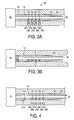
- FIG. 3Ashows a sectional view in portion of a fluid analyzing apparatus of the present invention.
- FIG. 3Bshows a sectional view in portion of another embodiment of a physiological fluid analyzing apparatus of the present invention.
- FIG. 4shows a sectional view in portion of a physiological fluid analyzing apparatus of FIG. 3A with the sampling needle in motion penetrating the physiological tissue of a patient.
- FIG. 5shows a sectional view in portion of a physiological fluid analyzing apparatus of FIG. 3A after collecting a physiological fluid.
- FIG. 6 and FIG. 7show sectional views illustrating anisotropic etching of silicon.
- FIG. 8 to 12show sectional views illustrating the process in forming a channel in a sampling needle according to the present invention.
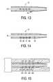
- FIG. 13shows a sectional view illustrating the formation of sensors in the sampling needle.
- FIG. 14shows a sectional view illustrating sensors after being formed in the sampling needle.
- FIG. 15shows a section view illustrating sensors and interrogating elements of another embodiment of the analyzing apparatus of the present invention.
- the present inventioninvolves a technique for a simple and reliable determination of physiological fluid parameters in a less painful way for the patient than current techniques.
- the present inventionemploys a physiological fluid sampling device that samples a physiological fluid from the patient and analyzes it substantially immediately, without extensive exposure to air, by utilizing sensors in a channel that receives the physiological fluid during the sampling procedure.
- the present inventionprovides a sampling needle of which an illustrative embodiment is shown in the plan view in FIG. 1 and the sectional view in FIG. 2 .
- the sampling needle 10includes a body 12 , which has a shaft portion 14 and a plate portion 16 .
- the shaft portion 14has a distal sharp tip (or point) 18 for puncturing the physiological tissue (or body tissue), such as the skin of an animal.
- the individual whose physiological tissue is being puncturedis herein referred to as the “patient,” which can be, for example, a person, a pet, a farm animal, etc.
- the term “distal”refers to the direction towards the patient during the physiological fluid-sampling procedure.
- the bodyhas a channel 20 which extends from then vicinity of the tip 18 proximally (i.e., the direction away form the tip).
- the channel 20has a distal opening 22 from which physiological fluid can enter at its distal end and a proximal opening 23 at its proximal end to provide a stop junction for capillary action.
- sensors 24 , 25 , 26 , 27for determining certain parameters (or characteristics) of the physiological fluid, such as pH, concentrations of blood gases, electrolytes, glucose, and the like.
- the sampling needle 10has a shaft of about 1 to 5 mm long with a cross sectional size of about 0.3 mm by 0.3 mm, preferably about 2 mm long with a cross sectional size of about 0.2 mm by 0.2 mm.
- the channel 20is generally about 0.1 mm by 0.1 mm, preferably about 0.06 mm by 0.06 mm in cross section. It is to be understood that these dimensions refer to the overall size and do not imply that the needles are necessarily square in cross section.
- the channelis shaped and sized to facilitate the conduction of the physiological fluid by capillary force and/or by suction.
- FIG. 3Ashows a sectional view in portion of an embodiment of a device for sampling and analyzing physiological fluid according to the present invention.
- the physiological fluid analyzer 28includes the sampling needle 10 of FIG. 1 .
- a shell 30confines the sampling needle 10 and allows movement only in the proximal-distal directions.
- the distal tip (or point)l 8is shielded by the shell 30 so that the physiological tissue 32 of the patient or another person will not be inadvertently injured by the distal tip 18 of the sampling needle 10 .
- the proximal end 34 of the sampling needle 10is secured to a driver (or launcher) 36 (which is schematically shown in portion in FIG.
- the shell 30also contains interrogation elements (or analysis sites) 36 A, 36 B, 37 A, 37 B, 38 A, 38 B, 39 A, 39 B proximate to the corresponding sensors 24 , 25 , 26 , 27 respectively, for sending or receiving signals to the sensors 24 - 27 .
- the interrogation elementsare optical components for irradiating the sensors 24 - 27 (namely, 36 A, 37 A, 38 A, 39 A) and receiving reflected light therefrom (namely, 36 B, 37 B, 38 B, 39 B).
- the interrogation elements 36 A, 36 B, 37 A, 37 B, 38 A, 38 B, 39 A, 39 Bare connected to conductors for transmitting current and signals to and/or from the interrogation elements.
- the conductors for transmitting signalscan also be optical fibers for transmitting light.
- signalscan be transmitted from these interrogation elements to a computer, which can either be housed with the driver 36 , or at a remote location.
- the interrogation elements 36 C, 37 C, 38 C, 39 Ccan be electrical conductors conveying signals sent from the sensors 24 C, 25 C, 26 C, 27 C via electrical conductors 24 D, 25 D, 26 D, 27 D.
- the sensors corresponding to sensors 24 C- 27 Ccan contain electrodes that sense analyte concentrations, and the electrodes can have conductors extending to the surface of the plate portion 16 facing the conducting interrogation elements corresponding to interrogation elements 36 C- 39 C.
- driver 36can also have a suction source (not shown in the figures) connected to the proximal opening 24 of the sampling needle 10 for effecting a suction to facilitate the flow of the physiological fluid up the channel 20 from the distal opening 22 .
- a suction sourcenot shown in the figures
- One such suction mechanismis described in U.S. Pat. No. 5,891,053 (Sesekura), U.S. Pat. No. 5,873,887 (King et al.) and U.S. Pat. No.
- a filtercan also be used to isolate the driver 36 from the sampling needle 12 such that the physiological fluid will not flow past it and contaminate the driver.
- the filter 41can be attached to the sampling needle 12 and is disposable.
- the devicecan have a disposable cap 43 for shielding the sharp point 18 of the sampling needle 12 for disposal to reduce bio-hazard exposure to a person in the vicinity.
- FIG. 4shows a sectional view in portion of the physiological fluid analyzer 28 when the sampling needle 10 has been driven forward to puncture the physiological tissue (e.g., skin) of the patient. It is noted that when the sampling needle 10 is driven forward (distally) it slides in the distal direction operatively against the shell 30 , which confines its freedom of motion in the distal direction when it is driven. It is to be understood that interposing material can be between the shell 30 and the sampling needle, and still allow the sampling needle to move relative to the shell in a translational manner.
- the sampling needle 10When the sampling needle 10 is driven distally with the tip 18 extending out of the shell 30 , the sensors 24 - 27 move away from the corresponding interrogation elements 36 A, 36 B, 37 A, 37 B, 38 A, 38 B, 39 A, 39 B and thus the interrogation elements will not interrogate the sensors 24 - 27 in this condition. After an adequate amount of the physiological fluid has passed into the sampling needle 10 , the sampling needle 10 is retracted back into the shell 30 so that the sharp tip 18 of the sample needle 10 will not be exposed as a bio-hazzard.
- the physiological fluid that flows from the body into the channel 20will quickly pass by the sensors 24 - 27 and continue on its way towards the proximal opening 23 . Since the channel 20 is narrow and only a small volume of air (or gas if the channel has been purposely filled with a storage gas, such as nitrogen before sampling) is in contact with the proximal portion of the physiological fluid passing up the channel 20 , the portion of the physiological fluid that eventually settles about the sensors 24 - 27 when the physiological fluid stops in the channel will not have exposure to air, and thus will have chemistry substantially similar to the physiological fluid in the physiological tissue.
- certain parameters of the physiological fluid which may be changed by exposure to aire.g., gas content of blood
- aire.g., gas content of blood
- the physiological fluidis interrogated after the sampling needle 10 is retracted to the position at which the sensors 24 - 27 are near to and coupled in alignment with their corresponding interrogation elements 36 A- 39 A in the shell 30 .
- the irradiators 36 A, 37 A, 38 A, 39 Aemit light of the appropriate frequencies into the sensors 24 - 27 and the light detectors 36 B, 37 B, 38 B, 39 B sense the light from the sensors 24 - 27 to determine the appropriate qualities of interest.
- the surface 41 A of the body 12 and the surface 41 B of the shell 30 where they interfaceis flat so that light can pass therethrough without much distortion or attenuation. It is noted that a wide variety of irradiators and sensors, as well as techniques of transmission of light or electrical signals are known in the art.
- any light ranging from x-ray to infra red lightcan be produced, transmitted to tiny areas, and detected.
- such lightcan be transmitted by fiber optics from suitable light sources from a location remote from the shell 30 to the interrogation elements 36 A- 39 A in the shell 30 .
- the term “remote”includes the situation in which the analyzing electronics are located in the driver 36 , or it may be a situation in which the analyzing electronics are off site, as in a different building, etc.
- fiber opticscan be used to receive the light from the sensors 24 - 27 , and transmit to a location remote from the shell 30 for analysis.
- light sources and light sensorscan be directly positioned at the interrogation elements.
- Suitable light sourcesmay include light emitting diodes, laser, fluorescent lamp, incandescent lamp, and the like. Filters may be used to select the desirable wavelengths directed to the sensors. If light sources of small sizes are used, they may be positioned at the irradiators 36 A, 37 A, 38 A, 39 A
- the interrogation elementscan be located on the inwardly facing surface of the shell 30 to be as close to the sensors 24 - 27 as is practical. This arrangement will minimize the intervening space and material through which the light will have to travel and thereby reduce noise. For example, optical fibers can reach all the way to be flush with the inwardly facing surface of the shell 30 near to the sensors 24 - 27 to transmit light. Further, by minimizing the thickness of the material on the sampling needle 12 that separates the sensors 24 - 27 from the shell 30 , the efficiency of transmission of signals or light between the interrogation elements and the sensors 24 - 27 can be increased.
- one or more of the sensors 24 - 27can contain an electrode for measuring analyte electrochemistry (e.g., pH, conductivity, etc.). Such electrodes will not be interrogated by optical means. Rather, electrical leads will lead from the sensors 24 - 27 through the interrogation elements similar to elements 36 A- 39 B. Such leads can lead to analyzers remote from the shell 30 .
- analyte electrochemistrye.g., pH, conductivity, etc.
- the sampling needle 10can be made with silicon integrated circuit (IC) techniques using wet and dry etching.
- ICsilicon integrated circuit
- Techniques for adapting IC technology for making needlescan be found, for example, in Kyle Lebouitz, Ph.D. dissertation entitled “MEMS Microshells for Microneedles, Microscale Fluid Visualization, and Vacuum Packaging of Microdevices,” University of California at Berkeley, 1998; U.S. Pat. No. 5,855,801 (Lin et al.), and U.S. Pat. No. 5,591,139 (Lin et al.), which are incorporated by reference in their entireties herein.
- the IC techniquestake advantage of the fact that silicon can be etched isotropically and anisotropically by using different etchant chemicals, thereby producing channels of different shapes and angles.
- Techniques of isotropic as well as anisotropic etching of siliconare known in the art, e.g., Kenneth E. Bean, “Anisotropic etching of silicon,” IEEE Trans. On Electron Devices , Vol ED-25, No. 10, p. 1185-1193, Oct. 1978; Kenneth E. Bean et al., “The influence of crystal orientation on silicon semiconductor processing,” Proceedings of the IEEE , Vol 57, No. 9, p. 1469-1476, September 1969; Don L.
- Isotropic etchantssuch as hydrofluoric acid, nitric acid, acetic acid, and the like, etch silicon in all crystalographic direction at the same rate.
- anisotropic etchantssuch as potassium hydroxide, hydrazine, ethylene-diamine (EDA), and the like, etch the (100) plane of the silicon crystal at a much higher rate than (111) planes.
- anisotropic etchantssuch as potassium hydroxide, hydrazine, ethylene-diamine (EDA), and the like, etch the (100) plane of the silicon crystal at a much higher rate than (111) planes.
- anisotropic etchantssuch as potassium hydroxide, hydrazine, ethylene-diamine (EDA), and the like, etch the (100) plane of the silicon crystal at a much higher rate than (111) planes.
- anisotropic etchantssuch as potassium hydroxide, hydrazine, ethylene-diamine (EDA), and the like, etch the (100) plane of the silicon crystal at a much higher rate than (111) planes.
- anisotropic etchantssuch as potassium hydroxide,
- the Bosch Processis a plasma etch process developed by Robert Bosch GmbH and is a known process for deep well etching. It implements concurrent and/or iterative deposition and etching chemistries and allows the engineering of sidewall passivation in accordance with the aspect ratio needed, the open area of silicon (load), and the use or nonuse of buried oxide as an etch stop.
- the current Bosch processuses an inductively coupled high density plasma source and fluorine etching species. The high density of neutral fluorine etching species ensures that mass transport within the plasma is diffusion dominated.
- glass and SiO 2can be etched with suitable chemicals, e.g., buffered hydrofluoric acid (HF) mixtures; glass, SiO 2 , polysilicon.
- suitable chemicalse.g., buffered hydrofluoric acid (HF) mixtures; glass, SiO 2 , polysilicon.
- Silicon nitridecan be dry-etched with plasma chemistry. Silicon nitride can also be wet-etched with phosphoric acid (H 3 PO 4 ).
- FIG. 8 to FIG. 12illustrate in sectional views how a sampling needle with a channel can be made. These figures correspond to the plate portion of the sampling needle. However, a person skilled in the art will know that shaft portion of the sampling needle can be fashioned using substantially similar techniques.
- a silicon substrate 40is etched anisotropically to form a trough, which is filled with SiO 2 42 , eventually resulting in a structure seen in FIG. 8 .
- the top of the SiO 2 42 and the substrate 40are flush, which can be accomplished by milling off any excessive substrate or SiO 2 material.
- a layer of phosphosilicate glass (PSG) 44can be laid on the SiO 2 42 extending slightly wider than and covering the SiO 2 material 42 . Thus the phosphosilicate glass 44 and the substrate 40 encircles the SiO 2 material 42 .
- PSGphosphosilicate glass
- the SiO 2 42 materialthen can be etched away forming a channel 46 .
- a layer of silicon nitride Si 3 N 4 48can then be laid on the substrate 40 and the PSG, except in certain areas where opening 50 are left unoccupied by Si 3 N 4 , see FIG. 10 .
- the openings 50are for providing access to etchants to etch away the PSG 44 , thus leaving a sampling channel 52 .
- More Si 3 N 4can be laid on the Si 3 N 4 layer 48 to make the resulting Si 3 N 4 layer 54 substantially flat to facilitate sliding against a surface. Openings 50 are left for introducing sensing chemicals for access by the physiological fluid that will flow past in the sampling channel 52 .
- FIG. 11shows the sectional view of the plate portion of a sampling needle.
- the shaft portion of the sampling needlecan have a section view similar to that in FIG. 12, in which the sampling channel 52 is bounded by the substrate 40 and the Si 3 N 4 layer 54 .
- the side edges 56 A, 56 B of the silicon substrate layer 40 in the shaft portioncan be formed by anisotropic etching. It is preferred that openings 50 be absent in the shaft portion to maintain the shaft stiffness to facilitate penetration into the physiological tissue of a patient.
- the top cover of the channelis made from silicon nitride. Polysilicon may also be used for making the cover.
- the needleWith anisotropic etching, the needle can have a cross-section that has stright sides (e.g., side edges 56 A, 56 B). It is noted that one skilled in the art can make a needle with sides that are not necessarily parallel in its cross section.
- the sensors 24 - 27may have electrodes or chemicals immobilized therein for sensing various parameters of the physiological fluid.
- sensors that can optically interrogatedare described. However, viewing the present disclosure, a person skilled in the art will understand that other sensors and methods of interrogation and signal communication can be used. Illustrative examples of sensors are described below.
- Optical pH sensorscan be based on light absorbance or fluorescence changes of a weakly dissociating dye in response to changes in pH.
- a weakly dissociating dye HA (in acid form) in solutionis in equilibrium with its base form A ⁇ according to the equation
- the pHcan be measured by measuring the concentration of either form of the dye, i.e., [HA] or [A ⁇ ].
- the acid form and the base formabsorb radiation in the different spectral regions.
- the acid formhas an absorption peak at about 430 nm and the base form has an absorption peak at about 550 nm.
- ⁇is the molar extinction coefficient of A ⁇
- Lis the length of the path traveled by the light
- Io and Iare the intensities of the transmitted light when A ⁇ is absent and present, respectively.
- Eq. S7yields an S shaped curve relating pH to the ratio of intensities (Io/I).
- ⁇is the optical collection efficiency of the instrument.
- I FAis related to pH by:
- Partial pressure of carbon dioxide, PCO 2is usually measured using the Severinghaus Principle, as shown below.
- CO 2 dissolved in waterdissociates to form hydrogen ions (H + ) and bicarbonate ions (HCO 3 ⁇ ).
- the medium in which the measurement is madeis an aqueous solution of NaHCO 3 , which dissociates completely into Na + and HCO 3 ⁇ ions, the total concentrations of HCO 3 ⁇ and H 2 O in the medium remain fairly constant and Eq. S12 can be shown to lead to
- the above equationindicates that CO 2 dissolved in a sample can be measured by measuring the pH of a bicarbonate solution in equilibrium with the sample.
- the sensor with the bicarbonate solution and a pH sensitive dye for optical sensingcan be encapsulated in a or membrane that allows only CO 2 to equilibrate with the buffer solution. Such a membrane acts as a barrier to the hydrogen ions in the test sample.
- Oxygenis an excellent quencher of fluorescence of many fluorophores.
- Optical techniques for oxygen sensingcan be based on fluorescence quenching of an excited state of a dye (fluorophore) molecule.
- the excitation of a fluorophore F and its quenching by an oxygen moleculeis represented by the following equations:
- hPlanck's constant
- v exis the excitation frequency of the radiation
- *represent the excited state of a substance.
- v emis the frequency of the radiation emitted at the decay of F*.
- the amount of oxygen presentcan be determined by measuring the oxygen quenching of an excited form of the fluorophore.
- ⁇ and ⁇are rate constants for radiative and nonradiative decays (i.e., without quencher).
- F*rate constants for radiative and nonradiative decays
- K svequals k q ⁇ o and is known as the Stem-Volmer constant
- Eq. S22 and Eq. S23are variations of the Stem-Volmer equation.
- the fluorophorecan be excited by a delta function and the rate of fluorescence decay is observed. According to Eq. S20 the lifetime of the fluoresce decay in the absence of fluorophore is
- Cations such as Na + , K + , Ca ++ , and Mg ++can be measured optically on very similar principles.
- an ion selective ionophoreSuch ionophores are lipophillic inclusion compounds which typically have a ring-like structure. In such a compound, the ring has several slightly electronegative atoms such as oxygen in it. The size of the ring and the total number of the oxygen atoms determine the relative preference (selectivity) of the ionophore to the cations of various sizes and charged states.
- Another class of ionophoreundergo conformational changes upon selective binding of a cation.
- BME-44forms a clam shell like enclosure for an included potassium ion.
- BME-44has two crown ethers connected by a 3 carbon chain. When a potassium ion is present, the two crown ethers fold over to form the clam shell.
- ionophoresare lipophillic and are sequestered in a hydrophobic polymer such as polyvinylchloride (PVC) along with a large amount of plasticizer.
- PVCpolyvinylchloride
- the ionophoresextract the selective ion from the aqueous solution in contact with the polymer surface. Because the ion finds itself in a more favorable energy environment in the ionophore than in the aqueous surrounding, it readily enters the hydrophobic phase of the polymer. However, this causes a charge imbalance in the polymer phase and at the surface of the polymer. The excess charge build-up in the polymer can be used to expel another less favored cation such as H + from the polymer phase. Alternatively, a charged double layer is formed at the surface of the polymer.
- the number of cations expelled or the potential gradient at the surfaceis proportional to the concentration of the cation in the aqueous solution. Similar ionophores are also available for some anions such as chloride ions. Extraction of an anion into the polymer phase will cause another anion to be expelled or a cation such as an hydrogen ion to accompany the anion into the polymer phase.
- the potential build up at the surface of the polymercan be measured optically using potential sensitive dyes.
- the fluoresce of some dyes, such as rhodamine B,is a strong function of the electrical charge in its vicinity.
- measurement of the fluorescence changes of a polymer film in which such a dye is immobilized along with an ionophorecan be used to measure the concentration of the selective ion in a sample.
- extraction of a positive ion into a hydrophobic polymer by an ionophorecan be made to expel another cation such as an hydrogen ion.
- a pH sensitive dyesuch as HPTS of phenol red or bromothymol blue
- the dyewill lose an hydrogen ion when a cation is extracted from the sample by the ionophore.
- the optical absorption or the fluorescence emission of the dyewill change as in the pH sensors described above. Reverse of this effect can be used to determine the concentration of anions.
- the folding of BME-44 in the presence of potassium ioncan be made to bring a fluorophore such as rhodamine and a quencher such as fluorescein closer together.
- a fluorophoresuch as rhodamine and a quencher such as fluorescein
- the Foerster energy transfer from the fluorophore to the quenchercauses the change in fluorescence in proportion to the number of potassium ions extracted.
- an electron donoris placed between a fluorophore and an ionophore (for cations)
- the ability of the donor to transfer an electron to the excited fluorophoreis blocked when a cation is located in the ionophore.
- the fluorescence processis nonradiative.
- the electron transfer to the fluorophoreis blocked and the fluorescence energy is radiatively emitted.
- Metabolites such as glucose, lactate and creatinineare measured using enzymatic conversion of these species (substrates) into another molecule such as hydrogen peroxide.
- the enzymatic conversion of the substratesis accompanied by consumption of another species such as oxygen.
- sensor measuring the concentration of any of these generated or consumed speciescan be used to determine the concentration of the substrate.
- Glucoseis converted into hydrogen peroxide while consuming oxygen by glucose oxidase (GOD) according to the following equation:
- Lactateis converted into pyruvate catalyzed by lactate dehydrogenase and mediated by NAD generating hydrogen ions:
- Creatininecan be measured using a multienzyme assay involving the following reactions:
- Creatine ⁇ Sarcosine+Ureaenzyme: Creatine Amidinohydrolase
- Equations S27 through S31indicate that glucose and creatinine can be measured using oxygen sensing methods discussed above.
- the oxygen tension in a sample like bloodcan vary considerably. It is therefore preferred to make this reaction oxygen independent.
- glucose sensingthis is achieved by using a mediator such as ferricyanide which replaces oxygen.
- a pH sensorcan be used for measuring lactate.
- a change in pH of the samplewill also affect the measurements of lactate. In these situations it is necessary to measure the PO 2 and pH of the sample and correct for the interferences.
- Sensor chemicals for interacting with the particular constituents of intereste.g., glucose, oxygen, hydrogen ions, metabolites, etc.
- the particular constituents of intereste.g., glucose, oxygen, hydrogen ions, metabolites, etc.
- the particular constituents of intereste.g., glucose, oxygen, hydrogen ions, metabolites, etc.
- the sampling needlesuch as the one shown in FIG. 1 . and FIG. 2 .
- one methodis to form a sampling needle with holes 60 , 61 , 62 , 63 extending through the body 12 from an external space to the channel 20 and then placing a small amount of a liquid or a solution of polymer precursor (which contains appropriate sensor chemicals), such as polymer precursor liquid 64 , into each hole.
- the small dimensions of the holes 60 , 61 , 62 , 63will facilitate wicking the liquid into the holes by capillary force.
- the polymer precursorcan then be solidified, which can be accomplished by radiation (e.g., ultraviolet light), heat, catalyst, etc., forming a sampling needle shown in FIG. 14, having sensors 65 , 66 , 67 , 68 .
- the sensorscan be flush with the Luminal wall 70 , extending slightly into the channel 20 from the Luminal wall 70 , or in the form of a depression (or cavity). Further, more than one layer of materials can be placed into the holes 60 , 61 , 62 , 63 , thereby immobilizing different chemicals or forming barriers to inhibit diffusion of different chemicals at different rates. Examples of materials that can be used to form the sensors, e.g., by light sensitive cross-linking polymerization to induce solidification, are known in the art.
- a solution of acrylamide monomers, a cross linker such as methylene bisacrylamide, an initiator and an indicator such as phenol redcan be placed in these holes.
- Polymerizationcan be allowed to occur and thus forming a hydrogel of polyacrylamide with phenol red immobilized therein.
- a hydrogelcan be used as a pH sensor based on optical absorption measurement of phenol red.
- the details of the chemical composition of materials to form such a sensorare available in the literature, e.g., Peterson, J. I., Goldstein, S. R., Fitzgerald, R. V., and Buckold, D. K., Fiberoptic pH probe for physiological use, Anal. Chem ., 52, pp 864-869 (1980).
- the sampling needles of the present inventioncan be made directly by etch processing of silicon.
- the sampling needlescan be made by making a master mold by etching processes and then by molding from the master mold.
- a similar technique using high aspect ratio photoresist patterns through the LIGA processcan be used.
- a combination of seed metal deposition followed by electroplating to form a metallized structureis followed by the complete removal of the photoresist pattern.
- Similar processeshave been described in the literature, both using and not using the LIGA process.
- the LIGA processcan be further referenced from “LIGA Process: Sensor Construction Techniques via X-Ray Lithography,” Technical Digest, IEEE Solid-State Sensor and Actuator Workshop, Hilton Head Island, SC, Jun. 6-9, 1988, pp 1-4.
- the remaining metallic shellbecomes the hollow channel with holes, the exception being that this structure is made from metallic material such as nickel rather than silicon, polysilicon, silicon nitride, and the like.
- a side of a planar hollow needleis defined initially on a planar silicon substrate, this definition is preferably by a photolithographic process.
- a thin layer of conductive seed metalsuch as about 500 Angstroms typically, of sputtered Nickel is deposited on a about 3000 Angstroms typically, thin oxide coated silicon wafer.
- a negative photoresist pattern of a planar needleis deposited and aligned on the thin oxide silicon wafer. This very thick photoresist in the range about 10 microns to 1000 microns typically, is processed with the LIGA procedure known to those in the art.
- the developed resist cavitiesform metal seed areas for electrodepositing a thick Nickel layer in the range about 0.1 to 0.25 mm thick.
- a very thick layer of photoresistpreferably in the range about 10 microns to 1000 microns thick typically, is deposited on the planar surface electrodeposited Nickel and remaining photoresist.
- a pattern resembling the inner core of the planar needleis aligned and developed on top of the previously defined planar needle side pattern.
- the photolithographic processcan be done by the method of LIGA.
- the core of the planar needlewould be remaining after respective wet development of the LIGA processed photoresist.
- This very thick wet resist processingis known to those in the art.
- a conductive seed metalis used to coat both the sides and the top of the remaining photoresist resembling the planar needle core.
- a follow-up photoresist patternwill be aligned and developed to coat areas where electrodeposition of the remaining planar needle will not occur.
- This mask patternwill define the remaining planar needles sides and top.
- the entire silicon substrateis again placed within an electrodeposition bath and the thick metal allowed to grow to thicknesses resembling its first thick metal layer.
- a thick hollow structure fabricated with nickel metalwill remain along with a very thin web of Nickel connecting all the planar needles.
- the nickel metal patterns being the hollow planar needles on the silicon wafercan be individually separated by a short wet acid nickel etch to removed the thin nickel webbing. An option may be to not remove this webbing and to remove the entire metal structure from the silicon wafer by dissolving the thin oxide layer between the nickel pattern and the silicon wafer.
- the individual planar needlescan be separated after further needle coatings or added processing for the respective usage application.
- sensor holes on the top or bottom sides of the hollow planar needlecan be easily patterned and processed during the photolithographic steps defining the initial bottom layer or final top mask definition, respectively. Areas where there should be sensor or port holes will contain photoresist inhibiting thick nickel layer electrodeposition.
Landscapes
- Health & Medical Sciences (AREA)
- Life Sciences & Earth Sciences (AREA)
- Physics & Mathematics (AREA)
- Engineering & Computer Science (AREA)
- Animal Behavior & Ethology (AREA)
- Veterinary Medicine (AREA)
- Biophysics (AREA)
- Biomedical Technology (AREA)
- Heart & Thoracic Surgery (AREA)
- Medical Informatics (AREA)
- Molecular Biology (AREA)
- Surgery (AREA)
- Pathology (AREA)
- General Health & Medical Sciences (AREA)
- Public Health (AREA)
- Hematology (AREA)
- Optics & Photonics (AREA)
- Manufacturing & Machinery (AREA)
- Spectroscopy & Molecular Physics (AREA)
- Chemical & Material Sciences (AREA)
- Chemical Kinetics & Catalysis (AREA)
- General Chemical & Material Sciences (AREA)
- Emergency Medicine (AREA)
- Dermatology (AREA)
- Pain & Pain Management (AREA)
- Measurement Of The Respiration, Hearing Ability, Form, And Blood Characteristics Of Living Organisms (AREA)
Abstract
Description
Claims (22)
Priority Applications (1)
| Application Number | Priority Date | Filing Date | Title |
|---|---|---|---|
| US09/517,711US6375627B1 (en) | 2000-03-02 | 2000-03-02 | Physiological fluid extraction with rapid analysis |
Applications Claiming Priority (1)
| Application Number | Priority Date | Filing Date | Title |
|---|---|---|---|
| US09/517,711US6375627B1 (en) | 2000-03-02 | 2000-03-02 | Physiological fluid extraction with rapid analysis |
Publications (1)
| Publication Number | Publication Date |
|---|---|
| US6375627B1true US6375627B1 (en) | 2002-04-23 |
Family
ID=24060914
Family Applications (1)
| Application Number | Title | Priority Date | Filing Date |
|---|---|---|---|
| US09/517,711Expired - LifetimeUS6375627B1 (en) | 2000-03-02 | 2000-03-02 | Physiological fluid extraction with rapid analysis |
Country Status (1)
| Country | Link |
|---|---|
| US (1) | US6375627B1 (en) |
Cited By (213)
| Publication number | Priority date | Publication date | Assignee | Title |
|---|---|---|---|---|
| US20020042594A1 (en)* | 1998-03-30 | 2002-04-11 | Paul Lum | Apparatus and method for penetration with shaft having a sensor for sensing penetration depth |
| US20020137998A1 (en)* | 2001-03-26 | 2002-09-26 | Wilson Smart | Silicon microprobe with integrated biosensor |
| US20020168290A1 (en)* | 2002-05-09 | 2002-11-14 | Yuzhakov Vadim V. | Physiological sample collection devices and methods of using the same |
| US20020177858A1 (en)* | 2000-10-16 | 2002-11-28 | Sherman Faiz Feisal | Microstructures and method for treating and conditioning skin which cause less irritation during exfoliation |
| US20020188224A1 (en)* | 2001-06-08 | 2002-12-12 | Roe Jeffrey N. | Test media cassette for bodily fluid testing device |
| US20030060730A1 (en)* | 2001-08-29 | 2003-03-27 | Edward Perez | Wicking methods and structures for use in sampling bodily fluids |
| US20030088191A1 (en)* | 2001-06-12 | 2003-05-08 | Freeman Dominique M. | Blood sampling device with diaphragm actuated lancet |
| US6565532B1 (en) | 2000-07-12 | 2003-05-20 | The Procter & Gamble Company | Microneedle apparatus used for marking skin and for dispensing semi-permanent subcutaneous makeup |
| US6591124B2 (en)* | 2001-05-11 | 2003-07-08 | The Procter & Gamble Company | Portable interstitial fluid monitoring system |
| US20030153939A1 (en)* | 2000-03-04 | 2003-08-14 | Michael Fritz | Blood lancet with hygienic tip protection |
| US20030199900A1 (en)* | 2002-04-19 | 2003-10-23 | Pelikan Technologies, Inc. | Method and apparatus for penetrating tissue |
| US20030199893A1 (en)* | 2002-04-19 | 2003-10-23 | Pelikan Technologies, Inc. | Method and apparatus for a multi-use body fluid sampling device with analyte sensing |
| US20030199903A1 (en)* | 2002-04-19 | 2003-10-23 | Pelikan Technologies, Inc. | Method and apparatus for penetrating tissue |
| US20030199823A1 (en)* | 1997-12-31 | 2003-10-23 | Minimed Inc. | Insertion device for an insertion set and method of using the same |
| US20030212346A1 (en)* | 2002-05-09 | 2003-11-13 | Vadim V. Yuzhakov | Methods of fabricating physiological sample collection devices |
| US20030211619A1 (en)* | 2002-05-09 | 2003-11-13 | Lorin Olson | Continuous strip of fluid sampling and testing devices and methods of making, packaging and using the same |
| US20030212424A1 (en)* | 2002-04-19 | 2003-11-13 | Pelikan Technologies, Inc. | Method and apparatus for lancet actuation |
| US20030212344A1 (en)* | 2002-05-09 | 2003-11-13 | Vadim Yuzhakov | Physiological sample collection devices and methods of using the same |
| US6652478B1 (en) | 1999-06-09 | 2003-11-25 | The Procter & Gamble Company | Intracutaneous edged microneedle apparatus |
| US20030220656A1 (en)* | 2002-05-24 | 2003-11-27 | Vladimir Gartstein | Method of exfoliation of skin using closely-packed microstructures |
| US6663820B2 (en) | 2001-03-14 | 2003-12-16 | The Procter & Gamble Company | Method of manufacturing microneedle structures using soft lithography and photolithography |
| US20030233112A1 (en)* | 2001-06-12 | 2003-12-18 | Don Alden | Self optimizing lancing device with adaptation means to temporal variations in cutaneous properties |
| US20040002682A1 (en)* | 1997-02-05 | 2004-01-01 | Medtronic Minimed, Inc. | Insertion device for an insertion set and method of using the same |
| US20040006285A1 (en)* | 1996-05-17 | 2004-01-08 | Douglas Joel S. | Methods and apparatus for sampling and analyzing body fluid |
| US20040010279A1 (en)* | 2002-04-19 | 2004-01-15 | Freeman Dominique M. | Device and method for variable speed lancet |
| US20040034318A1 (en)* | 2000-10-31 | 2004-02-19 | Michael Fritz | System for withdrawing blood |
| US20040059256A1 (en)* | 2001-09-26 | 2004-03-25 | Edward Perez | Method and apparatus for sampling bodily fluid |
| US20040073140A1 (en)* | 1996-05-17 | 2004-04-15 | Douglas Joel S. | Methods and apparatus for expressing body fluid from an incision |
| US20040087992A1 (en)* | 2002-08-09 | 2004-05-06 | Vladimir Gartstein | Microstructures for delivering a composition cutaneously to skin using rotatable structures |
| US20040122339A1 (en)* | 2002-12-24 | 2004-06-24 | Roe Steven N. | Sampling devices and methods utilizing biased capillary action |
| US20040164454A1 (en)* | 2003-02-24 | 2004-08-26 | The Procter & Gamble Company | Method for manufacturing microstructures having multiple microelements with through-holes |
| WO2004060446A3 (en)* | 2002-12-30 | 2004-09-30 | Pelikan Technologies Inc | Method and apparatus using optical techniques to measure analyte levels |
| US20040225312A1 (en)* | 2003-05-09 | 2004-11-11 | Phoenix Bioscience | Linearly lancing integrated pivot disposable |
| US6821281B2 (en) | 2000-10-16 | 2004-11-23 | The Procter & Gamble Company | Microstructures for treating and conditioning skin |
| US20050010134A1 (en)* | 1996-05-17 | 2005-01-13 | Douglas Joel S. | Blood and interstitial fluid sampling device |
| US20050021066A1 (en)* | 2001-08-29 | 2005-01-27 | Hans-Juergen Kuhr | Analytical device with lancet and test element |
| US20050033263A1 (en)* | 2003-08-07 | 2005-02-10 | Medtronic-Minimed, Inc. | System and method for restenosis mitigation |
| US20050070819A1 (en)* | 2003-03-31 | 2005-03-31 | Rosedale Medical, Inc. | Body fluid sampling constructions and techniques |
| US20050136099A1 (en)* | 2003-12-22 | 2005-06-23 | Unilever Home & Personal Care Usa, Division Of Conopco, Inc. | Exfoliating personal care wipe article |
| US6931277B1 (en) | 1999-06-09 | 2005-08-16 | The Procter & Gamble Company | Intracutaneous microneedle array apparatus |
| US20050201897A1 (en)* | 2002-11-26 | 2005-09-15 | Volker Zimmer | Body fluid testing device |
| US20050232815A1 (en)* | 2002-12-23 | 2005-10-20 | Werner Ruhl | Body fluid testing device |
| US20060008389A1 (en)* | 2003-01-23 | 2006-01-12 | Klaus-Dieter Sacherer | Magazine for annulary capillary lancets |
| US7025774B2 (en) | 2001-06-12 | 2006-04-11 | Pelikan Technologies, Inc. | Tissue penetration device |
| US7033371B2 (en) | 2001-06-12 | 2006-04-25 | Pelikan Technologies, Inc. | Electric lancet actuator |
| US20060229531A1 (en)* | 2005-02-01 | 2006-10-12 | Daniel Goldberger | Blood monitoring system |
| JP2007014381A (en)* | 2005-07-05 | 2007-01-25 | National Institute Of Advanced Industrial & Technology | Puncture device integrated biosensor |
| WO2007041062A2 (en) | 2005-09-30 | 2007-04-12 | Intuity Medical, Inc. | Devices and methods for facilitating fluid transport |
| US20070093728A1 (en)* | 1996-05-17 | 2007-04-26 | Douglas Joel S | Blood and interstitial fluid sampling device |
| US20070123801A1 (en)* | 2005-11-28 | 2007-05-31 | Daniel Goldberger | Wearable, programmable automated blood testing system |
| US7229458B2 (en) | 2002-04-19 | 2007-06-12 | Pelikan Technologies, Inc. | Method and apparatus for penetrating tissue |
| US20070148716A1 (en)* | 2005-12-27 | 2007-06-28 | Gorres Geoffrey H | Device for monitoring a patient for a urinary tract infection |
| US7244265B2 (en) | 2002-04-19 | 2007-07-17 | Pelikan Technologies, Inc. | Method and apparatus for penetrating tissue |
| US20070191716A1 (en)* | 2004-09-29 | 2007-08-16 | Daniel Goldberger | Blood monitoring system |
| US7288073B2 (en)* | 2001-07-20 | 2007-10-30 | Roche Diagnostics Operations, Inc. | System for withdrawing small amounts of body fluid |
| US7291117B2 (en) | 2002-04-19 | 2007-11-06 | Pelikan Technologies, Inc. | Method and apparatus for penetrating tissue |
| US7297122B2 (en) | 2002-04-19 | 2007-11-20 | Pelikan Technologies, Inc. | Method and apparatus for penetrating tissue |
| US20080021491A1 (en)* | 2002-04-19 | 2008-01-24 | Freeman Dominique M | Method and apparatus for penetrating tissue |
| US7331931B2 (en) | 2002-04-19 | 2008-02-19 | Pelikan Technologies, Inc. | Method and apparatus for penetrating tissue |
| WO2008048709A1 (en)* | 2006-10-15 | 2008-04-24 | Roche Diagnostic Gmbh | Diagnostic test element and process for its production |
| US7374544B2 (en) | 2002-04-19 | 2008-05-20 | Pelikan Technologies, Inc. | Method and apparatus for penetrating tissue |
| US20080183144A1 (en)* | 2007-01-22 | 2008-07-31 | Trautman Joseph C | Applicators for microneedles |
| US20080188725A1 (en)* | 2007-02-06 | 2008-08-07 | Markle David R | Optical systems and methods for ratiometric measurement of blood glucose concentration |
| US20080188722A1 (en)* | 2007-02-06 | 2008-08-07 | Markle David R | Optical determination of ph and glucose |
| US7410468B2 (en) | 2002-04-19 | 2008-08-12 | Pelikan Technologies, Inc. | Method and apparatus for penetrating tissue |
| US20080200838A1 (en)* | 2005-11-28 | 2008-08-21 | Daniel Goldberger | Wearable, programmable automated blood testing system |
| US20080275324A1 (en)* | 2006-05-23 | 2008-11-06 | Daniel Goldberger | Fluid Access Interface |
| WO2008141241A1 (en)* | 2007-05-10 | 2008-11-20 | Glumetrics, Inc. | Equilibrium non-consuming fluorescence sensor for real time intravascular glucose measurement |
| US20090018426A1 (en)* | 2007-05-10 | 2009-01-15 | Glumetrics, Inc. | Device and methods for calibrating analyte sensors |
| US7485128B2 (en) | 2002-04-19 | 2009-02-03 | Pelikan Technologies, Inc. | Method and apparatus for penetrating tissue |
| US20090054810A1 (en)* | 2003-03-24 | 2009-02-26 | Intuity Medical, Inc. | Analyte concentration detection devices and methods |
| US20090097063A1 (en)* | 2007-10-11 | 2009-04-16 | Canon Kabushiki Kaisha | Image processing system and image processing method |
| US20090123958A1 (en)* | 2004-11-08 | 2009-05-14 | Inkjet Technology Ltd. | Laboratory Devices, Methods and Systems Employing Acoustic Ejection Devices |
| US7547287B2 (en) | 2002-04-19 | 2009-06-16 | Pelikan Technologies, Inc. | Method and apparatus for penetrating tissue |
| US20090155330A1 (en)* | 2007-04-16 | 2009-06-18 | Corium International, Inc. | Vaccine Delivery via Microneedle Arrays |
| US20090177143A1 (en)* | 2007-11-21 | 2009-07-09 | Markle William H | Use of an equilibrium intravascular sensor to achieve tight glycemic control |
| US7563232B2 (en) | 2002-04-19 | 2009-07-21 | Pelikan Technologies, Inc. | Method and apparatus for penetrating tissue |
| US7582099B2 (en) | 2002-04-19 | 2009-09-01 | Pelikan Technologies, Inc | Method and apparatus for penetrating tissue |
| US20090227898A1 (en)* | 2006-10-15 | 2009-09-10 | Hans-Peter Haar | Diagnostic test element and process for its production |
| US7608042B2 (en) | 2004-09-29 | 2009-10-27 | Intellidx, Inc. | Blood monitoring system |
| US7648468B2 (en) | 2002-04-19 | 2010-01-19 | Pelikon Technologies, Inc. | Method and apparatus for penetrating tissue |
| US20100028390A1 (en)* | 2004-03-24 | 2010-02-04 | Cleary Gary W | Transdermal Delivery Device |
| US7666149B2 (en) | 1997-12-04 | 2010-02-23 | Peliken Technologies, Inc. | Cassette of lancet cartridges for sampling blood |
| US7674232B2 (en) | 2002-04-19 | 2010-03-09 | Pelikan Technologies, Inc. | Method and apparatus for penetrating tissue |
| US7682318B2 (en) | 2001-06-12 | 2010-03-23 | Pelikan Technologies, Inc. | Blood sampling apparatus and method |
| US7699791B2 (en) | 2001-06-12 | 2010-04-20 | Pelikan Technologies, Inc. | Method and apparatus for improving success rate of blood yield from a fingerstick |
| US20100121170A1 (en)* | 2008-09-12 | 2010-05-13 | Optiscan Biomedical Corporation | Fluid component analysis system and method for glucose monitoring and control |
| US20100120073A1 (en)* | 2007-05-08 | 2010-05-13 | Superior Medical Llc | Methods and devices for detecting organisms causing urinary tract infections |
| US7717863B2 (en) | 2002-04-19 | 2010-05-18 | Pelikan Technologies, Inc. | Method and apparatus for penetrating tissue |
| JP2010119865A (en)* | 2010-02-03 | 2010-06-03 | National Institute Of Advanced Industrial Science & Technology | Method of manufacturing biosensor integrated with puncture device |
| JP2010119864A (en)* | 2010-02-03 | 2010-06-03 | National Institute Of Advanced Industrial Science & Technology | Method of manufacturing biosensor integrated with puncture device |
| US20100145229A1 (en)* | 2001-01-22 | 2010-06-10 | Perez Edward P | Lancet device having capillary action |
| US7749174B2 (en) | 2001-06-12 | 2010-07-06 | Pelikan Technologies, Inc. | Method and apparatus for lancet launching device intergrated onto a blood-sampling cartridge |
| US7758518B2 (en) | 2001-06-08 | 2010-07-20 | Roche Diagnostics Operations, Inc. | Devices and methods for expression of bodily fluids from an incision |
| US20110006458A1 (en)* | 2009-04-24 | 2011-01-13 | Corium International, Inc. | Methods for manufacturing microprojection arrays |
| US7892183B2 (en) | 2002-04-19 | 2011-02-22 | Pelikan Technologies, Inc. | Method and apparatus for body fluid sampling and analyte sensing |
| US7901362B2 (en) | 2002-04-19 | 2011-03-08 | Pelikan Technologies, Inc. | Method and apparatus for penetrating tissue |
| US7901363B2 (en) | 1996-05-17 | 2011-03-08 | Roche Diagnostics Operations, Inc. | Body fluid sampling device and methods of use |
| US7909778B2 (en) | 2002-04-19 | 2011-03-22 | Pelikan Technologies, Inc. | Method and apparatus for penetrating tissue |
| US20110073560A1 (en)* | 2000-08-28 | 2011-03-31 | Nanopass Technologies Ltd. | Microneedle Structure And Production Method Therefor |
| US20110077480A1 (en)* | 2009-03-27 | 2011-03-31 | Intellidx, Inc. | Fluid transfer system and method |
| US20110105866A1 (en)* | 2009-11-04 | 2011-05-05 | Glumetrics, Inc. | Optical sensor configuration for ratiometric correction of blood glucose measurement |
| US20110152658A1 (en)* | 2009-12-17 | 2011-06-23 | Glumetrics, Inc. | Identification of aberrant measurements of in vivo glucose concentration using temperature |
| US7976476B2 (en) | 2002-04-19 | 2011-07-12 | Pelikan Technologies, Inc. | Device and method for variable speed lancet |
| US7981056B2 (en) | 2002-04-19 | 2011-07-19 | Pelikan Technologies, Inc. | Methods and apparatus for lancet actuation |
| US8007446B2 (en) | 2002-04-19 | 2011-08-30 | Pelikan Technologies, Inc. | Method and apparatus for penetrating tissue |
| US8021631B2 (en) | 2002-12-23 | 2011-09-20 | Roche Diagnostics Operations, Inc. | Body fluid testing device |
| USRE43187E1 (en)* | 2003-01-13 | 2012-02-14 | Isense Corporation | Methods for analyte sensing and measurement |
| US20120041341A1 (en)* | 2001-06-08 | 2012-02-16 | Juergen Rasch-Menges | Control solution packets and methods for calibrating bodily fluid sampling devices |
| US8197421B2 (en) | 2002-04-19 | 2012-06-12 | Pelikan Technologies, Inc. | Method and apparatus for penetrating tissue |
| US8221334B2 (en) | 2002-04-19 | 2012-07-17 | Sanofi-Aventis Deutschland Gmbh | Method and apparatus for penetrating tissue |
| US8251921B2 (en) | 2003-06-06 | 2012-08-28 | Sanofi-Aventis Deutschland Gmbh | Method and apparatus for body fluid sampling and analyte sensing |
| US8262614B2 (en) | 2003-05-30 | 2012-09-11 | Pelikan Technologies, Inc. | Method and apparatus for fluid injection |
| US8267870B2 (en) | 2002-04-19 | 2012-09-18 | Sanofi-Aventis Deutschland Gmbh | Method and apparatus for body fluid sampling with hybrid actuation |
| US8282576B2 (en) | 2003-09-29 | 2012-10-09 | Sanofi-Aventis Deutschland Gmbh | Method and apparatus for an improved sample capture device |
| US8296918B2 (en) | 2003-12-31 | 2012-10-30 | Sanofi-Aventis Deutschland Gmbh | Method of manufacturing a fluid sampling device with improved analyte detecting member configuration |
| US8333710B2 (en) | 2002-04-19 | 2012-12-18 | Sanofi-Aventis Deutschland Gmbh | Tissue penetration device |
| US8360992B2 (en) | 2002-04-19 | 2013-01-29 | Sanofi-Aventis Deutschland Gmbh | Method and apparatus for penetrating tissue |
| US8360993B2 (en) | 2005-09-30 | 2013-01-29 | Intuity Medical, Inc. | Method for body fluid sample extraction |
| US8372016B2 (en) | 2002-04-19 | 2013-02-12 | Sanofi-Aventis Deutschland Gmbh | Method and apparatus for body fluid sampling and analyte sensing |
| US8449524B2 (en) | 2007-10-10 | 2013-05-28 | Optiscan Biomedical Corporation | Fluid component analysis systems and methods for glucose monitoring and control |
| DE102012201390A1 (en)* | 2012-01-24 | 2013-07-25 | Fraunhofer-Gesellschaft zur Förderung der angewandten Forschung e.V. | Sensor arrangement for vacuum therapy system for healing wound of patient, has sensor elements which provide measurement signal comprising information regarding composition and amount of component of transported secretions of wound |
| US8512245B2 (en) | 2008-04-17 | 2013-08-20 | Glumetrics, Inc. | Sensor for percutaneous intravascular deployment without an indwelling cannula |
| US8574895B2 (en) | 2002-12-30 | 2013-11-05 | Sanofi-Aventis Deutschland Gmbh | Method and apparatus using optical techniques to measure analyte levels |
| US8641644B2 (en) | 2000-11-21 | 2014-02-04 | Sanofi-Aventis Deutschland Gmbh | Blood testing apparatus having a rotatable cartridge with multiple lancing elements and testing means |
| US8652831B2 (en) | 2004-12-30 | 2014-02-18 | Sanofi-Aventis Deutschland Gmbh | Method and apparatus for analyte measurement test time |
| US8668656B2 (en) | 2003-12-31 | 2014-03-11 | Sanofi-Aventis Deutschland Gmbh | Method and apparatus for improving fluidic flow and sample capture |
| US20140069111A1 (en)* | 2012-09-13 | 2014-03-13 | International Business Machines Corporation | Thermoelectric-enhanced air and liquid cooling of an electronic system |
| US8702624B2 (en) | 2006-09-29 | 2014-04-22 | Sanofi-Aventis Deutschland Gmbh | Analyte measurement device with a single shot actuator |
| US8715589B2 (en) | 2009-09-30 | 2014-05-06 | Medtronic Minimed, Inc. | Sensors with thromboresistant coating |
| US8784335B2 (en) | 2002-04-19 | 2014-07-22 | Sanofi-Aventis Deutschland Gmbh | Body fluid sampling device with a capacitive sensor |
| US8828203B2 (en) | 2004-05-20 | 2014-09-09 | Sanofi-Aventis Deutschland Gmbh | Printable hydrogels for biosensors |
| US8919605B2 (en) | 2009-11-30 | 2014-12-30 | Intuity Medical, Inc. | Calibration material delivery devices and methods |
| US8965476B2 (en) | 2010-04-16 | 2015-02-24 | Sanofi-Aventis Deutschland Gmbh | Tissue penetration device |
| US8969097B2 (en) | 2005-06-13 | 2015-03-03 | Intuity Medical, Inc. | Analyte detection devices and methods with hematocrit-volume correction and feedback control |
| US9114238B2 (en) | 2007-04-16 | 2015-08-25 | Corium International, Inc. | Solvent-cast microprotrusion arrays containing active ingredient |
| US20150374273A1 (en)* | 2008-10-29 | 2015-12-31 | Vasculogic, Llc | Automated vessel puncture device using three-dimensional(3d) near infrared (nir) imaging and a robotically driven needle |
| US9226699B2 (en) | 2002-04-19 | 2016-01-05 | Sanofi-Aventis Deutschland Gmbh | Body fluid sampling module with a continuous compression tissue interface surface |
| US9248267B2 (en) | 2002-04-19 | 2016-02-02 | Sanofi-Aventis Deustchland Gmbh | Tissue penetration device |
| US9314194B2 (en) | 2002-04-19 | 2016-04-19 | Sanofi-Aventis Deutschland Gmbh | Tissue penetration device |
| US9351680B2 (en) | 2003-10-14 | 2016-05-31 | Sanofi-Aventis Deutschland Gmbh | Method and apparatus for a variable user interface |
| US9375169B2 (en) | 2009-01-30 | 2016-06-28 | Sanofi-Aventis Deutschland Gmbh | Cam drive for managing disposable penetrating member actions with a single motor and motor and control system |
| US9386944B2 (en) | 2008-04-11 | 2016-07-12 | Sanofi-Aventis Deutschland Gmbh | Method and apparatus for analyte detecting device |
| US9427532B2 (en) | 2001-06-12 | 2016-08-30 | Sanofi-Aventis Deutschland Gmbh | Tissue penetration device |
| US9636051B2 (en) | 2008-06-06 | 2017-05-02 | Intuity Medical, Inc. | Detection meter and mode of operation |
| US9687641B2 (en) | 2010-05-04 | 2017-06-27 | Corium International, Inc. | Method and device for transdermal delivery of parathyroid hormone using a microprojection array |
| US9775553B2 (en) | 2004-06-03 | 2017-10-03 | Sanofi-Aventis Deutschland Gmbh | Method and apparatus for a fluid sampling device |
| JP2017529218A (en)* | 2014-09-23 | 2017-10-05 | ティアラブ リサーチ,インク. | Systems and methods for the integration of microfluid tear collection and lateral flow analysis of analytes of interest |
| US9782114B2 (en) | 2011-08-03 | 2017-10-10 | Intuity Medical, Inc. | Devices and methods for body fluid sampling and analysis |
| US9795747B2 (en) | 2010-06-02 | 2017-10-24 | Sanofi-Aventis Deutschland Gmbh | Methods and apparatus for lancet actuation |
| US20170326884A1 (en)* | 2014-11-25 | 2017-11-16 | Hewlett-Packard Development Company, L.P. | Liquid propelling component |
| US9820684B2 (en) | 2004-06-03 | 2017-11-21 | Sanofi-Aventis Deutschland Gmbh | Method and apparatus for a fluid sampling device |
| US9833183B2 (en) | 2008-05-30 | 2017-12-05 | Intuity Medical, Inc. | Body fluid sampling device—sampling site interface |
| US9962534B2 (en) | 2013-03-15 | 2018-05-08 | Corium International, Inc. | Microarray for delivery of therapeutic agent, methods of use, and methods of making |
| US10034628B2 (en) | 2003-06-11 | 2018-07-31 | Sanofi-Aventis Deutschland Gmbh | Low pain penetrating member |
| US10195409B2 (en) | 2013-03-15 | 2019-02-05 | Corium International, Inc. | Multiple impact microprojection applicators and methods of use |
| US10245422B2 (en) | 2013-03-12 | 2019-04-02 | Corium International, Inc. | Microprojection applicators and methods of use |
| US10330667B2 (en) | 2010-06-25 | 2019-06-25 | Intuity Medical, Inc. | Analyte monitoring methods and systems |
| US10384046B2 (en) | 2013-03-15 | 2019-08-20 | Corium, Inc. | Microarray for delivery of therapeutic agent and methods of use |
| US10384045B2 (en) | 2013-03-15 | 2019-08-20 | Corium, Inc. | Microarray with polymer-free microstructures, methods of making, and methods of use |
| US10383556B2 (en) | 2008-06-06 | 2019-08-20 | Intuity Medical, Inc. | Medical diagnostic devices and methods |
| US10624843B2 (en) | 2014-09-04 | 2020-04-21 | Corium, Inc. | Microstructure array, methods of making, and methods of use |
| US10729386B2 (en) | 2013-06-21 | 2020-08-04 | Intuity Medical, Inc. | Analyte monitoring system with audible feedback |
| US10772550B2 (en) | 2002-02-08 | 2020-09-15 | Intuity Medical, Inc. | Autonomous, ambulatory analyte monitor or drug delivery device |
| US10857093B2 (en) | 2015-06-29 | 2020-12-08 | Corium, Inc. | Microarray for delivery of therapeutic agent, methods of use, and methods of making |
| US11016052B2 (en) | 2007-08-30 | 2021-05-25 | Pepex Biomedical Inc. | Electrochemical sensor and method for manufacturing |
| US20210178117A1 (en)* | 2019-12-15 | 2021-06-17 | Advocath LLC | Self-intermittent urinary catheter extension with infection detection, a catheter assembly having an extension with infection detection and a catheter assembly having infection detection |
| US11045124B2 (en)* | 2014-06-04 | 2021-06-29 | Pepex Biomedical, Inc. | Electrochemical sensors and methods for making electrochemical sensors using advanced printing technology |
| US11052231B2 (en) | 2012-12-21 | 2021-07-06 | Corium, Inc. | Microarray for delivery of therapeutic agent and methods of use |
| US20210308419A1 (en)* | 2019-12-15 | 2021-10-07 | Advocath LLC | Self-intermittent urinary catheter extension with infection detection, a catheter assembly having an extension with infection detection and a catheter assembly having infection detection |
| US11150212B2 (en) | 2007-08-30 | 2021-10-19 | Pepex Biomedical, Inc. | Electrochemical sensor and method for manufacturing |
| US11224367B2 (en) | 2012-12-03 | 2022-01-18 | Pepex Biomedical, Inc. | Sensor module and method of using a sensor module |
| US11324889B2 (en) | 2020-02-14 | 2022-05-10 | Insulet Corporation | Compensation for missing readings from a glucose monitor in an automated insulin delivery system |
| US11386996B2 (en) | 2014-01-30 | 2022-07-12 | Insulet Netherlands B.V. | Therapeutic product delivery system and method of pairing |
| US11439754B1 (en) | 2021-12-01 | 2022-09-13 | Insulet Corporation | Optimizing embedded formulations for drug delivery |
| US11547800B2 (en) | 2020-02-12 | 2023-01-10 | Insulet Corporation | User parameter dependent cost function for personalized reduction of hypoglycemia and/or hyperglycemia in a closed loop artificial pancreas system |
| US11551802B2 (en) | 2020-02-11 | 2023-01-10 | Insulet Corporation | Early meal detection and calorie intake detection |
| US11565043B2 (en) | 2018-05-04 | 2023-01-31 | Insulet Corporation | Safety constraints for a control algorithm based drug delivery system |
| US11565039B2 (en) | 2018-10-11 | 2023-01-31 | Insulet Corporation | Event detection for drug delivery system |
| US11596740B2 (en) | 2015-02-18 | 2023-03-07 | Insulet Corporation | Fluid delivery and infusion devices, and methods of use thereof |
| US11607493B2 (en) | 2020-04-06 | 2023-03-21 | Insulet Corporation | Initial total daily insulin setting for user onboarding |
| US11628251B2 (en) | 2018-09-28 | 2023-04-18 | Insulet Corporation | Activity mode for artificial pancreas system |
| US11684716B2 (en) | 2020-07-31 | 2023-06-27 | Insulet Corporation | Techniques to reduce risk of occlusions in drug delivery systems |
| US11724027B2 (en) | 2016-09-23 | 2023-08-15 | Insulet Corporation | Fluid delivery device with sensor |
| US11738144B2 (en) | 2021-09-27 | 2023-08-29 | Insulet Corporation | Techniques enabling adaptation of parameters in aid systems by user input |
| US11801344B2 (en) | 2019-09-13 | 2023-10-31 | Insulet Corporation | Blood glucose rate of change modulation of meal and correction insulin bolus quantity |
| US11833329B2 (en) | 2019-12-20 | 2023-12-05 | Insulet Corporation | Techniques for improved automatic drug delivery performance using delivery tendencies from past delivery history and use patterns |
| US11857763B2 (en) | 2016-01-14 | 2024-01-02 | Insulet Corporation | Adjusting insulin delivery rates |
| US11865299B2 (en) | 2008-08-20 | 2024-01-09 | Insulet Corporation | Infusion pump systems and methods |
| US11904140B2 (en) | 2021-03-10 | 2024-02-20 | Insulet Corporation | Adaptable asymmetric medicament cost component in a control system for medicament delivery |
| US11929158B2 (en) | 2016-01-13 | 2024-03-12 | Insulet Corporation | User interface for diabetes management system |
| US11935637B2 (en) | 2019-09-27 | 2024-03-19 | Insulet Corporation | Onboarding and total daily insulin adaptivity |
| USD1020794S1 (en) | 2018-04-02 | 2024-04-02 | Bigfoot Biomedical, Inc. | Medication delivery device with icons |
| US11957875B2 (en) | 2019-12-06 | 2024-04-16 | Insulet Corporation | Techniques and devices providing adaptivity and personalization in diabetes treatment |
| USD1024090S1 (en) | 2019-01-09 | 2024-04-23 | Bigfoot Biomedical, Inc. | Display screen or portion thereof with graphical user interface associated with insulin delivery |
| US11969579B2 (en) | 2017-01-13 | 2024-04-30 | Insulet Corporation | Insulin delivery methods, systems and devices |
| US11986630B2 (en) | 2020-02-12 | 2024-05-21 | Insulet Corporation | Dual hormone delivery system for reducing impending hypoglycemia and/or hyperglycemia risk |
| US12036389B2 (en) | 2020-01-06 | 2024-07-16 | Insulet Corporation | Prediction of meal and/or exercise events based on persistent residuals |
| US12042630B2 (en) | 2017-01-13 | 2024-07-23 | Insulet Corporation | System and method for adjusting insulin delivery |
| US12064591B2 (en) | 2013-07-19 | 2024-08-20 | Insulet Corporation | Infusion pump system and method |
| US12076160B2 (en) | 2016-12-12 | 2024-09-03 | Insulet Corporation | Alarms and alerts for medication delivery devices and systems |
| US12097355B2 (en) | 2023-01-06 | 2024-09-24 | Insulet Corporation | Automatically or manually initiated meal bolus delivery with subsequent automatic safety constraint relaxation |
| US12106837B2 (en) | 2016-01-14 | 2024-10-01 | Insulet Corporation | Occlusion resolution in medication delivery devices, systems, and methods |
| US12115351B2 (en) | 2020-09-30 | 2024-10-15 | Insulet Corporation | Secure wireless communications between a glucose monitor and other devices |
| US12121701B2 (en) | 2021-01-29 | 2024-10-22 | Insulet Corporation | Systems and methods for incorporating co-formulations of insulin in an automatic insulin delivery system |
| US12121700B2 (en) | 2020-07-22 | 2024-10-22 | Insulet Corporation | Open-loop insulin delivery basal parameters based on insulin delivery records |
| US12128215B2 (en) | 2020-09-30 | 2024-10-29 | Insulet Corporation | Drug delivery device with integrated optical-based glucose monitor |
| US12318577B2 (en) | 2017-01-13 | 2025-06-03 | Insulet Corporation | System and method for adjusting insulin delivery |
| US12343502B2 (en) | 2017-01-13 | 2025-07-01 | Insulet Corporation | System and method for adjusting insulin delivery |
| US12370307B2 (en) | 2020-02-03 | 2025-07-29 | Insulet Corporation | Use of fuzzy logic in predicting user behavior affecting blood glucose concentration in a closed loop control system of an automated insulin delivery device |
| US12383166B2 (en) | 2016-05-23 | 2025-08-12 | Insulet Corporation | Insulin delivery system and methods with risk-based set points |
| US12406760B2 (en) | 2021-06-07 | 2025-09-02 | Insulet Corporation | Exercise safety prediction based on physiological conditions |
| US12431229B2 (en) | 2021-03-10 | 2025-09-30 | Insulet Corporation | Medicament delivery device with an adjustable and piecewise analyte level cost component to address persistent positive analyte level excursions |
Citations (14)
| Publication number | Priority date | Publication date | Assignee | Title |
|---|---|---|---|---|
| EP0317847A1 (en)* | 1987-11-17 | 1989-05-31 | Spacelabs, Inc. | Apparatus and method for blood chemistry analysis |
| US5046496A (en)* | 1989-04-26 | 1991-09-10 | Ppg Industries, Inc. | Sensor assembly for measuring analytes in fluids |
| US5057277A (en) | 1988-10-17 | 1991-10-15 | Hewlett-Packard Company | Chemically sensitive, dimensionally-stable organosilicon material composition |
| US5080865A (en)* | 1988-08-09 | 1992-01-14 | Avl Ag | One-way measuring element |
| US5194391A (en) | 1988-10-17 | 1993-03-16 | Hewlett-Packard Company | Chemically sensitive, dimensionally-stable organosilicon material composition and techniques |
| US5330634A (en)* | 1992-08-28 | 1994-07-19 | Via Medical Corporation | Calibration solutions useful for analyses of biological fluids and methods employing same |
| US5501893A (en) | 1992-12-05 | 1996-03-26 | Robert Bosch Gmbh | Method of anisotropically etching silicon |
| US5525518A (en)* | 1988-12-22 | 1996-06-11 | Radiometer Medical A/S | Method of photometric in vitro determination of a blood gas parameter in a blood sample |
| US5591139A (en) | 1994-06-06 | 1997-01-07 | The Regents Of The University Of California | IC-processed microneedles |
| US5700695A (en)* | 1994-06-30 | 1997-12-23 | Zia Yassinzadeh | Sample collection and manipulation method |
| US5758643A (en)* | 1996-07-29 | 1998-06-02 | Via Medical Corporation | Method and apparatus for monitoring blood chemistry |
| US5873887A (en) | 1996-10-25 | 1999-02-23 | Bayer Corporation | Blood sampling device |
| US5891053A (en) | 1995-05-25 | 1999-04-06 | Kabushiki Kaisya Advance | Blood-collecting device |
| US6099484A (en)* | 1996-05-17 | 2000-08-08 | Amira Medical | Methods and apparatus for sampling and analyzing body fluid |
- 2000
- 2000-03-02USUS09/517,711patent/US6375627B1/ennot_activeExpired - Lifetime
Patent Citations (15)
| Publication number | Priority date | Publication date | Assignee | Title |
|---|---|---|---|---|
| EP0317847A1 (en)* | 1987-11-17 | 1989-05-31 | Spacelabs, Inc. | Apparatus and method for blood chemistry analysis |
| US5080865A (en)* | 1988-08-09 | 1992-01-14 | Avl Ag | One-way measuring element |
| US5057277A (en) | 1988-10-17 | 1991-10-15 | Hewlett-Packard Company | Chemically sensitive, dimensionally-stable organosilicon material composition |
| US5194391A (en) | 1988-10-17 | 1993-03-16 | Hewlett-Packard Company | Chemically sensitive, dimensionally-stable organosilicon material composition and techniques |
| US5525518A (en)* | 1988-12-22 | 1996-06-11 | Radiometer Medical A/S | Method of photometric in vitro determination of a blood gas parameter in a blood sample |
| US5046496A (en)* | 1989-04-26 | 1991-09-10 | Ppg Industries, Inc. | Sensor assembly for measuring analytes in fluids |
| US5330634A (en)* | 1992-08-28 | 1994-07-19 | Via Medical Corporation | Calibration solutions useful for analyses of biological fluids and methods employing same |
| US5501893A (en) | 1992-12-05 | 1996-03-26 | Robert Bosch Gmbh | Method of anisotropically etching silicon |
| US5591139A (en) | 1994-06-06 | 1997-01-07 | The Regents Of The University Of California | IC-processed microneedles |
| US5855801A (en) | 1994-06-06 | 1999-01-05 | Lin; Liwei | IC-processed microneedles |
| US5700695A (en)* | 1994-06-30 | 1997-12-23 | Zia Yassinzadeh | Sample collection and manipulation method |
| US5891053A (en) | 1995-05-25 | 1999-04-06 | Kabushiki Kaisya Advance | Blood-collecting device |
| US6099484A (en)* | 1996-05-17 | 2000-08-08 | Amira Medical | Methods and apparatus for sampling and analyzing body fluid |
| US5758643A (en)* | 1996-07-29 | 1998-06-02 | Via Medical Corporation | Method and apparatus for monitoring blood chemistry |
| US5873887A (en) | 1996-10-25 | 1999-02-23 | Bayer Corporation | Blood sampling device |
Non-Patent Citations (11)
| Title |
|---|
| Don L. Kendall, "A New Theory for the anisotropic etching of silicon and some underdeveloped chemical micromachining concepts"; J. Vac. Sci. Technol. A 8(4), Jul./Aug. 1990. |
| Graham Davis, "Microfabricated Sensors and the Commercial Development of the i-stat point-of-care system", Chapter 2, pp. 47-76; ISBN 0-471-58505-X, 1998. |
| H. Robbins and B. Schwartz, "Chemical Etching of Silicon, II. The system HF, HN03, H20, and HC2H30", vol. 107, No. 2, Journal of the Electrochemical Society, vol. 107, No. 2. |
| Harry Robbins and Bertram Schwartz, "Chemical Etching of Silicon", Journal of the Electrochemical Society, Jun. 1959, vol. 6, No. 5, pp. 505-508. |
| Jason Alter, Ph.D, One-Step Separation of Plasma from Whole Blood for in-Vitro Diagnostics; Ginetic Engineering News, vol. 16, No. 5, Mar. 1, 1996. |
| John I. Peterson, Seth Goldstein, and Raphael V. Fitzgerald; "Fiber optic pH Probe for Phsiological Use", Anal. Chem. 1980, 52, 864-869. |
| Kenneth E. Bean and Paul S. Gleim, "the Influence of Crystal Orientation on Silicon Semiconductor Processing"; Proceedings of the IEEE, vol. 57, No. 9, Sep. 1969, pp. 1469-1476. |
| Kenneth E. Bean, "Anisotropic Etching of Silicon"; IEEE Transactions on Electron Devices, vol. Ed-25, No. 10, Oct. 1978. |
| Kyle Stanton Lebouitz, "MEMS Microshells for Microneedles, Microscale Fluid Fluid Visualization and Vacuum Packaging of Microdevices"; dissertation submitted in partial satisfaction of the requirements for the degree of Doctor of Philosophy, Fall 1998. |
| Michel J. Declerq, "A New C-Mos Technology Using Anistropic Etching of Silicon", IEEE Journal of Solid-State Circuits, vol. SC-10, No. 4, Aug. 1975. |
| W. Ehrfeld, F. Gotz, D. Munchmeyer, W. Schelb, and D. Schmidt; "Liga Process: Sensor Construction Technique via X-Ray Lithography"; 1998, IEEE. |
Cited By (450)
| Publication number | Priority date | Publication date | Assignee | Title |
|---|---|---|---|---|
| US7828749B2 (en) | 1996-05-17 | 2010-11-09 | Roche Diagnostics Operations, Inc. | Blood and interstitial fluid sampling device |
| US7731668B2 (en) | 1996-05-17 | 2010-06-08 | Roche Diagnostics Operations, Inc. | Methods and apparatus for sampling and analyzing body fluid |
| US20040006285A1 (en)* | 1996-05-17 | 2004-01-08 | Douglas Joel S. | Methods and apparatus for sampling and analyzing body fluid |
| US20070093728A1 (en)* | 1996-05-17 | 2007-04-26 | Douglas Joel S | Blood and interstitial fluid sampling device |
| US7247144B2 (en) | 1996-05-17 | 2007-07-24 | Roche Diagnostics Operations, Inc. | Methods and apparatus for sampling and analyzing body fluid |
| US7727168B2 (en) | 1996-05-17 | 2010-06-01 | Roche Diagnostics Operations, Inc. | Methods and apparatus for sampling and analyzing body fluid |
| US7666150B2 (en) | 1996-05-17 | 2010-02-23 | Roche Diagnostics Operations, Inc. | Blood and interstitial fluid sampling device |
| US7841991B2 (en) | 1996-05-17 | 2010-11-30 | Roche Diagnostics Operations, Inc. | Methods and apparatus for expressing body fluid from an incision |
| US8696596B2 (en) | 1996-05-17 | 2014-04-15 | Roche Diagnostics Operations, Inc. | Blood and interstitial fluid sampling device |
| US8123701B2 (en) | 1996-05-17 | 2012-02-28 | Roche Diagnostics Operations, Inc. | Methods and apparatus for sampling and analyzing body fluid |
| US8231549B2 (en) | 1996-05-17 | 2012-07-31 | Roche Diagnostics Operations, Inc. | Methods and apparatus for sampling and analyzing body fluid |
| US20040073140A1 (en)* | 1996-05-17 | 2004-04-15 | Douglas Joel S. | Methods and apparatus for expressing body fluid from an incision |
| US8690798B2 (en) | 1996-05-17 | 2014-04-08 | Roche Diagnostics Operations, Inc. | Methods and apparatus for sampling and analyzing body fluid |
| US20050010134A1 (en)* | 1996-05-17 | 2005-01-13 | Douglas Joel S. | Blood and interstitial fluid sampling device |
| US7901363B2 (en) | 1996-05-17 | 2011-03-08 | Roche Diagnostics Operations, Inc. | Body fluid sampling device and methods of use |
| US8740813B2 (en) | 1996-05-17 | 2014-06-03 | Roche Diagnostics Operations, Inc. | Methods and apparatus for expressing body fluid from an incision |
| US20040002682A1 (en)* | 1997-02-05 | 2004-01-01 | Medtronic Minimed, Inc. | Insertion device for an insertion set and method of using the same |
| US8628498B2 (en) | 1997-02-05 | 2014-01-14 | Medtronic Minimed, Inc. | Insertion device for an insertion set and method of using the same |
| US20070142776A9 (en)* | 1997-02-05 | 2007-06-21 | Medtronic Minimed, Inc. | Insertion device for an insertion set and method of using the same |
| US8641674B2 (en) | 1997-02-05 | 2014-02-04 | Medtronic Minimed, Inc. | Insertion device for an insertion set and method of using the same |
| US8292849B2 (en) | 1997-02-05 | 2012-10-23 | Medtronic Minimed, Inc. | Insertion device for an insertion set and method of using the same |
| US20110040256A1 (en)* | 1997-02-05 | 2011-02-17 | Medtronic Minimed, Inc. | Insertion Device for an Insertion Set and Method of Using the Same |
| US7666149B2 (en) | 1997-12-04 | 2010-02-23 | Peliken Technologies, Inc. | Cassette of lancet cartridges for sampling blood |
| US20030199823A1 (en)* | 1997-12-31 | 2003-10-23 | Minimed Inc. | Insertion device for an insertion set and method of using the same |
| US20020042594A1 (en)* | 1998-03-30 | 2002-04-11 | Paul Lum | Apparatus and method for penetration with shaft having a sensor for sensing penetration depth |
| US7780631B2 (en) | 1998-03-30 | 2010-08-24 | Pelikan Technologies, Inc. | Apparatus and method for penetration with shaft having a sensor for sensing penetration depth |
| US8439872B2 (en) | 1998-03-30 | 2013-05-14 | Sanofi-Aventis Deutschland Gmbh | Apparatus and method for penetration with shaft having a sensor for sensing penetration depth |
| US20050209565A1 (en)* | 1999-06-09 | 2005-09-22 | The Procter & Gamble Company | Intracutaneous microneedle array apparatus |
| US6931277B1 (en) | 1999-06-09 | 2005-08-16 | The Procter & Gamble Company | Intracutaneous microneedle array apparatus |
| US7416541B2 (en) | 1999-06-09 | 2008-08-26 | Corium International, Inc. | Intracutaneous microneedle array apparatus |
| US6652478B1 (en) | 1999-06-09 | 2003-11-25 | The Procter & Gamble Company | Intracutaneous edged microneedle apparatus |
| US20090043325A1 (en)* | 2000-03-04 | 2009-02-12 | Michael Fritz | Blood lancet with hygienic tip protection |
| US9901296B2 (en) | 2000-03-04 | 2018-02-27 | Roche Diabetes Care, Inc. | Blood lancet with hygienic tip protection |
| US20030153939A1 (en)* | 2000-03-04 | 2003-08-14 | Michael Fritz | Blood lancet with hygienic tip protection |
| US6565532B1 (en) | 2000-07-12 | 2003-05-20 | The Procter & Gamble Company | Microneedle apparatus used for marking skin and for dispensing semi-permanent subcutaneous makeup |
| US8454844B2 (en)* | 2000-08-28 | 2013-06-04 | Nanopass Technologies Ltd. | Microneedle structure and production method therefor |
| US20110073560A1 (en)* | 2000-08-28 | 2011-03-31 | Nanopass Technologies Ltd. | Microneedle Structure And Production Method Therefor |
| US6821281B2 (en) | 2000-10-16 | 2004-11-23 | The Procter & Gamble Company | Microstructures for treating and conditioning skin |
| US8702726B2 (en) | 2000-10-16 | 2014-04-22 | Corium International, Inc. | Method of exfoliation of skin using closely-packed microstructures |
| US7108681B2 (en) | 2000-10-16 | 2006-09-19 | Corium International, Inc. | Microstructures for delivering a composition cutaneously to skin |
| US8216190B2 (en) | 2000-10-16 | 2012-07-10 | Corium International, Inc. | Microstructures for delivering a composition cutaneously to skin |
| US20020177858A1 (en)* | 2000-10-16 | 2002-11-28 | Sherman Faiz Feisal | Microstructures and method for treating and conditioning skin which cause less irritation during exfoliation |
| US20060129174A1 (en)* | 2000-10-16 | 2006-06-15 | Corium International, Inc. | Microstructures for delivering a composition cutaneously to skin |
| US20050090803A1 (en)* | 2000-10-16 | 2005-04-28 | Sherman Faiz F. | Microstructures for treating and conditioning skin |
| US7131987B2 (en) | 2000-10-16 | 2006-11-07 | Corium International, Inc. | Microstructures and method for treating and conditioning skin which cause less irritation during exfoliation |
| US8636758B2 (en) | 2000-10-31 | 2014-01-28 | Roche Diagnostics Operations, Inc. | System for withdrawing blood |
| US20040034318A1 (en)* | 2000-10-31 | 2004-02-19 | Michael Fritz | System for withdrawing blood |
| US8043317B2 (en) | 2000-10-31 | 2011-10-25 | Roche Diagnostics Operations, Inc. | System for withdrawing blood |
| US9839387B2 (en) | 2000-10-31 | 2017-12-12 | Roche Diabetes Care, Inc. | System for withdrawing blood |
| US10617340B2 (en) | 2000-10-31 | 2020-04-14 | Roche Diabetes Care, Inc. | System for withdrawing blood |
| US8641644B2 (en) | 2000-11-21 | 2014-02-04 | Sanofi-Aventis Deutschland Gmbh | Blood testing apparatus having a rotatable cartridge with multiple lancing elements and testing means |
| US8257276B2 (en) | 2001-01-22 | 2012-09-04 | Roche Diagnostics Operations, Inc. | Lancet device having capillary action |
| US20100145229A1 (en)* | 2001-01-22 | 2010-06-10 | Perez Edward P | Lancet device having capillary action |
| US7803123B2 (en) | 2001-01-22 | 2010-09-28 | Roche Diagnostics Operations, Inc. | Lancet device having capillary action |
| US7763203B2 (en) | 2001-03-14 | 2010-07-27 | Corium International, Inc. | Method of manufacturing microneedle structures using photolithography |
| US6663820B2 (en) | 2001-03-14 | 2003-12-16 | The Procter & Gamble Company | Method of manufacturing microneedle structures using soft lithography and photolithography |
| US20040146611A1 (en)* | 2001-03-14 | 2004-07-29 | The Procter & Gamble Company | Method of manufacturing microneedle structures using soft lithography and photolithography |
| US20020137998A1 (en)* | 2001-03-26 | 2002-09-26 | Wilson Smart | Silicon microprobe with integrated biosensor |
| US20080097171A1 (en)* | 2001-03-26 | 2008-04-24 | Wilson Smart | Silicon microprobe with integrated biosensor |
| US7310543B2 (en) | 2001-03-26 | 2007-12-18 | Kumetrix, Inc. | Silicon microprobe with integrated biosensor |
| US6591124B2 (en)* | 2001-05-11 | 2003-07-08 | The Procter & Gamble Company | Portable interstitial fluid monitoring system |
| US7785272B2 (en) | 2001-06-08 | 2010-08-31 | Roche Diagnostics Operations, Inc. | Test media cassette for bodily fluid testing device |
| US20120041341A1 (en)* | 2001-06-08 | 2012-02-16 | Juergen Rasch-Menges | Control solution packets and methods for calibrating bodily fluid sampling devices |
| US9538941B2 (en) | 2001-06-08 | 2017-01-10 | Roche Diabetes Care, Inc. | Devices and methods for expression of bodily fluids from an incision |
| US8986223B2 (en) | 2001-06-08 | 2015-03-24 | Roche Diagnostics Operations, Inc. | Test media cassette for bodily fluid testing device |
| US8192372B2 (en) | 2001-06-08 | 2012-06-05 | Roche Diagnostics Operations, Inc. | Test media cassette for bodily fluid testing device |
| US20060079811A1 (en)* | 2001-06-08 | 2006-04-13 | Roche Diagnostics Operations, Inc. | Test media cassette for bodily fluid testing device |
| US20020188224A1 (en)* | 2001-06-08 | 2002-12-12 | Roe Jeffrey N. | Test media cassette for bodily fluid testing device |
| US7758518B2 (en) | 2001-06-08 | 2010-07-20 | Roche Diagnostics Operations, Inc. | Devices and methods for expression of bodily fluids from an incision |
| US6988996B2 (en) | 2001-06-08 | 2006-01-24 | Roche Diagnostics Operatons, Inc. | Test media cassette for bodily fluid testing device |
| US8257277B2 (en) | 2001-06-08 | 2012-09-04 | Roche Diagnostics Operations, Inc. | Test media cassette for bodily fluid testing device |
| US8772034B2 (en)* | 2001-06-08 | 2014-07-08 | Roche Diagnostics Operations, Inc. | Control solution packets and methods for calibrating bodily fluid sampling devices |
| US20100317935A1 (en)* | 2001-06-08 | 2010-12-16 | Roe Jeffrey N | Test media cassette for bodily fluid testing device |
| US8382683B2 (en) | 2001-06-12 | 2013-02-26 | Sanofi-Aventis Deutschland Gmbh | Tissue penetration device |
| US9802007B2 (en) | 2001-06-12 | 2017-10-31 | Sanofi-Aventis Deutschland Gmbh | Methods and apparatus for lancet actuation |
| US8211037B2 (en) | 2001-06-12 | 2012-07-03 | Pelikan Technologies, Inc. | Tissue penetration device |
| US8206319B2 (en) | 2001-06-12 | 2012-06-26 | Sanofi-Aventis Deutschland Gmbh | Tissue penetration device |
| US8206317B2 (en) | 2001-06-12 | 2012-06-26 | Sanofi-Aventis Deutschland Gmbh | Tissue penetration device |
| US8282577B2 (en) | 2001-06-12 | 2012-10-09 | Sanofi-Aventis Deutschland Gmbh | Method and apparatus for lancet launching device integrated onto a blood-sampling cartridge |
| US8337421B2 (en) | 2001-06-12 | 2012-12-25 | Sanofi-Aventis Deutschland Gmbh | Tissue penetration device |
| US8162853B2 (en) | 2001-06-12 | 2012-04-24 | Pelikan Technologies, Inc. | Tissue penetration device |
| US7682318B2 (en) | 2001-06-12 | 2010-03-23 | Pelikan Technologies, Inc. | Blood sampling apparatus and method |
| US7699791B2 (en) | 2001-06-12 | 2010-04-20 | Pelikan Technologies, Inc. | Method and apparatus for improving success rate of blood yield from a fingerstick |
| US8343075B2 (en) | 2001-06-12 | 2013-01-01 | Sanofi-Aventis Deutschland Gmbh | Tissue penetration device |
| US8123700B2 (en) | 2001-06-12 | 2012-02-28 | Pelikan Technologies, Inc. | Method and apparatus for lancet launching device integrated onto a blood-sampling cartridge |
| US8360991B2 (en) | 2001-06-12 | 2013-01-29 | Sanofi-Aventis Deutschland Gmbh | Tissue penetration device |
| US7316700B2 (en) | 2001-06-12 | 2008-01-08 | Pelikan Technologies, Inc. | Self optimizing lancing device with adaptation means to temporal variations in cutaneous properties |
| US20030088191A1 (en)* | 2001-06-12 | 2003-05-08 | Freeman Dominique M. | Blood sampling device with diaphragm actuated lancet |
| US9427532B2 (en) | 2001-06-12 | 2016-08-30 | Sanofi-Aventis Deutschland Gmbh | Tissue penetration device |
| US7041068B2 (en) | 2001-06-12 | 2006-05-09 | Pelikan Technologies, Inc. | Sampling module device and method |
| US8016774B2 (en) | 2001-06-12 | 2011-09-13 | Pelikan Technologies, Inc. | Tissue penetration device |
| US7749174B2 (en) | 2001-06-12 | 2010-07-06 | Pelikan Technologies, Inc. | Method and apparatus for lancet launching device intergrated onto a blood-sampling cartridge |
| US7033371B2 (en) | 2001-06-12 | 2006-04-25 | Pelikan Technologies, Inc. | Electric lancet actuator |
| US7988645B2 (en) | 2001-06-12 | 2011-08-02 | Pelikan Technologies, Inc. | Self optimizing lancing device with adaptation means to temporal variations in cutaneous properties |
| US7981055B2 (en) | 2001-06-12 | 2011-07-19 | Pelikan Technologies, Inc. | Tissue penetration device |
| US7025774B2 (en) | 2001-06-12 | 2006-04-11 | Pelikan Technologies, Inc. | Tissue penetration device |
| US7001344B2 (en) | 2001-06-12 | 2006-02-21 | Pelikan Technologies, Inc. | Blood sampling device with diaphragm actuated lancet |
| US7909775B2 (en) | 2001-06-12 | 2011-03-22 | Pelikan Technologies, Inc. | Method and apparatus for lancet launching device integrated onto a blood-sampling cartridge |
| US8216154B2 (en) | 2001-06-12 | 2012-07-10 | Sanofi-Aventis Deutschland Gmbh | Tissue penetration device |
| US8845550B2 (en) | 2001-06-12 | 2014-09-30 | Sanofi-Aventis Deutschland Gmbh | Tissue penetration device |
| US8622930B2 (en) | 2001-06-12 | 2014-01-07 | Sanofi-Aventis Deutschland Gmbh | Tissue penetration device |
| US9937298B2 (en) | 2001-06-12 | 2018-04-10 | Sanofi-Aventis Deutschland Gmbh | Tissue penetration device |
| US8721671B2 (en) | 2001-06-12 | 2014-05-13 | Sanofi-Aventis Deutschland Gmbh | Electric lancet actuator |
| US9694144B2 (en) | 2001-06-12 | 2017-07-04 | Sanofi-Aventis Deutschland Gmbh | Sampling module device and method |
| US8641643B2 (en) | 2001-06-12 | 2014-02-04 | Sanofi-Aventis Deutschland Gmbh | Sampling module device and method |
| US8679033B2 (en) | 2001-06-12 | 2014-03-25 | Sanofi-Aventis Deutschland Gmbh | Tissue penetration device |
| US20030233112A1 (en)* | 2001-06-12 | 2003-12-18 | Don Alden | Self optimizing lancing device with adaptation means to temporal variations in cutaneous properties |
| US8821413B2 (en) | 2001-07-20 | 2014-09-02 | Roche Diagnostics Operations, Inc. | System for withdrawing small amounts of body fluid |
| US7288073B2 (en)* | 2001-07-20 | 2007-10-30 | Roche Diagnostics Operations, Inc. | System for withdrawing small amounts of body fluid |
| US20080009767A1 (en)* | 2001-07-20 | 2008-01-10 | Roche Diagnostics Operations, Inc. | System for withdrawing small amounts of body fluid |
| US8388552B2 (en) | 2001-07-20 | 2013-03-05 | Roche Diagnostics Operations, Inc. | System for withdrawing small amounts of body fluid |
| US7993284B2 (en) | 2001-07-20 | 2011-08-09 | Roche Diagnostics Operations, Inc. | System for withdrawing small amounts of body fluid |
| US20050021066A1 (en)* | 2001-08-29 | 2005-01-27 | Hans-Juergen Kuhr | Analytical device with lancet and test element |
| US8523784B2 (en) | 2001-08-29 | 2013-09-03 | Roche Diagnostics Operations, Inc. | Analytical device with lancet and test element |
| US20030060730A1 (en)* | 2001-08-29 | 2003-03-27 | Edward Perez | Wicking methods and structures for use in sampling bodily fluids |
| US7264627B2 (en) | 2001-08-29 | 2007-09-04 | Roche Diagnostics Operations, Inc. | Wicking methods and structures for use in sampling bodily fluids |
| US9215993B2 (en) | 2001-08-29 | 2015-12-22 | Roche Diagnostics Operations, Inc. | Analytical device with lancet and test element |
| US7758516B2 (en) | 2001-09-26 | 2010-07-20 | Roche Diagnostics Operations, Inc. | Method and apparatus for sampling bodily fluid |
| US20040059256A1 (en)* | 2001-09-26 | 2004-03-25 | Edward Perez | Method and apparatus for sampling bodily fluid |
| US20040267160A9 (en)* | 2001-09-26 | 2004-12-30 | Edward Perez | Method and apparatus for sampling bodily fluid |
| US9560993B2 (en) | 2001-11-21 | 2017-02-07 | Sanofi-Aventis Deutschland Gmbh | Blood testing apparatus having a rotatable cartridge with multiple lancing elements and testing means |
| US10772550B2 (en) | 2002-02-08 | 2020-09-15 | Intuity Medical, Inc. | Autonomous, ambulatory analyte monitor or drug delivery device |
| US7258693B2 (en) | 2002-04-19 | 2007-08-21 | Pelikan Technologies, Inc. | Device and method for variable speed lancet |
| US9724021B2 (en) | 2002-04-19 | 2017-08-08 | Sanofi-Aventis Deutschland Gmbh | Method and apparatus for penetrating tissue |
| US9248267B2 (en) | 2002-04-19 | 2016-02-02 | Sanofi-Aventis Deustchland Gmbh | Tissue penetration device |
| US7648468B2 (en) | 2002-04-19 | 2010-01-19 | Pelikon Technologies, Inc. | Method and apparatus for penetrating tissue |
| US7708701B2 (en) | 2002-04-19 | 2010-05-04 | Pelikan Technologies, Inc. | Method and apparatus for a multi-use body fluid sampling device |
| US7713214B2 (en) | 2002-04-19 | 2010-05-11 | Pelikan Technologies, Inc. | Method and apparatus for a multi-use body fluid sampling device with optical analyte sensing |
| US9226699B2 (en) | 2002-04-19 | 2016-01-05 | Sanofi-Aventis Deutschland Gmbh | Body fluid sampling module with a continuous compression tissue interface surface |
| US9314194B2 (en) | 2002-04-19 | 2016-04-19 | Sanofi-Aventis Deutschland Gmbh | Tissue penetration device |
| US7717863B2 (en) | 2002-04-19 | 2010-05-18 | Pelikan Technologies, Inc. | Method and apparatus for penetrating tissue |
| US8435190B2 (en) | 2002-04-19 | 2013-05-07 | Sanofi-Aventis Deutschland Gmbh | Method and apparatus for penetrating tissue |
| US9186468B2 (en) | 2002-04-19 | 2015-11-17 | Sanofi-Aventis Deutschland Gmbh | Method and apparatus for penetrating tissue |
| US9339612B2 (en) | 2002-04-19 | 2016-05-17 | Sanofi-Aventis Deutschland Gmbh | Tissue penetration device |
| US9089678B2 (en) | 2002-04-19 | 2015-07-28 | Sanofi-Aventis Deutschland Gmbh | Method and apparatus for penetrating tissue |
| US9089294B2 (en) | 2002-04-19 | 2015-07-28 | Sanofi-Aventis Deutschland Gmbh | Analyte measurement device with a single shot actuator |
| US9072842B2 (en) | 2002-04-19 | 2015-07-07 | Sanofi-Aventis Deutschland Gmbh | Method and apparatus for penetrating tissue |
| US8430828B2 (en) | 2002-04-19 | 2013-04-30 | Sanofi-Aventis Deutschland Gmbh | Method and apparatus for a multi-use body fluid sampling device with sterility barrier release |
| US8414503B2 (en) | 2002-04-19 | 2013-04-09 | Sanofi-Aventis Deutschland Gmbh | Methods and apparatus for lancet actuation |
| US7582099B2 (en) | 2002-04-19 | 2009-09-01 | Pelikan Technologies, Inc | Method and apparatus for penetrating tissue |
| US8403864B2 (en) | 2002-04-19 | 2013-03-26 | Sanofi-Aventis Deutschland Gmbh | Method and apparatus for penetrating tissue |
| US8388551B2 (en) | 2002-04-19 | 2013-03-05 | Sanofi-Aventis Deutschland Gmbh | Method and apparatus for multi-use body fluid sampling device with sterility barrier release |
| US8382682B2 (en) | 2002-04-19 | 2013-02-26 | Sanofi-Aventis Deutschland Gmbh | Method and apparatus for penetrating tissue |
| US8491500B2 (en) | 2002-04-19 | 2013-07-23 | Sanofi-Aventis Deutschland Gmbh | Methods and apparatus for lancet actuation |
| US7563232B2 (en) | 2002-04-19 | 2009-07-21 | Pelikan Technologies, Inc. | Method and apparatus for penetrating tissue |
| US8372016B2 (en) | 2002-04-19 | 2013-02-12 | Sanofi-Aventis Deutschland Gmbh | Method and apparatus for body fluid sampling and analyte sensing |
| US7547287B2 (en) | 2002-04-19 | 2009-06-16 | Pelikan Technologies, Inc. | Method and apparatus for penetrating tissue |
| US20030199900A1 (en)* | 2002-04-19 | 2003-10-23 | Pelikan Technologies, Inc. | Method and apparatus for penetrating tissue |
| US9498160B2 (en) | 2002-04-19 | 2016-11-22 | Sanofi-Aventis Deutschland Gmbh | Method for penetrating tissue |
| US20030199893A1 (en)* | 2002-04-19 | 2003-10-23 | Pelikan Technologies, Inc. | Method and apparatus for a multi-use body fluid sampling device with analyte sensing |
| US7485128B2 (en) | 2002-04-19 | 2009-02-03 | Pelikan Technologies, Inc. | Method and apparatus for penetrating tissue |
| US8905945B2 (en) | 2002-04-19 | 2014-12-09 | Dominique M. Freeman | Method and apparatus for penetrating tissue |
| US20030199903A1 (en)* | 2002-04-19 | 2003-10-23 | Pelikan Technologies, Inc. | Method and apparatus for penetrating tissue |
| US8366637B2 (en) | 2002-04-19 | 2013-02-05 | Sanofi-Aventis Deutschland Gmbh | Method and apparatus for penetrating tissue |
| US8845549B2 (en) | 2002-04-19 | 2014-09-30 | Sanofi-Aventis Deutschland Gmbh | Method for penetrating tissue |
| US8496601B2 (en) | 2002-04-19 | 2013-07-30 | Sanofi-Aventis Deutschland Gmbh | Methods and apparatus for lancet actuation |
| US8808201B2 (en) | 2002-04-19 | 2014-08-19 | Sanofi-Aventis Deutschland Gmbh | Methods and apparatus for penetrating tissue |
| US8784335B2 (en) | 2002-04-19 | 2014-07-22 | Sanofi-Aventis Deutschland Gmbh | Body fluid sampling device with a capacitive sensor |
| US7875047B2 (en) | 2002-04-19 | 2011-01-25 | Pelikan Technologies, Inc. | Method and apparatus for a multi-use body fluid sampling device with sterility barrier release |
| US8360992B2 (en) | 2002-04-19 | 2013-01-29 | Sanofi-Aventis Deutschland Gmbh | Method and apparatus for penetrating tissue |
| US7892183B2 (en) | 2002-04-19 | 2011-02-22 | Pelikan Technologies, Inc. | Method and apparatus for body fluid sampling and analyte sensing |
| US7901362B2 (en) | 2002-04-19 | 2011-03-08 | Pelikan Technologies, Inc. | Method and apparatus for penetrating tissue |
| US7410468B2 (en) | 2002-04-19 | 2008-08-12 | Pelikan Technologies, Inc. | Method and apparatus for penetrating tissue |
| US7901365B2 (en) | 2002-04-19 | 2011-03-08 | Pelikan Technologies, Inc. | Method and apparatus for penetrating tissue |
| US7909777B2 (en)* | 2002-04-19 | 2011-03-22 | Pelikan Technologies, Inc | Method and apparatus for penetrating tissue |
| US7909774B2 (en) | 2002-04-19 | 2011-03-22 | Pelikan Technologies, Inc. | Method and apparatus for penetrating tissue |
| US7909778B2 (en) | 2002-04-19 | 2011-03-22 | Pelikan Technologies, Inc. | Method and apparatus for penetrating tissue |
| US9795334B2 (en)* | 2002-04-19 | 2017-10-24 | Sanofi-Aventis Deutschland Gmbh | Method and apparatus for penetrating tissue |
| US20030212424A1 (en)* | 2002-04-19 | 2003-11-13 | Pelikan Technologies, Inc. | Method and apparatus for lancet actuation |
| US7914465B2 (en) | 2002-04-19 | 2011-03-29 | Pelikan Technologies, Inc. | Method and apparatus for penetrating tissue |
| US8556829B2 (en) | 2002-04-19 | 2013-10-15 | Sanofi-Aventis Deutschland Gmbh | Method and apparatus for penetrating tissue |
| US9907502B2 (en) | 2002-04-19 | 2018-03-06 | Sanofi-Aventis Deutschland Gmbh | Method and apparatus for penetrating tissue |
| US8337420B2 (en) | 2002-04-19 | 2012-12-25 | Sanofi-Aventis Deutschland Gmbh | Tissue penetration device |
| US7938787B2 (en) | 2002-04-19 | 2011-05-10 | Pelikan Technologies, Inc. | Method and apparatus for penetrating tissue |
| US7959582B2 (en) | 2002-04-19 | 2011-06-14 | Pelikan Technologies, Inc. | Method and apparatus for penetrating tissue |
| US20040010279A1 (en)* | 2002-04-19 | 2004-01-15 | Freeman Dominique M. | Device and method for variable speed lancet |
| US7976476B2 (en) | 2002-04-19 | 2011-07-12 | Pelikan Technologies, Inc. | Device and method for variable speed lancet |
| US7374544B2 (en) | 2002-04-19 | 2008-05-20 | Pelikan Technologies, Inc. | Method and apparatus for penetrating tissue |
| US7981056B2 (en) | 2002-04-19 | 2011-07-19 | Pelikan Technologies, Inc. | Methods and apparatus for lancet actuation |
| US7371247B2 (en) | 2002-04-19 | 2008-05-13 | Pelikan Technologies, Inc | Method and apparatus for penetrating tissue |
| US7988644B2 (en) | 2002-04-19 | 2011-08-02 | Pelikan Technologies, Inc. | Method and apparatus for a multi-use body fluid sampling device with sterility barrier release |
| US20040049219A1 (en)* | 2002-04-19 | 2004-03-11 | Pelikan Technologies, Inc. | Methods and apparatus for lancet actuation |
| US8007446B2 (en) | 2002-04-19 | 2011-08-30 | Pelikan Technologies, Inc. | Method and apparatus for penetrating tissue |
| US7175642B2 (en) | 2002-04-19 | 2007-02-13 | Pelikan Technologies, Inc. | Methods and apparatus for lancet actuation |
| US7344507B2 (en) | 2002-04-19 | 2008-03-18 | Pelikan Technologies, Inc. | Method and apparatus for lancet actuation |
| US8337419B2 (en) | 2002-04-19 | 2012-12-25 | Sanofi-Aventis Deutschland Gmbh | Tissue penetration device |
| US7331931B2 (en) | 2002-04-19 | 2008-02-19 | Pelikan Technologies, Inc. | Method and apparatus for penetrating tissue |
| US8690796B2 (en) | 2002-04-19 | 2014-04-08 | Sanofi-Aventis Deutschland Gmbh | Method and apparatus for penetrating tissue |
| US8062231B2 (en) | 2002-04-19 | 2011-11-22 | Pelikan Technologies, Inc. | Method and apparatus for penetrating tissue |
| US8079960B2 (en) | 2002-04-19 | 2011-12-20 | Pelikan Technologies, Inc. | Methods and apparatus for lancet actuation |
| US8333710B2 (en) | 2002-04-19 | 2012-12-18 | Sanofi-Aventis Deutschland Gmbh | Tissue penetration device |
| US9839386B2 (en) | 2002-04-19 | 2017-12-12 | Sanofi-Aventis Deustschland Gmbh | Body fluid sampling device with capacitive sensor |
| US8562545B2 (en) | 2002-04-19 | 2013-10-22 | Sanofi-Aventis Deutschland Gmbh | Tissue penetration device |
| US7198606B2 (en) | 2002-04-19 | 2007-04-03 | Pelikan Technologies, Inc. | Method and apparatus for a multi-use body fluid sampling device with analyte sensing |
| US20080021491A1 (en)* | 2002-04-19 | 2008-01-24 | Freeman Dominique M | Method and apparatus for penetrating tissue |
| US7297122B2 (en) | 2002-04-19 | 2007-11-20 | Pelikan Technologies, Inc. | Method and apparatus for penetrating tissue |
| US7291117B2 (en) | 2002-04-19 | 2007-11-06 | Pelikan Technologies, Inc. | Method and apparatus for penetrating tissue |
| US8157748B2 (en) | 2002-04-19 | 2012-04-17 | Pelikan Technologies, Inc. | Methods and apparatus for lancet actuation |
| US8267870B2 (en) | 2002-04-19 | 2012-09-18 | Sanofi-Aventis Deutschland Gmbh | Method and apparatus for body fluid sampling with hybrid actuation |
| US8574168B2 (en) | 2002-04-19 | 2013-11-05 | Sanofi-Aventis Deutschland Gmbh | Method and apparatus for a multi-use body fluid sampling device with analyte sensing |
| US8197421B2 (en) | 2002-04-19 | 2012-06-12 | Pelikan Technologies, Inc. | Method and apparatus for penetrating tissue |
| US8197423B2 (en) | 2002-04-19 | 2012-06-12 | Pelikan Technologies, Inc. | Method and apparatus for penetrating tissue |
| US8202231B2 (en) | 2002-04-19 | 2012-06-19 | Sanofi-Aventis Deutschland Gmbh | Method and apparatus for penetrating tissue |
| US20070167874A1 (en)* | 2002-04-19 | 2007-07-19 | Dominique Freeman | Method and apparatus for penetrating tissue |
| US7244265B2 (en) | 2002-04-19 | 2007-07-17 | Pelikan Technologies, Inc. | Method and apparatus for penetrating tissue |
| US7674232B2 (en) | 2002-04-19 | 2010-03-09 | Pelikan Technologies, Inc. | Method and apparatus for penetrating tissue |
| US7232451B2 (en) | 2002-04-19 | 2007-06-19 | Pelikan Technologies, Inc. | Method and apparatus for penetrating tissue |
| US7229458B2 (en) | 2002-04-19 | 2007-06-12 | Pelikan Technologies, Inc. | Method and apparatus for penetrating tissue |
| US8221334B2 (en) | 2002-04-19 | 2012-07-17 | Sanofi-Aventis Deutschland Gmbh | Method and apparatus for penetrating tissue |
| US8579831B2 (en) | 2002-04-19 | 2013-11-12 | Sanofi-Aventis Deutschland Gmbh | Method and apparatus for penetrating tissue |
| US7226461B2 (en) | 2002-04-19 | 2007-06-05 | Pelikan Technologies, Inc. | Method and apparatus for a multi-use body fluid sampling device with sterility barrier release |
| US8235915B2 (en) | 2002-04-19 | 2012-08-07 | Sanofi-Aventis Deutschland Gmbh | Method and apparatus for penetrating tissue |
| US8636673B2 (en) | 2002-04-19 | 2014-01-28 | Sanofi-Aventis Deutschland Gmbh | Tissue penetration device |
| US20030143113A2 (en)* | 2002-05-09 | 2003-07-31 | Lifescan, Inc. | Physiological sample collection devices and methods of using the same |
| US20030212344A1 (en)* | 2002-05-09 | 2003-11-13 | Vadim Yuzhakov | Physiological sample collection devices and methods of using the same |
| US20030212346A1 (en)* | 2002-05-09 | 2003-11-13 | Vadim V. Yuzhakov | Methods of fabricating physiological sample collection devices |
| US20020168290A1 (en)* | 2002-05-09 | 2002-11-14 | Yuzhakov Vadim V. | Physiological sample collection devices and methods of using the same |
| EP1598011A3 (en)* | 2002-05-09 | 2006-03-01 | Lifescan, Inc. | Physiological sample collection devices and methods of using the same |
| CN1307420C (en)* | 2002-05-09 | 2007-03-28 | 生命扫描有限公司 | Test strip device for physiological samples |
| US20030211619A1 (en)* | 2002-05-09 | 2003-11-13 | Lorin Olson | Continuous strip of fluid sampling and testing devices and methods of making, packaging and using the same |
| EP1360931A1 (en)* | 2002-05-09 | 2003-11-12 | Lifescan, Inc. | Physiological sample collection devices and method of using the same |
| US7060192B2 (en) | 2002-05-09 | 2006-06-13 | Lifescan, Inc. | Methods of fabricating physiological sample collection devices |
| US7828827B2 (en) | 2002-05-24 | 2010-11-09 | Corium International, Inc. | Method of exfoliation of skin using closely-packed microstructures |
| US20030220656A1 (en)* | 2002-05-24 | 2003-11-27 | Vladimir Gartstein | Method of exfoliation of skin using closely-packed microstructures |
| US20040087992A1 (en)* | 2002-08-09 | 2004-05-06 | Vladimir Gartstein | Microstructures for delivering a composition cutaneously to skin using rotatable structures |
| US20050201897A1 (en)* | 2002-11-26 | 2005-09-15 | Volker Zimmer | Body fluid testing device |
| US7731900B2 (en) | 2002-11-26 | 2010-06-08 | Roche Diagnostics Operations, Inc. | Body fluid testing device |
| US8383041B2 (en) | 2002-12-23 | 2013-02-26 | Roche Diagnostics Operations, Inc. | Body fluid testing device |
| US7582258B2 (en) | 2002-12-23 | 2009-09-01 | Roche Diagnostics Operations, Inc. | Body fluid testing device |
| US8021631B2 (en) | 2002-12-23 | 2011-09-20 | Roche Diagnostics Operations, Inc. | Body fluid testing device |
| US20050232815A1 (en)* | 2002-12-23 | 2005-10-20 | Werner Ruhl | Body fluid testing device |
| US8574496B2 (en) | 2002-12-23 | 2013-11-05 | Roche Diagnostics Operations, Inc. | Body fluid testing device |
| US8083688B2 (en) | 2002-12-24 | 2011-12-27 | Roche Diagnostics Operations, Inc. | Sampling devices and methods utilizing biased capillary action |
| US20100145228A1 (en)* | 2002-12-24 | 2010-06-10 | Roe Steven N | Sampling devices and methods utilizing biased capillary action |
| US20040122339A1 (en)* | 2002-12-24 | 2004-06-24 | Roe Steven N. | Sampling devices and methods utilizing biased capillary action |
| WO2004060446A3 (en)* | 2002-12-30 | 2004-09-30 | Pelikan Technologies Inc | Method and apparatus using optical techniques to measure analyte levels |
| US9034639B2 (en) | 2002-12-30 | 2015-05-19 | Sanofi-Aventis Deutschland Gmbh | Method and apparatus using optical techniques to measure analyte levels |
| US8574895B2 (en) | 2002-12-30 | 2013-11-05 | Sanofi-Aventis Deutschland Gmbh | Method and apparatus using optical techniques to measure analyte levels |
| USRE43187E1 (en)* | 2003-01-13 | 2012-02-14 | Isense Corporation | Methods for analyte sensing and measurement |
| US20100185120A1 (en)* | 2003-01-23 | 2010-07-22 | Klaus-Dieter Sacherer | Magazine for annulary capillary lancets |
| US20060008389A1 (en)* | 2003-01-23 | 2006-01-12 | Klaus-Dieter Sacherer | Magazine for annulary capillary lancets |
| US9757063B2 (en)* | 2003-01-23 | 2017-09-12 | Roche Diabetes Care, Inc. | Magazine for annulary capillary lancets |
| JP2006516723A (en)* | 2003-01-23 | 2006-07-06 | エフ ホフマン−ラ ロッシュ アクチェン ゲゼルシャフト | Apparatus and method for collecting body fluid for analysis |
| US7740599B2 (en)* | 2003-01-23 | 2010-06-22 | Roche Diagnostics Operations, Inc. | Magazine for annulary capillary lancets |
| US20040164454A1 (en)* | 2003-02-24 | 2004-08-26 | The Procter & Gamble Company | Method for manufacturing microstructures having multiple microelements with through-holes |
| US7578954B2 (en) | 2003-02-24 | 2009-08-25 | Corium International, Inc. | Method for manufacturing microstructures having multiple microelements with through-holes |
| US9095292B2 (en) | 2003-03-24 | 2015-08-04 | Intuity Medical, Inc. | Analyte concentration detection devices and methods |
| US20090054810A1 (en)* | 2003-03-24 | 2009-02-26 | Intuity Medical, Inc. | Analyte concentration detection devices and methods |
| US8231832B2 (en) | 2003-03-24 | 2012-07-31 | Intuity Medical, Inc. | Analyte concentration detection devices and methods |
| US20100217155A1 (en)* | 2003-03-31 | 2010-08-26 | Intuity Medical, Inc. | Body fluid sampling constructions and techniques |
| US20050070819A1 (en)* | 2003-03-31 | 2005-03-31 | Rosedale Medical, Inc. | Body fluid sampling constructions and techniques |
| US20040225312A1 (en)* | 2003-05-09 | 2004-11-11 | Phoenix Bioscience | Linearly lancing integrated pivot disposable |
| US8262614B2 (en) | 2003-05-30 | 2012-09-11 | Pelikan Technologies, Inc. | Method and apparatus for fluid injection |
| US8251921B2 (en) | 2003-06-06 | 2012-08-28 | Sanofi-Aventis Deutschland Gmbh | Method and apparatus for body fluid sampling and analyte sensing |
| US10034628B2 (en) | 2003-06-11 | 2018-07-31 | Sanofi-Aventis Deutschland Gmbh | Low pain penetrating member |
| US20050033263A1 (en)* | 2003-08-07 | 2005-02-10 | Medtronic-Minimed, Inc. | System and method for restenosis mitigation |
| US20060247603A1 (en)* | 2003-08-07 | 2006-11-02 | Medtronic-Minimed, Inc. | System and method for restenosis mitigation |
| US8945910B2 (en) | 2003-09-29 | 2015-02-03 | Sanofi-Aventis Deutschland Gmbh | Method and apparatus for an improved sample capture device |
| US8282576B2 (en) | 2003-09-29 | 2012-10-09 | Sanofi-Aventis Deutschland Gmbh | Method and apparatus for an improved sample capture device |
| US9351680B2 (en) | 2003-10-14 | 2016-05-31 | Sanofi-Aventis Deutschland Gmbh | Method and apparatus for a variable user interface |
| US20050136099A1 (en)* | 2003-12-22 | 2005-06-23 | Unilever Home & Personal Care Usa, Division Of Conopco, Inc. | Exfoliating personal care wipe article |
| US8017145B2 (en) | 2003-12-22 | 2011-09-13 | Conopco, Inc. | Exfoliating personal care wipe article containing an array of projections |
| US8668656B2 (en) | 2003-12-31 | 2014-03-11 | Sanofi-Aventis Deutschland Gmbh | Method and apparatus for improving fluidic flow and sample capture |
| US8296918B2 (en) | 2003-12-31 | 2012-10-30 | Sanofi-Aventis Deutschland Gmbh | Method of manufacturing a fluid sampling device with improved analyte detecting member configuration |
| US9561000B2 (en) | 2003-12-31 | 2017-02-07 | Sanofi-Aventis Deutschland Gmbh | Method and apparatus for improving fluidic flow and sample capture |
| US7914480B2 (en) | 2004-03-24 | 2011-03-29 | Corium International, Inc. | Transdermal delivery device |
| US20100028390A1 (en)* | 2004-03-24 | 2010-02-04 | Cleary Gary W | Transdermal Delivery Device |
| US9261476B2 (en) | 2004-05-20 | 2016-02-16 | Sanofi Sa | Printable hydrogel for biosensors |
| US8828203B2 (en) | 2004-05-20 | 2014-09-09 | Sanofi-Aventis Deutschland Gmbh | Printable hydrogels for biosensors |
| US9775553B2 (en) | 2004-06-03 | 2017-10-03 | Sanofi-Aventis Deutschland Gmbh | Method and apparatus for a fluid sampling device |
| US9820684B2 (en) | 2004-06-03 | 2017-11-21 | Sanofi-Aventis Deutschland Gmbh | Method and apparatus for a fluid sampling device |
| US7608042B2 (en) | 2004-09-29 | 2009-10-27 | Intellidx, Inc. | Blood monitoring system |
| US20070191716A1 (en)* | 2004-09-29 | 2007-08-16 | Daniel Goldberger | Blood monitoring system |
| US20090123958A1 (en)* | 2004-11-08 | 2009-05-14 | Inkjet Technology Ltd. | Laboratory Devices, Methods and Systems Employing Acoustic Ejection Devices |
| US8652831B2 (en) | 2004-12-30 | 2014-02-18 | Sanofi-Aventis Deutschland Gmbh | Method and apparatus for analyte measurement test time |
| US20060229531A1 (en)* | 2005-02-01 | 2006-10-12 | Daniel Goldberger | Blood monitoring system |
| US10226208B2 (en) | 2005-06-13 | 2019-03-12 | Intuity Medical, Inc. | Analyte detection devices and methods with hematocrit/volume correction and feedback control |
| US9366636B2 (en) | 2005-06-13 | 2016-06-14 | Intuity Medical, Inc. | Analyte detection devices and methods with hematocrit/volume correction and feedback control |
| US8969097B2 (en) | 2005-06-13 | 2015-03-03 | Intuity Medical, Inc. | Analyte detection devices and methods with hematocrit-volume correction and feedback control |
| US11419532B2 (en) | 2005-06-13 | 2022-08-23 | Intuity Medical, Inc. | Analyte detection devices and methods with hematocrit/volume correction and feedback control |
| JP2007014381A (en)* | 2005-07-05 | 2007-01-25 | National Institute Of Advanced Industrial & Technology | Puncture device integrated biosensor |
| US8360994B2 (en) | 2005-09-30 | 2013-01-29 | Intuity Medical, Inc. | Arrangement for body fluid sample extraction |
| US10842427B2 (en) | 2005-09-30 | 2020-11-24 | Intuity Medical, Inc. | Body fluid sampling arrangements |
| US8382681B2 (en) | 2005-09-30 | 2013-02-26 | Intuity Medical, Inc. | Fully integrated wearable or handheld monitor |
| WO2007041062A2 (en) | 2005-09-30 | 2007-04-12 | Intuity Medical, Inc. | Devices and methods for facilitating fluid transport |
| US8795201B2 (en) | 2005-09-30 | 2014-08-05 | Intuity Medical, Inc. | Catalysts for body fluid sample extraction |
| US8801631B2 (en) | 2005-09-30 | 2014-08-12 | Intuity Medical, Inc. | Devices and methods for facilitating fluid transport |
| US11986298B2 (en) | 2005-09-30 | 2024-05-21 | Intuity Medical, Inc. | Devices and methods for facilitating fluid transport |
| US9380974B2 (en) | 2005-09-30 | 2016-07-05 | Intuity Medical, Inc. | Multi-site body fluid sampling and analysis cartridge |
| US9839384B2 (en) | 2005-09-30 | 2017-12-12 | Intuity Medical, Inc. | Body fluid sampling arrangements |
| US10441205B2 (en) | 2005-09-30 | 2019-10-15 | Intuity Medical, Inc. | Multi-site body fluid sampling and analysis cartridge |
| US10433780B2 (en) | 2005-09-30 | 2019-10-08 | Intuity Medical, Inc. | Devices and methods for facilitating fluid transport |
| US8360993B2 (en) | 2005-09-30 | 2013-01-29 | Intuity Medical, Inc. | Method for body fluid sample extraction |
| US9060723B2 (en) | 2005-09-30 | 2015-06-23 | Intuity Medical, Inc. | Body fluid sampling arrangements |
| EP1928315A4 (en)* | 2005-09-30 | 2010-05-26 | Intuity Medical Inc | Devices and methods for facilitating fluid transport |
| US20070123801A1 (en)* | 2005-11-28 | 2007-05-31 | Daniel Goldberger | Wearable, programmable automated blood testing system |
| US20080200838A1 (en)* | 2005-11-28 | 2008-08-21 | Daniel Goldberger | Wearable, programmable automated blood testing system |
| US20100274156A1 (en)* | 2005-12-27 | 2010-10-28 | Gorres Geoffrey H | Device for Monitoring a Patient for a Urinary Tract Infection |
| US20070148716A1 (en)* | 2005-12-27 | 2007-06-28 | Gorres Geoffrey H | Device for monitoring a patient for a urinary tract infection |
| US7727206B2 (en)* | 2005-12-27 | 2010-06-01 | Gorres Geoffrey H | Device for monitoring a patient for a urinary tract infection |
| US8343122B2 (en) | 2005-12-27 | 2013-01-01 | Geoffrey H Gorres | Device for monitoring a patient for a urinary tract infection |
| US8092385B2 (en) | 2006-05-23 | 2012-01-10 | Intellidx, Inc. | Fluid access interface |
| US20080275324A1 (en)* | 2006-05-23 | 2008-11-06 | Daniel Goldberger | Fluid Access Interface |
| US8702624B2 (en) | 2006-09-29 | 2014-04-22 | Sanofi-Aventis Deutschland Gmbh | Analyte measurement device with a single shot actuator |
| WO2008048709A1 (en)* | 2006-10-15 | 2008-04-24 | Roche Diagnostic Gmbh | Diagnostic test element and process for its production |
| US20090227898A1 (en)* | 2006-10-15 | 2009-09-10 | Hans-Peter Haar | Diagnostic test element and process for its production |
| US8052618B2 (en)* | 2006-10-15 | 2011-11-08 | Roche Diagnostics Operations, Inc. | Diagnostic test element and process for its production |
| US20080183144A1 (en)* | 2007-01-22 | 2008-07-31 | Trautman Joseph C | Applicators for microneedles |
| US8821446B2 (en) | 2007-01-22 | 2014-09-02 | Corium International, Inc. | Applicators for microneedles |
| US20080188725A1 (en)* | 2007-02-06 | 2008-08-07 | Markle David R | Optical systems and methods for ratiometric measurement of blood glucose concentration |
| US7751863B2 (en) | 2007-02-06 | 2010-07-06 | Glumetrics, Inc. | Optical determination of ph and glucose |
| US8983565B2 (en) | 2007-02-06 | 2015-03-17 | Medtronic Minimed, Inc. | Optical determination of pH and glucose |
| US8498682B2 (en) | 2007-02-06 | 2013-07-30 | Glumetrics, Inc. | Optical determination of pH and glucose |
| US20100274110A1 (en)* | 2007-02-06 | 2010-10-28 | GluMetrics, Inc | Optical determination of ph and glucose |
| US8838195B2 (en) | 2007-02-06 | 2014-09-16 | Medtronic Minimed, Inc. | Optical systems and methods for ratiometric measurement of blood glucose concentration |
| US20080188722A1 (en)* | 2007-02-06 | 2008-08-07 | Markle David R | Optical determination of ph and glucose |
| US9839378B2 (en) | 2007-02-06 | 2017-12-12 | Medtronic Minimed, Inc. | Optical systems and methods for ratiometric measurement of blood glucose concentration |
| US20090155330A1 (en)* | 2007-04-16 | 2009-06-18 | Corium International, Inc. | Vaccine Delivery via Microneedle Arrays |
| US9452280B2 (en) | 2007-04-16 | 2016-09-27 | Corium International, Inc. | Solvent-cast microprotrusion arrays containing active ingredient |
| US10238848B2 (en) | 2007-04-16 | 2019-03-26 | Corium International, Inc. | Solvent-cast microprotrusion arrays containing active ingredient |
| US9114238B2 (en) | 2007-04-16 | 2015-08-25 | Corium International, Inc. | Solvent-cast microprotrusion arrays containing active ingredient |
| US8911749B2 (en) | 2007-04-16 | 2014-12-16 | Corium International, Inc. | Vaccine delivery via microneedle arrays |
| US9498524B2 (en) | 2007-04-16 | 2016-11-22 | Corium International, Inc. | Method of vaccine delivery via microneedle arrays |
| US20100120073A1 (en)* | 2007-05-08 | 2010-05-13 | Superior Medical Llc | Methods and devices for detecting organisms causing urinary tract infections |
| US20090018426A1 (en)* | 2007-05-10 | 2009-01-15 | Glumetrics, Inc. | Device and methods for calibrating analyte sensors |
| US8738107B2 (en) | 2007-05-10 | 2014-05-27 | Medtronic Minimed, Inc. | Equilibrium non-consuming fluorescence sensor for real time intravascular glucose measurement |
| US20090018418A1 (en)* | 2007-05-10 | 2009-01-15 | Glumetrics, Inc. | Equilibrium non-consuming fluorescence sensor for real time intravascular glucose measurement |
| WO2008141241A1 (en)* | 2007-05-10 | 2008-11-20 | Glumetrics, Inc. | Equilibrium non-consuming fluorescence sensor for real time intravascular glucose measurement |
| US11150212B2 (en) | 2007-08-30 | 2021-10-19 | Pepex Biomedical, Inc. | Electrochemical sensor and method for manufacturing |
| US11016052B2 (en) | 2007-08-30 | 2021-05-25 | Pepex Biomedical Inc. | Electrochemical sensor and method for manufacturing |
| US9414782B2 (en) | 2007-10-10 | 2016-08-16 | Optiscan Biomedical Corporation | Fluid component analysis systems and methods for glucose monitoring and control |
| US8449524B2 (en) | 2007-10-10 | 2013-05-28 | Optiscan Biomedical Corporation | Fluid component analysis systems and methods for glucose monitoring and control |
| US20090097063A1 (en)* | 2007-10-11 | 2009-04-16 | Canon Kabushiki Kaisha | Image processing system and image processing method |
| US8535262B2 (en) | 2007-11-21 | 2013-09-17 | Glumetrics, Inc. | Use of an equilibrium intravascular sensor to achieve tight glycemic control |
| US8979790B2 (en) | 2007-11-21 | 2015-03-17 | Medtronic Minimed, Inc. | Use of an equilibrium sensor to monitor glucose concentration |
| US20090177143A1 (en)* | 2007-11-21 | 2009-07-09 | Markle William H | Use of an equilibrium intravascular sensor to achieve tight glycemic control |
| US8088097B2 (en) | 2007-11-21 | 2012-01-03 | Glumetrics, Inc. | Use of an equilibrium intravascular sensor to achieve tight glycemic control |
| US9386944B2 (en) | 2008-04-11 | 2016-07-12 | Sanofi-Aventis Deutschland Gmbh | Method and apparatus for analyte detecting device |
| US8512245B2 (en) | 2008-04-17 | 2013-08-20 | Glumetrics, Inc. | Sensor for percutaneous intravascular deployment without an indwelling cannula |
| US11045125B2 (en) | 2008-05-30 | 2021-06-29 | Intuity Medical, Inc. | Body fluid sampling device-sampling site interface |
| US9833183B2 (en) | 2008-05-30 | 2017-12-05 | Intuity Medical, Inc. | Body fluid sampling device—sampling site interface |
| US9636051B2 (en) | 2008-06-06 | 2017-05-02 | Intuity Medical, Inc. | Detection meter and mode of operation |
| US11553860B2 (en) | 2008-06-06 | 2023-01-17 | Intuity Medical, Inc. | Medical diagnostic devices and methods |
| US11986293B2 (en) | 2008-06-06 | 2024-05-21 | Intuity Medical, Inc. | Medical diagnostic devices and methods |
| US11399744B2 (en) | 2008-06-06 | 2022-08-02 | Intuity Medical, Inc. | Detection meter and mode of operation |
| US10383556B2 (en) | 2008-06-06 | 2019-08-20 | Intuity Medical, Inc. | Medical diagnostic devices and methods |
| US12296139B2 (en) | 2008-08-20 | 2025-05-13 | Insulet Corporation | Infusion pump systems and methods |
| US11865299B2 (en) | 2008-08-20 | 2024-01-09 | Insulet Corporation | Infusion pump systems and methods |
| US8417311B2 (en) | 2008-09-12 | 2013-04-09 | Optiscan Biomedical Corporation | Fluid component analysis system and method for glucose monitoring and control |
| US20100121170A1 (en)* | 2008-09-12 | 2010-05-13 | Optiscan Biomedical Corporation | Fluid component analysis system and method for glucose monitoring and control |
| US9302045B2 (en) | 2008-09-12 | 2016-04-05 | Optiscan Biomedical Corporation | Fluid component analysis system and method for glucose monitoring and control |
| US20150374273A1 (en)* | 2008-10-29 | 2015-12-31 | Vasculogic, Llc | Automated vessel puncture device using three-dimensional(3d) near infrared (nir) imaging and a robotically driven needle |
| US9743875B2 (en)* | 2008-10-29 | 2017-08-29 | Vasculogic, Llc | Automated vessel puncture device using three-dimensional(3D) near infrared (NIR) imaging and a robotically driven needle |
| US9375169B2 (en) | 2009-01-30 | 2016-06-28 | Sanofi-Aventis Deutschland Gmbh | Cam drive for managing disposable penetrating member actions with a single motor and motor and control system |
| US8753290B2 (en) | 2009-03-27 | 2014-06-17 | Intellectual Inspiration, Llc | Fluid transfer system and method |
| US20110077480A1 (en)* | 2009-03-27 | 2011-03-31 | Intellidx, Inc. | Fluid transfer system and method |
| US20110006458A1 (en)* | 2009-04-24 | 2011-01-13 | Corium International, Inc. | Methods for manufacturing microprojection arrays |
| US8715589B2 (en) | 2009-09-30 | 2014-05-06 | Medtronic Minimed, Inc. | Sensors with thromboresistant coating |
| US8467843B2 (en) | 2009-11-04 | 2013-06-18 | Glumetrics, Inc. | Optical sensor configuration for ratiometric correction of blood glucose measurement |
| US20110105866A1 (en)* | 2009-11-04 | 2011-05-05 | Glumetrics, Inc. | Optical sensor configuration for ratiometric correction of blood glucose measurement |
| US8700115B2 (en) | 2009-11-04 | 2014-04-15 | Glumetrics, Inc. | Optical sensor configuration for ratiometric correction of glucose measurement |
| US11933789B2 (en) | 2009-11-30 | 2024-03-19 | Intuity Medical, Inc. | Calibration material delivery devices and methods |
| US8919605B2 (en) | 2009-11-30 | 2014-12-30 | Intuity Medical, Inc. | Calibration material delivery devices and methods |
| US9897610B2 (en) | 2009-11-30 | 2018-02-20 | Intuity Medical, Inc. | Calibration material delivery devices and methods |
| US11002743B2 (en) | 2009-11-30 | 2021-05-11 | Intuity Medical, Inc. | Calibration material delivery devices and methods |
| US20110152658A1 (en)* | 2009-12-17 | 2011-06-23 | Glumetrics, Inc. | Identification of aberrant measurements of in vivo glucose concentration using temperature |
| JP2010119864A (en)* | 2010-02-03 | 2010-06-03 | National Institute Of Advanced Industrial Science & Technology | Method of manufacturing biosensor integrated with puncture device |
| JP2010119865A (en)* | 2010-02-03 | 2010-06-03 | National Institute Of Advanced Industrial Science & Technology | Method of manufacturing biosensor integrated with puncture device |
| US8965476B2 (en) | 2010-04-16 | 2015-02-24 | Sanofi-Aventis Deutschland Gmbh | Tissue penetration device |
| US11419816B2 (en) | 2010-05-04 | 2022-08-23 | Corium, Inc. | Method and device for transdermal delivery of parathyroid hormone using a microprojection array |
| US12377044B2 (en) | 2010-05-04 | 2025-08-05 | Panther Life Sciences Corporation | Method and device for transdermal delivery of parathyroid hormone using a microprojection array |
| US9687641B2 (en) | 2010-05-04 | 2017-06-27 | Corium International, Inc. | Method and device for transdermal delivery of parathyroid hormone using a microprojection array |
| US9795747B2 (en) | 2010-06-02 | 2017-10-24 | Sanofi-Aventis Deutschland Gmbh | Methods and apparatus for lancet actuation |
| US10330667B2 (en) | 2010-06-25 | 2019-06-25 | Intuity Medical, Inc. | Analyte monitoring methods and systems |
| US11382544B2 (en) | 2011-08-03 | 2022-07-12 | Intuity Medical, Inc. | Devices and methods for body fluid sampling and analysis |
| US9782114B2 (en) | 2011-08-03 | 2017-10-10 | Intuity Medical, Inc. | Devices and methods for body fluid sampling and analysis |
| US11051734B2 (en) | 2011-08-03 | 2021-07-06 | Intuity Medical, Inc. | Devices and methods for body fluid sampling and analysis |
| US11672452B2 (en) | 2011-08-03 | 2023-06-13 | Intuity Medical, Inc. | Devices and methods for body fluid sampling and analysis |
| DE102012201390A1 (en)* | 2012-01-24 | 2013-07-25 | Fraunhofer-Gesellschaft zur Förderung der angewandten Forschung e.V. | Sensor arrangement for vacuum therapy system for healing wound of patient, has sensor elements which provide measurement signal comprising information regarding composition and amount of component of transported secretions of wound |
| DE102012201390B4 (en) | 2012-01-24 | 2017-03-30 | Fraunhofer-Gesellschaft zur Förderung der angewandten Forschung e.V. | Sensor arrangement for a vacuum therapy system and vacuum therapy system with sensor functionality |
| US9095889B2 (en) | 2012-09-13 | 2015-08-04 | International Business Machines Corporation | Thermoelectric-enhanced air and liquid cooling of an electronic system |
| US20140069111A1 (en)* | 2012-09-13 | 2014-03-13 | International Business Machines Corporation | Thermoelectric-enhanced air and liquid cooling of an electronic system |
| US8925333B2 (en)* | 2012-09-13 | 2015-01-06 | International Business Machines Corporation | Thermoelectric-enhanced air and liquid cooling of an electronic system |
| US11224367B2 (en) | 2012-12-03 | 2022-01-18 | Pepex Biomedical, Inc. | Sensor module and method of using a sensor module |
| US11052231B2 (en) | 2012-12-21 | 2021-07-06 | Corium, Inc. | Microarray for delivery of therapeutic agent and methods of use |
| US11110259B2 (en) | 2013-03-12 | 2021-09-07 | Corium, Inc. | Microprojection applicators and methods of use |
| US10245422B2 (en) | 2013-03-12 | 2019-04-02 | Corium International, Inc. | Microprojection applicators and methods of use |
| US10384046B2 (en) | 2013-03-15 | 2019-08-20 | Corium, Inc. | Microarray for delivery of therapeutic agent and methods of use |
| US9962534B2 (en) | 2013-03-15 | 2018-05-08 | Corium International, Inc. | Microarray for delivery of therapeutic agent, methods of use, and methods of making |
| US10195409B2 (en) | 2013-03-15 | 2019-02-05 | Corium International, Inc. | Multiple impact microprojection applicators and methods of use |
| US10384045B2 (en) | 2013-03-15 | 2019-08-20 | Corium, Inc. | Microarray with polymer-free microstructures, methods of making, and methods of use |
| US11565097B2 (en) | 2013-03-15 | 2023-01-31 | Corium Pharma Solutions, Inc. | Microarray for delivery of therapeutic agent and methods of use |
| US10729386B2 (en) | 2013-06-21 | 2020-08-04 | Intuity Medical, Inc. | Analyte monitoring system with audible feedback |
| US12064591B2 (en) | 2013-07-19 | 2024-08-20 | Insulet Corporation | Infusion pump system and method |
| US11386996B2 (en) | 2014-01-30 | 2022-07-12 | Insulet Netherlands B.V. | Therapeutic product delivery system and method of pairing |
| US11045124B2 (en)* | 2014-06-04 | 2021-06-29 | Pepex Biomedical, Inc. | Electrochemical sensors and methods for making electrochemical sensors using advanced printing technology |
| US10624843B2 (en) | 2014-09-04 | 2020-04-21 | Corium, Inc. | Microstructure array, methods of making, and methods of use |
| JP2017529218A (en)* | 2014-09-23 | 2017-10-05 | ティアラブ リサーチ,インク. | Systems and methods for the integration of microfluid tear collection and lateral flow analysis of analytes of interest |
| US10369801B2 (en)* | 2014-11-25 | 2019-08-06 | Hewlett-Packard Development Company, L.P. | Liquid propelling component |
| US20170326884A1 (en)* | 2014-11-25 | 2017-11-16 | Hewlett-Packard Development Company, L.P. | Liquid propelling component |
| US11596740B2 (en) | 2015-02-18 | 2023-03-07 | Insulet Corporation | Fluid delivery and infusion devices, and methods of use thereof |
| US10857093B2 (en) | 2015-06-29 | 2020-12-08 | Corium, Inc. | Microarray for delivery of therapeutic agent, methods of use, and methods of making |
| US11929158B2 (en) | 2016-01-13 | 2024-03-12 | Insulet Corporation | User interface for diabetes management system |
| US12303668B2 (en) | 2016-01-14 | 2025-05-20 | Insulet Corporation | Adjusting insulin delivery rates |
| US12303667B2 (en) | 2016-01-14 | 2025-05-20 | Insulet Corporation | Adjusting insulin delivery rates |
| US12106837B2 (en) | 2016-01-14 | 2024-10-01 | Insulet Corporation | Occlusion resolution in medication delivery devices, systems, and methods |
| US11857763B2 (en) | 2016-01-14 | 2024-01-02 | Insulet Corporation | Adjusting insulin delivery rates |
| US12383166B2 (en) | 2016-05-23 | 2025-08-12 | Insulet Corporation | Insulin delivery system and methods with risk-based set points |
| US11724027B2 (en) | 2016-09-23 | 2023-08-15 | Insulet Corporation | Fluid delivery device with sensor |
| US12076160B2 (en) | 2016-12-12 | 2024-09-03 | Insulet Corporation | Alarms and alerts for medication delivery devices and systems |
| US12343502B2 (en) | 2017-01-13 | 2025-07-01 | Insulet Corporation | System and method for adjusting insulin delivery |
| US12042630B2 (en) | 2017-01-13 | 2024-07-23 | Insulet Corporation | System and method for adjusting insulin delivery |
| US12161841B2 (en) | 2017-01-13 | 2024-12-10 | Insulet Corporation | Insulin delivery methods, systems and devices |
| US12318577B2 (en) | 2017-01-13 | 2025-06-03 | Insulet Corporation | System and method for adjusting insulin delivery |
| US11969579B2 (en) | 2017-01-13 | 2024-04-30 | Insulet Corporation | Insulin delivery methods, systems and devices |
| USD1020794S1 (en) | 2018-04-02 | 2024-04-02 | Bigfoot Biomedical, Inc. | Medication delivery device with icons |
| US12090301B2 (en) | 2018-05-04 | 2024-09-17 | Insulet Corporation | Safety constraints for a control algorithm based drug delivery system |
| US11565043B2 (en) | 2018-05-04 | 2023-01-31 | Insulet Corporation | Safety constraints for a control algorithm based drug delivery system |
| US11628251B2 (en) | 2018-09-28 | 2023-04-18 | Insulet Corporation | Activity mode for artificial pancreas system |
| US11565039B2 (en) | 2018-10-11 | 2023-01-31 | Insulet Corporation | Event detection for drug delivery system |
| USD1024090S1 (en) | 2019-01-09 | 2024-04-23 | Bigfoot Biomedical, Inc. | Display screen or portion thereof with graphical user interface associated with insulin delivery |
| US11801344B2 (en) | 2019-09-13 | 2023-10-31 | Insulet Corporation | Blood glucose rate of change modulation of meal and correction insulin bolus quantity |
| US11935637B2 (en) | 2019-09-27 | 2024-03-19 | Insulet Corporation | Onboarding and total daily insulin adaptivity |
| US11957875B2 (en) | 2019-12-06 | 2024-04-16 | Insulet Corporation | Techniques and devices providing adaptivity and personalization in diabetes treatment |
| US20210308419A1 (en)* | 2019-12-15 | 2021-10-07 | Advocath LLC | Self-intermittent urinary catheter extension with infection detection, a catheter assembly having an extension with infection detection and a catheter assembly having infection detection |
| US20210178117A1 (en)* | 2019-12-15 | 2021-06-17 | Advocath LLC | Self-intermittent urinary catheter extension with infection detection, a catheter assembly having an extension with infection detection and a catheter assembly having infection detection |
| US20240108851A1 (en)* | 2019-12-15 | 2024-04-04 | Advocath LLC | Self-intermittent urinary catheter extension with infection detection, a catheter assembly having an extension with infection detection and a catheter assembly having infection detection |
| US11752299B2 (en)* | 2019-12-15 | 2023-09-12 | Advocath LLC | Self-intermittent urinary catheter extension with infection detection, a catheter assembly having an extension with infection detection and a catheter assembly having infection detection |
| US11752298B2 (en)* | 2019-12-15 | 2023-09-12 | Advocath LLC | Self-intermittent urinary catheter extension with infection detection, a catheter assembly having an extension with infection detection and a catheter assembly having infection detection |
| US12076500B2 (en)* | 2019-12-15 | 2024-09-03 | Advocath LLC | Self-intermittent urinary catheter extension with infection detection, a catheter assembly having an extension with infection detection and a catheter assembly having infection detection |
| US11833329B2 (en) | 2019-12-20 | 2023-12-05 | Insulet Corporation | Techniques for improved automatic drug delivery performance using delivery tendencies from past delivery history and use patterns |
| US12036389B2 (en) | 2020-01-06 | 2024-07-16 | Insulet Corporation | Prediction of meal and/or exercise events based on persistent residuals |
| US12370307B2 (en) | 2020-02-03 | 2025-07-29 | Insulet Corporation | Use of fuzzy logic in predicting user behavior affecting blood glucose concentration in a closed loop control system of an automated insulin delivery device |
| US11551802B2 (en) | 2020-02-11 | 2023-01-10 | Insulet Corporation | Early meal detection and calorie intake detection |
| US11986630B2 (en) | 2020-02-12 | 2024-05-21 | Insulet Corporation | Dual hormone delivery system for reducing impending hypoglycemia and/or hyperglycemia risk |
| US11547800B2 (en) | 2020-02-12 | 2023-01-10 | Insulet Corporation | User parameter dependent cost function for personalized reduction of hypoglycemia and/or hyperglycemia in a closed loop artificial pancreas system |
| US11324889B2 (en) | 2020-02-14 | 2022-05-10 | Insulet Corporation | Compensation for missing readings from a glucose monitor in an automated insulin delivery system |
| US11607493B2 (en) | 2020-04-06 | 2023-03-21 | Insulet Corporation | Initial total daily insulin setting for user onboarding |
| US12121700B2 (en) | 2020-07-22 | 2024-10-22 | Insulet Corporation | Open-loop insulin delivery basal parameters based on insulin delivery records |
| US11684716B2 (en) | 2020-07-31 | 2023-06-27 | Insulet Corporation | Techniques to reduce risk of occlusions in drug delivery systems |
| US12115351B2 (en) | 2020-09-30 | 2024-10-15 | Insulet Corporation | Secure wireless communications between a glucose monitor and other devices |
| US12128215B2 (en) | 2020-09-30 | 2024-10-29 | Insulet Corporation | Drug delivery device with integrated optical-based glucose monitor |
| US12121701B2 (en) | 2021-01-29 | 2024-10-22 | Insulet Corporation | Systems and methods for incorporating co-formulations of insulin in an automatic insulin delivery system |
| US12370309B2 (en) | 2021-03-10 | 2025-07-29 | Insulet Corporation | Adaptive update of automatic insulin delivery (AID) control parameters |
| US11904140B2 (en) | 2021-03-10 | 2024-02-20 | Insulet Corporation | Adaptable asymmetric medicament cost component in a control system for medicament delivery |
| US12431229B2 (en) | 2021-03-10 | 2025-09-30 | Insulet Corporation | Medicament delivery device with an adjustable and piecewise analyte level cost component to address persistent positive analyte level excursions |
| US12406760B2 (en) | 2021-06-07 | 2025-09-02 | Insulet Corporation | Exercise safety prediction based on physiological conditions |
| US11738144B2 (en) | 2021-09-27 | 2023-08-29 | Insulet Corporation | Techniques enabling adaptation of parameters in aid systems by user input |
| US11439754B1 (en) | 2021-12-01 | 2022-09-13 | Insulet Corporation | Optimizing embedded formulations for drug delivery |
| US12097355B2 (en) | 2023-01-06 | 2024-09-24 | Insulet Corporation | Automatically or manually initiated meal bolus delivery with subsequent automatic safety constraint relaxation |
Similar Documents
| Publication | Publication Date | Title |
|---|---|---|
| US6375627B1 (en) | Physiological fluid extraction with rapid analysis | |
| US8000762B2 (en) | Body fluid sampling device | |
| JP4365116B2 (en) | Puncture needle integrated minimal invasive optical format | |
| US6045541A (en) | Device for taking up fluid | |
| US20110009720A1 (en) | Continuous whole blood glucose monitor | |
| JP5680157B2 (en) | Information acquisition method and in vivo component measurement apparatus | |
| JP2004000600A (en) | Physiological sampling apparatus and method for using the sample | |
| ES2187165T3 (en) | DIAGNOSTIC TEST THAT NEEDS A SMALL VOLUME OF SAMPLE OF BILOGICAL FLUID. | |
| MXPA06009307A (en) | Body fluid sampling device | |
| HK1105847B (en) | Body fluid sampling device | |
| HK1142790B (en) | Body fluid sampling device |
Legal Events
| Date | Code | Title | Description |
|---|---|---|---|
| AS | Assignment | Owner name:AGILENT TECHNOLOGIES, CALIFORNIA Free format text:ASSIGNMENT OF ASSIGNORS INTEREST;ASSIGNORS:MAUZE, GANAPATI R.;LUM, PAUL;FREEMAN, DOMINIQUE;REEL/FRAME:010861/0236;SIGNING DATES FROM 20000301 TO 20000302 | |
| STCF | Information on status: patent grant | Free format text:PATENTED CASE | |
| FEPP | Fee payment procedure | Free format text:PAYOR NUMBER ASSIGNED (ORIGINAL EVENT CODE: ASPN); ENTITY STATUS OF PATENT OWNER: LARGE ENTITY | |
| FPAY | Fee payment | Year of fee payment:4 | |
| AS | Assignment | Owner name:PELIKAN TECHNOLOGIES, INC., CALIFORNIA Free format text:ASSIGNMENT OF ASSIGNORS INTEREST;ASSIGNOR:AGILENT TECHNOLOGIES, INC.;REEL/FRAME:022214/0209 Effective date:20090206 | |
| FPAY | Fee payment | Year of fee payment:8 | |
| AS | Assignment | Owner name:SANOFI-AVENTIS DEUTSCHLAND GMBH, GERMANY Free format text:ASSIGNMENT OF ASSIGNORS INTEREST;ASSIGNOR:PELIKAN TECHNOLOGIES, INC.;REEL/FRAME:028299/0562 Effective date:20120131 | |
| FPAY | Fee payment | Year of fee payment:12 |