US6368586B1 - Methods and compositions for enhancing the bioadhesive properties of polymers - Google Patents
Methods and compositions for enhancing the bioadhesive properties of polymersDownload PDFInfo
- Publication number
- US6368586B1 US6368586B1US09/535,421US53542100AUS6368586B1US 6368586 B1US6368586 B1US 6368586B1US 53542100 AUS53542100 AUS 53542100AUS 6368586 B1US6368586 B1US 6368586B1
- Authority
- US
- United States
- Prior art keywords
- oxide
- poly
- polymer
- metal compound
- polymers
- Prior art date
- Legal status (The legal status is an assumption and is not a legal conclusion. Google has not performed a legal analysis and makes no representation as to the accuracy of the status listed.)
- Expired - Lifetime
Links
- 229920000642polymerPolymers0.000titleclaimsabstractdescription203
- 238000000034methodMethods0.000titleclaimsabstractdescription78
- 239000000203mixtureSubstances0.000titleclaimsabstractdescription54
- 239000000227bioadhesiveSubstances0.000titleclaimsabstractdescription43
- 230000002708enhancing effectEffects0.000titleabstractdescription6
- 239000004005microsphereSubstances0.000claimsabstractdescription158
- 239000003814drugSubstances0.000claimsabstractdescription47
- 150000004706metal oxidesChemical class0.000claimsabstractdescription40
- 229910044991metal oxideInorganic materials0.000claimsabstractdescription39
- 239000012528membraneSubstances0.000claimsabstractdescription38
- 239000002245particleSubstances0.000claimsabstractdescription31
- 239000000032diagnostic agentSubstances0.000claimsabstractdescription20
- 229940039227diagnostic agentDrugs0.000claimsabstractdescription20
- 230000002496gastric effectEffects0.000claimsabstractdescription19
- 239000006185dispersionSubstances0.000claimsabstractdescription13
- 230000000241respiratory effectEffects0.000claimsabstractdescription7
- 229920000249biocompatible polymerPolymers0.000claimsabstractdescription6
- -1polyaryalkylenesPolymers0.000claimsdescription193
- 150000002736metal compoundsChemical class0.000claimsdescription104
- UQSXHKLRYXJYBZ-UHFFFAOYSA-NIron oxideChemical compound[Fe]=OUQSXHKLRYXJYBZ-UHFFFAOYSA-N0.000claimsdescription96
- 210000004379membraneAnatomy0.000claimsdescription34
- JEIPFZHSYJVQDO-UHFFFAOYSA-Niron(III) oxideInorganic materialsO=[Fe]O[Fe]=OJEIPFZHSYJVQDO-UHFFFAOYSA-N0.000claimsdescription29
- QPLDLSVMHZLSFG-UHFFFAOYSA-NCopper oxideChemical compound[Cu]=OQPLDLSVMHZLSFG-UHFFFAOYSA-N0.000claimsdescription26
- 239000000463materialSubstances0.000claimsdescription22
- 229920001577copolymerPolymers0.000claimsdescription18
- GNRSAWUEBMWBQH-UHFFFAOYSA-NoxonickelChemical compound[Ni]=OGNRSAWUEBMWBQH-UHFFFAOYSA-N0.000claimsdescription15
- 229920002732PolyanhydridePolymers0.000claimsdescription12
- AMWRITDGCCNYAT-UHFFFAOYSA-Lhydroxy(oxo)manganese;manganeseChemical compound[Mn].O[Mn]=O.O[Mn]=OAMWRITDGCCNYAT-UHFFFAOYSA-L0.000claimsdescription12
- 229910000480nickel oxideInorganic materials0.000claimsdescription12
- RVTZCBVAJQQJTK-UHFFFAOYSA-Noxygen(2-);zirconium(4+)Chemical compound[O-2].[O-2].[Zr+4]RVTZCBVAJQQJTK-UHFFFAOYSA-N0.000claimsdescription12
- 230000001225therapeutic effectEffects0.000claimsdescription12
- 229910001928zirconium oxideInorganic materials0.000claimsdescription12
- 239000011859microparticleSubstances0.000claimsdescription11
- 239000004800polyvinyl chlorideSubstances0.000claimsdescription11
- 229920000915polyvinyl chloridePolymers0.000claimsdescription11
- 229920002451polyvinyl alcoholPolymers0.000claimsdescription10
- 239000004793PolystyreneSubstances0.000claimsdescription9
- XLOMVQKBTHCTTD-UHFFFAOYSA-NZinc monoxideChemical compound[Zn]=OXLOMVQKBTHCTTD-UHFFFAOYSA-N0.000claimsdescription9
- 229910052751metalInorganic materials0.000claimsdescription9
- 239000002184metalSubstances0.000claimsdescription9
- ZNOKGRXACCSDPY-UHFFFAOYSA-Ntungsten trioxideChemical compoundO=[W](=O)=OZNOKGRXACCSDPY-UHFFFAOYSA-N0.000claimsdescription9
- 229920003169water-soluble polymerPolymers0.000claimsdescription9
- 229920002678cellulosePolymers0.000claimsdescription8
- 229920002223polystyrenePolymers0.000claimsdescription8
- 235000019422polyvinyl alcoholNutrition0.000claimsdescription8
- 229940124597therapeutic agentDrugs0.000claimsdescription8
- 229920003176water-insoluble polymerPolymers0.000claimsdescription8
- 239000004372Polyvinyl alcoholSubstances0.000claimsdescription7
- 235000010980celluloseNutrition0.000claimsdescription7
- 239000002105nanoparticleSubstances0.000claimsdescription7
- VYPSYNLAJGMNEJ-UHFFFAOYSA-NSilicium dioxideChemical compoundO=[Si]=OVYPSYNLAJGMNEJ-UHFFFAOYSA-N0.000claimsdescription6
- NIXOWILDQLNWCW-UHFFFAOYSA-Nacrylic acid groupChemical groupC(C=C)(=O)ONIXOWILDQLNWCW-UHFFFAOYSA-N0.000claimsdescription6
- CXKCTMHTOKXKQT-UHFFFAOYSA-Ncadmium oxideInorganic materials[Cd]=OCXKCTMHTOKXKQT-UHFFFAOYSA-N0.000claimsdescription6
- CFEAAQFZALKQPA-UHFFFAOYSA-Ncadmium(2+);oxygen(2-)Chemical compound[O-2].[Cd+2]CFEAAQFZALKQPA-UHFFFAOYSA-N0.000claimsdescription6
- 229910000420cerium oxideInorganic materials0.000claimsdescription6
- 229910000428cobalt oxideInorganic materials0.000claimsdescription6
- IVMYJDGYRUAWML-UHFFFAOYSA-Ncobalt(ii) oxideChemical compound[Co]=OIVMYJDGYRUAWML-UHFFFAOYSA-N0.000claimsdescription6
- 229910003440dysprosium oxideInorganic materials0.000claimsdescription6
- NLQFUUYNQFMIJW-UHFFFAOYSA-Ndysprosium(iii) oxideChemical compoundO=[Dy]O[Dy]=ONLQFUUYNQFMIJW-UHFFFAOYSA-N0.000claimsdescription6
- 229910001940europium oxideInorganic materials0.000claimsdescription6
- AEBZCFFCDTZXHP-UHFFFAOYSA-Neuropium(3+);oxygen(2-)Chemical compound[O-2].[O-2].[O-2].[Eu+3].[Eu+3]AEBZCFFCDTZXHP-UHFFFAOYSA-N0.000claimsdescription6
- 239000007788liquidSubstances0.000claimsdescription6
- 229910003443lutetium oxideInorganic materials0.000claimsdescription6
- 239000000395magnesium oxideSubstances0.000claimsdescription6
- CPLXHLVBOLITMK-UHFFFAOYSA-Nmagnesium oxideInorganic materials[Mg]=OCPLXHLVBOLITMK-UHFFFAOYSA-N0.000claimsdescription6
- AXZKOIWUVFPNLO-UHFFFAOYSA-Nmagnesium;oxygen(2-)Chemical compound[O-2].[Mg+2]AXZKOIWUVFPNLO-UHFFFAOYSA-N0.000claimsdescription6
- TWNQGVIAIRXVLR-UHFFFAOYSA-Noxo(oxoalumanyloxy)alumaneChemical compoundO=[Al]O[Al]=OTWNQGVIAIRXVLR-UHFFFAOYSA-N0.000claimsdescription6
- BMMGVYCKOGBVEV-UHFFFAOYSA-Noxo(oxoceriooxy)ceriumChemical compound[Ce]=O.O=[Ce]=OBMMGVYCKOGBVEV-UHFFFAOYSA-N0.000claimsdescription6
- MPARYNQUYZOBJM-UHFFFAOYSA-Noxo(oxolutetiooxy)lutetiumChemical compoundO=[Lu]O[Lu]=OMPARYNQUYZOBJM-UHFFFAOYSA-N0.000claimsdescription6
- 229920002635polyurethanePolymers0.000claimsdescription6
- 239000004814polyurethaneSubstances0.000claimsdescription6
- HYXGAEYDKFCVMU-UHFFFAOYSA-Nscandium oxideChemical compoundO=[Sc]O[Sc]=OHYXGAEYDKFCVMU-UHFFFAOYSA-N0.000claimsdescription6
- 229910052814silicon oxideInorganic materials0.000claimsdescription6
- 229920000623Cellulose acetate phthalatePolymers0.000claimsdescription5
- 239000001856Ethyl celluloseSubstances0.000claimsdescription5
- ZZSNKZQZMQGXPY-UHFFFAOYSA-NEthyl celluloseChemical groupCCOCC1OC(OC)C(OCC)C(OCC)C1OC1C(O)C(O)C(OC)C(CO)O1ZZSNKZQZMQGXPY-UHFFFAOYSA-N0.000claimsdescription5
- 229920002153Hydroxypropyl cellulosePolymers0.000claimsdescription5
- 229940081734cellulose acetate phthalateDrugs0.000claimsdescription5
- 229920001249ethyl cellulosePolymers0.000claimsdescription5
- 235000019325ethyl celluloseNutrition0.000claimsdescription5
- 239000001863hydroxypropyl celluloseSubstances0.000claimsdescription5
- 235000010977hydroxypropyl celluloseNutrition0.000claimsdescription5
- 210000004400mucous membraneAnatomy0.000claimsdescription5
- 229920000728polyesterPolymers0.000claimsdescription5
- 229920001223polyethylene glycolPolymers0.000claimsdescription5
- 229920000036polyvinylpyrrolidonePolymers0.000claimsdescription5
- 239000001267polyvinylpyrrolidoneSubstances0.000claimsdescription5
- 235000013855polyvinylpyrrolidoneNutrition0.000claimsdescription5
- DQEFEBPAPFSJLV-UHFFFAOYSA-NCellulose propionateChemical compoundCCC(=O)OCC1OC(OC(=O)CC)C(OC(=O)CC)C(OC(=O)CC)C1OC1C(OC(=O)CC)C(OC(=O)CC)C(OC(=O)CC)C(COC(=O)CC)O1DQEFEBPAPFSJLV-UHFFFAOYSA-N0.000claimsdescription4
- 229920002284Cellulose triacetatePolymers0.000claimsdescription4
- 239000005751Copper oxideSubstances0.000claimsdescription4
- 229920003171Poly (ethylene oxide)Polymers0.000claimsdescription4
- 229920001305Poly(isodecyl(meth)acrylate)Polymers0.000claimsdescription4
- 229920002319Poly(methyl acrylate)Polymers0.000claimsdescription4
- 239000004952PolyamideSubstances0.000claimsdescription4
- 239000002202Polyethylene glycolSubstances0.000claimsdescription4
- NNLVGZFZQQXQNW-ADJNRHBOSA-N[(2r,3r,4s,5r,6s)-4,5-diacetyloxy-3-[(2s,3r,4s,5r,6r)-3,4,5-triacetyloxy-6-(acetyloxymethyl)oxan-2-yl]oxy-6-[(2r,3r,4s,5r,6s)-4,5,6-triacetyloxy-2-(acetyloxymethyl)oxan-3-yl]oxyoxan-2-yl]methyl acetateChemical compoundO([C@@H]1O[C@@H]([C@H]([C@H](OC(C)=O)[C@H]1OC(C)=O)O[C@H]1[C@@H]([C@@H](OC(C)=O)[C@H](OC(C)=O)[C@@H](COC(C)=O)O1)OC(C)=O)COC(=O)C)[C@@H]1[C@@H](COC(C)=O)O[C@@H](OC(C)=O)[C@H](OC(C)=O)[C@H]1OC(C)=ONNLVGZFZQQXQNW-ADJNRHBOSA-N0.000claimsdescription4
- 229920002301cellulose acetatePolymers0.000claimsdescription4
- 229920006217cellulose acetate butyratePolymers0.000claimsdescription4
- 229920006218cellulose propionatePolymers0.000claimsdescription4
- 229910000431copper oxideInorganic materials0.000claimsdescription4
- OWCYYNSBGXMRQN-UHFFFAOYSA-Nholmium(3+);oxygen(2-)Chemical compound[O-2].[O-2].[O-2].[Ho+3].[Ho+3]OWCYYNSBGXMRQN-UHFFFAOYSA-N0.000claimsdescription4
- 229920003088hydroxypropyl methyl cellulosePolymers0.000claimsdescription4
- 239000001866hydroxypropyl methyl celluloseSubstances0.000claimsdescription4
- 235000010979hydroxypropyl methyl celluloseNutrition0.000claimsdescription4
- UFVKGYZPFZQRLF-UHFFFAOYSA-Nhydroxypropyl methyl celluloseChemical compoundOC1C(O)C(OC)OC(CO)C1OC1C(O)C(O)C(OC2C(C(O)C(OC3C(C(O)C(O)C(CO)O3)O)C(CO)O2)O)C(CO)O1UFVKGYZPFZQRLF-UHFFFAOYSA-N0.000claimsdescription4
- 238000004519manufacturing processMethods0.000claimsdescription4
- 125000005397methacrylic acid ester groupChemical group0.000claimsdescription4
- 229920000609methyl cellulosePolymers0.000claimsdescription4
- 239000001923methylcelluloseSubstances0.000claimsdescription4
- 235000010981methylcelluloseNutrition0.000claimsdescription4
- 229920001490poly(butyl methacrylate) polymerPolymers0.000claimsdescription4
- 229920001483poly(ethyl methacrylate) polymerPolymers0.000claimsdescription4
- 229920000212poly(isobutyl acrylate)Polymers0.000claimsdescription4
- 229920000205poly(isobutyl methacrylate)Polymers0.000claimsdescription4
- 229920000196poly(lauryl methacrylate)Polymers0.000claimsdescription4
- 229920003229poly(methyl methacrylate)Polymers0.000claimsdescription4
- 229920000184poly(octadecyl acrylate)Polymers0.000claimsdescription4
- 229920002647polyamidePolymers0.000claimsdescription4
- 229920000515polycarbonatePolymers0.000claimsdescription4
- 239000004417polycarbonateSubstances0.000claimsdescription4
- 229920000139polyethylene terephthalatePolymers0.000claimsdescription4
- 239000005020polyethylene terephthalateSubstances0.000claimsdescription4
- 229920000129polyhexylmethacrylatePolymers0.000claimsdescription4
- 229920000197polyisopropyl acrylatePolymers0.000claimsdescription4
- 239000004926polymethyl methacrylateSubstances0.000claimsdescription4
- 229920000182polyphenyl methacrylatePolymers0.000claimsdescription4
- 229920001155polypropylenePolymers0.000claimsdescription4
- 239000011118polyvinyl acetateSubstances0.000claimsdescription4
- 229920002689polyvinyl acetatePolymers0.000claimsdescription4
- 229920002554vinyl polymerPolymers0.000claimsdescription4
- SMZOUWXMTYCWNB-UHFFFAOYSA-N2-(2-methoxy-5-methylphenyl)ethanamineChemical compoundCOC1=CC=C(C)C=C1CCNSMZOUWXMTYCWNB-UHFFFAOYSA-N0.000claimsdescription3
- LTPBRCUWZOMYOC-UHFFFAOYSA-NBeryllium oxideChemical compoundO=[Be]LTPBRCUWZOMYOC-UHFFFAOYSA-N0.000claimsdescription3
- ODINCKMPIJJUCX-UHFFFAOYSA-NCalcium oxideChemical compound[Ca]=OODINCKMPIJJUCX-UHFFFAOYSA-N0.000claimsdescription3
- 229920008347Cellulose acetate propionatePolymers0.000claimsdescription3
- VYZAMTAEIAYCRO-UHFFFAOYSA-NChromiumChemical compound[Cr]VYZAMTAEIAYCRO-UHFFFAOYSA-N0.000claimsdescription3
- CERQOIWHTDAKMF-UHFFFAOYSA-NMethacrylic acidChemical compoundCC(=C)C(O)=OCERQOIWHTDAKMF-UHFFFAOYSA-N0.000claimsdescription3
- HEQHIXXLFUMNDC-UHFFFAOYSA-NO.O.O.O.O.O.O.[Tb].[Tb].[Tb].[Tb]Chemical compoundO.O.O.O.O.O.O.[Tb].[Tb].[Tb].[Tb]HEQHIXXLFUMNDC-UHFFFAOYSA-N0.000claimsdescription3
- 239000004743PolypropyleneSubstances0.000claimsdescription3
- GWEVSGVZZGPLCZ-UHFFFAOYSA-NTitan oxideChemical compoundO=[Ti]=OGWEVSGVZZGPLCZ-UHFFFAOYSA-N0.000claimsdescription3
- GHPGOEFPKIHBNM-UHFFFAOYSA-Nantimony(3+);oxygen(2-)Chemical compound[O-2].[O-2].[O-2].[Sb+3].[Sb+3]GHPGOEFPKIHBNM-UHFFFAOYSA-N0.000claimsdescription3
- QVQLCTNNEUAWMS-UHFFFAOYSA-Nbarium oxideChemical compound[Ba]=OQVQLCTNNEUAWMS-UHFFFAOYSA-N0.000claimsdescription3
- 229910052804chromiumInorganic materials0.000claimsdescription3
- 239000011651chromiumSubstances0.000claimsdescription3
- GNTDGMZSJNCJKK-UHFFFAOYSA-Ndivanadium pentaoxideChemical compoundO=[V](=O)O[V](=O)=OGNTDGMZSJNCJKK-UHFFFAOYSA-N0.000claimsdescription3
- 239000003937drug carrierSubstances0.000claimsdescription3
- ZXGIFJXRQHZCGJ-UHFFFAOYSA-Nerbium(3+);oxygen(2-)Chemical compound[O-2].[O-2].[O-2].[Er+3].[Er+3]ZXGIFJXRQHZCGJ-UHFFFAOYSA-N0.000claimsdescription3
- CMIHHWBVHJVIGI-UHFFFAOYSA-Ngadolinium(iii) oxideChemical compound[O-2].[O-2].[O-2].[Gd+3].[Gd+3]CMIHHWBVHJVIGI-UHFFFAOYSA-N0.000claimsdescription3
- YBMRDBCBODYGJE-UHFFFAOYSA-Ngermanium dioxideChemical compoundO=[Ge]=OYBMRDBCBODYGJE-UHFFFAOYSA-N0.000claimsdescription3
- 229910003437indium oxideInorganic materials0.000claimsdescription3
- PJXISJQVUVHSOJ-UHFFFAOYSA-Nindium(iii) oxideChemical compound[O-2].[O-2].[O-2].[In+3].[In+3]PJXISJQVUVHSOJ-UHFFFAOYSA-N0.000claimsdescription3
- MRELNEQAGSRDBK-UHFFFAOYSA-Nlanthanum(3+);oxygen(2-)Chemical compound[O-2].[O-2].[O-2].[La+3].[La+3]MRELNEQAGSRDBK-UHFFFAOYSA-N0.000claimsdescription3
- ZHAZHVYJQLBFNS-UHFFFAOYSA-Nmolybdenum;trihydrateChemical compoundO.O.O.[Mo].[Mo]ZHAZHVYJQLBFNS-UHFFFAOYSA-N0.000claimsdescription3
- PLDDOISOJJCEMH-UHFFFAOYSA-Nneodymium(3+);oxygen(2-)Chemical compound[O-2].[O-2].[O-2].[Nd+3].[Nd+3]PLDDOISOJJCEMH-UHFFFAOYSA-N0.000claimsdescription3
- QGLKJKCYBOYXKC-UHFFFAOYSA-NnonaoxidotritungstenChemical compoundO=[W]1(=O)O[W](=O)(=O)O[W](=O)(=O)O1QGLKJKCYBOYXKC-UHFFFAOYSA-N0.000claimsdescription3
- BPUBBGLMJRNUCC-UHFFFAOYSA-Noxygen(2-);tantalum(5+)Chemical compound[O-2].[O-2].[O-2].[O-2].[O-2].[Ta+5].[Ta+5]BPUBBGLMJRNUCC-UHFFFAOYSA-N0.000claimsdescription3
- UZLYXNNZYFBAQO-UHFFFAOYSA-Noxygen(2-);ytterbium(3+)Chemical compound[O-2].[O-2].[O-2].[Yb+3].[Yb+3]UZLYXNNZYFBAQO-UHFFFAOYSA-N0.000claimsdescription3
- 229920000233poly(alkylene oxides)Polymers0.000claimsdescription3
- 229920002627poly(phosphazenes)Polymers0.000claimsdescription3
- 229920002401polyacrylamidePolymers0.000claimsdescription3
- 229920001281polyalkylenePolymers0.000claimsdescription3
- 229920001515polyalkylene glycolPolymers0.000claimsdescription3
- 229920001296polysiloxanePolymers0.000claimsdescription3
- FKTOIHSPIPYAPE-UHFFFAOYSA-Nsamarium(iii) oxideChemical compound[O-2].[O-2].[O-2].[Sm+3].[Sm+3]FKTOIHSPIPYAPE-UHFFFAOYSA-N0.000claimsdescription3
- JPJALAQPGMAKDF-UHFFFAOYSA-Nselenium dioxideChemical compoundO=[Se]=OJPJALAQPGMAKDF-UHFFFAOYSA-N0.000claimsdescription3
- LAJZODKXOMJMPK-UHFFFAOYSA-Ntellurium dioxideChemical compoundO=[Te]=OLAJZODKXOMJMPK-UHFFFAOYSA-N0.000claimsdescription3
- ZIKATJAYWZUJPY-UHFFFAOYSA-Nthulium(iii) oxideChemical compound[O-2].[O-2].[O-2].[Tm+3].[Tm+3]ZIKATJAYWZUJPY-UHFFFAOYSA-N0.000claimsdescription3
- 229910001930tungsten oxideInorganic materials0.000claimsdescription3
- 230000002685pulmonary effectEffects0.000claimsdescription2
- 230000000069prophylactic effectEffects0.000claims6
- 229920001283Polyalkylene terephthalatePolymers0.000claims2
- JYTUFVYWTIKZGR-UHFFFAOYSA-Nholmium oxideInorganic materials[O][Ho]O[Ho][O]JYTUFVYWTIKZGR-UHFFFAOYSA-N0.000claims2
- 230000001850reproductive effectEffects0.000claims1
- 238000012377drug deliveryMethods0.000abstractdescription36
- 229940079593drugDrugs0.000abstractdescription32
- 102000004169proteins and genesHuman genes0.000abstractdescription20
- 108090000623proteins and genesProteins0.000abstractdescription20
- XEEYBQQBJWHFJM-UHFFFAOYSA-NIronChemical compound[Fe]XEEYBQQBJWHFJM-UHFFFAOYSA-N0.000abstractdescription12
- 229910052742ironInorganic materials0.000abstractdescription6
- 210000005000reproductive tractAnatomy0.000abstractdescription6
- 229920001282polysaccharidePolymers0.000abstractdescription5
- 239000005017polysaccharideSubstances0.000abstractdescription5
- OYPRJOBELJOOCE-UHFFFAOYSA-NCalciumChemical compound[Ca]OYPRJOBELJOOCE-UHFFFAOYSA-N0.000abstractdescription3
- RYGMFSIKBFXOCR-UHFFFAOYSA-NCopperChemical compound[Cu]RYGMFSIKBFXOCR-UHFFFAOYSA-N0.000abstractdescription3
- HCHKCACWOHOZIP-UHFFFAOYSA-NZincChemical compound[Zn]HCHKCACWOHOZIP-UHFFFAOYSA-N0.000abstractdescription3
- 229910052791calciumInorganic materials0.000abstractdescription3
- 239000011575calciumSubstances0.000abstractdescription3
- 239000010949copperSubstances0.000abstractdescription3
- 229910052802copperInorganic materials0.000abstractdescription3
- 229910052725zincInorganic materials0.000abstractdescription3
- 239000011701zincSubstances0.000abstractdescription3
- 150000004676glycansChemical class0.000abstract1
- 239000000243solutionSubstances0.000description39
- NOESYZHRGYRDHS-UHFFFAOYSA-NinsulinChemical compoundN1C(=O)C(NC(=O)C(CCC(N)=O)NC(=O)C(CCC(O)=O)NC(=O)C(C(C)C)NC(=O)C(NC(=O)CN)C(C)CC)CSSCC(C(NC(CO)C(=O)NC(CC(C)C)C(=O)NC(CC=2C=CC(O)=CC=2)C(=O)NC(CCC(N)=O)C(=O)NC(CC(C)C)C(=O)NC(CCC(O)=O)C(=O)NC(CC(N)=O)C(=O)NC(CC=2C=CC(O)=CC=2)C(=O)NC(CSSCC(NC(=O)C(C(C)C)NC(=O)C(CC(C)C)NC(=O)C(CC=2C=CC(O)=CC=2)NC(=O)C(CC(C)C)NC(=O)C(C)NC(=O)C(CCC(O)=O)NC(=O)C(C(C)C)NC(=O)C(CC(C)C)NC(=O)C(CC=2NC=NC=2)NC(=O)C(CO)NC(=O)CNC2=O)C(=O)NCC(=O)NC(CCC(O)=O)C(=O)NC(CCCNC(N)=N)C(=O)NCC(=O)NC(CC=3C=CC=CC=3)C(=O)NC(CC=3C=CC=CC=3)C(=O)NC(CC=3C=CC(O)=CC=3)C(=O)NC(C(C)O)C(=O)N3C(CCC3)C(=O)NC(CCCCN)C(=O)NC(C)C(O)=O)C(=O)NC(CC(N)=O)C(O)=O)=O)NC(=O)C(C(C)CC)NC(=O)C(CO)NC(=O)C(C(C)O)NC(=O)C1CSSCC2NC(=O)C(CC(C)C)NC(=O)C(NC(=O)C(CCC(N)=O)NC(=O)C(CC(N)=O)NC(=O)C(NC(=O)C(N)CC=1C=CC=CC=1)C(C)C)CC1=CN=CN1NOESYZHRGYRDHS-UHFFFAOYSA-N0.000description36
- 238000000576coating methodMethods0.000description33
- 239000011248coating agentSubstances0.000description28
- YMWUJEATGCHHMB-UHFFFAOYSA-NDichloromethaneChemical compoundClCClYMWUJEATGCHHMB-UHFFFAOYSA-N0.000description27
- 229960005191ferric oxideDrugs0.000description26
- 241000700159RattusSpecies0.000description24
- 229920001610polycaprolactonePolymers0.000description23
- 239000000126substanceSubstances0.000description23
- 239000004632polycaprolactoneSubstances0.000description20
- 229940125396insulinDrugs0.000description19
- 235000018102proteinsNutrition0.000description19
- 239000003826tabletSubstances0.000description19
- 210000001519tissueAnatomy0.000description19
- 230000035587bioadhesionEffects0.000description18
- 239000002904solventSubstances0.000description18
- 102000004877InsulinHuman genes0.000description17
- 108090001061InsulinProteins0.000description17
- WQZGKKKJIJFFOK-VFUOTHLCSA-Nbeta-D-glucoseChemical compoundOC[C@H]1O[C@@H](O)[C@H](O)[C@@H](O)[C@@H]1OWQZGKKKJIJFFOK-VFUOTHLCSA-N0.000description17
- WQZGKKKJIJFFOK-GASJEMHNSA-NGlucoseNatural productsOC[C@H]1OC(O)[C@H](O)[C@@H](O)[C@@H]1OWQZGKKKJIJFFOK-GASJEMHNSA-N0.000description16
- 210000001035gastrointestinal tractAnatomy0.000description16
- 239000008103glucoseSubstances0.000description16
- 210000003097mucusAnatomy0.000description16
- RTZKZFJDLAIYFH-UHFFFAOYSA-NDiethyl etherChemical compoundCCOCCRTZKZFJDLAIYFH-UHFFFAOYSA-N0.000description14
- 108090000288GlycoproteinsProteins0.000description13
- 102000003886GlycoproteinsHuman genes0.000description13
- 210000004369bloodAnatomy0.000description12
- 239000008280bloodSubstances0.000description12
- XLYOFNOQVPJJNP-UHFFFAOYSA-NwaterSubstancesOXLYOFNOQVPJJNP-UHFFFAOYSA-N0.000description12
- 229920002494ZeinPolymers0.000description11
- 210000004877mucosaAnatomy0.000description11
- 239000005019zeinSubstances0.000description11
- 229940093612zeinDrugs0.000description11
- 239000003094microcapsuleSubstances0.000description10
- 239000002253acidSubstances0.000description9
- 210000004027cellAnatomy0.000description9
- 229920001606poly(lactic acid-co-glycolic acid)Polymers0.000description9
- 238000011282treatmentMethods0.000description9
- VZCYOOQTPOCHFL-OWOJBTEDSA-NFumaric acidChemical compoundOC(=O)\C=C\C(O)=OVZCYOOQTPOCHFL-OWOJBTEDSA-N0.000description8
- RWRDLPDLKQPQOW-UHFFFAOYSA-NPyrrolidineChemical compoundC1CCNC1RWRDLPDLKQPQOW-UHFFFAOYSA-N0.000description8
- 235000010443alginic acidNutrition0.000description8
- 229920000615alginic acidPolymers0.000description8
- 230000015572biosynthetic processEffects0.000description8
- 238000009472formulationMethods0.000description8
- 238000010348incorporationMethods0.000description8
- 230000001965increasing effectEffects0.000description8
- 230000000968intestinal effectEffects0.000description8
- 239000002077nanosphereSubstances0.000description8
- 239000003921oilSubstances0.000description8
- 235000019198oilsNutrition0.000description8
- 229920000747poly(lactic acid)Polymers0.000description8
- FHVDTGUDJYJELY-UHFFFAOYSA-N6-{[2-carboxy-4,5-dihydroxy-6-(phosphanyloxy)oxan-3-yl]oxy}-4,5-dihydroxy-3-phosphanyloxane-2-carboxylic acidChemical compoundO1C(C(O)=O)C(P)C(O)C(O)C1OC1C(C(O)=O)OC(OP)C(O)C1OFHVDTGUDJYJELY-UHFFFAOYSA-N0.000description7
- FAPWRFPIFSIZLT-UHFFFAOYSA-MSodium chlorideChemical compound[Na+].[Cl-]FAPWRFPIFSIZLT-UHFFFAOYSA-M0.000description7
- 229940072056alginateDrugs0.000description7
- 239000011324beadSubstances0.000description7
- 230000008569processEffects0.000description7
- 238000000935solvent evaporationMethods0.000description7
- 210000002784stomachAnatomy0.000description7
- WFDIJRYMOXRFFG-UHFFFAOYSA-NAcetic anhydrideChemical compoundCC(=O)OC(C)=OWFDIJRYMOXRFFG-UHFFFAOYSA-N0.000description6
- YXFVVABEGXRONW-UHFFFAOYSA-NTolueneChemical compoundCC1=CC=CC=C1YXFVVABEGXRONW-UHFFFAOYSA-N0.000description6
- 239000000839emulsionSubstances0.000description6
- 239000011521glassSubstances0.000description6
- 238000000338in vitroMethods0.000description6
- 230000003993interactionEffects0.000description6
- 238000011068loading methodMethods0.000description6
- 239000003960organic solventSubstances0.000description6
- 239000003208petroleumSubstances0.000description6
- 239000011780sodium chlorideSubstances0.000description6
- 239000007864aqueous solutionSubstances0.000description5
- 238000004166bioassayMethods0.000description5
- 210000004204blood vesselAnatomy0.000description5
- 150000001875compoundsChemical class0.000description5
- 238000013270controlled releaseMethods0.000description5
- 239000012943hotmeltSubstances0.000description5
- 239000004626polylactic acidSubstances0.000description5
- 239000000843powderSubstances0.000description5
- 210000000813small intestineAnatomy0.000description5
- 238000003756stirringMethods0.000description5
- NCEXYHBECQHGNR-UHFFFAOYSA-NsulfasalazineNatural productsC1=C(O)C(C(=O)O)=CC(N=NC=2C=CC(=CC=2)S(=O)(=O)NC=2N=CC=CC=2)=C1NCEXYHBECQHGNR-UHFFFAOYSA-N0.000description5
- IJGRMHOSHXDMSA-UHFFFAOYSA-NAtomic nitrogenChemical compoundN#NIJGRMHOSHXDMSA-UHFFFAOYSA-N0.000description4
- 229920002134Carboxymethyl cellulosePolymers0.000description4
- UETNIIAIRMUTSM-UHFFFAOYSA-NJacareubinNatural productsCC1(C)OC2=CC3Oc4c(O)c(O)ccc4C(=O)C3C(=C2C=C1)OUETNIIAIRMUTSM-UHFFFAOYSA-N0.000description4
- 229920002873PolyethyleniminePolymers0.000description4
- 238000005266castingMethods0.000description4
- 229960004643cupric oxideDrugs0.000description4
- 238000001704evaporationMethods0.000description4
- 230000008020evaporationEffects0.000description4
- 238000001914filtrationMethods0.000description4
- 239000012530fluidSubstances0.000description4
- 239000001530fumaric acidSubstances0.000description4
- 210000002175goblet cellAnatomy0.000description4
- 239000000017hydrogelSubstances0.000description4
- 229920001477hydrophilic polymerPolymers0.000description4
- 238000001727in vivoMethods0.000description4
- 210000004347intestinal mucosaAnatomy0.000description4
- 210000000936intestineAnatomy0.000description4
- KBOPZPXVLCULAV-UHFFFAOYSA-NmesalamineChemical compoundNC1=CC=C(O)C(C(O)=O)=C1KBOPZPXVLCULAV-UHFFFAOYSA-N0.000description4
- 229960004963mesalazineDrugs0.000description4
- 230000004048modificationEffects0.000description4
- 238000012986modificationMethods0.000description4
- 150000004804polysaccharidesChemical class0.000description4
- 238000012552reviewMethods0.000description4
- 239000007787solidSubstances0.000description4
- 238000001694spray dryingMethods0.000description4
- NCEXYHBECQHGNR-QZQOTICOSA-NsulfasalazineChemical compoundC1=C(O)C(C(=O)O)=CC(\N=N\C=2C=CC(=CC=2)S(=O)(=O)NC=2N=CC=CC=2)=C1NCEXYHBECQHGNR-QZQOTICOSA-N0.000description4
- 229960001940sulfasalazineDrugs0.000description4
- 239000000725suspensionSubstances0.000description4
- VZCYOOQTPOCHFL-UHFFFAOYSA-Ntrans-butenedioic acidNatural productsOC(=O)C=CC(O)=OVZCYOOQTPOCHFL-UHFFFAOYSA-N0.000description4
- QTBSBXVTEAMEQO-UHFFFAOYSA-NAcetic acidChemical compoundCC(O)=OQTBSBXVTEAMEQO-UHFFFAOYSA-N0.000description3
- CSCPPACGZOOCGX-UHFFFAOYSA-NAcetoneChemical compoundCC(C)=OCSCPPACGZOOCGX-UHFFFAOYSA-N0.000description3
- 229920001661ChitosanPolymers0.000description3
- 208000011231Crohn diseaseDiseases0.000description3
- LFQSCWFLJHTTHZ-UHFFFAOYSA-NEthanolChemical compoundCCOLFQSCWFLJHTTHZ-UHFFFAOYSA-N0.000description3
- 108010010803GelatinProteins0.000description3
- 241001465754MetazoaSpecies0.000description3
- XUIMIQQOPSSXEZ-UHFFFAOYSA-NSiliconChemical compound[Si]XUIMIQQOPSSXEZ-UHFFFAOYSA-N0.000description3
- 229920002125Sokalan®Polymers0.000description3
- 210000003892absorptive cellAnatomy0.000description3
- 230000001070adhesive effectEffects0.000description3
- 239000000427antigenSubstances0.000description3
- 102000036639antigensHuman genes0.000description3
- 108091007433antigensProteins0.000description3
- 210000000709aortaAnatomy0.000description3
- 238000003556assayMethods0.000description3
- 230000000975bioactive effectEffects0.000description3
- 239000002775capsuleSubstances0.000description3
- 239000001768carboxy methyl celluloseSubstances0.000description3
- 150000001768cationsChemical class0.000description3
- 239000003795chemical substances by applicationSubstances0.000description3
- 210000001072colonAnatomy0.000description3
- 210000002808connective tissueAnatomy0.000description3
- 125000004122cyclic groupChemical group0.000description3
- 210000000805cytoplasmAnatomy0.000description3
- 210000000981epitheliumAnatomy0.000description3
- 239000007789gasSubstances0.000description3
- 210000003736gastrointestinal contentAnatomy0.000description3
- 239000008273gelatinSubstances0.000description3
- 229920000159gelatinPolymers0.000description3
- 235000019322gelatineNutrition0.000description3
- 235000011852gelatine dessertsNutrition0.000description3
- 239000003102growth factorSubstances0.000description3
- 239000012216imaging agentSubstances0.000description3
- 238000003384imaging methodMethods0.000description3
- 239000003446ligandSubstances0.000description3
- 150000002632lipidsChemical class0.000description3
- 230000014759maintenance of locationEffects0.000description3
- 230000007246mechanismEffects0.000description3
- 229910000000metal hydroxideInorganic materials0.000description3
- 239000002923metal particleSubstances0.000description3
- 102000039446nucleic acidsHuman genes0.000description3
- 108020004707nucleic acidsProteins0.000description3
- 150000007523nucleic acidsChemical class0.000description3
- 230000003204osmotic effectEffects0.000description3
- 230000035699permeabilityEffects0.000description3
- 210000000664rectumAnatomy0.000description3
- 239000000523sampleSubstances0.000description3
- 229910052710siliconInorganic materials0.000description3
- 239000010703siliconSubstances0.000description3
- 238000000527sonicationMethods0.000description3
- 125000003003spiro groupChemical group0.000description3
- 229920001059synthetic polymerPolymers0.000description3
- 239000011787zinc oxideSubstances0.000description3
- 241000894006BacteriaSpecies0.000description2
- 206010009900Colitis ulcerativeDiseases0.000description2
- 108010068370GlutensProteins0.000description2
- 206010061218InflammationDiseases0.000description2
- GCKMFJBGXUYNAG-HLXURNFRSA-NMethyltestosteroneChemical compoundC1CC2=CC(=O)CC[C@]2(C)[C@@H]2[C@@H]1[C@@H]1CC[C@](C)(O)[C@@]1(C)CC2GCKMFJBGXUYNAG-HLXURNFRSA-N0.000description2
- 229920001665Poly-4-vinylphenolPolymers0.000description2
- 229920000148Polycarbophil calciumPolymers0.000description2
- 239000004698PolyethyleneSubstances0.000description2
- 229920001710PolyorthoesterPolymers0.000description2
- 201000006704Ulcerative ColitisDiseases0.000description2
- 150000007513acidsChemical class0.000description2
- 230000009471actionEffects0.000description2
- 239000000853adhesiveSubstances0.000description2
- 210000000577adipose tissueAnatomy0.000description2
- 229920006318anionic polymerPolymers0.000description2
- 239000003242anti bacterial agentSubstances0.000description2
- 238000013459approachMethods0.000description2
- 230000008901benefitEffects0.000description2
- SQVRNKJHWKZAKO-UHFFFAOYSA-Nbeta-N-Acetyl-D-neuraminic acidNatural productsCC(=O)NC1C(O)CC(O)(C(O)=O)OC1C(O)C(O)COSQVRNKJHWKZAKO-UHFFFAOYSA-N0.000description2
- 230000005540biological transmissionEffects0.000description2
- 230000036760body temperatureEffects0.000description2
- QXJJQWWVWRCVQT-UHFFFAOYSA-Kcalcium;sodium;phosphateChemical compound[Na+].[Ca+2].[O-]P([O-])([O-])=OQXJJQWWVWRCVQT-UHFFFAOYSA-K0.000description2
- 150000001720carbohydratesChemical group0.000description2
- 235000010948carboxy methyl celluloseNutrition0.000description2
- 239000008112carboxymethyl-celluloseSubstances0.000description2
- 229940105329carboxymethylcelluloseDrugs0.000description2
- 230000015556catabolic processEffects0.000description2
- 238000004132cross linkingMethods0.000description2
- 238000006731degradation reactionMethods0.000description2
- 230000001419dependent effectEffects0.000description2
- 229910001873dinitrogenInorganic materials0.000description2
- 201000010099diseaseDiseases0.000description2
- 208000037265diseases, disorders, signs and symptomsDiseases0.000description2
- 238000004090dissolutionMethods0.000description2
- 239000012153distilled waterSubstances0.000description2
- 239000003792electrolyteSubstances0.000description2
- 210000002472endoplasmic reticulumAnatomy0.000description2
- 238000009505enteric coatingMethods0.000description2
- 239000002702enteric coatingSubstances0.000description2
- 210000002919epithelial cellAnatomy0.000description2
- 239000007850fluorescent dyeSubstances0.000description2
- 239000002778food additiveSubstances0.000description2
- 238000001879gelationMethods0.000description2
- 238000005469granulationMethods0.000description2
- 230000003179granulationEffects0.000description2
- 229940088597hormoneDrugs0.000description2
- 239000005556hormoneSubstances0.000description2
- 239000001257hydrogenSubstances0.000description2
- 229910052739hydrogenInorganic materials0.000description2
- 230000003301hydrolyzing effectEffects0.000description2
- 239000007943implantSubstances0.000description2
- 238000000099in vitro assayMethods0.000description2
- 230000004054inflammatory processEffects0.000description2
- 238000002347injectionMethods0.000description2
- 239000007924injectionSubstances0.000description2
- 150000002500ionsChemical class0.000description2
- 239000004922lacquerSubstances0.000description2
- 230000001926lymphatic effectEffects0.000description2
- 210000000713mesenteryAnatomy0.000description2
- 150000002739metalsChemical class0.000description2
- 210000000110microvilliAnatomy0.000description2
- 239000004570mortar (masonry)Substances0.000description2
- 210000000214mouthAnatomy0.000description2
- 229920005615natural polymerPolymers0.000description2
- 229940126578oral vaccineDrugs0.000description2
- UPWOEMHINGJHOB-UHFFFAOYSA-Noxo(oxocobaltiooxy)cobaltChemical compoundO=[Co]O[Co]=OUPWOEMHINGJHOB-UHFFFAOYSA-N0.000description2
- 210000001539phagocyteAnatomy0.000description2
- 238000005191phase separationMethods0.000description2
- 239000004584polyacrylic acidSubstances0.000description2
- 229920000573polyethylenePolymers0.000description2
- 239000002244precipitateSubstances0.000description2
- 238000010992refluxMethods0.000description2
- 238000011160researchMethods0.000description2
- 239000012465retentateSubstances0.000description2
- 150000003839saltsChemical class0.000description2
- 238000004626scanning electron microscopyMethods0.000description2
- CXMXRPHRNRROMY-UHFFFAOYSA-Nsebacic acidChemical compoundOC(=O)CCCCCCCCC(O)=OCXMXRPHRNRROMY-UHFFFAOYSA-N0.000description2
- SQVRNKJHWKZAKO-OQPLDHBCSA-Nsialic acidChemical compoundCC(=O)N[C@@H]1[C@@H](O)C[C@@](O)(C(O)=O)OC1[C@H](O)[C@H](O)COSQVRNKJHWKZAKO-OQPLDHBCSA-N0.000description2
- 125000005629sialic acid groupChemical group0.000description2
- 239000007921spraySubstances0.000description2
- 238000010561standard procedureMethods0.000description2
- 229920002994synthetic fiberPolymers0.000description2
- 229920001169thermoplasticPolymers0.000description2
- 229960005486vaccineDrugs0.000description2
- 239000006213vaginal ringSubstances0.000description2
- 210000003462veinAnatomy0.000description2
- 238000012800visualizationMethods0.000description2
- LNAZSHAWQACDHT-XIYTZBAFSA-N(2r,3r,4s,5r,6s)-4,5-dimethoxy-2-(methoxymethyl)-3-[(2s,3r,4s,5r,6r)-3,4,5-trimethoxy-6-(methoxymethyl)oxan-2-yl]oxy-6-[(2r,3r,4s,5r,6r)-4,5,6-trimethoxy-2-(methoxymethyl)oxan-3-yl]oxyoxaneChemical compoundCO[C@@H]1[C@@H](OC)[C@H](OC)[C@@H](COC)O[C@H]1O[C@H]1[C@H](OC)[C@@H](OC)[C@H](O[C@H]2[C@@H]([C@@H](OC)[C@H](OC)O[C@@H]2COC)OC)O[C@@H]1COCLNAZSHAWQACDHT-XIYTZBAFSA-N0.000description1
- IXPNQXFRVYWDDI-UHFFFAOYSA-N1-methyl-2,4-dioxo-1,3-diazinane-5-carboximidamideChemical compoundCN1CC(C(N)=N)C(=O)NC1=OIXPNQXFRVYWDDI-UHFFFAOYSA-N0.000description1
- GCKMFJBGXUYNAG-UHFFFAOYSA-N17alpha-methyltestosteroneNatural productsC1CC2=CC(=O)CCC2(C)C2C1C1CCC(C)(O)C1(C)CC2GCKMFJBGXUYNAG-UHFFFAOYSA-N0.000description1
- RPZANUYHRMRTTE-UHFFFAOYSA-N2,3,4-trimethoxy-6-(methoxymethyl)-5-[3,4,5-trimethoxy-6-(methoxymethyl)oxan-2-yl]oxyoxane;1-[[3,4,5-tris(2-hydroxybutoxy)-6-[4,5,6-tris(2-hydroxybutoxy)-2-(2-hydroxybutoxymethyl)oxan-3-yl]oxyoxan-2-yl]methoxy]butan-2-olChemical compoundCOC1C(OC)C(OC)C(COC)OC1OC1C(OC)C(OC)C(OC)OC1COC.CCC(O)COC1C(OCC(O)CC)C(OCC(O)CC)C(COCC(O)CC)OC1OC1C(OCC(O)CC)C(OCC(O)CC)C(OCC(O)CC)OC1COCC(O)CCRPZANUYHRMRTTE-UHFFFAOYSA-N0.000description1
- GPQPWRYYKBVZDW-UHFFFAOYSA-N2-ethyl-2-phenoxypropanedioic acidChemical compoundCCC(C(O)=O)(C(O)=O)OC1=CC=CC=C1GPQPWRYYKBVZDW-UHFFFAOYSA-N0.000description1
- 229920000178Acrylic resinPolymers0.000description1
- 239000004925Acrylic resinSubstances0.000description1
- 229920001817AgarPolymers0.000description1
- 229920000936AgarosePolymers0.000description1
- 244000247812Amorphophallus rivieriSpecies0.000description1
- 235000001206Amorphophallus rivieriNutrition0.000description1
- 102100026189Beta-galactosidaseHuman genes0.000description1
- 229920002498Beta-glucanPolymers0.000description1
- 108010076119CaseinsProteins0.000description1
- 102000008186CollagenHuman genes0.000description1
- 108010035532CollagenProteins0.000description1
- 239000001879CurdlanSubstances0.000description1
- 229920002558CurdlanPolymers0.000description1
- SHZGCJCMOBCMKK-UHFFFAOYSA-ND-mannomethyloseNatural productsCC1OC(O)C(O)C(O)C1OSHZGCJCMOBCMKK-UHFFFAOYSA-N0.000description1
- 208000034423DeliveryDiseases0.000description1
- 229920002307DextranPolymers0.000description1
- 108090000790EnzymesProteins0.000description1
- 102000004190EnzymesHuman genes0.000description1
- 102000009024Epidermal Growth FactorHuman genes0.000description1
- 102400001368Epidermal growth factorHuman genes0.000description1
- 101800003838Epidermal growth factorProteins0.000description1
- 102000018233Fibroblast Growth FactorHuman genes0.000description1
- 108050007372Fibroblast Growth FactorProteins0.000description1
- 229910001111Fine metalInorganic materials0.000description1
- 108010061711GliadinProteins0.000description1
- 229920002581GlucomannanPolymers0.000description1
- 229920002683GlycosaminoglycanPolymers0.000description1
- 208000022559Inflammatory bowel diseaseDiseases0.000description1
- 229920002752KonjacPolymers0.000description1
- WTDRDQBEARUVNC-LURJTMIESA-NL-DOPAChemical compoundOC(=O)[C@@H](N)CC1=CC=C(O)C(O)=C1WTDRDQBEARUVNC-LURJTMIESA-N0.000description1
- WTDRDQBEARUVNC-UHFFFAOYSA-NL-DopaNatural productsOC(=O)C(N)CC1=CC=C(O)C(O)=C1WTDRDQBEARUVNC-UHFFFAOYSA-N0.000description1
- SHZGCJCMOBCMKK-PQMKYFCFSA-NL-FucoseNatural productsC[C@H]1O[C@H](O)[C@@H](O)[C@@H](O)[C@@H]1OSHZGCJCMOBCMKK-PQMKYFCFSA-N0.000description1
- ONIBWKKTOPOVIA-BYPYZUCNSA-NL-ProlineChemical compoundOC(=O)[C@@H]1CCCN1ONIBWKKTOPOVIA-BYPYZUCNSA-N0.000description1
- SHZGCJCMOBCMKK-DHVFOXMCSA-NL-fucopyranoseChemical compoundC[C@@H]1OC(O)[C@@H](O)[C@H](O)[C@@H]1OSHZGCJCMOBCMKK-DHVFOXMCSA-N0.000description1
- PNNNRSAQSRJVSB-UHFFFAOYSA-NL-rhamnoseNatural productsCC(O)C(O)C(O)C(O)C=OPNNNRSAQSRJVSB-UHFFFAOYSA-N0.000description1
- 108010059881LactaseProteins0.000description1
- 102000004882LipaseHuman genes0.000description1
- 108090001060LipaseProteins0.000description1
- 239000004367LipaseSubstances0.000description1
- VVQNEPGJFQJSBK-UHFFFAOYSA-NMethyl methacrylateChemical compoundCOC(=O)C(C)=CVVQNEPGJFQJSBK-UHFFFAOYSA-N0.000description1
- 108010019160PancreatinProteins0.000description1
- 108091005804PeptidasesProteins0.000description1
- 102000035195PeptidasesHuman genes0.000description1
- 102000010780Platelet-Derived Growth FactorHuman genes0.000description1
- 108010038512Platelet-Derived Growth FactorProteins0.000description1
- 229920000954PolyglycolidePolymers0.000description1
- 108010039918PolylysineProteins0.000description1
- ONIBWKKTOPOVIA-UHFFFAOYSA-NProlineNatural productsOC(=O)C1CCCN1ONIBWKKTOPOVIA-UHFFFAOYSA-N0.000description1
- 240000004808Saccharomyces cerevisiaeSpecies0.000description1
- 102000007562Serum AlbuminHuman genes0.000description1
- 108010071390Serum AlbuminProteins0.000description1
- PMZURENOXWZQFD-UHFFFAOYSA-LSodium SulfateChemical compound[Na+].[Na+].[O-]S([O-])(=O)=OPMZURENOXWZQFD-UHFFFAOYSA-L0.000description1
- 238000003917TEM imageMethods0.000description1
- RTAQQCXQSZGOHL-UHFFFAOYSA-NTitaniumChemical compound[Ti]RTAQQCXQSZGOHL-UHFFFAOYSA-N0.000description1
- 102000004887Transforming Growth Factor betaHuman genes0.000description1
- 108090001012Transforming Growth Factor betaProteins0.000description1
- 102000009618Transforming Growth FactorsHuman genes0.000description1
- 108010009583Transforming Growth FactorsProteins0.000description1
- 241000224526TrichomonasSpecies0.000description1
- 239000007983Tris bufferSubstances0.000description1
- 229920002146Twinwall plasticPolymers0.000description1
- 208000025865UlcerDiseases0.000description1
- 108010055615ZeinProteins0.000description1
- QCWXUUIWCKQGHC-UHFFFAOYSA-NZirconiumChemical compound[Zr]QCWXUUIWCKQGHC-UHFFFAOYSA-N0.000description1
- 238000010521absorption reactionMethods0.000description1
- BZKPWHYZMXOIDC-UHFFFAOYSA-NacetazolamideChemical compoundCC(=O)NC1=NN=C(S(N)(=O)=O)S1BZKPWHYZMXOIDC-UHFFFAOYSA-N0.000description1
- 229960000571acetazolamideDrugs0.000description1
- DPXJVFZANSGRMM-UHFFFAOYSA-Nacetic acid;2,3,4,5,6-pentahydroxyhexanal;sodiumChemical compound[Na].CC(O)=O.OCC(O)C(O)C(O)C(O)C=ODPXJVFZANSGRMM-UHFFFAOYSA-N0.000description1
- 239000003929acidic solutionSubstances0.000description1
- 230000002378acidificating effectEffects0.000description1
- 150000001252acrylic acid derivativesChemical class0.000description1
- 239000013543active substanceSubstances0.000description1
- 230000001464adherent effectEffects0.000description1
- 239000002998adhesive polymerSubstances0.000description1
- 239000008272agarSubstances0.000description1
- 238000013019agitationMethods0.000description1
- 230000001476alcoholic effectEffects0.000description1
- PNNNRSAQSRJVSB-KCDKBNATSA-Naldehydo-L-fucoseChemical groupC[C@H](O)[C@@H](O)[C@@H](O)[C@H](O)C=OPNNNRSAQSRJVSB-KCDKBNATSA-N0.000description1
- 239000000783alginic acidSubstances0.000description1
- 229960001126alginic acidDrugs0.000description1
- 150000004781alginic acidsChemical class0.000description1
- 239000012670alkaline solutionSubstances0.000description1
- 229920013820alkyl cellulosePolymers0.000description1
- 238000002399angioplastyMethods0.000description1
- 229940121363anti-inflammatory agentDrugs0.000description1
- 239000002260anti-inflammatory agentSubstances0.000description1
- 230000002141anti-parasiteEffects0.000description1
- 230000001028anti-proliverative effectEffects0.000description1
- 239000003416antiarrhythmic agentSubstances0.000description1
- 229940088710antibiotic agentDrugs0.000description1
- 229940006133antiglaucoma drug and miotics carbonic anhydrase inhibitorsDrugs0.000description1
- 229940030600antihypertensive agentDrugs0.000description1
- 239000002220antihypertensive agentSubstances0.000description1
- 239000002246antineoplastic agentSubstances0.000description1
- 239000003096antiparasitic agentSubstances0.000description1
- 229940125687antiparasitic agentDrugs0.000description1
- 238000000889atomisationMethods0.000description1
- 229910052788bariumInorganic materials0.000description1
- DSAJWYNOEDNPEQ-UHFFFAOYSA-Nbarium atomChemical compound[Ba]DSAJWYNOEDNPEQ-UHFFFAOYSA-N0.000description1
- XDFCIPNJCBUZJN-UHFFFAOYSA-Nbarium(2+)Chemical compound[Ba+2]XDFCIPNJCBUZJN-UHFFFAOYSA-N0.000description1
- 108010005774beta-GalactosidaseProteins0.000description1
- UREBDLICKHMUKA-DVTGEIKXSA-NbetamethasoneChemical compoundC1CC2=CC(=O)C=C[C@]2(C)[C@]2(F)[C@@H]1[C@@H]1C[C@H](C)[C@@](C(=O)CO)(O)[C@@]1(C)C[C@@H]2OUREBDLICKHMUKA-DVTGEIKXSA-N0.000description1
- 229960002537betamethasoneDrugs0.000description1
- 230000003115biocidal effectEffects0.000description1
- 229920002988biodegradable polymerPolymers0.000description1
- 239000004621biodegradable polymerSubstances0.000description1
- 229920013641bioerodible polymerPolymers0.000description1
- 239000003124biologic agentSubstances0.000description1
- 239000012620biological materialSubstances0.000description1
- 230000036765blood levelEffects0.000description1
- 239000004067bulking agentSubstances0.000description1
- 229910052793cadmiumInorganic materials0.000description1
- BDOSMKKIYDKNTQ-UHFFFAOYSA-Ncadmium atomChemical compound[Cd]BDOSMKKIYDKNTQ-UHFFFAOYSA-N0.000description1
- 239000003489carbonate dehydratase inhibitorSubstances0.000description1
- 229920001525carrageenanPolymers0.000description1
- 239000000679carrageenanSubstances0.000description1
- 229940113118carrageenanDrugs0.000description1
- 239000000969carrierSubstances0.000description1
- 239000005018caseinSubstances0.000description1
- BECPQYXYKAMYBN-UHFFFAOYSA-Ncasein, tech.Chemical compoundNCCCCC(C(O)=O)N=C(O)C(CC(O)=O)N=C(O)C(CCC(O)=N)N=C(O)C(CC(C)C)N=C(O)C(CCC(O)=O)N=C(O)C(CC(O)=O)N=C(O)C(CCC(O)=O)N=C(O)C(C(C)O)N=C(O)C(CCC(O)=N)N=C(O)C(CCC(O)=N)N=C(O)C(CCC(O)=N)N=C(O)C(CCC(O)=O)N=C(O)C(CCC(O)=O)N=C(O)C(COP(O)(O)=O)N=C(O)C(CCC(O)=N)N=C(O)C(N)CC1=CC=CC=C1BECPQYXYKAMYBN-UHFFFAOYSA-N0.000description1
- 235000021240caseinsNutrition0.000description1
- 239000001913celluloseSubstances0.000description1
- 229920003086cellulose etherPolymers0.000description1
- 238000005119centrifugationMethods0.000description1
- MYPYJXKWCTUITO-KIIOPKALSA-Nchembl3301825Chemical compoundO([C@@H]1[C@@H](O)[C@H](O)[C@@H](CO)O[C@H]1OC1=C2C=C3C=C1OC1=CC=C(C=C1Cl)[C@@H](O)[C@H](C(N[C@@H](CC(N)=O)C(=O)N[C@H]3C(=O)N[C@H]1C(=O)N[C@H](C(N[C@H](C3=CC(O)=CC(O)=C3C=3C(O)=CC=C1C=3)C(O)=O)=O)[C@H](O)C1=CC=C(C(=C1)Cl)O2)=O)NC(=O)[C@@H](CC(C)C)NC)[C@H]1C[C@](C)(N)C(O)[C@H](C)O1MYPYJXKWCTUITO-KIIOPKALSA-N0.000description1
- 229940045110chitosanDrugs0.000description1
- 235000019365chlortetracyclineNutrition0.000description1
- 229920001436collagenPolymers0.000description1
- 239000000084colloidal systemSubstances0.000description1
- 238000009833condensationMethods0.000description1
- 230000005494condensationEffects0.000description1
- 229940124558contraceptive agentDrugs0.000description1
- 239000003433contraceptive agentSubstances0.000description1
- 238000013267controlled drug releaseMethods0.000description1
- 239000000599controlled substanceSubstances0.000description1
- 230000001276controlling effectEffects0.000description1
- 238000001816coolingMethods0.000description1
- 239000007771core particleSubstances0.000description1
- 239000002285corn oilSubstances0.000description1
- 235000005687corn oilNutrition0.000description1
- 239000013078crystalSubstances0.000description1
- 235000019316curdlanNutrition0.000description1
- 229940078035curdlanDrugs0.000description1
- 229940127089cytotoxic agentDrugs0.000description1
- 238000010908decantationMethods0.000description1
- 239000008367deionised waterSubstances0.000description1
- 229910021641deionized waterInorganic materials0.000description1
- 230000008021depositionEffects0.000description1
- 206010012601diabetes mellitusDiseases0.000description1
- 238000002059diagnostic imagingMethods0.000description1
- 229910003460diamondInorganic materials0.000description1
- 239000010432diamondSubstances0.000description1
- 238000009792diffusion processMethods0.000description1
- 230000001079digestive effectEffects0.000description1
- 102000038379digestive enzymesHuman genes0.000description1
- 108091007734digestive enzymesProteins0.000description1
- 238000006073displacement reactionMethods0.000description1
- 238000009826distributionMethods0.000description1
- 238000001035dryingMethods0.000description1
- 239000000428dustSubstances0.000description1
- 239000000975dyeSubstances0.000description1
- 230000000694effectsEffects0.000description1
- 229940088598enzymeDrugs0.000description1
- 229940116977epidermal growth factorDrugs0.000description1
- 150000002148estersChemical class0.000description1
- 229960004667ethyl celluloseDrugs0.000description1
- 238000001125extrusionMethods0.000description1
- 230000002349favourable effectEffects0.000description1
- 239000003527fibrinolytic agentSubstances0.000description1
- 229940126864fibroblast growth factorDrugs0.000description1
- 239000007888film coatingSubstances0.000description1
- 238000009501film coatingMethods0.000description1
- 239000000706filtrateSubstances0.000description1
- 239000010419fine particleSubstances0.000description1
- 235000013305foodNutrition0.000description1
- 235000013373food additiveNutrition0.000description1
- 238000004108freeze dryingMethods0.000description1
- 238000007710freezingMethods0.000description1
- 230000008014freezingEffects0.000description1
- 125000000524functional groupChemical group0.000description1
- 108010054561gastric mucus glycoproteinsProteins0.000description1
- 210000005095gastrointestinal systemAnatomy0.000description1
- 239000000499gelSubstances0.000description1
- 229960004580glibenclamideDrugs0.000description1
- 229940127208glucose-lowering drugDrugs0.000description1
- 235000021312glutenNutrition0.000description1
- ZNNLBTZKUZBEKO-UHFFFAOYSA-NglyburideChemical compoundCOC1=CC=C(Cl)C=C1C(=O)NCCC1=CC=C(S(=O)(=O)NC(=O)NC2CCCCC2)C=C1ZNNLBTZKUZBEKO-UHFFFAOYSA-N0.000description1
- 210000002288golgi apparatusAnatomy0.000description1
- 239000008187granular materialSubstances0.000description1
- 239000000122growth hormoneSubstances0.000description1
- 210000004837gut-associated lymphoid tissueAnatomy0.000description1
- 238000013007heat curingMethods0.000description1
- 238000003505heat denaturationMethods0.000description1
- 238000010562histological examinationMethods0.000description1
- 229920001519homopolymerPolymers0.000description1
- 239000003688hormone derivativeSubstances0.000description1
- 230000002209hydrophobic effectEffects0.000description1
- 229920001600hydrophobic polymerPolymers0.000description1
- XLYOFNOQVPJJNP-UHFFFAOYSA-MhydroxideChemical compound[OH-]XLYOFNOQVPJJNP-UHFFFAOYSA-M0.000description1
- 150000004679hydroxidesChemical class0.000description1
- 229920013821hydroxy alkyl cellulosePolymers0.000description1
- 125000002887hydroxy groupChemical group[H]O*0.000description1
- 229920003063hydroxymethyl cellulosePolymers0.000description1
- 229940031574hydroxymethyl celluloseDrugs0.000description1
- 229940071676hydroxypropylcelluloseDrugs0.000description1
- 210000003405ileumAnatomy0.000description1
- 150000003949imidesChemical class0.000description1
- 238000011065in-situ storageMethods0.000description1
- 230000002779inactivationEffects0.000description1
- 238000011534incubationMethods0.000description1
- 230000002757inflammatory effectEffects0.000description1
- 239000004615ingredientSubstances0.000description1
- 150000002484inorganic compoundsChemical class0.000description1
- 229910010272inorganic materialInorganic materials0.000description1
- 230000003914insulin secretionEffects0.000description1
- 230000002608insulinlikeEffects0.000description1
- 238000001990intravenous administrationMethods0.000description1
- 150000002505ironChemical class0.000description1
- 210000001630jejunumAnatomy0.000description1
- 239000000252konjacSubstances0.000description1
- 235000010485konjacNutrition0.000description1
- 229940116108lactaseDrugs0.000description1
- 230000003902lesionEffects0.000description1
- 235000019421lipaseNutrition0.000description1
- 210000003750lower gastrointestinal tractAnatomy0.000description1
- 239000000696magnetic materialSubstances0.000description1
- 238000002844meltingMethods0.000description1
- 230000008018meltingEffects0.000description1
- 150000002734metacrylic acid derivativesChemical class0.000description1
- 150000004692metal hydroxidesChemical class0.000description1
- 229910021645metal ionInorganic materials0.000description1
- 125000005395methacrylic acid groupChemical class0.000description1
- RFKMCNOHBTXSMU-UHFFFAOYSA-NmethoxyfluraneChemical compoundCOC(F)(F)C(Cl)ClRFKMCNOHBTXSMU-UHFFFAOYSA-N0.000description1
- 229960002455methoxyfluraneDrugs0.000description1
- 229960002900methylcelluloseDrugs0.000description1
- 229960001566methyltestosteroneDrugs0.000description1
- AQCHWTWZEMGIFD-UHFFFAOYSA-NmetolazoneChemical compoundCC1NC2=CC(Cl)=C(S(N)(=O)=O)C=C2C(=O)N1C1=CC=CC=C1CAQCHWTWZEMGIFD-UHFFFAOYSA-N0.000description1
- 229960002817metolazoneDrugs0.000description1
- 244000005700microbiomeSpecies0.000description1
- 238000002156mixingMethods0.000description1
- 239000000178monomerSubstances0.000description1
- 238000000465mouldingMethods0.000description1
- 239000013642negative controlSubstances0.000description1
- 239000002858neurotransmitter agentSubstances0.000description1
- 230000007935neutral effectEffects0.000description1
- 229920001220nitrocellulosPolymers0.000description1
- 229910052757nitrogenInorganic materials0.000description1
- 210000001331noseAnatomy0.000description1
- 235000015097nutrientsNutrition0.000description1
- 239000002674ointmentSubstances0.000description1
- 229940125395oral insulinDrugs0.000description1
- 229940126701oral medicationDrugs0.000description1
- 150000002894organic compoundsChemical class0.000description1
- 229940055695pancreatinDrugs0.000description1
- 244000045947parasiteSpecies0.000description1
- 239000001814pectinSubstances0.000description1
- 229920001277pectinPolymers0.000description1
- 235000010987pectinNutrition0.000description1
- 230000000149penetrating effectEffects0.000description1
- 230000008855peristalsisEffects0.000description1
- 239000000575pesticideSubstances0.000description1
- 210000001986peyer's patchAnatomy0.000description1
- 239000008177pharmaceutical agentSubstances0.000description1
- 239000000546pharmaceutical excipientSubstances0.000description1
- 238000010587phase diagramMethods0.000description1
- 238000000614phase inversion techniqueMethods0.000description1
- 239000006069physical mixtureSubstances0.000description1
- 239000002504physiological saline solutionSubstances0.000description1
- 230000003114pinocytic effectEffects0.000description1
- 229920001279poly(ester amides)Polymers0.000description1
- 229920001306poly(lactide-co-caprolactone)Polymers0.000description1
- 229950005134polycarbophilDrugs0.000description1
- 229920000656polylysinePolymers0.000description1
- 229920001451polypropylene glycolPolymers0.000description1
- 229920001290polyvinyl esterPolymers0.000description1
- 229920001289polyvinyl etherPolymers0.000description1
- 229920001291polyvinyl halidePolymers0.000description1
- 230000001376precipitating effectEffects0.000description1
- 238000002360preparation methodMethods0.000description1
- 238000003825pressingMethods0.000description1
- 102000004196processed proteins & peptidesHuman genes0.000description1
- 108090000765processed proteins & peptidesProteins0.000description1
- 238000012545processingMethods0.000description1
- 239000000047productSubstances0.000description1
- 230000002035prolonged effectEffects0.000description1
- 239000011253protective coatingSubstances0.000description1
- 230000001681protective effectEffects0.000description1
- 229940024999proteolytic enzymes for treatment of wounds and ulcersDrugs0.000description1
- 125000001453quaternary ammonium groupChemical group0.000description1
- 239000012857radioactive materialSubstances0.000description1
- 239000000700radioactive tracerSubstances0.000description1
- 238000011552rat modelMethods0.000description1
- 229920013730reactive polymerPolymers0.000description1
- 230000009257reactivityEffects0.000description1
- 230000001105regulatory effectEffects0.000description1
- 210000002345respiratory systemAnatomy0.000description1
- 230000004044responseEffects0.000description1
- 208000037803restenosisDiseases0.000description1
- 230000000630rising effectEffects0.000description1
- 108010038196saccharide-binding proteinsProteins0.000description1
- 230000000894saliuretic effectEffects0.000description1
- 238000004621scanning probe microscopyMethods0.000description1
- 230000003248secreting effectEffects0.000description1
- 230000028327secretionEffects0.000description1
- 210000002966serumAnatomy0.000description1
- 239000002002slurrySubstances0.000description1
- 235000010413sodium alginateNutrition0.000description1
- 239000000661sodium alginateSubstances0.000description1
- 229940005550sodium alginateDrugs0.000description1
- 235000019812sodium carboxymethyl celluloseNutrition0.000description1
- 235000019832sodium triphosphateNutrition0.000description1
- 238000000638solvent extractionMethods0.000description1
- 241000894007speciesSpecies0.000description1
- 210000000952spleenAnatomy0.000description1
- 238000010186stainingMethods0.000description1
- 238000004659sterilization and disinfectionMethods0.000description1
- 150000003431steroidsChemical class0.000description1
- 238000007920subcutaneous administrationMethods0.000description1
- 210000001913submandibular glandAnatomy0.000description1
- 239000000758substrateSubstances0.000description1
- GECHUMIMRBOMGK-UHFFFAOYSA-NsulfapyridineChemical compoundC1=CC(N)=CC=C1S(=O)(=O)NC1=CC=CC=N1GECHUMIMRBOMGK-UHFFFAOYSA-N0.000description1
- 229960002211sulfapyridineDrugs0.000description1
- 229940124530sulfonamideDrugs0.000description1
- 150000003456sulfonamidesChemical class0.000description1
- YROXIXLRRCOBKF-UHFFFAOYSA-NsulfonylureaChemical classOC(=N)N=S(=O)=OYROXIXLRRCOBKF-UHFFFAOYSA-N0.000description1
- 239000004094surface-active agentSubstances0.000description1
- 230000008961swellingEffects0.000description1
- 238000012360testing methodMethods0.000description1
- 229940085503testredDrugs0.000description1
- ZRKFYGHZFMAOKI-QMGMOQQFSA-NtgfbetaChemical compoundC([C@H](NC(=O)[C@H](C(C)C)NC(=O)CNC(=O)[C@H](CCC(O)=O)NC(=O)[C@H](CCCNC(N)=N)NC(=O)[C@H](CC(N)=O)NC(=O)[C@H](CC(C)C)NC(=O)[C@H]([C@@H](C)O)NC(=O)[C@H](CCC(O)=O)NC(=O)[C@H]([C@@H](C)O)NC(=O)[C@H](CC(C)C)NC(=O)CNC(=O)[C@H](C)NC(=O)[C@H](CO)NC(=O)[C@H](CCC(N)=O)NC(=O)[C@@H](NC(=O)[C@H](C)NC(=O)[C@H](C)NC(=O)[C@@H](NC(=O)[C@H](CC(C)C)NC(=O)[C@@H](N)CCSC)C(C)C)[C@@H](C)CC)C(=O)N[C@@H]([C@@H](C)O)C(=O)N[C@@H](C(C)C)C(=O)N[C@@H](CC=1C=CC=CC=1)C(=O)N[C@@H](C)C(=O)N1[C@@H](CCC1)C(=O)N[C@@H]([C@@H](C)O)C(=O)N[C@@H](CC(N)=O)C(=O)N[C@@H](CCC(O)=O)C(=O)N[C@@H](C)C(=O)N[C@@H](CC=1C=CC=CC=1)C(=O)N[C@@H](CCCNC(N)=N)C(=O)N[C@@H](C)C(=O)N[C@@H](CC(C)C)C(=O)N1[C@@H](CCC1)C(=O)N1[C@@H](CCC1)C(=O)N[C@@H](CCCNC(N)=N)C(=O)N[C@@H](CCC(O)=O)C(=O)N[C@@H](CCCNC(N)=N)C(=O)N[C@@H](CO)C(=O)N[C@@H](CCCNC(N)=N)C(=O)N[C@@H](CC(C)C)C(=O)N[C@@H](CC(C)C)C(O)=O)C1=CC=C(O)C=C1ZRKFYGHZFMAOKI-QMGMOQQFSA-N0.000description1
- 238000002560therapeutic procedureMethods0.000description1
- 229920001187thermosetting polymerPolymers0.000description1
- 239000004416thermosoftening plasticSubstances0.000description1
- 230000008467tissue growthEffects0.000description1
- 229910052719titaniumInorganic materials0.000description1
- 239000010936titaniumSubstances0.000description1
- 238000011200topical administrationMethods0.000description1
- 230000000699topical effectEffects0.000description1
- UNXRWKVEANCORM-UHFFFAOYSA-Itriphosphate(5-)Chemical compound[O-]P([O-])(=O)OP([O-])(=O)OP([O-])([O-])=OUNXRWKVEANCORM-UHFFFAOYSA-I0.000description1
- LENZDBCJOHFCAS-UHFFFAOYSA-NtrisChemical compoundOCC(N)(CO)COLENZDBCJOHFCAS-UHFFFAOYSA-N0.000description1
- 210000004881tumor cellAnatomy0.000description1
- 231100000397ulcerToxicity0.000description1
- VBEQCZHXXJYVRD-GACYYNSASA-NuroantheloneChemical compoundC([C@@H](C(=O)N[C@H](C(=O)N[C@@H](CS)C(=O)N[C@@H](CC(N)=O)C(=O)N[C@@H](CS)C(=O)N[C@H](C(=O)N[C@@H]([C@@H](C)CC)C(=O)NCC(=O)N[C@@H](CC=1C=CC(O)=CC=1)C(=O)N[C@@H](CO)C(=O)NCC(=O)N[C@@H](CC(O)=O)C(=O)N[C@@H](CCCNC(N)=N)C(=O)N[C@@H](CS)C(=O)N[C@@H](CCC(N)=O)C(=O)N[C@@H]([C@@H](C)O)C(=O)N[C@@H](CCCNC(N)=N)C(=O)N[C@@H](CC(O)=O)C(=O)N[C@@H](CC(C)C)C(=O)N[C@@H](CCCNC(N)=N)C(=O)N[C@@H](CC=1C2=CC=CC=C2NC=1)C(=O)N[C@@H](CC=1C2=CC=CC=C2NC=1)C(=O)N[C@@H](CCC(O)=O)C(=O)N[C@@H](CC(C)C)C(=O)N[C@@H](CCCNC(N)=N)C(O)=O)C(C)C)[C@@H](C)O)NC(=O)[C@H](CO)NC(=O)[C@H](CC(O)=O)NC(=O)[C@H](CC(C)C)NC(=O)[C@H](CO)NC(=O)[C@H](CCC(O)=O)NC(=O)[C@@H](NC(=O)[C@H](CC=1NC=NC=1)NC(=O)[C@H](CCSC)NC(=O)[C@H](CS)NC(=O)[C@@H](NC(=O)CNC(=O)CNC(=O)[C@H](CC(N)=O)NC(=O)[C@H](CC(C)C)NC(=O)[C@H](CS)NC(=O)[C@H](CC=1C=CC(O)=CC=1)NC(=O)CNC(=O)[C@H](CC(O)=O)NC(=O)[C@H](CC=1C=CC(O)=CC=1)NC(=O)[C@H](CO)NC(=O)[C@H](CO)NC(=O)[C@H]1N(CCC1)C(=O)[C@H](CS)NC(=O)CNC(=O)[C@H]1N(CCC1)C(=O)[C@H](CC=1C=CC(O)=CC=1)NC(=O)[C@H](CO)NC(=O)[C@@H](N)CC(N)=O)C(C)C)[C@@H](C)CC)C1=CC=C(O)C=C1VBEQCZHXXJYVRD-GACYYNSASA-N0.000description1
- 238000003828vacuum filtrationMethods0.000description1
- 210000001215vaginaAnatomy0.000description1
- 230000009677vaginal deliveryEffects0.000description1
- MYPYJXKWCTUITO-UHFFFAOYSA-NvancomycinNatural productsO1C(C(=C2)Cl)=CC=C2C(O)C(C(NC(C2=CC(O)=CC(O)=C2C=2C(O)=CC=C3C=2)C(O)=O)=O)NC(=O)C3NC(=O)C2NC(=O)C(CC(N)=O)NC(=O)C(NC(=O)C(CC(C)C)NC)C(O)C(C=C3Cl)=CC=C3OC3=CC2=CC1=C3OC1OC(CO)C(O)C(O)C1OC1CC(C)(N)C(O)C(C)O1MYPYJXKWCTUITO-UHFFFAOYSA-N0.000description1
- 229960001296zinc oxideDrugs0.000description1
- NWONKYPBYAMBJT-UHFFFAOYSA-Lzinc sulfateChemical compound[Zn+2].[O-]S([O-])(=O)=ONWONKYPBYAMBJT-UHFFFAOYSA-L0.000description1
- 229910000368zinc sulfateInorganic materials0.000description1
- 239000011686zinc sulphateSubstances0.000description1
- 229910052726zirconiumInorganic materials0.000description1
Images

Classifications
- A—HUMAN NECESSITIES
- A61—MEDICAL OR VETERINARY SCIENCE; HYGIENE
- A61K—PREPARATIONS FOR MEDICAL, DENTAL OR TOILETRY PURPOSES
- A61K9/00—Medicinal preparations characterised by special physical form
- A61K9/48—Preparations in capsules, e.g. of gelatin, of chocolate
- A61K9/50—Microcapsules having a gas, liquid or semi-solid filling; Solid microparticles or pellets surrounded by a distinct coating layer, e.g. coated microspheres, coated drug crystals
- A61K9/51—Nanocapsules; Nanoparticles
- A61K9/5107—Excipients; Inactive ingredients
- A61K9/513—Organic macromolecular compounds; Dendrimers
- A61K9/5146—Organic macromolecular compounds; Dendrimers obtained otherwise than by reactions only involving carbon-to-carbon unsaturated bonds, e.g. polyethylene glycol, polyamines, polyanhydrides
- A61K9/5153—Polyesters, e.g. poly(lactide-co-glycolide)
- A—HUMAN NECESSITIES
- A61—MEDICAL OR VETERINARY SCIENCE; HYGIENE
- A61K—PREPARATIONS FOR MEDICAL, DENTAL OR TOILETRY PURPOSES
- A61K47/00—Medicinal preparations characterised by the non-active ingredients used, e.g. carriers or inert additives; Targeting or modifying agents chemically bound to the active ingredient
- A61K47/02—Inorganic compounds
- A—HUMAN NECESSITIES
- A61—MEDICAL OR VETERINARY SCIENCE; HYGIENE
- A61K—PREPARATIONS FOR MEDICAL, DENTAL OR TOILETRY PURPOSES
- A61K9/00—Medicinal preparations characterised by special physical form
- A61K9/0012—Galenical forms characterised by the site of application
- A61K9/0053—Mouth and digestive tract, i.e. intraoral and peroral administration
- A61K9/006—Oral mucosa, e.g. mucoadhesive forms, sublingual droplets; Buccal patches or films; Buccal sprays
- A—HUMAN NECESSITIES
- A61—MEDICAL OR VETERINARY SCIENCE; HYGIENE
- A61K—PREPARATIONS FOR MEDICAL, DENTAL OR TOILETRY PURPOSES
- A61K9/00—Medicinal preparations characterised by special physical form
- A61K9/14—Particulate form, e.g. powders, Processes for size reducing of pure drugs or the resulting products, Pure drug nanoparticles
- A61K9/16—Agglomerates; Granulates; Microbeadlets ; Microspheres; Pellets; Solid products obtained by spray drying, spray freeze drying, spray congealing,(multiple) emulsion solvent evaporation or extraction
- A61K9/1605—Excipients; Inactive ingredients
- A61K9/1611—Inorganic compounds
- A—HUMAN NECESSITIES
- A61—MEDICAL OR VETERINARY SCIENCE; HYGIENE
- A61K—PREPARATIONS FOR MEDICAL, DENTAL OR TOILETRY PURPOSES
- A61K9/00—Medicinal preparations characterised by special physical form
- A61K9/14—Particulate form, e.g. powders, Processes for size reducing of pure drugs or the resulting products, Pure drug nanoparticles
- A61K9/16—Agglomerates; Granulates; Microbeadlets ; Microspheres; Pellets; Solid products obtained by spray drying, spray freeze drying, spray congealing,(multiple) emulsion solvent evaporation or extraction
- A61K9/1605—Excipients; Inactive ingredients
- A61K9/1629—Organic macromolecular compounds
- A61K9/1635—Organic macromolecular compounds obtained by reactions only involving carbon-to-carbon unsaturated bonds, e.g. polyvinyl pyrrolidone, poly(meth)acrylates
- A—HUMAN NECESSITIES
- A61—MEDICAL OR VETERINARY SCIENCE; HYGIENE
- A61K—PREPARATIONS FOR MEDICAL, DENTAL OR TOILETRY PURPOSES
- A61K9/00—Medicinal preparations characterised by special physical form
- A61K9/14—Particulate form, e.g. powders, Processes for size reducing of pure drugs or the resulting products, Pure drug nanoparticles
- A61K9/16—Agglomerates; Granulates; Microbeadlets ; Microspheres; Pellets; Solid products obtained by spray drying, spray freeze drying, spray congealing,(multiple) emulsion solvent evaporation or extraction
- A61K9/1605—Excipients; Inactive ingredients
- A61K9/1629—Organic macromolecular compounds
- A61K9/1641—Organic macromolecular compounds obtained otherwise than by reactions only involving carbon-to-carbon unsaturated bonds, e.g. polyethylene glycol, poloxamers
- A61K9/1647—Polyesters, e.g. poly(lactide-co-glycolide)
- A—HUMAN NECESSITIES
- A61—MEDICAL OR VETERINARY SCIENCE; HYGIENE
- A61K—PREPARATIONS FOR MEDICAL, DENTAL OR TOILETRY PURPOSES
- A61K9/00—Medicinal preparations characterised by special physical form
- A61K9/14—Particulate form, e.g. powders, Processes for size reducing of pure drugs or the resulting products, Pure drug nanoparticles
- A61K9/16—Agglomerates; Granulates; Microbeadlets ; Microspheres; Pellets; Solid products obtained by spray drying, spray freeze drying, spray congealing,(multiple) emulsion solvent evaporation or extraction
- A61K9/1605—Excipients; Inactive ingredients
- A61K9/1629—Organic macromolecular compounds
- A61K9/1658—Proteins, e.g. albumin, gelatin
Definitions
- the present inventionis generally in the area of polymeric drug delivery devices.
- Controlled release systems for drug deliveryare often designed to administer drugs in specific areas of the body. In the case of drug delivery via the gastrointestinal tract, it is critical that the drug not be entrained beyond the desired site of action and eliminated before it has had a chance to exert a topical effect or to pass into the bloodstream. If a drug delivery system can be made to adhere to the lining of the appropriate viscus, its contents will be delivered to the targeted tissue as a function of proximity and duration of the contact.
- An orally ingested productcan adhere to either the epithelial surface or the mucus.
- a polymeric drug delivery deviceadhere to the epithelium or to the mucous layer.
- Bioadhesion in the gastrointestinal tractproceeds in two stages: (1) viscoelastic deformation at the point of contact of the synthetic material into the mucus substrate, and (2) formation of bonds between the adhesive synthetic material and the mucus or the epithelial cells.
- adhesion of polymers to tissuesmay be achieved by (i) physical or mechanical bonds, (ii) primary or covalent chemical bonds, and/or (iii) secondary chemical bonds (i.e., ionic).
- Physical or mechanical bondscan result from deposition and inclusion of the adhesive material in the crevices of the mucus or the folds of the mucosa.
- Secondary chemical bonds, contributing to bioadhesive properties,consist of dispersive interactions (i.e., Van der Waals interactions) and stronger specific interactions, which include hydrogen bonds.
- the hydrophilic functional groups primarily responsible for forming hydrogen bondsare the hydroxyl and the carboxylic groups.
- microsphere formulationshave been proposed as a means for oral drug delivery. These formulations generally serve to protect the encapsulated compound and to deliver the compound into the blood stream. Enteric coated formulations have been widely used for many years to protect drugs administered orally, as well as to delay release. Other formulations designed to deliver compounds into the blood stream, as well as to protect the encapsulated drug, are formed of a hydrophobic protein, such as zein, as described in PCT/US90/06430 and PCT/US90/06433; “proteinoids”, as described in U.S. Pat. No. 4,976,968 to Steiner; or synthetic polymers, as described in European Patent application 0 333 523 by The UAB Research Foundation and Southern Research Institute.
- microparticles of less than ten microns in diameter that contain antigens, for use in oral administration of vaccinesare formed of polymers such as poly(lactide-co-glycolide), poly(glycolide), polyorthoesters, poly(esteramides), polyhydroxybutyric acid and polyanhydrides, and are absorbed through the Peyer's Patches in the intestine, principally as a function of size.
- Bioadhesionis defined as the ability of a material to adhere to a biological tissue for an extended period of time. Bioadhesion is clearly one solution to the problem of inadequate residence time resulting from the stomach emptying and intestinal peristalsis, and from displacement by cilliary movement.
- bioadhesive properties of polymersare affected by both the nature of the polymer and by the nature of the surrounding media.
- PCT WO 93/21906discloses methods for fabricating bioadhesive microspheres and for measuring bioadhesive forces between microspheres and selected segments of the gastrointestinal tract in vitro.
- Smart et al., J. Pharm. Pharmacol , 36:295-99 (1984)reported a method to test adhesion to mucosa using a polymer coated glass plate contacting a dish of mucosa.
- a variety of polymeric materialswere tested, including sodium alginate, sodium carboxymethyl-cellulose, gelatin, pectin and polyvinylpyrrolidone.
- GI mucusIn general, gastrointestinal (GI) mucus is made of 95% water and 5% electrolytes, lipids, proteins and glycoproteins, as described by Spiro, Annual Review of Biochemistry , 39:599-638 (1970); and Labat-Robert & Decaeus, Pathologie et Biologie (Paris), 24:241 (1979).
- the composition of the latter fractioncan vary greatly. Proteins, including the protein core of the glycoproteins, can make up anywhere from 60 to 80% of this fraction. Horowitz, “Mucopolysaccharides and Glycoproteins of the Alimentary Tract” in Alimentary Canal (eds. C. F. Code), pp. 1063-85 (Washington: American Physiological Society, 1967).
- the glycoproteinstypically have a molecular weight of approximately two million and consist of a protein core (approximately 18.6-25.6% by weight) with covalently attached carbohydrate side chains (approximately 81.4-74.4% by weight) terminating in either L-fucose or sialic acid residues.
- a protein coreapproximately 18.6-25.6% by weight
- carbohydrate side chainsapproximately 81.4-74.4% by weight
- the gastric mucous layer thicknesstypically varies from 5 to 200 ⁇ m in the rat and 10 to 400 ⁇ m in man. Occasionally, however, it can reach thicknesses as great as 1000 ⁇ m in man, as described by Spiro, “Glycoproteins,” Annual Review of Biochemistry , 39:599-638 (1970); Labat-Robert & Decaeus, Pathologie et Biologie (Paris) 24:241 (1979); and Allen et al., “Mucus Glycoprotein Structure, Gel Formation and Gastrointestinal Mucus Function” in J. Nugent & M. O'Connor, Eds., Mucus and Mucosa, Ciba Foundation Symposium 109, Pitman, London, 1984, pp.137.
- bioadhesive properties of a polymerare enhanced by incorporating a metal compound into the polymer to enhance the ability of the polymer to adhere to a tissue surface such as a mucosal membrane.
- Metal compounds which enhance the bioadhesive properties of a polymerpreferably are water-insoluble metal compounds, such as water-insoluble metal oxides and hydroxides, including oxides of calcium, iron, copper and zinc.
- the metal compoundscan be incorporated within a wide range of hydrophilic and hydrophobic polymers including proteins, polysaccharides and synthetic biocompatible polymers.
- metal oxidescan be incorporated within polymers used to form or coat drug delivery devices, such as microspheres, which contain a drug or diagnostic agent.
- the metal compoundscan be provided in the form of a fine dispersion of particles on the surface of a polymer that coats or forms the devices, which enhances the ability of the devices to bind to mucosal membranes.
- the polymersfor example in the form of microspheres, have improved ability to adhere to mucosal membranes, and thus can be used to deliver a drug or diagnostic agent via any of a range of mucosal membrane surfaces including those of the gastrointestinal, respiratory, excretory and reproductive tracts.
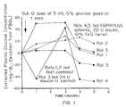
- FIG. 1is a graph comparing blood glucose levels in rats after administration of insulin in a saline solution and in poly(fumaric acid)/poly(lactide-co-glycolide) microspheres containing FeO.
- Polymers having a metal compound incorporated thereinare provided which have an improved ability to adhere to tissue surfaces, such as mucosal membranes.
- the metal compound incorporated into the polymercan be, for example, a water-insoluble metal oxide.
- the polymerscan be used to form drug delivery devices, such as polymeric microspheres, containing a therapeutic or diagnostic agent.
- drug delivery devicessuch as polymeric microspheres, containing a therapeutic or diagnostic agent.
- the polymers incorporating a metal compoundcan be used to form a wide variety of drug delivery devices, such as polymeric microspheres, which can be used to deliver therapeutic and diagnostic agents to mucosal membranes throughout the body including the gastrointestinal, respiratory and reproductive tracts.
- the metal compoundcan be incorporated into polymers forming or coating tablets, osmotic pumps, or any device capable of interacting with mucosal membranes.
- Metal compounds which can be incorporated into polymers to improve their bioadhesive propertiespreferably are water-insoluble metal compounds, such as water-insoluble metal oxides and metal hydroxides, which are capable of becoming incorporated into and associated with a polymer to thereby improve the bioadhesiveness of the polymer.
- a water-insoluble metal compoundis defined as a metal compound with little or no solubility in water, for example, less than about 0.0 to 0.9 mg/ml.
- the water-insoluble metal compoundscan be derived from a wide variety of metals, including, but not limited to, calcium, iron, copper, zinc, cadmium, zirconium and titanium.
- the water insoluble metal compoundpreferably is a metal oxide or hydroxide. Water insoluble metal compounds of multivalent metals are preferred.
- Representative metal oxides suitable for use in the compositions described hereininclude cobalt oxide (I) (CoO), cobalt oxide (II)(Co 2 O 3 ), selenium oxide (SeO 2 ), chromium double oxide (CrO 2 ), manganese oxide (MnO2), titanium oxide (TiO 2 ), lanthanum oxide (La 2 O 3 ), zirconium oxide (ZrO 2 ), silicon oxide (SiO 2 ), scandium oxide (Sc 2 O 3 ), beryllium oxide (BeO), tantalum oxide (Ta 2 O 5 ), cerium oxide (CeO2), neodymium oxide (Nd 2 O 3 ), vanadium oxide (V 2 O 5 ), molybdenum oxide (Mo 2 O 3 ), tungsten oxide (WO), tungsten trioxide (WO 3 ), samarium oxide (Sm 2 O 3 ), europium oxide (Eu 2 O 3 ), gadolinium oxide (Gd 2 O 3
- oxidesinclude barium oxide (BaO), calcium oxide (CaO), nickel oxide (III) (Ni 2 O 3 ), magnesium oxide (MgO), iron oxide (II) (FeO), iron oxide (III) (Fe 2 O 3 ), copper oxide (II) (CuO), cadmium oxide (CdO), and zirconium oxide (ZrO 2 ).
- the water-insoluble metal compoundscan be incorporated into the polymer by one of the following mechanisms: (a) physical mixtures which result in entrapment of the metal compound; (b) ionic interaction between metal compound and polymer; (c) surface modification of the polymers which would result in exposed metal compound on the surface; and (d) coating techniques such as fluidized bed, pan coating, or any similar methods known to those skilled in the art, which produce a metal compound enriched layer on the surface of the device.
- nanoparticles or microparticles of the water-insoluble metal compoundare incorporated into the polymer.
- Preferred properties defining the metal compoundinclude: (a) substantial insolubility in aqueous environments, such as acidic or basic aqueous environments (such as those present in the gastric lumen); and (b) ionizable surface charge at the pH of the aqueous environment.
- the pKa for ionization of the metal compoundmay influence binding of the polymers under different pH conditions.
- Table 1shows the acid ionization constants for certain aqua metal ions. CRC Handbook of Food Additives (T. E. Furia, Editor) 1968, Cleveland, OH.
- water-insoluble metal compoundssuch as ferric oxide, copper oxide and zinc oxide
- the incorporation of water-insoluble metal compoundscan tremendously improve adhesion of the polymer to tissue surfaces such as mucosal membranes, for example in the gastrointestinal system.
- the polymers incorporating a metal compoundthus can be used to form or coat drug delivery devices to improve their bioadhesive properties.
- the metal compoundis provided as a fine particulate dispersion of a water-insoluble metal oxide which is incorporated throughout the polymer or at least on the surface of the polymer which is to be adhered to a tissue surface.
- water-insoluble metal oxide particlesare incorporated into a polymer defining or coating a microsphere or microcapsule used for drug delivery.
- the metal oxideis present as a fine particulate dispersion on the surface of the microsphere.
- the metal compoundalso can be incorporated in an inner layer of the polymeric device and exposed only after degradation or else dissolution of a “protective” outer layer.
- a core particle containing drug and metalmay be covered with an enteric coating designed to dissolve when exposed to gastric fluid. The metal compound-enriched core then is exposed and become available for binding to GI mucosa.
- the fine metal oxide particlescan be produced for example by micronizing a metal oxide by mortar and pestle treatment to produce particles ranging in size, for example from 10.0 to 300 nm.
- the metal oxide particlescan be incorporated into the polymer, for example, by dissolving or dispersing the particles into a solution or dispersion of the polymer prior to microcapsule formation, and then can be incorporated into the polymer during microcapsule formation using a procedure for forming microcapsules such as one of those described in detail below.
- the incorporation of metal oxide particles on the surface of the microsphereadvantageously enhances the ability of the of the microsphere to bind to mucosal membranes or other tissue surfaces and improves the drug delivery properties of the microsphere.
- metal compounds which are incorporated into polymers to improve their bioadhesive propertiescan be metal compounds which are already approved by the FDA as either food or pharmaceutical additives, such as zinc oxide. While not being limited to any theory, it is possible that the mechanism by which the metal compounds promote adhesion may involve the ionic interaction of partially-ionized divalent or trivalent cations on the surface of the metal particles to negatively-charged molecules on tissue surfaces such as the glycosubstances found in the mucus including sialic acids.
- metal compoundsinto a wide range of polymers significantly increases bioadhesion of the polymers to tissue surfaces, including mucosal membrane surfaces.
- the metal compound-containing polymersactively bind to non-mucosal tissues and substances including mesentery, connective tissue and fatty tissue (see Example 7).
- water-insoluble metal compoundscan be incorporated into polymers to enhance adhesion of the polymers to mucosal membranes present throughout the gastrointestinal, respiratory (e.g., pulmonary), excretory and reproductive tracts.
- Polymers incorporating water-insoluble metal compoundsbind tenaciously to mucus membranes.
- the polymers incorporating a water-insoluble metal compoundthus can be used to form polymeric drug delivery devices for delivery of a drug via a particular mucosal membrane in the body.
- polymers incorporating a water-insoluble metal oxideare provided which are used to form or coat a drug delivery device such as a polymeric microsphere for delivery of a therapeutic or diagnostic agent via gastrointestinal mucosa.
- the intestinal mucosais formed of a continuous sheet of epithelial cells of absorptive and mucin-secreting cells. Overlying the mucosa is a discontinuous protective coating, the mucus, which is made of more than 95% water, as well as electrolytes, proteins, lipids and glycoproteins.
- the glycoproteinsare responsible for the gel-like characteristics of the mucus. These glycoproteins consist of a protein core with covalently attached carbohydrate chains terminating in either sialic acid or L-facose groups.
- the mucous glycoproteinsact as “dummy receptors” for carbohydrate binding ligands which have evolved in nature to allow microorganisms and parasites to establish themselves on the gut wall.
- One function of the mucusis to intercept these ligands and associated ineffective agents and thereby protect the mucosa.
- the enhanced binding of the polymers incorporating a metal compoundis due to the presence of partially ionized metal compounds, such as divalent or trivalent cations, on the surface of the polymer which interact, for example, via an ionic binding attraction with negatively charged glycosubstances such as sialic acid and L-fucose groups on the mucosal membrane surface.
- Multivalent ions such as divalent or trivalent cations in the metal compoundsgenerally have the strongest affinity for the negatively-charged mucin chains.
- Metal compoundscan be incorporated into a wide range of different polymers to improve the ability of the polymers to bind to tissue.
- water-insoluble metal compoundssuch as water-insoluble metal oxides can be incorporated into polymers used to form or coat drug delivery devices such as polymeric microspheres.
- Suitable polymers which can be used and into which the metal compounds can be incorporatedinclude soluble and water-insoluble, and biodegradable and nonbiodegradable polymers, including hydrogels, thermoplastics, and homopolymers, copolymers and blends of natural and synthetic polymers.
- Representative polymers which can be usedinclude hydrophilic polymers, such as those containing carboxylic groups, including polyacrylic acid.
- Bioerodible polymersincluding polyanhydrides, poly(hydroxy acids) and polyesters, as well as blends and copolymers thereof also can be used.
- Representative bioerodible poly(hydroxy acids) and copolymers thereof which can be usedinclude poly(lactic acid), poly(glycolic acid), poly(hydroxy-butyric acid), poly(hydroxyvaleric acid), poly(caprolactone), poly(lactide-co-caprolactone), and poly(lactide-co-glycolide).
- Polymers containing labile bondssuch as polyanhydrides and polyorthoesters, can be used optionally in a modified form with reduced hydrolytic reactivity.
- Representative natural polymers which also can be usedinclude proteins, such as zein, modified zein, casein, gelatin, gluten, serum albumin, or collagen, and polysaccharides such as dextrans, polyhyaluronic acid and alginic acid.
- Representative synthetic polymersinclude polyphosphazenes, polyamides, polycarbonates, polyacrylamides, polysiloxanes, polyurethanes and copolymers thereof. Celluloses also can be used.
- the term “celluloses”includes naturally occurring and synthetic celluloses, such as alkyl celluloses, cellulose ethers, cellulose esters, hydroxyalkyl celluloses and nitrocelluloses.
- Exemplary cellulosesinclude ethyl cellulose, methyl cellulose, carboxymethyl cellulose, hydroxymethyl cellulose, hydroxypropyl cellulose, hydroxypropyl methyl cellulose, hydroxybutyl methyl cellulose, cellulose acetate, cellulose propionate, cellulose acetate butyrate, cellulose acetate phthalate, cellulose triacetate and cellulose sulfate sodium salt.
- Polymers of acrylic and methacrylic acids or esters and copolymers thereofcan be used.
- Representative polymers which can be usedinclude poly(methyl methacrylate), poly(ethyl methacrylate), poly(butyl methacrylate), poly(isobutyl methacrylate), poly(hexyl methacrylate), poly(isodecyl methacrylate), poly(lauryl methacrylate), poly(phenyl methacrylate), poly(methyl acrylate), poly(isopropyl acrylate), poly(isobutyl acrylate), and poly(octadecyl acrylate).
- polymers which can be usedinclude polyalkylenes such as polyethylene and polypropylene; polyarylalkylenes such as polystyrene; poly(alkylene glycols), such as poly(ethylene glycol); poly(alkylene oxides), such as poly(ethylene oxide); and poly(alkylene terephthalates), such as poly(ethylene terephthalate).
- polyvinyl polymerscan be used, which, as defined herein includes polyvinyl alcohols, polyvinyl ethers, polyvinyl esters and polyvinyl halides.
- Exemplary polyvinyl polymersinclude poly(vinyl acetate), polyvinyl phenol and polyvinylpyrrolidone.
- Water soluble polymerscan be used.
- suitable water soluble polymersinclude polyvinyl alcohol, polyvinylpyrrolidone, methyl cellulose, hydroxypropyl cellulose, hydroxypropylmethyl cellulose and polyethylene glycol, copolymers of acrylic and methacrylic acid esters, and mixtures thereof. Water insoluble polymers also can be used.
- Suitable water insoluble polymersinclude ethylcellulose, cellulose acetate, cellulose propionate (lower, medium or -higher molecular weight), cellulose acetate propionate, cellulose acetate butyrate, cellulose acetate phthalate, cellulose triacetate, poly(methyl methacrylate), poly(ethyl methacrylate), poly(butyl methacrylate), poly(isobutyl methacrylate), poly(hexyl methacrylate), poly(isodecyl methacrylate), poly(lauryl methacrylate), poly(phenyl methacrylate), poly(methyl acrylate), poly(isopropyl acrylate), poly(isobutyl acrylate), poly(octadecyl acrylate), poly(ethylene), poly(ethylene) low density, poly(ethylene) high density, poly(propylene), poly(ethylene oxide), poly(ethylene terephthalate), poly(vinyl isobutyl ether),
- a water insoluble polymer and a water soluble polymerare used together, such as in a mixture.
- Such mixturesare useful in controlled drug release formulations, wherein the release rate can be controlled by varying the ratio of water soluble polymer to water insoluble polymer.
- These mixturesare useful, for example, when formed into microspheres, such as those having a diameter between about 10 ⁇ m and about 1 mm, preferably between about 400 ⁇ m and 1 mm, for oral administration and controlled release of drug.
- Polymers which alter viscosity as a function of temperature or shear or other physical forcesalso may be used.
- Poly(oxyalkylene) polymers and copolymerssuch as poly(ethylene oxide)-poly(propylene oxide) (PEO-PPO) or poly(ethylene oxide)-poly(butylene oxide) (PEO-PBO) copolymers, and copolymers and blends of these polymers with polymers such as poly(alpha-hydroxy acids), including but not limited to lactic, glycolic and hydroxybutyic acids, polycaprolactones, and polyvalerolactones, can be synthesized or commercially obtained.
- polyoxyalkylene copolymersare described in U.S. Pat. Nos. 3,829,506; 3,535,307; 3,036,118; 2,979,578; 2,677,700; and 2,675,619.
- Polyoxyalkylene copolymersare sold, for example, by BASF under the tradename PLURONICSTM. These materials are applied as viscous solutions at room temperature or lower which solidify at the higher body temperature. Other materials with this behavior are known in the art, and can be utilized as described herein. These include KLUCELTM (hydroxypropyl cellulose), and purified konjac glucomannan gum.
- Suitable polymersare polymeric lacquer substances based on acrylates and/or methacrylates, commonly called EURAGITTM polymers (sold by Rohm America, Inc.).
- Specific EURAGITTM polymerscan be selected having various permeability and water solubility, which properties can be pH dependent or pH independent.
- EURAGITTM RL and EURAGITTM RSare acrylic resins comprising copolymers of acrylic and methacrylic acid esters with a low content of quaternary ammonium groups, which are present as salts and give rise to the permeability of the lacquer films, wherein EURAGITTM RL is freely permeable and EURAGITTM RS is slightly permeable, independent of pH.
- EURAGITTM Lis pH dependent.
- EURAGITTM Lis an anionic polymer synthesized from methacrylic acid and methacrylic acid methyl ester. It is insoluble in acids and pure water, but becomes increasingly soluble in a neutral to weakly alkaline solution by forming salts with alkalis. Above pH 5.0, the polymer becomes increasingly permeable.
- thermoreversible polymersinclude natural gel-forming materials such as agarose, agar, furcellaran, ⁇ -carrageenan, ⁇ -1,3-glucans such as curdlan, gelatin, or polyoxyalkylene containing compounds, as described above.
- specific examplesinclude thermosetting biodegradable polymers for in vivo use described in U.S. Pat. No. 4,938,763 to Dunn, et al.
- polymerscan be obtained from sources such as Sigma Chemical Co., St. Louis, Mo.; Polysciences, Warrenton, Pa.; Aldrich, Milwaukee, Wis.; Fluka, Ronkonkoma, N.Y.; and BioRad, Richmond, Calif., or can be synthesized from monomers obtained from these or other suppliers using standard techniques.
- microspheresA wide variety of polymers can be used to form microspheres, wherein the polymer surface of the microsphere has incorporated therein a metal compound which enhances bioadhesive properties of the microsphere such as the ability of the microsphere to adhere to mucosal membranes.
- the metal compoundssuch as water-insoluble metal oxides, which enhance the bioadhesive properties of the polymers preferably are incorporated into the polymer before formation of the microspheres.
- the term “microspheres”includes microparticles and microcapsules having a core of a different material than the outer wall. Generally, the microspheres have a diameter from the nanometer range up to about 5 mm.
- the microspheresare about 500 ⁇ m to 1 mm in diameter.
- the microspheremay consist entirely of bioadhesive polymer incorporating a metal compound such as a water-insoluble metal oxide or can have only an outer coating of bioadhesive polymer incorporating the metal compound.
- polylactic acid microspherescan be fabricated using methods including solvent evaporation, hot-melt microencapsulation and spray drying.
- Polyanhydrides made of bis-carboxyphenoxypropane and sebacic acid or poly(fumaric-co-sebacic)can be prepared by hot-melt microencapsulation.
- Polystyrene microspherescan be prepared by solvent evaporation.
- Hydrogel microspherescan be prepared by dripping a polymer solution, such as alginate, chitosan, alginate/polyethylenimine (PEI) and carboxymethyl cellulose (CMC), from a reservoir though microdroplet forming device into a stirred ionic bath, as disclosed in PCT WO 93/21906.
- One or more metal compoundscan be incorporated into the polymeric microspheres either before or after formation.
- water-insoluble metal compoundssuch as a water-insoluble metal oxide
- the metal compoundcan be incorporated into the polymer after formation of the microsphere, for example by dispersing the microsphere in a solution or dispersion of the metal compound and then removing the solvent by evaporation or filtration. The metal compound can become incorporated into the polymer for example by ionic interactions.
- the metal compoundbe incorporated into the surface of the microcapsule to enable the metal compound to promote bioadhesion of the microcapsule to tissue surfaces such as mucosal membrane surfaces, or that degradation, dissolution or swelling of the outer layers can occur to expose the metal compound over time.
- Microspheres with different sizes (1-1000 ⁇ m) and morphologiescan be obtained by this method.
- This methodis useful for relatively stable polymers like polyesters and polystyrene.
- labile polymerssuch as polyanhydrides, may degrade during the fabrication process due to the presence of water.
- some of the following methods performed in completely anhydrous organic solventsare more useful.
- Microspherescan be formed from polymers such as polyesters and polyanhydrides using hot melt microencapsulation methods as described in Mathiowitz et al., Reactive Polymers , 6.275 (1987). In this method, the use of polymers with molecular weights between 3-75,000 Daltons is preferred.
- the polymerfirst is melted and then mixed with the solid particles of a substance to be incorporated that have been sieved to less than 50 ⁇ m. The mixture is suspended in a non-miscible solvent (like silicon oil), and, with continuous stirring, heated to 5° C. above the melting point of the polymer. Once the emulsion is stabilized, it is cooled until the polymer particles solidify. The resulting microspheres are washed by decantation with petroleum ether to give a free-flowing powder. Microspheres with sizes between 1 and 1000 ⁇ m are obtained with this method.
- microspheresusing spray drying techniques are well known in the art.
- the polymeris dissolved in an organic solvent such as methylene chloride.
- a known amount of a substance to be incorporatedis suspended (insoluble agent) or co-dissolved (soluble agent) in the polymer solution.
- the solution or the dispersionthen is spray-dried.
- Microspheres ranging between 1 and 10 ⁇ mare obtained.
- This methodis useful for preparing microspheres for imaging of the intestinal tract. Using the method, in addition to metal compounds, diagnostic imaging agents such as gases can be incorporated into the microspheres.
- Microspherescan be formed from polymers using a phase inversion method wherein a polymer is dissolved in a good solvent, fine particles of a substance to be incorporated, such as a drug, are mixed or dissolved in the polymer solution, and the mixture is poured into a strong non-solvent for the polymer, to spontaneously produce, under favorable conditions, polymeric microspheres, wherein the polymer is either coated on the particles or the particles are dispersed in the polymer.
- the methodcan be used to produce microparticles in a wide range of sizes, including, for example, about 100 nm to about 10 ⁇ m.
- Exemplary polymers which can be usedinclude polyvinylphenol and polylactic acid.
- Substances which can be incorporatedinclude, for example, imaging agents such as fluorescent dyes, or biologically active molecules such as proteins or nucleic acids.
- Protein microspherescan be formed by phase separation in a non-solvent followed by solvent removal as described in U.S. Pat. No. 5,271,961 to Mathiowitz et al. Proteins which can be used include prolamines such as zein.
- mixtures of proteins or a mixture of proteins and a bioerodable material polymeric material such as a polylactidecan be used.
- a prolamine solution and a substance to be incorporatedare contacted with a second liquid of limited miscibility with the proline solvent, and the mixture is agitated to form a dispersion.
- the prolamine solventthen is removed to produce stable prolamine microspheres without crosslinking or heat denaturation.
- Other prolamines which can be usedinclude gliadin, hordein and kafirin.
- Substances which can be incorporated in the microspheresinclude, in addition to the metal compound, pharmaceuticals, pesticides, nutrients and imaging agents.
- microspheresIn addition to the metal compound, biological agents such as proteins, short chain peptides, polysaccharides, nucleic acids, lipids, steroids, and organic and inorganic drugs can be incorporated into the microspheres.
- Polymers which can be used to form the microspheresinclude but are not limited to poly(lactic acid), poly(lactic-co-glycolic acid), poly(caprolactone), polycarbonates, polyamides and polyanhydrides.
- the microspheres produced by this methodare generally in the range of 5 to 1000 ⁇ m, preferably between about 30 and 50 ⁇ m.
- two hydrophilic polymersare dissolved in an aqueous solution.
- a substance to be incorporatedis dispersed or dissolved in the polymer solution, and the mixture is suspended in a continuous phase.
- the solventthen is slowly evaporated, creating microspheres with an inner core formed by one polymer and an outer layer of the second polymer.
- the continuous phasecan be either an organic oil, a volatile organic solvent, or an aqueous solution containing a third polymer that is not soluble with the first mixture of polymers and which will cause phase separation of the first two polymers as the mixture is stirred.
- Multilayer polymeric drug, protein, or cell delivery devicescan be prepared from two or more hydrophilic polymers using the method. Any two or more different biodegradable, or non-degradable, water soluble polymers which are not soluble in each other at a particular concentration as dictated by their phase diagrams may be used.
- the multilayer microcapsuleshave uniformly dimensioned layers of polymer and can incorporate a range of substances in addition to the metal compound including biologically active agents such as drugs or cells, or diagnostic agents such as dyes.
- Microspheres containing a polymeric core made of a first polymer and a uniform coating of a second polymer, and a substance incorporated into at least one of the polymers,can be made as described in U.S. Pat. No. 4,861,627.
- Microspheres made of gel-type polymersare produced through traditional ionic gelation techniques.
- the polymerfirst is dissolved in an aqueous solution, mixed with a substance to be incorporated, and then extruded through a microdroplet forming device, which in some instances employs a flow of nitrogen gas to break off the droplet.
- a slowly stirred ionic hardening bathis positioned below the extruding device to catch the forming microdroplets.
- the microspheresare left to incubate in the bath for twenty to thirty minutes in order to allow sufficient time for gelation to occur.
- Microsphere particle sizeis controlled by using various size extruders or varying either the nitrogen gas or polymer solution flow rates.
- Chitosan microspherescan be prepared by dissolving the polymer in acidic solution and crosslinking it with tripolyphosphate.
- Carboxymethyl cellulose (CMC) microspherescan be prepared by dissolving the polymer in acid solution and precipitating the microsphere with lead ions.
- Alginate/polyethylene imide (PEI)can be prepared in order to reduce the amount of carboxylic groups on the alginate microcapsule. The advantage of these systems is the ability to further modify their surface properties by the use of different chemistries. In the case of negatively charged polymers (e.g., alginate, CMC), positively charged ligands (e.g., polylysine, polyethyleneimine) of different molecular weights can be ionically attached.
- negatively charged polymerse.g., alginate, CMC
- positively charged ligandse.g., polylysine, polyethyleneimine
- Particlescan be formed and/or coated using fluidized bed techniques.
- One processis the Wurster air-suspension coating process for the coating of particles and tablets. The process consists of supporting the particles in a vertical column of heated air while the particles pass an atomizing nozzle that applies the coating material in the form of a spray. Enteric and film coating of tablets by this process typically requires approximately 30 minutes.
- Suitable coating materialsinclude, but are not limited to, cellulose acetate phthalate, ethylcellulose, hydroxypropyl methylcellylose, polyethylene glycol, and zein.
- the coating materialscan include the metal particles.
- the Wurster apparatusprovides controlled cyclic movement of the suspended particles by a rising stream of warm air, the humidity, temperature, and velocity of the air regulated.
- An air-suspended or fluidized bed of particleshas a random movement. If tablets move in and out of a coating zone in a random manner, the coating can be applies only at a slow rate.
- the Wurster apparatusprovides better drying and eventually a more uniform coating by imparting a controlled cyclic movement without or with less randomness.
- a support grid at the bottom of the vertical columntypically includes a course screen, e.g., 10 mesh, and a fine screen, e.g., 200 mesh. The fine screen offers considerably more resistance to the air flow than the coarse screen; thus, the greater amount of air flows through the coarse screen.
- the air flowing through coarse screenlifts the tablets upward in the column. As the velocity of the air stream is reduced due to diffusion of the stream and resistance of the tablets, the upward movement of the tablets ceases. Then the tablets enter the region of a still lower velocity air stream above the fine screen, where they dry and gently settle. As the dried and partially coated tablets approach the grid, they are again introduced into the higher-velocity air stream the coarse screen and enter into another cycle.
- the coating fluidis dispersed by atomization under pressure.
- a compressed-air inletis connected to the atomizing the solution or slurry of the coating material.
- the tablets, which are suspended above the coarse screen,have little contact with each other, so the coating fluid is readily distributed onto the surface of the tablets in the moving bed.
- the tabletsare presented many times in many different positions to the atomized spray; therefore, a uniform coating is built up on the tablets.
- Coatingis controlled by the weight of the coated tablets, formulation of the coating, temperature, time, and air velocity.
- Modifications of this equipmentmay be used for granulation and for coating granules with, for example, a dispersion of metal particles.
- the dry, blended ingredientsare injected into the column and the granulating solution is atomized into the column.
- the air pressure and flow patternare controlled, so the particles never touch the lower part of the column.
- the systemis enclosed to contain dust.
- the turbulent air flowprevents sticking to the sides of the column.
- the granulationsare spherical and free flowing. Particle sizes can vary from about 50 ⁇ m to about 2 mm or greater.
- Polymers incorporating a metal compound which improves the bioadhesive properties of the polymercan be used to form, or to coat, drug delivery devices such as microspheres or tablets containing any of a wide range of therapeutic and diagnostic agents.
- the drug delivery devicescan be administered by, e.g., oral, rectal, nasal or vaginal administration.
- the polymers incorporating a water-insoluble metal compoundmay be used to form bioadhesive microspheres containing a drug which is either dispersed throughout the polymer or dispersed within one or more areas within the microsphere.
- a wide range of materialscan be incorporated into the microspheres including organic compounds, inorganic compounds, proteins, polysaccharides, and nucleic acids, such as DNA, using standard techniques.
- useful proteinsinclude hormones such as insulin, growth hormones including somatometins, transforming growth factors, and other growth factors, antigens for oral vaccines, enzymes such as lactase or lipases, and digestive aids such as pancreatin.
- the polymers incorporating the metal compound and the diagnostic or therapeutic agentalso may be formulated as a tablet using methods available in the art.
- the incorporation of metal compounds into polymersincreases their ability to bind to mucous membranes.
- the incorporation of metal compounds into the polymerscan enhance the adhesion of the polymers to mammalian mucous membranes including those the entire gastrointestinal tract, respiratory, excretory and reproductive tracts, and thus can enhance the delivery of drugs incorporated into the polymers.
- the drug delivery systemsthus can be used for gastrointestinal, vaginal or respiratory delivery of a preselected drug or diagnostic agent.
- Polymers in the form of, for example, microspherescan be administered in a pharmaceutically acceptable carrier as, for example, a suspension or ointment to the mucosal membranes, via, e.g., the nose, mouth, rectum, or vagina.
- Pharmaceutically acceptable carriers for example for oral or topical administrationare known and determined based on compatibility with the polymeric material.
- Other carriersinclude bulking agents such as METAMUCILTM.
- Therapeutic or diagnostic agentswhich can be incorporated into microspheres or other drug delivery devices for application to the vaginal lining or other mucosal membrane lined orifices such as the rectum include spermacides, yeast or trichomonas treatments and anti-hemorrhoidal treatments.
- the metal compound-containing polymerscan be used in any muco-adherent delivery system including gastrointestinal delivery and vaginal delivery systems.
- the polymers incorporating a metal compoundcan be used to improve adhesion of vaginal rings used for delivery of contraceptives or hormones, or to improve the residence time of osmotic pumps.
- Microspheresalso may be formulated for adhesion and delivery of chemotherapeutic agents to tumor cells.
- Polymeric materialssuch as microspheres incorporating metal compounds which promote bioadhesiveness are useful for the oral administration of a wide range of drugs, such as sulfonamides (e.g., sulfasalazine) and glycocorticoids (e.g., bethamethasone) used for treatment of bowel diseases.
- drugssuch as sulfonamides (e.g., sulfasalazine) and glycocorticoids (e.g., bethamethasone) used for treatment of bowel diseases.
- ulcer treatmentssuch as CARAFATETM from Marion Pharmaceuticals, neurotransmitters such as L-DOPA, antihypertensives or saluretics such as Metolazone from Searle Pharmaceuticals, carbonic anhydrase inhibitors such as Acetazolamide from Lederle Pharmaceuticals, insulin like drugs such as glyburide, a blood glucose lowering drug of the sulfonylurea class, synthetic hormones such as Android F from Brown Pharmaceuticals and Testred (methyltestosterone) from ICN Pharmaceuticals, and antiparasitics such as mebendzole (VERMOXTM, Jannsen Pharmaceutical), and growth factors such as fibroblast growth factor (“FGF”), platelet derived growth factor (“PDGF”), epidermal growth factor (“EGF”), tissue growth factor- ⁇ (“TGF- ⁇ ”), and antibiotics such as vancomycin.
- FGFfibroblast growth factor
- PDGFplatelet derived growth factor
- EGF- ⁇epidermal growth factor
- TGF- ⁇tissue growth factor- ⁇
- Polymeric microspheres incorporating a metal compound to enhance bioadhesion, and a drug such as sulfasalazineare especially useful for treatment of inflammatory bowel diseases such as ulcerative colitis and Crohn's disease.
- inflammatory bowel diseasessuch as ulcerative colitis and Crohn's disease.
- ulcerative colitisinflammation is restricted to the colon
- Crohn's diseaseinflammatory lesions may be found throughout the gastrointestinal tract, from the mouth to the rectum.
- Sulfasalazineis one of the drugs that is used for treatment of these diseases.
- Sulfasalazineis cleaved by bacteria within the colon to sulfapyridine, an antibiotic, and to 5-amino salicylic acid, an anti-inflammatory agent.
- the 5-amino salicylic acidis the active drug and it is needed locally.
- the polymeric drug delivery systemscan improve the therapy by retaining the drug for a prolonged time in the intestinal tract.
- retention of 5-aminosalicylic acid in the upper intestineis of great importance, since bacteria cleave the sulfasalazin in the colon, and the usual way to treat inflammations in the upper intestine is by local administration of 5-aminosalicylic acid.
- the polymeric microspheresalso may be used for oral vaccines.
- Microspheres incorporating antigens for use as a vaccinecan be fabricated to have different retention times in the gastrointestinal tract. The different retention times, among other factors, can stimulate production of more than one type (IgG, IgM, IgA, IgE, etc.) of antibody.
- microspherescan be selected to optimize microsphere uptake, alone or in combination with other factors, including polymer composition.
- microspheresis defined as polymeric particles or capsules having a diameter on the order of 5 mm (5000 ⁇ m) or less, including particles or capsules having a diameter less than 1 mm, in the micrometer scale, or, for example, less than 1 ⁇ m, in the nanometer scale, for example 100-1000 nm.
- microspheres with a diameter less than about 10 ⁇ mmay be used. Enhanced uptake is achieved if the polymeric microspheres are loaded with metal oxide and fabricated to be smaller than 3 ⁇ m. In one embodiment, microspheres with a diameter between about 2 to 5 ⁇ m can be used, to enhance uptake into gut-associated lymphoid tissue, in particular into lymphatic cells and phagocytic cells. Additionally, microspheres less than about 2 ⁇ m, or optionally, less than about 1 ⁇ m in diameter, can be used, to enhance uptake by non-lymphatic cells and non-phagocytic cells. To reduce uptake, microspheres having a diameter greater than 10 ⁇ m can be used, for example, to enhance delivery of a drug or diagnostic agent in the microspheres to the gastrointestinal tract.
- Metal compound-containing polymersalso can be used to coat or form microspheres for the oral or intravenous administration of radio-opaque materials for use in imaging.
- a radio-opaque materialsuch as barium is coated with the polymer having the metal compound incorporated therein.
- radio-opaque materialsinclude gases or gas emitting compounds.
- Other radioactive materials or magnetic materialscan be used in place of, or in addition to, the radio-opaque materials.
- Polymers incorporating metal compoundsalso may be used to form or coat devices used as a perivascular treatment to prevent restenosis of blood vessels following balloon angioplasty.
- the metal compound-containing devicesmay be implanted outside damaged blood vessel walls and the bioadhesive properties used to retain the devices at the implant site and deliver anti-proliferative or thrombolytic drugs to the vessel wall, as described by Edelman et al., Proc. Natl. Acad. Sci ., USA 30:1513-17 (1993).
- the polymers incorporating a water insoluble metal compoundalso can be used in applications for controlled release of anti-arrhythmic agents.
- Levy et al., J. Pharm. Sci ., 83:156-1643 (1994)describes the use of non-bioadhesive polymeric implants attached to the heart for delivery of drugs to prevent arrthymias.
- Bioadhesive microspheres incorporating water-insoluble metal oxidesmay be used to deliver growth factors or other bioactive drugs to the heart in a site-specific manner after attachment to the pericardial sac.
- the delivery of bioactive drugs to the heart using alginate microsphereshas been described by Harada et al., J. Clin. Invest ., 94:623-30 (1994).
- bioadhesion of any of a wide range of different polymeric drug delivery devicescan be enhanced by the incorporation of a water-insoluble metal compound into the polymer.
- polymers incorporating a water-insoluble metal compoundcan be used to form microsphere delivery devices or used to coat pre-existing microspheres.
- Films, coatings and other devicesalso can be formed from polymers incorporating a water-insoluble metal compound to improve the bioadhesiveness of the devices.
- a polymeric coating of a polymer incorporating a water-insoluble metal compoundcan be coated on controlled-release drug delivery devices ranging from micrometer sized microspheres to millimeter sized pumps such as osmotic pumps, or on delivery devices such as vaginal rings. The bioadhesiveness of the devices thus can be improved and therefore their effectiveness in drug delivery applications can be enhanced.
- the films and coatingscan be formed using methods available in the art, including, for example, film casting, extrusion, melt casting, pressing, molding, and coating techniques such as pan coating.
- the metal compoundcan be incorporated into coatings applied by fluidized beds for the coating of large tablets.
- the benefits of bioadhesive drug delivery devicesinclude increased bioavailability, the protection of labile drugs from inactivation by digestive enzymes or other hydrolytic processes, and increased patient compliance because of reduced dosing regimens.
- FDA approved metal compoundssuch as zinc oxide can be used.
- the metal compoundshould be present on the surface of the device but still entrapped in polymer. Additionally, the loading of the metal compound must be sufficient to increase bioadhesion without disrupting the structural integrity of the device.
- the metal oxide powderwas dispersed in the polymer solution by mixing and probe sonication before addition to the PVA bath, and the rest of the process was performed as described for the control microspheres.
- PAN-PVCPoly(acrylonitrile-vinyl chloride) microspheres were prepared by spray-drying a 1.5% (w/v) solution of the polymer in acetone with and without ferric oxide (60% w/w). The resulting microspheres had sizes ranging from 1 to 20 ⁇ m. SEM indicated that the microspheres had a very high density of iron oxide exposed as cluster on the surface of the spheres as well as considerable surface texture.
- the effectiveness of metal oxides in enhancing the bioadhesive properties of the polymeric microsphereswas tested using a quantitative, in vitro everted intestinal sac bioassay, which provides a measure of adhesion of the polymers to intestinal mucosa.
- intestine from a ratwas washed with saline, everted and fashioned into fluid-filled sacs with a length of 3 cm.
- the sacswere incubated in 5 ml of physiological saline at 37° C. for 30 minutes with a 60 mg aliquot of metal oxide treated microspheres or metal oxide with rotation agitation. After 30 minutes of incubation, the quantity of unbound beads was determined gravimetrically after lyophilization and used to calculate the percentage of beads bound to intestinal mucosa. The “% bound” parameter thus was determined as an indicator of microsphere bioadhesion.
- Table 1shows the results of the in vitro assay of the bioadhesive properties of the metal oxide treated polymeric PCL and PAN-PVC microspheres.
- Polycaprylactone and polyacrylonitrile-vinyl chloride microspheresgenerally have limited bioadhesive properties in comparison to, e.g., polylactic acid spheres which have considerable bioadhesive properties.
- the results shown in Table 1illustrate that even relatively low loadings of ferric oxide (13% w/w) in microspheres made of non-bioadhesive polymers, such as polycaprylactone, can greatly improve the bioadhesive properties.
- Zein (prolamine) microsphereswere prepared by heat-curing an emulsified, alcoholic zein solution in a corn oil bath.
- the sphereswere prepared either as controls or with ferric oxide loading (54.5% w/w) and were washed with petroleum ether to remove residual oil and air-dried.
- the resulting microsphereswere in the size range of 1-30 ⁇ .
- the sphereswere tested using the rat everted intestinal sac bioassay described in Example 1 and the results indicated that ferric oxide increased bioadhesion two-fold (48% for ferric oxide-loaded spheres versus 28% for control spheres). SEM indicated that the zein loaded spheres had a very high density of iron clusters compared to the smooth surface morphology of the control spheres.
- Poly(fumaric-co-sebacic) acid 20:80(P(FA:SA) 20:80, 6 kDa), a polyanhydride synthesized using melt condensation of prepolymers, was fashioned into microspheres using the hot-melt technique.
- the polymerwas melted at 80° C., mixed with ferric oxide powder to produce a 12% (w/w) loading and dispersed into silicon oil heated to 90° C. with stirring.
- the resulting emulsion of molten polymer in oilwas hardened by cooling the system to ambient temperature and the solid spheres were recovered by filtration and washed with petroleum ether to remove oil.
- the sphereswere sieved to a size range of 106-500 ⁇ m and tested using the rat everted intestinal sac bioassay described in Example 1. The results indicated that 44% of the initial dose of spheres adhered to small intestine. SEM indicated iron oxide was distributed evenly throughout the spheres and that ferric oxide was present on the surface in high density.
- Polystyrene (2 KDa) microspheres containing 40% ferric oxide (w/w)were prepared by solvent evaporation in the size range 10-300 ⁇ m.
- the microsphereswere tested using the rat everted intestinal sac bioassay described in Example 1 which showed that the 38% of the initial dose of microspheres was bound to small intestine.
- the beadswere rinsed in 20 ml of petroleum ether, a non-solvent for the polymer solution (with miscibility in methylene chloride), briefly agitated by shaking and bath sonication for 30 s, drained and air dried.
- methylene chlorideleaves the viscous polymer solution adherent to the glass spheres and enters the non-solvent (petroleum ether), PCL precipitates as a continuous coating on the glass spheres through the mechanism of “phase inversion”.
- Isolated groups of sphereswere found in the jejunum, mostly as clumps buried in mucus and intestinal contents, but still in close apposition to the mucosa, nonetheless. No spheres were found in the lower GI tract, distal to the ileum.
- a 400 g unfasted ratwas anaesthetized with METOFANETM and fed 100 mg of zein microspheres containing 55% ferric oxide (w/w) suspended in 1.0 ml of saline by stomach tube.
- the animalwas sacrificed after 22 hrs and the entire GI tract was excised and examined. The particles were observed, buried in mucus and intestinal contents, throughout the entire length of the GI tract. Many spheres were in intimate contact with the mucosal epithelium. Histological examination revealed that the spheres were in contact with cells on the villous tips.
- PVApoly(vinyl alcohol)
- Iron-loaded microparticleswere observed throughout the small intestine both in the lumen and the absorptive mucosa. Many microspheres were seen in close contact with the microvilli, but surprisingly many spheres were observed inside intestinal absorptive cells and penetrating between junctions of adjacent cells.
- the spheres observed in the cytoplasm of the absorptive cellsranged between 30 and 300 nm and were distributed: (1) below the terminal web of the microvilli, occasionally observed in pinocytotic vesicles; (2) throughout the supranuclear region localized within membranous profiles of the endoplasmic reticulum (ER) and Golgi apparatus; (3) near the lateral membranes at the level of the nucleus. The spheres were not observed in the cytoplasm beneath the nucleus at the base of the cell. It was not uncommon to visualize at least 100-200 spheres in a TEM micrograph of a single absorptive cell, representing one plane of section.
- microspheres(exceeding 1 ⁇ m) were often observed inside goblet cells lining the intestinal villi.
- the sphereswere either inside mucin droplets (for “full” goblet cells) or else in the cytoplasm of goblet cells that had discharged their secretions (“empty” goblet cells).
- Nanoparticleswere produced by a phase inversion process.
- Fumaric acidwas purchased from Fisher Chemical, and recrystallized once from a 5% solution in 95% ethanol.
- the fumaric acidwas polymerized by refluxing for about 3.5 hr in acetic anhydride (20 g per 250 mL). After reflux, the excess acetic anhydride was removed by evaporation under vacuum, and stored at 4° C. overnight. Excess liquid acetic acid was removed via filtration if necessary, and the retentate was purified by dissolving in toluene with heat. The resulting solution then was filtered while warn, and the retentate discarded. The filtrate was allowed to crystallize at 4° C. overnight, and then washed with ether two times to remove any remaining toluene. The fumaric acid polymer precipitate was collected by filtration, dried under vacuum, and stored at ⁇ 20° C. in a sealed amber glass jar.
- the zinc-insulin suspensionthen was added to the polymer solution and Virtis-sheared at the highest setting causing an emulsion to form.
- This emulsionwas quickly dropped into 1 L of bath sonicated petroleum ether, and allowed to sit for 15 minutes.
- the nanosphereswere collected by vacuum filtration, air dried, frozen with liquid nitrogen and lyophilized for 24 hours.
- the FeO in the resulting P(FA)/PLGA microspheresalso provided an electron dense tracer for Transmission Electron Microscopic (“TEM”) visualization.
- TEMTransmission Electron Microscopic
- P(FA)/PLGA nanospherespermitted insulin release to extend over three days.
- An in vitro release study of nanospheres loaded with 1.6% insulin (w/w)showed that 60% of insulin was released within 2 hrs, and that 95% was released within 72 hrs.
- 25 mg of the nanosphereswere I.P. injected in PBS into two, fasted 300 g rats, and blood samples from the rat tail vein were tested at 1.5, 4, and 6 hrs post injection.
- the average fasting blood glucose levelwas 87 ⁇ 0.5 mg/dL. After 1.5 hrs, the level fell to 48 ⁇ 2 mg/dL; after 4 hrs, the level was 8 ⁇ 0.5 mg/dL; and after 6 hrs, the level increased to 38 ⁇ 14 mg/dL.
- Rats No. 1 and 20.5 mL saline
- Rat No. 324 I.U. insulin/0.5 mL saline (amorphous suspension)
- Rats No. 4 and 550 mg P(FA)/PLGA nanospheres containing 20 I.U. insulin and 10% w/w FeO
- Blood samples from the tail veinwere taken as an initial baseline and rats were subsequently tested for glucose tolerance following injection of a subcutaneous glucose load, consisting of 5 mL of 5% sterile glucose solution. Tutwiler et al., Diabetes , 27:856-67 (1978). At 1, 3, 4, and 5 hours postfeeding, blood samples were again taken and plasma glucose levels were measured spectrophotometrically at 505 nm using the Trinder glucose assay. The glucose levels normalized to fasting blood glucose baseline levels over time are shown in FIG. 1 .
- Rats No. 1 and 2showed expected responses to the glucose load. Blood glucose levels rose by 35% and 31% and then began dropping back to baseline. Rat No. 3, which received an oral insulin solution, showed a greater increase in serum glucose level (62% by 3 hours) and then also returned to baseline indicating some very limited bioavailability of unencapsulated insulin.
- Rat No. 5had only a 4% increase in blood sugar by 3 hours and then the glucose levels dropped to below baseline.
- Rat No. 4had very high fasting glucose level and had very erratic measured blood levels and died after 5 hrs.
- the rats fed insulin-loaded nanospheresappeared to be better able to control a glucose load than the rats not given the nanospheres (4% increase at 3 hours as opposed to ⁇ 30% increase), thus implying uptake and activity of the encapsulated insulin. Additionally, at 5 hours, only the rats fed the insulin spheres showed blood glucose levels significantly below baseline fasting levels.
Landscapes
- Health & Medical Sciences (AREA)
- Engineering & Computer Science (AREA)
- Bioinformatics & Cheminformatics (AREA)
- Chemical & Material Sciences (AREA)
- Public Health (AREA)
- Veterinary Medicine (AREA)
- Epidemiology (AREA)
- Life Sciences & Earth Sciences (AREA)
- Animal Behavior & Ethology (AREA)
- General Health & Medical Sciences (AREA)
- Medicinal Chemistry (AREA)
- Pharmacology & Pharmacy (AREA)
- Inorganic Chemistry (AREA)
- Physics & Mathematics (AREA)
- Biomedical Technology (AREA)
- Nanotechnology (AREA)
- Optics & Photonics (AREA)
- Nutrition Science (AREA)
- Physiology (AREA)
- Medicinal Preparation (AREA)
Abstract
Description
| TABLE 1 |
| Acid Ionization Constants (pKa at 25° C.) for Aqua Ions |
| Ba2+ | Ca2+ | Mg2+ | Ni2+ | Ni3+ | Fe2+ | Cu2+ | Al3+ | Fe3+ |
| 13.2 | 12.6 | 11.4 | 10.6 | 9.7 | 8.3 | 8.0 | 4.9 | 2.2 |
| TABLE 2 |
| Bioadhesion of Polymers |
| Initial Dose | Bound | % | ||
| Material | (mg) | (mg) | Bound | Std. Err. |
| PCL spheres (control) | 57.7 | 5.0 | 8.7 | 1.1 |
| PCL spheres + 13% FeO | 59.8 | 17.1 | 28.6 | 6.4 |
| (w/w) | ||||
| PCL spheres + 23% FeO | 63.3 | 18.5 | 29.1 | 11.1 |
| (w/w) | ||||
| FeO powder | 63.5 | 18.6 | 29.6 | 4.0 |
| PCL spheres + 9.1% CuO | 61.4 | 9.1 | 14.9 | 0.2 |
| (w/w) | ||||
| PCL spheres + 9.1% NiO | 64.5 | 7.9 | 12.2 | 5.2 |
| (w/w) | ||||
| PAN-PVC spheres | 54.1 | 3.9 | 7.3 | 1.9 |
| (control) | ||||
| PAN-PVC spheres + 60% | 61.0 | 16.4 | 26.8 | 3.0 |
| FeO (w/w) | ||||
| Polylactic acid (134K) | 60.4 | 16.9 | 28.0 | 9.9 |
| spheres (control) | ||||
| Polylactic acid (134K) | 60.5 | 20.2 | 33.3 | 1.6 |
| spheres (control) | ||||
| TABLE 3 |
| Microsphere Adhesion |
| Initial Dose | Bound | ||
| Sample | (mg) | (mg) | % Bound |
| 10% PCL kDa-Solvent | 60.6 | 5.7 | 9.4 |
| Evaporation (no metal) | |||
| 10% PCL 76 kDa-Solvent- | 56.6 | 8.3 | 14.7 |
| Evaporation w/13% FeO (w/w) | |||
| Glass beads (500-600 μm) coated | 49.2 | 46.7 | 94.9 |
| with 20% PCL w/16.7% CuO (w/w) | |||
| Spray-dried PAN-PVC (0.1-20 μm) | 57.1 | 21.4 | 37.5 |
| with 60% FeO (w/w) | |||
| Rats No. 1 and 2: | 0.5 mL saline |
| Rat No. 3: | 24 I.U. insulin/0.5 mL saline (amorphous suspension) |
| Rats No. 4 and 5: | 50 mg P(FA)/PLGA nanospheres containing 20 I.U. |
| insulin and 10% w/w FeO | |
Claims (31)
Priority Applications (1)
| Application Number | Priority Date | Filing Date | Title |
|---|---|---|---|
| US09/535,421US6368586B1 (en) | 1996-01-26 | 2000-03-27 | Methods and compositions for enhancing the bioadhesive properties of polymers |
Applications Claiming Priority (3)
| Application Number | Priority Date | Filing Date | Title |
|---|---|---|---|
| US08/592,565US5985312A (en) | 1996-01-26 | 1996-01-26 | Methods and compositions for enhancing the bioadhesive properties of polymers |
| US09/135,705US6123965A (en) | 1996-01-26 | 1998-08-18 | Methods and compositions for enhancing the bioadhesive properties of polymers |
| US09/535,421US6368586B1 (en) | 1996-01-26 | 2000-03-27 | Methods and compositions for enhancing the bioadhesive properties of polymers |
Related Parent Applications (1)
| Application Number | Title | Priority Date | Filing Date |
|---|---|---|---|
| US09/135,705Continuation-In-PartUS6123965A (en) | 1996-01-26 | 1998-08-18 | Methods and compositions for enhancing the bioadhesive properties of polymers |
Publications (1)
| Publication Number | Publication Date |
|---|---|
| US6368586B1true US6368586B1 (en) | 2002-04-09 |
Family
ID=26833582
Family Applications (1)
| Application Number | Title | Priority Date | Filing Date |
|---|---|---|---|
| US09/535,421Expired - LifetimeUS6368586B1 (en) | 1996-01-26 | 2000-03-27 | Methods and compositions for enhancing the bioadhesive properties of polymers |
Country Status (1)
| Country | Link |
|---|---|
| US (1) | US6368586B1 (en) |
Cited By (110)
| Publication number | Priority date | Publication date | Assignee | Title |
|---|---|---|---|---|
| US20020032434A1 (en)* | 1998-04-27 | 2002-03-14 | Chudzik Stephen J. | Bioactive agent release coating |
| US20020188037A1 (en)* | 1999-04-15 | 2002-12-12 | Chudzik Stephen J. | Method and system for providing bioactive agent release coating |
| US20030077225A1 (en)* | 2000-04-25 | 2003-04-24 | X-Bio | Hydrophilic polymer biomaterial having a specific magnetic resonance imaging signal and process for the preparation of said biomaterial |
| US20030083286A1 (en)* | 2001-08-22 | 2003-05-01 | Ching-Leou Teng | Bioadhesive compositions and methods for enhanced intestinal drug absorption |
| US6592852B1 (en)* | 2002-04-25 | 2003-07-15 | Unilever Home & Personal Care Usa, Division Of Conopco, Inc. | Zinc citrate beads in oral compositions |
| US20030212382A1 (en)* | 2002-02-20 | 2003-11-13 | Abbott Chun Lim | Methods of treating abnormal biological conditions by vaginal douching |
| US20030232087A1 (en)* | 2002-06-18 | 2003-12-18 | Lawin Laurie R. | Bioactive agent release coating with aromatic poly(meth)acrylates |
| US20040087998A1 (en)* | 2002-08-29 | 2004-05-06 | Scimed Life Systems, Inc. | Device and method for treatment of a vascular defect |
| US20040148016A1 (en)* | 2002-11-07 | 2004-07-29 | Klein Dean A. | Biocompatible medical device coatings |
| US20040215164A1 (en)* | 2002-02-20 | 2004-10-28 | Abbott Chun Lim | Methods of treating abnormal biological conditions using metal oxides |
| US20050008569A1 (en)* | 2001-06-26 | 2005-01-13 | The Board Of Trustees Of The University Of Illinois | Paramagnetic polymerized protein microspheres and methods of preparation thereof |
| US6863852B1 (en) | 2002-05-30 | 2005-03-08 | Zeus Industrial Products, Inc. | Fluoropolymer extrusions based on novel combinations of process parameters and clay minerals |
| US6887857B2 (en)* | 2001-04-27 | 2005-05-03 | Scimed Life Systems, Inc. | Microparticle protection of therapeutic agents |
| US20050131225A1 (en)* | 2001-12-06 | 2005-06-16 | University Of Iowa Research Foundation | Biodegradable oxidized cellulose esters and their uses as microspheres |
| US6919076B1 (en) | 1998-01-20 | 2005-07-19 | Pericor Science, Inc. | Conjugates of agents and transglutaminase substrate linking molecules |
| US20050175707A1 (en)* | 2002-04-23 | 2005-08-11 | Talton James D. | Process of forming and modifying particles and compositions produced thereby |
| US20050196443A1 (en)* | 2001-08-22 | 2005-09-08 | Isis Pharmaceuticals, Inc. | Pulsatile release compositions and methods for enhanced intestinal drug absorption |
| WO2005088314A1 (en)* | 2004-03-02 | 2005-09-22 | Universite Claude Bernard Lyon I | Hybrid nanoparticles including an ln2o3 core and having bioligands, and method for preparing same |
| US20050220839A1 (en)* | 2004-04-06 | 2005-10-06 | Dewitt David M | Coating compositions for bioactive agents |
| US6958148B1 (en) | 1998-01-20 | 2005-10-25 | Pericor Science, Inc. | Linkage of agents to body tissue using microparticles and transglutaminase |
| US20050271746A1 (en)* | 2004-05-18 | 2005-12-08 | Abbott Chun L | Topical treatments for abnormal biological conditions and method of topically treating such conditions |
| US20050281795A1 (en)* | 2004-06-17 | 2005-12-22 | Amano Enzyme Usa., Ltd. And Amano Enzyme,Inc. | Controlled release formulations of enzymes, microorganisms, and antibodies with mucoadhesive polymers |
| US20050288398A1 (en)* | 2001-07-20 | 2005-12-29 | Messersmith Phillip B | Polymeric compositions and related methods of use |
| WO2006003581A1 (en)* | 2004-06-29 | 2006-01-12 | Koninklijke Philips Electronics N.V. | System for manufacturing micro-spheres |
| US20060009550A1 (en)* | 2001-07-20 | 2006-01-12 | Messersmith Phillip B | Polymeric compositions and related methods of use |
| US20060035975A1 (en)* | 2004-02-26 | 2006-02-16 | Keith Alec D | Enteric coated oral pharmaceutical to erode kidney stones |
| US20060045865A1 (en)* | 2004-08-27 | 2006-03-02 | Spherics, Inc. | Controlled regional oral delivery |
| US20060067895A1 (en)* | 2004-09-29 | 2006-03-30 | Kao Corporation | Composite particle and process for producing the same |
| US20060067883A1 (en)* | 2004-09-24 | 2006-03-30 | Biosphere Medical, Inc. | Microspheres capable of binding radioisotopes, optionally comprising metallic microparticles, and methods of use thereof |
| US20060083772A1 (en)* | 2004-04-06 | 2006-04-20 | Dewitt David M | Coating compositions for bioactive agents |
| US20060241281A1 (en)* | 2001-07-20 | 2006-10-26 | Messersmith Phillip B | Peptidomimetic polymers for antifouling surfaces |
| US20060257443A1 (en)* | 2005-05-11 | 2006-11-16 | Grain Processing Corporation | Pest Control Composition and Method |
| US20070156113A1 (en)* | 2006-01-03 | 2007-07-05 | Heartcor | Organ shealth for percutaneous delivery of biological and pharmacological agents |
| US20070202173A1 (en)* | 2006-01-26 | 2007-08-30 | Jorge Cueto-Garcia | Biodegradable, non-toxic biological adhesive for use in abdominal surgery |
| US20070208141A1 (en)* | 2006-02-16 | 2007-09-06 | Shull Kenneth R | Modified acrylic block copolymers for hydrogels and pressure sensitive wet adhesives |
| US20070248637A1 (en)* | 2002-06-18 | 2007-10-25 | Surmodics, Inc. | Bioactive agent release coating and controlled humidity method |
| US20070260202A1 (en)* | 2002-02-20 | 2007-11-08 | Dominic Abbott | Deodorizer devices, systems and methods for controlling perspiration-related body odor |
| US20070281007A1 (en)* | 2004-08-27 | 2007-12-06 | Jacob Jules S | Mucoadhesive Oral Formulations of High Permeability, High Solubility Drugs |
| FR2906464A1 (en)* | 2006-09-29 | 2008-04-04 | Europ Finances | ACTIVE COMPOSITION FOR PREVENTING ACCUMULATION TO ABSORPTION OF ALCOHOLIC BEVERAGES |
| US20080085306A1 (en)* | 2006-08-31 | 2008-04-10 | Spherics, Inc. | Topiramate compositions and methods of enhancing its bioavailability |
| US20080118557A1 (en)* | 2006-11-17 | 2008-05-22 | Supernus Pharnaceuticals, Inc. | Sustained-release formulations of topiramate |
| US20080169059A1 (en)* | 2006-08-04 | 2008-07-17 | Messersmith Phillip B | Biomimetic modular adhesive complex: materials, methods and applications therefore |
| US20080171012A1 (en)* | 2007-01-11 | 2008-07-17 | Phillip Messersmith | Fouling Resistant Coatings and Methods of Making Same |
| US20080194739A1 (en)* | 2004-11-19 | 2008-08-14 | Takehiko Yamashita | Flame-Retardant Resin Composition, Process for Producing the Same, Method of Molding the Same |
| US7458384B1 (en)* | 2004-07-15 | 2008-12-02 | University Of Central Florida Research Foundation, Inc. | Surfactant incorporated nanostructure for pressure drop reduction in oil and gas lines |
| US20090061011A1 (en)* | 2007-09-03 | 2009-03-05 | Nanotherapeutics, Inc. | Compositions and methods for delivery of poorly soluble drugs |
| WO2009049083A1 (en)* | 2007-10-09 | 2009-04-16 | Washington University In St. Louis | Particles for imaging |
| US7622533B2 (en) | 2006-08-04 | 2009-11-24 | Nerites Corporation | Biomimetic compounds and synthetic methods therefor |
| US20100137902A1 (en)* | 2007-02-16 | 2010-06-03 | Nerites Corporation | Bioadhesive constructs |
| US20100330025A1 (en)* | 2002-07-19 | 2010-12-30 | Northwestern University | Surface Independent, Surface-Modifying, Multifunctional Coatings and Applications Thereof |
| WO2011005756A1 (en) | 2009-07-06 | 2011-01-13 | Puretech Ventures, Llc | Delivery of agents targeted to microbiota niches |
| WO2011010103A1 (en) | 2009-07-22 | 2011-01-27 | Imperial Innovations Limited | Methods and uses |
| WO2011022542A2 (en) | 2009-08-19 | 2011-02-24 | Puretech Ventures, Llc | Administration of factors normally present in a microbial niche to improve health |
| KR101026291B1 (en) | 2008-06-30 | 2011-03-31 | 주식회사 태강 | Anticancer composition containing SeO2 doped W-Mo-Li compound |
| US20110130465A1 (en)* | 2009-12-01 | 2011-06-02 | Nerites Corporation | Coatings for prevention of biofilms |
| WO2011152566A2 (en) | 2010-06-04 | 2011-12-08 | The University Of Tokyo | Composition for inducing proliferation or accumulation of regulatory t cells |
| US20120009267A1 (en)* | 2010-07-08 | 2012-01-12 | Brown University | Nanoparticle compositions and methods for improved oral delivery of active agents |
| WO2012064319A1 (en) | 2010-11-08 | 2012-05-18 | Colgate-Palmolive Company | Oral compositions containing microaggregates |
| EP2308473A4 (en)* | 2008-07-01 | 2013-01-09 | Nitto Denko Corp | Pharmaceutical composition containing surface-coated microparticles |
| US8425892B2 (en) | 2001-10-29 | 2013-04-23 | Columbia Laboratories, Inc. | Extended, controlled-release pharmaceutical compositions using charged polymers |
| WO2013080561A1 (en) | 2011-12-01 | 2013-06-06 | University Of Tokyo | Human-derived bacteria that induce proliferation or accumulation of regulatory t cells |
| US20130183452A1 (en)* | 2010-07-16 | 2013-07-18 | Biopolymer Technology Of Sweden Ab | Corrosion Inhibiting Coating Based on Cerium Oxide and a Catecholic Polymer |
| US8673286B2 (en) | 2007-04-09 | 2014-03-18 | Northwestern University | DOPA-functionalized, branched, poly(aklylene oxide) adhesives |
| US8779049B2 (en) | 2011-02-24 | 2014-07-15 | Panasonic Corporation | Outer casing and method for producing the same |
| WO2015156419A1 (en) | 2014-04-10 | 2015-10-15 | Riken | Compositions and methods for induction of th17 cells |
| US9320826B2 (en) | 2010-11-09 | 2016-04-26 | Kensey Nash Corporation | Adhesive compounds and methods use for hernia repair |
| US9447231B2 (en) | 2011-02-24 | 2016-09-20 | Panasonic Intellectual Property Management Co., Ltd. | Outer casing and method for producing the same |
| US9468607B2 (en) | 2007-10-09 | 2016-10-18 | Washington University | Ligand directed toroidal nanoparticles for therapy and diagnostic imaging |
| US9498439B2 (en) | 2010-04-15 | 2016-11-22 | Washington University | Prodrug compositions, prodrug nanoparticles, and methods of use thereof |
| RU2615370C2 (en)* | 2015-07-23 | 2017-04-04 | Колгейт-Палмолив Компани | Oral composition containing micro-aggregates |
| CN106710773A (en)* | 2016-12-12 | 2017-05-24 | 北京大学深圳研究生院 | Monodisperse magnetic porous silicon dioxide pellet and preparing method thereof |
| WO2017105990A1 (en) | 2015-12-14 | 2017-06-22 | Massachusetts Institute Of Technology | Ph-responsive mucoadhesive polymeric encapsulated microorganisms |
| US9687573B2 (en) | 2013-03-13 | 2017-06-27 | Biosphere Medical, Inc. | Compositions and associated methods for radioisotope-binding microparticles |
| US9764043B2 (en) | 2009-12-17 | 2017-09-19 | Washington University | Antithrombotic nanoparticle |
| US9764019B2 (en) | 2013-07-09 | 2017-09-19 | Vedanta Biosciences, Inc. | Compositions containing combinations of bioactive molecules derived from microbiota for treatment of disease |
| US9808500B2 (en) | 2009-12-17 | 2017-11-07 | Washington University | Antithrombotic nanoparticle |
| WO2017218680A1 (en) | 2016-06-14 | 2017-12-21 | Vedanta Biosciences, Inc. | Treatment of clostridium difficile infection |
| WO2017223273A1 (en) | 2016-06-22 | 2017-12-28 | President And Fellows Of Harvard College | Inhibition of colonic group 3 innate lymphoid cells |
| WO2018172483A1 (en) | 2017-03-22 | 2018-09-27 | Assistance Publique - Hopitaux De Paris | Method for determining the potential efficacy of anticancer treatment |
| WO2018237316A1 (en) | 2017-06-22 | 2018-12-27 | President And Fellows Of Harvard College | INHIBITION OF INNATED GROUP 3 LYMPHOID CELLS FROM THE COLON |
| WO2019017389A1 (en) | 2017-07-18 | 2019-01-24 | 学校法人慶應義塾 | Anti-bacterial composition against th1 cell-inducing bacteria |
| WO2019046401A1 (en) | 2017-08-29 | 2019-03-07 | Evelo Biosciences, Inc. | Treating cancer using a blautia strain |
| WO2019099682A1 (en) | 2017-11-15 | 2019-05-23 | Evelo Biosciences, Inc. | Compositions and methods for treating immune disorders using immune modulating lactococcus bacteria strains |
| WO2019099482A1 (en) | 2017-11-14 | 2019-05-23 | Evelo Biosciences, Inc. | Compositions and methods for treating disease using a blautia strain |
| WO2019152667A1 (en) | 2018-01-31 | 2019-08-08 | Evelo Biosciences, Inc. | Compositions and methods for treating immune disorders using lachnospiraceae bacteria |
| WO2019157003A1 (en) | 2018-02-06 | 2019-08-15 | Evelo Biosciences, Inc. | Compositions and methods for treating cancer and immune disorders using veillonella bacteria |
| WO2019169138A1 (en) | 2018-02-28 | 2019-09-06 | Evelo Biosciences, Inc. | Compositions and methods for treating cancer using paraclostridium benzoelyticum |
| WO2019169160A1 (en) | 2018-02-28 | 2019-09-06 | Evelo Biosciences, Inc. | Compositions and methods for treating cancer using ruminococcus gnavus |
| WO2019169168A1 (en) | 2018-02-28 | 2019-09-06 | Evelo Biosciences, Inc. | Compositions and methods for treating cancer using agathobaculum |
| WO2019169143A1 (en) | 2018-02-28 | 2019-09-06 | Evelo Biosciences, Inc. | Compositions and methods for treating cancer using turicibacter sanguinis |
| WO2019178057A1 (en) | 2018-03-12 | 2019-09-19 | Evelo Biosciences, Inc. | Methods of treating cancer using burkholderia |
| WO2019178487A2 (en) | 2018-03-15 | 2019-09-19 | Evelo Biosciences, Inc. | Compositions and methods for treating disease using klebsiella quasipneumoniae subsp. similipneumoniae |
| WO2019178490A1 (en) | 2018-03-15 | 2019-09-19 | Evelo Biosciences, Inc. | Compositions and methods for treating cancer and inflammation using klebsiella oxytoca |
| WO2019178494A1 (en) | 2018-03-15 | 2019-09-19 | Evelo Biosciences, Inc. | Compositions and methods for treating cancer and inflammation using tyzzerella nexilis |
| WO2020054728A1 (en) | 2018-09-10 | 2020-03-19 | 国立研究開発法人理化学研究所 | Composition for inhibiting trypsin activity, containing, as active ingredient, bacterium belonging to genus paraprevotella |
| WO2020118232A1 (en) | 2018-12-07 | 2020-06-11 | President And Fellows Of Harvard College | Identification of gut bacteria that promote an anti-tumor response to immunotherapy |
| WO2020179868A1 (en) | 2019-03-07 | 2020-09-10 | 学校法人慶應義塾 | Antibacterial composition against drug-resistant bacteria or pro-inflammatory bacteria |
| WO2021142279A1 (en) | 2020-01-10 | 2021-07-15 | Evelo Biosciences, Inc. | Compositions and methods of treatment using veillonella parvula |
| WO2021145458A1 (en) | 2020-01-16 | 2021-07-22 | 学校法人慶應義塾 | Composition for producing bile acids |
| WO2021167088A1 (en) | 2020-02-21 | 2021-08-26 | 良和 中岡 | Composition for alleviating pulmonary hypertension, method for predicting prognosis of pulmonary hypertension, method for assisting in determining severity of pulmonary hypertension, and method for assisting in diagnosing pulmonary hypertension |
| WO2022159711A1 (en) | 2021-01-21 | 2022-07-28 | Vedanta Biosciences, Inc. | Compositions and methods for treating hepatic encephalopathy |
| WO2022178292A1 (en) | 2021-02-18 | 2022-08-25 | Vedanta Biosciences, Inc. | Compositions and methods for suppressing pathogenic organisms |
| WO2022191183A1 (en) | 2021-03-08 | 2022-09-15 | 学校法人順天堂 | Composition and improving agent having inflammation-suppressing action |
| WO2022191182A1 (en) | 2021-03-08 | 2022-09-15 | 学校法人順天堂 | Composition and ameliorating agent having inflammation reducing effect |
| WO2022216670A1 (en) | 2021-04-05 | 2022-10-13 | Vedanta Biosciences, Inc. | Compositions and methods for treating cancer |
| WO2023278477A1 (en) | 2021-06-28 | 2023-01-05 | Vedanta Biosciences, Inc. | Methods of colonizing a microbiome, treating and/or preventing inflammatory bowel disease and graft versus host disease |
| WO2023224117A1 (en) | 2022-05-20 | 2023-11-23 | 大塚製薬株式会社 | Microorganism belonging to genus akkermansia |
| EP4410954A2 (en) | 2017-12-11 | 2024-08-07 | Vedanta Biosciences, Inc. | Compositions and methods for suppressing pathogenic organisms |
| US12214002B2 (en) | 2017-10-30 | 2025-02-04 | Seres Therapeutics, Inc. | Compositions and methods for treating antibiotic resistance |
| WO2025076184A1 (en) | 2023-10-03 | 2025-04-10 | Vedanta Biosciences, Inc. | Methods of decreasing dysbiosis and restoring a microbiome |
Citations (41)
| Publication number | Priority date | Publication date | Assignee | Title |
|---|---|---|---|---|
| US2675619A (en) | 1952-05-31 | 1954-04-20 | Mcbee Co | Drafting instrument |
| US2677700A (en) | 1951-05-31 | 1954-05-04 | Wyandotte Chemicals Corp | Polyoxyalkylene surface active agents |
| US2979578A (en) | 1958-10-30 | 1961-04-11 | Litton Systems Inc | Omnipositional rotary switch |
| US3036118A (en) | 1957-09-11 | 1962-05-22 | Wyandotte Chemicals Corp | Mixtures of novel conjugated polyoxyethylene-polyoxypropylene compounds |
| US3535307A (en) | 1964-01-02 | 1970-10-20 | Jefferson Chem Co Inc | High molecular weight polyether blocked polymers |
| US3563978A (en) | 1968-05-15 | 1971-02-16 | Irving L Ochs | Polyvalent metal complexes of natural polymers |
| US3829506A (en) | 1971-06-01 | 1974-08-13 | Basf Wyandotte Corp | Biodegradable surface active agents having good foam properties and foam stabilizing characteristics |
| US4169804A (en) | 1976-08-19 | 1979-10-02 | Minnesota Mining And Manufacturing Company | Magnetically responsive composite microparticle |
| EP0200508A2 (en) | 1985-04-27 | 1986-11-05 | Nitto Denko Corporation | Adhesive oral bandages and oral pharmaceutical preparations |
| US4861627A (en) | 1987-05-01 | 1989-08-29 | Massachusetts Institute Of Technology | Preparation of multiwall polymeric microcapsules |
| EP0333523A2 (en) | 1988-03-18 | 1989-09-20 | The Uab Research Foundation | Method of potentiating an immune response and compositions therefor |
| EP0363224A1 (en) | 1988-10-07 | 1990-04-11 | Block Drug Company Inc. | Treatment of periodontal disease |
| US4938763A (en) | 1988-10-03 | 1990-07-03 | Dunn Richard L | Biodegradable in-situ forming implants and methods of producing the same |
| US4976968A (en) | 1989-02-24 | 1990-12-11 | Clinical Technologies Associates, Inc. | Anhydrous delivery systems for pharmacological agents |
| WO1991006287A1 (en) | 1989-11-06 | 1991-05-16 | Enzytech, Inc. | Protein microspheres and methods of using them |
| WO1991006286A1 (en) | 1989-11-06 | 1991-05-16 | Enzytech, Inc. | Method for producing protein microspheres |
| US5019400A (en) | 1989-05-01 | 1991-05-28 | Enzytech, Inc. | Very low temperature casting of controlled release microspheres |
| WO1991014733A1 (en) | 1990-03-29 | 1991-10-03 | The Procter & Gamble Company | Adhesive composition with acidic organic adhesive polymer and amine-substituted polymer |
| US5122418A (en) | 1985-12-09 | 1992-06-16 | Shiseido Company Ltd. | Composite powder and production process |
| WO1992022201A1 (en)* | 1991-06-17 | 1992-12-23 | Baxter Diagnostics Inc. | Process for producing magnetically responsive polymer particles and application thereof |
| EP0526862A1 (en) | 1991-08-06 | 1993-02-10 | VECTORPHARMA INTERNATIONAL S.p.A. | Solid pharmaceutical compositions for oral administration with prolonged gastric residence |
| US5213788A (en) | 1988-09-29 | 1993-05-25 | Ranney David F | Physically and chemically stabilized polyatomic clusters for magnetic resonance image and spectral enhancement |
| US5219554A (en) | 1986-07-03 | 1993-06-15 | Advanced Magnetics, Inc. | Hydrated biodegradable superparamagnetic metal oxides |
| WO1993021906A1 (en) | 1992-04-24 | 1993-11-11 | Brown University Research Foundation | Bioadhesive microspheres and their use as drug delivery and imaging systems |
| US5271961A (en) | 1989-11-06 | 1993-12-21 | Alkermes Controlled Therapeutics, Inc. | Method for producing protein microspheres |
| US5281408A (en) | 1991-04-05 | 1994-01-25 | Unger Evan C | Low density microspheres and their use as contrast agents for computed tomography |
| EP0625353A1 (en) | 1993-05-18 | 1994-11-23 | ISTITUTO BIOCHIMICO ITALIANO GIOVANNI LORENZINI S.p.A. | Slow-release pharmaceutical composition containing a bile acid as an active ingredient |
| US5427767A (en) | 1991-05-28 | 1995-06-27 | Institut Fur Diagnostikforschung Gmbh An Der Freien Universitat Berlin | Nanocrystalline magnetic iron oxide particles-method for preparation and use in medical diagnostics and therapy |
| US5451393A (en) | 1991-01-25 | 1995-09-19 | Eastman Kodak Company | X-ray contrast compositions useful in medical imaging |
| WO1995033434A1 (en) | 1994-06-09 | 1995-12-14 | The Procter & Gamble Company | Denture stabilizing compositions |
| DE4434929A1 (en) | 1994-06-28 | 1996-01-04 | Georgi Dr Med Stankov | Adhesive anti-inflammatory nystatin ointment |
| US5498421A (en) | 1993-02-22 | 1996-03-12 | Vivorx Pharmaceuticals, Inc. | Composition useful for in vivo delivery of biologics and methods employing same |
| US5547682A (en) | 1989-12-22 | 1996-08-20 | Bioquest, Incorporated | Preparation and use of novel injectable RES avoiding inorganic particles for medical application |
| US5549915A (en) | 1993-01-29 | 1996-08-27 | Magnetic Delivered Therapeutics, Inc. | Magnetically responsive composition for carrying biologically active substances and methods of production |
| US5565215A (en) | 1993-07-23 | 1996-10-15 | Massachusettes Institute Of Technology | Biodegradable injectable particles for imaging |
| US5641515A (en) | 1995-04-04 | 1997-06-24 | Elan Corporation, Plc | Controlled release biodegradable nanoparticles containing insulin |
| JPH09255533A (en)* | 1996-03-22 | 1997-09-30 | Nippon Sheet Glass Co Ltd | Flaky metal oxide and cosmetic mixed with the same |
| JPH11253820A (en)* | 1998-03-13 | 1999-09-21 | Mitsuboshi Belting Ltd | Production of particulates-carrying material excellent in catalytic activity |
| US5955096A (en) | 1996-06-25 | 1999-09-21 | Brown University Research Foundation | Methods and compositions for enhancing the bioadhesive properties of polymers using organic excipients |
| US5985312A (en)* | 1996-01-26 | 1999-11-16 | Brown University Research Foundation | Methods and compositions for enhancing the bioadhesive properties of polymers |
| JPH11335569A (en)* | 1998-05-25 | 1999-12-07 | Mitsuboshi Belting Ltd | Production of fine particle |
- 2000
- 2000-03-27USUS09/535,421patent/US6368586B1/ennot_activeExpired - Lifetime
Patent Citations (48)
| Publication number | Priority date | Publication date | Assignee | Title |
|---|---|---|---|---|
| US2677700A (en) | 1951-05-31 | 1954-05-04 | Wyandotte Chemicals Corp | Polyoxyalkylene surface active agents |
| US2675619A (en) | 1952-05-31 | 1954-04-20 | Mcbee Co | Drafting instrument |
| US3036118A (en) | 1957-09-11 | 1962-05-22 | Wyandotte Chemicals Corp | Mixtures of novel conjugated polyoxyethylene-polyoxypropylene compounds |
| US2979578A (en) | 1958-10-30 | 1961-04-11 | Litton Systems Inc | Omnipositional rotary switch |
| US3535307A (en) | 1964-01-02 | 1970-10-20 | Jefferson Chem Co Inc | High molecular weight polyether blocked polymers |
| US3563978A (en) | 1968-05-15 | 1971-02-16 | Irving L Ochs | Polyvalent metal complexes of natural polymers |
| US3829506A (en) | 1971-06-01 | 1974-08-13 | Basf Wyandotte Corp | Biodegradable surface active agents having good foam properties and foam stabilizing characteristics |
| US4169804A (en) | 1976-08-19 | 1979-10-02 | Minnesota Mining And Manufacturing Company | Magnetically responsive composite microparticle |
| EP0200508A2 (en) | 1985-04-27 | 1986-11-05 | Nitto Denko Corporation | Adhesive oral bandages and oral pharmaceutical preparations |
| US5122418A (en) | 1985-12-09 | 1992-06-16 | Shiseido Company Ltd. | Composite powder and production process |
| US5219554A (en) | 1986-07-03 | 1993-06-15 | Advanced Magnetics, Inc. | Hydrated biodegradable superparamagnetic metal oxides |
| US4861627A (en) | 1987-05-01 | 1989-08-29 | Massachusetts Institute Of Technology | Preparation of multiwall polymeric microcapsules |
| EP0333523A2 (en) | 1988-03-18 | 1989-09-20 | The Uab Research Foundation | Method of potentiating an immune response and compositions therefor |
| US5213788A (en) | 1988-09-29 | 1993-05-25 | Ranney David F | Physically and chemically stabilized polyatomic clusters for magnetic resonance image and spectral enhancement |
| US4938763A (en) | 1988-10-03 | 1990-07-03 | Dunn Richard L | Biodegradable in-situ forming implants and methods of producing the same |
| US4938763B1 (en) | 1988-10-03 | 1995-07-04 | Atrix Lab Inc | Biodegradable in-situ forming implants and method of producing the same |
| EP0363224A1 (en) | 1988-10-07 | 1990-04-11 | Block Drug Company Inc. | Treatment of periodontal disease |
| US4976968A (en) | 1989-02-24 | 1990-12-11 | Clinical Technologies Associates, Inc. | Anhydrous delivery systems for pharmacological agents |
| US5019400A (en) | 1989-05-01 | 1991-05-28 | Enzytech, Inc. | Very low temperature casting of controlled release microspheres |
| US5271961A (en) | 1989-11-06 | 1993-12-21 | Alkermes Controlled Therapeutics, Inc. | Method for producing protein microspheres |
| WO1991006287A1 (en) | 1989-11-06 | 1991-05-16 | Enzytech, Inc. | Protein microspheres and methods of using them |
| WO1991006286A1 (en) | 1989-11-06 | 1991-05-16 | Enzytech, Inc. | Method for producing protein microspheres |
| US5547682A (en) | 1989-12-22 | 1996-08-20 | Bioquest, Incorporated | Preparation and use of novel injectable RES avoiding inorganic particles for medical application |
| WO1991014733A1 (en) | 1990-03-29 | 1991-10-03 | The Procter & Gamble Company | Adhesive composition with acidic organic adhesive polymer and amine-substituted polymer |
| US5451393A (en) | 1991-01-25 | 1995-09-19 | Eastman Kodak Company | X-ray contrast compositions useful in medical imaging |
| US5547656B1 (en) | 1991-04-05 | 1999-07-20 | Imarx Pharmaceutical Corp | Low density microspheres and their use as contrast agents for computed tomography and in other applications |
| US5547656A (en) | 1991-04-05 | 1996-08-20 | Imarx Pharmaceutical Corp. | Low density microspheres and their use as contrast agents for computed tomography, and in other applications |
| US5281408A (en) | 1991-04-05 | 1994-01-25 | Unger Evan C | Low density microspheres and their use as contrast agents for computed tomography |
| US5527521B1 (en) | 1991-04-05 | 1999-11-09 | Imarx Pharmaceutical Corp | Low density microspheres and suspensions and their use as contrast agents for computed tomography and in other applications |
| US5527521A (en) | 1991-04-05 | 1996-06-18 | Imarx Pharmaceutical Corp. | Low density microspheres and suspensions and their use as contrast agents for computed tomography and in other applications |
| US5427767A (en) | 1991-05-28 | 1995-06-27 | Institut Fur Diagnostikforschung Gmbh An Der Freien Universitat Berlin | Nanocrystalline magnetic iron oxide particles-method for preparation and use in medical diagnostics and therapy |
| WO1992022201A1 (en)* | 1991-06-17 | 1992-12-23 | Baxter Diagnostics Inc. | Process for producing magnetically responsive polymer particles and application thereof |
| EP0526862A1 (en) | 1991-08-06 | 1993-02-10 | VECTORPHARMA INTERNATIONAL S.p.A. | Solid pharmaceutical compositions for oral administration with prolonged gastric residence |
| WO1993021906A1 (en) | 1992-04-24 | 1993-11-11 | Brown University Research Foundation | Bioadhesive microspheres and their use as drug delivery and imaging systems |
| US5549915A (en) | 1993-01-29 | 1996-08-27 | Magnetic Delivered Therapeutics, Inc. | Magnetically responsive composition for carrying biologically active substances and methods of production |
| US5651989A (en) | 1993-01-29 | 1997-07-29 | Magnetic Delivered Therapeutics, Inc. | Magnetically responsive composition for carrying biologically active substances and methods of production and use |
| US5498421A (en) | 1993-02-22 | 1996-03-12 | Vivorx Pharmaceuticals, Inc. | Composition useful for in vivo delivery of biologics and methods employing same |
| EP0625353A1 (en) | 1993-05-18 | 1994-11-23 | ISTITUTO BIOCHIMICO ITALIANO GIOVANNI LORENZINI S.p.A. | Slow-release pharmaceutical composition containing a bile acid as an active ingredient |
| US5565215A (en) | 1993-07-23 | 1996-10-15 | Massachusettes Institute Of Technology | Biodegradable injectable particles for imaging |
| WO1995033434A1 (en) | 1994-06-09 | 1995-12-14 | The Procter & Gamble Company | Denture stabilizing compositions |
| DE4434929A1 (en) | 1994-06-28 | 1996-01-04 | Georgi Dr Med Stankov | Adhesive anti-inflammatory nystatin ointment |
| US5641515A (en) | 1995-04-04 | 1997-06-24 | Elan Corporation, Plc | Controlled release biodegradable nanoparticles containing insulin |
| US5985312A (en)* | 1996-01-26 | 1999-11-16 | Brown University Research Foundation | Methods and compositions for enhancing the bioadhesive properties of polymers |
| US6123965A (en)* | 1996-01-26 | 2000-09-26 | Brown University Research Foundation | Methods and compositions for enhancing the bioadhesive properties of polymers |
| JPH09255533A (en)* | 1996-03-22 | 1997-09-30 | Nippon Sheet Glass Co Ltd | Flaky metal oxide and cosmetic mixed with the same |
| US5955096A (en) | 1996-06-25 | 1999-09-21 | Brown University Research Foundation | Methods and compositions for enhancing the bioadhesive properties of polymers using organic excipients |
| JPH11253820A (en)* | 1998-03-13 | 1999-09-21 | Mitsuboshi Belting Ltd | Production of particulates-carrying material excellent in catalytic activity |
| JPH11335569A (en)* | 1998-05-25 | 1999-12-07 | Mitsuboshi Belting Ltd | Production of fine particle |
Non-Patent Citations (24)
Cited By (193)
| Publication number | Priority date | Publication date | Assignee | Title |
|---|---|---|---|---|
| US6958148B1 (en) | 1998-01-20 | 2005-10-25 | Pericor Science, Inc. | Linkage of agents to body tissue using microparticles and transglutaminase |
| US6919076B1 (en) | 1998-01-20 | 2005-07-19 | Pericor Science, Inc. | Conjugates of agents and transglutaminase substrate linking molecules |
| US20060067968A1 (en)* | 1998-04-27 | 2006-03-30 | Surmodics, Inc. | Bioactive agent release coating |
| US7442402B2 (en) | 1998-04-27 | 2008-10-28 | Surmodics, Inc. | Bioactive agent release coating |
| US20020032434A1 (en)* | 1998-04-27 | 2002-03-14 | Chudzik Stephen J. | Bioactive agent release coating |
| US6890583B2 (en)* | 1998-04-27 | 2005-05-10 | Surmodics, Inc. | Bioactive agent release coating |
| US20020188037A1 (en)* | 1999-04-15 | 2002-12-12 | Chudzik Stephen J. | Method and system for providing bioactive agent release coating |
| US20030077225A1 (en)* | 2000-04-25 | 2003-04-24 | X-Bio | Hydrophilic polymer biomaterial having a specific magnetic resonance imaging signal and process for the preparation of said biomaterial |
| US7407646B2 (en)* | 2000-04-25 | 2008-08-05 | L'assistance Publique-Hopitaux De Paris Etablissement Public De Sante | Hydrophilic polymer biomaterial having a specific magnetic resonance imaging signal and process for the preparation of said biomaterial |
| US20050124571A1 (en)* | 2001-04-27 | 2005-06-09 | Wendy Naimark | Microparticle protection of therapeutic agents |
| US6887857B2 (en)* | 2001-04-27 | 2005-05-03 | Scimed Life Systems, Inc. | Microparticle protection of therapeutic agents |
| US20050008569A1 (en)* | 2001-06-26 | 2005-01-13 | The Board Of Trustees Of The University Of Illinois | Paramagnetic polymerized protein microspheres and methods of preparation thereof |
| US7858679B2 (en) | 2001-07-20 | 2010-12-28 | Northwestern University | Polymeric compositions and related methods of use |
| US7618937B2 (en) | 2001-07-20 | 2009-11-17 | Northwestern University | Peptidomimetic polymers for antifouling surfaces |
| US20060241281A1 (en)* | 2001-07-20 | 2006-10-26 | Messersmith Phillip B | Peptidomimetic polymers for antifouling surfaces |
| US20060009550A1 (en)* | 2001-07-20 | 2006-01-12 | Messersmith Phillip B | Polymeric compositions and related methods of use |
| US8815793B2 (en) | 2001-07-20 | 2014-08-26 | Northwestern University | Polymeric compositions and related methods of use |
| US20050288398A1 (en)* | 2001-07-20 | 2005-12-29 | Messersmith Phillip B | Polymeric compositions and related methods of use |
| US7576067B2 (en) | 2001-08-22 | 2009-08-18 | Isis Pharmaceuticals, Inc. | Pulsatile release compositions and methods for enhanced intestinal oligonucleotide drug absorption |
| US20050196443A1 (en)* | 2001-08-22 | 2005-09-08 | Isis Pharmaceuticals, Inc. | Pulsatile release compositions and methods for enhanced intestinal drug absorption |
| US20030083286A1 (en)* | 2001-08-22 | 2003-05-01 | Ching-Leou Teng | Bioadhesive compositions and methods for enhanced intestinal drug absorption |
| US8425892B2 (en) | 2001-10-29 | 2013-04-23 | Columbia Laboratories, Inc. | Extended, controlled-release pharmaceutical compositions using charged polymers |
| US7649089B2 (en)* | 2001-12-06 | 2010-01-19 | University Of Iowa Research Foundation | Biodegradable oxidized cellulose esters and their uses as microspheres |
| US20050131225A1 (en)* | 2001-12-06 | 2005-06-16 | University Of Iowa Research Foundation | Biodegradable oxidized cellulose esters and their uses as microspheres |
| US8118789B2 (en) | 2002-02-20 | 2012-02-21 | Abbott Research Group, Inc. | Deodorizer devices and systems for controlling perspiration-related body odor |
| US20070260202A1 (en)* | 2002-02-20 | 2007-11-08 | Dominic Abbott | Deodorizer devices, systems and methods for controlling perspiration-related body odor |
| US7276056B2 (en) | 2002-02-20 | 2007-10-02 | Abbott Research Group, Inc. | Methods of treating abnormal biological conditions by vaginal douching |
| US7270653B2 (en)* | 2002-02-20 | 2007-09-18 | Abbott Research Group | Methods of treating abnormal biological conditions using metal oxides |
| US20030212382A1 (en)* | 2002-02-20 | 2003-11-13 | Abbott Chun Lim | Methods of treating abnormal biological conditions by vaginal douching |
| US20040215164A1 (en)* | 2002-02-20 | 2004-10-28 | Abbott Chun Lim | Methods of treating abnormal biological conditions using metal oxides |
| US20050175707A1 (en)* | 2002-04-23 | 2005-08-11 | Talton James D. | Process of forming and modifying particles and compositions produced thereby |
| US8501232B2 (en) | 2002-04-23 | 2013-08-06 | Nanotherapeutics, Inc. | Process of forming and modifying particles and compositions produced thereby |
| US6682720B2 (en) | 2002-04-25 | 2004-01-27 | Unilever Home & Personal Care Usa, Division Of Conopco, Inc. | Zinc citrate beads in oral compositions |
| US6592852B1 (en)* | 2002-04-25 | 2003-07-15 | Unilever Home & Personal Care Usa, Division Of Conopco, Inc. | Zinc citrate beads in oral compositions |
| US6863852B1 (en) | 2002-05-30 | 2005-03-08 | Zeus Industrial Products, Inc. | Fluoropolymer extrusions based on novel combinations of process parameters and clay minerals |
| US7833548B2 (en) | 2002-06-18 | 2010-11-16 | Surmodics, Inc. | Bioactive agent release coating and controlled humidity method |
| US20030232087A1 (en)* | 2002-06-18 | 2003-12-18 | Lawin Laurie R. | Bioactive agent release coating with aromatic poly(meth)acrylates |
| US20070248637A1 (en)* | 2002-06-18 | 2007-10-25 | Surmodics, Inc. | Bioactive agent release coating and controlled humidity method |
| US20110054417A1 (en)* | 2002-06-18 | 2011-03-03 | Surmodics, Inc. | Bioactive agent release coating and controlled humidity method |
| US20100330025A1 (en)* | 2002-07-19 | 2010-12-30 | Northwestern University | Surface Independent, Surface-Modifying, Multifunctional Coatings and Applications Thereof |
| US8911831B2 (en) | 2002-07-19 | 2014-12-16 | Northwestern University | Surface independent, surface-modifying, multifunctional coatings and applications thereof |
| US8075585B2 (en) | 2002-08-29 | 2011-12-13 | Stryker Corporation | Device and method for treatment of a vascular defect |
| US20040087998A1 (en)* | 2002-08-29 | 2004-05-06 | Scimed Life Systems, Inc. | Device and method for treatment of a vascular defect |
| US20040148016A1 (en)* | 2002-11-07 | 2004-07-29 | Klein Dean A. | Biocompatible medical device coatings |
| US7563460B2 (en)* | 2004-02-26 | 2009-07-21 | Med Five, Inc. | Enteric coated oral pharmaceutical to erode kidney stones |
| US20060035975A1 (en)* | 2004-02-26 | 2006-02-16 | Keith Alec D | Enteric coated oral pharmaceutical to erode kidney stones |
| WO2005088314A1 (en)* | 2004-03-02 | 2005-09-22 | Universite Claude Bernard Lyon I | Hybrid nanoparticles including an ln2o3 core and having bioligands, and method for preparing same |
| US8357545B2 (en) | 2004-03-02 | 2013-01-22 | Universite Claud Bernard Lyon I | Hybrid nanoparticles with Ln2O3 core and carrying biological ligands, and method of preparation thereof |
| US20070281324A1 (en)* | 2004-03-02 | 2007-12-06 | Pascal Perriat | Hybrid Nanoparticles With Ln2O3 Core and Carrying Biological Ligands, and Method of Preparation Thereof |
| US20050220841A1 (en)* | 2004-04-06 | 2005-10-06 | Dewitt David M | Coating compositions for bioactive agents |
| US20050220839A1 (en)* | 2004-04-06 | 2005-10-06 | Dewitt David M | Coating compositions for bioactive agents |
| US20060083772A1 (en)* | 2004-04-06 | 2006-04-20 | Dewitt David M | Coating compositions for bioactive agents |
| US20050220840A1 (en)* | 2004-04-06 | 2005-10-06 | Dewitt David M | Coating compositions for bioactive agents |
| US7544673B2 (en) | 2004-04-06 | 2009-06-09 | Surmodics, Inc. | Coating compositions for bioactive agents |
| US7541048B2 (en) | 2004-04-06 | 2009-06-02 | Surmodics, Inc. | Coating compositions for bioactive agents |
| WO2005115526A3 (en)* | 2004-05-18 | 2006-07-20 | Abbott Res Group Inc | Methods of treating abnormal biological conditions using metal oxides |
| US20050271746A1 (en)* | 2004-05-18 | 2005-12-08 | Abbott Chun L | Topical treatments for abnormal biological conditions and method of topically treating such conditions |
| US20050281795A1 (en)* | 2004-06-17 | 2005-12-22 | Amano Enzyme Usa., Ltd. And Amano Enzyme,Inc. | Controlled release formulations of enzymes, microorganisms, and antibodies with mucoadhesive polymers |
| CN1984708B (en)* | 2004-06-29 | 2014-01-29 | 皇家飞利浦电子股份有限公司 | Micro-spheres |
| US20080019904A1 (en)* | 2004-06-29 | 2008-01-24 | Koninklijke Philips Electronics, N.V. | System For Manufacturing Micro-Sheres |
| JP2008504950A (en)* | 2004-06-29 | 2008-02-21 | コーニンクレッカ フィリップス エレクトロニクス エヌ ヴィ | Micro sphere |
| WO2006003581A1 (en)* | 2004-06-29 | 2006-01-12 | Koninklijke Philips Electronics N.V. | System for manufacturing micro-spheres |
| US20090127505A1 (en)* | 2004-07-15 | 2009-05-21 | Research Foundation Of The University Of Central Florida, Inc. | Surfactant incorporated nanostructure for pressure drop reduction in oil and gas lines |
| US7458384B1 (en)* | 2004-07-15 | 2008-12-02 | University Of Central Florida Research Foundation, Inc. | Surfactant incorporated nanostructure for pressure drop reduction in oil and gas lines |
| US8252259B2 (en) | 2004-07-15 | 2012-08-28 | University Of Central Florida Research Foundation, Inc. | Surfactant incorporated nanostructure for pressure drop reduction in oil and gas lines |
| US20060045865A1 (en)* | 2004-08-27 | 2006-03-02 | Spherics, Inc. | Controlled regional oral delivery |
| US20070281007A1 (en)* | 2004-08-27 | 2007-12-06 | Jacob Jules S | Mucoadhesive Oral Formulations of High Permeability, High Solubility Drugs |
| US20060067883A1 (en)* | 2004-09-24 | 2006-03-30 | Biosphere Medical, Inc. | Microspheres capable of binding radioisotopes, optionally comprising metallic microparticles, and methods of use thereof |
| US20060067895A1 (en)* | 2004-09-29 | 2006-03-30 | Kao Corporation | Composite particle and process for producing the same |
| EP1642924A1 (en) | 2004-09-29 | 2006-04-05 | Kao Corporation | Composite particle and process for producing the same |
| US7691413B2 (en) | 2004-09-29 | 2010-04-06 | Kao Corporation | Composite particle and process for producing the same |
| US8735486B2 (en) | 2004-11-19 | 2014-05-27 | Panasonic Corporation | Flame-retardant resin composition, process for producing the same, method of molding the same |
| US20110101566A1 (en)* | 2004-11-19 | 2011-05-05 | Panasonic Corporation | Flame-retardant resin composition, process for producing the same, method of molding the same |
| US20080194739A1 (en)* | 2004-11-19 | 2008-08-14 | Takehiko Yamashita | Flame-Retardant Resin Composition, Process for Producing the Same, Method of Molding the Same |
| US20060257443A1 (en)* | 2005-05-11 | 2006-11-16 | Grain Processing Corporation | Pest Control Composition and Method |
| US20070156113A1 (en)* | 2006-01-03 | 2007-07-05 | Heartcor | Organ shealth for percutaneous delivery of biological and pharmacological agents |
| US20070202173A1 (en)* | 2006-01-26 | 2007-08-30 | Jorge Cueto-Garcia | Biodegradable, non-toxic biological adhesive for use in abdominal surgery |
| US8440227B2 (en) | 2006-01-26 | 2013-05-14 | Jorge Cueto-Garcia | Biodegradable, non-toxic biological adhesive and methods for use in abdominal surgery |
| US8252333B2 (en) | 2006-01-26 | 2012-08-28 | Jorge Cueto-Garcia | Biodegradable, non-toxic biological adhesive for use in abdominal surgery |
| US7732539B2 (en) | 2006-02-16 | 2010-06-08 | National Science Foundation | Modified acrylic block copolymers for hydrogels and pressure sensitive wet adhesives |
| US20090298999A1 (en)* | 2006-02-16 | 2009-12-03 | Northwestern University | Modified Acrylic Block Copolymers For Hydrogels and Pressure Sensitive Wet Adhesives |
| US20090247704A1 (en)* | 2006-02-16 | 2009-10-01 | Northwestern University | Modified Acrylic Block Copolymers For Hydrogels and Pressure Sensitive Wet Adhesives |
| US20090240000A1 (en)* | 2006-02-16 | 2009-09-24 | Northwestern University | Modified Acrylic Block Copolymers For Hydrogels and Pressure Sensitive Wet Adhesives |
| US7943703B2 (en) | 2006-02-16 | 2011-05-17 | Northwestern University | Modified acrylic block copolymers for hydrogels and pressure sensitive wet adhesives |
| US20070208141A1 (en)* | 2006-02-16 | 2007-09-06 | Shull Kenneth R | Modified acrylic block copolymers for hydrogels and pressure sensitive wet adhesives |
| US7622533B2 (en) | 2006-08-04 | 2009-11-24 | Nerites Corporation | Biomimetic compounds and synthetic methods therefor |
| US8563117B2 (en) | 2006-08-04 | 2013-10-22 | Phillip B. Messersmith | Biomimetic modular adhesive complex: materials, methods and applications therefore |
| US8575276B2 (en) | 2006-08-04 | 2013-11-05 | Knc Ner Acquisition Sub, Inc. | Biomimetic compounds and synthetic methods therefor |
| US8030413B2 (en) | 2006-08-04 | 2011-10-04 | Knc Ner Acquisition Sub, Inc. | Biomimetic compounds and synthetic methods therefor |
| US20080169059A1 (en)* | 2006-08-04 | 2008-07-17 | Messersmith Phillip B | Biomimetic modular adhesive complex: materials, methods and applications therefore |
| US9744137B2 (en) | 2006-08-31 | 2017-08-29 | Supernus Pharmaceuticals, Inc. | Topiramate compositions and methods of enhancing its bioavailability |
| US20080085306A1 (en)* | 2006-08-31 | 2008-04-10 | Spherics, Inc. | Topiramate compositions and methods of enhancing its bioavailability |
| FR2906464A1 (en)* | 2006-09-29 | 2008-04-04 | Europ Finances | ACTIVE COMPOSITION FOR PREVENTING ACCUMULATION TO ABSORPTION OF ALCOHOLIC BEVERAGES |
| US8298576B2 (en) | 2006-11-17 | 2012-10-30 | Supernus Pharmaceuticals, Inc. | Sustained-release formulations of topiramate |
| US8992989B2 (en) | 2006-11-17 | 2015-03-31 | Supernus Pharmaceuticals, Inc. | Sustained-release formulations of topiramate |
| US10314790B2 (en) | 2006-11-17 | 2019-06-11 | Supernus Pharmaceuticals, Inc. | Sustained-release formulations of topiramate |
| US20080118557A1 (en)* | 2006-11-17 | 2008-05-22 | Supernus Pharnaceuticals, Inc. | Sustained-release formulations of topiramate |
| US8298580B2 (en) | 2006-11-17 | 2012-10-30 | Supernus Pharmaceuticals, Inc. | Sustained-release formulations of topiramate |
| US8889191B2 (en) | 2006-11-17 | 2014-11-18 | Supernus Pharmaceuticals, Inc. | Sustained-release formulations of topiramate |
| US9622983B2 (en) | 2006-11-17 | 2017-04-18 | Supernus Pharmaceutcals, Inc. | Sustained-release formulations of topiramate |
| US8663683B2 (en) | 2006-11-17 | 2014-03-04 | Supernus Pharmaceuticals, Inc. | Sustained-release formulations of topiramate |
| US9549940B2 (en) | 2006-11-17 | 2017-01-24 | Supernus Pharmaceuticals, Inc. | Sustained-release formulations of topiramate |
| US8877248B1 (en) | 2006-11-17 | 2014-11-04 | Supernus Pharmaceuticals, Inc. | Sustained-release formulations of topiramate |
| US9555004B2 (en) | 2006-11-17 | 2017-01-31 | Supernus Pharmaceuticals, Inc. | Sustained-release formulations of topiramate |
| US20080171012A1 (en)* | 2007-01-11 | 2008-07-17 | Phillip Messersmith | Fouling Resistant Coatings and Methods of Making Same |
| US20100137902A1 (en)* | 2007-02-16 | 2010-06-03 | Nerites Corporation | Bioadhesive constructs |
| US8383092B2 (en) | 2007-02-16 | 2013-02-26 | Knc Ner Acquisition Sub, Inc. | Bioadhesive constructs |
| US8673286B2 (en) | 2007-04-09 | 2014-03-18 | Northwestern University | DOPA-functionalized, branched, poly(aklylene oxide) adhesives |
| US8377479B2 (en) | 2007-09-03 | 2013-02-19 | Nanotherapeutics, Inc. | Compositions and methods for delivery of poorly soluble drugs |
| US9554996B2 (en) | 2007-09-03 | 2017-01-31 | Nanotherapeutics, Inc. | Compositions and methods for delivery of poorly soluble drugs |
| US20090061011A1 (en)* | 2007-09-03 | 2009-03-05 | Nanotherapeutics, Inc. | Compositions and methods for delivery of poorly soluble drugs |
| US20100297019A1 (en)* | 2007-10-09 | 2010-11-25 | The Washington University | Particles for imaging |
| US9446150B2 (en) | 2007-10-09 | 2016-09-20 | Washington University | Particles for imaging |
| US9468607B2 (en) | 2007-10-09 | 2016-10-18 | Washington University | Ligand directed toroidal nanoparticles for therapy and diagnostic imaging |
| WO2009049083A1 (en)* | 2007-10-09 | 2009-04-16 | Washington University In St. Louis | Particles for imaging |
| KR101026291B1 (en) | 2008-06-30 | 2011-03-31 | 주식회사 태강 | Anticancer composition containing SeO2 doped W-Mo-Li compound |
| EP2308473A4 (en)* | 2008-07-01 | 2013-01-09 | Nitto Denko Corp | Pharmaceutical composition containing surface-coated microparticles |
| WO2011005756A1 (en) | 2009-07-06 | 2011-01-13 | Puretech Ventures, Llc | Delivery of agents targeted to microbiota niches |
| WO2011010103A1 (en) | 2009-07-22 | 2011-01-27 | Imperial Innovations Limited | Methods and uses |
| WO2011022542A2 (en) | 2009-08-19 | 2011-02-24 | Puretech Ventures, Llc | Administration of factors normally present in a microbial niche to improve health |
| US20110130465A1 (en)* | 2009-12-01 | 2011-06-02 | Nerites Corporation | Coatings for prevention of biofilms |
| US9808500B2 (en) | 2009-12-17 | 2017-11-07 | Washington University | Antithrombotic nanoparticle |
| US9764043B2 (en) | 2009-12-17 | 2017-09-19 | Washington University | Antithrombotic nanoparticle |
| US11872313B2 (en) | 2010-04-15 | 2024-01-16 | Washington University | Prodrug compositions, prodrug nanoparticles, and methods of use thereof |
| US11141379B2 (en) | 2010-04-15 | 2021-10-12 | Washington University | Prodrug compositions, prodrug nanoparticles, and methods of use thereof |
| US10201500B2 (en) | 2010-04-15 | 2019-02-12 | Washington University | Prodrug compositions, prodrug nanoparticles, and methods of use thereof |
| US9498439B2 (en) | 2010-04-15 | 2016-11-22 | Washington University | Prodrug compositions, prodrug nanoparticles, and methods of use thereof |
| WO2011152566A2 (en) | 2010-06-04 | 2011-12-08 | The University Of Tokyo | Composition for inducing proliferation or accumulation of regulatory t cells |
| EP3552613A1 (en) | 2010-06-04 | 2019-10-16 | The University of Tokyo | Composition for inducing proliferation or accumulation of regulatory t cells |
| EP4014982A2 (en) | 2010-06-04 | 2022-06-22 | The University of Tokyo | Composition for inducing proliferation or accumulation of regulatory t cells |
| EP4617662A2 (en) | 2010-06-04 | 2025-09-17 | The University of Tokyo | Composition for inducing proliferation or accumulation of regulatory t cells |
| EP3539548A1 (en) | 2010-06-04 | 2019-09-18 | The University of Tokyo | Composition for inducing proliferation or accumulation of regulatory cells |
| EP3178483A1 (en) | 2010-06-04 | 2017-06-14 | The University of Tokyo | Composition for inducing proliferation or accumulation of regulatory t cells |
| EP3750549A2 (en) | 2010-06-04 | 2020-12-16 | The University of Tokyo | Composition for inducing proliferation or accumulation of regulatory t cells |
| US20120009267A1 (en)* | 2010-07-08 | 2012-01-12 | Brown University | Nanoparticle compositions and methods for improved oral delivery of active agents |
| US8673359B2 (en)* | 2010-07-08 | 2014-03-18 | Brown University | Nanoparticle compositions and methods for improved oral delivery of active agents |
| US9617433B2 (en)* | 2010-07-16 | 2017-04-11 | Biopolymer Technology Of Sweden Ab | Corrosion inhibiting coating based on cerium oxide and a catecholic polymer |
| US20130183452A1 (en)* | 2010-07-16 | 2013-07-18 | Biopolymer Technology Of Sweden Ab | Corrosion Inhibiting Coating Based on Cerium Oxide and a Catecholic Polymer |
| WO2012064319A1 (en) | 2010-11-08 | 2012-05-18 | Colgate-Palmolive Company | Oral compositions containing microaggregates |
| US9271906B2 (en) | 2010-11-08 | 2016-03-01 | Colgate-Palmolive Company | Oral compositions containing microaggregates |
| US9320826B2 (en) | 2010-11-09 | 2016-04-26 | Kensey Nash Corporation | Adhesive compounds and methods use for hernia repair |
| US8779049B2 (en) | 2011-02-24 | 2014-07-15 | Panasonic Corporation | Outer casing and method for producing the same |
| US9447231B2 (en) | 2011-02-24 | 2016-09-20 | Panasonic Intellectual Property Management Co., Ltd. | Outer casing and method for producing the same |
| EP3569690A1 (en) | 2011-12-01 | 2019-11-20 | The University of Tokyo | Human-derived bacteria that induce proliferation or accumulation of regulatory t cells |
| EP3978598A1 (en) | 2011-12-01 | 2022-04-06 | The University of Tokyo | Human-derived bacteria that induce proliferation or accumulation of regulatory t cells |
| WO2013080561A1 (en) | 2011-12-01 | 2013-06-06 | University Of Tokyo | Human-derived bacteria that induce proliferation or accumulation of regulatory t cells |
| EP4477750A2 (en) | 2011-12-01 | 2024-12-18 | The University of Tokyo | Human-derived bacteria that induce proliferation or accumulation of regulatory t cells |
| US9687573B2 (en) | 2013-03-13 | 2017-06-27 | Biosphere Medical, Inc. | Compositions and associated methods for radioisotope-binding microparticles |
| US10434200B2 (en) | 2013-03-13 | 2019-10-08 | Biosphere Medical, Inc. | Compositions and associated methods for radioisotope-binding microparticles |
| US11052164B2 (en) | 2013-03-13 | 2021-07-06 | Biosphere Medical, Inc. | Compositions and associated methods for radioisotope-binding microparticles |
| US10130695B2 (en) | 2013-07-09 | 2018-11-20 | Vedanta Biosciences, Inc. | Compositions containing combinations of bioactive molecules derived from microbiota for treatment of disease |
| US9764019B2 (en) | 2013-07-09 | 2017-09-19 | Vedanta Biosciences, Inc. | Compositions containing combinations of bioactive molecules derived from microbiota for treatment of disease |
| WO2015156419A1 (en) | 2014-04-10 | 2015-10-15 | Riken | Compositions and methods for induction of th17 cells |
| RU2615370C2 (en)* | 2015-07-23 | 2017-04-04 | Колгейт-Палмолив Компани | Oral composition containing micro-aggregates |
| WO2017105990A1 (en) | 2015-12-14 | 2017-06-22 | Massachusetts Institute Of Technology | Ph-responsive mucoadhesive polymeric encapsulated microorganisms |
| US10548844B2 (en) | 2015-12-14 | 2020-02-04 | Massachusetts Institute Of Technology | pH-responsive mucoadhesive polymeric encapsulated microorganisms |
| EP4282489A2 (en) | 2016-06-14 | 2023-11-29 | Vedanta Biosciences, Inc. | Treatment of clostridium difficile infection |
| WO2017218680A1 (en) | 2016-06-14 | 2017-12-21 | Vedanta Biosciences, Inc. | Treatment of clostridium difficile infection |
| WO2017223273A1 (en) | 2016-06-22 | 2017-12-28 | President And Fellows Of Harvard College | Inhibition of colonic group 3 innate lymphoid cells |
| CN106710773A (en)* | 2016-12-12 | 2017-05-24 | 北京大学深圳研究生院 | Monodisperse magnetic porous silicon dioxide pellet and preparing method thereof |
| CN106710773B (en)* | 2016-12-12 | 2019-09-10 | 北京大学深圳研究生院 | A kind of monodisperse magnetic porous silica microballoon and preparation method thereof |
| WO2018172483A1 (en) | 2017-03-22 | 2018-09-27 | Assistance Publique - Hopitaux De Paris | Method for determining the potential efficacy of anticancer treatment |
| WO2018237316A1 (en) | 2017-06-22 | 2018-12-27 | President And Fellows Of Harvard College | INHIBITION OF INNATED GROUP 3 LYMPHOID CELLS FROM THE COLON |
| WO2019017389A1 (en) | 2017-07-18 | 2019-01-24 | 学校法人慶應義塾 | Anti-bacterial composition against th1 cell-inducing bacteria |
| WO2019046401A1 (en) | 2017-08-29 | 2019-03-07 | Evelo Biosciences, Inc. | Treating cancer using a blautia strain |
| US12214002B2 (en) | 2017-10-30 | 2025-02-04 | Seres Therapeutics, Inc. | Compositions and methods for treating antibiotic resistance |
| WO2019099482A1 (en) | 2017-11-14 | 2019-05-23 | Evelo Biosciences, Inc. | Compositions and methods for treating disease using a blautia strain |
| WO2019099682A1 (en) | 2017-11-15 | 2019-05-23 | Evelo Biosciences, Inc. | Compositions and methods for treating immune disorders using immune modulating lactococcus bacteria strains |
| EP4410954A2 (en) | 2017-12-11 | 2024-08-07 | Vedanta Biosciences, Inc. | Compositions and methods for suppressing pathogenic organisms |
| WO2019152667A1 (en) | 2018-01-31 | 2019-08-08 | Evelo Biosciences, Inc. | Compositions and methods for treating immune disorders using lachnospiraceae bacteria |
| WO2019157003A1 (en) | 2018-02-06 | 2019-08-15 | Evelo Biosciences, Inc. | Compositions and methods for treating cancer and immune disorders using veillonella bacteria |
| WO2019169143A1 (en) | 2018-02-28 | 2019-09-06 | Evelo Biosciences, Inc. | Compositions and methods for treating cancer using turicibacter sanguinis |
| WO2019169168A1 (en) | 2018-02-28 | 2019-09-06 | Evelo Biosciences, Inc. | Compositions and methods for treating cancer using agathobaculum |
| WO2019169160A1 (en) | 2018-02-28 | 2019-09-06 | Evelo Biosciences, Inc. | Compositions and methods for treating cancer using ruminococcus gnavus |
| WO2019169138A1 (en) | 2018-02-28 | 2019-09-06 | Evelo Biosciences, Inc. | Compositions and methods for treating cancer using paraclostridium benzoelyticum |
| WO2019178057A1 (en) | 2018-03-12 | 2019-09-19 | Evelo Biosciences, Inc. | Methods of treating cancer using burkholderia |
| WO2019178494A1 (en) | 2018-03-15 | 2019-09-19 | Evelo Biosciences, Inc. | Compositions and methods for treating cancer and inflammation using tyzzerella nexilis |
| WO2019178490A1 (en) | 2018-03-15 | 2019-09-19 | Evelo Biosciences, Inc. | Compositions and methods for treating cancer and inflammation using klebsiella oxytoca |
| WO2019178487A2 (en) | 2018-03-15 | 2019-09-19 | Evelo Biosciences, Inc. | Compositions and methods for treating disease using klebsiella quasipneumoniae subsp. similipneumoniae |
| WO2020054728A1 (en) | 2018-09-10 | 2020-03-19 | 国立研究開発法人理化学研究所 | Composition for inhibiting trypsin activity, containing, as active ingredient, bacterium belonging to genus paraprevotella |
| WO2020118232A1 (en) | 2018-12-07 | 2020-06-11 | President And Fellows Of Harvard College | Identification of gut bacteria that promote an anti-tumor response to immunotherapy |
| WO2020179868A1 (en) | 2019-03-07 | 2020-09-10 | 学校法人慶應義塾 | Antibacterial composition against drug-resistant bacteria or pro-inflammatory bacteria |
| WO2021142279A1 (en) | 2020-01-10 | 2021-07-15 | Evelo Biosciences, Inc. | Compositions and methods of treatment using veillonella parvula |
| WO2021145458A1 (en) | 2020-01-16 | 2021-07-22 | 学校法人慶應義塾 | Composition for producing bile acids |
| WO2021167088A1 (en) | 2020-02-21 | 2021-08-26 | 良和 中岡 | Composition for alleviating pulmonary hypertension, method for predicting prognosis of pulmonary hypertension, method for assisting in determining severity of pulmonary hypertension, and method for assisting in diagnosing pulmonary hypertension |
| WO2022159711A1 (en) | 2021-01-21 | 2022-07-28 | Vedanta Biosciences, Inc. | Compositions and methods for treating hepatic encephalopathy |
| WO2022178292A1 (en) | 2021-02-18 | 2022-08-25 | Vedanta Biosciences, Inc. | Compositions and methods for suppressing pathogenic organisms |
| WO2022191182A1 (en) | 2021-03-08 | 2022-09-15 | 学校法人順天堂 | Composition and ameliorating agent having inflammation reducing effect |
| WO2022191183A1 (en) | 2021-03-08 | 2022-09-15 | 学校法人順天堂 | Composition and improving agent having inflammation-suppressing action |
| WO2022216670A1 (en) | 2021-04-05 | 2022-10-13 | Vedanta Biosciences, Inc. | Compositions and methods for treating cancer |
| WO2023278477A1 (en) | 2021-06-28 | 2023-01-05 | Vedanta Biosciences, Inc. | Methods of colonizing a microbiome, treating and/or preventing inflammatory bowel disease and graft versus host disease |
| WO2023224117A1 (en) | 2022-05-20 | 2023-11-23 | 大塚製薬株式会社 | Microorganism belonging to genus akkermansia |
| WO2025076184A1 (en) | 2023-10-03 | 2025-04-10 | Vedanta Biosciences, Inc. | Methods of decreasing dysbiosis and restoring a microbiome |
Similar Documents
| Publication | Publication Date | Title |
|---|---|---|
| US6368586B1 (en) | Methods and compositions for enhancing the bioadhesive properties of polymers | |
| US6123965A (en) | Methods and compositions for enhancing the bioadhesive properties of polymers | |
| US6156348A (en) | Methods and compositions for enhancing the bioadhesive properties of polymers using organic excipients | |
| KR100422391B1 (en) | Preparation of biodegradable microspheres containing peptides by a melt process | |
| CA2485977C (en) | Short chain polymer for enhancing the bioadhesiveness of polymers on mucosal membrane | |
| Jalil et al. | Biodegradable poly (lactic acid) and poly (lactide-co-glycolide) microcapsules: problems associated with preparative techniques and release properties | |
| US6465626B1 (en) | Pharmaceutical compositions of chitosan with type-A gelatin | |
| US5679377A (en) | Protein microspheres and methods of using them | |
| US6632457B1 (en) | Composite hydrogel drug delivery systems | |
| JP2005513057A (en) | Bioadhesive drug delivery system with increased gastric retention | |
| JP2006514698A (en) | Nanoparticle bioactive substances | |
| KR20010022007A (en) | Biodegradable macromers for the controlled release of biologically active substances | |
| McGee et al. | The encapsulation of a model protein in poly (D, L lactide-co-glycolide) microparticles of various sizes: an evaluation of process reproducibility | |
| Singh et al. | Biodegradable polymeric microspheres as drug carriers; A review | |
| JP2022504492A (en) | Non-injectable hydrogel formulation for smart release | |
| Davis | Drug delivery systems | |
| JP2006028031A (en) | Medicine-sealed nano particle for transmucosa absorption | |
| Reddy et al. | A Mini-Review on Chitosan Microsphere Drug Delivery | |
| Chandrasekar et al. | AN EXTENSIVE REVIEW ON MUCOADHESIVE MICROSPHERES AS CARRIERS IN DRUG DELIVERY | |
| Samad et al. | 20 Microsphere |
Legal Events
| Date | Code | Title | Description |
|---|---|---|---|
| AS | Assignment | Owner name:UNIVERSITY RESEARCH FOUNDATION, BROWN, RHODE ISLAN Free format text:ASSIGNMENT OF ASSIGNORS INTEREST;ASSIGNORS:JACOB, JULES S.;MATHIOWITZ, EDITH;REEL/FRAME:010844/0856 Effective date:20000413 | |
| STCF | Information on status: patent grant | Free format text:PATENTED CASE | |
| CC | Certificate of correction | ||
| FPAY | Fee payment | Year of fee payment:4 | |
| REMI | Maintenance fee reminder mailed | ||
| FPAY | Fee payment | Year of fee payment:8 | |
| SULP | Surcharge for late payment | Year of fee payment:7 | |
| AS | Assignment | Owner name:MATHIOWITZ, EDITH, MASSACHUSETTS Free format text:NUNC PRO TUNC ASSIGNMENT;ASSIGNOR:BROWN UNIVERSITY RESEARCH FOUNDATION;REEL/FRAME:026475/0385 Effective date:20110304 Owner name:PEROSPHERE BIOPHARMACEUTICALS INC., RHODE ISLAND Free format text:ASSIGNMENT OF ASSIGNORS INTEREST;ASSIGNOR:MATHIOWITZ, EDITH;REEL/FRAME:026502/0651 Effective date:20090917 Owner name:MATHIOWITZ, EDITH, MASSACHUSETTS Free format text:ASSIGNMENT OF ASSIGNORS INTEREST;ASSIGNOR:BROWN UNIVERSITY;REEL/FRAME:026475/0493 Effective date:20090327 | |
| AS | Assignment | Owner name:PEROSPHERE INC., RHODE ISLAND Free format text:CHANGE OF NAME;ASSIGNOR:PEROSPHERE BIOPHARMACEUTICALS INC.;REEL/FRAME:027510/0901 Effective date:20110715 | |
| REMI | Maintenance fee reminder mailed | ||
| FPAY | Fee payment | Year of fee payment:12 | |
| SULP | Surcharge for late payment | Year of fee payment:11 |