US6364824B1 - Stimulating cell receptor activity using electromagnetic fields - Google Patents
Stimulating cell receptor activity using electromagnetic fieldsDownload PDFInfo
- Publication number
- US6364824B1 US6364824B1US09/438,749US43874999AUS6364824B1US 6364824 B1US6364824 B1US 6364824B1US 43874999 AUS43874999 AUS 43874999AUS 6364824 B1US6364824 B1US 6364824B1
- Authority
- US
- United States
- Prior art keywords
- receptor
- cell
- electromagnetic field
- bone
- growth factor
- Prior art date
- Legal status (The legal status is an assumption and is not a legal conclusion. Google has not performed a legal analysis and makes no representation as to the accuracy of the status listed.)
- Expired - Lifetime
Links
Images


Classifications
- A—HUMAN NECESSITIES
- A61—MEDICAL OR VETERINARY SCIENCE; HYGIENE
- A61N—ELECTROTHERAPY; MAGNETOTHERAPY; RADIATION THERAPY; ULTRASOUND THERAPY
- A61N2/00—Magnetotherapy
- A61N2/02—Magnetotherapy using magnetic fields produced by coils, including single turn loops or electromagnets
- A—HUMAN NECESSITIES
- A61—MEDICAL OR VETERINARY SCIENCE; HYGIENE
- A61N—ELECTROTHERAPY; MAGNETOTHERAPY; RADIATION THERAPY; ULTRASOUND THERAPY
- A61N1/00—Electrotherapy; Circuits therefor
- A61N1/40—Applying electric fields by inductive or capacitive coupling ; Applying radio-frequency signals
Definitions
- the present inventionrelates to stimulating cell receptor activity, and more particularly, to using electromagnetic fields to stimulate cell receptor activity.
- Osteoporosisis a disease characterized by a decrease in bone mass which leads to spontaneous bone fractures or a bone fracture occurring due to an impact that under normal conditions would not produce a bone fracture.
- the goal for treating osteoporosisis to build bone strength to a level sufficient to withstand normal loading conditions without failure.
- Bone massis determined by the balance between the activity of osteoclast, which destroy bone, and osteoblast, which build bone. During homeostasis, in which bone mass is maintained at a constant level, the activity of the osteoclast and osteoblast are equal. The amount of bone being turned over by the activity of bone cells is 5-10% per year.
- osteoporosisThe health impact of osteoporosis includes loss of the quality of life as osteoporotic bone fractures usually occur in the elder who have a diminished healing capacity. Furthermore, approximately 20% of elderly women who suffer from an osteoporotic hip fracture will die within the next year. The health care cost due to osteoporosis is between 5 and 10 billion dollars per year in the United States.
- Osteoporosis therapies targeted at preventing bone resorptioninclude Hormone Replacement Therapy (HRT) which replaces the lost estrogen with therapeutic estrogen.
- HRTHormone Replacement Therapy
- side effects of HRTsuch as increased rate of breast cancer or cervical cancer has fueled the search for a better means of decreasing bone resorption.
- HRTHormone Replacement Therapy
- Fosamax from Merckis the leading market contender in this class of anti-bone resorbing drugs.
- Another therapeuticis calcitonin which is a naturally occurring protein that inhibits osteoclast activity and is now available as a nasal spray thereby eliminating the need for injections.
- the second focus for osteoporosis therapyis to stimulate osteoblast to form more bone.
- Clinical studies conducted using osteoporotic patientsindicate that the predominant determinant of bone formation was the number of osteoblast.
- NaF treatmentfits into this category as evidence indicates that bone formation is increased with NaF treatment
- increased bone mass following NaF treatmentdoes not translate to stronger bones as the quality of bone is compromised.
- IGF-Iinsulin-like growth factor I
- TGFBtransforming growth factor beta
- a ligandsuch as IGF-I or TGFB is to activate specific receptors located on the surface of cells.
- IGF-Iactivates the IGF-I receptor and TGFB activates the TGFB receptor.
- TGFBactivates the TGFB receptor.
- a technique that has been the subject of studyis the use of an electromagnetic field (also referred to as a magnetic field) to activate receptors.
- This applicationdiscloses a method and apparatus for stimulating cell receptor activity for treatment of maladies such as osteoporosis.
- An electromagnetic field or signalis generated by a device positioned in proximity to one or more target cell receptors, such that the flux of the electromagnetic field extends through the target receptor.
- the electromagnetic fieldis then fluctuated at a predetermined rate.
- the devicecomprises a transmitter for generating an electromagnetic field having a predetermined rate of fluctuation and a positioning apparatus for positioning the transmitter such that the flux of the electromagnetic field extends through the target receptor.
- FIG. 1is illustrated a block diagram of a cell receptor spanning a cell membrane
- FIG. 2is a block diagram of a cellular environment
- FIG. 3is a block diagram of an inactive cell receptor and a cell receptor activated by a ligand
- FIG. 4is a block diagram of cell receptor activity induced in accordance with the principals of the present invention.
- FIG. 5is a flow chart describing steps of an exemplary method for activating a cell receptor
- FIG. 6Ais a graph of results of a test performed on MG-63 osteosarcoma (MG-63) cell lines;
- FIG. 6Bis a graph of results of another test performed on MG-63 cell lines.
- FIG. 7Ais a graph of results of another test performed on MG-63 cell lines.
- FIG. 7Bis a graph of results of another test performed on MG-63 cell lines.
- FIG. 8is a block diagram of a biological receptor activating device positioned onto a limb of a patient fore activating biological receptors.
- FIG. 9is a block diagram of transmitter heads forming a portion of the biological receptor activating device shown in FIG. 8 .
- the cell membrane 110can include, but is not limited to the membrane of bone cells, or any other desired target cell.
- the cell receptor 105can include, but is not limited to, the insulin-like growth factor-I (IGF-I) or the Transforming Growth Factor Beta (TGFb) receptor.
- IGF-Iinsulin-like growth factor-I
- TGFbTransforming Growth Factor Beta
- the cell receptor 105is typically a protein compound composed of multiple subunits.
- the subunitsinclude extracellular subunit 105 a which is outside of the cell membrane 110 , and an intracellular subunit 105 c which is inside the cell membrane 110 .
- the cellular environmentincludes any number of cells 205 and extracellular fluid 210 .
- the cell receptors 105 spanning the cell 205communicate events in the cell's 205 environment with appropriate intracellular machinery 215 , such as cell nucleii. Specific events are communicated to, and detected by the cell receptors 105 by means of ligands 220 .
- Ligands 220are typically composed of proteins and travel through the intracellular fluid 210 seeking to bind with the cell receptors 105 of cells 205 .
- the cell receptor 105When the ligand 220 binds with a cell receptor 105 , the cell receptor 105 generates a signal 225 which is delivered to the appropriate intracellular machinery 215 inside the cell 205 .
- the signalcauses the intracellular machinery 215 to perform certain specific actions.
- a cell receptor 105 that is bonded to ligand 220is considered to be in an active state, while a cell receptor 105 which has not bonded to a ligand 220 is said to be in an inactive state.
- Osteoporosisis a disease characterized by a decrease in bone mass which leads to spontaneous bone fractures or a bone fracture occurring due to an impact that under normal conditions would not produce a bone fracture.
- the goal for treating osteoporosisis to build bone strength to a level sufficient to withstand normal loading conditions without failure.
- the IGF-I ligand 220is a naturally occurring substance in the body which binds with the IGF-I receptor 105 and causes a signal 225 to be generated instructing the intracellular machinery 215 to divide pre-osteoblast cells 205 .
- the TGFb ligand 220is a naturally occurring substance in the body which binds with the TGFb receptor 105 and causes a signal 225 to be generated instructing the intracellular machinery 215 to differentiate the cell 205 into a bone cell.
- the same foregoing biological activitywill result if alternative means of activating the cell receptors 105 is achieved.
- FIG. 3there is illustrated a block diagram of an inactive (ell receptor 105 ( 1 ) and an activated cell receptor 105 ( 2 ) activated by ligand 220 .
- the intracellular subunit 105 ( 1 )( c ) and the extracellular subunit 105 ( 1 )( a ) of the inactive cell receptor 105 ( 1 )vibrate in specific relationship to each other.
- massis added to the extracellular subunit 105 ( 2 )( a ). This added mass changes the manner in which the intracellular subunit 105 ( 2 )( c ) and the extracellular subunit 105 ( 2 )( a ) vibrate with respect to each other.
- FIG. 4there is illustrated a block diagram of cell receptor activity induced in accordance with the principals of the present invention.
- the electromagnetic field 405is also referred to as a magnetic field.
- the term electromagnetic fieldshall be construed to include both electromagnetic fields and magnetic fields.
- the electromagnetic field 405causes the intracellular subunit 105 ( 1 )( c ) and the extracellular subunit 105 ( 1 )( a ) to vibrate in a manner simulating the vibrations of the intracellular subunit 105 ( 2 )( c ) and the extracellular subunit 105 ( 2 )( a ) of an activated cell receptor 105 ( 2 ).
- the vibrationsare detected by intracellular machinery 215 resulting in the same biological activity as with the activated cell receptor 105 ( 2 ).
- a transmitter 402is positioned in proximity to one or more target cell receptors 105 .
- An electromagnetic field 405is generated at step 510 , such that the flux of the electromagnetic field extends through the target cell receptors 105 .
- the electromagnetic field 405is fluctuated at a predetermined rate.
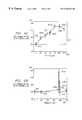
- FIG. 6Athere is illustrated a graph of results of a test performed on colonies of the MG-63 osteosarcoma (MG-63 ) cell line known by those skilled in the art for their ability to grow indefinitely and for their sensitivity to the IGF-I ligand.
- the horizontal axis 605measures the concentrations of various solutions of IGF-I ligands which were used to treat the MG-63 cell line colonies of equal cell count.
- the vertical axis 610measures the MG-63 cell count of each colony as a percentage of the cell count of a control group, represented by a plot 615 at 0 on the horizontal axis 605 and 100% on the vertical axis 610 .
- the cell count of each MG-63 cell line colony twenty-four hours after the treatment as a percentage of the cell count of the control groupis represented by plots 620 connected to form curve 625 .
- the cell count results, plots 620establish that the MG-63 cells proliferate when treated with the IFG-I ligand.
- a 150% peak 620 dis achieved with a 10 nanogram/millileter IGF-I ligand solution.
- FIG. 6Bthere is illustrated a graph of results of another test performed on colonies of the MG-63 cell line.
- the horizontal axis 655measures the rate of fluctuation of an electromagnetic field applied to the MG-63 cell line colonies of equal cell count.
- the vertical axis 660measures the cell count of each MG-63 cell line colony as a percentage of the cell count of a control group, represented by a plot 665 at 0 on the horizontal axis 655 and 100% on the vertical axis 660 .
- the cell count of each MG-63 cell line colony twenty-four hours after the treatment as a percentage of the cell count of the control groupis represented by plots 670 connected to form curve 675 .
- the cell count results, plots 670suggest no statistically significant effect for any rate of fluctuation below 375 Hz and any rate of fluctuation exceeding 385 Hz.
- the cell count of the MG-63 cell line colonies which were exposed to electromagnetic fields which fluctuated at rates between 375 Hz to 385 Hz, represented by plots 670 c, 670 d, and 670 erose sharply, with highest cell count achieved at a fluctuation rate of 379 Hz.
- the peak cell count of 150% achieved at a fluctuation rate of 379 Hz, represented by plot 670 cmimics the peak cell count, plot 620 d, achieved with the IGF-I ligand solution treatment.
- IGF-I receptors 105can be activated, thereby stimulating the proliferation of MG-63 cells 205 using the method steps illustrated in FIG. 5, wherein the electromagnetic field is fluctuated at a rate of approximately 379 Hz, or fluctuated at a rate between 375 Hz and 385 Hz, during step 515 .
- FIG. 7Athere is illustrated a graph of results of another test performed on MG-63 cell line colonies.
- the horizontal axis 705measures the concentrations of various solutions of TGFb ligands which were used to treat the MG-63 cell line colonies of equal Alkaline Phosphatase (ALP) activity.
- ALPAlkaline Phosphatase
- the vertical axis 710measures the ALP activity of each MG-63 cell line colony as a percentage of the ALP activity of a control group, represented by a plot 715 at 0 on the horizontal axis 705 and 100% on the vertical axis 710 .
- the ALP activity of each MG-63 cell line colony seventy-two hours after the treatment as a percentage of the cell count of the control groupis represented by plots 720 connected to form curve 725 .
- the ALP activityresults, plots 720 , establish that ALP activity is increased, and therefore bone cell differentiation, of the MG-63 cells when treated with the TGFb ligand solution, with a 375% plateau 720 e, 720 f achieved by a TGFb solutions exceeding 30 picograms/millileter concentration.
- FIG. 7Bthere is illustrated a graph of results of another test performed on MG-63 cell line colonies.
- the horizontal axis 755measures the rate of fluctuation of an electromagnetic field applied to the MG-63 cell line colonies of equal ALP activity.
- the vertical axis 760measures the ALP activity of each MG-63 cell line colony as a percentage of the ALP activity of a control group, represented by a plot 765 at 0 on the horizontal axis 755 and 100% on the vertical axis 760 .
- the ALP activity of each MC-63 cell line seventy-two hours after the treatment as a percentage of the ALP activity of the control groupis represented by plots 770 connected to form curve 775 .
- plots 770suggest no statistically significant effect for any rate of fluctuation below 130 Hz and any rate of fluctuation exceeding 140 Hz.
- the ALP activity, and therefore cell differentiation, of the MG-63 cell line colonies which were exposed to electromagnetic fields which fluctuated at rates between 130 Hz to 140 Hz, represented by plots 770 b , 770 c , 770 d , and 770 erose sharply, with highest ALP activity/cell differentiation rate of 150% achieved at a fluctuation rate of 133 Hz.
- TGFb receptors 105can be activated, thereby stimulating the differentiation of MG-63 cells 205 using the method steps illustrated in FIG. 5, wherein the electromagnetic field is fluctuated at a rate of approximately 133 Hz, or fluctuated at a rate between 130 Hz and 140 Hz, during step 515 . Additionally, no statistically significant activity occurred when the electromagnetic field was fluctuated at rates between 375 Hz to 385 Hz, represented by plots 770 g, 770 h, and 770 i . The foregoing leads to an important conclusion that fluctuating the electromagnetic field at a predetermined rate specifically activates only certain receptors. Therefore, the method steps of FIG. 5 can be applied to large number of cell receptors (as during a non-invasive medical procedure on a living osteoporosis patient) but only stimulates cell receptor activity or activities in certain desired target cells.
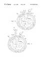
- FIG. 8there is illustrated a block diagram of a cell receptor activating device 805 for activating cell receptors, positioned onto a limb 810 of a patient.
- the area to b)e treated (treatment area) 815may be on the surface of the limb 810 or within the limb 810 and contains the biological receptors to be activated.
- the cell receptor activating device 805includes two transmitter heads 820 , 825 capable of generating an electromagnetic field.
- the transmitter heads 820 , and 825are positioned and secured in proximity to the treatment area 815 containing the target receptors such that the flux of the electromagnetic field extends through the target receptors by means of bands 830 , which can comprise a strap, a belt, a rope, or a tie.
- the transmitter heads 320 , and 825can also be positioned and secured in proximity to the treatment area 815 by a clamp, an adhesive, or via integration into a cast.
- Each transmitter head 820 , 825includes a housing 905 a , 905 b of a non-magnetic material, such as plastic which encloses a field coil 910 a , 910 b .
- Each transmitter head 820 , 825also includes an alternating (AC) current source 915 a , 915 b electrically connected to each field coil 910 a , 910 b .
- the AC current source 915 a , 915 bis capable of generating an AC current with a predetermined rate of fluctuation, which flows through the field coils 910 a , 910 b .
- the predetermined rate of fluctuationcan either be user settable, or set in accordance with manufacturing specifications.
- an electromagnetic fieldis generated. Additionally, the magnitude of the electromagnetic field is proportional to the instantaneous magnitude of the AC current. Therefore, the electromagnetic field fluctuates at the predetermined rate of fluctuation of the AC current.
Landscapes
- Health & Medical Sciences (AREA)
- Engineering & Computer Science (AREA)
- Biomedical Technology (AREA)
- Nuclear Medicine, Radiotherapy & Molecular Imaging (AREA)
- Radiology & Medical Imaging (AREA)
- Life Sciences & Earth Sciences (AREA)
- Animal Behavior & Ethology (AREA)
- General Health & Medical Sciences (AREA)
- Public Health (AREA)
- Veterinary Medicine (AREA)
- Medicines That Contain Protein Lipid Enzymes And Other Medicines (AREA)
Abstract
Description
Claims (8)
Priority Applications (1)
| Application Number | Priority Date | Filing Date | Title |
|---|---|---|---|
| US09/438,749US6364824B1 (en) | 1998-11-11 | 1999-11-11 | Stimulating cell receptor activity using electromagnetic fields |
Applications Claiming Priority (2)
| Application Number | Priority Date | Filing Date | Title |
|---|---|---|---|
| US10792798P | 1998-11-11 | 1998-11-11 | |
| US09/438,749US6364824B1 (en) | 1998-11-11 | 1999-11-11 | Stimulating cell receptor activity using electromagnetic fields |
Publications (1)
| Publication Number | Publication Date |
|---|---|
| US6364824B1true US6364824B1 (en) | 2002-04-02 |
Family
ID=22319214
Family Applications (1)
| Application Number | Title | Priority Date | Filing Date |
|---|---|---|---|
| US09/438,749Expired - LifetimeUS6364824B1 (en) | 1998-11-11 | 1999-11-11 | Stimulating cell receptor activity using electromagnetic fields |
Country Status (5)
| Country | Link |
|---|---|
| US (1) | US6364824B1 (en) |
| EP (1) | EP1137370A1 (en) |
| AU (1) | AU1719200A (en) |
| CA (1) | CA2348465A1 (en) |
| WO (1) | WO2000027295A1 (en) |
Cited By (30)
| Publication number | Priority date | Publication date | Assignee | Title |
|---|---|---|---|---|
| US6786859B2 (en) | 2001-08-17 | 2004-09-07 | Advanced Diagnostic Development Pty. Ltd. | Magnetic field therapy |
| US20050131458A1 (en)* | 2003-08-07 | 2005-06-16 | Batich Christopher D. | Biodegradable embolic agents |
| US7089060B1 (en)* | 2001-02-23 | 2006-08-08 | Amei Technologies Inc. | Methods of stimulating cell receptor activity using electromagnetic fields |
| US20060229603A1 (en)* | 2005-03-18 | 2006-10-12 | Olsen Ron A | Adjustable splint for osteosynthesis with modular joint |
| US20060229605A1 (en)* | 2005-03-18 | 2006-10-12 | Olsen Ron A | Adjustable splint for osteosynthesis with incrementing assembly for adjustment in predetermined increments |
| US20070162028A1 (en)* | 2005-12-09 | 2007-07-12 | Jesse Jackson | Cannulated screw |
| US20080103558A1 (en)* | 2006-10-30 | 2008-05-01 | Stuart Wenzel | Focused electromagnetic-wave and ultrasonic-wave structures for tissue stimulation |
| US20080125618A1 (en)* | 2006-11-28 | 2008-05-29 | Anderson Gregory S | Bone-activity stimulation apparatus and method |
| US7846162B2 (en) | 2005-05-18 | 2010-12-07 | Sonoma Orthopedic Products, Inc. | Minimally invasive actuable bone fixation devices |
| US20110015555A1 (en)* | 2009-07-14 | 2011-01-20 | Anderson Gregory S | Piezoelectric, micro-exercise apparatus and method |
| US7909825B2 (en) | 2006-11-22 | 2011-03-22 | Sonoma Orthepedic Products, Inc. | Fracture fixation device, tools and methods |
| US8287541B2 (en) | 2005-05-18 | 2012-10-16 | Sonoma Orthopedic Products, Inc. | Fracture fixation device, tools and methods |
| US8961516B2 (en) | 2005-05-18 | 2015-02-24 | Sonoma Orthopedic Products, Inc. | Straight intramedullary fracture fixation devices and methods |
| US9060820B2 (en) | 2005-05-18 | 2015-06-23 | Sonoma Orthopedic Products, Inc. | Segmented intramedullary fracture fixation devices and methods |
| US9155574B2 (en) | 2006-05-17 | 2015-10-13 | Sonoma Orthopedic Products, Inc. | Bone fixation device, tools and methods |
| USD762864S1 (en) | 2014-05-13 | 2016-08-02 | Pulse, Llc | Micro-coil array |
| USD763453S1 (en) | 2014-05-13 | 2016-08-09 | Pulse, Llc | Micro-coil array |
| US9498639B2 (en) | 2014-05-13 | 2016-11-22 | Pulse, Llc | Immersive, flux-guided, micro-coil apparatus and method |
| US9770278B2 (en) | 2014-01-17 | 2017-09-26 | Arthrex, Inc. | Dual tip guide wire |
| US9814499B2 (en) | 2014-09-30 | 2017-11-14 | Arthrex, Inc. | Intramedullary fracture fixation devices and methods |
| USD859672S1 (en) | 2018-03-05 | 2019-09-10 | Orthofix Inc. | Medical device |
| USD860467S1 (en) | 2018-03-05 | 2019-09-17 | Orthofix Inc. | Medical device |
| USD861899S1 (en) | 2018-03-05 | 2019-10-01 | Orthofix Inc. | Medical device |
| USD861898S1 (en) | 2018-03-05 | 2019-10-01 | Orthofix Inc. | Medical device |
| USD861900S1 (en) | 2018-03-05 | 2019-10-01 | Orthofix Inc. | Medical device |
| US10507333B2 (en) | 2009-07-14 | 2019-12-17 | Pulse, Llc | Immersive, flux-guided, micro-coil apparatus and method |
| USD871597S1 (en) | 2018-03-05 | 2019-12-31 | Orthofix Inc. | Medical device |
| US10874433B2 (en) | 2017-01-30 | 2020-12-29 | Stryker European Holdings I, Llc | Strut attachments for external fixation frame |
| US11191975B2 (en) | 2009-07-14 | 2021-12-07 | Pulse, Llc | Micro-coil wristband |
| US11878181B2 (en) | 2009-07-14 | 2024-01-23 | Pulse, Llc | Micro-coil wristband |
Families Citing this family (2)
| Publication number | Priority date | Publication date | Assignee | Title |
|---|---|---|---|---|
| ITUD20010095A1 (en)* | 2001-05-22 | 2002-11-22 | E Col Energy Srl | DEVICE TO GENERATE VIBRATIONS SUITABLE TO PRODUCE BENEFICIAL EFFECTS FOR LIVING ORGANISMS |
| WO2005061051A2 (en)* | 2003-12-22 | 2005-07-07 | Horst Leopold | Electromedical device |
Citations (15)
| Publication number | Priority date | Publication date | Assignee | Title |
|---|---|---|---|---|
| US3915151A (en) | 1973-03-23 | 1975-10-28 | Werner Kraus | Apparatus for promoting healing processes |
| US4266532A (en)* | 1976-11-17 | 1981-05-12 | Electro-Biology, Inc. | Modification of the growth, repair and maintenance behavior of living tissues and cells by a specific and selective change in electrical environment |
| WO1990007356A1 (en) | 1989-01-09 | 1990-07-12 | Life Resonances, Inc. | Using non-invasive magnetic fields for controlling osteoporosis |
| US4993413A (en) | 1988-09-22 | 1991-02-19 | The Research Foundation Of State University Of New York | Method and apparatus for inducing a current and voltage in living tissue |
| US5014699A (en) | 1986-05-23 | 1991-05-14 | Trustees Of The University Of Pennsylvania | Electromagnetic method and apparatus for healing living tissue |
| US5156587A (en) | 1983-09-01 | 1992-10-20 | Montone Liber J | Method for treating malignant cells |
| US5267939A (en) | 1989-01-09 | 1993-12-07 | Life Resonances, Inc. | Techniques for controlling osteoporosis using non-invasive magnetic fields |
| US5318561A (en) | 1988-03-23 | 1994-06-07 | Life Resonances Inc. | Deformable magnetic field aiding coils for use in controlling tissue growth |
| US5413596A (en) | 1993-11-29 | 1995-05-09 | The United States Of America As Represented By The United States Department Of Energy | Digital electronic bone growth stimulator |
| US5458558A (en) | 1988-03-23 | 1995-10-17 | Life Resonances, Inc. | Method for controlling tissue growth with an applied fluctuating magnetic field |
| US5743844A (en) | 1996-11-01 | 1998-04-28 | Amei Technologies, Inc. | High efficiency pulsed electromagnetic field (PEMF) stimulation therapy method and system |
| US5792209A (en) | 1996-04-01 | 1998-08-11 | Varner; Lawrence Norman | Osteoporosis-relief device |
| US5997464A (en) | 1997-08-29 | 1999-12-07 | Orthosoft, L.L.C. | Magnetic coil for pulsed electromagnetic field |
| US6004257A (en)* | 1994-05-25 | 1999-12-21 | Jacobson; Jerry I. | Method for ameliorating the aging process and the effects thereof utilizing electromagnetic energy |
| US6024691A (en)* | 1998-05-26 | 2000-02-15 | Amei Technologies Inc | Cervical collar with integrated electrical circuitry for electromagnetic field therapy |
- 1999
- 1999-11-11CACA002348465Apatent/CA2348465A1/ennot_activeAbandoned
- 1999-11-11AUAU17192/00Apatent/AU1719200A/ennot_activeAbandoned
- 1999-11-11USUS09/438,749patent/US6364824B1/ennot_activeExpired - Lifetime
- 1999-11-11EPEP99960289Apatent/EP1137370A1/ennot_activeWithdrawn
- 1999-11-11WOPCT/US1999/026708patent/WO2000027295A1/ennot_activeApplication Discontinuation
Patent Citations (15)
| Publication number | Priority date | Publication date | Assignee | Title |
|---|---|---|---|---|
| US3915151A (en) | 1973-03-23 | 1975-10-28 | Werner Kraus | Apparatus for promoting healing processes |
| US4266532A (en)* | 1976-11-17 | 1981-05-12 | Electro-Biology, Inc. | Modification of the growth, repair and maintenance behavior of living tissues and cells by a specific and selective change in electrical environment |
| US5156587A (en) | 1983-09-01 | 1992-10-20 | Montone Liber J | Method for treating malignant cells |
| US5014699A (en) | 1986-05-23 | 1991-05-14 | Trustees Of The University Of Pennsylvania | Electromagnetic method and apparatus for healing living tissue |
| US5318561A (en) | 1988-03-23 | 1994-06-07 | Life Resonances Inc. | Deformable magnetic field aiding coils for use in controlling tissue growth |
| US5458558A (en) | 1988-03-23 | 1995-10-17 | Life Resonances, Inc. | Method for controlling tissue growth with an applied fluctuating magnetic field |
| US4993413A (en) | 1988-09-22 | 1991-02-19 | The Research Foundation Of State University Of New York | Method and apparatus for inducing a current and voltage in living tissue |
| US5267939A (en) | 1989-01-09 | 1993-12-07 | Life Resonances, Inc. | Techniques for controlling osteoporosis using non-invasive magnetic fields |
| WO1990007356A1 (en) | 1989-01-09 | 1990-07-12 | Life Resonances, Inc. | Using non-invasive magnetic fields for controlling osteoporosis |
| US5413596A (en) | 1993-11-29 | 1995-05-09 | The United States Of America As Represented By The United States Department Of Energy | Digital electronic bone growth stimulator |
| US6004257A (en)* | 1994-05-25 | 1999-12-21 | Jacobson; Jerry I. | Method for ameliorating the aging process and the effects thereof utilizing electromagnetic energy |
| US5792209A (en) | 1996-04-01 | 1998-08-11 | Varner; Lawrence Norman | Osteoporosis-relief device |
| US5743844A (en) | 1996-11-01 | 1998-04-28 | Amei Technologies, Inc. | High efficiency pulsed electromagnetic field (PEMF) stimulation therapy method and system |
| US5997464A (en) | 1997-08-29 | 1999-12-07 | Orthosoft, L.L.C. | Magnetic coil for pulsed electromagnetic field |
| US6024691A (en)* | 1998-05-26 | 2000-02-15 | Amei Technologies Inc | Cervical collar with integrated electrical circuitry for electromagnetic field therapy |
Cited By (55)
| Publication number | Priority date | Publication date | Assignee | Title |
|---|---|---|---|---|
| US7089060B1 (en)* | 2001-02-23 | 2006-08-08 | Amei Technologies Inc. | Methods of stimulating cell receptor activity using electromagnetic fields |
| US6786859B2 (en) | 2001-08-17 | 2004-09-07 | Advanced Diagnostic Development Pty. Ltd. | Magnetic field therapy |
| US20040210102A1 (en)* | 2001-08-17 | 2004-10-21 | Van Mullekom Arnoldus Petrus | Magnetic field therapy |
| US20050131458A1 (en)* | 2003-08-07 | 2005-06-16 | Batich Christopher D. | Biodegradable embolic agents |
| US7575575B2 (en) | 2005-03-18 | 2009-08-18 | Ron Anthon Olsen | Adjustable splint for osteosynthesis with modular components |
| US7507240B2 (en) | 2005-03-18 | 2009-03-24 | Ron Anthon Olsen | Adjustable splint for osteosynthesis |
| US20060229604A1 (en)* | 2005-03-18 | 2006-10-12 | Olsen Ron A | Adjustable splint for osteosynthesis with modular components |
| US20060229605A1 (en)* | 2005-03-18 | 2006-10-12 | Olsen Ron A | Adjustable splint for osteosynthesis with incrementing assembly for adjustment in predetermined increments |
| US20060229603A1 (en)* | 2005-03-18 | 2006-10-12 | Olsen Ron A | Adjustable splint for osteosynthesis with modular joint |
| US20060229602A1 (en)* | 2005-03-18 | 2006-10-12 | Olsen Ron A | Adjustable splint for osteosynthesis |
| US7588571B2 (en) | 2005-03-18 | 2009-09-15 | Ron Anthon Olsen | Adjustable splint for osteosynthesis with modular joint |
| US9060820B2 (en) | 2005-05-18 | 2015-06-23 | Sonoma Orthopedic Products, Inc. | Segmented intramedullary fracture fixation devices and methods |
| US7914533B2 (en) | 2005-05-18 | 2011-03-29 | Sonoma Orthopedic Products, Inc. | Minimally invasive actuable bone fixation devices |
| US8287539B2 (en) | 2005-05-18 | 2012-10-16 | Sonoma Orthopedic Products, Inc. | Fracture fixation device, tools and methods |
| US8287541B2 (en) | 2005-05-18 | 2012-10-16 | Sonoma Orthopedic Products, Inc. | Fracture fixation device, tools and methods |
| US7846162B2 (en) | 2005-05-18 | 2010-12-07 | Sonoma Orthopedic Products, Inc. | Minimally invasive actuable bone fixation devices |
| US8961516B2 (en) | 2005-05-18 | 2015-02-24 | Sonoma Orthopedic Products, Inc. | Straight intramedullary fracture fixation devices and methods |
| US7942875B2 (en) | 2005-05-18 | 2011-05-17 | Sonoma Orthopedic Products, Inc. | Methods of using minimally invasive actuable bone fixation devices |
| US7731738B2 (en) | 2005-12-09 | 2010-06-08 | Orthopro, Llc | Cannulated screw |
| US20070162028A1 (en)* | 2005-12-09 | 2007-07-12 | Jesse Jackson | Cannulated screw |
| US9155574B2 (en) | 2006-05-17 | 2015-10-13 | Sonoma Orthopedic Products, Inc. | Bone fixation device, tools and methods |
| US20080103558A1 (en)* | 2006-10-30 | 2008-05-01 | Stuart Wenzel | Focused electromagnetic-wave and ultrasonic-wave structures for tissue stimulation |
| US8439917B2 (en) | 2006-11-22 | 2013-05-14 | Sonoma Orthopedic Products, Inc. | Fracture fixation device, tools and methods |
| US7909825B2 (en) | 2006-11-22 | 2011-03-22 | Sonoma Orthepedic Products, Inc. | Fracture fixation device, tools and methods |
| US9259250B2 (en) | 2006-11-22 | 2016-02-16 | Sonoma Orthopedic Products, Inc. | Fracture fixation device, tools and methods |
| US8758216B2 (en) | 2006-11-28 | 2014-06-24 | Gregory S. Anderson | Electromagnetic body tissue stimulation apparatus and method |
| US8147395B2 (en) | 2006-11-28 | 2012-04-03 | Gregory S. Anderson | Bone-activity stimulation apparatus and method |
| US20080125618A1 (en)* | 2006-11-28 | 2008-05-29 | Anderson Gregory S | Bone-activity stimulation apparatus and method |
| US11844956B2 (en) | 2009-07-14 | 2023-12-19 | Pulse, Llc | Immersive, flux-guided, micro-coil apparatus and method |
| US11191975B2 (en) | 2009-07-14 | 2021-12-07 | Pulse, Llc | Micro-coil wristband |
| US20110015555A1 (en)* | 2009-07-14 | 2011-01-20 | Anderson Gregory S | Piezoelectric, micro-exercise apparatus and method |
| US8439816B2 (en) | 2009-07-14 | 2013-05-14 | Pulse, Llc | Piezoelectric, micro-exercise apparatus and method |
| US8485960B2 (en) | 2009-07-14 | 2013-07-16 | Pulse, Llc | Piezoelectric, micro-exercise pad apparatus and method |
| US10507333B2 (en) | 2009-07-14 | 2019-12-17 | Pulse, Llc | Immersive, flux-guided, micro-coil apparatus and method |
| US20110144412A1 (en)* | 2009-07-14 | 2011-06-16 | Pulse, Llc | Piezoelectric, micro-exercise pad apparatus and method |
| US11878181B2 (en) | 2009-07-14 | 2024-01-23 | Pulse, Llc | Micro-coil wristband |
| US9770278B2 (en) | 2014-01-17 | 2017-09-26 | Arthrex, Inc. | Dual tip guide wire |
| US11813473B2 (en) | 2014-05-13 | 2023-11-14 | Newage, Inc. | Body-conforming, micro-coil, web apparatus and method |
| US11213692B2 (en) | 2014-05-13 | 2022-01-04 | Pulse, Llc | Body-conforming, micro-coil, web apparatus and method |
| US9498639B2 (en) | 2014-05-13 | 2016-11-22 | Pulse, Llc | Immersive, flux-guided, micro-coil apparatus and method |
| USD763453S1 (en) | 2014-05-13 | 2016-08-09 | Pulse, Llc | Micro-coil array |
| USD762864S1 (en) | 2014-05-13 | 2016-08-02 | Pulse, Llc | Micro-coil array |
| US10537747B2 (en) | 2014-05-13 | 2020-01-21 | Pulse, Llc | Immersive, flux-guided, micro-coil apparatus and method |
| US9814499B2 (en) | 2014-09-30 | 2017-11-14 | Arthrex, Inc. | Intramedullary fracture fixation devices and methods |
| US10548648B2 (en) | 2014-09-30 | 2020-02-04 | Arthrex, Inc. | Intramedullary fracture fixation devices and methods |
| US10874433B2 (en) | 2017-01-30 | 2020-12-29 | Stryker European Holdings I, Llc | Strut attachments for external fixation frame |
| US11723690B2 (en) | 2017-01-30 | 2023-08-15 | Stryker European Operations Holdings Llc | Strut attachments for external fixation frame |
| US12369948B2 (en) | 2017-01-30 | 2025-07-29 | Stryker European Operations Holdings Llc | Strut attachments for external fixation frame |
| USD904630S1 (en) | 2018-03-05 | 2020-12-08 | Orthofix Inc. | Medical device |
| USD871597S1 (en) | 2018-03-05 | 2019-12-31 | Orthofix Inc. | Medical device |
| USD861900S1 (en) | 2018-03-05 | 2019-10-01 | Orthofix Inc. | Medical device |
| USD861898S1 (en) | 2018-03-05 | 2019-10-01 | Orthofix Inc. | Medical device |
| USD861899S1 (en) | 2018-03-05 | 2019-10-01 | Orthofix Inc. | Medical device |
| USD860467S1 (en) | 2018-03-05 | 2019-09-17 | Orthofix Inc. | Medical device |
| USD859672S1 (en) | 2018-03-05 | 2019-09-10 | Orthofix Inc. | Medical device |
Also Published As
| Publication number | Publication date |
|---|---|
| WO2000027295A1 (en) | 2000-05-18 |
| EP1137370A1 (en) | 2001-10-04 |
| CA2348465A1 (en) | 2000-05-18 |
| AU1719200A (en) | 2000-05-29 |
Similar Documents
| Publication | Publication Date | Title |
|---|---|---|
| US6364824B1 (en) | Stimulating cell receptor activity using electromagnetic fields | |
| CN1918285B (en) | Systems and methods for upregulating bone morphogenetic protein (BMP) gene expression in bone cells by applying fields generated by specific and selective electrical and electromagnetic signals | |
| US10426967B2 (en) | Apparatus and method for electromagnetic treatment of neurological injury or condition caused by a stroke | |
| US9415233B2 (en) | Apparatus and method for electromagnetic treatment of neurological pain | |
| US9433797B2 (en) | Apparatus and method for electromagnetic treatment of neurodegenerative conditions | |
| US8065015B2 (en) | Regulation of genes via application of specific and selective electrical and electromagnetic signals | |
| Griffin et al. | Electrical stimulation in bone healing: critical analysis by evaluating levels of evidence | |
| JPH0211170A (en) | Method and apparatus for controlling tissue growth by applying magnetic field | |
| US20040073260A1 (en) | Regulation of type II collagen gene expression using specific and selective electrical and electromagnetic signals | |
| KR100856964B1 (en) | Regulation of Matrix Metalloproteinase Gene Expression Using Specific and Selective Electrical and Electromagnetic Signals | |
| JP2009538694A (en) | Regulation of transforming growth factor-beta (TGF-β) gene expression in living cells through the application of specific and selective electric and electromagnetic fields | |
| JP2009528073A (en) | Coil integrated device and method of using the same | |
| CN101415462A (en) | Self-contained electromagnetic cerebrofacial area treatment device and method of use | |
| WO2012102837A1 (en) | Method and device for treating osteoarthritis noninvasively | |
| CN101306228A (en) | Electromagnetic field and ultrasonic wave composite therapy device for osteoporosis | |
| KR20050059198A (en) | Resolution of aggrecan gene expression using specific and selective electrical and electromagnetic signals | |
| JP2008541938A (en) | A method for the regulation of osteochondral growth using pulsed electromagnetic field therapy | |
| CN104350150A (en) | Regulation of stem cell gene production with specific and selective electric and electromagnetic fields | |
| JP5596563B2 (en) | Control of fibroblast growth factor-2 (FGF-2) gene expression in living cells using specific and selective application of electric and electromagnetic fields | |
| Anglen | Enhancement of fracture healing with bone stimulators | |
| MXPA06007918A (en) | Up-regulation of bone morphogenetic protein (bmp) gene expression in bone cells by electromagnetic signals |
Legal Events
| Date | Code | Title | Description |
|---|---|---|---|
| AS | Assignment | Owner name:ORTHOFIX, INC., A CORPORATION OF MINNESOTA, TEXAS Free format text:ASSIGNMENT OF ASSIGNORS INTEREST;ASSIGNOR:FITZSIMMONS, ROBERT J.;REEL/FRAME:010392/0699 Effective date:19991110 | |
| STCF | Information on status: patent grant | Free format text:PATENTED CASE | |
| CC | Certificate of correction | ||
| AS | Assignment | Owner name:VETERANS AFFAIRS, DEPARTMENT OF, DISTRICT OF COLUM Free format text:CONFIRMATORY LICENSE;ASSIGNOR:FITZSIMONS, ROBERT J.;REEL/FRAME:014675/0607 Effective date:19980528 | |
| AS | Assignment | Owner name:WACHOVIA BANK, NATIONAL ASSOCIATION, AS ADMINISTRA Free format text:NOTICE OF GRANT OF SECURITY INTEREST;ASSIGNOR:ORTHOFIX INC.;REEL/FRAME:014402/0166 Effective date:20031230 | |
| FPAY | Fee payment | Year of fee payment:4 | |
| AS | Assignment | Owner name:ORTHOFIX, INC., TEXAS Free format text:TERMINATION OF SECURITY INTEREST;ASSIGNOR:WACHOVIA BANK, NATIONAL ASSOCIATION, AS ADMINISTRATIVE AGENT;REEL/FRAME:017596/0602 Effective date:20060501 | |
| AS | Assignment | Owner name:WACHOVIA BANK, NATIONAL ASSOCIATION, AS ADMINISTRA Free format text:NOTICE OF GRANT OF SECURITY INTEREST;ASSIGNOR:ORTHOFIX INC.;REEL/FRAME:018362/0596 Effective date:20060922 | |
| FPAY | Fee payment | Year of fee payment:8 | |
| AS | Assignment | Owner name:ORTHOFIX INC., TEXAS Free format text:TERMINATION OF SECURITY INTEREST IN PATENTS;ASSIGNOR:WELLS FARGO BANK, NATIONAL ASSOCIATION, SUCCESSOR-BY-MERGER TO WACHOVIA BANK, NATIONAL ASSOCIATION, AS ADMINISTRATIVE AGENT;REEL/FRAME:025150/0579 Effective date:20100827 | |
| AS | Assignment | Owner name:JPMORGAN CHASE BANK, N.A., AS ADMINISTRATIVE AGENT Free format text:SECURITY AGREEMENT;ASSIGNORS:AMEI TECHNOLOGIES, INC.;ORTHOFIX, INC.;REEL/FRAME:025406/0511 Effective date:20100830 | |
| FPAY | Fee payment | Year of fee payment:12 | |
| AS | Assignment | Owner name:JPMORGAN CHASE BANK, N.A., AS ADMINISTRATIVE AGENT Free format text:SECURITY INTEREST;ASSIGNOR:ORTHOFIX INC.;REEL/FRAME:036649/0261 Effective date:20150831 Owner name:ORTHOFIX, INC., TEXAS Free format text:RELEASE BY SECURED PARTY;ASSIGNOR:JPMORGAN CHASE BANK, N.A., AS ADMINISTRATIVE AGENT;REEL/FRAME:036676/0232 Effective date:20150830 Owner name:AMEI TECHNOLOGIES, INC., TEXAS Free format text:RELEASE BY SECURED PARTY;ASSIGNOR:JPMORGAN CHASE BANK, N.A., AS ADMINISTRATIVE AGENT;REEL/FRAME:036676/0232 Effective date:20150830 | |
| AS | Assignment | Owner name:JPMORGAN CHASE BANK, N.A., ILLINOIS Free format text:SECURITY INTEREST;ASSIGNOR:ORTHOFIX INC.;REEL/FRAME:050839/0155 Effective date:20191025 |