US6348064B1 - Wound site management and wound closure device - Google Patents
Wound site management and wound closure deviceDownload PDFInfo
- Publication number
- US6348064B1 US6348064B1US09/658,790US65879000AUS6348064B1US 6348064 B1US6348064 B1US 6348064B1US 65879000 AUS65879000 AUS 65879000AUS 6348064 B1US6348064 B1US 6348064B1
- Authority
- US
- United States
- Prior art keywords
- staple
- tissue
- prong
- prongs
- centerline axis
- Prior art date
- Legal status (The legal status is an assumption and is not a legal conclusion. Google has not performed a legal analysis and makes no representation as to the accuracy of the status listed.)
- Expired - Fee Related
Links
- 239000000463materialSubstances0.000claimsdescription8
- 230000035515penetrationEffects0.000claims3
- 238000000034methodMethods0.000abstractdescription36
- 230000007246mechanismEffects0.000abstractdescription17
- 210000001519tissueAnatomy0.000description64
- 210000001367arteryAnatomy0.000description15
- 210000003462veinAnatomy0.000description8
- 239000008280bloodSubstances0.000description6
- 210000004369bloodAnatomy0.000description6
- 230000008569processEffects0.000description6
- 230000002792vascularEffects0.000description5
- 210000001105femoral arteryAnatomy0.000description4
- 230000008439repair processEffects0.000description4
- 230000008901benefitEffects0.000description3
- 238000002405diagnostic procedureMethods0.000description3
- 238000003780insertionMethods0.000description3
- 230000037431insertionEffects0.000description3
- 238000013152interventional procedureMethods0.000description3
- 238000011084recoveryMethods0.000description3
- 230000003872anastomosisEffects0.000description2
- 238000013459approachMethods0.000description2
- 230000017531blood circulationEffects0.000description2
- 210000004351coronary vesselAnatomy0.000description2
- 230000003292diminished effectEffects0.000description2
- 230000005489elastic deformationEffects0.000description2
- 239000003550markerSubstances0.000description2
- 238000007789sealingMethods0.000description2
- 230000006641stabilisationEffects0.000description2
- 238000011105stabilizationMethods0.000description2
- 230000003068static effectEffects0.000description2
- 208000024172Cardiovascular diseaseDiseases0.000description1
- 102000008186CollagenHuman genes0.000description1
- 108010035532CollagenProteins0.000description1
- 208000018262Peripheral vascular diseaseDiseases0.000description1
- 229910001069Ti alloyInorganic materials0.000description1
- RTAQQCXQSZGOHL-UHFFFAOYSA-NTitaniumChemical compound[Ti]RTAQQCXQSZGOHL-UHFFFAOYSA-N0.000description1
- 238000004873anchoringMethods0.000description1
- 238000002399angioplastyMethods0.000description1
- 210000000709aortaAnatomy0.000description1
- 230000000740bleeding effectEffects0.000description1
- 210000002302brachial arteryAnatomy0.000description1
- 230000000747cardiac effectEffects0.000description1
- 239000000919ceramicSubstances0.000description1
- 239000000701coagulantSubstances0.000description1
- 229920001436collagenPolymers0.000description1
- 230000001010compromised effectEffects0.000description1
- 230000008602contractionEffects0.000description1
- 238000003745diagnosisMethods0.000description1
- 230000002526effect on cardiovascular systemEffects0.000description1
- 230000000694effectsEffects0.000description1
- 239000013013elastic materialSubstances0.000description1
- 239000000284extractSubstances0.000description1
- -1for exampleSubstances0.000description1
- 230000023597hemostasisEffects0.000description1
- 238000002357laparoscopic surgeryMethods0.000description1
- 210000004072lungAnatomy0.000description1
- 238000012986modificationMethods0.000description1
- 230000004048modificationEffects0.000description1
- 229910001000nickel titaniumInorganic materials0.000description1
- HLXZNVUGXRDIFK-UHFFFAOYSA-Nnickel titaniumChemical compound[Ti].[Ti].[Ti].[Ti].[Ti].[Ti].[Ti].[Ti].[Ti].[Ti].[Ti].[Ni].[Ni].[Ni].[Ni].[Ni].[Ni].[Ni].[Ni].[Ni].[Ni].[Ni].[Ni].[Ni].[Ni]HLXZNVUGXRDIFK-UHFFFAOYSA-N0.000description1
- 210000002321radial arteryAnatomy0.000description1
- 239000012781shape memory materialSubstances0.000description1
- 210000004872soft tissueAnatomy0.000description1
- 230000000087stabilizing effectEffects0.000description1
- 239000010935stainless steelSubstances0.000description1
- 229910001220stainless steelInorganic materials0.000description1
- 210000002435tendonAnatomy0.000description1
- 210000000115thoracic cavityAnatomy0.000description1
- 230000001732thrombotic effectEffects0.000description1
- 230000017423tissue regenerationEffects0.000description1
- 229910052719titaniumInorganic materials0.000description1
- 239000010936titaniumSubstances0.000description1
Images


Classifications
- A—HUMAN NECESSITIES
- A61—MEDICAL OR VETERINARY SCIENCE; HYGIENE
- A61B—DIAGNOSIS; SURGERY; IDENTIFICATION
- A61B17/00—Surgical instruments, devices or methods
- A61B17/068—Surgical staplers, e.g. containing multiple staples or clamps
- A—HUMAN NECESSITIES
- A61—MEDICAL OR VETERINARY SCIENCE; HYGIENE
- A61B—DIAGNOSIS; SURGERY; IDENTIFICATION
- A61B17/00—Surgical instruments, devices or methods
- A61B17/0057—Implements for plugging an opening in the wall of a hollow or tubular organ, e.g. for sealing a vessel puncture or closing a cardiac septal defect
- A—HUMAN NECESSITIES
- A61—MEDICAL OR VETERINARY SCIENCE; HYGIENE
- A61B—DIAGNOSIS; SURGERY; IDENTIFICATION
- A61B17/00—Surgical instruments, devices or methods
- A61B17/064—Surgical staples, i.e. penetrating the tissue
- A—HUMAN NECESSITIES
- A61—MEDICAL OR VETERINARY SCIENCE; HYGIENE
- A61B—DIAGNOSIS; SURGERY; IDENTIFICATION
- A61B17/00—Surgical instruments, devices or methods
- A61B17/064—Surgical staples, i.e. penetrating the tissue
- A61B17/0644—Surgical staples, i.e. penetrating the tissue penetrating the tissue, deformable to closed position
- A—HUMAN NECESSITIES
- A61—MEDICAL OR VETERINARY SCIENCE; HYGIENE
- A61B—DIAGNOSIS; SURGERY; IDENTIFICATION
- A61B17/00—Surgical instruments, devices or methods
- A61B17/068—Surgical staplers, e.g. containing multiple staples or clamps
- A61B17/0682—Surgical staplers, e.g. containing multiple staples or clamps for applying U-shaped staples or clamps, e.g. without a forming anvil
- A61B17/0684—Surgical staplers, e.g. containing multiple staples or clamps for applying U-shaped staples or clamps, e.g. without a forming anvil having a forming anvil staying above the tissue during stapling
- A—HUMAN NECESSITIES
- A61—MEDICAL OR VETERINARY SCIENCE; HYGIENE
- A61B—DIAGNOSIS; SURGERY; IDENTIFICATION
- A61B17/00—Surgical instruments, devices or methods
- A61B17/11—Surgical instruments, devices or methods for performing anastomosis; Buttons for anastomosis
- A—HUMAN NECESSITIES
- A61—MEDICAL OR VETERINARY SCIENCE; HYGIENE
- A61B—DIAGNOSIS; SURGERY; IDENTIFICATION
- A61B17/00—Surgical instruments, devices or methods
- A61B17/12—Surgical instruments, devices or methods for ligaturing or otherwise compressing tubular parts of the body, e.g. blood vessels or umbilical cord
- A61B17/122—Clamps or clips, e.g. for the umbilical cord
- A61B17/1227—Spring clips
- A—HUMAN NECESSITIES
- A61—MEDICAL OR VETERINARY SCIENCE; HYGIENE
- A61B—DIAGNOSIS; SURGERY; IDENTIFICATION
- A61B17/00—Surgical instruments, devices or methods
- A61B17/28—Surgical forceps
- A61B17/29—Forceps for use in minimally invasive surgery
- A61B17/2909—Handles
- A—HUMAN NECESSITIES
- A61—MEDICAL OR VETERINARY SCIENCE; HYGIENE
- A61B—DIAGNOSIS; SURGERY; IDENTIFICATION
- A61B17/00—Surgical instruments, devices or methods
- A61B17/0057—Implements for plugging an opening in the wall of a hollow or tubular organ, e.g. for sealing a vessel puncture or closing a cardiac septal defect
- A61B2017/00637—Implements for plugging an opening in the wall of a hollow or tubular organ, e.g. for sealing a vessel puncture or closing a cardiac septal defect for sealing trocar wounds through abdominal wall
- A—HUMAN NECESSITIES
- A61—MEDICAL OR VETERINARY SCIENCE; HYGIENE
- A61B—DIAGNOSIS; SURGERY; IDENTIFICATION
- A61B17/00—Surgical instruments, devices or methods
- A61B17/0057—Implements for plugging an opening in the wall of a hollow or tubular organ, e.g. for sealing a vessel puncture or closing a cardiac septal defect
- A61B2017/00646—Type of implements
- A61B2017/00663—Type of implements the implement being a suture
- A—HUMAN NECESSITIES
- A61—MEDICAL OR VETERINARY SCIENCE; HYGIENE
- A61B—DIAGNOSIS; SURGERY; IDENTIFICATION
- A61B17/00—Surgical instruments, devices or methods
- A61B17/0057—Implements for plugging an opening in the wall of a hollow or tubular organ, e.g. for sealing a vessel puncture or closing a cardiac septal defect
- A61B2017/00646—Type of implements
- A61B2017/00668—Type of implements the implement being a tack or a staple
- A—HUMAN NECESSITIES
- A61—MEDICAL OR VETERINARY SCIENCE; HYGIENE
- A61B—DIAGNOSIS; SURGERY; IDENTIFICATION
- A61B17/00—Surgical instruments, devices or methods
- A61B17/064—Surgical staples, i.e. penetrating the tissue
- A61B2017/0641—Surgical staples, i.e. penetrating the tissue having at least three legs as part of one single body
- A—HUMAN NECESSITIES
- A61—MEDICAL OR VETERINARY SCIENCE; HYGIENE
- A61B—DIAGNOSIS; SURGERY; IDENTIFICATION
- A61B17/00—Surgical instruments, devices or methods
- A61B17/28—Surgical forceps
- A61B17/29—Forceps for use in minimally invasive surgery
- A61B17/2909—Handles
- A61B2017/2912—Handles transmission of forces to actuating rod or piston
- A61B2017/2913—Handles transmission of forces to actuating rod or piston cams or guiding means
- A—HUMAN NECESSITIES
- A61—MEDICAL OR VETERINARY SCIENCE; HYGIENE
- A61B—DIAGNOSIS; SURGERY; IDENTIFICATION
- A61B17/00—Surgical instruments, devices or methods
- A61B17/28—Surgical forceps
- A61B17/29—Forceps for use in minimally invasive surgery
- A61B17/2909—Handles
- A61B2017/2912—Handles transmission of forces to actuating rod or piston
- A61B2017/2919—Handles transmission of forces to actuating rod or piston details of linkages or pivot points
- A61B2017/292—Handles transmission of forces to actuating rod or piston details of linkages or pivot points connection of actuating rod to handle, e.g. ball end in recess
- A—HUMAN NECESSITIES
- A61—MEDICAL OR VETERINARY SCIENCE; HYGIENE
- A61B—DIAGNOSIS; SURGERY; IDENTIFICATION
- A61B90/00—Instruments, implements or accessories specially adapted for surgery or diagnosis and not covered by any of the groups A61B1/00 - A61B50/00, e.g. for luxation treatment or for protecting wound edges
- A61B90/06—Measuring instruments not otherwise provided for
- A61B2090/062—Measuring instruments not otherwise provided for penetration depth
Definitions
- the present inventionrelates to a wound site management and wound closure device and method, for use during and after a medical procedure. More specifically, the present invention relates to a staple and stapling device for closing a puncture made in the wall of an artery or vein during a medical procedure.
- the puncturemay be the result of a catheter-based intervention, although any puncture is contemplated, accidental or intentional.
- the present inventionhas particular utility for use in and around the femoral, radial, and brachial arteries after coronary/cardiac procedures. Other utilities include soft-tissue anchoring, tendon and artery joining, meniscal repair, thoracic lung closure, heart repair, endoscopic procedures, esophageal repair, laparoscopy, skin/epidermal wound closure and general tissue closure.
- Catheters/catheterization proceduresare well known, and typically involve insertions through the femoral artery for diagnosis or to treat cardiovascular and/or peripheral vascular diseases. After a diagnostic or interventional catheterization, the puncture formed by the catheter must be closed. The puncture opening in the artery typically ranges from 5F for a diagnostic procedure to 6-10F for an interventional procedure. Traditionally, intense pressure has been applied to the puncture site for at least 30-45 minutes after removal of the catheter. Other approaches include a thrombotic or collagen plug, and/or other suturing methodology for sealing the puncture.
- U.S. Pat. No. 5,709,335 issued to Heckdiscloses a wholly distal surgical stapling instrument for stapling a tubular tissue structure to a luminal structure, such as a vascular lumen.
- This devicecan be used for anastomotic stapling of a tubular vessel having two untethered ends, and is especially useful for making the primary anastomotic connection of a bypass vein to a coronary artery or to the aorta.
- the deviceessentially includes a rod that is placed within the tubular vessel and an anvil that forces staples (associated with the rod) to bend outwardly against the vessel and a target (such as a coronary artery).
- a targetsuch as a coronary artery
- this devicerequires that the stapler device be placed within the tubular vessel (e.g., vein or artery) for operation. While this device is useful when stapling a graft vein or the like, unfortunately, this device would be inappropriate when the entirety of the tubular tissue is not accessible, such as wound closure following a percutaneous transluminal diagnostic and interventional procedures and less invasive medical procedures.
- prior arthas failed to provide a device that permits a doctor or clinician to gain access to a puncture site and remain centered on that site throughout the entire procedure, including closure of the puncture. Additionally, prior art devices do not permit a doctor or clinician to directly or indirectly view the wound site, for example through an endoscope, and thus success of the procedure at the site may be compromised.
- the present inventionprovides a tissue staple comprising a plurality of prongs connected to a plurality of tabs and arranged about a centerline axis.
- the prongshave a shoulder portion extending substantially orthogonal from the prong toward the centerline axis.
- Each pronghas a tapered tissue-piercing portion on the distal end thereof.
- the staple of the present inventioncomprises a plurality of prongs arranged about a centerline axis, each prong having a shoulder portion extending substantially orthogonal from the prong toward said centerline axis, and a plurality of web portions connecting each prong to one another, each prong having a tapered tissue-piercing portion on the distal end thereof.
- the present inventionprovides a stapler that includes an elongated sleeve having an inside diameter, an elongated rod with a flared mandrel coupled to a distal end, the rod and mandrel sized to fit within the inside diameter of the tube, an actuator mechanism to move the rod relative to the sleeve, a staple adapted to fit between said mandrel and said sleeve, and, said actuator mechanism adapted to move said mandrel relative to said staple and said sleeve causing said staple to close on tissue located about a wound site.
- the staplerinclude a distal tip comprising a sleeve and a rod inserted into said sleeve, said rod comprising a flared distal tip; an actuator coupled to said sleeve and said rod, said actuator adapted to cause said sleeve to move relative to said rod; and a tissue staple comprising a plurality of tissue piercing prongs placed around said rod between said sleeve and said flared distal tip.
- Wound closure proceduresinclude a process for closing a wound comprising the steps of: inserting an introducer into a tissue wound, placing a sheath around the introducer and locating the sheath approximate to said wound, inserting the distal end of a stapler into said sheath to approach the tissue wound site, said stapler including a tissue staple on the distal end of said stapler,expanding a portion of the staple about said wound, and contracting at least a portion of said staple pulling together the tissue surrounding the wound.
- the present inventionprovides an introducer that includes a sheath having an inside diameter and a distal end, a dilator sized to fit within the inside diameter of the sheath, and a plurality of wire guides having first ends and second ends, the first ends coupled to the distal end of the sheath, wherein the sheath being approximated to a wound site and the wire guides placed into the wound site to hold said sheath approximately centered on said wound site.
- the introducer of the present inventionincludes a tubular sheath, and at least one flexible wire guide affixed to the sheath, said wire guide placed into a wound site to hold said sheath approximately centered on said wound site.
- Other procedural embodimentsinclude a method for stabilizing a wound site, comprising the steps of: approximating an elongated sheath to a wound site; inserting one or more wire guides into the wound site; placing said wire guides approximate to tissue surrounding said wound site; and centering said sheath about said wound site.
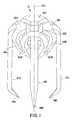
- FIGS. 1-3are isometric views of one embodiment of the staple of the present invention in formed, opened and deployed positions, respectively;
- FIG. 3Adepicts an isometric view of alternative staple of the embodiment of FIGS. 1-3;
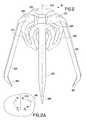
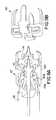
- FIGS. 4-6are isometric views of another embodiment of the staple of the present invention in formed, opened and deployed positions, respectively;
- FIG. 7depicts one embodiment of the stapler of the present invention.
- FIG. 8is an isometric view of the distal tip of the stapler of FIG. 7 adapted to hold and deploy the staple of FIGS. 1-6;
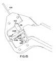
- FIGS. 12-15are isometric views of an exemplary staple deployment mechanism of the stapler of the present invention.
- FIGS. 16 and 17are isometric views of another exemplary staple deployment mechanism of the stapler of the present invention.
- FIGS. 18-26depict various views of procedural embodiments of the present invention, including FIG. 20 depicting one embodiment of the introducer of the present invention.
- a stapleis provided to close a tissue wound after a medical procedure.
- the staple of the present inventionis to close an artery or vein following a diagnostic or interventional procedure, it should be recognized at the outset that the staple may be used for general tissue repair, not just limited to vascular repair.
- the staple of the present inventioncan be formed of any biocompatible and/or bioabsorbable materials, including, for example, Titanium (and Titanium alloys), stainless steel, polymeric materials (synthetic and/or natural), ceramic, etc.
- the staple of the present inventionis preferably formed of a deformable material (such as those listed above) that undergoes plastic deformation (i.e., deformation with negligible elastic component.)
- plastic deformationi.e., deformation with negligible elastic component.
- the staple of the present inventionundergoes two positions of deformation: a first position to extend the distal ends of the prongs of the staple outwardly to grab a greater amount of tissue (and also to grab tissue away from the wound locus), and a second position to move the prongs inwardly to close the wound.
- FIGS. 1, 2 and 3depict one embodiment of staple 10 of the present invention.
- FIG. 1is the staple in it's formed position
- FIG. 2is the staple just prior to deployment into tissue with the prongs extended outwardly
- FIG. 3is the staple closed around tissue.
- the staple 10 of this embodimentcomprises a plurality of prongs 12 A- 12 D and a plurality of tabs 14 A- 14 D, arranged about a centerline axis 100 .
- Common portions, or shoulders 16 A- 16 Dare formed where the tabs meet the prongs.
- Each shoulderis common to both the prong and the tab and is generally defined by a relatively flat portion generally orthogonal to the centerline axis.
- Each proximal portion of the Uextends first generally outward from the shoulder, and second bends inwardly and distally toward centerline axis 100 , connecting together nearest the centerline axis to form the U shape.
- the U-shapedefines slots 20 A- 20 D within each tab having a base positioned at the bottom thereof.
- the staple 10is deformed so that prongs 12 A- 12 D extend outwardly from the centerline axis, prior to deployment into tissue. It is advantageous to extend the prongs outwardly as shown so as to grasp a large portion of tissue, and so that insertion of the prongs into the tissue occurs at a locus away from the wound site, thereby providing a more consistent wound closure (by closing the wound with more of the surrounding tissue) and ensuring complete (or near complete) closure of the wound.
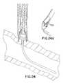
- a force F 1is applied to tabs 14 A- 14 D, as shown in relief in FIG. 2 A.
- Force F 1is generally outward (from the centerline axis) and proximal to the top of the staple, as shown in relief in FIG. 2 A.
- This forcecauses the tabs to move outward from the centerline axis 100 .
- the outward movement of the tabscauses the shoulder portions to pivot roughly about the juncture between the shoulder and the prong (i.e., at the outer portion of the shoulder), causing the inner portions of the shoulders to move inwardly toward the centerline axis and distally. Since the prongs are attached to the outer portion of the shoulders, the movement of the shoulders in this manner causes the prongs to move outwardly.
- FIGS. 4-6depict another embodiment of a staple 50 of the present invention.
- FIG. 4is the staple in it's formed position
- FIG. 5is the staple just prior to deployment into tissue with the prongs extended outwardly
- FIG. 6is the staple closed around tissue.
- the staple 50 of this embodimentcomprises a plurality of prongs 52 - 52 arranged about a centerline axis 100 .
- a shoulder 56 A- 56 Dis provided and is generally defined by a relatively flat surface, generally orthogonal to centerline axis. Shoulders 56 A- 56 D may be viewed as an extension of each prong, bent inwardly toward the centerline axis.
- webs 54 A- 54 Dare connected to and between each prong, and are formed to extend inwardly from each prong toward the centerline axis, creating a U shape generally orthogonal to the centerline axis (as opposed to the previous embodiment in which the U-shaped tab is positioned generally parallel to the centerline axis).
- the staple 50is deformed so that prongs 52 A- 52 D extend outwardly from the centerline axis, prior to deployment into tissue.
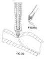
- a force F 1is applied to webs 54 A- 54 D, as shown in relief in FIG. 5 A.
- Force F 1is generally outward from the centerline axis and causes the webs to deform outwardly, i.e., straightening the bend of the web by moving the centermost point of the web outwardly. By deformation of the web portions in this manner, the prongs move outwardly.
- the cross-sectional diameter of the staplegets larger at the distal end (with respect to the cross-sectional diameter of the formed staple of FIG. 4 ). Note that the movement of the prongs is generally greater at the distal portions thereof than at the proximal portions thereof, thus producing a staple with outwardly extending prongs.
- a holding forcemay be applied downwardly (i.e., substantially parallel to the centerline axis) against the top of the webs in slots 60 A- 60 D to hold the staple in place.
- these forcesare simultaneously applied to each web of the staple to produce uniform deformation of each prong of the staple.
- the deformation of the stapleis plastic, so that the staple does not tend to return to the shape depicted in FIG. 4 . Deformation of the staple into this position will be described in greater detail below in reference to the preferred stapler device of the present invention.
- FIG. 6depicts the staple 50 in a closed or deployed position.
- the closed positionas stated herein generally means that the prongs of the staple are moved inwardly toward each other.
- a force F 3is applied to the inner surfaces 62 A- 62 D of the shoulders. This force is generally orthogonal to the centerline axis, and the angle between each force approximates the angle between the inner surfaces 62 A- 62 D about the centerline axis (which, in the staple of this embodiment is approximately 90 degrees). This force urges the shoulders outwardly. Note that shoulders can only extend outwardly as far as the web portions will permit.
- each prongis the same length, or that each prong has the same overall dimensions.
- the entire staple, or selected portions thereofcan be alternatively fashioned from an elastic or shape memory (e.g., nitinol, and/or other elastic materials, including for example temperature dependant shape memory materials) material thereby permitting elastic deformation from the a static closed position to an expanded position and then elastically close about the wound.
- the embodiment of FIGS. 4-6can be adapted with a tissue stop positioned along the length of the prong, as shown in FIG. 3 A.
- FIGS. 9, 10 A, 10 B, 11 A and 11 Bdepict the working relationship between the staple 10 ′ and/or 50 of the present invention and the mandrel 114 /sleeve 112 of the stapler mechanism 200 .
- the staple 10 ′is placed between the mandrel 114 and sleeve 112 .
- Slots 20 A- 20 D of the stapleengage fingers 116 A- 116 D of the sleeve.
- the prongs 12 A- 12 D of the stapleare dimensioned so as to fit over the mandrel, and tabs 14 A- 14 D are dimensioned so as to fit over the rod 110 , as shown.
- tabs 14 A- 14 Dare dimensioned so as to fit over the rod 110 , as shown.
- the staple 50engages the mandrel 114 and sleeve 112 (not shown). This is a static position, as no forces are applied to the staple to cause deformation.
- the staple 10 ′is urged into the first deformed position (of FIG. 2) by the relative movement of the rod/mandrel and the sleeve. As shown, the mandrel is urged proximally. As the mandrel moves, the tabs of the staple meet the narrowest part of the mandrel. Further movement forces the tabs to move outwardly, causing the prongs to likewise move outwardly (as described above with reference to FIG. 2 ).
- FIGS. 12-15depict an exemplary actuator mechanism 104 , showing the relative motion of the sleeve 112 and the mandrel rod 110 .
- the mechanismincludes a cam 408 movable in a linear motion along a slot 412 . Movement of the cam can be manual or through an electronically controllable motor (not shown).
- the cam 408has lobes 408 A and 408 C located on a first side of the cam 408 and a lobe 408 B located on a second and opposing side of the cam 408 .
- a first cam follower 418is coupled to the mandrel rod 110 , and is selectably engagable with lobes 408 A and 408 C.
- a second cam follower 416is coupled to the sleeve 112 , and is selectably engagable with lobe 408 B.
- FIG. 12depicts that neither cam follower is in contact with the lobes, and is indicative of an initial position of the mechanism.
- FIG. 13depicts the mechanism 104 in a position to expand the staple between the mandrel 114 and the sleeve 112 , as shown in FIG. 9 A.
- cam 408As cam 408 is moved (as indicated by the arrow), lobe 408 A urges cam follower 418 along slot 426 .
- the mandrel rod 110is moved proximally, causing the prongs to extend outwardly (as shown in FIGS. 2 and 5) as a result of the force of the mandrel 114 on the tabs or the web portions. With further movement of the cam 408 (FIG.
- FIGS. 16 and 17show an alternative cam mechanism. Similar to the previous example, cam 608 is urged in a direction indicated by the arrow to cause relative motion between the mandrel rod and the sleeve. Lobes 608 A and 608 B are located on opposite sides of cam 608 . As the cam 608 is moved along slot 612 , the lobe 608 A urges a cam follower 618 in a linear motion along a slot 626 . This urges the cam follower 618 proximally. The cam follower 618 is coupled to a mandrel rod 604 . This deforms staple 10 / 50 in the second configuration (see FIG. 2 or 5 ).
- cam follower 618moves distally to stay in contact with the lobe 608 A. This urges mandrel rod 604 distally.
- cam follower 616urges lobe 608 B to urge cam follower 616 distally.
- the cam follower 616is coupled to a sleeve 606 . This urges sleeve 606 distally.
- the downward slope of lobe 608 Ais parallel with upward slope of lobe 608 B so the mandrel rod 604 and the sleeve 606 move distally in unison and the staple is advanced into the tissue.
- the actuation mechanismcan include a rotating drum (not shown) to replace the cam 408 and 612 .
- the drummay be adapted with lobes formed thereon, similar to lobes 408 A- 408 C and 608 A- 608 B, respectively.
- Other alternativesmay include a rotating screw having a variable width in accordance with lobes 408 A- 408 C or 608 A- 608 B to actuate the mandrel rod and/or sleeve.
- direct linkagemay be used to actuate the mandrel rod and/or sleeve.
- FIGS. 18-25Adepict procedural embodiments of wound site management during and after a medical procedure, such as angioplasty.
- FIG. 18depicts a conventional tubular dilator 500 extending through the skin of a patient.
- the dilator 500is left in the skin following a completed medical procedure.
- the wound sitemust be stabilized.
- the blood flowmay not be completely stopped, the blood flow is reduced to a point where the coagulants in the blood can complete the wound closure.
- the doctorinserts a flexible guide wire 502 through an opening 504 in the end of the dilator 500 .
- FIG. 19shows the step of removing the introducer 500 from the wound site after the guide wire 502 is properly inserted through the skin and into the vessel.
- FIG. 20depicts the introducer 510 of the present invention, and continues the process from FIGS. 18 and 19 where the introducer 510 slides over the guide wire 502 through an opening in the introducer 510 and a portion of the introducer is placed into the vessel. Details of the introducer 510 are disclosed below.
- FIG. 20depicts the introducer 510 inserted over the guide wire 502 (already in the artery) and inserted into the vessel.
- the introducerincludes a hollow elongated guide sheath 512 and dilator 520 .
- the doctorurges the distal tip 516 of the dilator 520 into and through the guide sheath 512 (over guide wire 502 ).
- a flexible distal end 516 of the dilator 520is inserted into the wound, until a blood marker BM indicates that the dilator 520 is properly positioned in the artery.
- the blood marker BM located at a predetermined length along the dilator 520allows blood to flow through a cavity 540 to alert the doctor that the dilator 520 , and more specifically the flexible distal tip 516 , is properly inserted into a vessel.
- the distal tip 516 of the dilatorincludes a tapered portion 522 to facilitate easier ingress into the artery.
- An additional blood marking passageway(not shown) can be included on the distal end of sheath 512 as precautionary indicator of the depth of the sheath. Presence of blood in this additional passageway is indicative of the sheath being pressed too far and into the arterial wall or into the artery.
- the diameter of the distal end of the guide sheath 512can expand if outward pressure is applied from the inside surface of the guide sheath 512 . More preferably, slits or weakened tear seams (described below) are formed in the distal end of the guide sheath 512 to allow the diameter of the guide sheath to increase when pressure is applied.
- a feature of the guide sheath of the present inventionis the use of two or more wire guides to maintain the sheath centered on the wound site, to permit opposing sides of the wound to approximate, and to ensure that the closure device (e.g., stapler/staple, suturing device, cauterization, etc) remains centered about the wound so that wound closure is centered.
- the closure devicee.g., stapler/staple, suturing device, cauterization, etc
- wire guidesare formed on opposing sides of the guide sheath 512 .
- the wire guidesare delivered into the artery by the dilator 520 , as shown in FIGS. 21 and 26.
- the wire guides 514are preferably flexible, and removably coupled to the distal end 516 of the dilator 520 and deployed into the wound, as shown in FIG. 26 .
- the wire guidescan be held in openings or slots (not shown) on the sides of the dilator. Once the dilator is properly inserted into the wound to a proper depth (as indicated by the BM passageway), the dilator is removed from the wound and the guide sheath. To remove the dilator 520 from the guide sheath 512 , the doctor first holds the guide sheath 512 and advances the dilator 520 forward to release the wire guides, and then backward through the sheath to remove. This decouples the guide wires 514 A and 514 B from the openings. After the guide rod has been inserted a predetermined distance, the doctor simply extracts the guide rod. This leaves the guide sheath 512 centered on the wound with the guide wires 514 A and 514 B extending inside the wound.
- a puncture in an artery or veinhas a general tendency to manifest a circumferential slit or an elongated opening, since the cell structure forming this tissue forms circumferentially (rather than longitudinal) to support radial contraction of the vessel.
- the wire guides 514 A and 514 B of the presentenable the wound to approximate the natural state of the wound, i.e., elongated circumferentially.
- the sheathhas a diameter approximately equal to the diameter of the opening or wound, and the wire guides 514 A and 514 B on the sides of the sheath approximate the dimension of the long axis of the wound in its natural, elongated state, as best shown in FIG. 23 .
- Approximation in this sensemay mean that the wire guides are less than or greater than (or equal to) this diameter.
- the wire guides in this positionlimit movement of the sheath along the long axis of the vessel, and since the wire guides span the elongated wound, movement along the short axis is likewise limited. This ensures that any device inserted through the sheath is approximately centered on the wound.
- the wound openingtends to assume the shape shown in FIG. 23 even in the absence of the wire guides, the opposing tissue located along the short axis tends to approximate.
- the present inventiontakes advantage of this tendency for positioning and alignment of a sheath.
- the tissue along the short axistends to approximate more (i.e., the tissue is stretched along the long axis).
- sufficient wound site managementdoes not require that the wire guides stretch the wound. Rather, if the position of the wire guides are shorter than the wound length, the wire guides still serve to maintain the sheath generally centered at the wound. In both circumstances, the wire guides ensure that a staple deployment is centered, and that a significant amount of tissue is grasped by the staple for closure. Also, if the wound opening in the tissue is held taught by the introducer, there is less tendency for the tissue surrounding the opening to slip down into the vessel during staple deployment (which would reduce the effectiveness of the closure).
- the guide wires 514are preferably disposed on opposing sides of the guide sheath 512 , and more preferable, the guide wires are inserted into the wound opening transversally to the long axis of the artery or vein, so that the wound is pulled taught in a transverse direction.
- FIG. 22shows the distal end of a stapler 104 with a staple 10 / 50 being inserted through the guide sheath 512 of the introducer 510 .
- FIG. 22Adepicts a relief view of the introducer 510 , and more clearly depicts the slits or weakened tear seams 700 .
- the staplecan be deployed into the tissue.
- FIG. 24shows the first step of staple deployment, the process of which is described in detail above. Note that in FIG. 24A, the extension of the staple prongs causes the weakened tear seam or slits to separate.
- FIGS. 25 and 25Adepict the staple fully deployed into tissue, the process of which is described above.
- the staplercan now be removed from the guide sheath 512 .
- the guide sheath 512can now be urged away from the wound opening WO and the guide wires 514 A and 514 B are extracted from the closed opening.
Landscapes
- Health & Medical Sciences (AREA)
- Life Sciences & Earth Sciences (AREA)
- Surgery (AREA)
- Heart & Thoracic Surgery (AREA)
- Engineering & Computer Science (AREA)
- Biomedical Technology (AREA)
- Nuclear Medicine, Radiotherapy & Molecular Imaging (AREA)
- Medical Informatics (AREA)
- Molecular Biology (AREA)
- Animal Behavior & Ethology (AREA)
- General Health & Medical Sciences (AREA)
- Public Health (AREA)
- Veterinary Medicine (AREA)
- Cardiology (AREA)
- Surgical Instruments (AREA)
Abstract
Description
Claims (18)
Priority Applications (1)
| Application Number | Priority Date | Filing Date | Title |
|---|---|---|---|
| US09/658,790US6348064B1 (en) | 2000-09-01 | 2000-09-11 | Wound site management and wound closure device |
Applications Claiming Priority (2)
| Application Number | Priority Date | Filing Date | Title |
|---|---|---|---|
| US23023400P | 2000-09-01 | 2000-09-01 | |
| US09/658,790US6348064B1 (en) | 2000-09-01 | 2000-09-11 | Wound site management and wound closure device |
Publications (1)
| Publication Number | Publication Date |
|---|---|
| US6348064B1true US6348064B1 (en) | 2002-02-19 |
Family
ID=22864437
Family Applications (3)
| Application Number | Title | Priority Date | Filing Date |
|---|---|---|---|
| US09/658,786Expired - Fee RelatedUS6322580B1 (en) | 2000-09-01 | 2000-09-11 | Wound site management and wound closure device |
| US09/658,787Expired - Fee RelatedUS6506210B1 (en) | 2000-09-01 | 2000-09-11 | Wound site management and wound closure device |
| US09/658,790Expired - Fee RelatedUS6348064B1 (en) | 2000-09-01 | 2000-09-11 | Wound site management and wound closure device |
Family Applications Before (2)
| Application Number | Title | Priority Date | Filing Date |
|---|---|---|---|
| US09/658,786Expired - Fee RelatedUS6322580B1 (en) | 2000-09-01 | 2000-09-11 | Wound site management and wound closure device |
| US09/658,787Expired - Fee RelatedUS6506210B1 (en) | 2000-09-01 | 2000-09-11 | Wound site management and wound closure device |
Country Status (6)
| Country | Link |
|---|---|
| US (3) | US6322580B1 (en) |
| EP (1) | EP1341446A4 (en) |
| JP (1) | JP2004511275A (en) |
| AU (1) | AU2000274774A1 (en) |
| CA (1) | CA2420227A1 (en) |
| WO (1) | WO2002019924A1 (en) |
Cited By (150)
| Publication number | Priority date | Publication date | Assignee | Title |
|---|---|---|---|---|
| US20020072768A1 (en)* | 2000-12-07 | 2002-06-13 | Ginn Richard S. | Apparatus and methods for providing tactile feedback while delivering a closure device |
| US20020133193A1 (en)* | 2000-01-05 | 2002-09-19 | Ginn Richard S. | Integrated vascular device with puncture site closure component and sealant and methods of use |
| US20020133183A1 (en)* | 2000-09-29 | 2002-09-19 | Lentz David Christian | Coated medical devices |
| US20020193808A1 (en)* | 2000-01-05 | 2002-12-19 | Belef W. Martin | Apparatus and methods for delivering a closure device |
| US20030078598A1 (en)* | 2000-01-05 | 2003-04-24 | Integrated Vascular Systems, Inc. | Vascular sheath with bioabsorbable puncture site closure apparatus and methods of use |
| WO2003020106A3 (en)* | 2001-08-28 | 2003-06-05 | Ethicon Inc | Composite staple for completing an anastomosis |
| US20030199924A1 (en)* | 2000-09-08 | 2003-10-23 | James Coleman | Surgical stapler |
| US6645205B2 (en) | 2001-08-15 | 2003-11-11 | Core Medical, Inc. | Apparatus and methods for reducing lung volume |
| WO2003071957A3 (en)* | 2002-02-21 | 2003-12-04 | Integrated Vascular Sys Inc | Sheath and plunger apparatus and methods for delivering a closure device |
| US20040009289A1 (en)* | 2000-12-07 | 2004-01-15 | Carley Michael T. | Closure device and methods for making and using them |
| US20040028502A1 (en)* | 2001-06-07 | 2004-02-12 | Christy Cummins | Surgical staple |
| US20040116949A1 (en)* | 2002-12-11 | 2004-06-17 | Ewers Richard C. | Apparatus and methods for forming gastrointestinal tissue approximations |
| US20040127940A1 (en)* | 2000-12-14 | 2004-07-01 | Ginn Richard S. | Apparatus and methods for sealing vascular punctures |
| US20040147958A1 (en)* | 2002-12-11 | 2004-07-29 | Usgi Medical | Apparatus and methods for forming and securing gastrointestinal tissue folds |
| US20040153122A1 (en)* | 2003-01-30 | 2004-08-05 | Integrated Vascular Systems, Inc. | Clip applier and methods of use |
| US20040225305A1 (en)* | 1999-06-25 | 2004-11-11 | Usgi Medical | Apparatus and methods for forming and securing gastrointestinal tissue folds |
| US20040236355A1 (en)* | 2001-08-09 | 2004-11-25 | Thomas Anthony | Surgical stapling device |
| US20040249391A1 (en)* | 2001-08-09 | 2004-12-09 | Christy Cummins | Surgical stapling device and method |
| US20050065397A1 (en)* | 2003-01-15 | 2005-03-24 | Usgi Medical Inc. | Endoluminal tool deployment system |
| US20050085854A1 (en)* | 2003-10-17 | 2005-04-21 | Ensure Medical, Inc. | Locator and closure device and method of use |
| US20050119695A1 (en)* | 2000-12-07 | 2005-06-02 | Carley Michael T. | Closure device and methods for making and using them |
| WO2004112652A3 (en)* | 2003-06-20 | 2005-07-21 | Medtronic Vascular Inc | Device, system, and method for contracting tissue in a mammalian body |
| US20050177189A1 (en)* | 2000-12-14 | 2005-08-11 | Ginn Richard S. | Plug with detachable guidewire element and methods for use |
| US20050203488A1 (en)* | 2004-03-09 | 2005-09-15 | Usgi Medical Inc. | Apparatus and methods for mapping out endoluminal gastrointestinal surgery |
| US20050203500A1 (en)* | 2004-03-09 | 2005-09-15 | Usgi Medical Inc. | Apparatus and methods for mapping out endoluminal gastrointestinal surgery |
| US20050228413A1 (en)* | 2004-04-12 | 2005-10-13 | Binmoeller Kenneth F | Automated transluminal tissue targeting and anchoring devices and methods |
| US20050251206A1 (en)* | 2004-05-07 | 2005-11-10 | Usgi Medical Corporation | Apparatus and methods for positioning and securing anchors |
| US20050251161A1 (en)* | 2004-05-07 | 2005-11-10 | Usgi Medical Inc. | Needle assembly for tissue manipulation |
| US20050251209A1 (en)* | 2004-05-07 | 2005-11-10 | Usgi Medical Inc. | Apparatus and methods for positioning and securing anchors |
| US20050251165A1 (en)* | 2004-05-07 | 2005-11-10 | Usgi Medical Inc. | Tissue manipulation and securement system |
| US20050250985A1 (en)* | 2004-05-07 | 2005-11-10 | Usgi Medical Inc. | Self-locking removable apparatus and methods for manipulating and securing tissue |
| US20050251162A1 (en)* | 2004-05-07 | 2005-11-10 | Usgi Medical Inc. | Apparatus and methods for manipulating and securing tissue |
| US20050251176A1 (en)* | 2004-05-07 | 2005-11-10 | Usgi Medical Inc. | System for treating gastroesophageal reflux disease |
| US20050251208A1 (en)* | 2004-05-07 | 2005-11-10 | Usgi Medical Inc. | Linear anchors for anchoring to tissue |
| US20050251210A1 (en)* | 2004-05-07 | 2005-11-10 | Usgi Medical Inc. | Methods and apparatus for grasping and cinching tissue anchors |
| US20050256537A1 (en)* | 2002-07-03 | 2005-11-17 | Christy Cummins | Surgical stapling device |
| US20050267528A1 (en)* | 2000-12-14 | 2005-12-01 | Ensure Medical, Inc. | Vascular plug having composite construction |
| US20050277966A1 (en)* | 2004-06-09 | 2005-12-15 | Usgi Medical Inc. | Compressible tissue anchor assemblies |
| US20050277981A1 (en)* | 2004-06-09 | 2005-12-15 | Usgi Medical Inc. | Apparatus and methods for optimizing anchoring force |
| US20050277983A1 (en)* | 2004-06-09 | 2005-12-15 | Usgi Medical Inc. | System for optimizing anchoring force |
| US20060020276A1 (en)* | 2004-07-23 | 2006-01-26 | Usgi Medical Inc. | Apparatus and methods for achieving prolonged maintenance of gastrointestinal tissue folds |
| US20060030869A1 (en)* | 2002-11-14 | 2006-02-09 | By-Pass, Inc. | Adhesive anastomosis connection system |
| US20060111741A1 (en)* | 2004-11-23 | 2006-05-25 | Instrasurgical, Llc | Wound closure device |
| US20060135971A1 (en)* | 2004-05-07 | 2006-06-22 | Usgi Medical Inc. | System for treating gastroesophageal reflux disease |
| US20060144479A1 (en)* | 2002-12-31 | 2006-07-06 | Integrated Vascular Systems, Inc. | Methods for manufacturing a clip and clip |
| US20060155309A1 (en)* | 2002-09-25 | 2006-07-13 | By-Pass, Inc | Sliding surgical clip |
| US20060190037A1 (en)* | 2000-01-05 | 2006-08-24 | Ginn Richard S | Integrated vascular device with puncture site closure component and sealant and methods of use |
| US20060217744A1 (en)* | 2005-03-28 | 2006-09-28 | Cardica, Inc. | Vascular closure system |
| US20060217762A1 (en)* | 2004-06-09 | 2006-09-28 | Usgi Medical, Inc. | Compressible tissue anchor assemblies |
| US20060253037A1 (en)* | 2005-05-04 | 2006-11-09 | Ensure Medical, Inc. | Locator and closure device and method of use |
| US20060271073A1 (en)* | 2005-05-26 | 2006-11-30 | Usgi Medical Inc. | Methods and apparatus for securing and deploying tissue anchors |
| US20060271074A1 (en)* | 2005-05-26 | 2006-11-30 | Ewers Richard C | Methods and apparatus for securing and deploying tissue anchors |
| US20060282087A1 (en)* | 2005-06-09 | 2006-12-14 | Binmoeller Kenneth F | Methods and devices for endosonography-guided fundopexy |
| US20070135825A1 (en)* | 2005-06-09 | 2007-06-14 | Binmoeller Kenneth F | Methods and devices for anchoring to tissue |
| US20070175488A1 (en)* | 2006-01-27 | 2007-08-02 | Usgi Medical Inc. | Methods and apparatus for revision of obesity procedures |
| US20070250160A1 (en)* | 2006-04-21 | 2007-10-25 | Medtronic Vascular, Inc. | Device, system, and method for treating cardiac valve regurgitation |
| US20070265631A1 (en)* | 2003-02-03 | 2007-11-15 | Biomedical Enterprises, Inc. | System and method for force, displacement, and rate control of shaped memory material implants |
| US20080065152A1 (en)* | 2006-09-08 | 2008-03-13 | Abbott Laboratories | Apparatus and method for delivering a closure element |
| US20080071294A1 (en)* | 2006-09-15 | 2008-03-20 | Bender Theodore M | Apparatus and method for closure of patent foramen ovale |
| US7361180B2 (en) | 2004-05-07 | 2008-04-22 | Usgi Medical, Inc. | Apparatus for manipulating and securing tissue |
| US20080147114A1 (en)* | 1998-05-29 | 2008-06-19 | Bypass, Inc. | Vascular port device |
| US20080177300A1 (en)* | 2007-01-24 | 2008-07-24 | Medtronic Vascular, Inc. | Low-Profile Vasculare Closure Systems and Methods of Using Same |
| US20080217376A1 (en)* | 2007-03-08 | 2008-09-11 | Cardica, Inc. | Surgical Stapler |
| US20080243151A1 (en)* | 2004-04-12 | 2008-10-02 | Binmoeller Kenneth F | Luminal Structure Anchoring Devices and Methods |
| US20080287989A1 (en)* | 2007-05-17 | 2008-11-20 | Arch Day Design, Llc | Tissue holding implants |
| US7458978B1 (en) | 2005-03-28 | 2008-12-02 | Cardica, Inc. | Vascular closure system utilizing a staple |
| US20080312686A1 (en)* | 2005-07-01 | 2008-12-18 | Abbott Laboratories | Antimicrobial closure element and closure element applier |
| US20080319475A1 (en)* | 2007-06-25 | 2008-12-25 | Abbott Laboratories | Methods, Devices, and Apparatus for Managing Access Through Tissue |
| US20090030380A1 (en)* | 2004-12-08 | 2009-01-29 | Xlumena, Inc. | Method and Apparatus for Performing Needle Guided Interventions |
| US20090093826A1 (en)* | 2007-10-05 | 2009-04-09 | Cardica, Inc. | Patent Foramen Ovale Closure System |
| US7533790B1 (en) | 2007-03-08 | 2009-05-19 | Cardica, Inc. | Surgical stapler |
| US20090157101A1 (en)* | 2007-12-17 | 2009-06-18 | Abbott Laboratories | Tissue closure system and methods of use |
| US20090157103A1 (en)* | 2007-12-18 | 2009-06-18 | Abbott Laboratories | Modular clip applier |
| US20090254121A1 (en)* | 2008-04-02 | 2009-10-08 | Cardica, Inc. | Vascular Closure with Multi-Pronged Clip |
| US20090259249A1 (en)* | 2008-04-10 | 2009-10-15 | Medtronic Vascular, Inc. | Arteriotomy stapling system for non-orthogonal tissue tracks and methods of use therein |
| US20090264923A1 (en)* | 2008-04-17 | 2009-10-22 | Medtronic Vascular, Inc | Vascular Puncture Stapling System |
| US20090281379A1 (en)* | 2008-05-12 | 2009-11-12 | Xlumena, Inc. | System and method for transluminal access |
| US20090281557A1 (en)* | 2008-05-12 | 2009-11-12 | Xlumena, Inc. | Tissue anchor for securing tissue layers |
| US20100038402A1 (en)* | 2008-08-18 | 2010-02-18 | Olympus Corporation | Hollow Tissue Inosculation Apparatus |
| US20100042144A1 (en)* | 2008-08-12 | 2010-02-18 | Steven Bennett | Medical Device for Wound Closure and Method of Use |
| USD611144S1 (en) | 2006-06-28 | 2010-03-02 | Abbott Laboratories | Apparatus for delivering a closure element |
| US20100076467A1 (en)* | 2008-09-19 | 2010-03-25 | Olympus Corporation | Staple to inosculate hollow tissues |
| US20100087854A1 (en)* | 2008-08-12 | 2010-04-08 | Joshua Stopek | Medical device for wound closure and method of use |
| US20100114124A1 (en)* | 2005-08-03 | 2010-05-06 | Brian Kelleher | Method and apparatus for partioning an organ within the body |
| US20100114159A1 (en)* | 2008-10-30 | 2010-05-06 | Abbott Vascular Inc. | Closure device |
| US20100160958A1 (en)* | 2008-12-22 | 2010-06-24 | Abbott Laboratories | Closure Device |
| US20100179589A1 (en)* | 2009-01-09 | 2010-07-15 | Abbott Vascular Inc. | Rapidly eroding anchor |
| US7806904B2 (en) | 2000-12-07 | 2010-10-05 | Integrated Vascular Systems, Inc. | Closure device |
| US7806910B2 (en) | 2002-11-26 | 2010-10-05 | Abbott Laboratories | Multi-element biased suture clip |
| US20100268175A1 (en)* | 2009-04-21 | 2010-10-21 | Xlumena, Inc. | System and method for delivering expanding trocar through a sheath |
| US20100268029A1 (en)* | 2009-04-21 | 2010-10-21 | Xlumena, Inc. | Methods and apparatus for advancing a device from one body lumen to another |
| US7850709B2 (en) | 2002-06-04 | 2010-12-14 | Abbott Vascular Inc. | Blood vessel closure clip and delivery device |
| US20110054492A1 (en)* | 2009-08-26 | 2011-03-03 | Abbott Laboratories | Medical device for repairing a fistula |
| US7918869B2 (en) | 2004-05-07 | 2011-04-05 | Usgi Medical, Inc. | Methods and apparatus for performing endoluminal gastroplasty |
| US20110112622A1 (en)* | 2009-05-29 | 2011-05-12 | Xlumena, Inc. | Apparatus and method for deploying stent across adjacent tissue layers |
| US7942898B2 (en) | 2002-12-11 | 2011-05-17 | Usgi Medical, Inc. | Delivery systems and methods for gastric reduction |
| US7942884B2 (en) | 2002-12-11 | 2011-05-17 | Usgi Medical, Inc. | Methods for reduction of a gastric lumen |
| US20110137394A1 (en)* | 2009-05-29 | 2011-06-09 | Xlumena, Inc. | Methods and systems for penetrating adjacent tissue layers |
| US20110144664A1 (en)* | 2003-01-30 | 2011-06-16 | Integrated Vascular Systems, Inc. | Clip applier and methods of use |
| US20110172753A1 (en)* | 2003-07-18 | 2011-07-14 | University of Connecticut, a Connecticut public institution of higher education | Endoprostheses |
| US20110224719A1 (en)* | 2010-03-15 | 2011-09-15 | Abbott Cardiovascular Systems, Inc. | Bioabsorbable plug |
| US20110230897A1 (en)* | 2003-01-30 | 2011-09-22 | Integrated Vascular Systems, Inc. | Clip applier and methods of use |
| US8048108B2 (en) | 2005-08-24 | 2011-11-01 | Abbott Vascular Inc. | Vascular closure methods and apparatuses |
| US8056790B2 (en) | 2008-09-19 | 2011-11-15 | Olympus Corporation | Hollow tissue inosculation apparatus |
| US8057510B2 (en) | 2000-12-14 | 2011-11-15 | Ensure Medical, Inc. | Plug with collet and apparatus and method for delivering such plugs |
| US8075587B2 (en) | 2000-12-14 | 2011-12-13 | Ensure Medical, Inc. | Apparatus and methods for sealing vascular punctures |
| US8074860B2 (en) | 2008-09-19 | 2011-12-13 | Olympus Corporation | Hollow tissue inosculation apparatus |
| US8202293B2 (en) | 2003-01-30 | 2012-06-19 | Integrated Vascular Systems, Inc. | Clip applier and methods of use |
| US8216252B2 (en) | 2004-05-07 | 2012-07-10 | Usgi Medical, Inc. | Tissue manipulation and securement system |
| US8313497B2 (en) | 2005-07-01 | 2012-11-20 | Abbott Laboratories | Clip applier and methods of use |
| US8444657B2 (en) | 2004-05-07 | 2013-05-21 | Usgi Medical, Inc. | Apparatus and methods for rapid deployment of tissue anchors |
| US8556932B2 (en) | 2011-05-19 | 2013-10-15 | Abbott Cardiovascular Systems, Inc. | Collapsible plug for tissue closure |
| US8556930B2 (en) | 2006-06-28 | 2013-10-15 | Abbott Laboratories | Vessel closure device |
| US8579934B2 (en) | 2003-10-17 | 2013-11-12 | Ensure Medical, Inc. | Locator and delivery device and method of use |
| US8590760B2 (en) | 2004-05-25 | 2013-11-26 | Abbott Vascular Inc. | Surgical stapler |
| US8603116B2 (en) | 2010-08-04 | 2013-12-10 | Abbott Cardiovascular Systems, Inc. | Closure device with long tines |
| US8617184B2 (en) | 2011-02-15 | 2013-12-31 | Abbott Cardiovascular Systems, Inc. | Vessel closure system |
| US20140017025A1 (en)* | 2011-03-17 | 2014-01-16 | A. Raymond Et Cie | Smart material actuated fasteners |
| US8690910B2 (en) | 2000-12-07 | 2014-04-08 | Integrated Vascular Systems, Inc. | Closure device and methods for making and using them |
| US8758399B2 (en) | 2010-08-02 | 2014-06-24 | Abbott Cardiovascular Systems, Inc. | Expandable bioabsorbable plug apparatus and method |
| US8758400B2 (en) | 2000-01-05 | 2014-06-24 | Integrated Vascular Systems, Inc. | Closure system and methods of use |
| US8808310B2 (en) | 2006-04-20 | 2014-08-19 | Integrated Vascular Systems, Inc. | Resettable clip applier and reset tools |
| US8821534B2 (en) | 2010-12-06 | 2014-09-02 | Integrated Vascular Systems, Inc. | Clip applier having improved hemostasis and methods of use |
| US8858594B2 (en) | 2008-12-22 | 2014-10-14 | Abbott Laboratories | Curved closure device |
| US8870916B2 (en) | 2006-07-07 | 2014-10-28 | USGI Medical, Inc | Low profile tissue anchors, tissue anchor systems, and methods for their delivery and use |
| US8893947B2 (en) | 2007-12-17 | 2014-11-25 | Abbott Laboratories | Clip applier and methods of use |
| US8905937B2 (en) | 2009-02-26 | 2014-12-09 | Integrated Vascular Systems, Inc. | Methods and apparatus for locating a surface of a body lumen |
| US8920442B2 (en) | 2005-08-24 | 2014-12-30 | Abbott Vascular Inc. | Vascular opening edge eversion methods and apparatuses |
| US8926654B2 (en) | 2005-05-04 | 2015-01-06 | Cordis Corporation | Locator and closure device and method of use |
| US8926633B2 (en)* | 2005-06-24 | 2015-01-06 | Abbott Laboratories | Apparatus and method for delivering a closure element |
| US9089311B2 (en) | 2009-01-09 | 2015-07-28 | Abbott Vascular Inc. | Vessel closure devices and methods |
| US9089674B2 (en) | 2000-10-06 | 2015-07-28 | Integrated Vascular Systems, Inc. | Apparatus and methods for positioning a vascular sheath |
| US9149276B2 (en) | 2011-03-21 | 2015-10-06 | Abbott Cardiovascular Systems, Inc. | Clip and deployment apparatus for tissue closure |
| US9173644B2 (en) | 2009-01-09 | 2015-11-03 | Abbott Vascular Inc. | Closure devices, systems, and methods |
| US9265514B2 (en) | 2012-04-17 | 2016-02-23 | Miteas Ltd. | Manipulator for grasping tissue |
| US9282965B2 (en) | 2008-05-16 | 2016-03-15 | Abbott Laboratories | Apparatus and methods for engaging tissue |
| US9332976B2 (en) | 2011-11-30 | 2016-05-10 | Abbott Cardiovascular Systems, Inc. | Tissue closure device |
| US9364209B2 (en) | 2012-12-21 | 2016-06-14 | Abbott Cardiovascular Systems, Inc. | Articulating suturing device |
| US9381041B2 (en) | 2009-04-21 | 2016-07-05 | Xlumena, Inc. | Methods and devices for access across adjacent tissue layers |
| US9414824B2 (en) | 2009-01-16 | 2016-08-16 | Abbott Vascular Inc. | Closure devices, systems, and methods |
| US9414820B2 (en) | 2009-01-09 | 2016-08-16 | Abbott Vascular Inc. | Closure devices, systems, and methods |
| US9456811B2 (en) | 2005-08-24 | 2016-10-04 | Abbott Vascular Inc. | Vascular closure methods and apparatuses |
| US9486191B2 (en) | 2009-01-09 | 2016-11-08 | Abbott Vascular, Inc. | Closure devices |
| US9579091B2 (en) | 2000-01-05 | 2017-02-28 | Integrated Vascular Systems, Inc. | Closure system and methods of use |
| US9649108B2 (en) | 2015-02-24 | 2017-05-16 | Orthovestments, Llc | Orthopedic bone staple with polyaxial compression capability |
| US9918706B2 (en)* | 2010-06-07 | 2018-03-20 | Kardium Inc. | Closing openings in anatomical tissue |
| US10064623B2 (en)* | 2010-12-07 | 2018-09-04 | Globetek 2000 Pty Ltd | Surgical clip and clip manipulation device therefor |
| USD851250S1 (en) | 2015-11-19 | 2019-06-11 | Orthovestments, Llc | Bone staple |
| US10952732B2 (en) | 2013-02-21 | 2021-03-23 | Boston Scientific Scimed Inc. | Devices and methods for forming an anastomosis |
| US12303105B2 (en) | 2004-04-12 | 2025-05-20 | Boston Scientific Scimed, Inc. | Luminal structure anchoring devices and methods |
Families Citing this family (175)
| Publication number | Priority date | Publication date | Assignee | Title |
|---|---|---|---|---|
| NL1007349C2 (en)* | 1997-10-24 | 1999-04-27 | Suyker Wilhelmus Joseph Leonardus | System for the mechanical production of anastomoses between hollow structures; as well as device and applicator for use therewith. |
| US7435249B2 (en) | 1997-11-12 | 2008-10-14 | Covidien Ag | Electrosurgical instruments which reduces collateral damage to adjacent tissue |
| US6726686B2 (en) | 1997-11-12 | 2004-04-27 | Sherwood Services Ag | Bipolar electrosurgical instrument for sealing vessels |
| US6228083B1 (en) | 1997-11-14 | 2001-05-08 | Sherwood Services Ag | Laparoscopic bipolar electrosurgical instrument |
| US7267677B2 (en) | 1998-10-23 | 2007-09-11 | Sherwood Services Ag | Vessel sealing instrument |
| US7118570B2 (en) | 2001-04-06 | 2006-10-10 | Sherwood Services Ag | Vessel sealing forceps with disposable electrodes |
| US7364577B2 (en) | 2002-02-11 | 2008-04-29 | Sherwood Services Ag | Vessel sealing system |
| US7582087B2 (en) | 1998-10-23 | 2009-09-01 | Covidien Ag | Vessel sealing instrument |
| US6964668B2 (en) | 1999-03-04 | 2005-11-15 | Abbott Laboratories | Articulating suturing device and method |
| US8137364B2 (en) | 2003-09-11 | 2012-03-20 | Abbott Laboratories | Articulating suturing device and method |
| US20030109875A1 (en) | 1999-10-22 | 2003-06-12 | Tetzlaff Philip M. | Open vessel sealing forceps with disposable electrodes |
| US6682540B1 (en)* | 1999-11-05 | 2004-01-27 | Onux Medical, Inc. | Apparatus and method for placing multiple sutures |
| US6533762B2 (en)* | 2000-09-01 | 2003-03-18 | Angiolink Corporation | Advanced wound site management systems and methods |
| US8551134B2 (en)* | 2000-09-01 | 2013-10-08 | Medtronic Vascular, Inc. | Wound site management and wound closure device |
| US6966917B1 (en)* | 2000-11-09 | 2005-11-22 | Innovation Interventional Technologies B.V. | Deformable connector for mechanically connecting hollow structures |
| ES2262639T3 (en) | 2001-04-06 | 2006-12-01 | Sherwood Services Ag | SHUTTER AND DIVIDER OF GLASSES WITH BUMPER MEMBERS N OCONDUCTIVES. |
| EP1527747B1 (en) | 2001-04-06 | 2015-09-30 | Covidien AG | Electrosurgical instrument which reduces collateral damage to adjacent tissue |
| US7094245B2 (en)* | 2001-10-05 | 2006-08-22 | Scimed Life Systems, Inc. | Device and method for through the scope endoscopic hemostatic clipping |
| US7931649B2 (en) | 2002-10-04 | 2011-04-26 | Tyco Healthcare Group Lp | Vessel sealing instrument with electrical cutting mechanism |
| US7276068B2 (en) | 2002-10-04 | 2007-10-02 | Sherwood Services Ag | Vessel sealing instrument with electrical cutting mechanism |
| US7270664B2 (en) | 2002-10-04 | 2007-09-18 | Sherwood Services Ag | Vessel sealing instrument with electrical cutting mechanism |
| US7799026B2 (en) | 2002-11-14 | 2010-09-21 | Covidien Ag | Compressible jaw configuration with bipolar RF output electrodes for soft tissue fusion |
| US7343920B2 (en)* | 2002-12-20 | 2008-03-18 | Toby E Bruce | Connective tissue repair system |
| EP1601298B1 (en) | 2003-03-13 | 2016-09-07 | Covidien AG | Bipolar concentric electrode assembly for soft tissue fusion |
| US8100864B2 (en)* | 2003-04-30 | 2012-01-24 | Kunkel Sanford S | Portal device |
| CA2523675C (en) | 2003-05-01 | 2016-04-26 | Sherwood Services Ag | Electrosurgical instrument which reduces thermal damage to adjacent tissue |
| US7160299B2 (en) | 2003-05-01 | 2007-01-09 | Sherwood Services Ag | Method of fusing biomaterials with radiofrequency energy |
| JP5137230B2 (en) | 2003-05-15 | 2013-02-06 | コヴィディエン・アクチェンゲゼルシャフト | Tissue sealer with non-conductive variable stop member and method for sealing tissue |
| US7150749B2 (en) | 2003-06-13 | 2006-12-19 | Sherwood Services Ag | Vessel sealer and divider having elongated knife stroke and safety cutting mechanism |
| US7857812B2 (en) | 2003-06-13 | 2010-12-28 | Covidien Ag | Vessel sealer and divider having elongated knife stroke and safety for cutting mechanism |
| USD956973S1 (en) | 2003-06-13 | 2022-07-05 | Covidien Ag | Movable handle for endoscopic vessel sealer and divider |
| US7156846B2 (en) | 2003-06-13 | 2007-01-02 | Sherwood Services Ag | Vessel sealer and divider for use with small trocars and cannulas |
| US7462188B2 (en) | 2003-09-26 | 2008-12-09 | Abbott Laboratories | Device and method for suturing intracardiac defects |
| ATE510572T1 (en) | 2003-09-28 | 2011-06-15 | Guido Schnyder | BIODEGRADABLE AND/OR BIOABSORBABLE MEMBER FOR VESSEL CLOSURE |
| EP1680028B1 (en) | 2003-10-17 | 2012-01-25 | Tyco Healthcare Group LP | Surgical stapling device |
| US9848938B2 (en) | 2003-11-13 | 2017-12-26 | Covidien Ag | Compressible jaw configuration with bipolar RF output electrodes for soft tissue fusion |
| US7367976B2 (en) | 2003-11-17 | 2008-05-06 | Sherwood Services Ag | Bipolar forceps having monopolar extension |
| US7500975B2 (en) | 2003-11-19 | 2009-03-10 | Covidien Ag | Spring loaded reciprocating tissue cutting mechanism in a forceps-style electrosurgical instrument |
| US7811283B2 (en) | 2003-11-19 | 2010-10-12 | Covidien Ag | Open vessel sealing instrument with hourglass cutting mechanism and over-ratchet safety |
| US7131970B2 (en) | 2003-11-19 | 2006-11-07 | Sherwood Services Ag | Open vessel sealing instrument with cutting mechanism |
| US7442193B2 (en) | 2003-11-20 | 2008-10-28 | Covidien Ag | Electrically conductive/insulative over-shoe for tissue fusion |
| US7780662B2 (en) | 2004-03-02 | 2010-08-24 | Covidien Ag | Vessel sealing system using capacitive RF dielectric heating |
| US20050267520A1 (en) | 2004-05-12 | 2005-12-01 | Modesitt D B | Access and closure device and method |
| US7678133B2 (en) | 2004-07-10 | 2010-03-16 | Arstasis, Inc. | Biological tissue closure device and method |
| US7515970B2 (en)* | 2004-08-18 | 2009-04-07 | Cardiac Pacemakers, Inc. | Transeptal lead |
| US7195631B2 (en) | 2004-09-09 | 2007-03-27 | Sherwood Services Ag | Forceps with spring loaded end effector assembly |
| US7540872B2 (en) | 2004-09-21 | 2009-06-02 | Covidien Ag | Articulating bipolar electrosurgical instrument |
| US20070055305A1 (en)* | 2004-09-23 | 2007-03-08 | Guido Schnyder | Biodegradable and/or bioabsorbable member for vascular sealing |
| US7955332B2 (en) | 2004-10-08 | 2011-06-07 | Covidien Ag | Mechanism for dividing tissue in a hemostat-style instrument |
| US7909823B2 (en) | 2005-01-14 | 2011-03-22 | Covidien Ag | Open vessel sealing instrument |
| US7686804B2 (en) | 2005-01-14 | 2010-03-30 | Covidien Ag | Vessel sealer and divider with rotating sealer and cutter |
| US7491202B2 (en) | 2005-03-31 | 2009-02-17 | Covidien Ag | Electrosurgical forceps with slow closure sealing plates and method of sealing tissue |
| CN103190942A (en)* | 2005-05-12 | 2013-07-10 | 阿尔斯塔西斯公司 | Access and closure device and method |
| US8083754B2 (en) | 2005-08-08 | 2011-12-27 | Abbott Laboratories | Vascular suturing device with needle capture |
| US8758397B2 (en)* | 2005-08-24 | 2014-06-24 | Abbott Vascular Inc. | Vascular closure methods and apparatuses |
| US7722607B2 (en) | 2005-09-30 | 2010-05-25 | Covidien Ag | In-line vessel sealer and divider |
| US7922953B2 (en) | 2005-09-30 | 2011-04-12 | Covidien Ag | Method for manufacturing an end effector assembly |
| US7789878B2 (en) | 2005-09-30 | 2010-09-07 | Covidien Ag | In-line vessel sealer and divider |
| CA2561034C (en) | 2005-09-30 | 2014-12-09 | Sherwood Services Ag | Flexible endoscopic catheter with an end effector for coagulating and transfecting tissue |
| ES2381560T3 (en) | 2005-09-30 | 2012-05-29 | Covidien Ag | Insulating sleeve for electrosurgical forceps |
| US7879035B2 (en) | 2005-09-30 | 2011-02-01 | Covidien Ag | Insulating boot for electrosurgical forceps |
| US8298232B2 (en) | 2006-01-24 | 2012-10-30 | Tyco Healthcare Group Lp | Endoscopic vessel sealer and divider for large tissue structures |
| US8241282B2 (en) | 2006-01-24 | 2012-08-14 | Tyco Healthcare Group Lp | Vessel sealing cutting assemblies |
| US8734443B2 (en) | 2006-01-24 | 2014-05-27 | Covidien Lp | Vessel sealer and divider for large tissue structures |
| US8882766B2 (en) | 2006-01-24 | 2014-11-11 | Covidien Ag | Method and system for controlling delivery of energy to divide tissue |
| US7749249B2 (en) | 2006-02-21 | 2010-07-06 | Kardium Inc. | Method and device for closing holes in tissue |
| US8021389B2 (en)* | 2006-05-17 | 2011-09-20 | Warsaw Orthopedic, Inc. | Surgical staple assembly |
| US20070270688A1 (en) | 2006-05-19 | 2007-11-22 | Daniel Gelbart | Automatic atherectomy system |
| US9119633B2 (en) | 2006-06-28 | 2015-09-01 | Kardium Inc. | Apparatus and method for intra-cardiac mapping and ablation |
| US8920411B2 (en)* | 2006-06-28 | 2014-12-30 | Kardium Inc. | Apparatus and method for intra-cardiac mapping and ablation |
| US8449605B2 (en) | 2006-06-28 | 2013-05-28 | Kardium Inc. | Method for anchoring a mitral valve |
| US11389232B2 (en) | 2006-06-28 | 2022-07-19 | Kardium Inc. | Apparatus and method for intra-cardiac mapping and ablation |
| US10028783B2 (en) | 2006-06-28 | 2018-07-24 | Kardium Inc. | Apparatus and method for intra-cardiac mapping and ablation |
| US7776037B2 (en) | 2006-07-07 | 2010-08-17 | Covidien Ag | System and method for controlling electrode gap during tissue sealing |
| US7837610B2 (en)* | 2006-08-02 | 2010-11-23 | Kardium Inc. | System for improving diastolic dysfunction |
| US8597297B2 (en) | 2006-08-29 | 2013-12-03 | Covidien Ag | Vessel sealing instrument with multiple electrode configurations |
| US20080065153A1 (en)* | 2006-09-08 | 2008-03-13 | Warsaw Orthopedic, Inc. | Surgical staple |
| US8136711B2 (en) | 2006-09-08 | 2012-03-20 | Tyco Healthcare Group Lp | Dissection tip and introducer for surgical instrument |
| US20080065154A1 (en)* | 2006-09-08 | 2008-03-13 | Warsaw Orthopedic, Inc | Surgical staple |
| US8403196B2 (en) | 2006-09-08 | 2013-03-26 | Covidien Lp | Dissection tip and introducer for surgical instrument |
| US8070746B2 (en) | 2006-10-03 | 2011-12-06 | Tyco Healthcare Group Lp | Radiofrequency fusion of cardiac tissue |
| USD649249S1 (en) | 2007-02-15 | 2011-11-22 | Tyco Healthcare Group Lp | End effectors of an elongated dissecting and dividing instrument |
| US9693841B2 (en)* | 2007-04-02 | 2017-07-04 | Ension, Inc. | Surface treated staples, sutures and dental floss and methods of manufacturing the same |
| US7803165B2 (en)* | 2007-04-04 | 2010-09-28 | Ethicon Endo-Surgery, Inc. | Device for plicating and fastening gastric tissue |
| US7815653B2 (en)* | 2007-04-04 | 2010-10-19 | Ethicon Endo-Surgery, Inc. | Method for plicating and fastening gastric tissue |
| US8267935B2 (en) | 2007-04-04 | 2012-09-18 | Tyco Healthcare Group Lp | Electrosurgical instrument reducing current densities at an insulator conductor junction |
| US7803166B2 (en)* | 2007-04-04 | 2010-09-28 | Ethicon Endo-Surgery, Inc. | Method for plicating and fastening gastric tissue |
| US7951159B2 (en)* | 2007-04-04 | 2011-05-31 | Ethicon Endo-Surgery, Inc. | Method for plicating and fastening gastric tissue |
| US7799040B2 (en)* | 2007-04-04 | 2010-09-21 | Ethicon Endo-Surgery, Inc. | Device for plicating and fastening gastric tissue |
| US20080262541A1 (en) | 2007-04-23 | 2008-10-23 | Medtronic Vascular, Inc. | Blood vessel closure |
| US7727251B2 (en)* | 2007-04-25 | 2010-06-01 | Medtronic Vascular, Inc. | Low profile dilator for arteriotomy closure system |
| US8574244B2 (en) | 2007-06-25 | 2013-11-05 | Abbott Laboratories | System for closing a puncture in a vessel wall |
| US20090076597A1 (en)* | 2007-09-19 | 2009-03-19 | Jonathan Micheal Dahlgren | System for mechanical adjustment of medical implants |
| US7877853B2 (en) | 2007-09-20 | 2011-02-01 | Tyco Healthcare Group Lp | Method of manufacturing end effector assembly for sealing tissue |
| US7877852B2 (en) | 2007-09-20 | 2011-02-01 | Tyco Healthcare Group Lp | Method of manufacturing an end effector assembly for sealing tissue |
| US8251996B2 (en) | 2007-09-28 | 2012-08-28 | Tyco Healthcare Group Lp | Insulating sheath for electrosurgical forceps |
| US8235993B2 (en) | 2007-09-28 | 2012-08-07 | Tyco Healthcare Group Lp | Insulating boot for electrosurgical forceps with exohinged structure |
| US8235992B2 (en) | 2007-09-28 | 2012-08-07 | Tyco Healthcare Group Lp | Insulating boot with mechanical reinforcement for electrosurgical forceps |
| US8267936B2 (en) | 2007-09-28 | 2012-09-18 | Tyco Healthcare Group Lp | Insulating mechanically-interfaced adhesive for electrosurgical forceps |
| AU2008221509B2 (en) | 2007-09-28 | 2013-10-10 | Covidien Lp | Dual durometer insulating boot for electrosurgical forceps |
| US8236025B2 (en) | 2007-09-28 | 2012-08-07 | Tyco Healthcare Group Lp | Silicone insulated electrosurgical forceps |
| US8221416B2 (en) | 2007-09-28 | 2012-07-17 | Tyco Healthcare Group Lp | Insulating boot for electrosurgical forceps with thermoplastic clevis |
| US9023043B2 (en) | 2007-09-28 | 2015-05-05 | Covidien Lp | Insulating mechanically-interfaced boot and jaws for electrosurgical forceps |
| US20090105744A1 (en)* | 2007-10-17 | 2009-04-23 | Modesitt D Bruce | Methods for forming tracts in tissue |
| US8906011B2 (en) | 2007-11-16 | 2014-12-09 | Kardium Inc. | Medical device for use in bodily lumens, for example an atrium |
| US20090187215A1 (en)* | 2007-12-19 | 2009-07-23 | Abbott Laboratories | Methods and apparatus to reduce a dimension of an implantable device in a smaller state |
| US8489172B2 (en)* | 2008-01-25 | 2013-07-16 | Kardium Inc. | Liposuction system |
| US8764748B2 (en) | 2008-02-06 | 2014-07-01 | Covidien Lp | End effector assembly for electrosurgical device and method for making the same |
| US8623276B2 (en) | 2008-02-15 | 2014-01-07 | Covidien Lp | Method and system for sterilizing an electrosurgical instrument |
| US20090287304A1 (en)* | 2008-05-13 | 2009-11-19 | Kardium Inc. | Medical Device for Constricting Tissue or a Bodily Orifice, for example a mitral valve |
| US8469956B2 (en) | 2008-07-21 | 2013-06-25 | Covidien Lp | Variable resistor jaw |
| JP2011528606A (en)* | 2008-07-21 | 2011-11-24 | アルスタシス,インコーポレイテッド | Apparatus and method for forming a tract in tissue |
| JP2011528605A (en)* | 2008-07-21 | 2011-11-24 | アルスタシス,インコーポレイテッド | Device, method, and kit for forming a tube in tissue |
| US8162973B2 (en) | 2008-08-15 | 2012-04-24 | Tyco Healthcare Group Lp | Method of transferring pressure in an articulating surgical instrument |
| US8257387B2 (en) | 2008-08-15 | 2012-09-04 | Tyco Healthcare Group Lp | Method of transferring pressure in an articulating surgical instrument |
| US9603652B2 (en) | 2008-08-21 | 2017-03-28 | Covidien Lp | Electrosurgical instrument including a sensor |
| US8784417B2 (en) | 2008-08-28 | 2014-07-22 | Covidien Lp | Tissue fusion jaw angle improvement |
| US8317787B2 (en) | 2008-08-28 | 2012-11-27 | Covidien Lp | Tissue fusion jaw angle improvement |
| US8795274B2 (en) | 2008-08-28 | 2014-08-05 | Covidien Lp | Tissue fusion jaw angle improvement |
| US8303582B2 (en) | 2008-09-15 | 2012-11-06 | Tyco Healthcare Group Lp | Electrosurgical instrument having a coated electrode utilizing an atomic layer deposition technique |
| US9375254B2 (en) | 2008-09-25 | 2016-06-28 | Covidien Lp | Seal and separate algorithm |
| US8535312B2 (en) | 2008-09-25 | 2013-09-17 | Covidien Lp | Apparatus, system and method for performing an electrosurgical procedure |
| US8968314B2 (en) | 2008-09-25 | 2015-03-03 | Covidien Lp | Apparatus, system and method for performing an electrosurgical procedure |
| US8142473B2 (en) | 2008-10-03 | 2012-03-27 | Tyco Healthcare Group Lp | Method of transferring rotational motion in an articulating surgical instrument |
| US8469957B2 (en) | 2008-10-07 | 2013-06-25 | Covidien Lp | Apparatus, system, and method for performing an electrosurgical procedure |
| US9023058B2 (en)* | 2008-10-07 | 2015-05-05 | Kardium Inc. | Surgical instrument and method for tensioning and securing a flexible suture |
| US8888791B2 (en)* | 2008-10-07 | 2014-11-18 | Kardium Inc. | Surgical instrument and method for tensioning and securing a flexible suture |
| US8016827B2 (en) | 2008-10-09 | 2011-09-13 | Tyco Healthcare Group Lp | Apparatus, system, and method for performing an electrosurgical procedure |
| US8636761B2 (en) | 2008-10-09 | 2014-01-28 | Covidien Lp | Apparatus, system, and method for performing an endoscopic electrosurgical procedure |
| US8486107B2 (en) | 2008-10-20 | 2013-07-16 | Covidien Lp | Method of sealing tissue using radiofrequency energy |
| US8197479B2 (en) | 2008-12-10 | 2012-06-12 | Tyco Healthcare Group Lp | Vessel sealer and divider |
| US8114122B2 (en) | 2009-01-13 | 2012-02-14 | Tyco Healthcare Group Lp | Apparatus, system, and method for performing an electrosurgical procedure |
| US8187273B2 (en) | 2009-05-07 | 2012-05-29 | Tyco Healthcare Group Lp | Apparatus, system, and method for performing an electrosurgical procedure |
| US20110125178A1 (en)* | 2009-05-15 | 2011-05-26 | Michael Drews | Devices, methods and kits for forming tracts in tissue |
| US8246618B2 (en) | 2009-07-08 | 2012-08-21 | Tyco Healthcare Group Lp | Electrosurgical jaws with offset knife |
| US8133254B2 (en) | 2009-09-18 | 2012-03-13 | Tyco Healthcare Group Lp | In vivo attachable and detachable end effector assembly and laparoscopic surgical instrument and methods therefor |
| AU2010298315A1 (en)* | 2009-09-22 | 2012-04-19 | Arstasis, Inc. | Devices, methods, and kits for forming tracts in tissue |
| US8112871B2 (en) | 2009-09-28 | 2012-02-14 | Tyco Healthcare Group Lp | Method for manufacturing electrosurgical seal plates |
| WO2011041571A2 (en) | 2009-10-01 | 2011-04-07 | Kardium Inc. | Medical device, kit and method for constricting tissue or a bodily orifice, for example, a mitral valve |
| US9370353B2 (en) | 2010-09-01 | 2016-06-21 | Abbott Cardiovascular Systems, Inc. | Suturing devices and methods |
| US8940002B2 (en) | 2010-09-30 | 2015-01-27 | Kardium Inc. | Tissue anchor system |
| US9113940B2 (en) | 2011-01-14 | 2015-08-25 | Covidien Lp | Trigger lockout and kickback mechanism for surgical instruments |
| CA2764494A1 (en) | 2011-01-21 | 2012-07-21 | Kardium Inc. | Enhanced medical device for use in bodily cavities, for example an atrium |
| US9452016B2 (en) | 2011-01-21 | 2016-09-27 | Kardium Inc. | Catheter system |
| US11259867B2 (en) | 2011-01-21 | 2022-03-01 | Kardium Inc. | High-density electrode-based medical device system |
| US9480525B2 (en) | 2011-01-21 | 2016-11-01 | Kardium, Inc. | High-density electrode-based medical device system |
| US9072511B2 (en) | 2011-03-25 | 2015-07-07 | Kardium Inc. | Medical kit for constricting tissue or a bodily orifice, for example, a mitral valve |
| US20140039549A1 (en)* | 2011-04-21 | 2014-02-06 | Novogate Medical Ltd | Tissue closure device and method of deliver and uses thereof |
| US9414822B2 (en) | 2011-05-19 | 2016-08-16 | Abbott Cardiovascular Systems, Inc. | Tissue eversion apparatus and tissue closure device and methods for use thereof |
| USD680220S1 (en) | 2012-01-12 | 2013-04-16 | Coviden IP | Slider handle for laparoscopic device |
| DE102012100308A1 (en)* | 2012-01-13 | 2013-07-18 | Technische Universität Berlin | Device for applying an active substance to or in an organic tissue and method |
| USD777926S1 (en) | 2012-01-20 | 2017-01-31 | Kardium Inc. | Intra-cardiac procedure device |
| USD777925S1 (en) | 2012-01-20 | 2017-01-31 | Kardium Inc. | Intra-cardiac procedure device |
| US8858573B2 (en) | 2012-04-10 | 2014-10-14 | Abbott Cardiovascular Systems, Inc. | Apparatus and method for suturing body lumens |
| US9017321B2 (en) | 2012-05-21 | 2015-04-28 | Kardium, Inc. | Systems and methods for activating transducers |
| US10827977B2 (en) | 2012-05-21 | 2020-11-10 | Kardium Inc. | Systems and methods for activating transducers |
| US9198592B2 (en) | 2012-05-21 | 2015-12-01 | Kardium Inc. | Systems and methods for activating transducers |
| US20130317481A1 (en) | 2012-05-25 | 2013-11-28 | Arstasis, Inc. | Vascular access configuration |
| US20130317438A1 (en) | 2012-05-25 | 2013-11-28 | Arstasis, Inc. | Vascular access configuration |
| US9241707B2 (en) | 2012-05-31 | 2016-01-26 | Abbott Cardiovascular Systems, Inc. | Systems, methods, and devices for closing holes in body lumens |
| US9730691B2 (en)* | 2013-02-12 | 2017-08-15 | The Cleveland Clinic Foundation | Endovascular staple clip and stapler |
| US9936951B2 (en) | 2013-03-12 | 2018-04-10 | Covidien Lp | Interchangeable tip reload |
| CN105451670B (en) | 2013-08-07 | 2018-09-04 | 柯惠有限合伙公司 | Surgery forceps |
| US20150051641A1 (en)* | 2013-08-15 | 2015-02-19 | Cook Medical Technologies Llc | Percutaneous closure device |
| US9700312B2 (en) | 2014-01-28 | 2017-07-11 | Covidien Lp | Surgical apparatus |
| US9936952B2 (en) | 2014-02-03 | 2018-04-10 | Covidien Lp | Introducer assembly for a surgical fastener applying apparatus |
| US9936958B2 (en)* | 2014-05-16 | 2018-04-10 | Boston Scientific Scimed, Inc. | Zip clip |
| US10231777B2 (en) | 2014-08-26 | 2019-03-19 | Covidien Lp | Methods of manufacturing jaw members of an end-effector assembly for a surgical instrument |
| US10368936B2 (en) | 2014-11-17 | 2019-08-06 | Kardium Inc. | Systems and methods for selecting, activating, or selecting and activating transducers |
| US10722184B2 (en) | 2014-11-17 | 2020-07-28 | Kardium Inc. | Systems and methods for selecting, activating, or selecting and activating transducers |
| US10987159B2 (en) | 2015-08-26 | 2021-04-27 | Covidien Lp | Electrosurgical end effector assemblies and electrosurgical forceps configured to reduce thermal spread |
| US10213250B2 (en) | 2015-11-05 | 2019-02-26 | Covidien Lp | Deployment and safety mechanisms for surgical instruments |
| US11166759B2 (en) | 2017-05-16 | 2021-11-09 | Covidien Lp | Surgical forceps |
| JP7714671B2 (en)* | 2021-03-04 | 2025-07-29 | テルモ メディカル コーポレーション | Device and method for achieving hemostasis in blood vessels |
| EP4287961B1 (en)* | 2021-08-17 | 2025-08-27 | Boston Scientific Medical Device Limited | Over the scope clip with compliant mechanism |
Citations (9)
| Publication number | Priority date | Publication date | Assignee | Title |
|---|---|---|---|---|
| US264290A (en) | 1882-09-12 | Belt-fastener | ||
| US5122156A (en) | 1990-12-14 | 1992-06-16 | United States Surgical Corporation | Apparatus for securement and attachment of body organs |
| US5234447A (en) | 1990-08-28 | 1993-08-10 | Robert L. Kaster | Side-to-end vascular anastomotic staple apparatus |
| US5478357A (en) | 1993-11-12 | 1995-12-26 | Lever Brothers Company, Division Of Conopco, Inc. | Activation of bleach precursors with imine quaternary salts |
| US5695504A (en) | 1995-02-24 | 1997-12-09 | Heartport, Inc. | Devices and methods for performing a vascular anastomosis |
| US5709335A (en) | 1994-06-17 | 1998-01-20 | Heartport, Inc. | Surgical stapling instrument and method thereof |
| US5919207A (en) | 1998-06-02 | 1999-07-06 | Taheri; Syde A. | Percutaneous arterial closure with staples |
| US6004341A (en) | 1996-12-05 | 1999-12-21 | Loma Linda University Medical Center | Vascular wound closure device |
| US6022372A (en) | 1997-02-11 | 2000-02-08 | X-Site, L.L.C. | Arterial stapling device |
Family Cites Families (15)
| Publication number | Priority date | Publication date | Assignee | Title |
|---|---|---|---|---|
| US4317451A (en)* | 1980-02-19 | 1982-03-02 | Ethicon, Inc. | Plastic surgical staple |
| US5391183A (en)* | 1990-09-21 | 1995-02-21 | Datascope Investment Corp | Device and method sealing puncture wounds |
| GR1002290B (en)* | 1991-06-03 | 1996-05-02 | Ethicon Inc. | Absorbable anastomosic fastener means. |
| US5282827A (en)* | 1991-11-08 | 1994-02-01 | Kensey Nash Corporation | Hemostatic puncture closure system and method of use |
| US5403278A (en)* | 1992-04-15 | 1995-04-04 | Datascope Investment Corp. | Device and method for treating hematomas and false aneurysms |
| US5292332A (en)* | 1992-07-27 | 1994-03-08 | Lee Benjamin I | Methods and device for percutanceous sealing of arterial puncture sites |
| US5478354A (en)* | 1993-07-14 | 1995-12-26 | United States Surgical Corporation | Wound closing apparatus and method |
| US5797933A (en)* | 1996-07-16 | 1998-08-25 | Heartport, Inc. | Coronary shunt and method of use |
| US5674231A (en)* | 1995-10-20 | 1997-10-07 | United States Surgical Corporation | Apparatus and method for vascular hole closure |
| WO1998005259A1 (en)* | 1996-08-06 | 1998-02-12 | Quinton Instrument Company | Insertion assembly and method of inserting a hemostatic closure device into an incision |
| US5954057A (en)* | 1997-02-12 | 1999-09-21 | Li Medical Technologies, Inc. | Soft tissue suspension clip, clip assembly, emplacement tool and method |
| EP0955011A1 (en)* | 1998-05-06 | 1999-11-10 | EOS Sarl | Orthopaedic staple |
| US6048357A (en)* | 1998-07-09 | 2000-04-11 | X-Site, L.L.C. | Anchoring device and method for sealing punctures in vessels |
| AU758665B2 (en)* | 1998-07-22 | 2003-03-27 | Angiolink Corporation | Vascular suction cannula, dilator and surgical stapler |
| US6110184A (en)* | 1999-08-04 | 2000-08-29 | Weadock; Kevin S. | Introducer with vascular sealing mechanism |
- 2000
- 2000-09-11USUS09/658,786patent/US6322580B1/ennot_activeExpired - Fee Related
- 2000-09-11USUS09/658,787patent/US6506210B1/ennot_activeExpired - Fee Related
- 2000-09-11AUAU2000274774Apatent/AU2000274774A1/ennot_activeAbandoned
- 2000-09-11JPJP2002524412Apatent/JP2004511275A/enactivePending
- 2000-09-11EPEP00963346Apatent/EP1341446A4/ennot_activeWithdrawn
- 2000-09-11USUS09/658,790patent/US6348064B1/ennot_activeExpired - Fee Related
- 2000-09-11CACA002420227Apatent/CA2420227A1/ennot_activeAbandoned
- 2000-09-11WOPCT/US2000/024841patent/WO2002019924A1/enactiveApplication Filing
Patent Citations (9)
| Publication number | Priority date | Publication date | Assignee | Title |
|---|---|---|---|---|
| US264290A (en) | 1882-09-12 | Belt-fastener | ||
| US5234447A (en) | 1990-08-28 | 1993-08-10 | Robert L. Kaster | Side-to-end vascular anastomotic staple apparatus |
| US5122156A (en) | 1990-12-14 | 1992-06-16 | United States Surgical Corporation | Apparatus for securement and attachment of body organs |
| US5478357A (en) | 1993-11-12 | 1995-12-26 | Lever Brothers Company, Division Of Conopco, Inc. | Activation of bleach precursors with imine quaternary salts |
| US5709335A (en) | 1994-06-17 | 1998-01-20 | Heartport, Inc. | Surgical stapling instrument and method thereof |
| US5695504A (en) | 1995-02-24 | 1997-12-09 | Heartport, Inc. | Devices and methods for performing a vascular anastomosis |
| US6004341A (en) | 1996-12-05 | 1999-12-21 | Loma Linda University Medical Center | Vascular wound closure device |
| US6022372A (en) | 1997-02-11 | 2000-02-08 | X-Site, L.L.C. | Arterial stapling device |
| US5919207A (en) | 1998-06-02 | 1999-07-06 | Taheri; Syde A. | Percutaneous arterial closure with staples |
Cited By (363)
| Publication number | Priority date | Publication date | Assignee | Title |
|---|---|---|---|---|
| US20080147114A1 (en)* | 1998-05-29 | 2008-06-19 | Bypass, Inc. | Vascular port device |
| US7704264B2 (en) | 1999-06-25 | 2010-04-27 | Usgi Medical, Inc. | Apparatus and methods for forming and securing gastrointestinal tissue folds |
| US20050245945A1 (en)* | 1999-06-25 | 2005-11-03 | Usgi Medical Inc. | Apparatus and methods for forming and securing gastrointestinal tissue folds |
| US7955340B2 (en) | 1999-06-25 | 2011-06-07 | Usgi Medical, Inc. | Apparatus and methods for forming and securing gastrointestinal tissue folds |
| US20050075653A1 (en)* | 1999-06-25 | 2005-04-07 | Usgi Medical Inc. | Apparatus and methods for forming and securing gastrointestinal tissue folds |
| US20040225183A1 (en)* | 1999-06-25 | 2004-11-11 | Usgi Medical | Apparatus and methods for forming and securing gastrointestinal tissue folds |
| US20040225305A1 (en)* | 1999-06-25 | 2004-11-11 | Usgi Medical | Apparatus and methods for forming and securing gastrointestinal tissue folds |
| US8574243B2 (en) | 1999-06-25 | 2013-11-05 | Usgi Medical, Inc. | Apparatus and methods for forming and securing gastrointestinal tissue folds |
| US20100211086A1 (en)* | 1999-06-25 | 2010-08-19 | Usgi Medical, Inc. | Apparatus and methods for forming and securing gastrointestinal tissue folds |
| US7744613B2 (en) | 1999-06-25 | 2010-06-29 | Usgi Medical, Inc. | Apparatus and methods for forming and securing gastrointestinal tissue folds |
| US8343175B2 (en) | 1999-06-25 | 2013-01-01 | Usgi Medical, Inc. | Apparatus and methods for forming and securing gastrointestinal tissue folds |
| US20060195123A1 (en)* | 2000-01-05 | 2006-08-31 | Ginn Richard S | Vascular sheath with bioabsorbable puncture site closure apparatus and methods of use |
| US7901428B2 (en) | 2000-01-05 | 2011-03-08 | Integrated Vascular Systems, Inc. | Vascular sheath with bioabsorbable puncture site closure apparatus and methods of use |
| US8956388B2 (en) | 2000-01-05 | 2015-02-17 | Integrated Vascular Systems, Inc. | Integrated vascular device with puncture site closure component and sealant |
| US6942674B2 (en) | 2000-01-05 | 2005-09-13 | Integrated Vascular Systems, Inc. | Apparatus and methods for delivering a closure device |
| US8758400B2 (en) | 2000-01-05 | 2014-06-24 | Integrated Vascular Systems, Inc. | Closure system and methods of use |
| US8758396B2 (en) | 2000-01-05 | 2014-06-24 | Integrated Vascular Systems, Inc. | Vascular sheath with bioabsorbable puncture site closure apparatus and methods of use |
| US20020133193A1 (en)* | 2000-01-05 | 2002-09-19 | Ginn Richard S. | Integrated vascular device with puncture site closure component and sealant and methods of use |
| US7931669B2 (en) | 2000-01-05 | 2011-04-26 | Integrated Vascular Systems, Inc. | Integrated vascular device with puncture site closure component and sealant and methods of use |
| US20060190037A1 (en)* | 2000-01-05 | 2006-08-24 | Ginn Richard S | Integrated vascular device with puncture site closure component and sealant and methods of use |
| US10111664B2 (en) | 2000-01-05 | 2018-10-30 | Integrated Vascular Systems, Inc. | Closure system and methods of use |
| US9050087B2 (en) | 2000-01-05 | 2015-06-09 | Integrated Vascular Systems, Inc. | Integrated vascular device with puncture site closure component and sealant and methods of use |
| US20020193808A1 (en)* | 2000-01-05 | 2002-12-19 | Belef W. Martin | Apparatus and methods for delivering a closure device |
| US9579091B2 (en) | 2000-01-05 | 2017-02-28 | Integrated Vascular Systems, Inc. | Closure system and methods of use |
| US20030078598A1 (en)* | 2000-01-05 | 2003-04-24 | Integrated Vascular Systems, Inc. | Vascular sheath with bioabsorbable puncture site closure apparatus and methods of use |
| US20050273136A1 (en)* | 2000-01-05 | 2005-12-08 | Integrated Vascular Systems, Inc. | Apparatus and methods for delivering a closure device |
| US7819895B2 (en) | 2000-01-05 | 2010-10-26 | Integrated Vascular Systems, Inc. | Vascular sheath with bioabsorbable puncture site closure apparatus and methods of use |
| US7828817B2 (en) | 2000-01-05 | 2010-11-09 | Integrated Vascular Systems, Inc. | Apparatus and methods for delivering a closure device |
| US20030199924A1 (en)* | 2000-09-08 | 2003-10-23 | James Coleman | Surgical stapler |
| US9402625B2 (en) | 2000-09-08 | 2016-08-02 | Abbott Vascular Inc. | Surgical stapler |
| US6926731B2 (en) | 2000-09-08 | 2005-08-09 | James Coleman | Surgical stapler |
| US9060769B2 (en) | 2000-09-08 | 2015-06-23 | Abbott Vascular Inc. | Surgical stapler |
| US8784447B2 (en) | 2000-09-08 | 2014-07-22 | Abbott Vascular Inc. | Surgical stapler |
| US8303609B2 (en)* | 2000-09-29 | 2012-11-06 | Cordis Corporation | Coated medical devices |
| US20020133183A1 (en)* | 2000-09-29 | 2002-09-19 | Lentz David Christian | Coated medical devices |
| US9089674B2 (en) | 2000-10-06 | 2015-07-28 | Integrated Vascular Systems, Inc. | Apparatus and methods for positioning a vascular sheath |
| US8486108B2 (en) | 2000-12-07 | 2013-07-16 | Integrated Vascular Systems, Inc. | Closure device and methods for making and using them |
| US20090177212A1 (en)* | 2000-12-07 | 2009-07-09 | Integrated Vascular Systems, Inc. | Closure device and methods for making and using them |
| US9554786B2 (en) | 2000-12-07 | 2017-01-31 | Integrated Vascular Systems, Inc. | Closure device and methods for making and using them |
| US20110144668A1 (en)* | 2000-12-07 | 2011-06-16 | Integrated Vascular Systems, Inc. | Closure device |
| US20020072768A1 (en)* | 2000-12-07 | 2002-06-13 | Ginn Richard S. | Apparatus and methods for providing tactile feedback while delivering a closure device |
| US20070276416A1 (en)* | 2000-12-07 | 2007-11-29 | Integrated Vascular Systems, Inc. | Closure device and methods for making and using them |
| US20050119695A1 (en)* | 2000-12-07 | 2005-06-02 | Carley Michael T. | Closure device and methods for making and using them |
| US9320522B2 (en) | 2000-12-07 | 2016-04-26 | Integrated Vascular Systems, Inc. | Closure device and methods for making and using them |
| US20110071565A1 (en)* | 2000-12-07 | 2011-03-24 | Integrated Vascular Systems, Inc. | Apparatus and methods for providing tactile feedback while delivering a closure device |
| US10245013B2 (en) | 2000-12-07 | 2019-04-02 | Integrated Vascular Systems, Inc. | Closure device and methods for making and using them |
| US8486092B2 (en) | 2000-12-07 | 2013-07-16 | Integrated Vascular Systems, Inc. | Closure device and methods for making and using them |
| US20040009289A1 (en)* | 2000-12-07 | 2004-01-15 | Carley Michael T. | Closure device and methods for making and using them |
| US7887555B2 (en) | 2000-12-07 | 2011-02-15 | Integrated Vascular Systems, Inc. | Closure device and methods for making and using them |
| US7879071B2 (en) | 2000-12-07 | 2011-02-01 | Integrated Vascular Systems, Inc. | Closure device and methods for making and using them |
| US9585646B2 (en) | 2000-12-07 | 2017-03-07 | Integrated Vascular Systems, Inc. | Closure device and methods for making and using them |
| US7842068B2 (en) | 2000-12-07 | 2010-11-30 | Integrated Vascular Systems, Inc. | Apparatus and methods for providing tactile feedback while delivering a closure device |
| US20040073236A1 (en)* | 2000-12-07 | 2004-04-15 | Integrated Vascular Systems, Inc. | Closure device and methods for making and using them |
| US8257390B2 (en) | 2000-12-07 | 2012-09-04 | Integrated Vascular Systems, Inc. | Closure device and methods for making and using them |
| US8690910B2 (en) | 2000-12-07 | 2014-04-08 | Integrated Vascular Systems, Inc. | Closure device and methods for making and using them |
| US8603136B2 (en) | 2000-12-07 | 2013-12-10 | Integrated Vascular Systems, Inc. | Apparatus and methods for providing tactile feedback while delivering a closure device |
| US8236026B2 (en) | 2000-12-07 | 2012-08-07 | Integrated Vascular Systems, Inc. | Closure device and methods for making and using them |
| US8597325B2 (en) | 2000-12-07 | 2013-12-03 | Integrated Vascular Systems, Inc. | Apparatus and methods for providing tactile feedback while delivering a closure device |
| US8128644B2 (en) | 2000-12-07 | 2012-03-06 | Integrated Vascular Systems, Inc. | Closure device and methods for making and using them |
| US7806904B2 (en) | 2000-12-07 | 2010-10-05 | Integrated Vascular Systems, Inc. | Closure device |
| US8182497B2 (en) | 2000-12-07 | 2012-05-22 | Integrated Vascular Systems, Inc. | Closure device |
| US8083768B2 (en) | 2000-12-14 | 2011-12-27 | Ensure Medical, Inc. | Vascular plug having composite construction |
| US7753933B2 (en) | 2000-12-14 | 2010-07-13 | Ensure Medical, Inc. | Plug with detachable guidewire element and methods for use |
| US9655602B2 (en) | 2000-12-14 | 2017-05-23 | CARDINAL HEALTH SWITZERLAND 515 GmbH | Vascular plug having composite construction |
| US9492148B2 (en) | 2000-12-14 | 2016-11-15 | CARDINAL HEALTH SWITZERLAND 515 GmbH | Apparatus and methods for sealing vascular punctures |
| US20050177189A1 (en)* | 2000-12-14 | 2005-08-11 | Ginn Richard S. | Plug with detachable guidewire element and methods for use |
| US8057510B2 (en) | 2000-12-14 | 2011-11-15 | Ensure Medical, Inc. | Plug with collet and apparatus and method for delivering such plugs |
| US20050267528A1 (en)* | 2000-12-14 | 2005-12-01 | Ensure Medical, Inc. | Vascular plug having composite construction |
| US8075587B2 (en) | 2000-12-14 | 2011-12-13 | Ensure Medical, Inc. | Apparatus and methods for sealing vascular punctures |
| US20040127940A1 (en)* | 2000-12-14 | 2004-07-01 | Ginn Richard S. | Apparatus and methods for sealing vascular punctures |
| US20100274281A1 (en)* | 2000-12-14 | 2010-10-28 | Ensure Medical, Inc. | Plug with detachable guidewire element and methods for use |
| US8409248B2 (en) | 2000-12-14 | 2013-04-02 | Core Medical, Inc. | Plug with detachable guidewire element and methods for use |
| US8888812B2 (en) | 2000-12-14 | 2014-11-18 | Cordis Corporation | Plug with collet and apparatus and methods for delivering such plugs |
| US20040028502A1 (en)* | 2001-06-07 | 2004-02-12 | Christy Cummins | Surgical staple |
| US8728119B2 (en) | 2001-06-07 | 2014-05-20 | Abbott Vascular Inc. | Surgical staple |
| US20070010854A1 (en)* | 2001-06-07 | 2007-01-11 | Christy Cummins | Surgical Staple |
| US7887563B2 (en) | 2001-06-07 | 2011-02-15 | Abbott Vascular Inc. | Surgical staple |
| US7108709B2 (en)* | 2001-06-07 | 2006-09-19 | Christy Cummins | Surgical staple |
| US20050267530A1 (en)* | 2001-06-07 | 2005-12-01 | Christy Cummins | Surgical staple |
| US7918873B2 (en) | 2001-06-07 | 2011-04-05 | Abbott Vascular Inc. | Surgical staple |
| US20040249391A1 (en)* | 2001-08-09 | 2004-12-09 | Christy Cummins | Surgical stapling device and method |
| US7008435B2 (en) | 2001-08-09 | 2006-03-07 | Christy Cummins | Surgical stapling device and method |
| US20040236355A1 (en)* | 2001-08-09 | 2004-11-25 | Thomas Anthony | Surgical stapling device |
| US7163551B2 (en) | 2001-08-09 | 2007-01-16 | Thomas Anthony | Surgical stapling device |
| US7128747B2 (en) | 2001-08-15 | 2006-10-31 | Gateway Medical | Apparatus and methods for reducing lung volume |
| US6645205B2 (en) | 2001-08-15 | 2003-11-11 | Core Medical, Inc. | Apparatus and methods for reducing lung volume |
| US20030216778A1 (en)* | 2001-08-28 | 2003-11-20 | Weadock Kevin S | Composite staple for completing an anastomosis |
| AU2002313732B2 (en)* | 2001-08-28 | 2006-04-27 | Ethicon, Inc. | Composite staple for completing an anastomosis |
| WO2003020106A3 (en)* | 2001-08-28 | 2003-06-05 | Ethicon Inc | Composite staple for completing an anastomosis |
| US7220272B2 (en)* | 2001-08-28 | 2007-05-22 | Ethicon, Inc. | Composite staple and method for using same |
| US6629988B2 (en)* | 2001-08-28 | 2003-10-07 | Ethicon, Inc. | Composite staple for completing an anastomosis |
| US6695867B2 (en) | 2002-02-21 | 2004-02-24 | Integrated Vascular Systems, Inc. | Plunger apparatus and methods for delivering a closure device |
| US20040073255A1 (en)* | 2002-02-21 | 2004-04-15 | Ginn Richard S | Plunger apparatus and methods for delivering a closure device |
| WO2003071957A3 (en)* | 2002-02-21 | 2003-12-04 | Integrated Vascular Sys Inc | Sheath and plunger apparatus and methods for delivering a closure device |
| US10201340B2 (en) | 2002-02-21 | 2019-02-12 | Integrated Vascular Systems, Inc. | Sheath apparatus and methods for delivering a closure device |
| US9498196B2 (en) | 2002-02-21 | 2016-11-22 | Integrated Vascular Systems, Inc. | Sheath apparatus and methods for delivering a closure device |
| US8007512B2 (en)* | 2002-02-21 | 2011-08-30 | Integrated Vascular Systems, Inc. | Plunger apparatus and methods for delivering a closure device |
| US6749621B2 (en) | 2002-02-21 | 2004-06-15 | Integrated Vascular Systems, Inc. | Sheath apparatus and methods for delivering a closure device |
| US8579932B2 (en) | 2002-02-21 | 2013-11-12 | Integrated Vascular Systems, Inc. | Sheath apparatus and methods for delivering a closure device |
| WO2003071956A3 (en)* | 2002-02-21 | 2003-12-04 | Integrated Vascular Sys Inc | Apparatus and methods for delivering a closure device |
| US7850709B2 (en) | 2002-06-04 | 2010-12-14 | Abbott Vascular Inc. | Blood vessel closure clip and delivery device |
| US9295469B2 (en) | 2002-06-04 | 2016-03-29 | Abbott Vascular Inc. | Blood vessel closure clip and delivery device |
| US8469995B2 (en) | 2002-06-04 | 2013-06-25 | Abbott Vascular Inc. | Blood vessel closure clip and delivery device |
| US8192459B2 (en) | 2002-06-04 | 2012-06-05 | Abbott Vascular Inc. | Blood vessel closure clip and delivery device |
| US20110144663A1 (en)* | 2002-06-04 | 2011-06-16 | Abbott Vascular Inc. | Blood Vessel Closure Clip and Delivery Device |
| US9980728B2 (en) | 2002-06-04 | 2018-05-29 | Abbott Vascular Inc | Blood vessel closure clip and delivery device |
| US20050256537A1 (en)* | 2002-07-03 | 2005-11-17 | Christy Cummins | Surgical stapling device |
| US7111768B2 (en) | 2002-07-03 | 2006-09-26 | Christy Cummins | Surgical stapling device |
| US20060155309A1 (en)* | 2002-09-25 | 2006-07-13 | By-Pass, Inc | Sliding surgical clip |
| US20060030869A1 (en)* | 2002-11-14 | 2006-02-09 | By-Pass, Inc. | Adhesive anastomosis connection system |
| US7806910B2 (en) | 2002-11-26 | 2010-10-05 | Abbott Laboratories | Multi-element biased suture clip |
| US7618426B2 (en) | 2002-12-11 | 2009-11-17 | Usgi Medical, Inc. | Apparatus and methods for forming gastrointestinal tissue approximations |
| US20090018552A1 (en)* | 2002-12-11 | 2009-01-15 | Usgi Medical, Inc. | Apparatus and methods for forming and securing gastrointestinal tissue folds |
| US7942884B2 (en) | 2002-12-11 | 2011-05-17 | Usgi Medical, Inc. | Methods for reduction of a gastric lumen |
| US20050065536A1 (en)* | 2002-12-11 | 2005-03-24 | Usgi Medical Inc. | Apparatus and methods for forming gastrointestinal tissue approximations |
| US20040147958A1 (en)* | 2002-12-11 | 2004-07-29 | Usgi Medical | Apparatus and methods for forming and securing gastrointestinal tissue folds |
| US7942898B2 (en) | 2002-12-11 | 2011-05-17 | Usgi Medical, Inc. | Delivery systems and methods for gastric reduction |
| US7416554B2 (en) | 2002-12-11 | 2008-08-26 | Usgi Medical Inc | Apparatus and methods for forming and securing gastrointestinal tissue folds |
| US8216260B2 (en) | 2002-12-11 | 2012-07-10 | Usgi Medical, Inc. | Apparatus and methods for forming and securing gastrointestinal tissue folds |
| US8262676B2 (en) | 2002-12-11 | 2012-09-11 | Usgi Medical, Inc. | Apparatus and methods for forming gastrointestinal tissue approximations |
| US8066719B2 (en) | 2002-12-11 | 2011-11-29 | Ewers Richard C | Apparatus and methods for forming gastrointestinal tissue approximations |
| US20040116949A1 (en)* | 2002-12-11 | 2004-06-17 | Ewers Richard C. | Apparatus and methods for forming gastrointestinal tissue approximations |
| US20100010457A1 (en)* | 2002-12-11 | 2010-01-14 | Usgi Medical, Inc. | Apparatus and methods for forming gastrointestinal tissue approximations |
| US20110060355A1 (en)* | 2002-12-31 | 2011-03-10 | Integrated Vacular Systems, Inc. | Methods for manufacturing a clip and clip |
| US20090177213A1 (en)* | 2002-12-31 | 2009-07-09 | Integrated Vascular Systems, Inc. | Methods For Manufacturing A Clip And Clip |
| US7854810B2 (en) | 2002-12-31 | 2010-12-21 | Integrated Vascular Systems, Inc. | Methods for manufacturing a clip and clip |
| US7850797B2 (en) | 2002-12-31 | 2010-12-14 | Integrated Vascular Systems, Inc. | Methods for manufacturing a clip and clip |
| US20060144479A1 (en)* | 2002-12-31 | 2006-07-06 | Integrated Vascular Systems, Inc. | Methods for manufacturing a clip and clip |
| US8585836B2 (en) | 2002-12-31 | 2013-11-19 | Integrated Vascular Systems, Inc. | Methods for manufacturing a clip and clip |
| US8202283B2 (en) | 2002-12-31 | 2012-06-19 | Integrated Vascular Systems, Inc. | Methods for manufacturing a clip and clip |
| US20050065397A1 (en)* | 2003-01-15 | 2005-03-24 | Usgi Medical Inc. | Endoluminal tool deployment system |
| US7918845B2 (en) | 2003-01-15 | 2011-04-05 | Usgi Medical, Inc. | Endoluminal tool deployment system |
| US9398914B2 (en) | 2003-01-30 | 2016-07-26 | Integrated Vascular Systems, Inc. | Methods of use of a clip applier |
| US7905900B2 (en)* | 2003-01-30 | 2011-03-15 | Integrated Vascular Systems, Inc. | Clip applier and methods of use |
| US11589856B2 (en) | 2003-01-30 | 2023-02-28 | Integrated Vascular Systems, Inc. | Clip applier and methods of use |
| US20110230897A1 (en)* | 2003-01-30 | 2011-09-22 | Integrated Vascular Systems, Inc. | Clip applier and methods of use |
| US20110144664A1 (en)* | 2003-01-30 | 2011-06-16 | Integrated Vascular Systems, Inc. | Clip applier and methods of use |
| US8398656B2 (en) | 2003-01-30 | 2013-03-19 | Integrated Vascular Systems, Inc. | Clip applier and methods of use |
| US8529587B2 (en) | 2003-01-30 | 2013-09-10 | Integrated Vascular Systems, Inc. | Methods of use of a clip applier |
| US8926656B2 (en) | 2003-01-30 | 2015-01-06 | Integated Vascular Systems, Inc. | Clip applier and methods of use |
| US20040153123A1 (en)* | 2003-01-30 | 2004-08-05 | Integrated Vascular Systems, Inc. | Clip applier and methods of use |
| US8202293B2 (en) | 2003-01-30 | 2012-06-19 | Integrated Vascular Systems, Inc. | Clip applier and methods of use |
| US20040153122A1 (en)* | 2003-01-30 | 2004-08-05 | Integrated Vascular Systems, Inc. | Clip applier and methods of use |
| US8202294B2 (en) | 2003-01-30 | 2012-06-19 | Integrated Vascular Systems, Inc. | Clip applier and methods of use |
| US9271707B2 (en) | 2003-01-30 | 2016-03-01 | Integrated Vascular Systems, Inc. | Clip applier and methods of use |
| US7867249B2 (en)* | 2003-01-30 | 2011-01-11 | Integrated Vascular Systems, Inc. | Clip applier and methods of use |
| US10398418B2 (en) | 2003-01-30 | 2019-09-03 | Integrated Vascular Systems, Inc. | Clip applier and methods of use |
| US10695114B2 (en)* | 2003-02-03 | 2020-06-30 | William Casey Fox | Method for force, displacement, and rate control of shaped memory material implants |
| US20070265631A1 (en)* | 2003-02-03 | 2007-11-15 | Biomedical Enterprises, Inc. | System and method for force, displacement, and rate control of shaped memory material implants |
| US20160278825A1 (en)* | 2003-02-03 | 2016-09-29 | William Casey Fox | Force, displacement, and rate controlled shaped memory material implants |
| WO2004112652A3 (en)* | 2003-06-20 | 2005-07-21 | Medtronic Vascular Inc | Device, system, and method for contracting tissue in a mammalian body |
| US20070093869A1 (en)* | 2003-06-20 | 2007-04-26 | Medtronic Vascular, Inc. | Device, system, and method for contracting tissue in a mammalian body |
| US20110172753A1 (en)* | 2003-07-18 | 2011-07-14 | University of Connecticut, a Connecticut public institution of higher education | Endoprostheses |
| US8852229B2 (en) | 2003-10-17 | 2014-10-07 | Cordis Corporation | Locator and closure device and method of use |
| US8579934B2 (en) | 2003-10-17 | 2013-11-12 | Ensure Medical, Inc. | Locator and delivery device and method of use |
| US20050085854A1 (en)* | 2003-10-17 | 2005-04-21 | Ensure Medical, Inc. | Locator and closure device and method of use |
| US9510817B2 (en) | 2003-12-12 | 2016-12-06 | Usgi Medical, Inc. | Apparatus for manipulating and securing tissue |
| US10045871B2 (en) | 2003-12-12 | 2018-08-14 | Usgi Medical, Inc. | Apparatus for manipulating and securing tissue |
| US20090270856A1 (en)* | 2004-03-09 | 2009-10-29 | Usgi Medical, Inc. | Apparatus and methods for performing mucosectomy |
| US7571729B2 (en) | 2004-03-09 | 2009-08-11 | Usgi Medical, Inc. | Apparatus and methods for performing mucosectomy |
| US7703459B2 (en) | 2004-03-09 | 2010-04-27 | Usgi Medical, Inc. | Apparatus and methods for mapping out endoluminal gastrointestinal surgery |
| US20050203488A1 (en)* | 2004-03-09 | 2005-09-15 | Usgi Medical Inc. | Apparatus and methods for mapping out endoluminal gastrointestinal surgery |
| US20050203500A1 (en)* | 2004-03-09 | 2005-09-15 | Usgi Medical Inc. | Apparatus and methods for mapping out endoluminal gastrointestinal surgery |
| US20050203489A1 (en)* | 2004-03-09 | 2005-09-15 | Usgi Medical Inc. | Apparatus and methods for performing mucosectomy |
| US10945735B2 (en) | 2004-04-12 | 2021-03-16 | Boston Scientific Scimed, Inc. | Luminal structure anchoring devices and methods |
| US20080243151A1 (en)* | 2004-04-12 | 2008-10-02 | Binmoeller Kenneth F | Luminal Structure Anchoring Devices and Methods |
| US11857160B2 (en) | 2004-04-12 | 2024-01-02 | Boston Scientific Scimed, Inc. | Luminal structure anchoring devices and methods |
| US20050228413A1 (en)* | 2004-04-12 | 2005-10-13 | Binmoeller Kenneth F | Automated transluminal tissue targeting and anchoring devices and methods |
| US8425539B2 (en) | 2004-04-12 | 2013-04-23 | Xlumena, Inc. | Luminal structure anchoring devices and methods |
| US12303105B2 (en) | 2004-04-12 | 2025-05-20 | Boston Scientific Scimed, Inc. | Luminal structure anchoring devices and methods |
| US20050251162A1 (en)* | 2004-05-07 | 2005-11-10 | Usgi Medical Inc. | Apparatus and methods for manipulating and securing tissue |
| US20050251206A1 (en)* | 2004-05-07 | 2005-11-10 | Usgi Medical Corporation | Apparatus and methods for positioning and securing anchors |
| US20050250985A1 (en)* | 2004-05-07 | 2005-11-10 | Usgi Medical Inc. | Self-locking removable apparatus and methods for manipulating and securing tissue |
| US20050251189A1 (en)* | 2004-05-07 | 2005-11-10 | Usgi Medical Inc. | Multi-position tissue manipulation assembly |
| US20050251165A1 (en)* | 2004-05-07 | 2005-11-10 | Usgi Medical Inc. | Tissue manipulation and securement system |
| US20050251209A1 (en)* | 2004-05-07 | 2005-11-10 | Usgi Medical Inc. | Apparatus and methods for positioning and securing anchors |
| US7390329B2 (en) | 2004-05-07 | 2008-06-24 | Usgi Medical, Inc. | Methods for grasping and cinching tissue anchors |
| US20050251176A1 (en)* | 2004-05-07 | 2005-11-10 | Usgi Medical Inc. | System for treating gastroesophageal reflux disease |
| US20050251157A1 (en)* | 2004-05-07 | 2005-11-10 | Usgi Medical Inc. | Apparatus and methods for positioning and securing anchors |
| US7918869B2 (en) | 2004-05-07 | 2011-04-05 | Usgi Medical, Inc. | Methods and apparatus for performing endoluminal gastroplasty |
| US20050251205A1 (en)* | 2004-05-07 | 2005-11-10 | Usgi Medical Inc. | Apparatus and methods for positioning and securing anchors |
| US8236009B2 (en) | 2004-05-07 | 2012-08-07 | Usgi Medical, Inc. | Needle assembly for tissue manipulation |
| US20050251161A1 (en)* | 2004-05-07 | 2005-11-10 | Usgi Medical Inc. | Needle assembly for tissue manipulation |
| US7361180B2 (en) | 2004-05-07 | 2008-04-22 | Usgi Medical, Inc. | Apparatus for manipulating and securing tissue |
| US20100249814A1 (en)* | 2004-05-07 | 2010-09-30 | Usgi Medical, Inc. | Tissue manipulation and securement system |
| US8216253B2 (en) | 2004-05-07 | 2012-07-10 | Usgi Medical, Inc. | Apparatus for manipulating and securing tissue |
| US8444657B2 (en) | 2004-05-07 | 2013-05-21 | Usgi Medical, Inc. | Apparatus and methods for rapid deployment of tissue anchors |
| US8926634B2 (en) | 2004-05-07 | 2015-01-06 | Usgi Medical, Inc. | Apparatus and methods for manipulating and securing tissue |
| US7736374B2 (en) | 2004-05-07 | 2010-06-15 | Usgi Medical, Inc. | Tissue manipulation and securement system |
| US8216252B2 (en) | 2004-05-07 | 2012-07-10 | Usgi Medical, Inc. | Tissue manipulation and securement system |
| US7736378B2 (en) | 2004-05-07 | 2010-06-15 | Usgi Medical, Inc. | Apparatus and methods for positioning and securing anchors |
| US8257394B2 (en) | 2004-05-07 | 2012-09-04 | Usgi Medical, Inc. | Apparatus and methods for positioning and securing anchors |
| US8828027B2 (en) | 2004-05-07 | 2014-09-09 | U.S.G.I. Medical, Inc. | Tissue manipulation and securement system |
| US20050250984A1 (en)* | 2004-05-07 | 2005-11-10 | Usgi Medical Inc. | Multiple removable apparatus and methods for manipulating and securing tissue |
| US20060135971A1 (en)* | 2004-05-07 | 2006-06-22 | Usgi Medical Inc. | System for treating gastroesophageal reflux disease |
| US20080200930A1 (en)* | 2004-05-07 | 2008-08-21 | Usgi Medical, Inc. | Apparatus for manipulating and securing tissue |
| US7621925B2 (en) | 2004-05-07 | 2009-11-24 | Usgi Medical, Inc. | Needle assembly for tissue manipulation |
| US7347863B2 (en) | 2004-05-07 | 2008-03-25 | Usgi Medical, Inc. | Apparatus and methods for manipulating and securing tissue |
| US20050251208A1 (en)* | 2004-05-07 | 2005-11-10 | Usgi Medical Inc. | Linear anchors for anchoring to tissue |
| US8057511B2 (en) | 2004-05-07 | 2011-11-15 | Usgi Medical, Inc. | Apparatus and methods for positioning and securing anchors |
| US11045341B2 (en) | 2004-05-07 | 2021-06-29 | Usgi Medical, Inc. | Apparatus for manipulating and securing tissue |
| US20050251210A1 (en)* | 2004-05-07 | 2005-11-10 | Usgi Medical Inc. | Methods and apparatus for grasping and cinching tissue anchors |
| US8308765B2 (en) | 2004-05-07 | 2012-11-13 | Usgi Medical, Inc. | Apparatus and methods for positioning and securing anchors |
| US7601159B2 (en) | 2004-05-07 | 2009-10-13 | Usgi Medical, Inc. | Interlocking tissue anchor apparatus and methods |
| US8590760B2 (en) | 2004-05-25 | 2013-11-26 | Abbott Vascular Inc. | Surgical stapler |
| US7695493B2 (en) | 2004-06-09 | 2010-04-13 | Usgi Medical, Inc. | System for optimizing anchoring force |
| US20060217762A1 (en)* | 2004-06-09 | 2006-09-28 | Usgi Medical, Inc. | Compressible tissue anchor assemblies |
| US7736379B2 (en) | 2004-06-09 | 2010-06-15 | Usgi Medical, Inc. | Compressible tissue anchor assemblies |
| US20050277983A1 (en)* | 2004-06-09 | 2005-12-15 | Usgi Medical Inc. | System for optimizing anchoring force |
| US8740940B2 (en) | 2004-06-09 | 2014-06-03 | Usgi Medical, Inc. | Compressible tissue anchor assemblies |
| US20050277966A1 (en)* | 2004-06-09 | 2005-12-15 | Usgi Medical Inc. | Compressible tissue anchor assemblies |
| US20050277981A1 (en)* | 2004-06-09 | 2005-12-15 | Usgi Medical Inc. | Apparatus and methods for optimizing anchoring force |
| US8382800B2 (en) | 2004-06-09 | 2013-02-26 | Usgi Medical, Inc. | Compressible tissue anchor assemblies |
| US8206417B2 (en) | 2004-06-09 | 2012-06-26 | Usgi Medical Inc. | Apparatus and methods for optimizing anchoring force |
| US7678135B2 (en) | 2004-06-09 | 2010-03-16 | Usgi Medical, Inc. | Compressible tissue anchor assemblies |
| US20060020276A1 (en)* | 2004-07-23 | 2006-01-26 | Usgi Medical Inc. | Apparatus and methods for achieving prolonged maintenance of gastrointestinal tissue folds |
| US7182763B2 (en) | 2004-11-23 | 2007-02-27 | Instrasurgical, Llc | Wound closure device |
| US20060111741A1 (en)* | 2004-11-23 | 2006-05-25 | Instrasurgical, Llc | Wound closure device |
| US20090030380A1 (en)* | 2004-12-08 | 2009-01-29 | Xlumena, Inc. | Method and Apparatus for Performing Needle Guided Interventions |
| US8328837B2 (en) | 2004-12-08 | 2012-12-11 | Xlumena, Inc. | Method and apparatus for performing needle guided interventions |
| US8617196B2 (en) | 2004-12-08 | 2013-12-31 | Xlumena, Inc. | Method and apparatus for performing needle guided interventions |
| US7320692B1 (en) | 2005-03-28 | 2008-01-22 | Cardica, Inc. | Tissue closure system |
| US7458978B1 (en) | 2005-03-28 | 2008-12-02 | Cardica, Inc. | Vascular closure system utilizing a staple |
| US20080114397A1 (en)* | 2005-03-28 | 2008-05-15 | Cardica, Inc. | Method for Closing an Opening in Tissue With a Splayable Staple |
| US20060217744A1 (en)* | 2005-03-28 | 2006-09-28 | Cardica, Inc. | Vascular closure system |
| US7744610B2 (en) | 2005-03-28 | 2010-06-29 | Cardica, Inc. | System for closing a tissue structure from inside |
| US7344544B2 (en) | 2005-03-28 | 2008-03-18 | Cardica, Inc. | Vascular closure system |
| US20080017691A1 (en)* | 2005-03-28 | 2008-01-24 | Cardica, Inc. | System for Closing a Tissue Structure from Inside |
| US7670348B2 (en) | 2005-03-28 | 2010-03-02 | Cardica, Inc. | Heart defect closure apparatus |
| US7727245B2 (en) | 2005-03-28 | 2010-06-01 | Cardica, Inc. | Method for closing an opening in tissue with a splayable staple |
| US20060253037A1 (en)* | 2005-05-04 | 2006-11-09 | Ensure Medical, Inc. | Locator and closure device and method of use |
| US9289198B2 (en) | 2005-05-04 | 2016-03-22 | Cordis Corporation | Locator and closure device and method of use |
| US8926654B2 (en) | 2005-05-04 | 2015-01-06 | Cordis Corporation | Locator and closure device and method of use |
| US8088144B2 (en) | 2005-05-04 | 2012-01-03 | Ensure Medical, Inc. | Locator and closure device and method of use |
| US9585651B2 (en) | 2005-05-26 | 2017-03-07 | Usgi Medical, Inc. | Methods and apparatus for securing and deploying tissue anchors |
| US20060271073A1 (en)* | 2005-05-26 | 2006-11-30 | Usgi Medical Inc. | Methods and apparatus for securing and deploying tissue anchors |
| US20060271074A1 (en)* | 2005-05-26 | 2006-11-30 | Ewers Richard C | Methods and apparatus for securing and deploying tissue anchors |
| US8298291B2 (en) | 2005-05-26 | 2012-10-30 | Usgi Medical, Inc. | Methods and apparatus for securing and deploying tissue anchors |
| US8784437B2 (en) | 2005-06-09 | 2014-07-22 | Xlumena, Inc. | Methods and devices for endosonography-guided fundoplexy |
| US20070135825A1 (en)* | 2005-06-09 | 2007-06-14 | Binmoeller Kenneth F | Methods and devices for anchoring to tissue |
| US20060282087A1 (en)* | 2005-06-09 | 2006-12-14 | Binmoeller Kenneth F | Methods and devices for endosonography-guided fundopexy |
| US8777967B2 (en) | 2005-06-09 | 2014-07-15 | Xlumena, Inc. | Methods and devices for anchoring to tissue |
| US8926633B2 (en)* | 2005-06-24 | 2015-01-06 | Abbott Laboratories | Apparatus and method for delivering a closure element |
| US8518057B2 (en) | 2005-07-01 | 2013-08-27 | Abbott Laboratories | Clip applier and methods of use |
| US11344304B2 (en) | 2005-07-01 | 2022-05-31 | Abbott Laboratories | Clip applier and methods of use |
| US10085753B2 (en) | 2005-07-01 | 2018-10-02 | Abbott Laboratories | Clip applier and methods of use |
| US12070214B2 (en) | 2005-07-01 | 2024-08-27 | Abbott Laboratories | Clip applier and methods of use |
| US20080312686A1 (en)* | 2005-07-01 | 2008-12-18 | Abbott Laboratories | Antimicrobial closure element and closure element applier |
| US9050068B2 (en) | 2005-07-01 | 2015-06-09 | Abbott Laboratories | Clip applier and methods of use |
| US8313497B2 (en) | 2005-07-01 | 2012-11-20 | Abbott Laboratories | Clip applier and methods of use |
| US20100114124A1 (en)* | 2005-08-03 | 2010-05-06 | Brian Kelleher | Method and apparatus for partioning an organ within the body |
| US8920442B2 (en) | 2005-08-24 | 2014-12-30 | Abbott Vascular Inc. | Vascular opening edge eversion methods and apparatuses |
| US8048108B2 (en) | 2005-08-24 | 2011-11-01 | Abbott Vascular Inc. | Vascular closure methods and apparatuses |
| US9456811B2 (en) | 2005-08-24 | 2016-10-04 | Abbott Vascular Inc. | Vascular closure methods and apparatuses |
| US20070175488A1 (en)* | 2006-01-27 | 2007-08-02 | Usgi Medical Inc. | Methods and apparatus for revision of obesity procedures |
| US8726909B2 (en) | 2006-01-27 | 2014-05-20 | Usgi Medical, Inc. | Methods and apparatus for revision of obesity procedures |
| US8808310B2 (en) | 2006-04-20 | 2014-08-19 | Integrated Vascular Systems, Inc. | Resettable clip applier and reset tools |
| US20070250160A1 (en)* | 2006-04-21 | 2007-10-25 | Medtronic Vascular, Inc. | Device, system, and method for treating cardiac valve regurgitation |
| US7442207B2 (en)* | 2006-04-21 | 2008-10-28 | Medtronic Vascular, Inc. | Device, system, and method for treating cardiac valve regurgitation |
| US9962144B2 (en) | 2006-06-28 | 2018-05-08 | Abbott Laboratories | Vessel closure device |
| US8556930B2 (en) | 2006-06-28 | 2013-10-15 | Abbott Laboratories | Vessel closure device |
| USD611144S1 (en) | 2006-06-28 | 2010-03-02 | Abbott Laboratories | Apparatus for delivering a closure element |
| US8870916B2 (en) | 2006-07-07 | 2014-10-28 | USGI Medical, Inc | Low profile tissue anchors, tissue anchor systems, and methods for their delivery and use |
| US8758398B2 (en) | 2006-09-08 | 2014-06-24 | Integrated Vascular Systems, Inc. | Apparatus and method for delivering a closure element |
| US20080065152A1 (en)* | 2006-09-08 | 2008-03-13 | Abbott Laboratories | Apparatus and method for delivering a closure element |
| US7875053B2 (en) | 2006-09-15 | 2011-01-25 | Cardica, Inc. | Apparatus and method for closure of patent foramen ovale |
| US20080071294A1 (en)* | 2006-09-15 | 2008-03-20 | Bender Theodore M | Apparatus and method for closure of patent foramen ovale |
| US7766208B2 (en)* | 2007-01-24 | 2010-08-03 | Medtronic Vascular, Inc. | Low-profile vascular closure systems and methods of using same |
| US20080177300A1 (en)* | 2007-01-24 | 2008-07-24 | Medtronic Vascular, Inc. | Low-Profile Vasculare Closure Systems and Methods of Using Same |
| US20090134198A1 (en)* | 2007-03-08 | 2009-05-28 | Cardica, Inc. | Surgical Method for Stapling Tissue |
| US7473258B2 (en) | 2007-03-08 | 2009-01-06 | Cardica, Inc. | Surgical stapler |
| US20090072006A1 (en)* | 2007-03-08 | 2009-03-19 | Cardica, Inc. | Surgical Stapler With Splaying Mechanism |
| US8066720B2 (en) | 2007-03-08 | 2011-11-29 | Cardica, Inc. | Surgical method for stapling tissue |
| US7753250B2 (en) | 2007-03-08 | 2010-07-13 | Cardica, Inc. | Surgical stapler with splaying mechanism |
| US20080217376A1 (en)* | 2007-03-08 | 2008-09-11 | Cardica, Inc. | Surgical Stapler |
| US7533790B1 (en) | 2007-03-08 | 2009-05-19 | Cardica, Inc. | Surgical stapler |
| US20080287989A1 (en)* | 2007-05-17 | 2008-11-20 | Arch Day Design, Llc | Tissue holding implants |
| US8226681B2 (en) | 2007-06-25 | 2012-07-24 | Abbott Laboratories | Methods, devices, and apparatus for managing access through tissue |
| US20080319475A1 (en)* | 2007-06-25 | 2008-12-25 | Abbott Laboratories | Methods, Devices, and Apparatus for Managing Access Through Tissue |
| US20090093826A1 (en)* | 2007-10-05 | 2009-04-09 | Cardica, Inc. | Patent Foramen Ovale Closure System |
| US8893947B2 (en) | 2007-12-17 | 2014-11-25 | Abbott Laboratories | Clip applier and methods of use |
| US8672953B2 (en) | 2007-12-17 | 2014-03-18 | Abbott Laboratories | Tissue closure system and methods of use |
| US20090157101A1 (en)* | 2007-12-17 | 2009-06-18 | Abbott Laboratories | Tissue closure system and methods of use |
| US20090157103A1 (en)* | 2007-12-18 | 2009-06-18 | Abbott Laboratories | Modular clip applier |
| US7841502B2 (en) | 2007-12-18 | 2010-11-30 | Abbott Laboratories | Modular clip applier |
| US8820602B2 (en) | 2007-12-18 | 2014-09-02 | Abbott Laboratories | Modular clip applier |
| US20090254121A1 (en)* | 2008-04-02 | 2009-10-08 | Cardica, Inc. | Vascular Closure with Multi-Pronged Clip |
| US20090259249A1 (en)* | 2008-04-10 | 2009-10-15 | Medtronic Vascular, Inc. | Arteriotomy stapling system for non-orthogonal tissue tracks and methods of use therein |
| US7942301B2 (en)* | 2008-04-17 | 2011-05-17 | Medtronic Vascular, Inc. | Vascular puncture stapling system |
| US20090264923A1 (en)* | 2008-04-17 | 2009-10-22 | Medtronic Vascular, Inc | Vascular Puncture Stapling System |
| US20090281557A1 (en)* | 2008-05-12 | 2009-11-12 | Xlumena, Inc. | Tissue anchor for securing tissue layers |
| US10076330B2 (en) | 2008-05-12 | 2018-09-18 | Xlumena, Inc. | Tissue anchor for securing tissue layers |
| US8454632B2 (en) | 2008-05-12 | 2013-06-04 | Xlumena, Inc. | Tissue anchor for securing tissue layers |
| US20090281379A1 (en)* | 2008-05-12 | 2009-11-12 | Xlumena, Inc. | System and method for transluminal access |
| US9282965B2 (en) | 2008-05-16 | 2016-03-15 | Abbott Laboratories | Apparatus and methods for engaging tissue |
| US10413295B2 (en) | 2008-05-16 | 2019-09-17 | Abbott Laboratories | Engaging element for engaging tissue |
| US9271706B2 (en) | 2008-08-12 | 2016-03-01 | Covidien Lp | Medical device for wound closure and method of use |
| US11013504B2 (en) | 2008-08-12 | 2021-05-25 | Covidien Lp | Medical device for wound closure and method of use |
| US9943302B2 (en) | 2008-08-12 | 2018-04-17 | Covidien Lp | Medical device for wound closure and method of use |
| US20100042144A1 (en)* | 2008-08-12 | 2010-02-18 | Steven Bennett | Medical Device for Wound Closure and Method of Use |
| US10722224B2 (en) | 2008-08-12 | 2020-07-28 | Covidien Lp | Medical device for wound closure and method of use |
| US20100087854A1 (en)* | 2008-08-12 | 2010-04-08 | Joshua Stopek | Medical device for wound closure and method of use |
| US8052025B2 (en) | 2008-08-18 | 2011-11-08 | Olympus Corporation | Hollow tissue inosculation apparatus |
| US20100038402A1 (en)* | 2008-08-18 | 2010-02-18 | Olympus Corporation | Hollow Tissue Inosculation Apparatus |
| US20100076467A1 (en)* | 2008-09-19 | 2010-03-25 | Olympus Corporation | Staple to inosculate hollow tissues |
| US8056790B2 (en) | 2008-09-19 | 2011-11-15 | Olympus Corporation | Hollow tissue inosculation apparatus |
| US8074860B2 (en) | 2008-09-19 | 2011-12-13 | Olympus Corporation | Hollow tissue inosculation apparatus |
| US8657852B2 (en) | 2008-10-30 | 2014-02-25 | Abbott Vascular Inc. | Closure device |
| US20100114159A1 (en)* | 2008-10-30 | 2010-05-06 | Abbott Vascular Inc. | Closure device |
| US8398676B2 (en) | 2008-10-30 | 2013-03-19 | Abbott Vascular Inc. | Closure device |
| US9241696B2 (en) | 2008-10-30 | 2016-01-26 | Abbott Vascular Inc. | Closure device |
| US8323312B2 (en) | 2008-12-22 | 2012-12-04 | Abbott Laboratories | Closure device |
| US20100160958A1 (en)* | 2008-12-22 | 2010-06-24 | Abbott Laboratories | Closure Device |
| US8858594B2 (en) | 2008-12-22 | 2014-10-14 | Abbott Laboratories | Curved closure device |
| US11439378B2 (en) | 2009-01-09 | 2022-09-13 | Abbott Cardiovascular Systems, Inc. | Closure devices and methods |
| US12383247B2 (en) | 2009-01-09 | 2025-08-12 | Abbott Vascular, Inc. | Closure devices and methods |
| US9314230B2 (en) | 2009-01-09 | 2016-04-19 | Abbott Vascular Inc. | Closure device with rapidly eroding anchor |
| US9414820B2 (en) | 2009-01-09 | 2016-08-16 | Abbott Vascular Inc. | Closure devices, systems, and methods |
| US9486191B2 (en) | 2009-01-09 | 2016-11-08 | Abbott Vascular, Inc. | Closure devices |
| US9173644B2 (en) | 2009-01-09 | 2015-11-03 | Abbott Vascular Inc. | Closure devices, systems, and methods |
| US20100179589A1 (en)* | 2009-01-09 | 2010-07-15 | Abbott Vascular Inc. | Rapidly eroding anchor |
| US10537313B2 (en) | 2009-01-09 | 2020-01-21 | Abbott Vascular, Inc. | Closure devices and methods |
| US9089311B2 (en) | 2009-01-09 | 2015-07-28 | Abbott Vascular Inc. | Vessel closure devices and methods |
| US9414824B2 (en) | 2009-01-16 | 2016-08-16 | Abbott Vascular Inc. | Closure devices, systems, and methods |
| US8905937B2 (en) | 2009-02-26 | 2014-12-09 | Integrated Vascular Systems, Inc. | Methods and apparatus for locating a surface of a body lumen |
| US20100268175A1 (en)* | 2009-04-21 | 2010-10-21 | Xlumena, Inc. | System and method for delivering expanding trocar through a sheath |
| US9381041B2 (en) | 2009-04-21 | 2016-07-05 | Xlumena, Inc. | Methods and devices for access across adjacent tissue layers |
| US20100268029A1 (en)* | 2009-04-21 | 2010-10-21 | Xlumena, Inc. | Methods and apparatus for advancing a device from one body lumen to another |
| US9364259B2 (en) | 2009-04-21 | 2016-06-14 | Xlumena, Inc. | System and method for delivering expanding trocar through a sheath |
| US9888926B2 (en) | 2009-05-29 | 2018-02-13 | Boston Scientific Scimed, Inc. | Apparatus and method for deploying stent across adjacent tissue layers |
| US20110112622A1 (en)* | 2009-05-29 | 2011-05-12 | Xlumena, Inc. | Apparatus and method for deploying stent across adjacent tissue layers |
| US20110137394A1 (en)* | 2009-05-29 | 2011-06-09 | Xlumena, Inc. | Methods and systems for penetrating adjacent tissue layers |
| US8357193B2 (en) | 2009-05-29 | 2013-01-22 | Xlumena, Inc. | Apparatus and method for deploying stent across adjacent tissue layers |
| US20110054492A1 (en)* | 2009-08-26 | 2011-03-03 | Abbott Laboratories | Medical device for repairing a fistula |
| US9585647B2 (en) | 2009-08-26 | 2017-03-07 | Abbott Laboratories | Medical device for repairing a fistula |
| US8303624B2 (en) | 2010-03-15 | 2012-11-06 | Abbott Cardiovascular Systems, Inc. | Bioabsorbable plug |
| US20110224719A1 (en)* | 2010-03-15 | 2011-09-15 | Abbott Cardiovascular Systems, Inc. | Bioabsorbable plug |
| US9918706B2 (en)* | 2010-06-07 | 2018-03-20 | Kardium Inc. | Closing openings in anatomical tissue |
| US10603022B2 (en) | 2010-06-07 | 2020-03-31 | Kardium Inc. | Closing openings in anatomical tissue |
| US8758399B2 (en) | 2010-08-02 | 2014-06-24 | Abbott Cardiovascular Systems, Inc. | Expandable bioabsorbable plug apparatus and method |
| US8603116B2 (en) | 2010-08-04 | 2013-12-10 | Abbott Cardiovascular Systems, Inc. | Closure device with long tines |
| US8821534B2 (en) | 2010-12-06 | 2014-09-02 | Integrated Vascular Systems, Inc. | Clip applier having improved hemostasis and methods of use |
| US11246596B2 (en) | 2010-12-07 | 2022-02-15 | Globetek 2000 Pty Ltd | Surgical clip and clip manipulation device therefor |
| US10064623B2 (en)* | 2010-12-07 | 2018-09-04 | Globetek 2000 Pty Ltd | Surgical clip and clip manipulation device therefor |
| US8617184B2 (en) | 2011-02-15 | 2013-12-31 | Abbott Cardiovascular Systems, Inc. | Vessel closure system |
| US20140017025A1 (en)* | 2011-03-17 | 2014-01-16 | A. Raymond Et Cie | Smart material actuated fasteners |
| US9488208B2 (en)* | 2011-03-17 | 2016-11-08 | A. Raymond & Cie | Smart material actuated fasteners |
| US9149276B2 (en) | 2011-03-21 | 2015-10-06 | Abbott Cardiovascular Systems, Inc. | Clip and deployment apparatus for tissue closure |
| US8556932B2 (en) | 2011-05-19 | 2013-10-15 | Abbott Cardiovascular Systems, Inc. | Collapsible plug for tissue closure |
| US9332976B2 (en) | 2011-11-30 | 2016-05-10 | Abbott Cardiovascular Systems, Inc. | Tissue closure device |
| US9265514B2 (en) | 2012-04-17 | 2016-02-23 | Miteas Ltd. | Manipulator for grasping tissue |
| US9610088B2 (en) | 2012-04-17 | 2017-04-04 | A-Base Korlatolt Felelossegu Tarsasag | Manipulator for grasping tissue |
| US10441302B2 (en) | 2012-04-17 | 2019-10-15 | A-Base Korlatolt Felelossegu Tarsasag | Manipulator for grasping tissue |
| US11633203B2 (en) | 2012-04-17 | 2023-04-25 | A-Base Korlatolt Felelossegu Tarsasag | Manipulator for grasping tissue |
| US12207831B2 (en) | 2012-04-17 | 2025-01-28 | A-Base Korlatolt Felelossegu Tarsasag | Manipulator for grasping tissue |
| US12343072B2 (en) | 2012-05-17 | 2025-07-01 | Boston Scientific Scimed, Inc. | Methods and devices for access across adjacent tissue layers |
| US11672518B2 (en) | 2012-12-21 | 2023-06-13 | Abbott Cardiovascular Systems, Inc. | Articulating suturing device |
| US9364209B2 (en) | 2012-12-21 | 2016-06-14 | Abbott Cardiovascular Systems, Inc. | Articulating suturing device |
| US10537312B2 (en) | 2012-12-21 | 2020-01-21 | Abbott Cardiovascular Systems, Inc. | Articulating suturing device |
| US10952732B2 (en) | 2013-02-21 | 2021-03-23 | Boston Scientific Scimed Inc. | Devices and methods for forming an anastomosis |
| US12201299B2 (en) | 2013-02-21 | 2025-01-21 | Boston Scientific Scimed, Inc. | Devices and methods for forming an anastomosis |
| US9649108B2 (en) | 2015-02-24 | 2017-05-16 | Orthovestments, Llc | Orthopedic bone staple with polyaxial compression capability |
| USD851250S1 (en) | 2015-11-19 | 2019-06-11 | Orthovestments, Llc | Bone staple |
Also Published As
| Publication number | Publication date |
|---|---|
| EP1341446A4 (en) | 2007-06-20 |
| AU2000274774A1 (en) | 2002-03-22 |
| WO2002019924A1 (en) | 2002-03-14 |
| US6506210B1 (en) | 2003-01-14 |
| US6322580B1 (en) | 2001-11-27 |
| EP1341446A1 (en) | 2003-09-10 |
| CA2420227A1 (en) | 2002-03-14 |
| JP2004511275A (en) | 2004-04-15 |
Similar Documents
| Publication | Publication Date | Title |
|---|---|---|
| US6348064B1 (en) | Wound site management and wound closure device | |
| US20030097140A1 (en) | Wound site management and wound closure device | |
| US7198631B2 (en) | Advanced wound site management systems and methods | |
| US6755842B2 (en) | Advanced wound site management systems and methods | |
| US7074232B2 (en) | Advanced wound site management systems and methods | |
| US8551134B2 (en) | Wound site management and wound closure device | |
| US6149660A (en) | Method and apparatus for delivery of an appliance in a vessel | |
| EP1032314B1 (en) | Suturing apparatus | |
| US7087066B2 (en) | Surgical clips and methods for tissue approximation | |
| US7063711B1 (en) | Vascular surgery | |
| US20040093024A1 (en) | Advanced wound site management systems and methods | |
| US20050021054A1 (en) | Sealing clip, delivery systems, and methods | |
| WO2008033558A2 (en) | Tissue closure, delivery device and method of use | |
| EP3785643A1 (en) | Medical apparatus and method for closing an opening in a tissue |
Legal Events
| Date | Code | Title | Description |
|---|---|---|---|
| AS | Assignment | Owner name:ANGIOLINK CORPORATION, MASSACHUSETTS Free format text:ASSIGNMENT OF ASSIGNORS INTEREST;ASSIGNOR:KANNER, GLENN;REEL/FRAME:011101/0369 Effective date:20000901 | |
| AS | Assignment | Owner name:SILICON VALLEY BANK DBA SILICON VALLEY EAST, CALIF Free format text:SECURITY AGREEMENT;ASSIGNOR:ANGIOLINK CORPORATION;REEL/FRAME:013269/0269 Effective date:20021125 | |
| FEPP | Fee payment procedure | Free format text:PAT HOLDER NO LONGER CLAIMS SMALL ENTITY STATUS, ENTITY STATUS SET TO UNDISCOUNTED (ORIGINAL EVENT CODE: STOL); ENTITY STATUS OF PATENT OWNER: LARGE ENTITY | |
| REFU | Refund | Free format text:REFUND - SURCHARGE, PETITION TO ACCEPT PYMT AFTER EXP, UNINTENTIONAL (ORIGINAL EVENT CODE: R2551); ENTITY STATUS OF PATENT OWNER: LARGE ENTITY | |
| AS | Assignment | Owner name:ANGIOLINK CORPORATION, MASSACHUSETTS Free format text:RELEASE;ASSIGNOR:SILICON VALLEY BANK;REEL/FRAME:016116/0370 Effective date:20041221 | |
| AS | Assignment | Owner name:ANGIOLINK CORPORATION, MASSACHUSETTS Free format text:RELEASE OF SECURITY INTEREST;ASSIGNOR:SILICON VALLEY BANK D/B/A SILICON VALLEY EAST;REEL/FRAME:015918/0223 Effective date:20041102 | |
| AS | Assignment | Owner name:MEDTRONIC ANGIOLINK, INC., CALIFORNIA Free format text:MERGER;ASSIGNORS:ANGIOLINK CORPORATION;SWINGLINE ACQUISITION CORP.;REEL/FRAME:016016/0151 Effective date:20041101 | |
| FPAY | Fee payment | Year of fee payment:4 | |
| FPAY | Fee payment | Year of fee payment:8 | |
| REMI | Maintenance fee reminder mailed | ||
| LAPS | Lapse for failure to pay maintenance fees | ||
| STCH | Information on status: patent discontinuation | Free format text:PATENT EXPIRED DUE TO NONPAYMENT OF MAINTENANCE FEES UNDER 37 CFR 1.362 | |
| FP | Lapsed due to failure to pay maintenance fee | Effective date:20140219 |