US6340367B1 - Radiopaque markers and methods of using the same - Google Patents
Radiopaque markers and methods of using the sameDownload PDFInfo
- Publication number
- US6340367B1 US6340367B1US08/905,821US90582197AUS6340367B1US 6340367 B1US6340367 B1US 6340367B1US 90582197 AUS90582197 AUS 90582197AUS 6340367 B1US6340367 B1US 6340367B1
- Authority
- US
- United States
- Prior art keywords
- marker
- endoprosthesis
- radiopaque
- implantable
- implantable endoprosthesis
- Prior art date
- Legal status (The legal status is an assumption and is not a legal conclusion. Google has not performed a legal analysis and makes no representation as to the accuracy of the status listed.)
- Expired - Lifetime
Links
Images


Classifications
- A—HUMAN NECESSITIES
- A61—MEDICAL OR VETERINARY SCIENCE; HYGIENE
- A61F—FILTERS IMPLANTABLE INTO BLOOD VESSELS; PROSTHESES; DEVICES PROVIDING PATENCY TO, OR PREVENTING COLLAPSING OF, TUBULAR STRUCTURES OF THE BODY, e.g. STENTS; ORTHOPAEDIC, NURSING OR CONTRACEPTIVE DEVICES; FOMENTATION; TREATMENT OR PROTECTION OF EYES OR EARS; BANDAGES, DRESSINGS OR ABSORBENT PADS; FIRST-AID KITS
- A61F2/00—Filters implantable into blood vessels; Prostheses, i.e. artificial substitutes or replacements for parts of the body; Appliances for connecting them with the body; Devices providing patency to, or preventing collapsing of, tubular structures of the body, e.g. stents
- A61F2/82—Devices providing patency to, or preventing collapsing of, tubular structures of the body, e.g. stents
- A61F2/86—Stents in a form characterised by the wire-like elements; Stents in the form characterised by a net-like or mesh-like structure
- A61F2/90—Stents in a form characterised by the wire-like elements; Stents in the form characterised by a net-like or mesh-like structure characterised by a net-like or mesh-like structure
- A—HUMAN NECESSITIES
- A61—MEDICAL OR VETERINARY SCIENCE; HYGIENE
- A61F—FILTERS IMPLANTABLE INTO BLOOD VESSELS; PROSTHESES; DEVICES PROVIDING PATENCY TO, OR PREVENTING COLLAPSING OF, TUBULAR STRUCTURES OF THE BODY, e.g. STENTS; ORTHOPAEDIC, NURSING OR CONTRACEPTIVE DEVICES; FOMENTATION; TREATMENT OR PROTECTION OF EYES OR EARS; BANDAGES, DRESSINGS OR ABSORBENT PADS; FIRST-AID KITS
- A61F2/00—Filters implantable into blood vessels; Prostheses, i.e. artificial substitutes or replacements for parts of the body; Appliances for connecting them with the body; Devices providing patency to, or preventing collapsing of, tubular structures of the body, e.g. stents
- A61F2/82—Devices providing patency to, or preventing collapsing of, tubular structures of the body, e.g. stents
- A61F2/86—Stents in a form characterised by the wire-like elements; Stents in the form characterised by a net-like or mesh-like structure
- A—HUMAN NECESSITIES
- A61—MEDICAL OR VETERINARY SCIENCE; HYGIENE
- A61F—FILTERS IMPLANTABLE INTO BLOOD VESSELS; PROSTHESES; DEVICES PROVIDING PATENCY TO, OR PREVENTING COLLAPSING OF, TUBULAR STRUCTURES OF THE BODY, e.g. STENTS; ORTHOPAEDIC, NURSING OR CONTRACEPTIVE DEVICES; FOMENTATION; TREATMENT OR PROTECTION OF EYES OR EARS; BANDAGES, DRESSINGS OR ABSORBENT PADS; FIRST-AID KITS
- A61F2220/00—Fixations or connections for prostheses classified in groups A61F2/00 - A61F2/26 or A61F2/82 or A61F9/00 or A61F11/00 or subgroups thereof
- A61F2220/0025—Connections or couplings between prosthetic parts, e.g. between modular parts; Connecting elements
- A61F2220/005—Connections or couplings between prosthetic parts, e.g. between modular parts; Connecting elements using adhesives
- A—HUMAN NECESSITIES
- A61—MEDICAL OR VETERINARY SCIENCE; HYGIENE
- A61F—FILTERS IMPLANTABLE INTO BLOOD VESSELS; PROSTHESES; DEVICES PROVIDING PATENCY TO, OR PREVENTING COLLAPSING OF, TUBULAR STRUCTURES OF THE BODY, e.g. STENTS; ORTHOPAEDIC, NURSING OR CONTRACEPTIVE DEVICES; FOMENTATION; TREATMENT OR PROTECTION OF EYES OR EARS; BANDAGES, DRESSINGS OR ABSORBENT PADS; FIRST-AID KITS
- A61F2250/00—Special features of prostheses classified in groups A61F2/00 - A61F2/26 or A61F2/82 or A61F9/00 or A61F11/00 or subgroups thereof
- A61F2250/0058—Additional features; Implant or prostheses properties not otherwise provided for
- A61F2250/0059—Additional features; Implant or prostheses properties not otherwise provided for temporary
- A—HUMAN NECESSITIES
- A61—MEDICAL OR VETERINARY SCIENCE; HYGIENE
- A61F—FILTERS IMPLANTABLE INTO BLOOD VESSELS; PROSTHESES; DEVICES PROVIDING PATENCY TO, OR PREVENTING COLLAPSING OF, TUBULAR STRUCTURES OF THE BODY, e.g. STENTS; ORTHOPAEDIC, NURSING OR CONTRACEPTIVE DEVICES; FOMENTATION; TREATMENT OR PROTECTION OF EYES OR EARS; BANDAGES, DRESSINGS OR ABSORBENT PADS; FIRST-AID KITS
- A61F2250/00—Special features of prostheses classified in groups A61F2/00 - A61F2/26 or A61F2/82 or A61F9/00 or A61F11/00 or subgroups thereof
- A61F2250/0058—Additional features; Implant or prostheses properties not otherwise provided for
- A61F2250/0096—Markers and sensors for detecting a position or changes of a position of an implant, e.g. RF sensors, ultrasound markers
- A61F2250/0098—Markers and sensors for detecting a position or changes of a position of an implant, e.g. RF sensors, ultrasound markers radio-opaque, e.g. radio-opaque markers
Definitions
- This inventionrelates generally to a retrievable radiopaque marker or a discrete radiopaque marker for use on an implantable endoprosthesis such as a stent.
- Implantable endoprosthesesincluding stents, stent-grafts, and grafts are used in percutaneous transluminal coronary angioplasty and in other medical procedures to repair and support diseased or damaged arteries and body lumens. Grafts are implanted to cover or bridge leaks or dissections in vessels. Stent-grafts are stents which generally have a porous coating attachment and may be implanted by percutaneous transluminal angioplasty. Unsupported grafts are porous tubes which are typically implanted by surgical cut-down.
- the surgical delivery device and implantable endoprosthesismay be visualized if they are radiopaque and offer radiographic contrast relative to the body.
- radiographic contrast solutionmay be injected into the body lumen so that the lumen may be seen in the fluoroscopic image.
- an implantable endoprosthesisIn order for an implantable endoprosthesis to be radiopaque, it must be made from a material possessing radiographic density higher than a surrounding host tissue and have sufficient thickness to affect the transmission of x-rays to produce contrast in the image. Reference is made to the clad composite stent shown in U.S. Pat. No. 5,630,840.
- An implantable endoprosthesismay be made of metals including tantalum or platinum having relatively high radiographic densities. Other metals such as stainless steel, superalloys, nitinol, and titanium having lower radiographic densities may also be used. Reference is made to implantable devices shown in U.S. Pat. Nos. 4,655,771; 4,954,126; and 5,061,275.
- An implantable polymeric endoprosthesisis generally radiolucent and does not possess sufficient radiographic density to be easily imaged by fluoroscopy.
- polymersmay be mixed with radiopaque filler materials prior to molding or extruding in order to enhance the radiographic density.
- fillersmay be used with polymers.
- changes in the properties of the polymermay occur.
- the addition of fillersmay reduce the strength or ductility of the polymer.
- radiopaque markerfor use in medical devices, particularly in temporary medical devices having low radiopacity.
- the need to improve the radiopacity of a relatively low radiopaque implantable endoprosthesis or improve imaging in low radiopaque conditionsis particularly important for surgery, micro-surgery, neuro-surgery, and conventional angioplasty procedures performed under fluoroscopy. Physicians are constantly being challenged to place small implants at remote intraluminal locations.
- markersfor use in implantable endoprostheses to improve radiopacity and the locatability of endoprostheses in various medical procedures.
- Providing temporary radiopacityis especially advantageous for implantable endoprostheses having little or no radiopacity.
- the markersallow radiographic identification of one or more locations of interest on an implantable endoprosthesis.
- the locations of interestmay include one or more covered or coated regions.
- Alternative embodimentsinclude threading the markers adjacent a helical strand in the implantable endoprosthesis, circumferentially around the implantable endoprosthesis, in a straight line in the axial direction of the implantable endoprosthesis, or disposing the wire in the form of pigtail-shaped rings, coils, or knots around filament crossing points in the implantable endoprosthesis.
- Temporary retrievable radiopaque markers in the fabric or covering materials of an implantable endoprosthesisare advantageous for indicating the location of the fabric or covering during implantation. After implantation, the temporary retrievable radiopaque marker may be retrieved so as not to effect the function of the endoprosthesis.
- a disadvantage of some permanent radiopaque markersis that they may compromise structural integrity, may not be biocompatible or biostable, and may be more thrombogenic than the implantable endoprosthesis.
- the temporary retrievable radiopaque marker of the present inventionadvantageously allows most any implantable endoprosthesis to have temporary radiopacity over a predetermined portion of its structure, and assists with proper positioning and locatability of the implantable endoprosthesis in a body lumen.
- Radiopacityis most desirable during placement of the implant. Once the implantable endoprosthesis is implanted, it may be more desirable to image the device with techniques such as ultrasound, magnetic resonance, and endoscopy and avoid further radiation exposure to the patient. Temporary radiopacity may be made by incorporating non-integral, retrievable radiopaque constituents into the implant. Thus, light metals, thin radiopaque metals, polymers, and ceramics may be utilized for a wide range of properties and flexibility in design of the endoprosthesis.
- Attenuationis the change in the number of photons in the incident x-ray beam due to the interaction with an absorber.
- To image an object implanted in the bodyit would be desirable to have the object attenuate x-rays more than body tissue, bone, and fat so that the difference in contrast will be obvious in a radiograph.
- the difficulty in selecting a radiopaque material for surgical implantsis that the material must have desirable radiographic characteristics and biocompatibility.
- a substance which absorbs more x-rayscan be deposited on or mixed in with the implant material. If the implant absorbs more x-rays than the surrounding medium (for example tissue in the body), it will be visible as a sharp change in contrast on an x-ray film or fluoroscopy image.
- the fraction of x-ray energy transmitted through the absorberis quantitatively predicted by the following equation described in The Physics of Radiology , Fourth Ed., H. Johns, J. Cunningham, 1983, pp. 137-142.
- NN 0 e ⁇ x
- Nnumber of photons transmitted through x
- N 0number of photons in the incident beam
- N/N 0would be the fraction of incident x-ray energy that is transmitted through the absorber.
- a more radiopaque materialwould have a lesser fraction of transmitted energy than a more radiolucent material. Therefore, to enhance the radiopacity of a material, such as the marker material, it would be desirable to select a material with high x-ray absorbing capability to minimize the fraction of transmitted energy.
- This radiopacity capabilityis proportional to the linear attenuation coefficient and the thickness of the absorber material. The higher the attenuation coefficient of the absorber material for a given thickness, the more radiopaque the absorber will be. The attenuation produced by an absorber is dependent upon the number of electrons and atoms present in the absorber.
- Radiopacityis therefore generally proportional to the atomic number (number of electrons in the atom) of the material.
- Candidate materials for enhancing the radiopacity of surgical implantswould have higher atomic numbers than the elements present in the body and would have to be biocompatible. The atomic number must be sufficiently high so that relatively small thickness of absorber material can be used in the body.
- Table 1describes a number of elements and their respective atomic numbers and certain linear attenuation coefficients.
- the elements hydrogen, oxygen, carbon, and nitrogenare commonly found in the body and in polymers, so elements with higher atomic numbers than these should enhance the radiopacity of a polymer implant or marker.
- Tantalum, zirconium, titanium, barium, bismuth, and iodineare known to be non-toxic in certain concentrations and thus are candidate elements for enhancing radiopacity of a polymer marker in an implant.
- These elementscan be added to the polymer in various loading percentages and the threshhold above which the loading causes unsatisfactory changes in the polymer characteristics can be determined through material and device testing.
- the elements which can be added in quantities sufficient to enhance radiopacity and maintain an acceptable level of polymer properties and which are biocompatiblecould be utilized in markers.
- the biocompatible elements with a range of atomic numbers from about 22 to about 83 and having linear attenuation coefficients in the range from about 10 to about 120 cm ⁇ 1 at 50 KeVshould provide enough enhancement in radiopacity without excessive thickness being necessary to be useful in markers.
- These elementswould include at least titanium, vanadium, chromium, iron, cobalt, nickel, copper, bromine, zirconium, niobium, molybdenum, silver, iodine, barium, tantalum, tungsten, platinum, gold, and bismuth.
- the preferred metallic elements for biocompatibility and radiopacityare titanium, zirconium, tantalum, and platinum.
- the preferred organic elements for biocompatibility and radiopacityare bromine, iodine, barium, and bismuth.
- Especially preferred elementsare tantalum, platinum, barium, and bismuth because of their high atomic numbers and biocompatibility (atomic numbers from 56 to 83 and linear attenuation coefficients from 30 to 120). Tantalum and platinum are used as stent components and barium sulfate and bismuth trioxide are used as radiopaque enhancements for polymer catheters.
- the inventionrelates to an implantable endoprosthesis and radiopaque marker system.
- the systemincludes an implantable endoprosthesis adapted to be disposed in a body lumen and at least one elongate marker.
- the markerhas a proximal end, a distal end, a thickness, and at least one radiopaque portion.
- the radiopaque portionincludes a radiopaque material.
- the markeris removably attached to at least a portion of the implantable endoprosthesis and is removeable from the endoprosthesis when the endoprosthesis is in vivo.
- the radiopaque materialmay be at least partially dispersed from the marker over time.
- the radiopaque materialmay have a linear attenuation coefficient of from about 10 cm ⁇ 1 at 50 KeV to about 120 cm ⁇ 1 at 50 KeV.
- the thickness of the markermay range from about 20 microns to about 500 microns and the radiopaque material may have at least one element with an atomic number of from about 22 to about 83.
- the markermay include an oxide or salt material having at least one element with an atomic number of from about 22 to about 83.
- the markermay include barium sulfate, bismuth trioxide, iodine, iodide, titanium oxide, zirconium oxide, gold, platinum, silver, tantalum, niobium, stainless steel, or combinations thereof.
- the markermay be coated or alloyed with a radiopaque material that has a linear attenuation coefficient of from about 10 cm ⁇ 1 at 50 KeV to about 120 cm ⁇ 1 at 50 KeV.
- the markermay cross at least one portion of the implantable endoprosthesis.
- the markermay be a wire, mono-filament, multi-filament, ribbon, suture, spring, or combinations thereof.
- the markermay include metals, polymers, copolymers, ceramics, or combinations thereof.
- the markermay include at least one hollow, cavity, or porous portion.
- the markermay include at least one hollow, cavity, or porous portion therein adapted to receive the radiopaque material removably attached therein.
- the proximal end of the markermay be connected to at least one of the implantable endoprosthesis delivery device or a handle.
- the proximal end of the markermay have a hook, knob, ring, or eyelet attached thereto.
- the marker systemmay include a delivery device wherein the implantable endoprosthesis and marker are disposed in the delivery device and adapted for implantation into a body lumen.
- the implantable endoprosthesismay include a stent, stent-graft, graft, filter, occlusive device, or valve.
- the marker systemmay include at least one elongate wire removably attached to the implantable endoprosthesis wherein the marker crosses at least a portion of the implantable endoprosthesis and crosses the at least one elongate wire.
- the inventionalso relates to an implantable endoprosthesis and radiopaque marker system.
- the marker systemincludes an implantable endoprosthesis adapted to be disposed in a body lumen and at least one elongate marker.
- the markeris removably attached to the implantable endoprosthesis.
- the markerhas a proximal end, a distal end, a thickness, at least one hollow, cavity, or porous portion, and at least one radiopaque material having a linear attenuation coefficient of from about 10 cm ⁇ 1 at 50 KeV to about 120 cm ⁇ 1 at 50 KeV wherein the radiopaque material is removably attached to at least one of the hollow, cavity, or porous portions.
- the radiopaque portionmay include a liquid, solid, powder, gel, wire, mono-filament, multi-filament, pellet, particle, or combinations thereof.
- the inventionalso relates to a method of marking an implantable endoprosthesis including removably-attaching at least one elongate marker having a proximal and distal end to a portion of an implantable endoprosthesis to form an assembly.
- the markerincludes at least one radiopaque material having a linear attenuation coefficient of from about 10 cm ⁇ 1 at 50 KeV to about 120 cm ⁇ 1 at 50 KeV; disposing the implantable endoprosthesis and marker assembly in a delivery system; inserting the delivery system in a body lumen; deploying the implantable endoprosthesis and marker assembly from the delivery system into the body lumen; and removing at least a portion of marker from the implantable endoprosthesis.
- the methodmay further include performing one or more medical procedures using the markers as a surgical guide prior to removing at least a portion of the marker from the endoprosthesis.
- the markermay include a radiopaque portion and a secondary portion. The radiopaque portion is first substantially removed from the implantable endoprosthesis prior to removal of the remaining secondary portion of the marker. Removing the marker from the implantable endoprosthesis may be performed by a force controlled from outside the body.
- the methodmay further include removably-attaching at least one wire to at least a portion of the implantable endoprosthesis and crossing the wire or the elongate marker over the other such that one of the marker or the wire requires removal prior to removal of the other from the implantable endoprosthesis.
- the inventionalso relates to an implantable endoprosthesis and radiopaque marker system.
- the marker systemincludes an implantable endoprosthesis having a tubular and radially expandable structure adapted to be disposed in a body lumen and at least one elongate marker.
- the markeris removably attached to the implantable endoprosthesis.
- the markerincludes a radiopaque material having a linear attenuation coefficient of from about 10 cm ⁇ 1 at 50 KeV to about 120 cm ⁇ 1 at 50 KeV, a proximal end, a distal end, and a thickness.
- the radiopaque materialdisperses into the body when in vivo.
- the implantable endoprosthesismay include an axially flexible structure including a plurality of the elongate elements which are interwoven in a braid-like configuration.
- the inventionalso relates to a temporary radiopaque marker.
- the markerincludes an elongate marker having a proximal end, a distal end, an average thickness of from about 20 microns to about 500 microns, and includes a radiopaque material having a linear attenuation coefficient of from about 10 cm ⁇ 1 at 50 KeV to about 120 cm ⁇ 1 at 50 KeV.
- the markeris adapted to be removably attached to an implantable endoprosthesis.
- the proximal end of the markermay include a hook, knob, or eyelet.
- the inventionalso relates to in combination, a discrete radiopaque marker and implantable endoprosthesis.
- the implantable endoprosthesishas one or more attachment areas and is adapted to be disposed in a body lumen.
- One or more elongate markershave a proximal end, a distal end, and one or more portions therebetween.
- the markershave a thickness of from about 20 microns to about 500 microns and include a radiopaque material having a linear attenuation coefficient of from about 10 cm ⁇ 1 at 50 KeV to about 120 cm ⁇ 1 at 50 KeV.
- the one or more portions of the markerare deformed and permanently disposed about the one or more attachment areas of the endoprosthesis.
- the markersmay be deformed by plastic deformation, elastic deformation, or combinations thereof.
- the markermay include a twist, knot, crimp, weld, and combinations thereof.
- the one or more portionsmay be ductile.
- the markermay be a spring. The deformation of one or more portions of the marker into an attachment position on the attachment area thereby prevents the marker from releasing from the implantable endoprosthesis.
- FIG. 1is a side view of an implantable endoprosthesis delivery system including a retrievable radiopaque marker disposed on an implantable endoprosthesis;
- FIG. 2is a side view of the delivery system and a deployed retrievable radiopaque marker and implantable endoprosthesis in a body lumen;
- FIG. 3is a side view of one possible arrangement of a retrievable radiopaque marker being retrieved from a deployed implantable endoprosthesis in a body lumen;
- FIGS. 4 a , 4 b , and 4 care cross-sectional views of three alternative marker dispositions on an implantable endoprosthesis at section 4 — 4 of FIG. 2;
- FIG. 5is a side view of a retrievable radiopaque marker disposed on an implantable endoprosthesis
- FIG. 6is a side view of a retrievable radiopaque marker disposed in a helical pattern about the perimeter of an implantable endoprosthesis;
- FIG. 7is a side view illustrating one possible arrangement of a straight wire and retrievable radiopaque marker disposed on an implantable endoprosthesis
- FIG. 8is a side view of a delivery device illustrating one arrangement of a wire and retrievable radiopaque marker
- FIG. 9is a side view of a relatively flexible retrievable radiopaque marker
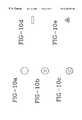
- FIGS. 10 a - 10 eare cross-sectional views of five alternative radiopaque markers at section 10 — 10 of FIG. 9;
- FIGS. 11 a - 11 care side views of three alternative radiopaque markers
- FIG. 12is a side view illustrating one possible arrangement of discrete radiopaque markers disposed on an implantable endoprosthesis
- FIG. 13is the detail bounded by the dashed-line circle in FIG. 12 illustrating a radiopaque marker disposed around one implantable endoprosthesis wire crossing point;
- FIG. 14is a side view illustrating a discrete radiopaque marker
- FIG. 15illustrates the discrete radiopaque marker positioned on an embolization occlusion coil intravascular device.
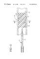
- FIGS. 1-3illustrate a stent delivery device 10 in various stages of deployment having one or more retrievable markers 14 disposed on an implantable endoprosthesis 16 .
- the retrievable radiopaque markers 14are disposed on the endoprosthesis 16 preferably before loading into the outer tube of a delivery device 10 .
- a proximal end 14 a of the retrievable radiopaque marker 14may be attached at portion 8 which is on the outside surface of the inner tube of a delivery device 10 and an area proximal of the proximal end of the implantable endoprosthesis 16 .
- Other attachment areasare also possible on the delivery device 10 .
- Attachment of the proximal end 14 a of the retrievable radiopaque marker 14 to the delivery devicemay be made by mechanical (e.g., clamp or frictional contact on the surface, interweaving to components in the device, or tying), thermal (e.g. metal or polymer welding), or chemical (e.g., adhesive or gel bond) fastening systems.
- a predetermined length of the retrievable radiopaque marker 14may be gathered at or around portion 8 to allow the implantable endoprosthesis 16 to deploy from the delivery device 10 .
- the retrievable radiopaque marker 14may be disposed on the implantable endoprosthesis 16 , be disposed in a channel or lumen of the delivery device 10 , and exit out a port 17 in the hub 19 and be attached to handle 21 .
- the handle 21may be a ring or a similar shape device adapted to be grasped and aid in retrieval and manipulation of the retrievable radiopaque marker 14 or straight wire 18 .
- the markers of the inventioncan be segregated into types; retrievable temporary and discrete permanent markers.
- a retrievable temporary markeris generally a strand or strands of material having radiopacity which is loosely or removably incorporated within the implantable device and which can be removed from the device sometime after implantation by pulling on a free end of the marker or by having the marker extend beyond the device to an attachment point on the delivery system or extend through the delivery system and out of the body where it can be grabbed and pulled free of the implant.
- a discrete permanent markeris generally a strand of material having radiopacity which is securely attached to the implantable device and does not significantly extend away from the device.
- a retrievable temporary markeris a radiopaque strand of material loosely passed through or threaded into a braided tubular stent with an end of marker extending away from the stent and attached to the inner tube of the coaxial tube delivery system.
- the markeris used to locate the position of the stent with regard to the stricture.
- the delivery systemis normally pulled out of the body along the guidewire. The radiopaque marker would be pulled free of the stent as the delivery system is retrieved.
- An example of a discrete permanent markeris a coil, knot, or ring of tantalum wire around a feature of a stent, such as a stent wire crossing point.
- the tantalum wireis wrapped, coiled, or tied around the stent wire and thereby is permanently mechanically attached to the device.
- the tantalum wire endsare clipped off such that the marker is present as a small, tight ring around a feature of the stent.
- the stent with the attached markersis loaded and deployed from the delivery system and the markers are not retrieved when the delivery system is removed.
- the function of the retrievable radiopaque markeris to temporarily indicate on a radiographic image the location of the stent within the treatment site and the length of the expanded stent can be determined by measuring the length of the marker as it follows the stent shape if the marker was threaded along a stent wire helix or axially along a line in the stent.
- the markercan be threaded circumferentially at each end of the stent covering in a covered stent or stent-graft to indicate the location of the radiolucent covering material.
- the stent expansion during deploymentcan be observed radiographically by watching the radiopaque marker helical or circumferential strand open up as the self-expanding stent is released from its radially constrained state.
- Discrete markershave the same functional purpose as the retrievable markers, but they can be more easily used to mark the specific locations of features of interest on the stent For example, a discrete marker can be added to the center of the stent length to aid the physician in centering the stent within the stricture. Discrete markers could be used to attach covering fabrics or films to stents to make stent grafts so that the location of the covering on the stent could be determined radiographically.
- the retrievable and discrete markerscan be made from biocompatible metal wires containing elements with relatively high atomic numbers such as titanium, tantalum, zirconium, and platinum.
- the radiopaque elementscan be added by metallurgically alloying or by making clad composite structures. Another type of marker would be to combine titanium, tantalum, zirconium, or platinum metal or oxide powder with a polymer matrix. Polyethylene or silicone are examples of biocompatible polymers that could be used as a matrix material. Combination could be performed by compounding with the polymer resin or coating.
- Organic radiopaque powders containing elements or salts or oxides of elementssuch as bromine, iodine, iodide, barium, and bismuth could be used instead of metal powders.
- a retrievable, temporary radiopaque markercan be in the form of a strand of metal or polymer containing radiopaque elements, oxides, or salts of elements with atomic numbers in the range of from about 22 to about 83 loosely threaded along a helical, circumferential, or axial orientation in an endoprosthesis such as a stent, stent-graft, graft, filter, occlusive device, and valve with a free end of the marker extending out from the endoprosthesis such that it is attached to the delivery system or passed outside of the body and the marker and is separated from the implanted endoprosthesis by pulling it free and out of the body.
- the radiopaque materialhas a linear attenuation coefficient of from about 10 cm ⁇ 1 at 50 KeV to about 120 cm ⁇ 1 at 50 KeV.
- a retrievable, temporary radiopaque markercan be in the form of a strand of metal or polymer containing radiopaque elements, oxides, or salts of elements with atomic numbers in the range of from about 22 to about 83 formed into a spring and disposed within an endoprosthesis such as a stent, stent-graft, graft, filter, occlusive device, and valve with a free end of the marker extending out from the endoprosthesis such that it is attached to the delivery system or passed outside of the body and the marker and is separated from the implanted endoprosthesis by pulling it free and out of the body.
- the radiopaque materialhas a linear attenuation coefficient of from about 10 cm ⁇ 1 at 50 KeV to about 120 cm ⁇ 1 at 50 KeV.
- a retrievable, temporary radiopaque markercan be in the form of a strand of ductile metal wire, ribbon, or braided wire containing radiopaque metallic elements with atomic numbers in the range of from about 22 to about 83, preferably titanium, tantalum, zirconium, and platinum disposed within an endoprosthesis such as a stent, stent-graft, graft, filter, occlusive device, and valve with a free end of the marker extending out from the endoprosthesis such that it is attached to the delivery system or passed outside of the body and the marker and is separated from the implanted endoprosthesis by pulling it free and out of the body.
- the radiopaque materialhas a linear attenuation coefficient of from about 10 cm ⁇ 1 at 50 KeV to about 120 cm ⁇ 1 at 50 KeV.
- a retrievable, temporary radiopaque markercan be in the form of a strand of ductile metal wire, ribbon, or braided wire containing radiopaque metallic elements with atomic numbers in the range of from about 22 to about 83, preferably titanium, tantalum, zirconium, and platinum coated or clad composite stainless steel or Elgiloy® wire disposed on an endoprosthesis such as a stent, stent-graft, graft, filter, occlusive device, and valve with a free end of the marker extending out from the endoprosthesis such that it is attached to the delivery system or passed outside of the body and the marker is separated from the implanted endoprosthesis by pulling it free and out of the body.
- the radiopaque materialhas a linear attenuation coefficient of from about 10 cm ⁇ 1 at 50 KeV to about 120 cm ⁇ 1 at 50 KeV.
- a retrievable, temporary radiopaque markercan be in the form of a strand of ductile polyethylene or silicone polymer monofilament, ribbon, or multifilament wire containing radiopaque metallic elements with atomic numbers in the range of from about 22 to about 83, preferably compounded or coated with titanium, tantalum, zirconium, and platinum metal powders or bromine, iodine, iodide, barium, and bismuth element, oxides or salts disposed within an endoprosthesis such as a stent, stent-graft, graft, filter, occlusive device, and valve with a free end of the marker extending out from the endoprosthesis such that it is attached to the delivery system or passed outside of the body and the marker and is separated from the implanted endoprosthesis by pulling it free and out of the body.
- the radiopaque materialhas a linear attenuation coefficient of from about 10 cm ⁇ 1 at 50 KeV to about 120 cm ⁇ 1
- a retrievable, temporary radiopaque markercan be in the form of a ductile polymer or metal matrix composite wire containing radiopaque metallic elements with atomic numbers in the range of from about 22 to about 83, preferably titanium, tantalum, zirconium, and platinum metal powders or bromine, iodine, iodide, barium, and bismuth element, oxides or salt powders disposed within an endoprosthesis such as a stent, stent-graft, graft, filter, occlusive device, and valve with a free end of the marker extending out from the endoprosthesis such that it is attached to the delivery system or passed outside of the body and the marker and is separated from the implanted endoprosthesis by pulling it free and out of the body.
- the radiopaque materialhas a linear attenuation coefficient of from about 10 cm ⁇ 1 at 50 KeV to about 120 cm ⁇ 1 at 50 KeV.
- a discrete, permanent radiopaque markercan be in the form of a ductile metal wire, ribbon, or braided wire containing radiopaque metallic elements with atomic numbers in the range of from about 22 to about 83, preferably titanium, tantalum, zirconium, and platinum attached by wrapping, coiling, or tying around features within an endoprosthesis such as a stent, stent-graft, graft, filter, occlusive device, and valve such that the marker stays permanently attached by mechanical or adhesive forces to the endoprosthesis during deployment from the delivery system for the life of the implant.
- the radiopaque materialhas a linear attenuation coefficient of from about 10 cm ⁇ 1 at 50 KeV to about 120 cm ⁇ 1 at 50 KeV.
- a discrete, permanent radiopaque markercan be in the form of a strand of ductile metal wire, ribbon, or braided wire containing radiopaque metallic elements with atomic numbers in the range of from about 22 to about 83, preferably titanium, tantalum, zirconium, and platinum coated or clad composite stainless steel or Elgiloy® wire ductile metal wire, ribbon, or braided wire containing radiopaque metallic elements with atomic numbers in the range of from about 22 to about 83, preferably titanium, tantalum, zirconium, and platinum attached by wrapping, coiling, or tying around features within an endoprosthesis such as a stent, stent-graft, graft, filter, occlusive device, and valve such that the marker stays permanently attached by mechanical or adhesive forces to the endoprosthesis during deployment from the delivery system for the life of the implant.
- the radiopaque materialhas a linear attenuation coefficient of from about 10 cm ⁇ 1 at 50 KeV to about 120
- a discrete, permanent radiopaque markercan be in the form of a strand of ductile polyethylene or silicone polymer monofilament, ribbon, or multifilament wire containing radiopaque metallic elements with atomic numbers in the range of from about 22 to about 83, preferably compounded or coated with titanium, tantalum, zirconium, and platinum metal powders or bromine, iodine, iodide, barium, and bismuth element, oxides or salts attached by wrapping, coiling, or tying around features within an endoprosthesis such as a stent, stent-graft, graft, filter, occlusive device, and valve such that the marker stays permanently attached by mechanical or adhesive forces to the endoprosthesis during deployment from the delivery system for the life of the implant.
- the radiopaque materialhas a linear attenuation coefficient of from about 10 cm ⁇ 1 at 50 KeV to about 120 cm ⁇ 1 at 50 KeV.
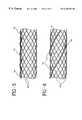
- FIGS. 2-3illustrate an implantable endoprosthesis 16 in a body lumen 12 .
- Implantable endoprostheses known in the artinclude stents, stent-grafts, grafts, filters, occlusive devices, and valves, all of which may incorporate the retrievable radiopaque marker 14 or discrete marker.
- FIGS. 4 a - 4 cillustrate three alternative locations on an implantable endoprosthesis 16 for disposing the retrievable radiopaque marker 14 .
- the retrievable radiopaque marker 14may be an elongate element including a thread, filament, or ribbon such as a highly radiopaque wire relatively loosely woven into or wrapped around the inside, outside, or ends of the implantable endoprosthesis 16 .
- FIGS. 5-6illustrating the retrievable radiopaque marker 14 disposed in two alternative patterns on the implantable endoprosthesis 16 .
- FIG. 5shows the marker 14 interwoven or interbraided loosely along the longitudinal axis of the endoprosthesis 16 .
- FIG. 6shows the marker 14 disposed in a helical pattern about the implantable endoprosthesis 16 .
- Other patterns and dispositions of the marker 14 on the endoprosthesis 16are also possible.
- One or more markers 14may be temporarily disposed on the implantable endoprosthesis 16 in alternative patterns to advantageously provide temporary radiopacity to predetermined locations on the implantable endoprosthesis 16 .
- the retrievable radiopaque marker 14may be applied temporarily to one or more surfaces of the implantable endoprosthesis 16 with a relatively weak bioabsorbable adhesive or gelatin, for instance, as shown in FIGS. 4 a and 4 c .
- the retrievable radiopaque marker 14may be formed into a spring having spring force characteristics and be applied on the inside surface of the implantable endoprosthesis 16 as shown in FIG. 4 c . Spring force allows the retrievable radiopaque marker 14 to press against the interior of the implantable endoprosthesis 16 and provide temporary radiopacity thereto.
- the retrievable radiopaque marker 14may be braided to form a rope or cable.
- the retrievable radiopaque marker 14may be woven or inter-braided into the implantable endoprosthesis 16 during manufacture.
- the retrievable radiopaque marker 14may adjust with expansion of the implantable endoprosthesis 16 and thereby advantageously provides radiopacity and viewing of the implantable endoprosthesis 16 position or size during fluoroscopy.
- the delivery device 10 and the retrievable radiopaque marker 14may be removed from the body.
- one end of the retrievable radiopaque marker 14may be attached to the delivery device 10 and the other end may be disposed at predetermined locations on the implantable endoprosthesis 16 .
- the retrievable radiopaque marker 14may be pulled away from implantable endoprosthesis 16 and removed from the body.
- the retrievable radiopaque marker 14may be loosely incorporated into the implantable endoprosthesis 16 and be easily retrieved without disturbing the implantable endoprosthesis 16 or body tissue. Alternatively, the retrievable radiopaque marker 14 may remain on the implantable endoprosthesis 16 for a period of time if there is a need for follow-up angiography, and then be ultimately removed.
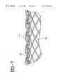
- FIGS. 7-8illustrating an alternative embodiment including a retrievable radiopaque marker 14 and wire 18 .
- the wire 18is used to prevent removal of the marker 14 without first removal of the wire 18 .
- the retrievable radiopaque marker 14is relatively loosely woven or inter-braided in and out of the implantable endoprosthesis 16 , and is maintained in place by another relatively straight, flexible and adjacent movable wire 18 .
- the marker 14 and wire 18may be made by various methods and materials including polymers, metals, ceramics, or similar materials.
- the wire 18may be placed inside, outside, or penetrate between filaments of the implantable endoprosthesis 16 .
- the wire 18 and retrievable radiopaque marker 14are disposed at desired predetermined areas and in various patterns on the implantable endoprosthesis 16 .
- Various combinations of the wire 18 and retrievable radiopaque marker 14are possible including multiple markers 14 or wires 18 .
- the retrievable radiopaque marker 14 and wire 18may be disposed on the implantable endoprosthesis 16 , be disposed in a channel or lumen of the delivery device 10 , and exit out a port 17 in the hub 19 and be attached to handle 21 .
- the handle 21may be a ring or a similar shape device adapted to be grasped and aid in retrieval and manipulation of the retrievable radiopaque marker 14 .
- the wire 18may be removed proximally by a force which liberates the retrievable radiopaque marker 14 and allows removal thereof.
- a limited amount of interweaving or interbraiding of the retrievable radiopaque marker 14 or wire 18is generally desired in order to minimize the force required for retrieval.
- the retrievable radiopaque marker 14 or wire 18may be coated with a biocompatible material having a low coefficient of friction for ease of removal from the implantable endoprosthesis 16 .
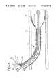
- FIG. 9illustrating a retrievable radiopaque marker 14 preferably made from a relatively flexible wire, suture, filament, ribbon, braided wires, or combinations thereof including radiopaque material such as a metal, metallic alloy, or polymer containing a material that is highly radiopaque.
- radiopaque materialsuch as a metal, metallic alloy, or polymer containing a material that is highly radiopaque.
- FIGS. 10 a - 10 eillustrate alternative cross-sectional embodiments of the retrievable radiopaque marker 14 taken through the line 10 — 10 of FIG. 9 .
- FIG. 10 ashows a substantially solid member
- FIG. 10 bshows a hollow member
- FIG. 10 cshows a member having pores extending radially into the member
- FIG. 10 dshows a rectangular or ribbon member
- FIG. 10 eshows a braided hollow member.
- FIG. 10 emay also be a substantially solid braided member.
- a composite radiopaque marker 14may be made from materials coated or compounded with a radiopaque substance such as iodine, zirconium oxide, barium sulfate, bismuth trioxide, or a related oxide or salt substance.
- Composite radiopaque materialsmay be a radiopaque material containing at least one element having an atomic number, preferably higher than about 22.
- Another radiopaque marker 14may include gold, platinum, metal, tantalum, metallic alloy, or a polymer containing a radiopaque filler such as barium sulfate, bismuth trioxide, iodine, iodide, or like materials.
- FIGS. 11 a - 11 cillustrating alternative embodiments of a portion of the retrievable radiopaque marker 14 .
- the retrievable radiopaque marker 14may have at least one hollow portion 15 which extends throughout the marker 14 for temporary or permanent containment of a retrievable radiopaque material.
- a radiopaque core 13 as shown in FIG. 11 cmay be disposed in and retrieved from a hollow portion 15 in the retrievable radiopaque marker 14 .
- One end of the radiopaque core 13may be attached to the delivery device 10 by a wire or the like and removed from the retrievable radiopaque marker 14 and body lumen by a force originating from outside the body.
- the outside case of the marker 14may remain disposed on the implantable endoprosthesis 16 or be removed therefrom.
- the temporary radiopaque core 13may be solid or include a casing surrounding a solid, gel, powder, or combination thereof and be held in place with a relatively weak bioabsorbable adhesive gelatin, friction, or by other mechanical or chemical means known in the art in a hollow 15 , cavity, or porous portion.
- the temporary radiopaque core 13preferably is made of a radiopaque material that has a linear attenuation coefficient of from about 10 cm ⁇ 1 at 50 KeV to about 120 cm ⁇ 1 at 50 KeV and is adapted to be removably attachable in at least one hollow 15 , cavity, or porous portion in the marker 14 .
- the core 13may remain in the hollow 15 , cavity, or porous portion of the marker 14 and be removed when the marker 14 is retrieved from the body.
- one or more closed cavities within the marker 14 or pores on the surface as shown in FIG. 10 c or pores extending through to a hollow or cavity portion within the marker 14may be utilized for temporary or permanent containment of a retrievable radiopaque materials or be utilized for a passageway for dispersal of the radiopaque materials contained in the marker 14 into the body.
- FIG. 12illustrates discrete radiopaque markers 24 made by forming small rings or coils of radiopaque wire around features of the implantable endoprosthesis 16 .
- Relatively small and discrete wire loop (pigtail) radiopaque markers 24are shown at the wire crossing points on the tubular braid.
- FIG. 13illustrates the detail bounded by the dashed-line circle in FIG. 12 showing a radiopaque wire loop marker 24 around one implantable endoprosthesis 16 wire crossing point.
- FIG. 14illustrates the marker 24 of FIG. 12 and FIG. 13 and shows wire ends 24 a , 24 b which simply pass over each other to form an enclosed loop or overlap.
- the discrete radiopaque markers 24may be plastically or elastically deformable.
- the markers 24may be springs or spring-like for attachment purposes.
- the ends 24 a , 24 bmay be tied, knotted, crimped, spot welded, or bent.
- the markers 24may be relatively small and comprise a single loop or pigtail of wire around one filament crossing point, filament, an embolization coil, or the like.
- the marker 24is preferably made of a biocompatible radiopaque material that is ductile including pure tantalum, platinum, gold, zirconium, niobium, titanium, stainless steel, or combinations thereof.
- the marker 24may be a pig-tail, coil, or knot design and is preferably formed from an elongate member such as a wire and shaped accordingly onto the implantable endoprosthesis 16 .
- the marker 24advantageously allows custom marking of the implantable endoprosthesis 16 without the need to acquire preformed marker bands or to devise a complicated manufacturing operation such as swaging, threading, or braiding.
- the discrete radiopaque markers 24may be easily and quickly added to the implantable endoprosthesis 16 . Also, only small, specific sites are marked by the marker 24 so a minimum amount of foreign body material would be added to the implantable endoprosthesis 16 .
- the discrete radiopaque markers 24may be used on an implantable endoprosthesis 16 made of a bioabsorbable polymer including polylactide.
- the markers 14 , 24should preferably be smaller than the size of the element in the implantable endoprosthesis 16 .
- the size of the markers 14 , 24is also dependent on the type of radiopaque material used. For example, tantalum wire (0.006′ diameter, hard drawn) may be used. The smaller diameter wire fits through most weaves, is deformable, and may be cut to size.
- FIGS. 12-13illustrating discrete markers 24 looped one or more times about a filament or filament crossing point to prevent release therefrom.
- the ends 24 a , 24 bare clipped and positioned to lie in a plane parallel to the longitudinal axis of the implantable endoprosthesis 16 .
- the marker 24may be disposed on one or more filament crossing or every other filament crossing point around the circumference of the braid in one circular transverse plane.
- the markers 24may be positioned to form one or more circumferential rings on the implantable endoprosthesis 16 .
- the markers 24may be positioned along an embolization occlusion coil intravascular device or filament at predetermined locations as illustrated in FIG. 15 .
- the marker 24may be plastically deformed and the marker ends 24 a , 24 b may be looped one or more times about a portion of the implantable endoprosthesis 16 and then pulled to provide a snug disposition.
- the ends 24 a , 24 bmay then be tied, twisted, knotted, welded or adhesively connected together and thereafter clipped and positioned to lie in an unobtrusive low-profile position.
- the retrievable radiopaque marker 14 and discrete radiopaque marker 24may be constructed using a number of methods and materials, in a wide variety of sizes and styles for the greater efficiency and convenience of a user.
- a bioabsorbable markerthat may advantageously be used in conjunction with the present invention is disclosed in J. Stinson's U.S. patent application entitled “Bioabsorbable Marker Having Radiopaque Constituents And Method Of Using Same”, Ser. No. 08/904,951, filed concurrently herewith, and commonly assigned to the assignee of this application.
- a bioabsorbable stentthat may advantageously be used in conjunction with the present invention is disclosed in J. Stinson's United States patent application entitled “Bioabsorbable Implantable Endoprosthesis With Reservoir And Method Of Using Same”, U.S. Pat. No. 5,980,564 filed concurrently herewith, and commonly assigned to the assignee of this application.
Landscapes
- Health & Medical Sciences (AREA)
- Engineering & Computer Science (AREA)
- Biomedical Technology (AREA)
- Cardiology (AREA)
- Oral & Maxillofacial Surgery (AREA)
- Transplantation (AREA)
- Heart & Thoracic Surgery (AREA)
- Vascular Medicine (AREA)
- Life Sciences & Earth Sciences (AREA)
- Animal Behavior & Ethology (AREA)
- General Health & Medical Sciences (AREA)
- Public Health (AREA)
- Veterinary Medicine (AREA)
- Media Introduction/Drainage Providing Device (AREA)
- Prostheses (AREA)
Abstract
Description
| TABLE 1 | ||
| Linear Attenuation | ||
| Atomic Number or | Coefficient | |
| Element or Material | Effective Atomic Number | at 50 KeV, cm−1 |
| hydrogen | 1 | .000028 |
| carbon | 6 | .417 |
| fat | 6.46 | .1925 |
| water | 7.51 | .2245 |
| muscle | 7.64 | .2330 |
| air | 7.78 | .00025 |
| nitrogen | 7 | .00023 |
| oxygen | 8 | .00028 |
| bone | 12.31 | .5727 |
| titanium | 22 | |
| iron | 26 | 15.2 |
| cobalt | 27 | 18.8 |
| bromine | 35 | |
| zirconium | 40 | |
| iodine | 53 | 45 |
| barium | 56 | 58 |
| tantalum | 73 | 111 |
| platinum | 78 | 108 |
| gold | 79 | 101 |
| lead | 82 | 88.7 |
| bismuth | 83 | 62 |
| TABLE 2 | ||||||
| Preferred Marker | Preferred Marker | |||||
| Metal | Organic | |||||
| Radiopaque | Radiopaque | Preferred Marker | ||||
| Marker Type | Description | Function | Devices | Agents | Agents | Matrix Materials |
| Retrievable, | threading on helix | mark overall stent | braided tubular | Ti, Ta, Zr, Pt | Br, I, Ba, Bi | Polyethylene, |
| Temporary | length, location in | stents, filters, | silicone, stainless | |||
| vessel | occlusion, valves | steel, Elgiloy ® | ||||
| Retrievable, | threading around | mark stent ends, | braided tubular | Ti, Ta, Zr, Pt | Br, I, Ba, Bi | Polyethylene, |
| Temporary | circumference | location in vessel, | stents, filters, | silicone, stainless | ||
| covering length, | occlusion, valves, | steel, Elgiloy ® | ||||
| expansion | stent grafts | |||||
| Retrievable, | threading on | mark overall stent | braided tubular | Ti, Ta, Zr, Pt | Br, I, Ba, Bi | Polyethylene, |
| Temporary | straight axial line | length, location in | stents, filters, | silicone, stainless | ||
| vessel | occlusion, valves, | steel, Elgiloy ® | ||||
| stent grafts | ||||||
| Retrievable, | spring | mark overall stent | braided tubular | Ti, Ta, Zr, Pt | Br, I, Ba, Bi | Polyethylene, |
| Temporary | length, location in | stents, filters, | silicone, stainless | |||
| vessel, expansion | occlusion, valves, | steel, Elgiloy ® | ||||
| stent grafts | ||||||
| Discrete, | pigtail rings | mark stent ends | braided tubular | Ti, Ta, Zr, Pt | Br, I, Ba, Bi | Polyethylene, |
| Permanent | or center, location | stents, filters, | silicone, stainless | |||
| in vessel, | occlusion, valves, | steel, Elgiloy ® | ||||
| expansion | stent grafts | |||||
| Discrete, | coils | mark stent ends | braided tubular | Ti, Ta, Zr, Pt | Br, I, Ba, Bi | Polyethylene |
| Permanent | or center, location | stents, filters, | silicone, stainless | |||
| in vessel, | occlusion, valves, | steel, Elgiloy ® | ||||
| expansion | stent grafts | |||||
| Discrete, | knots | mark stent ends | braided tubular | Ti, Ta, Zr, Pt | Br, I, Ba, Bi | Polyethylene, |
| Permanent | or center, location | stents, filters, | silicone, stainless | |||
| in vessel, | occlusion, valves, | steel, Elgiloy ® | ||||
| expansion | stent grafts | |||||
Claims (57)
Priority Applications (10)
| Application Number | Priority Date | Filing Date | Title |
|---|---|---|---|
| US08/905,821US6340367B1 (en) | 1997-08-01 | 1997-08-01 | Radiopaque markers and methods of using the same |
| CA002428667ACA2428667C (en) | 1997-08-01 | 1998-05-27 | Endoprostheses with permanently disposed radiopaque markers |
| CA002238830ACA2238830C (en) | 1997-08-01 | 1998-05-27 | Radiopaque markers and methods of using the same |
| EP04078111AEP1532943B1 (en) | 1997-08-01 | 1998-06-08 | Radiopaque markers and methods of using the same |
| EP98201863AEP0894481B1 (en) | 1997-08-01 | 1998-06-08 | Radiopaque markers |
| DE69830281TDE69830281T2 (en) | 1997-08-01 | 1998-06-08 | Radiopaque markings |
| DE69841130TDE69841130D1 (en) | 1997-08-01 | 1998-06-08 | Radiopaque markers and procedures for their application |
| JP17521298AJP4246289B2 (en) | 1997-08-01 | 1998-06-22 | Radiopaque labels and methods of use |
| US09/264,549US6251135B1 (en) | 1997-08-01 | 1999-03-08 | Radiopaque marker system and method of use |
| US10/008,716US7083641B2 (en) | 1997-08-01 | 2001-11-13 | Radiopaque markers for implantable prostheses |
Applications Claiming Priority (1)
| Application Number | Priority Date | Filing Date | Title |
|---|---|---|---|
| US08/905,821US6340367B1 (en) | 1997-08-01 | 1997-08-01 | Radiopaque markers and methods of using the same |
Related Child Applications (2)
| Application Number | Title | Priority Date | Filing Date |
|---|---|---|---|
| US09/264,549ContinuationUS6251135B1 (en) | 1997-08-01 | 1999-03-08 | Radiopaque marker system and method of use |
| US10/008,716DivisionUS7083641B2 (en) | 1997-08-01 | 2001-11-13 | Radiopaque markers for implantable prostheses |
Publications (1)
| Publication Number | Publication Date |
|---|---|
| US6340367B1true US6340367B1 (en) | 2002-01-22 |
Family
ID=25421532
Family Applications (3)
| Application Number | Title | Priority Date | Filing Date |
|---|---|---|---|
| US08/905,821Expired - LifetimeUS6340367B1 (en) | 1997-08-01 | 1997-08-01 | Radiopaque markers and methods of using the same |
| US09/264,549Expired - LifetimeUS6251135B1 (en) | 1997-08-01 | 1999-03-08 | Radiopaque marker system and method of use |
| US10/008,716Expired - Fee RelatedUS7083641B2 (en) | 1997-08-01 | 2001-11-13 | Radiopaque markers for implantable prostheses |
Family Applications After (2)
| Application Number | Title | Priority Date | Filing Date |
|---|---|---|---|
| US09/264,549Expired - LifetimeUS6251135B1 (en) | 1997-08-01 | 1999-03-08 | Radiopaque marker system and method of use |
| US10/008,716Expired - Fee RelatedUS7083641B2 (en) | 1997-08-01 | 2001-11-13 | Radiopaque markers for implantable prostheses |
Country Status (5)
| Country | Link |
|---|---|
| US (3) | US6340367B1 (en) |
| EP (2) | EP1532943B1 (en) |
| JP (1) | JP4246289B2 (en) |
| CA (2) | CA2428667C (en) |
| DE (2) | DE69830281T2 (en) |
Cited By (137)
| Publication number | Priority date | Publication date | Assignee | Title |
|---|---|---|---|---|
| US20020035324A1 (en)* | 1998-12-24 | 2002-03-21 | Sirimanne D. Laksen | Subcutaneous cavity marking device and method |
| US20020058882A1 (en)* | 1998-06-22 | 2002-05-16 | Artemis Medical, Incorporated | Biopsy localization method and device |
| US20020151933A1 (en)* | 2001-03-05 | 2002-10-17 | Sheldon Jeffery J. | Methods for securing strands of woven medical devices and devices formed thereby |
| US20020188314A1 (en)* | 2001-06-07 | 2002-12-12 | Microvena Corporation | Radiopaque distal embolic protection device |
| US6551352B2 (en)* | 2001-05-03 | 2003-04-22 | Bionx Implants, Inc. | Method for attaching axial filaments to a self expanding stent |
| US20030114919A1 (en)* | 2001-12-10 | 2003-06-19 | Mcquiston Jesse | Polymeric stent with metallic rings |
| US6585755B2 (en)* | 2001-06-29 | 2003-07-01 | Advanced Cardiovascular | Polymeric stent suitable for imaging by MRI and fluoroscopy |
| US6616617B1 (en)* | 1997-12-05 | 2003-09-09 | Micrus Corporation | Vasoocclusive device for treatment of aneurysms |
| US20030203991A1 (en)* | 2002-04-30 | 2003-10-30 | Hydromer, Inc. | Coating composition for multiple hydrophilic applications |
| WO2003032807A3 (en)* | 2001-10-19 | 2004-02-19 | Mann Alfred E Found Scient Res | System and method for removing implanted devices |
| US20040054413A1 (en)* | 2002-09-16 | 2004-03-18 | Howmedica Osteonics Corp. | Radiovisible hydrogel intervertebral disc nucleus |
| US20040073291A1 (en)* | 2002-10-09 | 2004-04-15 | Brian Brown | Intraluminal medical device having improved visibility |
| US20040111149A1 (en)* | 1997-08-01 | 2004-06-10 | Stinson Jonathan S. | Bioabsorbable marker having radiopaque constituents |
| US20040111146A1 (en)* | 2002-12-04 | 2004-06-10 | Mccullagh Orla | Stent-graft attachment |
| US20040122312A1 (en)* | 2002-11-18 | 2004-06-24 | Inrad, Inc. | Apparatus and method for implanting a preloaded localization wire |
| US20040127970A1 (en)* | 2002-12-30 | 2004-07-01 | Saunders Richard J. | Polymer link hybrid stent |
| US20040158310A1 (en)* | 2003-02-06 | 2004-08-12 | Jan Weber | Medical device with magnetic resonance visibility enhancing structure |
| US20040193208A1 (en)* | 2003-03-27 | 2004-09-30 | Scimed Life Systems, Inc. | Radiopaque embolic protection filter membrane |
| US20040204660A1 (en)* | 1998-06-22 | 2004-10-14 | Artemis Medical, Inc. | Biopsy localization method and device |
| US20040254637A1 (en)* | 2003-06-16 | 2004-12-16 | Endotex Interventional Systems, Inc. | Sleeve stent marker |
| US20040267349A1 (en)* | 2003-06-27 | 2004-12-30 | Kobi Richter | Amorphous metal alloy medical devices |
| US20050025752A1 (en)* | 2000-03-15 | 2005-02-03 | Kutryk Michael J. B. | Medical device with coating for capturing genetically-altered cells and methods for using same |
| US20050033407A1 (en)* | 2003-08-07 | 2005-02-10 | Scimed Life Systems, Inc. | Stent designs which enable the visibility of the inside of the stent during MRI |
| US20050064224A1 (en)* | 2003-09-22 | 2005-03-24 | Bavaro Vincent Peter | Polymeric marker with high radiopacity |
| US20050065437A1 (en)* | 2003-09-24 | 2005-03-24 | Scimed Life Systems, Inc. | Medical device with markers for magnetic resonance visibility |
| US20050085895A1 (en)* | 2003-10-15 | 2005-04-21 | Scimed Life Systems, Inc. | RF-based markers for MRI visualization of medical devices |
| US20050113686A1 (en)* | 2003-11-21 | 2005-05-26 | Peckham John E. | Rotational markers |
| US20050137519A1 (en)* | 2003-12-17 | 2005-06-23 | Scimed Life Systems, Inc. | Composite catheter braid |
| DE10361942A1 (en)* | 2003-12-24 | 2005-07-21 | Restate Patent Ag | Radioopaque marker for medical implants |
| US20050165470A1 (en)* | 2004-01-22 | 2005-07-28 | Jan Weber | Medical devices |
| US20050215874A1 (en)* | 2004-03-12 | 2005-09-29 | Lixiao Wang | MRI and X-ray visualization |
| US20050267568A1 (en)* | 2004-05-25 | 2005-12-01 | Chestnut Medical Technologies, Inc. | Flexible vascular occluding device |
| US20050271701A1 (en)* | 2000-03-15 | 2005-12-08 | Orbus Medical Technologies, Inc. | Progenitor endothelial cell capturing with a drug eluting implantable medical device |
| US20050283042A1 (en)* | 2003-03-28 | 2005-12-22 | Steve Meyer | Cardiac harness having radiopaque coating and method of use |
| US20060025795A1 (en)* | 1999-06-17 | 2006-02-02 | Inrad, Inc. | Apparatus for the percutaneous marking of a lesion |
| US20060069405A1 (en)* | 2004-09-20 | 2006-03-30 | Schaeffer Darin G | Anti-thrombus filter having enhanced identifying features |
| US20060111649A1 (en)* | 2004-11-19 | 2006-05-25 | Scimed Life Systems, Inc. | Catheter having improved torque response and curve retention |
| US20060116573A1 (en)* | 2003-11-17 | 2006-06-01 | Inrad, Inc. | Self Contained, Self Piercing, Side-Expelling Marking Apparatus |
| US20060122691A1 (en)* | 1998-12-03 | 2006-06-08 | Jacob Richter | Hybrid stent |
| US20060153729A1 (en)* | 2005-01-13 | 2006-07-13 | Stinson Jonathan S | Medical devices and methods of making the same |
| US7083641B2 (en)* | 1997-08-01 | 2006-08-01 | Schneider (Usa) Inc. | Radiopaque markers for implantable prostheses |
| US20060178727A1 (en)* | 1998-12-03 | 2006-08-10 | Jacob Richter | Hybrid amorphous metal alloy stent |
| US7101391B2 (en) | 2000-09-18 | 2006-09-05 | Inflow Dynamics Inc. | Primarily niobium stent |
| US20060206201A1 (en)* | 2004-05-25 | 2006-09-14 | Chestnut Medical Technologies, Inc. | Flexible vascular occluding device |
| US20060224228A1 (en)* | 1998-09-30 | 2006-10-05 | Mark Dehdashtian | Methods and apparatus for intraluminal placement of a bifurcated intraluminal graft |
| WO2006111964A2 (en) | 2005-04-18 | 2006-10-26 | Denx, Advanced Medical Systems Ltd. | Methods and apparatus for dental implantation |
| US20060257817A1 (en)* | 2005-05-12 | 2006-11-16 | Robert Shelton | Dental implant placement locator and method of use |
| US20060259126A1 (en)* | 2005-05-05 | 2006-11-16 | Jason Lenz | Medical devices and methods of making the same |
| US20060266474A1 (en)* | 1997-12-18 | 2006-11-30 | Schneider (Usa) Inc. | Stent-graft with bioabsorbable structural support |
| US20060276910A1 (en)* | 2005-06-01 | 2006-12-07 | Jan Weber | Endoprostheses |
| US20070021811A1 (en)* | 2005-07-19 | 2007-01-25 | Cardiac Pacemakers, Inc. | Medical device including radiopaque polymer coated coil and method therefor |
| US20070021763A1 (en)* | 2004-11-22 | 2007-01-25 | Inrad, Inc. | Removable Localizing Wire |
| US20070087026A1 (en)* | 2005-10-07 | 2007-04-19 | Inrad, Inc. | Drug-Eluting Tissue Marker |
| US20070123977A1 (en)* | 2000-03-15 | 2007-05-31 | Orbusneich Medical, Inc. | Progenitor Endothelial Cell Capturing with a Drug Eluting Implantable Medical Device |
| US20070129789A1 (en)* | 2000-03-15 | 2007-06-07 | Orbusneich Medical, Inc. | Progenitor Endothelial Cell Capturing with a Drug Eluting Implantable Medical Device |
| US20070208373A1 (en)* | 2006-02-22 | 2007-09-06 | Zaver Steven G | Embolic protection systems having radiopaque filter mesh |
| US20070219642A1 (en)* | 1998-12-03 | 2007-09-20 | Jacob Richter | Hybrid stent having a fiber or wire backbone |
| US20070233175A1 (en)* | 2006-03-31 | 2007-10-04 | Zaver Steven G | Embolic protection devices having radiopaque markers |
| US20080008654A1 (en)* | 2006-07-07 | 2008-01-10 | Boston Scientific Scimed, Inc. | Medical devices having a temporary radiopaque coating |
| US20080033522A1 (en)* | 2006-08-03 | 2008-02-07 | Med Institute, Inc. | Implantable Medical Device with Particulate Coating |
| US20080065193A1 (en)* | 2006-09-08 | 2008-03-13 | Boston Scientific Scimed, Inc. | Stent |
| US20080077230A1 (en)* | 2006-09-21 | 2008-03-27 | Barry Heaney | Stent with support element |
| US20080085293A1 (en)* | 2006-08-22 | 2008-04-10 | Jenchen Yang | Drug eluting stent and therapeutic methods using c-Jun N-terminal kinase inhibitor |
| US20080167703A1 (en)* | 2005-02-17 | 2008-07-10 | Piotr Kasprzak | Stent Prosthesis For Vascular Surgery In Particular In The Area Of The Aortic Arch |
| US20080188924A1 (en)* | 2002-04-01 | 2008-08-07 | Advanced Cardiovascular Systems, Inc. | Hybrid stent and method of making |
| US20080208308A1 (en)* | 2007-02-27 | 2008-08-28 | Medtronic Vascular, Inc. | High Temperature Oxidation-Reduction Process to Form Porous Structures on a Medical Implant |
| US20080288046A1 (en)* | 2007-05-16 | 2008-11-20 | Boston Scientific Scimed, Inc. | Method of Attaching Radiopaque Markers to Intraluminal Medical Devices, and Devices Formed Using the Same |
| US20080290076A1 (en)* | 2006-10-22 | 2008-11-27 | Idev Technologies, Inc. | Methods for Securing Strand Ends and the Resulting Devices |
| US20090099643A1 (en)* | 1999-02-01 | 2009-04-16 | Hideki Hyodoh | Woven intravascular devices and methods for making the same |
| US7582112B2 (en) | 2003-02-10 | 2009-09-01 | Boston Scientific Scimed, Inc. | Metal stent with surface layer of noble metal oxide and method of fabrication |
| US20090234433A1 (en)* | 1998-12-03 | 2009-09-17 | Medinol Ltd. | Helical hybrid stent |
| US20100063550A1 (en)* | 2008-09-11 | 2010-03-11 | Innovasis, Inc, | Radiolucent screw with radiopaque marker |
| US20100191318A1 (en)* | 2003-01-17 | 2010-07-29 | Scimed Life Systems, Inc. | Medical devices |
| US20100234726A1 (en)* | 1998-12-24 | 2010-09-16 | Sirimanne D Laksen | Device and method for safe location and marking of a biopsy cavity |
| US7803180B2 (en) | 2005-04-04 | 2010-09-28 | Flexible Stenting Solutions, Inc. | Flexible stent |
| US20100274350A1 (en)* | 2009-04-22 | 2010-10-28 | Medinol Ltd. | Helical hybrid stent |
| US20100280591A1 (en)* | 2009-04-30 | 2010-11-04 | Kyong-Min Shin | Drawstring for removal of stent |
| US20110060365A1 (en)* | 2009-09-10 | 2011-03-10 | Innovasis, Inc. | Radiolucent stabilizing rod with radiopaque marker |
| US20110218570A1 (en)* | 2010-03-08 | 2011-09-08 | Innovasis, Inc. | Radiolucent bone plate with radiopaque marker |
| US20110270405A1 (en)* | 2010-04-30 | 2011-11-03 | Boston Scientific Scimed, Inc. | Duodenal Metabolic Stent |
| US8064987B2 (en) | 2006-10-23 | 2011-11-22 | C. R. Bard, Inc. | Breast marker |
| US8118864B1 (en)* | 2004-05-25 | 2012-02-21 | Endovascular Technologies, Inc. | Drug delivery endovascular graft |
| US8157862B2 (en) | 1997-10-10 | 2012-04-17 | Senorx, Inc. | Tissue marking implant |
| US8177792B2 (en) | 2002-06-17 | 2012-05-15 | Senorx, Inc. | Plugged tip delivery tube for marker placement |
| US8219182B2 (en) | 1999-02-02 | 2012-07-10 | Senorx, Inc. | Cavity-filling biopsy site markers |
| US8224424B2 (en) | 1999-02-02 | 2012-07-17 | Senorx, Inc. | Tissue site markers for in vivo imaging |
| US8311610B2 (en) | 2008-01-31 | 2012-11-13 | C. R. Bard, Inc. | Biopsy tissue marker |
| US8361082B2 (en) | 1999-02-02 | 2013-01-29 | Senorx, Inc. | Marker delivery device with releasable plug |
| US8401622B2 (en) | 2006-12-18 | 2013-03-19 | C. R. Bard, Inc. | Biopsy marker with in situ-generated imaging properties |
| US8419656B2 (en) | 2004-11-22 | 2013-04-16 | Bard Peripheral Vascular, Inc. | Post decompression marker introducer system |
| US8447386B2 (en) | 2003-05-23 | 2013-05-21 | Senorx, Inc. | Marker or filler forming fluid |
| US8498693B2 (en) | 1999-02-02 | 2013-07-30 | Senorx, Inc. | Intracorporeal marker and marker delivery device |
| US8500794B2 (en) | 2007-08-02 | 2013-08-06 | Flexible Stenting Solutions, Inc. | Flexible stent |
| US8512395B2 (en) | 2010-12-30 | 2013-08-20 | Boston Scientific Scimed, Inc. | Stent with horseshoe shaped bridges |
| US8545548B2 (en) | 2007-03-30 | 2013-10-01 | DePuy Synthes Products, LLC | Radiopaque markers for implantable stents and methods for manufacturing the same |
| US8626269B2 (en) | 2003-05-23 | 2014-01-07 | Senorx, Inc. | Fibrous marker and intracorporeal delivery thereof |
| US8623067B2 (en) | 2004-05-25 | 2014-01-07 | Covidien Lp | Methods and apparatus for luminal stenting |
| US8634899B2 (en) | 2003-11-17 | 2014-01-21 | Bard Peripheral Vascular, Inc. | Multi mode imaging marker |
| US8663313B2 (en) | 2011-03-03 | 2014-03-04 | Boston Scientific Scimed, Inc. | Low strain high strength stent |
| US8670818B2 (en) | 2008-12-30 | 2014-03-11 | C. R. Bard, Inc. | Marker delivery device for tissue marker placement |
| US8668737B2 (en) | 1997-10-10 | 2014-03-11 | Senorx, Inc. | Tissue marking implant |
| US8718745B2 (en) | 2000-11-20 | 2014-05-06 | Senorx, Inc. | Tissue site markers for in vivo imaging |
| US8790388B2 (en) | 2011-03-03 | 2014-07-29 | Boston Scientific Scimed, Inc. | Stent with reduced profile |
| USD715442S1 (en) | 2013-09-24 | 2014-10-14 | C. R. Bard, Inc. | Tissue marker for intracorporeal site identification |
| USD715942S1 (en) | 2013-09-24 | 2014-10-21 | C. R. Bard, Inc. | Tissue marker for intracorporeal site identification |
| USD716450S1 (en) | 2013-09-24 | 2014-10-28 | C. R. Bard, Inc. | Tissue marker for intracorporeal site identification |
| USD716451S1 (en) | 2013-09-24 | 2014-10-28 | C. R. Bard, Inc. | Tissue marker for intracorporeal site identification |
| US9039755B2 (en) | 2003-06-27 | 2015-05-26 | Medinol Ltd. | Helical hybrid stent |
| US9095343B2 (en) | 2005-05-25 | 2015-08-04 | Covidien Lp | System and method for delivering and deploying an occluding device within a vessel |
| US9114001B2 (en) | 2012-10-30 | 2015-08-25 | Covidien Lp | Systems for attaining a predetermined porosity of a vascular device |
| US9149376B2 (en) | 2008-10-06 | 2015-10-06 | Cordis Corporation | Reconstrainable stent delivery system |
| US9149341B2 (en) | 1999-02-02 | 2015-10-06 | Senorx, Inc | Deployment of polysaccharide markers for treating a site within a patient |
| US9157174B2 (en) | 2013-02-05 | 2015-10-13 | Covidien Lp | Vascular device for aneurysm treatment and providing blood flow into a perforator vessel |
| US9233015B2 (en) | 2012-06-15 | 2016-01-12 | Trivascular, Inc. | Endovascular delivery system with an improved radiopaque marker scheme |
| US9327061B2 (en) | 2008-09-23 | 2016-05-03 | Senorx, Inc. | Porous bioabsorbable implant |
| US9393021B2 (en) | 2004-05-25 | 2016-07-19 | Covidien Lp | Flexible vascular occluding device |
| US9452070B2 (en) | 2012-10-31 | 2016-09-27 | Covidien Lp | Methods and systems for increasing a density of a region of a vascular device |
| US9579077B2 (en) | 2006-12-12 | 2017-02-28 | C.R. Bard, Inc. | Multiple imaging mode tissue marker |
| US9615915B2 (en) | 2014-07-25 | 2017-04-11 | Focal Therapeutics, Inc. | Implantable devices and techniques for oncoplastic surgery |
| US9669113B1 (en) | 1998-12-24 | 2017-06-06 | Devicor Medical Products, Inc. | Device and method for safe location and marking of a biopsy cavity |
| US9820824B2 (en) | 1999-02-02 | 2017-11-21 | Senorx, Inc. | Deployment of polysaccharide markers for treating a site within a patent |
| US9943427B2 (en) | 2012-11-06 | 2018-04-17 | Covidien Lp | Shaped occluding devices and methods of using the same |
| US10004618B2 (en) | 2004-05-25 | 2018-06-26 | Covidien Lp | Methods and apparatus for luminal stenting |
| US10022255B2 (en) | 2016-04-11 | 2018-07-17 | Idev Technologies, Inc. | Stent delivery system having anisotropic sheath |
| US10052185B2 (en) | 2016-02-12 | 2018-08-21 | Covidien Lp | Vascular device marker attachment |
| WO2018217653A1 (en) | 2017-05-22 | 2018-11-29 | Boston Scientific Scimed, Inc. | Devices and methods of use with devices having a radiopaque filament |
| US10265089B2 (en) | 2016-02-12 | 2019-04-23 | Covidien Lp | Vascular device visibility |
| US20190201218A1 (en)* | 2016-10-04 | 2019-07-04 | Yasuhiro Shobayashi | Flexible stent |
| US10342635B2 (en) | 2005-04-20 | 2019-07-09 | Bard Peripheral Vascular, Inc. | Marking device with retractable cannula |
| US10413381B2 (en) | 2012-04-26 | 2019-09-17 | Focal Therapeutics, Inc. | Surgical implant for marking soft tissue |
| US10722289B2 (en) | 2015-12-23 | 2020-07-28 | Rhode Island Hospital | Thermal accelerant compositions and methods of use |
| US11219502B2 (en) | 2017-09-11 | 2022-01-11 | Medtronic Advanced Energy, Llc | Transformative shape-memory polymer tissue cavity marker devices, systems and deployment methods |
| US11324567B2 (en) | 2018-02-01 | 2022-05-10 | Medtronic Advanced Energy, Llc | Expandable tissue cavity marker devices, systems and deployment methods |
| US11559414B2 (en) | 2018-09-13 | 2023-01-24 | Olympus Corporation | Stent |
| US11752361B2 (en) | 2009-06-01 | 2023-09-12 | Hologic, Inc. | Diagnostic or therapeutic procedure using implantable targets |
| US11883246B2 (en) | 2012-11-21 | 2024-01-30 | Trustees Of Boston University | Tissue markers and uses thereof |
| US12016624B2 (en) | 2015-12-23 | 2024-06-25 | Rhode Island Hospital | Thermal accelerant compositions and methods of use |
Families Citing this family (246)
| Publication number | Priority date | Publication date | Assignee | Title |
|---|---|---|---|---|
| US6240616B1 (en) | 1997-04-15 | 2001-06-05 | Advanced Cardiovascular Systems, Inc. | Method of manufacturing a medicated porous metal prosthesis |
| US10028851B2 (en) | 1997-04-15 | 2018-07-24 | Advanced Cardiovascular Systems, Inc. | Coatings for controlling erosion of a substrate of an implantable medical device |
| US8172897B2 (en) | 1997-04-15 | 2012-05-08 | Advanced Cardiovascular Systems, Inc. | Polymer and metal composite implantable medical devices |
| US6776792B1 (en) | 1997-04-24 | 2004-08-17 | Advanced Cardiovascular Systems Inc. | Coated endovascular stent |
| US5980564A (en)* | 1997-08-01 | 1999-11-09 | Schneider (Usa) Inc. | Bioabsorbable implantable endoprosthesis with reservoir |
| US6287335B1 (en)* | 1999-04-26 | 2001-09-11 | William J. Drasler | Intravascular folded tubular endoprosthesis |
| DE10004832A1 (en) | 2000-01-31 | 2001-08-16 | Ethicon Gmbh | Flat implant with X-ray visible elements |
| US6494894B2 (en)* | 2000-03-16 | 2002-12-17 | Scimed Life Systems, Inc. | Coated wire |
| US6520952B1 (en)* | 2000-03-23 | 2003-02-18 | Neich Medical Co., Ltd. | Ceramic reinforced catheter |
| US6628982B1 (en)* | 2000-03-30 | 2003-09-30 | The Regents Of The University Of Michigan | Internal marker device for identification of biological substances |
| US7875283B2 (en)* | 2000-04-13 | 2011-01-25 | Advanced Cardiovascular Systems, Inc. | Biodegradable polymers for use with implantable medical devices |
| US8109994B2 (en) | 2003-01-10 | 2012-02-07 | Abbott Cardiovascular Systems, Inc. | Biodegradable drug delivery material for stent |
| US6527801B1 (en)* | 2000-04-13 | 2003-03-04 | Advanced Cardiovascular Systems, Inc. | Biodegradable drug delivery material for stent |
| US20030114918A1 (en)* | 2000-04-28 | 2003-06-19 | Garrison Michi E. | Stent graft assembly and method |
| EP1309289A2 (en) | 2000-08-18 | 2003-05-14 | Atritech, Inc. | Expandable implant devices for filtering blood flow from atrial appendages |
| US6478815B1 (en)* | 2000-09-18 | 2002-11-12 | Inflow Dynamics Inc. | Vascular and endoluminal stents |
| US6863685B2 (en) | 2001-03-29 | 2005-03-08 | Cordis Corporation | Radiopacity intraluminal medical device |
| US6783793B1 (en) | 2000-10-26 | 2004-08-31 | Advanced Cardiovascular Systems, Inc. | Selective coating of medical devices |
| US7776310B2 (en)* | 2000-11-16 | 2010-08-17 | Microspherix Llc | Flexible and/or elastic brachytherapy seed or strand |
| US6514193B2 (en)* | 2000-11-16 | 2003-02-04 | Microspherix Llc | Method of administering a therapeutically active substance |
| US8197535B2 (en) | 2001-06-19 | 2012-06-12 | Cordis Corporation | Low profile improved radiopacity intraluminal medical device |
| US6565659B1 (en) | 2001-06-28 | 2003-05-20 | Advanced Cardiovascular Systems, Inc. | Stent mounting assembly and a method of using the same to coat a stent |
| US7989018B2 (en) | 2001-09-17 | 2011-08-02 | Advanced Cardiovascular Systems, Inc. | Fluid treatment of a polymeric coating on an implantable medical device |
| US7285304B1 (en) | 2003-06-25 | 2007-10-23 | Advanced Cardiovascular Systems, Inc. | Fluid treatment of a polymeric coating on an implantable medical device |
| US6863683B2 (en) | 2001-09-19 | 2005-03-08 | Abbott Laboratoris Vascular Entities Limited | Cold-molding process for loading a stent onto a stent delivery system |
| US9561123B2 (en) | 2002-08-30 | 2017-02-07 | C.R. Bard, Inc. | Highly flexible stent and method of manufacture |
| US6878162B2 (en)* | 2002-08-30 | 2005-04-12 | Edwards Lifesciences Ag | Helical stent having improved flexibility and expandability |
| US7001422B2 (en)* | 2002-09-23 | 2006-02-21 | Cordis Neurovascular, Inc | Expandable stent and delivery system |
| US6814746B2 (en) | 2002-11-01 | 2004-11-09 | Ev3 Peripheral, Inc. | Implant delivery system with marker interlock |
| US8435550B2 (en) | 2002-12-16 | 2013-05-07 | Abbot Cardiovascular Systems Inc. | Anti-proliferative and anti-inflammatory agent combination for treatment of vascular disorders with an implantable medical device |
| US7758881B2 (en) | 2004-06-30 | 2010-07-20 | Advanced Cardiovascular Systems, Inc. | Anti-proliferative and anti-inflammatory agent combination for treatment of vascular disorders with an implantable medical device |
| US7186789B2 (en)* | 2003-06-11 | 2007-03-06 | Advanced Cardiovascular Systems, Inc. | Bioabsorbable, biobeneficial polyester polymers for use in drug eluting stent coatings |
| US20050033157A1 (en)* | 2003-07-25 | 2005-02-10 | Klein Dean A. | Multi-modality marking material and method |
| DE20314393U1 (en)* | 2003-09-16 | 2004-03-04 | Campus Medizin & Technik Gmbh | Medical device for inserting into organs, e.g. catheter, provided with helical x=ray opaque region |
| US7198675B2 (en) | 2003-09-30 | 2007-04-03 | Advanced Cardiovascular Systems | Stent mandrel fixture and method for selectively coating surfaces of a stent |
| JP2007512919A (en)* | 2003-12-04 | 2007-05-24 | ザ ブリガム アンド ウィメンズ ホスピタル インコーポレイテッド | Aortic annuloplasty ring |
| EP2529699B1 (en)* | 2003-12-23 | 2014-01-29 | Sadra Medical, Inc. | Repositionable heart valve |
| US8287584B2 (en) | 2005-11-14 | 2012-10-16 | Sadra Medical, Inc. | Medical implant deployment tool |
| US8828078B2 (en) | 2003-12-23 | 2014-09-09 | Sadra Medical, Inc. | Methods and apparatus for endovascular heart valve replacement comprising tissue grasping elements |
| US7959666B2 (en) | 2003-12-23 | 2011-06-14 | Sadra Medical, Inc. | Methods and apparatus for endovascularly replacing a heart valve |
| US7329279B2 (en) | 2003-12-23 | 2008-02-12 | Sadra Medical, Inc. | Methods and apparatus for endovascularly replacing a patient's heart valve |
| US11278398B2 (en) | 2003-12-23 | 2022-03-22 | Boston Scientific Scimed, Inc. | Methods and apparatus for endovascular heart valve replacement comprising tissue grasping elements |
| US8603160B2 (en) | 2003-12-23 | 2013-12-10 | Sadra Medical, Inc. | Method of using a retrievable heart valve anchor with a sheath |
| US8840663B2 (en) | 2003-12-23 | 2014-09-23 | Sadra Medical, Inc. | Repositionable heart valve method |
| US8579962B2 (en) | 2003-12-23 | 2013-11-12 | Sadra Medical, Inc. | Methods and apparatus for performing valvuloplasty |
| US8182528B2 (en) | 2003-12-23 | 2012-05-22 | Sadra Medical, Inc. | Locking heart valve anchor |
| US20120041550A1 (en) | 2003-12-23 | 2012-02-16 | Sadra Medical, Inc. | Methods and Apparatus for Endovascular Heart Valve Replacement Comprising Tissue Grasping Elements |
| US7381219B2 (en) | 2003-12-23 | 2008-06-03 | Sadra Medical, Inc. | Low profile heart valve and delivery system |
| US20050137694A1 (en) | 2003-12-23 | 2005-06-23 | Haug Ulrich R. | Methods and apparatus for endovascularly replacing a patient's heart valve |
| US9526609B2 (en) | 2003-12-23 | 2016-12-27 | Boston Scientific Scimed, Inc. | Methods and apparatus for endovascularly replacing a patient's heart valve |
| US20050137687A1 (en) | 2003-12-23 | 2005-06-23 | Sadra Medical | Heart valve anchor and method |
| US8343213B2 (en) | 2003-12-23 | 2013-01-01 | Sadra Medical, Inc. | Leaflet engagement elements and methods for use thereof |
| US7780725B2 (en) | 2004-06-16 | 2010-08-24 | Sadra Medical, Inc. | Everting heart valve |
| US7445631B2 (en) | 2003-12-23 | 2008-11-04 | Sadra Medical, Inc. | Methods and apparatus for endovascularly replacing a patient's heart valve |
| US9005273B2 (en) | 2003-12-23 | 2015-04-14 | Sadra Medical, Inc. | Assessing the location and performance of replacement heart valves |
| US7563324B1 (en) | 2003-12-29 | 2009-07-21 | Advanced Cardiovascular Systems Inc. | System and method for coating an implantable medical device |
| US7854756B2 (en) | 2004-01-22 | 2010-12-21 | Boston Scientific Scimed, Inc. | Medical devices |
| US20050214339A1 (en)* | 2004-03-29 | 2005-09-29 | Yiwen Tang | Biologically degradable compositions for medical applications |
| EP1744796B1 (en)* | 2004-04-20 | 2015-03-18 | Genzyme Corporation | Surgical mesh-like implant |
| US7553377B1 (en) | 2004-04-27 | 2009-06-30 | Advanced Cardiovascular Systems, Inc. | Apparatus and method for electrostatic coating of an abluminal stent surface |
| US8568469B1 (en) | 2004-06-28 | 2013-10-29 | Advanced Cardiovascular Systems, Inc. | Stent locking element and a method of securing a stent on a delivery system |
| US8241554B1 (en) | 2004-06-29 | 2012-08-14 | Advanced Cardiovascular Systems, Inc. | Method of forming a stent pattern on a tube |
| US8778256B1 (en) | 2004-09-30 | 2014-07-15 | Advanced Cardiovascular Systems, Inc. | Deformation of a polymer tube in the fabrication of a medical article |
| US7971333B2 (en) | 2006-05-30 | 2011-07-05 | Advanced Cardiovascular Systems, Inc. | Manufacturing process for polymetric stents |
| US7731890B2 (en) | 2006-06-15 | 2010-06-08 | Advanced Cardiovascular Systems, Inc. | Methods of fabricating stents with enhanced fracture toughness |
| US8747879B2 (en) | 2006-04-28 | 2014-06-10 | Advanced Cardiovascular Systems, Inc. | Method of fabricating an implantable medical device to reduce chance of late inflammatory response |
| US8747878B2 (en) | 2006-04-28 | 2014-06-10 | Advanced Cardiovascular Systems, Inc. | Method of fabricating an implantable medical device by controlling crystalline structure |
| US9283099B2 (en) | 2004-08-25 | 2016-03-15 | Advanced Cardiovascular Systems, Inc. | Stent-catheter assembly with a releasable connection for stent retention |
| US20060074480A1 (en) | 2004-09-01 | 2006-04-06 | Pst, Llc | Stent and method for manufacturing the stent |
| US20060060266A1 (en)* | 2004-09-01 | 2006-03-23 | Pst, Llc | Stent and method for manufacturing the stent |
| US7229471B2 (en) | 2004-09-10 | 2007-06-12 | Advanced Cardiovascular Systems, Inc. | Compositions containing fast-leaching plasticizers for improved performance of medical devices |
| US8043553B1 (en) | 2004-09-30 | 2011-10-25 | Advanced Cardiovascular Systems, Inc. | Controlled deformation of a polymer tube with a restraining surface in fabricating a medical article |
| US8173062B1 (en) | 2004-09-30 | 2012-05-08 | Advanced Cardiovascular Systems, Inc. | Controlled deformation of a polymer tube in fabricating a medical article |
| US7875233B2 (en) | 2004-09-30 | 2011-01-25 | Advanced Cardiovascular Systems, Inc. | Method of fabricating a biaxially oriented implantable medical device |
| US7632307B2 (en) | 2004-12-16 | 2009-12-15 | Advanced Cardiovascular Systems, Inc. | Abluminal, multilayer coating constructs for drug-delivery stents |
| DE102005003632A1 (en) | 2005-01-20 | 2006-08-17 | Fraunhofer-Gesellschaft zur Förderung der angewandten Forschung e.V. | Catheter for the transvascular implantation of heart valve prostheses |
| US9301862B2 (en) | 2005-01-28 | 2016-04-05 | Boston Scientific Scimed, Inc. | Stent retrieval member and devices and methods for retrieving or repositioning a stent |
| US20060201601A1 (en)* | 2005-03-03 | 2006-09-14 | Icon Interventional Systems, Inc. | Flexible markers |
| US7381048B2 (en) | 2005-04-12 | 2008-06-03 | Advanced Cardiovascular Systems, Inc. | Stents with profiles for gripping a balloon catheter and molds for fabricating stents |
| US7962208B2 (en) | 2005-04-25 | 2011-06-14 | Cardiac Pacemakers, Inc. | Method and apparatus for pacing during revascularization |
| EP1887974B1 (en) | 2005-05-13 | 2018-04-25 | Boston Scientific Limited | Integrated stent repositioning and retrieval loop |
| US7291166B2 (en) | 2005-05-18 | 2007-11-06 | Advanced Cardiovascular Systems, Inc. | Polymeric stent patterns |
| US7622070B2 (en) | 2005-06-20 | 2009-11-24 | Advanced Cardiovascular Systems, Inc. | Method of manufacturing an implantable polymeric medical device |
| EP1741469A1 (en)* | 2005-07-08 | 2007-01-10 | Engineers & Doctors Wallstén Medical A/S | Method of guiding an irradiation equipment |
| US7867277B1 (en) | 2005-07-15 | 2011-01-11 | Nuvasive Inc. | Spinal fusion implant and related methods |
| US8623088B1 (en) | 2005-07-15 | 2014-01-07 | Nuvasive, Inc. | Spinal fusion implant and related methods |
| US8187159B2 (en) | 2005-07-22 | 2012-05-29 | Biocompatibles, UK | Therapeutic member including a rail used in brachytherapy and other radiation therapy |
| US7736293B2 (en) | 2005-07-22 | 2010-06-15 | Biocompatibles Uk Limited | Implants for use in brachytherapy and other radiation therapy that resist migration and rotation |
| US7658880B2 (en) | 2005-07-29 | 2010-02-09 | Advanced Cardiovascular Systems, Inc. | Polymeric stent polishing method and apparatus |
| US7297758B2 (en) | 2005-08-02 | 2007-11-20 | Advanced Cardiovascular Systems, Inc. | Method for extending shelf-life of constructs of semi-crystallizable polymers |
| US7476245B2 (en) | 2005-08-16 | 2009-01-13 | Advanced Cardiovascular Systems, Inc. | Polymeric stent patterns |
| AU2006282828B2 (en) | 2005-08-23 | 2013-01-31 | Smith & Nephew, Inc | Telemetric orthopaedic implant |
| US9248034B2 (en) | 2005-08-23 | 2016-02-02 | Advanced Cardiovascular Systems, Inc. | Controlled disintegrating implantable medical devices |
| US20070055359A1 (en)* | 2005-08-26 | 2007-03-08 | Messer Stephen C | Vascular graft marker |
| US7712606B2 (en) | 2005-09-13 | 2010-05-11 | Sadra Medical, Inc. | Two-part package for medical implant |
| US7815682B1 (en)* | 2005-09-24 | 2010-10-19 | Nuvasive, Inc. | Spinal fusion implant and related methods |
| EP1769774A1 (en) | 2005-10-03 | 2007-04-04 | Noureddine Frid | Radiopaque endoprostheses |
| US9131999B2 (en) | 2005-11-18 | 2015-09-15 | C.R. Bard Inc. | Vena cava filter with filament |
| US7867547B2 (en) | 2005-12-19 | 2011-01-11 | Advanced Cardiovascular Systems, Inc. | Selectively coating luminal surfaces of stents |
| US20070213813A1 (en) | 2005-12-22 | 2007-09-13 | Symetis Sa | Stent-valves for valve replacement and associated methods and systems for surgery |
| US20070156230A1 (en) | 2006-01-04 | 2007-07-05 | Dugan Stephen R | Stents with radiopaque markers |
| US7951185B1 (en) | 2006-01-06 | 2011-05-31 | Advanced Cardiovascular Systems, Inc. | Delivery of a stent at an elevated temperature |
| JP2009526572A (en) | 2006-02-14 | 2009-07-23 | アンギオメット ゲゼルシャフト ミット ベシュレンクテル ハフツング ウント コムパニー メディツィンテヒニク コマンデイトゲゼルシャフト | Highly flexible stent and manufacturing method |
| EP1988851A2 (en) | 2006-02-14 | 2008-11-12 | Sadra Medical, Inc. | Systems and methods for delivering a medical implant |
| US9849216B2 (en) | 2006-03-03 | 2017-12-26 | Smith & Nephew, Inc. | Systems and methods for delivering a medicament |
| US7964210B2 (en) | 2006-03-31 | 2011-06-21 | Abbott Cardiovascular Systems Inc. | Degradable polymeric implantable medical devices with a continuous phase and discrete phase |
| US7655031B2 (en) | 2006-04-28 | 2010-02-02 | Codman & Shurtleff, Inc. | Stent delivery system with improved retraction member |
| US8690935B2 (en)* | 2006-04-28 | 2014-04-08 | DePuy Synthes Products, LLC | Stent delivery system with threaded engagement and method |
| US8003156B2 (en) | 2006-05-04 | 2011-08-23 | Advanced Cardiovascular Systems, Inc. | Rotatable support elements for stents |
| US7604627B2 (en)* | 2006-05-11 | 2009-10-20 | Kourosh Kojouri | Nasopharyngeal sheath for nasogastric intubation |
| US8535368B2 (en)* | 2006-05-19 | 2013-09-17 | Boston Scientific Scimed, Inc. | Apparatus for loading and delivering a stent |
| US7761968B2 (en) | 2006-05-25 | 2010-07-27 | Advanced Cardiovascular Systems, Inc. | Method of crimping a polymeric stent |
| US20130325107A1 (en) | 2006-05-26 | 2013-12-05 | Abbott Cardiovascular Systems Inc. | Stents With Radiopaque Markers |
| US7951194B2 (en) | 2006-05-26 | 2011-05-31 | Abbott Cardiovascular Sysetms Inc. | Bioabsorbable stent with radiopaque coating |
| US7959940B2 (en) | 2006-05-30 | 2011-06-14 | Advanced Cardiovascular Systems, Inc. | Polymer-bioceramic composite implantable medical devices |
| US7842737B2 (en) | 2006-09-29 | 2010-11-30 | Abbott Cardiovascular Systems Inc. | Polymer blend-bioceramic composite implantable medical devices |
| US8343530B2 (en) | 2006-05-30 | 2013-01-01 | Abbott Cardiovascular Systems Inc. | Polymer-and polymer blend-bioceramic composite implantable medical devices |
| US8486135B2 (en) | 2006-06-01 | 2013-07-16 | Abbott Cardiovascular Systems Inc. | Implantable medical devices fabricated from branched polymers |
| US8034287B2 (en) | 2006-06-01 | 2011-10-11 | Abbott Cardiovascular Systems Inc. | Radiation sterilization of medical devices |
| US8603530B2 (en) | 2006-06-14 | 2013-12-10 | Abbott Cardiovascular Systems Inc. | Nanoshell therapy |
| US8048448B2 (en) | 2006-06-15 | 2011-11-01 | Abbott Cardiovascular Systems Inc. | Nanoshells for drug delivery |
| US8535372B1 (en) | 2006-06-16 | 2013-09-17 | Abbott Cardiovascular Systems Inc. | Bioabsorbable stent with prohealing layer |
| US8333000B2 (en) | 2006-06-19 | 2012-12-18 | Advanced Cardiovascular Systems, Inc. | Methods for improving stent retention on a balloon catheter |
| US8017237B2 (en) | 2006-06-23 | 2011-09-13 | Abbott Cardiovascular Systems, Inc. | Nanoshells on polymers |
| US9072820B2 (en) | 2006-06-26 | 2015-07-07 | Advanced Cardiovascular Systems, Inc. | Polymer composite stent with polymer particles |
| US8128688B2 (en) | 2006-06-27 | 2012-03-06 | Abbott Cardiovascular Systems Inc. | Carbon coating on an implantable device |
| US7794776B1 (en) | 2006-06-29 | 2010-09-14 | Abbott Cardiovascular Systems Inc. | Modification of polymer stents with radiation |
| US7740791B2 (en) | 2006-06-30 | 2010-06-22 | Advanced Cardiovascular Systems, Inc. | Method of fabricating a stent with features by blow molding |
| US7823263B2 (en) | 2006-07-11 | 2010-11-02 | Abbott Cardiovascular Systems Inc. | Method of removing stent islands from a stent |
| US7998404B2 (en) | 2006-07-13 | 2011-08-16 | Advanced Cardiovascular Systems, Inc. | Reduced temperature sterilization of stents |
| US7757543B2 (en) | 2006-07-13 | 2010-07-20 | Advanced Cardiovascular Systems, Inc. | Radio frequency identification monitoring of stents |
| US7794495B2 (en) | 2006-07-17 | 2010-09-14 | Advanced Cardiovascular Systems, Inc. | Controlled degradation of stents |
| USD741488S1 (en) | 2006-07-17 | 2015-10-20 | Nuvasive, Inc. | Spinal fusion implant |
| US7886419B2 (en) | 2006-07-18 | 2011-02-15 | Advanced Cardiovascular Systems, Inc. | Stent crimping apparatus and method |
| DE102006033399B4 (en)* | 2006-07-19 | 2009-04-09 | Jotec Gmbh | Marker system and delivery system for such a marker system |
| US8016879B2 (en) | 2006-08-01 | 2011-09-13 | Abbott Cardiovascular Systems Inc. | Drug delivery after biodegradation of the stent scaffolding |
| US9173733B1 (en) | 2006-08-21 | 2015-11-03 | Abbott Cardiovascular Systems Inc. | Tracheobronchial implantable medical device and methods of use |
| US7923022B2 (en) | 2006-09-13 | 2011-04-12 | Advanced Cardiovascular Systems, Inc. | Degradable polymeric implantable medical devices with continuous phase and discrete phase |
| US8099849B2 (en) | 2006-12-13 | 2012-01-24 | Abbott Cardiovascular Systems Inc. | Optimizing fracture toughness of polymeric stent |
| EP4005537A1 (en)* | 2007-02-12 | 2022-06-01 | C.R. Bard Inc. | Highly flexible stent and method of manufacture |
| US8333799B2 (en) | 2007-02-12 | 2012-12-18 | C. R. Bard, Inc. | Highly flexible stent and method of manufacture |
| DE102007015670A1 (en)* | 2007-03-31 | 2008-10-02 | Biotronik Vi Patent Ag | Stent with radially expandable body |
| US8262723B2 (en) | 2007-04-09 | 2012-09-11 | Abbott Cardiovascular Systems Inc. | Implantable medical devices fabricated from polymer blends with star-block copolymers |
| US7896915B2 (en) | 2007-04-13 | 2011-03-01 | Jenavalve Technology, Inc. | Medical device for treating a heart valve insufficiency |
| US7829008B2 (en) | 2007-05-30 | 2010-11-09 | Abbott Cardiovascular Systems Inc. | Fabricating a stent from a blow molded tube |
| US7959857B2 (en) | 2007-06-01 | 2011-06-14 | Abbott Cardiovascular Systems Inc. | Radiation sterilization of medical devices |
| US8293260B2 (en) | 2007-06-05 | 2012-10-23 | Abbott Cardiovascular Systems Inc. | Elastomeric copolymer coatings containing poly (tetramethyl carbonate) for implantable medical devices |
| US8202528B2 (en) | 2007-06-05 | 2012-06-19 | Abbott Cardiovascular Systems Inc. | Implantable medical devices with elastomeric block copolymer coatings |
| US8425591B1 (en) | 2007-06-11 | 2013-04-23 | Abbott Cardiovascular Systems Inc. | Methods of forming polymer-bioceramic composite medical devices with bioceramic particles |
| US8048441B2 (en) | 2007-06-25 | 2011-11-01 | Abbott Cardiovascular Systems, Inc. | Nanobead releasing medical devices |
| US7901452B2 (en) | 2007-06-27 | 2011-03-08 | Abbott Cardiovascular Systems Inc. | Method to fabricate a stent having selected morphology to reduce restenosis |
| US7955381B1 (en) | 2007-06-29 | 2011-06-07 | Advanced Cardiovascular Systems, Inc. | Polymer-bioceramic composite implantable medical device with different types of bioceramic particles |
| US9393137B2 (en) | 2007-09-24 | 2016-07-19 | Boston Scientific Scimed, Inc. | Method for loading a stent into a delivery system |
| US7691125B2 (en)* | 2007-10-04 | 2010-04-06 | Wilson-Cook Medical Inc. | System and method for forming a stent of a desired length at an endoluminal site |
| US9101698B2 (en)* | 2007-12-05 | 2015-08-11 | Abbott Cardiovascular Systems Inc. | Bioabsorbable stent with radiopaque layer and method of fabrication |
| US9668775B2 (en)* | 2008-06-03 | 2017-06-06 | Jeffrey Scott Smith | Pedicle screw |
| FR2926215B1 (en)* | 2008-01-14 | 2010-01-01 | Balt Extrusion | SYSTEM FOR PREVENTING ANEVISM OR THE LIKE IN A BLOOD VESSEL |
| US9044318B2 (en) | 2008-02-26 | 2015-06-02 | Jenavalve Technology Gmbh | Stent for the positioning and anchoring of a valvular prosthesis |
| BR112012021347A2 (en) | 2008-02-26 | 2019-09-24 | Jenavalve Tecnology Inc | stent for positioning and anchoring a valve prosthesis at an implantation site in a patient's heart |
| US20090264985A1 (en)* | 2008-04-17 | 2009-10-22 | Medtronic Vascular, Inc. | Branch Vessel Suture Stent System and Method |
| DE102008033170A1 (en)* | 2008-07-15 | 2010-01-21 | Acandis Gmbh & Co. Kg | A braided mesh implant and method of making such an implant |
| EP3238661B1 (en) | 2008-10-10 | 2019-05-22 | Boston Scientific Scimed, Inc. | Medical devices and delivery systems for delivering medical devices |
| BRPI0920250A2 (en) | 2008-10-15 | 2016-11-22 | Smith & Nephew Inc | composite internal fasteners |
| US20100137908A1 (en)* | 2008-12-01 | 2010-06-03 | Zimmer Spine, Inc. | Dynamic Stabilization System Components Including Readily Visualized Polymeric Compositions |
| NZ596179A (en)* | 2009-04-29 | 2014-05-30 | Cleveland Clinic Foundation | Apparatus and method for replacing a diseased cardiac valve |
| CA2762811C (en)* | 2009-05-20 | 2023-03-21 | Arsenal Medical, Inc. | Self-expandable medical device comprising polymeric strands and coatings thereon |
| US9265633B2 (en) | 2009-05-20 | 2016-02-23 | 480 Biomedical, Inc. | Drug-eluting medical implants |
| US9386942B2 (en) | 2009-06-26 | 2016-07-12 | Cianna Medical, Inc. | Apparatus, systems, and methods for localizing markers or tissue structures within a body |
| EP3106089B1 (en)* | 2009-06-26 | 2020-12-02 | Cianna Medical, Inc. | System for localizing markers or tissue structures within a body |
| DE102009037739A1 (en)* | 2009-06-29 | 2010-12-30 | Be Innovative Gmbh | Percutaneously implantable valve stent, device for its application and method for producing the valve stent |
| US8828040B2 (en)* | 2009-07-07 | 2014-09-09 | Thomas G. Goff | Device and methods for delivery and transfer of temporary radiopaque element |
| USD731063S1 (en) | 2009-10-13 | 2015-06-02 | Nuvasive, Inc. | Spinal fusion implant |
| MX2012005150A (en)* | 2009-11-03 | 2012-08-17 | Large Bore Closure L L C | Closure device. |
| US8808353B2 (en) | 2010-01-30 | 2014-08-19 | Abbott Cardiovascular Systems Inc. | Crush recoverable polymer scaffolds having a low crossing profile |
| US8568471B2 (en) | 2010-01-30 | 2013-10-29 | Abbott Cardiovascular Systems Inc. | Crush recoverable polymer scaffolds |
| US8920486B2 (en) | 2010-05-18 | 2014-12-30 | RBKPark, LLC | Medical device |
| US10856978B2 (en) | 2010-05-20 | 2020-12-08 | Jenavalve Technology, Inc. | Catheter system |
| WO2011147849A1 (en) | 2010-05-25 | 2011-12-01 | Jenavalve Technology Inc. | Prosthetic heart valve and transcatheter delivered endoprosthesis comprising a prosthetic heart valve and a stent |
| AU2010210022B1 (en)* | 2010-08-05 | 2011-09-08 | Cook Incorporated | Stent graft having a marker and a reinforcing and marker ring |
| AU2011300644B2 (en) | 2010-09-10 | 2015-08-20 | Symetis Sa | Valve replacement devices and a system comprising the valve replacement device and a delivery device therefor |
| PL2649122T3 (en)* | 2010-12-10 | 2017-02-28 | Dsm Ip Assets B.V. | Hppe member and method of making a hppe member |
| WO2012106657A2 (en) | 2011-02-04 | 2012-08-09 | Concentric Medical, Inc. | Vascular and bodily duct treatment devices and methods |
| EP2688516B1 (en) | 2011-03-21 | 2022-08-17 | Cephea Valve Technologies, Inc. | Disk-based valve apparatus |
| EP2520251A1 (en) | 2011-05-05 | 2012-11-07 | Symetis SA | Method and Apparatus for Compressing Stent-Valves |
| US8998976B2 (en) | 2011-07-12 | 2015-04-07 | Boston Scientific Scimed, Inc. | Coupling system for medical devices |
| US8726483B2 (en) | 2011-07-29 | 2014-05-20 | Abbott Cardiovascular Systems Inc. | Methods for uniform crimping and deployment of a polymer scaffold |
| US9131926B2 (en) | 2011-11-10 | 2015-09-15 | Boston Scientific Scimed, Inc. | Direct connect flush system |
| US8940014B2 (en) | 2011-11-15 | 2015-01-27 | Boston Scientific Scimed, Inc. | Bond between components of a medical device |
| US8951243B2 (en) | 2011-12-03 | 2015-02-10 | Boston Scientific Scimed, Inc. | Medical device handle |
| US9277993B2 (en) | 2011-12-20 | 2016-03-08 | Boston Scientific Scimed, Inc. | Medical device delivery systems |
| US9510945B2 (en) | 2011-12-20 | 2016-12-06 | Boston Scientific Scimed Inc. | Medical device handle |
| WO2013112547A1 (en) | 2012-01-25 | 2013-08-01 | Boston Scientific Scimed, Inc. | Valve assembly with a bioabsorbable gasket and a replaceable valve implant |
| US9883941B2 (en) | 2012-06-19 | 2018-02-06 | Boston Scientific Scimed, Inc. | Replacement heart valve |
| BR112015009302A2 (en) | 2012-10-25 | 2017-07-04 | Arterial Remodeling Tech S A | Method of applying a radiopaque marker and apparatus |
| US8870948B1 (en) | 2013-07-17 | 2014-10-28 | Cephea Valve Technologies, Inc. | System and method for cardiac valve repair and replacement |
| EP3034580B1 (en) | 2013-08-13 | 2020-04-29 | DIC Corporation | Resin composition and molded article |
| CN105491978A (en) | 2013-08-30 | 2016-04-13 | 耶拿阀门科技股份有限公司 | Radially collapsible frame for a prosthetic valve and method for manufacturing such a frame |
| US9901445B2 (en) | 2014-11-21 | 2018-02-27 | Boston Scientific Scimed, Inc. | Valve locking mechanism |
| WO2016093877A1 (en) | 2014-12-09 | 2016-06-16 | Cephea Valve Technologies, Inc. | Replacement cardiac valves and methods of use and manufacture |
| WO2016115375A1 (en) | 2015-01-16 | 2016-07-21 | Boston Scientific Scimed, Inc. | Displacement based lock and release mechanism |
| US9861477B2 (en) | 2015-01-26 | 2018-01-09 | Boston Scientific Scimed Inc. | Prosthetic heart valve square leaflet-leaflet stitch |
| WO2016126524A1 (en) | 2015-02-03 | 2016-08-11 | Boston Scientific Scimed, Inc. | Prosthetic heart valve having tubular seal |
| US9788942B2 (en) | 2015-02-03 | 2017-10-17 | Boston Scientific Scimed Inc. | Prosthetic heart valve having tubular seal |
| US9999527B2 (en) | 2015-02-11 | 2018-06-19 | Abbott Cardiovascular Systems Inc. | Scaffolds having radiopaque markers |
| US10285809B2 (en) | 2015-03-06 | 2019-05-14 | Boston Scientific Scimed Inc. | TAVI anchoring assist device |
| US10426617B2 (en) | 2015-03-06 | 2019-10-01 | Boston Scientific Scimed, Inc. | Low profile valve locking mechanism and commissure assembly |
| US10080652B2 (en) | 2015-03-13 | 2018-09-25 | Boston Scientific Scimed, Inc. | Prosthetic heart valve having an improved tubular seal |
| EP3270825B1 (en) | 2015-03-20 | 2020-04-22 | JenaValve Technology, Inc. | Heart valve prosthesis delivery system |
| US10709555B2 (en) | 2015-05-01 | 2020-07-14 | Jenavalve Technology, Inc. | Device and method with reduced pacemaker rate in heart valve replacement |
| AU2016262564B2 (en) | 2015-05-14 | 2020-11-05 | Cephea Valve Technologies, Inc. | Replacement mitral valves |
| WO2018136959A1 (en) | 2017-01-23 | 2018-07-26 | Cephea Valve Technologies, Inc. | Replacement mitral valves |
| EP3294220B1 (en) | 2015-05-14 | 2023-12-06 | Cephea Valve Technologies, Inc. | Cardiac valve delivery devices and systems |
| US9700443B2 (en) | 2015-06-12 | 2017-07-11 | Abbott Cardiovascular Systems Inc. | Methods for attaching a radiopaque marker to a scaffold |
| US10195392B2 (en) | 2015-07-02 | 2019-02-05 | Boston Scientific Scimed, Inc. | Clip-on catheter |
| WO2017004377A1 (en) | 2015-07-02 | 2017-01-05 | Boston Scientific Scimed, Inc. | Adjustable nosecone |
| US10136991B2 (en) | 2015-08-12 | 2018-11-27 | Boston Scientific Scimed Inc. | Replacement heart valve implant |
| US10179041B2 (en) | 2015-08-12 | 2019-01-15 | Boston Scientific Scimed Icn. | Pinless release mechanism |
| US10779940B2 (en) | 2015-09-03 | 2020-09-22 | Boston Scientific Scimed, Inc. | Medical device handle |
| US10342660B2 (en) | 2016-02-02 | 2019-07-09 | Boston Scientific Inc. | Tensioned sheathing aids |
| US10245136B2 (en) | 2016-05-13 | 2019-04-02 | Boston Scientific Scimed Inc. | Containment vessel with implant sheathing guide |
| US10583005B2 (en) | 2016-05-13 | 2020-03-10 | Boston Scientific Scimed, Inc. | Medical device handle |
| WO2017195125A1 (en) | 2016-05-13 | 2017-11-16 | Jenavalve Technology, Inc. | Heart valve prosthesis delivery system and method for delivery of heart valve prosthesis with introducer sheath and loading system |
| US10201416B2 (en) | 2016-05-16 | 2019-02-12 | Boston Scientific Scimed, Inc. | Replacement heart valve implant with invertible leaflets |
| WO2017218877A1 (en) | 2016-06-17 | 2017-12-21 | Cephea Valve Technologies, Inc. | Cardiac valve delivery devices and systems |
| AU2018203053B2 (en) | 2017-01-23 | 2020-03-05 | Cephea Valve Technologies, Inc. | Replacement mitral valves |
| WO2018138658A1 (en) | 2017-01-27 | 2018-08-02 | Jenavalve Technology, Inc. | Heart valve mimicry |
| US10828154B2 (en) | 2017-06-08 | 2020-11-10 | Boston Scientific Scimed, Inc. | Heart valve implant commissure support structure |
| EP3661458A1 (en) | 2017-08-01 | 2020-06-10 | Boston Scientific Scimed, Inc. | Medical implant locking mechanism |
| US10939996B2 (en) | 2017-08-16 | 2021-03-09 | Boston Scientific Scimed, Inc. | Replacement heart valve commissure assembly |
| CA3030162C (en)* | 2018-01-15 | 2023-05-02 | Mariner Endosurgery Inc. | Organ retention device and system, and use of same for laparoscopic surgery |
| US11246625B2 (en) | 2018-01-19 | 2022-02-15 | Boston Scientific Scimed, Inc. | Medical device delivery system with feedback loop |
| EP3740160A2 (en) | 2018-01-19 | 2020-11-25 | Boston Scientific Scimed Inc. | Inductance mode deployment sensors for transcatheter valve system |
| EP3749252A1 (en) | 2018-02-07 | 2020-12-16 | Boston Scientific Scimed, Inc. | Medical device delivery system with alignment feature |
| EP3758651B1 (en) | 2018-02-26 | 2022-12-07 | Boston Scientific Scimed, Inc. | Embedded radiopaque marker in adaptive seal |
| DE102018110591B4 (en)* | 2018-05-03 | 2022-11-03 | Acandis Gmbh | Medical device with a fibrin coating, system and set with such a device and method of manufacture |
| CN112399836B (en) | 2018-05-15 | 2024-10-15 | 波士顿科学国际有限公司 | Replacement heart valve commissure assembly |
| US11241310B2 (en) | 2018-06-13 | 2022-02-08 | Boston Scientific Scimed, Inc. | Replacement heart valve delivery device |
| CN113329716A (en)* | 2018-11-16 | 2021-08-31 | 微仙美国有限公司 | Radiopaque vascular prosthesis |
| US11241312B2 (en) | 2018-12-10 | 2022-02-08 | Boston Scientific Scimed, Inc. | Medical device delivery system including a resistance member |
| EP3924009A4 (en) | 2019-02-14 | 2022-11-16 | Videra Surgical Inc. | MARKER FOR ONCOLOGICAL AND OTHER PROCEDURES |
| US11439504B2 (en) | 2019-05-10 | 2022-09-13 | Boston Scientific Scimed, Inc. | Replacement heart valve with improved cusp washout and reduced loading |
| US12133796B2 (en)* | 2020-05-08 | 2024-11-05 | St. Jude Medical, Cardiology Division, Inc. | Prosthetic heart valve with radiopaque elements |
| AU2021287905A1 (en) | 2020-06-09 | 2023-01-19 | Arsenal Medical, Inc. | Embolic compositions and methods |
| US11291565B1 (en)* | 2021-04-22 | 2022-04-05 | Vascular Graft Solutions Ltd. | Asymmetric external support for stabilizing a vein graft used in a coronary arterial bypass graft (CABG) procedure, and applications thereof |
| WO2023070084A1 (en)* | 2021-10-22 | 2023-04-27 | Videra Surgical Inc. | Auto contourable radiopaque fiducial marker without artifact |
| WO2024102411A1 (en) | 2022-11-09 | 2024-05-16 | Jenavalve Technology, Inc. | Catheter system for sequential deployment of an expandable implant |
Citations (82)
| Publication number | Priority date | Publication date | Assignee | Title |
|---|---|---|---|---|
| US4202349A (en) | 1978-04-24 | 1980-05-13 | Jones James W | Radiopaque vessel markers |
| WO1980002641A1 (en) | 1979-06-06 | 1980-12-11 | S Bowald | Blood vessel prosthesis |
| US4447239A (en) | 1979-03-19 | 1984-05-08 | Dr. Eduard Fresenius Chemisch-Pharmazeutishe Industry Kg | Catheter with radiographic contrast strips |
| US4475972A (en) | 1981-10-01 | 1984-10-09 | Ontario Research Foundation | Implantable material |
| EP0183372A1 (en) | 1984-10-19 | 1986-06-04 | RAYCHEM CORPORATION (a Delaware corporation) | Prosthetic stent |
| US4655771A (en) | 1982-04-30 | 1987-04-07 | Shepherd Patents S.A. | Prosthesis comprising an expansible or contractile tubular body |
| US4681110A (en) | 1985-12-02 | 1987-07-21 | Wiktor Dominik M | Catheter arrangement having a blood vessel liner, and method of using it |
| US4693237A (en) | 1986-01-21 | 1987-09-15 | Hoffman Richard B | Radiopaque coded ring markers for use in identifying surgical grafts |
| US4722344A (en) | 1986-05-23 | 1988-02-02 | Critikon, Inc. | Radiopaque polyurethanes and catheters formed therefrom |
| US4738740A (en) | 1985-11-21 | 1988-04-19 | Corvita Corporation | Method of forming implantable vascular grafts |
| US4787391A (en) | 1985-06-17 | 1988-11-29 | Elefteriades John A | Anastomotic marking device and related method |
| WO1990001969A1 (en) | 1988-08-24 | 1990-03-08 | Slepian Marvin J | Biodegradable polymeric endoluminal sealing |
| WO1990004982A1 (en) | 1988-11-10 | 1990-05-17 | Biocon Oy | Biodegradable surgical implants and devices |
| US4968317A (en) | 1987-01-13 | 1990-11-06 | Toermaelae Pertti | Surgical materials and devices |
| US5015183A (en) | 1989-08-07 | 1991-05-14 | Fenick Thomas J | Locating device and method of placing a tooth implant |
| US5024232A (en) | 1986-10-07 | 1991-06-18 | The Research Foundation Of State University Of Ny | Novel radiopaque heavy metal polymer complexes, compositions of matter and articles prepared therefrom |
| US5026377A (en) | 1989-07-13 | 1991-06-25 | American Medical Systems, Inc. | Stent placement instrument and method |
| WO1991010766A1 (en) | 1990-01-15 | 1991-07-25 | Albany International Corp. | Braid structure |
| US5047050A (en) | 1987-02-17 | 1991-09-10 | Alberto Arpesani | Internal prosthesis with radiopaque annular portions |
| US5059211A (en) | 1987-06-25 | 1991-10-22 | Duke University | Absorbable vascular stent |
| US5061275A (en) | 1986-04-21 | 1991-10-29 | Medinvent S.A. | Self-expanding prosthesis |
| US5085629A (en) | 1988-10-06 | 1992-02-04 | Medical Engineering Corporation | Biodegradable stent |
| US5116360A (en) | 1990-12-27 | 1992-05-26 | Corvita Corporation | Mesh composite graft |
| US5133742A (en) | 1990-06-15 | 1992-07-28 | Corvita Corporation | Crack-resistant polycarbonate urethane polymer prostheses |
| US5133660A (en) | 1989-08-07 | 1992-07-28 | Fenick Thomas J | Device for locating the optimum position for a tooth implant |
| WO1992016166A1 (en) | 1991-03-25 | 1992-10-01 | Meadox Medical Inc. | Vascular prosthesis |
| US5160341A (en) | 1990-11-08 | 1992-11-03 | Advanced Surgical Intervention, Inc. | Resorbable urethral stent and apparatus for its insertion |
| US5163951A (en) | 1990-12-27 | 1992-11-17 | Corvita Corporation | Mesh composite graft |
| US5177170A (en) | 1992-07-02 | 1993-01-05 | Miles Inc. | Radiopaque polyurethanes |
| US5201757A (en) | 1992-04-03 | 1993-04-13 | Schneider (Usa) Inc. | Medial region deployment of radially self-expanding stents |
| US5203777A (en) | 1992-03-19 | 1993-04-20 | Lee Peter Y | Radiopaque marker system for a tubular device |
| US5229431A (en) | 1990-06-15 | 1993-07-20 | Corvita Corporation | Crack-resistant polycarbonate urethane polymer prostheses and the like |
| US5256158A (en) | 1991-05-17 | 1993-10-26 | Act Medical, Inc. | Device having a radiopaque marker for endoscopic accessories and method of making same |
| WO1994006372A1 (en) | 1992-09-14 | 1994-03-31 | Meadox Medicals, Inc. | A radially self-expanding implantable intraluminal device |
| WO1994006373A1 (en) | 1992-09-14 | 1994-03-31 | Meadox Medicals, Inc. | Three-dimensional braided soft tissue prosthesis |
| US5320602A (en) | 1993-05-14 | 1994-06-14 | Wilson-Cook Medical, Inc. | Peel-away endoscopic retrograde cholangio pancreatography catheter and a method for using the same |
| US5346981A (en) | 1993-01-13 | 1994-09-13 | Miles Inc. | Radiopaque polyurethanes |
| US5354257A (en) | 1991-01-29 | 1994-10-11 | Med Institute, Inc. | Minimally invasive medical device for providing a radiation treatment |
| US5366504A (en) | 1992-05-20 | 1994-11-22 | Boston Scientific Corporation | Tubular medical prosthesis |
| US5405402A (en) | 1993-04-14 | 1995-04-11 | Intermedics Orthopedics, Inc. | Implantable prosthesis with radiographic marker |
| US5415546A (en) | 1993-03-23 | 1995-05-16 | Cox, Sr.; Ronald W. | Radiopaque dental composite and materials |
| US5423849A (en) | 1993-01-15 | 1995-06-13 | Target Therapeutics, Inc. | Vasoocclusion device containing radiopaque fibers |
| US5429617A (en) | 1993-12-13 | 1995-07-04 | The Spectranetics Corporation | Radiopaque tip marker for alignment of a catheter within a body |
| US5433727A (en) | 1994-08-16 | 1995-07-18 | Sideris; Eleftherios B. | Centering buttoned device for the occlusion of large defects for occluding |
| US5441517A (en) | 1991-11-08 | 1995-08-15 | Kensey Nash Corporation | Hemostatic puncture closure system and method of use |
| EP0679372A2 (en) | 1994-04-25 | 1995-11-02 | Advanced Cardiovascular Systems, Inc. | Radiopaque stent markers |
| US5464450A (en) | 1991-10-04 | 1995-11-07 | Scimed Lifesystems Inc. | Biodegradable drug delivery vascular stent |
| US5464650A (en) | 1993-04-26 | 1995-11-07 | Medtronic, Inc. | Intravascular stent and method |
| US5478352A (en) | 1990-10-01 | 1995-12-26 | Quinton Instrument Company | Insertion assembly and method of inserting a vessel plug into the body of a patient |
| EP0689807A2 (en) | 1994-06-27 | 1996-01-03 | Advanced Cardiovascular Systems, Inc. | Biodegradable mesh-and-film stent |
| US5484444A (en) | 1992-10-31 | 1996-01-16 | Schneider (Europe) A.G. | Device for the implantation of self-expanding endoprostheses |
| WO1996005872A1 (en) | 1994-08-19 | 1996-02-29 | Biomat B.V. | Radiopaque polymers and methods for preparation thereof |
| US5498227A (en) | 1993-09-15 | 1996-03-12 | Mawad; Michel E. | Retrievable, shielded radiotherapy implant |
| US5500013A (en) | 1991-10-04 | 1996-03-19 | Scimed Life Systems, Inc. | Biodegradable drug delivery vascular stent |
| EP0709068A2 (en) | 1994-10-27 | 1996-05-01 | Medinol Ltd. | X-ray visible stent |
| US5527337A (en) | 1987-06-25 | 1996-06-18 | Duke University | Bioabsorbable stent and method of making the same |
| US5545208A (en) | 1990-02-28 | 1996-08-13 | Medtronic, Inc. | Intralumenal drug eluting prosthesis |
| US5551954A (en) | 1991-10-04 | 1996-09-03 | Scimed Life Systems, Inc. | Biodegradable drug delivery vascular stent |
| US5556413A (en) | 1994-03-11 | 1996-09-17 | Advanced Cardiovascular Systems, Inc. | Coiled stent with locking ends |
| WO1996040000A1 (en) | 1995-06-07 | 1996-12-19 | Angiomedics Ii Incorporated | Curable fiber composite stent and delivery system |
| US5591222A (en) | 1991-10-18 | 1997-01-07 | Susawa; Takashi | Method of manufacturing a device to dilate ducts in vivo |
| US5591224A (en) | 1992-03-19 | 1997-01-07 | Medtronic, Inc. | Bioelastomeric stent |
| US5591172A (en) | 1991-06-14 | 1997-01-07 | Ams Medinvent S.A. | Transluminal implantation device |
| US5591226A (en) | 1995-01-23 | 1997-01-07 | Schneider (Usa) Inc. | Percutaneous stent-graft and method for delivery thereof |
| US5609627A (en) | 1994-02-09 | 1997-03-11 | Boston Scientific Technology, Inc. | Method for delivering a bifurcated endoluminal prosthesis |
| WO1997011724A1 (en) | 1995-09-27 | 1997-04-03 | Biocon Oy | Biodegradable implant manufactured of polymer-based material and a method for manufacturing the same |
| US5628787A (en) | 1993-01-19 | 1997-05-13 | Schneider (Usa) Inc. | Clad composite stent |
| US5628788A (en) | 1995-11-07 | 1997-05-13 | Corvita Corporation | Self-expanding endoluminal stent-graft |
| US5628755A (en) | 1995-02-20 | 1997-05-13 | Schneider (Europe) A.G. | Balloon catheter and stent delivery system |
| US5630840A (en) | 1993-01-19 | 1997-05-20 | Schneider (Usa) Inc | Clad composite stent |
| EP0775472A2 (en) | 1995-11-21 | 1997-05-28 | Schneider (Usa) Inc. | Expandable stent graft covered with expanded polytetrafluoroethylene |
| US5645559A (en) | 1992-05-08 | 1997-07-08 | Schneider (Usa) Inc | Multiple layer stent |
| US5653747A (en) | 1992-12-21 | 1997-08-05 | Corvita Corporation | Luminal graft endoprostheses and manufacture thereof |
| US5662703A (en) | 1995-04-14 | 1997-09-02 | Schneider (Usa) Inc. | Rolling membrane stent delivery device |
| US5674286A (en) | 1991-02-12 | 1997-10-07 | United States Surgical Corporation | Bioabsorbable medical implants |
| US5674277A (en) | 1994-12-23 | 1997-10-07 | Willy Rusch Ag | Stent for placement in a body tube |
| US5676146A (en) | 1996-09-13 | 1997-10-14 | Osteotech, Inc. | Surgical implant containing a resorbable radiopaque marker and method of locating such within a body |
| US5683449A (en) | 1995-02-24 | 1997-11-04 | Marcade; Jean Paul | Modular bifurcated intraluminal grafts and methods for delivering and assembling same |
| US5725517A (en) | 1995-10-05 | 1998-03-10 | Deroyal Industries, Inc. | Absorbent woven article including radiopaque element woven therein and anchored at the ends thereof |
| US5741327A (en) | 1997-05-06 | 1998-04-21 | Global Therapeutics, Inc. | Surgical stent featuring radiopaque markers |
| WO1998018408A1 (en) | 1996-10-25 | 1998-05-07 | Bionix Implants Oy | Surgical implant |
| US5762625A (en) | 1992-09-08 | 1998-06-09 | Kabushikikaisha Igaki Iryo Sekkei | Luminal stent and device for inserting luminal stent |
Family Cites Families (10)
| Publication number | Priority date | Publication date | Assignee | Title |
|---|---|---|---|---|
| US4354257A (en) | 1980-05-23 | 1982-10-12 | Fairchild Camera And Instrument Corporation | Sense amplifier for CCD memory |
| US5019090A (en)* | 1988-09-01 | 1991-05-28 | Corvita Corporation | Radially expandable endoprosthesis and the like |
| US5019085A (en)* | 1988-10-25 | 1991-05-28 | Cordis Corporation | Apparatus and method for placement of a stent within a subject vessel |
| CH678393A5 (en)* | 1989-01-26 | 1991-09-13 | Ulrich Prof Dr Med Sigwart | |
| US5342348A (en)* | 1992-12-04 | 1994-08-30 | Kaplan Aaron V | Method and device for treating and enlarging body lumens |
| US5474563A (en)* | 1993-03-25 | 1995-12-12 | Myler; Richard | Cardiovascular stent and retrieval apparatus |
| ATE372745T1 (en)* | 1995-12-14 | 2007-09-15 | Gore Enterprise Holdings Inc | Kink-resistant Stent Graft |
| US5824042A (en)* | 1996-04-05 | 1998-10-20 | Medtronic, Inc. | Endoluminal prostheses having position indicating markers |
| US6174329B1 (en)* | 1996-08-22 | 2001-01-16 | Advanced Cardiovascular Systems, Inc. | Protective coating for a stent with intermediate radiopaque coating |
| US6340367B1 (en)* | 1997-08-01 | 2002-01-22 | Boston Scientific Scimed, Inc. | Radiopaque markers and methods of using the same |
- 1997
- 1997-08-01USUS08/905,821patent/US6340367B1/ennot_activeExpired - Lifetime
- 1998
- 1998-05-27CACA002428667Apatent/CA2428667C/ennot_activeExpired - Fee Related
- 1998-05-27CACA002238830Apatent/CA2238830C/ennot_activeExpired - Fee Related
- 1998-06-08DEDE69830281Tpatent/DE69830281T2/ennot_activeExpired - Lifetime
- 1998-06-08DEDE69841130Tpatent/DE69841130D1/ennot_activeExpired - Lifetime
- 1998-06-08EPEP04078111Apatent/EP1532943B1/ennot_activeExpired - Lifetime
- 1998-06-08EPEP98201863Apatent/EP0894481B1/ennot_activeExpired - Lifetime
- 1998-06-22JPJP17521298Apatent/JP4246289B2/ennot_activeExpired - Fee Related
- 1999
- 1999-03-08USUS09/264,549patent/US6251135B1/ennot_activeExpired - Lifetime
- 2001
- 2001-11-13USUS10/008,716patent/US7083641B2/ennot_activeExpired - Fee Related
Patent Citations (91)
| Publication number | Priority date | Publication date | Assignee | Title |
|---|---|---|---|---|
| US4202349A (en) | 1978-04-24 | 1980-05-13 | Jones James W | Radiopaque vessel markers |
| US4447239A (en) | 1979-03-19 | 1984-05-08 | Dr. Eduard Fresenius Chemisch-Pharmazeutishe Industry Kg | Catheter with radiographic contrast strips |
| WO1980002641A1 (en) | 1979-06-06 | 1980-12-11 | S Bowald | Blood vessel prosthesis |
| US4475972A (en) | 1981-10-01 | 1984-10-09 | Ontario Research Foundation | Implantable material |
| US4954126B1 (en) | 1982-04-30 | 1996-05-28 | Ams Med Invent S A | Prosthesis comprising an expansible or contractile tubular body |
| US4655771A (en) | 1982-04-30 | 1987-04-07 | Shepherd Patents S.A. | Prosthesis comprising an expansible or contractile tubular body |
| US4655771B1 (en) | 1982-04-30 | 1996-09-10 | Medinvent Ams Sa | Prosthesis comprising an expansible or contractile tubular body |
| US4954126A (en) | 1982-04-30 | 1990-09-04 | Shepherd Patents S.A. | Prosthesis comprising an expansible or contractile tubular body |
| EP0183372A1 (en) | 1984-10-19 | 1986-06-04 | RAYCHEM CORPORATION (a Delaware corporation) | Prosthetic stent |
| US4787391A (en) | 1985-06-17 | 1988-11-29 | Elefteriades John A | Anastomotic marking device and related method |
| US4738740A (en) | 1985-11-21 | 1988-04-19 | Corvita Corporation | Method of forming implantable vascular grafts |
| US4681110A (en) | 1985-12-02 | 1987-07-21 | Wiktor Dominik M | Catheter arrangement having a blood vessel liner, and method of using it |
| US4693237A (en) | 1986-01-21 | 1987-09-15 | Hoffman Richard B | Radiopaque coded ring markers for use in identifying surgical grafts |
| US5061275A (en) | 1986-04-21 | 1991-10-29 | Medinvent S.A. | Self-expanding prosthesis |
| US4722344A (en) | 1986-05-23 | 1988-02-02 | Critikon, Inc. | Radiopaque polyurethanes and catheters formed therefrom |
| US5024232A (en) | 1986-10-07 | 1991-06-18 | The Research Foundation Of State University Of Ny | Novel radiopaque heavy metal polymer complexes, compositions of matter and articles prepared therefrom |
| US4968317B1 (en) | 1987-01-13 | 1999-01-05 | Biocon Oy | Surgical materials and devices |
| US4968317A (en) | 1987-01-13 | 1990-11-06 | Toermaelae Pertti | Surgical materials and devices |
| US5047050A (en) | 1987-02-17 | 1991-09-10 | Alberto Arpesani | Internal prosthesis with radiopaque annular portions |
| US5527337A (en) | 1987-06-25 | 1996-06-18 | Duke University | Bioabsorbable stent and method of making the same |
| US5306286A (en) | 1987-06-25 | 1994-04-26 | Duke University | Absorbable stent |
| US5059211A (en) | 1987-06-25 | 1991-10-22 | Duke University | Absorbable vascular stent |
| WO1990001969A1 (en) | 1988-08-24 | 1990-03-08 | Slepian Marvin J | Biodegradable polymeric endoluminal sealing |
| US5085629A (en) | 1988-10-06 | 1992-02-04 | Medical Engineering Corporation | Biodegradable stent |
| WO1990004982A1 (en) | 1988-11-10 | 1990-05-17 | Biocon Oy | Biodegradable surgical implants and devices |
| US5026377A (en) | 1989-07-13 | 1991-06-25 | American Medical Systems, Inc. | Stent placement instrument and method |
| US5133660A (en) | 1989-08-07 | 1992-07-28 | Fenick Thomas J | Device for locating the optimum position for a tooth implant |
| US5015183A (en) | 1989-08-07 | 1991-05-14 | Fenick Thomas J | Locating device and method of placing a tooth implant |
| WO1991010766A1 (en) | 1990-01-15 | 1991-07-25 | Albany International Corp. | Braid structure |
| US5725567A (en) | 1990-02-28 | 1998-03-10 | Medtronic, Inc. | Method of making a intralumenal drug eluting prosthesis |
| US5545208A (en) | 1990-02-28 | 1996-08-13 | Medtronic, Inc. | Intralumenal drug eluting prosthesis |
| US5229431A (en) | 1990-06-15 | 1993-07-20 | Corvita Corporation | Crack-resistant polycarbonate urethane polymer prostheses and the like |
| US5133742A (en) | 1990-06-15 | 1992-07-28 | Corvita Corporation | Crack-resistant polycarbonate urethane polymer prostheses |
| US5478352A (en) | 1990-10-01 | 1995-12-26 | Quinton Instrument Company | Insertion assembly and method of inserting a vessel plug into the body of a patient |
| US5160341A (en) | 1990-11-08 | 1992-11-03 | Advanced Surgical Intervention, Inc. | Resorbable urethral stent and apparatus for its insertion |
| US5116360A (en) | 1990-12-27 | 1992-05-26 | Corvita Corporation | Mesh composite graft |
| US5163951A (en) | 1990-12-27 | 1992-11-17 | Corvita Corporation | Mesh composite graft |
| US5354257A (en) | 1991-01-29 | 1994-10-11 | Med Institute, Inc. | Minimally invasive medical device for providing a radiation treatment |
| US5674286A (en) | 1991-02-12 | 1997-10-07 | United States Surgical Corporation | Bioabsorbable medical implants |
| WO1992016166A1 (en) | 1991-03-25 | 1992-10-01 | Meadox Medical Inc. | Vascular prosthesis |
| US5256158A (en) | 1991-05-17 | 1993-10-26 | Act Medical, Inc. | Device having a radiopaque marker for endoscopic accessories and method of making same |
| US5591172A (en) | 1991-06-14 | 1997-01-07 | Ams Medinvent S.A. | Transluminal implantation device |
| US5464450A (en) | 1991-10-04 | 1995-11-07 | Scimed Lifesystems Inc. | Biodegradable drug delivery vascular stent |
| US5551954A (en) | 1991-10-04 | 1996-09-03 | Scimed Life Systems, Inc. | Biodegradable drug delivery vascular stent |
| US5500013A (en) | 1991-10-04 | 1996-03-19 | Scimed Life Systems, Inc. | Biodegradable drug delivery vascular stent |
| US5591222A (en) | 1991-10-18 | 1997-01-07 | Susawa; Takashi | Method of manufacturing a device to dilate ducts in vivo |
| US5441517A (en) | 1991-11-08 | 1995-08-15 | Kensey Nash Corporation | Hemostatic puncture closure system and method of use |
| US5591224A (en) | 1992-03-19 | 1997-01-07 | Medtronic, Inc. | Bioelastomeric stent |
| US5203777A (en) | 1992-03-19 | 1993-04-20 | Lee Peter Y | Radiopaque marker system for a tubular device |
| US5201757A (en) | 1992-04-03 | 1993-04-13 | Schneider (Usa) Inc. | Medial region deployment of radially self-expanding stents |
| US5645559A (en) | 1992-05-08 | 1997-07-08 | Schneider (Usa) Inc | Multiple layer stent |
| US5366504A (en) | 1992-05-20 | 1994-11-22 | Boston Scientific Corporation | Tubular medical prosthesis |
| US5177170A (en) | 1992-07-02 | 1993-01-05 | Miles Inc. | Radiopaque polyurethanes |
| US5762625A (en) | 1992-09-08 | 1998-06-09 | Kabushikikaisha Igaki Iryo Sekkei | Luminal stent and device for inserting luminal stent |
| WO1994006372A1 (en) | 1992-09-14 | 1994-03-31 | Meadox Medicals, Inc. | A radially self-expanding implantable intraluminal device |
| WO1994006373A1 (en) | 1992-09-14 | 1994-03-31 | Meadox Medicals, Inc. | Three-dimensional braided soft tissue prosthesis |
| US5484444A (en) | 1992-10-31 | 1996-01-16 | Schneider (Europe) A.G. | Device for the implantation of self-expanding endoprostheses |
| US5653747A (en) | 1992-12-21 | 1997-08-05 | Corvita Corporation | Luminal graft endoprostheses and manufacture thereof |
| US5346981A (en) | 1993-01-13 | 1994-09-13 | Miles Inc. | Radiopaque polyurethanes |
| US5423849A (en) | 1993-01-15 | 1995-06-13 | Target Therapeutics, Inc. | Vasoocclusion device containing radiopaque fibers |
| US5630840A (en) | 1993-01-19 | 1997-05-20 | Schneider (Usa) Inc | Clad composite stent |
| US5628787A (en) | 1993-01-19 | 1997-05-13 | Schneider (Usa) Inc. | Clad composite stent |
| US5415546A (en) | 1993-03-23 | 1995-05-16 | Cox, Sr.; Ronald W. | Radiopaque dental composite and materials |
| US5405402A (en) | 1993-04-14 | 1995-04-11 | Intermedics Orthopedics, Inc. | Implantable prosthesis with radiographic marker |
| US5624411A (en) | 1993-04-26 | 1997-04-29 | Medtronic, Inc. | Intravascular stent and method |
| US5464650A (en) | 1993-04-26 | 1995-11-07 | Medtronic, Inc. | Intravascular stent and method |
| US5320602A (en) | 1993-05-14 | 1994-06-14 | Wilson-Cook Medical, Inc. | Peel-away endoscopic retrograde cholangio pancreatography catheter and a method for using the same |
| US5498227A (en) | 1993-09-15 | 1996-03-12 | Mawad; Michel E. | Retrievable, shielded radiotherapy implant |
| US5429617A (en) | 1993-12-13 | 1995-07-04 | The Spectranetics Corporation | Radiopaque tip marker for alignment of a catheter within a body |
| US5609627A (en) | 1994-02-09 | 1997-03-11 | Boston Scientific Technology, Inc. | Method for delivering a bifurcated endoluminal prosthesis |
| US5556413A (en) | 1994-03-11 | 1996-09-17 | Advanced Cardiovascular Systems, Inc. | Coiled stent with locking ends |
| EP0679372A2 (en) | 1994-04-25 | 1995-11-02 | Advanced Cardiovascular Systems, Inc. | Radiopaque stent markers |
| US5725572A (en) | 1994-04-25 | 1998-03-10 | Advanced Cardiovascular Systems, Inc. | Radiopaque stent |
| EP0689807A2 (en) | 1994-06-27 | 1996-01-03 | Advanced Cardiovascular Systems, Inc. | Biodegradable mesh-and-film stent |
| US5433727A (en) | 1994-08-16 | 1995-07-18 | Sideris; Eleftherios B. | Centering buttoned device for the occlusion of large defects for occluding |
| WO1996005872A1 (en) | 1994-08-19 | 1996-02-29 | Biomat B.V. | Radiopaque polymers and methods for preparation thereof |
| EP0709068A2 (en) | 1994-10-27 | 1996-05-01 | Medinol Ltd. | X-ray visible stent |
| US5674277A (en) | 1994-12-23 | 1997-10-07 | Willy Rusch Ag | Stent for placement in a body tube |
| US5591226A (en) | 1995-01-23 | 1997-01-07 | Schneider (Usa) Inc. | Percutaneous stent-graft and method for delivery thereof |
| US5628755A (en) | 1995-02-20 | 1997-05-13 | Schneider (Europe) A.G. | Balloon catheter and stent delivery system |
| US5683449A (en) | 1995-02-24 | 1997-11-04 | Marcade; Jean Paul | Modular bifurcated intraluminal grafts and methods for delivering and assembling same |
| US5662703A (en) | 1995-04-14 | 1997-09-02 | Schneider (Usa) Inc. | Rolling membrane stent delivery device |
| WO1996040000A1 (en) | 1995-06-07 | 1996-12-19 | Angiomedics Ii Incorporated | Curable fiber composite stent and delivery system |
| WO1997011724A1 (en) | 1995-09-27 | 1997-04-03 | Biocon Oy | Biodegradable implant manufactured of polymer-based material and a method for manufacturing the same |
| US5725517A (en) | 1995-10-05 | 1998-03-10 | Deroyal Industries, Inc. | Absorbent woven article including radiopaque element woven therein and anchored at the ends thereof |
| US5628788A (en) | 1995-11-07 | 1997-05-13 | Corvita Corporation | Self-expanding endoluminal stent-graft |
| EP0775472A2 (en) | 1995-11-21 | 1997-05-28 | Schneider (Usa) Inc. | Expandable stent graft covered with expanded polytetrafluoroethylene |
| US5676146A (en) | 1996-09-13 | 1997-10-14 | Osteotech, Inc. | Surgical implant containing a resorbable radiopaque marker and method of locating such within a body |
| US5676146B1 (en) | 1996-09-13 | 2000-04-18 | Osteotech Inc | Surgical implant containing a resorbable radiopaque marker and method of locating such within a body |
| WO1998018408A1 (en) | 1996-10-25 | 1998-05-07 | Bionix Implants Oy | Surgical implant |
| US5741327A (en) | 1997-05-06 | 1998-04-21 | Global Therapeutics, Inc. | Surgical stent featuring radiopaque markers |
Non-Patent Citations (26)
| Title |
|---|
| Advances in Controlled Release Technology: Polymeric Delivery System for Drugs, Pesticides and Foods: New Methods of Drug Delivery, Robert Langer, Science vol. 249, pp. 1527-1533. |
| Advances in Controlled Release Technology: Polymeric Delivery System s for Drugs, Pesticides and Foods: Fundamentals of pH. and Temperature-Sensitive Polymers, Nicholas A. Peppas, pp. 32-45. |
| Advertisement for radiopaque polymers for medical device manufacturing, New England Urethane, Inc. |
| Advertisement for Spire Corporation radiopaque coating technology, Medical Products Manufacturing News, Mar. 1977, p. 30. |
| Bicomponent vascular grafts consisting of synthetic absorbable fibers. I. In Vitro Study, Tarng-Jenn Yu and C.C. Chu, Journal of Biomedical Materoa;s Research, vol. 27, 1329-1339 (1993). |
| Bioabsorbable Stent and Method of Making the Same, Assignee: Duke University. |
| Bioabsorbable, Drug-Eluting, Intracoronary Stents: Design and Future Applications, R.S. Schwartz, et al., Coronary Stents (1992), pp. 135-154. |
| Biocompatibility of solid poly (ortho ester), M. Ekholm, et al., Journal of Materials Science: Materials in Medicine 8, (1997), pp. 265-269. |
| Biomedical Applications of Synthetic Biodegradable Polymers, Editited by Jeffrey O. Hollinger, D.D.S., Ph.D., CRC Press, p. 21. |
| Current Status of Biodegradable Stents, J.F. Tanguay, M.D., J.P. Zidar, M.D., H.R. Phillips, III, M.D., and R. S. Stack, M.D., Cardiology Clinics, vol. 12, No. 4, Nov. 1994, pp. 699-713. |
| Deformation Characteristics of a Bioabsorbable Intravascular Stent, C. Mauli Agrawal, Ph.D., PE. and Howard G. Clark, Ph.D., Investigative Radiology, Dec. 1992, vol. 27, pp. 1020-1024. |
| Development of a Polymer Endovascular Prosthesis and Its Implantation in Porcine Arteries, William J. Van der Giessen, M.D., et al., Journal of Interventional Cardiology, vol. 5, No. 3, 1992, pp. 175-185. |
| Enhancement of the Mechanical Properties of Polylactides by solid-state extrusion, Walter Weiler and Sylwester Gogolewski, Biomaterials 17 (1996), pp. 529-535. |
| Gianturco-Roubin Flex-Stent GRII, M-D-D-I Report-"The Gray Sheet", Mar. 4, 1996. |
| In Vitro Degradation of Polylactides Depending on Different Processes, M. Dauner, E. Muller, B. Wagner, and H. Planck, Degradation Phenomena on Polymeric Biomaterials, H. Planck, M. Dauner, M. Renardy (eds), Springer-Verlag, Berlin, 1992, pp. 107-122. |
| Long-term in vivo degradation and bone reaction to various polylactides, P. Mainil-Varlet, B. Rahn, and S. Gogolewski, Biomaterials 1997, vol. 18, No. 3, pp. 257-266. |
| Perspectives on the In Vivo Responses of Biodegradable Polymers, James M. Anderson, M.D., Ph.D., CRC Press Inc., (1995) pp. 223-233, 1995. |
| Role of Polymers in improving the results of stenting in coronary arteries, Tao Peng, et al., Biomaterials 1996, vol. 17, No. 7, pp. 685-694. |
| Seventh Complex Coronary Angioplasty Course, May 1996, p. 257. |
| Studies on a new radiopaque polymeric biomaterial, A. Benzina, M.A.B. Kruft, F. Bar, F.H. van der Veen, C.W. Bastiaansen, V. Heijnen, C. Reutelingsperger, and L.H. Koole, Biomaterials 1994, vol. 15 No. 14, pp. 1122-1128. |
| Studies on radio-opaque polymeric biomaterials with potential applications to endovascular prostheses, M. Kruft, A. Benzina, R. Blezer, and L. Koole, Biomaterials 1996, vol. 17, No. 18, pp. 1803-1812. |
| Synthetic Biabsorbable Polymers, Thomas H. Barrows, Ph.D., High Performance Biomaterials, Szycher ed., pp. 243-257. |
| Ten Years of Stenting: What Next?, Ulrich Sigwart, M.D., FRCP, FACC, FESC, Journal of Interventional Cardiology, vol. 10, No. 3, pp. 195-205. |
| The Physics of Radiology, H.E. Johns and J.R. Cunningham, pp. 137-142. |
| The Polymer Stent, Jean-François Tanguay, et al., Endoluminal Stenting, pp. 216-225. |
| Wound Closure Biomaterials and Devices, C.C. Chu, J.A. von Fraunhofer, amd H.P. Greisler, CRC Press, Boca Raton, FL, 1997, pp. 131-235. |
Cited By (319)
| Publication number | Priority date | Publication date | Assignee | Title |
|---|---|---|---|---|
| US20090259125A1 (en)* | 1997-08-01 | 2009-10-15 | Boston Scientific Scimed, Inc. | Bioabsorbable Marker Having Radiopaque Constituents And Method of Using the Same |
| US20060004440A1 (en)* | 1997-08-01 | 2006-01-05 | Stinson Jonathan S | Bioabsorbable marker having radiopaque constituents and method of using the same |
| US7083641B2 (en)* | 1997-08-01 | 2006-08-01 | Schneider (Usa) Inc. | Radiopaque markers for implantable prostheses |
| US7553325B2 (en) | 1997-08-01 | 2009-06-30 | Boston Scientific Scimed, Inc. | Bioabsorbable marker having radiopaque constituents |
| US20040111149A1 (en)* | 1997-08-01 | 2004-06-10 | Stinson Jonathan S. | Bioabsorbable marker having radiopaque constituents |
| US9039763B2 (en) | 1997-10-10 | 2015-05-26 | Senorx, Inc. | Tissue marking implant |
| US8157862B2 (en) | 1997-10-10 | 2012-04-17 | Senorx, Inc. | Tissue marking implant |
| US8668737B2 (en) | 1997-10-10 | 2014-03-11 | Senorx, Inc. | Tissue marking implant |
| US20070016233A1 (en)* | 1997-12-05 | 2007-01-18 | Ferrera David A | Vasoocclusive device for treatment of aneurysms |
| US20040243168A1 (en)* | 1997-12-05 | 2004-12-02 | Ferrera David A. | Vasoocclusive device for treatment of aneurysms |
| US6616617B1 (en)* | 1997-12-05 | 2003-09-09 | Micrus Corporation | Vasoocclusive device for treatment of aneurysms |
| US7326225B2 (en)* | 1997-12-05 | 2008-02-05 | Micrus Endovascular Corporation | Vasoocclusive device for treatment of aneurysms |
| US20060266474A1 (en)* | 1997-12-18 | 2006-11-30 | Schneider (Usa) Inc. | Stent-graft with bioabsorbable structural support |
| US9833343B2 (en) | 1997-12-18 | 2017-12-05 | Boston Scientific Scimed, Inc. | Stent-graft with bioabsorbable structural support |
| US7699887B2 (en) | 1997-12-18 | 2010-04-20 | Boston Scientific Scimed, Inc. | Stent-graft with bioabsorbable structural support |
| US20050033195A1 (en)* | 1998-06-22 | 2005-02-10 | Fulton Richard E. | Biopsy localization method and device |
| US8292822B2 (en) | 1998-06-22 | 2012-10-23 | Devicor Medical Products, Inc. | Biopsy localization method and device |
| US20040267155A1 (en)* | 1998-06-22 | 2004-12-30 | Fulton Richard Eustis | Biopsy localization method and device |
| US20020058882A1 (en)* | 1998-06-22 | 2002-05-16 | Artemis Medical, Incorporated | Biopsy localization method and device |
| US20040204660A1 (en)* | 1998-06-22 | 2004-10-14 | Artemis Medical, Inc. | Biopsy localization method and device |
| US10010380B2 (en) | 1998-06-22 | 2018-07-03 | Devicor Medical Products, Inc. | Biopsy localization method and device |
| US20060224228A1 (en)* | 1998-09-30 | 2006-10-05 | Mark Dehdashtian | Methods and apparatus for intraluminal placement of a bifurcated intraluminal graft |
| US20060178727A1 (en)* | 1998-12-03 | 2006-08-10 | Jacob Richter | Hybrid amorphous metal alloy stent |
| US20070219642A1 (en)* | 1998-12-03 | 2007-09-20 | Jacob Richter | Hybrid stent having a fiber or wire backbone |
| US8382821B2 (en) | 1998-12-03 | 2013-02-26 | Medinol Ltd. | Helical hybrid stent |
| US20060122691A1 (en)* | 1998-12-03 | 2006-06-08 | Jacob Richter | Hybrid stent |
| US20090234433A1 (en)* | 1998-12-03 | 2009-09-17 | Medinol Ltd. | Helical hybrid stent |
| US20050080339A1 (en)* | 1998-12-24 | 2005-04-14 | Vivant Medical, Inc. | Biopsy cavity marking device |
| US7668582B2 (en)* | 1998-12-24 | 2010-02-23 | Ethicon Endo-Surgery, Inc. | Biopsy site marker |
| US20020107437A1 (en)* | 1998-12-24 | 2002-08-08 | Sirimanne D. Laksen | Subcutaneous cavity marking device and method |
| US20020035324A1 (en)* | 1998-12-24 | 2002-03-21 | Sirimanne D. Laksen | Subcutaneous cavity marking device and method |
| US20050080338A1 (en)* | 1998-12-24 | 2005-04-14 | Sirimanne D. Laksen | Biopsy cavity marking device and method |
| US20050059888A1 (en)* | 1998-12-24 | 2005-03-17 | Sirimanne D. Laksen | Biopsy cavity marking device and method |
| US20050080337A1 (en)* | 1998-12-24 | 2005-04-14 | Vivant Medical, Inc. | Biopsy site marker |
| US20050085724A1 (en)* | 1998-12-24 | 2005-04-21 | Vivant Medical, Inc. | Biopsy cavity marking device and method |
| US9986974B2 (en)* | 1998-12-24 | 2018-06-05 | Devicor Medical Products, Inc. | Biopsy cavity marking device |
| US20060036159A1 (en)* | 1998-12-24 | 2006-02-16 | Sirimanne D L | Biopsy cavity marking device |
| US9380998B2 (en) | 1998-12-24 | 2016-07-05 | Devicor Medical Products, Inc. | Subcutaneous cavity marking device and method |
| US20060079770A1 (en)* | 1998-12-24 | 2006-04-13 | Sirimanne D L | Biopsy site marker |
| US9669113B1 (en) | 1998-12-24 | 2017-06-06 | Devicor Medical Products, Inc. | Device and method for safe location and marking of a biopsy cavity |
| US8320994B2 (en) | 1998-12-24 | 2012-11-27 | Devicor Medical Products, Inc. | Biopsy cavity marking device and method |
| US8320993B2 (en)* | 1998-12-24 | 2012-11-27 | Devicor Medical Products, Inc. | Subcutaneous cavity marking device |
| US9492570B2 (en)* | 1998-12-24 | 2016-11-15 | Devicor Medical Products, Inc. | Device and method for safe location and marking of a biopsy cavity |
| US20100234726A1 (en)* | 1998-12-24 | 2010-09-16 | Sirimanne D Laksen | Device and method for safe location and marking of a biopsy cavity |
| US8306602B2 (en) | 1998-12-24 | 2012-11-06 | Devicor Medical Products, Inc. | Biopsy cavity marking device |
| US8600481B2 (en) | 1998-12-24 | 2013-12-03 | Devicor Medical Products, Inc. | Subcutaneous cavity marking device |
| US20090099643A1 (en)* | 1999-02-01 | 2009-04-16 | Hideki Hyodoh | Woven intravascular devices and methods for making the same |
| US9925074B2 (en) | 1999-02-01 | 2018-03-27 | Board Of Regents, The University Of Texas System | Plain woven stents |
| US8414635B2 (en) | 1999-02-01 | 2013-04-09 | Idev Technologies, Inc. | Plain woven stents |
| US8974516B2 (en) | 1999-02-01 | 2015-03-10 | Board Of Regents, The University Of Texas System | Plain woven stents |
| US8876880B2 (en) | 1999-02-01 | 2014-11-04 | Board Of Regents, The University Of Texas System | Plain woven stents |
| US8626270B2 (en) | 1999-02-02 | 2014-01-07 | Senorx, Inc. | Cavity-filling biopsy site markers |
| US8224424B2 (en) | 1999-02-02 | 2012-07-17 | Senorx, Inc. | Tissue site markers for in vivo imaging |
| US9820824B2 (en) | 1999-02-02 | 2017-11-21 | Senorx, Inc. | Deployment of polysaccharide markers for treating a site within a patent |
| US8965486B2 (en) | 1999-02-02 | 2015-02-24 | Senorx, Inc. | Cavity filling biopsy site markers |
| US8219182B2 (en) | 1999-02-02 | 2012-07-10 | Senorx, Inc. | Cavity-filling biopsy site markers |
| US10172674B2 (en) | 1999-02-02 | 2019-01-08 | Senorx, Inc. | Intracorporeal marker and marker delivery device |
| US9044162B2 (en) | 1999-02-02 | 2015-06-02 | Senorx, Inc. | Marker delivery device with releasable plug |
| US8361082B2 (en) | 1999-02-02 | 2013-01-29 | Senorx, Inc. | Marker delivery device with releasable plug |
| US9649093B2 (en) | 1999-02-02 | 2017-05-16 | Senorx, Inc. | Cavity-filling biopsy site markers |
| US9861294B2 (en) | 1999-02-02 | 2018-01-09 | Senorx, Inc. | Marker delivery device with releasable plug |
| US9237937B2 (en) | 1999-02-02 | 2016-01-19 | Senorx, Inc. | Cavity-filling biopsy site markers |
| US9149341B2 (en) | 1999-02-02 | 2015-10-06 | Senorx, Inc | Deployment of polysaccharide markers for treating a site within a patient |
| US8498693B2 (en) | 1999-02-02 | 2013-07-30 | Senorx, Inc. | Intracorporeal marker and marker delivery device |
| US8579931B2 (en) | 1999-06-17 | 2013-11-12 | Bard Peripheral Vascular, Inc. | Apparatus for the percutaneous marking of a lesion |
| US10463446B2 (en)* | 1999-06-17 | 2019-11-05 | Bard Peripheral Vascular, Inc. | Apparatus for the percutaneous marking of a lesion |
| US20090093714A1 (en)* | 1999-06-17 | 2009-04-09 | Bard Peripheral Vascular, Inc | Apparatus for the percutaneous marking of a lesion |
| US8052708B2 (en) | 1999-06-17 | 2011-11-08 | Bard Peripheral Vascular, Inc. | Apparatus for the percutaneous marking of a lesion |
| US9579159B2 (en) | 1999-06-17 | 2017-02-28 | Bard Peripheral Vascular, Inc. | Apparatus for the percutaneous marking of a lesion |
| US20060025795A1 (en)* | 1999-06-17 | 2006-02-02 | Inrad, Inc. | Apparatus for the percutaneous marking of a lesion |
| US20050271701A1 (en)* | 2000-03-15 | 2005-12-08 | Orbus Medical Technologies, Inc. | Progenitor endothelial cell capturing with a drug eluting implantable medical device |
| US20050025752A1 (en)* | 2000-03-15 | 2005-02-03 | Kutryk Michael J. B. | Medical device with coating for capturing genetically-altered cells and methods for using same |
| US9522217B2 (en) | 2000-03-15 | 2016-12-20 | Orbusneich Medical, Inc. | Medical device with coating for capturing genetically-altered cells and methods for using same |
| US20070123977A1 (en)* | 2000-03-15 | 2007-05-31 | Orbusneich Medical, Inc. | Progenitor Endothelial Cell Capturing with a Drug Eluting Implantable Medical Device |
| US20070129789A1 (en)* | 2000-03-15 | 2007-06-07 | Orbusneich Medical, Inc. | Progenitor Endothelial Cell Capturing with a Drug Eluting Implantable Medical Device |
| US8088060B2 (en) | 2000-03-15 | 2012-01-03 | Orbusneich Medical, Inc. | Progenitor endothelial cell capturing with a drug eluting implantable medical device |
| US7101391B2 (en) | 2000-09-18 | 2006-09-05 | Inflow Dynamics Inc. | Primarily niobium stent |
| US8718745B2 (en) | 2000-11-20 | 2014-05-06 | Senorx, Inc. | Tissue site markers for in vivo imaging |
| US20020151933A1 (en)* | 2001-03-05 | 2002-10-17 | Sheldon Jeffery J. | Methods for securing strands of woven medical devices and devices formed thereby |
| US8764817B2 (en)* | 2001-03-05 | 2014-07-01 | Idev Technologies, Inc. | Methods for securing strands of woven medical devices and devices formed thereby |
| US6551352B2 (en)* | 2001-05-03 | 2003-04-22 | Bionx Implants, Inc. | Method for attaching axial filaments to a self expanding stent |
| US20060030878A1 (en)* | 2001-06-07 | 2006-02-09 | Ev3 Inc. | Radiopaque distal embolic protection device |
| US20020188314A1 (en)* | 2001-06-07 | 2002-12-12 | Microvena Corporation | Radiopaque distal embolic protection device |
| US6585755B2 (en)* | 2001-06-29 | 2003-07-01 | Advanced Cardiovascular | Polymeric stent suitable for imaging by MRI and fluoroscopy |
| WO2003032807A3 (en)* | 2001-10-19 | 2004-02-19 | Mann Alfred E Found Scient Res | System and method for removing implanted devices |
| US20030114919A1 (en)* | 2001-12-10 | 2003-06-19 | Mcquiston Jesse | Polymeric stent with metallic rings |
| US7691461B1 (en) | 2002-04-01 | 2010-04-06 | Advanced Cardiovascular Systems, Inc. | Hybrid stent and method of making |
| US20110198327A1 (en)* | 2002-04-01 | 2011-08-18 | Abbott Cardiovascular Systems Inc. | Hybrid stent and method of making |
| US7959999B2 (en) | 2002-04-01 | 2011-06-14 | Abbott Cardiovascular Systems, Inc. | Hybrid stent and method of making |
| US9649210B2 (en) | 2002-04-01 | 2017-05-16 | Abbott Cardiovasular Systems Inc. | Hybrid stent and method of making |
| US20080188924A1 (en)* | 2002-04-01 | 2008-08-07 | Advanced Cardiovascular Systems, Inc. | Hybrid stent and method of making |
| US20030203991A1 (en)* | 2002-04-30 | 2003-10-30 | Hydromer, Inc. | Coating composition for multiple hydrophilic applications |
| US7008979B2 (en) | 2002-04-30 | 2006-03-07 | Hydromer, Inc. | Coating composition for multiple hydrophilic applications |
| US8177792B2 (en) | 2002-06-17 | 2012-05-15 | Senorx, Inc. | Plugged tip delivery tube for marker placement |
| US8784433B2 (en) | 2002-06-17 | 2014-07-22 | Senorx, Inc. | Plugged tip delivery tube for marker placement |
| US8197547B2 (en) | 2002-09-16 | 2012-06-12 | Howmedica Osteonics Corp. | Radiovisible hydrogel intervertebral disc nucleus |
| US20050267583A1 (en)* | 2002-09-16 | 2005-12-01 | Howmedica Osteonics Corp. | Radiovisible hydrogel intervertebral disc nucleus |
| US20040054413A1 (en)* | 2002-09-16 | 2004-03-18 | Howmedica Osteonics Corp. | Radiovisible hydrogel intervertebral disc nucleus |
| US20060255503A1 (en)* | 2002-09-16 | 2006-11-16 | Howmedica Osteonics Corp. | Radiovisible hydrogel intervertebral disc nucleus |
| US7331986B2 (en) | 2002-10-09 | 2008-02-19 | Boston Scientific Scimed, Inc. | Intraluminal medical device having improved visibility |
| US20040073291A1 (en)* | 2002-10-09 | 2004-04-15 | Brian Brown | Intraluminal medical device having improved visibility |
| US20070219624A1 (en)* | 2002-10-09 | 2007-09-20 | Boston Scientific Scimed, Inc. | Intraluminal medical device having improved visibility |
| US8038705B2 (en) | 2002-10-09 | 2011-10-18 | Boston Scientific Scimed, Inc. | Intraluminal medical device having improved visibility |
| US9848956B2 (en) | 2002-11-18 | 2017-12-26 | Bard Peripheral Vascular, Inc. | Self-contained, self-piercing, side-expelling marking apparatus |
| US8886292B2 (en) | 2002-11-18 | 2014-11-11 | Bard Peripheral Vascular, Inc. | Apparatus for implanting a preloaded localization wire |
| US11504206B2 (en) | 2002-11-18 | 2022-11-22 | Bard Peripheral Vascular, Inc. | Apparatus for implanting a preloaded localization wire |
| US20040122312A1 (en)* | 2002-11-18 | 2004-06-24 | Inrad, Inc. | Apparatus and method for implanting a preloaded localization wire |
| US9707042B2 (en) | 2002-11-18 | 2017-07-18 | Bard Peripheral Vascular, Inc. | Apparatus for implanting a preloaded localization wire |
| US8131346B2 (en) | 2002-11-18 | 2012-03-06 | Bard Peripheral Vascular, Inc. | Apparatus and method for implanting a preloaded localization wire |
| US10813716B2 (en) | 2002-11-18 | 2020-10-27 | Bard Peripheral Vascular, Inc. | Self-contained, self-piercing, side-expelling marking apparatus |
| US20040111146A1 (en)* | 2002-12-04 | 2004-06-10 | Mccullagh Orla | Stent-graft attachment |
| US20040127970A1 (en)* | 2002-12-30 | 2004-07-01 | Saunders Richard J. | Polymer link hybrid stent |
| US20090029030A1 (en)* | 2002-12-30 | 2009-01-29 | Advanced Cardiovascular Systems, Inc. | Polymer link hybrid stent |
| US7645409B2 (en) | 2002-12-30 | 2010-01-12 | Advanced Cardiovascular Systems, Inc. | Polymer link hybrid stent |
| US7455687B2 (en) | 2002-12-30 | 2008-11-25 | Advanced Cardiovascular Systems, Inc. | Polymer link hybrid stent |
| US20100191318A1 (en)* | 2003-01-17 | 2010-07-29 | Scimed Life Systems, Inc. | Medical devices |
| US20040158310A1 (en)* | 2003-02-06 | 2004-08-12 | Jan Weber | Medical device with magnetic resonance visibility enhancing structure |
| US7172624B2 (en) | 2003-02-06 | 2007-02-06 | Boston Scientific Scimed, Inc. | Medical device with magnetic resonance visibility enhancing structure |
| US20070123975A1 (en)* | 2003-02-06 | 2007-05-31 | Jan Weber | Medical device with magnetic resonance visibility enhancing structure |
| US7582112B2 (en) | 2003-02-10 | 2009-09-01 | Boston Scientific Scimed, Inc. | Metal stent with surface layer of noble metal oxide and method of fabrication |
| US20070135833A1 (en)* | 2003-03-27 | 2007-06-14 | Boston Scientific Scimed, Inc. | Radiopaque embolic protection filter membrane |
| US20040193208A1 (en)* | 2003-03-27 | 2004-09-30 | Scimed Life Systems, Inc. | Radiopaque embolic protection filter membrane |
| US20050283042A1 (en)* | 2003-03-28 | 2005-12-22 | Steve Meyer | Cardiac harness having radiopaque coating and method of use |
| US8639315B2 (en) | 2003-05-23 | 2014-01-28 | Senorx, Inc. | Marker or filler forming fluid |
| US10299881B2 (en) | 2003-05-23 | 2019-05-28 | Senorx, Inc. | Marker or filler forming fluid |
| US8880154B2 (en) | 2003-05-23 | 2014-11-04 | Senorx, Inc. | Fibrous marker and intracorporeal delivery thereof |
| US10045832B2 (en) | 2003-05-23 | 2018-08-14 | Senorx, Inc. | Marker or filler forming fluid |
| US8626269B2 (en) | 2003-05-23 | 2014-01-07 | Senorx, Inc. | Fibrous marker and intracorporeal delivery thereof |
| US8447386B2 (en) | 2003-05-23 | 2013-05-21 | Senorx, Inc. | Marker or filler forming fluid |
| US9801688B2 (en) | 2003-05-23 | 2017-10-31 | Senorx, Inc. | Fibrous marker and intracorporeal delivery thereof |
| US20040254637A1 (en)* | 2003-06-16 | 2004-12-16 | Endotex Interventional Systems, Inc. | Sleeve stent marker |
| US9039755B2 (en) | 2003-06-27 | 2015-05-26 | Medinol Ltd. | Helical hybrid stent |
| US9603731B2 (en) | 2003-06-27 | 2017-03-28 | Medinol Ltd. | Helical hybrid stent |
| US20040267349A1 (en)* | 2003-06-27 | 2004-12-30 | Kobi Richter | Amorphous metal alloy medical devices |
| US9456910B2 (en) | 2003-06-27 | 2016-10-04 | Medinol Ltd. | Helical hybrid stent |
| US10363152B2 (en) | 2003-06-27 | 2019-07-30 | Medinol Ltd. | Helical hybrid stent |
| US7479157B2 (en) | 2003-08-07 | 2009-01-20 | Boston Scientific Scimed, Inc. | Stent designs which enable the visibility of the inside of the stent during MRI |
| US20050033407A1 (en)* | 2003-08-07 | 2005-02-10 | Scimed Life Systems, Inc. | Stent designs which enable the visibility of the inside of the stent during MRI |
| US20080065010A1 (en)* | 2003-09-22 | 2008-03-13 | Advanced Cardiovascular Systems, Inc. | Polymeric marker with high radiopacity for use in medical devices |
| US20050064223A1 (en)* | 2003-09-22 | 2005-03-24 | Bavaro Vincent Peter | Polymeric marker with high radiopacity |
| US7833597B2 (en) | 2003-09-22 | 2010-11-16 | Advanced Cardiovascular Systems, Inc. | Polymeric marker with high radiopacity for use in medical devices |
| US20050064224A1 (en)* | 2003-09-22 | 2005-03-24 | Bavaro Vincent Peter | Polymeric marker with high radiopacity |
| US7303798B2 (en) | 2003-09-22 | 2007-12-04 | Advanced Cardiovascular Systems, Inc. | Polymeric marker with high radiopacity for use in medical devices |
| US8637132B2 (en) | 2003-09-22 | 2014-01-28 | Advanced Cardiovascular Systems, Inc. | Polymeric marker with high radiopacity for use in medical devices |
| US20050065437A1 (en)* | 2003-09-24 | 2005-03-24 | Scimed Life Systems, Inc. | Medical device with markers for magnetic resonance visibility |
| US20050085895A1 (en)* | 2003-10-15 | 2005-04-21 | Scimed Life Systems, Inc. | RF-based markers for MRI visualization of medical devices |
| US7819820B2 (en) | 2003-11-17 | 2010-10-26 | Bard Peripheral Vascular, Inc. | Self contained, self piercing, side-expelling marking apparatus |
| US20060116573A1 (en)* | 2003-11-17 | 2006-06-01 | Inrad, Inc. | Self Contained, Self Piercing, Side-Expelling Marking Apparatus |
| US8634899B2 (en) | 2003-11-17 | 2014-01-21 | Bard Peripheral Vascular, Inc. | Multi mode imaging marker |
| US20050113686A1 (en)* | 2003-11-21 | 2005-05-26 | Peckham John E. | Rotational markers |
| US8014849B2 (en)* | 2003-11-21 | 2011-09-06 | Stryker Corporation | Rotational markers |
| US20050137519A1 (en)* | 2003-12-17 | 2005-06-23 | Scimed Life Systems, Inc. | Composite catheter braid |
| US7955313B2 (en) | 2003-12-17 | 2011-06-07 | Boston Scientific Scimed, Inc. | Composite catheter braid |
| DE10361942A1 (en)* | 2003-12-24 | 2005-07-21 | Restate Patent Ag | Radioopaque marker for medical implants |
| US20070191708A1 (en)* | 2003-12-24 | 2007-08-16 | Bodo Gerold | Radio-opaque marker for medical implants |
| US8871829B2 (en) | 2003-12-24 | 2014-10-28 | Biotronik Vi Patent Ag | Radio-opaque marker for medical implants |
| US20050165470A1 (en)* | 2004-01-22 | 2005-07-28 | Jan Weber | Medical devices |
| US20100082093A1 (en)* | 2004-01-22 | 2010-04-01 | Scimed Life Systems, Inc. | Medical Devices |
| US7632299B2 (en)* | 2004-01-22 | 2009-12-15 | Boston Scientific Scimed, Inc. | Medical devices |
| US20050215874A1 (en)* | 2004-03-12 | 2005-09-29 | Lixiao Wang | MRI and X-ray visualization |
| US7761138B2 (en) | 2004-03-12 | 2010-07-20 | Boston Scientific Scimed, Inc. | MRI and X-ray visualization |
| US9050205B2 (en) | 2004-05-25 | 2015-06-09 | Covidien Lp | Methods and apparatus for luminal stenting |
| US8398701B2 (en) | 2004-05-25 | 2013-03-19 | Covidien Lp | Flexible vascular occluding device |
| US20050267568A1 (en)* | 2004-05-25 | 2005-12-01 | Chestnut Medical Technologies, Inc. | Flexible vascular occluding device |
| US12042411B2 (en) | 2004-05-25 | 2024-07-23 | Covidien Lp | Methods and apparatus for luminal stenting |
| US11771433B2 (en) | 2004-05-25 | 2023-10-03 | Covidien Lp | Flexible vascular occluding device |
| US9855047B2 (en) | 2004-05-25 | 2018-01-02 | Covidien Lp | Flexible vascular occluding device |
| US9125659B2 (en) | 2004-05-25 | 2015-09-08 | Covidien Lp | Flexible vascular occluding device |
| US8118864B1 (en)* | 2004-05-25 | 2012-02-21 | Endovascular Technologies, Inc. | Drug delivery endovascular graft |
| US9295568B2 (en) | 2004-05-25 | 2016-03-29 | Covidien Lp | Methods and apparatus for luminal stenting |
| US10004618B2 (en) | 2004-05-25 | 2018-06-26 | Covidien Lp | Methods and apparatus for luminal stenting |
| US10918389B2 (en) | 2004-05-25 | 2021-02-16 | Covidien Lp | Flexible vascular occluding device |
| US20060206201A1 (en)* | 2004-05-25 | 2006-09-14 | Chestnut Medical Technologies, Inc. | Flexible vascular occluding device |
| US9801744B2 (en) | 2004-05-25 | 2017-10-31 | Covidien Lp | Methods and apparatus for luminal stenting |
| US8617234B2 (en) | 2004-05-25 | 2013-12-31 | Covidien Lp | Flexible vascular occluding device |
| US10765542B2 (en) | 2004-05-25 | 2020-09-08 | Covidien Lp | Methods and apparatus for luminal stenting |
| US8623067B2 (en) | 2004-05-25 | 2014-01-07 | Covidien Lp | Methods and apparatus for luminal stenting |
| US9393021B2 (en) | 2004-05-25 | 2016-07-19 | Covidien Lp | Flexible vascular occluding device |
| US8628564B2 (en) | 2004-05-25 | 2014-01-14 | Covidien Lp | Methods and apparatus for luminal stenting |
| US20060069405A1 (en)* | 2004-09-20 | 2006-03-30 | Schaeffer Darin G | Anti-thrombus filter having enhanced identifying features |
| US20060111649A1 (en)* | 2004-11-19 | 2006-05-25 | Scimed Life Systems, Inc. | Catheter having improved torque response and curve retention |
| US8170648B2 (en) | 2004-11-22 | 2012-05-01 | Bard Peripheral Vascular, Inc. | Removable localizing wire |
| US20070021763A1 (en)* | 2004-11-22 | 2007-01-25 | Inrad, Inc. | Removable Localizing Wire |
| US8744554B2 (en) | 2004-11-22 | 2014-06-03 | Bard Peripheral Vascular, Inc. | Removable localizing wire |
| US8419656B2 (en) | 2004-11-22 | 2013-04-16 | Bard Peripheral Vascular, Inc. | Post decompression marker introducer system |
| US20060153729A1 (en)* | 2005-01-13 | 2006-07-13 | Stinson Jonathan S | Medical devices and methods of making the same |
| US20100228336A1 (en)* | 2005-01-13 | 2010-09-09 | Stinson Jonathan S | Medical devices and methods of making the same |
| US7727273B2 (en) | 2005-01-13 | 2010-06-01 | Boston Scientific Scimed, Inc. | Medical devices and methods of making the same |
| US7938854B2 (en) | 2005-01-13 | 2011-05-10 | Boston Scientific Scimed, Inc. | Medical devices and methods of making the same |
| US20080167703A1 (en)* | 2005-02-17 | 2008-07-10 | Piotr Kasprzak | Stent Prosthesis For Vascular Surgery In Particular In The Area Of The Aortic Arch |
| US8123796B2 (en) | 2005-02-17 | 2012-02-28 | Piotr Kasprzak | Stent prosthesis for vascular surgery in particular in the area of the aortic arch |
| US7803180B2 (en) | 2005-04-04 | 2010-09-28 | Flexible Stenting Solutions, Inc. | Flexible stent |
| US9592137B2 (en) | 2005-04-04 | 2017-03-14 | Flexible Stenting Solutions, Inc. | Flexible stent |
| EP3135238A1 (en) | 2005-04-18 | 2017-03-01 | Image Navigation Ltd | Methods and apparatus for dental implantation |
| US8172573B2 (en) | 2005-04-18 | 2012-05-08 | Image Navigation Ltd | Methods and apparatus for dental implantation |
| WO2006111964A2 (en) | 2005-04-18 | 2006-10-26 | Denx, Advanced Medical Systems Ltd. | Methods and apparatus for dental implantation |
| US20080171305A1 (en)* | 2005-04-18 | 2008-07-17 | Denx, Advanced Medical System Ltd. | Methods and Apparatus For Dental Implantation |
| US10357328B2 (en) | 2005-04-20 | 2019-07-23 | Bard Peripheral Vascular, Inc. and Bard Shannon Limited | Marking device with retractable cannula |
| US11278370B2 (en) | 2005-04-20 | 2022-03-22 | Bard Peripheral Vascular, Inc. | Marking device with retractable cannula |
| US10342635B2 (en) | 2005-04-20 | 2019-07-09 | Bard Peripheral Vascular, Inc. | Marking device with retractable cannula |
| US20060259126A1 (en)* | 2005-05-05 | 2006-11-16 | Jason Lenz | Medical devices and methods of making the same |
| US20060257817A1 (en)* | 2005-05-12 | 2006-11-16 | Robert Shelton | Dental implant placement locator and method of use |
| US8215957B2 (en)* | 2005-05-12 | 2012-07-10 | Robert Shelton | Dental implant placement locator and method of use |
| US9095343B2 (en) | 2005-05-25 | 2015-08-04 | Covidien Lp | System and method for delivering and deploying an occluding device within a vessel |
| US20060276910A1 (en)* | 2005-06-01 | 2006-12-07 | Jan Weber | Endoprostheses |
| US20070021811A1 (en)* | 2005-07-19 | 2007-01-25 | Cardiac Pacemakers, Inc. | Medical device including radiopaque polymer coated coil and method therefor |
| US20070087026A1 (en)* | 2005-10-07 | 2007-04-19 | Inrad, Inc. | Drug-Eluting Tissue Marker |
| US8052658B2 (en) | 2005-10-07 | 2011-11-08 | Bard Peripheral Vascular, Inc. | Drug-eluting tissue marker |
| US8486028B2 (en) | 2005-10-07 | 2013-07-16 | Bard Peripheral Vascular, Inc. | Tissue marking apparatus having drug-eluting tissue marker |
| US20070208373A1 (en)* | 2006-02-22 | 2007-09-06 | Zaver Steven G | Embolic protection systems having radiopaque filter mesh |
| US9610181B2 (en) | 2006-02-22 | 2017-04-04 | Covidien Lp | Stents having radiopaque mesh |
| US10433988B2 (en) | 2006-02-22 | 2019-10-08 | Covidien Lp | Stents having radiopaque mesh |
| US8394119B2 (en) | 2006-02-22 | 2013-03-12 | Covidien Lp | Stents having radiopaque mesh |
| US8152833B2 (en) | 2006-02-22 | 2012-04-10 | Tyco Healthcare Group Lp | Embolic protection systems having radiopaque filter mesh |
| US9320590B2 (en) | 2006-02-22 | 2016-04-26 | Covidien Lp | Stents having radiopaque mesh |
| US11382777B2 (en) | 2006-02-22 | 2022-07-12 | Covidien Lp | Stents having radiopaque mesh |
| US10548707B2 (en) | 2006-03-31 | 2020-02-04 | Covidien Lp | Embolic protection devices having radiopaque elements |
| US9089404B2 (en) | 2006-03-31 | 2015-07-28 | Covidien Lp | Embolic protection devices having radiopaque elements |
| US20070233175A1 (en)* | 2006-03-31 | 2007-10-04 | Zaver Steven G | Embolic protection devices having radiopaque markers |
| US20080008654A1 (en)* | 2006-07-07 | 2008-01-10 | Boston Scientific Scimed, Inc. | Medical devices having a temporary radiopaque coating |
| US20080033522A1 (en)* | 2006-08-03 | 2008-02-07 | Med Institute, Inc. | Implantable Medical Device with Particulate Coating |
| US20080085293A1 (en)* | 2006-08-22 | 2008-04-10 | Jenchen Yang | Drug eluting stent and therapeutic methods using c-Jun N-terminal kinase inhibitor |
| US20080065193A1 (en)* | 2006-09-08 | 2008-03-13 | Boston Scientific Scimed, Inc. | Stent |
| US8414637B2 (en) | 2006-09-08 | 2013-04-09 | Boston Scientific Scimed, Inc. | Stent |
| US20080077230A1 (en)* | 2006-09-21 | 2008-03-27 | Barry Heaney | Stent with support element |
| US7875069B2 (en)* | 2006-09-21 | 2011-01-25 | Boston Scientific Scimed, Inc. | Stent with support element |
| US9408730B2 (en) | 2006-10-22 | 2016-08-09 | Idev Technologies, Inc. | Secured strand end devices |
| US9408729B2 (en) | 2006-10-22 | 2016-08-09 | Idev Technologies, Inc. | Secured strand end devices |
| US9895242B2 (en) | 2006-10-22 | 2018-02-20 | Idev Technologies, Inc. | Secured strand end devices |
| US20080290076A1 (en)* | 2006-10-22 | 2008-11-27 | Idev Technologies, Inc. | Methods for Securing Strand Ends and the Resulting Devices |
| US9149374B2 (en) | 2006-10-22 | 2015-10-06 | Idev Technologies, Inc. | Methods for manufacturing secured strand end devices |
| US8419788B2 (en) | 2006-10-22 | 2013-04-16 | Idev Technologies, Inc. | Secured strand end devices |
| US9629736B2 (en) | 2006-10-22 | 2017-04-25 | Idev Technologies, Inc. | Secured strand end devices |
| US10470902B2 (en) | 2006-10-22 | 2019-11-12 | Idev Technologies, Inc. | Secured strand end devices |
| US8966733B2 (en) | 2006-10-22 | 2015-03-03 | Idev Technologies, Inc. | Secured strand end devices |
| US8739382B2 (en) | 2006-10-22 | 2014-06-03 | Idev Technologies, Inc. | Secured strand end devices |
| US9585776B2 (en) | 2006-10-22 | 2017-03-07 | Idev Technologies, Inc. | Secured strand end devices |
| US8437834B2 (en) | 2006-10-23 | 2013-05-07 | C. R. Bard, Inc. | Breast marker |
| US8064987B2 (en) | 2006-10-23 | 2011-11-22 | C. R. Bard, Inc. | Breast marker |
| US10682200B2 (en) | 2006-12-12 | 2020-06-16 | C. R. Bard, Inc. | Multiple imaging mode tissue marker |
| US11471244B2 (en) | 2006-12-12 | 2022-10-18 | C.R. Bard, Inc. | Multiple imaging mode tissue marker |
| US9901415B2 (en) | 2006-12-12 | 2018-02-27 | C. R. Bard, Inc. | Multiple imaging mode tissue marker |
| US9579077B2 (en) | 2006-12-12 | 2017-02-28 | C.R. Bard, Inc. | Multiple imaging mode tissue marker |
| US9042965B2 (en) | 2006-12-18 | 2015-05-26 | C. R. Bard, Inc. | Biopsy marker with in situ-generated imaging properties |
| US8401622B2 (en) | 2006-12-18 | 2013-03-19 | C. R. Bard, Inc. | Biopsy marker with in situ-generated imaging properties |
| US20080208308A1 (en)* | 2007-02-27 | 2008-08-28 | Medtronic Vascular, Inc. | High Temperature Oxidation-Reduction Process to Form Porous Structures on a Medical Implant |
| US8545548B2 (en) | 2007-03-30 | 2013-10-01 | DePuy Synthes Products, LLC | Radiopaque markers for implantable stents and methods for manufacturing the same |
| US9693885B2 (en) | 2007-03-30 | 2017-07-04 | DePuy Synthes Products, Inc. | Radiopaque markers for implantable stents and methods for manufacturing the same |
| US7810223B2 (en) | 2007-05-16 | 2010-10-12 | Boston Scientific Scimed, Inc. | Method of attaching radiopaque markers to intraluminal medical devices, and devices formed using the same |
| US20080288046A1 (en)* | 2007-05-16 | 2008-11-20 | Boston Scientific Scimed, Inc. | Method of Attaching Radiopaque Markers to Intraluminal Medical Devices, and Devices Formed Using the Same |
| US8500794B2 (en) | 2007-08-02 | 2013-08-06 | Flexible Stenting Solutions, Inc. | Flexible stent |
| US8311610B2 (en) | 2008-01-31 | 2012-11-13 | C. R. Bard, Inc. | Biopsy tissue marker |
| US20100063550A1 (en)* | 2008-09-11 | 2010-03-11 | Innovasis, Inc, | Radiolucent screw with radiopaque marker |
| US10194950B2 (en) | 2008-09-11 | 2019-02-05 | Innovasis, Inc. | Radiolucent screw with radiopaque marker |
| US9408649B2 (en) | 2008-09-11 | 2016-08-09 | Innovasis, Inc. | Radiolucent screw with radiopaque marker |
| US20110172718A1 (en)* | 2008-09-11 | 2011-07-14 | Innovasis, Inc. | Radiolucent screw with radiopaque marker |
| US11833275B2 (en) | 2008-09-23 | 2023-12-05 | Senorx, Inc. | Porous bioabsorbable implant |
| US9327061B2 (en) | 2008-09-23 | 2016-05-03 | Senorx, Inc. | Porous bioabsorbable implant |
| US10786604B2 (en) | 2008-09-23 | 2020-09-29 | Senorx, Inc. | Porous bioabsorbable implant |
| US9149376B2 (en) | 2008-10-06 | 2015-10-06 | Cordis Corporation | Reconstrainable stent delivery system |
| US10010438B2 (en) | 2008-10-06 | 2018-07-03 | Flexible Stenting Solutions, Inc. | Reconstrainable stent delivery system |
| US8670818B2 (en) | 2008-12-30 | 2014-03-11 | C. R. Bard, Inc. | Marker delivery device for tissue marker placement |
| US11779431B2 (en) | 2008-12-30 | 2023-10-10 | C. R. Bard, Inc. | Marker delivery device for tissue marker placement |
| US10258428B2 (en) | 2008-12-30 | 2019-04-16 | C. R. Bard, Inc. | Marker delivery device for tissue marker placement |
| US9155639B2 (en) | 2009-04-22 | 2015-10-13 | Medinol Ltd. | Helical hybrid stent |
| US20100274350A1 (en)* | 2009-04-22 | 2010-10-28 | Medinol Ltd. | Helical hybrid stent |
| US20100280591A1 (en)* | 2009-04-30 | 2010-11-04 | Kyong-Min Shin | Drawstring for removal of stent |
| US8398699B2 (en)* | 2009-04-30 | 2013-03-19 | Taewoong Medical Co., Ltd. | Drawstring for removal of stent |
| US11752361B2 (en) | 2009-06-01 | 2023-09-12 | Hologic, Inc. | Diagnostic or therapeutic procedure using implantable targets |
| US20110060365A1 (en)* | 2009-09-10 | 2011-03-10 | Innovasis, Inc. | Radiolucent stabilizing rod with radiopaque marker |
| US9433439B2 (en) | 2009-09-10 | 2016-09-06 | Innovasis, Inc. | Radiolucent stabilizing rod with radiopaque marker |
| US8801712B2 (en) | 2010-03-08 | 2014-08-12 | Innovasis, Inc. | Radiolucent bone plate with radiopaque marker |
| US20110218570A1 (en)* | 2010-03-08 | 2011-09-08 | Innovasis, Inc. | Radiolucent bone plate with radiopaque marker |
| US10278841B2 (en) | 2010-04-30 | 2019-05-07 | Boston Scientific Scimed, Inc. | Duodenal metabolic stent |
| US20110270405A1 (en)* | 2010-04-30 | 2011-11-03 | Boston Scientific Scimed, Inc. | Duodenal Metabolic Stent |
| US8512395B2 (en) | 2010-12-30 | 2013-08-20 | Boston Scientific Scimed, Inc. | Stent with horseshoe shaped bridges |
| US8790388B2 (en) | 2011-03-03 | 2014-07-29 | Boston Scientific Scimed, Inc. | Stent with reduced profile |
| US8663313B2 (en) | 2011-03-03 | 2014-03-04 | Boston Scientific Scimed, Inc. | Low strain high strength stent |
| US12357418B2 (en) | 2012-04-26 | 2025-07-15 | Hologic, Inc. | Surgical implant for marking soft tissue |
| US11944509B2 (en) | 2012-04-26 | 2024-04-02 | Hologic, Inc. | Surgical implant for marking soft tissue |
| US11529211B2 (en) | 2012-04-26 | 2022-12-20 | Hologic, Inc. | Surgical implant for marking soft tissue |
| US10500014B2 (en) | 2012-04-26 | 2019-12-10 | Focal Therapeutics, Inc. | Surgical implant for marking soft tissue |
| US10413381B2 (en) | 2012-04-26 | 2019-09-17 | Focal Therapeutics, Inc. | Surgical implant for marking soft tissue |
| US9233015B2 (en) | 2012-06-15 | 2016-01-12 | Trivascular, Inc. | Endovascular delivery system with an improved radiopaque marker scheme |
| US10034787B2 (en) | 2012-06-15 | 2018-07-31 | Trivascular, Inc. | Endovascular delivery system with an improved radiopaque marker scheme |
| US11013626B2 (en) | 2012-06-15 | 2021-05-25 | Trivascular, Inc. | Endovascular delivery system with an improved radiopaque marker scheme |
| US9114001B2 (en) | 2012-10-30 | 2015-08-25 | Covidien Lp | Systems for attaining a predetermined porosity of a vascular device |
| US9301831B2 (en) | 2012-10-30 | 2016-04-05 | Covidien Lp | Methods for attaining a predetermined porosity of a vascular device |
| US9907643B2 (en) | 2012-10-30 | 2018-03-06 | Covidien Lp | Systems for attaining a predetermined porosity of a vascular device |
| US10952878B2 (en) | 2012-10-31 | 2021-03-23 | Covidien Lp | Methods and systems for increasing a density of a region of a vascular device |
| US10206798B2 (en) | 2012-10-31 | 2019-02-19 | Covidien Lp | Methods and systems for increasing a density of a region of a vascular device |
| US9452070B2 (en) | 2012-10-31 | 2016-09-27 | Covidien Lp | Methods and systems for increasing a density of a region of a vascular device |
| US9943427B2 (en) | 2012-11-06 | 2018-04-17 | Covidien Lp | Shaped occluding devices and methods of using the same |
| US12310800B2 (en) | 2012-11-21 | 2025-05-27 | Trustees Of Boston University | Injection applicator for tissue markers |
| US11883246B2 (en) | 2012-11-21 | 2024-01-30 | Trustees Of Boston University | Tissue markers and uses thereof |
| US9157174B2 (en) | 2013-02-05 | 2015-10-13 | Covidien Lp | Vascular device for aneurysm treatment and providing blood flow into a perforator vessel |
| US9561122B2 (en) | 2013-02-05 | 2017-02-07 | Covidien Lp | Vascular device for aneurysm treatment and providing blood flow into a perforator vessel |
| USD716451S1 (en) | 2013-09-24 | 2014-10-28 | C. R. Bard, Inc. | Tissue marker for intracorporeal site identification |
| USD716450S1 (en) | 2013-09-24 | 2014-10-28 | C. R. Bard, Inc. | Tissue marker for intracorporeal site identification |
| USD715442S1 (en) | 2013-09-24 | 2014-10-14 | C. R. Bard, Inc. | Tissue marker for intracorporeal site identification |
| USD715942S1 (en) | 2013-09-24 | 2014-10-21 | C. R. Bard, Inc. | Tissue marker for intracorporeal site identification |
| US11534289B2 (en) | 2014-07-25 | 2022-12-27 | Hologic, Inc. | Implantable devices and techniques for oncoplastic surgery |
| US9615915B2 (en) | 2014-07-25 | 2017-04-11 | Focal Therapeutics, Inc. | Implantable devices and techniques for oncoplastic surgery |
| US9980809B2 (en) | 2014-07-25 | 2018-05-29 | Focal Therapeutics, Inc. | Implantable devices and techniques for oncoplastic surgery |
| US12016624B2 (en) | 2015-12-23 | 2024-06-25 | Rhode Island Hospital | Thermal accelerant compositions and methods of use |
| US10722289B2 (en) | 2015-12-23 | 2020-07-28 | Rhode Island Hospital | Thermal accelerant compositions and methods of use |
| US11801116B2 (en) | 2016-02-12 | 2023-10-31 | Covidien Lp | Vascular device marker attachment |
| US10265089B2 (en) | 2016-02-12 | 2019-04-23 | Covidien Lp | Vascular device visibility |
| US10052185B2 (en) | 2016-02-12 | 2018-08-21 | Covidien Lp | Vascular device marker attachment |
| US10799332B2 (en) | 2016-02-12 | 2020-10-13 | Covidien Lp | Vascular device marker attachment |
| US11045215B2 (en) | 2016-02-12 | 2021-06-29 | Covidien Lp | Vascular device visibility |
| US10022255B2 (en) | 2016-04-11 | 2018-07-17 | Idev Technologies, Inc. | Stent delivery system having anisotropic sheath |
| US20190201218A1 (en)* | 2016-10-04 | 2019-07-04 | Yasuhiro Shobayashi | Flexible stent |
| US11241321B2 (en)* | 2016-10-04 | 2022-02-08 | Yasuhiro Shobayashi | Flexible stent |
| WO2018217653A1 (en) | 2017-05-22 | 2018-11-29 | Boston Scientific Scimed, Inc. | Devices and methods of use with devices having a radiopaque filament |
| US10850073B2 (en) | 2017-05-22 | 2020-12-01 | Boston Scientific Scimed, Inc. | Devices and methods of use with devices having a radiopaque filament |
| US11219502B2 (en) | 2017-09-11 | 2022-01-11 | Medtronic Advanced Energy, Llc | Transformative shape-memory polymer tissue cavity marker devices, systems and deployment methods |
| US11324567B2 (en) | 2018-02-01 | 2022-05-10 | Medtronic Advanced Energy, Llc | Expandable tissue cavity marker devices, systems and deployment methods |
| US11559414B2 (en) | 2018-09-13 | 2023-01-24 | Olympus Corporation | Stent |
Also Published As
| Publication number | Publication date |
|---|---|
| EP0894481B1 (en) | 2005-05-25 |
| US6251135B1 (en) | 2001-06-26 |
| CA2428667A1 (en) | 1999-02-01 |
| EP0894481A2 (en) | 1999-02-03 |
| JP2000060975A (en) | 2000-02-29 |
| EP1532943A2 (en) | 2005-05-25 |
| EP1532943A3 (en) | 2006-05-10 |
| CA2428667C (en) | 2008-09-09 |
| DE69830281T2 (en) | 2005-10-13 |
| DE69841130D1 (en) | 2009-10-15 |
| US20020082683A1 (en) | 2002-06-27 |
| JP4246289B2 (en) | 2009-04-02 |
| US7083641B2 (en) | 2006-08-01 |
| CA2238830A1 (en) | 1999-02-01 |
| CA2238830C (en) | 2003-07-29 |
| EP1532943B1 (en) | 2009-09-02 |
| DE69830281D1 (en) | 2005-06-30 |
| EP0894481A3 (en) | 1999-11-24 |
Similar Documents
| Publication | Publication Date | Title |
|---|---|---|
| US6340367B1 (en) | Radiopaque markers and methods of using the same | |
| US6626936B2 (en) | Bioabsorbable marker having radiopaque constituents | |
| AU762169B2 (en) | Wire reinforced vascular prosthesis | |
| US6371979B1 (en) | Stent delivery system | |
| CA2806462C (en) | Stent graft having a marker and a reinforcing and marker ring | |
| EP2117463B1 (en) | Radiopaque polymeric stent | |
| US9393137B2 (en) | Method for loading a stent into a delivery system | |
| EP1281374A2 (en) | Braided prosthesis | |
| US20250312174A1 (en) | Stent braid pattern with enhanced radiopacity |
Legal Events
| Date | Code | Title | Description |
|---|---|---|---|
| AS | Assignment | Owner name:SCHNEIDER (USA) INC., MINNESOTA Free format text:ASSIGNMENT OF ASSIGNORS INTEREST;ASSIGNORS:STINSON, JONATHAN S.;CLERC, CLAUDE O.;REEL/FRAME:008657/0160 Effective date:19970731 | |
| STCF | Information on status: patent grant | Free format text:PATENTED CASE | |
| FEPP | Fee payment procedure | Free format text:PAYOR NUMBER ASSIGNED (ORIGINAL EVENT CODE: ASPN); ENTITY STATUS OF PATENT OWNER: LARGE ENTITY | |
| FPAY | Fee payment | Year of fee payment:4 | |
| AS | Assignment | Owner name:BOSTON SCIENTIFIC SCIMED, INC., MINNESOTA Free format text:CHANGE OF NAME;ASSIGNOR:SCHNEIDER (USA) INC.;REEL/FRAME:018454/0858 Effective date:19990427 Owner name:BOSTON SCIENTIFIC SCIMED, INC., MINNESOTA Free format text:CHANGE OF NAME;ASSIGNOR:SCIMED LIFE SYSTEMS, INC.;REEL/FRAME:018454/0866 Effective date:20050101 Owner name:SCIMED LIFE SYSTEMS, INC., MINNESOTA Free format text:MERGER;ASSIGNOR:BOSTON SCIENTIFIC SCIMED, INC.;REEL/FRAME:018454/0834 Effective date:20050101 | |
| FPAY | Fee payment | Year of fee payment:8 | |
| FPAY | Fee payment | Year of fee payment:12 |