US6334865B1 - Percutaneous tissue track closure assembly and method - Google Patents
Percutaneous tissue track closure assembly and methodDownload PDFInfo
- Publication number
- US6334865B1 US6334865B1US09/361,663US36166399AUS6334865B1US 6334865 B1US6334865 B1US 6334865B1US 36166399 AUS36166399 AUS 36166399AUS 6334865 B1US6334865 B1US 6334865B1
- Authority
- US
- United States
- Prior art keywords
- barrier
- assembly according
- tube
- delivery tube
- carrier
- Prior art date
- Legal status (The legal status is an assumption and is not a legal conclusion. Google has not performed a legal analysis and makes no representation as to the accuracy of the status listed.)
- Expired - Fee Related
Links
- 238000000034methodMethods0.000titledescription8
- 230000004888barrier functionEffects0.000claimsabstractdescription270
- 239000000463materialSubstances0.000claimsabstractdescription118
- 230000009969flowable effectEffects0.000claimsabstractdescription88
- 230000002439hemostatic effectEffects0.000claimsabstractdescription44
- 239000008280bloodSubstances0.000claimsabstractdescription26
- 210000004369bloodAnatomy0.000claimsabstractdescription26
- 230000023555blood coagulationEffects0.000claimsabstractdescription4
- 239000012503blood componentSubstances0.000claimsdescription9
- 230000000007visual effectEffects0.000claimsdescription7
- 108090000190ThrombinProteins0.000claimsdescription4
- 239000012530fluidSubstances0.000claimsdescription4
- 229960004072thrombinDrugs0.000claimsdescription4
- 239000012528membraneSubstances0.000claimsdescription3
- 229920000728polyesterPolymers0.000claimsdescription3
- 230000013011matingEffects0.000claims4
- 210000004204blood vesselAnatomy0.000abstractdescription33
- 125000006850spacer groupChemical group0.000description6
- 210000001367arteryAnatomy0.000description4
- 230000000740bleeding effectEffects0.000description4
- 239000003795chemical substances by applicationSubstances0.000description3
- 238000007789sealingMethods0.000description3
- 238000002399angioplastyMethods0.000description2
- 230000008901benefitEffects0.000description2
- 229920000642polymerPolymers0.000description2
- 239000007787solidSubstances0.000description2
- 230000000153supplemental effectEffects0.000description2
- 230000001225therapeutic effectEffects0.000description2
- 235000001674Agaricus brunnescensNutrition0.000description1
- 206010053567CoagulopathiesDiseases0.000description1
- 102000008186CollagenHuman genes0.000description1
- 108010035532CollagenProteins0.000description1
- 108010049003FibrinogenProteins0.000description1
- 102000008946FibrinogenHuman genes0.000description1
- 239000000853adhesiveSubstances0.000description1
- 230000001070adhesive effectEffects0.000description1
- 239000003242anti bacterial agentSubstances0.000description1
- 239000000504antifibrinolytic agentSubstances0.000description1
- 239000000022bacteriostatic agentSubstances0.000description1
- 230000017531blood circulationEffects0.000description1
- 238000006243chemical reactionMethods0.000description1
- 230000035602clottingEffects0.000description1
- 229920001436collagenPolymers0.000description1
- 230000000295complement effectEffects0.000description1
- 238000002405diagnostic procedureMethods0.000description1
- 210000001105femoral arteryAnatomy0.000description1
- 229940012952fibrinogenDrugs0.000description1
- 210000003811fingerAnatomy0.000description1
- 230000023597hemostasisEffects0.000description1
- 238000003780insertionMethods0.000description1
- 230000037431insertionEffects0.000description1
- 230000003993interactionEffects0.000description1
- 208000028867ischemiaDiseases0.000description1
- 239000007788liquidSubstances0.000description1
- 238000004519manufacturing processMethods0.000description1
- 239000000203mixtureSubstances0.000description1
- 238000012986modificationMethods0.000description1
- 230000004048modificationEffects0.000description1
- 230000002093peripheral effectEffects0.000description1
- 229920005644polyethylene terephthalate glycol copolymerPolymers0.000description1
- 238000004513sizingMethods0.000description1
- 238000002560therapeutic procedureMethods0.000description1
- 210000003813thumbAnatomy0.000description1
- 230000007704transitionEffects0.000description1
- 230000002792vascularEffects0.000description1
- 210000003462veinAnatomy0.000description1
Images


Classifications
- A—HUMAN NECESSITIES
- A61—MEDICAL OR VETERINARY SCIENCE; HYGIENE
- A61B—DIAGNOSIS; SURGERY; IDENTIFICATION
- A61B17/00—Surgical instruments, devices or methods
- A61B17/0057—Implements for plugging an opening in the wall of a hollow or tubular organ, e.g. for sealing a vessel puncture or closing a cardiac septal defect
- A—HUMAN NECESSITIES
- A61—MEDICAL OR VETERINARY SCIENCE; HYGIENE
- A61B—DIAGNOSIS; SURGERY; IDENTIFICATION
- A61B17/00—Surgical instruments, devices or methods
- A61B2017/00004—(bio)absorbable, (bio)resorbable or resorptive
- A—HUMAN NECESSITIES
- A61—MEDICAL OR VETERINARY SCIENCE; HYGIENE
- A61B—DIAGNOSIS; SURGERY; IDENTIFICATION
- A61B17/00—Surgical instruments, devices or methods
- A61B17/0057—Implements for plugging an opening in the wall of a hollow or tubular organ, e.g. for sealing a vessel puncture or closing a cardiac septal defect
- A61B2017/00637—Implements for plugging an opening in the wall of a hollow or tubular organ, e.g. for sealing a vessel puncture or closing a cardiac septal defect for sealing trocar wounds through abdominal wall
- A—HUMAN NECESSITIES
- A61—MEDICAL OR VETERINARY SCIENCE; HYGIENE
- A61B—DIAGNOSIS; SURGERY; IDENTIFICATION
- A61B17/00—Surgical instruments, devices or methods
- A61B17/0057—Implements for plugging an opening in the wall of a hollow or tubular organ, e.g. for sealing a vessel puncture or closing a cardiac septal defect
- A61B2017/00646—Type of implements
- A61B2017/0065—Type of implements the implement being an adhesive
Definitions
- Various therapeutic and diagnostic medical proceduresinvolve accessing a vein or artery through a percutaneous tissue track.
- Femoral arteriesare commonly accessed during various procedures, such as angiograms, angioplasties, catheterization and peripheral artery angioplasty.
- Accessing the blood vesseltypically includes insertion of a relatively large diameter introducer sheath along the percutaneous tissue track and into an access opening in the blood vessel.
- Medical instruments, including guidewires and various catheters,are then introduced into the patient's vascular system through the introducer sheath.
- the introducer sheathis removed leaving a relatively large access opening in the vessel wall which must be closed to stop bleeding.
- Thishas been traditionally accomplished through the use of digital pressure at the puncture site.
- Thisrequires that direct pressure be applied for an extended period of time, such as 45 minutes to an hour, to effectively stop bleeding from the access opening.
- Mechanical substitutes for finger pressurehave been used, but can be uncomfortable for the patient.
- Using digital pressure to stop bleedingis not only expensive from the standpoint of the time of the trained medical person applying the pressure, it is also quite physically difficult to maintain a constant pressure at the puncture site for such an extended period.
- applying direct pressure to the puncture sitecauses the vessel being accessed to be blocked which can create its own problems, such as ischemia.
- hemostasis materialshave been used to halt blood flow from the blood vessel access opening. These materials are typically positioned along the percutaneous tissue track using a balloon catheter, the balloon being situated at the distal end of the catheter within the blood vessel. When the balloon is inflated, it effectively seals the opening in the blood vessel to permit the hemostatic material to be properly positioned at the access opening in the blood vessel without being introduced into the vessel. After a period of time, the balloon is deflated and the balloon catheter is withdrawn from the blood vessel and tissue track. These devices require a very small balloon and can be expensive.
- the present inventionis directed to a percutaneous tissue track closure assembly and a method for sealing the percutaneous tissue track using a semipermeable barrier at the end of the tissue track and hemostatic flowable material within the tissue track so that blood or blood components passing through the semipermeable barrier interact with the hemostatic material to effectively seal the tissue track.
- the hemostatic materialpreferably includes both material which swells upon contact with blood or other aqueous fluids and material which causes blood to clot.
- Using the semipermeable barrierprevents passage of the hemostatic flowable material through the blood vessel access opening and into the blood vessel, while permitting a relatively controlled amount of blood to flow into the percutaneous tissue track to interact with the hemostatic flowable material.
- One aspect of the inventionrelates to a method for sealing the percutaneous tissue track.
- a semipermeable barrieris established at the distal end of the tissue track at the blood vessel puncture site. Hemostatic material is introduced into the tissue track.
- the semipermeable barrierpermits blood, or at least one blood component, to pass from the blood vessel into the tissue track to interact with the hemostatic material and effectively seal the tissue track.
- the semipermeable barrierprevents the hemostatic material from passing through the access opening and into the blood vessel.
- a percutaneous tissue track closure assemblyincludes broadly a barrier assembly, a flowable material assembly and a delivery tube alignment device.
- the barrier assemblyincludes an elongate barrier carrier, typically a tube, having a distal end.
- the barrieris mounted to the distal end of the barrier carrier.
- the semipermeable barrierpermits blood or blood components to pass through the barrier, but prevents the passage of the hemostatic flowable material through the barrier into the vessel.
- the barriercan be placed in a laterally retracted, undeployed configuration for passage into and out of the blood vessel, and in a laterally expanded, deployed configuration, when in the blood vessel, by a user-operated barrier actuator.
- the barrier actuatoris, in one embodiment, in the form of a thin wire extending from the barrier and through the tubular barrier carrier; the barrier actuator is pushed to place the barrier in the undeployed configuration and pulled to expand the barrier into its laterally expanded, deployed configuration so the barrier can be used to block the access opening in the blood vessel.
- the barrier actuatoris in the form of two coaxial tubes, the outer one extending from the barrier and acting as barrier carrier, and the inner one bonded to the outer one at the distal end and acting as a barrier actuator.
- the outer tubeis slit in several places, such as four, in the distal area located directly under the barrier. When the inner tube is pulled proximally relative to the outer tube, the sections of the outer tube located between the slits buckle outwardly and extend into arms which force the barrier to expand into a discus-like or mushroom shape.
- a barrier carrieris in the form at least one barrier carrier tube, and preferably in the form of of inner and outer barrier carrrier tubes, having longitudinally-extending weakened regions, the weakened regions typically being slits formed near the distal ends.
- the weakened regions of the inner barrier carrier tubeare circumferentially offset from the weakened regions of the outer barrier carrier tube.
- a barrier actuatortypically in the form of a pull wire or tube, is used to pull on the distal ends of both inner and outer barrier carrier tubes causing the inner and outer barrier carrier tubes to buckle at the weakened regions thus causing the arms defined between the weakened regions to be deflected outwardly creating gaps therebetween.
- the laterally extending arms of the inner barrier carrier tubeextend between the gaps created between the arms of the outer barrier carrier tube.
- the armscreate fluid-flow-permitting gaps therebetween. It has been found by properly sizing these fluid-flow-permitting gaps, a semipermeable membrane need not be used. Depending upon the maximum size permitted for the fluid-flow-permitting gaps, it may be possible to eliminate the need for the inner barrier carrier tube. Also, in some cases a third barrier carrier tube with its own set of laterally-expandable arms may be used.
- the flowable material assemblyincludes a delivery tube and a source of a hemostatic flowable material, typically a syringe device.
- the syringe deviceis mounted to the proximal end of the delivery tube.
- the delivery tubeis positioned along the barrier carrier so that the distal end of the delivery tube is adjacent the distal end of the barrier carrier through the use of the delivery tube alignment device.
- the elongate barrier carriermay be mounted within the delivery tube to define a flowable material path between the two.
- the flowable material pathmay be generally annular in shape.
- the delivery tubemay be in the form of a laterally collapsible tube.
- the laterally collapsible tubemay be mounted to and be external of the elongate barrier carrier. This would permit the inside diameter of the introducer sheath, through which the barrier carrier and collapsible delivery tube is passed, to be of a smaller diameter than would be required if the delivery tube were not collapsible.
- the distal ends of the barrier assembly and the delivery tubeare inserted through the percutaneous tissue track so that the distal end of the barrier carrier extends through the access opening in the blood vessel so that the barrier is positioned within the blood vessel.
- the barrier actuatoris operated to place the semipermeable barrier into the laterally expanded, deployed configuration so that the barrier can be positioned against and effectively cover the access opening in the blood vessel.
- the hemostatic flowable materialis then directed into the percutaneous tissue track.
- the semipermeable barrieris designed to prevent the hemostatic flowable material from entering the blood vessel.
- the hemostatic flowable materialpreferably includes a flowable gel material which swells upon contact with blood or other aqueous fluid, and a blood clotting agent which causes blood or blood components to clot, thus sealing the tissue track by creating an effective plug within the tissue track.
- a blood clotting agentwhich causes blood or blood components to clot, thus sealing the tissue track by creating an effective plug within the tissue track.
- the delivery tube alignment deviceincludes a thread or other filament secured to the distal end of the barrier carrier.
- the thread or filamentpasses through the delivery tube and prevents the distal end of the delivery tube from moving distally past a chosen position along the barrier carrier.
- the proximal ends of the delivery tube and barrier carriercan be temporarily secured together using, for example, tape.
- the delivery tube alignment devicemay also comprise guides, secured to and extending laterally from one of the barrier carrier and delivery tube, which engage and slide along the other of the barrier carrier and delivery tube together, and a stop element that prevents movement of the distal end of the delivery tube past a chosen position at the distal end of the barrier carrier.
- Another delivery tube alignment deviceincludes indicia or marks on the delivery tube and the barrier carrier. While using marks or indicia to properly position the distal end of the delivery tube is quite simple from a manufacturing standpoint, it relies on visual alignment of the indicia rather than mechanical alignment of the parts.
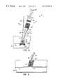
- FIG. 1illustrates an introducer catheter within a percutaneous tissue track and a barrier assembly passing through the introducer catheter with the semipermeable barrier within the blood vessel in its laterally retracted, undeployed configuration
- FIG. 2is similar to FIG. 1, but with the introducer sheath removed from the percutaneous tissue track and the barrier in its laterally expanded, deployed configuration covering the access opening in the blood vessel;
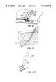
- FIG. 3shows a flowable material delivery tube passing over a thread extending from the distal end of the barrier sheath of FIG. 2, the distal end of the delivery tube being generally aligned with the attachment point of the thread to the barrier sheath;
- FIG. 4illustrates a percutaneous tissue track closure assembly made according to the invention showing the barrier actuator extending from the open proximal end of the barrier sheath, a syringe filled with a hemostatic flowable material secured to the Luer fitting at the proximal end of the delivery tube and the introduction of the hemostatic flowable material from the syringe through the open distal end of the delivery tube into the percutaneous tissue track with the hemostatic flowable material being prevented from entering the blood vessel by the deployed barrier;
- FIG. 5illustrates the barrier assembly and delivery tube being withdrawn from the percutaneous tissue track after the percutaneous tissue track has been substantially filled with the hemostatic flowable material and the hemostatic flowable material has interacted with blood passing through the semipermeable barrier to effectively form a plug made of swollen flowable material and clotted blood;
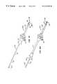
- FIG. 6illustrates an alternative embodiment of the invention in which the thread-type delivery tube alignment device of FIGS. 1-5 has been replaced by guides positioned along the barrier carrier which engage the delivery tube, the delivery tube including a stop to properly position the open distal end of the delivery tube relative to the distal end of the barrier carrier;
- FIGS. 7 and 8illustrate further alternative embodiments of the invention in which the barrier sheath and delivery tube include slides and slide openings to guide the delivery tube along the barrier sheath;
- FIG. 9illustrates three alternative embodiments of differently shaped slides which could be used with the embodiments of FIG. 7 and 8;
- FIG. 10illustrates a further embodiment of the invention in which the barrier carrier and delivery tube are combined into a single structure including a main lumen, through which the flowable material passes, and a supplemental lumen, through which the barrier actuator passes, the combination tube having a number of flowable material exits at the distal end of the combination tube and along the length of the combination tube;
- FIG. 11illustrates an alternative embodiment of the barrier assembly of FIG. 2 in which the barrier sheath has been replaced by a solid barrier carrier with the barrier actuator being external of the barrier carrier and guided along the barrier carrier by several guide loops;
- FIG. 12is a view similar to FIG. 3 but with the thread passing out through a hole at the distal end of the flowable material delivery tube;
- FIG. 13illustrates an alternative embodiment of the barrier assembly of FIGS. 1-5 with the barrier within a blood vessel in a collapsed condition
- FIGS. 13A and 13Bare enlarged views which show the distal end of the barrier assembly of FIG. 13 in a radially-expanded, deployed condition;
- FIG. 13Cshows the barrier assembly of FIG. 13 with the barrier in the deployed condition of FIGS. 13A and 13B and the introducer sheath removed;
- FIG. 13Dshows the barrier assembly of FIG. 13C with the distal end of a flowable material delivery tube positioned adjacent the deployed barrier;
- FIG. 13Eis an enlarged view of the distal ends of the barrier assembly and delivery tube of FIG. 13D;
- FIG. 14is an enlarged isometric view of the distal portion of a further barrier assembly made according to the invention with the barrier in a collapsed configuration;
- FIG. 14Ais a simplified cross-sectional view taken along line 14 A— 14 A of FIG. 14;
- FIG. 14Billustrates the barrier assembly of FIG. 14 with the barrier in a laterally-expanded, fluid-flow-permitting configuration
- FIG. 15illustrates the barrier assembly of FIG. 14 with a further embodiment of a delivery tube mounted over the barrier carrier of the barrier assembly;
- FIG. 15Aillustrates the device of FIG. 15 with the spacer tube retracted opening up an annular flowable material path between the delivery tube and the barrier sheath;
- FIG. 16illustrates a further embodiment of the invention in which the barrier assembly of FIG. 14 has a laterally-collapsible flowable material delivery tube mounted to it;
- FIG. 16Ais a cross-sectional view taken along line 16 A— 16 A of FIG. 16 with the delivery tube in an expanded condition.
- a percutaneous tissue track closure assembly 2is seen to include a barrier assembly 4 , a thread 8 and a flowable material assembly 6 coupled to and aligned with the barrier assembly 4 using thread 8 .
- Thread 8acts as an alignment device for properly positioning the barrier assembly and flowable material assembly relative to one another as will be described in more detail below.
- an introducer sheath 10is shown extending along a percutaneous tissue track 12 and extending a short distance through an access opening 14 formed in the wall 16 of a blood vessel 18 .
- Introducer sheath 10had been used to introduce appropriate medical devices, such as guidewires and catheters, into blood vessel 18 during a prior therapeutic or diagnostic procedure.
- the distal end of the barrier assembly 4is passed through the introducer sheath.
- Barrier assembly 4includes a tubular barrier carrier 20 housing a flexible, wire-like barrier actuator 22 therein.
- the distal end 24 of barrier actuator 22is secured to the center of a semipermeable barrier 26 , the semipermeable barrier being connected to the distal end 28 of barrier carrier 20 .
- Barrier 26is constructed so that it can assume the laterally retracted, undeployed configuration of FIG. 1 or the laterally expanded, deployed configuration of FIG. 2 by either pushing or pulling on barrier actuator 22 . Therefore, barrier actuator 22 is flexible but has sufficient columnar strength to move barrier 26 between the laterally expanded configuration of FIG. 2 and the laterally retracted configuration of FIG. 1 .
- Barrier 26is preferably mesh-like material which permits a restricted flow of blood through the barrier but prevents a hemostatic flowable material 30 , originally within syringe 6 , from passing through barrier 26 and into blood vessel 18 .
- FIG. 2illustrates barrier assembly 4 within percutaneous tissue track 12 after barrier actuator 22 has been pulled to cause barrier 26 to be deformed into its laterally expanded, mushroom-like deployed configuration and introducer sheath 10 has been removed.
- FIG. 2also illustrates thread 8 extending from a position 32 adjacent the distal end 28 of barrier carrier 20 .
- FIG. 3illustrates the placement of a flowable material delivery tube 34 over thread 8 until the open distal end 36 of tube 34 , which acts as the delivery tube exit, is adjacent position 32 at the end of thread 8 .
- thread 8extends out from the Luer fitting 38 at the proximal end of delivery tube 34 .
- Luer fitting 38is mounted to a Luer fitting 40 at the distal end of syringe 6 . As shown in FIG.
- thread 8is captured between the Luer fittings 38 , 40 , thus securing open distal end 36 of delivery tube 34 adjacent position 32 along barrier carrier 20 .
- the userdepresses the thumb pad 42 of syringe 6 causing piston 44 to move distally in the direction of the arrow to force hemostatic flowable material 30 from syringe 6 , through tube 34 , out open distal end 36 , and into tissue track 12 .
- FIG. 4also shows alignment markings, indicators or indicia 45 on barrier carrier 20 .
- Markings 45can be used instead of or in addition to thread 8 as an alignment device.
- markings 45are positioned to be aligned with the lower edge of Luer fitting 38 when distal end 36 is properly positioned.
- Hemostatic flowable material 30may be a material which either swells upon contact with an aqueous liquid, such as blood or aqueous blood components, or causes blood or one or more blood components to clot upon contact with the hemostatic flowable material, and preferably both.
- hemostatic flowable material 30includes a bioabsorbable, flowable, granular gel as described in U.S. patent application Ser. Nos. 09/032,370, filed Feb. 27, 1998; Ser. No. 08/903,674, filed Jul. 31, 1997; No. 60/050,437, filed Jun. 18, 1997; and Ser. No. 08/704,852, filed Aug. 27, 1996, entitled Fragmented Polymeric Compositions and Methods for Their Use.
- hemostatic flowable material 30includes thrombin or thrombin and fibrinogen as the clotting agent.
- Flowable material delivery tube 34is preferably at least a 16 gauge, and preferably a 15 gauge, tube.
- Flowable material 30can also include other agents, such as antibacterial agents, antifibrinolytic agents, or bacteriostatic agents.
- percutaneous tissue track 12can be sufficiently filled without moving open distal end 36 of delivery tube 34 from the position as shown in FIG. 4 .
- One way to sever thread 8would be to include a cutout or notch at distal end 36 of tube 34 so that the user could catch the end of thread 8 within the cutout or notch and then rotate assembly 6 until the thread is severed. At this point, open distal end 36 can be backed out of path 12 while maintaining barrier assembly 4 in place, thus back-filling tissue track 12 .
- barrier actuator 22is extended to move barrier 26 from the deployed configuration of FIG. 2 to the undeployed configuration of FIG. 1; barrier assembly 4 can then be withdrawn from tissue track 12 as suggested in FIG. 5 . Any opening or gap which may be left by the retreating barrier carrier 20 and tube 34 will be quickly filled by hemostatic flowable material 30 .
- FIGS. 6-10illustrate alternative embodiments with like reference numerals referring to like elements.
- Barrier carrier 20 Aincludes at least two delivery guides 46 which guide the movement of delivery tube 34 A along barrier carrier 20 A.
- Delivery tube 34 Aincludes a stop 48 which engages the proximal-most guide 46 when the open distal end 36 of delivery tube 34 A is properly aligned at the distal end 28 of barrier carrier 20 A.
- FIG. 7illustrates a different type of guide element in which barrier carrier 20 B includes a slide opening 50 and delivery tube 34 B includes a complementary, T-shaped slide 52 .
- Delivery tube 34 Bwould preferably include a stop element similar to stop 48 of FIG. 6; such a stop element is not shown in FIG. 7 .
- FIG. 8illustrates an alternative embodiment of the structure of FIG. 7 in which slide opening 50 C is formed in delivery tube 34 C, rather than as a part of barrier carrier 20 B, and slide 52 C is formed as an extension of barrier carrier 20 C. The fit between slide opening 50 C and slide 52 C may be relatively tight so that substantially no hemostatic flowable material can flow through the gap between the two.
- FIG. 9illustrates three alternatively-shaped slides 52 D, 52 E and 52 F which could be used with embodiments similar to the embodiments of FIGS. 7 and 8.
- FIG. 10illustrates an embodiment in which the barrier carrier and delivery tube are incorporated into a combination tube 56 .
- Combination tube 56includes a main lumen 58 , through which flowable material 30 passes, and a supplemental lumen 60 , through which barrier actuator 22 passes.
- tube 56has a number of flowable material exits 62 , 64 along at least part of its length; this helps eliminate the need for backing the delivery tube out of tissue track 12 to back fill the tissue track with flowable material 30 .
- combination tube 56acts as the barrier carrier alignment device to eliminate the need for thread 8 of FIGS. 1-5 and 11 , markings 45 of FIG. 4, guides 46 and stop 48 of FIG. 6, and slide opening 50 and slides 52 of FIGS. 7-9.
- FIG. 11illustrates an embodiment in which the barrier carrier has been replaced by an elongate barrier carrier 20 D.
- Barrier carrier 20 Dis solid but has a number of guide loops 66 extending from the barrier carrier along its length to guide barrier actuator 22 .
- FIG. 12illustrates a further embodiment in which thread 8 passes through the open distal end 36 E of tube 34 E and then through a hole 70 formed in tube 34 E. This eliminates the need to sever thread 8 when it is desired to back-fill tissue track 12 .
- FIG. 13illustrates a further barrier assembly 4 F which uses, as shown in FIGS. 13A and 13B, an outer tube 20 F as the barrier carrier and an inner tube 22 F as the barrier actuator.
- Barrier 26 Fis mounted over the distal end of outer tube 20 F.
- Outer tube 20 Fhas a number, such as four, of axially-extending slits 72 located centrally beneath barrier 26 F. Pulling inner tube 22 F axially relative to outer tube 20 F causes the slit region of the outer tube to buckle outwardly from the collapsed condition of FIG. 13 to the expanded, deployed condition of FIGS. 13A-13E.
- FIG. 13shows barrier assembly 4 F having been passed through introducer sheath 10 with barrier 26 F within blood vessel 18 .
- FIG. 13Cillustrates barrier 26 F in a deployed condition, pressed against the wall 16 of the blood vessel with introducer sheath 10 removed.
- FIG. 13Dshows a delivery tube 34 F having an integral tube clip 74 , see FIG. 13E, at its distal end which clips to and slides along outer tube 20 F.
- Fitting 38 Fcan be coupled a source of hemostatic flowable material, such as a syringe.
- FIG. 14illustrates the distal end of the further alternative embodiment of a barrier assembly 4 G which is somewhat similar to the embodiment of FIG. 13B but differs primarily in that it does not include the semipermeable barrier 26 F of the FIG. 13B embodiment.
- Barrier assembly 4includes a barrier carrier 20 G including a first, outer barrier carrier tube 76 and a second, inner barrier carrier tube 78 .
- Tubes 76 , 78each have a series of four equally-spaced slits 80 , 82 , see FIG. 14A, at their distal ends.
- Slits 80 , 82are located between the tip 84 of barrier carrier 20 G and a metallic stop ring 86 , the use of which is described below.
- Tubes 76 , 78are free to move relative to one another in the area of slits 80 , 82 . However, tubes 76 , 78 are prevented from any significant relative longitudinal or rotational movement so that by pulling on barrier actuator 22 G, both tubes 76 , 78 buckle in the region of slits 80 , 82 . This causes the laterally-expandable arms 88 , 90 to buckle, that is deflected outwardly, to the deployed configuration of FIG. 14 B. As seen in FIGS. 14A and 14B, slits 80 , 82 are circumferentially offset so arms 90 of inner barrier carrier tube 78 extend through the opening created between the outwardly deflected arms 88 of outer barrier carrier tube 76 .
- Gaps 91are small enough to prevent flow of hemostatic flowable material 30 therethrough but large enough to permit passage of a suitable amount of blood into tissue track 12 for interaction with material 30 .
- FIG. 15illustrates a further embodiment of the invention using barrier assembly 4 G of FIG. 14 .
- Barrier assembly 4 Gis housed within a spacer tube 92 , the spacer tube being housed within a hollow delivery tube 34 H.
- the distal end 94 of spacer tube 92abuts stop ring 86 and is tapered to provide a smooth transition between barrier assembly 4 G and delivery tube 34 H as tube 92 is introduced into tissue track 12 .
- barrier actuator 22 Gis pulled thus causing arms 88 , 90 to be laterally expanded so that the barrier is in a deployed position.
- Spacer tube 92is then partially withdrawn as shown in FIG.
- FIGS. 15Ato permit material 30 to be introduced into the generally annular flowable material path 96 defined between delivery tube 34 H and barrier carrier tube 76 .
- Flowable material 30passes through a flowable material delivery port 98 at the proximal end of delivery tube 34 H, along path 96 and out of the exit 99 of path 96 .
- the embodiment of FIGS. 15 and 15Apermits the flowable material to be properly introduced adjacent to barrier 26 G and backfilled up into tissue path 12 .
- spacer tube 92 and delivery tube 34 Hcan be removed from barrier carrier 20 G.
- barrier actuator 22 Gis pushed distally causing barrier 26 G to move to the collapsed configuration of FIG. 14 to permit barrier assembly 4 G to be removed from the tissue track.
- FIGS. 16 and 16Aillustrate a further embodiment of the invention incorporating barrier assembly 4 G of FIG. 14 together with a laterally collapsible delivery tube 34 I.
- Laterally-collapsible delivery tube 34 Iis mounted over outer barrier carrier tube 76 and defines a flexible, laterally-collapsible flowable material path 100 having an entrance 102 at a proximal end of path 100 and an exit 104 at a distal end of path 100 adjacent to barrier 26 G.
- Material 30is introduced into path 100 at entrance 102 through the use of a tube 106 having a fitting 108 at its proximal end coupleable to a conventional syringe or other supply of hemostatic flowable material 30 .
- Tube 106need not be inserted very far along path 100 of tube 341 to provide a sufficient seal between laterally-collapsible tube 341 and tube 106 .
- tube 34 Iis made of heat-shrinkable polyester; however, other materials, such as PET, PETG or PVC, could also be used.
- Path 100is shown in FIG. 16A as being somewhat kidney-shaped. Other shapes for path 100 when laterally-collapsible tube 341 is in its expanded or extended condition can also be used.
- laterally-collapsible tube 34 Iis mounted over outer barrier carrier tube 76 through the use of an integral mounting sleeve 110 surrounding tube 76 . If desired, other methods of mounting tube 34 I to tube 76 could be used, such as through the use of an adhesive or heat bonding.
- barrier assembly 4 G with delivery tube 34 I mounted theretois typically deployed through an introducer sheath.
- the introducer sheathwould then be removed, actuator 22 G would be actuated to cause barrier 26 G to be deployed, and material 30 would be introduced into percutaneous tissue track 12 using tube 106 inserted through entrance 102 of flowable material path 100 .
- barrier actuator 22 Gis pushed distally relative to tubes 76 , 78 causing barrier 26 to move from the deployed configuration shown in FIG. 14B to the undeployed configuration of FIG. 14 .
- Barrier assembly, 4 G and flowable material delivery tube 341 therewithcan then be removed from tissue track 12 .
Landscapes
- Health & Medical Sciences (AREA)
- Surgery (AREA)
- Life Sciences & Earth Sciences (AREA)
- Biomedical Technology (AREA)
- Nuclear Medicine, Radiotherapy & Molecular Imaging (AREA)
- Engineering & Computer Science (AREA)
- Cardiology (AREA)
- Heart & Thoracic Surgery (AREA)
- Medical Informatics (AREA)
- Molecular Biology (AREA)
- Animal Behavior & Ethology (AREA)
- General Health & Medical Sciences (AREA)
- Public Health (AREA)
- Veterinary Medicine (AREA)
- Surgical Instruments (AREA)
Abstract
Description
Claims (68)
Priority Applications (8)
| Application Number | Priority Date | Filing Date | Title |
|---|---|---|---|
| US09/361,663US6334865B1 (en) | 1998-08-04 | 1999-07-27 | Percutaneous tissue track closure assembly and method |
| JP2000563193AJP2003521270A (en) | 1998-08-04 | 1999-07-30 | Percutaneous tissue tract occlusion assemblies and methods |
| AU52477/99AAU5247799A (en) | 1998-08-04 | 1999-07-30 | Percutaneous tissue track closure assembly and method |
| PCT/US1999/017361WO2000007505A1 (en) | 1998-08-04 | 1999-07-30 | Percutaneous tissue track closure assembly and method |
| EP99937696AEP1109498A4 (en) | 1998-08-04 | 1999-07-30 | Percutaneous tissue track closure assembly and method |
| US09/882,296US6613070B2 (en) | 1998-08-04 | 2001-06-14 | System and method for sealing vascular penetrations with hemostatic gels |
| US09/957,176US6699262B2 (en) | 1998-08-04 | 2001-09-19 | Percutaneous tissue track closure assembly and method |
| US10/776,479US20040162578A1 (en) | 1998-08-04 | 2004-02-10 | Percutaneous tissue track closure assembly and method |
Applications Claiming Priority (2)
| Application Number | Priority Date | Filing Date | Title |
|---|---|---|---|
| US99530698P | 1998-08-04 | 1998-08-04 | |
| US09/361,663US6334865B1 (en) | 1998-08-04 | 1999-07-27 | Percutaneous tissue track closure assembly and method |
Related Child Applications (2)
| Application Number | Title | Priority Date | Filing Date |
|---|---|---|---|
| US09/882,296Continuation-In-PartUS6613070B2 (en) | 1998-08-04 | 2001-06-14 | System and method for sealing vascular penetrations with hemostatic gels |
| US09/957,176DivisionUS6699262B2 (en) | 1998-08-04 | 2001-09-19 | Percutaneous tissue track closure assembly and method |
Publications (1)
| Publication Number | Publication Date |
|---|---|
| US6334865B1true US6334865B1 (en) | 2002-01-01 |
Family
ID=27001377
Family Applications (1)
| Application Number | Title | Priority Date | Filing Date |
|---|---|---|---|
| US09/361,663Expired - Fee RelatedUS6334865B1 (en) | 1998-08-04 | 1999-07-27 | Percutaneous tissue track closure assembly and method |
Country Status (1)
| Country | Link |
|---|---|
| US (1) | US6334865B1 (en) |
Cited By (170)
| Publication number | Priority date | Publication date | Assignee | Title |
|---|---|---|---|---|
| US20020026208A1 (en)* | 2000-01-05 | 2002-02-28 | Medical Technology Group, Inc. | Apparatus and methods for delivering a closure device |
| US20020072768A1 (en)* | 2000-12-07 | 2002-06-13 | Ginn Richard S. | Apparatus and methods for providing tactile feedback while delivering a closure device |
| US20020133193A1 (en)* | 2000-01-05 | 2002-09-19 | Ginn Richard S. | Integrated vascular device with puncture site closure component and sealant and methods of use |
| US20030021823A1 (en)* | 2001-06-27 | 2003-01-30 | Rudiger Landers | Coated polymer material, its use and process for its production |
| US20030078234A1 (en)* | 2001-02-12 | 2003-04-24 | Marine Polymer Technologies Inc. | Methods for treating a breach or puncture in a blood vessel |
| US20030078598A1 (en)* | 2000-01-05 | 2003-04-24 | Integrated Vascular Systems, Inc. | Vascular sheath with bioabsorbable puncture site closure apparatus and methods of use |
| US20030144695A1 (en)* | 1999-09-13 | 2003-07-31 | Mcguckin James F. | Vascular hole closure device |
| US20040009289A1 (en)* | 2000-12-07 | 2004-01-15 | Carley Michael T. | Closure device and methods for making and using them |
| US6689148B2 (en) | 1998-08-14 | 2004-02-10 | Incept Llc | Methods and apparatus for intraluminal deposition of hydrogels |
| US20040060410A1 (en)* | 2002-09-30 | 2004-04-01 | Leung Jeffrey C. | Barbed sutures |
| US20040073255A1 (en)* | 2002-02-21 | 2004-04-15 | Ginn Richard S | Plunger apparatus and methods for delivering a closure device |
| US20040093027A1 (en)* | 2002-03-04 | 2004-05-13 | Walter Fabisiak | Barbed tissue connector for sealing vascular puncture wounds |
| US6749622B2 (en) | 1999-09-13 | 2004-06-15 | Rex Medical, L.P. | Vascular closure |
| US20040122452A1 (en)* | 2001-05-30 | 2004-06-24 | Satiety, Inc. | Obesity treatment tools and methods |
| US20040153122A1 (en)* | 2003-01-30 | 2004-08-05 | Integrated Vascular Systems, Inc. | Clip applier and methods of use |
| US20040243052A1 (en)* | 2003-06-02 | 2004-12-02 | Vascular Solutions, Inc. | Vascular access closure system |
| US20050010248A1 (en)* | 2003-07-10 | 2005-01-13 | Scimed Life Systems, Inc. | System for closing an opening in a body cavity |
| US20050033326A1 (en)* | 1999-09-13 | 2005-02-10 | Briganti Richard T. | Vascular hole closure device |
| US20050049634A1 (en)* | 2003-08-07 | 2005-03-03 | Scimed Life Systems, Inc. | Medical closure device |
| US20050080452A1 (en)* | 2002-06-12 | 2005-04-14 | Radi Medical Systems Ab | Closure device |
| US20050119695A1 (en)* | 2000-12-07 | 2005-06-02 | Carley Michael T. | Closure device and methods for making and using them |
| US20050131459A1 (en)* | 2002-06-12 | 2005-06-16 | Radi Medical Systems Ab | Closure device |
| US20050155612A1 (en)* | 1999-09-20 | 2005-07-21 | Nuvasive, Inc. | Annulotomy closure device and related methods |
| US6932833B1 (en) | 2002-04-01 | 2005-08-23 | Bobby W. Presley | Method and barrier for limiting fluid movement through a tissue rent |
| US20050216057A1 (en)* | 2000-09-08 | 2005-09-29 | James Coleman | Surgical stapler |
| US20050248062A1 (en)* | 2004-05-10 | 2005-11-10 | Alexandr Shkolnik | Process for the production of a three-dimensional object with resolution improvement by "pixel-shift" |
| US20050248061A1 (en)* | 2004-05-07 | 2005-11-10 | Alexandr Shkolnik | Process for the production of a three-dimensional object with an improved separation of hardened material layers from a construction plane |
| US20050267531A1 (en)* | 2004-05-14 | 2005-12-01 | Ruff Gregory L | Suture methods and devices |
| US20050267530A1 (en)* | 2001-06-07 | 2005-12-01 | Christy Cummins | Surgical staple |
| US20050273136A1 (en)* | 2000-01-05 | 2005-12-08 | Integrated Vascular Systems, Inc. | Apparatus and methods for delivering a closure device |
| US20050274768A1 (en)* | 2004-05-25 | 2005-12-15 | Christy Cummins | Surgical stapler |
| US20050283193A1 (en)* | 2004-06-18 | 2005-12-22 | Radi Medical Systems Ab | Introducer guide |
| US20060115805A1 (en)* | 2002-12-11 | 2006-06-01 | Hansen John E | Gelatine-based materials as swabs |
| US20060116635A1 (en)* | 2004-11-29 | 2006-06-01 | Med Enclosure L.L.C. | Arterial closure device |
| US20060122462A1 (en)* | 2004-11-17 | 2006-06-08 | Roth Alex T | Remote tissue retraction device |
| US20060144479A1 (en)* | 2002-12-31 | 2006-07-06 | Integrated Vascular Systems, Inc. | Methods for manufacturing a clip and clip |
| US20060155327A1 (en)* | 1999-09-13 | 2006-07-13 | Briganti Richard T | Vascular hole closure device |
| US20060190037A1 (en)* | 2000-01-05 | 2006-08-24 | Ginn Richard S | Integrated vascular device with puncture site closure component and sealant and methods of use |
| US20060276839A1 (en)* | 1999-09-13 | 2006-12-07 | Rex Medical | Septal defect closure device |
| US20070009578A1 (en)* | 2004-07-09 | 2007-01-11 | Lene Moller | Haemostatic composition comprising hyaluronic acid |
| US20070021778A1 (en)* | 2005-06-24 | 2007-01-25 | Abbott Laboratories Abbott Vascular Devices | Apparatus and method for delivering a closure element |
| US20070049968A1 (en)* | 2005-08-24 | 2007-03-01 | Sibbitt Wilmer L Jr | Vascular opening edge eversion methods and apparatuses |
| US20070160543A1 (en)* | 2004-01-30 | 2007-07-12 | Lene Moller | Haemostatic sprays and compositions |
| US20070208360A1 (en)* | 2004-02-13 | 2007-09-06 | Demarais Denise M | Methods and devices for reducing hollow organ volume |
| US20070250080A1 (en)* | 2006-04-20 | 2007-10-25 | Integrated Vascular Systems, Inc. | Resettable clip applier and reset tools |
| US20070260349A1 (en)* | 2006-04-28 | 2007-11-08 | Envisiontec Gmbh | Device and method for producing a three-dimensional object by means of mask exposure |
| US20070270891A1 (en)* | 2005-04-22 | 2007-11-22 | Mcguckin James F Jr | Closure device for left atrial appendage |
| US20080004636A1 (en)* | 2005-07-01 | 2008-01-03 | Abbott Laboratories | Clip applier and methods of use |
| US20080021586A1 (en)* | 2006-07-19 | 2008-01-24 | Volker Schillen | Method and device for producing a three-dimensional object, and computer and data carrier useful therefor |
| US20080038396A1 (en)* | 2006-04-28 | 2008-02-14 | Envisiontec Gmbh | Device and method for producing a three-dimensional object by means of mask exposure |
| US7341595B2 (en) | 1999-09-13 | 2008-03-11 | Rex Medical, L.P | Vascular hole closure device |
| US20080065152A1 (en)* | 2006-09-08 | 2008-03-13 | Abbott Laboratories | Apparatus and method for delivering a closure element |
| US20080086109A1 (en)* | 2004-11-29 | 2008-04-10 | Paul Shabty | Arterial closure device |
| US20080113293A1 (en)* | 2006-11-15 | 2008-05-15 | Alexandr Shkolnik | Continuous generative process for producing a three-dimensional object |
| US20080132925A1 (en)* | 2004-02-27 | 2008-06-05 | Satiety, Inc. | Methods and devices for reducing hollow organ volume |
| US20080161645A1 (en)* | 2005-02-10 | 2008-07-03 | G.I. View Ltd. | Advancement Techniques For Gastrointestinal Tool With Guiding Element |
| US20080208205A1 (en)* | 2007-02-26 | 2008-08-28 | Paul Edward Kraemer | Cable system and methods |
| US20080255612A1 (en)* | 2007-04-13 | 2008-10-16 | Angiotech Pharmaceuticals, Inc. | Self-retaining systems for surgical procedures |
| US20080312686A1 (en)* | 2005-07-01 | 2008-12-18 | Abbott Laboratories | Antimicrobial closure element and closure element applier |
| US20080319475A1 (en)* | 2007-06-25 | 2008-12-25 | Abbott Laboratories | Methods, Devices, and Apparatus for Managing Access Through Tissue |
| US20090020901A1 (en)* | 2007-07-04 | 2009-01-22 | Envisiontec Gmbh | Process and device for producing a three-dimensional object |
| US20090112259A1 (en)* | 2007-10-31 | 2009-04-30 | Angiotech Pharmaceuticals, Inc. | Recombinant expressed bioadsorbable polyhydroxyalkonate monofilament and multi-filaments self-retaining sutures |
| US20090130449A1 (en)* | 2007-10-26 | 2009-05-21 | Envisiontec Gmbh | Process and freeform fabrication system for producing a three-dimensional object |
| US20090143819A1 (en)* | 2007-10-31 | 2009-06-04 | D Agostino William L | Coatings for modifying monofilament and multi-filaments self-retaining sutures |
| US20090157101A1 (en)* | 2007-12-17 | 2009-06-18 | Abbott Laboratories | Tissue closure system and methods of use |
| US20090157102A1 (en)* | 2007-12-17 | 2009-06-18 | Abbott Laboratories | Clip applier and methods of use |
| US20090157103A1 (en)* | 2007-12-18 | 2009-06-18 | Abbott Laboratories | Modular clip applier |
| US20090187213A1 (en)* | 2007-07-13 | 2009-07-23 | Mcguckin Jr James F | Vascular hole closure device |
| US20090187215A1 (en)* | 2007-12-19 | 2009-07-23 | Abbott Laboratories | Methods and apparatus to reduce a dimension of an implantable device in a smaller state |
| US20090204065A1 (en)* | 2007-03-29 | 2009-08-13 | Wright Clifford A | Suction wand |
| US20090228021A1 (en)* | 2008-03-06 | 2009-09-10 | Leung Jeffrey C | Matrix material |
| US20090287244A1 (en)* | 2008-05-16 | 2009-11-19 | Abbott Laboratories Vascular Enterprises Limited | Apparatus and methods for engaging tissue |
| US20090287229A1 (en)* | 2008-05-19 | 2009-11-19 | Ams Research Corporation | Collapsible Tissue Anchor Device and Method |
| USD611144S1 (en) | 2006-06-28 | 2010-03-02 | Abbott Laboratories | Apparatus for delivering a closure element |
| US20100114156A1 (en)* | 2008-10-30 | 2010-05-06 | Abbott Vascular Inc. | Closure device |
| US20100160958A1 (en)* | 2008-12-22 | 2010-06-24 | Abbott Laboratories | Closure Device |
| US20100168790A1 (en)* | 2008-12-22 | 2010-07-01 | Abbott Laboratories | Curved closure device |
| US20100179589A1 (en)* | 2009-01-09 | 2010-07-15 | Abbott Vascular Inc. | Rapidly eroding anchor |
| US20100179572A1 (en)* | 2009-01-09 | 2010-07-15 | Abbott Vascular Inc. | Closure devices, systems, and methods |
| US20100179590A1 (en)* | 2009-01-09 | 2010-07-15 | Abbott Vascular Inc. | Vessel closure devices and methods |
| US20100185234A1 (en)* | 2009-01-16 | 2010-07-22 | Abbott Vascular Inc. | Closure devices, systems, and methods |
| US20100211000A1 (en)* | 2008-08-26 | 2010-08-19 | Killion Douglas P | Method and system for sealing percutaneous punctures |
| US20100217132A1 (en)* | 2009-02-26 | 2010-08-26 | Abbott Laboratories | Methods and apparatus for locating a surface of a body lumen |
| US7806910B2 (en) | 2002-11-26 | 2010-10-05 | Abbott Laboratories | Multi-element biased suture clip |
| US7806904B2 (en) | 2000-12-07 | 2010-10-05 | Integrated Vascular Systems, Inc. | Closure device |
| US20100298868A1 (en)* | 1993-05-03 | 2010-11-25 | Quill Medical, Inc. | Tissue connector with yieldable barbs equally spaced about the periphery of the connector at successive axial locations |
| US20100298878A1 (en)* | 2002-08-09 | 2010-11-25 | Quill Medical, Inc. | Double ended barbed suture with an intermediate body |
| US20100298848A1 (en)* | 2001-06-29 | 2010-11-25 | Quill Medical, Inc. | Continuous stitch wound closure utilizing one-way suture |
| US20100294107A1 (en)* | 2001-08-31 | 2010-11-25 | Quill Medical, Inc. | Method for cutting a suture to create tissue retainers of a desired shape and size |
| US7850709B2 (en) | 2002-06-04 | 2010-12-14 | Abbott Vascular Inc. | Blood vessel closure clip and delivery device |
| US20110009901A1 (en)* | 2009-07-13 | 2011-01-13 | Tom Holman | Metal vascular aperture closure device |
| US20110021964A1 (en)* | 2008-02-29 | 2011-01-27 | Ferrosan Medical Devices A/S | Device for Promotion of Hemostasis and/or Wound Healing |
| US20110029013A1 (en)* | 2008-02-15 | 2011-02-03 | Mcguckin James F | Vascular Hole Closure Device |
| US20110046669A1 (en)* | 2008-02-21 | 2011-02-24 | Angiotech Pharmaceuticals, Inc. | Method and apparatus for elevating retainers on self-retaining sutures |
| US20110046668A1 (en)* | 2007-12-19 | 2011-02-24 | Angiotech Pharmaceuticals, Inc. | Self-retaining sutures with heat-contact mediated retainers |
| US20110054492A1 (en)* | 2009-08-26 | 2011-03-03 | Abbott Laboratories | Medical device for repairing a fistula |
| US7923431B2 (en) | 2001-12-21 | 2011-04-12 | Ferrosan Medical Devices A/S | Haemostatic kit, a method of preparing a haemostatic agent and a method of promoting haemostatis |
| US20110089610A1 (en)* | 2009-10-19 | 2011-04-21 | Global Filtration Systems | Resin Solidification Substrate and Assembly |
| US20110106152A1 (en)* | 2008-01-30 | 2011-05-05 | Angiotech Pharmaceuticals, Inc. | Apparatus and method for forming self-retaining sutures |
| US20110106148A1 (en)* | 2000-01-05 | 2011-05-05 | Integrated Vascular Systems, Inc. | Closure system and methods of use |
| US20110144664A1 (en)* | 2003-01-30 | 2011-06-16 | Integrated Vascular Systems, Inc. | Clip applier and methods of use |
| US20110224719A1 (en)* | 2010-03-15 | 2011-09-15 | Abbott Cardiovascular Systems, Inc. | Bioabsorbable plug |
| US20110230897A1 (en)* | 2003-01-30 | 2011-09-22 | Integrated Vascular Systems, Inc. | Clip applier and methods of use |
| US8032996B2 (en) | 2003-05-13 | 2011-10-11 | Quill Medical, Inc. | Apparatus for forming barbs on a suture |
| US8048108B2 (en) | 2005-08-24 | 2011-11-01 | Abbott Vascular Inc. | Vascular closure methods and apparatuses |
| US8070772B2 (en) | 2008-02-15 | 2011-12-06 | Rex Medical, L.P. | Vascular hole closure device |
| US8118834B1 (en) | 2007-12-20 | 2012-02-21 | Angiotech Pharmaceuticals, Inc. | Composite self-retaining sutures and method |
| US8202293B2 (en) | 2003-01-30 | 2012-06-19 | Integrated Vascular Systems, Inc. | Clip applier and methods of use |
| US8216273B1 (en) | 2008-02-25 | 2012-07-10 | Ethicon, Inc. | Self-retainers with supporting structures on a suture |
| USRE43955E1 (en) | 2004-05-10 | 2013-02-05 | Envisiontec Gmbh | Process for the production of a three-dimensional object with resolution improvement by pixel-shift |
| US20130138149A1 (en)* | 2011-11-28 | 2013-05-30 | St. Jude Medical Puerto Rico Llc | Large bore anchor device |
| US8465689B2 (en) | 2007-01-17 | 2013-06-18 | 3D Systems, Inc. | Elevator and method for tilting solid image build platform for reducing air entrainment and for build release |
| US8491629B2 (en) | 2008-02-15 | 2013-07-23 | Rex Medical | Vascular hole closure delivery device |
| US8556930B2 (en) | 2006-06-28 | 2013-10-15 | Abbott Laboratories | Vessel closure device |
| US8556932B2 (en) | 2011-05-19 | 2013-10-15 | Abbott Cardiovascular Systems, Inc. | Collapsible plug for tissue closure |
| US8603116B2 (en) | 2010-08-04 | 2013-12-10 | Abbott Cardiovascular Systems, Inc. | Closure device with long tines |
| US8615856B1 (en) | 2008-01-30 | 2013-12-31 | Ethicon, Inc. | Apparatus and method for forming self-retaining sutures |
| US8617184B2 (en) | 2011-02-15 | 2013-12-31 | Abbott Cardiovascular Systems, Inc. | Vessel closure system |
| US8641732B1 (en) | 2008-02-26 | 2014-02-04 | Ethicon, Inc. | Self-retaining suture with variable dimension filament and method |
| US20140058440A1 (en)* | 2012-08-24 | 2014-02-27 | St. Jude Medical Puerto Rico Llc | Balloon bailout and bioadhesive delivery device for suture based closure and methods |
| US8690910B2 (en) | 2000-12-07 | 2014-04-08 | Integrated Vascular Systems, Inc. | Closure device and methods for making and using them |
| US8721681B2 (en) | 2002-09-30 | 2014-05-13 | Ethicon, Inc. | Barbed suture in combination with surgical needle |
| US8758399B2 (en) | 2010-08-02 | 2014-06-24 | Abbott Cardiovascular Systems, Inc. | Expandable bioabsorbable plug apparatus and method |
| US8777987B2 (en) | 2007-09-27 | 2014-07-15 | Ethicon, Inc. | Self-retaining sutures including tissue retainers having improved strength |
| US20140243854A1 (en)* | 2003-06-06 | 2014-08-28 | Abbott Laboratories | Sizing and positioning adapter for medical instruments |
| US8821534B2 (en) | 2010-12-06 | 2014-09-02 | Integrated Vascular Systems, Inc. | Clip applier having improved hemostasis and methods of use |
| US20140257375A1 (en)* | 2013-03-11 | 2014-09-11 | St. Jude Medical Puerto Rico Llc | Active securement detachable sealing tip for extra-vascular closure device and methods |
| US8876865B2 (en) | 2008-04-15 | 2014-11-04 | Ethicon, Inc. | Self-retaining sutures with bi-directional retainers or uni-directional retainers |
| US8916077B1 (en) | 2007-12-19 | 2014-12-23 | Ethicon, Inc. | Self-retaining sutures with retainers formed from molten material |
| US8920463B2 (en) | 2008-02-15 | 2014-12-30 | Rex Medical, L.P. | Vascular hole closure device |
| US8920462B2 (en) | 2008-02-15 | 2014-12-30 | Rex Medical, L.P. | Vascular hole closure device |
| US8932328B2 (en) | 2008-11-03 | 2015-01-13 | Ethicon, Inc. | Length of self-retaining suture and method and device for using the same |
| US8961560B2 (en) | 2008-05-16 | 2015-02-24 | Ethicon, Inc. | Bidirectional self-retaining sutures with laser-marked and/or non-laser marked indicia and methods |
| USRE45426E1 (en) | 1997-05-21 | 2015-03-17 | Ethicon, Inc. | Surgical methods using one-way suture |
| US9089674B2 (en) | 2000-10-06 | 2015-07-28 | Integrated Vascular Systems, Inc. | Apparatus and methods for positioning a vascular sheath |
| US9149276B2 (en) | 2011-03-21 | 2015-10-06 | Abbott Cardiovascular Systems, Inc. | Clip and deployment apparatus for tissue closure |
| US9173644B2 (en) | 2009-01-09 | 2015-11-03 | Abbott Vascular Inc. | Closure devices, systems, and methods |
| US9226738B2 (en) | 2008-02-15 | 2016-01-05 | Rex Medical, L.P. | Vascular hole closure delivery device |
| US9248580B2 (en) | 2002-09-30 | 2016-02-02 | Ethicon, Inc. | Barb configurations for barbed sutures |
| US9265858B2 (en) | 2012-06-12 | 2016-02-23 | Ferrosan Medical Devices A/S | Dry haemostatic composition |
| US9332976B2 (en) | 2011-11-30 | 2016-05-10 | Abbott Cardiovascular Systems, Inc. | Tissue closure device |
| US9364209B2 (en) | 2012-12-21 | 2016-06-14 | Abbott Cardiovascular Systems, Inc. | Articulating suturing device |
| US9456811B2 (en) | 2005-08-24 | 2016-10-04 | Abbott Vascular Inc. | Vascular closure methods and apparatuses |
| US9486191B2 (en) | 2009-01-09 | 2016-11-08 | Abbott Vascular, Inc. | Closure devices |
| US20160345995A1 (en)* | 2014-02-12 | 2016-12-01 | Olympus Corporation | Treatment device |
| US9527244B2 (en) | 2014-02-10 | 2016-12-27 | Global Filtration Systems | Apparatus and method for forming three-dimensional objects from solidifiable paste |
| US9579091B2 (en) | 2000-01-05 | 2017-02-28 | Integrated Vascular Systems, Inc. | Closure system and methods of use |
| US9636035B2 (en) | 2005-12-14 | 2017-05-02 | Scibase Ab | Medical apparatus for determination of biological conditions using impedance measurements |
| US9675341B2 (en) | 2010-11-09 | 2017-06-13 | Ethicon Inc. | Emergency self-retaining sutures and packaging |
| US9724078B2 (en) | 2013-06-21 | 2017-08-08 | Ferrosan Medical Devices A/S | Vacuum expanded dry composition and syringe for retaining same |
| US9955962B2 (en) | 2010-06-11 | 2018-05-01 | Ethicon, Inc. | Suture delivery tools for endoscopic and robot-assisted surgery and methods |
| US10111980B2 (en) | 2013-12-11 | 2018-10-30 | Ferrosan Medical Devices A/S | Dry composition comprising an extrusion enhancer |
| US10188384B2 (en) | 2011-06-06 | 2019-01-29 | Ethicon, Inc. | Methods and devices for soft palate tissue elevation procedures |
| USD843573S1 (en) | 2015-11-13 | 2019-03-19 | Access Closure, Inc. | Vascular closure apparatus |
| US10245011B2 (en) | 2012-12-25 | 2019-04-02 | Osaka University | Hemostatic agent applicator |
| USD847988S1 (en) | 2015-11-13 | 2019-05-07 | Access Closure, Inc. | Handle grip |
| US10420546B2 (en) | 2010-05-04 | 2019-09-24 | Ethicon, Inc. | Self-retaining systems having laser-cut retainers |
| US10456123B2 (en) | 2014-11-14 | 2019-10-29 | Access Closure, Inc. | Apparatus and method for sealing a vascular puncture |
| USD865166S1 (en) | 2015-11-13 | 2019-10-29 | Access Closure, Inc. | Sheath adapter |
| US10492780B2 (en) | 2011-03-23 | 2019-12-03 | Ethicon, Inc. | Self-retaining variable loop sutures |
| US10653837B2 (en) | 2014-12-24 | 2020-05-19 | Ferrosan Medical Devices A/S | Syringe for retaining and mixing first and second substances |
| US10737479B2 (en) | 2017-01-12 | 2020-08-11 | Global Filtration Systems | Method of making three-dimensional objects using both continuous and discontinuous solidification |
| US10918796B2 (en) | 2015-07-03 | 2021-02-16 | Ferrosan Medical Devices A/S | Syringe for mixing two components and for retaining a vacuum in a storage condition |
| US11007296B2 (en) | 2010-11-03 | 2021-05-18 | Ethicon, Inc. | Drug-eluting self-retaining sutures and methods relating thereto |
| US11046818B2 (en) | 2014-10-13 | 2021-06-29 | Ferrosan Medical Devices A/S | Dry composition for use in haemostasis and wound healing |
| US11109849B2 (en) | 2012-03-06 | 2021-09-07 | Ferrosan Medical Devices A/S | Pressurized container containing haemostatic paste |
| US11504105B2 (en) | 2019-01-25 | 2022-11-22 | Rex Medical L.P. | Vascular hole closure device |
| US11801324B2 (en) | 2018-05-09 | 2023-10-31 | Ferrosan Medical Devices A/S | Method for preparing a haemostatic composition |
| CN116965964A (en)* | 2023-08-11 | 2023-10-31 | 江苏省中医院 | Duodenal mucosa remodeling catheter for animal experiments |
| WO2025125953A1 (en)* | 2023-12-13 | 2025-06-19 | Medtronic, Inc. | Devices, systems, and methods for closing tissue punctures |
Citations (17)
| Publication number | Priority date | Publication date | Assignee | Title |
|---|---|---|---|---|
| US4744364A (en) | 1987-02-17 | 1988-05-17 | Intravascular Surgical Instruments, Inc. | Device for sealing percutaneous puncture in a vessel |
| US4890612A (en) | 1987-02-17 | 1990-01-02 | Kensey Nash Corporation | Device for sealing percutaneous puncture in a vessel |
| US5108421A (en) | 1990-10-01 | 1992-04-28 | Quinton Instrument Company | Insertion assembly and method of inserting a vessel plug into the body of a patient |
| EP0493810A1 (en) | 1990-12-27 | 1992-07-08 | NOVOSTE CORPORATION (a Florida Corporation) | Wound clotting device |
| US5221259A (en) | 1990-12-27 | 1993-06-22 | Novoste Corporation | Wound treating device and method of using same |
| US5370660A (en)* | 1993-11-01 | 1994-12-06 | Cordis Corporation | Apparatus and method for delivering a vessel plug into the body of a patient |
| US5383896A (en) | 1993-05-25 | 1995-01-24 | Gershony; Gary | Vascular sealing device |
| US5413571A (en) | 1992-07-16 | 1995-05-09 | Sherwood Medical Company | Device for sealing hemostatic incisions |
| US5419765A (en) | 1990-12-27 | 1995-05-30 | Novoste Corporation | Wound treating device and method for treating wounds |
| US5486195A (en) | 1993-07-26 | 1996-01-23 | Myers; Gene | Method and apparatus for arteriotomy closure |
| US5626601A (en) | 1995-10-27 | 1997-05-06 | Gary Gershony | Vascular sealing apparatus and method |
| US5653730A (en) | 1993-09-28 | 1997-08-05 | Hemodynamics, Inc. | Surface opening adhesive sealer |
| US5728132A (en) | 1996-04-08 | 1998-03-17 | Tricardia, L.L.C. | Self-sealing vascular access device |
| US5752974A (en) | 1995-12-18 | 1998-05-19 | Collagen Corporation | Injectable or implantable biomaterials for filling or blocking lumens and voids of the body |
| US5855559A (en) | 1997-02-14 | 1999-01-05 | Tricardia, Inc. | Hemostatic agent delivery device having built-in pressure sensor |
| US6045570A (en) | 1997-02-11 | 2000-04-04 | Biointerventional Corporation | Biological sealant mixture and system for use in percutaneous occlusion of puncture sites and tracts in the human body and method |
| US6193670B1 (en) | 1997-02-14 | 2001-02-27 | Tricardia, Llc | Hemostatic agent delivery device having built-in pressure sensor |
- 1999
- 1999-07-27USUS09/361,663patent/US6334865B1/ennot_activeExpired - Fee Related
Patent Citations (19)
| Publication number | Priority date | Publication date | Assignee | Title |
|---|---|---|---|---|
| US4744364A (en) | 1987-02-17 | 1988-05-17 | Intravascular Surgical Instruments, Inc. | Device for sealing percutaneous puncture in a vessel |
| US4890612A (en) | 1987-02-17 | 1990-01-02 | Kensey Nash Corporation | Device for sealing percutaneous puncture in a vessel |
| US5108421A (en) | 1990-10-01 | 1992-04-28 | Quinton Instrument Company | Insertion assembly and method of inserting a vessel plug into the body of a patient |
| US5591205A (en) | 1990-10-01 | 1997-01-07 | Quinton Instrument Company | Insertion assembly and method of inserting a vessel plug into the body of a patient |
| EP0493810A1 (en) | 1990-12-27 | 1992-07-08 | NOVOSTE CORPORATION (a Florida Corporation) | Wound clotting device |
| US5221259A (en) | 1990-12-27 | 1993-06-22 | Novoste Corporation | Wound treating device and method of using same |
| US5330446A (en) | 1990-12-27 | 1994-07-19 | Weldon Thomas D | Wound treating device using an inflatable membrane and method for treating wounds |
| US5419765A (en) | 1990-12-27 | 1995-05-30 | Novoste Corporation | Wound treating device and method for treating wounds |
| US5413571A (en) | 1992-07-16 | 1995-05-09 | Sherwood Medical Company | Device for sealing hemostatic incisions |
| US5383896A (en) | 1993-05-25 | 1995-01-24 | Gershony; Gary | Vascular sealing device |
| US5486195A (en) | 1993-07-26 | 1996-01-23 | Myers; Gene | Method and apparatus for arteriotomy closure |
| US5653730A (en) | 1993-09-28 | 1997-08-05 | Hemodynamics, Inc. | Surface opening adhesive sealer |
| US5370660A (en)* | 1993-11-01 | 1994-12-06 | Cordis Corporation | Apparatus and method for delivering a vessel plug into the body of a patient |
| US5626601A (en) | 1995-10-27 | 1997-05-06 | Gary Gershony | Vascular sealing apparatus and method |
| US5752974A (en) | 1995-12-18 | 1998-05-19 | Collagen Corporation | Injectable or implantable biomaterials for filling or blocking lumens and voids of the body |
| US5728132A (en) | 1996-04-08 | 1998-03-17 | Tricardia, L.L.C. | Self-sealing vascular access device |
| US6045570A (en) | 1997-02-11 | 2000-04-04 | Biointerventional Corporation | Biological sealant mixture and system for use in percutaneous occlusion of puncture sites and tracts in the human body and method |
| US5855559A (en) | 1997-02-14 | 1999-01-05 | Tricardia, Inc. | Hemostatic agent delivery device having built-in pressure sensor |
| US6193670B1 (en) | 1997-02-14 | 2001-02-27 | Tricardia, Llc | Hemostatic agent delivery device having built-in pressure sensor |
Cited By (469)
| Publication number | Priority date | Publication date | Assignee | Title |
|---|---|---|---|---|
| US20100298868A1 (en)* | 1993-05-03 | 2010-11-25 | Quill Medical, Inc. | Tissue connector with yieldable barbs equally spaced about the periphery of the connector at successive axial locations |
| US8246652B2 (en) | 1993-05-03 | 2012-08-21 | Ethicon, Inc. | Suture with a pointed end and an anchor end and with equally spaced yieldable tissue grasping barbs located at successive axial locations |
| US20100298637A1 (en)* | 1993-05-03 | 2010-11-25 | Quill Medical, Inc. | Suture with a pointed end and an anchor end and with equally spaced tissue grasping protrusions located at successive axial locations |
| USRE45426E1 (en) | 1997-05-21 | 2015-03-17 | Ethicon, Inc. | Surgical methods using one-way suture |
| US7776063B2 (en) | 1998-08-14 | 2010-08-17 | Incept Llc | In situ materials formation |
| US20070288052A1 (en)* | 1998-08-14 | 2007-12-13 | Incept Llc | In situ materials formation |
| US7914541B2 (en) | 1998-08-14 | 2011-03-29 | Incept, Llc | In situ materials formation |
| US20080132936A1 (en)* | 1998-08-14 | 2008-06-05 | Incept Llc | In situ materials formation |
| US20050080445A1 (en)* | 1998-08-14 | 2005-04-14 | Incept Llc | Methods and apparatus for intraluminal deposition of hydrogels |
| US6689148B2 (en) | 1998-08-14 | 2004-02-10 | Incept Llc | Methods and apparatus for intraluminal deposition of hydrogels |
| US7220270B2 (en) | 1998-08-14 | 2007-05-22 | Incept Llc | Methods and apparatus for intraluminal deposition of hydrogels |
| US8083766B2 (en) | 1999-09-13 | 2011-12-27 | Rex Medical, Lp | Septal defect closure device |
| US7942888B2 (en) | 1999-09-13 | 2011-05-17 | Rex Medical, L.P. | Vascular hole closure device |
| US6749622B2 (en) | 1999-09-13 | 2004-06-15 | Rex Medical, L.P. | Vascular closure |
| US7341595B2 (en) | 1999-09-13 | 2008-03-11 | Rex Medical, L.P | Vascular hole closure device |
| US7662161B2 (en) | 1999-09-13 | 2010-02-16 | Rex Medical, L.P | Vascular hole closure device |
| US8597324B2 (en) | 1999-09-13 | 2013-12-03 | Rex Medical L.P. | Vascular hole closure device |
| US7662168B2 (en) | 1999-09-13 | 2010-02-16 | Rex Medical, L.P. | Vascular closure |
| US20100305588A1 (en)* | 1999-09-13 | 2010-12-02 | Briganti Richard T | Vascular hole closure device |
| US20050033326A1 (en)* | 1999-09-13 | 2005-02-10 | Briganti Richard T. | Vascular hole closure device |
| US9968345B2 (en) | 1999-09-13 | 2018-05-15 | Rex Medical, L.P. | Vascular hole closure device |
| US20050055049A1 (en)* | 1999-09-13 | 2005-03-10 | Mcguckin James F. | Vascular closure |
| US20030144695A1 (en)* | 1999-09-13 | 2003-07-31 | Mcguckin James F. | Vascular hole closure device |
| US20060276839A1 (en)* | 1999-09-13 | 2006-12-07 | Rex Medical | Septal defect closure device |
| US20070293880A1 (en)* | 1999-09-13 | 2007-12-20 | Rex Medical | Vascular hole closure device |
| US20060155327A1 (en)* | 1999-09-13 | 2006-07-13 | Briganti Richard T | Vascular hole closure device |
| US8128653B2 (en) | 1999-09-13 | 2012-03-06 | Rex Medical, L.P. | Vascular hole closure device |
| US7267679B2 (en) | 1999-09-13 | 2007-09-11 | Rex Medical, L.P | Vascular hole closure device |
| US6964674B1 (en)* | 1999-09-20 | 2005-11-15 | Nuvasive, Inc. | Annulotomy closure device |
| US7901430B2 (en) | 1999-09-20 | 2011-03-08 | Nuvasive, Inc. | Annulotomy closure device and related methods |
| US20080071301A1 (en)* | 1999-09-20 | 2008-03-20 | Nuvasive, Inc. | Annulotomy closure device and related methods |
| US7883527B2 (en) | 1999-09-20 | 2011-02-08 | Nuvasive, Inc. | Annulotomy closure device and related methods |
| US20080140108A1 (en)* | 1999-09-20 | 2008-06-12 | Nuvasive, Inc | Annulotomy closure device and related methods |
| US20050155612A1 (en)* | 1999-09-20 | 2005-07-21 | Nuvasive, Inc. | Annulotomy closure device and related methods |
| US9277903B2 (en) | 1999-09-20 | 2016-03-08 | Nuvasive, Inc. | Annulotomy closure device and related methods |
| US20020026208A1 (en)* | 2000-01-05 | 2002-02-28 | Medical Technology Group, Inc. | Apparatus and methods for delivering a closure device |
| US6780197B2 (en)* | 2000-01-05 | 2004-08-24 | Integrated Vascular Systems, Inc. | Apparatus and methods for delivering a vascular closure device to a body lumen |
| US20050273136A1 (en)* | 2000-01-05 | 2005-12-08 | Integrated Vascular Systems, Inc. | Apparatus and methods for delivering a closure device |
| US20080221616A1 (en)* | 2000-01-05 | 2008-09-11 | Integrated Vascular Systems, Inc. | Integrated vascular device with puncture site closure component and sealant and methods of use |
| US20080210737A1 (en)* | 2000-01-05 | 2008-09-04 | Integrated Vascular Systems, Inc. | Integrated vascular device with puncture site closure component and sealant and methods of use |
| US7931669B2 (en) | 2000-01-05 | 2011-04-26 | Integrated Vascular Systems, Inc. | Integrated vascular device with puncture site closure component and sealant and methods of use |
| US20020133193A1 (en)* | 2000-01-05 | 2002-09-19 | Ginn Richard S. | Integrated vascular device with puncture site closure component and sealant and methods of use |
| US9579091B2 (en) | 2000-01-05 | 2017-02-28 | Integrated Vascular Systems, Inc. | Closure system and methods of use |
| US7819895B2 (en) | 2000-01-05 | 2010-10-26 | Integrated Vascular Systems, Inc. | Vascular sheath with bioabsorbable puncture site closure apparatus and methods of use |
| US20030078598A1 (en)* | 2000-01-05 | 2003-04-24 | Integrated Vascular Systems, Inc. | Vascular sheath with bioabsorbable puncture site closure apparatus and methods of use |
| US20060190037A1 (en)* | 2000-01-05 | 2006-08-24 | Ginn Richard S | Integrated vascular device with puncture site closure component and sealant and methods of use |
| US8956388B2 (en) | 2000-01-05 | 2015-02-17 | Integrated Vascular Systems, Inc. | Integrated vascular device with puncture site closure component and sealant |
| US20060195124A1 (en)* | 2000-01-05 | 2006-08-31 | Ginn Richard S | Vascular sheath with bioabsorbable puncture site closure apparatus and methods of use |
| US20060195123A1 (en)* | 2000-01-05 | 2006-08-31 | Ginn Richard S | Vascular sheath with bioabsorbable puncture site closure apparatus and methods of use |
| US20110106148A1 (en)* | 2000-01-05 | 2011-05-05 | Integrated Vascular Systems, Inc. | Closure system and methods of use |
| US7828817B2 (en) | 2000-01-05 | 2010-11-09 | Integrated Vascular Systems, Inc. | Apparatus and methods for delivering a closure device |
| US10111664B2 (en) | 2000-01-05 | 2018-10-30 | Integrated Vascular Systems, Inc. | Closure system and methods of use |
| US9050087B2 (en) | 2000-01-05 | 2015-06-09 | Integrated Vascular Systems, Inc. | Integrated vascular device with puncture site closure component and sealant and methods of use |
| US7901428B2 (en) | 2000-01-05 | 2011-03-08 | Integrated Vascular Systems, Inc. | Vascular sheath with bioabsorbable puncture site closure apparatus and methods of use |
| US8758396B2 (en) | 2000-01-05 | 2014-06-24 | Integrated Vascular Systems, Inc. | Vascular sheath with bioabsorbable puncture site closure apparatus and methods of use |
| US8758400B2 (en) | 2000-01-05 | 2014-06-24 | Integrated Vascular Systems, Inc. | Closure system and methods of use |
| US20080269802A1 (en)* | 2000-09-08 | 2008-10-30 | Abbott Vascular Inc. | Surgical stapler |
| US9402625B2 (en) | 2000-09-08 | 2016-08-02 | Abbott Vascular Inc. | Surgical stapler |
| US8784447B2 (en) | 2000-09-08 | 2014-07-22 | Abbott Vascular Inc. | Surgical stapler |
| US20050216057A1 (en)* | 2000-09-08 | 2005-09-29 | James Coleman | Surgical stapler |
| US9060769B2 (en) | 2000-09-08 | 2015-06-23 | Abbott Vascular Inc. | Surgical stapler |
| US9089674B2 (en) | 2000-10-06 | 2015-07-28 | Integrated Vascular Systems, Inc. | Apparatus and methods for positioning a vascular sheath |
| US20110071565A1 (en)* | 2000-12-07 | 2011-03-24 | Integrated Vascular Systems, Inc. | Apparatus and methods for providing tactile feedback while delivering a closure device |
| US8603136B2 (en) | 2000-12-07 | 2013-12-10 | Integrated Vascular Systems, Inc. | Apparatus and methods for providing tactile feedback while delivering a closure device |
| US8690910B2 (en) | 2000-12-07 | 2014-04-08 | Integrated Vascular Systems, Inc. | Closure device and methods for making and using them |
| US10245013B2 (en) | 2000-12-07 | 2019-04-02 | Integrated Vascular Systems, Inc. | Closure device and methods for making and using them |
| US20090177212A1 (en)* | 2000-12-07 | 2009-07-09 | Integrated Vascular Systems, Inc. | Closure device and methods for making and using them |
| US20070270904A1 (en)* | 2000-12-07 | 2007-11-22 | Integrated Vascular Systems, Inc. | Apparatus and Methods for Providing Tactile Feedback while Delivering a Closure Device |
| US20070276416A1 (en)* | 2000-12-07 | 2007-11-29 | Integrated Vascular Systems, Inc. | Closure device and methods for making and using them |
| US20040073236A1 (en)* | 2000-12-07 | 2004-04-15 | Integrated Vascular Systems, Inc. | Closure device and methods for making and using them |
| US9320522B2 (en) | 2000-12-07 | 2016-04-26 | Integrated Vascular Systems, Inc. | Closure device and methods for making and using them |
| US7879071B2 (en) | 2000-12-07 | 2011-02-01 | Integrated Vascular Systems, Inc. | Closure device and methods for making and using them |
| US20050119695A1 (en)* | 2000-12-07 | 2005-06-02 | Carley Michael T. | Closure device and methods for making and using them |
| US20040009289A1 (en)* | 2000-12-07 | 2004-01-15 | Carley Michael T. | Closure device and methods for making and using them |
| US7887555B2 (en) | 2000-12-07 | 2011-02-15 | Integrated Vascular Systems, Inc. | Closure device and methods for making and using them |
| US7842068B2 (en) | 2000-12-07 | 2010-11-30 | Integrated Vascular Systems, Inc. | Apparatus and methods for providing tactile feedback while delivering a closure device |
| US8597325B2 (en) | 2000-12-07 | 2013-12-03 | Integrated Vascular Systems, Inc. | Apparatus and methods for providing tactile feedback while delivering a closure device |
| US8182497B2 (en) | 2000-12-07 | 2012-05-22 | Integrated Vascular Systems, Inc. | Closure device |
| US20020072768A1 (en)* | 2000-12-07 | 2002-06-13 | Ginn Richard S. | Apparatus and methods for providing tactile feedback while delivering a closure device |
| US9585646B2 (en) | 2000-12-07 | 2017-03-07 | Integrated Vascular Systems, Inc. | Closure device and methods for making and using them |
| US20110144668A1 (en)* | 2000-12-07 | 2011-06-16 | Integrated Vascular Systems, Inc. | Closure device |
| US7806904B2 (en) | 2000-12-07 | 2010-10-05 | Integrated Vascular Systems, Inc. | Closure device |
| US8236026B2 (en) | 2000-12-07 | 2012-08-07 | Integrated Vascular Systems, Inc. | Closure device and methods for making and using them |
| US9554786B2 (en) | 2000-12-07 | 2017-01-31 | Integrated Vascular Systems, Inc. | Closure device and methods for making and using them |
| US20060190038A1 (en)* | 2000-12-07 | 2006-08-24 | Carley Michael T | Closure device and methods for making and using them |
| US20060167484A1 (en)* | 2000-12-07 | 2006-07-27 | Carley Michael T | Closure device and methods for making and using them |
| US8257390B2 (en) | 2000-12-07 | 2012-09-04 | Integrated Vascular Systems, Inc. | Closure device and methods for making and using them |
| US8128644B2 (en) | 2000-12-07 | 2012-03-06 | Integrated Vascular Systems, Inc. | Closure device and methods for making and using them |
| US8486108B2 (en) | 2000-12-07 | 2013-07-16 | Integrated Vascular Systems, Inc. | Closure device and methods for making and using them |
| US8486092B2 (en) | 2000-12-07 | 2013-07-16 | Integrated Vascular Systems, Inc. | Closure device and methods for making and using them |
| US20070072826A1 (en)* | 2001-02-12 | 2007-03-29 | Vournokis John N | Compositions and methods for modulation of vascular structure and/or function |
| US8481512B2 (en) | 2001-02-12 | 2013-07-09 | Marine Polymer Technologies, Inc. | Compositions and methods for modulation of vascular structure and/or function |
| US8859528B2 (en) | 2001-02-12 | 2014-10-14 | Marine Polymer Technologies, Inc. | Compositions and methods for modulation of vascular structure and/or function |
| US20030078234A1 (en)* | 2001-02-12 | 2003-04-24 | Marine Polymer Technologies Inc. | Methods for treating a breach or puncture in a blood vessel |
| US7115588B2 (en) | 2001-02-12 | 2006-10-03 | Marine Polymer Technologies Inc. | Methods for treating a breach or puncture in a blood vessel |
| US8794243B2 (en) | 2001-05-30 | 2014-08-05 | Ethicon Endo-Surgery, Inc. | Obesity treatment tools and methods |
| US20070167963A1 (en)* | 2001-05-30 | 2007-07-19 | Deem Mark E | Obesity treatment tools and methods |
| US8613749B2 (en)* | 2001-05-30 | 2013-12-24 | Ethicon Endo-Surgery, Inc. | Obesity treatment tools and methods |
| US8419755B2 (en) | 2001-05-30 | 2013-04-16 | Ethicon Endo-Surgery, Inc. | Obesity treatment tools and methods |
| US8075577B2 (en) | 2001-05-30 | 2011-12-13 | Ethicon Endo-Surgery, Inc. | Obesity treatment tools and methods |
| US8080022B2 (en) | 2001-05-30 | 2011-12-20 | Ethicon Endo-Surgery, Inc. | Obesity treatment tools and methods |
| US8137367B2 (en) | 2001-05-30 | 2012-03-20 | Ethicon Endo-Surgery, Inc. | Obesity treatment tools and methods |
| US8137366B2 (en) | 2001-05-30 | 2012-03-20 | Ethicon Endo-Surgery, Inc. | Obesity treatment tools and methods |
| US20070213748A1 (en)* | 2001-05-30 | 2007-09-13 | Deem Mark E | Obesity treatment tools and methods |
| US20070282349A1 (en)* | 2001-05-30 | 2007-12-06 | Deem Mark E | Obesity treatment tools and methods |
| US20070213740A1 (en)* | 2001-05-30 | 2007-09-13 | Deem Mark E | Obesity treatment tools and methods |
| US8123765B2 (en) | 2001-05-30 | 2012-02-28 | Ethicon Endo-Surgery, Inc. | Obesity treatment tools and methods |
| US20040122452A1 (en)* | 2001-05-30 | 2004-06-24 | Satiety, Inc. | Obesity treatment tools and methods |
| US20070250083A1 (en)* | 2001-05-30 | 2007-10-25 | Satiety, Inc. | Obesity treatment tools and methods |
| US7918873B2 (en) | 2001-06-07 | 2011-04-05 | Abbott Vascular Inc. | Surgical staple |
| US20050267530A1 (en)* | 2001-06-07 | 2005-12-01 | Christy Cummins | Surgical staple |
| US20070010854A1 (en)* | 2001-06-07 | 2007-01-11 | Christy Cummins | Surgical Staple |
| US20110144691A1 (en)* | 2001-06-07 | 2011-06-16 | Abbott Vascular Inc. | Surgical staple |
| US8728119B2 (en) | 2001-06-07 | 2014-05-20 | Abbott Vascular Inc. | Surgical staple |
| US20110137340A1 (en)* | 2001-06-07 | 2011-06-09 | Abbott Vascular Inc. | Surgical staple |
| US7887563B2 (en) | 2001-06-07 | 2011-02-15 | Abbott Vascular Inc. | Surgical staple |
| US20030021823A1 (en)* | 2001-06-27 | 2003-01-30 | Rudiger Landers | Coated polymer material, its use and process for its production |
| US8747437B2 (en) | 2001-06-29 | 2014-06-10 | Ethicon, Inc. | Continuous stitch wound closure utilizing one-way suture |
| US8777989B2 (en) | 2001-06-29 | 2014-07-15 | Ethicon, Inc. | Subcutaneous sinusoidal wound closure utilizing one-way suture |
| US20100298848A1 (en)* | 2001-06-29 | 2010-11-25 | Quill Medical, Inc. | Continuous stitch wound closure utilizing one-way suture |
| US8764796B2 (en) | 2001-06-29 | 2014-07-01 | Ethicon, Inc. | Suture method |
| US20100318124A1 (en)* | 2001-06-29 | 2010-12-16 | Quill Medical, Inc. | Subcutaneous sinusoidal wound closure utilizing one-way suture |
| US8764776B2 (en) | 2001-06-29 | 2014-07-01 | Ethicon, Inc. | Anastomosis method using self-retaining sutures |
| US8777988B2 (en) | 2001-06-29 | 2014-07-15 | Ethicon, Inc. | Methods for using self-retaining sutures in endoscopic procedures |
| US20100313723A1 (en)* | 2001-08-31 | 2010-12-16 | Quill Medical, Inc. | System for cutting a suture to create tissue retainers of a desired shape and size |
| US7996967B2 (en) | 2001-08-31 | 2011-08-16 | Quill Medical, Inc. | System for variable-angle cutting of a suture to create tissue retainers of a desired shape and size |
| US8015678B2 (en) | 2001-08-31 | 2011-09-13 | Quill Medical, Inc. | Method for cutting a suture to create tissue retainers of a desired shape and size |
| US20110093010A1 (en)* | 2001-08-31 | 2011-04-21 | Quill Medical, Inc. | Barbed suture created having barbs defined by variable-angle cut |
| US8926659B2 (en) | 2001-08-31 | 2015-01-06 | Ethicon, Inc. | Barbed suture created having barbs defined by variable-angle cut |
| US7996968B2 (en) | 2001-08-31 | 2011-08-16 | Quill Medical, Inc. | Automated method for cutting tissue retainers on a suture |
| US8020263B2 (en) | 2001-08-31 | 2011-09-20 | Quill Medical, Inc. | Automated system for cutting tissue retainers on a suture |
| US20100294107A1 (en)* | 2001-08-31 | 2010-11-25 | Quill Medical, Inc. | Method for cutting a suture to create tissue retainers of a desired shape and size |
| US8011072B2 (en) | 2001-08-31 | 2011-09-06 | Quill Medical, Inc. | Method for variable-angle cutting of a suture to create tissue retainers of a desired shape and size |
| US20100294103A1 (en)* | 2001-08-31 | 2010-11-25 | Quill Medical, Inc. | System for variable-angle cutting of a suture to create tissue retainers of a desired shape and size |
| US8028387B2 (en) | 2001-08-31 | 2011-10-04 | Quill Medical, Inc. | System for supporting and cutting suture thread to create tissue retainers thereon |
| US8028388B2 (en) | 2001-08-31 | 2011-10-04 | Quill Medical, Inc. | System for cutting a suture to create tissue retainers of a desired shape and size |
| US8283320B2 (en) | 2001-12-21 | 2012-10-09 | Ferrosan Medical Devices A/S | Haemostatic kit, a method of preparing a haemostatic agent and a method of promoting haemostasis |
| US7923431B2 (en) | 2001-12-21 | 2011-04-12 | Ferrosan Medical Devices A/S | Haemostatic kit, a method of preparing a haemostatic agent and a method of promoting haemostatis |
| US10201340B2 (en) | 2002-02-21 | 2019-02-12 | Integrated Vascular Systems, Inc. | Sheath apparatus and methods for delivering a closure device |
| US8579932B2 (en) | 2002-02-21 | 2013-11-12 | Integrated Vascular Systems, Inc. | Sheath apparatus and methods for delivering a closure device |
| US9498196B2 (en) | 2002-02-21 | 2016-11-22 | Integrated Vascular Systems, Inc. | Sheath apparatus and methods for delivering a closure device |
| EP1595505A3 (en)* | 2002-02-21 | 2006-04-26 | Integrated Vascular Systems, Inc. | Apparatus for delivering a closure device |
| US8007512B2 (en) | 2002-02-21 | 2011-08-30 | Integrated Vascular Systems, Inc. | Plunger apparatus and methods for delivering a closure device |
| US20040073255A1 (en)* | 2002-02-21 | 2004-04-15 | Ginn Richard S | Plunger apparatus and methods for delivering a closure device |
| US20040093027A1 (en)* | 2002-03-04 | 2004-05-13 | Walter Fabisiak | Barbed tissue connector for sealing vascular puncture wounds |
| US6932833B1 (en) | 2002-04-01 | 2005-08-23 | Bobby W. Presley | Method and barrier for limiting fluid movement through a tissue rent |
| US20050267527A1 (en)* | 2002-04-01 | 2005-12-01 | Bobby Presley | Method and barrier for limiting fluid movement through a tissue rent |
| US7850709B2 (en) | 2002-06-04 | 2010-12-14 | Abbott Vascular Inc. | Blood vessel closure clip and delivery device |
| US9295469B2 (en) | 2002-06-04 | 2016-03-29 | Abbott Vascular Inc. | Blood vessel closure clip and delivery device |
| US8192459B2 (en) | 2002-06-04 | 2012-06-05 | Abbott Vascular Inc. | Blood vessel closure clip and delivery device |
| US9980728B2 (en) | 2002-06-04 | 2018-05-29 | Abbott Vascular Inc | Blood vessel closure clip and delivery device |
| US8469995B2 (en) | 2002-06-04 | 2013-06-25 | Abbott Vascular Inc. | Blood vessel closure clip and delivery device |
| US20110144663A1 (en)* | 2002-06-04 | 2011-06-16 | Abbott Vascular Inc. | Blood Vessel Closure Clip and Delivery Device |
| US8088143B2 (en)* | 2002-06-12 | 2012-01-03 | Radi Medical Systems Ab | Closure device |
| US8308758B2 (en)* | 2002-06-12 | 2012-11-13 | Radi Medical Systems Ab | Closure device |
| US20050131459A1 (en)* | 2002-06-12 | 2005-06-16 | Radi Medical Systems Ab | Closure device |
| US20050080452A1 (en)* | 2002-06-12 | 2005-04-14 | Radi Medical Systems Ab | Closure device |
| US20100318122A1 (en)* | 2002-08-09 | 2010-12-16 | Quill Medical, Inc. | Multiple suture thread configuration with an intermediate connector |
| US8679158B2 (en) | 2002-08-09 | 2014-03-25 | Ethicon, Inc. | Multiple suture thread configuration with an intermediate connector |
| US8083770B2 (en) | 2002-08-09 | 2011-12-27 | Quill Medical, Inc. | Suture anchor and method |
| US8734486B2 (en) | 2002-08-09 | 2014-05-27 | Ethicon, Inc. | Multiple suture thread configuration with an intermediate connector |
| US20100298878A1 (en)* | 2002-08-09 | 2010-11-25 | Quill Medical, Inc. | Double ended barbed suture with an intermediate body |
| US8690914B2 (en) | 2002-08-09 | 2014-04-08 | Ethicon, Inc. | Suture with an intermediate barbed body |
| US20110009902A1 (en)* | 2002-08-09 | 2011-01-13 | Quill Medical, Inc. | Multiple suture thread configuration with an intermediate connector |
| US8652170B2 (en) | 2002-08-09 | 2014-02-18 | Ethicon, Inc. | Double ended barbed suture with an intermediate body |
| US8852232B2 (en) | 2002-09-30 | 2014-10-07 | Ethicon, Inc. | Self-retaining sutures having effective holding strength and tensile strength |
| US20040060410A1 (en)* | 2002-09-30 | 2004-04-01 | Leung Jeffrey C. | Barbed sutures |
| US9248580B2 (en) | 2002-09-30 | 2016-02-02 | Ethicon, Inc. | Barb configurations for barbed sutures |
| US20100298874A1 (en)* | 2002-09-30 | 2010-11-25 | Quill Medical, Inc. | Sutures with barbs that overlap and cover projections |
| US8795332B2 (en) | 2002-09-30 | 2014-08-05 | Ethicon, Inc. | Barbed sutures |
| US20100298875A1 (en)* | 2002-09-30 | 2010-11-25 | Quill Medical, Inc. | Self-retaining sutures having effective holding strength and tensile strength |
| US8721681B2 (en) | 2002-09-30 | 2014-05-13 | Ethicon, Inc. | Barbed suture in combination with surgical needle |
| US8734485B2 (en) | 2002-09-30 | 2014-05-27 | Ethicon, Inc. | Sutures with barbs that overlap and cover projections |
| US8821540B2 (en) | 2002-09-30 | 2014-09-02 | Ethicon, Inc. | Self-retaining sutures having effective holding strength and tensile strength |
| US7806910B2 (en) | 2002-11-26 | 2010-10-05 | Abbott Laboratories | Multi-element biased suture clip |
| US7955288B2 (en) | 2002-12-11 | 2011-06-07 | Ferrosan Medical Devices A/S | Gelatine-based materials as swabs |
| US20060115805A1 (en)* | 2002-12-11 | 2006-06-01 | Hansen John E | Gelatine-based materials as swabs |
| US8202283B2 (en) | 2002-12-31 | 2012-06-19 | Integrated Vascular Systems, Inc. | Methods for manufacturing a clip and clip |
| US20110060355A1 (en)* | 2002-12-31 | 2011-03-10 | Integrated Vacular Systems, Inc. | Methods for manufacturing a clip and clip |
| US8585836B2 (en) | 2002-12-31 | 2013-11-19 | Integrated Vascular Systems, Inc. | Methods for manufacturing a clip and clip |
| US7854810B2 (en) | 2002-12-31 | 2010-12-21 | Integrated Vascular Systems, Inc. | Methods for manufacturing a clip and clip |
| US20090177213A1 (en)* | 2002-12-31 | 2009-07-09 | Integrated Vascular Systems, Inc. | Methods For Manufacturing A Clip And Clip |
| US20060144479A1 (en)* | 2002-12-31 | 2006-07-06 | Integrated Vascular Systems, Inc. | Methods for manufacturing a clip and clip |
| US7850797B2 (en) | 2002-12-31 | 2010-12-14 | Integrated Vascular Systems, Inc. | Methods for manufacturing a clip and clip |
| US20110166584A1 (en)* | 2003-01-30 | 2011-07-07 | Integrated Vascular Systems, Inc. | Clip applier and methods of use |
| US10398418B2 (en) | 2003-01-30 | 2019-09-03 | Integrated Vascular Systems, Inc. | Clip applier and methods of use |
| US11589856B2 (en) | 2003-01-30 | 2023-02-28 | Integrated Vascular Systems, Inc. | Clip applier and methods of use |
| US7905900B2 (en) | 2003-01-30 | 2011-03-15 | Integrated Vascular Systems, Inc. | Clip applier and methods of use |
| US9271707B2 (en) | 2003-01-30 | 2016-03-01 | Integrated Vascular Systems, Inc. | Clip applier and methods of use |
| US20110230897A1 (en)* | 2003-01-30 | 2011-09-22 | Integrated Vascular Systems, Inc. | Clip applier and methods of use |
| US8398656B2 (en) | 2003-01-30 | 2013-03-19 | Integrated Vascular Systems, Inc. | Clip applier and methods of use |
| US7867249B2 (en) | 2003-01-30 | 2011-01-11 | Integrated Vascular Systems, Inc. | Clip applier and methods of use |
| US20110144664A1 (en)* | 2003-01-30 | 2011-06-16 | Integrated Vascular Systems, Inc. | Clip applier and methods of use |
| US8529587B2 (en) | 2003-01-30 | 2013-09-10 | Integrated Vascular Systems, Inc. | Methods of use of a clip applier |
| US8926656B2 (en) | 2003-01-30 | 2015-01-06 | Integated Vascular Systems, Inc. | Clip applier and methods of use |
| US20040153122A1 (en)* | 2003-01-30 | 2004-08-05 | Integrated Vascular Systems, Inc. | Clip applier and methods of use |
| US9398914B2 (en) | 2003-01-30 | 2016-07-26 | Integrated Vascular Systems, Inc. | Methods of use of a clip applier |
| US8202294B2 (en) | 2003-01-30 | 2012-06-19 | Integrated Vascular Systems, Inc. | Clip applier and methods of use |
| US8202293B2 (en) | 2003-01-30 | 2012-06-19 | Integrated Vascular Systems, Inc. | Clip applier and methods of use |
| US8032996B2 (en) | 2003-05-13 | 2011-10-11 | Quill Medical, Inc. | Apparatus for forming barbs on a suture |
| US20040243052A1 (en)* | 2003-06-02 | 2004-12-02 | Vascular Solutions, Inc. | Vascular access closure system |
| US7488340B2 (en) | 2003-06-02 | 2009-02-10 | Vascular Solutions, Inc. | Vascular access closure system |
| US9592045B2 (en)* | 2003-06-06 | 2017-03-14 | Abbott Laboratories | Sizing and positioning adapter for medical instruments |
| US20140243854A1 (en)* | 2003-06-06 | 2014-08-28 | Abbott Laboratories | Sizing and positioning adapter for medical instruments |
| US10327760B2 (en) | 2003-06-06 | 2019-06-25 | Abbott Laboratories | Sizing and positioning adapter for medical instruments |
| US20050010248A1 (en)* | 2003-07-10 | 2005-01-13 | Scimed Life Systems, Inc. | System for closing an opening in a body cavity |
| US7942897B2 (en)* | 2003-07-10 | 2011-05-17 | Boston Scientific Scimed, Inc. | System for closing an opening in a body cavity |
| US20050049634A1 (en)* | 2003-08-07 | 2005-03-03 | Scimed Life Systems, Inc. | Medical closure device |
| US20080097522A1 (en)* | 2003-08-07 | 2008-04-24 | Boston Scientific Scimed, Inc. | Medical closure device |
| US8292917B2 (en) | 2003-08-07 | 2012-10-23 | Boston Scientific Scimed, Inc. | Medical closure device |
| US7923031B2 (en) | 2004-01-30 | 2011-04-12 | Ferrosan Medical Devices A/S | Haemostatic sprays and compositions |
| US20070160543A1 (en)* | 2004-01-30 | 2007-07-12 | Lene Moller | Haemostatic sprays and compositions |
| US8828025B2 (en) | 2004-02-13 | 2014-09-09 | Ethicon Endo-Surgery, Inc. | Methods and devices for reducing hollow organ volume |
| US20070208360A1 (en)* | 2004-02-13 | 2007-09-06 | Demarais Denise M | Methods and devices for reducing hollow organ volume |
| US8257365B2 (en) | 2004-02-13 | 2012-09-04 | Ethicon Endo-Surgery, Inc. | Methods and devices for reducing hollow organ volume |
| US8057384B2 (en) | 2004-02-27 | 2011-11-15 | Ethicon Endo-Surgery, Inc. | Methods and devices for reducing hollow organ volume |
| US20080132925A1 (en)* | 2004-02-27 | 2008-06-05 | Satiety, Inc. | Methods and devices for reducing hollow organ volume |
| US20050248061A1 (en)* | 2004-05-07 | 2005-11-10 | Alexandr Shkolnik | Process for the production of a three-dimensional object with an improved separation of hardened material layers from a construction plane |
| US8394313B2 (en) | 2004-05-07 | 2013-03-12 | Envisiontec Gmbh | Process for the production of a three-dimensional object with an improved separation of hardened material layers from a construction plane |
| US20110062633A1 (en)* | 2004-05-07 | 2011-03-17 | Envisiontec Gmbh | Process for the production of a three-dimensional object with an improved separation of hardened material layers from a construction plane |
| US7845930B2 (en) | 2004-05-07 | 2010-12-07 | Envisiontec Gmbh | Process for the production of a three-dimensional object with an improved separation of hardened material layers from a construction plane |
| US8862260B2 (en) | 2004-05-10 | 2014-10-14 | Envisiontec Gmbh | Process for the production of a three-dimensional object with resolution improvement by “pixel shift” |
| US20050248062A1 (en)* | 2004-05-10 | 2005-11-10 | Alexandr Shkolnik | Process for the production of a three-dimensional object with resolution improvement by "pixel-shift" |
| US20110196529A1 (en)* | 2004-05-10 | 2011-08-11 | Envisiontec Gmbh | Process for the Production of a Three-Dimensional Object With Resolution Improvement by "Pixel Shift" |
| USRE43955E1 (en) | 2004-05-10 | 2013-02-05 | Envisiontec Gmbh | Process for the production of a three-dimensional object with resolution improvement by pixel-shift |
| US20110009992A1 (en)* | 2004-05-10 | 2011-01-13 | Envisiontec Gmbh | Process for the Production of a Three-Dimensional Object With Resolution Improvement by Pixel Shift |
| US7790093B2 (en) | 2004-05-10 | 2010-09-07 | Envisiontec Gmbh | Process for the production of a three-dimensional object with resolution improvement by “pixel-shift” |
| US11723654B2 (en) | 2004-05-14 | 2023-08-15 | Ethicon, Inc. | Suture methods and devices |
| US8721664B2 (en) | 2004-05-14 | 2014-05-13 | Ethicon, Inc. | Suture methods and devices |
| US20050267531A1 (en)* | 2004-05-14 | 2005-12-01 | Ruff Gregory L | Suture methods and devices |
| US10779815B2 (en) | 2004-05-14 | 2020-09-22 | Ethicon, Inc. | Suture methods and devices |
| US10548592B2 (en) | 2004-05-14 | 2020-02-04 | Ethicon, Inc. | Suture methods and devices |
| US8590760B2 (en) | 2004-05-25 | 2013-11-26 | Abbott Vascular Inc. | Surgical stapler |
| US20050274768A1 (en)* | 2004-05-25 | 2005-12-15 | Christy Cummins | Surgical stapler |
| US20050283193A1 (en)* | 2004-06-18 | 2005-12-22 | Radi Medical Systems Ab | Introducer guide |
| US20070009578A1 (en)* | 2004-07-09 | 2007-01-11 | Lene Moller | Haemostatic composition comprising hyaluronic acid |
| US8021684B2 (en) | 2004-07-09 | 2011-09-20 | Ferrosan Medical Devices A/S | Haemostatic composition comprising hyaluronic acid |
| US8092378B2 (en) | 2004-11-17 | 2012-01-10 | Ethicon Endo-Surgery, Inc. | Remote tissue retraction device |
| US8403839B2 (en) | 2004-11-17 | 2013-03-26 | Ethicon Endo-Surgery, Inc. | Remote tissue retraction device |
| US8795166B2 (en) | 2004-11-17 | 2014-08-05 | Ethicon Endo-Surgery, Inc. | Remote tissue retraction device |
| US20110092777A1 (en)* | 2004-11-17 | 2011-04-21 | Satiety, Inc. | Remote tissue retraction device |
| US8784306B2 (en) | 2004-11-17 | 2014-07-22 | Ethicon Endo-Surgery, Inc. | Remote tissue retraction device |
| US8939902B2 (en) | 2004-11-17 | 2015-01-27 | Ethicon Endo-Surgery, Inc. | Remote tissue retraction device |
| US20080091078A1 (en)* | 2004-11-17 | 2008-04-17 | Satiety, Inc. | Remote tissue retraction device |
| US20080091076A1 (en)* | 2004-11-17 | 2008-04-17 | Satiety, Inc. | Remote tissue retraction device |
| US8454503B2 (en) | 2004-11-17 | 2013-06-04 | Ethicon Endo-Surgery, Inc. | Remote tissue retraction device |
| US8403838B2 (en) | 2004-11-17 | 2013-03-26 | Ethicon Endo-Surgery, Inc. | Remote tissue retraction device |
| US20060122462A1 (en)* | 2004-11-17 | 2006-06-08 | Roth Alex T | Remote tissue retraction device |
| US20060116635A1 (en)* | 2004-11-29 | 2006-06-01 | Med Enclosure L.L.C. | Arterial closure device |
| US20080086109A1 (en)* | 2004-11-29 | 2008-04-10 | Paul Shabty | Arterial closure device |
| US20080161645A1 (en)* | 2005-02-10 | 2008-07-03 | G.I. View Ltd. | Advancement Techniques For Gastrointestinal Tool With Guiding Element |
| US10080481B2 (en)* | 2005-02-10 | 2018-09-25 | G.I. View Ltd. | Advancement techniques for gastrointestinal tool with guiding element |
| US20070270891A1 (en)* | 2005-04-22 | 2007-11-22 | Mcguckin James F Jr | Closure device for left atrial appendage |
| US9901350B2 (en) | 2005-04-22 | 2018-02-27 | Rex Medical, L.P. | Closure device for left atrial appendage |
| US8740934B2 (en) | 2005-04-22 | 2014-06-03 | Rex Medical, L.P. | Closure device for left atrial appendage |
| US20070021778A1 (en)* | 2005-06-24 | 2007-01-25 | Abbott Laboratories Abbott Vascular Devices | Apparatus and method for delivering a closure element |
| US8926633B2 (en) | 2005-06-24 | 2015-01-06 | Abbott Laboratories | Apparatus and method for delivering a closure element |
| US8313497B2 (en) | 2005-07-01 | 2012-11-20 | Abbott Laboratories | Clip applier and methods of use |
| US10085753B2 (en) | 2005-07-01 | 2018-10-02 | Abbott Laboratories | Clip applier and methods of use |
| US9050068B2 (en) | 2005-07-01 | 2015-06-09 | Abbott Laboratories | Clip applier and methods of use |
| US20080312686A1 (en)* | 2005-07-01 | 2008-12-18 | Abbott Laboratories | Antimicrobial closure element and closure element applier |
| US20080004636A1 (en)* | 2005-07-01 | 2008-01-03 | Abbott Laboratories | Clip applier and methods of use |
| US11344304B2 (en) | 2005-07-01 | 2022-05-31 | Abbott Laboratories | Clip applier and methods of use |
| US12070214B2 (en) | 2005-07-01 | 2024-08-27 | Abbott Laboratories | Clip applier and methods of use |
| US8518057B2 (en) | 2005-07-01 | 2013-08-27 | Abbott Laboratories | Clip applier and methods of use |
| US9456811B2 (en) | 2005-08-24 | 2016-10-04 | Abbott Vascular Inc. | Vascular closure methods and apparatuses |
| US8920442B2 (en) | 2005-08-24 | 2014-12-30 | Abbott Vascular Inc. | Vascular opening edge eversion methods and apparatuses |
| US20070049968A1 (en)* | 2005-08-24 | 2007-03-01 | Sibbitt Wilmer L Jr | Vascular opening edge eversion methods and apparatuses |
| US8048108B2 (en) | 2005-08-24 | 2011-11-01 | Abbott Vascular Inc. | Vascular closure methods and apparatuses |
| US9636035B2 (en) | 2005-12-14 | 2017-05-02 | Scibase Ab | Medical apparatus for determination of biological conditions using impedance measurements |
| US8808310B2 (en) | 2006-04-20 | 2014-08-19 | Integrated Vascular Systems, Inc. | Resettable clip applier and reset tools |
| US20070250080A1 (en)* | 2006-04-20 | 2007-10-25 | Integrated Vascular Systems, Inc. | Resettable clip applier and reset tools |
| US8126580B2 (en) | 2006-04-26 | 2012-02-28 | Envisiontec Gmbh | Device and method for producing a three-dimensional object by means of mask exposure |
| US20100249979A1 (en)* | 2006-04-26 | 2010-09-30 | Envisiontec Gmbh | Device and method for producing a three-dimensional object by means of mask exposure |
| US7894921B2 (en) | 2006-04-28 | 2011-02-22 | Envisiontec Gmbh | Device and method for producing a three-dimensional object by means of mask exposure |
| US20110101570A1 (en)* | 2006-04-28 | 2011-05-05 | Envisiontec Gmbh | Device and Method for Producing a Three-Dimensional Object by Means of Mask Exposure |
| US20070260349A1 (en)* | 2006-04-28 | 2007-11-08 | Envisiontec Gmbh | Device and method for producing a three-dimensional object by means of mask exposure |
| US7783371B2 (en) | 2006-04-28 | 2010-08-24 | Envisiontec Gmbh | Device and method for producing a three-dimensional object by means of mask exposure |
| US20080038396A1 (en)* | 2006-04-28 | 2008-02-14 | Envisiontec Gmbh | Device and method for producing a three-dimensional object by means of mask exposure |
| US8815143B2 (en) | 2006-04-28 | 2014-08-26 | Envisiontec Gmbh | Method for producing a three-dimensional object by means of mask exposure |
| US9962144B2 (en) | 2006-06-28 | 2018-05-08 | Abbott Laboratories | Vessel closure device |
| US8556930B2 (en) | 2006-06-28 | 2013-10-15 | Abbott Laboratories | Vessel closure device |
| USD611144S1 (en) | 2006-06-28 | 2010-03-02 | Abbott Laboratories | Apparatus for delivering a closure element |
| US7831328B2 (en) | 2006-07-19 | 2010-11-09 | Envisiontec Gmbh | Method and device for producing a three-dimensional object, and computer and data carrier useful therefor |
| US20090132081A1 (en)* | 2006-07-19 | 2009-05-21 | Envisiontec Gmbh | Method and device for producing a three-dimensional object, and computer and data carrier useful therefor |
| US20080021586A1 (en)* | 2006-07-19 | 2008-01-24 | Volker Schillen | Method and device for producing a three-dimensional object, and computer and data carrier useful therefor |
| US20080065152A1 (en)* | 2006-09-08 | 2008-03-13 | Abbott Laboratories | Apparatus and method for delivering a closure element |
| US8758398B2 (en) | 2006-09-08 | 2014-06-24 | Integrated Vascular Systems, Inc. | Apparatus and method for delivering a closure element |
| US20080113293A1 (en)* | 2006-11-15 | 2008-05-15 | Alexandr Shkolnik | Continuous generative process for producing a three-dimensional object |
| US7892474B2 (en) | 2006-11-15 | 2011-02-22 | Envisiontec Gmbh | Continuous generative process for producing a three-dimensional object |
| US8465689B2 (en) | 2007-01-17 | 2013-06-18 | 3D Systems, Inc. | Elevator and method for tilting solid image build platform for reducing air entrainment and for build release |
| US20080208205A1 (en)* | 2007-02-26 | 2008-08-28 | Paul Edward Kraemer | Cable system and methods |
| US20090204065A1 (en)* | 2007-03-29 | 2009-08-13 | Wright Clifford A | Suction wand |
| US8012141B2 (en)* | 2007-03-29 | 2011-09-06 | Wright Clifford A | Suction wand |
| US8915943B2 (en) | 2007-04-13 | 2014-12-23 | Ethicon, Inc. | Self-retaining systems for surgical procedures |
| US20080255612A1 (en)* | 2007-04-13 | 2008-10-16 | Angiotech Pharmaceuticals, Inc. | Self-retaining systems for surgical procedures |
| US8793863B2 (en) | 2007-04-13 | 2014-08-05 | Ethicon, Inc. | Method and apparatus for forming retainers on a suture |
| US20080319475A1 (en)* | 2007-06-25 | 2008-12-25 | Abbott Laboratories | Methods, Devices, and Apparatus for Managing Access Through Tissue |
| US8226681B2 (en) | 2007-06-25 | 2012-07-24 | Abbott Laboratories | Methods, devices, and apparatus for managing access through tissue |
| US9067361B2 (en) | 2007-07-04 | 2015-06-30 | Envisiontec Gmbh | Process and device for producing a three-dimensional object |
| US8845316B2 (en) | 2007-07-04 | 2014-09-30 | Envisiontec Gmbh | Process and device for producing a three-dimensional object |
| US20090020901A1 (en)* | 2007-07-04 | 2009-01-22 | Envisiontec Gmbh | Process and device for producing a three-dimensional object |
| US10220565B2 (en) | 2007-07-04 | 2019-03-05 | Envisiontec Gmbh | Process and device for producing a three-dimensional object |
| US8906059B2 (en) | 2007-07-13 | 2014-12-09 | Rex Medical, L.P. | Vascular hole closure device |
| US20090187213A1 (en)* | 2007-07-13 | 2009-07-23 | Mcguckin Jr James F | Vascular hole closure device |
| US9498893B2 (en) | 2007-09-27 | 2016-11-22 | Ethicon, Inc. | Self-retaining sutures including tissue retainers having improved strength |
| US8777987B2 (en) | 2007-09-27 | 2014-07-15 | Ethicon, Inc. | Self-retaining sutures including tissue retainers having improved strength |
| US8658076B2 (en) | 2007-10-26 | 2014-02-25 | Envisiontec Gmbh | Process and freeform fabrication system for producing a three-dimensional object |
| US8003040B2 (en) | 2007-10-26 | 2011-08-23 | Envisiontec Gmbh | Process and freeform fabrication system for producing a three-dimensional object |
| US20090146344A1 (en)* | 2007-10-26 | 2009-06-11 | Envisiontec Gmbh | Process and freeform fabrication system for producing a three-dimensional object |
| US20090130449A1 (en)* | 2007-10-26 | 2009-05-21 | Envisiontec Gmbh | Process and freeform fabrication system for producing a three-dimensional object |
| US8110135B2 (en) | 2007-10-26 | 2012-02-07 | Envisiontec Gmbh | Process and freeform fabrication system for producing a three-dimensional object |
| US20090112259A1 (en)* | 2007-10-31 | 2009-04-30 | Angiotech Pharmaceuticals, Inc. | Recombinant expressed bioadsorbable polyhydroxyalkonate monofilament and multi-filaments self-retaining sutures |
| US20090143819A1 (en)* | 2007-10-31 | 2009-06-04 | D Agostino William L | Coatings for modifying monofilament and multi-filaments self-retaining sutures |
| US20090157101A1 (en)* | 2007-12-17 | 2009-06-18 | Abbott Laboratories | Tissue closure system and methods of use |
| US8672953B2 (en) | 2007-12-17 | 2014-03-18 | Abbott Laboratories | Tissue closure system and methods of use |
| US8893947B2 (en) | 2007-12-17 | 2014-11-25 | Abbott Laboratories | Clip applier and methods of use |
| US20090157102A1 (en)* | 2007-12-17 | 2009-06-18 | Abbott Laboratories | Clip applier and methods of use |
| US20090157103A1 (en)* | 2007-12-18 | 2009-06-18 | Abbott Laboratories | Modular clip applier |
| US8820602B2 (en) | 2007-12-18 | 2014-09-02 | Abbott Laboratories | Modular clip applier |
| US7841502B2 (en) | 2007-12-18 | 2010-11-30 | Abbott Laboratories | Modular clip applier |
| US20110066164A1 (en)* | 2007-12-18 | 2011-03-17 | Abbott Laboratories | Modular clip applier |
| US8916077B1 (en) | 2007-12-19 | 2014-12-23 | Ethicon, Inc. | Self-retaining sutures with retainers formed from molten material |
| US20110046668A1 (en)* | 2007-12-19 | 2011-02-24 | Angiotech Pharmaceuticals, Inc. | Self-retaining sutures with heat-contact mediated retainers |
| US20090187215A1 (en)* | 2007-12-19 | 2009-07-23 | Abbott Laboratories | Methods and apparatus to reduce a dimension of an implantable device in a smaller state |
| US8771313B2 (en) | 2007-12-19 | 2014-07-08 | Ethicon, Inc. | Self-retaining sutures with heat-contact mediated retainers |
| US8118834B1 (en) | 2007-12-20 | 2012-02-21 | Angiotech Pharmaceuticals, Inc. | Composite self-retaining sutures and method |
| US9044225B1 (en) | 2007-12-20 | 2015-06-02 | Ethicon, Inc. | Composite self-retaining sutures and method |
| US8875607B2 (en) | 2008-01-30 | 2014-11-04 | Ethicon, Inc. | Apparatus and method for forming self-retaining sutures |
| US8615856B1 (en) | 2008-01-30 | 2013-12-31 | Ethicon, Inc. | Apparatus and method for forming self-retaining sutures |
| US20110106152A1 (en)* | 2008-01-30 | 2011-05-05 | Angiotech Pharmaceuticals, Inc. | Apparatus and method for forming self-retaining sutures |
| US8920463B2 (en) | 2008-02-15 | 2014-12-30 | Rex Medical, L.P. | Vascular hole closure device |
| US10108646B2 (en) | 2008-02-15 | 2018-10-23 | Rex Medical, L.P. | Vascular hole closure delivery device |
| US10390807B2 (en) | 2008-02-15 | 2019-08-27 | Rex Medical, L.P. | Vascular hole closure device |
| US9463005B2 (en) | 2008-02-15 | 2016-10-11 | Rex Medical, L.P. | Vascular hole closure device |
| US8920462B2 (en) | 2008-02-15 | 2014-12-30 | Rex Medical, L.P. | Vascular hole closure device |
| US10390808B2 (en) | 2008-02-15 | 2019-08-27 | Rex Medical, L.P | Vascular hole closure device |
| US9339261B2 (en) | 2008-02-15 | 2016-05-17 | Rex Medical, L.P. | Vascular hole closure delivery device |
| US12349883B2 (en) | 2008-02-15 | 2025-07-08 | Rex Medical, L.P. | Vascular hole closure device |
| US11020104B2 (en) | 2008-02-15 | 2021-06-01 | Rex Medical L.P. | Vascular hole closure delivery device |
| US20110029013A1 (en)* | 2008-02-15 | 2011-02-03 | Mcguckin James F | Vascular Hole Closure Device |
| US8491629B2 (en) | 2008-02-15 | 2013-07-23 | Rex Medical | Vascular hole closure delivery device |
| US9295458B2 (en) | 2008-02-15 | 2016-03-29 | Rex Medical, L.P. | Vascular hole closure delivery device |
| US8968361B2 (en) | 2008-02-15 | 2015-03-03 | Rex Medical, L.P. | Vascular hole closure device |
| US8070772B2 (en) | 2008-02-15 | 2011-12-06 | Rex Medical, L.P. | Vascular hole closure device |
| US9782155B2 (en) | 2008-02-15 | 2017-10-10 | Rex Medical, L.P. | Vascular hole closure device |
| US11064986B2 (en) | 2008-02-15 | 2021-07-20 | Rex Medical, L.P. | Vascular hole closure device |
| US9924930B2 (en) | 2008-02-15 | 2018-03-27 | Rex Medical, L.P. | Vascular hole closure device |
| US11123059B2 (en) | 2008-02-15 | 2021-09-21 | Rex Medical, L.P. | Vascular hole closure delivery device |
| US11369354B2 (en) | 2008-02-15 | 2022-06-28 | Rex Medical L.P. | Vascular hole closure delivery device |
| US10342524B2 (en) | 2008-02-15 | 2019-07-09 | Rex Medical, L.P. | Vascular hole closure device |
| US10098621B2 (en) | 2008-02-15 | 2018-10-16 | Rex Medical, Lp. | Vascular hole closure delivery device |
| US9943300B2 (en) | 2008-02-15 | 2018-04-17 | Rex Medical, L.P. | Vascular hole closure device |
| US9226738B2 (en) | 2008-02-15 | 2016-01-05 | Rex Medical, L.P. | Vascular hole closure delivery device |
| US10004486B2 (en) | 2008-02-15 | 2018-06-26 | Rex Medical, L.P. | Vascular hole closure delivery device |
| US9125647B2 (en) | 2008-02-21 | 2015-09-08 | Ethicon, Inc. | Method and apparatus for elevating retainers on self-retaining sutures |
| US20110046669A1 (en)* | 2008-02-21 | 2011-02-24 | Angiotech Pharmaceuticals, Inc. | Method and apparatus for elevating retainers on self-retaining sutures |
| US8460338B2 (en) | 2008-02-25 | 2013-06-11 | Ethicon, Inc. | Self-retainers with supporting structures on a suture |
| US8216273B1 (en) | 2008-02-25 | 2012-07-10 | Ethicon, Inc. | Self-retainers with supporting structures on a suture |
| US8641732B1 (en) | 2008-02-26 | 2014-02-04 | Ethicon, Inc. | Self-retaining suture with variable dimension filament and method |
| US8642831B2 (en) | 2008-02-29 | 2014-02-04 | Ferrosan Medical Devices A/S | Device for promotion of hemostasis and/or wound healing |
| US20110021964A1 (en)* | 2008-02-29 | 2011-01-27 | Ferrosan Medical Devices A/S | Device for Promotion of Hemostasis and/or Wound Healing |
| US9533069B2 (en) | 2008-02-29 | 2017-01-03 | Ferrosan Medical Devices A/S | Device for promotion of hemostasis and/or wound healing |
| US20090228021A1 (en)* | 2008-03-06 | 2009-09-10 | Leung Jeffrey C | Matrix material |
| US8876865B2 (en) | 2008-04-15 | 2014-11-04 | Ethicon, Inc. | Self-retaining sutures with bi-directional retainers or uni-directional retainers |
| US8961560B2 (en) | 2008-05-16 | 2015-02-24 | Ethicon, Inc. | Bidirectional self-retaining sutures with laser-marked and/or non-laser marked indicia and methods |
| US9282965B2 (en) | 2008-05-16 | 2016-03-15 | Abbott Laboratories | Apparatus and methods for engaging tissue |
| US10413295B2 (en) | 2008-05-16 | 2019-09-17 | Abbott Laboratories | Engaging element for engaging tissue |
| US20090287244A1 (en)* | 2008-05-16 | 2009-11-19 | Abbott Laboratories Vascular Enterprises Limited | Apparatus and methods for engaging tissue |
| US9017382B2 (en)* | 2008-05-19 | 2015-04-28 | Ams Research Corporation | Collapsible tissue anchor device and method |
| US20090287229A1 (en)* | 2008-05-19 | 2009-11-19 | Ams Research Corporation | Collapsible Tissue Anchor Device and Method |
| US20100211000A1 (en)* | 2008-08-26 | 2010-08-19 | Killion Douglas P | Method and system for sealing percutaneous punctures |
| US8506592B2 (en) | 2008-08-26 | 2013-08-13 | St. Jude Medical, Inc. | Method and system for sealing percutaneous punctures |
| US8845683B2 (en) | 2008-08-26 | 2014-09-30 | St. Jude Medical, Inc. | Method and system for sealing percutaneous punctures |
| US20100114156A1 (en)* | 2008-10-30 | 2010-05-06 | Abbott Vascular Inc. | Closure device |
| US20100114159A1 (en)* | 2008-10-30 | 2010-05-06 | Abbott Vascular Inc. | Closure device |
| US9241696B2 (en) | 2008-10-30 | 2016-01-26 | Abbott Vascular Inc. | Closure device |
| US8398676B2 (en) | 2008-10-30 | 2013-03-19 | Abbott Vascular Inc. | Closure device |
| US8657852B2 (en) | 2008-10-30 | 2014-02-25 | Abbott Vascular Inc. | Closure device |
| US10441270B2 (en) | 2008-11-03 | 2019-10-15 | Ethicon, Inc. | Length of self-retaining suture and method and device for using the same |
| US11234689B2 (en) | 2008-11-03 | 2022-02-01 | Ethicon, Inc. | Length of self-retaining suture and method and device for using the same |
| US8932328B2 (en) | 2008-11-03 | 2015-01-13 | Ethicon, Inc. | Length of self-retaining suture and method and device for using the same |
| US8858594B2 (en) | 2008-12-22 | 2014-10-14 | Abbott Laboratories | Curved closure device |
| US20100160958A1 (en)* | 2008-12-22 | 2010-06-24 | Abbott Laboratories | Closure Device |
| US8323312B2 (en) | 2008-12-22 | 2012-12-04 | Abbott Laboratories | Closure device |
| US20100168790A1 (en)* | 2008-12-22 | 2010-07-01 | Abbott Laboratories | Curved closure device |
| US12383247B2 (en) | 2009-01-09 | 2025-08-12 | Abbott Vascular, Inc. | Closure devices and methods |
| US9314230B2 (en) | 2009-01-09 | 2016-04-19 | Abbott Vascular Inc. | Closure device with rapidly eroding anchor |
| US9089311B2 (en) | 2009-01-09 | 2015-07-28 | Abbott Vascular Inc. | Vessel closure devices and methods |
| US20100179572A1 (en)* | 2009-01-09 | 2010-07-15 | Abbott Vascular Inc. | Closure devices, systems, and methods |
| US20100179589A1 (en)* | 2009-01-09 | 2010-07-15 | Abbott Vascular Inc. | Rapidly eroding anchor |
| US11439378B2 (en) | 2009-01-09 | 2022-09-13 | Abbott Cardiovascular Systems, Inc. | Closure devices and methods |
| US9173644B2 (en) | 2009-01-09 | 2015-11-03 | Abbott Vascular Inc. | Closure devices, systems, and methods |
| US9414820B2 (en) | 2009-01-09 | 2016-08-16 | Abbott Vascular Inc. | Closure devices, systems, and methods |
| US10537313B2 (en) | 2009-01-09 | 2020-01-21 | Abbott Vascular, Inc. | Closure devices and methods |
| US20100179590A1 (en)* | 2009-01-09 | 2010-07-15 | Abbott Vascular Inc. | Vessel closure devices and methods |
| US9486191B2 (en) | 2009-01-09 | 2016-11-08 | Abbott Vascular, Inc. | Closure devices |
| US9414824B2 (en) | 2009-01-16 | 2016-08-16 | Abbott Vascular Inc. | Closure devices, systems, and methods |
| US20100185234A1 (en)* | 2009-01-16 | 2010-07-22 | Abbott Vascular Inc. | Closure devices, systems, and methods |
| US8905937B2 (en) | 2009-02-26 | 2014-12-09 | Integrated Vascular Systems, Inc. | Methods and apparatus for locating a surface of a body lumen |
| US20100217132A1 (en)* | 2009-02-26 | 2010-08-26 | Abbott Laboratories | Methods and apparatus for locating a surface of a body lumen |
| US20110009900A1 (en)* | 2009-07-13 | 2011-01-13 | Tom Holman | Metal vascular aperture closure device |
| US20110009901A1 (en)* | 2009-07-13 | 2011-01-13 | Tom Holman | Metal vascular aperture closure device |
| US8252022B2 (en) | 2009-07-13 | 2012-08-28 | Vascular Solutions, Inc. | Metal vascular aperture closure device |
| US8192456B2 (en) | 2009-07-13 | 2012-06-05 | Vascular Solutions, Inc. | Metal vascular aperture closure device |
| US20110054492A1 (en)* | 2009-08-26 | 2011-03-03 | Abbott Laboratories | Medical device for repairing a fistula |
| US9585647B2 (en) | 2009-08-26 | 2017-03-07 | Abbott Laboratories | Medical device for repairing a fistula |
| US8372330B2 (en) | 2009-10-19 | 2013-02-12 | Global Filtration Systems | Resin solidification substrate and assembly |
| US20110089610A1 (en)* | 2009-10-19 | 2011-04-21 | Global Filtration Systems | Resin Solidification Substrate and Assembly |
| US9486944B2 (en) | 2009-10-19 | 2016-11-08 | Global Filtration Systems | Resin solidification substrate and assembly |
| US10894355B2 (en) | 2009-10-19 | 2021-01-19 | Global Filtration Systems | Resin solidification substrate and assembly |
| US11633910B2 (en) | 2009-10-19 | 2023-04-25 | Global Filtration Systems | Resin solidification substrate and assembly |
| US20110224719A1 (en)* | 2010-03-15 | 2011-09-15 | Abbott Cardiovascular Systems, Inc. | Bioabsorbable plug |
| US8303624B2 (en) | 2010-03-15 | 2012-11-06 | Abbott Cardiovascular Systems, Inc. | Bioabsorbable plug |
| US10952721B2 (en) | 2010-05-04 | 2021-03-23 | Ethicon, Inc. | Laser cutting system and methods for creating self-retaining sutures |
| US11234692B2 (en) | 2010-05-04 | 2022-02-01 | Cilag Gmbh International | Self-retaining system having laser-cut retainers |
| US10420546B2 (en) | 2010-05-04 | 2019-09-24 | Ethicon, Inc. | Self-retaining systems having laser-cut retainers |
| US9955962B2 (en) | 2010-06-11 | 2018-05-01 | Ethicon, Inc. | Suture delivery tools for endoscopic and robot-assisted surgery and methods |
| US8758399B2 (en) | 2010-08-02 | 2014-06-24 | Abbott Cardiovascular Systems, Inc. | Expandable bioabsorbable plug apparatus and method |
| US8603116B2 (en) | 2010-08-04 | 2013-12-10 | Abbott Cardiovascular Systems, Inc. | Closure device with long tines |
| US11007296B2 (en) | 2010-11-03 | 2021-05-18 | Ethicon, Inc. | Drug-eluting self-retaining sutures and methods relating thereto |
| US9675341B2 (en) | 2010-11-09 | 2017-06-13 | Ethicon Inc. | Emergency self-retaining sutures and packaging |
| US8821534B2 (en) | 2010-12-06 | 2014-09-02 | Integrated Vascular Systems, Inc. | Clip applier having improved hemostasis and methods of use |
| US8617184B2 (en) | 2011-02-15 | 2013-12-31 | Abbott Cardiovascular Systems, Inc. | Vessel closure system |
| US9149276B2 (en) | 2011-03-21 | 2015-10-06 | Abbott Cardiovascular Systems, Inc. | Clip and deployment apparatus for tissue closure |
| US10492780B2 (en) | 2011-03-23 | 2019-12-03 | Ethicon, Inc. | Self-retaining variable loop sutures |
| US11690614B2 (en) | 2011-03-23 | 2023-07-04 | Ethicon, Inc. | Self-retaining variable loop sutures |
| US8556932B2 (en) | 2011-05-19 | 2013-10-15 | Abbott Cardiovascular Systems, Inc. | Collapsible plug for tissue closure |
| US10188384B2 (en) | 2011-06-06 | 2019-01-29 | Ethicon, Inc. | Methods and devices for soft palate tissue elevation procedures |
| US20130138149A1 (en)* | 2011-11-28 | 2013-05-30 | St. Jude Medical Puerto Rico Llc | Large bore anchor device |
| US9492156B2 (en)* | 2011-11-28 | 2016-11-15 | St. Jude Medical Puerto Rico Llc | Large bore anchor device |
| US10285677B2 (en) | 2011-11-28 | 2019-05-14 | St. Jude Medical Puerto Rico Llc | Large bore anchor device |
| US9332976B2 (en) | 2011-11-30 | 2016-05-10 | Abbott Cardiovascular Systems, Inc. | Tissue closure device |
| US11109849B2 (en) | 2012-03-06 | 2021-09-07 | Ferrosan Medical Devices A/S | Pressurized container containing haemostatic paste |
| US9265858B2 (en) | 2012-06-12 | 2016-02-23 | Ferrosan Medical Devices A/S | Dry haemostatic composition |
| US10799611B2 (en) | 2012-06-12 | 2020-10-13 | Ferrosan Medical Devices A/S | Dry haemostatic composition |
| US9999703B2 (en) | 2012-06-12 | 2018-06-19 | Ferrosan Medical Devices A/S | Dry haemostatic composition |
| US10016200B2 (en)* | 2012-08-24 | 2018-07-10 | St. Jude Medical Puerto Rico Llc | Balloon bailout and bioadhesive delivery device for suture based closure and methods |
| WO2014031147A1 (en)* | 2012-08-24 | 2014-02-27 | St. Jude Medical Puerto Rico Llc | Balloon bailout and bioadhesive delivery device for suture based closure and methods |
| US20140058440A1 (en)* | 2012-08-24 | 2014-02-27 | St. Jude Medical Puerto Rico Llc | Balloon bailout and bioadhesive delivery device for suture based closure and methods |
| US10537312B2 (en) | 2012-12-21 | 2020-01-21 | Abbott Cardiovascular Systems, Inc. | Articulating suturing device |
| US9364209B2 (en) | 2012-12-21 | 2016-06-14 | Abbott Cardiovascular Systems, Inc. | Articulating suturing device |
| US11672518B2 (en) | 2012-12-21 | 2023-06-13 | Abbott Cardiovascular Systems, Inc. | Articulating suturing device |
| US10245011B2 (en) | 2012-12-25 | 2019-04-02 | Osaka University | Hemostatic agent applicator |
| US20140257375A1 (en)* | 2013-03-11 | 2014-09-11 | St. Jude Medical Puerto Rico Llc | Active securement detachable sealing tip for extra-vascular closure device and methods |
| US9107646B2 (en)* | 2013-03-11 | 2015-08-18 | St. Jude Medical Puerto Rico Llc | Active securement detachable sealing tip for extra-vascular closure device and methods |
| US9724078B2 (en) | 2013-06-21 | 2017-08-08 | Ferrosan Medical Devices A/S | Vacuum expanded dry composition and syringe for retaining same |
| US10595837B2 (en) | 2013-06-21 | 2020-03-24 | Ferrosan Medical Devices A/S | Vacuum expanded dry composition and syringe for retaining same |
| US11103616B2 (en) | 2013-12-11 | 2021-08-31 | Ferrosan Medical Devices A/S | Dry composition comprising an extrusion enhancer |
| US10111980B2 (en) | 2013-12-11 | 2018-10-30 | Ferrosan Medical Devices A/S | Dry composition comprising an extrusion enhancer |
| US9975296B2 (en) | 2014-02-10 | 2018-05-22 | Global Filtration Systems | Apparatus and method for forming three-dimensional objects from solidifiable paste |
| US9527244B2 (en) | 2014-02-10 | 2016-12-27 | Global Filtration Systems | Apparatus and method for forming three-dimensional objects from solidifiable paste |
| US20160345995A1 (en)* | 2014-02-12 | 2016-12-01 | Olympus Corporation | Treatment device |
| US11046818B2 (en) | 2014-10-13 | 2021-06-29 | Ferrosan Medical Devices A/S | Dry composition for use in haemostasis and wound healing |
| US11832804B2 (en) | 2014-11-14 | 2023-12-05 | Access Closure, Inc. | Apparatus and method for sealing a vascular puncture |
| US10456123B2 (en) | 2014-11-14 | 2019-10-29 | Access Closure, Inc. | Apparatus and method for sealing a vascular puncture |
| US10653837B2 (en) | 2014-12-24 | 2020-05-19 | Ferrosan Medical Devices A/S | Syringe for retaining and mixing first and second substances |
| US10918796B2 (en) | 2015-07-03 | 2021-02-16 | Ferrosan Medical Devices A/S | Syringe for mixing two components and for retaining a vacuum in a storage condition |
| USD843573S1 (en) | 2015-11-13 | 2019-03-19 | Access Closure, Inc. | Vascular closure apparatus |
| USD865166S1 (en) | 2015-11-13 | 2019-10-29 | Access Closure, Inc. | Sheath adapter |
| USD847988S1 (en) | 2015-11-13 | 2019-05-07 | Access Closure, Inc. | Handle grip |
| USD1042819S1 (en) | 2015-11-13 | 2024-09-17 | Access Closure, Inc. | Sheath adapter |
| US11413856B2 (en) | 2017-01-12 | 2022-08-16 | Global Filtration Systems | Method of making three-dimensional objects using both continuous and discontinuous solidification |
| US10737479B2 (en) | 2017-01-12 | 2020-08-11 | Global Filtration Systems | Method of making three-dimensional objects using both continuous and discontinuous solidification |
| US11801324B2 (en) | 2018-05-09 | 2023-10-31 | Ferrosan Medical Devices A/S | Method for preparing a haemostatic composition |
| US11504105B2 (en) | 2019-01-25 | 2022-11-22 | Rex Medical L.P. | Vascular hole closure device |
| CN116965964A (en)* | 2023-08-11 | 2023-10-31 | 江苏省中医院 | Duodenal mucosa remodeling catheter for animal experiments |
| CN116965964B (en)* | 2023-08-11 | 2024-04-26 | 江苏省中医院 | Duodenal mucosa remodeling catheter for animal experiments |
| WO2025125953A1 (en)* | 2023-12-13 | 2025-06-19 | Medtronic, Inc. | Devices, systems, and methods for closing tissue punctures |
Similar Documents
| Publication | Publication Date | Title |
|---|---|---|
| US6334865B1 (en) | Percutaneous tissue track closure assembly and method | |
| US6699262B2 (en) | Percutaneous tissue track closure assembly and method | |
| US6613070B2 (en) | System and method for sealing vascular penetrations with hemostatic gels | |
| US11399815B2 (en) | Vascular closure devices and methods providing hemostatic enhancement | |
| US11534150B2 (en) | Apparatus and methods for sealing a vascular puncture | |
| EP1959888B1 (en) | Drug eluting vascular closure device | |
| CA2276731C (en) | Hemostatic puncture closure device | |
| CA2231560C (en) | Apparatus and method for percutaneous sealing of blood vessel punctures | |
| US6071300A (en) | Apparatus and method for percutaneous sealing of blood vessel punctures | |
| CA2634675C (en) | Systems and methods for closing a vessel wound | |
| CA2302626C (en) | Percutaneous hemostatic suturing device and method | |
| JP2007508862A (en) | Locator and closure device and method of use | |
| US12337133B2 (en) | Access closure device | |
| AU2003235043B2 (en) | Hemostatic puncture closure device | |
| AU5828998A (en) | Hemostatic agent delivery device having built-in pressure sensor |
Legal Events
| Date | Code | Title | Description |
|---|---|---|---|
| AS | Assignment | Owner name:FUSION MEDICAL TECHNOLOGIES, INC., CALIFORNIA Free format text:ASSIGNMENT OF ASSIGNORS INTEREST;ASSIGNORS:REDMOND, RUSSELL J.;VIDAL, CLAUDE A.;REICH, CARY J.;AND OTHERS;REEL/FRAME:010294/0402;SIGNING DATES FROM 19990902 TO 19990903 | |
| REMI | Maintenance fee reminder mailed | ||
| LAPS | Lapse for failure to pay maintenance fees | ||
| STCH | Information on status: patent discontinuation | Free format text:PATENT EXPIRED DUE TO NONPAYMENT OF MAINTENANCE FEES UNDER 37 CFR 1.362 | |
| FP | Lapsed due to failure to pay maintenance fee | Effective date:20060101 | |
| AS | Assignment | Owner name:BAXTER HEALTHCARE CORPORATION,ILLINOIS Free format text:MERGER;ASSIGNOR:FUSION MEDICAL TECHNOLOGIES, INC.;REEL/FRAME:024381/0522 Effective date:20020627 Owner name:BAXTER INTERNATIONAL INC.,ILLINOIS Free format text:ASSIGNMENT OF ASSIGNORS INTEREST;ASSIGNOR:BAXTER HEALTHCARE CORPORATION;REEL/FRAME:024383/0257 Effective date:20100422 Owner name:BAXTER HEALTHCARE S.A.,SWITZERLAND Free format text:ASSIGNMENT OF ASSIGNORS INTEREST;ASSIGNOR:BAXTER HEALTHCARE CORPORATION;REEL/FRAME:024383/0257 Effective date:20100422 |