US6306096B1 - Volumetric image ultrasound transducer underfluid catheter system - Google Patents
Volumetric image ultrasound transducer underfluid catheter systemDownload PDFInfo
- Publication number
- US6306096B1 US6306096B1US09/586,193US58619300AUS6306096B1US 6306096 B1US6306096 B1US 6306096B1US 58619300 AUS58619300 AUS 58619300AUS 6306096 B1US6306096 B1US 6306096B1
- Authority
- US
- United States
- Prior art keywords
- catheter
- distal end
- view
- underfluid
- ultrasound
- Prior art date
- Legal status (The legal status is an assumption and is not a legal conclusion. Google has not performed a legal analysis and makes no representation as to the accuracy of the status listed.)
- Expired - Fee Related
Links
- 238000002604ultrasonographyMethods0.000titleclaimsabstractdescription52
- 238000002592echocardiographyMethods0.000claimsdescription8
- 238000000034methodMethods0.000claimsdescription6
- 239000004020conductorSubstances0.000claimsdescription5
- 230000002526effect on cardiovascular systemEffects0.000claims4
- 238000003384imaging methodMethods0.000abstractdescription44
- 230000001225therapeutic effectEffects0.000abstractdescription28
- 239000013078crystalSubstances0.000abstractdescription17
- 210000004204blood vesselAnatomy0.000abstractdescription9
- 210000002216heartAnatomy0.000abstractdescription9
- 239000012530fluidSubstances0.000abstractdescription5
- 238000012285ultrasound imagingMethods0.000abstractdescription4
- 210000003932urinary bladderAnatomy0.000abstractdescription3
- 230000000004hemodynamic effectEffects0.000description17
- 238000002679ablationMethods0.000description11
- 238000012800visualizationMethods0.000description9
- 238000001574biopsyMethods0.000description8
- 230000007170pathologyEffects0.000description8
- 230000008901benefitEffects0.000description7
- 230000000747cardiac effectEffects0.000description7
- 238000005516engineering processMethods0.000description7
- 230000002792vascularEffects0.000description7
- 238000003491arrayMethods0.000description6
- 210000003484anatomyAnatomy0.000description5
- 230000017531blood circulationEffects0.000description5
- 230000004807localizationEffects0.000description5
- 238000005259measurementMethods0.000description5
- 229920002379silicone rubberPolymers0.000description5
- 239000004945silicone rubberSubstances0.000description5
- 238000002560therapeutic procedureMethods0.000description5
- 210000004369bloodAnatomy0.000description4
- 239000008280bloodSubstances0.000description4
- 230000000694effectsEffects0.000description4
- 239000000463materialSubstances0.000description4
- 239000004033plasticSubstances0.000description4
- 238000001356surgical procedureMethods0.000description4
- 206010028980NeoplasmDiseases0.000description3
- 238000012512characterization methodMethods0.000description3
- 230000007547defectEffects0.000description3
- 239000003814drugSubstances0.000description3
- 238000012544monitoring processMethods0.000description3
- 210000000056organAnatomy0.000description3
- 230000000007visual effectEffects0.000description3
- 230000001154acute effectEffects0.000description2
- 238000011161developmentMethods0.000description2
- 238000002059diagnostic imagingMethods0.000description2
- 238000002405diagnostic procedureMethods0.000description2
- 238000010586diagramMethods0.000description2
- 229940079593drugDrugs0.000description2
- 230000003902lesionEffects0.000description2
- 231100000241scarToxicity0.000description2
- 210000000591tricuspid valveAnatomy0.000description2
- 210000002620vena cava superiorAnatomy0.000description2
- 208000032544CicatrixDiseases0.000description1
- 206010049119Emotional distressDiseases0.000description1
- 239000004593EpoxySubstances0.000description1
- 206010066087Intracardiac massDiseases0.000description1
- 206010049082Pancreatic massDiseases0.000description1
- FAPWRFPIFSIZLT-UHFFFAOYSA-MSodium chlorideChemical compound[Na+].[Cl-]FAPWRFPIFSIZLT-UHFFFAOYSA-M0.000description1
- 208000007536ThrombosisDiseases0.000description1
- 230000002411adverseEffects0.000description1
- 230000004075alterationEffects0.000description1
- 230000003466anti-cipated effectEffects0.000description1
- 238000013459approachMethods0.000description1
- 210000003157atrial septumAnatomy0.000description1
- 210000000941bileAnatomy0.000description1
- 210000000013bile ductAnatomy0.000description1
- 230000036772blood pressureEffects0.000description1
- 210000005242cardiac chamberAnatomy0.000description1
- 210000000748cardiovascular systemAnatomy0.000description1
- 238000010276constructionMethods0.000description1
- 238000003745diagnosisMethods0.000description1
- 210000003743erythrocyteAnatomy0.000description1
- 238000000605extractionMethods0.000description1
- 210000003754fetusAnatomy0.000description1
- 238000001914filtrationMethods0.000description1
- 238000002594fluoroscopyMethods0.000description1
- 208000001130gallstonesDiseases0.000description1
- 201000010235heart cancerDiseases0.000description1
- 208000024348heart neoplasmDiseases0.000description1
- 208000025339heart septal defectDiseases0.000description1
- 230000002452interceptive effectEffects0.000description1
- 238000007917intracranial administrationMethods0.000description1
- 238000002430laser surgeryMethods0.000description1
- 210000004185liverAnatomy0.000description1
- 210000001370mediastinumAnatomy0.000description1
- 238000012986modificationMethods0.000description1
- 230000004048modificationEffects0.000description1
- 230000037361pathwayEffects0.000description1
- 230000035515penetrationEffects0.000description1
- 229920000642polymerPolymers0.000description1
- 238000007674radiofrequency ablationMethods0.000description1
- 210000000574retroperitoneal spaceAnatomy0.000description1
- 238000005070samplingMethods0.000description1
- 230000037387scarsEffects0.000description1
- 239000011780sodium chlorideSubstances0.000description1
- 210000000626ureterAnatomy0.000description1
- 238000009834vaporizationMethods0.000description1
- 230000008016vaporizationEffects0.000description1
- 210000001835visceraAnatomy0.000description1
Images


Classifications
- A—HUMAN NECESSITIES
- A61—MEDICAL OR VETERINARY SCIENCE; HYGIENE
- A61B—DIAGNOSIS; SURGERY; IDENTIFICATION
- A61B17/00—Surgical instruments, devices or methods
- A61B17/32—Surgical cutting instruments
- A61B17/3205—Excision instruments
- A61B17/3207—Atherectomy devices working by cutting or abrading; Similar devices specially adapted for non-vascular obstructions
- A61B17/320758—Atherectomy devices working by cutting or abrading; Similar devices specially adapted for non-vascular obstructions with a rotating cutting instrument, e.g. motor driven
- A—HUMAN NECESSITIES
- A61—MEDICAL OR VETERINARY SCIENCE; HYGIENE
- A61B—DIAGNOSIS; SURGERY; IDENTIFICATION
- A61B8/00—Diagnosis using ultrasonic, sonic or infrasonic waves
- A61B8/06—Measuring blood flow
- A—HUMAN NECESSITIES
- A61—MEDICAL OR VETERINARY SCIENCE; HYGIENE
- A61B—DIAGNOSIS; SURGERY; IDENTIFICATION
- A61B8/00—Diagnosis using ultrasonic, sonic or infrasonic waves
- A61B8/12—Diagnosis using ultrasonic, sonic or infrasonic waves in body cavities or body tracts, e.g. by using catheters
- A—HUMAN NECESSITIES
- A61—MEDICAL OR VETERINARY SCIENCE; HYGIENE
- A61B—DIAGNOSIS; SURGERY; IDENTIFICATION
- A61B8/00—Diagnosis using ultrasonic, sonic or infrasonic waves
- A61B8/44—Constructional features of the ultrasonic, sonic or infrasonic diagnostic device
- A61B8/4483—Constructional features of the ultrasonic, sonic or infrasonic diagnostic device characterised by features of the ultrasound transducer
- A61B8/4488—Constructional features of the ultrasonic, sonic or infrasonic diagnostic device characterised by features of the ultrasound transducer the transducer being a phased array
- A—HUMAN NECESSITIES
- A61—MEDICAL OR VETERINARY SCIENCE; HYGIENE
- A61B—DIAGNOSIS; SURGERY; IDENTIFICATION
- A61B17/00—Surgical instruments, devices or methods
- A61B17/00234—Surgical instruments, devices or methods for minimally invasive surgery
- A61B2017/00238—Type of minimally invasive operation
- A61B2017/00243—Type of minimally invasive operation cardiac
- A—HUMAN NECESSITIES
- A61—MEDICAL OR VETERINARY SCIENCE; HYGIENE
- A61B—DIAGNOSIS; SURGERY; IDENTIFICATION
- A61B90/00—Instruments, implements or accessories specially adapted for surgery or diagnosis and not covered by any of the groups A61B1/00 - A61B50/00, e.g. for luxation treatment or for protecting wound edges
- A61B90/36—Image-producing devices or illumination devices not otherwise provided for
- A61B90/37—Surgical systems with images on a monitor during operation
- A61B2090/378—Surgical systems with images on a monitor during operation using ultrasound
- A61B2090/3782—Surgical systems with images on a monitor during operation using ultrasound transmitter or receiver in catheter or minimal invasive instrument
- A61B2090/3784—Surgical systems with images on a monitor during operation using ultrasound transmitter or receiver in catheter or minimal invasive instrument both receiver and transmitter being in the instrument or receiver being also transmitter
- A—HUMAN NECESSITIES
- A61—MEDICAL OR VETERINARY SCIENCE; HYGIENE
- A61B—DIAGNOSIS; SURGERY; IDENTIFICATION
- A61B8/00—Diagnosis using ultrasonic, sonic or infrasonic waves
- A61B8/44—Constructional features of the ultrasonic, sonic or infrasonic diagnostic device
- A61B8/4444—Constructional features of the ultrasonic, sonic or infrasonic diagnostic device related to the probe
- A61B8/445—Details of catheter construction
Definitions
- Noninvasive ultrasonic imaging systemsare widely used for performing ultrasonic imaging and taking measurements. Such systems typically use scan heads which are placed against a patients skin. Exemplary uses for such systems include heart and internal organ examinations as well as examinations of developing fetuses. These systems operate by transmitting ultrasonic waves into the body, receiving echoes returned from tissue interfaces upon which the waves impinge, and translating the received echo information into a structural representation of the planar slice of the body through which the ultrasonic waves are directed.
- Catheter based invasive ultrasound imaging systemsare a relatively new addition to ultrasound armamentarian.
- Conventional underfluid transducers for use on cathetersare comprised of crystal arrays (e.g. linear phased array) or a single crystal translated over a surface, producing a tomographic field of view in an azimuthal plane of the array.
- Typical arraysinclude: 1) linear array (linear sequential array), usually producing a rectangular or rhomboidal picture; 2) cylindrical array or rotating crystal, producing a round pie-shaped tomographic cut of structures; and 3) sector array (linear phased array), producing a triangular shaped image emanating from a small transducer source. All images are tomographic in nature and are focused in the azimuthal and elevation plane. The intent of having these conventional transducer configurations is to produce a thin ultrasound cut of the insonated structures. Such tomographic planes by nature are thin and of high resolution.
- the present inventionrelates generally to a volumetric, 3-dimensional image ultrasound transducer underfluid catheter system.
- the present inventionalso relates to an ultrasonic and interventional catheter device operated in an intracardiac or transvascular system, with the aid of a volumetric 3-dimensional imaging capability.
- the present inventionrelates to a catheter apparatus comprising an underfluid catheter body having proximal and distal ends.
- An ultrasonic transducer arrayis mounted longitudinally along the catheter body proximate the distal end.
- the transducer arrayhas a volumetric field of view that projects radially/laterally outward from the catheter. Additional features in such wide volumetric field of view can be imaged, measured, or intervened by an underfluid therapeutic device with an aid of the real-time image.
- a first dimensionreferred to as an azimuthal direction
- a second dimensionreferred to as a depth direction
- a third dimensionis perpendicular to both the azimuthal and the depth directions.
- the transducer arraycomprises a linear phased array having a single row of piezoelectric crystals
- a 3-dimensional field of viewcan be generated by focusing the ultrasound signals in the azimuthal direction (parallel to the longitudinal axis of the catheter) and diverging the ultrasound signals in the elevational direction (transverse to the longitudinal axis of the catheter).
- the ultrasonic signalscan be diverged in the elevational direction through the use of lenses.
- the signalscan be diverged by mounting a silicone rubber concave lens or a plastic convex lens in front of the transducer array.
- the transducer arraycomprises multiple rows of piezoelectric crystals
- a 3-dimensional field of viewcan be generated by electronically phasing the ultrasound signals in both the azimuthal and elevational directions.
- lensescan be used in association with multiple row arrays to further widen the field of view.
- catheters constructed in accordance with the principles of the present inventioncan optionally include one or more ports that extend longitudinally through the catheter bodies.
- the portsare preferably adapted for guiding therapeutic instruments through the catheter and preferably have exit ends adjacent to the field of view of the catheter imaging system.
- the portsguide the therapeutic instruments such that the operative ends of a therapeutic instruments are directed toward the 3-dimensional field of view of the imaging system.
- catheters constructed in accordance with the principles of the present inventioncan include one or more guidewire ports which extend longitudinally through the catheters and are adapted for receiving guidewires.
- One advantage of the present inventionis to provide real-time 3-dimensional images of underfluid features so as to visualize contiguous anatomy, such as a large volume of tissues without frequently rotating, flexing, or extending the catheter.
- the present inventionprovides a much better underfluid “eye”—a 3-dimensional “motion picture”—for an operator when he/she intervenes the underfluid features by using an underfluid therapeutic device. These images provide the operator a direct aid without opening a large area of a body.
- a further advantageis that the present invention have numerous clinical applications.
- Oneis related to underfluid imaging: There is a considerable need to increase the field of view when imaging from within chambers or blood vessels. The physical space of the chambers or blood vessels is small, and the anatomy in question is closed approximated and usually totally surrounds the transducer. A conventional tomographic presentation provides only a limited slice, thus requiring frequent manipulation of transducer in order to visualize contiguous anatomy.
- the present inventionis a solution to visualizing larger volumes of tissue.
- the underfluid defocusing transducer arraydoes not appreciably affect the electronics and does not require alteration in the display format.
- the catheter apparatusis immersed in fluid, a homogeneous, low scattering medium, which is an ideal environment for this particular transducer modification.
- Another main clinical applicationis related to underfluid intervention: In diagnostic and therapeutic procedures, there is an increasing need for volumetric 3-dimensional visualization which would improve anatomic localization and recognition of continuous structures and events.
- a further advantage of the present inventionis that by using such a catheter system, major surgical procedures can be avoided. It dramatically reduces the patient's physical pain in operation and mental distress after operation due to any large visible scars, etc.
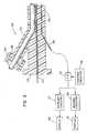
- FIG. 1is a partial perspective view of an embodiment of a catheter in accordance with the principles of the present invention
- FIG. 2is an enlarged cross-sectional view taken proximate the distal end of the catheter shown in FIG. 1;
- FIG. 3is a block diagram in part and sectional diagram in part illustrating an embodiment of a system utilizing the catheter shown in FIG. 1;
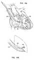
- FIG. 4Ais an illustration illustrating an application of a catheter in accordance with the principles of the present invention.
- FIG. 4Bis a partially enlarged illustration of the catheter shown in FIG. 4 A.
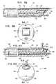
- FIG. 5Ashows a partial perspective and cross-sectional view of a first alternate embodiment of a catheter in accordance with the principles of the present invention
- FIG. 5Bshows a view of the distal end of the embodiment of the catheter shown in FIG. 5A;
- FIG. 6Ashows a partial perspective and cross-sectional view of a second alternate embodiment of a catheter in accordance with the principles of the present invention
- FIG. 6Bshows a view of the distal end of the catheter shown in FIG. 6A;
- FIG. 7Ashows a partial perspective and cross-sectional view of a variation of the second alternate embodiment of the catheter shown in FIG. 6A;
- FIG. 7Bshows a view of the distal end of the embodiment of the catheter shown in FIG. 7A;
- FIG. 8Ashows a partial perspective and cross-sectional view of a third alternate embodiment of a catheter in accordance with the principles of the present invention
- FIG. 8Bshows a view of the distal end of the catheter shown in FIG. 8A;
- FIG. 8Cshows a view of the distal end of the catheter shown in FIG. 8A having an alternatively shaped secondary port
- FIG. 9Ashows partial perspective and cross-sectional view of a fourth alternate embodiment of a catheter in accordance with the principles of the present invention.
- FIG. 9Bshows a view of the distal end of the catheter shown in FIG. 9 A.
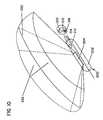
- FIG. 10is a partial schematic view of an embodiment of an underfluid catheter system in accordance with the principles of the present invention.
- FIG. 11is an enlarged view illustrating the catheter system operated underfluid with an aid of a volumetric field of view.
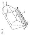
- FIG. 12is an enlarged perspective view illustrating the catheter system providing a volumetric field of view.
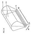
- FIG. 13is an enlarged perspective view of an ultrasonic transducer array having a single row of crystals covered by a lens which provides a volumetric field of view.
- FIG. 14is an enlarged perspective view of an alternative ultrasonic transducer array having multiple rows of crystals which provides a volumetric field of view.
- FIG. 15is an enlarged perspective view of a second alternative ultrasonic transducer array having equal number of rows and columns of crystals which provides a volumetric field of view.
- FIG. 16is an enlarged schematic cross-sectional view of a concave silicone rubber lens being placed on the ultrasonic transducer array, providing an outwardly defocused ultrasound beam.
- FIG. 17is an enlarged schematic cross-sectional view of a convex plastic lens being placed on the ultrasonic transducer array, providing an outwardly defocused ultrasound beam.
- FIG. 18is an enlarged schematic cross-sectional view of a convex silicone rubber lens being placed on the ultrasonic transducer array, providing an inwardly focused ultrasound beam.
- catheter 20includes an elongated flexible or rigid tubular catheter body 22 having a proximal end 24 and a distal end 26 .
- catheter 20includes proximate its longitudinal distal end 26 a phased array ultrasonic transducer 30 which is used to transmit ultrasound and receive resultant echoes so as to provide a field of view within which Doppler flow rates can be measured and features imaged.
- ultrasonic transducerscan be used in the present invention, such as any mechanical types, or any dynamic array types, or any offset stereoscopic imaging types, or any multidimensional imaging types incorporated into a virtual reality environment for underblood operation, etc.
- An electrical conductoris disposed in the catheter body 22 for electrically connecting transducer 30 to control circuitry 34 external of catheter body 22 .
- An access port 40is disposed in catheter body 22 and extends from proximate the proximal end 24 of catheter body 22 to proximate the distal end 26 of catheter body 22 .
- Access port 40is configured to receive a therapeutic device, such as a catheter, medication, sensors, etc., so as to enable such items to be delivered via access port 40 to distal end 26 of catheter body 22 for operation within the ultrasonic transducer field of view. Such items might be used for intervention; e.g., ablation catheter, surgical device, etc., monitoring blood pressure, sampling blood, etc.
- a guide wire access port 42is also disposed within catheter body 22 and extends from proximate proximal end 24 of the catheter body 22 to proximate distal end 26 of catheter body 22 for receiving a guide wire 44 .
- the ultrasonic transducerpreferably has a frequency of 5 to 30 megahertz (MHz) and more preferably a frequency of 7 to 10 MHz. Intracardiac imaging in an adult will require image penetration of up to 2 to 10 centimeters (cm).
- catheter body 22preferably has a diameter of 4 to 24 French (one French divided by Pi equals one millimeter (mm)) and, more preferably, a diameter of 6 to 12 French.
- access port 40has a diameter of 7 to 8 French and guide wire port 42 has a diameter of 0.025 to 0.038 inches.
- catheter 20 of the present inventioncan be utilized in a medical system including the appropriate control circuitry 34 for controlling operation of the ultrasonic transducer.
- control circuitry 34is electrically interconnected to transceiver circuitry 35 (T/R) for receiving and transmitting signals via a cable 36 to ultrasonic transducer 30 .
- transceiver circuitry 35is electrically interconnected to Doppler circuitry 37 and an appropriate display device 38 for displaying hemodynamics or blood flow.
- transceiver circuitry 35is electrically interconnected to suitable imaging circuitry 39 which is interconnected to a display 41 for displaying images.
- control circuitry 34might be designed to cause ultrasonic transducer 30 to vibrate so as to cause an appropriate ultrasound wave to project from proximate the distal end 26 of catheter body 22 .
- the ultrasound waverepresented by lines 50 in FIG. 2, will propagate through the blood surrounding distal end 26 and a portion of the body structure. A portion of the ultrasound wave so transmitted will be reflected back from both the moving red blood cells and the like and the body structures to impinge upon transducer 30 .
- An electrical signalis thereby generated and transmitted by the cable 36 to the input of transceiver 35 .
- a signalmight then be transmitted to Doppler circuitry 37 which will include conventional amplifying and filtering circuitry commonly used in Doppler flow metering equipment.
- Doppler circuitry 37will analyze the Doppler shift between the transmitted frequency and the receive frequency to thereby derive an output proportional to flow rate. This output may then be conveniently displayed at display 38 which might be a conventional display terminal. Accordingly, the user will be able to obtain a readout of blood flow rates or hemodynamic information.
- control circuitry 34will likewise trigger ultrasonic transducer 30 via transceiver 35 to vibrate and produce an ultrasound wave. Once again, a portion of the wave or energy will be reflected back to ultrasonic transducer 30 by the body features. A corresponding signal will then be sent by cable 36 to transceiver circuitry 35 . A corresponding signal is then sent to the imaging circuitry 39 which will analyze the incoming signal to provide, at display 41 , which also might be a conventional display apparatus, an image of the body features.
- This imagingcan occur while a therapeutic or surgical device is being used at distal end 26 of catheter 20 within the field of view provided by ultrasonic transducer 30 . Accordingly, the user will be able to monitor his/her actions and the result thereof.
- ultrasonic transducer 30might include a piezoelectric polymer, such as Polyvinylidenedifloride (PVDF) 60 , which is bonded by an epoxy layer 62 to a depression 64 approximate distal end 26 .
- PVDFPolyvinylidenedifloride
- FIGS. 2, 4 A, and 4 Bultrasonic transducer 30 might include a piezoelectric polymer, such as Polyvinylidenedifloride (PVDF) 60 , which is bonded by an epoxy layer 62 to a depression 64 approximate distal end 26 .
- PVDFPolyvinylidenedifloride
- the operational portion 56 of the therapeutic deviceis illustrated as generally being capable of operation in the field of view of ultrasonic transducer 30 . Accordingly, it is possible for the user to monitor operation of the therapeutic device by use of the ultrasonic transducer. Moreover, it is possible for the user to monitor the features of the body within the field of view before, during and after interventional activity.
- ultrasonic transducerscan be used in the present invention, such as any mechanical types, or any dynamic array types, or any offset stereoscopic imaging types, or any multidimensional imaging types incorporated into a virtual reality environment for underblood operation, etc., so that all forms of field of views, such as 1) tomographic (slices), 2) stereoscopic, 3) three-dimensional, 4) virtual reality (multidimensional) can be provided in the present invention.
- the orientations of the scan array on the cathetercan be include side-view, end-view, multiview (two or more views that are moveable or imminently directional transducer referred to in the literature as “omnidirectional”), etc.
- FIG. 5Ashows a partial cross-sectional view of a first alternative embodiment 70 of the catheter apparatus.
- the catheter apparatushas an elongated flexible or rigid body 72 having a longitudinal axis and a proximal end 74 and a distal end 76 .
- a port 78Disposed proximate a second side of body 72 is a port 78 extending through body 72 from proximate proximal end 74 to proximate distal end 76 of body 72 .
- Port 78is for receiving and delivering to distal end 76 of body 72 a working tool 84 .
- Working tool 84 shown in the Figuresis illustrative only, others types of tools now known or later developed may also be delivered to distal end 76 through port 78 .
- Proximate a first side of body 72is a guide wire port 80 extending through body 72 from proximate proximal end 74 to proximate distal end 76 . Shown in guide port 80 is
- Distal end 76is disposed at an oblique angle to the longitudinal axis of body 72 , the first side of body 72 extending further in the direction of the distal end than the second side of body 72 .
- An ultrasonic transducer 82having a first side and a second side, is disposed at an oblique angle to the longitudinal axis of body 72 approximately corresponding to the oblique angle of distal end 76 of body 72 .
- the first side of ultrasonic transducer 82is disposed proximate the first side of body 72 and the second side of transducer 82 is disposed proximate the second side of body 72 .
- transducer 82Extending from transducer 82 to proximate proximal end 74 of body 72 is an electrical conductor 83 connecting transducer 82 to control circuitry external of catheter 70 , as described with respect to catheter 20 above. Having transducer 82 disposed on an oblique angle toward port 78 allows for easy visualization of tools, such as tool 84 , extending beyond distal end 76 of body 72 .
- FIG. 5Bshows a view of distal end 76 of body 72 , showing guide wire port means 80 , transducer 82 , and port means 78 .
- FIG. 6Ashows a partial cross-sectional view of a second alternative embodiment of the catheter in accordance with the present invention, generally referred to as 88 .
- catheter 88has an elongated flexible or rigid body 90 having a proximal end 92 and a distal end 94 .
- Catheter 88also has a port 96 extending through body 90 from proximate proximal end 92 to proximate distal end 94 .
- Port 96has a distal end 97 proximal distal end 94 of body 90 .
- Distal end 97 of port 96exits body 90 at an acute angle to a first side of body 90 toward distal end 94 .
- Port 96is for receiving and delivering to distal end 94 a working tool, such as working tool 84 .
- Catheter 88also has a guide wire port 98 extending through body 90 from proximate proximal end 92 to proximate distal end 94 .
- Guide wire port 98is for receiving a guide wire 86 .
- transducer 100disposed to a first side of body 90 between distal end 94 and distal end 97 of port 96 .
- Extending from transducer 100 to proximate proximal end 92 of body 90is an electrical conductor 102 disposed in the catheter body 90 for electrically connecting transducer 100 to control circuitry external of the catheter.
- FIG. 6Bshows a view of distal end 94 of catheter 88 , as shown in FIG. 6 A.
- FIG. 7Ashows second alternative embodiment 104 , as shown in FIG. 6A, except instead of having a guide wire port 98 , this variation of the second alternative embodiment 104 has a deflection wire guidance system 106 for manipulating distal end 94 .
- FIG. 7Bshows a view of distal end 94 of the catheter shown in FIG. 7 A.
- FIG. 8Ashows a third alternative embodiment 110 of the catheter in accordance with the present invention.
- Third alternative embodiment 110has a body 112 having a distal end 114 and proximal end. Disposed proximate a first side of body 112 is a primary port 118 extending through body 112 from proximate proximal end 116 to proximate distal end 114 .
- Primary port 118has a distal end 119 proximate distal end 114 of body 112 .
- proximate a second side of body 112is a secondary port 120 extending through body 112 from proximate proximal end 116 to proximate distal end 114 .
- Secondary port 120has a distal end 121 proximate distal end 114 of body 112 .
- transducer 122is mounted proximate distal end 114 of body 112 . Extending from transducer 122 through body 112 to proximate proximal end is an electrical conductor for electrically connecting the transducer 122 to control circuitry external of the catheter. Transducer 122 is disposed between distal ends of primary and secondary ports 119 and 121 , respectively. With working ports 118 and 120 oppositely disposed on either side of transducer 122 , it is possible to conduct two simultaneous applications, such as holding an object with a first tool disposed through one port and operating on the object held by the first tool with a second tool disposed through the other port. A typical working tool 123 and working tool 84 are shown disposed within ports 118 and 120 .
- FIG. 8Bshows a view of distal end 114 of catheter 110 .
- FIG. 8Cshows a view of a distal end 124 of a catheter 126 substantially like catheter 110 shown in FIG. 8 A and FIG. 8B, except that catheter 126 has a primary port 128 having an arc-like shaped cross-section, rather than a circular shaped cross-section.
- a circular cross-sectionhas been shown in the Figures for the various ports described herein, the size and shape of the ports can be varied without departing from the principals of the present invention.
- FIG. 9Ashows a fourth alternative embodiment of a catheter 130 of the present invention.
- Catheter 130is similar to catheter 70 shown in FIG. 5 A and FIG. 5B except that a plurality of ports 132 are disposed proximate a second side of flexible body 131 , rather than one port 78 , as shown in FIG. 5 A.
- a plurality of portsit is possible, for example, to use a therapeutic tool through one port while simultaneously suctioning and removing debris through another port; or a therapeutic tool can be used through one port while simultaneously electrophysiologically monitoring, suctioning and/or biopsying through a second port, third or fourth port.
- the use of the catheter of the present inventionis described with respect to the preferred embodiment 20 . It is understood that the use of alternative embodiments 70 , 88 , 110 , 126 and 130 is analogous.
- the userwould insert flexible catheter body 22 into the body via the appropriate vascular access to the desired location in the body, such as selected venous locations, heart chamber, etc.
- a guide wiremight be first inserted into place and then the catheter body fed along the guide wire.
- the usermight then insert a surgical device into the body through access port 40 and feed the surgical device to proximate distal end 26 of catheter body 22 .
- the userPrior to, during and after operation of the surgical device, the user might obtain both hemodynamic measurements and images from the ultrasonic transducer field of view. By operation of the surgical device within the field of view of transducer, the user can monitor operation of the surgical device at all times.
- Frequency agilityrefers to the ability of a transducer to send and receive at various frequencies, most commonly 3, 5, and 7 MHz. It is also appreciated that a single frequency from a single transducer device can be sent and received. In general, higher frequencies are used to image fine detail of more proximal or closely related objects while lower frequency transducers scan more remote objects with less detail.
- the proposed deviceoptimally uses a 5 to 20 mHz transducer with the most optimally applied frequency of 7 to 10 mHz.
- the lower frequency used in the UIHCreflects the need to image larger objects such as the cardiac septa, valves, and extravascular anatomy.
- InterventionOne primary function of this catheter system is to guide the logical and safe use of various a) ablation, b) laser, c) cutting, occluding, e) etc., catheter-based interventional tools.
- the inventionhas the access port through which other technologies (devices) can be passed. Once the interventional tool exits the catheter tip, it can be directed repeatedly and selectively to specific site for controlled intervention.
- the inventionis also an imaging system capable of visualizing intracardiac, intravascular, and extravascular structures. Because the transducer frequencies utilized are usually lower than intravascular systems, the catheter 20 can see multiple cardiac cavities and visualize structures outside the vascular system.
- the imaging capabilityis basically two-fold: 1) diagnostic and 2) application.
- the catheter 20can effectively perform diagnostic intracardiac and transvascular imaging. This application will more than likely be performed just prior to an interventional application. The intervention then will follow using the same catheter system and its unique delivery capability.
- diagnostic imaginginclude 1) accurate visualization and measurement of an intracardiac defect, 2) characterization of valve orifice, 3) localization of a tumor and its connections, 4) etc.
- Extravascular diagnoseswould include 1) visualize pancreatic mass/pathology, 2) retroperitoneal pathology, 3) intracranial imaging, 4) recognition of perivascular pathology, and 5) imaging of other fluid containing space such as urinary bladder, bile system, fluid filled orifice or cavity (e.g. filled saline), etc.
- Application imagingrefers to the use of the catheter and its imaging capability to deliver and then apply another technology such as 1) occlusion device for closure of a septal defect, 2) ablation catheters for treatment of bypass tracts, 3) creation of a defect such as that with the blade septostomy catheter or laser-based catheter system, and 4) directing of valvuloplasty (such as prostrate surgery, placement of stents, gallstone removal etc.), etc.
- occlusion devicefor closure of a septal defect
- ablation cathetersfor treatment of bypass tracts
- creation of a defectsuch as that with the blade septostomy catheter or laser-based catheter system
- valvuloplastysuch as prostrate surgery, placement of stents, gallstone removal etc.
- the catheter 20is a truly combined ultrasound Doppler and conventional hemodynamic catheter. There are Doppler catheters, and there are catheters capable of imaging and measuring hemodynamic pressure. However, the catheter 20 is capable of Doppler hemodynamics (continuous and pulsed wave Doppler) as well as high-fidelity hemodynamic pressure recording while simultaneously imaging the heart and blood vessel. The catheter 20 provides a combination of imaging, hemodynamic, and interventional delivery catheter.
- intracardiac ultrasoundis capable of 1) imaging, 2) delivering a therapeutic device, and 3) obtaining simultaneous hemodynamics which can be used to develop less invasive cardiac surgical techniques.
- This simultaneous use of one or more devices within the heart or vascular treeopens up the potential to develop less invasive surgical therapies.
- Exampleswould include 1) removal of a cardiac tumor by visually grasping the tumor with one device and visually cutting its attachment with a second device, thus allowing less invasive extraction of intracardiac mass lesions, 2) visually placing an electrophysiologic catheter on a bypass tract and then with direct ultrasound visualization ablate the underlying tract with the second device, 3) visually performing laser surgery such as creating an intra-atrial defect, vaporization of obstructing thrombus such as is seen in pseudointimal occlusion of conduits, 4) visually removing a foreign body from the heart or vascular tree, and 5) directing intravascular surgery from within a blood vessel or monitoring concomitant hemodynamic changes.
- Radio-frequency ablationPresently a bypass tract is localized by an electrophysiologic study which systematically maps the atrioventricular valve annulus. Positioning of the ablation catheter is determined by x-ray fluoroscopy and certain electrical measurements which relate the distance of the ablation catheter from a reference catheter. The catheter 20 will allow an operator to map the atrioventricular valve under direct ultrasound visualization. Thus, increased accuracy of catheter placement, precision of the applied therapy, and immediate assessment of outcome would result.
- the above ablation techniquewould be particularly applicable for right-sided bypass tracts (in and around the tricuspid valve annulus). This would be accomplished by placement of the catheter 20 through the superior vena cava above the tricuspid annulus.
- the catheter 20could be placed across the atrial septum under direct ultrasound visualization.
- the mitral annuluscould thus be mapped directly and the localized bypass tract precisely ablated under visual ultrasonic and hemodynamic direction. Complications such as valve perforation, multiple imprecise applications of ablation energy, and inadvertent ablation of normal conduction tissue would be substantially reduced.
- the catheter 20will allow visualization of perivascular and extravascular pathology. Transvascular or transorgan imaging and localization of pathology out of the immediate vascular tree will result in a substantial step forward in the diagnosis and possible treatment of difficult to reach pathology.
- the catheter 20cannot only diagnose but guide a biopsy needle and therapeutic device to an extravascular lesion in question.
- the retroperitoneum, mediastinum, and basal cerebrovascular pathologyare logical areas of interest. Accurate characterization of various pathologies will be more feasible. Every organ has its own vascular system, and the proposed ultrasound transvascular system is an ideal tool to assess difficult to reach areas of the body.
- the vascular systemis a conduit to each organ, and the catheter 20 can be delivered to each organ. Characterization of the underlying parenchyma and possible transvascular biopsy or treatment will ultimately be developed.
- the catheter 20opens the potential not only to visualize but to directly intervene with the same catheter system.
- There are numerous intraoperative catheter-based systemswhich to date use conventional x-ray to accomplish their goal of placement and application of a specified therapy.
- the catheter 20has all the prerequisites of an ideal imaging and interventional instrument and has the ability to 1) image, 2) obtain hemodynamics by multiple means (pressure dynamics and Doppler), 3) function as a diagnostic as well as therapeutic device, and 4) accommodate other unique technologies which would enhance the application of both systems.
- intravascular, transvascular, and intracardiac devicescould be delivered through the port means described above within or about the heart and blood vessels of the body.
- the catheters described abovecould also be used in any ectogenic tissue, such as liver, parenchyma, bile ducts, ureters, urinary bladder, and intracranial—i.e., any place in the body which is echogenic which would allow passage of a catheter for either diagnostic or therapeutic applications using ultrasound visualization.
- the catheter 20is a new and exciting innovation to invasive medicine. There are multiple other and yet-to-be determined applications. However, the new concept described opens the potential development of less expensive, more precise, and safe intravascular and transvascular diagnostic and surgical devices.
- the catheter 20is very much different from any conventional ultrasound catheter-based system.
- the catheter 20incorporates image and hemodynamic capability as well as the ability to deliver other diverse technologies to specified sites within the cardiovascular system (heart and blood vessels).
- the catheter 20is seen as an ideal diagnostic and therapeutic tool for future development.
- the proposed applicationsfoster greater preciseness, adaptability, and safety.
- Ultrasoundpermits visualization from within blood-filled spaces as well as through blood-filled spaces into other water-or fluid-filled tissue.
- the catheter 20will evolve into the ultimate interventional system.
- FIG. 4Ais an illustration showing one potential use of the ultrasound imaging and hemodynamic catheter (UIHC).
- UIHCultrasound imaging and hemodynamic catheter
- the UIHCis advanced from the superior vena cava to the tricuspid valve annulus. Simultaneously visualized in the annulus, electrophysiologic and ultimately and ablation procedure are performed. The ability to directly visualize and direct therapeutic catheter devices highlights only one of the many applications of the UIHC.
- FIG. 10Another embodiment of the catheter system, generally in accordance with the principles of the present invention is shown in FIG. 10, which is designated as reference numeral 200 .
- the catheter system 200has a catheter body 202 and an ultrasonic transducer array 204 mounted on proximate the distal end of the catheter body 202 . It is appreciated that other parts of the catheter system can be similar to those in the catheter systems 20 , 70 , 88 , 104 , 110 , and 130 as shown in FIGS. 1, 5 A, 6 A, 7 A, 8 A, and 9 A, respectively.
- FIG. 10shows a partial schematic view of the catheter system 200 .
- the catheter body 202 of the catheter system 200is inserted into an underfluid cavity of a body 206 .
- a therapeutic device 208projects from the catheter system 200 proximate the distal end of the catheter system 200 and manipulates features in the cavity of the body 208 . This manipulation is under observation of a 3-dimensional image shown on a display, which can be similarly connected to the ultrasound, transducer array as shown in FIG. 3, outside the body 208 proximate the proximal end of the catheter system 200 .
- the underfluid catheter body 202has tool port 210 disposed in the catheter body 202 extending from proximate the proximal end to proximate the distal end of the catheter body 202 for receiving the therapeutic device 208 , such as a catheter, medication, sensor, surgical device, etc., so as to enable such items to be delivered via the tool port to proximate the distal end of the catheter body 202 .
- the tool portis optional.
- additional tool portscan be disposed in the catheter body 202 .
- the therapeutic device 208is projected into an underfluid environment, as shown in FIGS. 11-12, and operated therein with the aid of a volumetric 3-dimensional image of the underfluid environment and the therapeutic device 208 .
- the catheter system 200can also optionally include a guidewire port 212 disposed in the catheter body 202 extending from proximate the proximal end to proximate the distal end of the catheter body 202 for receiving a guide wire 214 .
- the guide wire 214guides the catheter body 202 when inserting into a body, such as the body 206 .
- the catheter body 202can also house some encased electronics 216 .
- the ultrasonic transducer array 204is mounted on a side of the catheter body 202 .
- the array 204can also be mounted on the tip of the catheter body 202 .
- the catheter body 202is a flexible catheter capable of manual or electronic interactive flexible tip.
- the guidewire port 212has a diameter of 0.035 inches. It is appreciated that the range of the diameter of the guidewire port 212 can be varied from 0.025 to 0.038 inches.
- the tool port 210 for transporting the therapeutic device 208is a 7 French port. It is appreciated that the range of the tool port 210 can be varied from 3 French to 20 French.
- the ultrasonic transducer array 204is comprised of a single row of individual crystals 218 . Each crystal 218 is arranged side by side.
- a field of view generated by the ultrasonic transducer array 204has a primary tomographic plane 220 in azimuthal dimension along an AZ axis.
- the row of the array 204is parallel to the AZ axis.
- An elevation axis (EL)is perpendicular to the AZ axis.
- a primary beam from the ultrasonic transducer array 204lies in the primary tomographic plane 220 .
- the primary beamhas usually a sector configuration (generally a fan or triangle shape) or a linear configuration (generally rectangular shape).
- the volumetric field of viewcan be produced by defocusing the primary tomographic plane 220 such that a plurality of elevation planes 222 spread laterally outward from the primary tomographic plane 220 .
- the primary tomographic plane 220 and the elevation planes 222together form a volumetric field of view.
- a lens 224is placed on the top of the ultrasonic transducer array 204 .
- the ultrasound beams which are usually collimatedare defocused along the elevation direction (EL) after the beams go through the lens 224 (or other lenses 226 , 228 as shown in FIGS. 16 and 17 ).
- the lenses 224 , 226 , or 228are preferred to be made from materials such as a plastic material or silicone rubber. It is appreciated that other types materials can be used to make the lens.
- the lens 226is a concave lens, preferably made of silicone rubber, which transmits ultrasound waves slower than the surrounding environment, such as body tissues.
- the ultrasound wavespass through the lens 226 and then impact on the body tissues.
- the speed of the ultrasound wavesis slower in the lens but faster in tissue (e.g. 1,540 m/sec). Accordingly, the transmitted ultrasound waves, after passing through more slowly transmitted lens 226 and striking faster transmitted body tissues, will be directed outward. As a result, the collimated ultrasound beams are defocused in the elevation dimension.
- the defocusingcan also be achieved by placing a convex lens 228 on the ultrasonic transducer array as shown in FIG. 17 .
- the convex lens 228preferably made of plastic, transmits ultrasound waves faster than the surrounding environment, such as body tissues.
- the ultrasound wavespass through the convex lens 228 and then impact on the body tissues.
- the ultrasound beamsare pulled outward due to the faster velocity in the convex lens 228 .
- the ultrasound beamsare defocused in the elevation dimension.
- FIG. 18,demonstrates a way of using a lens 230 to, in fact, focus the beams from the transducer array.
- the collimated ultrasound beamsare generated from the ultrasonic transducer array.
- the convex lens 230transmits ultrasound waves slower than the surrounding environment, such as body tissues, do. Accordingly, the ultrasound beams are pulled inward due to the faster velocity in body tissues. As a result, the ultrasound beams are focused toward the primary tomographic plane 220 .
- FIG. 14shows an alternative embodiment of ultrasonic transducer array 204 ′ which is comprised of multiple rows of individual piezoelectric crystals 218 .
- the rows of the array 204 ′are parallel to the AZ axis.
- the columns of the array 204 ′are parallel to the elevation dimension along the EL axis, which is perpendicular to AZ axis.
- This type of arrayis also called volumetric one and one-half (1 and 1 ⁇ 2) dimensional array.
- the “elevation” image and the ultimate 3-dimensional imageare the result of phasing the crystals in the elevation direction as well as in the azimuthal direction.
- FIG. 15shows a second alternative embodiment of ultrasonic transducer array 204 ′′ which is comprised of equal number of crystals 218 in all dimensions. Similar to the one and one-half (1 and 1 ⁇ 2) dimensional array 204 ′, the rows of the array 204 ′′ are parallel to the AZ axis, and the columns of the array 204 ′′ are parallel to the EL axis. This type of array is also called a two (2) dimensional array. The “elevation” image and the ultimate 3-dimensional image are the result of phasing the crystals in the EL direction as well as the AZ direction.
- the volumetric field of view as shown in FIGS. 14-15provides 3-dimensional images of structures under observation. Further, the volumetric field of view not only shows, for example, a primary tomographic cut, but also volumes of features, such as tissue.
- FIGS. 14 and 15it will be appreciated that no lens is required to generate a volumetric image. Consequently, the ultrasonic beams are focused in both the azimuthal and elevational directions.
- the volumetric imageis generated because the arrays of FIGS. 14 and 15 are 2 dimensional.
- a volumetric imagecan be generated by electronically phasing and steering the ultrasonic impulses in both the azimuthal and the elevational directions.
- the ultrasound transduceris a 7-10 MHz sector array transducer. It is appreciated that the range of the sector array transducer can be varied from 3.7 MHz to 30 MHz.
- the lenses 224 , 226 , 228 , 230can be made of different materials which will have variable effects on the transmitted ultrasound beams.
- a 3-dimensional imagecan be seen on a 2-dimensional display outside the body 206 in a real-time operation. Elevation defocusing in using a lens does not interfere with the inherent frame rate or adversely affect conventional echo data.
- the lenscan also be fabricated to reduce the strength of the dominant tomographic plane (AZ plane).
- AZ planedominant tomographic plane
- the lensis optional, for example, as shown in FIGS. 14 and 15 whereby the beams are phased in both the azimuthal and elevational planes.
- the present inventionhas numerous clinical applications.
- One of whichis the underfluid imaging when imaging from within chambers, cavities or blood vessels. Since the physical space is small, and the anatomy in question is closely approximated and usually totally surrounds the transducer, a 3-dimensional imaging is a solution to visualizing larger volumes of underfluid tissue.
- the defocusing lens or electronically controlled phasing in both the azimuthal and elevational directionsi.e., using multi-dimensional arrays such as 1-1 ⁇ 2 dimensional or 2-dimensional arrays
- Working port(s) and guidewire(s)are optional. Further, catheter lengths and transducer frequencies are variable.
- Another applicationwhen the working port is optionally used in the catheter, is to intervene or manipulate an underfluid structure, such as cutting an underfluid tissue, etc., by a therapeutic device, such as the therapeutic devices 50 , 84 , 123 , 208 shown in FIGS. 4E, 5 A, 8 A, and 12 , respectively.
- a therapeutic devicesuch as the therapeutic devices 50 , 84 , 123 , 208 shown in FIGS. 4E, 5 A, 8 A, and 12 , respectively.
- diagnostic and therapeutic procedurescan be performed with better spatial orientation.
- the measurementcan also be performed under direct volumetric visual guidance in the present invention.
Landscapes
- Health & Medical Sciences (AREA)
- Life Sciences & Earth Sciences (AREA)
- Surgery (AREA)
- Heart & Thoracic Surgery (AREA)
- Molecular Biology (AREA)
- Nuclear Medicine, Radiotherapy & Molecular Imaging (AREA)
- Veterinary Medicine (AREA)
- Public Health (AREA)
- Engineering & Computer Science (AREA)
- Biomedical Technology (AREA)
- General Health & Medical Sciences (AREA)
- Medical Informatics (AREA)
- Animal Behavior & Ethology (AREA)
- Biophysics (AREA)
- Physics & Mathematics (AREA)
- Radiology & Medical Imaging (AREA)
- Pathology (AREA)
- Hematology (AREA)
- Vascular Medicine (AREA)
- Gynecology & Obstetrics (AREA)
- Ultra Sonic Daignosis Equipment (AREA)
- Media Introduction/Drainage Providing Device (AREA)
Abstract
Description
This application is a Continuation of application Ser. No. 09/087,520, filed May 29, 1998 and now U.S. Pat. No. 6,099,475, which is a Continuation of application Ser. No. 09/003,248, filed Jan. 6, 1998 now U.S. Pat. No. 6,129,672, which is a Continuation of application Ser. No. 08/678,380, filed Jun. 28, 1996 (now issued as U.S. Pat. No. 5,704,361), which is a Continuation-in-Part of application Ser. No. 08/305,138, filed Sep. 13, 1994 (now abandoned), which is a Continuation of application Ser. No. 07/972,626, filed Nov. 6, 1992 (now issued as U.S. Pat. No. 5,345,940), which is a Continuation-in-Part of application Ser. No. 07/790,580, filed Nov. 8, 1991 (now issued as U.S. Pat. No. 5,325,860), which application(s) are incorporated herein by reference.
Noninvasive ultrasonic imaging systems are widely used for performing ultrasonic imaging and taking measurements. Such systems typically use scan heads which are placed against a patients skin. Exemplary uses for such systems include heart and internal organ examinations as well as examinations of developing fetuses. These systems operate by transmitting ultrasonic waves into the body, receiving echoes returned from tissue interfaces upon which the waves impinge, and translating the received echo information into a structural representation of the planar slice of the body through which the ultrasonic waves are directed.
Catheter based invasive ultrasound imaging systems, typically used for intracardiac or transvascular imaging, are a relatively new addition to ultrasound armamentarian. Conventional underfluid transducers for use on catheters are comprised of crystal arrays (e.g. linear phased array) or a single crystal translated over a surface, producing a tomographic field of view in an azimuthal plane of the array. Typical arrays include: 1) linear array (linear sequential array), usually producing a rectangular or rhomboidal picture; 2) cylindrical array or rotating crystal, producing a round pie-shaped tomographic cut of structures; and 3) sector array (linear phased array), producing a triangular shaped image emanating from a small transducer source. All images are tomographic in nature and are focused in the azimuthal and elevation plane. The intent of having these conventional transducer configurations is to produce a thin ultrasound cut of the insonated structures. Such tomographic planes by nature are thin and of high resolution.
The narrow field of view provided by conventional catheter transducer configurations is problematic because structures lying outside of the plane of view can only be visualized by reorienting or manipulating the catheter. Due to the tortuous and confined nature of a typical catheter pathway, catheter manipulation is impractical and often impossible. Consequently, the localization of specific targets is difficult and at times can be disorienting because of an inability to appreciate contiguous anatomic landmarks.
Advances in 3-dimensional imaging capabilities have been made with respect to non-catheter related ultrasonic imaging systems. For example, U.S. Pat. No. 5,305,756, issued to Entrekin et al., which is hereby incorporated by reference, discloses general 3-dimensional imaging techniques in a non-catheter based context. What is needed is a catheter based imaging system that utilizes 3-dimensional imaging techniques to provide a wide field of view so as to improve anatomic localization for precision underfluid diagnostics and interventions.
The present invention relates generally to a volumetric, 3-dimensional image ultrasound transducer underfluid catheter system.
The present invention also relates to an ultrasonic and interventional catheter device operated in an intracardiac or transvascular system, with the aid of a volumetric 3-dimensional imaging capability.
In one particular embodiment, the present invention relates to a catheter apparatus comprising an underfluid catheter body having proximal and distal ends. An ultrasonic transducer array is mounted longitudinally along the catheter body proximate the distal end. The transducer array has a volumetric field of view that projects radially/laterally outward from the catheter. Features in such wide volumetric field of view can be imaged, measured, or intervened by an underfluid therapeutic device with an aid of the real-time image.
It is significant that the transducer array described in the previous paragraph has a 3-dimensional field of view. A first dimension; referred to as an azimuthal direction, is aligned with the length of the transducer array. A second dimension, referred to as a depth direction, is the depth into the body which an ultrasonic signal is transmitted and from which an echo return. A third dimension, referred to as an elevation direction, is perpendicular to both the azimuthal and the depth directions.
If the transducer array comprises a linear phased array having a single row of piezoelectric crystals, a 3-dimensional field of view can be generated by focusing the ultrasound signals in the azimuthal direction (parallel to the longitudinal axis of the catheter) and diverging the ultrasound signals in the elevational direction (transverse to the longitudinal axis of the catheter). The ultrasonic signals can be diverged in the elevational direction through the use of lenses. For example, the signals can be diverged by mounting a silicone rubber concave lens or a plastic convex lens in front of the transducer array.
If the transducer array comprises multiple rows of piezoelectric crystals, a 3-dimensional field of view can be generated by electronically phasing the ultrasound signals in both the azimuthal and elevational directions. Of course, lenses can be used in association with multiple row arrays to further widen the field of view.
It will be appreciated that catheters constructed in accordance with the principles of the present invention can optionally include one or more ports that extend longitudinally through the catheter bodies. The ports are preferably adapted for guiding therapeutic instruments through the catheter and preferably have exit ends adjacent to the field of view of the catheter imaging system. In operation, the ports guide the therapeutic instruments such that the operative ends of a therapeutic instruments are directed toward the 3-dimensional field of view of the imaging system.
It will also be appreciated that catheters constructed in accordance with the principles of the present invention can include one or more guidewire ports which extend longitudinally through the catheters and are adapted for receiving guidewires.
One advantage of the present invention is to provide real-time 3-dimensional images of underfluid features so as to visualize contiguous anatomy, such as a large volume of tissues without frequently rotating, flexing, or extending the catheter.
Another advantage is that the present invention provides a much better underfluid “eye”—a 3-dimensional “motion picture”—for an operator when he/she intervenes the underfluid features by using an underfluid therapeutic device. These images provide the operator a direct aid without opening a large area of a body.
A further advantage is that the present invention have numerous clinical applications. One is related to underfluid imaging: There is a considerable need to increase the field of view when imaging from within chambers or blood vessels. The physical space of the chambers or blood vessels is small, and the anatomy in question is closed approximated and usually totally surrounds the transducer. A conventional tomographic presentation provides only a limited slice, thus requiring frequent manipulation of transducer in order to visualize contiguous anatomy. The present invention is a solution to visualizing larger volumes of tissue. The underfluid defocusing transducer array does not appreciably affect the electronics and does not require alteration in the display format. The catheter apparatus is immersed in fluid, a homogeneous, low scattering medium, which is an ideal environment for this particular transducer modification.
Another main clinical application is related to underfluid intervention: In diagnostic and therapeutic procedures, there is an increasing need for volumetric 3-dimensional visualization which would improve anatomic localization and recognition of continuous structures and events.
A further advantage of the present invention is that by using such a catheter system, major surgical procedures can be avoided. It dramatically reduces the patient's physical pain in operation and mental distress after operation due to any large visible scars, etc.
These and various other advantages and features of novelty which characterize the invention are pointed out with particularity in the claims annexed hereto and forming a part hereof. However, for a better understanding of the invention, its advantages and objects obtained by its use, reference should be made to the drawings which form a further part hereof, and to the accompanying descriptive matter, in which there is illustrated and described a preferred embodiment of the invention.
A better understanding of the construction and operational characteristics of a preferred embodiment(s) can be realized from a reading of the following detailed description, especially in light of the accompanying drawings in which like reference numerals in the several views generally refer to corresponding parts.
FIG. 1 is a partial perspective view of an embodiment of a catheter in accordance with the principles of the present invention;
FIG. 2 is an enlarged cross-sectional view taken proximate the distal end of the catheter shown in FIG. 1;
FIG. 3 is a block diagram in part and sectional diagram in part illustrating an embodiment of a system utilizing the catheter shown in FIG. 1;
FIG. 4A is an illustration illustrating an application of a catheter in accordance with the principles of the present invention;
FIG. 4B is a partially enlarged illustration of the catheter shown in FIG.4A.
FIG. 5A shows a partial perspective and cross-sectional view of a first alternate embodiment of a catheter in accordance with the principles of the present invention;
FIG. 5B shows a view of the distal end of the embodiment of the catheter shown in FIG. 5A;
FIG. 6A shows a partial perspective and cross-sectional view of a second alternate embodiment of a catheter in accordance with the principles of the present invention;
FIG. 6B shows a view of the distal end of the catheter shown in FIG. 6A;
FIG. 7A shows a partial perspective and cross-sectional view of a variation of the second alternate embodiment of the catheter shown in FIG. 6A;
FIG. 7B shows a view of the distal end of the embodiment of the catheter shown in FIG. 7A;
FIG. 8A shows a partial perspective and cross-sectional view of a third alternate embodiment of a catheter in accordance with the principles of the present invention;
FIG. 8B shows a view of the distal end of the catheter shown in FIG. 8A;
FIG. 8C shows a view of the distal end of the catheter shown in FIG. 8A having an alternatively shaped secondary port;,
FIG. 9A shows partial perspective and cross-sectional view of a fourth alternate embodiment of a catheter in accordance with the principles of the present invention; and
FIG. 9B shows a view of the distal end of the catheter shown in FIG.9A.
FIG. 10 is a partial schematic view of an embodiment of an underfluid catheter system in accordance with the principles of the present invention.
FIG. 11 is an enlarged view illustrating the catheter system operated underfluid with an aid of a volumetric field of view.
FIG. 12 is an enlarged perspective view illustrating the catheter system providing a volumetric field of view.
FIG. 13 is an enlarged perspective view of an ultrasonic transducer array having a single row of crystals covered by a lens which provides a volumetric field of view.
FIG. 14 is an enlarged perspective view of an alternative ultrasonic transducer array having multiple rows of crystals which provides a volumetric field of view.
FIG. 15 is an enlarged perspective view of a second alternative ultrasonic transducer array having equal number of rows and columns of crystals which provides a volumetric field of view.
FIG. 16 is an enlarged schematic cross-sectional view of a concave silicone rubber lens being placed on the ultrasonic transducer array, providing an outwardly defocused ultrasound beam.
FIG. 17 is an enlarged schematic cross-sectional view of a convex plastic lens being placed on the ultrasonic transducer array, providing an outwardly defocused ultrasound beam.
FIG. 18 is an enlarged schematic cross-sectional view of a convex silicone rubber lens being placed on the ultrasonic transducer array, providing an inwardly focused ultrasound beam.
Referring now to FIG. 1-3, there is, generally illustrated byreference numeral 20, a catheter in accordance with the principles of the present invention. As shown,catheter 20 includes an elongated flexible or rigidtubular catheter body 22 having aproximal end 24 and adistal end 26.Catheter 20 includes proximate its longitudinal distal end26 a phased arrayultrasonic transducer 30 which is used to transmit ultrasound and receive resultant echoes so as to provide a field of view within which Doppler flow rates can be measured and features imaged. It is appreciated that the other types of ultrasonic transducers can be used in the present invention, such as any mechanical types, or any dynamic array types, or any offset stereoscopic imaging types, or any multidimensional imaging types incorporated into a virtual reality environment for underblood operation, etc. An electrical conductor is disposed in thecatheter body 22 for electrically connectingtransducer 30 to controlcircuitry 34 external ofcatheter body 22. Anaccess port 40 is disposed incatheter body 22 and extends from proximate theproximal end 24 ofcatheter body 22 to proximate thedistal end 26 ofcatheter body 22.Access port 40 is configured to receive a therapeutic device, such as a catheter, medication, sensors, etc., so as to enable such items to be delivered viaaccess port 40 todistal end 26 ofcatheter body 22 for operation within the ultrasonic transducer field of view. Such items might be used for intervention; e.g., ablation catheter, surgical device, etc., monitoring blood pressure, sampling blood, etc. A guidewire access port 42 is also disposed withincatheter body 22 and extends from proximateproximal end 24 of thecatheter body 22 to proximatedistal end 26 ofcatheter body 22 for receiving aguide wire 44.
In the preferred embodiment of the present invention, the ultrasonic transducer preferably has a frequency of 5 to 30 megahertz (MHz) and more preferably a frequency of 7 to 10 MHz. Intracardiac imaging in an adult will require image penetration of up to 2 to 10 centimeters (cm). In the preferred embodiment,catheter body 22 preferably has a diameter of 4 to 24 French (one French divided by Pi equals one millimeter (mm)) and, more preferably, a diameter of 6 to 12 French. In the preferred embodiment,access port 40 has a diameter of 7 to 8 French and guidewire port 42 has a diameter of 0.025 to 0.038 inches.
As generally illustrated in FIG. 3,catheter 20 of the present invention can be utilized in a medical system including theappropriate control circuitry 34 for controlling operation of the ultrasonic transducer. As illustrated in FIG. 3,control circuitry 34 is electrically interconnected to transceiver circuitry35 (T/R) for receiving and transmitting signals via acable 36 toultrasonic transducer 30. In turn,transceiver circuitry 35 is electrically interconnected toDoppler circuitry 37 and anappropriate display device 38 for displaying hemodynamics or blood flow. In addition,transceiver circuitry 35 is electrically interconnected tosuitable imaging circuitry 39 which is interconnected to adisplay 41 for displaying images.
During operation,control circuitry 34 might be designed to causeultrasonic transducer 30 to vibrate so as to cause an appropriate ultrasound wave to project from proximate thedistal end 26 ofcatheter body 22. The ultrasound wave, represented bylines 50 in FIG. 2, will propagate through the blood surroundingdistal end 26 and a portion of the body structure. A portion of the ultrasound wave so transmitted will be reflected back from both the moving red blood cells and the like and the body structures to impinge upontransducer 30. An electrical signal is thereby generated and transmitted by thecable 36 to the input oftransceiver 35. A signal might then be transmitted toDoppler circuitry 37 which will include conventional amplifying and filtering circuitry commonly used in Doppler flow metering equipment.Doppler circuitry 37 will analyze the Doppler shift between the transmitted frequency and the receive frequency to thereby derive an output proportional to flow rate. This output may then be conveniently displayed atdisplay 38 which might be a conventional display terminal. Accordingly, the user will be able to obtain a readout of blood flow rates or hemodynamic information.
In order to obtain imaging information,control circuitry 34 will likewise triggerultrasonic transducer 30 viatransceiver 35 to vibrate and produce an ultrasound wave. Once again, a portion of the wave or energy will be reflected back toultrasonic transducer 30 by the body features. A corresponding signal will then be sent bycable 36 totransceiver circuitry 35. A corresponding signal is then sent to theimaging circuitry 39 which will analyze the incoming signal to provide, atdisplay 41, which also might be a conventional display apparatus, an image of the body features.
This imaging can occur while a therapeutic or surgical device is being used atdistal end 26 ofcatheter 20 within the field of view provided byultrasonic transducer 30. Accordingly, the user will be able to monitor his/her actions and the result thereof.
As illustrated in FIG. 3,catheter body 22 might include proximate its proximal end24 asuitable mounting structure 52 to theaccess port 40. A therapeutic orsurgical device structure 53 might be suitably attached to structure52 by suitable means, e.g., threaded, etc. As illustrated, an elongated cable-like member 54 will extend alongaccess port 40 and slightly beyonddistal end 26 ofcatheter body 22 wherein anoperative portion 56 of the surgical tool might be interconnected.
Additional detail ofdistal end 26 ofcatheter body 22 is illustrated in FIGS. 2,4A, and4B. As illustrated in FIGS. 2,4A, and4B,ultrasonic transducer 30 might include a piezoelectric polymer, such as Polyvinylidenedifloride (PVDF)60, which is bonded by anepoxy layer 62 to adepression 64 approximatedistal end 26. Although some detail is provided with respect to an embodiment of an ultrasonic transducer which might be used, it will be appreciated that various types of transducers having various configurations and orientations might be utilized in keeping with the present invention.
As illustrated in FIGS. 4A and 4B, theoperational portion 56 of the therapeutic device is illustrated as generally being capable of operation in the field of view ofultrasonic transducer 30. Accordingly, it is possible for the user to monitor operation of the therapeutic device by use of the ultrasonic transducer. Moreover, it is possible for the user to monitor the features of the body within the field of view before, during and after interventional activity. It is appreciated that the other types of ultrasonic transducers can be used in the present invention, such as any mechanical types, or any dynamic array types, or any offset stereoscopic imaging types, or any multidimensional imaging types incorporated into a virtual reality environment for underblood operation, etc., so that all forms of field of views, such as 1) tomographic (slices), 2) stereoscopic, 3) three-dimensional, 4) virtual reality (multidimensional) can be provided in the present invention. In addition, it is appreciated that the orientations of the scan array on the catheter can be include side-view, end-view, multiview (two or more views that are moveable or imminently directional transducer referred to in the literature as “omnidirectional”), etc.
FIG. 5A shows a partial cross-sectional view of a firstalternative embodiment 70 of the catheter apparatus. The catheter apparatus has an elongated flexible orrigid body 72 having a longitudinal axis and aproximal end 74 and adistal end 76. Disposed proximate a second side ofbody 72 is aport 78 extending throughbody 72 from proximateproximal end 74 to proximatedistal end 76 ofbody 72.Port 78 is for receiving and delivering todistal end 76 of body72 a workingtool 84. Workingtool 84 shown in the Figures is illustrative only, others types of tools now known or later developed may also be delivered todistal end 76 throughport 78. Proximate a first side ofbody 72 is aguide wire port 80 extending throughbody 72 from proximateproximal end 74 to proximatedistal end 76. Shown inguide port 80 is aguide wire 86.
FIG. 5B shows a view ofdistal end 76 ofbody 72, showing guide wire port means80,transducer 82, and port means78.
FIG. 6A shows a partial cross-sectional view of a second alternative embodiment of the catheter in accordance with the present invention, generally referred to as88. Like firstalternative embodiment 70,catheter 88 has an elongated flexible orrigid body 90 having aproximal end 92 and adistal end 94.Catheter 88 also has aport 96 extending throughbody 90 from proximateproximal end 92 to proximatedistal end 94.Port 96 has adistal end 97 proximaldistal end 94 ofbody 90.Distal end 97 ofport 96exits body 90 at an acute angle to a first side ofbody 90 towarddistal end 94.Port 96 is for receiving and delivering to distal end94 a working tool, such as workingtool 84.Catheter 88 also has aguide wire port 98 extending throughbody 90 from proximateproximal end 92 to proximatedistal end 94.Guide wire port 98 is for receiving aguide wire 86.
Also shown in FIG. 6A is atransducer 100 disposed to a first side ofbody 90 betweendistal end 94 anddistal end 97 ofport 96. Extending fromtransducer 100 to proximateproximal end 92 ofbody 90 is anelectrical conductor 102 disposed in thecatheter body 90 for electrically connectingtransducer 100 to control circuitry external of the catheter. Withtransducer 100 disposed to the first side ofbody 90 anddistal end 97 ofport 96 exitingbody 90 at an acute angle relative to the first side ofbody 90 towarddistal end 94, working tools extending fromdistal end 97 ofport 96 will be within the field of view oftransducer 100.
FIG. 6B shows a view ofdistal end 94 ofcatheter 88, as shown in FIG.6A.
FIG. 7A shows secondalternative embodiment 104, as shown in FIG. 6A, except instead of having aguide wire port 98, this variation of the secondalternative embodiment 104 has a deflectionwire guidance system 106 for manipulatingdistal end 94. FIG. 7B shows a view ofdistal end 94 of the catheter shown in FIG.7A.
FIG. 8A shows a thirdalternative embodiment 110 of the catheter in accordance with the present invention. Thirdalternative embodiment 110 has abody 112 having adistal end 114 and proximal end. Disposed proximate a first side ofbody 112 is aprimary port 118 extending throughbody 112 from proximate proximal end116 to proximatedistal end 114.Primary port 118 has adistal end 119 proximatedistal end 114 ofbody 112. Oppositely disposed fromprimary port 118, proximate a second side ofbody 112 is asecondary port 120 extending throughbody 112 from proximate proximal end116 to proximatedistal end 114.Secondary port 120 has adistal end 121 proximatedistal end 114 ofbody 112.
Mounted proximatedistal end 114 ofbody 112 is atransducer 122. Extending fromtransducer 122 throughbody 112 to proximate proximal end is an electrical conductor for electrically connecting thetransducer 122 to control circuitry external of the catheter.Transducer 122 is disposed between distal ends of primary andsecondary ports ports transducer 122, it is possible to conduct two simultaneous applications, such as holding an object with a first tool disposed through one port and operating on the object held by the first tool with a second tool disposed through the other port. Atypical working tool 123 and workingtool 84 are shown disposed withinports
Although the third alternative embodiment of thecatheter 110 of the present invention does not include a guide wire port means, a guide wire could be used inprimary port 118 orsecondary port 120 to initially positioncatheter 110. Then the guide wire could be retracted fromport distal end 114 ofcatheter 110.
FIG. 8C shows a view of adistal end 124 of acatheter 126 substantially likecatheter 110 shown in FIG.8A and FIG. 8B, except thatcatheter 126 has aprimary port 128 having an arc-like shaped cross-section, rather than a circular shaped cross-section. Although a circular cross-section has been shown in the Figures for the various ports described herein, the size and shape of the ports can be varied without departing from the principals of the present invention.
FIG. 9A shows a fourth alternative embodiment of acatheter 130 of the present invention.Catheter 130 is similar tocatheter 70 shown in FIG.5A and FIG. 5B except that a plurality ofports 132 are disposed proximate a second side offlexible body 131, rather than oneport 78, as shown in FIG.5A. With a plurality of ports, it is possible, for example, to use a therapeutic tool through one port while simultaneously suctioning and removing debris through another port; or a therapeutic tool can be used through one port while simultaneously electrophysiologically monitoring, suctioning and/or biopsying through a second port, third or fourth port.
The use of the catheter of the present invention is described with respect to thepreferred embodiment 20. It is understood that the use ofalternative embodiments flexible catheter body 22 into the body via the appropriate vascular access to the desired location in the body, such as selected venous locations, heart chamber, etc. In one approach, a guide wire might be first inserted into place and then the catheter body fed along the guide wire. The user might then insert a surgical device into the body throughaccess port 40 and feed the surgical device to proximatedistal end 26 ofcatheter body 22. Prior to, during and after operation of the surgical device, the user might obtain both hemodynamic measurements and images from the ultrasonic transducer field of view. By operation of the surgical device within the field of view of transducer, the user can monitor operation of the surgical device at all times.
I. Detailed Features of the Disclosed Catheters:
A. Frequency agility Ultrasound frequency: Frequency agility refers to the ability of a transducer to send and receive at various frequencies, most commonly 3, 5, and 7 MHz. It is also appreciated that a single frequency from a single transducer device can be sent and received. In general, higher frequencies are used to image fine detail of more proximal or closely related objects while lower frequency transducers scan more remote objects with less detail. The proposed device optimally uses a 5 to 20 mHz transducer with the most optimally applied frequency of 7 to 10 mHz. The lower frequency used in the UIHC reflects the need to image larger objects such as the cardiac septa, valves, and extravascular anatomy.
B. Catheter size: Catheter diameters will generally be larger than intravascular catheters and will range 4 to 24 French with theoptimal catheter diameter 6 to 12 French (French size =French divided by Pi plus millimeter diameter).
C. Intervention: One primary function of this catheter system is to guide the logical and safe use of various a) ablation, b) laser, c) cutting, occluding, e) etc., catheter-based interventional tools. The invention has the access port through which other technologies (devices) can be passed. Once the interventional tool exits the catheter tip, it can be directed repeatedly and selectively to specific site for controlled intervention.
D. Imaging: The invention is also an imaging system capable of visualizing intracardiac, intravascular, and extravascular structures. Because the transducer frequencies utilized are usually lower than intravascular systems, thecatheter 20 can see multiple cardiac cavities and visualize structures outside the vascular system. The imaging capability is basically two-fold: 1) diagnostic and 2) application.
1.Diagnostic imaging: Thecatheter 20 can effectively perform diagnostic intracardiac and transvascular imaging. This application will more than likely be performed just prior to an interventional application. The intervention then will follow using the same catheter system and its unique delivery capability. Some examples of diagnostic imaging include 1) accurate visualization and measurement of an intracardiac defect, 2) characterization of valve orifice, 3) localization of a tumor and its connections, 4) etc. Extravascular diagnoses would include 1) visualize pancreatic mass/pathology, 2) retroperitoneal pathology, 3) intracranial imaging, 4) recognition of perivascular pathology, and 5) imaging of other fluid containing space such as urinary bladder, bile system, fluid filled orifice or cavity (e.g. filled saline), etc.
2.Application imaging refers to the use of the catheter and its imaging capability to deliver and then apply another technology such as 1) occlusion device for closure of a septal defect, 2) ablation catheters for treatment of bypass tracts, 3) creation of a defect such as that with the blade septostomy catheter or laser-based catheter system, and 4) directing of valvuloplasty (such as prostrate surgery, placement of stents, gallstone removal etc.), etc. By direct imaging of an application, such as ablation, the procedure will be able to be performed more safely and repeatedly, and the result can be better assessed.
E. Hemodynamics: Thecatheter 20 is a truly combined ultrasound Doppler and conventional hemodynamic catheter. There are Doppler catheters, and there are catheters capable of imaging and measuring hemodynamic pressure. However, thecatheter 20 is capable of Doppler hemodynamics (continuous and pulsed wave Doppler) as well as high-fidelity hemodynamic pressure recording while simultaneously imaging the heart and blood vessel. Thecatheter 20 provides a combination of imaging, hemodynamic, and interventional delivery catheter.
II. Analogy with Other Existing Therapeutic Technologies:
Like interventional peritoneoscopy, intracardiac ultrasound is capable of 1) imaging, 2) delivering a therapeutic device, and 3) obtaining simultaneous hemodynamics which can be used to develop less invasive cardiac surgical techniques. This simultaneous use of one or more devices within the heart or vascular tree opens up the potential to develop less invasive surgical therapies. Examples would include 1) removal of a cardiac tumor by visually grasping the tumor with one device and visually cutting its attachment with a second device, thus allowing less invasive extraction of intracardiac mass lesions, 2) visually placing an electrophysiologic catheter on a bypass tract and then with direct ultrasound visualization ablate the underlying tract with the second device, 3) visually performing laser surgery such as creating an intra-atrial defect, vaporization of obstructing thrombus such as is seen in pseudointimal occlusion of conduits, 4) visually removing a foreign body from the heart or vascular tree, and 5) directing intravascular surgery from within a blood vessel or monitoring concomitant hemodynamic changes.
III. Selected Applications Include the Following:
A. Radio-frequency ablation: Presently a bypass tract is localized by an electrophysiologic study which systematically maps the atrioventricular valve annulus. Positioning of the ablation catheter is determined by x-ray fluoroscopy and certain electrical measurements which relate the distance of the ablation catheter from a reference catheter. Thecatheter 20 will allow an operator to map the atrioventricular valve under direct ultrasound visualization. Thus, increased accuracy of catheter placement, precision of the applied therapy, and immediate assessment of outcome would result.
The above ablation technique would be particularly applicable for right-sided bypass tracts (in and around the tricuspid valve annulus). This would be accomplished by placement of thecatheter 20 through the superior vena cava above the tricuspid annulus.
For left-sided bypass tracts, thecatheter 20 could be placed across the atrial septum under direct ultrasound visualization. The mitral annulus could thus be mapped directly and the localized bypass tract precisely ablated under visual ultrasonic and hemodynamic direction. Complications such as valve perforation, multiple imprecise applications of ablation energy, and inadvertent ablation of normal conduction tissue would be substantially reduced.
Ablation of bypass tracts would be an ideal utilization of the proposed ultrasonic interventional catheter system.
B. Cardiac biopsy: In the era of safe cardiac biopsy, there is a need for precision biopsy. Ultrasound direction of the biopsy device to an intracardiac tumor, avoidance of scar, and selective biopsy of suspect tissue are feasible with thecatheter 20 device. One of the more frequently life-threatening complications in the cardiac catheterization laboratory is catheter perforation of the heart. Such complications most commonly accompany cardiac biopsy, electrophysiologic catheter manipulation, and valvuloplasty. Use of an intracardiac ultrasound imaging, hemodynamics, and delivery catheter should substantially increase or improve safety of these procedures.
C. Transvascular diagnoses: Thecatheter 20 will allow visualization of perivascular and extravascular pathology. Transvascular or transorgan imaging and localization of pathology out of the immediate vascular tree will result in a substantial step forward in the diagnosis and possible treatment of difficult to reach pathology. Thecatheter 20 cannot only diagnose but guide a biopsy needle and therapeutic device to an extravascular lesion in question. The retroperitoneum, mediastinum, and basal cerebrovascular pathology are logical areas of interest. Accurate characterization of various pathologies will be more feasible. Every organ has its own vascular system, and the proposed ultrasound transvascular system is an ideal tool to assess difficult to reach areas of the body. The vascular system is a conduit to each organ, and thecatheter 20 can be delivered to each organ. Characterization of the underlying parenchyma and possible transvascular biopsy or treatment will ultimately be developed.
D. Ultrasound manipulation of therapeutic devices within the heart and blood vessels: Thecatheter 20 opens the potential not only to visualize but to directly intervene with the same catheter system. There are numerous intraoperative catheter-based systems which to date use conventional x-ray to accomplish their goal of placement and application of a specified therapy. There is a need for a device which can more precisely guide such catheter-based systems. It is too expensive and technically impractical to incorporate ultrasound into every catheter based technology. Thecatheter 20 has all the prerequisites of an ideal imaging and interventional instrument and has the ability to 1) image, 2) obtain hemodynamics by multiple means (pressure dynamics and Doppler), 3) function as a diagnostic as well as therapeutic device, and 4) accommodate other unique technologies which would enhance the application of both systems.
E. General applications: It is anticipated that intravascular, transvascular, and intracardiac devices could be delivered through the port means described above within or about the heart and blood vessels of the body. The catheters described above, however, could also be used in any ectogenic tissue, such as liver, parenchyma, bile ducts, ureters, urinary bladder, and intracranial—i.e., any place in the body which is echogenic which would allow passage of a catheter for either diagnostic or therapeutic applications using ultrasound visualization.
F. Expanding applications of technologies: Thecatheter 20 is a new and exciting innovation to invasive medicine. There are multiple other and yet-to-be determined applications. However, the new concept described opens the potential development of less expensive, more precise, and safe intravascular and transvascular diagnostic and surgical devices.
IV. Summary:
Thecatheter 20 is very much different from any conventional ultrasound catheter-based system. Thecatheter 20 incorporates image and hemodynamic capability as well as the ability to deliver other diverse technologies to specified sites within the cardiovascular system (heart and blood vessels). Thecatheter 20 is seen as an ideal diagnostic and therapeutic tool for future development. The proposed applications foster greater preciseness, adaptability, and safety. Ultrasound permits visualization from within blood-filled spaces as well as through blood-filled spaces into other water-or fluid-filled tissue. Thecatheter 20 will evolve into the ultimate interventional system.
FIG. 4A is an illustration showing one potential use of the ultrasound imaging and hemodynamic catheter (UIHC). In this particular example, the UIHC is advanced from the superior vena cava to the tricuspid valve annulus. Simultaneously visualized in the annulus, electrophysiologic and ultimately and ablation procedure are performed. The ability to directly visualize and direct therapeutic catheter devices highlights only one of the many applications of the UIHC.
Another embodiment of the catheter system, generally in accordance with the principles of the present invention is shown in FIG. 10, which is designated asreference numeral 200. Thecatheter system 200 has acatheter body 202 and anultrasonic transducer array 204 mounted on proximate the distal end of thecatheter body 202. It is appreciated that other parts of the catheter system can be similar to those in thecatheter systems catheter system 200.
In FIG. 11, thecatheter body 202 of thecatheter system 200 is inserted into an underfluid cavity of abody 206. In FIG. 12, atherapeutic device 208 projects from thecatheter system 200 proximate the distal end of thecatheter system 200 and manipulates features in the cavity of thebody 208. This manipulation is under observation of a 3-dimensional image shown on a display, which can be similarly connected to the ultrasound, transducer array as shown in FIG. 3, outside thebody 208 proximate the proximal end of thecatheter system 200.
Likewise, theunderfluid catheter body 202 hastool port 210 disposed in thecatheter body 202 extending from proximate the proximal end to proximate the distal end of thecatheter body 202 for receiving thetherapeutic device 208, such as a catheter, medication, sensor, surgical device, etc., so as to enable such items to be delivered via the tool port to proximate the distal end of thecatheter body 202. It will be appreciated that the tool port is optional. It will also be appreciated that additional tool ports can be disposed in thecatheter body 202. Thetherapeutic device 208 is projected into an underfluid environment, as shown in FIGS. 11-12, and operated therein with the aid of a volumetric 3-dimensional image of the underfluid environment and thetherapeutic device 208.
Further, thecatheter system 200 can also optionally include aguidewire port 212 disposed in thecatheter body 202 extending from proximate the proximal end to proximate the distal end of thecatheter body 202 for receiving aguide wire 214. Theguide wire 214 guides thecatheter body 202 when inserting into a body, such as thebody 206.
Further, thecatheter system 200 includes a control circuit which can be similar to thecontrol circuitry 34 shown in FIG.3. Thecontrol circuitry 34 is used to control the operation of theultrasonic transducer array 204. Thecontrol circuitry 34 is electrically interconnected to a transceiver circuitry35 (T/R) for receiving and transmitting signals via acable 36 toultrasonic transducer array 204. In turn, thetransceiver circuitry 35 is electrically interconnected to a measuring circuitry, such as theDoppler circuitry 37, which is interconnected to afirst display 38 for displaying hemodynamics, blood flow, etc. In addition, thetransceiver circuitry 35 is electrically interconnected to animaging circuitry 39 which is interconnected to asecond display 41 for displaying a 3-dimensional image of the underfluid environment.
As shown in FIG. 10, thecatheter body 202 can also house some encasedelectronics 216.
In the preferred embodiment of the present invention, theultrasonic transducer array 204 is mounted on a side of thecatheter body 202. Thearray 204 can also be mounted on the tip of thecatheter body 202. Thecatheter body 202 is a flexible catheter capable of manual or electronic interactive flexible tip. Theguidewire port 212 has a diameter of 0.035 inches. It is appreciated that the range of the diameter of theguidewire port 212 can be varied from 0.025 to 0.038 inches. Thetool port 210 for transporting thetherapeutic device 208 is a 7 French port. It is appreciated that the range of thetool port 210 can be varied from 3 French to 20 French.
As shown in FIG. 13, theultrasonic transducer array 204 is comprised of a single row ofindividual crystals 218. Eachcrystal 218 is arranged side by side. A field of view generated by theultrasonic transducer array 204 has aprimary tomographic plane 220 in azimuthal dimension along an AZ axis. The row of thearray 204 is parallel to the AZ axis. An elevation axis (EL) is perpendicular to the AZ axis. A primary beam from theultrasonic transducer array 204 lies in theprimary tomographic plane 220. The primary beam has usually a sector configuration (generally a fan or triangle shape) or a linear configuration (generally rectangular shape).
The volumetric field of view can be produced by defocusing theprimary tomographic plane 220 such that a plurality ofelevation planes 222 spread laterally outward from theprimary tomographic plane 220. Theprimary tomographic plane 220 and the elevation planes222 together form a volumetric field of view. To defocus theprimary tomographic plane 220 as shown in FIG. 13, alens 224 is placed on the top of theultrasonic transducer array 204. The ultrasound beams which are usually collimated are defocused along the elevation direction (EL) after the beams go through the lens224 (orother lenses
Thelenses
In FIG. 16, thelens 226 is a concave lens, preferably made of silicone rubber, which transmits ultrasound waves slower than the surrounding environment, such as body tissues. The ultrasound waves pass through thelens 226 and then impact on the body tissues. The speed of the ultrasound waves is slower in the lens but faster in tissue (e.g. 1,540 m/sec). Accordingly, the transmitted ultrasound waves, after passing through more slowly transmittedlens 226 and striking faster transmitted body tissues, will be directed outward. As a result, the collimated ultrasound beams are defocused in the elevation dimension.
The defocusing can also be achieved by placing aconvex lens 228 on the ultrasonic transducer array as shown in FIG.17. Theconvex lens 228, preferably made of plastic, transmits ultrasound waves faster than the surrounding environment, such as body tissues. The ultrasound waves pass through theconvex lens 228 and then impact on the body tissues. The ultrasound beams are pulled outward due to the faster velocity in theconvex lens 228. As a result, the ultrasound beams are defocused in the elevation dimension.
FIG. 18, on the other hand, demonstrates a way of using alens 230 to, in fact, focus the beams from the transducer array. The collimated ultrasound beams are generated from the ultrasonic transducer array. Theconvex lens 230 transmits ultrasound waves slower than the surrounding environment, such as body tissues, do. Accordingly, the ultrasound beams are pulled inward due to the faster velocity in body tissues. As a result, the ultrasound beams are focused toward theprimary tomographic plane 220.
FIG. 14 shows an alternative embodiment ofultrasonic transducer array 204′ which is comprised of multiple rows of individualpiezoelectric crystals 218. The rows of thearray 204′ are parallel to the AZ axis. The columns of thearray 204′ are parallel to the elevation dimension along the EL axis, which is perpendicular to AZ axis. This type of array is also called volumetric one and one-half (1 and ½) dimensional array. The “elevation” image and the ultimate 3-dimensional image are the result of phasing the crystals in the elevation direction as well as in the azimuthal direction.
FIG. 15 shows a second alternative embodiment ofultrasonic transducer array 204″ which is comprised of equal number ofcrystals 218 in all dimensions. Similar to the one and one-half (1 and ½)dimensional array 204′, the rows of thearray 204″ are parallel to the AZ axis, and the columns of thearray 204″ are parallel to the EL axis. This type of array is also called a two (2) dimensional array. The “elevation” image and the ultimate 3-dimensional image are the result of phasing the crystals in the EL direction as well as the AZ direction.
Accordingly, the volumetric field of view as shown in FIGS. 14-15 provides 3-dimensional images of structures under observation. Further, the volumetric field of view not only shows, for example, a primary tomographic cut, but also volumes of features, such as tissue.
In the embodiments of FIGS. 14 and 15, it will be appreciated that no lens is required to generate a volumetric image. Consequently, the ultrasonic beams are focused in both the azimuthal and elevational directions. The volumetric image is generated because the arrays of FIGS. 14 and 15 are 2 dimensional. As a result, a volumetric image can be generated by electronically phasing and steering the ultrasonic impulses in both the azimuthal and the elevational directions.
In the preferred embodiment, the ultrasound transducer is a 7-10 MHz sector array transducer. It is appreciated that the range of the sector array transducer can be varied from 3.7 MHz to 30 MHz.
It is also appreciated that thelenses body 206 in a real-time operation. Elevation defocusing in using a lens does not interfere with the inherent frame rate or adversely affect conventional echo data.
The lens can also be fabricated to reduce the strength of the dominant tomographic plane (AZ plane). One means of accomplishing this is by changing the attenuation characteristics of the lens so as to reduce the tomographic effect and enhance the volumetric effect of the insonated and displayed object.
The lens is optional, for example, as shown in FIGS. 14 and 15 whereby the beams are phased in both the azimuthal and elevational planes.
The present invention has numerous clinical applications. One of which is the underfluid imaging when imaging from within chambers, cavities or blood vessels. Since the physical space is small, and the anatomy in question is closely approximated and usually totally surrounds the transducer, a 3-dimensional imaging is a solution to visualizing larger volumes of underfluid tissue. In this imaging application, the defocusing lens or electronically controlled phasing in both the azimuthal and elevational directions (i.e., using multi-dimensional arrays such as1-½dimensional or 2-dimensional arrays) produces volumetric images. Working port(s) and guidewire(s) are optional. Further, catheter lengths and transducer frequencies are variable.
Another application, when the working port is optionally used in the catheter, is to intervene or manipulate an underfluid structure, such as cutting an underfluid tissue, etc., by a therapeutic device, such as thetherapeutic devices
Another application, when the guidewire is optionally used in the catheter, is to measure some underfluid features, such as blood flow, etc. The measurement can also be performed under direct volumetric visual guidance in the present invention.
Other generic applications include Doppler blood flow determination, color flow imaging, etc.
Thus, the preferred embodiment of the present invention has been described in detail. It is to be understood, however, that even though numerous characteristics and advantages of the present invention have been set forth in the foregoing description, together with details of the structure and function of the invention, the disclosure is illustrative only, and changes may be made in detail, especially in matters of shape, size and arrangement of parts within the principles of the invention to the full extent indicated by the broad general meaning of the terms in which the appended claims are expressed.
Claims (2)
1. A method of viewing a cardiovascular underfluid structure, comprising:
disposing a catheter in the cardiovascular underfluid structure, the catheter comprising:
an elongated body having proximal and distal ends;
a linear phased-array ultrasonic transducer mounted proximate the distal end of the elongated body to transmit ultrasound and receive resultant echoes so as to provide a field of view within which features can be imaged; and
an electrical conductor disposed in the elongated body for electrically connecting the linear phased-array ultrasonic transducer to control circuitry external of the catheter;
transmitting ultrasound from the linear phased-array ultrasonic transducer;
receiving resultant echoes of the ultrasound using the linear phased-array ultrasonic transducer; and
analyzing the echoes to image features.
2. A method of viewing a cardiovascular underfluid structure, comprising:
disposing a catheter in the cardiovascular underfluid structure, the catheter comprising:
an elongated body having proximal and distal ends; and
a linear phased-array ultrasonic transducer mounted proximate the distal end of the elongated body to transmit ultrasound and receive resultant echoes so as to provide a field of view within which features can be imaged;
transmitting ultrasound from the linear phased-array ultrasonic transducer;
receiving resultant echoes of the ultrasound using the linear phased-array ultrasonic transducer; and
analyzing the echoes to image features.
Priority Applications (6)
| Application Number | Priority Date | Filing Date | Title |
|---|---|---|---|
| US07/790,580US5325860A (en) | 1991-11-08 | 1991-11-08 | Ultrasonic and interventional catheter and method |
| PCT/US1992/009835WO1993008738A1 (en) | 1991-11-08 | 1992-11-06 | Transvascular ultrasound hemodynamic catheter and method |
| US07/972,626US5345940A (en) | 1991-11-08 | 1992-11-06 | Transvascular ultrasound hemodynamic and interventional catheter and method |
| US09/586,193US6306096B1 (en) | 1991-11-08 | 2000-06-02 | Volumetric image ultrasound transducer underfluid catheter system |
| US10/003,666US20020058873A1 (en) | 1991-11-08 | 2001-10-23 | Volumetric image ultrasound transducer underfluid catheter system |
| US10/401,287US7156812B2 (en) | 1991-11-08 | 2003-03-27 | Volumetric image ultrasound transducer underfluid catheter system |
Applications Claiming Priority (8)
| Application Number | Priority Date | Filing Date | Title |
|---|---|---|---|
| US07/790,580US5325860A (en) | 1991-11-08 | 1991-11-08 | Ultrasonic and interventional catheter and method |
| US07/972,626US5345940A (en) | 1991-11-08 | 1992-11-06 | Transvascular ultrasound hemodynamic and interventional catheter and method |
| US30513894A | 1994-09-13 | 1994-09-13 | |
| US08/678,380US5704361A (en) | 1991-11-08 | 1996-06-28 | Volumetric image ultrasound transducer underfluid catheter system |
| US09/003,248US6129672A (en) | 1991-11-08 | 1998-01-06 | Volumetric image ultrasound transducer underfluid catheter system |
| US09/087,520US6099475A (en) | 1991-11-08 | 1998-05-29 | Volumetric image ultrasound transducer underfluid catheter system |
| US09/586,193US6306096B1 (en) | 1991-11-08 | 2000-06-02 | Volumetric image ultrasound transducer underfluid catheter system |
| US10/003,666US20020058873A1 (en) | 1991-11-08 | 2001-10-23 | Volumetric image ultrasound transducer underfluid catheter system |
Related Parent Applications (1)
| Application Number | Title | Priority Date | Filing Date |
|---|---|---|---|
| US09/087,520ContinuationUS6099475A (en) | 1991-11-08 | 1998-05-29 | Volumetric image ultrasound transducer underfluid catheter system |
Related Child Applications (1)
| Application Number | Title | Priority Date | Filing Date |
|---|---|---|---|
| US10/003,666ContinuationUS20020058873A1 (en) | 1991-11-08 | 2001-10-23 | Volumetric image ultrasound transducer underfluid catheter system |
Publications (1)
| Publication Number | Publication Date |
|---|---|
| US6306096B1true US6306096B1 (en) | 2001-10-23 |
Family
ID=32046202
Family Applications (5)
| Application Number | Title | Priority Date | Filing Date |
|---|---|---|---|
| US07/790,580Expired - LifetimeUS5325860A (en) | 1991-11-08 | 1991-11-08 | Ultrasonic and interventional catheter and method |
| US07/972,626Expired - LifetimeUS5345940A (en) | 1991-11-08 | 1992-11-06 | Transvascular ultrasound hemodynamic and interventional catheter and method |
| US09/586,193Expired - Fee RelatedUS6306096B1 (en) | 1991-11-08 | 2000-06-02 | Volumetric image ultrasound transducer underfluid catheter system |
| US10/003,666AbandonedUS20020058873A1 (en) | 1991-11-08 | 2001-10-23 | Volumetric image ultrasound transducer underfluid catheter system |
| US10/401,287Expired - Fee RelatedUS7156812B2 (en) | 1991-11-08 | 2003-03-27 | Volumetric image ultrasound transducer underfluid catheter system |
Family Applications Before (2)
| Application Number | Title | Priority Date | Filing Date |
|---|---|---|---|
| US07/790,580Expired - LifetimeUS5325860A (en) | 1991-11-08 | 1991-11-08 | Ultrasonic and interventional catheter and method |
| US07/972,626Expired - LifetimeUS5345940A (en) | 1991-11-08 | 1992-11-06 | Transvascular ultrasound hemodynamic and interventional catheter and method |
Family Applications After (2)
| Application Number | Title | Priority Date | Filing Date |
|---|---|---|---|
| US10/003,666AbandonedUS20020058873A1 (en) | 1991-11-08 | 2001-10-23 | Volumetric image ultrasound transducer underfluid catheter system |
| US10/401,287Expired - Fee RelatedUS7156812B2 (en) | 1991-11-08 | 2003-03-27 | Volumetric image ultrasound transducer underfluid catheter system |
Country Status (2)
| Country | Link |
|---|---|
| US (5) | US5325860A (en) |
| WO (1) | WO1993008738A1 (en) |
Cited By (43)
| Publication number | Priority date | Publication date | Assignee | Title |
|---|---|---|---|---|
| US20030105509A1 (en)* | 1995-11-13 | 2003-06-05 | Yue-Teh Jang | Catheter system having imaging, balloon angioplasty, and stent deployment capabilities, and method of use for guided stent deployment |
| US20030158477A1 (en)* | 2001-11-09 | 2003-08-21 | Dorin Panescu | Systems and methods for guiding catheters using registered images |
| US6659957B1 (en) | 1998-03-05 | 2003-12-09 | Gil M. Vardi | Optical-acoustic imaging device |
| US20050080336A1 (en)* | 2002-07-22 | 2005-04-14 | Ep Medsystems, Inc. | Method and apparatus for time gating of medical images |
| US20050203410A1 (en)* | 2004-02-27 | 2005-09-15 | Ep Medsystems, Inc. | Methods and systems for ultrasound imaging of the heart from the pericardium |
| US20050228290A1 (en)* | 2004-04-07 | 2005-10-13 | Ep Medsystems, Inc. | Steerable ultrasound catheter |
| US20050240103A1 (en)* | 2004-04-20 | 2005-10-27 | Ep Medsystems, Inc. | Method and apparatus for ultrasound imaging with autofrequency selection |
| US20070118035A1 (en)* | 2005-11-22 | 2007-05-24 | General Electric Company | Catheter tip |
| US20070116408A1 (en)* | 2005-11-22 | 2007-05-24 | Eberle Michael J | Optical imaging probe connector |
| US7245789B2 (en) | 2002-10-07 | 2007-07-17 | Vascular Imaging Corporation | Systems and methods for minimally-invasive optical-acoustic imaging |
| US20070167794A1 (en)* | 2005-12-14 | 2007-07-19 | Ep Medsystems, Inc. | Method and system for evaluating valvular function |
| US20070232949A1 (en)* | 2006-03-31 | 2007-10-04 | Ep Medsystems, Inc. | Method For Simultaneous Bi-Atrial Mapping Of Atrial Fibrillation |
| US20070299479A1 (en)* | 2006-06-27 | 2007-12-27 | Ep Medsystems, Inc. | Method for Reversing Ventricular Dyssynchrony |
| US20080051660A1 (en)* | 2004-01-16 | 2008-02-28 | The University Of Houston System | Methods and apparatuses for medical imaging |
| US20080146942A1 (en)* | 2006-12-13 | 2008-06-19 | Ep Medsystems, Inc. | Catheter Position Tracking Methods Using Fluoroscopy and Rotational Sensors |
| US20080146928A1 (en)* | 2006-12-14 | 2008-06-19 | Ep Medsystems, Inc. | Method and System for Configuration of a Pacemaker and For Placement of Pacemaker Electrodes |
| WO2008109760A3 (en)* | 2007-03-06 | 2008-10-23 | Broncus Tech Inc | Blood vessel sensing catheter having working lumen for medical appliances |
| US20080287797A1 (en)* | 2007-05-15 | 2008-11-20 | General Electric Company | Fluid-fillable ultrasound imaging catheter tips |
| US20080312536A1 (en)* | 2007-06-16 | 2008-12-18 | Ep Medsystems, Inc. | Oscillating Phased-Array Ultrasound Imaging Catheter System |
| US7648462B2 (en) | 2002-01-16 | 2010-01-19 | St. Jude Medical, Atrial Fibrillation Division, Inc. | Safety systems and methods for ensuring safe use of intra-cardiac ultrasound catheters |
| US20100076317A1 (en)* | 2006-11-30 | 2010-03-25 | Koninklijke Philips Electronics N.V. | Catheter with ultrasound transducer and variable focus lens used in aneurysm assessment |
| US7713210B2 (en) | 2004-11-23 | 2010-05-11 | St. Jude Medical, Atrial Fibrillation Division, Inc. | Method and apparatus for localizing an ultrasound catheter |
| US20100280390A1 (en)* | 2007-01-11 | 2010-11-04 | Koninklijke Philips Electronics N.V. | Catheter for three-dimensional intracardiac echocardiography and system including the same |
| US7830069B2 (en) | 2004-04-20 | 2010-11-09 | Sunnybrook Health Sciences Centre | Arrayed ultrasonic transducer |
| US7901358B2 (en) | 2005-11-02 | 2011-03-08 | Visualsonics Inc. | High frequency array ultrasound system |
| US20110077525A1 (en)* | 2009-05-07 | 2011-03-31 | Aloka Co., Ltd. | Ultrasound Systems and Methods For Orthopedic Applications |
| US20110208062A1 (en)* | 2009-05-07 | 2011-08-25 | Aloka Company, Ltd. | Ultrasound Systems and Methods For Orthopedic Applications |
| US8052607B2 (en) | 2008-04-22 | 2011-11-08 | St. Jude Medical, Atrial Fibrillation Division, Inc. | Ultrasound imaging catheter with pivoting head |
| US8057394B2 (en) | 2007-06-30 | 2011-11-15 | St. Jude Medical, Atrial Fibrillation Division, Inc. | Ultrasound image processing to render three-dimensional images from two-dimensional images |
| WO2012051603A2 (en) | 2010-10-15 | 2012-04-19 | Cyprian Emeka Uzoh | Method and substrates for making photovoltaic cells |
| US8316518B2 (en) | 2008-09-18 | 2012-11-27 | Visualsonics Inc. | Methods for manufacturing ultrasound transducers and other components |
| US8560048B2 (en) | 2008-10-02 | 2013-10-15 | Vascular Imaging Corporation | Optical ultrasound receiver |
| US8608724B2 (en) | 2004-07-19 | 2013-12-17 | Broncus Medical Inc. | Devices for delivering substances through an extra-anatomic opening created in an airway |
| US8709034B2 (en) | 2011-05-13 | 2014-04-29 | Broncus Medical Inc. | Methods and devices for diagnosing, monitoring, or treating medical conditions through an opening through an airway wall |
| US9173047B2 (en) | 2008-09-18 | 2015-10-27 | Fujifilm Sonosite, Inc. | Methods for manufacturing ultrasound transducers and other components |
| US9184369B2 (en) | 2008-09-18 | 2015-11-10 | Fujifilm Sonosite, Inc. | Methods for manufacturing ultrasound transducers and other components |
| US9345532B2 (en) | 2011-05-13 | 2016-05-24 | Broncus Medical Inc. | Methods and devices for ablation of tissue |
| US9533128B2 (en) | 2003-07-18 | 2017-01-03 | Broncus Medical Inc. | Devices for maintaining patency of surgically created channels in tissue |
| RU2651875C2 (en)* | 2012-09-01 | 2018-04-24 | Конинклейке Филипс Н.В. | Ultrasonic measurement of the volume flow for ablation planning |
| US10070846B2 (en) | 2012-09-01 | 2018-09-11 | Koninklijke Philips N.V. | Ultrasonic volume flow measurement for ablation therapy |
| US10117564B2 (en) | 2010-04-16 | 2018-11-06 | Hitachi Healthcare Americas Corporation | Ultrasound and detachable instrument for procedures |
| US10272260B2 (en) | 2011-11-23 | 2019-04-30 | Broncus Medical Inc. | Methods and devices for diagnosing, monitoring, or treating medical conditions through an opening through an airway wall |
| US11660073B2 (en)* | 2015-10-30 | 2023-05-30 | Georgia Tech Research Corporation | Foldable 2-D CMUT-on-CMOS arrays |
Families Citing this family (349)
| Publication number | Priority date | Publication date | Assignee | Title |
|---|---|---|---|---|
| US5588432A (en)* | 1988-03-21 | 1996-12-31 | Boston Scientific Corporation | Catheters for imaging, sensing electrical potentials, and ablating tissue |
| US5749914A (en)* | 1989-01-06 | 1998-05-12 | Advanced Coronary Intervention | Catheter for obstructed stent |
| US5704361A (en)* | 1991-11-08 | 1998-01-06 | Mayo Foundation For Medical Education And Research | Volumetric image ultrasound transducer underfluid catheter system |
| US5713363A (en)* | 1991-11-08 | 1998-02-03 | Mayo Foundation For Medical Education And Research | Ultrasound catheter and method for imaging and hemodynamic monitoring |
| US5325860A (en) | 1991-11-08 | 1994-07-05 | Mayo Foundation For Medical Education And Research | Ultrasonic and interventional catheter and method |
| USRE41334E1 (en) | 1992-09-23 | 2010-05-11 | St. Jude Medical, Atrial Fibrillation Division, Inc. | Endocardial mapping system |
| CA2678625A1 (en)* | 1992-09-23 | 1994-03-31 | St. Jude Medical, Atrial Fibrillation Division, Inc. | Endocardial mapping system |
| US7189208B1 (en) | 1992-09-23 | 2007-03-13 | Endocardial Solutions, Inc. | Method for measuring heart electrophysiology |
| US7930012B2 (en)* | 1992-09-23 | 2011-04-19 | St. Jude Medical, Atrial Fibrillation Division, Inc. | Chamber location method |
| US20070016071A1 (en)* | 1993-02-01 | 2007-01-18 | Volcano Corporation | Ultrasound transducer assembly |
| WO1994027501A1 (en)* | 1993-05-24 | 1994-12-08 | Boston Scientific Corporation | Medical acoustic imaging catheter and guidewire |
| WO1994027502A1 (en)* | 1993-05-25 | 1994-12-08 | Boston Scientific Corporation | Medical acoustic imaging catheter and guidewire |
| US5630837A (en)* | 1993-07-01 | 1997-05-20 | Boston Scientific Corporation | Acoustic ablation |
| US5860974A (en)* | 1993-07-01 | 1999-01-19 | Boston Scientific Corporation | Heart ablation catheter with expandable electrode and method of coupling energy to an electrode on a catheter shaft |
| US5571088A (en)* | 1993-07-01 | 1996-11-05 | Boston Scientific Corporation | Ablation catheters |
| EP0706345B1 (en)* | 1993-07-01 | 2003-02-19 | Boston Scientific Limited | Imaging, electrical potential sensing, and ablation catheters |
| US5840031A (en)* | 1993-07-01 | 1998-11-24 | Boston Scientific Corporation | Catheters for imaging, sensing electrical potentials and ablating tissue |
| US5391199A (en)* | 1993-07-20 | 1995-02-21 | Biosense, Inc. | Apparatus and method for treating cardiac arrhythmias |
| US5409000A (en)* | 1993-09-14 | 1995-04-25 | Cardiac Pathways Corporation | Endocardial mapping and ablation system utilizing separately controlled steerable ablation catheter with ultrasonic imaging capabilities and method |
| US5456689A (en)* | 1993-10-13 | 1995-10-10 | Arnold J. Kresch | Method and device for tissue resection |
| JP2833456B2 (en)* | 1993-11-22 | 1998-12-09 | 株式会社東芝 | Insertable ultrasound system |
| US5474075A (en)* | 1993-11-24 | 1995-12-12 | Thomas Jefferson University | Brush-tipped catheter for ultrasound imaging |
| DE69417580T2 (en)* | 1993-12-22 | 1999-12-16 | Sulzer Osypka Gmbh | ULTRASONICALLY MARKED INTRACARDIAL ABLATION CATHETER |
| US5873828A (en)* | 1994-02-18 | 1999-02-23 | Olympus Optical Co., Ltd. | Ultrasonic diagnosis and treatment system |
| US5672172A (en)* | 1994-06-23 | 1997-09-30 | Vros Corporation | Surgical instrument with ultrasound pulse generator |
| US5549601A (en)* | 1994-10-11 | 1996-08-27 | Devices For Vascular Intervention, Inc. | Delivery of intracorporeal probes |
| US6032673A (en)* | 1994-10-13 | 2000-03-07 | Femrx, Inc. | Methods and devices for tissue removal |
| US5766016A (en)* | 1994-11-14 | 1998-06-16 | Georgia Tech Research Corporation | Surgical simulator and method for simulating surgical procedure |
| US6176842B1 (en) | 1995-03-08 | 2001-01-23 | Ekos Corporation | Ultrasound assembly for use with light activated drugs |
| US6210356B1 (en) | 1998-08-05 | 2001-04-03 | Ekos Corporation | Ultrasound assembly for use with a catheter |
| DE69630285T2 (en)* | 1995-03-08 | 2004-07-15 | Ekos Corp., Bothell | ULTRASOUND THERAPY DEVICE |
| EP0749723A1 (en)* | 1995-06-23 | 1996-12-27 | Arno Schnorrenberg Chirurgiemechanik | Intestinal ultrasound probe for trans-intestinal diagnosis in birds, reptiles and/or small mammals |
| JPH11508790A (en)* | 1995-06-30 | 1999-08-03 | ボストン・サイエンティフィック・コーポレイション | Ultrasound projection catheter with cutting element |
| US6302875B1 (en)* | 1996-10-11 | 2001-10-16 | Transvascular, Inc. | Catheters and related devices for forming passageways between blood vessels or other anatomical structures |
| US6726677B1 (en)* | 1995-10-13 | 2004-04-27 | Transvascular, Inc. | Stabilized tissue penetrating catheters |
| US6375615B1 (en) | 1995-10-13 | 2002-04-23 | Transvascular, Inc. | Tissue penetrating catheters having integral imaging transducers and their methods of use |
| US6283951B1 (en)* | 1996-10-11 | 2001-09-04 | Transvascular, Inc. | Systems and methods for delivering drugs to selected locations within the body |
| DE69633411T2 (en)* | 1995-10-13 | 2005-10-20 | Transvascular, Inc., Menlo Park | METHOD AND DEVICE FOR PREVENTING ARTERIAL ATTRACTIONS AND / OR FOR CARRYING OUT OTHER TRANSVASCULAR INTERVENTIONS |
| ATE440559T1 (en) | 1995-10-13 | 2009-09-15 | Medtronic Vascular Inc | DEVICE FOR INTERSTITIAL TRANSVASCULAR PROCEDURES |
| US5803083A (en)* | 1995-11-09 | 1998-09-08 | Cordis Corporation | Guiding catheter with ultrasound imaging capability |
| AU733341B2 (en) | 1996-02-02 | 2001-05-10 | Transvascular, Inc. | A device, system and method for interstitial transvascular intervention |
| US5769843A (en)* | 1996-02-20 | 1998-06-23 | Cormedica | Percutaneous endomyocardial revascularization |
| WO1997032277A1 (en)* | 1996-02-29 | 1997-09-04 | Acuson Corporation | Multiple ultrasound image registration system, method and transducer |
| US5891133A (en)* | 1996-03-29 | 1999-04-06 | Eclipse Surgical Technologies, Inc. | Apparatus for laser-assisted intra-coronary transmyocardial revascularization and other applications |
| US5699805A (en)* | 1996-06-20 | 1997-12-23 | Mayo Foundation For Medical Education And Research | Longitudinal multiplane ultrasound transducer underfluid catheter system |
| US6296608B1 (en)* | 1996-07-08 | 2001-10-02 | Boston Scientific Corporation | Diagnosing and performing interventional procedures on tissue in vivo |
| US5692506A (en)* | 1996-08-01 | 1997-12-02 | Linder; Gerald S. | Transnasal conduit and method of use |
| USD390656S (en) | 1996-08-01 | 1998-02-10 | Linder Gerald S | Transnasal conduit |
| US5800450A (en)* | 1996-10-03 | 1998-09-01 | Interventional Technologies Inc. | Neovascularization catheter |
| US6117153A (en) | 1996-10-03 | 2000-09-12 | Interventional Technologies, Inc. | Neovascularization catheter |
| US5893848A (en)* | 1996-10-24 | 1999-04-13 | Plc Medical Systems, Inc. | Gauging system for monitoring channel depth in percutaneous endocardial revascularization |
| US5848969A (en)* | 1996-10-28 | 1998-12-15 | Ep Technologies, Inc. | Systems and methods for visualizing interior tissue regions using expandable imaging structures |
| FR2755020B1 (en)* | 1996-10-31 | 1999-05-28 | Benhalima Bouziane | DEVICE FOR PERFORMING TRANSOESOPHAGAL ECHOCARDIOGRAPHY AND CARDIOVERSION |
| US5810008A (en)* | 1996-12-03 | 1998-09-22 | Isg Technologies Inc. | Apparatus and method for visualizing ultrasonic images |
| US5853368A (en)* | 1996-12-23 | 1998-12-29 | Hewlett-Packard Company | Ultrasound imaging catheter having an independently-controllable treatment structure |
| US5954654A (en)* | 1997-01-31 | 1999-09-21 | Acuson Corporation | Steering mechanism and steering line for a catheter-mounted ultrasonic transducer |
| USD455210S1 (en) | 1997-01-31 | 2002-04-02 | Acuson Corporation | Ultrasonic transducer assembly controller |
| US5938616A (en) | 1997-01-31 | 1999-08-17 | Acuson Corporation | Steering mechanism and steering line for a catheter-mounted ultrasonic transducer |
| US5846205A (en)* | 1997-01-31 | 1998-12-08 | Acuson Corporation | Catheter-mounted, phased-array ultrasound transducer with improved imaging |
| US6464645B1 (en) | 1997-01-31 | 2002-10-15 | Acuson Corporation | Ultrasonic transducer assembly controller |
| US5730741A (en)* | 1997-02-07 | 1998-03-24 | Eclipse Surgical Technologies, Inc. | Guided spiral catheter |
| US6045508A (en)* | 1997-02-27 | 2000-04-04 | Acuson Corporation | Ultrasonic probe, system and method for two-dimensional imaging or three-dimensional reconstruction |
| US5876345A (en)* | 1997-02-27 | 1999-03-02 | Acuson Corporation | Ultrasonic catheter, system and method for two dimensional imaging or three-dimensional reconstruction |
| US6086534A (en)* | 1997-03-07 | 2000-07-11 | Cardiogenesis Corporation | Apparatus and method of myocardial revascularization using ultrasonic pulse-echo distance ranging |
| CA2201458C (en) | 1997-04-01 | 2001-06-12 | George A. Vilos | Improved resectoscope |
| US6001069A (en)* | 1997-05-01 | 1999-12-14 | Ekos Corporation | Ultrasound catheter for providing a therapeutic effect to a vessel of a body |
| US6676626B1 (en)* | 1998-05-01 | 2004-01-13 | Ekos Corporation | Ultrasound assembly with increased efficacy |
| US6723063B1 (en)* | 1998-06-29 | 2004-04-20 | Ekos Corporation | Sheath for use with an ultrasound element |
| US6582392B1 (en) | 1998-05-01 | 2003-06-24 | Ekos Corporation | Ultrasound assembly for use with a catheter |
| US6012457A (en) | 1997-07-08 | 2000-01-11 | The Regents Of The University Of California | Device and method for forming a circumferential conduction block in a pulmonary vein |
| US6024740A (en) | 1997-07-08 | 2000-02-15 | The Regents Of The University Of California | Circumferential ablation device assembly |
| US5971983A (en) | 1997-05-09 | 1999-10-26 | The Regents Of The University Of California | Tissue ablation device and method of use |
| US6171247B1 (en) | 1997-06-13 | 2001-01-09 | Mayo Foundation For Medical Education And Research | Underfluid catheter system and method having a rotatable multiplane transducer |
| US5846204A (en)* | 1997-07-02 | 1998-12-08 | Hewlett-Packard Company | Rotatable ultrasound imaging catheter |
| US6869431B2 (en) | 1997-07-08 | 2005-03-22 | Atrionix, Inc. | Medical device with sensor cooperating with expandable member |
| US6652515B1 (en) | 1997-07-08 | 2003-11-25 | Atrionix, Inc. | Tissue ablation device assembly and method for electrically isolating a pulmonary vein ostium from an atrial wall |
| US6966908B2 (en) | 1997-07-08 | 2005-11-22 | Atrionix, Inc. | Tissue ablation device assembly and method for electrically isolating a pulmonary vein ostium from an atrial wall |
| US6997925B2 (en) | 1997-07-08 | 2006-02-14 | Atrionx, Inc. | Tissue ablation device assembly and method for electrically isolating a pulmonary vein ostium from an atrial wall |
| US6117101A (en) | 1997-07-08 | 2000-09-12 | The Regents Of The University Of California | Circumferential ablation device assembly |
| US6500174B1 (en) | 1997-07-08 | 2002-12-31 | Atrionix, Inc. | Circumferential ablation device assembly and methods of use and manufacture providing an ablative circumferential band along an expandable member |
| US7037316B2 (en) | 1997-07-24 | 2006-05-02 | Mcguckin Jr James F | Rotational thrombectomy device |
| US6090118A (en) | 1998-07-23 | 2000-07-18 | Mcguckin, Jr.; James F. | Rotational thrombectomy apparatus and method with standing wave |
| WO2001028618A2 (en) | 1999-10-22 | 2001-04-26 | Boston Scientific Corporation | Double balloon thrombectomy catheter |
| US6027451A (en) | 1997-09-26 | 2000-02-22 | Ep Technologies, Inc. | Method and apparatus for fixing the anatomical orientation of a displayed ultrasound generated image |
| US6083166A (en)* | 1997-12-02 | 2000-07-04 | Situs Corporation | Method and apparatus for determining a measure of tissue manipulation |
| AU2004999A (en)* | 1998-01-15 | 1999-08-02 | Lumend, Inc. | Catheter apparatus for guided transvascular treatment of arterial occlusions |
| US6081738A (en)* | 1998-01-15 | 2000-06-27 | Lumend, Inc. | Method and apparatus for the guided bypass of coronary occlusions |
| AU1927399A (en) | 1998-01-16 | 1999-08-02 | Lumend, Inc. | Catheter apparatus for treating arterial occlusions |
| US5865748A (en)* | 1998-01-16 | 1999-02-02 | Guidant Corporation | Guided directional coronary atherectomy distal linear encoder |
| USD465024S1 (en) | 1998-01-31 | 2002-10-29 | Acuson Corporation | Ultrasonic transducer assembly controller |
| US6159196A (en)* | 1998-03-09 | 2000-12-12 | Ruiz; Carlos | Methods and apparatus for transvascular muscular revascularization and drug delivery |
| US6173199B1 (en)* | 1998-05-05 | 2001-01-09 | Syncro Medical Innovations, Inc. | Method and apparatus for intubation of a patient |
| US7263397B2 (en) | 1998-06-30 | 2007-08-28 | St. Jude Medical, Atrial Fibrillation Division, Inc. | Method and apparatus for catheter navigation and location and mapping in the heart |
| US7187973B2 (en)* | 1998-06-30 | 2007-03-06 | Endocardial Solutions, Inc. | Congestive heart failure pacing optimization method and device |
| US7806829B2 (en) | 1998-06-30 | 2010-10-05 | St. Jude Medical, Atrial Fibrillation Division, Inc. | System and method for navigating an ultrasound catheter to image a beating heart |
| US7670297B1 (en) | 1998-06-30 | 2010-03-02 | St. Jude Medical, Atrial Fibrillation Division, Inc. | Chamber mapping system |
| US20030078227A1 (en)* | 1998-07-02 | 2003-04-24 | Greenleaf James F. | Site-directed transfection with ultrasound and cavitation nuclei |
| US6059731A (en)* | 1998-08-19 | 2000-05-09 | Mayo Foundation For Medical Education And Research | Simultaneous side-and-end viewing underfluid catheter |
| DE29814889U1 (en)* | 1998-08-19 | 1999-12-30 | Burgard, Gunther, Dr., 66424 Homburg | Resection instrument |
| US6022362A (en)* | 1998-09-03 | 2000-02-08 | Rubicor Medical, Inc. | Excisional biopsy devices and methods |
| AU2002336282B2 (en)* | 1998-09-03 | 2004-11-25 | Rubicor Medical, Inc. | Excisional Biopsy Devices and Methods |
| US7329253B2 (en)* | 2003-12-09 | 2008-02-12 | Rubicor Medical, Inc. | Suction sleeve and interventional devices having such a suction sleeve |
| US7517348B2 (en)* | 1998-09-03 | 2009-04-14 | Rubicor Medical, Inc. | Devices and methods for performing procedures on a breast |
| US6440147B1 (en)* | 1998-09-03 | 2002-08-27 | Rubicor Medical, Inc. | Excisional biopsy devices and methods |
| US6936014B2 (en)* | 2002-10-16 | 2005-08-30 | Rubicor Medical, Inc. | Devices and methods for performing procedures on a breast |
| US6312402B1 (en)* | 1998-09-24 | 2001-11-06 | Ekos Corporation | Ultrasound catheter for improving blood flow to the heart |
| US6217518B1 (en) | 1998-10-01 | 2001-04-17 | Situs Corporation | Medical instrument sheath comprising a flexible ultrasound transducer |
| US6645145B1 (en) | 1998-11-19 | 2003-11-11 | Siemens Medical Solutions Usa, Inc. | Diagnostic medical ultrasound systems and transducers utilizing micro-mechanical components |
| US6645147B1 (en) | 1998-11-25 | 2003-11-11 | Acuson Corporation | Diagnostic medical ultrasound image and system for contrast agent imaging |
| US6607502B1 (en) | 1998-11-25 | 2003-08-19 | Atrionix, Inc. | Apparatus and method incorporating an ultrasound transducer onto a delivery member |
| US6174286B1 (en) | 1998-11-25 | 2001-01-16 | Acuson Corporation | Medical diagnostic ultrasound method and system for element switching |
| US6224556B1 (en) | 1998-11-25 | 2001-05-01 | Acuson Corporation | Diagnostic medical ultrasound system and method for using a sparse array |
| US7524289B2 (en)* | 1999-01-25 | 2009-04-28 | Lenker Jay A | Resolution optical and ultrasound devices for imaging and treatment of body lumens |
| US6556695B1 (en)* | 1999-02-05 | 2003-04-29 | Mayo Foundation For Medical Education And Research | Method for producing high resolution real-time images, of structure and function during medical procedures |
| US6855123B2 (en) | 2002-08-02 | 2005-02-15 | Flow Cardia, Inc. | Therapeutic ultrasound system |
| US8506519B2 (en) | 1999-02-16 | 2013-08-13 | Flowcardia, Inc. | Pre-shaped therapeutic catheter |
| US6398736B1 (en) | 1999-03-31 | 2002-06-04 | Mayo Foundation For Medical Education And Research | Parametric imaging ultrasound catheter |
| US6306097B1 (en) | 1999-06-17 | 2001-10-23 | Acuson Corporation | Ultrasound imaging catheter guiding assembly with catheter working port |
| US6299622B1 (en)* | 1999-08-19 | 2001-10-09 | Fox Hollow Technologies, Inc. | Atherectomy catheter with aligned imager |
| US8414543B2 (en) | 1999-10-22 | 2013-04-09 | Rex Medical, L.P. | Rotational thrombectomy wire with blocking device |
| AU2626401A (en)* | 2000-01-03 | 2001-07-16 | Johns Hopkins University, The | Surgical devices and methods of use thereof for enhanced tactile perception |
| US6589164B1 (en)* | 2000-02-15 | 2003-07-08 | Transvascular, Inc. | Sterility barriers for insertion of non-sterile apparatus into catheters or other medical devices |
| WO2001065537A2 (en) | 2000-03-02 | 2001-09-07 | Mayo Foundation For Medical Education And Research | Apparatus and method of holding and manipulating small ultrasound transducers |
| US7037270B2 (en)* | 2000-03-02 | 2006-05-02 | Mayo Foundation For Medical Education And Research | Small ultrasound transducers |
| US6577904B1 (en) | 2000-03-30 | 2003-06-10 | Cardiac Pacemakers, Inc. | Ultrasound echogenic cardiac lead |
| US7083628B2 (en) | 2002-09-03 | 2006-08-01 | Edwards Lifesciences Corporation | Single catheter mitral valve repair device and method for use |
| ATE378009T1 (en) | 2000-05-16 | 2007-11-15 | Atrionix Inc | DEVICE WITH AN ULTRASONIC TRANSDUCER ON A FEEDER |
| ES2240470T3 (en) | 2000-06-13 | 2005-10-16 | Atrionix, Inc. | SURGICAL ABLATION PROBE THAT ALLOWS TO PERFORM A CIRCULAR INJURY. |
| US6964647B1 (en) | 2000-10-06 | 2005-11-15 | Ellaz Babaev | Nozzle for ultrasound wound treatment |
| US6475148B1 (en) | 2000-10-25 | 2002-11-05 | Acuson Corporation | Medical diagnostic ultrasound-aided drug delivery system and method |
| US6601581B1 (en) | 2000-11-01 | 2003-08-05 | Advanced Medical Applications, Inc. | Method and device for ultrasound drug delivery |
| US20020072739A1 (en) | 2000-12-07 | 2002-06-13 | Roberta Lee | Methods and devices for radiofrequency electrosurgery |
| US6533803B2 (en) | 2000-12-22 | 2003-03-18 | Advanced Medical Applications, Inc. | Wound treatment method and device with combination of ultrasound and laser energy |
| US6761729B2 (en) | 2000-12-22 | 2004-07-13 | Advanced Medicalapplications, Inc. | Wound treatment method and device with combination of ultrasound and laser energy |
| US7914470B2 (en) | 2001-01-12 | 2011-03-29 | Celleration, Inc. | Ultrasonic method and device for wound treatment |
| US8235919B2 (en) | 2001-01-12 | 2012-08-07 | Celleration, Inc. | Ultrasonic method and device for wound treatment |
| US6960173B2 (en)* | 2001-01-30 | 2005-11-01 | Eilaz Babaev | Ultrasound wound treatment method and device using standing waves |
| US6623444B2 (en) | 2001-03-21 | 2003-09-23 | Advanced Medical Applications, Inc. | Ultrasonic catheter drug delivery method and device |
| US6478754B1 (en) | 2001-04-23 | 2002-11-12 | Advanced Medical Applications, Inc. | Ultrasonic method and device for wound treatment |
| US8123789B2 (en)* | 2002-04-29 | 2012-02-28 | Rohit Khanna | Central nervous system cooling catheter |
| US20030069502A1 (en)* | 2001-05-29 | 2003-04-10 | Makin Inder Raj. S. | Ultrasound feedback in medically-treated patients |
| US7211044B2 (en) | 2001-05-29 | 2007-05-01 | Ethicon Endo-Surgery, Inc. | Method for mapping temperature rise using pulse-echo ultrasound |
| US7846096B2 (en) | 2001-05-29 | 2010-12-07 | Ethicon Endo-Surgery, Inc. | Method for monitoring of medical treatment using pulse-echo ultrasound |
| US6558400B2 (en)* | 2001-05-30 | 2003-05-06 | Satiety, Inc. | Obesity treatment tools and methods |
| US6697667B1 (en) | 2001-05-31 | 2004-02-24 | Advanced Cardiovascular Systems, Inc. | Apparatus and method for locating coronary sinus |
| US6716178B1 (en) | 2001-05-31 | 2004-04-06 | Advanced Cardiovascular Systems, Inc. | Apparatus and method for performing thermal and laser doppler velocimetry measurements |
| US7532920B1 (en) | 2001-05-31 | 2009-05-12 | Advanced Cardiovascular Systems, Inc. | Guidewire with optical fiber |
| US7329223B1 (en) | 2001-05-31 | 2008-02-12 | Abbott Cardiovascular Systems Inc. | Catheter with optical fiber sensor |
| US6572628B2 (en)* | 2001-06-28 | 2003-06-03 | Cordis Neurovascular, Inc. | Method and apparatus for placing a medical agent into a vessel of the body |
| US20040019318A1 (en)* | 2001-11-07 | 2004-01-29 | Wilson Richard R. | Ultrasound assembly for use with a catheter |
| EP1647232B1 (en) | 2001-12-03 | 2011-08-17 | Ekos Corporation | Catheter with multiple ultrasound radiating members |
| CA2468835A1 (en) | 2001-12-03 | 2003-06-12 | Ekos Corporation | Small vessel ultrasound catheter |
| US6979293B2 (en)* | 2001-12-14 | 2005-12-27 | Ekos Corporation | Blood flow reestablishment determination |
| US6958040B2 (en)* | 2001-12-28 | 2005-10-25 | Ekos Corporation | Multi-resonant ultrasonic catheter |
| EP1465701A4 (en) | 2002-01-15 | 2008-08-13 | Univ California | SYSTEM AND METHOD FOR DIRECTIONAL ULTRASONIC THERAPY OF SKELETAL JOINTS |
| US20080146943A1 (en)* | 2006-12-14 | 2008-06-19 | Ep Medsystems, Inc. | Integrated Beam Former And Isolation For An Ultrasound Probe |
| US20050124898A1 (en)* | 2002-01-16 | 2005-06-09 | Ep Medsystems, Inc. | Method and apparatus for isolating a catheter interface |
| US7819826B2 (en)* | 2002-01-23 | 2010-10-26 | The Regents Of The University Of California | Implantable thermal treatment method and apparatus |
| WO2003072165A2 (en)* | 2002-02-28 | 2003-09-04 | Ekos Corporation | Ultrasound assembly for use with a catheter |
| US8226629B1 (en) | 2002-04-01 | 2012-07-24 | Ekos Corporation | Ultrasonic catheter power control |
| US6704590B2 (en) | 2002-04-05 | 2004-03-09 | Cardiac Pacemakers, Inc. | Doppler guiding catheter using sensed blood turbulence levels |
| US20040106896A1 (en)* | 2002-11-29 | 2004-06-03 | The Regents Of The University Of California | System and method for forming a non-ablative cardiac conduction block |
| US6932804B2 (en) | 2003-01-21 | 2005-08-23 | The Regents Of The University Of California | System and method for forming a non-ablative cardiac conduction block |
| CN1649641A (en)* | 2002-05-08 | 2005-08-03 | 加利福尼亚大学董事会 | Systems and methods for forming unresectable heart block |
| US20040006355A1 (en)* | 2002-07-03 | 2004-01-08 | Rubicor Medical, Inc. | Methods and devices for cutting and collecting soft tissue |
| US7044956B2 (en)* | 2002-07-03 | 2006-05-16 | Rubicor Medical, Inc. | Methods and devices for cutting and collecting soft tissue |
| US20050245822A1 (en)* | 2002-07-22 | 2005-11-03 | Ep Medsystems, Inc. | Method and apparatus for imaging distant anatomical structures in intra-cardiac ultrasound imaging |
| US20070167809A1 (en)* | 2002-07-22 | 2007-07-19 | Ep Medsystems, Inc. | Method and System For Estimating Cardiac Ejection Volume And Placing Pacemaker Electrodes Using Speckle Tracking |
| US20070083118A1 (en)* | 2002-07-22 | 2007-04-12 | Ep Medsystems, Inc. | Method and System For Estimating Cardiac Ejection Volume Using Ultrasound Spectral Doppler Image Data |
| US9955994B2 (en) | 2002-08-02 | 2018-05-01 | Flowcardia, Inc. | Ultrasound catheter having protective feature against breakage |
| US8133236B2 (en) | 2006-11-07 | 2012-03-13 | Flowcardia, Inc. | Ultrasound catheter having protective feature against breakage |
| US7220233B2 (en) | 2003-04-08 | 2007-05-22 | Flowcardia, Inc. | Ultrasound catheter devices and methods |
| US7604608B2 (en) | 2003-01-14 | 2009-10-20 | Flowcardia, Inc. | Ultrasound catheter and methods for making and using same |
| US7137963B2 (en) | 2002-08-26 | 2006-11-21 | Flowcardia, Inc. | Ultrasound catheter for disrupting blood vessel obstructions |
| US7335180B2 (en) | 2003-11-24 | 2008-02-26 | Flowcardia, Inc. | Steerable ultrasound catheter |
| US6942677B2 (en) | 2003-02-26 | 2005-09-13 | Flowcardia, Inc. | Ultrasound catheter apparatus |
| US7258690B2 (en) | 2003-03-28 | 2007-08-21 | Relievant Medsystems, Inc. | Windowed thermal ablation probe |
| US6907884B2 (en) | 2002-09-30 | 2005-06-21 | Depay Acromed, Inc. | Method of straddling an intraosseous nerve |
| US8361067B2 (en) | 2002-09-30 | 2013-01-29 | Relievant Medsystems, Inc. | Methods of therapeutically heating a vertebral body to treat back pain |
| US6921371B2 (en) | 2002-10-14 | 2005-07-26 | Ekos Corporation | Ultrasound radiating members for catheter |
| US6887263B2 (en)* | 2002-10-18 | 2005-05-03 | Radiant Medical, Inc. | Valved connector assembly and sterility barriers for heat exchange catheters and other closed loop catheters |
| US7029451B2 (en)* | 2002-11-06 | 2006-04-18 | Rubicor Medical, Inc. | Excisional devices having selective cutting and atraumatic configurations and methods of using same |
| US7317950B2 (en)* | 2002-11-16 | 2008-01-08 | The Regents Of The University Of California | Cardiac stimulation system with delivery of conductive agent |
| US7267650B2 (en)* | 2002-12-16 | 2007-09-11 | Cardiac Pacemakers, Inc. | Ultrasound directed guiding catheter system and method |
| US7771372B2 (en) | 2003-01-03 | 2010-08-10 | Ekos Corporation | Ultrasonic catheter with axial energy field |
| EP1619995A2 (en) | 2003-04-22 | 2006-02-01 | Ekos Corporation | Ultrasound enhanced central venous catheter |
| US7122011B2 (en)* | 2003-06-18 | 2006-10-17 | Rubicor Medical, Inc. | Methods and devices for cutting and collecting soft tissue |
| US7662099B2 (en)* | 2003-06-30 | 2010-02-16 | Ethicon, Inc. | Method and instrumentation to sense thermal lesion formation by ultrasound imaging |
| US7998073B2 (en)* | 2003-08-04 | 2011-08-16 | Imacor Inc. | Ultrasound imaging with reduced noise |
| US7758510B2 (en) | 2003-09-19 | 2010-07-20 | Flowcardia, Inc. | Connector for securing ultrasound catheter to transducer |
| US20050215895A1 (en)* | 2003-11-12 | 2005-09-29 | Popp Richard L | Devices and methods for obtaining three-dimensional images of an internal body site |
| US8641627B2 (en)* | 2003-11-26 | 2014-02-04 | Imacor Inc. | Transesophageal ultrasound using a narrow probe |
| EP1713537A4 (en) | 2004-01-29 | 2009-04-29 | Ekos Corp | Method and apparatus for detecting vascular conditions with a catheter |
| WO2005094540A2 (en)* | 2004-03-25 | 2005-10-13 | Hong Mun K | Total occlusion recanalization facilitating device |
| US7883468B2 (en)* | 2004-05-18 | 2011-02-08 | Ethicon Endo-Surgery, Inc. | Medical system having an ultrasound source and an acoustic coupling medium |
| US7951095B2 (en)* | 2004-05-20 | 2011-05-31 | Ethicon Endo-Surgery, Inc. | Ultrasound medical system |
| US7473250B2 (en) | 2004-05-21 | 2009-01-06 | Ethicon Endo-Surgery, Inc. | Ultrasound medical system and method |
| US7806839B2 (en)* | 2004-06-14 | 2010-10-05 | Ethicon Endo-Surgery, Inc. | System and method for ultrasound therapy using grating lobes |
| US7488288B2 (en)* | 2004-07-29 | 2009-02-10 | Fujinon Corporation | Ultrasonic endoscope |
| US7540852B2 (en) | 2004-08-26 | 2009-06-02 | Flowcardia, Inc. | Ultrasound catheter devices and methods |
| US20060184070A1 (en)* | 2004-11-12 | 2006-08-17 | Hansmann Douglas R | External ultrasonic therapy |
| US20060111704A1 (en)* | 2004-11-22 | 2006-05-25 | Rox Medical, Inc. | Devices, systems, and methods for energy assisted arterio-venous fistula creation |
| KR100714682B1 (en)* | 2004-12-02 | 2007-05-04 | 삼성전자주식회사 | File system path processing device and method |
| US20060173387A1 (en)* | 2004-12-10 | 2006-08-03 | Douglas Hansmann | Externally enhanced ultrasonic therapy |
| DE602005007316D1 (en)* | 2005-01-18 | 2008-07-17 | Esaote Spa | Method for ultrasound imaging and probe for 3D gynecological examination |
| US8221343B2 (en) | 2005-01-20 | 2012-07-17 | Flowcardia, Inc. | Vibrational catheter devices and methods for making same |
| US8007440B2 (en)* | 2005-02-08 | 2011-08-30 | Volcano Corporation | Apparatus and methods for low-cost intravascular ultrasound imaging and for crossing severe vascular occlusions |
| WO2006094044A1 (en)* | 2005-02-28 | 2006-09-08 | Wilson-Cook Medical Inc. | Echogenic markers on gi medical devices |
| US8070685B2 (en)* | 2005-04-15 | 2011-12-06 | Imacor Inc. | Connectorized probe for transesophageal echocardiography |
| US20090118612A1 (en) | 2005-05-06 | 2009-05-07 | Sorin Grunwald | Apparatus and Method for Vascular Access |
| US20070016069A1 (en) | 2005-05-06 | 2007-01-18 | Sorin Grunwald | Ultrasound sensor |
| US8932208B2 (en) | 2005-05-26 | 2015-01-13 | Maquet Cardiovascular Llc | Apparatus and methods for performing minimally-invasive surgical procedures |
| DE102005028882A1 (en)* | 2005-06-22 | 2007-01-04 | Siemens Ag | A solution and method for assisting imaging on a patient |
| US7713218B2 (en) | 2005-06-23 | 2010-05-11 | Celleration, Inc. | Removable applicator nozzle for ultrasound wound therapy device |
| US7785277B2 (en) | 2005-06-23 | 2010-08-31 | Celleration, Inc. | Removable applicator nozzle for ultrasound wound therapy device |
| US7625343B2 (en) | 2005-07-01 | 2009-12-01 | Scimed Life Systems, Inc. | Concave phased array imaging catheter |
| US8784336B2 (en) | 2005-08-24 | 2014-07-22 | C. R. Bard, Inc. | Stylet apparatuses and methods of manufacture |
| US20070232941A1 (en)* | 2005-10-27 | 2007-10-04 | Stan Rabinovich | System, apparatus, and method for imaging and treating tissue |
| US8047996B2 (en) | 2005-10-31 | 2011-11-01 | Volcano Corporation | System and method for reducing angular geometric distortion in an imaging device |
| US20070167825A1 (en)* | 2005-11-30 | 2007-07-19 | Warren Lee | Apparatus for catheter tips, including mechanically scanning ultrasound probe catheter tip |
| US20070167821A1 (en)* | 2005-11-30 | 2007-07-19 | Warren Lee | Rotatable transducer array for volumetric ultrasound |
| US20070167824A1 (en)* | 2005-11-30 | 2007-07-19 | Warren Lee | Method of manufacture of catheter tips, including mechanically scanning ultrasound probe catheter tip, and apparatus made by the method |
| US20070167826A1 (en)* | 2005-11-30 | 2007-07-19 | Warren Lee | Apparatuses for thermal management of actuated probes, such as catheter distal ends |
| US7867169B2 (en)* | 2005-12-02 | 2011-01-11 | Abbott Cardiovascular Systems Inc. | Echogenic needle catheter configured to produce an improved ultrasound image |
| US20070167793A1 (en)* | 2005-12-14 | 2007-07-19 | Ep Medsystems, Inc. | Method and system for enhancing spectral doppler presentation |
| US20070208256A1 (en)* | 2006-03-03 | 2007-09-06 | Medtronic Vascular, Inc. | Multiple Branch Tubular Prosthesis and Methods |
| US9259208B2 (en) | 2006-03-24 | 2016-02-16 | B-K Medical Aps | Ultrasound probe |
| WO2007110076A1 (en)* | 2006-03-24 | 2007-10-04 | B-K Medical Aps | Biopsy system |
| US7785286B2 (en)* | 2006-03-30 | 2010-08-31 | Volcano Corporation | Method and system for imaging, diagnosing, and/or treating an area of interest in a patient's body |
| US8840560B2 (en)* | 2006-04-04 | 2014-09-23 | Volcano Corporation | Ultrasound catheter and hand-held device for manipulating a transducer on the catheter's distal end |
| US9282984B2 (en) | 2006-04-05 | 2016-03-15 | Flowcardia, Inc. | Therapeutic ultrasound system |
| US20070239010A1 (en)* | 2006-04-11 | 2007-10-11 | Medtronic Vascular, Inc. | Catheters with Laterally Deployable Elements and Linear Ultrasound Arrays |
| US7794393B2 (en)* | 2006-04-13 | 2010-09-14 | Larsen Dane M | Resectoscopic device and method |
| EP2015846A2 (en) | 2006-04-24 | 2009-01-21 | Ekos Corporation | Ultrasound therapy system |
| US7794402B2 (en)* | 2006-05-15 | 2010-09-14 | Advanced Cardiovascular Systems, Inc. | Echogenic needle catheter configured to produce an improved ultrasound image |
| US7612773B2 (en)* | 2006-05-22 | 2009-11-03 | Magnin Paul A | Apparatus and method for rendering for display forward-looking image data |
| US8562547B2 (en) | 2006-06-07 | 2013-10-22 | Eliaz Babaev | Method for debriding wounds |
| US7431704B2 (en) | 2006-06-07 | 2008-10-07 | Bacoustics, Llc | Apparatus and method for the treatment of tissue with ultrasound energy by direct contact |
| US20080009733A1 (en)* | 2006-06-27 | 2008-01-10 | Ep Medsystems, Inc. | Method for Evaluating Regional Ventricular Function and Incoordinate Ventricular Contraction |
| EP2056713A4 (en)* | 2006-08-14 | 2009-12-02 | Novelis Inc | IMAGING DEVICE, IMAGING SYSTEM, AND IMAGING METHODS |
| US8388546B2 (en) | 2006-10-23 | 2013-03-05 | Bard Access Systems, Inc. | Method of locating the tip of a central venous catheter |
| US7794407B2 (en) | 2006-10-23 | 2010-09-14 | Bard Access Systems, Inc. | Method of locating the tip of a central venous catheter |
| DE102006050885B4 (en)* | 2006-10-27 | 2016-11-03 | Siemens Healthcare Gmbh | Device for generating tissue section images |
| DE102006050886B4 (en)* | 2006-10-27 | 2016-12-22 | Siemens Healthcare Gmbh | Medical instrument and device for generating tissue sections |
| US8192363B2 (en) | 2006-10-27 | 2012-06-05 | Ekos Corporation | Catheter with multiple ultrasound radiating members |
| US8246643B2 (en) | 2006-11-07 | 2012-08-21 | Flowcardia, Inc. | Ultrasound catheter having improved distal end |
| EP1930045A1 (en)* | 2006-12-08 | 2008-06-11 | BIOTRONIK CRM Patent AG | Implantable medical system with acoustic sensor to measure mitral blood flow |
| US20080146940A1 (en)* | 2006-12-14 | 2008-06-19 | Ep Medsystems, Inc. | External and Internal Ultrasound Imaging System |
| US8491521B2 (en) | 2007-01-04 | 2013-07-23 | Celleration, Inc. | Removable multi-channel applicator nozzle |
| US10182833B2 (en) | 2007-01-08 | 2019-01-22 | Ekos Corporation | Power parameters for ultrasonic catheter |
| ES2538110T3 (en) | 2007-01-08 | 2015-06-17 | Ekos Corporation | Power parameters for ultrasonic catheter |
| EP2170181B1 (en) | 2007-06-22 | 2014-04-16 | Ekos Corporation | Method and apparatus for treatment of intracranial hemorrhages |
| JP5660890B2 (en) | 2007-06-26 | 2015-01-28 | バソノバ・インコーポレイテッドVasonova, Inc. | Vascular access and guidance system |
| US8285362B2 (en)* | 2007-06-28 | 2012-10-09 | W. L. Gore & Associates, Inc. | Catheter with deflectable imaging device |
| US8852112B2 (en) | 2007-06-28 | 2014-10-07 | W. L. Gore & Associates, Inc. | Catheter with deflectable imaging device and bendable electrical conductor |
| US8864675B2 (en) | 2007-06-28 | 2014-10-21 | W. L. Gore & Associates, Inc. | Catheter |
| US8702609B2 (en)* | 2007-07-27 | 2014-04-22 | Meridian Cardiovascular Systems, Inc. | Image-guided intravascular therapy catheters |
| JP2010537744A (en)* | 2007-08-30 | 2010-12-09 | シンクロ メディカル イノベーションズ, インコーポレイテッド | Guided catheter with removable magnetic guide |
| WO2009045265A1 (en) | 2007-10-05 | 2009-04-09 | Maquet Cardiovascular, Llc | Devices and methods for minimally-invasive surgical procedures |
| US9649048B2 (en) | 2007-11-26 | 2017-05-16 | C. R. Bard, Inc. | Systems and methods for breaching a sterile field for intravascular placement of a catheter |
| US10524691B2 (en) | 2007-11-26 | 2020-01-07 | C. R. Bard, Inc. | Needle assembly including an aligned magnetic element |
| US9521961B2 (en) | 2007-11-26 | 2016-12-20 | C. R. Bard, Inc. | Systems and methods for guiding a medical instrument |
| US10751509B2 (en) | 2007-11-26 | 2020-08-25 | C. R. Bard, Inc. | Iconic representations for guidance of an indwelling medical device |
| US8781555B2 (en) | 2007-11-26 | 2014-07-15 | C. R. Bard, Inc. | System for placement of a catheter including a signal-generating stylet |
| US10449330B2 (en) | 2007-11-26 | 2019-10-22 | C. R. Bard, Inc. | Magnetic element-equipped needle assemblies |
| US8849382B2 (en) | 2007-11-26 | 2014-09-30 | C. R. Bard, Inc. | Apparatus and display methods relating to intravascular placement of a catheter |
| ES2465915T3 (en) | 2007-11-26 | 2014-06-09 | C.R. Bard, Inc. | Integrated system for intravascular catheter placement |
| US9636031B2 (en) | 2007-11-26 | 2017-05-02 | C.R. Bard, Inc. | Stylets for use with apparatus for intravascular placement of a catheter |
| WO2009100198A2 (en)* | 2008-02-08 | 2009-08-13 | Mayo Foundation For Medical Education And Research | Transapical heart port |
| US8478382B2 (en) | 2008-02-11 | 2013-07-02 | C. R. Bard, Inc. | Systems and methods for positioning a catheter |
| US8792964B2 (en)* | 2008-03-12 | 2014-07-29 | Siemens Aktiengesellschaft | Method and apparatus for conducting an interventional procedure involving heart valves using a robot-based X-ray device |
| EP2280652A4 (en)* | 2008-05-30 | 2012-12-19 | Gore Enterprise Holdings Inc | Real time ultrasound catheter probe |
| US8197413B2 (en)* | 2008-06-06 | 2012-06-12 | Boston Scientific Scimed, Inc. | Transducers, devices and systems containing the transducers, and methods of manufacture |
| US9901714B2 (en) | 2008-08-22 | 2018-02-27 | C. R. Bard, Inc. | Catheter assembly including ECG sensor and magnetic assemblies |
| EP2331182A1 (en)* | 2008-09-02 | 2011-06-15 | Syncro Medical Innovations, Inc. | Magnetic device for guiding catheter and method of use therefor |
| CA2737374C (en) | 2008-09-26 | 2017-03-28 | Relievant Medsystems, Inc. | Systems and methods for navigating an instrument through bone |
| US10028753B2 (en) | 2008-09-26 | 2018-07-24 | Relievant Medsystems, Inc. | Spine treatment kits |
| US8437833B2 (en) | 2008-10-07 | 2013-05-07 | Bard Access Systems, Inc. | Percutaneous magnetic gastrostomy |
| US8583218B2 (en) | 2008-10-31 | 2013-11-12 | Vascular Imaging Corporation | Optical imaging probe connector |
| US8361039B2 (en)* | 2009-01-26 | 2013-01-29 | Schatz Richard A | Myocardial injector with spring loaded protective array |
| US9366938B1 (en) | 2009-02-17 | 2016-06-14 | Vescent Photonics, Inc. | Electro-optic beam deflector device |
| EP2243561B1 (en)* | 2009-04-23 | 2018-11-28 | Esaote S.p.A. | Array of electroacoustic transducers and electronic probe for three-dimensional images comprising said transducer array |
| JP5795576B2 (en) | 2009-06-12 | 2015-10-14 | バード・アクセス・システムズ,インコーポレーテッド | Method of operating a computer-based medical device that uses an electrocardiogram (ECG) signal to position an intravascular device in or near the heart |
| US8226566B2 (en) | 2009-06-12 | 2012-07-24 | Flowcardia, Inc. | Device and method for vascular re-entry |
| US9532724B2 (en) | 2009-06-12 | 2017-01-03 | Bard Access Systems, Inc. | Apparatus and method for catheter navigation using endovascular energy mapping |
| PL2448636T3 (en) | 2009-07-03 | 2014-11-28 | Ekos Corp | Power parameters for ultrasonic catheter |
| EP2464407A4 (en) | 2009-08-10 | 2014-04-02 | Bard Access Systems Inc | Devices and methods for endovascular electrography |
| WO2011044421A1 (en) | 2009-10-08 | 2011-04-14 | C. R. Bard, Inc. | Spacers for use with an ultrasound probe |
| US20110160591A1 (en)* | 2009-12-30 | 2011-06-30 | General Electric Company | Fetal heart rate monitor with wide search area |
| WO2011097312A1 (en) | 2010-02-02 | 2011-08-11 | C.R. Bard, Inc. | Apparatus and method for catheter navigation and tip location |
| EP4122385A1 (en) | 2010-05-28 | 2023-01-25 | C. R. Bard, Inc. | Insertion guidance system for needles and medical components |
| EP2912999B1 (en) | 2010-05-28 | 2022-06-29 | C. R. Bard, Inc. | Apparatus for use with needle insertion guidance system |
| CN103228219B (en) | 2010-08-09 | 2016-04-27 | C·R·巴德股份有限公司 | Support and Covering Structures for Ultrasound Probe Heads |
| BR112013002431B1 (en) | 2010-08-20 | 2021-06-29 | C.R. Bard, Inc | SYSTEM FOR RECONFIRMING THE POSITION OF A CATHETER INSIDE A PATIENT |
| JP6291253B2 (en) | 2010-08-27 | 2018-03-14 | イーコス・コーポレイシヨン | Ultrasound catheter |
| US8801693B2 (en) | 2010-10-29 | 2014-08-12 | C. R. Bard, Inc. | Bioimpedance-assisted placement of a medical device |
| US11458290B2 (en) | 2011-05-11 | 2022-10-04 | Ekos Corporation | Ultrasound system |
| RU2609203C2 (en) | 2011-07-06 | 2017-01-30 | Си.Ар. Бард, Инк. | Determination and calibration of needle length for needle guidance system |
| USD724745S1 (en) | 2011-08-09 | 2015-03-17 | C. R. Bard, Inc. | Cap for an ultrasound probe |
| USD699359S1 (en) | 2011-08-09 | 2014-02-11 | C. R. Bard, Inc. | Ultrasound probe head |
| US9211107B2 (en) | 2011-11-07 | 2015-12-15 | C. R. Bard, Inc. | Ruggedized ultrasound hydrogel insert |
| AU2012362524B2 (en) | 2011-12-30 | 2018-12-13 | Relievant Medsystems, Inc. | Systems and methods for treating back pain |
| US9603615B2 (en) | 2012-01-18 | 2017-03-28 | C.R. Bard, Inc. | Vascular re-entry device |
| WO2013177577A2 (en) | 2012-05-25 | 2013-11-28 | Eberle Michael J | Optical fiber pressure sensor |
| EP2861153A4 (en) | 2012-06-15 | 2016-10-19 | Bard Inc C R | Apparatus and methods for detection of a removable cap on an ultrasound probe |
| WO2014005155A1 (en) | 2012-06-30 | 2014-01-03 | Cibiem, Inc. | Carotid body ablation via directed energy |
| RU2640564C2 (en) | 2012-08-02 | 2018-01-09 | Бард Периферэл Васкьюлар | Ultrasonic catheter system |
| US10588691B2 (en) | 2012-09-12 | 2020-03-17 | Relievant Medsystems, Inc. | Radiofrequency ablation of tissue within a vertebral body |
| US9936881B2 (en) | 2012-10-04 | 2018-04-10 | Vascular Imaging Corporation | Polarization scrambling for intra-body fiber optic sensor |
| WO2014071161A1 (en) | 2012-11-05 | 2014-05-08 | Relievant Medsystems, Inc. | System and methods for creating curved paths through bone and modulating nerves within the bone |
| EP2934650B1 (en)* | 2012-12-19 | 2018-03-07 | Muffin Incorporated | Apparatus for the retrieval of an intravascular filter |
| WO2014159274A1 (en) | 2013-03-14 | 2014-10-02 | Ekos Corporation | Method and apparatus for drug delivery to a target site |
| WO2014159702A2 (en) | 2013-03-14 | 2014-10-02 | Vascular Imaging Corporation | Optical fiber ribbon imaging guidewire and methods |
| EP2986229A4 (en) | 2013-04-16 | 2016-09-28 | Transmed7 Llc | Methods, devices and therapeutic platform for automated, selectable, soft tissue resection |
| CN103417249B (en)* | 2013-07-15 | 2015-06-03 | 南京航空航天大学 | Ultrasonic detection and treatment integrating endoscope |
| US9724151B2 (en) | 2013-08-08 | 2017-08-08 | Relievant Medsystems, Inc. | Modulating nerves within bone using bone fasteners |
| WO2015051003A1 (en) | 2013-10-04 | 2015-04-09 | Vascular Imaging Corporation | Imaging techniques using an imaging guidewire |
| JP6517832B2 (en) | 2013-11-18 | 2019-05-22 | コーニンクレッカ フィリップス エヌ ヴェKoninklijke Philips N.V. | Guided thrombus dispersion catheter |
| US10307135B2 (en) | 2013-11-20 | 2019-06-04 | Advanced Access Solutions, Inc. | Intravascular ultrasound needle guide |
| US10537255B2 (en) | 2013-11-21 | 2020-01-21 | Phyzhon Health Inc. | Optical fiber pressure sensor |
| WO2015080901A1 (en) | 2013-11-26 | 2015-06-04 | Celleration Inc. | Systems and methods for producing and delivering ultrasonic therapies for wound treatment and healing |
| FR3016525B1 (en) | 2014-01-22 | 2021-08-27 | Centre Hospitalier Univ Bordeaux | CATHETER FOR INTERVENTIONS UNDER VISUAL CONTROL ON THE HEART |
| EP3753502B1 (en) | 2014-02-06 | 2022-11-09 | Nipro Corporation | Catheter having cutter with torque shaft |
| JP6303557B2 (en)* | 2014-02-06 | 2018-04-04 | ニプロ株式会社 | catheter |
| WO2015120256A2 (en) | 2014-02-06 | 2015-08-13 | C.R. Bard, Inc. | Systems and methods for guidance and placement of an intravascular device |
| EP3116408B1 (en) | 2014-03-12 | 2018-12-19 | Cibiem, Inc. | Ultrasound ablation catheter |
| US10092742B2 (en) | 2014-09-22 | 2018-10-09 | Ekos Corporation | Catheter system |
| US10258240B1 (en) | 2014-11-24 | 2019-04-16 | Vascular Imaging Corporation | Optical fiber pressure sensor |
| US10973584B2 (en) | 2015-01-19 | 2021-04-13 | Bard Access Systems, Inc. | Device and method for vascular access |
| US12390188B2 (en) | 2015-04-13 | 2025-08-19 | Nelson Jorge TEIXEIRA DOS SANTOS PAULO | Device for intra-cardiac and intra-vascular surgical procedure having an endoluminal ultrasound probe |
| CN107708581B (en) | 2015-06-10 | 2021-11-19 | Ekos公司 | Ultrasonic wave guide tube |
| WO2016210325A1 (en) | 2015-06-26 | 2016-12-29 | C.R. Bard, Inc. | Connector interface for ecg-based catheter positioning system |
| US11000207B2 (en) | 2016-01-29 | 2021-05-11 | C. R. Bard, Inc. | Multiple coil system for tracking a medical device |
| US20180140321A1 (en) | 2016-11-23 | 2018-05-24 | C. R. Bard, Inc. | Catheter With Retractable Sheath And Methods Thereof |
| EP3551085A1 (en)* | 2016-12-07 | 2019-10-16 | Boston Scientific Scimed, Inc. | Systems for real-time biopsy needle and target tissue visualization |
| US11596726B2 (en) | 2016-12-17 | 2023-03-07 | C.R. Bard, Inc. | Ultrasound devices for removing clots from catheters and related methods |
| US10758256B2 (en) | 2016-12-22 | 2020-09-01 | C. R. Bard, Inc. | Ultrasonic endovascular catheter |
| US10582983B2 (en) | 2017-02-06 | 2020-03-10 | C. R. Bard, Inc. | Ultrasonic endovascular catheter with a controllable sheath |
| US10188368B2 (en) | 2017-06-26 | 2019-01-29 | Andreas Hadjicostis | Image guided intravascular therapy catheter utilizing a thin chip multiplexor |
| US10492760B2 (en) | 2017-06-26 | 2019-12-03 | Andreas Hadjicostis | Image guided intravascular therapy catheter utilizing a thin chip multiplexor |
| US11109909B1 (en) | 2017-06-26 | 2021-09-07 | Andreas Hadjicostis | Image guided intravascular therapy catheter utilizing a thin ablation electrode |
| US10973461B2 (en) | 2018-01-10 | 2021-04-13 | Biosense Webster (Israel) Ltd. | Mapping of intra-body cavity using a distributed ultrasound array on basket catheter |
| US10992079B2 (en) | 2018-10-16 | 2021-04-27 | Bard Access Systems, Inc. | Safety-equipped connection systems and methods thereof for establishing electrical connections |
| EP4017384A1 (en) | 2019-08-22 | 2022-06-29 | Edwards Lifesciences Corporation | Puncture needles |
| AU2020346827A1 (en) | 2019-09-12 | 2022-03-31 | Relievant Medsystems, Inc. | Systems and methods for tissue modulation |
| CR20220218A (en) | 2019-11-14 | 2022-08-22 | Edwards Lifesciences Corp | Transcatheter medical implant delivery |
| US12082876B1 (en) | 2020-09-28 | 2024-09-10 | Relievant Medsystems, Inc. | Introducer drill |
| EP4268150A4 (en) | 2020-12-22 | 2024-12-18 | Relievant Medsystems, Inc. | PREDICTION OF CANDIDATES FOR SPINAL NEUROMODULATION |
| US12433668B1 (en) | 2021-11-08 | 2025-10-07 | Relievant Medsystems, Inc. | Impedance stoppage mitigation during radiofrequency tissue ablation procedures |
Citations (123)
| Publication number | Priority date | Publication date | Assignee | Title |
|---|---|---|---|---|
| US3789833A (en) | 1971-03-31 | 1974-02-05 | Medische Faculteit Rotterdam | Heart examination by means of ultrasound waves |
| US3817089A (en) | 1971-06-30 | 1974-06-18 | Interscience Res Inst | Rotating probe high data acquistion rate apparatus |
| US3938502A (en) | 1972-02-22 | 1976-02-17 | Nicolaas Bom | Apparatus with a catheter for examining hollow organs or bodies with the ultrasonic waves |
| US4028934A (en) | 1975-11-04 | 1977-06-14 | Yeda Research & Development Co. Ltd. | Ultrasonic stereoscopic imaging device |
| US4110723A (en) | 1974-03-27 | 1978-08-29 | Walter Hetz | Ultrasonic apparatus for medical diagnosis |
| US4327738A (en) | 1979-10-19 | 1982-05-04 | Green Philip S | Endoscopic method & apparatus including ultrasonic B-scan imaging |
| US4354502A (en) | 1979-08-28 | 1982-10-19 | The Board Of Regents Of The University Of Washington | Intravascular catheter including untrasonic transducer for use in detection and aspiration of air emboli |
| US4374525A (en) | 1980-04-28 | 1983-02-22 | Olympus Optical Co., Ltd. | Ultrasonic diagnostic apparatus for endoscope |
| US4391282A (en) | 1979-10-24 | 1983-07-05 | Olympus Optical Company Limited | Coeliac cavity ultrasonic diagnosis apparatus |
| US4462408A (en)* | 1982-05-17 | 1984-07-31 | Advanced Technology Laboratories, Inc. | Ultrasonic endoscope having elongated array mounted in manner allowing it to remain flexible |
| US4466444A (en) | 1981-05-20 | 1984-08-21 | Olympus Optical Co., Ltd. | Ultrasonic diagnostic apparatus |
| US4543960A (en) | 1983-04-11 | 1985-10-01 | Advanced Technology Laboratories, Inc. | Transesophageal echo cardiography scanhead |
| US4546771A (en)* | 1982-03-04 | 1985-10-15 | Indianapolis Center For Advanced Research, Inc. (Icfar) | Acoustic microscope |
| US4550607A (en) | 1984-05-07 | 1985-11-05 | Acuson | Phased array acoustic imaging system |
| US4561446A (en)* | 1981-10-15 | 1985-12-31 | Siemens Aktiengesellschaft | Ultrasonic probe which can be introduced into a body |
| US4582067A (en) | 1983-02-14 | 1986-04-15 | Washington Research Foundation | Method for endoscopic blood flow detection by the use of ultrasonic energy |
| US4699009A (en) | 1985-11-05 | 1987-10-13 | Acuson | Dynamically focused linear phased array acoustic imaging system |
| US4748985A (en) | 1985-05-10 | 1988-06-07 | Olympus Optical Co., Ltd. | Ultrasonic imaging apparatus having circulating cooling liquid for cooling ultrasonic transducers thereof |
| US4756313A (en) | 1986-11-05 | 1988-07-12 | Advanced Diagnostic Medical Systems, Inc. | Ultrasonic probe |
| US4757821A (en) | 1986-11-12 | 1988-07-19 | Corazonix Corporation | Omnidirectional ultrasonic probe |
| US4771788A (en) | 1986-07-18 | 1988-09-20 | Pfizer Hospital Products Group, Inc. | Doppler tip wire guide |
| US4794931A (en) | 1986-02-28 | 1989-01-03 | Cardiovascular Imaging Systems, Inc. | Catheter apparatus, system and method for intravascular two-dimensional ultrasonography |
| US4802487A (en)* | 1987-03-26 | 1989-02-07 | Washington Research Foundation | Endoscopically deliverable ultrasound imaging system |
| US4841977A (en) | 1987-05-26 | 1989-06-27 | Inter Therapy, Inc. | Ultra-thin acoustic transducer and balloon catheter using same in imaging array subassembly |
| US4841979A (en) | 1988-01-25 | 1989-06-27 | Capistrano Labs, Inc. | Ultrasonic prostate probe assembly |
| US4869258A (en) | 1986-12-05 | 1989-09-26 | Siemens Aktiengesellschaft | Intracavitary ultrasound scanner means |
| US4869256A (en) | 1987-04-22 | 1989-09-26 | Olympus Optical Co., Ltd. | Endoscope apparatus |
| US4887605A (en) | 1988-02-18 | 1989-12-19 | Angelsen Bjorn A J | Laser catheter delivery system for controlled atheroma ablation combining laser angioplasty and intra-arterial ultrasonic imagining |
| US4911170A (en) | 1988-08-22 | 1990-03-27 | General Electric Company | High frequency focused ultrasonic transducer for invasive tissue characterization |
| US4917097A (en) | 1987-10-27 | 1990-04-17 | Endosonics Corporation | Apparatus and method for imaging small cavities |
| US4930515A (en) | 1988-10-04 | 1990-06-05 | Diasonics, Inc. | Ultrasound probe with multi-orientation tip-mounted transducer |
| US4936281A (en) | 1989-04-13 | 1990-06-26 | Everest Medical Corporation | Ultrasonically enhanced RF ablation catheter |
| US4947852A (en) | 1988-10-05 | 1990-08-14 | Cardiometrics, Inc. | Apparatus and method for continuously measuring volumetric blood flow using multiple transducer and catheter for use therewith |
| US4951677A (en) | 1988-03-21 | 1990-08-28 | Prutech Research And Development Partnership Ii | Acoustic imaging catheter and the like |
| US4957111A (en) | 1985-09-13 | 1990-09-18 | Pfizer Hospital Products Group, Inc. | Method of using a doppler catheter |
| US5000185A (en) | 1986-02-28 | 1991-03-19 | Cardiovascular Imaging Systems, Inc. | Method for intravascular two-dimensional ultrasonography and recanalization |
| US5002059A (en) | 1989-07-26 | 1991-03-26 | Boston Scientific Corporation | Tip filled ultrasound catheter |
| US5010886A (en) | 1989-08-18 | 1991-04-30 | Intertherapy, Inc. | Medical probe assembly having combined ultrasonic imaging and laser ablation capabilities |
| US5014710A (en) | 1988-09-13 | 1991-05-14 | Acuson Corporation | Steered linear color doppler imaging |
| US5022399A (en) | 1989-05-10 | 1991-06-11 | Biegeleisen Ken P | Venoscope |
| US5029588A (en) | 1989-06-15 | 1991-07-09 | Cardiovascular Imaging Systems, Inc. | Laser catheter with imaging capability |
| US5038789A (en) | 1989-09-28 | 1991-08-13 | Frazin Leon J | Method and device for doppler-guided retrograde catheterization |
| US5070879A (en) | 1989-11-30 | 1991-12-10 | Acoustic Imaging Technologies Corp. | Ultrasound imaging method and apparatus |
| US5076278A (en) | 1990-10-15 | 1991-12-31 | Catheter Technology Co. | Annular ultrasonic transducers employing curved surfaces useful in catheter localization |
| US5076279A (en) | 1990-07-17 | 1991-12-31 | Acuson Corporation | Needle guide for assembly upon an ultrasound imaging transducer |
| US5081993A (en) | 1987-11-11 | 1992-01-21 | Circulation Research Limited | Methods and apparatus for the examination and treatment of internal organs |
| US5085221A (en) | 1990-06-14 | 1992-02-04 | Interspec, Inc. | Ultrasonic imaging probe |
| US5105819A (en) | 1988-09-01 | 1992-04-21 | Kon-Tron Elektronik AG | Ultrasound endoscope device |
| US5107844A (en) | 1989-04-06 | 1992-04-28 | Olympus Optical Co., Ltd. | Ultrasonic observing apparatus |
| US5115814A (en) | 1989-08-18 | 1992-05-26 | Intertherapy, Inc. | Intravascular ultrasonic imaging probe and methods of using same |
| US5125410A (en) | 1989-10-13 | 1992-06-30 | Olympus Optical Co., Ltd. | Integrated ultrasonic diagnosis device utilizing intra-blood-vessel probe |
| US5135001A (en) | 1990-12-05 | 1992-08-04 | C. R. Bard, Inc. | Ultrasound sheath for medical diagnostic instruments |
| US5140558A (en) | 1988-08-29 | 1992-08-18 | Acoustic Imaging Technologies Corporation | Focused ultrasound imaging system and method |
| US5148810A (en) | 1990-02-12 | 1992-09-22 | Acuson Corporation | Variable origin-variable angle acoustic scanning method and apparatus |
| US5152294A (en) | 1989-12-14 | 1992-10-06 | Aloka Co., Ltd. | Three-dimensional ultrasonic scanner |
| US5159931A (en) | 1988-11-25 | 1992-11-03 | Riccardo Pini | Apparatus for obtaining a three-dimensional reconstruction of anatomic structures through the acquisition of echographic images |
| US5161537A (en) | 1990-03-26 | 1992-11-10 | Matsushita Electric Industrial Co., Ltd. | Ultrasonic diagnostic system |
| US5165413A (en) | 1988-09-13 | 1992-11-24 | Acuson Corporation | Steered linear color doppler imaging |
| US5174296A (en) | 1990-03-29 | 1992-12-29 | Fujitsu Limited | Ultrasonic probe having a piezoelectrical element |
| US5181514A (en) | 1991-05-21 | 1993-01-26 | Hewlett-Packard Company | Transducer positioning system |
| US5183048A (en) | 1991-06-24 | 1993-02-02 | Endosonics Corporation | Method and apparatus for removing artifacts from an ultrasonically generated image of a small cavity |
| US5186175A (en) | 1990-07-11 | 1993-02-16 | Kabushiki Kaisha Toshiba | Ultrasonic diagnostic apparatus |
| US5186177A (en) | 1991-12-05 | 1993-02-16 | General Electric Company | Method and apparatus for applying synthetic aperture focusing techniques to a catheter based system for high frequency ultrasound imaging of small vessels |
| US5193546A (en) | 1991-05-15 | 1993-03-16 | Alexander Shaknovich | Coronary intravascular ultrasound imaging method and apparatus |
| US5199437A (en) | 1991-09-09 | 1993-04-06 | Sensor Electronics, Inc. | Ultrasonic imager |
| US5199433A (en) | 1989-02-06 | 1993-04-06 | Arzco Medical Systems, Inc. | Esophageal recording/pacing catheter with thermistor and cardiac imaging transceiver |
| US5211168A (en) | 1991-12-20 | 1993-05-18 | Hewlett-Packard Company | Moving electrode transducer for real time ultrasound imaging for use in medical applications |
| US5215002A (en) | 1992-02-14 | 1993-06-01 | Fmc Corporation | Single vessel rotary processor |
| US5222501A (en) | 1992-01-31 | 1993-06-29 | Duke University | Methods for the diagnosis and ablation treatment of ventricular tachycardia |
| US5226422A (en) | 1991-05-08 | 1993-07-13 | Advanced Technology Laboratories, Inc. | Transesophageal echocardiography scanner with rotating image plane |
| US5235986A (en) | 1990-02-12 | 1993-08-17 | Acuson Corporation | Variable origin-variable angle acoustic scanning method and apparatus for a curved linear array |
| US5243988A (en) | 1991-03-13 | 1993-09-14 | Scimed Life Systems, Inc. | Intravascular imaging apparatus and methods for use and manufacture |
| US5257629A (en) | 1989-05-26 | 1993-11-02 | Intravascular Research Limited | Methods and apparatus for the examination and treatment of internal organs |
| US5261408A (en) | 1990-02-12 | 1993-11-16 | Acuson Corporation | Variable origin-variable acoustic scanning method and apparatus |
| US5269307A (en)* | 1992-01-31 | 1993-12-14 | Tetrad Corporation | Medical ultrasonic imaging system with dynamic focusing |
| US5285788A (en) | 1992-10-16 | 1994-02-15 | Acuson Corporation | Ultrasonic tissue imaging method and apparatus with doppler velocity and acceleration processing |
| US5291893A (en) | 1992-10-09 | 1994-03-08 | Acoustic Imaging Technologies Corporation | Endo-luminal ultrasonic instrument and method for its use |
| US5295486A (en) | 1989-05-03 | 1994-03-22 | Wollschlaeger Helmut | Transesophageal echocardiography device |
| US5297553A (en) | 1992-09-23 | 1994-03-29 | Acuson Corporation | Ultrasound transducer with improved rigid backing |
| US5299578A (en) | 1990-08-02 | 1994-04-05 | B.V. Optische Industrie "De Oude Delft" | Endoscopic probe |
| US5305755A (en) | 1991-03-12 | 1994-04-26 | Fujitsu Limited | Ultrasonic probe, having transducer array capable of turning around its aperture axis and having a convex lens comprising a viscous resin |
| US5305756A (en) | 1993-04-05 | 1994-04-26 | Advanced Technology Laboratories, Inc. | Volumetric ultrasonic imaging with diverging elevational ultrasound beams |
| US5311871A (en) | 1993-01-12 | 1994-05-17 | Yock Paul G | Syringe with ultrasound emitting transducer for flow-directed cannulation of arteries and veins |
| US5320104A (en) | 1991-04-17 | 1994-06-14 | Hewlett-Packard Company | Transesophageal ultrasound probe |
| US5325860A (en) | 1991-11-08 | 1994-07-05 | Mayo Foundation For Medical Education And Research | Ultrasonic and interventional catheter and method |
| US5329496A (en) | 1992-10-16 | 1994-07-12 | Duke University | Two-dimensional array ultrasonic transducers |
| US5329927A (en) | 1993-02-25 | 1994-07-19 | Echo Cath, Inc. | Apparatus and method for locating an interventional medical device with a ultrasound color imaging system |
| US5360007A (en) | 1991-03-24 | 1994-11-01 | Hitachi, Ltd. | Ultrasonic apparatus |
| US5373845A (en) | 1992-05-22 | 1994-12-20 | Echo Cath, Ltd. | Apparatus and method for forward looking volume imaging |
| US5373849A (en) | 1993-01-19 | 1994-12-20 | Cardiovascular Imaging Systems, Inc. | Forward viewing imaging catheter |
| US5377685A (en) | 1993-12-17 | 1995-01-03 | Baylis Medical Company, Inc. | Ultrasound catheter with mechanically steerable beam |
| US5385148A (en) | 1993-07-30 | 1995-01-31 | The Regents Of The University Of California | Cardiac imaging and ablation catheter |
| US5398689A (en) | 1993-06-16 | 1995-03-21 | Hewlett-Packard Company | Ultrasonic probe assembly and cable therefor |
| US5402793A (en) | 1993-11-19 | 1995-04-04 | Advanced Technology Laboratories, Inc. | Ultrasonic transesophageal probe for the imaging and diagnosis of multiple scan planes |
| US5415175A (en) | 1993-09-07 | 1995-05-16 | Acuson Corporation | Broadband phased array transducer design with frequency controlled two dimension capability and methods for manufacture thereof |
| US5421336A (en) | 1994-04-04 | 1995-06-06 | Echo Cath, Inc. | Method for attaching an interventional medical device to a vibratory member associated with visualization by an ultrasound imaging system |
| US5425370A (en) | 1994-03-23 | 1995-06-20 | Echocath, Inc. | Method and apparatus for locating vibrating devices |
| US5437283A (en) | 1992-12-11 | 1995-08-01 | Tetrad Corporation | Endosurgical ultrasonic probe with integrated biopsy actuator |
| US5438998A (en) | 1993-09-07 | 1995-08-08 | Acuson Corporation | Broadband phased array transducer design with frequency controlled two dimension capability and methods for manufacture thereof |
| US5438997A (en) | 1991-03-13 | 1995-08-08 | Sieben; Wayne | Intravascular imaging apparatus and methods for use and manufacture |
| US5460181A (en) | 1994-10-06 | 1995-10-24 | Hewlett Packard Co. | Ultrasonic transducer for three dimensional imaging |
| US5464016A (en) | 1993-05-24 | 1995-11-07 | Boston Scientific Corporation | Medical acoustic imaging catheter and guidewire |
| US5465724A (en) | 1993-05-28 | 1995-11-14 | Acuson Corporation | Compact rotationally steerable ultrasound transducer |
| US5467779A (en) | 1994-07-18 | 1995-11-21 | General Electric Company | Multiplanar probe for ultrasonic imaging |
| US5469852A (en) | 1993-03-12 | 1995-11-28 | Kabushiki Kaisha Toshiba | Ultrasound diagnosis apparatus and probe therefor |
| US5474075A (en) | 1993-11-24 | 1995-12-12 | Thomas Jefferson University | Brush-tipped catheter for ultrasound imaging |
| US5479929A (en) | 1994-06-27 | 1996-01-02 | Acuson Corporation | Drive system with a multiturn rotary stop |
| US5485846A (en) | 1992-06-30 | 1996-01-23 | Cardiovascular Imaging Systems, Inc. | Automated longitudinal position translator for ultrasonic imaging probes, and methods of using same |
| US5487388A (en) | 1994-11-01 | 1996-01-30 | Interspec. Inc. | Three dimensional ultrasonic scanning devices and techniques |
| US5499630A (en) | 1993-11-22 | 1996-03-19 | Kabushiki Kaisha Toshiba | Catheter type ultrasound probe |
| US5503152A (en) | 1994-09-28 | 1996-04-02 | Tetrad Corporation | Ultrasonic transducer assembly and method for three-dimensional imaging |
| US5549111A (en) | 1994-08-05 | 1996-08-27 | Acuson Corporation | Method and apparatus for adjustable frequency scanning in ultrasound imaging |
| US5562095A (en) | 1992-12-24 | 1996-10-08 | Victoria Hospital Corporation | Three dimensional ultrasound imaging system |
| US5569276A (en) | 1990-05-21 | 1996-10-29 | Cardiovascular Imaging Systems, Inc. | Intravascular catheter having combined imaging abrasion head |
| US5630416A (en) | 1994-09-19 | 1997-05-20 | Fujitsu, Ltd. | Ultrasonic diagnostic probe |
| US5634464A (en) | 1992-10-05 | 1997-06-03 | Cardiovascular Imaging Systems Inc. | Method and apparatus for ultrasound imaging and atherectomy |
| US5697377A (en) | 1995-11-22 | 1997-12-16 | Medtronic, Inc. | Catheter mapping system and method |
| US5699805A (en) | 1996-06-20 | 1997-12-23 | Mayo Foundation For Medical Education And Research | Longitudinal multiplane ultrasound transducer underfluid catheter system |
| US5704361A (en) | 1991-11-08 | 1998-01-06 | Mayo Foundation For Medical Education And Research | Volumetric image ultrasound transducer underfluid catheter system |
| US5713363A (en) | 1991-11-08 | 1998-02-03 | Mayo Foundation For Medical Education And Research | Ultrasound catheter and method for imaging and hemodynamic monitoring |
| US5749833A (en) | 1995-08-15 | 1998-05-12 | Hakki; A-Hamid | Combined echo-electrocardiographic probe |
| US5876345A (en) | 1997-02-27 | 1999-03-02 | Acuson Corporation | Ultrasonic catheter, system and method for two dimensional imaging or three-dimensional reconstruction |
| US5904651A (en) | 1996-10-28 | 1999-05-18 | Ep Technologies, Inc. | Systems and methods for visualizing tissue during diagnostic or therapeutic procedures |
Family Cites Families (47)
| Publication number | Priority date | Publication date | Assignee | Title |
|---|---|---|---|---|
| US4162282A (en)* | 1976-04-22 | 1979-07-24 | Coulter Electronics, Inc. | Method for producing uniform particles |
| US4142412A (en) | 1976-05-12 | 1979-03-06 | Sutures Inc. | Doppler flow meter and method |
| US4141347A (en) | 1976-09-21 | 1979-02-27 | Sri International | Real-time ultrasonic B-scan imaging and Doppler profile display system and method |
| US4276885A (en)* | 1979-05-04 | 1981-07-07 | Rasor Associates, Inc | Ultrasonic image enhancement |
| US4313444A (en) | 1979-05-14 | 1982-02-02 | New York Institute Of Technology | Method and apparatus for ultrasonic Doppler detection |
| US4310505A (en)* | 1979-11-08 | 1982-01-12 | California Institute Of Technology | Lipid vesicles bearing carbohydrate surfaces as lymphatic directed vehicles for therapeutic and diagnostic substances |
| US4657756A (en)* | 1980-11-17 | 1987-04-14 | Schering Aktiengesellschaft | Microbubble precursors and apparatus for their production and use |
| WO1982001642A1 (en)* | 1980-11-17 | 1982-05-27 | Med Inc Ultra | Microbubble precursors and methods for their production and use |
| US4533254A (en)* | 1981-04-17 | 1985-08-06 | Biotechnology Development Corporation | Apparatus for forming emulsions |
| EP0068961A3 (en)* | 1981-06-26 | 1983-02-02 | Thomson-Csf | Apparatus for the local heating of biological tissue |
| US4467779A (en)* | 1982-09-15 | 1984-08-28 | Vsesojuzny Nauchnoissledovatelsky Institut Ispolzovania Gaza V Narodnom Khozyaistve | Radiation tube |
| DE3374522D1 (en)* | 1982-10-26 | 1987-12-23 | University Of Aberdeen | |
| US4509526A (en) | 1983-02-08 | 1985-04-09 | Lawrence Medical Systems, Inc. | Method and system for non-invasive ultrasound Doppler cardiac output measurement |
| US4900540A (en)* | 1983-06-20 | 1990-02-13 | Trustees Of The University Of Massachusetts | Lipisomes containing gas for ultrasound detection |
| FR2563725B1 (en)* | 1984-05-03 | 1988-07-15 | Dory Jacques | APPARATUS FOR EXAMINING AND LOCATING ULTRASONIC TUMORS WITH A LOCALIZED HYPERTHERMAL TREATMENT DEVICE |
| DE3346405A1 (en)* | 1983-12-22 | 1985-07-04 | Kernforschungszentrum Karlsruhe Gmbh, 7500 Karlsruhe | METHOD FOR THE IMPROVED SEPARATION OF THE RECOVERY OF THE URAN AND PLUTONIUM FUELS AND THE IMPROVED SEPARATION OF THE FUELS TO BE RECOVERED FROM HIM IN A REPROCESSING PROCESS |
| GB8407557D0 (en)* | 1984-03-23 | 1984-05-02 | Hayward J A | Polymeric lipsomes |
| US4728575A (en)* | 1984-04-27 | 1988-03-01 | Vestar, Inc. | Contrast agents for NMR imaging |
| US4620546A (en)* | 1984-06-30 | 1986-11-04 | Kabushiki Kaisha Toshiba | Ultrasound hyperthermia apparatus |
| US4880635B1 (en)* | 1984-08-08 | 1996-07-02 | Liposome Company | Dehydrated liposomes |
| US4921706A (en)* | 1984-11-20 | 1990-05-01 | Massachusetts Institute Of Technology | Unilamellar lipid vesicles and method for their formation |
| US4671295A (en) | 1985-01-15 | 1987-06-09 | Applied Biometrics, Inc. | Method for measuring cardiac output |
| US4689986A (en)* | 1985-03-13 | 1987-09-01 | The University Of Michigan | Variable frequency gas-bubble-manipulating apparatus and method |
| US4733669A (en) | 1985-05-24 | 1988-03-29 | Cardiometrics, Inc. | Blood flow measurement catheter |
| US4865836A (en)* | 1986-01-14 | 1989-09-12 | Fluoromed Pharmaceutical, Inc. | Brominated perfluorocarbon emulsions for internal animal use for contrast enhancement and oxygen transport |
| US4737323A (en)* | 1986-02-13 | 1988-04-12 | Liposome Technology, Inc. | Liposome extrusion method |
| JPS62258676A (en) | 1986-05-01 | 1987-11-11 | 小林製薬株式会社 | Catheter |
| US4799931A (en)* | 1986-05-14 | 1989-01-24 | Lindstrom Richard L | Intracorneal lens |
| US4728578A (en)* | 1986-08-13 | 1988-03-01 | The Lubrizol Corporation | Compositions containing basic metal salts and/or non-Newtonian colloidal disperse systems and vinyl aromatic containing polymers |
| US4781871A (en)* | 1986-09-18 | 1988-11-01 | Liposome Technology, Inc. | High-concentration liposome processing method |
| US5148610A (en)* | 1986-12-15 | 1992-09-22 | Gte Valenite Corporation | Precision parallel mechanical float |
| US4893624A (en)* | 1988-06-21 | 1990-01-16 | Massachusetts Institute Of Technology | Diffuse focus ultrasound hyperthermia system |
| IL91664A (en)* | 1988-09-28 | 1993-05-13 | Yissum Res Dev Co | Ammonium transmembrane gradient system for efficient loading of liposomes with amphipathic drugs and their controlled release |
| JPH02177965A (en) | 1988-12-29 | 1990-07-11 | M & M:Kk | Catheter of endoscope with balloon for blood vessel |
| US5033789A (en)* | 1989-03-31 | 1991-07-23 | Aisin Seiki Kabushiki Kaisha | Convertible car body structure |
| JPH03151943A (en) | 1989-11-08 | 1991-06-28 | Matsushita Electric Ind Co Ltd | Ultrasonic probe |
| JP2949783B2 (en) | 1990-06-04 | 1999-09-20 | オリンパス光学工業株式会社 | Endoscopic treatment device |
| US5167537A (en)* | 1991-05-10 | 1992-12-01 | Amphenol Corporation | High density mlv contact assembly |
| JP3151943B2 (en) | 1992-07-03 | 2001-04-03 | ブラザー工業株式会社 | Tape cassette |
| US5215092A (en) | 1992-02-25 | 1993-06-01 | Interspec, Inc. | Ultrasonic probe assembly |
| US5236408A (en)* | 1992-07-21 | 1993-08-17 | International Paper Box Machine Company, Inc. | Method and apparatus for forming carton blanks with hemmed edges |
| WO1995019143A1 (en) | 1994-01-14 | 1995-07-20 | Paul G. Yock And Robert J. Siegel, A Joint Venture | Ultrasonic ablation of stenoses and occlusions with imaging guidance |
| US5469930A (en)* | 1994-06-17 | 1995-11-28 | Harley-Davidson, Inc. | Motorcycle rear wheel suspension |
| US5562096A (en) | 1994-06-28 | 1996-10-08 | Acuson Corporation | Ultrasonic transducer probe with axisymmetric lens |
| US5570691A (en) | 1994-08-05 | 1996-11-05 | Acuson Corporation | Method and apparatus for real-time, concurrent adaptive focusing in an ultrasound beamformer imaging system |
| US5555534A (en) | 1994-08-05 | 1996-09-10 | Acuson Corporation | Method and apparatus for doppler receive beamformer system |
| US5667373A (en) | 1994-08-05 | 1997-09-16 | Acuson Corporation | Method and apparatus for coherent image formation |
- 1991
- 1991-11-08USUS07/790,580patent/US5325860A/ennot_activeExpired - Lifetime
- 1992
- 1992-11-06USUS07/972,626patent/US5345940A/ennot_activeExpired - Lifetime
- 1992-11-06WOPCT/US1992/009835patent/WO1993008738A1/ennot_activeApplication Discontinuation
- 2000
- 2000-06-02USUS09/586,193patent/US6306096B1/ennot_activeExpired - Fee Related
- 2001
- 2001-10-23USUS10/003,666patent/US20020058873A1/ennot_activeAbandoned
- 2003
- 2003-03-27USUS10/401,287patent/US7156812B2/ennot_activeExpired - Fee Related
Patent Citations (129)
| Publication number | Priority date | Publication date | Assignee | Title |
|---|---|---|---|---|
| US3789833A (en) | 1971-03-31 | 1974-02-05 | Medische Faculteit Rotterdam | Heart examination by means of ultrasound waves |
| US3817089A (en) | 1971-06-30 | 1974-06-18 | Interscience Res Inst | Rotating probe high data acquistion rate apparatus |
| US3938502A (en) | 1972-02-22 | 1976-02-17 | Nicolaas Bom | Apparatus with a catheter for examining hollow organs or bodies with the ultrasonic waves |
| US4110723A (en) | 1974-03-27 | 1978-08-29 | Walter Hetz | Ultrasonic apparatus for medical diagnosis |
| US4028934A (en) | 1975-11-04 | 1977-06-14 | Yeda Research & Development Co. Ltd. | Ultrasonic stereoscopic imaging device |
| US4354502A (en) | 1979-08-28 | 1982-10-19 | The Board Of Regents Of The University Of Washington | Intravascular catheter including untrasonic transducer for use in detection and aspiration of air emboli |
| US4327738A (en) | 1979-10-19 | 1982-05-04 | Green Philip S | Endoscopic method & apparatus including ultrasonic B-scan imaging |
| US4391282A (en) | 1979-10-24 | 1983-07-05 | Olympus Optical Company Limited | Coeliac cavity ultrasonic diagnosis apparatus |
| US4374525A (en) | 1980-04-28 | 1983-02-22 | Olympus Optical Co., Ltd. | Ultrasonic diagnostic apparatus for endoscope |
| US4466444A (en) | 1981-05-20 | 1984-08-21 | Olympus Optical Co., Ltd. | Ultrasonic diagnostic apparatus |
| US4561446A (en)* | 1981-10-15 | 1985-12-31 | Siemens Aktiengesellschaft | Ultrasonic probe which can be introduced into a body |
| US4546771A (en)* | 1982-03-04 | 1985-10-15 | Indianapolis Center For Advanced Research, Inc. (Icfar) | Acoustic microscope |
| US4462408A (en)* | 1982-05-17 | 1984-07-31 | Advanced Technology Laboratories, Inc. | Ultrasonic endoscope having elongated array mounted in manner allowing it to remain flexible |
| US4582067A (en) | 1983-02-14 | 1986-04-15 | Washington Research Foundation | Method for endoscopic blood flow detection by the use of ultrasonic energy |
| US4543960A (en) | 1983-04-11 | 1985-10-01 | Advanced Technology Laboratories, Inc. | Transesophageal echo cardiography scanhead |
| US4550607A (en) | 1984-05-07 | 1985-11-05 | Acuson | Phased array acoustic imaging system |
| US4748985A (en) | 1985-05-10 | 1988-06-07 | Olympus Optical Co., Ltd. | Ultrasonic imaging apparatus having circulating cooling liquid for cooling ultrasonic transducers thereof |
| US4957111A (en) | 1985-09-13 | 1990-09-18 | Pfizer Hospital Products Group, Inc. | Method of using a doppler catheter |
| US4699009A (en) | 1985-11-05 | 1987-10-13 | Acuson | Dynamically focused linear phased array acoustic imaging system |
| US4794931A (en) | 1986-02-28 | 1989-01-03 | Cardiovascular Imaging Systems, Inc. | Catheter apparatus, system and method for intravascular two-dimensional ultrasonography |
| EP0234951B1 (en) | 1986-02-28 | 1994-07-27 | Cardiovascular Imaging Systems, Inc. | Catheter apparatus |
| US5313949A (en) | 1986-02-28 | 1994-05-24 | Cardiovascular Imaging Systems Incorporated | Method and apparatus for intravascular two-dimensional ultrasonography |
| US5000185A (en) | 1986-02-28 | 1991-03-19 | Cardiovascular Imaging Systems, Inc. | Method for intravascular two-dimensional ultrasonography and recanalization |
| US4771788A (en) | 1986-07-18 | 1988-09-20 | Pfizer Hospital Products Group, Inc. | Doppler tip wire guide |
| US4756313A (en) | 1986-11-05 | 1988-07-12 | Advanced Diagnostic Medical Systems, Inc. | Ultrasonic probe |
| US4757821A (en) | 1986-11-12 | 1988-07-19 | Corazonix Corporation | Omnidirectional ultrasonic probe |
| US4869258A (en) | 1986-12-05 | 1989-09-26 | Siemens Aktiengesellschaft | Intracavitary ultrasound scanner means |
| US4802487A (en)* | 1987-03-26 | 1989-02-07 | Washington Research Foundation | Endoscopically deliverable ultrasound imaging system |
| US4869256A (en) | 1987-04-22 | 1989-09-26 | Olympus Optical Co., Ltd. | Endoscope apparatus |
| US4841977A (en) | 1987-05-26 | 1989-06-27 | Inter Therapy, Inc. | Ultra-thin acoustic transducer and balloon catheter using same in imaging array subassembly |
| US4917097A (en) | 1987-10-27 | 1990-04-17 | Endosonics Corporation | Apparatus and method for imaging small cavities |
| US5081993A (en) | 1987-11-11 | 1992-01-21 | Circulation Research Limited | Methods and apparatus for the examination and treatment of internal organs |
| US4841979A (en) | 1988-01-25 | 1989-06-27 | Capistrano Labs, Inc. | Ultrasonic prostate probe assembly |
| US4887605A (en) | 1988-02-18 | 1989-12-19 | Angelsen Bjorn A J | Laser catheter delivery system for controlled atheroma ablation combining laser angioplasty and intra-arterial ultrasonic imagining |
| US4951677A (en) | 1988-03-21 | 1990-08-28 | Prutech Research And Development Partnership Ii | Acoustic imaging catheter and the like |
| US4911170A (en) | 1988-08-22 | 1990-03-27 | General Electric Company | High frequency focused ultrasonic transducer for invasive tissue characterization |
| US5140558A (en) | 1988-08-29 | 1992-08-18 | Acoustic Imaging Technologies Corporation | Focused ultrasound imaging system and method |
| US5105819A (en) | 1988-09-01 | 1992-04-21 | Kon-Tron Elektronik AG | Ultrasound endoscope device |
| US5014710A (en) | 1988-09-13 | 1991-05-14 | Acuson Corporation | Steered linear color doppler imaging |
| US5165413A (en) | 1988-09-13 | 1992-11-24 | Acuson Corporation | Steered linear color doppler imaging |
| US4930515A (en) | 1988-10-04 | 1990-06-05 | Diasonics, Inc. | Ultrasound probe with multi-orientation tip-mounted transducer |
| US4947852A (en) | 1988-10-05 | 1990-08-14 | Cardiometrics, Inc. | Apparatus and method for continuously measuring volumetric blood flow using multiple transducer and catheter for use therewith |
| US5159931A (en) | 1988-11-25 | 1992-11-03 | Riccardo Pini | Apparatus for obtaining a three-dimensional reconstruction of anatomic structures through the acquisition of echographic images |
| US5199433A (en) | 1989-02-06 | 1993-04-06 | Arzco Medical Systems, Inc. | Esophageal recording/pacing catheter with thermistor and cardiac imaging transceiver |
| US5107844A (en) | 1989-04-06 | 1992-04-28 | Olympus Optical Co., Ltd. | Ultrasonic observing apparatus |
| US4936281A (en) | 1989-04-13 | 1990-06-26 | Everest Medical Corporation | Ultrasonically enhanced RF ablation catheter |
| US5295486A (en) | 1989-05-03 | 1994-03-22 | Wollschlaeger Helmut | Transesophageal echocardiography device |
| US5022399A (en) | 1989-05-10 | 1991-06-11 | Biegeleisen Ken P | Venoscope |
| US5257629A (en) | 1989-05-26 | 1993-11-02 | Intravascular Research Limited | Methods and apparatus for the examination and treatment of internal organs |
| US5029588A (en) | 1989-06-15 | 1991-07-09 | Cardiovascular Imaging Systems, Inc. | Laser catheter with imaging capability |
| US5002059A (en) | 1989-07-26 | 1991-03-26 | Boston Scientific Corporation | Tip filled ultrasound catheter |
| US5115814A (en) | 1989-08-18 | 1992-05-26 | Intertherapy, Inc. | Intravascular ultrasonic imaging probe and methods of using same |
| US5010886A (en) | 1989-08-18 | 1991-04-30 | Intertherapy, Inc. | Medical probe assembly having combined ultrasonic imaging and laser ablation capabilities |
| US5038789A (en) | 1989-09-28 | 1991-08-13 | Frazin Leon J | Method and device for doppler-guided retrograde catheterization |
| US5125410A (en) | 1989-10-13 | 1992-06-30 | Olympus Optical Co., Ltd. | Integrated ultrasonic diagnosis device utilizing intra-blood-vessel probe |
| US5070879A (en) | 1989-11-30 | 1991-12-10 | Acoustic Imaging Technologies Corp. | Ultrasound imaging method and apparatus |
| US5152294A (en) | 1989-12-14 | 1992-10-06 | Aloka Co., Ltd. | Three-dimensional ultrasonic scanner |
| US5261408A (en) | 1990-02-12 | 1993-11-16 | Acuson Corporation | Variable origin-variable acoustic scanning method and apparatus |
| US5235986A (en) | 1990-02-12 | 1993-08-17 | Acuson Corporation | Variable origin-variable angle acoustic scanning method and apparatus for a curved linear array |
| US5148810A (en) | 1990-02-12 | 1992-09-22 | Acuson Corporation | Variable origin-variable angle acoustic scanning method and apparatus |
| US5161537A (en) | 1990-03-26 | 1992-11-10 | Matsushita Electric Industrial Co., Ltd. | Ultrasonic diagnostic system |
| US5174296A (en) | 1990-03-29 | 1992-12-29 | Fujitsu Limited | Ultrasonic probe having a piezoelectrical element |
| US5569276A (en) | 1990-05-21 | 1996-10-29 | Cardiovascular Imaging Systems, Inc. | Intravascular catheter having combined imaging abrasion head |
| US5085221A (en) | 1990-06-14 | 1992-02-04 | Interspec, Inc. | Ultrasonic imaging probe |
| US5186175A (en) | 1990-07-11 | 1993-02-16 | Kabushiki Kaisha Toshiba | Ultrasonic diagnostic apparatus |
| US5076279A (en) | 1990-07-17 | 1991-12-31 | Acuson Corporation | Needle guide for assembly upon an ultrasound imaging transducer |
| US5351691A (en) | 1990-08-02 | 1994-10-04 | B.V. Optische Inductrie "De Oude Delft" | Endoscopic probe |
| US5299578A (en) | 1990-08-02 | 1994-04-05 | B.V. Optische Industrie "De Oude Delft" | Endoscopic probe |
| US5076278A (en) | 1990-10-15 | 1991-12-31 | Catheter Technology Co. | Annular ultrasonic transducers employing curved surfaces useful in catheter localization |
| US5135001A (en) | 1990-12-05 | 1992-08-04 | C. R. Bard, Inc. | Ultrasound sheath for medical diagnostic instruments |
| US5305755A (en) | 1991-03-12 | 1994-04-26 | Fujitsu Limited | Ultrasonic probe, having transducer array capable of turning around its aperture axis and having a convex lens comprising a viscous resin |
| US5438997A (en) | 1991-03-13 | 1995-08-08 | Sieben; Wayne | Intravascular imaging apparatus and methods for use and manufacture |
| US5243988A (en) | 1991-03-13 | 1993-09-14 | Scimed Life Systems, Inc. | Intravascular imaging apparatus and methods for use and manufacture |
| US5360007A (en) | 1991-03-24 | 1994-11-01 | Hitachi, Ltd. | Ultrasonic apparatus |
| US5320104A (en) | 1991-04-17 | 1994-06-14 | Hewlett-Packard Company | Transesophageal ultrasound probe |
| US5226422A (en) | 1991-05-08 | 1993-07-13 | Advanced Technology Laboratories, Inc. | Transesophageal echocardiography scanner with rotating image plane |
| US5193546A (en) | 1991-05-15 | 1993-03-16 | Alexander Shaknovich | Coronary intravascular ultrasound imaging method and apparatus |
| US5181514A (en) | 1991-05-21 | 1993-01-26 | Hewlett-Packard Company | Transducer positioning system |
| US5183048A (en) | 1991-06-24 | 1993-02-02 | Endosonics Corporation | Method and apparatus for removing artifacts from an ultrasonically generated image of a small cavity |
| US5199437A (en) | 1991-09-09 | 1993-04-06 | Sensor Electronics, Inc. | Ultrasonic imager |
| US5713363A (en) | 1991-11-08 | 1998-02-03 | Mayo Foundation For Medical Education And Research | Ultrasound catheter and method for imaging and hemodynamic monitoring |
| US5704361A (en) | 1991-11-08 | 1998-01-06 | Mayo Foundation For Medical Education And Research | Volumetric image ultrasound transducer underfluid catheter system |
| US5325860A (en) | 1991-11-08 | 1994-07-05 | Mayo Foundation For Medical Education And Research | Ultrasonic and interventional catheter and method |
| US5345940A (en) | 1991-11-08 | 1994-09-13 | Mayo Foundation For Medical Education And Research | Transvascular ultrasound hemodynamic and interventional catheter and method |
| US5186177A (en) | 1991-12-05 | 1993-02-16 | General Electric Company | Method and apparatus for applying synthetic aperture focusing techniques to a catheter based system for high frequency ultrasound imaging of small vessels |
| US5211168A (en) | 1991-12-20 | 1993-05-18 | Hewlett-Packard Company | Moving electrode transducer for real time ultrasound imaging for use in medical applications |
| US5222501A (en) | 1992-01-31 | 1993-06-29 | Duke University | Methods for the diagnosis and ablation treatment of ventricular tachycardia |
| US5269307A (en)* | 1992-01-31 | 1993-12-14 | Tetrad Corporation | Medical ultrasonic imaging system with dynamic focusing |
| US5215002A (en) | 1992-02-14 | 1993-06-01 | Fmc Corporation | Single vessel rotary processor |
| US5373845A (en) | 1992-05-22 | 1994-12-20 | Echo Cath, Ltd. | Apparatus and method for forward looking volume imaging |
| US5485846A (en) | 1992-06-30 | 1996-01-23 | Cardiovascular Imaging Systems, Inc. | Automated longitudinal position translator for ultrasonic imaging probes, and methods of using same |
| US5297553A (en) | 1992-09-23 | 1994-03-29 | Acuson Corporation | Ultrasound transducer with improved rigid backing |
| US5634464A (en) | 1992-10-05 | 1997-06-03 | Cardiovascular Imaging Systems Inc. | Method and apparatus for ultrasound imaging and atherectomy |
| US5291893A (en) | 1992-10-09 | 1994-03-08 | Acoustic Imaging Technologies Corporation | Endo-luminal ultrasonic instrument and method for its use |
| US5329496A (en) | 1992-10-16 | 1994-07-12 | Duke University | Two-dimensional array ultrasonic transducers |
| US5285788A (en) | 1992-10-16 | 1994-02-15 | Acuson Corporation | Ultrasonic tissue imaging method and apparatus with doppler velocity and acceleration processing |
| US5437283A (en) | 1992-12-11 | 1995-08-01 | Tetrad Corporation | Endosurgical ultrasonic probe with integrated biopsy actuator |
| US5562095A (en) | 1992-12-24 | 1996-10-08 | Victoria Hospital Corporation | Three dimensional ultrasound imaging system |
| US5311871A (en) | 1993-01-12 | 1994-05-17 | Yock Paul G | Syringe with ultrasound emitting transducer for flow-directed cannulation of arteries and veins |
| US5373849A (en) | 1993-01-19 | 1994-12-20 | Cardiovascular Imaging Systems, Inc. | Forward viewing imaging catheter |
| US5329927A (en) | 1993-02-25 | 1994-07-19 | Echo Cath, Inc. | Apparatus and method for locating an interventional medical device with a ultrasound color imaging system |
| US5343865A (en) | 1993-02-25 | 1994-09-06 | Echocath | Apparatus and method for locating an interventional medical device with a ultrasound color imaging system |
| US5469852A (en) | 1993-03-12 | 1995-11-28 | Kabushiki Kaisha Toshiba | Ultrasound diagnosis apparatus and probe therefor |
| US5305756A (en) | 1993-04-05 | 1994-04-26 | Advanced Technology Laboratories, Inc. | Volumetric ultrasonic imaging with diverging elevational ultrasound beams |
| US5464016A (en) | 1993-05-24 | 1995-11-07 | Boston Scientific Corporation | Medical acoustic imaging catheter and guidewire |
| US5465724A (en) | 1993-05-28 | 1995-11-14 | Acuson Corporation | Compact rotationally steerable ultrasound transducer |
| US5398689A (en) | 1993-06-16 | 1995-03-21 | Hewlett-Packard Company | Ultrasonic probe assembly and cable therefor |
| US5385148A (en) | 1993-07-30 | 1995-01-31 | The Regents Of The University Of California | Cardiac imaging and ablation catheter |
| US5415175A (en) | 1993-09-07 | 1995-05-16 | Acuson Corporation | Broadband phased array transducer design with frequency controlled two dimension capability and methods for manufacture thereof |
| US5438998A (en) | 1993-09-07 | 1995-08-08 | Acuson Corporation | Broadband phased array transducer design with frequency controlled two dimension capability and methods for manufacture thereof |
| US5402793A (en) | 1993-11-19 | 1995-04-04 | Advanced Technology Laboratories, Inc. | Ultrasonic transesophageal probe for the imaging and diagnosis of multiple scan planes |
| US5479930A (en) | 1993-11-19 | 1996-01-02 | Advanced Technology Laboratories, Inc. | Ultrasonic transesophageal probe with articulation control for the imaging and diagnosis of multiple scan planes |
| US5499630A (en) | 1993-11-22 | 1996-03-19 | Kabushiki Kaisha Toshiba | Catheter type ultrasound probe |
| US5474075A (en) | 1993-11-24 | 1995-12-12 | Thomas Jefferson University | Brush-tipped catheter for ultrasound imaging |
| US5377685A (en) | 1993-12-17 | 1995-01-03 | Baylis Medical Company, Inc. | Ultrasound catheter with mechanically steerable beam |
| US5425370A (en) | 1994-03-23 | 1995-06-20 | Echocath, Inc. | Method and apparatus for locating vibrating devices |
| US5421336A (en) | 1994-04-04 | 1995-06-06 | Echo Cath, Inc. | Method for attaching an interventional medical device to a vibratory member associated with visualization by an ultrasound imaging system |
| US5479929A (en) | 1994-06-27 | 1996-01-02 | Acuson Corporation | Drive system with a multiturn rotary stop |
| US5467779A (en) | 1994-07-18 | 1995-11-21 | General Electric Company | Multiplanar probe for ultrasonic imaging |
| US5549111A (en) | 1994-08-05 | 1996-08-27 | Acuson Corporation | Method and apparatus for adjustable frequency scanning in ultrasound imaging |
| US5630416A (en) | 1994-09-19 | 1997-05-20 | Fujitsu, Ltd. | Ultrasonic diagnostic probe |
| US5503152A (en) | 1994-09-28 | 1996-04-02 | Tetrad Corporation | Ultrasonic transducer assembly and method for three-dimensional imaging |
| US5460181A (en) | 1994-10-06 | 1995-10-24 | Hewlett Packard Co. | Ultrasonic transducer for three dimensional imaging |
| US5487388A (en) | 1994-11-01 | 1996-01-30 | Interspec. Inc. | Three dimensional ultrasonic scanning devices and techniques |
| US5749833A (en) | 1995-08-15 | 1998-05-12 | Hakki; A-Hamid | Combined echo-electrocardiographic probe |
| US5697377A (en) | 1995-11-22 | 1997-12-16 | Medtronic, Inc. | Catheter mapping system and method |
| US5699805A (en) | 1996-06-20 | 1997-12-23 | Mayo Foundation For Medical Education And Research | Longitudinal multiplane ultrasound transducer underfluid catheter system |
| US5904651A (en) | 1996-10-28 | 1999-05-18 | Ep Technologies, Inc. | Systems and methods for visualizing tissue during diagnostic or therapeutic procedures |
| US5876345A (en) | 1997-02-27 | 1999-03-02 | Acuson Corporation | Ultrasonic catheter, system and method for two dimensional imaging or three-dimensional reconstruction |
Non-Patent Citations (37)
Cited By (101)
| Publication number | Priority date | Publication date | Assignee | Title |
|---|---|---|---|---|
| US20030105509A1 (en)* | 1995-11-13 | 2003-06-05 | Yue-Teh Jang | Catheter system having imaging, balloon angioplasty, and stent deployment capabilities, and method of use for guided stent deployment |
| US7044964B2 (en) | 1995-11-13 | 2006-05-16 | Cardiovascular Imaging Systems, Inc. | Catheter system having imaging, balloon angioplasty, and stent deployment capabilities, and method of use for guided stent deployment |
| US6659957B1 (en) | 1998-03-05 | 2003-12-09 | Gil M. Vardi | Optical-acoustic imaging device |
| US20040082844A1 (en)* | 1998-03-05 | 2004-04-29 | Vardi Gil M. | Optical-acoustic imaging device |
| US8926519B2 (en) | 1998-03-05 | 2015-01-06 | Vascular Imaging Corporation | Opitcal-acoustic imaging device |
| US7527594B2 (en) | 1998-03-05 | 2009-05-05 | Vascular Imaging Corporation | Optical-acoustic imaging device |
| US9532766B2 (en) | 1998-03-05 | 2017-01-03 | Vascular Imaging Corporation | Optical-acoustic imaging device |
| US20030158477A1 (en)* | 2001-11-09 | 2003-08-21 | Dorin Panescu | Systems and methods for guiding catheters using registered images |
| US8175680B2 (en) | 2001-11-09 | 2012-05-08 | Boston Scientific Scimed, Inc. | Systems and methods for guiding catheters using registered images |
| US7648462B2 (en) | 2002-01-16 | 2010-01-19 | St. Jude Medical, Atrial Fibrillation Division, Inc. | Safety systems and methods for ensuring safe use of intra-cardiac ultrasound catheters |
| US7314446B2 (en) | 2002-07-22 | 2008-01-01 | Ep Medsystems, Inc. | Method and apparatus for time gating of medical images |
| US20050080336A1 (en)* | 2002-07-22 | 2005-04-14 | Ep Medsystems, Inc. | Method and apparatus for time gating of medical images |
| US7245789B2 (en) | 2002-10-07 | 2007-07-17 | Vascular Imaging Corporation | Systems and methods for minimally-invasive optical-acoustic imaging |
| US8731340B2 (en) | 2002-10-07 | 2014-05-20 | Vascular Imaging Corporation | Systems and methods for minimally-invasive optical-acoustic imaging |
| US8391652B2 (en) | 2002-10-07 | 2013-03-05 | Vascular Imaging Corporation | Systems and methods for minimally-invasive optical-acoustic imaging |
| US20090059727A1 (en)* | 2002-10-07 | 2009-03-05 | Vascular Imaging Corporation | Systems and methods for minimally-invasive optical-acoustic imaging |
| US7660492B2 (en) | 2002-10-07 | 2010-02-09 | Vascular Imaging Corporation | Systems and methods for minimally-invasive optical-acoustic imaging |
| US8059923B2 (en) | 2002-10-07 | 2011-11-15 | Vascular Imaging Corporation | Systems and methods for minimally-invasive optical-acoustic imaging |
| US9339192B2 (en) | 2002-10-07 | 2016-05-17 | Vascular Imaging Corporation | Systems and methods for minimally-invasive optical-acoustic imaging |
| US9192307B2 (en) | 2002-10-07 | 2015-11-24 | Vascular Imaging Corporation | Systems and methods for minimally-invasive optical-acoustic imaging |
| US7447388B2 (en) | 2002-10-07 | 2008-11-04 | Vascular Imaging Corporation | Systems and methods for minimally-invasive optical-acoustic imaging |
| US9533128B2 (en) | 2003-07-18 | 2017-01-03 | Broncus Medical Inc. | Devices for maintaining patency of surgically created channels in tissue |
| US20080051660A1 (en)* | 2004-01-16 | 2008-02-28 | The University Of Houston System | Methods and apparatuses for medical imaging |
| US20050203410A1 (en)* | 2004-02-27 | 2005-09-15 | Ep Medsystems, Inc. | Methods and systems for ultrasound imaging of the heart from the pericardium |
| US7507205B2 (en) | 2004-04-07 | 2009-03-24 | St. Jude Medical, Atrial Fibrillation Division, Inc. | Steerable ultrasound catheter |
| US20050228290A1 (en)* | 2004-04-07 | 2005-10-13 | Ep Medsystems, Inc. | Steerable ultrasound catheter |
| US7830069B2 (en) | 2004-04-20 | 2010-11-09 | Sunnybrook Health Sciences Centre | Arrayed ultrasonic transducer |
| US7654958B2 (en) | 2004-04-20 | 2010-02-02 | St. Jude Medical, Atrial Fibrillation Division, Inc. | Method and apparatus for ultrasound imaging with autofrequency selection |
| US20050240103A1 (en)* | 2004-04-20 | 2005-10-27 | Ep Medsystems, Inc. | Method and apparatus for ultrasound imaging with autofrequency selection |
| US8784400B2 (en) | 2004-07-19 | 2014-07-22 | Broncus Medical Inc. | Devices for delivering substances through an extra-anatomic opening created in an airway |
| US10369339B2 (en) | 2004-07-19 | 2019-08-06 | Broncus Medical Inc. | Devices for delivering substances through an extra-anatomic opening created in an airway |
| US8608724B2 (en) | 2004-07-19 | 2013-12-17 | Broncus Medical Inc. | Devices for delivering substances through an extra-anatomic opening created in an airway |
| US11357960B2 (en) | 2004-07-19 | 2022-06-14 | Broncus Medical Inc. | Devices for delivering substances through an extra-anatomic opening created in an airway |
| US10639004B2 (en) | 2004-11-23 | 2020-05-05 | St. Jude Medical, Atrial Fibrillation Division, Inc. | Method and apparatus for localizing an ultrasound catheter |
| US7713210B2 (en) | 2004-11-23 | 2010-05-11 | St. Jude Medical, Atrial Fibrillation Division, Inc. | Method and apparatus for localizing an ultrasound catheter |
| USRE46185E1 (en) | 2005-11-02 | 2016-10-25 | Fujifilm Sonosite, Inc. | High frequency array ultrasound system |
| US7901358B2 (en) | 2005-11-02 | 2011-03-08 | Visualsonics Inc. | High frequency array ultrasound system |
| US7599588B2 (en) | 2005-11-22 | 2009-10-06 | Vascular Imaging Corporation | Optical imaging probe connector |
| US9557490B2 (en) | 2005-11-22 | 2017-01-31 | Vascular Imaging Corporation | Optical imaging probe |
| US20070118035A1 (en)* | 2005-11-22 | 2007-05-24 | General Electric Company | Catheter tip |
| US8320723B2 (en) | 2005-11-22 | 2012-11-27 | Vascular Imaging Corporation | Optical imaging probe connector |
| US8861908B2 (en) | 2005-11-22 | 2014-10-14 | Vascular Imaging Corporation | Optical imaging probe |
| US9198581B2 (en) | 2005-11-22 | 2015-12-01 | Vascular Imaging Corporation | Optical imaging probe |
| US7819802B2 (en) | 2005-11-22 | 2010-10-26 | General Electric Company | Catheter tip |
| US20100014810A1 (en)* | 2005-11-22 | 2010-01-21 | Vascular Imaging Corporation | Optical imaging probe connector |
| US20070116408A1 (en)* | 2005-11-22 | 2007-05-24 | Eberle Michael J | Optical imaging probe connector |
| US7881573B2 (en) | 2005-11-22 | 2011-02-01 | Vascular Imaging Corporation | Optical imaging probe connector |
| US20070167794A1 (en)* | 2005-12-14 | 2007-07-19 | Ep Medsystems, Inc. | Method and system for evaluating valvular function |
| US8070684B2 (en) | 2005-12-14 | 2011-12-06 | St. Jude Medical, Atrial Fibrillation Division, Inc. | Method and system for evaluating valvular function |
| US20070232949A1 (en)* | 2006-03-31 | 2007-10-04 | Ep Medsystems, Inc. | Method For Simultaneous Bi-Atrial Mapping Of Atrial Fibrillation |
| US20070299479A1 (en)* | 2006-06-27 | 2007-12-27 | Ep Medsystems, Inc. | Method for Reversing Ventricular Dyssynchrony |
| US9913969B2 (en) | 2006-10-05 | 2018-03-13 | Broncus Medical Inc. | Devices for delivering substances through an extra-anatomic opening created in an airway |
| US20100076317A1 (en)* | 2006-11-30 | 2010-03-25 | Koninklijke Philips Electronics N.V. | Catheter with ultrasound transducer and variable focus lens used in aneurysm assessment |
| US20080146942A1 (en)* | 2006-12-13 | 2008-06-19 | Ep Medsystems, Inc. | Catheter Position Tracking Methods Using Fluoroscopy and Rotational Sensors |
| US8187190B2 (en) | 2006-12-14 | 2012-05-29 | St. Jude Medical, Atrial Fibrillation Division, Inc. | Method and system for configuration of a pacemaker and for placement of pacemaker electrodes |
| US20080146928A1 (en)* | 2006-12-14 | 2008-06-19 | Ep Medsystems, Inc. | Method and System for Configuration of a Pacemaker and For Placement of Pacemaker Electrodes |
| US8702612B2 (en) | 2007-01-11 | 2014-04-22 | Koninklijke Philips N.V. | Catheter for three-dimensional intracardiac echocardiography and system including the same |
| US20100280390A1 (en)* | 2007-01-11 | 2010-11-04 | Koninklijke Philips Electronics N.V. | Catheter for three-dimensional intracardiac echocardiography and system including the same |
| WO2008109760A3 (en)* | 2007-03-06 | 2008-10-23 | Broncus Tech Inc | Blood vessel sensing catheter having working lumen for medical appliances |
| US8235908B2 (en) | 2007-03-06 | 2012-08-07 | Broncus Medical | Blood vessel sensing catheter having working lumen for medical appliances |
| US20090275840A1 (en)* | 2007-03-06 | 2009-11-05 | Roschak Edmund J | Blood vessel sensing catheter having working lumen for medical appliances |
| US8721553B2 (en) | 2007-05-15 | 2014-05-13 | General Electric Company | Fluid-fillable ultrasound imaging catheter tips |
| US20080287797A1 (en)* | 2007-05-15 | 2008-11-20 | General Electric Company | Fluid-fillable ultrasound imaging catheter tips |
| US20080312536A1 (en)* | 2007-06-16 | 2008-12-18 | Ep Medsystems, Inc. | Oscillating Phased-Array Ultrasound Imaging Catheter System |
| US8317711B2 (en) | 2007-06-16 | 2012-11-27 | St. Jude Medical, Atrial Fibrillation Division, Inc. | Oscillating phased-array ultrasound imaging catheter system |
| US11217000B2 (en) | 2007-06-30 | 2022-01-04 | St. Jude Medical, Atrial Fibrillation Division, Inc. | Ultrasound image processing to render three-dimensional images from two-dimensional images |
| US8057394B2 (en) | 2007-06-30 | 2011-11-15 | St. Jude Medical, Atrial Fibrillation Division, Inc. | Ultrasound image processing to render three-dimensional images from two-dimensional images |
| US8622915B2 (en) | 2007-06-30 | 2014-01-07 | St. Jude Medical, Atrial Fibrillation Division, Inc. | Ultrasound image processing to render three-dimensional images from two-dimensional images |
| US9697634B2 (en) | 2007-06-30 | 2017-07-04 | St. Jude Medical, Atrial Fibrillation Division, Inc. | Ultrasound image processing to render three-dimensional images from two-dimensional images |
| US8052607B2 (en) | 2008-04-22 | 2011-11-08 | St. Jude Medical, Atrial Fibrillation Division, Inc. | Ultrasound imaging catheter with pivoting head |
| US9173047B2 (en) | 2008-09-18 | 2015-10-27 | Fujifilm Sonosite, Inc. | Methods for manufacturing ultrasound transducers and other components |
| US8316518B2 (en) | 2008-09-18 | 2012-11-27 | Visualsonics Inc. | Methods for manufacturing ultrasound transducers and other components |
| US12029131B2 (en) | 2008-09-18 | 2024-07-02 | Fujifilm Sonosite, Inc. | Methods for patterning electrodes of ultrasound transducers and other components |
| US11845108B2 (en) | 2008-09-18 | 2023-12-19 | Fujifilm Sonosite, Inc. | Methods for manufacturing ultrasound transducers and other components |
| US10596597B2 (en) | 2008-09-18 | 2020-03-24 | Fujifilm Sonosite, Inc. | Methods for manufacturing ultrasound transducers and other components |
| US9184369B2 (en) | 2008-09-18 | 2015-11-10 | Fujifilm Sonosite, Inc. | Methods for manufacturing ultrasound transducers and other components |
| US9935254B2 (en) | 2008-09-18 | 2018-04-03 | Fujifilm Sonosite, Inc. | Methods for manufacturing ultrasound transducers and other components |
| US11094875B2 (en) | 2008-09-18 | 2021-08-17 | Fujifilm Sonosite, Inc. | Methods for manufacturing ultrasound transducers and other components |
| US9555443B2 (en) | 2008-09-18 | 2017-01-31 | Fujifilm Sonosite, Inc. | Methods for manufacturing ultrasound transducers and other components |
| US8560048B2 (en) | 2008-10-02 | 2013-10-15 | Vascular Imaging Corporation | Optical ultrasound receiver |
| US9579026B2 (en) | 2008-10-02 | 2017-02-28 | Vascular Imaging Corporation | Optical ultrasound receiver |
| US9078561B2 (en) | 2008-10-02 | 2015-07-14 | Vascular Imaging Corporation | Optical ultrasound receiver |
| US8343056B2 (en) | 2009-05-07 | 2013-01-01 | Hitachi Aloka Medical, Ltd. | Ultrasound systems and methods for orthopedic applications |
| US8206306B2 (en) | 2009-05-07 | 2012-06-26 | Hitachi Aloka Medical, Ltd. | Ultrasound systems and methods for orthopedic applications |
| US20110208062A1 (en)* | 2009-05-07 | 2011-08-25 | Aloka Company, Ltd. | Ultrasound Systems and Methods For Orthopedic Applications |
| US20110077525A1 (en)* | 2009-05-07 | 2011-03-31 | Aloka Co., Ltd. | Ultrasound Systems and Methods For Orthopedic Applications |
| US10117564B2 (en) | 2010-04-16 | 2018-11-06 | Hitachi Healthcare Americas Corporation | Ultrasound and detachable instrument for procedures |
| WO2012051603A2 (en) | 2010-10-15 | 2012-04-19 | Cyprian Emeka Uzoh | Method and substrates for making photovoltaic cells |
| US9993306B2 (en) | 2011-05-13 | 2018-06-12 | Broncus Medical Inc. | Methods and devices for diagnosing, monitoring, or treating medical conditions through an opening through an airway wall |
| US10631938B2 (en) | 2011-05-13 | 2020-04-28 | Broncus Medical Inc. | Methods and devices for diagnosing, monitoring, or treating medical conditions through an opening through an airway wall |
| US8709034B2 (en) | 2011-05-13 | 2014-04-29 | Broncus Medical Inc. | Methods and devices for diagnosing, monitoring, or treating medical conditions through an opening through an airway wall |
| US8932316B2 (en) | 2011-05-13 | 2015-01-13 | Broncus Medical Inc. | Methods and devices for diagnosing, monitoring, or treating medical conditions through an opening through an airway wall |
| US9486229B2 (en) | 2011-05-13 | 2016-11-08 | Broncus Medical Inc. | Methods and devices for excision of tissue |
| US9421070B2 (en) | 2011-05-13 | 2016-08-23 | Broncus Medical Inc. | Methods and devices for diagnosing, monitoring, or treating medical conditions through an opening through an airway wall |
| US12016640B2 (en) | 2011-05-13 | 2024-06-25 | Broncus Medical Inc. | Methods and devices for diagnosing, monitoring, or treating medical conditions through an opening through an airway wall |
| US9345532B2 (en) | 2011-05-13 | 2016-05-24 | Broncus Medical Inc. | Methods and devices for ablation of tissue |
| US10272260B2 (en) | 2011-11-23 | 2019-04-30 | Broncus Medical Inc. | Methods and devices for diagnosing, monitoring, or treating medical conditions through an opening through an airway wall |
| US10149721B2 (en) | 2012-09-01 | 2018-12-11 | Koninkijke Philips N.V. | Ultrasonic volume flow measurement for ablation planning |
| US10070846B2 (en) | 2012-09-01 | 2018-09-11 | Koninklijke Philips N.V. | Ultrasonic volume flow measurement for ablation therapy |
| RU2651875C2 (en)* | 2012-09-01 | 2018-04-24 | Конинклейке Филипс Н.В. | Ultrasonic measurement of the volume flow for ablation planning |
| US11660073B2 (en)* | 2015-10-30 | 2023-05-30 | Georgia Tech Research Corporation | Foldable 2-D CMUT-on-CMOS arrays |
Also Published As
| Publication number | Publication date |
|---|---|
| WO1993008738A1 (en) | 1993-05-13 |
| US5345940A (en) | 1994-09-13 |
| US20020058873A1 (en) | 2002-05-16 |
| US5325860A (en) | 1994-07-05 |
| US7156812B2 (en) | 2007-01-02 |
| US20040068191A1 (en) | 2004-04-08 |
Similar Documents
| Publication | Publication Date | Title |
|---|---|---|
| US6306096B1 (en) | Volumetric image ultrasound transducer underfluid catheter system | |
| US6129672A (en) | Volumetric image ultrasound transducer underfluid catheter system | |
| US5713363A (en) | Ultrasound catheter and method for imaging and hemodynamic monitoring | |
| US11000185B2 (en) | Devices, systems, and methods for visualizing an occluded vessel | |
| US5699805A (en) | Longitudinal multiplane ultrasound transducer underfluid catheter system | |
| CA2121353C (en) | Ultrasonic and interventional catheter and method | |
| US6066096A (en) | Imaging probes and catheters for volumetric intraluminal ultrasound imaging and related systems | |
| EP2077760B1 (en) | Image guided catheters | |
| US6171247B1 (en) | Underfluid catheter system and method having a rotatable multiplane transducer | |
| US20160081656A1 (en) | Image Guided Catheters and Methods of Use | |
| US20130204138A1 (en) | Steerable catheter navigation with the use of interference ultrasonography | |
| US20090292204A1 (en) | Method and device for recognizing tissue structure using doppler effect | |
| Collins et al. | A robotically steerable guidewire with forward-viewing ultrasound: Development of technology for minimally-invasive imaging | |
| JP2021536282A (en) | Control and display of imaging surfaces for intraluminal ultrasonography, as well as related devices, systems and methods. | |
| Liang et al. | Volumetric ultrasound imaging with a sparse matrix array and integrated fiber-optic sensing for robust needle tracking in interventional procedures |
Legal Events
| Date | Code | Title | Description |
|---|---|---|---|
| CC | Certificate of correction | ||
| FPAY | Fee payment | Year of fee payment:4 | |
| FPAY | Fee payment | Year of fee payment:8 | |
| REMI | Maintenance fee reminder mailed | ||
| LAPS | Lapse for failure to pay maintenance fees | ||
| STCH | Information on status: patent discontinuation | Free format text:PATENT EXPIRED DUE TO NONPAYMENT OF MAINTENANCE FEES UNDER 37 CFR 1.362 | |
| FP | Lapsed due to failure to pay maintenance fee | Effective date:20131023 |