US6305804B1 - Non-invasive measurement of blood component using retinal imaging - Google Patents
Non-invasive measurement of blood component using retinal imagingDownload PDFInfo
- Publication number
- US6305804B1 US6305804B1US09/534,782US53478200AUS6305804B1US 6305804 B1US6305804 B1US 6305804B1US 53478200 AUS53478200 AUS 53478200AUS 6305804 B1US6305804 B1US 6305804B1
- Authority
- US
- United States
- Prior art keywords
- light
- fundus
- image
- wavelengths
- eye
- Prior art date
- Legal status (The legal status is an assumption and is not a legal conclusion. Google has not performed a legal analysis and makes no representation as to the accuracy of the status listed.)
- Expired - Fee Related
Links
- 239000012503blood componentSubstances0.000titleclaimsabstractdescription35
- 238000005259measurementMethods0.000titleabstractdescription23
- 238000003384imaging methodMethods0.000titledescription18
- 230000002207retinal effectEffects0.000titledescription15
- BPYKTIZUTYGOLE-IFADSCNNSA-NBilirubinChemical compoundN1C(=O)C(C)=C(C=C)\C1=C\C1=C(C)C(CCC(O)=O)=C(CC2=C(C(C)=C(\C=C/3C(=C(C=C)C(=O)N\3)C)N2)CCC(O)=O)N1BPYKTIZUTYGOLE-IFADSCNNSA-N0.000claimsabstractdescription102
- 239000012491analyteSubstances0.000claimsabstractdescription24
- 108010054147HemoglobinsProteins0.000claimsabstractdescription23
- 102000001554HemoglobinsHuman genes0.000claimsabstractdescription23
- 210000003733optic diskAnatomy0.000claimsabstractdescription22
- 239000000306componentSubstances0.000claimsabstractdescription12
- 210000001747pupilAnatomy0.000claimsabstractdescription12
- 238000000034methodMethods0.000claimsdescription35
- 210000004369bloodAnatomy0.000claimsdescription23
- 239000008280bloodSubstances0.000claimsdescription23
- 210000004204blood vesselAnatomy0.000claimsdescription16
- WQZGKKKJIJFFOK-GASJEMHNSA-NGlucoseNatural productsOC[C@H]1OC(O)[C@H](O)[C@@H](O)[C@@H]1OWQZGKKKJIJFFOK-GASJEMHNSA-N0.000claimsdescription10
- 239000008103glucoseSubstances0.000claimsdescription10
- 230000017531blood circulationEffects0.000claimsdescription9
- 238000001514detection methodMethods0.000claimsdescription8
- 210000004087corneaAnatomy0.000claimsdescription4
- 238000004891communicationMethods0.000claimsdescription3
- 210000001232limbus corneaeAnatomy0.000claimsdescription3
- 125000002791glucosyl groupChemical groupC1([C@H](O)[C@@H](O)[C@H](O)[C@H](O1)CO)*0.000claims1
- 210000001525retinaAnatomy0.000abstractdescription36
- 238000005286illuminationMethods0.000abstractdescription27
- 210000001210retinal vesselAnatomy0.000abstractdescription6
- 239000000126substanceSubstances0.000abstractdescription5
- 230000003595spectral effectEffects0.000abstractdescription3
- 238000004458analytical methodMethods0.000description18
- 210000002966serumAnatomy0.000description12
- 230000003287optical effectEffects0.000description9
- 238000013528artificial neural networkMethods0.000description8
- 230000006870functionEffects0.000description8
- 230000001537neural effectEffects0.000description8
- 230000008901benefitEffects0.000description6
- 238000000338in vitroMethods0.000description6
- INGWEZCOABYORO-UHFFFAOYSA-N2-(furan-2-yl)-7-methyl-1h-1,8-naphthyridin-4-oneChemical compoundN=1C2=NC(C)=CC=C2C(O)=CC=1C1=CC=CO1INGWEZCOABYORO-UHFFFAOYSA-N0.000description5
- 238000010521absorption reactionMethods0.000description5
- 201000010099diseaseDiseases0.000description5
- 208000037265diseases, disorders, signs and symptomsDiseases0.000description5
- 230000033001locomotionEffects0.000description5
- 108010064719OxyhemoglobinsProteins0.000description4
- 210000002159anterior chamberAnatomy0.000description4
- 108010002255deoxyhemoglobinProteins0.000description4
- 238000010586diagramMethods0.000description4
- 230000000694effectsEffects0.000description4
- 238000010191image analysisMethods0.000description4
- 210000004400mucous membraneAnatomy0.000description4
- 238000012545processingMethods0.000description4
- 238000012360testing methodMethods0.000description4
- 230000000007visual effectEffects0.000description4
- 206010023126JaundiceDiseases0.000description3
- 230000005540biological transmissionEffects0.000description3
- 208000015181infectious diseaseDiseases0.000description3
- 230000031700light absorptionEffects0.000description3
- 238000003909pattern recognitionMethods0.000description3
- 230000004256retinal imageEffects0.000description3
- 238000012935AveragingMethods0.000description2
- QVGXLLKOCUKJST-UHFFFAOYSA-Natomic oxygenChemical compound[O]QVGXLLKOCUKJST-UHFFFAOYSA-N0.000description2
- 238000004159blood analysisMethods0.000description2
- 238000009534blood testMethods0.000description2
- 238000004364calculation methodMethods0.000description2
- 230000004087circulationEffects0.000description2
- 150000001875compoundsChemical class0.000description2
- 230000001419dependent effectEffects0.000description2
- 238000013461designMethods0.000description2
- 238000005516engineering processMethods0.000description2
- 229910052736halogenInorganic materials0.000description2
- 150000002367halogensChemical class0.000description2
- 239000012678infectious agentSubstances0.000description2
- 210000005036nerveAnatomy0.000description2
- 210000001328optic nerveAnatomy0.000description2
- 229910052760oxygenInorganic materials0.000description2
- 239000001301oxygenSubstances0.000description2
- 229920003023plasticPolymers0.000description2
- -1polyethylenePolymers0.000description2
- 230000010349pulsationEffects0.000description2
- 210000001927retinal arteryAnatomy0.000description2
- 210000005166vasculatureAnatomy0.000description2
- 208000021130Bilirubin encephalopathyDiseases0.000description1
- 208000002177CataractDiseases0.000description1
- 208000001860Eye InfectionsDiseases0.000description1
- 208000001953HypotensionDiseases0.000description1
- 235000009714Hyptis pectinataNutrition0.000description1
- 239000004698PolyethyleneSubstances0.000description1
- 239000004743PolypropyleneSubstances0.000description1
- 239000004793PolystyreneSubstances0.000description1
- 241000320681Tridens flavusSpecies0.000description1
- 239000006096absorbing agentSubstances0.000description1
- 238000013459approachMethods0.000description1
- 238000000149argon plasma sinteringMethods0.000description1
- 210000001367arteryAnatomy0.000description1
- WQZGKKKJIJFFOK-VFUOTHLCSA-Nbeta-D-glucoseChemical compoundOC[C@H]1O[C@@H](O)[C@H](O)[C@@H](O)[C@@H]1OWQZGKKKJIJFFOK-VFUOTHLCSA-N0.000description1
- 230000008033biological extinctionEffects0.000description1
- 230000036772blood pressureEffects0.000description1
- 238000004422calculation algorithmMethods0.000description1
- 210000003161choroidAnatomy0.000description1
- 238000011161developmentMethods0.000description1
- 238000003745diagnosisMethods0.000description1
- 230000010339dilationEffects0.000description1
- 210000000624ear auricleAnatomy0.000description1
- 208000011323eye infectious diseaseDiseases0.000description1
- 239000006260foamSubstances0.000description1
- 238000003331infrared imagingMethods0.000description1
- 208000006663kernicterusDiseases0.000description1
- 238000011545laboratory measurementMethods0.000description1
- 238000011005laboratory methodMethods0.000description1
- 239000004973liquid crystal related substanceSubstances0.000description1
- 208000012866low blood pressureDiseases0.000description1
- 238000012544monitoring processMethods0.000description1
- 230000002911mydriatic effectEffects0.000description1
- 238000012624non-invasive in vivo measurementMethods0.000description1
- 230000000737periodic effectEffects0.000description1
- 238000006552photochemical reactionMethods0.000description1
- 238000001126phototherapyMethods0.000description1
- 239000004033plasticSubstances0.000description1
- 230000010287polarizationEffects0.000description1
- 229920000573polyethylenePolymers0.000description1
- 229920001155polypropylenePolymers0.000description1
- 229920002223polystyrenePolymers0.000description1
- 239000002244precipitateSubstances0.000description1
- 230000002265preventionEffects0.000description1
- 238000009877renderingMethods0.000description1
- 210000001957retinal veinAnatomy0.000description1
- 238000012552reviewMethods0.000description1
- 201000008525senile cataractDiseases0.000description1
- 230000035945sensitivityEffects0.000description1
- 230000035939shockEffects0.000description1
- 238000004088simulationMethods0.000description1
- 238000001228spectrumMethods0.000description1
- 230000002459sustained effectEffects0.000description1
- 210000001519tissueAnatomy0.000description1
- 238000012546transferMethods0.000description1
- 229910052724xenonInorganic materials0.000description1
- FHNFHKCVQCLJFQ-UHFFFAOYSA-Nxenon atomChemical compound[Xe]FHNFHKCVQCLJFQ-UHFFFAOYSA-N0.000description1
Images


Classifications
- A—HUMAN NECESSITIES
- A61—MEDICAL OR VETERINARY SCIENCE; HYGIENE
- A61B—DIAGNOSIS; SURGERY; IDENTIFICATION
- A61B5/00—Measuring for diagnostic purposes; Identification of persons
- A61B5/145—Measuring characteristics of blood in vivo, e.g. gas concentration or pH-value ; Measuring characteristics of body fluids or tissues, e.g. interstitial fluid or cerebral tissue
- A61B5/14532—Measuring characteristics of blood in vivo, e.g. gas concentration or pH-value ; Measuring characteristics of body fluids or tissues, e.g. interstitial fluid or cerebral tissue for measuring glucose, e.g. by tissue impedance measurement
- A—HUMAN NECESSITIES
- A61—MEDICAL OR VETERINARY SCIENCE; HYGIENE
- A61B—DIAGNOSIS; SURGERY; IDENTIFICATION
- A61B3/00—Apparatus for testing the eyes; Instruments for examining the eyes
- A61B3/10—Objective types, i.e. instruments for examining the eyes independent of the patients' perceptions or reactions
- A61B3/12—Objective types, i.e. instruments for examining the eyes independent of the patients' perceptions or reactions for looking at the eye fundus, e.g. ophthalmoscopes
- A61B3/1225—Objective types, i.e. instruments for examining the eyes independent of the patients' perceptions or reactions for looking at the eye fundus, e.g. ophthalmoscopes using coherent radiation
- A61B3/1233—Objective types, i.e. instruments for examining the eyes independent of the patients' perceptions or reactions for looking at the eye fundus, e.g. ophthalmoscopes using coherent radiation for measuring blood flow, e.g. at the retina
- A—HUMAN NECESSITIES
- A61—MEDICAL OR VETERINARY SCIENCE; HYGIENE
- A61B—DIAGNOSIS; SURGERY; IDENTIFICATION
- A61B5/00—Measuring for diagnostic purposes; Identification of persons
- A61B5/145—Measuring characteristics of blood in vivo, e.g. gas concentration or pH-value ; Measuring characteristics of body fluids or tissues, e.g. interstitial fluid or cerebral tissue
- A61B5/14546—Measuring characteristics of blood in vivo, e.g. gas concentration or pH-value ; Measuring characteristics of body fluids or tissues, e.g. interstitial fluid or cerebral tissue for measuring analytes not otherwise provided for, e.g. ions, cytochromes
- A—HUMAN NECESSITIES
- A61—MEDICAL OR VETERINARY SCIENCE; HYGIENE
- A61B—DIAGNOSIS; SURGERY; IDENTIFICATION
- A61B5/00—Measuring for diagnostic purposes; Identification of persons
- A61B5/145—Measuring characteristics of blood in vivo, e.g. gas concentration or pH-value ; Measuring characteristics of body fluids or tissues, e.g. interstitial fluid or cerebral tissue
- A61B5/1455—Measuring characteristics of blood in vivo, e.g. gas concentration or pH-value ; Measuring characteristics of body fluids or tissues, e.g. interstitial fluid or cerebral tissue using optical sensors, e.g. spectral photometrical oximeters
- A—HUMAN NECESSITIES
- A61—MEDICAL OR VETERINARY SCIENCE; HYGIENE
- A61B—DIAGNOSIS; SURGERY; IDENTIFICATION
- A61B3/00—Apparatus for testing the eyes; Instruments for examining the eyes
- A61B3/10—Objective types, i.e. instruments for examining the eyes independent of the patients' perceptions or reactions
- A61B3/14—Arrangements specially adapted for eye photography
- A61B3/145—Arrangements specially adapted for eye photography by video means
- A—HUMAN NECESSITIES
- A61—MEDICAL OR VETERINARY SCIENCE; HYGIENE
- A61B—DIAGNOSIS; SURGERY; IDENTIFICATION
- A61B5/00—Measuring for diagnostic purposes; Identification of persons
- A61B5/0002—Remote monitoring of patients using telemetry, e.g. transmission of vital signals via a communication network
- A61B5/0004—Remote monitoring of patients using telemetry, e.g. transmission of vital signals via a communication network characterised by the type of physiological signal transmitted
- A61B5/0013—Medical image data
Definitions
- This inventionpertains to the field of non-invasive in vivo measurement of blood components such as glucose, hemoglobin, and bilirubin.
- the measurement of the concentration of blood components such as hemoglobin and glucosehas required the drawing of a blood sample for in vitro analysis.
- the need to draw blood for analysisis undesirable for several reasons, including discomfort to the patient, the time required of medical personnel to draw and handle the samples, and the potential risk of spread of disease through punctures of the skin. Repeated drawing of blood samples is especially undesirable in infants.
- Many diabeticsmust test their blood up to six times a day to monitor their blood glucose levels. It would thus be desirable to be able to obtain fast and reliable estimates of the concentration of blood components in blood, such as hemoglobin and glucose, through a simple and non-invasive technique.
- Prior effortshave involved an examination of blood in the skin or extremities, such as fingers and ear lobes, or in observable surface blood vessels, but these efforts have had limited practical success due to the presence of tissue components that interfere with accurate reading of only the concentration of blood components.
- the present inventioncombines the accuracy of in vitro laboratory testing of blood components and the advantages of rapidly-repeatable non-invasive technology.
- the inventionutilizes a hand-held or stationary instrument for retinal imaging that allows non-invasive measurement of certain blood components in the retinal blood vessels. Illuminating light of selected wavelengths in the visible or infrared range is projected into the eye onto the fundus, and the light reflected back and out (e.g., through the pupil) is detected and analyzed, preferably using the area of the optic disk for analyzing the retinal vessels overlaying the optic disk for most blood components to be measured. Specific wavelengths of illuminating light may be chosen for each blood component to be analyzed depending on the spectral characteristics of the particular substance being analyzed.
- the reflected image from the retinais utilized to measure blood components, such as hemoglobin, glucose and bilirubin.
- the utilization of the retina as a site for obtaining blood component datahas several advantages, including the ease of visualizing the data because of the natural window provided by the eye.
- the reflected light from the fundus at visually significant wavelengthsis much less scattered than light reflected from the skin or mucous membranes since the eye is naturally immune to scatter.
- the retinacreates a uniform background for imaging, and the optical devices and techniques required for obtaining retinal images have been extensively developed and studied because of the need for ophthalmologists to image the retina for diagnosis of disease states.
- the blood flow to the retinais very even and repeatable even across a number of disease states.
- the bodymaintains even blood flow to the retina except in states of extremely low blood pressure.
- the devicemay be rested upon the orbit of the patient for ease of use and, if desired, a disposable plastic cover may be used to further minimize the risk of transmission of infectious agents.
- a covermay be transparent and fully cover the device, or not, depending upon requirements of the imaging system and the need to prevent incidental contact with the device.
- a hand-held or stationary instrument for retinal imagingmay be used to obtain non-invasive measurement of photoreactive analytes, an example of which is serum bilirubin.
- Illuminating light of selected wavelengths in the visible rangeis projected into the eye onto the fundus. Specific wavelengths of illuminating light are chosen so that serum bilirubin can be measured.
- Analysis of the reflected image from the fundusis utilized to measure bilirubin.
- measurement of the reflected light from the vessels overlying the optic diskis preferred, in accordance with the invention, it is also typically possible to obtain bilirubin measurements from light reflected from the fundus generally.
- bilirubinis extruded from the choroid into the nerve layer of the retina.
- this nerve layerstains yellow from the elevated bilirubin levels. This yellow color is directly proportional to the elevated serum bilirubin levels and changes rapidly with changes in serum bilirubin.
- the bilirubin moleculeexhibits peak absorption of light at 470 nm. However, when exposed to light at or near this wavelength, the molecule breaks down into optically inactive molecules. The intact bilirubin molecule reflects light at and near a wavelength of approximately 550 nm (yellow light), and is not affected by this light.
- the retina of the patient's eyemay first be imaged with light that does not break down bilirubin, e.g., light at a wavelength of 550 nm with little or no light at 470 nm.
- the intensity of the reflected light at or near the maximum reflection wavelength of 550 nmis detected.
- the retinais imaged again using light that breaks down bilirubin, e.g., using light at 470 nm followed by or combined with light at 550 nm, which is projected into the eye.
- the reflected light at 550 nm that is passed out through the pupilis detected to image the retina a second time.
- the bilirubin moleculeWith the addition of light at 470 nm, the bilirubin molecule is rendered optically inactive and will no longer reflect at 550 nm.
- the difference in the reflected image intensity at 550 nm from the first image to the second imageis a function of the bilirubin concentration.
- a neural network or other processing techniquemay be used to analyze the two data sets of the images captured by the retinal camera.
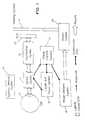
- FIG. 1is a schematic diagram of an apparatus for measurement of the concentration of blood components in accordance with the invention.
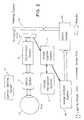
- FIG. 2is a schematic diagram of a modified form of the apparatus for measurement of blood components in accordance with the invention.
- FIG. 3is a schematic side view of a hand-held illumination and camera system that may be utilized in accordance with the invention.
- FIG. 4is an illustrative front view of the illumination and camera system of FIG. 3 .
- FIG. 5is a schematic diagram of a further apparatus in accordance with the invention that incorporates a communications link to a remote processing system.
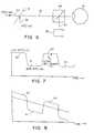
- FIG. 6is a simplified schematic diagram of apparatus in accordance with the invention for measuring bilirubin concentration in blood.
- FIG. 7is an illustrative graph of intensity of light at 550 nm reflected from the retina as a function of time, showing the effect of the periodic illumination of the eye at 470 nm.
- FIG. 8is an illustrative graph, similar to FIG. 7, of reflected light intensity over time showing the effect of changes in blood flow in the retinal vessels.
- FIG. 9is a graph of the intensity of light at 550 nm reflected from the retina showing the effect of single pulse illumination at 470 nm.
- FIG. 10is a graph illustrating the correlation between hemoglobin concentration measured in accordance with the invention versus concentration measured by a standard in vitro technique.
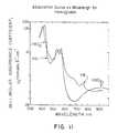
- FIG. 11are graphs of light absorption as a function of wavelength for deoxyhemoglobin and oxyhemoglobin.
- the present inventionmay be used to measure various analytes. These analytes may be photoreactive or non-photoreactive. Table 1 below summarizes examples of the preferred methodologies in accordance with the invention for the measurement of photoreactive and non-photoreactive analytes. However, it is understood that these are only illustrative of the analytes that may be measured using the present invention.
- Target MoleculeNon-Photoreactive Analyte
- Photoreactive AnalyteExamples: Hemoglobin Glucose Bilirubin Illumination: Visible IR Discrete wavelength Light illumination visible light IR illumination Target Site: Retinal disk Retinal disk Retina vasculature vasculature Imaging: Single image Multiple Multiple time lapsed images images at different wavelengths Scanned sensor array
- FIG. 1illustrates a blood analysis apparatus of the invention in conjunction with the eye of the patient, shown illustratively at 10 in FIG. 1 .
- the blood analysis apparatusincludes an optics system 11 comprised of lenses for projecting illuminating light onto the fundus, generally although not necessarily directly through the pupil, and for receiving the light reflected from the fundus passed out through the pupil, and for focusing that light to form an image.
- the lensespreferably include a final lens which can be positioned close to (e.g., approximately 3 mm from) the cornea of the eye, providing a 10 to 30 degree conical view of the retina to be illuminated and imaged.
- Such lens systemsare of conventional design and are utilized for macroview lens systems in retinal video cameras.
- An illumination system 12provides selected illuminating light for viewing and imaging the retina.
- the illumination systemis preferably a monochromatic or multiple discrete wavelength light source that provides light for viewing and imaging the retina.
- the systemprovides light for viewing and imaging coaxially to reduce the likelihood of extraneous reflections from the interior or exterior of the eye.
- the light from the illumination systemmay be projected through the pupil.
- the frequency content of this light sourceis selected dependent upon the compound to be analyzed.
- Illumination lightmay be composed of two (or more) separate lighting systems, such as a xenon strobe, or multiple laser diodes, for imaging, and a halogen source for viewing. Infrared imaging may be done utilizing a filtered halogen or laser diode source.
- the lightis reflected from the fundus of the eye 10 and passed through the pupil opening of the eye to the optics system 11 and through the illumination system 12 , entering, e.g., a charge coupled device (CCD) detector 22 .
- the illumination system 12may be similar to systems used in existing non-mydriatic fundus cameras, preferably modified to provide a coaxial design for illumination and imaging.
- a viewing system 14for example, a liquid crystal display (LCD) screen, may receive the image data and display the image for use by the operator for initially locating the patient's retina, based on an image from the optical system in real time.
- a coaxial “scene” or visual targetmay be included in the visual field of the device so that a patient can fixate his or her eye on this scene and reduce eye motion.
- the location of this visual targetwill bring the optic disk into the approximate center of the CCD detector.
- the scenemay include a visually pleasant object such as a familiar animal.
- the LCD (or other display) screenis typically located on a desktop power source that is attached to the hand-held camera by a cable. While such displays may be used in the present invention, the LCD screen (or other display device) is preferably placed on the back of the hand-held camera unit, so that the operator can more easily locate the retina, having the patient's eye and the LCD screen in the same line-of-sight.
- the Nidek NM100 camerautilizes a coaxial imaging system with an infrared source, with imaging done through reflected light outside the optical system.
- the inventionmay be carried out with a dilated eye pupil, it is preferable that the imaging of the retina be carried out without requiring dilation of the pupil for speed of measurement and patient convenience.
- the camerapreferably includes a shield (not shown) to prevent ambient light from entering the optical system 11 to minimize extraneous reflections and the introduction of optical noise.
- the optical system 11also interfaces with a locate and focus system 16 , which utilizes feedback from an image capture system 17 , also interfaced to the optic system 11 , to automatically find and bring the optic disk into focus.
- a convolver or other pattern recognition softwaremay be utilized to locate the optic disk area by finding the circular pattern of the optic nerve area. After using the pattern recognition information to more precisely locate the optic nerve area in the center of the viewing field, the image may then be magnified using a series of lenses in the optics system 11 such that the optic disk area virtually fills the active area of the CCD (or other detector).
- the optical systempreferably tracks the movement of the fundus while zooming the optics such that the optic disk is centered and occupies most of the optical field of view.
- the optical systemmay be formed to track the motion of the fundus through a motor drive system that slightly gimbals the lens system. This motion system is driven and controlled in a closed loop manner utilizing the feedback of the pattern recognition software.
- the image capture system 17is selectively controlled by the operator and uses feature and pattern recognition to drive the locate and auto focus system 16 to capture and store an appropriate image for analysis.
- Image captureitself is analogous to the function provided by a “digital still camera.”
- the image capture systemmay utilize feature and pattern recognition to drive the locate and focus system to capture and store an appropriate image for analysis.
- Commercially available pattern recognition softwaremay be used.
- Actual imagingis preferably timed based on the patient's blood flow, which is preferably sensed from blood vessels through the patient's skin around the ocular structure through a systolic sensing system 13 .
- the light reflected from the retinais preferably detected and an image formed at the time of systole, thus ensuring the maximum blood flow in the vasculature of the retina.
- Detection of the patient's systolic statecan be made through any commonly known means such as commercially available blood pressure transducers.
- An image analysis system 18is interfaced with the image capture system 17 to analyze the light reflected from the retina to quantitatively determine the amount of the particular target analyte compound present.
- the resultsmay be displayed to the operator via the output system 20 .
- This systempresents results as well as any feedback associated with the acquisition of the data, and may include an LCD display screen or other display devices.
- a modified imaging methodology as shown in FIG. 2may also be used wherein an imaging device 21 functions in a scanning mode (which may be similar to that utilized in common video cameras) to provide multiple images of the retina to the image capture system 17 .
- the image recognition system 15then acts to select a valid image from the series of images stored by the image capture system 17 . Once selection of an appropriate image is made, calculation of the magnitude of the desired analyte proceeds using the image analysis system 18 as before. Image validity is based on focus, closeness to center of the fundus, and obliqueness of the fundus in the image.
- FIG. 3illustrates a hand-held camera and illumination unit 25 for the analysis system of the invention in which the optic system 11 is mounted
- FIG. 4illustrates the output viewing system 14 at the back end of the hand-held unit 25 to enable the operator to review the image being obtained of the retina on a real-time basis.
- a disposable transparent plastice.g. polyethylene, polystyrene, polypropylene, etc.
- the covermay include a soft portion (e.g. foam) that comes into contact with the skin around the orbit. This makes the cover comfortable and, in addition, prevents ambient light from entering into the camera lens during measurements.
- the illumination sourcemay instead scan across the area of interest on the retina, with the reflected beam being read by a single sensor (or a small multiplicity of sensors) that are sensitive in the infrared (IR) wavelengths above normal CCD sensitivity. This method may be used to digitally reconstruct the retinal image if desired.
- IRinfrared
- image processing and analysismay take place at a location remote from the clinical setting by using a wired or wireless internet link (or dedicated communication link) to transfer data from the image capture system 17 to a central computer at a remote location (i.e., anywhere in the world linked by the internet) at which the image analysis system 18 is implemented.
- the output data from the output system 20may be transferred back through an access link 29 to the viewing system 14 at the clinic (or to another location, as desired).
- HemoglobinNon-Photoreactive Analyte
- the image of the optic disk returning from the eyemay be split into three simultaneous and equal images. Each image is then passed through a filter with the preferred wavelengths for the images of 640, 766 and 800 nm. Different wavelengths of light are used for the analysis of other substances such as glucose. Although these preferred wavelengths will yield accurate measurement of hemoglobin, other wavelengths in the visible or near IR may also be used effectively by this invention for hemoglobin.
- the measurement of hemoglobinmay be carried out in the present invention using wavelengths of light at which the extinction coefficients for oxyhemoglobin and deoxyhemoglobin match.
- the total absorption of energy at that wavelengthwill be proportional to the total hemoglobin. Examples of such wavelengths are 550 nm and 800 nm.
- Absorption curves for oxyhemoglobin (HbO 2 ) and deoxyhemoglobin (Hb) as a function of illumination wavelengthare shown in FIG. 11 .
- the absorption curvesare closely matched in the wavelength range from about 380 nm to 580 nm, and particularly from about 530 nm to 580 nm, allowing most (or all) wavelengths in these ranges to be used.
- the areas that are preferably used for analysisare the retinal arteries and veins overlaying the optic disk.
- Previous work by Hickam et al.suggested that when the optic disk is illuminated with light directed toward the fundus, the optic disk acts as a light source directing the light back through the retinal vessels that lay over the disk. See, John B. Hickam, et al., “A Study of Retinal Venous Blood Oxygen Saturation in Human Subjects by Photographic Means,” Circulation, Vol. XXVII, Mar., 1963, pp. 375-385. Hickam, et al.
- the retinal arteriesare the sub-target areas that are analyzed by measuring the amplitude of the signal at each selected wavelength. This is done by determining the effective reflected image intensity of the pixels for the sub-target areas corresponding to the arteries at each selected wavelength.
- the peak concentration of bloodis determined by viewing the peak absorption at each measured wavelength.
- the sub-target areais then normalized by this peak and then may be averaged using any common digital averaging technique, such as a convolver.
- a histogramis created to represent different signal levels after averaging.
- These data comprised of histogram values and values representing the rate of change in local intensityare then the direct input into an analysis program, e.g., a neural network, for analysis.
- a computer implemented neural networkis preferably utilized to analyze the pixel data for comparison to known blood concentrations.
- a preferred type of neural networkis Back Propagation.
- the effect of differences in illumination from one retinal image to anotheris preferably neutralized by comparing the background illumination of the optic disk to the vessel illumination and “zeroing” for differences in illumination. This allows the input from the sub-target areas to be consistent from patient to patient and camera to camera.
- the present inventionis well suited to be carried out on a “real time” basis with the apparatus of FIGS. 1 and 2 as described above, it may be implemented using photographic fundus images which are scanned and processed.
- photographic imagesa group of individuals was tested to measure hemoglobin levels utilizing the present invention.
- a standard in vitro testwas also performed on a sample of blood drawn from each individual. The clinical data were obtained as follows. Each ophthalmologist in the clinic agreed to refer for study patients that already had their eyes dilated. A physician asked the patient if they would agree to have their eyes photographed and to allow blood to be obtained for a hemoglobin test. An informed consent document was signed by each patient. A retinal photographer with the clinic took all of the retinal photographs.
- the slide film usedwas Kodachrome 64 Select, 24 exposures per roll.
- the photographertook photos of each eye, with the fovea centered in the picture.
- the exact same conditionswere used for each photograph including aperture, shutter speed, and illumination.
- a codewas given for each patient to insure confidentiality and to allow accurate tracking of each patient. The code appeared on each photograph and the same code appeared on the blood tube.
- a licensed phlebotomistobtained antecubital blood samples from each patient. These samples were drawn with vacutainers into purple-top tubes for later analysis. The tubes were stored cold and run the same evening. Following the analysis, the blood tubes were saved if later analysis were required. The blood samples were analyzed for hemoglobin content with a HemoCue® B-Hemoglobin Analyzer. The machine was calibrated with calibration slides. This device is CLIA certified and the technician who analyzed the samples was also CLIA certified. Each sample was run three times on the machine to ensure accurate results. These results were recorded along with the patient's ID code. Kodachrome 64 Select Slide Film was used for each retinal photograph. The film was sent to Kodak for processing. The Kodak development lab used the same lot of chemicals for all of the slides. The developed slides were then scanned into a digital format (jpeg files) with a Polaroid Sprintscan 4000 scanner.
- the neural net employed for exemplificationwas the “Back Propagation” neural net which is contained in the MATLAB Toolbox of MATLAB Version 5.3.
- the neural netis trained with the data sets determined above by the following technique:
- Correlation data showing the measurements of hemoglobin concentration for each individual as determined in accordance with the present invention versus the hemoglobin measurement values obtained by the in vitro laboratory measurementsare shown in FIG. 10 . These data indicate a strong correlation, 0.89, between the measurements obtained by the present invention and measurements obtained by standard in vitro laboratory techniques.
- the results of the analysis of the serum concentration values of the blood componentsmay be displayed on an output system 20 to the operator.
- the output system 20may utilize the LCD screen 14 of the viewing system to present data to the operator as well as feedback on any problems occurring with the acquisition of the image or the device. In the event that the image is not acceptable and will not allow the device to calculate accurate blood values, the output system 20 preferably prompts the operator to capture another image.
- various degrees of light scatteringmay be encountered. This scattering of the light may be the result of various disease states including senile cataracts.
- a polarized light sourcemay be included in the device to measure the degree of scattering. The returned light at orthogonal polarization may be used as a measure of scattering. This data is fed into the neural network as the scattering variable from patient to patient to allow the device to account for this variable.
- analytes that are measured with the device of the inventionare also secreted into the anterior chamber of the eye.
- the aforementioned embodiment of the devicedirects light through the anterior chamber to the retina.
- the image returned from the retinaagain passes through this area of the eye.
- the presence of the analyte in this section of the eyecould create false data sets and render the device inaccurate. Therefore, when measuring analytes such as glucose that are present in the anterior chamber, the light may be directed along an alternate path to the retina.
- the illuminating light emitting from the devicemay be directed toward the retina at an angle into the eye just lateral to the cornea near the corneoscleral junction (limbus). This angle avoids light passing through the anterior chamber of the eye.
- the exiting lightwould then be detected at an angle at the opposite side of the eye, again just lateral to the cornea near the corneoscleral junction (limbus).
- this area near the surface of the eyeis not optically clear in the visual range of the electromagnetic spectrum, it is transparent in the longer wavelengths (>1000 nm) needed to measure analytes such as glucose.
- the concentration of a photoreactive analytemay be determined by taking advantage of its photoreactivity.
- bilirubin concentrationmay be determined by taking advantage of the fact that the bilirubin molecule exhibits peak absorption of light at 470 nm, and that at or near this wavelength the molecule breaks down into optically inactive molecules.
- the bilirubin moleculewill also effectively break down when exposed to light in the range of 470 nm ⁇ 30 nm.
- the bilirubin moleculeordinarily reflects light maximally at or near a wavelength of approximately 550 nm (yellow light).
- a time lapse image of the retinamay be made wherein the first image is illuminated at 550 nm ( ⁇ 30 nm) followed by a second image illuminated at 470 nm ( ⁇ 30 nm) and a third image illuminated at 550 nm ( ⁇ 30 nm).
- the second and third imagesare separated in time sufficiently that the photoreactive analyte will change chemical state.
- the changeis almost instantaneous and the two wavelengths may be projected simultaneously as well as sequentially.
- the first exposureprovides an image including the photoreactive analyte (bilirubin in this example) while the third exposure provides a reference point for this patient without the photoreactive analyte (bilirubin in this example) present.
- concentration of the photoreactive analytecan be computed using a neural net or other well known analysis process based on differences in the first and third exposures.
- Light at wavelengths other than 550 nmmay also be used to illuminate the retina as long as such wavelengths do not precipitate a photo-chemical reaction from the analyte (bilirubin in this example).
- light at a wavelength of 550 nmis directed on a beam 30 to a partially transmissive and reflective mirror 31 , passing therethrough on a beam path 32 to a beam splitter 34 and thence to the eye 10 .
- the light at a wavelength of 550 nmis directed at the retina to create a first image.
- Light from a source at 470 nmis provided on a path 36 to the partial mirror 31 and is reflected on the path 32 through the beam splitter 34 to the eye 10 .
- the reflected light at 470 nmis directed by the element 34 on a path 37 to an absorber 38 .
- the light at 470 nmis pulsed onto the retina at very short time intervals. With each pulsation of light at 470 nm, the bilirubin molecule is rendered optically inactive and is not reflected from the retina at 550 nm. As illustrated in FIG. 7, the reflected 550 m light intensity is high at the intervals 40 when there is no 470 nm light illumination and is lower at the intervals 41 at which the 470 nm light is applied. The difference in reflected light intensity from the peak to the trough with each pulsation is a function of the bilirubin concentration. The time intervals 40 and 41 chosen will be dependent on the blood flow to the retina. FIG. 8 illustrates the change over time with blood flow.
- a time intervalis required such that there is a partial filling up of the retinal circulation by blood that contains bilirubin.
- the inventionmay also be implemented using a single pulse application of 470 nm light to measure the bilirubin concentration as shown in FIG. 9 .
- the data corresponding to the returned light at 550 nm comprised of pixel valuesare then the direct input into a neural network or other well known procedure for analysis.
- a neural networkis preferably utilized to analyze the pixel data for comparison to known blood concentrations of bilirubin.
- the difference in pixel data from reflected light at 550 nm before and after the 470 nm light pulseis a direct function of the serum bilirubin level.
- the results of the serum values of the bilirubinmay be displayed on an output system 20 to the operator.
- the output system 20may utilize the LCD screen of the viewing system to present data to the operator as well as feedback on any problems occurring with the acquisition of the image or the device.
- the output system 20preferably prompts the operator to capture another image.
- Other photo-reactive analytesmay be measured in a manner analogous to the measurement of bilirubin as discussed above.
Landscapes
- Health & Medical Sciences (AREA)
- Life Sciences & Earth Sciences (AREA)
- Physics & Mathematics (AREA)
- Medical Informatics (AREA)
- Surgery (AREA)
- Biophysics (AREA)
- Veterinary Medicine (AREA)
- Engineering & Computer Science (AREA)
- Biomedical Technology (AREA)
- Heart & Thoracic Surgery (AREA)
- Public Health (AREA)
- Molecular Biology (AREA)
- General Health & Medical Sciences (AREA)
- Animal Behavior & Ethology (AREA)
- Optics & Photonics (AREA)
- Pathology (AREA)
- Emergency Medicine (AREA)
- Hematology (AREA)
- Ophthalmology & Optometry (AREA)
- Spectroscopy & Molecular Physics (AREA)
- Measurement Of The Respiration, Hearing Ability, Form, And Blood Characteristics Of Living Organisms (AREA)
- Eye Examination Apparatus (AREA)
Abstract
Description
This application claims the benefit of provisional patent applications No. 60/165,195, filed Nov. 12, 1999 and No. 60/126,212, filed Mar. 25, 1999, which are incorporated herein by reference.
This invention pertains to the field of non-invasive in vivo measurement of blood components such as glucose, hemoglobin, and bilirubin.
The measurement of the concentration of blood components such as hemoglobin and glucose has required the drawing of a blood sample for in vitro analysis. The need to draw blood for analysis is undesirable for several reasons, including discomfort to the patient, the time required of medical personnel to draw and handle the samples, and the potential risk of spread of disease through punctures of the skin. Repeated drawing of blood samples is especially undesirable in infants. Many diabetics must test their blood up to six times a day to monitor their blood glucose levels. It would thus be desirable to be able to obtain fast and reliable estimates of the concentration of blood components in blood, such as hemoglobin and glucose, through a simple and non-invasive technique. Prior efforts have involved an examination of blood in the skin or extremities, such as fingers and ear lobes, or in observable surface blood vessels, but these efforts have had limited practical success due to the presence of tissue components that interfere with accurate reading of only the concentration of blood components.
There are approximately four million newborns in the United States alone each year. About 50% of newborns are clinically jaundiced from elevated bilirubin levels. If the serum bilirubin reaches very high levels during the post-natal period, kernicterus, neural damage resulting from sustained high levels of serum bilirubin, may occur. Frequent monitoring of serum bilirubin is critical to the care of these infants. Of the newborns that have recognizable jaundice during the first 5 days of life, 1.7 million receive at least one blood test for bilirubin. Of those tested, about 700,000 undergo phototherapy treatment; these infants receive an estimated two to three additional blood tests. Presently, blood is drawn through the heel of the neonate, resulting in occasional infections and other complications. Other drawbacks to this process are its high cost and the delay in lab results reaching the physician. Recently introduced non-invasive devices for measuring bilirubin do not provide the accuracy level required to diagnose or treat elevated serum bilirubin levels, rendering them virtually useless in practice.
The present invention combines the accuracy of in vitro laboratory testing of blood components and the advantages of rapidly-repeatable non-invasive technology. The invention utilizes a hand-held or stationary instrument for retinal imaging that allows non-invasive measurement of certain blood components in the retinal blood vessels. Illuminating light of selected wavelengths in the visible or infrared range is projected into the eye onto the fundus, and the light reflected back and out (e.g., through the pupil) is detected and analyzed, preferably using the area of the optic disk for analyzing the retinal vessels overlaying the optic disk for most blood components to be measured. Specific wavelengths of illuminating light may be chosen for each blood component to be analyzed depending on the spectral characteristics of the particular substance being analyzed. The reflected image from the retina is utilized to measure blood components, such as hemoglobin, glucose and bilirubin.
The utilization of the retina as a site for obtaining blood component data has several advantages, including the ease of visualizing the data because of the natural window provided by the eye. The reflected light from the fundus at visually significant wavelengths is much less scattered than light reflected from the skin or mucous membranes since the eye is naturally immune to scatter. The retina creates a uniform background for imaging, and the optical devices and techniques required for obtaining retinal images have been extensively developed and studied because of the need for ophthalmologists to image the retina for diagnosis of disease states. In addition, the blood flow to the retina is very even and repeatable even across a number of disease states. For example, although patients in shock have reduced blood flow to the skin and mucous membranes, allowing false data to be obtained with current technology that examines the skin and mucous membranes, the body maintains even blood flow to the retina except in states of extremely low blood pressure. Furthermore, in the present invention, there is no physical contact required between the device and the skin or mucous membranes, thereby eliminating the potential for transmission of infectious agents associated with devices that require patient contact. The device may be rested upon the orbit of the patient for ease of use and, if desired, a disposable plastic cover may be used to further minimize the risk of transmission of infectious agents. Such a cover may be transparent and fully cover the device, or not, depending upon requirements of the imaging system and the need to prevent incidental contact with the device.
In accordance with the invention, a hand-held or stationary instrument for retinal imaging may be used to obtain non-invasive measurement of photoreactive analytes, an example of which is serum bilirubin. Illuminating light of selected wavelengths in the visible range is projected into the eye onto the fundus. Specific wavelengths of illuminating light are chosen so that serum bilirubin can be measured. Analysis of the reflected image from the fundus is utilized to measure bilirubin. Although measurement of the reflected light from the vessels overlying the optic disk is preferred, in accordance with the invention, it is also typically possible to obtain bilirubin measurements from light reflected from the fundus generally.
During disease states when the serum bilirubin levels are above normal (e.g., newborn jaundice), bilirubin is extruded from the choroid into the nerve layer of the retina. During newborn jaundice, this nerve layer stains yellow from the elevated bilirubin levels. This yellow color is directly proportional to the elevated serum bilirubin levels and changes rapidly with changes in serum bilirubin. The bilirubin molecule exhibits peak absorption of light at 470 nm. However, when exposed to light at or near this wavelength, the molecule breaks down into optically inactive molecules. The intact bilirubin molecule reflects light at and near a wavelength of approximately 550 nm (yellow light), and is not affected by this light. In the present invention, the retina of the patient's eye may first be imaged with light that does not break down bilirubin, e.g., light at a wavelength of 550 nm with little or no light at 470 nm. The intensity of the reflected light at or near the maximum reflection wavelength of 550 nm is detected. Then the retina is imaged again using light that breaks down bilirubin, e.g., using light at 470 nm followed by or combined with light at 550 nm, which is projected into the eye. The reflected light at 550 nm that is passed out through the pupil is detected to image the retina a second time. With the addition of light at 470 nm, the bilirubin molecule is rendered optically inactive and will no longer reflect at 550 nm. The difference in the reflected image intensity at 550 nm from the first image to the second image is a function of the bilirubin concentration. A neural network or other processing technique may be used to analyze the two data sets of the images captured by the retinal camera.
Further objects, features and advantages will be apparent from the following detailed description when taken in conjunction with the accompanying drawings.
In the drawings:
FIG. 1 is a schematic diagram of an apparatus for measurement of the concentration of blood components in accordance with the invention.
FIG. 2 is a schematic diagram of a modified form of the apparatus for measurement of blood components in accordance with the invention.
FIG. 3 is a schematic side view of a hand-held illumination and camera system that may be utilized in accordance with the invention.
FIG. 4 is an illustrative front view of the illumination and camera system of FIG.3.
FIG. 5 is a schematic diagram of a further apparatus in accordance with the invention that incorporates a communications link to a remote processing system.
FIG. 6 is a simplified schematic diagram of apparatus in accordance with the invention for measuring bilirubin concentration in blood.
FIG. 7 is an illustrative graph of intensity of light at 550 nm reflected from the retina as a function of time, showing the effect of the periodic illumination of the eye at 470 nm.
FIG. 8 is an illustrative graph, similar to FIG. 7, of reflected light intensity over time showing the effect of changes in blood flow in the retinal vessels.
FIG. 9 is a graph of the intensity of light at 550 nm reflected from the retina showing the effect of single pulse illumination at 470 nm.
FIG. 10 is a graph illustrating the correlation between hemoglobin concentration measured in accordance with the invention versus concentration measured by a standard in vitro technique.
FIG. 11 are graphs of light absorption as a function of wavelength for deoxyhemoglobin and oxyhemoglobin.
The present invention may be used to measure various analytes. These analytes may be photoreactive or non-photoreactive. Table 1 below summarizes examples of the preferred methodologies in accordance with the invention for the measurement of photoreactive and non-photoreactive analytes. However, it is understood that these are only illustrative of the analytes that may be measured using the present invention.
| TABLE 1 |
| Methodologies |
| Target Molecule: | Non-Photoreactive Analyte | Photoreactive Analyte |
| Examples: | Hemoglobin | Glucose | Bilirubin |
| Illumination: | Visible | IR | Discrete wavelength |
| Light | illumination | visible light | |
| IR | |||
| illumination | |||
| Target Site: | Retinal disk | Retinal disk | Retina |
| vasculature | vasculature | ||
| Imaging: | Single image | Multiple | Multiple time lapsed |
| images | images at different | ||
| wavelengths | |||
| Scanned sensor | |||
| array | |||
With reference to the drawings, FIG. 1 illustrates a blood analysis apparatus of the invention in conjunction with the eye of the patient, shown illustratively at10 in FIG.1. The blood analysis apparatus includes anoptics system 11 comprised of lenses for projecting illuminating light onto the fundus, generally although not necessarily directly through the pupil, and for receiving the light reflected from the fundus passed out through the pupil, and for focusing that light to form an image. The lenses preferably include a final lens which can be positioned close to (e.g., approximately 3 mm from) the cornea of the eye, providing a 10 to 30 degree conical view of the retina to be illuminated and imaged. Such lens systems are of conventional design and are utilized for macroview lens systems in retinal video cameras.
Anillumination system 12 provides selected illuminating light for viewing and imaging the retina. The illumination system is preferably a monochromatic or multiple discrete wavelength light source that provides light for viewing and imaging the retina. Preferably, the system provides light for viewing and imaging coaxially to reduce the likelihood of extraneous reflections from the interior or exterior of the eye. The light from the illumination system may be projected through the pupil. The frequency content of this light source is selected dependent upon the compound to be analyzed. Illumination light may be composed of two (or more) separate lighting systems, such as a xenon strobe, or multiple laser diodes, for imaging, and a halogen source for viewing. Infrared imaging may be done utilizing a filtered halogen or laser diode source. The light is reflected from the fundus of theeye 10 and passed through the pupil opening of the eye to theoptics system 11 and through theillumination system 12, entering, e.g., a charge coupled device (CCD)detector 22. Theillumination system 12 may be similar to systems used in existing non-mydriatic fundus cameras, preferably modified to provide a coaxial design for illumination and imaging. Aviewing system 14, for example, a liquid crystal display (LCD) screen, may receive the image data and display the image for use by the operator for initially locating the patient's retina, based on an image from the optical system in real time. A coaxial “scene” or visual target may be included in the visual field of the device so that a patient can fixate his or her eye on this scene and reduce eye motion. In addition to reducing eye motion, the location of this visual target will bring the optic disk into the approximate center of the CCD detector. In devices intended for children, the scene may include a visually pleasant object such as a familiar animal. In the currently commercially available video cameras designed for retinal imaging, the LCD (or other display) screen is typically located on a desktop power source that is attached to the hand-held camera by a cable. While such displays may be used in the present invention, the LCD screen (or other display device) is preferably placed on the back of the hand-held camera unit, so that the operator can more easily locate the retina, having the patient's eye and the LCD screen in the same line-of-sight. Current retinal video camera systems that may be modified and utilized in the invention and which include anoptics system 11 and an illumination system include the Nidek NM100 Hand-Held Non-Mydriatic Fundus Camera and the Topcon TRC-50EX (TRC-NW5S/TRC-NW5SF) Non-Mydriatic Retinal Camera. The Nidek NM100 camera utilizes a coaxial imaging system with an infrared source, with imaging done through reflected light outside the optical system. Although the invention may be carried out with a dilated eye pupil, it is preferable that the imaging of the retina be carried out without requiring dilation of the pupil for speed of measurement and patient convenience. The camera preferably includes a shield (not shown) to prevent ambient light from entering theoptical system 11 to minimize extraneous reflections and the introduction of optical noise.
Theoptical system 11 also interfaces with a locate and focussystem 16, which utilizes feedback from animage capture system 17, also interfaced to theoptic system 11, to automatically find and bring the optic disk into focus. A convolver or other pattern recognition software may be utilized to locate the optic disk area by finding the circular pattern of the optic nerve area. After using the pattern recognition information to more precisely locate the optic nerve area in the center of the viewing field, the image may then be magnified using a series of lenses in theoptics system 11 such that the optic disk area virtually fills the active area of the CCD (or other detector). The optical system preferably tracks the movement of the fundus while zooming the optics such that the optic disk is centered and occupies most of the optical field of view. The optical system may be formed to track the motion of the fundus through a motor drive system that slightly gimbals the lens system. This motion system is driven and controlled in a closed loop manner utilizing the feedback of the pattern recognition software.
Theimage capture system 17 is selectively controlled by the operator and uses feature and pattern recognition to drive the locate andauto focus system 16 to capture and store an appropriate image for analysis. Image capture itself is analogous to the function provided by a “digital still camera.” The image capture system may utilize feature and pattern recognition to drive the locate and focus system to capture and store an appropriate image for analysis. Commercially available pattern recognition software may be used. Actual imaging is preferably timed based on the patient's blood flow, which is preferably sensed from blood vessels through the patient's skin around the ocular structure through asystolic sensing system 13. The light reflected from the retina is preferably detected and an image formed at the time of systole, thus ensuring the maximum blood flow in the vasculature of the retina. Detection of the patient's systolic state can be made through any commonly known means such as commercially available blood pressure transducers. Animage analysis system 18 is interfaced with theimage capture system 17 to analyze the light reflected from the retina to quantitatively determine the amount of the particular target analyte compound present. The results may be displayed to the operator via theoutput system 20. This system presents results as well as any feedback associated with the acquisition of the data, and may include an LCD display screen or other display devices.
A modified imaging methodology as shown in FIG. 2 may also be used wherein animaging device 21 functions in a scanning mode (which may be similar to that utilized in common video cameras) to provide multiple images of the retina to theimage capture system 17. Theimage recognition system 15 then acts to select a valid image from the series of images stored by theimage capture system 17. Once selection of an appropriate image is made, calculation of the magnitude of the desired analyte proceeds using theimage analysis system 18 as before. Image validity is based on focus, closeness to center of the fundus, and obliqueness of the fundus in the image.
FIG. 3 illustrates a hand-held camera andillumination unit 25 for the analysis system of the invention in which theoptic system 11 is mounted, and FIG. 4 illustrates theoutput viewing system 14 at the back end of the hand-heldunit 25 to enable the operator to review the image being obtained of the retina on a real-time basis. If desired, a disposable transparent plastic (e.g. polyethylene, polystyrene, polypropylene, etc.), which is selected to transmit light in the wavelengths required, may be used to cover theunit 25 during use and then disposed of to minimize the risk of transmission of infection. Eye infections are particularly common in newborns and a cover would assist in the prevention of these infections. Furthermore, the cover may include a soft portion (e.g. foam) that comes into contact with the skin around the orbit. This makes the cover comfortable and, in addition, prevents ambient light from entering into the camera lens during measurements.
Currently available CCD detectors may not be sensitive to wavelengths longer than approximately 1000 nm. In measuring analytes requiring the sensing of these longer wavelengths, an alternative approach may be used. The illumination source may instead scan across the area of interest on the retina, with the reflected beam being read by a single sensor (or a small multiplicity of sensors) that are sensitive in the infrared (IR) wavelengths above normal CCD sensitivity. This method may be used to digitally reconstruct the retinal image if desired.
As illustrated in FIG. 5, image processing and analysis may take place at a location remote from the clinical setting by using a wired or wireless internet link (or dedicated communication link) to transfer data from theimage capture system 17 to a central computer at a remote location (i.e., anywhere in the world linked by the internet) at which theimage analysis system 18 is implemented. The output data from theoutput system 20 may be transferred back through anaccess link 29 to theviewing system 14 at the clinic (or to another location, as desired).
In one implementation of the device for the detection of hemoglobin using multiple beam splitters, the image of the optic disk returning from the eye may be split into three simultaneous and equal images. Each image is then passed through a filter with the preferred wavelengths for the images of 640, 766 and 800 nm. Different wavelengths of light are used for the analysis of other substances such as glucose. Although these preferred wavelengths will yield accurate measurement of hemoglobin, other wavelengths in the visible or near IR may also be used effectively by this invention for hemoglobin.
To obtain accurate measurements of hemoglobin concentration without having to account for different levels of oxygen saturation, the measurement of hemoglobin may be carried out in the present invention using wavelengths of light at which the extinction coefficients for oxyhemoglobin and deoxyhemoglobin match. Thus, the total absorption of energy at that wavelength will be proportional to the total hemoglobin. Examples of such wavelengths are 550 nm and 800 nm. Absorption curves for oxyhemoglobin (HbO2) and deoxyhemoglobin (Hb) as a function of illumination wavelength are shown in FIG.11. As illustrated therein, the absorption curves are closely matched in the wavelength range from about 380 nm to 580 nm, and particularly from about 530 nm to 580 nm, allowing most (or all) wavelengths in these ranges to be used.
The areas that are preferably used for analysis are the retinal arteries and veins overlaying the optic disk. Previous work by Hickam et al. suggested that when the optic disk is illuminated with light directed toward the fundus, the optic disk acts as a light source directing the light back through the retinal vessels that lay over the disk. See, John B. Hickam, et al., “A Study of Retinal Venous Blood Oxygen Saturation in Human Subjects by Photographic Means,” Circulation, Vol. XXVII, Mar., 1963, pp. 375-385. Hickam, et al. believed that these vessels then functioned as cuvettes, filled with blood, that could be analyzed by the spectral pattern that is measured from the light passing through these vessels and emitted out through the pupil. In actuality, light does not pass through these vessels and reflect off the optic disk and then pass through these vessels again as suggested in Hickam, et al. Instead, light reflects directly off the surfaces of these vessels and the blood components within these vessels. In the present invention, the retinal arteries are the sub-target areas that are analyzed by measuring the amplitude of the signal at each selected wavelength. This is done by determining the effective reflected image intensity of the pixels for the sub-target areas corresponding to the arteries at each selected wavelength. Within the sub-target area, the peak concentration of blood is determined by viewing the peak absorption at each measured wavelength. The sub-target area is then normalized by this peak and then may be averaged using any common digital averaging technique, such as a convolver. Within the sub-target area, a histogram is created to represent different signal levels after averaging. These data comprised of histogram values and values representing the rate of change in local intensity are then the direct input into an analysis program, e.g., a neural network, for analysis. A computer implemented neural network is preferably utilized to analyze the pixel data for comparison to known blood concentrations. A preferred type of neural network is Back Propagation.
The effect of differences in illumination from one retinal image to another is preferably neutralized by comparing the background illumination of the optic disk to the vessel illumination and “zeroing” for differences in illumination. This allows the input from the sub-target areas to be consistent from patient to patient and camera to camera.
Although the present invention is well suited to be carried out on a “real time” basis with the apparatus of FIGS. 1 and 2 as described above, it may be implemented using photographic fundus images which are scanned and processed. As an example of the present invention using photographic images, a group of individuals was tested to measure hemoglobin levels utilizing the present invention. A standard in vitro test was also performed on a sample of blood drawn from each individual. The clinical data were obtained as follows. Each ophthalmologist in the clinic agreed to refer for study patients that already had their eyes dilated. A physician asked the patient if they would agree to have their eyes photographed and to allow blood to be obtained for a hemoglobin test. An informed consent document was signed by each patient. A retinal photographer with the clinic took all of the retinal photographs. The slide film used was Kodachrome 64 Select, 24 exposures per roll. The photographer took photos of each eye, with the fovea centered in the picture. The exact same conditions were used for each photograph including aperture, shutter speed, and illumination. A code was given for each patient to insure confidentiality and to allow accurate tracking of each patient. The code appeared on each photograph and the same code appeared on the blood tube.
A licensed phlebotomist obtained antecubital blood samples from each patient. These samples were drawn with vacutainers into purple-top tubes for later analysis. The tubes were stored cold and run the same evening. Following the analysis, the blood tubes were saved if later analysis were required. The blood samples were analyzed for hemoglobin content with a HemoCue® B-Hemoglobin Analyzer. The machine was calibrated with calibration slides. This device is CLIA certified and the technician who analyzed the samples was also CLIA certified. Each sample was run three times on the machine to ensure accurate results. These results were recorded along with the patient's ID code. Kodachrome 64 Select Slide Film was used for each retinal photograph. The film was sent to Kodak for processing. The Kodak development lab used the same lot of chemicals for all of the slides. The developed slides were then scanned into a digital format (jpeg files) with a Polaroid Sprintscan 4000 scanner.
Multiple images of each subject were taken, and images were selected for analysis based on uniformity of illumination, quality of focus, obliqueness of fovea to illumination axis, and systole status. The (normalized) green portion of the RGB image data in the jpeg file was used for analysis, where green =G/(R+G+B). As illustrated in FIG. 11, in the green wavelength range (about 500 nm to 550 nm), the absorption of oxy- and deoxyhemoglobin is similar. The image analysis proceeded with the following steps for each selected image:
Find the optic disk.
Create a circular area of data entirely within the optic disk that is less in diameter than the optic disk. Convolve the data in this circle with a small convolver (0.5 mm) to smooth the data.
Find the maximum and minimum levels of green in pixels within the circle. Use this min. level to normalize the values of all pixels within the circle.
Compute the gradient of green within the circle.
Find the centroid of the gradient magnitude.
Construct a circle (subtarget) of the image that is approximately 0.5 mm in diameter centered at the centroid and within the disk. (This procedure locates a sub-target area within a retinal vessel in the optic disk.)
Using the data in the pixels having green magnitudes in the range of 25-45 % of the maximum level of green in the subtarget, create a histogram of this area with 5 buckets for values and 5 buckets for derivative magnitudes. For each magnitude range, calculate the average derivative (gradient) magnitude and average magnitude.
These values then become the input for a learning algorithm, such as a neural network simulation, which is trained to associate measured hemoglobin with the data sets.
The neural net employed for exemplification was the “Back Propagation” neural net which is contained in the MATLAB Toolbox of MATLAB Version 5.3.
The neural net is trained with the data sets determined above by the following technique:
(a) remove one data set (representing one image) from the total number of sets.
(b) use the data sets remaining to train the neural net mentioned above.
(c) use the trained neural net to calculate a hemoglobin value from the data set left out at step (a) above.
By repeating the neural net calculation above, a curve was constructed of non-invasively calculated values versus actual lab values.
Correlation data showing the measurements of hemoglobin concentration for each individual as determined in accordance with the present invention versus the hemoglobin measurement values obtained by the in vitro laboratory measurements are shown in FIG.10. These data indicate a strong correlation, 0.89, between the measurements obtained by the present invention and measurements obtained by standard in vitro laboratory techniques.
The results of the analysis of the serum concentration values of the blood components may be displayed on anoutput system 20 to the operator. Theoutput system 20 may utilize theLCD screen 14 of the viewing system to present data to the operator as well as feedback on any problems occurring with the acquisition of the image or the device. In the event that the image is not acceptable and will not allow the device to calculate accurate blood values, theoutput system 20 preferably prompts the operator to capture another image.
In a subset of the patient population, because of particular anatomical characteristics of the eye, various degrees of light scattering may be encountered. This scattering of the light may be the result of various disease states including senile cataracts. A polarized light source may be included in the device to measure the degree of scattering. The returned light at orthogonal polarization may be used as a measure of scattering. This data is fed into the neural network as the scattering variable from patient to patient to allow the device to account for this variable.
Several analytes that are measured with the device of the invention are also secreted into the anterior chamber of the eye. The aforementioned embodiment of the device directs light through the anterior chamber to the retina. The image returned from the retina again passes through this area of the eye. The presence of the analyte in this section of the eye could create false data sets and render the device inaccurate. Therefore, when measuring analytes such as glucose that are present in the anterior chamber, the light may be directed along an alternate path to the retina. The illuminating light emitting from the device may be directed toward the retina at an angle into the eye just lateral to the cornea near the corneoscleral junction (limbus). This angle avoids light passing through the anterior chamber of the eye. The exiting light would then be detected at an angle at the opposite side of the eye, again just lateral to the cornea near the corneoscleral junction (limbus). Although this area near the surface of the eye is not optically clear in the visual range of the electromagnetic spectrum, it is transparent in the longer wavelengths (>1000 nm) needed to measure analytes such as glucose.
Further in accordance with the present invention, the concentration of a photoreactive analyte may be determined by taking advantage of its photoreactivity. For example, bilirubin concentration may be determined by taking advantage of the fact that the bilirubin molecule exhibits peak absorption of light at 470 nm, and that at or near this wavelength the molecule breaks down into optically inactive molecules. The bilirubin molecule will also effectively break down when exposed to light in the range of 470 nm±30 nm. The bilirubin molecule ordinarily reflects light maximally at or near a wavelength of approximately 550 nm (yellow light). In accordance with the invention, a time lapse image of the retina may be made wherein the first image is illuminated at 550 nm (±30 nm) followed by a second image illuminated at 470 nm (±30 nm) and a third image illuminated at 550 nm (±30 nm). The second and third images are separated in time sufficiently that the photoreactive analyte will change chemical state. In the case of bilirubin, the change is almost instantaneous and the two wavelengths may be projected simultaneously as well as sequentially. The first exposure provides an image including the photoreactive analyte (bilirubin in this example) while the third exposure provides a reference point for this patient without the photoreactive analyte (bilirubin in this example) present. The concentration of the photoreactive analyte can be computed using a neural net or other well known analysis process based on differences in the first and third exposures. Light at wavelengths other than 550 nm may also be used to illuminate the retina as long as such wavelengths do not precipitate a photo-chemical reaction from the analyte (bilirubin in this example).
In one embodiment of the invention, with reference to FIG.6 and with bilirubin as the target analyte, light at a wavelength of 550 nm is directed on abeam 30 to a partially transmissive andreflective mirror 31, passing therethrough on abeam path 32 to abeam splitter 34 and thence to theeye 10. The light at a wavelength of 550 nm is directed at the retina to create a first image. Light from a source at 470 nm is provided on apath 36 to thepartial mirror 31 and is reflected on thepath 32 through thebeam splitter 34 to theeye 10. The reflected light at 470 nm is directed by theelement 34 on apath 37 to anabsorber 38. The light at 470 nm is pulsed onto the retina at very short time intervals. With each pulsation of light at 470 nm, the bilirubin molecule is rendered optically inactive and is not reflected from the retina at 550 nm. As illustrated in FIG. 7, the reflected 550 m light intensity is high at theintervals 40 when there is no 470 nm light illumination and is lower at theintervals 41 at which the 470 nm light is applied. The difference in reflected light intensity from the peak to the trough with each pulsation is a function of the bilirubin concentration. Thetime intervals
The data corresponding to the returned light at 550 nm comprised of pixel values are then the direct input into a neural network or other well known procedure for analysis. A neural network is preferably utilized to analyze the pixel data for comparison to known blood concentrations of bilirubin. The difference in pixel data from reflected light at 550 nm before and after the 470 nm light pulse is a direct function of the serum bilirubin level.
The results of the serum values of the bilirubin may be displayed on anoutput system 20 to the operator. Theoutput system 20 may utilize the LCD screen of the viewing system to present data to the operator as well as feedback on any problems occurring with the acquisition of the image or the device. In the event that the image is not acceptable and will not allow the device to calculate accurate bilirubin values, theoutput system 20 preferably prompts the operator to capture another image. Other photo-reactive analytes may be measured in a manner analogous to the measurement of bilirubin as discussed above.
It is understood that the invention is not limited to the embodiments set forth herein as illustrative, but embraces all such forms thereof as come within the scope of the following claims.
Claims (21)
1. A method for the detection of the concentration of a blood component in an individual comprising:
(a) projecting light having selected wavelengths into an eye of the individual to illuminate the fundus;
(b) detecting the light reflected from the fundus and forming an image of a portion of the fundus containing blood vessels;
(c) analyzing selected wavelength components in the detected light from the blood vessels in the image to determine the concentration of a selected blood component.
2. The method of claim1 wherein the selected blood component is hemoglobin.
3. The method of claim1 wherein the selected blood component is glucose.
4. The method of claim1 wherein in the step of analyzing the wavelength components, only light from the regions of the image corresponding to blood vessels is analyzed.
5. The method of claim1 wherein the light projected into the eye contains wavelengths in the visible or near infrared range and wherein detecting the light reflected from the fundus includes detecting the light in the visible or near infrared range.
6. The method of claim1 wherein the step of forming an image forms an image of the blood vessels in the region overlying the optic disk of the eye and wherein the step of analyzing the wavelength components analyzes the light reflected from the optic disk.
7. The method of claim1 wherein the step of projecting light into the eye is carried out by projecting light through the pupil of the eye onto the fundus.
8. The method of claim1 wherein the step of projecting light into the eye is carried out by projecting light taking a path at an angle into the eye just lateral to the cornea, near the corneoscleral junction.
9. A method for the detection of the concentration of a blood component in an individual comprising:
(a) projecting light having selected wavelengths into an eye of the individual to illuminate the fundus;
(b) detecting the light reflected from the fundus and forming an image of a portion of the fundus containing blood vessels;
(c) analyzing selected wavelength components in the detected light from the blood vessels in the image to determine the concentration of a selected blood component, wherein the selected blood component is bilirubin.
10. A method for the detection of the concentration of a blood component in an individual comprising:
(a) projecting light having selected wavelengths into an eye of the individual to illuminate the fundus;
(b) detecting the light reflected from the fundus and forming an image of a portion of the fundus containing blood vessels;
(c) analyzing selected wavelength components in the detected light from the blood vessels in the image to determine the concentration of a selected blood component;
(d) sensing the blood flow of the individual being tested to detect systole and wherein this information is used to time the step of detecting the light reflected from the fundus and forming an image.
11. A method for the detection of the concentration of a blood component in an individual comprising:
(a) projecting light having selected wavelengths into an eye of the individual to illuminate the fundus;
(b) detecting the light reflected from the fundus and forming an image of a portion of the fundus containing blood vessels;
(c) analyzing selected wavelength components in the detected light from the blood vessels in the image to determine the concentration of a selected blood component, wherein multiple images are formed of the fundus at discrete times over a period of time and including selecting an image from the multiple images and analyzing that image to determine the concentration of the blood component.
12. A method for the detection of the concentration of a blood component in an individual comprising:
(a) projecting light having selected wavelengths into an eye of the individual to illuminate the fundus;
(b) detecting the light reflected from the fundus and forming an image of a portion of the fundus containing blood vessels;
(c) analyzing selected wavelength components in the detected light from the blood vessels in the image to determine the concentration of a selected blood component, wherein the image is split into multiple images that are passed through filters that pass different wavelengths to provide multiple filtered images, and analyzing the multiple filtered images to determine the concentration of the blood component.
13. The method of claim10 wherein the blood component is hemoglobin and the multiple images are filtered to pass light centered at wavelengths of 640 nm, 766 nm and 800 nm.
14. A method for the detection of the concentration of a blood component in an individual comprising:
(a) projecting light having selected wavelengths into an eye of the individual to illuminate the fundus;
(b) detecting the light reflected from the fundus and forming an image of a portion of the fundus containing blood vessels;
(c) analyzing selected wavelength components in the detected light from the blood vessels in the image to determine the concentration of a selected blood component;
(d) transmitting data corresponding to the image over a communications link to a remote location, and performing the step of analyzing selected wavelength components at the remote location.
15. A method of detecting the concentration of a photoreactive analyte in the blood of an individual comprising:
(a) projecting light into the eye of the individual to illuminate the fundus that contains wavelengths not including light at wavelengths that will break down the photoreactive analyte and detecting the light reflected from the fundus to determine the intensity of the reflected light at these wavelengths;
(b) projecting light into the eye to illuminate the fundus containing wavelengths that break down the photoreactive analyte, and projecting light into the eye to illuminate the fundus containing the wavelengths that do not break down the photoreactive analyte and detecting the intensity of such light reflected from the fundus; and
(c) determining the difference between the intensity of light detected with no wavelengths that break down the photoreactive analyte projected on the fundus and the intensity of light detected when light which breaks down the photoreactive analyte has been projected onto the fundus, from which the analyte concentration may be determined.
16. The method of claim15 wherein the target analyte is bilirubin or an associated molecule.
17. The method of claim16 wherein in the steps of projecting light that do not break down bilirubin, the light projected does not contain wavelengths in the range of 470 nm±30 mn.
18. The method of claim17 wherein the light projected that does not break down bilirubin contains light centered at 550 nm.
19. The method of claim17 wherein the step of projecting light that breaks down bilirubin comprises projecting light having wavelengths in the range of 470 nm±30 nm.
20. The method of claim16 wherein step (b) includes projecting light containing wavelengths at 470 nm and at 550 nm simultaneously.
21. The method of claim16 wherein step (b) is carried out by projecting light at 470 nm followed thereafter by projecting light containing wavelengths at 550 nm to illuminate the fundus, and wherein the light at 550 nm reflected from the fundus is detected.
Priority Applications (2)
| Application Number | Priority Date | Filing Date | Title |
|---|---|---|---|
| US09/534,782US6305804B1 (en) | 1999-03-25 | 2000-03-24 | Non-invasive measurement of blood component using retinal imaging |
| US09/952,491US6477394B2 (en) | 1999-03-25 | 2001-09-12 | Non-invasive measurement of blood components using retinal imaging |
Applications Claiming Priority (3)
| Application Number | Priority Date | Filing Date | Title |
|---|---|---|---|
| US12621299P | 1999-03-25 | 1999-03-25 | |
| US16519599P | 1999-11-12 | 1999-11-12 | |
| US09/534,782US6305804B1 (en) | 1999-03-25 | 2000-03-24 | Non-invasive measurement of blood component using retinal imaging |
Related Child Applications (1)
| Application Number | Title | Priority Date | Filing Date |
|---|---|---|---|
| US09/952,491ContinuationUS6477394B2 (en) | 1999-03-25 | 2001-09-12 | Non-invasive measurement of blood components using retinal imaging |
Publications (1)
| Publication Number | Publication Date |
|---|---|
| US6305804B1true US6305804B1 (en) | 2001-10-23 |
Family
ID=27383371
Family Applications (2)
| Application Number | Title | Priority Date | Filing Date |
|---|---|---|---|
| US09/534,782Expired - Fee RelatedUS6305804B1 (en) | 1999-03-25 | 2000-03-24 | Non-invasive measurement of blood component using retinal imaging |
| US09/952,491Expired - Fee RelatedUS6477394B2 (en) | 1999-03-25 | 2001-09-12 | Non-invasive measurement of blood components using retinal imaging |
Family Applications After (1)
| Application Number | Title | Priority Date | Filing Date |
|---|---|---|---|
| US09/952,491Expired - Fee RelatedUS6477394B2 (en) | 1999-03-25 | 2001-09-12 | Non-invasive measurement of blood components using retinal imaging |
Country Status (1)
| Country | Link |
|---|---|
| US (2) | US6305804B1 (en) |
Cited By (158)
| Publication number | Priority date | Publication date | Assignee | Title |
|---|---|---|---|---|
| US20020093645A1 (en)* | 2000-11-02 | 2002-07-18 | Heacock Gregory L. | System for capturing an image of the retina for identification |
| US6477393B1 (en)* | 2000-07-19 | 2002-11-05 | Trw Inc. | Non-invasive blood glucose measurement techniques |
| US6477394B2 (en) | 1999-03-25 | 2002-11-05 | Fovioptics, Inc. | Non-invasive measurement of blood components using retinal imaging |
| US6523954B1 (en)* | 2000-11-21 | 2003-02-25 | Iscreen, Llc | System and method for eye screening |
| US6556854B1 (en)* | 1998-11-20 | 2003-04-29 | Fuji Photo Film Co., Ltd. | Blood vessel imaging system using homodyne and heterodyne effects |
| US6650915B2 (en) | 2001-09-13 | 2003-11-18 | Fovioptics, Inc. | Non-invasive measurement of blood analytes using photodynamics |
| US20030216650A1 (en)* | 2002-05-15 | 2003-11-20 | E-Eyecare Gmbh | Diagnosis appliance |
| US20040087844A1 (en)* | 2002-11-01 | 2004-05-06 | Brian Yen | Apparatus and method for pattern delivery of radiation and biological characteristic analysis |
| US20040087843A1 (en)* | 2002-08-26 | 2004-05-06 | Rice Mark J. | Non-invasive psychophysical measurement of glucose using photodynamics |
| WO2004112599A1 (en)* | 2003-06-20 | 2004-12-29 | Lions Eye Institute Limited | Opthalmic camera, opthalmic camera adaptor and methods for determining a haemoglobin and glucose level of a patient |
| US20050010091A1 (en)* | 2003-06-10 | 2005-01-13 | Woods Joe W. | Non-invasive measurement of blood glucose using retinal imaging |
| US20050057721A1 (en)* | 2002-12-19 | 2005-03-17 | Kolanko Christopher J. | Method for diagnosing a disease state using ocular characteristics |
| US20050094099A1 (en)* | 2003-10-30 | 2005-05-05 | Welch Allyn, Inc. | Apparatus and method for diagnosis of optically identifiable ophthalmic conditions |
| US20050254008A1 (en)* | 2002-06-14 | 2005-11-17 | Ferguson R D | Monitoring blood flow in the retina using a line-scanning laser ophthalmoscope |
| US20060025658A1 (en)* | 2003-10-30 | 2006-02-02 | Welch Allyn, Inc. | Apparatus and method of diagnosis of optically identifiable ophthalmic conditions |
| US20060094942A1 (en)* | 2004-11-04 | 2006-05-04 | Q Step Technologies, Inc. | Non-invasive measurement system and method for measuring the concentration of an optically-active substance |
| US20060147095A1 (en)* | 2005-01-03 | 2006-07-06 | Usher David B | Method and system for automatically capturing an image of a retina |
| US20060200013A1 (en)* | 2001-10-22 | 2006-09-07 | Smith John L | Systems and methods for maintaining optical fixation and alignment |
| US20060253002A1 (en)* | 2005-01-13 | 2006-11-09 | Md Biotech, Inc | Noninvasive method for determining the presence of systemic hypertension in a subject |
| US20060254126A1 (en)* | 2000-12-21 | 2006-11-16 | Graham Butler | Dual use hydrocarbon fuel composition |
| US20070092115A1 (en)* | 2005-10-26 | 2007-04-26 | Usher David B | Method and system for detecting biometric liveness |
| US20070191695A1 (en)* | 2005-12-06 | 2007-08-16 | Abrams Daniel J | Intra-operative ocular parameter sensing |
| US20070236663A1 (en)* | 2006-04-05 | 2007-10-11 | Acunetx, Inc. | Image-based system to observe and document eye responses having a reflective protractor for measurement of stimulus position |
| US7297151B2 (en) | 2002-04-19 | 2007-11-20 | Elikan Technologies, Inc. | Method and apparatus for body fluid sampling with improved sensing |
| US20070270673A1 (en)* | 2005-12-06 | 2007-11-22 | Abrams Daniel J | Ocular parameter sensing for cerebral perfusion monitoring and other applications |
| US7308293B2 (en) | 2001-08-02 | 2007-12-11 | Glucovista, Llc | Non-invasive glucose meter |
| US7316700B2 (en) | 2001-06-12 | 2008-01-08 | Pelikan Technologies, Inc. | Self optimizing lancing device with adaptation means to temporal variations in cutaneous properties |
| US7344507B2 (en) | 2002-04-19 | 2008-03-18 | Pelikan Technologies, Inc. | Method and apparatus for lancet actuation |
| US7344894B2 (en) | 2001-10-16 | 2008-03-18 | Agilent Technologies, Inc. | Thermal regulation of fluidic samples within a diagnostic cartridge |
| US7374544B2 (en) | 2002-04-19 | 2008-05-20 | Pelikan Technologies, Inc. | Method and apparatus for penetrating tissue |
| US7410468B2 (en) | 2002-04-19 | 2008-08-12 | Pelikan Technologies, Inc. | Method and apparatus for penetrating tissue |
| US20080212026A1 (en)* | 2006-09-06 | 2008-09-04 | Eye Marker Systems, Inc. | Noninvasive ocular monitor and method for measuring and analyzing physiological data |
| US7481776B2 (en) | 2002-04-19 | 2009-01-27 | Pelikan Technologies, Inc. | Method and apparatus for penetrating tissue |
| US7488573B2 (en) | 2002-06-26 | 2009-02-10 | Children's Hospital Medical Center | In vivo angiogenesis assay |
| US7491178B2 (en) | 2002-04-19 | 2009-02-17 | Pelikan Technologies, Inc. | Method and apparatus for penetrating tissue |
| US7524293B2 (en) | 2002-04-19 | 2009-04-28 | Pelikan Technologies, Inc. | Method and apparatus for penetrating tissue |
| US7537571B2 (en) | 2001-06-12 | 2009-05-26 | Pelikan Technologies, Inc. | Integrated blood sampling analysis system with multi-use sampling module |
| US20090147217A1 (en)* | 2007-10-03 | 2009-06-11 | Lance Molnar | Apparatus, Methods and Systems for Non-Invasive Ocular Assessment of Neurological Function |
| US7547287B2 (en) | 2002-04-19 | 2009-06-16 | Pelikan Technologies, Inc. | Method and apparatus for penetrating tissue |
| US7563232B2 (en) | 2002-04-19 | 2009-07-21 | Pelikan Technologies, Inc. | Method and apparatus for penetrating tissue |
| US7582063B2 (en) | 2000-11-21 | 2009-09-01 | Pelikan Technologies, Inc. | Blood testing apparatus having a rotatable cartridge with multiple lancing elements and testing means |
| US7582099B2 (en) | 2002-04-19 | 2009-09-01 | Pelikan Technologies, Inc | Method and apparatus for penetrating tissue |
| US7604592B2 (en) | 2003-06-13 | 2009-10-20 | Pelikan Technologies, Inc. | Method and apparatus for a point of care device |
| US20090270717A1 (en)* | 2008-04-25 | 2009-10-29 | Welch Allyn, Inc. | Apparatus and method for diagnosis of optically identifiable ophthalmic conditions |
| US7648468B2 (en) | 2002-04-19 | 2010-01-19 | Pelikon Technologies, Inc. | Method and apparatus for penetrating tissue |
| US7666149B2 (en) | 1997-12-04 | 2010-02-23 | Peliken Technologies, Inc. | Cassette of lancet cartridges for sampling blood |
| US7674232B2 (en) | 2002-04-19 | 2010-03-09 | Pelikan Technologies, Inc. | Method and apparatus for penetrating tissue |
| US7682318B2 (en) | 2001-06-12 | 2010-03-23 | Pelikan Technologies, Inc. | Blood sampling apparatus and method |
| US7699791B2 (en) | 2001-06-12 | 2010-04-20 | Pelikan Technologies, Inc. | Method and apparatus for improving success rate of blood yield from a fingerstick |
| US7713214B2 (en) | 2002-04-19 | 2010-05-11 | Pelikan Technologies, Inc. | Method and apparatus for a multi-use body fluid sampling device with optical analyte sensing |
| US7717863B2 (en) | 2002-04-19 | 2010-05-18 | Pelikan Technologies, Inc. | Method and apparatus for penetrating tissue |
| US7731729B2 (en) | 2002-04-19 | 2010-06-08 | Pelikan Technologies, Inc. | Method and apparatus for penetrating tissue |
| US20100195048A1 (en)* | 2009-01-15 | 2010-08-05 | Physical Sciences, Inc. | Adaptive Optics Line Scanning Ophthalmoscope |
| US7822454B1 (en) | 2005-01-03 | 2010-10-26 | Pelikan Technologies, Inc. | Fluid sampling device with improved analyte detecting member configuration |
| US7833171B2 (en) | 2002-04-19 | 2010-11-16 | Pelikan Technologies, Inc. | Method and apparatus for penetrating tissue |
| US7841992B2 (en) | 2001-06-12 | 2010-11-30 | Pelikan Technologies, Inc. | Tissue penetration device |
| US7850621B2 (en) | 2003-06-06 | 2010-12-14 | Pelikan Technologies, Inc. | Method and apparatus for body fluid sampling and analyte sensing |
| US7862520B2 (en) | 2002-04-19 | 2011-01-04 | Pelikan Technologies, Inc. | Body fluid sampling module with a continuous compression tissue interface surface |
| US7874994B2 (en) | 2002-04-19 | 2011-01-25 | Pelikan Technologies, Inc. | Method and apparatus for penetrating tissue |
| US7892183B2 (en) | 2002-04-19 | 2011-02-22 | Pelikan Technologies, Inc. | Method and apparatus for body fluid sampling and analyte sensing |
| US7901362B2 (en) | 2002-04-19 | 2011-03-08 | Pelikan Technologies, Inc. | Method and apparatus for penetrating tissue |
| US7909775B2 (en) | 2001-06-12 | 2011-03-22 | Pelikan Technologies, Inc. | Method and apparatus for lancet launching device integrated onto a blood-sampling cartridge |
| US7909778B2 (en) | 2002-04-19 | 2011-03-22 | Pelikan Technologies, Inc. | Method and apparatus for penetrating tissue |
| US7914465B2 (en) | 2002-04-19 | 2011-03-29 | Pelikan Technologies, Inc. | Method and apparatus for penetrating tissue |
| US7959582B2 (en) | 2002-04-19 | 2011-06-14 | Pelikan Technologies, Inc. | Method and apparatus for penetrating tissue |
| US7976476B2 (en) | 2002-04-19 | 2011-07-12 | Pelikan Technologies, Inc. | Device and method for variable speed lancet |
| US20110234978A1 (en)* | 2010-01-21 | 2011-09-29 | Hammer Daniel X | Multi-functional Adaptive Optics Retinal Imaging |
| WO2012047851A1 (en)* | 2010-10-08 | 2012-04-12 | Edwards Lifesciences Corporation | Continuous measurement of total hemoglobin |
| US8197421B2 (en) | 2002-04-19 | 2012-06-12 | Pelikan Technologies, Inc. | Method and apparatus for penetrating tissue |
| US8219169B2 (en) | 2008-02-11 | 2012-07-10 | Glucovista Inc. | Apparatus and method using light retro-reflected from a retina to non-invasively measure the blood concentration of a substance |
| US8221334B2 (en) | 2002-04-19 | 2012-07-17 | Sanofi-Aventis Deutschland Gmbh | Method and apparatus for penetrating tissue |
| US8267870B2 (en) | 2002-04-19 | 2012-09-18 | Sanofi-Aventis Deutschland Gmbh | Method and apparatus for body fluid sampling with hybrid actuation |
| US8282576B2 (en) | 2003-09-29 | 2012-10-09 | Sanofi-Aventis Deutschland Gmbh | Method and apparatus for an improved sample capture device |
| US8333710B2 (en) | 2002-04-19 | 2012-12-18 | Sanofi-Aventis Deutschland Gmbh | Tissue penetration device |
| US8364218B2 (en) | 2008-02-11 | 2013-01-29 | Glucovista, Inc. | Apparatus and method for non-invasive measurement of the concentration of a substance in subjects blood |
| US8435190B2 (en) | 2002-04-19 | 2013-05-07 | Sanofi-Aventis Deutschland Gmbh | Method and apparatus for penetrating tissue |
| US8439872B2 (en) | 1998-03-30 | 2013-05-14 | Sanofi-Aventis Deutschland Gmbh | Apparatus and method for penetration with shaft having a sensor for sensing penetration depth |
| WO2013076336A1 (en)* | 2011-11-23 | 2013-05-30 | Instrumentación Y Oftalmología (Insoft, S.L.) | Method and device for measuring haemoglobin in the eye |
| JP2013184018A (en)* | 2012-03-12 | 2013-09-19 | Topcon Corp | Optical image measurement system |
| US8652831B2 (en) | 2004-12-30 | 2014-02-18 | Sanofi-Aventis Deutschland Gmbh | Method and apparatus for analyte measurement test time |
| US8668656B2 (en) | 2003-12-31 | 2014-03-11 | Sanofi-Aventis Deutschland Gmbh | Method and apparatus for improving fluidic flow and sample capture |
| US8702624B2 (en) | 2006-09-29 | 2014-04-22 | Sanofi-Aventis Deutschland Gmbh | Analyte measurement device with a single shot actuator |
| US8721671B2 (en) | 2001-06-12 | 2014-05-13 | Sanofi-Aventis Deutschland Gmbh | Electric lancet actuator |
| US8807751B2 (en) | 2008-04-22 | 2014-08-19 | Annidis Health Systems Corp. | Retinal fundus surveillance method and apparatus |
| US8828203B2 (en) | 2004-05-20 | 2014-09-09 | Sanofi-Aventis Deutschland Gmbh | Printable hydrogels for biosensors |
| US8965476B2 (en) | 2010-04-16 | 2015-02-24 | Sanofi-Aventis Deutschland Gmbh | Tissue penetration device |
| CN104414618A (en)* | 2013-09-09 | 2015-03-18 | 张艳 | Apparatus and methods for characterizing biomechanical properties of eye tissue |
| US9034639B2 (en) | 2002-12-30 | 2015-05-19 | Sanofi-Aventis Deutschland Gmbh | Method and apparatus using optical techniques to measure analyte levels |
| US9039179B2 (en) | 2012-12-11 | 2015-05-26 | Elwha Llc | Unobtrusive active eye interrogation |
| US9039180B2 (en) | 2012-12-11 | 2015-05-26 | Elwah LLC | Self-aligning unobtrusive active eye interrogation |
| US9072842B2 (en) | 2002-04-19 | 2015-07-07 | Sanofi-Aventis Deutschland Gmbh | Method and apparatus for penetrating tissue |
| US9101297B2 (en) | 2012-12-11 | 2015-08-11 | Elwha Llc | Time-based unobtrusive active eye interrogation |
| US9144401B2 (en) | 2003-06-11 | 2015-09-29 | Sanofi-Aventis Deutschland Gmbh | Low pain penetrating member |
| US20150359459A1 (en)* | 2013-03-12 | 2015-12-17 | University Of Washington Through Its Center For Commercialization | Systems, devices, and methods for estimating bilirubin levels |
| US9226699B2 (en) | 2002-04-19 | 2016-01-05 | Sanofi-Aventis Deutschland Gmbh | Body fluid sampling module with a continuous compression tissue interface surface |
| US9237390B2 (en) | 2013-05-10 | 2016-01-12 | Aac Acoustic Technologies (Shenzhen) Co., Ltd. | Electromagnetic transducer |
| US9248267B2 (en) | 2002-04-19 | 2016-02-02 | Sanofi-Aventis Deustchland Gmbh | Tissue penetration device |
| US9314194B2 (en) | 2002-04-19 | 2016-04-19 | Sanofi-Aventis Deutschland Gmbh | Tissue penetration device |
| US9345427B2 (en) | 2006-06-29 | 2016-05-24 | Accuvein, Inc. | Method of using a combination vein contrast enhancer and bar code scanning device |
| US9351639B2 (en) | 2012-03-17 | 2016-05-31 | Visunex Medical Systems Co. Ltd. | Eye imaging apparatus with a wide field of view and related methods |
| US9351680B2 (en) | 2003-10-14 | 2016-05-31 | Sanofi-Aventis Deutschland Gmbh | Method and apparatus for a variable user interface |
| US9375169B2 (en) | 2009-01-30 | 2016-06-28 | Sanofi-Aventis Deutschland Gmbh | Cam drive for managing disposable penetrating member actions with a single motor and motor and control system |
| US9386944B2 (en) | 2008-04-11 | 2016-07-12 | Sanofi-Aventis Deutschland Gmbh | Method and apparatus for analyte detecting device |
| US9430819B2 (en) | 2007-06-28 | 2016-08-30 | Accuvein, Inc. | Automatic alignment of a contrast enhancement system |
| US9427532B2 (en) | 2001-06-12 | 2016-08-30 | Sanofi-Aventis Deutschland Gmbh | Tissue penetration device |
| US9442065B2 (en) | 2014-09-29 | 2016-09-13 | Zyomed Corp. | Systems and methods for synthesis of zyotons for use in collision computing for noninvasive blood glucose and other measurements |
| US9492117B2 (en) | 2006-01-10 | 2016-11-15 | Accuvein, Inc. | Practitioner-mounted micro vein enhancer |
| US9523682B2 (en) | 2011-11-16 | 2016-12-20 | Becton, Dickinson And Company | Methods and systems for detecting an analyte in a sample |
| US9554738B1 (en) | 2016-03-30 | 2017-01-31 | Zyomed Corp. | Spectroscopic tomography systems and methods for noninvasive detection and measurement of analytes using collision computing |
| US9560993B2 (en) | 2001-11-21 | 2017-02-07 | Sanofi-Aventis Deutschland Gmbh | Blood testing apparatus having a rotatable cartridge with multiple lancing elements and testing means |
| US9649061B2 (en) | 2015-03-10 | 2017-05-16 | Becton, Dickinson And Company | Biological fluid micro-sample management device |
| US9655517B2 (en) | 2012-02-02 | 2017-05-23 | Visunex Medical Systems Co. Ltd. | Portable eye imaging apparatus |
| US9678065B2 (en) | 2013-01-11 | 2017-06-13 | Becton, Dickinson And Company | Low-cost point-of-care assay device |
| US9693723B2 (en) | 2014-10-14 | 2017-07-04 | Becton, Dickinson And Company | Blood sample management using open cell foam |
| US9788788B2 (en) | 2006-01-10 | 2017-10-17 | AccuVein, Inc | Three dimensional imaging of veins |
| US9789267B2 (en) | 2009-07-22 | 2017-10-17 | Accuvein, Inc. | Vein scanner with user interface |
| US9795747B2 (en) | 2010-06-02 | 2017-10-24 | Sanofi-Aventis Deutschland Gmbh | Methods and apparatus for lancet actuation |
| US9797899B2 (en) | 2013-11-06 | 2017-10-24 | Becton, Dickinson And Company | Microfluidic devices, and methods of making and using the same |
| US9820684B2 (en) | 2004-06-03 | 2017-11-21 | Sanofi-Aventis Deutschland Gmbh | Method and apparatus for a fluid sampling device |
| US9839386B2 (en) | 2002-04-19 | 2017-12-12 | Sanofi-Aventis Deustschland Gmbh | Body fluid sampling device with capacitive sensor |
| US9848765B2 (en)* | 2013-07-10 | 2017-12-26 | Commonwealth Scientific and Industrail Research Organisation | Quantifying a blood vessel reflection parameter of the retina |
| US9848773B2 (en)* | 2015-01-26 | 2017-12-26 | Visunex Medical Systems Co. Ltd. | Disposable cap for an eye imaging apparatus and related methods |
| US9854977B2 (en) | 2006-01-10 | 2018-01-02 | Accuvein, Inc. | Scanned laser vein contrast enhancer using a single laser, and modulation circuitry |
| US9898659B2 (en) | 2013-05-19 | 2018-02-20 | Commonwealth Scientific And Industrial Research Organisation | System and method for remote medical diagnosis |
| US20180055454A1 (en)* | 2015-09-25 | 2018-03-01 | Sanmina Corporation | System and method for health monitoring including a user device and biosensor |
| CN108078544A (en)* | 2017-12-28 | 2018-05-29 | 中国人民解放军成都军区总医院 | Jaundice measurement device of being grown up and its detection method |
| US9986908B2 (en) | 2014-06-23 | 2018-06-05 | Visunex Medical Systems Co. Ltd. | Mechanical features of an eye imaging apparatus |
| US10016178B2 (en) | 2012-02-02 | 2018-07-10 | Visunex Medical Systems Co. Ltd. | Eye imaging apparatus and systems |
| US10018640B2 (en) | 2013-11-13 | 2018-07-10 | Becton, Dickinson And Company | Optical imaging system and methods for using the same |
| US10238294B2 (en) | 2006-06-29 | 2019-03-26 | Accuvein, Inc. | Scanned laser vein contrast enhancer using one laser |
| US10357200B2 (en) | 2006-06-29 | 2019-07-23 | Accuvein, Inc. | Scanning laser vein contrast enhancer having releasable handle and scan head |
| US10376147B2 (en) | 2012-12-05 | 2019-08-13 | AccuVeiw, Inc. | System and method for multi-color laser imaging and ablation of cancer cells using fluorescence |
| US10568518B2 (en) | 2012-08-02 | 2020-02-25 | Accuvein, Inc. | Device for detecting and illuminating the vasculature using an FPGA |
| US10578606B2 (en) | 2015-09-01 | 2020-03-03 | Becton, Dickinson And Company | Depth filtration device for separating specimen phases |
| US10694995B2 (en) | 2017-12-05 | 2020-06-30 | Renegade Optophysics, Llc | Diagnostic eye goggle system |
| US10736580B2 (en) | 2016-09-24 | 2020-08-11 | Sanmina Corporation | System and method of a biosensor for detection of microvascular responses |
| US10744261B2 (en) | 2015-09-25 | 2020-08-18 | Sanmina Corporation | System and method of a biosensor for detection of vasodilation |
| US10744262B2 (en) | 2015-07-19 | 2020-08-18 | Sanmina Corporation | System and method for health monitoring by an ear piece |
| US10750981B2 (en) | 2015-09-25 | 2020-08-25 | Sanmina Corporation | System and method for health monitoring including a remote device |
| US10813588B2 (en) | 2006-01-10 | 2020-10-27 | Accuvein, Inc. | Micro vein enhancer |
| US10888280B2 (en) | 2016-09-24 | 2021-01-12 | Sanmina Corporation | System and method for obtaining health data using a neural network |
| GB2544946B (en)* | 2014-08-31 | 2021-03-10 | Berestka John | Systems and methods for analyzing the eye |
| US10945676B2 (en) | 2015-09-25 | 2021-03-16 | Sanmina Corporation | System and method for blood typing using PPG technology |
| US10952682B2 (en) | 2015-07-19 | 2021-03-23 | Sanmina Corporation | System and method of a biosensor for detection of health parameters |
| US10973470B2 (en) | 2015-07-19 | 2021-04-13 | Sanmina Corporation | System and method for screening and prediction of severity of infection |
| US11051697B2 (en) | 2006-06-29 | 2021-07-06 | Accuvein, Inc. | Multispectral detection and presentation of an object's characteristics |
| US11191460B1 (en) | 2020-07-15 | 2021-12-07 | Shani Biotechnologies LLC | Device and method for measuring blood components |
| US11253198B2 (en) | 2006-01-10 | 2022-02-22 | Accuvein, Inc. | Stand-mounted scanned laser vein contrast enhancer |
| US11278240B2 (en) | 2006-01-10 | 2022-03-22 | Accuvein, Inc. | Trigger-actuated laser vein contrast enhancer |
| US11298061B2 (en) | 2014-10-14 | 2022-04-12 | Becton, Dickinson And Company | Blood sample management using open cell foam |
| US11375961B2 (en) | 2015-09-25 | 2022-07-05 | Trilinear Bioventures, Llc | Vehicular health monitoring system and method |
| US11675434B2 (en) | 2018-03-15 | 2023-06-13 | Trilinear Bioventures, Llc | System and method for motion detection using a PPG sensor |
| US11737690B2 (en) | 2015-09-25 | 2023-08-29 | Trilinear Bioventures, Llc | System and method for monitoring nitric oxide levels using a non-invasive, multi-band biosensor |
| US11744487B2 (en) | 2015-07-19 | 2023-09-05 | Trilinear Bioventures, Llc | System and method for glucose monitoring |
| USD999379S1 (en) | 2010-07-22 | 2023-09-19 | Accuvein, Inc. | Vein imager and cradle in combination |
| US12048560B2 (en) | 2006-01-10 | 2024-07-30 | Accuvein, Inc. | Vein scanner configured for single-handed lifting and use |
| US12295744B2 (en) | 2006-01-10 | 2025-05-13 | Accuvein, Inc. | Micro vein enhancer with two lasers and two optical detectors configured for removing surface topology |
| US12408865B2 (en) | 2006-01-10 | 2025-09-09 | Accuvein Inc. | Vein imaging device with differential image resolution at the center and the extremities of the vein image |
Families Citing this family (55)
| Publication number | Priority date | Publication date | Assignee | Title |
|---|---|---|---|---|
| JP2002119481A (en)* | 2000-10-18 | 2002-04-23 | Canon Inc | Ophthalmic imaging equipment |
| US20040064057A1 (en)* | 2002-07-31 | 2004-04-01 | Massachusetts Institute Of Technology | Measuring circulating blood volume through retinal vasculometry |
| US20080001851A1 (en)* | 2006-06-28 | 2008-01-03 | Searete Llc | Cosmetic enhancement mirror |
| US9155373B2 (en) | 2004-08-02 | 2015-10-13 | Invention Science Fund I, Llc | Medical overlay mirror |
| US7876289B2 (en) | 2004-08-02 | 2011-01-25 | The Invention Science Fund I, Llc | Medical overlay mirror |
| US7636072B2 (en)* | 2004-08-02 | 2009-12-22 | Searete Llc | Cosmetic enhancement mirror |
| US7679580B2 (en)* | 2004-08-02 | 2010-03-16 | Searete Llc | Time-lapsing mirror |
| US7683858B2 (en)* | 2004-08-02 | 2010-03-23 | Searete Llc | Cosmetic enhancement mirror |
| US7705800B2 (en)* | 2004-09-15 | 2010-04-27 | Searete Llc | Multi-angle mirror |
| US7714804B2 (en)* | 2004-09-15 | 2010-05-11 | Searete Llc | Multi-angle mirror |
| US7663571B2 (en)* | 2004-08-02 | 2010-02-16 | Searete Llc | Time-lapsing mirror |
| US7679581B2 (en)* | 2004-08-02 | 2010-03-16 | Searete Llc | Medical overlay mirror |
| US7657125B2 (en)* | 2004-08-02 | 2010-02-02 | Searete Llc | Time-lapsing data methods and systems |
| US20060122522A1 (en)* | 2004-12-03 | 2006-06-08 | Abhi Chavan | Devices and methods for positioning and anchoring implantable sensor devices |
| CA2599168A1 (en)* | 2005-02-24 | 2006-08-31 | Ernest E. Braxton | Apparatus and method for non-invasive measurement of intracranial pressure |
| WO2006100685A2 (en)* | 2005-03-25 | 2006-09-28 | Cnoga Holdings Ltd | Optical sensor device and image processing unit for measuring chemical concentrations, chemical saturations and biophysical parameters |
| NZ603604A (en) | 2005-05-09 | 2014-02-28 | Theranos Inc | Point-of-care fluidic systems and uses thereof |
| EP1743570B1 (en)* | 2005-07-12 | 2009-02-18 | Sysmex Corporation | Non-invasive living body measuring device |
| US11287421B2 (en) | 2006-03-24 | 2022-03-29 | Labrador Diagnostics Llc | Systems and methods of sample processing and fluid control in a fluidic system |
| US8741230B2 (en) | 2006-03-24 | 2014-06-03 | Theranos, Inc. | Systems and methods of sample processing and fluid control in a fluidic system |
| JP2009533157A (en) | 2006-04-12 | 2009-09-17 | プロテウス バイオメディカル インコーポレイテッド | Embedded sealed structure without voids |
| US20070253040A1 (en)* | 2006-04-28 | 2007-11-01 | Eastman Kodak Company | Color scanning to enhance bitonal image |
| EP2083687A1 (en)* | 2006-09-29 | 2009-08-05 | Ottawa Health Research Institute | Correlation technique for analysis of clinical condition |
| US8088593B2 (en) | 2007-10-02 | 2012-01-03 | Theranos, Inc. | Modular point-of-care devices, systems, and uses thereof |
| US20090099428A1 (en)* | 2007-10-03 | 2009-04-16 | Evasight Instruments Inc. | Method of blood oxygen saturation by ophthalmic measurement |
| US8652186B2 (en)* | 2008-06-04 | 2014-02-18 | Restoration Robotics, Inc. | System and method for selecting follicular units for harvesting |
| US9107697B2 (en)* | 2008-06-04 | 2015-08-18 | Restoration Robotics, Inc. | System and method for selecting follicular units for harvesting |
| US20100061976A1 (en)* | 2008-07-24 | 2010-03-11 | Searete Llc, A Limited Liability Corporation Of The State Of Delaware | Method for treating or preventing osteoporosis by reducing follicle stimulating hormone to cyclic physiological levels in a mammalian subject |
| US8514277B2 (en)* | 2008-11-05 | 2013-08-20 | Dyer Holdings, Llc | Video infrared retinal image scanner |
| US7896498B2 (en)* | 2009-03-30 | 2011-03-01 | Ottawa Hospital Research Institute | Apparatus and method for optical measurements |
| JP5945282B2 (en) | 2011-01-21 | 2016-07-05 | セラノス, インコーポレイテッド | System and method for maximizing sample usage |
| US8380541B1 (en) | 2011-09-25 | 2013-02-19 | Theranos, Inc. | Systems and methods for collecting and transmitting assay results |
| US9632102B2 (en) | 2011-09-25 | 2017-04-25 | Theranos, Inc. | Systems and methods for multi-purpose analysis |
| US8840838B2 (en) | 2011-09-25 | 2014-09-23 | Theranos, Inc. | Centrifuge configurations |
| US9619627B2 (en) | 2011-09-25 | 2017-04-11 | Theranos, Inc. | Systems and methods for collecting and transmitting assay results |
| US9664702B2 (en) | 2011-09-25 | 2017-05-30 | Theranos, Inc. | Fluid handling apparatus and configurations |
| US8475739B2 (en) | 2011-09-25 | 2013-07-02 | Theranos, Inc. | Systems and methods for fluid handling |
| US9268915B2 (en) | 2011-09-25 | 2016-02-23 | Theranos, Inc. | Systems and methods for diagnosis or treatment |
| US20140170735A1 (en) | 2011-09-25 | 2014-06-19 | Elizabeth A. Holmes | Systems and methods for multi-analysis |
| US10012664B2 (en) | 2011-09-25 | 2018-07-03 | Theranos Ip Company, Llc | Systems and methods for fluid and component handling |
| US9810704B2 (en) | 2013-02-18 | 2017-11-07 | Theranos, Inc. | Systems and methods for multi-analysis |
| US9250229B2 (en) | 2011-09-25 | 2016-02-02 | Theranos, Inc. | Systems and methods for multi-analysis |
| US9171020B2 (en)* | 2012-04-30 | 2015-10-27 | Sap Se | Deleting records in a multi-level storage architecture |
| US9465844B2 (en) | 2012-04-30 | 2016-10-11 | Sap Se | Unified table query processing |
| US9165010B2 (en)* | 2012-04-30 | 2015-10-20 | Sap Se | Logless atomic data movement |
| US11010415B2 (en) | 2012-04-30 | 2021-05-18 | Sap Se | Fixed string dictionary |
| US9465829B2 (en) | 2012-04-30 | 2016-10-11 | Sap Se | Partial merge |
| US10162766B2 (en) | 2012-04-30 | 2018-12-25 | Sap Se | Deleting records in a multi-level storage architecture without record locks |
| KR102335739B1 (en) | 2014-12-19 | 2021-12-06 | 삼성전자주식회사 | Apparatus and method for measuring a blood glucose in a noninvasive manner |
| NZ773836A (en) | 2015-03-16 | 2022-07-01 | Magic Leap Inc | Methods and systems for diagnosing and treating health ailments |
| JP6923552B2 (en) | 2016-04-08 | 2021-08-18 | マジック リープ, インコーポレイテッドMagic Leap,Inc. | Augmented reality systems and methods with varifocal lens elements |
| KR102650592B1 (en)* | 2016-09-22 | 2024-03-22 | 매직 립, 인코포레이티드 | Augmented reality spectroscopy |
| IL311431A (en) | 2017-02-23 | 2024-05-01 | Magic Leap Inc | Display system with variable power reflector |
| WO2019183399A1 (en) | 2018-03-21 | 2019-09-26 | Magic Leap, Inc. | Augmented reality system and method for spectroscopic analysis |
| US20220039654A1 (en)* | 2020-08-10 | 2022-02-10 | Welch Allyn, Inc. | Eye imaging devices |
Citations (42)
| Publication number | Priority date | Publication date | Assignee | Title |
|---|---|---|---|---|
| US4029085A (en) | 1976-03-26 | 1977-06-14 | Purdue Research Foundation | Method for determining bilirubin concentration from skin reflectance |
| US4194217A (en) | 1978-03-31 | 1980-03-18 | Bosch Francois J G Van Den | Method and apparatus for in-vivo spectroscopic analysis |
| US4305398A (en) | 1977-12-30 | 1981-12-15 | Minolta Camera Kabushiki Kaisha | Eye fundus oximeter |
| US4331132A (en) | 1979-04-05 | 1982-05-25 | Olympus Optical Company Ltd. | Angioscope |
| US4485820A (en) | 1982-05-10 | 1984-12-04 | The Johns Hopkins University | Method and apparatus for the continuous monitoring of hemoglobin saturation in the blood of premature infants |
| WO1988000447A1 (en) | 1986-07-15 | 1988-01-28 | Winkelman James W | In vivo analysis of red blood cell indices |
| US4758081A (en) | 1985-07-18 | 1988-07-19 | Bausch & Lomb Incorporated | Control of laser photocoagulation using Raman radiation |
| US4877322A (en) | 1987-04-30 | 1989-10-31 | Eyedentify, Inc. | Method and apparatus for measuring blood oxygen levels in selected areas of the eye fundus |
| US4998533A (en) | 1986-07-15 | 1991-03-12 | Winkelman James W | Apparatus and method for in vivo analysis of red and white blood cell indices |
| WO1992007278A1 (en) | 1990-10-12 | 1992-04-30 | Sopha Imaging | Magnetic system presenting a highly accessible homogeneous magnetic field |
| WO1992017765A1 (en) | 1991-03-28 | 1992-10-15 | Johnson & Johnson Professional Products Limited | Method and apparatus for glucose concentration monitoring |
| US5204532A (en) | 1989-01-19 | 1993-04-20 | Futrex, Inc. | Method for providing general calibration for near infrared instruments for measurement of blood glucose |
| US5219400A (en) | 1991-06-11 | 1993-06-15 | The United States Of America As Represented By The Secretary Of The Army | Noninvasive method for quantitation of oxyhemoglobin saturation by near-infrared reflectance spectrophotometry |
| US5222496A (en) | 1990-02-02 | 1993-06-29 | Angiomedics Ii, Inc. | Infrared glucose sensor |
| US5222495A (en) | 1990-02-02 | 1993-06-29 | Angiomedics Ii, Inc. | Non-invasive blood analysis by near infrared absorption measurements using two closely spaced wavelengths |
| US5259382A (en) | 1991-03-04 | 1993-11-09 | Kronberg James W | Optical transcutaneous bilirubin detector |
| US5262304A (en) | 1989-12-18 | 1993-11-16 | Nippon Shoji Kabushiki Kaisha | Method for optical measurement of bilirubin and reagent therefor |
| US5277181A (en) | 1991-12-12 | 1994-01-11 | Vivascan Corporation | Noninvasive measurement of hematocrit and hemoglobin content by differential optical analysis |
| US5318022A (en) | 1991-03-01 | 1994-06-07 | John Taboada | Method and apparatus for determining hemoglobin oxygenation such as in ocular and other vascular beds |
| US5353790A (en) | 1992-01-17 | 1994-10-11 | Board Of Regents, The University Of Texas System | Method and apparatus for optical measurement of bilirubin in tissue |
| US5377674A (en) | 1992-05-08 | 1995-01-03 | Kuestner; J. Todd | Method for non-invasive and in-vitro hemoglobin concentration measurement |
| US5424545A (en) | 1992-07-15 | 1995-06-13 | Myron J. Block | Non-invasive non-spectrophotometric infrared measurement of blood analyte concentrations |
| US5434412A (en) | 1992-07-15 | 1995-07-18 | Myron J. Block | Non-spectrophotometric measurement of analyte concentrations and optical properties of objects |
| US5452723A (en) | 1992-07-24 | 1995-09-26 | Massachusetts Institute Of Technology | Calibrated spectrographic imaging |
| US5507288A (en) | 1994-05-05 | 1996-04-16 | Boehringer Mannheim Gmbh | Analytical system for monitoring a substance to be analyzed in patient-blood |
| US5551422A (en) | 1992-11-09 | 1996-09-03 | Boehringer Mannheim Gmbh | Method and apparatus for analytical determination of glucose in a biological matrix |
| US5598842A (en) | 1993-09-03 | 1997-02-04 | Toa Medical Electronics Co., Ltd. | Non-invasive blood analyzer and method using the same |
| WO1997015229A1 (en) | 1995-10-23 | 1997-05-01 | Cytometrics, Inc. | Method and apparatus for reflected imaging analysis |
| US5668621A (en) | 1995-04-28 | 1997-09-16 | Nidek Company, Ltd. | Hand-held fundus camera with shared light path |
| US5692503A (en) | 1995-03-10 | 1997-12-02 | Kuenstner; J. Todd | Method for noninvasive (in-vivo) total hemoglobin, oxyhemogolobin, deoxyhemoglobin, carboxyhemoglobin and methemoglobin concentration determination |
| US5722398A (en) | 1994-11-15 | 1998-03-03 | Toa Medical Electronics Co., Ltd. | Apparatus for measuring concentration of hemoglobin and method for the same |
| US5770454A (en) | 1994-05-19 | 1998-06-23 | Boehringer Mannheim Gmbh | Method and aparatus for determining an analyte in a biological sample |
| US5771891A (en) | 1995-05-10 | 1998-06-30 | Massachusetts Inst Technology | Apparatus and method for non-invasive blood analyte measurement |
| US5776060A (en) | 1997-02-20 | 1998-07-07 | University Of Alabama In Huntsville | Method and apparatus for measuring blood oxygen saturation within a retinal vessel with light having several selected wavelengths |
| US5784162A (en) | 1993-08-18 | 1998-07-21 | Applied Spectral Imaging Ltd. | Spectral bio-imaging methods for biological research, medical diagnostics and therapy |
| US5792050A (en) | 1992-07-06 | 1998-08-11 | Alam; Mary K. | Near-infrared noninvasive spectroscopic determination of pH |
| US5818048A (en) | 1992-07-15 | 1998-10-06 | Optix Lp | Rapid non-invasive optical analysis using broad bandpass spectral processing |
| US5820557A (en) | 1996-03-01 | 1998-10-13 | Terumo Kabushiki Kaisha | Blood glucose measurement apparatus |
| US5893364A (en) | 1995-11-29 | 1999-04-13 | Boehringer Mannheim Gmbh | Apparatus for light reflection measurements |
| US5919132A (en) | 1998-03-26 | 1999-07-06 | Universite De Montreal | On-line and real-time spectroreflectometry measurement of oxygenation in a patient's eye |
| US5935076A (en)* | 1997-02-10 | 1999-08-10 | University Of Alabama In Huntsville | Method and apparatus for accurately measuring the transmittance of blood within a retinal vessel |
| US6011986A (en) | 1995-06-07 | 2000-01-04 | Masimo Corporation | Manual and automatic probe calibration |
Family Cites Families (1)
| Publication number | Priority date | Publication date | Assignee | Title |
|---|---|---|---|---|
| US6305804B1 (en) | 1999-03-25 | 2001-10-23 | Fovioptics, Inc. | Non-invasive measurement of blood component using retinal imaging |
- 2000
- 2000-03-24USUS09/534,782patent/US6305804B1/ennot_activeExpired - Fee Related
- 2001
- 2001-09-12USUS09/952,491patent/US6477394B2/ennot_activeExpired - Fee Related
Patent Citations (45)
| Publication number | Priority date | Publication date | Assignee | Title |
|---|---|---|---|---|
| US4029085A (en) | 1976-03-26 | 1977-06-14 | Purdue Research Foundation | Method for determining bilirubin concentration from skin reflectance |
| US4305398A (en) | 1977-12-30 | 1981-12-15 | Minolta Camera Kabushiki Kaisha | Eye fundus oximeter |
| US4194217A (en) | 1978-03-31 | 1980-03-18 | Bosch Francois J G Van Den | Method and apparatus for in-vivo spectroscopic analysis |
| US4331132A (en) | 1979-04-05 | 1982-05-25 | Olympus Optical Company Ltd. | Angioscope |
| US4485820A (en) | 1982-05-10 | 1984-12-04 | The Johns Hopkins University | Method and apparatus for the continuous monitoring of hemoglobin saturation in the blood of premature infants |
| US4758081A (en) | 1985-07-18 | 1988-07-19 | Bausch & Lomb Incorporated | Control of laser photocoagulation using Raman radiation |
| US4998533A (en) | 1986-07-15 | 1991-03-12 | Winkelman James W | Apparatus and method for in vivo analysis of red and white blood cell indices |
| WO1988000447A1 (en) | 1986-07-15 | 1988-01-28 | Winkelman James W | In vivo analysis of red blood cell indices |
| US4877322A (en) | 1987-04-30 | 1989-10-31 | Eyedentify, Inc. | Method and apparatus for measuring blood oxygen levels in selected areas of the eye fundus |
| US5204532A (en) | 1989-01-19 | 1993-04-20 | Futrex, Inc. | Method for providing general calibration for near infrared instruments for measurement of blood glucose |
| US5576544A (en) | 1989-01-19 | 1996-11-19 | Futrex, Inc. | Method for providing general calibration for near infrared instruments for measurement of blood glucose |
| US5262304A (en) | 1989-12-18 | 1993-11-16 | Nippon Shoji Kabushiki Kaisha | Method for optical measurement of bilirubin and reagent therefor |
| US5222496A (en) | 1990-02-02 | 1993-06-29 | Angiomedics Ii, Inc. | Infrared glucose sensor |
| US5222495A (en) | 1990-02-02 | 1993-06-29 | Angiomedics Ii, Inc. | Non-invasive blood analysis by near infrared absorption measurements using two closely spaced wavelengths |
| WO1992007278A1 (en) | 1990-10-12 | 1992-04-30 | Sopha Imaging | Magnetic system presenting a highly accessible homogeneous magnetic field |
| US5318022A (en) | 1991-03-01 | 1994-06-07 | John Taboada | Method and apparatus for determining hemoglobin oxygenation such as in ocular and other vascular beds |
| US5259382A (en) | 1991-03-04 | 1993-11-09 | Kronberg James W | Optical transcutaneous bilirubin detector |
| WO1992017765A1 (en) | 1991-03-28 | 1992-10-15 | Johnson & Johnson Professional Products Limited | Method and apparatus for glucose concentration monitoring |
| US5219400A (en) | 1991-06-11 | 1993-06-15 | The United States Of America As Represented By The Secretary Of The Army | Noninvasive method for quantitation of oxyhemoglobin saturation by near-infrared reflectance spectrophotometry |
| US5277181A (en) | 1991-12-12 | 1994-01-11 | Vivascan Corporation | Noninvasive measurement of hematocrit and hemoglobin content by differential optical analysis |
| US5353790A (en) | 1992-01-17 | 1994-10-11 | Board Of Regents, The University Of Texas System | Method and apparatus for optical measurement of bilirubin in tissue |
| US5377674A (en) | 1992-05-08 | 1995-01-03 | Kuestner; J. Todd | Method for non-invasive and in-vitro hemoglobin concentration measurement |
| US5792050A (en) | 1992-07-06 | 1998-08-11 | Alam; Mary K. | Near-infrared noninvasive spectroscopic determination of pH |
| US5434412A (en) | 1992-07-15 | 1995-07-18 | Myron J. Block | Non-spectrophotometric measurement of analyte concentrations and optical properties of objects |
| US5818048A (en) | 1992-07-15 | 1998-10-06 | Optix Lp | Rapid non-invasive optical analysis using broad bandpass spectral processing |
| US5424545A (en) | 1992-07-15 | 1995-06-13 | Myron J. Block | Non-invasive non-spectrophotometric infrared measurement of blood analyte concentrations |
| US5452723A (en) | 1992-07-24 | 1995-09-26 | Massachusetts Institute Of Technology | Calibrated spectrographic imaging |
| US5676143A (en) | 1992-11-09 | 1997-10-14 | Boehringer Mannheim Gmbh | Apparatus for analytical determination of glucose in a biological matrix |
| US5551422A (en) | 1992-11-09 | 1996-09-03 | Boehringer Mannheim Gmbh | Method and apparatus for analytical determination of glucose in a biological matrix |
| US5784162A (en) | 1993-08-18 | 1998-07-21 | Applied Spectral Imaging Ltd. | Spectral bio-imaging methods for biological research, medical diagnostics and therapy |
| US5598842A (en) | 1993-09-03 | 1997-02-04 | Toa Medical Electronics Co., Ltd. | Non-invasive blood analyzer and method using the same |
| US5507288B1 (en) | 1994-05-05 | 1997-07-08 | Boehringer Mannheim Gmbh | Analytical system for monitoring a substance to be analyzed in patient-blood |
| US5507288A (en) | 1994-05-05 | 1996-04-16 | Boehringer Mannheim Gmbh | Analytical system for monitoring a substance to be analyzed in patient-blood |
| US5770454A (en) | 1994-05-19 | 1998-06-23 | Boehringer Mannheim Gmbh | Method and aparatus for determining an analyte in a biological sample |
| US5722398A (en) | 1994-11-15 | 1998-03-03 | Toa Medical Electronics Co., Ltd. | Apparatus for measuring concentration of hemoglobin and method for the same |
| US5692503A (en) | 1995-03-10 | 1997-12-02 | Kuenstner; J. Todd | Method for noninvasive (in-vivo) total hemoglobin, oxyhemogolobin, deoxyhemoglobin, carboxyhemoglobin and methemoglobin concentration determination |
| US5668621A (en) | 1995-04-28 | 1997-09-16 | Nidek Company, Ltd. | Hand-held fundus camera with shared light path |
| US5771891A (en) | 1995-05-10 | 1998-06-30 | Massachusetts Inst Technology | Apparatus and method for non-invasive blood analyte measurement |
| US6011986A (en) | 1995-06-07 | 2000-01-04 | Masimo Corporation | Manual and automatic probe calibration |
| WO1997015229A1 (en) | 1995-10-23 | 1997-05-01 | Cytometrics, Inc. | Method and apparatus for reflected imaging analysis |
| US5893364A (en) | 1995-11-29 | 1999-04-13 | Boehringer Mannheim Gmbh | Apparatus for light reflection measurements |
| US5820557A (en) | 1996-03-01 | 1998-10-13 | Terumo Kabushiki Kaisha | Blood glucose measurement apparatus |
| US5935076A (en)* | 1997-02-10 | 1999-08-10 | University Of Alabama In Huntsville | Method and apparatus for accurately measuring the transmittance of blood within a retinal vessel |
| US5776060A (en) | 1997-02-20 | 1998-07-07 | University Of Alabama In Huntsville | Method and apparatus for measuring blood oxygen saturation within a retinal vessel with light having several selected wavelengths |
| US5919132A (en) | 1998-03-26 | 1999-07-06 | Universite De Montreal | On-line and real-time spectroreflectometry measurement of oxygenation in a patient's eye |
Non-Patent Citations (1)
| Title |
|---|
| John B. Hickam, et al., "A Study of Retinal Venus Blood Oxygen Saturation in Human Subjects by Photographic Means," Circulation, vol. XXVII, Mar. 1963, pp. 375-385. |
Cited By (319)
| Publication number | Priority date | Publication date | Assignee | Title |
|---|---|---|---|---|
| US7666149B2 (en) | 1997-12-04 | 2010-02-23 | Peliken Technologies, Inc. | Cassette of lancet cartridges for sampling blood |
| US8439872B2 (en) | 1998-03-30 | 2013-05-14 | Sanofi-Aventis Deutschland Gmbh | Apparatus and method for penetration with shaft having a sensor for sensing penetration depth |
| US6556854B1 (en)* | 1998-11-20 | 2003-04-29 | Fuji Photo Film Co., Ltd. | Blood vessel imaging system using homodyne and heterodyne effects |
| US6477394B2 (en) | 1999-03-25 | 2002-11-05 | Fovioptics, Inc. | Non-invasive measurement of blood components using retinal imaging |
| US6477393B1 (en)* | 2000-07-19 | 2002-11-05 | Trw Inc. | Non-invasive blood glucose measurement techniques |
| US7224822B2 (en)* | 2000-11-02 | 2007-05-29 | Retinal Technologies, L.L.C. | System for capturing an image of the retina for identification |
| US20020093645A1 (en)* | 2000-11-02 | 2002-07-18 | Heacock Gregory L. | System for capturing an image of the retina for identification |
| US6523954B1 (en)* | 2000-11-21 | 2003-02-25 | Iscreen, Llc | System and method for eye screening |
| WO2002076334A3 (en)* | 2000-11-21 | 2003-04-03 | Iscreen Llc | System and method for eye screening |
| US7582063B2 (en) | 2000-11-21 | 2009-09-01 | Pelikan Technologies, Inc. | Blood testing apparatus having a rotatable cartridge with multiple lancing elements and testing means |
| US20060254126A1 (en)* | 2000-12-21 | 2006-11-16 | Graham Butler | Dual use hydrocarbon fuel composition |
| US9802007B2 (en) | 2001-06-12 | 2017-10-31 | Sanofi-Aventis Deutschland Gmbh | Methods and apparatus for lancet actuation |
| US8123700B2 (en) | 2001-06-12 | 2012-02-28 | Pelikan Technologies, Inc. | Method and apparatus for lancet launching device integrated onto a blood-sampling cartridge |
| US8845550B2 (en) | 2001-06-12 | 2014-09-30 | Sanofi-Aventis Deutschland Gmbh | Tissue penetration device |
| US9427532B2 (en) | 2001-06-12 | 2016-08-30 | Sanofi-Aventis Deutschland Gmbh | Tissue penetration device |
| US8641643B2 (en) | 2001-06-12 | 2014-02-04 | Sanofi-Aventis Deutschland Gmbh | Sampling module device and method |
| US8382683B2 (en) | 2001-06-12 | 2013-02-26 | Sanofi-Aventis Deutschland Gmbh | Tissue penetration device |
| US8721671B2 (en) | 2001-06-12 | 2014-05-13 | Sanofi-Aventis Deutschland Gmbh | Electric lancet actuator |
| US8360991B2 (en) | 2001-06-12 | 2013-01-29 | Sanofi-Aventis Deutschland Gmbh | Tissue penetration device |
| US8343075B2 (en) | 2001-06-12 | 2013-01-01 | Sanofi-Aventis Deutschland Gmbh | Tissue penetration device |
| US8282577B2 (en) | 2001-06-12 | 2012-10-09 | Sanofi-Aventis Deutschland Gmbh | Method and apparatus for lancet launching device integrated onto a blood-sampling cartridge |
| US8216154B2 (en) | 2001-06-12 | 2012-07-10 | Sanofi-Aventis Deutschland Gmbh | Tissue penetration device |
| US8211037B2 (en) | 2001-06-12 | 2012-07-03 | Pelikan Technologies, Inc. | Tissue penetration device |
| US8206317B2 (en) | 2001-06-12 | 2012-06-26 | Sanofi-Aventis Deutschland Gmbh | Tissue penetration device |
| US7682318B2 (en) | 2001-06-12 | 2010-03-23 | Pelikan Technologies, Inc. | Blood sampling apparatus and method |
| US7537571B2 (en) | 2001-06-12 | 2009-05-26 | Pelikan Technologies, Inc. | Integrated blood sampling analysis system with multi-use sampling module |
| US7699791B2 (en) | 2001-06-12 | 2010-04-20 | Pelikan Technologies, Inc. | Method and apparatus for improving success rate of blood yield from a fingerstick |
| US8206319B2 (en) | 2001-06-12 | 2012-06-26 | Sanofi-Aventis Deutschland Gmbh | Tissue penetration device |
| US8162853B2 (en) | 2001-06-12 | 2012-04-24 | Pelikan Technologies, Inc. | Tissue penetration device |
| US8679033B2 (en) | 2001-06-12 | 2014-03-25 | Sanofi-Aventis Deutschland Gmbh | Tissue penetration device |
| US8622930B2 (en) | 2001-06-12 | 2014-01-07 | Sanofi-Aventis Deutschland Gmbh | Tissue penetration device |
| US8016774B2 (en) | 2001-06-12 | 2011-09-13 | Pelikan Technologies, Inc. | Tissue penetration device |
| US9694144B2 (en) | 2001-06-12 | 2017-07-04 | Sanofi-Aventis Deutschland Gmbh | Sampling module device and method |
| US7841992B2 (en) | 2001-06-12 | 2010-11-30 | Pelikan Technologies, Inc. | Tissue penetration device |
| US7988645B2 (en) | 2001-06-12 | 2011-08-02 | Pelikan Technologies, Inc. | Self optimizing lancing device with adaptation means to temporal variations in cutaneous properties |
| US7981055B2 (en) | 2001-06-12 | 2011-07-19 | Pelikan Technologies, Inc. | Tissue penetration device |
| US7850622B2 (en) | 2001-06-12 | 2010-12-14 | Pelikan Technologies, Inc. | Tissue penetration device |
| US7316700B2 (en) | 2001-06-12 | 2008-01-08 | Pelikan Technologies, Inc. | Self optimizing lancing device with adaptation means to temporal variations in cutaneous properties |
| US7909775B2 (en) | 2001-06-12 | 2011-03-22 | Pelikan Technologies, Inc. | Method and apparatus for lancet launching device integrated onto a blood-sampling cartridge |
| US7308293B2 (en) | 2001-08-02 | 2007-12-11 | Glucovista, Llc | Non-invasive glucose meter |
| US20080111971A1 (en)* | 2001-08-02 | 2008-05-15 | Gluco Vista | Non-invasive glucose meter |
| US20100152558A1 (en)* | 2001-08-02 | 2010-06-17 | Glucovista, Llc | Non-invasive glucose meter |
| US8364219B2 (en)* | 2001-08-02 | 2013-01-29 | Gluco Vista, Inc. | Non-invasive glucose meter |
| US7643859B2 (en) | 2001-08-02 | 2010-01-05 | Glucovista, Llc | Non-invasive glucose meter |
| US20040147820A1 (en)* | 2001-09-13 | 2004-07-29 | Fovioptics, Inc. | Non-invasive measurement of blood analytes using photodynamics |
| US6650915B2 (en) | 2001-09-13 | 2003-11-18 | Fovioptics, Inc. | Non-invasive measurement of blood analytes using photodynamics |
| US6889069B2 (en) | 2001-09-13 | 2005-05-03 | Fovioptics Inc. | Non-invasive measurement of blood analytes using photodynamics |
| US7344894B2 (en) | 2001-10-16 | 2008-03-18 | Agilent Technologies, Inc. | Thermal regulation of fluidic samples within a diagnostic cartridge |
| US20060200013A1 (en)* | 2001-10-22 | 2006-09-07 | Smith John L | Systems and methods for maintaining optical fixation and alignment |
| US9560993B2 (en) | 2001-11-21 | 2017-02-07 | Sanofi-Aventis Deutschland Gmbh | Blood testing apparatus having a rotatable cartridge with multiple lancing elements and testing means |
| US7833171B2 (en) | 2002-04-19 | 2010-11-16 | Pelikan Technologies, Inc. | Method and apparatus for penetrating tissue |
| US7874994B2 (en) | 2002-04-19 | 2011-01-25 | Pelikan Technologies, Inc. | Method and apparatus for penetrating tissue |
| US9795334B2 (en) | 2002-04-19 | 2017-10-24 | Sanofi-Aventis Deutschland Gmbh | Method and apparatus for penetrating tissue |
| US7547287B2 (en) | 2002-04-19 | 2009-06-16 | Pelikan Technologies, Inc. | Method and apparatus for penetrating tissue |
| US7563232B2 (en) | 2002-04-19 | 2009-07-21 | Pelikan Technologies, Inc. | Method and apparatus for penetrating tissue |
| US8690796B2 (en) | 2002-04-19 | 2014-04-08 | Sanofi-Aventis Deutschland Gmbh | Method and apparatus for penetrating tissue |
| US7491178B2 (en) | 2002-04-19 | 2009-02-17 | Pelikan Technologies, Inc. | Method and apparatus for penetrating tissue |
| US7582099B2 (en) | 2002-04-19 | 2009-09-01 | Pelikan Technologies, Inc | Method and apparatus for penetrating tissue |
| US8202231B2 (en) | 2002-04-19 | 2012-06-19 | Sanofi-Aventis Deutschland Gmbh | Method and apparatus for penetrating tissue |
| US8579831B2 (en) | 2002-04-19 | 2013-11-12 | Sanofi-Aventis Deutschland Gmbh | Method and apparatus for penetrating tissue |
| US9089294B2 (en) | 2002-04-19 | 2015-07-28 | Sanofi-Aventis Deutschland Gmbh | Analyte measurement device with a single shot actuator |
| US7648468B2 (en) | 2002-04-19 | 2010-01-19 | Pelikon Technologies, Inc. | Method and apparatus for penetrating tissue |
| US8905945B2 (en) | 2002-04-19 | 2014-12-09 | Dominique M. Freeman | Method and apparatus for penetrating tissue |
| US7481776B2 (en) | 2002-04-19 | 2009-01-27 | Pelikan Technologies, Inc. | Method and apparatus for penetrating tissue |
| US7674232B2 (en) | 2002-04-19 | 2010-03-09 | Pelikan Technologies, Inc. | Method and apparatus for penetrating tissue |
| US9498160B2 (en) | 2002-04-19 | 2016-11-22 | Sanofi-Aventis Deutschland Gmbh | Method for penetrating tissue |
| US7410468B2 (en) | 2002-04-19 | 2008-08-12 | Pelikan Technologies, Inc. | Method and apparatus for penetrating tissue |
| US8435190B2 (en) | 2002-04-19 | 2013-05-07 | Sanofi-Aventis Deutschland Gmbh | Method and apparatus for penetrating tissue |
| US8430828B2 (en) | 2002-04-19 | 2013-04-30 | Sanofi-Aventis Deutschland Gmbh | Method and apparatus for a multi-use body fluid sampling device with sterility barrier release |
| US7713214B2 (en) | 2002-04-19 | 2010-05-11 | Pelikan Technologies, Inc. | Method and apparatus for a multi-use body fluid sampling device with optical analyte sensing |
| US7717863B2 (en) | 2002-04-19 | 2010-05-18 | Pelikan Technologies, Inc. | Method and apparatus for penetrating tissue |
| US8414503B2 (en) | 2002-04-19 | 2013-04-09 | Sanofi-Aventis Deutschland Gmbh | Methods and apparatus for lancet actuation |
| US7731729B2 (en) | 2002-04-19 | 2010-06-08 | Pelikan Technologies, Inc. | Method and apparatus for penetrating tissue |
| US8197423B2 (en) | 2002-04-19 | 2012-06-12 | Pelikan Technologies, Inc. | Method and apparatus for penetrating tissue |
| US9314194B2 (en) | 2002-04-19 | 2016-04-19 | Sanofi-Aventis Deutschland Gmbh | Tissue penetration device |
| US8403864B2 (en) | 2002-04-19 | 2013-03-26 | Sanofi-Aventis Deutschland Gmbh | Method and apparatus for penetrating tissue |
| US9248267B2 (en) | 2002-04-19 | 2016-02-02 | Sanofi-Aventis Deustchland Gmbh | Tissue penetration device |
| US8388551B2 (en) | 2002-04-19 | 2013-03-05 | Sanofi-Aventis Deutschland Gmbh | Method and apparatus for multi-use body fluid sampling device with sterility barrier release |
| US7374544B2 (en) | 2002-04-19 | 2008-05-20 | Pelikan Technologies, Inc. | Method and apparatus for penetrating tissue |
| US8197421B2 (en) | 2002-04-19 | 2012-06-12 | Pelikan Technologies, Inc. | Method and apparatus for penetrating tissue |
| US7344507B2 (en) | 2002-04-19 | 2008-03-18 | Pelikan Technologies, Inc. | Method and apparatus for lancet actuation |
| US9226699B2 (en) | 2002-04-19 | 2016-01-05 | Sanofi-Aventis Deutschland Gmbh | Body fluid sampling module with a continuous compression tissue interface surface |
| US7862520B2 (en) | 2002-04-19 | 2011-01-04 | Pelikan Technologies, Inc. | Body fluid sampling module with a continuous compression tissue interface surface |
| US9839386B2 (en) | 2002-04-19 | 2017-12-12 | Sanofi-Aventis Deustschland Gmbh | Body fluid sampling device with capacitive sensor |
| US7892183B2 (en) | 2002-04-19 | 2011-02-22 | Pelikan Technologies, Inc. | Method and apparatus for body fluid sampling and analyte sensing |
| US7901362B2 (en) | 2002-04-19 | 2011-03-08 | Pelikan Technologies, Inc. | Method and apparatus for penetrating tissue |
| US9186468B2 (en) | 2002-04-19 | 2015-11-17 | Sanofi-Aventis Deutschland Gmbh | Method and apparatus for penetrating tissue |
| US8382682B2 (en) | 2002-04-19 | 2013-02-26 | Sanofi-Aventis Deutschland Gmbh | Method and apparatus for penetrating tissue |
| US7909774B2 (en) | 2002-04-19 | 2011-03-22 | Pelikan Technologies, Inc. | Method and apparatus for penetrating tissue |
| US7909778B2 (en) | 2002-04-19 | 2011-03-22 | Pelikan Technologies, Inc. | Method and apparatus for penetrating tissue |
| US7909777B2 (en) | 2002-04-19 | 2011-03-22 | Pelikan Technologies, Inc | Method and apparatus for penetrating tissue |
| US7914465B2 (en) | 2002-04-19 | 2011-03-29 | Pelikan Technologies, Inc. | Method and apparatus for penetrating tissue |
| US7938787B2 (en) | 2002-04-19 | 2011-05-10 | Pelikan Technologies, Inc. | Method and apparatus for penetrating tissue |
| US7959582B2 (en) | 2002-04-19 | 2011-06-14 | Pelikan Technologies, Inc. | Method and apparatus for penetrating tissue |
| US7976476B2 (en) | 2002-04-19 | 2011-07-12 | Pelikan Technologies, Inc. | Device and method for variable speed lancet |
| US9724021B2 (en) | 2002-04-19 | 2017-08-08 | Sanofi-Aventis Deutschland Gmbh | Method and apparatus for penetrating tissue |
| US7981056B2 (en) | 2002-04-19 | 2011-07-19 | Pelikan Technologies, Inc. | Methods and apparatus for lancet actuation |
| US8337420B2 (en) | 2002-04-19 | 2012-12-25 | Sanofi-Aventis Deutschland Gmbh | Tissue penetration device |
| US7297151B2 (en) | 2002-04-19 | 2007-11-20 | Elikan Technologies, Inc. | Method and apparatus for body fluid sampling with improved sensing |
| US7988644B2 (en) | 2002-04-19 | 2011-08-02 | Pelikan Technologies, Inc. | Method and apparatus for a multi-use body fluid sampling device with sterility barrier release |
| US8007446B2 (en) | 2002-04-19 | 2011-08-30 | Pelikan Technologies, Inc. | Method and apparatus for penetrating tissue |
| US7524293B2 (en) | 2002-04-19 | 2009-04-28 | Pelikan Technologies, Inc. | Method and apparatus for penetrating tissue |
| US8337419B2 (en) | 2002-04-19 | 2012-12-25 | Sanofi-Aventis Deutschland Gmbh | Tissue penetration device |
| US8062231B2 (en) | 2002-04-19 | 2011-11-22 | Pelikan Technologies, Inc. | Method and apparatus for penetrating tissue |
| US8333710B2 (en) | 2002-04-19 | 2012-12-18 | Sanofi-Aventis Deutschland Gmbh | Tissue penetration device |
| US8079960B2 (en) | 2002-04-19 | 2011-12-20 | Pelikan Technologies, Inc. | Methods and apparatus for lancet actuation |
| US8267870B2 (en) | 2002-04-19 | 2012-09-18 | Sanofi-Aventis Deutschland Gmbh | Method and apparatus for body fluid sampling with hybrid actuation |
| US8221334B2 (en) | 2002-04-19 | 2012-07-17 | Sanofi-Aventis Deutschland Gmbh | Method and apparatus for penetrating tissue |
| US9072842B2 (en) | 2002-04-19 | 2015-07-07 | Sanofi-Aventis Deutschland Gmbh | Method and apparatus for penetrating tissue |
| US9089678B2 (en) | 2002-04-19 | 2015-07-28 | Sanofi-Aventis Deutschland Gmbh | Method and apparatus for penetrating tissue |
| US20030216650A1 (en)* | 2002-05-15 | 2003-11-20 | E-Eyecare Gmbh | Diagnosis appliance |
| US20050254008A1 (en)* | 2002-06-14 | 2005-11-17 | Ferguson R D | Monitoring blood flow in the retina using a line-scanning laser ophthalmoscope |
| US7404640B2 (en)* | 2002-06-14 | 2008-07-29 | Physical Sciences, Inc. | Monitoring blood flow in the retina using a line-scanning laser ophthalmoscope |
| US7488573B2 (en) | 2002-06-26 | 2009-02-10 | Children's Hospital Medical Center | In vivo angiogenesis assay |
| US6895264B2 (en) | 2002-08-26 | 2005-05-17 | Fovioptics Inc. | Non-invasive psychophysical measurement of glucose using photodynamics |
| US20040087843A1 (en)* | 2002-08-26 | 2004-05-06 | Rice Mark J. | Non-invasive psychophysical measurement of glucose using photodynamics |
| US20070149869A1 (en)* | 2002-11-01 | 2007-06-28 | Brian Yen | Apparatus and Method for Pattern Delivery of Radiation and Biological Characteristic Analysis |
| US20040087844A1 (en)* | 2002-11-01 | 2004-05-06 | Brian Yen | Apparatus and method for pattern delivery of radiation and biological characteristic analysis |
| CN1741765B (en)* | 2002-11-01 | 2010-05-26 | 严衍钧 | Apparatus for radiation sample delivery and biological property analysis |
| US7233817B2 (en) | 2002-11-01 | 2007-06-19 | Brian Yen | Apparatus and method for pattern delivery of radiation and biological characteristic analysis |
| WO2004041348A3 (en)* | 2002-11-01 | 2005-07-14 | Brian Yen | Apparatus and method for pattern delivery of radiation and biological characteristic analysis |
| US8092020B2 (en) | 2002-12-19 | 2012-01-10 | Kolanko Christopher J | Method for diagnosing a disease state using ocular characteristics |
| US20110172555A1 (en)* | 2002-12-19 | 2011-07-14 | Eye Marker Systems, Inc. | Method for diagnosing a disease state using ocular characteristics |
| US7703918B2 (en) | 2002-12-19 | 2010-04-27 | Eye Marker Systems, Inc. | Method for diagnosing a disease state using ocular characteristics |
| US20050057721A1 (en)* | 2002-12-19 | 2005-03-17 | Kolanko Christopher J. | Method for diagnosing a disease state using ocular characteristics |
| US9034639B2 (en) | 2002-12-30 | 2015-05-19 | Sanofi-Aventis Deutschland Gmbh | Method and apparatus using optical techniques to measure analyte levels |
| US7850621B2 (en) | 2003-06-06 | 2010-12-14 | Pelikan Technologies, Inc. | Method and apparatus for body fluid sampling and analyte sensing |
| US8251921B2 (en) | 2003-06-06 | 2012-08-28 | Sanofi-Aventis Deutschland Gmbh | Method and apparatus for body fluid sampling and analyte sensing |
| US20050010091A1 (en)* | 2003-06-10 | 2005-01-13 | Woods Joe W. | Non-invasive measurement of blood glucose using retinal imaging |
| US20050267343A1 (en)* | 2003-06-10 | 2005-12-01 | Fovioptics, Inc. | Non-invasive measurement of blood glucose using retinal imaging |
| US9144401B2 (en) | 2003-06-11 | 2015-09-29 | Sanofi-Aventis Deutschland Gmbh | Low pain penetrating member |
| US10034628B2 (en) | 2003-06-11 | 2018-07-31 | Sanofi-Aventis Deutschland Gmbh | Low pain penetrating member |
| US7604592B2 (en) | 2003-06-13 | 2009-10-20 | Pelikan Technologies, Inc. | Method and apparatus for a point of care device |
| WO2004112599A1 (en)* | 2003-06-20 | 2004-12-29 | Lions Eye Institute Limited | Opthalmic camera, opthalmic camera adaptor and methods for determining a haemoglobin and glucose level of a patient |
| US20060147189A1 (en)* | 2003-06-20 | 2006-07-06 | Kanagasingam Yogesan | Ophthalmic camera, ophthalmic camera adaptor and methods for determining a haemoglobin and glucose level of a patient |
| US7499634B2 (en) | 2003-06-20 | 2009-03-03 | The Lions Eye Institute Ltd. | Ophthalmic camera, ophthalmic camera adaptor and methods for determining a haemoglobin and glucose level of a patient |
| US8282576B2 (en) | 2003-09-29 | 2012-10-09 | Sanofi-Aventis Deutschland Gmbh | Method and apparatus for an improved sample capture device |
| US8945910B2 (en) | 2003-09-29 | 2015-02-03 | Sanofi-Aventis Deutschland Gmbh | Method and apparatus for an improved sample capture device |
| US9351680B2 (en) | 2003-10-14 | 2016-05-31 | Sanofi-Aventis Deutschland Gmbh | Method and apparatus for a variable user interface |
| US8702234B2 (en) | 2003-10-30 | 2014-04-22 | Welch Allyn, Inc. | Diagnosis of optically identifiable ophthalmic conditions |
| US9060728B2 (en) | 2003-10-30 | 2015-06-23 | Welch Allyn, Inc. | Apparatus for health correlation assessment |
| US8439501B2 (en) | 2003-10-30 | 2013-05-14 | Welch Allyn, Inc. | Diagnosis of optically identifiable ophthalmic conditions |
| US7708403B2 (en) | 2003-10-30 | 2010-05-04 | Welch Allyn, Inc. | Apparatus and method for diagnosis of optically identifiable ophthalmic conditions |
| US20100238405A1 (en)* | 2003-10-30 | 2010-09-23 | Welch Allyn, Inc. | Diagnosis of optically identifiable ophthalmic conditions |
| US20100014050A1 (en)* | 2003-10-30 | 2010-01-21 | Newman Richard W | Apparatus and method of diagnosis of optically identifiable ophthalmic conditions |
| US9563742B2 (en) | 2003-10-30 | 2017-02-07 | Welch Allyn, Inc. | Apparatus for diagnosis of optically identifiable ophthalmic conditions |
| US20050094099A1 (en)* | 2003-10-30 | 2005-05-05 | Welch Allyn, Inc. | Apparatus and method for diagnosis of optically identifiable ophthalmic conditions |
| US20060025658A1 (en)* | 2003-10-30 | 2006-02-02 | Welch Allyn, Inc. | Apparatus and method of diagnosis of optically identifiable ophthalmic conditions |
| US20150286794A1 (en)* | 2003-10-30 | 2015-10-08 | Welch Allyn, Inc. | Apparatus for health correlation assessment |
| US8075136B2 (en) | 2003-10-30 | 2011-12-13 | Welch Allyn, Inc. | Apparatus and method of diagnosis of optically identifiable ophthalmic conditions |
| US7575321B2 (en) | 2003-10-30 | 2009-08-18 | Welch Allyn, Inc. | Apparatus and method of diagnosis of optically identifiable ophthalmic conditions |
| US8296918B2 (en) | 2003-12-31 | 2012-10-30 | Sanofi-Aventis Deutschland Gmbh | Method of manufacturing a fluid sampling device with improved analyte detecting member configuration |
| US8668656B2 (en) | 2003-12-31 | 2014-03-11 | Sanofi-Aventis Deutschland Gmbh | Method and apparatus for improving fluidic flow and sample capture |
| US9561000B2 (en) | 2003-12-31 | 2017-02-07 | Sanofi-Aventis Deutschland Gmbh | Method and apparatus for improving fluidic flow and sample capture |
| US8828203B2 (en) | 2004-05-20 | 2014-09-09 | Sanofi-Aventis Deutschland Gmbh | Printable hydrogels for biosensors |
| US9261476B2 (en) | 2004-05-20 | 2016-02-16 | Sanofi Sa | Printable hydrogel for biosensors |
| US9820684B2 (en) | 2004-06-03 | 2017-11-21 | Sanofi-Aventis Deutschland Gmbh | Method and apparatus for a fluid sampling device |
| US20060094942A1 (en)* | 2004-11-04 | 2006-05-04 | Q Step Technologies, Inc. | Non-invasive measurement system and method for measuring the concentration of an optically-active substance |
| US7167736B2 (en) | 2004-11-04 | 2007-01-23 | Q Step Technologies, Inc. | Non-invasive measurement system and method for measuring the concentration of an optically-active substance |
| US8652831B2 (en) | 2004-12-30 | 2014-02-18 | Sanofi-Aventis Deutschland Gmbh | Method and apparatus for analyte measurement test time |
| US20060147095A1 (en)* | 2005-01-03 | 2006-07-06 | Usher David B | Method and system for automatically capturing an image of a retina |
| US7822454B1 (en) | 2005-01-03 | 2010-10-26 | Pelikan Technologies, Inc. | Fluid sampling device with improved analyte detecting member configuration |
| US8613710B2 (en) | 2005-01-13 | 2013-12-24 | Christopher J. Kolanko | Noninvasive method for determining the presence of systemic hypertension in a subject |
| US20060253002A1 (en)* | 2005-01-13 | 2006-11-09 | Md Biotech, Inc | Noninvasive method for determining the presence of systemic hypertension in a subject |
| US20070092115A1 (en)* | 2005-10-26 | 2007-04-26 | Usher David B | Method and system for detecting biometric liveness |
| US20070191695A1 (en)* | 2005-12-06 | 2007-08-16 | Abrams Daniel J | Intra-operative ocular parameter sensing |
| US20070270673A1 (en)* | 2005-12-06 | 2007-11-22 | Abrams Daniel J | Ocular parameter sensing for cerebral perfusion monitoring and other applications |
| US11399768B2 (en) | 2006-01-10 | 2022-08-02 | Accuvein, Inc. | Scanned laser vein contrast enhancer utilizing surface topology |
| US9788787B2 (en) | 2006-01-10 | 2017-10-17 | Accuvein, Inc. | Patient-mounted micro vein enhancer |
| US11191482B2 (en) | 2006-01-10 | 2021-12-07 | Accuvein, Inc. | Scanned laser vein contrast enhancer imaging in an alternating frame mode |
| US10813588B2 (en) | 2006-01-10 | 2020-10-27 | Accuvein, Inc. | Micro vein enhancer |
| US9854977B2 (en) | 2006-01-10 | 2018-01-02 | Accuvein, Inc. | Scanned laser vein contrast enhancer using a single laser, and modulation circuitry |
| US11253198B2 (en) | 2006-01-10 | 2022-02-22 | Accuvein, Inc. | Stand-mounted scanned laser vein contrast enhancer |
| US12295744B2 (en) | 2006-01-10 | 2025-05-13 | Accuvein, Inc. | Micro vein enhancer with two lasers and two optical detectors configured for removing surface topology |
| US11278240B2 (en) | 2006-01-10 | 2022-03-22 | Accuvein, Inc. | Trigger-actuated laser vein contrast enhancer |
| US12408865B2 (en) | 2006-01-10 | 2025-09-09 | Accuvein Inc. | Vein imaging device with differential image resolution at the center and the extremities of the vein image |
| US11357449B2 (en) | 2006-01-10 | 2022-06-14 | Accuvein, Inc. | Micro vein enhancer for hands-free imaging for a venipuncture procedure |
| US9492117B2 (en) | 2006-01-10 | 2016-11-15 | Accuvein, Inc. | Practitioner-mounted micro vein enhancer |
| US9949688B2 (en) | 2006-01-10 | 2018-04-24 | Accuvein, Inc. | Micro vein enhancer with a dual buffer mode of operation |
| US12089951B2 (en) | 2006-01-10 | 2024-09-17 | AccuVeiw, Inc. | Scanned laser vein contrast enhancer with scanning correlated to target distance |
| US10500350B2 (en) | 2006-01-10 | 2019-12-10 | Accuvein, Inc. | Combination vein contrast enhancer and bar code scanning device |
| US10470706B2 (en) | 2006-01-10 | 2019-11-12 | Accuvein, Inc. | Micro vein enhancer for hands-free imaging for a venipuncture procedure |
| US12048560B2 (en) | 2006-01-10 | 2024-07-30 | Accuvein, Inc. | Vein scanner configured for single-handed lifting and use |
| US11484260B2 (en) | 2006-01-10 | 2022-11-01 | Accuvein, Inc. | Patient-mounted micro vein enhancer |
| US10258748B2 (en) | 2006-01-10 | 2019-04-16 | Accuvein, Inc. | Vein scanner with user interface for controlling imaging parameters |
| US11638558B2 (en) | 2006-01-10 | 2023-05-02 | Accuvein, Inc. | Micro vein enhancer |
| US11642080B2 (en) | 2006-01-10 | 2023-05-09 | Accuvein, Inc. | Portable hand-held vein-image-enhancing device |
| US10617352B2 (en) | 2006-01-10 | 2020-04-14 | Accuvein, Inc. | Patient-mounted micro vein enhancer |
| US11109806B2 (en) | 2006-01-10 | 2021-09-07 | Accuvein, Inc. | Three dimensional imaging of veins |
| US11172880B2 (en) | 2006-01-10 | 2021-11-16 | Accuvein, Inc. | Vein imager with a dual buffer mode of operation |
| US9788788B2 (en) | 2006-01-10 | 2017-10-17 | AccuVein, Inc | Three dimensional imaging of veins |
| US7338166B2 (en)* | 2006-04-05 | 2008-03-04 | Acunetx, Inc. | Image-based system to observe and document eye responses having a reflective protractor for measurement of stimulus position |
| US20070236663A1 (en)* | 2006-04-05 | 2007-10-11 | Acunetx, Inc. | Image-based system to observe and document eye responses having a reflective protractor for measurement of stimulus position |
| US11051755B2 (en) | 2006-06-29 | 2021-07-06 | Accuvein, Inc. | Scanned laser vein contrast enhancer using a retro collective mirror |
| US10238294B2 (en) | 2006-06-29 | 2019-03-26 | Accuvein, Inc. | Scanned laser vein contrast enhancer using one laser |
| US9345427B2 (en) | 2006-06-29 | 2016-05-24 | Accuvein, Inc. | Method of using a combination vein contrast enhancer and bar code scanning device |
| US11523739B2 (en) | 2006-06-29 | 2022-12-13 | Accuvein, Inc. | Multispectral detection and presentation of an object's characteristics |
| US10357200B2 (en) | 2006-06-29 | 2019-07-23 | Accuvein, Inc. | Scanning laser vein contrast enhancer having releasable handle and scan head |
| US11051697B2 (en) | 2006-06-29 | 2021-07-06 | Accuvein, Inc. | Multispectral detection and presentation of an object's characteristics |
| US12193838B2 (en) | 2006-06-29 | 2025-01-14 | Accuvein, Inc. | Scanned laser vein contrast enhancer with reduced laser intensity during scan line reversals |
| US8109633B2 (en) | 2006-09-06 | 2012-02-07 | Lance Molnar | Noninvasive ocular monitor and method for measuring and analyzing physiological data |
| US20080212026A1 (en)* | 2006-09-06 | 2008-09-04 | Eye Marker Systems, Inc. | Noninvasive ocular monitor and method for measuring and analyzing physiological data |
| US8702624B2 (en) | 2006-09-29 | 2014-04-22 | Sanofi-Aventis Deutschland Gmbh | Analyte measurement device with a single shot actuator |
| US9430819B2 (en) | 2007-06-28 | 2016-08-30 | Accuvein, Inc. | Automatic alignment of a contrast enhancement system |
| US10096096B2 (en) | 2007-06-28 | 2018-10-09 | Accuvein, Inc. | Automatic alignment of a contrast enhancement system |
| US11132774B2 (en) | 2007-06-28 | 2021-09-28 | Accuvein, Inc. | Automatic alignment of a contrast enhancement system |
| US10580119B2 (en) | 2007-06-28 | 2020-03-03 | Accuvein, Inc. | Automatic alignment of a contrast enhancement system |
| US11847768B2 (en) | 2007-06-28 | 2023-12-19 | Accuvein Inc. | Automatic alignment of a contrast enhancement system |
| US9760982B2 (en) | 2007-06-28 | 2017-09-12 | Accuvein, Inc. | Automatic alignment of a contrast enhancement system |
| US10713766B2 (en) | 2007-06-28 | 2020-07-14 | Accuvein, Inc. | Automatic alignment of a contrast enhancement system |
| US20110058144A1 (en)* | 2007-10-03 | 2011-03-10 | Lance Molnar | Apparatus, methods and systems for non-invasive ocular assessment of neurological function |
| US20090147217A1 (en)* | 2007-10-03 | 2009-06-11 | Lance Molnar | Apparatus, Methods and Systems for Non-Invasive Ocular Assessment of Neurological Function |
| US7854511B2 (en) | 2007-10-03 | 2010-12-21 | Eye Marker Systems, Inc. | Apparatus, methods and systems for non-invasive ocular assessment of neurological function |
| US8364218B2 (en) | 2008-02-11 | 2013-01-29 | Glucovista, Inc. | Apparatus and method for non-invasive measurement of the concentration of a substance in subjects blood |
| US8219169B2 (en) | 2008-02-11 | 2012-07-10 | Glucovista Inc. | Apparatus and method using light retro-reflected from a retina to non-invasively measure the blood concentration of a substance |
| US9386944B2 (en) | 2008-04-11 | 2016-07-12 | Sanofi-Aventis Deutschland Gmbh | Method and apparatus for analyte detecting device |
| US8807751B2 (en) | 2008-04-22 | 2014-08-19 | Annidis Health Systems Corp. | Retinal fundus surveillance method and apparatus |
| US20090270717A1 (en)* | 2008-04-25 | 2009-10-29 | Welch Allyn, Inc. | Apparatus and method for diagnosis of optically identifiable ophthalmic conditions |
| US20100195048A1 (en)* | 2009-01-15 | 2010-08-05 | Physical Sciences, Inc. | Adaptive Optics Line Scanning Ophthalmoscope |
| US8201943B2 (en) | 2009-01-15 | 2012-06-19 | Physical Sciences, Inc. | Adaptive optics line scanning ophthalmoscope |
| US9375169B2 (en) | 2009-01-30 | 2016-06-28 | Sanofi-Aventis Deutschland Gmbh | Cam drive for managing disposable penetrating member actions with a single motor and motor and control system |
| US12426835B2 (en) | 2009-07-22 | 2025-09-30 | Accuvein, Inc. | Vein scanner with housing configured for single-handed lifting and use |
| US11826166B2 (en) | 2009-07-22 | 2023-11-28 | Accuvein, Inc. | Vein scanner with housing configured for single-handed lifting and use |
| US9789267B2 (en) | 2009-07-22 | 2017-10-17 | Accuvein, Inc. | Vein scanner with user interface |
| USD999380S1 (en) | 2009-07-22 | 2023-09-19 | Accuvein, Inc. | Vein imager and cradle in combination |
| US12318219B2 (en) | 2009-07-22 | 2025-06-03 | Accuvein Inc. | Vein scanner with limited photodiode FOV moving synchronously with scanned laser light |
| US10518046B2 (en) | 2009-07-22 | 2019-12-31 | Accuvein, Inc. | Vein scanner with user interface |
| US12433535B2 (en) | 2009-07-22 | 2025-10-07 | Accuvein, Inc. | Method and system for optimizing a projected vein image and identifying vein locations using vein scanner |
| US20110234978A1 (en)* | 2010-01-21 | 2011-09-29 | Hammer Daniel X | Multi-functional Adaptive Optics Retinal Imaging |
| US8696122B2 (en) | 2010-01-21 | 2014-04-15 | Physical Sciences, Inc. | Multi-functional adaptive optics retinal imaging |
| US8965476B2 (en) | 2010-04-16 | 2015-02-24 | Sanofi-Aventis Deutschland Gmbh | Tissue penetration device |
| US9795747B2 (en) | 2010-06-02 | 2017-10-24 | Sanofi-Aventis Deutschland Gmbh | Methods and apparatus for lancet actuation |
| USD999379S1 (en) | 2010-07-22 | 2023-09-19 | Accuvein, Inc. | Vein imager and cradle in combination |
| USD998152S1 (en) | 2010-07-22 | 2023-09-05 | Accuvein, Inc. | Vein imager cradle |
| CN103250044A (en)* | 2010-10-08 | 2013-08-14 | 爱德华兹生命科学公司 | Continuous measurement of total hemoglobin |
| WO2012047851A1 (en)* | 2010-10-08 | 2012-04-12 | Edwards Lifesciences Corporation | Continuous measurement of total hemoglobin |
| US9523682B2 (en) | 2011-11-16 | 2016-12-20 | Becton, Dickinson And Company | Methods and systems for detecting an analyte in a sample |
| JP2014533567A (en)* | 2011-11-23 | 2014-12-15 | インストゥルメンタシオン イ オフタルモロヒア(インソフト,エセ.エレ.) | Method and apparatus for measuring hemoglobin |
| US20140340636A1 (en)* | 2011-11-23 | 2014-11-20 | Instrumentaction Y Oftalmologia Insoft S.L. | Method and device for measuring haemoglobin in the eye |
| WO2013076336A1 (en)* | 2011-11-23 | 2013-05-30 | Instrumentación Y Oftalmología (Insoft, S.L.) | Method and device for measuring haemoglobin in the eye |
| US10016178B2 (en) | 2012-02-02 | 2018-07-10 | Visunex Medical Systems Co. Ltd. | Eye imaging apparatus and systems |
| US10258309B2 (en) | 2012-02-02 | 2019-04-16 | Visunex Medical Systems Co., Ltd. | Eye imaging apparatus and systems |
| US9655517B2 (en) | 2012-02-02 | 2017-05-23 | Visunex Medical Systems Co. Ltd. | Portable eye imaging apparatus |
| JP2013184018A (en)* | 2012-03-12 | 2013-09-19 | Topcon Corp | Optical image measurement system |
| US9907468B2 (en) | 2012-03-17 | 2018-03-06 | Visunex Medical Systems Co. Ltd. | Eye imaging apparatus with sequential illumination |
| US9907467B2 (en) | 2012-03-17 | 2018-03-06 | Visunex Medical Systems Co. Ltd. | Eye imaging apparatus with a wide field of view and related methods |
| US9351639B2 (en) | 2012-03-17 | 2016-05-31 | Visunex Medical Systems Co. Ltd. | Eye imaging apparatus with a wide field of view and related methods |
| US10568518B2 (en) | 2012-08-02 | 2020-02-25 | Accuvein, Inc. | Device for detecting and illuminating the vasculature using an FPGA |
| US11510617B2 (en) | 2012-08-02 | 2022-11-29 | Accuvein, Inc. | Device for detecting and illuminating the vasculature using an FPGA |
| US11439307B2 (en) | 2012-12-05 | 2022-09-13 | Accuvein, Inc. | Method for detecting fluorescence and ablating cancer cells of a target surgical area |
| US10376147B2 (en) | 2012-12-05 | 2019-08-13 | AccuVeiw, Inc. | System and method for multi-color laser imaging and ablation of cancer cells using fluorescence |
| US10376148B2 (en) | 2012-12-05 | 2019-08-13 | Accuvein, Inc. | System and method for laser imaging and ablation of cancer cells using fluorescence |
| US10517483B2 (en) | 2012-12-05 | 2019-12-31 | Accuvein, Inc. | System for detecting fluorescence and projecting a representative image |
| US10080495B2 (en) | 2012-12-11 | 2018-09-25 | Elwha Llc | Unobtrusive active eye interrogation |
| US9039179B2 (en) | 2012-12-11 | 2015-05-26 | Elwha Llc | Unobtrusive active eye interrogation |
| US9289121B2 (en) | 2012-12-11 | 2016-03-22 | Elwha Llc | Self-aligning unobtrusive active eye interrogation |
| US9039180B2 (en) | 2012-12-11 | 2015-05-26 | Elwah LLC | Self-aligning unobtrusive active eye interrogation |
| US10251541B2 (en) | 2012-12-11 | 2019-04-09 | Elwha Llc | Unobtrusive active eye interrogation |
| US9101297B2 (en) | 2012-12-11 | 2015-08-11 | Elwha Llc | Time-based unobtrusive active eye interrogation |
| US9339181B2 (en) | 2012-12-11 | 2016-05-17 | Elwha Llc | Time-based unobtrusive active eye interrogation |
| US9480398B2 (en) | 2012-12-11 | 2016-11-01 | Elwha Llc | Unobtrusive active eye interrogation |
| US9678065B2 (en) | 2013-01-11 | 2017-06-13 | Becton, Dickinson And Company | Low-cost point-of-care assay device |
| US10285624B2 (en)* | 2013-03-12 | 2019-05-14 | University Of Washington | Systems, devices, and methods for estimating bilirubin levels |
| US20150359459A1 (en)* | 2013-03-12 | 2015-12-17 | University Of Washington Through Its Center For Commercialization | Systems, devices, and methods for estimating bilirubin levels |
| US9237390B2 (en) | 2013-05-10 | 2016-01-12 | Aac Acoustic Technologies (Shenzhen) Co., Ltd. | Electromagnetic transducer |
| US9898659B2 (en) | 2013-05-19 | 2018-02-20 | Commonwealth Scientific And Industrial Research Organisation | System and method for remote medical diagnosis |
| AU2014289978B2 (en)* | 2013-07-10 | 2018-06-07 | Commonwealth Scientific And Industrial Research Organisation | Quantifying a blood vessel reflection parameter of the retina |
| US9848765B2 (en)* | 2013-07-10 | 2017-12-26 | Commonwealth Scientific and Industrail Research Organisation | Quantifying a blood vessel reflection parameter of the retina |
| CN104414618A (en)* | 2013-09-09 | 2015-03-18 | 张艳 | Apparatus and methods for characterizing biomechanical properties of eye tissue |
| US10073093B2 (en) | 2013-11-06 | 2018-09-11 | Becton, Dickinson And Company | Microfluidic devices, and methods of making and using the same |
| US9797899B2 (en) | 2013-11-06 | 2017-10-24 | Becton, Dickinson And Company | Microfluidic devices, and methods of making and using the same |
| US10663476B2 (en) | 2013-11-13 | 2020-05-26 | Becton, Dickinson And Company | Optical imaging system and methods for using the same |
| US10018640B2 (en) | 2013-11-13 | 2018-07-10 | Becton, Dickinson And Company | Optical imaging system and methods for using the same |
| US9986908B2 (en) | 2014-06-23 | 2018-06-05 | Visunex Medical Systems Co. Ltd. | Mechanical features of an eye imaging apparatus |
| GB2544946B (en)* | 2014-08-31 | 2021-03-10 | Berestka John | Systems and methods for analyzing the eye |
| US9459201B2 (en) | 2014-09-29 | 2016-10-04 | Zyomed Corp. | Systems and methods for noninvasive blood glucose and other analyte detection and measurement using collision computing |
| US9459203B2 (en) | 2014-09-29 | 2016-10-04 | Zyomed, Corp. | Systems and methods for generating and using projector curve sets for universal calibration for noninvasive blood glucose and other measurements |
| US9459202B2 (en) | 2014-09-29 | 2016-10-04 | Zyomed Corp. | Systems and methods for collision computing for detection and noninvasive measurement of blood glucose and other substances and events |
| US9453794B2 (en) | 2014-09-29 | 2016-09-27 | Zyomed Corp. | Systems and methods for blood glucose and other analyte detection and measurement using collision computing |
| US9448165B2 (en) | 2014-09-29 | 2016-09-20 | Zyomed Corp. | Systems and methods for control of illumination or radiation collection for blood glucose and other analyte detection and measurement using collision computing |
| US9448164B2 (en) | 2014-09-29 | 2016-09-20 | Zyomed Corp. | Systems and methods for noninvasive blood glucose and other analyte detection and measurement using collision computing |
| US9442065B2 (en) | 2014-09-29 | 2016-09-13 | Zyomed Corp. | Systems and methods for synthesis of zyotons for use in collision computing for noninvasive blood glucose and other measurements |
| US9610018B2 (en) | 2014-09-29 | 2017-04-04 | Zyomed Corp. | Systems and methods for measurement of heart rate and other heart-related characteristics from photoplethysmographic (PPG) signals using collision computing |
| US9693723B2 (en) | 2014-10-14 | 2017-07-04 | Becton, Dickinson And Company | Blood sample management using open cell foam |
| US10888261B2 (en) | 2014-10-14 | 2021-01-12 | Becton, Dickinson And Company | Blood sample management using open cell foam |
| US10219731B2 (en) | 2014-10-14 | 2019-03-05 | Becton, Dickinson And Company | Blood sample management using open cell foam |
| US11298061B2 (en) | 2014-10-14 | 2022-04-12 | Becton, Dickinson And Company | Blood sample management using open cell foam |
| US10595762B2 (en) | 2014-10-14 | 2020-03-24 | Becton, Dickinson And Company | Blood sample management using open cell foam |
| US11134875B2 (en) | 2014-10-14 | 2021-10-05 | Becton, Dickinson And Company | Blood sample management using open cell foam |
| US9848773B2 (en)* | 2015-01-26 | 2017-12-26 | Visunex Medical Systems Co. Ltd. | Disposable cap for an eye imaging apparatus and related methods |
| US9873117B2 (en) | 2015-03-10 | 2018-01-23 | Becton, Dickinson And Company | Biological fluid micro-sample management device |
| US9649061B2 (en) | 2015-03-10 | 2017-05-16 | Becton, Dickinson And Company | Biological fluid micro-sample management device |
| US10744262B2 (en) | 2015-07-19 | 2020-08-18 | Sanmina Corporation | System and method for health monitoring by an ear piece |
| US11744487B2 (en) | 2015-07-19 | 2023-09-05 | Trilinear Bioventures, Llc | System and method for glucose monitoring |
| US10952682B2 (en) | 2015-07-19 | 2021-03-23 | Sanmina Corporation | System and method of a biosensor for detection of health parameters |
| US11666703B2 (en) | 2015-07-19 | 2023-06-06 | Trilinear Bioventures, Llc | System and method for health monitoring by an ear piece |
| US10973470B2 (en) | 2015-07-19 | 2021-04-13 | Sanmina Corporation | System and method for screening and prediction of severity of infection |
| US11366095B2 (en) | 2015-09-01 | 2022-06-21 | Becton, Dickinson And Company | Depth filtration device for separating specimen phases |
| US10578606B2 (en) | 2015-09-01 | 2020-03-03 | Becton, Dickinson And Company | Depth filtration device for separating specimen phases |
| US11808757B2 (en) | 2015-09-01 | 2023-11-07 | Becton, Dickinson And Company | Depth filtration device for separating specimen phases |
| US10750981B2 (en) | 2015-09-25 | 2020-08-25 | Sanmina Corporation | System and method for health monitoring including a remote device |
| US10945676B2 (en) | 2015-09-25 | 2021-03-16 | Sanmina Corporation | System and method for blood typing using PPG technology |
| US11375961B2 (en) | 2015-09-25 | 2022-07-05 | Trilinear Bioventures, Llc | Vehicular health monitoring system and method |
| US10932727B2 (en) | 2015-09-25 | 2021-03-02 | Sanmina Corporation | System and method for health monitoring including a user device and biosensor |
| US11737690B2 (en) | 2015-09-25 | 2023-08-29 | Trilinear Bioventures, Llc | System and method for monitoring nitric oxide levels using a non-invasive, multi-band biosensor |
| US20180055454A1 (en)* | 2015-09-25 | 2018-03-01 | Sanmina Corporation | System and method for health monitoring including a user device and biosensor |
| US10744261B2 (en) | 2015-09-25 | 2020-08-18 | Sanmina Corporation | System and method of a biosensor for detection of vasodilation |
| US11980741B2 (en) | 2015-09-25 | 2024-05-14 | Trilinear Bioventures, Llc | System and method of a biosensor for detection of vasodilation |
| US12376802B2 (en) | 2015-09-25 | 2025-08-05 | Trilinear Bioventures, Llc | System and method for health monitoring including a user device and biosensor |
| US10231674B2 (en)* | 2015-09-25 | 2019-03-19 | Sanmina Corporation | System and method for health monitoring including a user device and biosensor |
| US12156751B2 (en) | 2015-09-25 | 2024-12-03 | Trilinear Bioventures, Llc | System and method for determining blood disorders for a blood type using PPG technology |
| US9554738B1 (en) | 2016-03-30 | 2017-01-31 | Zyomed Corp. | Spectroscopic tomography systems and methods for noninvasive detection and measurement of analytes using collision computing |
| US10888280B2 (en) | 2016-09-24 | 2021-01-12 | Sanmina Corporation | System and method for obtaining health data using a neural network |
| US10736580B2 (en) | 2016-09-24 | 2020-08-11 | Sanmina Corporation | System and method of a biosensor for detection of microvascular responses |
| US12011301B2 (en) | 2016-09-24 | 2024-06-18 | Trilinear Bioventures, Llc | System and method of a biosensor for detection of microvascular responses |
| US10694995B2 (en) | 2017-12-05 | 2020-06-30 | Renegade Optophysics, Llc | Diagnostic eye goggle system |
| CN108078544A (en)* | 2017-12-28 | 2018-05-29 | 中国人民解放军成都军区总医院 | Jaundice measurement device of being grown up and its detection method |
| US11675434B2 (en) | 2018-03-15 | 2023-06-13 | Trilinear Bioventures, Llc | System and method for motion detection using a PPG sensor |
| US11191460B1 (en) | 2020-07-15 | 2021-12-07 | Shani Biotechnologies LLC | Device and method for measuring blood components |
Also Published As
| Publication number | Publication date |
|---|---|
| US6477394B2 (en) | 2002-11-05 |
| US20020072658A1 (en) | 2002-06-13 |
Similar Documents
| Publication | Publication Date | Title |
|---|---|---|
| US6305804B1 (en) | Non-invasive measurement of blood component using retinal imaging | |
| US11800990B2 (en) | Perfusion assessment using transmission laser speckle imaging | |
| JP3701031B2 (en) | Noninvasive blood test equipment | |
| AU2002251229B2 (en) | Retinal function camera | |
| JP4621496B2 (en) | Line scan ophthalmoscope | |
| US20050267344A1 (en) | Non-invasive measurement of blood glucose using retinal imaging | |
| US20090147217A1 (en) | Apparatus, Methods and Systems for Non-Invasive Ocular Assessment of Neurological Function | |
| US20230064792A1 (en) | Illumination of an eye fundus using non-scanning coherent light | |
| AU2002251229A1 (en) | Retinal function camera | |
| US11172816B2 (en) | Digital 3D infrared slit lamp with pupil measurement | |
| CA2323434C (en) | Spectroreflectometric measurement of oxygenation in a patient's eye | |
| US6244712B1 (en) | Optical scanning spectroscopic apparatus and associated method | |
| US5919132A (en) | On-line and real-time spectroreflectometry measurement of oxygenation in a patient's eye | |
| WO2001078589A1 (en) | Non-invasive measurement of blood components using retinal imaging | |
| JP2004525694A (en) | Non-invasive spectrophotometer | |
| Lompado et al. | Multispectral confocal scanning laser ophthalmoscope for retinal vessel oximetry | |
| Di Cecilia et al. | Hyperspectral imaging of the human iris | |
| JP3580607B2 (en) | Blood glucose measurement device using corneal natural fluorescence | |
| RU204680U1 (en) | DEVICE FOR CONTACT INFRARED VISUALIZATION OF BLOOD VESSELS BY BACKSCATTERED LASER RADIATION | |
| CA2596285C (en) | Spectroreflectometry measurement of oxygenation in a patient's eye | |
| Di Cecilia et al. | Spectral repeatability of a hyperspectral system for human iris imaging | |
| Kanagami | Ophthalmic Photography |
Legal Events
| Date | Code | Title | Description |
|---|---|---|---|
| AS | Assignment | Owner name:BLUE LAKE PRODUCTS, INC., TENNESSEE Free format text:ASSIGNMENT OF ASSIGNORS INTEREST;ASSIGNORS:RICE, MARK J.;SWEAT, ROBERT H., JR.;RIOUX, JAMES M.;AND OTHERS;REEL/FRAME:011050/0979;SIGNING DATES FROM 20000731 TO 20000801 | |
| AS | Assignment | Owner name:FOVIOPTICS, INC., TENNESSEE Free format text:ASSIGNMENT OF ASSIGNORS INTEREST;ASSIGNOR:BLUE LAKE PRODUCTS, INC.;REEL/FRAME:011899/0341 Effective date:20010320 | |
| FPAY | Fee payment | Year of fee payment:4 | |
| FPAY | Fee payment | Year of fee payment:8 | |
| REMI | Maintenance fee reminder mailed | ||
| LAPS | Lapse for failure to pay maintenance fees | ||
| STCH | Information on status: patent discontinuation | Free format text:PATENT EXPIRED DUE TO NONPAYMENT OF MAINTENANCE FEES UNDER 37 CFR 1.362 | |
| FP | Lapsed due to failure to pay maintenance fee | Effective date:20131023 |