US6299895B1 - Device and method for treating ophthalmic diseases - Google Patents
Device and method for treating ophthalmic diseasesDownload PDFInfo
- Publication number
- US6299895B1 US6299895B1US09/155,066US15506699AUS6299895B1US 6299895 B1US6299895 B1US 6299895B1US 15506699 AUS15506699 AUS 15506699AUS 6299895 B1US6299895 B1US 6299895B1
- Authority
- US
- United States
- Prior art keywords
- biologically active
- active molecule
- eye
- capsule
- jacket
- Prior art date
- Legal status (The legal status is an assumption and is not a legal conclusion. Google has not performed a legal analysis and makes no representation as to the accuracy of the status listed.)
- Expired - Lifetime
Links
Images


Classifications
- A—HUMAN NECESSITIES
- A61—MEDICAL OR VETERINARY SCIENCE; HYGIENE
- A61F—FILTERS IMPLANTABLE INTO BLOOD VESSELS; PROSTHESES; DEVICES PROVIDING PATENCY TO, OR PREVENTING COLLAPSING OF, TUBULAR STRUCTURES OF THE BODY, e.g. STENTS; ORTHOPAEDIC, NURSING OR CONTRACEPTIVE DEVICES; FOMENTATION; TREATMENT OR PROTECTION OF EYES OR EARS; BANDAGES, DRESSINGS OR ABSORBENT PADS; FIRST-AID KITS
- A61F9/00—Methods or devices for treatment of the eyes; Devices for putting in contact-lenses; Devices to correct squinting; Apparatus to guide the blind; Protective devices for the eyes, carried on the body or in the hand
- A61F9/0008—Introducing ophthalmic products into the ocular cavity or retaining products therein
- A61F9/0017—Introducing ophthalmic products into the ocular cavity or retaining products therein implantable in, or in contact with, the eye, e.g. ocular inserts
- A—HUMAN NECESSITIES
- A61—MEDICAL OR VETERINARY SCIENCE; HYGIENE
- A61K—PREPARATIONS FOR MEDICAL, DENTAL OR TOILETRY PURPOSES
- A61K9/00—Medicinal preparations characterised by special physical form
- A61K9/0012—Galenical forms characterised by the site of application
- A61K9/0048—Eye, e.g. artificial tears
- A61K9/0051—Ocular inserts, ocular implants
Definitions
- This inventionrelates to devices and methods for treatment of ophthalmic diseases and disorders using encapsulated cells for intraocular and periocular delivery of biologically active molecules.
- Oral ingestion of a drug or injection of a drug at a site other than the eyecan provide a drug systemically.
- systemic administrationdoes not provide effective levels of the drug specifically to the eye.
- adequate levels of drugcannot be achieved or maintained by oral or parenteral routes of administration.
- repeated administration of the drugmay be necessary to achieve these concentrations.
- thismay produce undesired systemic toxicity.
- subcutaneously or intramuscularly administered alpha-interferon in adultsmay result in complications such as flu-like symptoms with fatigue, anorexia, nausea, vomiting, thrombocytopenia, and leukopenia.
- Ophthalmic conditionshave also been treated using drugs applied directly to the eye in either liquid or ointment form.
- This route of administrationhowever is only effective in treating problems involving the superficial surface of the eye and diseases which involve the cornea and anterior segment of the eye.
- Topical administration of drugsis ineffective in achieving adequate concentrations of drug in the sclera, vitreous, or posterior segment of the eye.
- topical eye dropsmay drain from the eye through the nasolacrimal duct and into the systemic circulation, further diluting the medication and risking unwanted systemic side effects.
- the drugis administered indiscriminately to all tissue compartments of the eye, including those that may not need the medication and may in fact suffer unwanted side effects to the drug.
- Topical insertsrequire patient self-administration and thus education on insertion and removal. This demands a certain degree of manual dexterity, which can be problematic for geriatric patients. In many instances such inserts may cause eye irritation. These devices are prone to inadvertent loss due to lid laxity. In addition, these devices provide drug only to the cornea and anterior chamber, and do not provide any pharmacologic advantage over eye drops.
- Another extraocular insertis a contact lens delivery system that releases medication over an extended period. See, e.g., JAMA 260:24, p. 3556 (1988).
- the lensgenerally only lasts for a matter of hours or days before dissolving or releasing all of the therapeutic compound. Continuous delivery of medication is inconvenient, requiring frequent re-application. Again, these contact lenses only provide drug to the cornea and anterior chamber.
- Direct delivery of drugscan also be accomplished by the intraocular injection of the drug, or microspheres that contain the drug.
- microspherestend to migrate within the eye, either into the visual axis or into adjacent tissue sites.
- the deviceconsists of a nonerodible, polymer-based, sustained-release package containing ganciclovir, a nonproteinaceous nucleoside analog.
- the deviceis surgically implanted in the vitreous humor of the eye to treat cytomegalovirus retinitis. See, e.g., Anand, R., et al., Arch. Ophthalmol ., 111, pp 223-227 (1993).
- Clinical treatment for retinal and choroidal neovascularizationincludes destruction of new vessels using photocoagulation or cryotherapy.
- side effectsare numerous and include failure to control neovascularizaion, destruction of macula and central vision, and decrease in peripheral vision. See, e.g., Aiello, L. P., et al., PNAS , 92, pp. 10457-10461 (1995).
- BDNF and CNTFhave been shown to slow degeneration of retinal ganglion cells and photoreceptors in various animal models. See, e.g., Genetic Technology News , vol. 13, no. 1 (January 1993).
- Nerve growth factorhas been shown to enace retinal ganglion cell survival after optic nerve section and has also been shown to promote recovery of retinal neurons after ischemia. See, e.g., Siliprandi, et al., Invest. Ophthalmol . & Vis. Sci ., 34, pp. 3232-3245 (1993).
- This inventionprovides a novel method of treating ophthalmic diseases and disorders by intraocular and periocular delivery of a continuously-produced source of a suitable biologically active molecule (“BAM”).
- BAMbiologically active molecule
- a capsule containing a cellular source of the BAMis surgically placed in the desired location in the eye.
- the capsule jacketcomprises a membrane surrounding the encapsulated cells and interposes a physical barrier between the cells and the patient.
- the capsulemay be retrieved from the patient.
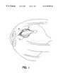
- FIG. 1is a schematic diagram of a horizontal cross section of the eye, indicating a macrocapsule implanted in the vitreous.
- the diagramis not to scale, and for the sake of clarity shows the capsule in an approximate placement—when actually placed in the human eye, the preferred vitreous placement is in the anterior portion of the vitreous.
- the letter “A”refers to the sciera
- “B”refers to Tenon's capsule
- “C”refers to the conjunctiva.
- FIG. 2is a schematic diagram of a side view of the eye showing an implanted capsule beneath Tenon's capsule.
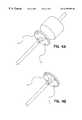
- FIG. 3Ashows a device with fangible hub assembly for loading.
- FIG. 3Bdepicts the device after detachment of the frangible hub.
- the devicehas an eyelet for tethering the device in the eye.
- FIG. 4Ashows a device with frangible hub assembly for loading.
- FIG. 4Bdepicts the device after detachment of the frangible hub.
- the devicehas a disk for tethering the device.
- This inventionrelates to delivery of biologically active molecules (“BAMs”) intraocularly (eg., in the anterior chamber, posterior chamber, or vitreous) or periocularly (e.g., within or beneath Tenon's capsule), or both.
- BAMsbiologically active molecules
- the inventionmay be useful in providing controlled and sustained release of BAMs effective in treating various ophthalmic disorders, ophthalmic diseases, or diseases which have ocular effects.
- Drugcan be delivered to the eye directly, reducing or minimizing unwanted peripheral side effects; very small doses of drug (nanogram or low microgram quantities rather than milligrams) can be delivered compared with topical applications, also potentially lessening side effects; the viable cells of the devices continuously produce newly synthesized product, avoiding the fluctuation in drug dose that characterizes injection delivery of drugs; and the devices and methods of this invention are less invasive than many of the prior art devices and surgical techniques, which result in a large number of retinal detachments.
- ophthalmic diseases and disordersare associated with one or more of three types of indications: (1) angiogenesis, (2) inflammation, and (3) degeneration.
- the devices of the present inventionpermit delivery of anti-angiogenic factors; anti-inflammatory factors; factors that retard cell degeneration, promote cell sparing, or promote cell growth; and combinations of the foregoing
- one of ordinary skill in the artcan administer any suitable molecule or combination of molecules from the three groups at the dosages specified below.
- Diabetic retinopathyfor example, is characterized by angiogenesis.
- This inventioncontemplates treating diabetic retinopathy by implanting devices delivering one or more anti-angiogenic factors either intraocularly, preferably in the vitreous, or periocularly, preferably in the sub-Tenon's region. We most prefer delivery into the vitreous for this indication. It is also desirable to co-deliver one or more neurotrophic factors, either intraocularly or periocularly, preferably intraocularly, and most preferably intravitreally.
- Uveitisinvolves inflammation.
- This inventioncontemplates treating uveitis by intraocular, preferably vitreal or anterior chamber, implantation of devices secreting one or more anti-inflammatory factors.
- Retinitis pigmentosaby comparison, is characterized by retinal degeneration.
- This inventioncontemplates treating retinitis pigmentosa by intraocular, preferably vitreal, placement of devices secreting one or more neurotrophic factors.
- Age-related macular degenerationinvolves both angiogenesis and retinal degeneration.
- This inventioncontemplates treating this disorder by using the inventive devices to deliver one or more neurotrophic factors intraocularly, preferably to the vitreous, and/or one or more anti-angiogenic factors intraocularly or periocularly, preferably periocularly, most preferably to the sub-Tenon's region.
- Glaucomais characterized by increased ocular pressure and loss of retinal ganglion cells.
- Treatments for glaucoma contemplated in this inventioninclude delivery of one or more neuroprotective agents that protect cells from excitotoxic damage.
- Such agentsinclude N-methyl-D-aspartate (NMDA) antagonists, cytokines, and neurotrophic factors, delivered intraocularly, preferably intravitrealy.
- NMDAN-methyl-D-aspartate
- Any suitable BAMmay be delivered according to the devices, systems, and methods of this invention.
- Such moleculesinclude neurotransmitters, cytolines, lymphokines, neuroprotective agents, neurotrophic factors, hormones, enzymes, antibodies, and active fragments thereof.
- Three preferred types of BAMsare contemplated for delivery using the devices of the present invention: (1) anti-angiogenic factors, (2) anti-inflammatory factors, and (3) factors that retard cell degeneration, promote cell sparing, or promote cell growth.
- the anti-angogenic factors contemplated for useinclude vasculostatin, angiostatin, endostatin, anti-integrins, vascular endothelial growth factor inhibitors (VEGF-inhibitors), platelet factor 4, heparinase, and bFGF-binding molecules.
- VEGF-inhibitorsvascular endothelial growth factor inhibitors
- platelet factor 4heparinase
- bFGF-binding moleculesvascular endothelial growth factor inhibitors
- the VEGF receptors Flt and Flkare also contemplated. When delivered in the soluble form these molecules compete with the VEGF receptors on vascular endothelial cells to inhibit endothelial cell growth.
- VEGF inhibitorsmay include VEGF-neutralizing chimeric proteins such as soluble VEGF receptors. See Aiello, PNAS , 92, 10457 (1995). In particular, they may be VEGF-receptor-IgG chimeric proteins.
- Another VEGF inhibitor contemplated for use in the present inventionis antisense phosphorothiotac oligodeoxynucleotides (PS-ODNs).
- Intraocularlypreferably in the vitreous, we contemplate delivery of an anti-angiogenic factor in a dosage range of 50 pg to 500 ng, preferably 100 pg to 100 ng, and most preferably 1 ng to 50 ng per eye per patient per day.
- a dosage rangeof 50 pg to 500 ng, preferably 100 pg to 100 ng, and most preferably 1 ng to 50 ng per eye per patient per day.
- slightly higher dosage rangesare contemplated of up to 1 ⁇ g per patient per day.
- anti-inflammatory factors contemplated for use in the present inventioninclude antiflammins (see, e.g., U.S. Pat. No. 5,266,562, incorporated herein by reference), beta-interferon (IFN- ⁇ ), alpha-interferon (IFN- ⁇ ), TGF-beta, interleukin-10 (IL-10), and glucocorticoids and mineralocorticoids from adrenal cortical cells.
- antiflamminssee, e.g., U.S. Pat. No. 5,266,562, incorporated herein by reference
- beta-interferonIFN- ⁇
- alpha-interferonIFN- ⁇
- TGF-betaTGF-beta
- IL-10interleukin-10
- glucocorticoids and mineralocorticoids from adrenal cortical cellsglucocorticoids and mineralocorticoids from adrenal cortical cells.
- certain BAMsmay have more than one activity.
- IFN- ⁇ and IFN- ⁇may have activities as both
- Intraocularlypreferably in the vitreous, we contemplate delivery of an anti-inflammatory factor in a dosage range of 50 pg to 500 ng, preferably 100 pg to 100 ng, and most preferably 1 ng to 50 ng per eye per patient per day.
- an anti-inflammatory factorin a dosage range of 50 pg to 500 ng, preferably 100 pg to 100 ng, and most preferably 1 ng to 50 ng per eye per patient per day.
- slightly higher dosage rangesare contemplated of up to 1 ⁇ g per patient per day.
- neurotrophic factorsinclude neurotrophin 4/5 (NT-4/5), cardiotrophin-1 (CT-1), ciliary neurotrophic factor (CNTF), glial cell line derived neurotrophic factor (GDNF), nerve growth factor (NGF), insulin-like growth factor-1 (IGF-1), neurotrophin 3 (NT-3), brain-derived neurotrophic factor (BDNF), PDGF, neurturin, acidic fibroblast growth factor (aFGF), basic fibroblast growth factor (FGF), EGF, neuregulins, heregulins, TGF-alpha, bone morphogenic proteins (BMP-1, BMP-2, BMP-7, etc.), the hedgehog family (sonic hedgehog, indian hedgehog, and desert hedgehog, etc.), the family of transforming growth factors (including, e.g., TGF ⁇ -1, TGF ⁇ -2,
- Intraocularlypreferably in the vitreous, we contemplate delivery of a neurotrophic factor in a dosage range of 50 pg to 500 ng, preferably 100 pg to 100 ng, and most preferably 1 ng to 50 ng per eye per patient per day.
- a neurotrophic factorin a dosage range of 50 pg to 500 ng, preferably 100 pg to 100 ng, and most preferably 1 ng to 50 ng per eye per patient per day.
- slightly higher dosage rangesare contemplated of up to 1 ⁇ g per patient per day.
- Modified, truncated, and mutein forms of the above-mentioned moleculesare also contemplated.
- active fragments of these growth factorsi.e., those fragments of growth factors having biological activity sufficient to achieve a therapeutic effect
- growth factor molecules modified by attachment of one or more polyethylene glycol (PEG) or other repeating polymeric moietiesare also contemplated.
- a gene of interesti.e., a gene that encodes a suitable BAM
- a gene that encodes a suitable BAMcan be inserted into a cloning site of a suitable expression vector by using standard techniques.
- the nucleic acid and amino acid sequences of the human (and other mammalian) genes encoding the above identified BAMsare known. See, e.g., U.S. Pat. No. 4,997,929; 5,141,856; 5,364,769; 5,453,361; WO 93/06116; WO 95/30686, incorporated herein by reference.
- the expression vector containing the gene of interestmay then be used to transfect the desired cell line.
- Standard transfection techniquessuch as calcium phosphate co-precipitation, DEAE-dextran transfection or electroporation may be utilized.
- Commercially available mammalian transfection kitsmay be purchased from e.g., Stratagene.
- Transgenic-mouse-derived cell linescan also be used. See, e.g., Hammang et al., Methods in Neurosci ., 21, p. 281 (1994).
- a wide variety of host/expression vector combinationsmay be used to express the gene encoding the growth factor, or other BAM(s) of interest.
- Suitable promotersinclude, for example, the early and late promoters of SV40 or adenovirus and other known non-retroviral promoters capable of controlling gene expression.
- Useful expression vectorsmay consist of segments of chromosomal, non-chromosomal, and synthetic DNA sequences, such as various known derivatives of SV40 and known bacterial plasmids, e.g., pUC, pBlueScriptTM plasmids from E. coli including pBR322, pCR1, pMB9, and their derivatives.
- synthetic DNA sequencessuch as various known derivatives of SV40 and known bacterial plasmids, e.g., pUC, pBlueScriptTM plasmids from E. coli including pBR322, pCR1, pMB9, and their derivatives.
- Expression vectors containing the geneticin (G418) or hygromycin drug selection genesare also useful. These vectors can employ a variety of different enhancer/promoter regions to drive the expression of both a biologic gene of interest (e.g., NGF) and/or a gene conferring resistance to selection with toxin such as G418 or hygromycin B.
- a biologic gene of intereste.g., NGF
- a gene conferring resistance to selection with toxinsuch as G418 or hygromycin B.
- a variety of different mammalian promoterscan be employed to direct the expression of the genes for G418 and hygromycin B and/or the biologic gene of interest.
- expression vectorsexamples include the commercially available pRC/CMV, pRC/RSV, and pCDNA1NEO (In Vitrogen).
- the promoteris selected from the following group:
- hDBHhuman dopamine beta hydroxylase
- hTHhuman tyrosine hydroxylase
- hPNMThuman phenylethanolamine N-methyltransferase
- mGFAPmouse glial fibrillary acidic protein
- MBPmyelin basic protein
- mNF-Lmouse neurofilament-light subunit
- hPohuman P 0 , the promoter for the gene encoding the major myelin glycoprotein in the peripheral nervous system
- mMt-1mouse metabothionein I
- rNSErat neuron-specific enolase
- the phosphoglycerate kinase (PGK) promoteris used. See, e.g., Adra et al., Gene , 60, pp. 65-74 (1987).
- the pPI vectoris one preferred expression vector using the PGK promoter to drive the expression of the gene of interest (i.e. the gene encoding the BAM).
- This vectoralso uses the SV40 early promoter to drive expression of neo phosphotransferase, a selectable marker.
- the pPI vectoralso contains a mutant DHFR gene suitable for MTX amplification.
- the pNUT expression vectorwhich contains the EDNA of the mutant DHFR and the entire pUC18 sequence including the polylinker, can be used. See, e.g., Aebischer, P., et al., Transplantation , 58, pp. 1275-1277 (1994); Baetge et al., PNAS , 83, pp. 5454-58 (1986).
- the pNUT expression vectorcan be modified such that the DHFR coding sequence is replaced by the coding sequence for G418 or hygromycin drug resistance.
- the SV40 promoter within the pNUT expression vectorcan also be replaced with any suitable constitutively expressed manum promoter, such as those discussed above.
- Increased expressioncan be achieved by increasing or amplifying the copy number of the transgene encoding the desired molecule, using amplification methods well known in the art.
- amplification methodsinclude, e.g., DHFR amplification (see, e.g, Kaufman et al., U.S. Pat. No. 4,470,461) or glutamine synthetase (“GS”) amplification (see, e.g., U.S. Pat. No. 5,122,464, and European published application EP 338,841).
- a wide variety of cellsmay be used. These include well known, publicly available immortalized cell lines as well as dividing primary cell cultures. Examples of suitable publicly available cell lines include, Chinese hamster ovary (CHO), mouse fibroblast (L-M), NIH Swiss mouse embryo (NIH/3T3), African green monkey cell lines (including COS-1, COS-7, BSC-1, BSC40, BMT-10, and Vero), rat adrenal pheochromocytoma (PC12 and PC 12A), AT3, rat glial tumor (C6), astrocytes, and other fibroblast cell lines.
- Primary cells that may be usedinclude EGF-responsive neural stem cells and their differentiated progeny Reynolds and Weiss, Science , 255, pp.
- bFGF-responsive neural progenitor stem cellsderived from the CNS of mammals (Richards et al., PNAS 89, pp. 8591-8595 (1992); Ray et al., PNAS 90, pp. 3602-3606 (1993)), CNS neural stem cells that are both EGF-responsive and bFGF-responsive, primary fibroblasts, Schwann cells, ⁇ -TC cells, Hep-G2 cells, oligodendrocytes and their precursors, myoblasts (including L6 and C 2 C 12 cells), chondrocytes or chondroblasts, and the like.
- Conditionally-immortalized cellsmay also be used.
- Such cellsinclude cells with temperature sensitive oncogenes, or cells engineered with chimneric genes composed of an oncogene under the direction of an inducible promoter element.
- BHKbaby hamster kidney
- the suitable cell typesinclude cells from allogeneic and xenogeneic sources.
- a particular advantage to using xenogeneic cellsis that in the unlikely event of membrane or device failure, such cells are more likely to be targeted for destruction by the immune system.
- priniy cellsmimcluding primary cells that can be induced to divide using mitogens such as EGF or bFGF or the like
- cell linesconditionally-immortalized or otherwise, derived from various regions of the eye.
- Potentially useful cell typesinclude lens epithelial cells, glial and neuronal elements of the neural retina, photoreceptor cells, retinal pigmented epithelial cells, Schwann cells and other ciliary body cells, and the like. Such cells can be allogeneic or xenogeneic.
- a biocompatible capsulemeans that the capsule, upon implantation in a host mammal, does not elicit a detrimental host response sufficient to result in the reection of the capsule or to render it inoperable, for example through degradation.
- an immunoisolatory capsulemeans that the capsule upon implantation into a mammalian host minimizes the deleterious effects of the host's immune system on the cells within its core.
- the capsuleshould provide a physical barrier sufficient to prevent detrimental immunological contact between the isolated cells and the host's immune system.
- the thickness of this physical barriercan vary, but it will always be sufficiently thick to prevent direct contact between the cells and/or substances on either side of the barrier.
- the thickness of this regiongenerally ranges between 5 and 200 microns; thicknesses of 10 to 100 microns are preferred, and thickness of 20 to 75 microns are particularly preferred.
- jacket norinal molecular weight cutoff (MWCO) valuesbetween 50-2000 kD are contemplated.
- the MWCOis between 50-700 kD.
- the MWCOis between 70-300 kD. See, e.g., WO 92/19195.
- the jacketcan be microporous. See, e.g., U.S. Pat. Nos. 4,968,733; 4,976,859; and 4,629,563; all incorporated herein by reference.
- biocompatible capsulesare suitable for delivery of molecules according to this invention.
- Useful biocompatible polymer capsulescomprise (a) a core which contains a cell or cels, either suspended in a liquid medium or immobilized within a biocompatible matrix, and (b) a surrounding jacket comprising a membrane which does not contain isolated cells, which is biocompatible, and permits diffusion of the cell-produced BAM into the eye.
- a capsule having a liquid corecomprising, e.g., a nutrient medium, and optionally containing a source of additional factors to sustain cell viability and function.
- the coremay comprise a biocompatible matrix of a hydrogel or other biocompable matrix material which stabilizes the position of the cells.
- hydrogelherein refers to a three dimensional network of cross-linked hydrophilic polymers. The network is in the form of a gel, substantially composed of water, preferably gels being greater than 90% water.
- any suitable matrix or spacermay be employed within the core, including precipitated chitosan, synthetic polymers and polymer blends, microcarriers, and the like, depending upon the growth characteristics of the cells to be encapsulated.
- the capsulemay have an internal scaffold. The scaffold may prevent cells from aggregating and improve cellular distribution within the device. See PCT publication no. WO 96/02646.
- the capsulesare immunoisolatory.
- the capsulecan be any suitable configuration, including cylindrical, rectangular, disk-shaped, patch-shaped, ovoid, stellate, or spherical. Configurations that tend to lead to migration of the capsules from the site of implantation, such as spherical, are not preferred. For implantations in the vitreous, flat sheets may not be preferred because they may block the visual path to the retina.
- the devicehas a tether that aids in maintaining device placement during implant and aids in retrieval.
- a tethermay have any suitable shape that is adapted to secure the capsule in place.
- the tetheris shaped like an eyelet, so that suture may be used to secure the tether (and thus the capsule) to the sclera, or other suitable ocular structure.
- the tetheris continuous with the capsule at one end, and forms a pre-threaded suture needle at the other end.
- the capsules contemplated herehave a minimum core volume of about 1 to 20 ⁇ l, most preferably about 1 to 10 ⁇ l.
- the fiberwill have an inside diameter of less than 1000 microns, preferably less than 950 microns.
- the deviceis configured to have an 870 ⁇ m inner diameter and a length of about 8.5 mm.
- the deviceis configured to have a 500 ⁇ m inner diameter and a length of 10.5 mm.
- the capsulewill preferably be between 0.4 cm to 1.5 cm in length, most preferably between 0.5 to 1.0 cm in length. Longer devices may be accommodated in the eye, however, a curved or arcuate shape may be required for secure and appropriate placement.
- the hollow fiber configurationis preferred for intraocular placement.
- a hollow fiber configuration(with dimensions substantially as above) or a flat sheet configuration is contemplated.
- the upper limit contemplated for a flat sheetis approximately 5 mm ⁇ 5 mm—assuming a square shape. Other shapes with approximately the same surface area are also contemplated.
- the hydraulic permeabilitywill typically be in the range of 1-100 mls/min/M 2 /mmHg, preferably in the range of 25 to 70 mls/min/M 2 /mmHg.
- the glucose mass transfer coefficient of the capsuledefined, measured, and calculated as described by Dionne et al., ASAIO Abstracts , p. 99 (1993), and Colton et al., The Kidney , eds., Brenner BM and Rector FC, pp. 2425-89 (1981) will be greater than 10 ⁇ 6 cm/sec, preferably greater than 10 ⁇ 4 cm/sec.
- the capsule jacketmay be manufactured from various polymers and polymer blends including polyaaylates (including acrylic copolymers), polyvinylidenes, polyvinyl chloride copolymers, polyurethanes, polystyrenes, polyamides, cellulose acetates, cellulose nitrates, polysulfones (including polyether sulfones), polyphosphazenes, polyacryionitriles, poly(acrylonitrile/covinyl chloride), as well as derivatives, copolymers, and mixtures thereof.
- Capsules manufactured from such materialsare described, e.g., in U.S. Pat. Nos. 5,284,761 and 5,158,881, incorporated herein by reference.
- Capsules formed from a polyether sulfone (PES) fibersuch as those described in U.S. Pat. Nos. 4,976,859 and 4,968,733, incorporated herein by reference, may also be used.
- capsulesDepending on the outer surface morphology, capsules have been categorized as Type 1 (T1), Type 2 (T2), Type 1 ⁇ 2 (T1 ⁇ 2), or Type 4 (T4).
- T1Type 1
- T2Type 2
- T1 ⁇ 2Type 1 ⁇ 2
- T4Type 4
- Such membranesare described, e.g., in Lacy et al., “Maintenance Of Normoglycemia In Diabetic Mice By Subcutaneous Xenografts Of Encapsulated Islets,” Science , 254, pp. 1782-84 (1991), Dionne et al., WO 92/19195 and Baetge, WO 95/05452.
- Capsule jackets with permselective immunoisolatory membranesare preferable for sites that are not immunologically privileged.
- microporous membranes or permselective membranesmay be suitable for immunologically privileged sites.
- capsules made from the PES membranesFor implantation into immunologically privileged sites, we prefer capsules made from the PES membranes.
- any suitable method of sealing the capsulesmay be used, including the employment of polymer adhesives and/or crimping, knotting, and heat sealing. These sealing techniques are known in the art.
- any suitable “dry” sealing methodcan also be used. In such methods, a substantially non-porous fitting is provided through which the cell-containing solution is introduced. Subsequent to filling, the capsule is sealed.
- Such a methodis described in copending U.S. application Ser. No. 08/082,407, herein incorporated by reference (see also PCT/US94/07015). That application describes the frangible hub assembly shown diagrammatically in FIGS. 3 and 4 that can be used to conveniently load and seal the devices of this invention.
- ophthalmic disorderscharacterized by but not limited to angiogenesis, inflammation, degeneration, or some combination thereof
- ophthalmic disordersinclude uveitis, retinitis pigmentosa, age-related macular degeneration and other acquired disorders, retinopathy, retinal vascular diseases and other vascular anomalies, endophthalmitis, infectious diseases, inflammatory but non-infectious diseases, ocular ischemia syndrome, peripheral retinal degenerations, retinal degenerations and tumors, choroidal disorders and tumors, vitreous disorders, retinal detachment, non-penetrating and penetrating trauma, post-cataract complications, and inflammatory optic neuropathies.
- Age-related macular degenerationincludes but is not limited to dry age-related macular degeneration, exudative age-related macular degeneration, and myopic degeneration.
- Retinopathyincludes but is not limited to diabetic retinopathy, proliferative vitreoretinopathy, and toxic retinopathy.
- the present inventionmay be useful for the treatment of ocular neovascularization, a condition associated with many ocular diseases and disorders and accounting for a majority of severe visual loss.
- ocular neovascularizationa condition associated with many ocular diseases and disorders and accounting for a majority of severe visual loss.
- retinal ischemia-associated ocular neovascularizationa major cause of blindness in diabetes and many other diseases
- corneal neovascularizationwhich predisposes patients to corneal graft failure
- neovascularization associated with diabetic retinopathy, central retinal vein occlusion, and possibly age-related macular degenerationmay be useful for the treatment of ocular neovascularization, a condition associated with many ocular diseases and disorders and accounting for a majority of severe visual loss.
- retinal ischemia-associated ocular neovascularizationa major cause of blindness in diabetes and many other diseases
- corneal neovascularizationwhich predisposes patients to
- the present inventionmay also be used to treat ocular symptoms resulting from diseases or conditions that have both ocular and non-ocular symptoms
- Some examplesinclude AIDS-related disorders such as cytomegalovirus retinitis and disorders of the vitreous; pregnancy-related disorders such as hypertensive changes in the retina; and ocular effects of various infectious diseases such as tuberculosis, syphilis, lyme disease, parasitic disease, toxocara canis, ophthalmoryiasis, cyst cercosis, and fingal infections.
- living cellsare encapsulated and surgically inserted (under retrobulbar anesthesia) into the vitreous of the eye.
- the devicemay be implanted through the sclera, with a portion of the device protruding through the sclera. Most preferably, the entire body of the device is implanted in the vitreous, with no portion of the device protruding into or through the sclera.
- the deviceis tethered to the sclera (or other suitable ocular structure).
- the tethermay comprise a suture eyelet (FIG. 3 ), or disk (FIG. 4 ), or any other suitable anchoring means.
- the devicecan remain in the vitreous as long as necessary to achieve the desired prophylaxis or therapy.
- Such therapiesfor example include promotion of neuron or photoreceptor survival or rear, or inhibition and/or reversal of retinal or choroidal neovasculanzation, as well as inhibition of uveal, retinal, and optic nerve inflammation.
- This embodimentis preferable for delivering the BAM to the retina.
- the BAMpreferably a trophic factor
- the BAMmay be delivered to the retina or the RPE.
- retinal neovascularizationmay be best treated by delivering an anti-angiogenic factor to the vitreous.
- cell-loaded devicesare implanted periocularly, within or beneath the space known as Tenon's capsule.
- This embodimentis less invasive than implantation into the vitreous and thus is generally preferred.
- This route of administrationalso permits delivery of BAMs (e.g., trophic factors and the like) to the RPE or the retina.
- BAMse.g., trophic factors and the like
- This embodimentis especially preferred for treating choroidal neovascularization and inflammation of the optic nerve and uveal tract.
- delivery from this implantation sitewill permit circulation of the desired BAM to the choroidal vasculature, the retinal vasculature, and the optic nerve.
- periocular delivery(unplanting beneath Tenon's capsule) of anti-angiogenic molecules, anti-inflammatory molecules (such as cytokines and hormones), and neurotrophic factors to the choroidal vasculature to treat macular degeneration (choroidal neovascularization).
- anti-angiogenic moleculessuch as cytokines and hormones
- neurotrophic factorssuch as cytokines and hormones
- choroidal vasculatureto treat macular degeneration (choroidal neovascularization).
- Delivery of anti-angiogenic factors directly to the choroidal vasculature (periocularly) or to the vitreous (intraocularly) using the devices and methods of this inventionmay reduce the above-mentioned problems and may permit the treatment of poorly defined or occult choroidal neovascularization. It may also provide a way of reducing or preventing recurrent choroidal neovascularization via adjunctive or maintenance therapy.
- the pNUT vector carrying the desired gene or genesis transfected into baby hamster kidney (BHK) cells or C 2 C 12 myoblast cels using a standard calcium phosphate transfection procedure and selected with increasing concentrations of methotrexate (1 ⁇ M to a maximum of 200 ⁇ M) over 8 weeks to produce stable, amplified cell lines. Following this selection, the engineered cells may be maintained in vitro in 50-200 ⁇ M methotrexate, prior to encapsulation.
- the present inventioncontemplates co-delivery of different factors.
- One of ordinary skill in the artmay deliver one or more anti-angiogenic factors, anti-inflammatory factors or factors retarding cell degeneration, promoting cell sparing, or promoting cell growth, depending on the indications of the particular ophthalmic disorder.
- itmay be preferable to deliver one or more neurotrophic factors together with one or more anti-angiogenic factors, or one or more anti-inflammatory molecules.
- One exampleis co-delivery of NT-4/5 with endostatin.
- the neurotrophic factorcan promote photoreceptor survival while the heparinase would act as an anti-angiogenic factor.
- Co-deliverycan be accomplished in a number of ways.
- First, cellsmay be transfected with separate constructs containing the genes encoding the described molecules.
- Second, cellsmay be transfected with a single construct containing two or more genes and the necessary control elements.
- either two or more separately engineered cell linescan be co-encapsulated, or more than one device can be implanted at the site of interest.
- devicesmay be implanted in two or more different sites in the eye concurrently, to deliver the same or different BAMs.
- a neurotrophic factorto the vitreous to supply the neural retina (ganglion cells to the RPE) and to deliver an anti-angiogenic factor via the sub-Tenon's space to supply the choroidal vasculature.
- treatment using more than one deviceis contemplated and up to five devices per eye, we prefer implantation of three devices or less per eye.
- Dosagecan be varied by any suitable method known in the art. This includes changing (1) the number of cells per device, (2) the number of devices per eye, or (3) the level of BAM production per cell.
- Cellular productioncan be varied by changing, for example, the copy number of the gene for the BAM in the transduced cell, or the efficiency of the promoter driving expression of the BAM.
- This inventionalso contemplates use of different cell types during the course of the treatment regime.
- a patientmay be implanted with a capsule device containing a first cell type (e.g., BHK cells). If after time, the patient develops an immune response to that cell type, the capsule can be retrieved, or explanted, and a second capsule can be implanted containing a second cell type (e.g., CHO cells).
- a first cell typee.g., BHK cells
- a second capsulee.g., CHO cells
- capsules with a lower MWCOmay be used to further prevent interaction of molecules of the patient's immune system with the encapsulated cells.
- the methods and devices of this inventionare intended for use in a primate, preferably human host, recipient, patient, subject or individual.
- BHK-hNGF cells(Winn et al., PNAS , 1994) were produced as follows:
- the pNUT-hNGF constructwas introduced into BHK cells by using a standard calcium phosphate-mediated transfection method.
- BHK cellswere grown in Dulbecco's modified Eagle's medium/10%/a fetal bovine serum/antibiotic/antimycotic/L-glutamine (GIBCO) in 5% CO 2 /95% air and at 37° C.
- Transfected BHK cellswere selected in medium containing 200 ⁇ M methotrexate (Sigma) for 3-4 weeks, and resistant cells were maintained as a polyclonal population either with or without 200 ⁇ M methotrexate.
- the cellswere maintained in DMEM with 10% FBS, L-glutamine with 50 ⁇ M methotrexate prior to these experiments. The cells were passaged 1 to 2 times per week in the presence of methotrexate.
- the BHK-hNGF cells and BHK control cellswere washed with Hank's buffer, then trypsinized and mixed with Zyderm® collagen matrix. The cell lines and matrix were loaded into separate Hamilton syringes that were equipped with blunted, 25-gauge needles.
- the encapsulation procedurewas as follows: The hollow fibers were fabricated from polyether sulfone PES) with an approximate outside diameter of 720 ⁇ m and a wall thickness of approximately 100 ⁇ m (AKZO-Nobel Wüppertal, Germany). These fibers are described in U.S. Pat. Nos. 4,976,859 and 4,968,733, herein incorporated by reference.
- the devicescomprise:
- CMlight cured methacrylate
- the deviceshad a septal fixture at the proxial end for cellular loading access and were sealed at the distal end.
- BHK cellswere prepared as a single-cell suspension and infused into the septal port at a density of 15K cells per ⁇ l after mixing 1:1 with physiologic collagen (nitrogen: PC-1). After infusing 1.5 ⁇ l of the cellular suspension, the septum was removed, and the access port was sealed with LCM 23 resin.
- the components of the deviceare commercially available.
- the LCM glueis available from Ablestik Laboratories (Newark, Del.); Luxtrak Adhesives LCM23 and LCM24).
- the patientis prepared and draped in the usual fashion after a retrobulbar injection of 3 cc 2% Cylocaine is given to the eye.
- a speculumis inserted beneath the upper and lower lids.
- the operating microscopeis brought into position.
- a perpendicular incisionis made through both conjunctiva and Tenon's capsule in the superotemporal quadrant approximately 4 mmn back from the limbus.
- the incisionis extended approximately 4-5 mm back from the limbus.
- a blunt-tipped scissoris inserted through the incision and is used to bluntly dissect back an additional 5 mm or so on the scleral surface.
- a membrane device as described in Example 1is placed in position through this incision to come to rest on the surface of the sclera.
- the end of the device that is closest to the limbushas a small loop that is attached to the cell-loaded device.
- a #10-0 nylon sutureis passed through this loop and sutured into the superficial sclera to anchor the membrane to the sclera.
- both Tenon's capsule and the conjunctivaare closed with #6-0 plain gut sutures. The speculum is removed and the procedure is concluded.
- the patientis prepared and draped in the usual fashion after a retrobulbar injection of 2% xylocaine is given to the eye.
- a speculumis inserted into the upper and lower lids and the microscope is brought into position.
- a small incisionis made through both the conjunctiva and Tenon's capsule parallel to and approximately 4 mm from the limbus in the supranasal quadrant
- the area exposedis cauterized with a wet-field cautery apparatus
- a 3 mm incisionis then made perpendicular to the limbus approximately 4 mm back from the limbus.
- the incisionis made through the sciera and into the vitreous cavity with a #65 blade. Any of the vitreous which presents itself in the incision is cut away and removed.
- a membrane device as described in Example 1is inserted through the incision into the vitreous cavity.
- a small 2 mm loopthat is attached to the membrane.
- the loopremains outside the sclera.
- the scierais closed with interrupted #9-0 nylon sutures.
- the #9-0 nylon suturesare also used to anchor this loop of the device to the sclera.
- the conjunctivais closed with #6-0 plain gut sutures.
- Interferon- ⁇IFN ⁇ -2A or IFN ⁇ -2B
- Candidate cell linesare genetically engineered to express the interferon molecules.
- Various interferonsmay be used; however, we prefer to use an IFN ⁇ -2A or ⁇ -2B. More than one interferon molecule may be delivered at one time.
- Various cell linescan also be utilized, we prefer BHK cells.
- Cell lineswill be encapsulated in pre-assembled devices substantially according to example 1. Following the device manufacture, a tether is applied. This tether contains an eyelet through which suture material can be passed. The tether is then used to anchor the device in place to avoid device drift or loss. The cell-loaded devices win be held for a standard period to assure device sterility. The capsule is implanted beneath the Tenon's capsule according to example 2.
- IFN ⁇ -2a therapyThe effects of IFN ⁇ -2a therapy are assessed by visual acuity, clinical appearance, and fluorescein angiographic appearance.
- the clinical appearance of the fundusis assessed subjectively with particular reference to macular elevation by subretinal fluid and the presence of intraretinal hemorrhage.
- Deviceswill be removed using the same preparation and surgical procedure as described above. The device will be placed in vitro and assayed for 24 hours for release of IFN- ⁇ . After the assay period, the device will be submitted for routine histological analysis to determine the extent of cell survival.
- BHK-hNGF clone 36 cellswere produced according to example 1. The cells were then encapsulated into 4mm LCM 24 light-cured capsules made from AKZO microporous 10/10 membranes according to example 1. The capsules were implanted in neonatal feline eyes substantially according to example 4 for 1 month.
- CMConditioned medium
- Encapsulated cells in the polymeric deviceswere also tested for their ability to release bioactive hNGF by placing the devices in individual wells of a 24-well plate and allowing them to equilibrate for 1-2 days in serum-free defined PC1 medium (Hycor, Portland, Me.); the medium was then removed and replaced with 1 ml of fresh PC1 for an additional 24 hour. This CM was collected, placed on the PC12A cells, and evaluated. Neurite processes that were equal to or greater than three times the length of the cell-body diameter were scored as positive. In addition, the rate of neurite induction and the stability of the neurites was examined.
- the level of NGF secretionwas also tested by ELISA. Quantitation of hNGF released from both encapsulated and unencapsulated BHK-hNGF cells was performed by a two-site enzyme immunoassay. The protocol was a modification of that described by Boehringer Mannheim using Nunc-Immuno Maxisorp ELISA plates. After color development (30 min.), the samples were analyzed on a plate reader and measured against recombinant mouse NGF protein standards.
- BHK cells that secreted hCNTF or NT4/5were produced and encapsulated substantially according to Example 1.
- Pig modelsare considered one of the most appropriate animal models for the human eye, based on size and vasculature.
Landscapes
- Health & Medical Sciences (AREA)
- Animal Behavior & Ethology (AREA)
- Ophthalmology & Optometry (AREA)
- Veterinary Medicine (AREA)
- Public Health (AREA)
- General Health & Medical Sciences (AREA)
- Life Sciences & Earth Sciences (AREA)
- Heart & Thoracic Surgery (AREA)
- Vascular Medicine (AREA)
- Biomedical Technology (AREA)
- Engineering & Computer Science (AREA)
- Chemical & Material Sciences (AREA)
- Medicinal Chemistry (AREA)
- Pharmacology & Pharmacy (AREA)
- Epidemiology (AREA)
- Medicinal Preparation (AREA)
- Medicines That Contain Protein Lipid Enzymes And Other Medicines (AREA)
- Pharmaceuticals Containing Other Organic And Inorganic Compounds (AREA)
Abstract
Description
| ELISA | ELISA | Capsule | |||
| Capsule | Pre-1* | Pre-2* | ELISA Post | Histology | |
| No. | BAM | pg/24 h. | pg/24 hr | Explant | Cell Survival |
| 1 | NGF | 152 | 329 | 268 | (+) |
| 2 | NGF | 271 | 162 | 156 | (+) |
| 7 | Control | nd** | nd | 0 | (+) |
| 8 | Control | nd | nd | 0 | (+) |
| *Devices were assayed twice prior to implantation, once prior to shipment to the collaborators' laboratory, and a second time immediately prior to implantation, with a 48-hour time interval between the two assays. “Pre-1” refers to the results of the first assay, and “Pre-2” refers to the results of the second assay. | |||||
| **“nd” is an abbreviation for “not detected.” | |||||
| ELISA | Capsule | |||
| Capsule | ELISA Pre | Post | Histology | |
| No. | BAM | pg/24h. | Explant | Cell Survival |
| 5 | NGF | 1800 | nd* | (−) |
| 6 | NGF | 3900 | 291 | (+) |
| 18 | Control | nd | nd | (−) |
| 19 | Control | nd | nd | (−) |
| *“nd” is an abbreviation for “not detected.” | ||||
Claims (50)
Priority Applications (3)
| Application Number | Priority Date | Filing Date | Title |
|---|---|---|---|
| US09/155,066US6299895B1 (en) | 1997-03-24 | 1997-03-24 | Device and method for treating ophthalmic diseases |
| US09/973,325US6436427B1 (en) | 1996-03-22 | 2001-10-09 | Device and method for treating ophthalmic diseases |
| US10/224,521US6649184B2 (en) | 1996-03-22 | 2002-08-20 | Device and method for treating ophthalmic diseases |
Applications Claiming Priority (2)
| Application Number | Priority Date | Filing Date | Title |
|---|---|---|---|
| PCT/US1997/004701WO1997034586A2 (en) | 1996-03-22 | 1997-03-24 | Device and method for treating ophthalmic diseases |
| US09/155,066US6299895B1 (en) | 1997-03-24 | 1997-03-24 | Device and method for treating ophthalmic diseases |
Related Parent Applications (3)
| Application Number | Title | Priority Date | Filing Date |
|---|---|---|---|
| US08/620,982Continuation-In-PartUS5904144A (en) | 1996-03-22 | 1996-03-22 | Method for treating ophthalmic diseases |
| US08/620,982ContinuationUS5904144A (en) | 1996-03-22 | 1996-03-22 | Method for treating ophthalmic diseases |
| PCT/US1997/004701A-371-Of-InternationalWO1997034586A2 (en) | 1996-03-22 | 1997-03-24 | Device and method for treating ophthalmic diseases |
Related Child Applications (1)
| Application Number | Title | Priority Date | Filing Date |
|---|---|---|---|
| US09/973,325ContinuationUS6436427B1 (en) | 1996-03-22 | 2001-10-09 | Device and method for treating ophthalmic diseases |
Publications (1)
| Publication Number | Publication Date |
|---|---|
| US6299895B1true US6299895B1 (en) | 2001-10-09 |
Family
ID=22553994
Family Applications (3)
| Application Number | Title | Priority Date | Filing Date |
|---|---|---|---|
| US09/155,066Expired - LifetimeUS6299895B1 (en) | 1996-03-22 | 1997-03-24 | Device and method for treating ophthalmic diseases |
| US09/973,325Expired - LifetimeUS6436427B1 (en) | 1996-03-22 | 2001-10-09 | Device and method for treating ophthalmic diseases |
| US10/224,521Expired - LifetimeUS6649184B2 (en) | 1996-03-22 | 2002-08-20 | Device and method for treating ophthalmic diseases |
Family Applications After (2)
| Application Number | Title | Priority Date | Filing Date |
|---|---|---|---|
| US09/973,325Expired - LifetimeUS6436427B1 (en) | 1996-03-22 | 2001-10-09 | Device and method for treating ophthalmic diseases |
| US10/224,521Expired - LifetimeUS6649184B2 (en) | 1996-03-22 | 2002-08-20 | Device and method for treating ophthalmic diseases |
Country Status (1)
| Country | Link |
|---|---|
| US (3) | US6299895B1 (en) |
Cited By (115)
| Publication number | Priority date | Publication date | Assignee | Title |
|---|---|---|---|---|
| US6436427B1 (en)* | 1996-03-22 | 2002-08-20 | Neurotech S.A. | Device and method for treating ophthalmic diseases |
| US20020143284A1 (en)* | 2001-04-03 | 2002-10-03 | Hosheng Tu | Drug-releasing trabecular implant for glaucoma treatment |
| US6500449B2 (en)* | 1999-11-24 | 2002-12-31 | Universite De Paris V Rene-Descartes | Intraocular transplantation of encapsulated cells |
| US6562005B1 (en)* | 2001-05-02 | 2003-05-13 | David Donath | Catheter button system and surgical method of anchoring catheter button |
| WO2003092564A1 (en)* | 2002-05-01 | 2003-11-13 | Moorfields Eye Hospital Nhs Trust | Device for delivery of biologically active agents |
| US20040068753A1 (en)* | 2002-10-02 | 2004-04-08 | Robertson Neil C. | Video transmission systems and methods for a home network |
| US20040121943A1 (en)* | 2002-12-20 | 2004-06-24 | Wei-Cherng Hsu | Drug-free biodegradable 3D porous collagen-glycosaminoglycan scaffold |
| US20040230043A1 (en)* | 1998-07-06 | 2004-11-18 | Johansen Teit E. | Novel neurotrophic factors |
| WO2005065600A1 (en)* | 2004-01-12 | 2005-07-21 | Nulens Ltd | Eye wall anchored fixtures |
| US20060105921A1 (en)* | 2002-11-05 | 2006-05-18 | Naozumi Arimoto | Lubricating oil |
| US20060233858A1 (en)* | 2005-03-08 | 2006-10-19 | Allergan, Inc. | Systems and methods providing targeted intraocular drug delivery |
| US20060253151A1 (en)* | 2004-01-12 | 2006-11-09 | Nun Joshua B | Eye wall anchored fixtures |
| WO2005082049A3 (en)* | 2004-02-20 | 2007-02-01 | Univ Leland Stanford Junior | Artificial biocompatible material as a support for cells in a retinal implant |
| US20070082841A1 (en)* | 2005-09-27 | 2007-04-12 | Aciont, Inc. | Ocular administration of immunosuppressive agents |
| US20070098692A1 (en)* | 2000-02-11 | 2007-05-03 | Genvec, Inc. | Materials and methods for treating ocular-related disorders |
| US7297130B2 (en) | 2000-04-14 | 2007-11-20 | Glaukos Corporation | Implant with anchor |
| US20070287756A1 (en)* | 2004-04-23 | 2007-12-13 | Toru Nakazawa | Methods and Compositions for Preserving the Viability of Photoreceptor Cells |
| US20080039385A1 (en)* | 2004-08-19 | 2008-02-14 | Biogen Idec Ma Inc. | Neublastin Variants |
| US20080233647A1 (en)* | 2001-03-12 | 2008-09-25 | Biogen Idec Ma Inc. | Novel neurotrophic factors |
| US7431710B2 (en) | 2002-04-08 | 2008-10-07 | Glaukos Corporation | Ocular implants with anchors and methods thereof |
| US20080249287A1 (en)* | 2004-08-19 | 2008-10-09 | Biogen Idec Ma Inc. | Refolding Transforming Growth Factor Beta Family Proteins |
| US20080286250A1 (en)* | 2005-10-28 | 2008-11-20 | Jens Tornoe | Implantable Biocompatible Immunoisolatory Vehicle for Delivery of Gdnf |
| EP1997523A2 (en) | 2007-05-31 | 2008-12-03 | Life Spring Biotech Co., Ltd. | Structure for modulating intraocular pressure |
| US20090083819A1 (en)* | 2003-01-15 | 2009-03-26 | Cisco Technology, Inc. | Optimization Of A Full Duplex Wideband Communications System |
| US20090136552A1 (en)* | 2004-07-30 | 2009-05-28 | Mette Gronborg | Growth factors nsg28, nsg30, and nsg32 |
| EP2075254A1 (en) | 2004-03-30 | 2009-07-01 | NsGene A/S | Therapeutic use of a growth factor, NsG33 |
| US7563255B2 (en) | 2001-05-03 | 2009-07-21 | Massachusetts Eye And Ear Infirmary | Implantable drug delivery device and use thereof |
| US7563241B2 (en) | 2001-04-07 | 2009-07-21 | Glaukos Corporation | Implant and methods thereof for treatment of ocular disorders |
| US20100034808A1 (en)* | 2006-11-21 | 2010-02-11 | Toru Nakazawa | Compositions and methods for preserving cells of the eye |
| US20100056440A1 (en)* | 2006-03-01 | 2010-03-04 | Biogen Idec Ma Inc. | Compositions and methods for administering gdnf ligand family proteins |
| US20100087774A1 (en)* | 2002-09-21 | 2010-04-08 | Glaukos Corporation | Ocular implant with anchoring mechanism and multiple outlets |
| US7708711B2 (en) | 2000-04-14 | 2010-05-04 | Glaukos Corporation | Ocular implant with therapeutic agents and methods thereof |
| US20100261654A1 (en)* | 2007-05-01 | 2010-10-14 | Inserm (Institut National De La Sante Et De La Recherche Medicale) | Compositions and methods for increasing vascularization |
| US7850637B2 (en) | 1999-04-26 | 2010-12-14 | Glaukos Corporation | Shunt device and method for treating glaucoma |
| WO2010147653A1 (en) | 2009-06-16 | 2010-12-23 | Bikam Pharmaceuticals, Inc. | Opsin-binding ligands, compositions and methods of use |
| US7861272B2 (en) | 2000-11-14 | 2010-12-28 | Russ Samuel H | Networked subscriber television distribution |
| US20100331975A1 (en)* | 2009-06-25 | 2010-12-30 | Optonol Ltd. | Fiber matrix for maintaining space in soft tissues |
| US7867186B2 (en) | 2002-04-08 | 2011-01-11 | Glaukos Corporation | Devices and methods for treatment of ocular disorders |
| US7879079B2 (en) | 2001-08-28 | 2011-02-01 | Glaukos Corporation | Implant delivery system and methods thereof for treating ocular disorders |
| US7908625B2 (en) | 2002-10-02 | 2011-03-15 | Robertson Neil C | Networked multimedia system |
| WO2011039648A1 (en) | 2009-09-30 | 2011-04-07 | Glaxo Wellcome Manufacturing Pte Ltd. | Methods of administration and treatment |
| US20110111008A1 (en)* | 2009-10-08 | 2011-05-12 | Weng Tao | Use of PEDF in an Encapsulated Cell-Based Delivery System |
| US7951155B2 (en) | 2002-03-15 | 2011-05-31 | Glaukos Corporation | Combined treatment for cataract and glaucoma treatment |
| EP2329821A1 (en) | 2005-11-29 | 2011-06-08 | GlaxoSmithKline LLC | Treatment of ocular neovascular disorders such as macular degeneration, angiod streaks, uveitis and macular edema |
| US20110135648A1 (en)* | 2007-08-08 | 2011-06-09 | Biogen Idec Ma Inc. | Anti-neublastin antibodies and uses thereof |
| EP2374451A2 (en) | 2005-07-27 | 2011-10-12 | University of Florida Research Foundation, Inc. | Histone deacetylase inhibitors (HDAC) that correct protein misfolding and uses thereof |
| WO2011133964A2 (en) | 2010-04-23 | 2011-10-27 | Massachusetts Eye And Ear Infirmary | Methods and compositions for preserving photoreceptor and retinal pigment epithelial cells |
| US8094640B2 (en) | 2003-01-15 | 2012-01-10 | Robertson Neil C | Full duplex wideband communications system for a local coaxial network |
| EP2407536A1 (en) | 2006-12-22 | 2012-01-18 | Bioaxone Therapeutique Inc. | ADP-ribosyl transferase fusion variant proteins |
| US8119114B2 (en) | 2001-02-01 | 2012-02-21 | Biogen Idec Ma Inc. | Polymer conjugates of mutated neublastin |
| US8127326B2 (en) | 2000-11-14 | 2012-02-28 | Claussen Paul J | Proximity detection using wireless connectivity in a communications system |
| US8142364B2 (en) | 2001-05-02 | 2012-03-27 | Dose Medical Corporation | Method of monitoring intraocular pressure and treating an ocular disorder |
| US8163875B2 (en) | 2003-04-18 | 2012-04-24 | Biogen Idec Ma Inc. | Polymer conjugated glycosylated neublastin |
| WO2012061045A2 (en) | 2010-11-01 | 2012-05-10 | Massachusetts Eye And Ear Infirmary | Methods and compositions for preserving retinal ganglion cells |
| WO2012174064A1 (en) | 2011-06-14 | 2012-12-20 | Bikam Pharmaceuticals, Inc. | Opsin-binding ligands, compositions and methods of use |
| US8337445B2 (en) | 2001-05-03 | 2012-12-25 | Glaukos Corporation | Ocular implant with double anchor mechanism |
| WO2013003467A2 (en) | 2011-06-27 | 2013-01-03 | Massachusetts Eye And Ear Infirmary | Methods for treating ocular inflammatory disorders |
| WO2013025840A1 (en) | 2011-08-15 | 2013-02-21 | Massachusetts Eye And Ear Infirmary | Methods for preserving photoreceptor cell viability following retinal detachment |
| WO2013058809A1 (en) | 2011-10-19 | 2013-04-25 | Bikam Pharmaceuticals, Inc. | Opsin-binding ligands, compositions and methods of use |
| WO2013081642A1 (en) | 2011-12-01 | 2013-06-06 | Bikam Pharmaceuticals, Inc. | Opsin-binding ligands, compositions and methods of use |
| US8506515B2 (en) | 2006-11-10 | 2013-08-13 | Glaukos Corporation | Uveoscleral shunt and methods for implanting same |
| US20130330302A1 (en)* | 2008-04-22 | 2013-12-12 | Regenerative Research Foundation | Retinal pigment epithelial stem cells |
| US8617094B2 (en) | 2002-03-07 | 2013-12-31 | Glaukos Corporation | Fluid infusion methods for glaucoma treatment |
| US8808687B2 (en) | 2010-07-12 | 2014-08-19 | Mark Humayun | Biocompatible substrate for facilitating interconnections between stem cells and target tissues and methods for implanting same |
| US8877489B2 (en) | 2011-12-05 | 2014-11-04 | California Institute Of Technology | Ultrathin parylene-C semipermeable membranes for biomedical applications |
| US8911734B2 (en) | 2010-12-01 | 2014-12-16 | Alderbio Holdings Llc | Methods of preventing or treating pain using anti-NGF antibodies that selectively inhibit the association of NGF with TrkA, without affecting the association of NGF with p75 |
| WO2015070083A1 (en) | 2013-11-07 | 2015-05-14 | Editas Medicine,Inc. | CRISPR-RELATED METHODS AND COMPOSITIONS WITH GOVERNING gRNAS |
| US9067988B2 (en) | 2010-12-01 | 2015-06-30 | Alderbio Holdings Llc | Methods of preventing or treating pain using anti-NGF antibodies |
| WO2015103480A1 (en) | 2014-01-02 | 2015-07-09 | Massachusetts Eye & Ear Infirmary | Treating ocular neovascularization |
| US9078878B2 (en) | 2010-12-01 | 2015-07-14 | Alderbio Holdings Llc | Anti-NGF antibodies that selectively inhibit the association of NGF with TrkA, without affecting the association of NGF with p75 |
| WO2015134812A1 (en) | 2014-03-05 | 2015-09-11 | Editas Medicine, Inc. | Crispr/cas-related methods and compositions for treating usher syndrome and retinitis pigmentosa |
| EP2944628A1 (en) | 2011-11-30 | 2015-11-18 | Bikam Pharmaceuticals, Inc. | Opsin-binding ligands, compositions and methods of use |
| US9248013B2 (en) | 2011-12-05 | 2016-02-02 | California Institute Of Technology | 3-Dimensional parylene scaffold cage |
| US9301875B2 (en) | 2002-04-08 | 2016-04-05 | Glaukos Corporation | Ocular disorder treatment implants with multiple opening |
| US20160310572A1 (en)* | 2013-12-02 | 2016-10-27 | Wayne State University | Compositions and methods to diagnose diabetes and/or to treat negative effects of diabetes |
| US9539324B2 (en) | 2010-12-01 | 2017-01-10 | Alderbio Holdings, Llc | Methods of preventing inflammation and treating pain using anti-NGF compositions |
| US9592151B2 (en) | 2013-03-15 | 2017-03-14 | Glaukos Corporation | Systems and methods for delivering an ocular implant to the suprachoroidal space within an eye |
| WO2017075475A1 (en) | 2015-10-30 | 2017-05-04 | Editas Medicine, Inc. | Crispr/cas-related methods and compositions for treating herpes simplex virus |
| US9730638B2 (en) | 2013-03-13 | 2017-08-15 | Glaukos Corporation | Intraocular physiological sensor |
| US9762970B2 (en) | 2002-10-04 | 2017-09-12 | Tech 5 | Access of stored video from peer devices in a local network |
| US9884909B2 (en) | 2010-12-01 | 2018-02-06 | Alderbio Holdings Llc | Anti-NGF compositions and use thereof |
| WO2018034945A1 (en) | 2016-08-19 | 2018-02-22 | The United States Of America, As Represented By The Secretary, Department Of Health And Human Services | Selective estrogen-receptor modulators (serms) confer protection against photoreceptor degeneration |
| US9938521B2 (en) | 2014-03-10 | 2018-04-10 | Editas Medicine, Inc. | CRISPR/CAS-related methods and compositions for treating leber's congenital amaurosis 10 (LCA10) |
| WO2018081504A1 (en) | 2016-10-28 | 2018-05-03 | Editas Medicine, Inc. | Crispr/cas-related methods and compositions for treating herpes simplex virus |
| US10022419B2 (en) | 2011-10-21 | 2018-07-17 | Massachusetts Eye And Ear Infirmary | Methods for treating spinal cord injury |
| WO2018204848A1 (en) | 2017-05-05 | 2018-11-08 | University Of Pittsburgh - Of The Commonwealth System Of Higher Education | Ocular applications of matrix bound vesicles (mbvs) |
| US10138485B2 (en) | 2008-09-22 | 2018-11-27 | Rxi Pharmaceuticals Corporation | Neutral nanotransporters |
| US10184124B2 (en) | 2010-03-24 | 2019-01-22 | Phio Pharmaceuticals Corp. | RNA interference in ocular indications |
| US10206813B2 (en) | 2009-05-18 | 2019-02-19 | Dose Medical Corporation | Implants with controlled drug delivery features and methods of using same |
| US10240149B2 (en) | 2010-03-24 | 2019-03-26 | Phio Pharmaceuticals Corp. | Reduced size self-delivering RNAi compounds |
| US10245178B1 (en) | 2011-06-07 | 2019-04-02 | Glaukos Corporation | Anterior chamber drug-eluting ocular implant |
| US10328125B2 (en) | 2006-02-27 | 2019-06-25 | Gloriana Therapeutics, Inc. | Treatments for neurological disorders |
| WO2019169291A1 (en) | 2018-03-02 | 2019-09-06 | The United States Of America, As Represented By The Secretary, Department Of Health And Human Services | Use of il-34 to treat retinal inflammation and neurodegeneration |
| US10478206B2 (en) | 2011-04-29 | 2019-11-19 | University Of Southern California | Instruments and methods for the implantation of cell-seeded substrates |
| US10479992B2 (en) | 2009-02-04 | 2019-11-19 | Phio Pharmaceuticals Corp. | RNA duplexes with single stranded phosphorothioate nucleotide regions for additional functionality |
| WO2020176552A1 (en) | 2019-02-25 | 2020-09-03 | Editas Medicine, Inc. | Crispr/rna-guided nuclease-related methods and compositions for treating rho-associated autosomal-dominant retinitis pigmentosa (adrp) |
| US10959941B2 (en) | 2014-05-29 | 2021-03-30 | Glaukos Corporation | Implants with controlled drug delivery features and methods of using same |
| WO2021146625A1 (en) | 2020-01-17 | 2021-07-22 | The United States Of America, As Represented By The Secretary, Department Of Health And Human Services | Gene therapy for treatment of crx-autosomal dominant retinopathies |
| US11141493B2 (en) | 2014-03-10 | 2021-10-12 | Editas Medicine, Inc. | Compositions and methods for treating CEP290-associated disease |
| US11214610B2 (en) | 2010-12-01 | 2022-01-04 | H. Lundbeck A/S | High-purity production of multi-subunit proteins such as antibodies in transformed microbes such as Pichia pastoris |
| WO2022006439A2 (en) | 2020-07-02 | 2022-01-06 | The United States Of America, As Represented By The Secretary, Department Of Health And Human Services | Compound embodiments for treating retinal degeneration and method embodiments of making and using the same |
| US11279934B2 (en) | 2014-04-28 | 2022-03-22 | Phio Pharmaceuticals Corp. | Methods for treating cancer using nucleic acids targeting MDM2 or MYCN |
| US11318043B2 (en) | 2016-04-20 | 2022-05-03 | Dose Medical Corporation | Bioresorbable ocular drug delivery device |
| US11339437B2 (en) | 2014-03-10 | 2022-05-24 | Editas Medicine, Inc. | Compositions and methods for treating CEP290-associated disease |
| US11363951B2 (en) | 2011-09-13 | 2022-06-21 | Glaukos Corporation | Intraocular physiological sensor |
| WO2022221741A1 (en) | 2021-04-16 | 2022-10-20 | Editas Medicine, Inc. | Crispr/rna-guided nuclease-related methods and compositions for treating rho-associated autosomal-dominant retinitis pigmentosa (adrp) |
| US11564833B2 (en) | 2015-09-25 | 2023-01-31 | Glaukos Corporation | Punctal implants with controlled drug delivery features and methods of using same |
| US11566263B2 (en) | 2016-08-02 | 2023-01-31 | Editas Medicine, Inc. | Compositions and methods for treating CEP290 associated disease |
| US11925578B2 (en) | 2015-09-02 | 2024-03-12 | Glaukos Corporation | Drug delivery implants with bi-directional delivery capacity |
| WO2024196814A1 (en) | 2023-03-17 | 2024-09-26 | The United States Of America, As Represented By The Secretary, Department Of Health And Human Services | Methods for treatment of age-related macular degeneration |
| WO2025002450A1 (en) | 2023-06-30 | 2025-01-02 | Shenzhen Genturn Life Co., Ltd. | Engineered reprogramming factors and uses thereof for treating eye diseases |
| US12201555B2 (en) | 2009-05-18 | 2025-01-21 | Dose Medical Corporation | Drug eluting ocular implant |
| WO2025083169A1 (en) | 2023-10-17 | 2025-04-24 | Tenpoint Therapeutics Limited | Combination of a vegf inhibitor and a complement pathway inhibitor for treating ocular disorders |
| US12295985B2 (en) | 2011-02-28 | 2025-05-13 | The Schepens Eye Research Institute, Inc. | Methods of reducing severity of glaucoma by intravitreal administration of insulin-like growth factor binding protein-like 1 (IGFBPL1) |
| US12419783B2 (en) | 2010-11-24 | 2025-09-23 | Glaukos Corporation | Drug eluting ocular implant |
Families Citing this family (31)
| Publication number | Priority date | Publication date | Assignee | Title |
|---|---|---|---|---|
| IT1305294B1 (en) | 1999-01-29 | 2001-05-04 | Alessandro Lambiase | USE OF THE NERVE GROWTH FACTOR IN THE THERAPY OF PATHOLOGIES DEPENDING ON INTRAOCULAR TISSUES. |
| US20050119737A1 (en)* | 2000-01-12 | 2005-06-02 | Bene Eric A. | Ocular implant and methods for making and using same |
| DE10133870A1 (en)* | 2001-07-12 | 2003-02-06 | Chris P Lohmann | Ophthalmic agent, use of EGF for the treatment of dry eye syndrome and insert for the administration of EGF to the eye |
| CA2498489C (en)* | 2002-09-29 | 2010-02-23 | Surmodics, Inc. | Method for subretinal administration of therapeutics including steroids;method for localizing pharmacodynamic action at the choroid and the retina; and related methods for treatment and/or prevention of retinal diseases |
| US20040236343A1 (en)* | 2003-05-23 | 2004-11-25 | Taylor Jon B. | Insertion tool for ocular implant and method for using same |
| MXPA06004329A (en) | 2003-10-20 | 2006-06-05 | Nsgene As | In vivo gene therapy of parkinson's disease. |
| US20080286323A1 (en)* | 2004-01-19 | 2008-11-20 | Nsgene A/S | Human Therapeutic Cells Secreting Nerve Growth Factor |
| WO2006002366A2 (en)* | 2004-06-24 | 2006-01-05 | Surmodics, Inc. | Biodegradable ocular devices, methods and systems |
| US20060110428A1 (en) | 2004-07-02 | 2006-05-25 | Eugene Dejuan | Methods and devices for the treatment of ocular conditions |
| ES2432556T3 (en)* | 2004-08-04 | 2013-12-04 | Evonik Corporation | Methods for manufacturing supply devices and their devices |
| ATE526421T1 (en) | 2005-02-14 | 2011-10-15 | Univ Iowa Res Found | METHODS AND REAGENTS FOR THE TREATMENT AND DIAGNOSIS OF AGE-RELATED MACULAR DEGENERATION |
| US20070077270A1 (en)* | 2005-03-28 | 2007-04-05 | Clemson University | Delivery devices and methods for long-term, targeted delivery of therapeutic agents to the eye and ear |
| WO2006110487A1 (en)* | 2005-04-08 | 2006-10-19 | Surmodics, Inc. | Sustained release implants for subretinal delivery |
| ITRM20050447A1 (en)* | 2005-08-19 | 2007-02-20 | Anabasis S R L | USE OF THE NERVE GROWTH FACTOR IN COLLIRIUM IN THE PATHOLOGY OF THE CENTRAL NERVOUS SYSTEM, SUCH AS ALZHEIMER'S DISEASE AND PARKINSON'S DISEASE. |
| US8168584B2 (en) | 2005-10-08 | 2012-05-01 | Potentia Pharmaceuticals, Inc. | Methods of treating age-related macular degeneration by compstatin and analogs thereof |
| PL1951279T3 (en) | 2005-10-08 | 2017-12-29 | Apellis Pharmaceuticals, Inc. | Compstatin and analogs thereof for eye disorders |
| EP2479284B1 (en) | 2006-07-13 | 2017-09-20 | University of Iowa Research Foundation | Methods and reagents for treatment and diagnosis of vascular disorders and age-related macular degeneration |
| US8124601B2 (en)* | 2007-11-21 | 2012-02-28 | Bristol-Myers Squibb Company | Compounds for the treatment of Hepatitis C |
| JP5502751B2 (en) | 2007-12-20 | 2014-05-28 | エボニック コーポレイション | Process for preparing microparticles with low residual solvent concentration |
| US20110184439A1 (en)* | 2008-05-09 | 2011-07-28 | University Of Pittsburgh-Of The Commonwealth System Of Higher Education | Biological Matrix for Cardiac Repair |
| WO2010083842A2 (en) | 2009-01-23 | 2010-07-29 | Nsgene A/S | Expression of neuropeptides in mammalian cells |
| CA2775077C (en)* | 2009-09-22 | 2018-05-01 | Evonik Degussa Corporation | Implant devices having varying bioactive agent loading configurations |
| CA2853379C (en) | 2011-10-27 | 2020-11-24 | Wellstat Ophthalmics Corporation | Vectors encoding rod-derived cone viability factor |
| WO2014078731A2 (en) | 2012-11-15 | 2014-05-22 | Apellis Pharmaceuticals, Inc. | Cell-reactive, long-acting, or targeted compstatin analogs and related compositions and methods |
| DE102013004595A1 (en) | 2013-03-15 | 2014-09-18 | Emergent Product Development Germany Gmbh | RSV vaccines |
| US10308687B2 (en) | 2013-03-15 | 2019-06-04 | Apellis Pharmaceuticals, Inc. | Cell-penetrating compstatin analogs and uses thereof |
| BR112018006810A2 (en) | 2015-10-07 | 2018-10-23 | Apellis Pharmaceuticals Inc | dosage regimens |
| BR112019020955A2 (en) | 2017-04-07 | 2020-05-05 | Apellis Pharmaceuticals Inc | dosage regimens and related compositions and methods |
| JP2021507884A (en) | 2017-12-15 | 2021-02-25 | アペリス・ファーマシューティカルズ・インコーポレイテッドApellis Pharmaceuticals,Inc. | Administration regimen and related compositions and methods |
| AU2019247467B2 (en) | 2018-04-06 | 2023-01-19 | The Trustees Of The University Of Pennsylvania | Compstatin analogs with increased solubility and improved pharmacokinetic properties |
| WO2024206928A1 (en) | 2023-03-30 | 2024-10-03 | Pharma Cinq, Llc | VECTOR ENCODING ROD-DERIVED CONE VIABILITY FACTOR AND HUMAN IgK SIGNAL SEQUENCE |
Citations (9)
| Publication number | Priority date | Publication date | Assignee | Title |
|---|---|---|---|---|
| US5266562A (en) | 1987-11-19 | 1993-11-30 | The United States Of America As Represented By The Secretary Of The Department Of Health And Human Services | Anti-inflammatory agents |
| US5279298A (en) | 1992-11-20 | 1994-01-18 | The Johns Hopkins University | Method and apparatus to identify and treat neovascular membranes in the eye |
| US5382514A (en) | 1992-03-31 | 1995-01-17 | The United States Of America As Represented By The Secretary Of The Department Of Health And Human Services | In vivo angiogenesis assay |
| WO1995013765A1 (en) | 1993-11-15 | 1995-05-26 | Oculex Pharmaceuticals, Inc. | Biocompatible ocular implants |
| WO1995028166A2 (en) | 1994-04-15 | 1995-10-26 | Cytotherapeutics, Inc. | Method for implanting encapsulated cells in a host |
| US5466233A (en) | 1994-04-25 | 1995-11-14 | Escalon Ophthalmics, Inc. | Tack for intraocular drug delivery and method for inserting and removing same |
| US5472436A (en) | 1994-07-26 | 1995-12-05 | Fremstad; Daria A. | Ocular appliance for delivering medication |
| US5476511A (en) | 1992-05-04 | 1995-12-19 | Allergan, Inc. | Subconjunctival implants for ocular drug delivery |
| US5521215A (en) | 1989-11-07 | 1996-05-28 | Ramot University Authority For Applied Research And Industrial Development Ltd. | NMDA-blocking pharmaceuticals |
Family Cites Families (36)
| Publication number | Priority date | Publication date | Assignee | Title |
|---|---|---|---|---|
| US3828777A (en) | 1971-11-08 | 1974-08-13 | Alza Corp | Microporous ocular device |
| NL188266C (en) | 1975-07-29 | 1992-05-18 | Merck & Co Inc | PROCESS FOR THE PREPARATION OF AN ORGANIC IMPLANT. |
| US4164559A (en) | 1977-09-21 | 1979-08-14 | Cornell Research Foundation, Inc. | Collagen drug delivery device |
| US4186184A (en) | 1977-12-27 | 1980-01-29 | Alza Corporation | Selective administration of drug with ocular therapeutic system |
| US4300557A (en) | 1980-01-07 | 1981-11-17 | The United States Of America As Represented By The Secretary Of The Department Of Health And Human Services | Method for treating intraocular malignancies |
| US4730013A (en) | 1981-10-08 | 1988-03-08 | Merck & Co., Inc. | Biosoluble ocular insert |
| US4740461A (en) | 1983-12-27 | 1988-04-26 | Genetics Institute, Inc. | Vectors and methods for transformation of eucaryotic cells |
| GB8601597D0 (en) | 1986-01-23 | 1986-02-26 | Wilson R H | Nucleotide sequences |
| US5147647A (en) | 1986-10-02 | 1992-09-15 | Sohrab Darougar | Ocular insert for the fornix |
| US5322691A (en) | 1986-10-02 | 1994-06-21 | Sohrab Darougar | Ocular insert with anchoring protrusions |
| US4863457A (en) | 1986-11-24 | 1989-09-05 | Lee David A | Drug delivery device |
| US5158881A (en) | 1987-11-17 | 1992-10-27 | Brown University Research Foundation | Method and system for encapsulating cells in a tubular extrudate in separate cell compartments |
| US5283187A (en) | 1987-11-17 | 1994-02-01 | Brown University Research Foundation | Cell culture-containing tubular capsule produced by co-extrusion |
| US4853224A (en) | 1987-12-22 | 1989-08-01 | Visionex | Biodegradable ocular implants |
| GB8809129D0 (en) | 1988-04-18 | 1988-05-18 | Celltech Ltd | Recombinant dna methods vectors and host cells |
| DE3829766A1 (en) | 1988-09-01 | 1990-03-22 | Akzo Gmbh | METHOD FOR PRODUCING MEMBRANES |
| DE3829752A1 (en) | 1988-09-01 | 1990-03-22 | Akzo Gmbh | INTEGRAL ASYMMETRICAL POLYAETHERSULPHONE MEMBRANE, METHOD FOR THE PRODUCTION AND USE FOR ULTRAFILTRATION AND MICROFILTRATION |
| US4997929A (en) | 1989-01-05 | 1991-03-05 | Synergen, Inc. | Purified ciliary neurotrophic factor |
| US5141856A (en) | 1989-01-05 | 1992-08-25 | Synergen, Inc. | Expression of purified ciliary neurotrophic factor |
| US5098443A (en) | 1989-03-23 | 1992-03-24 | University Of Miami | Method of implanting intraocular and intraorbital implantable devices for the controlled release of pharmacological agents |
| US5180820A (en) | 1989-08-30 | 1993-01-19 | Barde Yves Alain | Brain-derived neurotrophic factor |
| US5164188A (en) | 1989-11-22 | 1992-11-17 | Visionex, Inc. | Biodegradable ocular implants |
| US5185152A (en) | 1990-01-10 | 1993-02-09 | Peyman Gholam A | Method and apparatus for controlled release drug delivery to the cornea and anterior chamber of the eye |
| US5606031A (en) | 1990-04-06 | 1997-02-25 | Lile; Jack | Production and purification of biologically active recombinant neurotrophic protein in bacteria |
| US5364769A (en) | 1990-09-25 | 1994-11-15 | Genentech, Inc. | Nucleic acid encoding neurotrophic factor four (NT-4), vectors, host cells and methods of production |
| US5378475A (en) | 1991-02-21 | 1995-01-03 | University Of Kentucky Research Foundation | Sustained release drug delivery devices |
| ES2107537T3 (en) | 1991-04-25 | 1997-12-01 | Univ Brown Res Found | IMMUNO INSULATED BIOCOMPATIBLE VEHICLE IMPLANTABLE TO SUPPLY SELECTED THERAPEUTIC PRODUCTS. |
| EP1243652A3 (en) | 1991-09-20 | 2003-03-26 | Amgen Inc., | Glial derived neurotrophic factor |
| US5384333A (en) | 1992-03-17 | 1995-01-24 | University Of Miami | Biodegradable injectable drug delivery polymer |
| AU6230394A (en) | 1993-01-20 | 1994-11-21 | Biotransplant Incorporated | Retroviral vectors capable of expressing multimeric proteins from multiple translational initiation sites |
| US5490875A (en) | 1993-07-13 | 1996-02-13 | American Maize Technology, Inc. | Adhesive composition for cigarette seams |
| EP1179350A3 (en) | 1993-08-12 | 2003-01-02 | Cytotherapeutics, Inc. | Encapsulated cell system for implantation into the human CNS |
| WO1995013766A1 (en) | 1993-11-18 | 1995-05-26 | Allergan, Inc. | Deformable lens insertion apparatus |
| MX9605109A (en) | 1994-04-26 | 1997-08-30 | Childrens Medical Center | Angiostatin and method of use for inhibition of angiogenesis. |
| US6299895B1 (en)* | 1997-03-24 | 2001-10-09 | Neurotech S.A. | Device and method for treating ophthalmic diseases |
| US5904144A (en)* | 1996-03-22 | 1999-05-18 | Cytotherapeutics, Inc. | Method for treating ophthalmic diseases |
- 1997
- 1997-03-24USUS09/155,066patent/US6299895B1/ennot_activeExpired - Lifetime
- 2001
- 2001-10-09USUS09/973,325patent/US6436427B1/ennot_activeExpired - Lifetime
- 2002
- 2002-08-20USUS10/224,521patent/US6649184B2/ennot_activeExpired - Lifetime
Patent Citations (10)
| Publication number | Priority date | Publication date | Assignee | Title |
|---|---|---|---|---|
| US5266562A (en) | 1987-11-19 | 1993-11-30 | The United States Of America As Represented By The Secretary Of The Department Of Health And Human Services | Anti-inflammatory agents |
| US5521215A (en) | 1989-11-07 | 1996-05-28 | Ramot University Authority For Applied Research And Industrial Development Ltd. | NMDA-blocking pharmaceuticals |
| US5382514A (en) | 1992-03-31 | 1995-01-17 | The United States Of America As Represented By The Secretary Of The Department Of Health And Human Services | In vivo angiogenesis assay |
| US5476511A (en) | 1992-05-04 | 1995-12-19 | Allergan, Inc. | Subconjunctival implants for ocular drug delivery |
| US5279298A (en) | 1992-11-20 | 1994-01-18 | The Johns Hopkins University | Method and apparatus to identify and treat neovascular membranes in the eye |
| WO1995013765A1 (en) | 1993-11-15 | 1995-05-26 | Oculex Pharmaceuticals, Inc. | Biocompatible ocular implants |
| WO1995028166A2 (en) | 1994-04-15 | 1995-10-26 | Cytotherapeutics, Inc. | Method for implanting encapsulated cells in a host |
| US5550050A (en) | 1994-04-15 | 1996-08-27 | Cytotherapeutics, Inc. | Method for implanting encapsulated cells in a host |
| US5466233A (en) | 1994-04-25 | 1995-11-14 | Escalon Ophthalmics, Inc. | Tack for intraocular drug delivery and method for inserting and removing same |
| US5472436A (en) | 1994-07-26 | 1995-12-05 | Fremstad; Daria A. | Ocular appliance for delivering medication |
Cited By (222)
| Publication number | Priority date | Publication date | Assignee | Title |
|---|---|---|---|---|
| US6436427B1 (en)* | 1996-03-22 | 2002-08-20 | Neurotech S.A. | Device and method for treating ophthalmic diseases |
| US20040230043A1 (en)* | 1998-07-06 | 2004-11-18 | Johansen Teit E. | Novel neurotrophic factors |
| US10492950B2 (en) | 1999-04-26 | 2019-12-03 | Glaukos Corporation | Shunt device and method for treating ocular disorders |
| US8152752B2 (en) | 1999-04-26 | 2012-04-10 | Glaukos Corporation | Shunt device and method for treating glaucoma |
| US7850637B2 (en) | 1999-04-26 | 2010-12-14 | Glaukos Corporation | Shunt device and method for treating glaucoma |
| US8388568B2 (en) | 1999-04-26 | 2013-03-05 | Glaukos Corporation | Shunt device and method for treating ocular disorders |
| US8771217B2 (en) | 1999-04-26 | 2014-07-08 | Glaukos Corporation | Shunt device and method for treating ocular disorders |
| US9827143B2 (en) | 1999-04-26 | 2017-11-28 | Glaukos Corporation | Shunt device and method for treating ocular disorders |
| US9492320B2 (en) | 1999-04-26 | 2016-11-15 | Glaukos Corporation | Shunt device and method for treating ocular disorders |
| US10568762B2 (en) | 1999-04-26 | 2020-02-25 | Glaukos Corporation | Stent for treating ocular disorders |
| US6500449B2 (en)* | 1999-11-24 | 2002-12-31 | Universite De Paris V Rene-Descartes | Intraocular transplantation of encapsulated cells |
| US20070098692A1 (en)* | 2000-02-11 | 2007-05-03 | Genvec, Inc. | Materials and methods for treating ocular-related disorders |
| US8814820B2 (en) | 2000-04-14 | 2014-08-26 | Glaukos Corporation | Ocular implant with therapeutic agent and methods thereof |
| US7708711B2 (en) | 2000-04-14 | 2010-05-04 | Glaukos Corporation | Ocular implant with therapeutic agents and methods thereof |
| US8273050B2 (en) | 2000-04-14 | 2012-09-25 | Glaukos Corporation | Ocular implant with anchor and therapeutic agent |
| US8801648B2 (en) | 2000-04-14 | 2014-08-12 | Glaukos Corporation | Ocular implant with anchor and methods thereof |
| US7297130B2 (en) | 2000-04-14 | 2007-11-20 | Glaukos Corporation | Implant with anchor |
| US7867205B2 (en) | 2000-04-14 | 2011-01-11 | Glaukos Corporation | Method of delivering an implant for treating an ocular disorder |
| US9993368B2 (en) | 2000-04-14 | 2018-06-12 | Glaukos Corporation | System and method for treating an ocular disorder |
| US8348877B2 (en) | 2000-04-14 | 2013-01-08 | Dose Medical Corporation | Ocular implant with therapeutic agents and methods thereof |
| US9789001B2 (en) | 2000-04-14 | 2017-10-17 | Dose Medical Corporation | Ocular implant with therapeutic agents and methods thereof |
| US8333742B2 (en) | 2000-04-14 | 2012-12-18 | Glaukos Corporation | Method of delivering an implant for treating an ocular disorder |
| US10485702B2 (en) | 2000-04-14 | 2019-11-26 | Glaukos Corporation | System and method for treating an ocular disorder |
| US9066782B2 (en) | 2000-04-14 | 2015-06-30 | Dose Medical Corporation | Ocular implant with therapeutic agents and methods thereof |
| US8808219B2 (en) | 2000-04-14 | 2014-08-19 | Glaukos Corporation | Implant delivery device and methods thereof for treatment of ocular disorders |
| US7861272B2 (en) | 2000-11-14 | 2010-12-28 | Russ Samuel H | Networked subscriber television distribution |
| US8127326B2 (en) | 2000-11-14 | 2012-02-28 | Claussen Paul J | Proximity detection using wireless connectivity in a communications system |
| US8119114B2 (en) | 2001-02-01 | 2012-02-21 | Biogen Idec Ma Inc. | Polymer conjugates of mutated neublastin |
| US20100292142A1 (en)* | 2001-03-12 | 2010-11-18 | Biogen Idec Ma Inc. | Novel neurotrophic factors |
| US8217146B2 (en) | 2001-03-12 | 2012-07-10 | Biogen Idec Ma Inc. | Neurotrophic factors and methods of use thereof |
| US20080233647A1 (en)* | 2001-03-12 | 2008-09-25 | Biogen Idec Ma Inc. | Novel neurotrophic factors |
| US7655463B2 (en) | 2001-03-12 | 2010-02-02 | Biogen Idec Ma Inc. | Methods of activating RET receptor tyrosine kinase using neurotrophic factors |
| US20020143284A1 (en)* | 2001-04-03 | 2002-10-03 | Hosheng Tu | Drug-releasing trabecular implant for glaucoma treatment |
| US9155654B2 (en) | 2001-04-07 | 2015-10-13 | Glaukos Corporation | Ocular system with anchoring implant and therapeutic agent |
| US9572963B2 (en) | 2001-04-07 | 2017-02-21 | Glaukos Corporation | Ocular disorder treatment methods and systems |
| US8579846B2 (en) | 2001-04-07 | 2013-11-12 | Glaukos Corporation | Ocular implant systems |
| US7563241B2 (en) | 2001-04-07 | 2009-07-21 | Glaukos Corporation | Implant and methods thereof for treatment of ocular disorders |
| US8075511B2 (en) | 2001-04-07 | 2011-12-13 | Glaukos Corporation | System for treating ocular disorders and methods thereof |
| US10406029B2 (en) | 2001-04-07 | 2019-09-10 | Glaukos Corporation | Ocular system with anchoring implant and therapeutic agent |
| US9987472B2 (en) | 2001-04-07 | 2018-06-05 | Glaukos Corporation | Ocular implant delivery systems |
| US7857782B2 (en) | 2001-04-07 | 2010-12-28 | Glaukos Corporation | Ocular implant delivery system and method thereof |
| US8062244B2 (en) | 2001-04-07 | 2011-11-22 | Glaukos Corporation | Self-trephining implant and methods thereof for treatment of ocular disorders |
| US10828473B2 (en) | 2001-04-07 | 2020-11-10 | Glaukos Corporation | Ocular implant delivery system and methods thereof |
| US8118768B2 (en) | 2001-04-07 | 2012-02-21 | Dose Medical Corporation | Drug eluting ocular implant with anchor and methods thereof |
| US6562005B1 (en)* | 2001-05-02 | 2003-05-13 | David Donath | Catheter button system and surgical method of anchoring catheter button |
| US8142364B2 (en) | 2001-05-02 | 2012-03-27 | Dose Medical Corporation | Method of monitoring intraocular pressure and treating an ocular disorder |
| US8337445B2 (en) | 2001-05-03 | 2012-12-25 | Glaukos Corporation | Ocular implant with double anchor mechanism |
| US7563255B2 (en) | 2001-05-03 | 2009-07-21 | Massachusetts Eye And Ear Infirmary | Implantable drug delivery device and use thereof |
| US10285856B2 (en) | 2001-08-28 | 2019-05-14 | Glaukos Corporation | Implant delivery system and methods thereof for treating ocular disorders |
| US7879079B2 (en) | 2001-08-28 | 2011-02-01 | Glaukos Corporation | Implant delivery system and methods thereof for treating ocular disorders |
| US9561131B2 (en) | 2001-08-28 | 2017-02-07 | Glaukos Corporation | Implant delivery system and methods thereof for treating ocular disorders |
| US9220632B2 (en) | 2002-03-07 | 2015-12-29 | Glaukos Corporation | Fluid infusion methods for ocular disorder treatment |
| US8617094B2 (en) | 2002-03-07 | 2013-12-31 | Glaukos Corporation | Fluid infusion methods for glaucoma treatment |
| US7951155B2 (en) | 2002-03-15 | 2011-05-31 | Glaukos Corporation | Combined treatment for cataract and glaucoma treatment |
| US8882781B2 (en) | 2002-03-15 | 2014-11-11 | Glaukos Corporation | Combined treatment for cataract and glaucoma treatment |
| US7431710B2 (en) | 2002-04-08 | 2008-10-07 | Glaukos Corporation | Ocular implants with anchors and methods thereof |
| US7867186B2 (en) | 2002-04-08 | 2011-01-11 | Glaukos Corporation | Devices and methods for treatment of ocular disorders |
| US9597230B2 (en) | 2002-04-08 | 2017-03-21 | Glaukos Corporation | Devices and methods for glaucoma treatment |
| US10485701B2 (en) | 2002-04-08 | 2019-11-26 | Glaukos Corporation | Devices and methods for glaucoma treatment |
| US9301875B2 (en) | 2002-04-08 | 2016-04-05 | Glaukos Corporation | Ocular disorder treatment implants with multiple opening |
| US7879001B2 (en) | 2002-04-08 | 2011-02-01 | Glaukos Corporation | Devices and methods for treatment of ocular disorders |
| WO2003092564A1 (en)* | 2002-05-01 | 2003-11-13 | Moorfields Eye Hospital Nhs Trust | Device for delivery of biologically active agents |
| US8007459B2 (en) | 2002-09-21 | 2011-08-30 | Glaukos Corporation | Ocular implant with anchoring mechanism and multiple outlets |
| US20100087774A1 (en)* | 2002-09-21 | 2010-04-08 | Glaukos Corporation | Ocular implant with anchoring mechanism and multiple outlets |
| US20040068753A1 (en)* | 2002-10-02 | 2004-04-08 | Robertson Neil C. | Video transmission systems and methods for a home network |
| US7908625B2 (en) | 2002-10-02 | 2011-03-15 | Robertson Neil C | Networked multimedia system |
| US9762970B2 (en) | 2002-10-04 | 2017-09-12 | Tech 5 | Access of stored video from peer devices in a local network |
| US20060105921A1 (en)* | 2002-11-05 | 2006-05-18 | Naozumi Arimoto | Lubricating oil |
| US20040121943A1 (en)* | 2002-12-20 | 2004-06-24 | Wei-Cherng Hsu | Drug-free biodegradable 3D porous collagen-glycosaminoglycan scaffold |
| US7544368B2 (en) | 2002-12-20 | 2009-06-09 | Life Spring Biotech Co., Ltd. | Structure for modulating intraocular pressure |
| US7865925B2 (en) | 2003-01-15 | 2011-01-04 | Robertson Neil C | Optimization of a full duplex wideband communications system |
| US8094640B2 (en) | 2003-01-15 | 2012-01-10 | Robertson Neil C | Full duplex wideband communications system for a local coaxial network |
| US20090083819A1 (en)* | 2003-01-15 | 2009-03-26 | Cisco Technology, Inc. | Optimization Of A Full Duplex Wideband Communications System |
| US8163875B2 (en) | 2003-04-18 | 2012-04-24 | Biogen Idec Ma Inc. | Polymer conjugated glycosylated neublastin |
| US8642732B2 (en) | 2003-04-18 | 2014-02-04 | Biogen Idec Ma Inc. | Polymer-conjugated glycosylated neublastin |
| WO2005065600A1 (en)* | 2004-01-12 | 2005-07-21 | Nulens Ltd | Eye wall anchored fixtures |
| AU2005204012B2 (en)* | 2004-01-12 | 2009-08-13 | Nulens Ltd | Eye wall anchored fixtures |
| US20060253151A1 (en)* | 2004-01-12 | 2006-11-09 | Nun Joshua B | Eye wall anchored fixtures |
| US7976520B2 (en)* | 2004-01-12 | 2011-07-12 | Nulens Ltd. | Eye wall anchored fixtures |
| WO2005082049A3 (en)* | 2004-02-20 | 2007-02-01 | Univ Leland Stanford Junior | Artificial biocompatible material as a support for cells in a retinal implant |
| EP2289911A2 (en) | 2004-03-30 | 2011-03-02 | NsGene A/S | Therapeutic use of a growth factor, NsG33 |
| EP2075254A1 (en) | 2004-03-30 | 2009-07-01 | NsGene A/S | Therapeutic use of a growth factor, NsG33 |
| US20070287756A1 (en)* | 2004-04-23 | 2007-12-13 | Toru Nakazawa | Methods and Compositions for Preserving the Viability of Photoreceptor Cells |
| US9549895B2 (en) | 2004-04-23 | 2017-01-24 | Massachusetts Eye And Ear Infirmary | Methods and compositions for preserving the viability of photoreceptor cells |
| US20090136552A1 (en)* | 2004-07-30 | 2009-05-28 | Mette Gronborg | Growth factors nsg28, nsg30, and nsg32 |
| US8263553B2 (en) | 2004-08-19 | 2012-09-11 | Biogen Idec Ma Inc. | Neublastin variants |
| US20080249287A1 (en)* | 2004-08-19 | 2008-10-09 | Biogen Idec Ma Inc. | Refolding Transforming Growth Factor Beta Family Proteins |
| US8722862B2 (en) | 2004-08-19 | 2014-05-13 | Biogen Idec Ma Inc. | Refolding transforming growth factor beta family proteins |
| US8969042B2 (en) | 2004-08-19 | 2015-03-03 | Biogen Idec Ma Inc. | Refolding transforming growth factor beta family proteins |
| US20080039385A1 (en)* | 2004-08-19 | 2008-02-14 | Biogen Idec Ma Inc. | Neublastin Variants |
| US20060233858A1 (en)* | 2005-03-08 | 2006-10-19 | Allergan, Inc. | Systems and methods providing targeted intraocular drug delivery |
| EP2374451A2 (en) | 2005-07-27 | 2011-10-12 | University of Florida Research Foundation, Inc. | Histone deacetylase inhibitors (HDAC) that correct protein misfolding and uses thereof |
| US20070082841A1 (en)* | 2005-09-27 | 2007-04-12 | Aciont, Inc. | Ocular administration of immunosuppressive agents |
| WO2007038687A3 (en)* | 2005-09-27 | 2007-09-20 | Aciont Inc | Ocular administration of immunosuppressive agents |
| US20080286250A1 (en)* | 2005-10-28 | 2008-11-20 | Jens Tornoe | Implantable Biocompatible Immunoisolatory Vehicle for Delivery of Gdnf |
| EP2329821A1 (en) | 2005-11-29 | 2011-06-08 | GlaxoSmithKline LLC | Treatment of ocular neovascular disorders such as macular degeneration, angiod streaks, uveitis and macular edema |
| US10328125B2 (en) | 2006-02-27 | 2019-06-25 | Gloriana Therapeutics, Inc. | Treatments for neurological disorders |
| US20100056440A1 (en)* | 2006-03-01 | 2010-03-04 | Biogen Idec Ma Inc. | Compositions and methods for administering gdnf ligand family proteins |
| US9962290B2 (en) | 2006-11-10 | 2018-05-08 | Glaukos Corporation | Uveoscleral shunt and methods for implanting same |
| US8506515B2 (en) | 2006-11-10 | 2013-08-13 | Glaukos Corporation | Uveoscleral shunt and methods for implanting same |
| US12186237B2 (en) | 2006-11-10 | 2025-01-07 | Glaukos Corporation | Uveoscleral shunt and methods for implanting same |
| US10828195B2 (en) | 2006-11-10 | 2020-11-10 | Glaukos Corporation | Uveoscleral shunt and methods for implanting same |
| US9914782B2 (en) | 2006-11-21 | 2018-03-13 | Massachusetts Eye And Ear Infirmary | Methods for preserving the viability of retinal ganglion cells in patients with glaucoma by an anti-TNF receptor 2 (anti-TNFR2) antibody |
| US20100034808A1 (en)* | 2006-11-21 | 2010-02-11 | Toru Nakazawa | Compositions and methods for preserving cells of the eye |
| EP2407536A1 (en) | 2006-12-22 | 2012-01-18 | Bioaxone Therapeutique Inc. | ADP-ribosyl transferase fusion variant proteins |
| US20100261654A1 (en)* | 2007-05-01 | 2010-10-14 | Inserm (Institut National De La Sante Et De La Recherche Medicale) | Compositions and methods for increasing vascularization |
| US9138461B2 (en) | 2007-05-01 | 2015-09-22 | Biogen Ma Inc. | Compositions and methods for increasing vascularization |
| US8329655B2 (en) | 2007-05-01 | 2012-12-11 | Biogen Idec Ma Inc. | Methods for increasing vascularization |
| EP1997523A2 (en) | 2007-05-31 | 2008-12-03 | Life Spring Biotech Co., Ltd. | Structure for modulating intraocular pressure |
| US20110135648A1 (en)* | 2007-08-08 | 2011-06-09 | Biogen Idec Ma Inc. | Anti-neublastin antibodies and uses thereof |
| US10034916B2 (en)* | 2008-04-22 | 2018-07-31 | Regenerative Research Foundation | Methods of treating a retinal disease by retinal pigment epithelial stem cells |
| US20180333459A1 (en)* | 2008-04-22 | 2018-11-22 | Regenerative Research Foundation | Retinal Pigment Epithelial Stem Cells |
| US20130330302A1 (en)* | 2008-04-22 | 2013-12-12 | Regenerative Research Foundation | Retinal pigment epithelial stem cells |
| US10138485B2 (en) | 2008-09-22 | 2018-11-27 | Rxi Pharmaceuticals Corporation | Neutral nanotransporters |
| US11396654B2 (en) | 2008-09-22 | 2022-07-26 | Phio Pharmaceuticals Corp. | Neutral nanotransporters |
| US11667915B2 (en) | 2009-02-04 | 2023-06-06 | Phio Pharmaceuticals Corp. | RNA duplexes with single stranded phosphorothioate nucleotide regions for additional functionality |
| US10479992B2 (en) | 2009-02-04 | 2019-11-19 | Phio Pharmaceuticals Corp. | RNA duplexes with single stranded phosphorothioate nucleotide regions for additional functionality |
| US10206813B2 (en) | 2009-05-18 | 2019-02-19 | Dose Medical Corporation | Implants with controlled drug delivery features and methods of using same |
| US11426306B2 (en) | 2009-05-18 | 2022-08-30 | Dose Medical Corporation | Implants with controlled drug delivery features and methods of using same |
| US12376989B2 (en) | 2009-05-18 | 2025-08-05 | Glaukos Corporation | Drug eluting ocular implants and methods of treating an ocular disorder |
| US12201557B2 (en) | 2009-05-18 | 2025-01-21 | Dose Medical Corporation | Drug eluting ocular implant and method of treating an ocular disorder |
| US12201555B2 (en) | 2009-05-18 | 2025-01-21 | Dose Medical Corporation | Drug eluting ocular implant |
| EP3100723A1 (en) | 2009-06-16 | 2016-12-07 | Bikam Pharmaceuticals, Inc. | Opsin-binding ligands, compositions and methods of use |
| WO2010147653A1 (en) | 2009-06-16 | 2010-12-23 | Bikam Pharmaceuticals, Inc. | Opsin-binding ligands, compositions and methods of use |
| US20100331975A1 (en)* | 2009-06-25 | 2010-12-30 | Optonol Ltd. | Fiber matrix for maintaining space in soft tissues |
| US8961600B2 (en) | 2009-06-25 | 2015-02-24 | Optonol Ltd. | Method of using a fiber matrix for maintaining space in soft tissues |
| WO2011039648A1 (en) | 2009-09-30 | 2011-04-07 | Glaxo Wellcome Manufacturing Pte Ltd. | Methods of administration and treatment |
| US20110111008A1 (en)* | 2009-10-08 | 2011-05-12 | Weng Tao | Use of PEDF in an Encapsulated Cell-Based Delivery System |
| US11118178B2 (en) | 2010-03-24 | 2021-09-14 | Phio Pharmaceuticals Corp. | Reduced size self-delivering RNAI compounds |
| US10662430B2 (en) | 2010-03-24 | 2020-05-26 | Phio Pharmaceuticals Corp. | RNA interference in ocular indications |
| US11584933B2 (en) | 2010-03-24 | 2023-02-21 | Phio Pharmaceuticals Corp. | RNA interference in ocular indications |
| US10184124B2 (en) | 2010-03-24 | 2019-01-22 | Phio Pharmaceuticals Corp. | RNA interference in ocular indications |
| US10240149B2 (en) | 2010-03-24 | 2019-03-26 | Phio Pharmaceuticals Corp. | Reduced size self-delivering RNAi compounds |
| US10639345B2 (en) | 2010-04-23 | 2020-05-05 | Massachusetts Eye And Ear Infirmary | Compositions for preserving photoreceptor cells, retinal pigment epithelial cells, or visual function |
| US10149884B2 (en) | 2010-04-23 | 2018-12-11 | Massachusetts Eye And Ear Infirmary | Methods and compositions for preserving photoreceptor and retinal pigment epithelial cells |
| US9492432B2 (en) | 2010-04-23 | 2016-11-15 | Massachusetts Eye And Ear Infirmary | Methods and compositions for preserving photoreceptor and retinal pigment epithelial cells |
| WO2011133964A2 (en) | 2010-04-23 | 2011-10-27 | Massachusetts Eye And Ear Infirmary | Methods and compositions for preserving photoreceptor and retinal pigment epithelial cells |
| US10188769B2 (en) | 2010-07-12 | 2019-01-29 | University Of Southern California | Biocompatible substrate for facilitating interconnections between stem cells and target tissues and methods for implanting same |
| US11154639B2 (en) | 2010-07-12 | 2021-10-26 | University Of Southern California | Biocompatible substrate for facilitating interconnections between stem cells and target tissues and methods for implanting same |
| US8808687B2 (en) | 2010-07-12 | 2014-08-19 | Mark Humayun | Biocompatible substrate for facilitating interconnections between stem cells and target tissues and methods for implanting same |
| US10617735B2 (en) | 2010-11-01 | 2020-04-14 | Massachusetts Eye And Ear Infirmary | Methods and compositions for preserving retinal ganglion cells |
| WO2012061045A2 (en) | 2010-11-01 | 2012-05-10 | Massachusetts Eye And Ear Infirmary | Methods and compositions for preserving retinal ganglion cells |
| US9993517B2 (en) | 2010-11-01 | 2018-06-12 | Massachusetts Eye And Ear Infirmary | Methods and compositions for preserving retinal ganglion cells |
| US12419783B2 (en) | 2010-11-24 | 2025-09-23 | Glaukos Corporation | Drug eluting ocular implant |
| US10457727B2 (en) | 2010-12-01 | 2019-10-29 | Alderbio Holdings Llc | Methods of preventing inflammation and treating pain using anti-NGF compositions |
| US10344083B2 (en) | 2010-12-01 | 2019-07-09 | Alderbio Holdings Llc | Anti-NGF compositions and use thereof |
| US9718882B2 (en) | 2010-12-01 | 2017-08-01 | Alderbio Holdings Llc | Anti-NGF antibodies that selectively inhibit the association of NGF with TrkA, without affecting the association of NGF with P75 |
| US9783602B2 (en) | 2010-12-01 | 2017-10-10 | Alderbio Holdings Llc | Anti-NGF compositions and use thereof |
| US9067988B2 (en) | 2010-12-01 | 2015-06-30 | Alderbio Holdings Llc | Methods of preventing or treating pain using anti-NGF antibodies |
| US9738713B2 (en) | 2010-12-01 | 2017-08-22 | Alderbio Holdings Llc | Methods of preventing or treating pain using anti-NGF antibodies |
| US11214610B2 (en) | 2010-12-01 | 2022-01-04 | H. Lundbeck A/S | High-purity production of multi-subunit proteins such as antibodies in transformed microbes such as Pichia pastoris |
| US9783601B2 (en) | 2010-12-01 | 2017-10-10 | Alderbio Holdings Llc | Methods of preventing inflammation and treating pain using anti-NGF compositions |
| US9078878B2 (en) | 2010-12-01 | 2015-07-14 | Alderbio Holdings Llc | Anti-NGF antibodies that selectively inhibit the association of NGF with TrkA, without affecting the association of NGF with p75 |
| US9884909B2 (en) | 2010-12-01 | 2018-02-06 | Alderbio Holdings Llc | Anti-NGF compositions and use thereof |
| US10221236B2 (en) | 2010-12-01 | 2019-03-05 | Alderbio Holdings Llc | Anti-NGF antibodies that selectively inhibit the association of NGF with TRKA without affecting the association of NGF with P75 |
| US10227402B2 (en) | 2010-12-01 | 2019-03-12 | Alderbio Holdings Llc | Anti-NGF antibodies and anti-NGF antibody fragments |
| US8911734B2 (en) | 2010-12-01 | 2014-12-16 | Alderbio Holdings Llc | Methods of preventing or treating pain using anti-NGF antibodies that selectively inhibit the association of NGF with TrkA, without affecting the association of NGF with p75 |
| US9539324B2 (en) | 2010-12-01 | 2017-01-10 | Alderbio Holdings, Llc | Methods of preventing inflammation and treating pain using anti-NGF compositions |
| US12295985B2 (en) | 2011-02-28 | 2025-05-13 | The Schepens Eye Research Institute, Inc. | Methods of reducing severity of glaucoma by intravitreal administration of insulin-like growth factor binding protein-like 1 (IGFBPL1) |
| US10478206B2 (en) | 2011-04-29 | 2019-11-19 | University Of Southern California | Instruments and methods for the implantation of cell-seeded substrates |
| US10245178B1 (en) | 2011-06-07 | 2019-04-02 | Glaukos Corporation | Anterior chamber drug-eluting ocular implant |
| WO2012174064A1 (en) | 2011-06-14 | 2012-12-20 | Bikam Pharmaceuticals, Inc. | Opsin-binding ligands, compositions and methods of use |
| WO2013003467A2 (en) | 2011-06-27 | 2013-01-03 | Massachusetts Eye And Ear Infirmary | Methods for treating ocular inflammatory disorders |
| US10842806B2 (en) | 2011-06-27 | 2020-11-24 | Massachusetts Eye And Ear Infirmary | Methods for treating ocular inflammatory disorders |
| US9724357B2 (en) | 2011-08-15 | 2017-08-08 | Massachusetts Eye & Ear Infirmary | Methods for preserving photoreceptor cell viability following retinal detachment |
| WO2013025840A1 (en) | 2011-08-15 | 2013-02-21 | Massachusetts Eye And Ear Infirmary | Methods for preserving photoreceptor cell viability following retinal detachment |
| US11363951B2 (en) | 2011-09-13 | 2022-06-21 | Glaukos Corporation | Intraocular physiological sensor |
| WO2013058809A1 (en) | 2011-10-19 | 2013-04-25 | Bikam Pharmaceuticals, Inc. | Opsin-binding ligands, compositions and methods of use |
| US10022419B2 (en) | 2011-10-21 | 2018-07-17 | Massachusetts Eye And Ear Infirmary | Methods for treating spinal cord injury |
| US10799552B2 (en) | 2011-10-21 | 2020-10-13 | Massachusetts Eye And Ear Infirmary | Methods for treating diabetic neurotherapy |
| EP2944628A1 (en) | 2011-11-30 | 2015-11-18 | Bikam Pharmaceuticals, Inc. | Opsin-binding ligands, compositions and methods of use |
| WO2013081642A1 (en) | 2011-12-01 | 2013-06-06 | Bikam Pharmaceuticals, Inc. | Opsin-binding ligands, compositions and methods of use |
| US9642940B2 (en) | 2011-12-05 | 2017-05-09 | California Institute Of Technology | 3-dimensional parylene scaffold cage |
| US8877489B2 (en) | 2011-12-05 | 2014-11-04 | California Institute Of Technology | Ultrathin parylene-C semipermeable membranes for biomedical applications |
| US9248013B2 (en) | 2011-12-05 | 2016-02-02 | California Institute Of Technology | 3-Dimensional parylene scaffold cage |
| US11318225B2 (en) | 2011-12-05 | 2022-05-03 | California Institute Of Technology | Ultrathin parylene-C semipermeable membranes for biomedical applications |
| US9730638B2 (en) | 2013-03-13 | 2017-08-15 | Glaukos Corporation | Intraocular physiological sensor |
| US10849558B2 (en) | 2013-03-13 | 2020-12-01 | Glaukos Corporation | Intraocular physiological sensor |
| US10188551B2 (en) | 2013-03-15 | 2019-01-29 | Glaukos Corporation | Systems and methods for delivering an ocular implant to the suprachoroidal space within an eye |
| US9592151B2 (en) | 2013-03-15 | 2017-03-14 | Glaukos Corporation | Systems and methods for delivering an ocular implant to the suprachoroidal space within an eye |
| US12208034B2 (en) | 2013-03-15 | 2025-01-28 | Dose Medical Corporation | Implants with controlled drug delivery features and methods of using same |
| US11253394B2 (en) | 2013-03-15 | 2022-02-22 | Dose Medical Corporation | Controlled drug delivery ocular implants and methods of using same |
| US12427057B2 (en) | 2013-03-15 | 2025-09-30 | Glaukos Corporation | Controlled drug delivery ocular implants and methods of using same |
| US9834791B2 (en) | 2013-11-07 | 2017-12-05 | Editas Medicine, Inc. | CRISPR-related methods and compositions with governing gRNAS |
| US10190137B2 (en) | 2013-11-07 | 2019-01-29 | Editas Medicine, Inc. | CRISPR-related methods and compositions with governing gRNAS |
| WO2015070083A1 (en) | 2013-11-07 | 2015-05-14 | Editas Medicine,Inc. | CRISPR-RELATED METHODS AND COMPOSITIONS WITH GOVERNING gRNAS |
| US10640788B2 (en) | 2013-11-07 | 2020-05-05 | Editas Medicine, Inc. | CRISPR-related methods and compositions with governing gRNAs |
| EP4372090A2 (en) | 2013-11-07 | 2024-05-22 | Editas Medicine, Inc. | Crispr-related methods and compositions with governing grnas |
| US11390887B2 (en) | 2013-11-07 | 2022-07-19 | Editas Medicine, Inc. | CRISPR-related methods and compositions with governing gRNAS |
| EP3363903A1 (en) | 2013-11-07 | 2018-08-22 | Editas Medicine, Inc. | Crispr-related methods and compositions with governing grnas |
| US20160310572A1 (en)* | 2013-12-02 | 2016-10-27 | Wayne State University | Compositions and methods to diagnose diabetes and/or to treat negative effects of diabetes |
| WO2015103480A1 (en) | 2014-01-02 | 2015-07-09 | Massachusetts Eye & Ear Infirmary | Treating ocular neovascularization |
| US11028388B2 (en) | 2014-03-05 | 2021-06-08 | Editas Medicine, Inc. | CRISPR/Cas-related methods and compositions for treating Usher syndrome and retinitis pigmentosa |
| EP3957735A1 (en) | 2014-03-05 | 2022-02-23 | Editas Medicine, Inc. | Crispr/cas-related methods and compositions for treating usher syndrome and retinitis pigmentosa |
| WO2015134812A1 (en) | 2014-03-05 | 2015-09-11 | Editas Medicine, Inc. | Crispr/cas-related methods and compositions for treating usher syndrome and retinitis pigmentosa |
| US9938521B2 (en) | 2014-03-10 | 2018-04-10 | Editas Medicine, Inc. | CRISPR/CAS-related methods and compositions for treating leber's congenital amaurosis 10 (LCA10) |
| US11339437B2 (en) | 2014-03-10 | 2022-05-24 | Editas Medicine, Inc. | Compositions and methods for treating CEP290-associated disease |
| US11268086B2 (en) | 2014-03-10 | 2022-03-08 | Editas Medicine, Inc. | CRISPR/CAS-related methods and compositions for treating Leber's Congenital Amaurosis 10 (LCA10) |
| US10253312B2 (en) | 2014-03-10 | 2019-04-09 | Editas Medicine, Inc. | CRISPR/CAS-related methods and compositions for treating Leber's Congenital Amaurosis 10 (LCA10) |
| US12234449B2 (en) | 2014-03-10 | 2025-02-25 | Editas Medicine, Inc. | CRISPR/Cas-related methods and compositions for treating Leber's congenital amaurosis 10 (LCA10) |
| US11141493B2 (en) | 2014-03-10 | 2021-10-12 | Editas Medicine, Inc. | Compositions and methods for treating CEP290-associated disease |
| US11279934B2 (en) | 2014-04-28 | 2022-03-22 | Phio Pharmaceuticals Corp. | Methods for treating cancer using nucleic acids targeting MDM2 or MYCN |
| US11992551B2 (en) | 2014-05-29 | 2024-05-28 | Glaukos Corporation | Implants with controlled drug delivery features and methods of using same |
| US10959941B2 (en) | 2014-05-29 | 2021-03-30 | Glaukos Corporation | Implants with controlled drug delivery features and methods of using same |
| US11925578B2 (en) | 2015-09-02 | 2024-03-12 | Glaukos Corporation | Drug delivery implants with bi-directional delivery capacity |
| US11564833B2 (en) | 2015-09-25 | 2023-01-31 | Glaukos Corporation | Punctal implants with controlled drug delivery features and methods of using same |
| WO2017075475A1 (en) | 2015-10-30 | 2017-05-04 | Editas Medicine, Inc. | Crispr/cas-related methods and compositions for treating herpes simplex virus |
| US11318043B2 (en) | 2016-04-20 | 2022-05-03 | Dose Medical Corporation | Bioresorbable ocular drug delivery device |
| US11566263B2 (en) | 2016-08-02 | 2023-01-31 | Editas Medicine, Inc. | Compositions and methods for treating CEP290 associated disease |
| WO2018034945A1 (en) | 2016-08-19 | 2018-02-22 | The United States Of America, As Represented By The Secretary, Department Of Health And Human Services | Selective estrogen-receptor modulators (serms) confer protection against photoreceptor degeneration |
| WO2018081504A1 (en) | 2016-10-28 | 2018-05-03 | Editas Medicine, Inc. | Crispr/cas-related methods and compositions for treating herpes simplex virus |
| US11390861B2 (en) | 2016-10-28 | 2022-07-19 | Editas Medicine, Inc. | CRISPR/cas-related methods and compositions for treating herpes simplex virus |
| WO2018204848A1 (en) | 2017-05-05 | 2018-11-08 | University Of Pittsburgh - Of The Commonwealth System Of Higher Education | Ocular applications of matrix bound vesicles (mbvs) |
| US11638724B2 (en) | 2017-05-05 | 2023-05-02 | University of Pittsburgh—of the Commonwealth System of Higher Education | Ocular applications of matrix bound vesicles (MBVs) |
| WO2019169291A1 (en) | 2018-03-02 | 2019-09-06 | The United States Of America, As Represented By The Secretary, Department Of Health And Human Services | Use of il-34 to treat retinal inflammation and neurodegeneration |
| WO2020176552A1 (en) | 2019-02-25 | 2020-09-03 | Editas Medicine, Inc. | Crispr/rna-guided nuclease-related methods and compositions for treating rho-associated autosomal-dominant retinitis pigmentosa (adrp) |
| WO2021146625A1 (en) | 2020-01-17 | 2021-07-22 | The United States Of America, As Represented By The Secretary, Department Of Health And Human Services | Gene therapy for treatment of crx-autosomal dominant retinopathies |
| WO2022006439A2 (en) | 2020-07-02 | 2022-01-06 | The United States Of America, As Represented By The Secretary, Department Of Health And Human Services | Compound embodiments for treating retinal degeneration and method embodiments of making and using the same |
| WO2022221741A1 (en) | 2021-04-16 | 2022-10-20 | Editas Medicine, Inc. | Crispr/rna-guided nuclease-related methods and compositions for treating rho-associated autosomal-dominant retinitis pigmentosa (adrp) |
| WO2024196814A1 (en) | 2023-03-17 | 2024-09-26 | The United States Of America, As Represented By The Secretary, Department Of Health And Human Services | Methods for treatment of age-related macular degeneration |
| WO2025002450A1 (en) | 2023-06-30 | 2025-01-02 | Shenzhen Genturn Life Co., Ltd. | Engineered reprogramming factors and uses thereof for treating eye diseases |
| WO2025083169A1 (en) | 2023-10-17 | 2025-04-24 | Tenpoint Therapeutics Limited | Combination of a vegf inhibitor and a complement pathway inhibitor for treating ocular disorders |
Also Published As
| Publication number | Publication date |
|---|---|
| US20020061327A1 (en) | 2002-05-23 |
| US6649184B2 (en) | 2003-11-18 |
| US6436427B1 (en) | 2002-08-20 |
| US20030031700A1 (en) | 2003-02-13 |
Similar Documents
| Publication | Publication Date | Title |
|---|---|---|
| US6299895B1 (en) | Device and method for treating ophthalmic diseases | |
| EP0927024B1 (en) | Device and method for treating ophthalmic diseases | |
| EP1971288B1 (en) | Micronized device for the delivery of biologically active molecules and methods of use thereof | |
| US12357566B2 (en) | Use of encapsulated cell therapy for treatment of ophthalmic disorders | |
| US20160120695A1 (en) | Use of pedf in an encapsulated cell-based delivery system | |
| AU708536B2 (en) | System and method for delivery of cytokines using encapsulated cytokine-secreting cells |
Legal Events
| Date | Code | Title | Description |
|---|---|---|---|
| AS | Assignment | Owner name:CYTOTHERAPEUTICS, INC., RHODE ISLAND Free format text:ASSIGNMENT OF ASSIGNORS INTEREST;ASSIGNORS:HAMMANG, JOSEPH P.;BAETGE, E. EDWARD;TSIARAS, WILLIAM G.;REEL/FRAME:009930/0865;SIGNING DATES FROM 19990319 TO 19990331 | |
| AS | Assignment | Owner name:NEUROTECH S.A., FRANCE Free format text:ASSIGNMENT OF ASSIGNORS INTEREST;ASSIGNOR:CYTOTHERAPEUTICS, INC.;REEL/FRAME:010567/0917 Effective date:19991229 | |
| STCF | Information on status: patent grant | Free format text:PATENTED CASE | |
| FPAY | Fee payment | Year of fee payment:4 | |
| AS | Assignment | Owner name:NEUROTECH S.A., FRANCE Free format text:ASSIGNMENT OF ASSIGNORS INTEREST;ASSIGNOR:CYTOTHERAPEUTICS, INC.;REEL/FRAME:017073/0187 Effective date:19991229 | |
| AS | Assignment | Owner name:NEUROTECH S.A., FRANCE Free format text:ASSIGNMENT OF ASSIGNORS INTEREST;ASSIGNOR:CYTOTHERAPEUTICS, INC.;REEL/FRAME:017794/0001 Effective date:19991229 | |
| AS | Assignment | Owner name:WISCONSIN ALUMNI RESEARCH FOUNDATION, WISCONSIN Free format text:ASSIGNMENT OF ASSIGNORS INTEREST;ASSIGNOR:SPEAR, PETER D.;REEL/FRAME:019062/0410 Effective date:20061012 | |
| CC | Certificate of correction | ||
| AS | Assignment | Owner name:NEUROTECH USA, INC., RHODE ISLAND Free format text:ASSIGNMENT OF ASSIGNORS INTEREST;ASSIGNOR:NEUROTECH, S.A.;REEL/FRAME:020487/0321 Effective date:20071025 | |
| AS | Assignment | Owner name:NATIONAL INSTITUTES OF HEALTH (NIH), U.S. DEPT. OF Free format text:EXECUTIVE ORDER 9424, CONFIRMATORY LICENSE;ASSIGNOR:UNIVERSITY OF WISCONSIN-MADISON;REEL/FRAME:022002/0635 Effective date:20070529 | |
| FPAY | Fee payment | Year of fee payment:8 | |
| FEPP | Fee payment procedure | Free format text:PAT HOLDER CLAIMS SMALL ENTITY STATUS, ENTITY STATUS SET TO SMALL (ORIGINAL EVENT CODE: LTOS); ENTITY STATUS OF PATENT OWNER: SMALL ENTITY | |
| AS | Assignment | Owner name:LFG AMERICA PTY LIMITED, AUSTRALIA Free format text:SECURITY AGREEMENT;ASSIGNOR:NEUROTECH USA, INC.;REEL/FRAME:025562/0340 Effective date:20101223 | |
| FPAY | Fee payment | Year of fee payment:12 | |
| SULP | Surcharge for late payment | Year of fee payment:11 |