US6299650B1 - Method for in vitro production of bone - Google Patents
Method for in vitro production of boneDownload PDFInfo
- Publication number
- US6299650B1 US6299650B1US09/621,178US62117800AUS6299650B1US 6299650 B1US6299650 B1US 6299650B1US 62117800 AUS62117800 AUS 62117800AUS 6299650 B1US6299650 B1US 6299650B1
- Authority
- US
- United States
- Prior art keywords
- cells
- matrix
- substrate
- culture medium
- bone
- Prior art date
- Legal status (The legal status is an assumption and is not a legal conclusion. Google has not performed a legal analysis and makes no representation as to the accuracy of the status listed.)
- Expired - Fee Related
Links
Images


Classifications
- A—HUMAN NECESSITIES
- A61—MEDICAL OR VETERINARY SCIENCE; HYGIENE
- A61L—METHODS OR APPARATUS FOR STERILISING MATERIALS OR OBJECTS IN GENERAL; DISINFECTION, STERILISATION OR DEODORISATION OF AIR; CHEMICAL ASPECTS OF BANDAGES, DRESSINGS, ABSORBENT PADS OR SURGICAL ARTICLES; MATERIALS FOR BANDAGES, DRESSINGS, ABSORBENT PADS OR SURGICAL ARTICLES
- A61L27/00—Materials for grafts or prostheses or for coating grafts or prostheses
- A61L27/36—Materials for grafts or prostheses or for coating grafts or prostheses containing ingredients of undetermined constitution or reaction products thereof, e.g. transplant tissue, natural bone, extracellular matrix
- A61L27/3604—Materials for grafts or prostheses or for coating grafts or prostheses containing ingredients of undetermined constitution or reaction products thereof, e.g. transplant tissue, natural bone, extracellular matrix characterised by the human or animal origin of the biological material, e.g. hair, fascia, fish scales, silk, shellac, pericardium, pleura, renal tissue, amniotic membrane, parenchymal tissue, fetal tissue, muscle tissue, fat tissue, enamel
- A61L27/3608—Bone, e.g. demineralised bone matrix [DBM], bone powder
- A—HUMAN NECESSITIES
- A61—MEDICAL OR VETERINARY SCIENCE; HYGIENE
- A61L—METHODS OR APPARATUS FOR STERILISING MATERIALS OR OBJECTS IN GENERAL; DISINFECTION, STERILISATION OR DEODORISATION OF AIR; CHEMICAL ASPECTS OF BANDAGES, DRESSINGS, ABSORBENT PADS OR SURGICAL ARTICLES; MATERIALS FOR BANDAGES, DRESSINGS, ABSORBENT PADS OR SURGICAL ARTICLES
- A61L27/00—Materials for grafts or prostheses or for coating grafts or prostheses
- A61L27/36—Materials for grafts or prostheses or for coating grafts or prostheses containing ingredients of undetermined constitution or reaction products thereof, e.g. transplant tissue, natural bone, extracellular matrix
- A61L27/3641—Materials for grafts or prostheses or for coating grafts or prostheses containing ingredients of undetermined constitution or reaction products thereof, e.g. transplant tissue, natural bone, extracellular matrix characterised by the site of application in the body
- A61L27/3645—Connective tissue
- A61L27/365—Bones
- A—HUMAN NECESSITIES
- A61—MEDICAL OR VETERINARY SCIENCE; HYGIENE
- A61L—METHODS OR APPARATUS FOR STERILISING MATERIALS OR OBJECTS IN GENERAL; DISINFECTION, STERILISATION OR DEODORISATION OF AIR; CHEMICAL ASPECTS OF BANDAGES, DRESSINGS, ABSORBENT PADS OR SURGICAL ARTICLES; MATERIALS FOR BANDAGES, DRESSINGS, ABSORBENT PADS OR SURGICAL ARTICLES
- A61L27/00—Materials for grafts or prostheses or for coating grafts or prostheses
- A61L27/36—Materials for grafts or prostheses or for coating grafts or prostheses containing ingredients of undetermined constitution or reaction products thereof, e.g. transplant tissue, natural bone, extracellular matrix
- A61L27/38—Materials for grafts or prostheses or for coating grafts or prostheses containing ingredients of undetermined constitution or reaction products thereof, e.g. transplant tissue, natural bone, extracellular matrix containing added animal cells
- A61L27/3804—Materials for grafts or prostheses or for coating grafts or prostheses containing ingredients of undetermined constitution or reaction products thereof, e.g. transplant tissue, natural bone, extracellular matrix containing added animal cells characterised by specific cells or progenitors thereof, e.g. fibroblasts, connective tissue cells, kidney cells
- A61L27/3808—Endothelial cells
- A—HUMAN NECESSITIES
- A61—MEDICAL OR VETERINARY SCIENCE; HYGIENE
- A61L—METHODS OR APPARATUS FOR STERILISING MATERIALS OR OBJECTS IN GENERAL; DISINFECTION, STERILISATION OR DEODORISATION OF AIR; CHEMICAL ASPECTS OF BANDAGES, DRESSINGS, ABSORBENT PADS OR SURGICAL ARTICLES; MATERIALS FOR BANDAGES, DRESSINGS, ABSORBENT PADS OR SURGICAL ARTICLES
- A61L27/00—Materials for grafts or prostheses or for coating grafts or prostheses
- A61L27/36—Materials for grafts or prostheses or for coating grafts or prostheses containing ingredients of undetermined constitution or reaction products thereof, e.g. transplant tissue, natural bone, extracellular matrix
- A61L27/38—Materials for grafts or prostheses or for coating grafts or prostheses containing ingredients of undetermined constitution or reaction products thereof, e.g. transplant tissue, natural bone, extracellular matrix containing added animal cells
- A61L27/3804—Materials for grafts or prostheses or for coating grafts or prostheses containing ingredients of undetermined constitution or reaction products thereof, e.g. transplant tissue, natural bone, extracellular matrix containing added animal cells characterised by specific cells or progenitors thereof, e.g. fibroblasts, connective tissue cells, kidney cells
- A61L27/3821—Bone-forming cells, e.g. osteoblasts, osteocytes, osteoprogenitor cells
- A—HUMAN NECESSITIES
- A61—MEDICAL OR VETERINARY SCIENCE; HYGIENE
- A61L—METHODS OR APPARATUS FOR STERILISING MATERIALS OR OBJECTS IN GENERAL; DISINFECTION, STERILISATION OR DEODORISATION OF AIR; CHEMICAL ASPECTS OF BANDAGES, DRESSINGS, ABSORBENT PADS OR SURGICAL ARTICLES; MATERIALS FOR BANDAGES, DRESSINGS, ABSORBENT PADS OR SURGICAL ARTICLES
- A61L27/00—Materials for grafts or prostheses or for coating grafts or prostheses
- A61L27/36—Materials for grafts or prostheses or for coating grafts or prostheses containing ingredients of undetermined constitution or reaction products thereof, e.g. transplant tissue, natural bone, extracellular matrix
- A61L27/38—Materials for grafts or prostheses or for coating grafts or prostheses containing ingredients of undetermined constitution or reaction products thereof, e.g. transplant tissue, natural bone, extracellular matrix containing added animal cells
- A61L27/3804—Materials for grafts or prostheses or for coating grafts or prostheses containing ingredients of undetermined constitution or reaction products thereof, e.g. transplant tissue, natural bone, extracellular matrix containing added animal cells characterised by specific cells or progenitors thereof, e.g. fibroblasts, connective tissue cells, kidney cells
- A61L27/3834—Cells able to produce different cell types, e.g. hematopoietic stem cells, mesenchymal stem cells, marrow stromal cells, embryonic stem cells
- A—HUMAN NECESSITIES
- A61—MEDICAL OR VETERINARY SCIENCE; HYGIENE
- A61L—METHODS OR APPARATUS FOR STERILISING MATERIALS OR OBJECTS IN GENERAL; DISINFECTION, STERILISATION OR DEODORISATION OF AIR; CHEMICAL ASPECTS OF BANDAGES, DRESSINGS, ABSORBENT PADS OR SURGICAL ARTICLES; MATERIALS FOR BANDAGES, DRESSINGS, ABSORBENT PADS OR SURGICAL ARTICLES
- A61L27/00—Materials for grafts or prostheses or for coating grafts or prostheses
- A61L27/36—Materials for grafts or prostheses or for coating grafts or prostheses containing ingredients of undetermined constitution or reaction products thereof, e.g. transplant tissue, natural bone, extracellular matrix
- A61L27/38—Materials for grafts or prostheses or for coating grafts or prostheses containing ingredients of undetermined constitution or reaction products thereof, e.g. transplant tissue, natural bone, extracellular matrix containing added animal cells
- A61L27/3839—Materials for grafts or prostheses or for coating grafts or prostheses containing ingredients of undetermined constitution or reaction products thereof, e.g. transplant tissue, natural bone, extracellular matrix containing added animal cells characterised by the site of application in the body
- A61L27/3843—Connective tissue
- A—HUMAN NECESSITIES
- A61—MEDICAL OR VETERINARY SCIENCE; HYGIENE
- A61L—METHODS OR APPARATUS FOR STERILISING MATERIALS OR OBJECTS IN GENERAL; DISINFECTION, STERILISATION OR DEODORISATION OF AIR; CHEMICAL ASPECTS OF BANDAGES, DRESSINGS, ABSORBENT PADS OR SURGICAL ARTICLES; MATERIALS FOR BANDAGES, DRESSINGS, ABSORBENT PADS OR SURGICAL ARTICLES
- A61L27/00—Materials for grafts or prostheses or for coating grafts or prostheses
- A61L27/36—Materials for grafts or prostheses or for coating grafts or prostheses containing ingredients of undetermined constitution or reaction products thereof, e.g. transplant tissue, natural bone, extracellular matrix
- A61L27/38—Materials for grafts or prostheses or for coating grafts or prostheses containing ingredients of undetermined constitution or reaction products thereof, e.g. transplant tissue, natural bone, extracellular matrix containing added animal cells
- A61L27/3895—Materials for grafts or prostheses or for coating grafts or prostheses containing ingredients of undetermined constitution or reaction products thereof, e.g. transplant tissue, natural bone, extracellular matrix containing added animal cells using specific culture conditions, e.g. stimulating differentiation of stem cells, pulsatile flow conditions
- C—CHEMISTRY; METALLURGY
- C12—BIOCHEMISTRY; BEER; SPIRITS; WINE; VINEGAR; MICROBIOLOGY; ENZYMOLOGY; MUTATION OR GENETIC ENGINEERING
- C12N—MICROORGANISMS OR ENZYMES; COMPOSITIONS THEREOF; PROPAGATING, PRESERVING, OR MAINTAINING MICROORGANISMS; MUTATION OR GENETIC ENGINEERING; CULTURE MEDIA
- C12N5/00—Undifferentiated human, animal or plant cells, e.g. cell lines; Tissues; Cultivation or maintenance thereof; Culture media therefor
- C12N5/0068—General culture methods using substrates
- C—CHEMISTRY; METALLURGY
- C12—BIOCHEMISTRY; BEER; SPIRITS; WINE; VINEGAR; MICROBIOLOGY; ENZYMOLOGY; MUTATION OR GENETIC ENGINEERING
- C12N—MICROORGANISMS OR ENZYMES; COMPOSITIONS THEREOF; PROPAGATING, PRESERVING, OR MAINTAINING MICROORGANISMS; MUTATION OR GENETIC ENGINEERING; CULTURE MEDIA
- C12N5/00—Undifferentiated human, animal or plant cells, e.g. cell lines; Tissues; Cultivation or maintenance thereof; Culture media therefor
- C12N5/06—Animal cells or tissues; Human cells or tissues
- C12N5/0602—Vertebrate cells
- C12N5/0652—Cells of skeletal and connective tissues; Mesenchyme
- C12N5/0654—Osteocytes, Osteoblasts, Odontocytes; Bones, Teeth
- A—HUMAN NECESSITIES
- A61—MEDICAL OR VETERINARY SCIENCE; HYGIENE
- A61K—PREPARATIONS FOR MEDICAL, DENTAL OR TOILETRY PURPOSES
- A61K35/00—Medicinal preparations containing materials or reaction products thereof with undetermined constitution
- A61K35/12—Materials from mammals; Compositions comprising non-specified tissues or cells; Compositions comprising non-embryonic stem cells; Genetically modified cells
- A61K2035/124—Materials from mammals; Compositions comprising non-specified tissues or cells; Compositions comprising non-embryonic stem cells; Genetically modified cells the cells being hematopoietic, bone marrow derived or blood cells
- A—HUMAN NECESSITIES
- A61—MEDICAL OR VETERINARY SCIENCE; HYGIENE
- A61L—METHODS OR APPARATUS FOR STERILISING MATERIALS OR OBJECTS IN GENERAL; DISINFECTION, STERILISATION OR DEODORISATION OF AIR; CHEMICAL ASPECTS OF BANDAGES, DRESSINGS, ABSORBENT PADS OR SURGICAL ARTICLES; MATERIALS FOR BANDAGES, DRESSINGS, ABSORBENT PADS OR SURGICAL ARTICLES
- A61L2430/00—Materials or treatment for tissue regeneration
- A61L2430/02—Materials or treatment for tissue regeneration for reconstruction of bones; weight-bearing implants
- C—CHEMISTRY; METALLURGY
- C12—BIOCHEMISTRY; BEER; SPIRITS; WINE; VINEGAR; MICROBIOLOGY; ENZYMOLOGY; MUTATION OR GENETIC ENGINEERING
- C12N—MICROORGANISMS OR ENZYMES; COMPOSITIONS THEREOF; PROPAGATING, PRESERVING, OR MAINTAINING MICROORGANISMS; MUTATION OR GENETIC ENGINEERING; CULTURE MEDIA
- C12N2502/00—Coculture with; Conditioned medium produced by
- C12N2502/13—Coculture with; Conditioned medium produced by connective tissue cells; generic mesenchyme cells, e.g. so-called "embryonic fibroblasts"
- C12N2502/1311—Osteocytes, osteoblasts, odontoblasts
- C—CHEMISTRY; METALLURGY
- C12—BIOCHEMISTRY; BEER; SPIRITS; WINE; VINEGAR; MICROBIOLOGY; ENZYMOLOGY; MUTATION OR GENETIC ENGINEERING
- C12N—MICROORGANISMS OR ENZYMES; COMPOSITIONS THEREOF; PROPAGATING, PRESERVING, OR MAINTAINING MICROORGANISMS; MUTATION OR GENETIC ENGINEERING; CULTURE MEDIA
- C12N2533/00—Supports or coatings for cell culture, characterised by material
- C12N2533/10—Mineral substrates
- C12N2533/18—Calcium salts, e.g. apatite, Mineral components from bones, teeth, shells
Definitions
- the cells applied in step (a)may be undifferentiated cells directly applied as such, or they may be cells that, prior to step (a), have been multiplied in a culture medium without the substrate.
- the inventionalso comprises a method of restoring a load-bearing structure in a mammal, including man, comprising introducing into the mammalian subject a substrate coated with a continuous matrix produced as described above as an implant into the site of the structure to be restored.
- the structure to be restoredis in particular a joint structure or mandible (tooth) structure.
- This methodalso applies to prostheses and to revision surgery.
- the methodcan be combined with introducing a bone filler obtained by the method described above.
- X-ray microanalysisshows the presence of Ca and P (see FIG. 2 ).
- Human bone marrow cellscan be cultured under similar conditions to those described for the rat bone marrow culture technique. Instead of using foetal calf serum in the culture medium the use of autologous serum (from the donor patient) or synthetic serum is equally possible. To date, characterisation has also demonstrated the osteogenic capacity of this culture system, as outlined below:
- Osteoclast culturesOsteoclasts (TRAP positive cells) were seen on the different materials, although resorption of the underlying substrates was not observed.
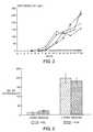
- FIG. 3is a graphical representation of osteocalcin release over time into culture medium from various crystalline hydroxyapatite substrata.
Landscapes
- Health & Medical Sciences (AREA)
- Life Sciences & Earth Sciences (AREA)
- Engineering & Computer Science (AREA)
- Biomedical Technology (AREA)
- Chemical & Material Sciences (AREA)
- Zoology (AREA)
- Cell Biology (AREA)
- General Health & Medical Sciences (AREA)
- Animal Behavior & Ethology (AREA)
- Epidemiology (AREA)
- Public Health (AREA)
- Transplantation (AREA)
- Oral & Maxillofacial Surgery (AREA)
- Botany (AREA)
- Veterinary Medicine (AREA)
- Medicinal Chemistry (AREA)
- Dermatology (AREA)
- Chemical Kinetics & Catalysis (AREA)
- Genetics & Genomics (AREA)
- Wood Science & Technology (AREA)
- Organic Chemistry (AREA)
- Biotechnology (AREA)
- Bioinformatics & Cheminformatics (AREA)
- Urology & Nephrology (AREA)
- Orthopedic Medicine & Surgery (AREA)
- Microbiology (AREA)
- Biochemistry (AREA)
- General Engineering & Computer Science (AREA)
- Vascular Medicine (AREA)
- Developmental Biology & Embryology (AREA)
- Rheumatology (AREA)
- Molecular Biology (AREA)
- Hematology (AREA)
- Micro-Organisms Or Cultivation Processes Thereof (AREA)
- Materials For Medical Uses (AREA)
- Meat, Egg Or Seafood Products (AREA)
Abstract
Description
Claims (17)
Priority Applications (2)
| Application Number | Priority Date | Filing Date | Title |
|---|---|---|---|
| US09/621,178US6299650B1 (en) | 1996-03-01 | 2000-07-21 | Method for in vitro production of bone |
| US09/877,169US20010032016A1 (en) | 1996-03-01 | 2001-06-08 | Method for in vitro production of bone |
Applications Claiming Priority (6)
| Application Number | Priority Date | Filing Date | Title |
|---|---|---|---|
| EP96200553 | 1996-03-01 | ||
| EP96200553 | 1996-03-01 | ||
| EP96202536 | 1996-09-09 | ||
| EP96202536 | 1996-09-11 | ||
| US08/810,266US6152964A (en) | 1996-03-01 | 1997-03-03 | Method for in vitro production of bone |
| US09/621,178US6299650B1 (en) | 1996-03-01 | 2000-07-21 | Method for in vitro production of bone |
Related Parent Applications (1)
| Application Number | Title | Priority Date | Filing Date |
|---|---|---|---|
| US08/810,266ContinuationUS6152964A (en) | 1996-03-01 | 1997-03-03 | Method for in vitro production of bone |
Related Child Applications (1)
| Application Number | Title | Priority Date | Filing Date |
|---|---|---|---|
| US09/877,169ContinuationUS20010032016A1 (en) | 1996-03-01 | 2001-06-08 | Method for in vitro production of bone |
Publications (1)
| Publication Number | Publication Date |
|---|---|
| US6299650B1true US6299650B1 (en) | 2001-10-09 |
Family
ID=26142564
Family Applications (3)
| Application Number | Title | Priority Date | Filing Date |
|---|---|---|---|
| US08/810,266Expired - Fee RelatedUS6152964A (en) | 1996-03-01 | 1997-03-03 | Method for in vitro production of bone |
| US09/621,178Expired - Fee RelatedUS6299650B1 (en) | 1996-03-01 | 2000-07-21 | Method for in vitro production of bone |
| US09/877,169AbandonedUS20010032016A1 (en) | 1996-03-01 | 2001-06-08 | Method for in vitro production of bone |
Family Applications Before (1)
| Application Number | Title | Priority Date | Filing Date |
|---|---|---|---|
| US08/810,266Expired - Fee RelatedUS6152964A (en) | 1996-03-01 | 1997-03-03 | Method for in vitro production of bone |
Family Applications After (1)
| Application Number | Title | Priority Date | Filing Date |
|---|---|---|---|
| US09/877,169AbandonedUS20010032016A1 (en) | 1996-03-01 | 2001-06-08 | Method for in vitro production of bone |
Country Status (4)
| Country | Link |
|---|---|
| US (3) | US6152964A (en) |
| AT (1) | ATE295875T1 (en) |
| CA (1) | CA2198978A1 (en) |
| DE (1) | DE69733292T2 (en) |
Cited By (28)
| Publication number | Priority date | Publication date | Assignee | Title |
|---|---|---|---|---|
| US20020031498A1 (en)* | 1998-02-23 | 2002-03-14 | Aastrom Biosciences, Inc. | Human lineage committed cell composition with enhanced proliferative potential, biological effector function, or both; methods for obtaining same; and their uses |
| US6582471B1 (en)* | 1997-08-14 | 2003-06-24 | Sulzer Innotec Ag | Composition and device for in vivo cartilage repair |
| US20030153078A1 (en)* | 2000-03-13 | 2003-08-14 | Jeanette Libera | Method for in vitro production of three-dimensional vital cartilage or bone tissue and use thereof as transplant material |
| US20030166274A1 (en)* | 2001-11-15 | 2003-09-04 | Hewitt Charles W. | Three-dimensional matrix for producing living tissue equivalents |
| US6667034B2 (en) | 1989-06-15 | 2003-12-23 | The Regents Of The University Of Michigan | Methods for regulating the specific lineages of cells produced in a human hematopoietic cell culture, methods for assaying the effect of substances on lineage-specific cell production, and cell compositions produced by these cultures |
| US20040156088A1 (en)* | 2003-02-06 | 2004-08-12 | Evans & Sutherland Computer Corporation | GLV based fiber optic transmitter |
| US20070012054A1 (en)* | 2005-03-17 | 2007-01-18 | Electrolux Home Products, Inc. | Electronic refrigeration control system |
| US20070173950A1 (en)* | 2006-01-25 | 2007-07-26 | Zanella John M | Modification of chemical forces of bone constructs |
| WO2009064956A1 (en)* | 2007-11-14 | 2009-05-22 | Osteosphere, Llc | A novel tissue culture platform for screening of potential bone remodeling agents |
| US7556649B2 (en) | 2000-04-07 | 2009-07-07 | Zimmer Orthobiologics, Inc. | Methods and compositions for treating intervertebral disc degeneration |
| US7579322B2 (en) | 2001-12-21 | 2009-08-25 | Zimmer Orthobiologics, Inc. | Compositions and methods for promoting myocardial and peripheral angiogenesis |
| US8613938B2 (en) | 2010-11-15 | 2013-12-24 | Zimmer Orthobiologics, Inc. | Bone void fillers |
| US8690874B2 (en) | 2000-12-22 | 2014-04-08 | Zimmer Orthobiologics, Inc. | Composition and process for bone growth and repair |
| US8742072B2 (en) | 2006-12-21 | 2014-06-03 | Zimmer Orthobiologics, Inc. | Bone growth particles and osteoinductive composition thereof |
| US8829166B2 (en) | 2002-06-26 | 2014-09-09 | Zimmer Orthobiologics, Inc. | Rapid isolation of osteoinductive protein mixtures from mammalian bone tissue |
| US11608486B2 (en) | 2015-07-02 | 2023-03-21 | Terumo Bct, Inc. | Cell growth with mechanical stimuli |
| US11613727B2 (en) | 2010-10-08 | 2023-03-28 | Terumo Bct, Inc. | Configurable methods and systems of growing and harvesting cells in a hollow fiber bioreactor system |
| US11624046B2 (en) | 2017-03-31 | 2023-04-11 | Terumo Bct, Inc. | Cell expansion |
| US11629332B2 (en) | 2017-03-31 | 2023-04-18 | Terumo Bct, Inc. | Cell expansion |
| US11634677B2 (en) | 2016-06-07 | 2023-04-25 | Terumo Bct, Inc. | Coating a bioreactor in a cell expansion system |
| US11667876B2 (en) | 2013-11-16 | 2023-06-06 | Terumo Bct, Inc. | Expanding cells in a bioreactor |
| US11667881B2 (en) | 2014-09-26 | 2023-06-06 | Terumo Bct, Inc. | Scheduled feed |
| US11685883B2 (en) | 2016-06-07 | 2023-06-27 | Terumo Bct, Inc. | Methods and systems for coating a cell growth surface |
| US11795432B2 (en) | 2014-03-25 | 2023-10-24 | Terumo Bct, Inc. | Passive replacement of media |
| US11965175B2 (en) | 2016-05-25 | 2024-04-23 | Terumo Bct, Inc. | Cell expansion |
| US12043823B2 (en) | 2021-03-23 | 2024-07-23 | Terumo Bct, Inc. | Cell capture and expansion |
| US12152699B2 (en) | 2022-02-28 | 2024-11-26 | Terumo Bct, Inc. | Multiple-tube pinch valve assembly |
| US12234441B2 (en) | 2017-03-31 | 2025-02-25 | Terumo Bct, Inc. | Cell expansion |
Families Citing this family (9)
| Publication number | Priority date | Publication date | Assignee | Title |
|---|---|---|---|---|
| US6811776B2 (en)* | 2000-12-27 | 2004-11-02 | The Regents Of The University Of Michigan | Process for ex vivo formation of mammalian bone and uses thereof |
| US20030119771A1 (en)* | 2001-08-22 | 2003-06-26 | Rompaey Luc Van | Modulators of bone homeostasis identified in a high-throughput screen |
| FR2833610B1 (en)* | 2001-12-14 | 2007-01-26 | Natural Implant | IN VITRO CULTIVE CELL CONSTRUCTS, PREPARATION AND USES |
| RU2240135C1 (en)* | 2003-02-17 | 2004-11-20 | Научно-исследовательский институт трансплантологии и искусственных органов Министерства здравоохранения Российской Федерации | Cell culture comprising precursor cells of osteogenesis, implant based on thereof and its applying for recovery bone integrity |
| EP1634072B1 (en)* | 2003-06-11 | 2007-06-27 | Centre National De La Recherche Scientifique (Cnrs) | Models for a skeletal system |
| DE102007005946A1 (en)* | 2007-02-01 | 2008-08-14 | Stiftung Caesar | Therapeutic composition and use of a cell-free substance |
| US20100055078A1 (en)* | 2008-08-15 | 2010-03-04 | The Government of the U.S.A, as represented by the Department of Veterans Affairs | Methods of making a transplantable bone repair system using multipotent stem cells |
| IL204116A (en) | 2010-02-23 | 2013-10-31 | Sheltagen Medical Ltd | Three-dimensional bone implant and method for producing same |
| US9238090B1 (en) | 2014-12-24 | 2016-01-19 | Fettech, Llc | Tissue-based compositions |
Citations (6)
| Publication number | Priority date | Publication date | Assignee | Title |
|---|---|---|---|---|
| US4963489A (en) | 1987-04-14 | 1990-10-16 | Marrow-Tech, Inc. | Three-dimensional cell and tissue culture system |
| WO1992010563A1 (en) | 1990-12-10 | 1992-06-25 | Universite Paris Vii | Immortalized cell lines and applications thereof, especially for the production of differentiated cells |
| WO1995003011A1 (en) | 1993-07-23 | 1995-02-02 | Rice University | Bone regeneration templates |
| JPH07194373A (en) | 1993-12-30 | 1995-08-01 | Nitta Gelatin Inc | Method for culturing bone marrow cell, mixture for culturing the same cell and material for transplanting to defective part of hard tissue |
| US5908784A (en) | 1995-11-16 | 1999-06-01 | Case Western Reserve University | In vitro chondrogenic induction of human mesenchymal stem cells |
| US5972703A (en) | 1994-08-12 | 1999-10-26 | The Regents Of The University Of Michigan | Bone precursor cells: compositions and methods |
Family Cites Families (22)
| Publication number | Priority date | Publication date | Assignee | Title |
|---|---|---|---|---|
| US4352887A (en)* | 1979-10-29 | 1982-10-05 | Albert Einstein College Of Medicine Of Yeshiva University | Method and article for culturing differentiated cells |
| US4314380A (en)* | 1980-09-26 | 1982-02-09 | Koken Co., Ltd. | Artificial bone |
| US4485096A (en)* | 1982-02-26 | 1984-11-27 | Massachusetts Institute Of Technology | Tissue-equivalent and method for preparation thereof |
| US5628781A (en)* | 1985-06-06 | 1997-05-13 | Thomas Jefferson University | Implant materials, methods of treating the surface of implants with microvascular endothelial cells, and the treated implants themselves |
| US5266480A (en)* | 1986-04-18 | 1993-11-30 | Advanced Tissue Sciences, Inc. | Three-dimensional skin culture system |
| US5124316A (en)* | 1986-11-14 | 1992-06-23 | President And Fellows Of Harvard College | Method for periodontal regeneration |
| US5041138A (en)* | 1986-11-20 | 1991-08-20 | Massachusetts Institute Of Technology | Neomorphogenesis of cartilage in vivo from cell culture |
| US4846835A (en)* | 1987-06-15 | 1989-07-11 | Grande Daniel A | Technique for healing lesions in cartilage |
| US4904259A (en)* | 1988-04-29 | 1990-02-27 | Samuel Itay | Compositions and methods for repair of cartilage and bone |
| US5197985A (en)* | 1990-11-16 | 1993-03-30 | Caplan Arnold I | Method for enhancing the implantation and differentiation of marrow-derived mesenchymal cells |
| US5226914A (en)* | 1990-11-16 | 1993-07-13 | Caplan Arnold I | Method for treating connective tissue disorders |
| FR2679250B1 (en)* | 1991-07-19 | 1995-07-13 | Inoteb | USE OF POROUS CALCIUM CARBONATE AS A SUPPORT MATERIAL FOR THE IN VITRO CULTURE OF EUKARYOTIC CELLS. |
| GB9125052D0 (en)* | 1991-11-26 | 1992-01-22 | Isis Innovation | Culture of bone cells |
| JP3310291B2 (en)* | 1992-08-13 | 2002-08-05 | ザ・トラスティーズ・オブ・ザ・ユニバーシティ・オブ・ペンシルバニア | Bioactive substance template for in vitro synthesis of bone tissue |
| US5541106A (en)* | 1993-04-05 | 1996-07-30 | Desmos, Inc. | Cell matrix stimulated attachment and hemidesmosome assembly |
| US5707962A (en)* | 1994-09-28 | 1998-01-13 | Gensci Regeneration Sciences Inc. | Compositions with enhanced osteogenic potential, method for making the same and therapeutic uses thereof |
| CZ138397A3 (en)* | 1994-11-08 | 1997-10-15 | Bradley Michael John Stringer | Human cellular lines |
| US5688531A (en)* | 1994-12-23 | 1997-11-18 | Ramot University Authority For Applied Research And Industrial Development, Ltd. | Method for regulating bone forming cells |
| IT1282207B1 (en)* | 1995-11-20 | 1998-03-16 | Fidia Advanced Biopolymers Srl | HUMAN BONE MARROW STEM CELL CULTURE SYSTEMS IN THREE-DIMENSIONAL MATRIXES CONSISTING OF HYALURONIC ACID ESTERS |
| EP0877795A4 (en)* | 1996-01-16 | 2003-03-05 | Depuy Orthopaedics Inc | Isolation of precursor cells from hematopoietic and non-hematopoietic tissues and their use |
| AU2730497A (en)* | 1996-04-17 | 1997-11-07 | Case Western Reserve University | Cryopreservation and extensive subculturing of human mesenchymal stem cells |
| DE69739540D1 (en)* | 1996-04-19 | 2009-10-01 | Osiris Therapeutics Inc | THE RECONSTRUCTION AND REINFORCEMENT OF BONES BY MEANS OF MESENCHYMAL STEM CELLS |
- 1997
- 1997-03-03DEDE69733292Tpatent/DE69733292T2/ennot_activeExpired - Fee Related
- 1997-03-03USUS08/810,266patent/US6152964A/ennot_activeExpired - Fee Related
- 1997-03-03ATAT97200620Tpatent/ATE295875T1/ennot_activeIP Right Cessation
- 1997-03-03CACA002198978Apatent/CA2198978A1/ennot_activeAbandoned
- 2000
- 2000-07-21USUS09/621,178patent/US6299650B1/ennot_activeExpired - Fee Related
- 2001
- 2001-06-08USUS09/877,169patent/US20010032016A1/ennot_activeAbandoned
Patent Citations (7)
| Publication number | Priority date | Publication date | Assignee | Title |
|---|---|---|---|---|
| US4963489A (en) | 1987-04-14 | 1990-10-16 | Marrow-Tech, Inc. | Three-dimensional cell and tissue culture system |
| WO1992010563A1 (en) | 1990-12-10 | 1992-06-25 | Universite Paris Vii | Immortalized cell lines and applications thereof, especially for the production of differentiated cells |
| WO1995003011A1 (en) | 1993-07-23 | 1995-02-02 | Rice University | Bone regeneration templates |
| US5522895A (en) | 1993-07-23 | 1996-06-04 | Rice University | Biodegradable bone templates |
| JPH07194373A (en) | 1993-12-30 | 1995-08-01 | Nitta Gelatin Inc | Method for culturing bone marrow cell, mixture for culturing the same cell and material for transplanting to defective part of hard tissue |
| US5972703A (en) | 1994-08-12 | 1999-10-26 | The Regents Of The University Of Michigan | Bone precursor cells: compositions and methods |
| US5908784A (en) | 1995-11-16 | 1999-06-01 | Case Western Reserve University | In vitro chondrogenic induction of human mesenchymal stem cells |
Non-Patent Citations (2)
| Title |
|---|
| Puleo et al., "Osteoblast responses to orthopedic implant material in vitro," J. Biomed. Mat. Res., 25:711-723 (1991). |
| Radder et al., "Bone-bonding behaviour of poly(ethylene oxide)-polybutylene terephthalate copolymer coatings and bulk implants: a comparative study," Biomaterials, 16(7):507-513 (1995). |
Cited By (47)
| Publication number | Priority date | Publication date | Assignee | Title |
|---|---|---|---|---|
| US6667034B2 (en) | 1989-06-15 | 2003-12-23 | The Regents Of The University Of Michigan | Methods for regulating the specific lineages of cells produced in a human hematopoietic cell culture, methods for assaying the effect of substances on lineage-specific cell production, and cell compositions produced by these cultures |
| US20040063201A1 (en)* | 1989-06-15 | 2004-04-01 | The Regents Of The University Of Michigan | Methods for regulating the specific lineages of cells produced in a human hematopoietic cell culture, methods for assaying the effect of substances of lineage-specific cell production, and cell compositions produced by these cultures |
| US6582471B1 (en)* | 1997-08-14 | 2003-06-24 | Sulzer Innotec Ag | Composition and device for in vivo cartilage repair |
| USRE41286E1 (en) | 1997-08-14 | 2010-04-27 | Zimmer Orthobiologics, Inc. | Compositions for regeneration and repair of cartilage lesions |
| US20030236574A1 (en)* | 1997-08-14 | 2003-12-25 | Sulzer Innotec Ag | Composition and device for in vivo cartilagerepair |
| US6835566B2 (en) | 1998-02-23 | 2004-12-28 | Aastrom Biosciences, Inc. | Human lineage committed cell composition with enhanced proliferative potential, biological effector function, or both; methods for obtaining same; and their uses |
| US20020034819A1 (en)* | 1998-02-23 | 2002-03-21 | Alan K. Smith | Human lineage committed cell composition with enhanced proliferative potential, biological effector function, or both; methods for obtaining same; and their uses |
| US20020031498A1 (en)* | 1998-02-23 | 2002-03-14 | Aastrom Biosciences, Inc. | Human lineage committed cell composition with enhanced proliferative potential, biological effector function, or both; methods for obtaining same; and their uses |
| US20040062755A1 (en)* | 1998-02-23 | 2004-04-01 | Aastrom Biosciences, Inc. | Human lineage committed cell composition with enhanced proliferative potential, biological effector function, or both; methods for obtaining same; and their uses |
| US20060093581A1 (en)* | 1998-02-23 | 2006-05-04 | Aastrom Biosciences, Inc. | Human lineage committed cell composition with enhanced proliferative potential, biological effector function, or both: methods for obtaining same; and their uses |
| US20040185557A1 (en)* | 1998-02-23 | 2004-09-23 | Aastrom Biosciences, Inc. | Human linkeage committed cell composition with enhanced proliferative potential, biological effector function, or both; methods for obtaining same; and their uses |
| US20030153078A1 (en)* | 2000-03-13 | 2003-08-14 | Jeanette Libera | Method for in vitro production of three-dimensional vital cartilage or bone tissue and use thereof as transplant material |
| US7887843B2 (en)* | 2000-03-13 | 2011-02-15 | Co.don Aktiengesllschaft | Method for in vitro production of three-dimensional vital cartilage tissue and use thereof as transplant material |
| US7556649B2 (en) | 2000-04-07 | 2009-07-07 | Zimmer Orthobiologics, Inc. | Methods and compositions for treating intervertebral disc degeneration |
| US8690874B2 (en) | 2000-12-22 | 2014-04-08 | Zimmer Orthobiologics, Inc. | Composition and process for bone growth and repair |
| US20030166274A1 (en)* | 2001-11-15 | 2003-09-04 | Hewitt Charles W. | Three-dimensional matrix for producing living tissue equivalents |
| US7579322B2 (en) | 2001-12-21 | 2009-08-25 | Zimmer Orthobiologics, Inc. | Compositions and methods for promoting myocardial and peripheral angiogenesis |
| US8829166B2 (en) | 2002-06-26 | 2014-09-09 | Zimmer Orthobiologics, Inc. | Rapid isolation of osteoinductive protein mixtures from mammalian bone tissue |
| US20040156088A1 (en)* | 2003-02-06 | 2004-08-12 | Evans & Sutherland Computer Corporation | GLV based fiber optic transmitter |
| US20070012054A1 (en)* | 2005-03-17 | 2007-01-18 | Electrolux Home Products, Inc. | Electronic refrigeration control system |
| US20070173950A1 (en)* | 2006-01-25 | 2007-07-26 | Zanella John M | Modification of chemical forces of bone constructs |
| US7749555B2 (en)* | 2006-01-25 | 2010-07-06 | Medtronic, Inc | Modification of chemical forces of bone constructs |
| US8742072B2 (en) | 2006-12-21 | 2014-06-03 | Zimmer Orthobiologics, Inc. | Bone growth particles and osteoinductive composition thereof |
| WO2009064956A1 (en)* | 2007-11-14 | 2009-05-22 | Osteosphere, Llc | A novel tissue culture platform for screening of potential bone remodeling agents |
| US11613727B2 (en) | 2010-10-08 | 2023-03-28 | Terumo Bct, Inc. | Configurable methods and systems of growing and harvesting cells in a hollow fiber bioreactor system |
| US11773363B2 (en) | 2010-10-08 | 2023-10-03 | Terumo Bct, Inc. | Configurable methods and systems of growing and harvesting cells in a hollow fiber bioreactor system |
| US11746319B2 (en) | 2010-10-08 | 2023-09-05 | Terumo Bct, Inc. | Customizable methods and systems of growing and harvesting cells in a hollow fiber bioreactor system |
| US8613938B2 (en) | 2010-11-15 | 2013-12-24 | Zimmer Orthobiologics, Inc. | Bone void fillers |
| US11708554B2 (en) | 2013-11-16 | 2023-07-25 | Terumo Bct, Inc. | Expanding cells in a bioreactor |
| US11667876B2 (en) | 2013-11-16 | 2023-06-06 | Terumo Bct, Inc. | Expanding cells in a bioreactor |
| US11795432B2 (en) | 2014-03-25 | 2023-10-24 | Terumo Bct, Inc. | Passive replacement of media |
| US11667881B2 (en) | 2014-09-26 | 2023-06-06 | Terumo Bct, Inc. | Scheduled feed |
| US12065637B2 (en) | 2014-09-26 | 2024-08-20 | Terumo Bct, Inc. | Scheduled feed |
| US11608486B2 (en) | 2015-07-02 | 2023-03-21 | Terumo Bct, Inc. | Cell growth with mechanical stimuli |
| US11965175B2 (en) | 2016-05-25 | 2024-04-23 | Terumo Bct, Inc. | Cell expansion |
| US11634677B2 (en) | 2016-06-07 | 2023-04-25 | Terumo Bct, Inc. | Coating a bioreactor in a cell expansion system |
| US11685883B2 (en) | 2016-06-07 | 2023-06-27 | Terumo Bct, Inc. | Methods and systems for coating a cell growth surface |
| US11999929B2 (en) | 2016-06-07 | 2024-06-04 | Terumo Bct, Inc. | Methods and systems for coating a cell growth surface |
| US12077739B2 (en) | 2016-06-07 | 2024-09-03 | Terumo Bct, Inc. | Coating a bioreactor in a cell expansion system |
| US11629332B2 (en) | 2017-03-31 | 2023-04-18 | Terumo Bct, Inc. | Cell expansion |
| US11624046B2 (en) | 2017-03-31 | 2023-04-11 | Terumo Bct, Inc. | Cell expansion |
| US11702634B2 (en) | 2017-03-31 | 2023-07-18 | Terumo Bct, Inc. | Expanding cells in a bioreactor |
| US12234441B2 (en) | 2017-03-31 | 2025-02-25 | Terumo Bct, Inc. | Cell expansion |
| US12359170B2 (en) | 2017-03-31 | 2025-07-15 | Terumo Bct, Inc. | Expanding cells in a bioreactor |
| US12043823B2 (en) | 2021-03-23 | 2024-07-23 | Terumo Bct, Inc. | Cell capture and expansion |
| US12152699B2 (en) | 2022-02-28 | 2024-11-26 | Terumo Bct, Inc. | Multiple-tube pinch valve assembly |
| US12209689B2 (en) | 2022-02-28 | 2025-01-28 | Terumo Kabushiki Kaisha | Multiple-tube pinch valve assembly |
Also Published As
| Publication number | Publication date |
|---|---|
| US6152964A (en) | 2000-11-28 |
| CA2198978A1 (en) | 1997-09-01 |
| DE69733292T2 (en) | 2006-04-27 |
| ATE295875T1 (en) | 2005-06-15 |
| US20010032016A1 (en) | 2001-10-18 |
| DE69733292D1 (en) | 2005-06-23 |
Similar Documents
| Publication | Publication Date | Title |
|---|---|---|
| US6299650B1 (en) | Method for in vitro production of bone | |
| DeBruijn et al. | Bone induction by implants coated with cultured osteogenic bone marrow cells | |
| EP0739631B1 (en) | Laminar bone support for cartilage growth | |
| van den Dolder et al. | Bone tissue reconstruction using titanium fiber mesh combined with rat bone marrow stromal cells | |
| Du et al. | Three‐dimensional nano‐HAp/collagen matrix loading with osteogenic cells in organ culture | |
| Uemura et al. | Transplantation of cultured bone cells using combinations of scaffolds and culture techniques | |
| Abukawa et al. | Reconstruction of mandibular defects with autologous tissue-engineered bone | |
| Liu et al. | Bone morphogenetic protein 2 incorporated into biomimetic coatings retains its biological activity | |
| US4609551A (en) | Process of and material for stimulating growth of cartilage and bony tissue at anatomical sites | |
| US5700289A (en) | Tissue-engineered bone repair using cultured periosteal cells | |
| Elgendy et al. | Osteoblast-like cell (MC3T3-E1) proliferation on bioerodible polymers: an approach towards the development of a bone-bioerodible polymer composite material | |
| JP2858782B2 (en) | Production of articular cartilage and bone regenerating composition, skeletal tissue transplant, and method of regenerating skeletal tissue | |
| US5855610A (en) | Engineering of strong, pliable tissues | |
| JP4777568B2 (en) | Implant material and method for producing the same | |
| JP4406283B2 (en) | Tissue regeneration substrate, transplant material, and production method thereof | |
| Kanczler et al. | Bone tissue engineering and bone regeneration | |
| WO2005011765A1 (en) | Method of constructing artificial joint | |
| EP0891421A4 (en) | SUBSTRATE OF BIOACTIVE MATERIAL FOR INCREASED CELLULAR FIXATION AND FUNCTION | |
| Yang et al. | Tricalcium phosphate and glutaraldehyde crosslinked gelatin incorporating bone morphogenetic protein—a viable scaffold for bone tissue engineering | |
| Ripamonti | The induction of bone in osteogenic composites of bone matrix and porous hydroxyapatite replicas: an experimental study on the baboon (Papio ursinus) | |
| EP0798374B1 (en) | Method for in vitro production of bone | |
| Sautier et al. | In vitro bone formation on coral granules | |
| JPWO2004052418A1 (en) | Bone-cartilage tissue regeneration implant | |
| Hofman et al. | Effects of Laddecreg; on the formation of calcified bone matrix in rat calvariae cells culture | |
| Faucheux et al. | Biocompatibility testing of a bovine hydroxy—apatite ceramic material with the use of osteo-progenitor cells isolated from human bone marrow |
Legal Events
| Date | Code | Title | Description |
|---|---|---|---|
| AS | Assignment | Owner name:ISOTIS N.V., NETHERLANDS Free format text:ASSIGNMENT OF ASSIGNORS INTEREST;ASSIGNOR:ISOTIS B.V.;REEL/FRAME:011751/0426 Effective date:20000918 | |
| CC | Certificate of correction | ||
| FPAY | Fee payment | Year of fee payment:4 | |
| AS | Assignment | Owner name:OCTOPLUS SCIENCES B.V., NETHERLANDS Free format text:ASSIGNMENT OF ASSIGNORS INTEREST;ASSIGNOR:ISOTIS N.V.;REEL/FRAME:020540/0095 Effective date:20080121 | |
| AS | Assignment | Owner name:LSP III OMNI INVESTMENT COOPERATIEF U.A., NETHERLA Free format text:SECURITY AGREEMENT;ASSIGNORS:OCTOPLUS SCIENCES B.V.;CHIENNA B.V.;OCTOPLUS DEVELOPMENT B.V.;AND OTHERS;REEL/FRAME:021018/0559 Effective date:20080521 Owner name:S.R. ONE, LIMITED, PENNSYLVANIA Free format text:SECURITY AGREEMENT;ASSIGNORS:OCTOPLUS SCIENCES B.V.;CHIENNA B.V.;OCTOPLUS DEVELOPMENT B.V.;AND OTHERS;REEL/FRAME:021018/0559 Effective date:20080521 | |
| AS | Assignment | Owner name:BIOLEX THERAPEUTICS, INC., NORTH CAROLINA Free format text:SECURITY AGREEMENT;ASSIGNORS:OCTOPLUS SCIENCES B.V.;CHIENNA B.V.;OCTOPLUS DEVELOPMENT B.V.;AND OTHERS;REEL/FRAME:021230/0881 Effective date:20080710 | |
| AS | Assignment | Owner name:OCTOPLUS SCIENCES B.V., NETHERLANDS Free format text:TERMINATION OF SECURITY INTEREST;ASSIGNOR:BIOLEX THERAPEUTICS, INC.;REEL/FRAME:021701/0168 Effective date:20081003 Owner name:CHIENNA B.V., NETHERLANDS Free format text:TERMINATION OF SECURITY INTEREST;ASSIGNOR:BIOLEX THERAPEUTICS, INC.;REEL/FRAME:021701/0168 Effective date:20081003 Owner name:OCTOPLUS DEVELOPMENT B.V., NETHERLANDS Free format text:TERMINATION OF SECURITY INTEREST;ASSIGNOR:BIOLEX THERAPEUTICS, INC.;REEL/FRAME:021701/0168 Effective date:20081003 Owner name:OCTOPLUS TECHNOLOGIES B.V., NETHERLANDS Free format text:TERMINATION OF SECURITY INTEREST;ASSIGNOR:BIOLEX THERAPEUTICS, INC.;REEL/FRAME:021701/0168 Effective date:20081003 Owner name:OCTOPLUS SCIENCES B.V.,NETHERLANDS Free format text:TERMINATION OF SECURITY INTEREST;ASSIGNOR:BIOLEX THERAPEUTICS, INC.;REEL/FRAME:021701/0168 Effective date:20081003 Owner name:CHIENNA B.V.,NETHERLANDS Free format text:TERMINATION OF SECURITY INTEREST;ASSIGNOR:BIOLEX THERAPEUTICS, INC.;REEL/FRAME:021701/0168 Effective date:20081003 Owner name:OCTOPLUS DEVELOPMENT B.V.,NETHERLANDS Free format text:TERMINATION OF SECURITY INTEREST;ASSIGNOR:BIOLEX THERAPEUTICS, INC.;REEL/FRAME:021701/0168 Effective date:20081003 Owner name:OCTOPLUS TECHNOLOGIES B.V.,NETHERLANDS Free format text:TERMINATION OF SECURITY INTEREST;ASSIGNOR:BIOLEX THERAPEUTICS, INC.;REEL/FRAME:021701/0168 Effective date:20081003 | |
| REMI | Maintenance fee reminder mailed | ||
| LAPS | Lapse for failure to pay maintenance fees | ||
| STCH | Information on status: patent discontinuation | Free format text:PATENT EXPIRED DUE TO NONPAYMENT OF MAINTENANCE FEES UNDER 37 CFR 1.362 | |
| FP | Lapsed due to failure to pay maintenance fee | Effective date:20091009 |