US6299619B1 - Methods for embolizing a target vascular site - Google Patents
Methods for embolizing a target vascular siteDownload PDFInfo
- Publication number
- US6299619B1 US6299619B1US09/542,145US54214500AUS6299619B1US 6299619 B1US6299619 B1US 6299619B1US 54214500 AUS54214500 AUS 54214500AUS 6299619 B1US6299619 B1US 6299619B1
- Authority
- US
- United States
- Prior art keywords
- microcatheter
- vascular site
- passing
- embolizing
- target vascular
- Prior art date
- Legal status (The legal status is an assumption and is not a legal conclusion. Google has not performed a legal analysis and makes no representation as to the accuracy of the status listed.)
- Expired - Lifetime
Links
Images


Classifications
- A—HUMAN NECESSITIES
- A61—MEDICAL OR VETERINARY SCIENCE; HYGIENE
- A61L—METHODS OR APPARATUS FOR STERILISING MATERIALS OR OBJECTS IN GENERAL; DISINFECTION, STERILISATION OR DEODORISATION OF AIR; CHEMICAL ASPECTS OF BANDAGES, DRESSINGS, ABSORBENT PADS OR SURGICAL ARTICLES; MATERIALS FOR BANDAGES, DRESSINGS, ABSORBENT PADS OR SURGICAL ARTICLES
- A61L31/00—Materials for other surgical articles, e.g. stents, stent-grafts, shunts, surgical drapes, guide wires, materials for adhesion prevention, occluding devices, surgical gloves, tissue fixation devices
- A61L31/02—Inorganic materials
- A61L31/022—Metals or alloys
- A—HUMAN NECESSITIES
- A61—MEDICAL OR VETERINARY SCIENCE; HYGIENE
- A61B—DIAGNOSIS; SURGERY; IDENTIFICATION
- A61B17/00—Surgical instruments, devices or methods
- A61B17/12—Surgical instruments, devices or methods for ligaturing or otherwise compressing tubular parts of the body, e.g. blood vessels or umbilical cord
- A61B17/12022—Occluding by internal devices, e.g. balloons or releasable wires
- A61B17/12099—Occluding by internal devices, e.g. balloons or releasable wires characterised by the location of the occluder
- A61B17/12109—Occluding by internal devices, e.g. balloons or releasable wires characterised by the location of the occluder in a blood vessel
- A61B17/12113—Occluding by internal devices, e.g. balloons or releasable wires characterised by the location of the occluder in a blood vessel within an aneurysm
- A—HUMAN NECESSITIES
- A61—MEDICAL OR VETERINARY SCIENCE; HYGIENE
- A61B—DIAGNOSIS; SURGERY; IDENTIFICATION
- A61B17/00—Surgical instruments, devices or methods
- A61B17/12—Surgical instruments, devices or methods for ligaturing or otherwise compressing tubular parts of the body, e.g. blood vessels or umbilical cord
- A61B17/12022—Occluding by internal devices, e.g. balloons or releasable wires
- A61B17/12131—Occluding by internal devices, e.g. balloons or releasable wires characterised by the type of occluding device
- A61B17/1214—Coils or wires
- A61B17/12145—Coils or wires having a pre-set deployed three-dimensional shape
- A—HUMAN NECESSITIES
- A61—MEDICAL OR VETERINARY SCIENCE; HYGIENE
- A61B—DIAGNOSIS; SURGERY; IDENTIFICATION
- A61B17/00—Surgical instruments, devices or methods
- A61B17/12—Surgical instruments, devices or methods for ligaturing or otherwise compressing tubular parts of the body, e.g. blood vessels or umbilical cord
- A61B17/12022—Occluding by internal devices, e.g. balloons or releasable wires
- A61B17/12131—Occluding by internal devices, e.g. balloons or releasable wires characterised by the type of occluding device
- A61B17/1214—Coils or wires
- A61B17/12154—Coils or wires having stretch limiting means
- A—HUMAN NECESSITIES
- A61—MEDICAL OR VETERINARY SCIENCE; HYGIENE
- A61B—DIAGNOSIS; SURGERY; IDENTIFICATION
- A61B17/00—Surgical instruments, devices or methods
- A61B17/12—Surgical instruments, devices or methods for ligaturing or otherwise compressing tubular parts of the body, e.g. blood vessels or umbilical cord
- A61B17/12022—Occluding by internal devices, e.g. balloons or releasable wires
- A61B17/12131—Occluding by internal devices, e.g. balloons or releasable wires characterised by the type of occluding device
- A61B17/12163—Occluding by internal devices, e.g. balloons or releasable wires characterised by the type of occluding device having a string of elements connected to each other
- A—HUMAN NECESSITIES
- A61—MEDICAL OR VETERINARY SCIENCE; HYGIENE
- A61B—DIAGNOSIS; SURGERY; IDENTIFICATION
- A61B17/00—Surgical instruments, devices or methods
- A61B17/12—Surgical instruments, devices or methods for ligaturing or otherwise compressing tubular parts of the body, e.g. blood vessels or umbilical cord
- A61B17/12022—Occluding by internal devices, e.g. balloons or releasable wires
- A61B17/12131—Occluding by internal devices, e.g. balloons or releasable wires characterised by the type of occluding device
- A61B17/12181—Occluding by internal devices, e.g. balloons or releasable wires characterised by the type of occluding device formed by fluidized, gelatinous or cellular remodelable materials, e.g. embolic liquids, foams or extracellular matrices
- A61B17/1219—Occluding by internal devices, e.g. balloons or releasable wires characterised by the type of occluding device formed by fluidized, gelatinous or cellular remodelable materials, e.g. embolic liquids, foams or extracellular matrices expandable in contact with liquids
- A—HUMAN NECESSITIES
- A61—MEDICAL OR VETERINARY SCIENCE; HYGIENE
- A61L—METHODS OR APPARATUS FOR STERILISING MATERIALS OR OBJECTS IN GENERAL; DISINFECTION, STERILISATION OR DEODORISATION OF AIR; CHEMICAL ASPECTS OF BANDAGES, DRESSINGS, ABSORBENT PADS OR SURGICAL ARTICLES; MATERIALS FOR BANDAGES, DRESSINGS, ABSORBENT PADS OR SURGICAL ARTICLES
- A61L31/00—Materials for other surgical articles, e.g. stents, stent-grafts, shunts, surgical drapes, guide wires, materials for adhesion prevention, occluding devices, surgical gloves, tissue fixation devices
- A61L31/14—Materials characterised by their function or physical properties, e.g. injectable or lubricating compositions, shape-memory materials, surface modified materials
- A61L31/145—Hydrogels or hydrocolloids
- A—HUMAN NECESSITIES
- A61—MEDICAL OR VETERINARY SCIENCE; HYGIENE
- A61L—METHODS OR APPARATUS FOR STERILISING MATERIALS OR OBJECTS IN GENERAL; DISINFECTION, STERILISATION OR DEODORISATION OF AIR; CHEMICAL ASPECTS OF BANDAGES, DRESSINGS, ABSORBENT PADS OR SURGICAL ARTICLES; MATERIALS FOR BANDAGES, DRESSINGS, ABSORBENT PADS OR SURGICAL ARTICLES
- A61L31/00—Materials for other surgical articles, e.g. stents, stent-grafts, shunts, surgical drapes, guide wires, materials for adhesion prevention, occluding devices, surgical gloves, tissue fixation devices
- A61L31/14—Materials characterised by their function or physical properties, e.g. injectable or lubricating compositions, shape-memory materials, surface modified materials
- A61L31/18—Materials at least partially X-ray or laser opaque
- A—HUMAN NECESSITIES
- A61—MEDICAL OR VETERINARY SCIENCE; HYGIENE
- A61B—DIAGNOSIS; SURGERY; IDENTIFICATION
- A61B17/00—Surgical instruments, devices or methods
- A61B2017/00004—(bio)absorbable, (bio)resorbable or resorptive
- A—HUMAN NECESSITIES
- A61—MEDICAL OR VETERINARY SCIENCE; HYGIENE
- A61B—DIAGNOSIS; SURGERY; IDENTIFICATION
- A61B17/00—Surgical instruments, devices or methods
- A61B2017/00831—Material properties
- A61B2017/00867—Material properties shape memory effect
- A—HUMAN NECESSITIES
- A61—MEDICAL OR VETERINARY SCIENCE; HYGIENE
- A61B—DIAGNOSIS; SURGERY; IDENTIFICATION
- A61B17/00—Surgical instruments, devices or methods
- A61B17/12—Surgical instruments, devices or methods for ligaturing or otherwise compressing tubular parts of the body, e.g. blood vessels or umbilical cord
- A61B17/12022—Occluding by internal devices, e.g. balloons or releasable wires
- A61B2017/1205—Introduction devices
- A—HUMAN NECESSITIES
- A61—MEDICAL OR VETERINARY SCIENCE; HYGIENE
- A61L—METHODS OR APPARATUS FOR STERILISING MATERIALS OR OBJECTS IN GENERAL; DISINFECTION, STERILISATION OR DEODORISATION OF AIR; CHEMICAL ASPECTS OF BANDAGES, DRESSINGS, ABSORBENT PADS OR SURGICAL ARTICLES; MATERIALS FOR BANDAGES, DRESSINGS, ABSORBENT PADS OR SURGICAL ARTICLES
- A61L2430/00—Materials or treatment for tissue regeneration
- A61L2430/36—Materials or treatment for tissue regeneration for embolization or occlusion, e.g. vaso-occlusive compositions or devices
Definitions
- the present inventionrelates to the field of methods and devices for the embolization of vascular aneurysms and similar vascular abnormalities. More specifically, the present invention relates to an embolic device that is inserted into a vascular site such as an aneurysm to create an embolism therein and a method for embolizing a vascular site using the device.
- vascular embolizationhas been used to control vascular bleeding, to occlude the blood supply to tumors, and to occlude vascular aneurysms, particularly intracranial aneurysms.
- vascular embolization for the treatment of aneurysmshas received much attention.
- the balloonis carried into the aneurysm at the tip of the catheter, and it is inflated inside the aneurysm with a solidifying fluid (typically a polymerizable resin or gel) to occlude the aneurysm.
- a solidifying fluidtypically a polymerizable resin or gel
- the balloonis then detached from the catheter by gentle traction on the catheter.
- the balloon-type embolization devicecan provide an effective occlusion of many types of aneurysms, it is difficult to retrieve or move after the solidifying fluid sets, and it is difficult to visualize unless it is filled with a contrast material. Furthermore, there are risks of balloon rupture during inflation and of premature detachment of the balloon from the catheter.
- Another approachis the direct injection of a liquid polymer embolic agent into the vascular site to be occluded.
- a liquid polymer used in the direct injection techniqueis a rapidly polymerizing liquid, such as a cyanoacrylate resin, particularly isobutyl cyanoacrylate, that is delivered to the target site as a liquid, and then is polymerized in situ.
- a liquid polymer that is precipitated at the target site from a carrier solutionhas been used.
- An example of this type of embolic agentis a cellulose acetate polymer mixed with bismuth trioxide and dissolved in dimethyl sulfoxide (DMSO). Another type is ethylene vinyl alcohol dissolved in DMSO.
- microcoilsmay be made of a biocompatible metal alloy (typically platinum and tungsten) or a suitable polymer. If made of metal, the coil may be provided with Dacron fibers to increase thrombogenicity. The coil is deployed through a microcatheter to the vascular site. Examples of microcoils are disclosed in the following U.S. patents: U.S. Pat. No. 4,994,069—Ritchart et al.; U.S. Pat. No. 5,133,731—Butler et al.; U.S. Pat. No. 5,226,911—Chee et al.; U.S. Pat. No.
- microcoil approachhas met with some success in treating small aneurysms with narrow necks, but the coil must be tightly packed into the aneurysm to avoid shifting that can lead to recanalization.
- Microcoilshave been less successful in the treatment of larger aneurysms, especially those with relatively wide necks.
- a disadvantage of microcoilsis that they are not easily retrievable; if a coil migrates out of the aneurysm, a second procedure to retrieve it and move it back into place is necessary. Furthermore, complete packing of an aneurysm using microcoils can be difficult to achieve in practice.
- GDCGuglielmi Detachable Coil
- U.S. Pat. No. 5,122,136Guglielmi Detachable Coil
- the GDCemploys a platinum wire coil fixed to a stainless steel delivery wire by a solder connection. After the coil is placed inside an aneurysm, an electrical current is applied to the delivery wire, which heats sufficiently to melt the solder junction, thereby detaching the coil from the delivery wire. The application of the current also creates a positive electrical charge on the coil, which attracts negatively-charged blood cells, platelets, and fibrinogen, thereby increasing the thrombogenicity of the coil.
- Several coils of different diameters and lengthscan be packed into an aneurysm until the aneurysm is completely filled. The coils thus create and hold a thrombus within the aneurysm, inhibiting its displacement and its fragmentation.
- the advantages of the GDC procedureare the ability to withdraw and relocate the coil if it migrates from its desired location, and the enhanced ability to promote the formation of a stable thrombus within the aneurysm. Nevertheless, as in conventional microcoil techniques, the successful use of the GDC procedure has been substantially limited to small aneurysms with narrow necks.
- Still another approach to the embolization of an abnormal vascular siteis the injection into the site of a biocompatible hydrogel, such as poly (2-hydroxyethyl methacrylate) (“pHEMA” or “PHEMA”); or a polyvinyl alcohol foam (“PAF”).
- a biocompatible hydrogelsuch as poly (2-hydroxyethyl methacrylate) (“pHEMA” or “PHEMA”); or a polyvinyl alcohol foam (“PAF”).
- U.S. Pat. No. 5,823,198—Jones et al.discloses an expansible PVA foam plug that is delivered to the interior of an aneurysm at the end of a guidewire.
- the plugcomprises a plurality of pellets or particles that expand into an open-celled structure upon exposure to the fluids within the aneurysm so as to embolize the aneurysm.
- the pelletsare coated with a blood-soluble restraining agent to maintain them in a compressed state and attached to the guidewire until delivered to the aneurysm. Because there is no mechanical connection between the pellets and the guidewire (other than the relatively weak temporary bond provided by the restraining agent), however, premature release and migration of some of the pellets remains a possibility.
- an aneurysm treatment device and methodthat can substantially fill aneurysms of a large range of sizes, configurations, and neck widths with a thrombogenic medium with a minimal risk of inadvertent aneurysm rupture or blood vessel wall damage.
- a method and devicethat also allow for the precise locational deployment of the medium, while also minimizing the potential for migration away from the target location.
- a method and device meeting these criteriashould also be relatively easy to use in a clinical setting. Such ease of use, for example, should preferably include a provision for good visualization of the device during and after deployment in an aneurysm.
- an embolization devicecomprises one or more expansible, hydrophilic embolizing elements non-releasably carried on a filamentous carrier at spaced intervals along the length of the carrier.
- the carrieris a suitable length of very thin, highly flexible filament of nickel/titanium alloy.
- the embolizing elementsare separated from each other on the carrier by radiopaque spacers in the form of highly flexible microcoils made of platinum or platinum/tungsten alloy, as in the thrombogenic microcoils of the prior art, as described above.
- the embolizing elementsare made of a hydrophilic, macroporous, polymeric, hydrogel foam material, in particular a swellable foam matrix formed as a macroporous solid comprising a foam stabilizing agent and a polymer or copolymer of a free radical polymerizable hydrophilic olefm monomer cross-linked with up to about 10% by weight of a multiolefin-functional cross-linking agent.
- a foam stabilizing agentand a polymer or copolymer of a free radical polymerizable hydrophilic olefm monomer cross-linked with up to about 10% by weight of a multiolefin-functional cross-linking agent.
- a second aspect of the present inventionis a method for embolizing a vascular site, comprising, in the preferred embodiment the steps of: (a) passing a microcatheter intravascularly so that its distal end is introduced into a target vascular site; (b) passing a vaso-occlusive device through the microcatheter into the target vascular site so that the vaso-occlusive device assumes a three-dimensional configuration that fills a portion of the volume of the target vascular site; (c) providing a vascular embolization device comprising at least one expansible embolizing element non-releasably connected to a filamentous carrier; (d) passing the embolization device through the microcatheter so that it emerges from the distal end of the microcatheter into the target vascular site; and (e) expanding the embolizing element or elements in situ substantially to fill the remaining volume of the target vascular site while maintaining the connection between the embolizing element or elements and the carrier.
- the vaso-occlusive deviceis of the type that is initially in the form of an elongate, flexible, filamentous element for delivery through the microcatheter, and that assumes a three-dimensional geometry upon installation in the target vascular site.
- One such deviceis the above-described GDC (U.S. Pat. No. 5,122,136—Guglielmi et al., the disclosure of which is incorporated herein by reference).
- Other such devicesare describe in, for example, U.S. Pat. No. 5,766,219—Horton; U.S. Pat. No. 5,690,671—McGurk et al.; and U.S. Pat. No. 5,911,731—Pham et al., the disclosures of which are incorporated herein by reference. Still other types of vaso-occlusive devices known in the art may also perform satisfactorily in this method.
- the methodcomprises the steps of: (a) deploying an intravascular device to a position in a blood vessel adjacent to a target vascular site; (b) providing a vascular embolization device comprising at least one expansible embolizing element non-releasably connected to a filamentous carrier; (c) passing a microcatheter intravascularly so that the distal end of the microcatheter passes through the intravascular device into the target vascular site; (d) passing the embolization device through the microcatheter so that it emerges from the distal end of the microcatheter into the target vascular site; and (e) expanding the embolizing element or elements in situ substantially to fill the volume of the target vascular site while maintaining the connection between the embolizing element or elements and the carrier.
- step of providing the embolization devicemay follow the step of passing the microcatheter intravascularly.
- the intravascular devicemay be of the type disclosed in U.S. Pat. No. 5,980,514—Kupiecki et al., the disclosure of which is incorporated herein by reference.
- This intravascular devicecomprises a filamentous element that is introduced by a microcatheter to the juncture of an aneurysm or the like, and that then assumes the configuration of a coil adjacent the neck of the aneurysm.
- the step of passing a vaso-occlusive device or an intravascular device through the microcatheter to the target vascular sitemay be omitted.
- the embolization bodies or elementsin the preferred embodiment, have an initial configuration in the form of small, substantially cylindrical “micropellets” of small enough outside diameter to fit within the microcatheter.
- the bodiesare hydrophilically expansible into an expanded configuration in which they substantially conform to and fill the vascular site.
- the present inventionprovides a number of significant advantages. Specifically, the present invention provides an effective vascular embolization device that can be deployed within a vascular site with excellent locational control, and with a lower risk of vascular rupture, tissue damage, or migration than with prior art devices. Furthermore, the embolization device effects a conformal fit within the site that promotes effective embolization, and yet its ability to be delivered to the site through a microcatheter facilitates precise and highly controllable deployment.
- the essentially filamentous initial configuration of the embolization devicewhereby it readily conforms to the interior dimensions of the vascular site, allows it to be used effectively to embolize vascular sites having a wide variety of sizes, configurations, and (in the particular case of aneurysms) neck widths.
- FIG. 1is an elevational view of a vascular embolization device in accordance with a preferred embodiment of the invention
- FIG. 2is a cross-sectional view taken along line 2 — 2 of FIG. 1;
- FIG. 3is a cross-sectional view taken along line 3 — 3 of FIG. 2;
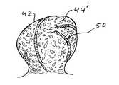
- FIGS. 4 through 7are semischematic views showing the steps in a method of embolizing a vascular site (specifically, an aneurysm) in accordance with one embodiment of the embolizing method aspect of the present invention
- FIG. 8is a detailed perspective view of mechanism by which the embolization device of the present invention is preferably attached to the distal end of a deployment instrument;
- FIG. 9is a detailed perspective view, similar to that of FIG. 8, showing the embolization device of the present invention after it has been separated from the deployment instrument;
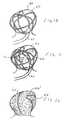
- FIGS. 10, 11 , and 12are semischematic views showing steps that, in addition to those illustrated in FIGS. 4-7, constitute a method of embolizing a vascular site in accordance with a preferred embodiment of the embolizing method aspect of the present invention.
- FIG. 13is a semischematic view showing a step in a method of embolizing a vascular site in accordance with an alternative embodiment of the embolizing method aspect of the present invention.
- the Embolization DeviceThe Embolization Device.
- the embolization device 10comprises a plurality of embolizing bodies, each configured as a substantially cylindrical “micropellet” 12 , located at spaced intervals along a filamentous carrier 14 .
- the number of micropellets 12will vary, depending on the length of the carrier 14 , which, turn, will depend on the size of the vascular site to be embolized. For a large vascular site, for example, eight to twelve micropellets may be used, although an even larger number may be used if necessary. In some applications (e.g., very small aneurysms), as few as one or two micropellets may be used.
- the carrier 14is also carried on the carrier 14 .
- the carrier 14has a distal portion on which is carried a relatively long distal microcoil segment 18 that is retained in place by a distal retention member 20 .
- the carrier 14has a proximal portion on which is carried a relatively long proximal microcoil segment 22 .
- the proximal end of the device 10is terminated by a hydrogel linkage element 24 , to be described below.
- the spacers 16 , the distal microcoil segment 18 , and the proximal microcoil segment 22are all highly flexible, and they are preferably made of platinum or platinum/tungsten wire, which has the advantages of being biocompatible and radiopaque.
- the micropellets 12are non-releasably carried on the carrier 14 . They may be fixed in place on the filamentous carrier 14 , either mechanically or by a suitable biocompatible, water-insoluble adhesive, or they may be simply strung loosely on the carrier 14 between successive spacers 16 .
- the micropellets 12are preferably formed of a biocompatible, macroporous, hydrophilic hydrogel foam material, in particular a water-swellable foam matrix formed as a macroporous solid comprising a foam stabilizing agent and a polymer or copolymer of a free radical polymerizable hydrophilic olefin monomer cross-linked with up to about 10% by weight of a multiolefm-functional cross-linking agent.
- a suitable material of this typeis described in U.S. Pat. No. 5,570,585—Park et al., the disclosure of which is incorporated herein by reference.
- Another suitable material for the micropellets 12is a porous hydrated polyvinyl alcohol (PVA) foam gel prepared from a polyvinyl alcohol solution in a mixed solvent consisting of water and a watermiscible organic solvent, as described, for example, in U.S. Pat. No. 4,663,358—Hyon et al., the disclosure of which is incorporated herein by reference.
- PVApolyvinyl alcohol
- Other suitable PVA structuresare described in U.S. Pat. No. 5,823,198—Jones et al. and U.S. Pat. No. 5,258,042—Mehta, the disclosures of which are incorporated herein by reference.
- Another suitable materialis a collagen foam, of the type described in U.S. Pat. No.
- each of the embolizing micropellets 12has an initial diameter of not more than about 0.5 mm prior to expansion in situ, with an expanded diameter of at least about 3 mm.
- the micropellets 12may be compressed to the desired size from a significantly larger initial configuration. The compression is performed by squeezing or crimping the micropellets 12 in a suitable implement or fixture, and then “setting” them in the compressed configuration by heating and/or drying.
- Each of the micropellets 12is swellable or expansible to many times (at least about 25 times, preferably about 70 times, and up to about 100 times) its initial (compressed) volume, primarily by the hydrophilic absorption of water molecules from an aqueous solution (e.g., resident blood plasma and/or injected saline solution), and secondarily by the filling of its pores with blood.
- the micropellets 12may be coated with a water-soluble coating (not shown), such as a starch, to provide a time-delayed expansion.
- a temperature-sensitive coatingthat disintegrates in response to normal human body temperature. See, e.g., U.S. Pat. No. 5,120,349—Stewart et al. and U.S. Pat. No. 5,129,180—Stewart.
- the foam material of the embolizing micropellet 12may advantageously be modified, or provided with additives, to make the device 10 visible by conventional imaging techniques.
- the foamcan be impregnated with a water-insoluble radiopaque material such as barium sulfate, as described by Thanoo et al., “Radiopaque Hydrogel Microspheres”, J. Microencapsuladon , Vol. 6, No. 2, pp. 233-244 (1989).
- the hydrogel monomerscan be copolymerized with radiopaque materials, as described in Horák et al., “New Radiopaque PolyHEMA-Based Hydrogel Particles”, J. Biomedical Matenals Research , Vol. 34, pp. 183-188 (1997).
- the micropellets 12may optionally include bioactive or therapeutic agents to promote thrombosis, cellular ingrowth, and/or epithelialization. See, e.g, Vacanti et al., “Tissue Engineering: The Design and Fabrication of Living Replacement Devices for Surgical Reconstruction and Transplantation,” The Lancet (Vol. 354, Supplement 1), pp. 32-34 (July, 1999); Langer, “Tissue Engineering: A New Field and Its Challenges,” Phamaceutical Research , Vol. 14., No. 7, pp. 840-841 (July, 1997); Persidis, “Tissue Engineering,” Nature Biotechnology , Vol. 17, pp. 508-510 (May, 1999).
- the filamentous carrier 14is preferably a length of nickel/titanium wire, such as that marketed under the trade name “Nitinol”. Wire of this alloy is highly flexible, and it has an excellent “elastic memory”, whereby it can be formed into a desired shape to which it will return when it is deformed.
- the wire that forms the carrier 14has a diameter of approximately 0.04 mm, and it is heat-treated to form a multi-looped structure that may assume a variety of three-dimensional shapes, such as a helix, a sphere, or an ovoid (as disclosed, for example, in U.S. Pat. No.
- the intermediate portion of the carrier 14i.e., the portion that includes the micropellets 12
- the proximal portionthat carries the proximal microcoil segment 22
- the distal portionthat carries the distal microcoil segment 18
- the carrier 14may be formed of a single wire, or it may be formed of a cable or braided structure of several ultra-thin wires.
- the carrier 14may be made of a thin filament of a suitable polymer, such as a PVA, that is formed in a looped structure.
- the polymermay be impregnated with a radiopaque material (e.g., barium sulfate or particles of gold, tantalum, or platinum), or it may enclose a core of nickel/titanium wire.
- the carrier 14may be constructed as a “cable” of thin polymer fibers that includes fibers of an expansile polymer, such as polyvinyl alcohol (PVA), at spaced intervals to form the micropellets 12 .
- PVApolyvinyl alcohol
- Still another alternative construction for the carrier 14is a continuous length of microcoil.
- the micropellets 12would be attached at spaced intervals along the length of the carrier 14 .
- the hydrogel linkage element 24is advantageously made of the same material as the micropellets 12 . Indeed, the most proximal of the micropellets 12 may function as the linkage element 24 .
- the linkage element 24is attached to the proximal end of the carrier 14 by a suitable biocompatible adhesive.
- the purpose of the linkage element 24is to removably attach the device 10 to a deployment instrument 30 (FIGS. 8 and 9 ).
- the deployment instrument 30comprises a length of platinum or platinum/tungsten microcoil outer portion 32 with a flexible wire core 34 of the same or a similar metal.
- the deployment instrument 30has a distal portion 36 at which the microcoil outer portion 32 has coils that are more distantly-spaced (i.e., have a greater pitch).
- the device 10is initially attached to the deployment instrument 30 by means of the linkage element 24 .
- the linkage element 24is installed, in a compressed state, so that it encompasses and engages both the proximal end of the embolization device 10 and the distal portion 36 of the deployment instrument 30 .
- the linkage element 24binds the deployment instrument 30 and the embolization device 10 together.
- the linkage element 24expands greatly, thereby loosening its grip on the distal portion 36 of the deployment instrument 30 , and thus allowing the embolization device 10 to be separated from the deployment instrument 30 by pulling the latter proximally out of and away from the linkage element 24 .
- FIGS. 4 through 7One method of embolizing a vascular site using the embolization device 10 is illustrated in FIGS. 4 through 7.
- a microcatheter 40is threaded intravascularly, by known methods, until its distal end is located within the targeted vascular site (here, an aneurysm 42 ).
- this threading operationis typically performed by first introducing a catheter guidewire (not shown) along the desired microcatheter path, and then feeding the microcatheter 40 over the catheter guidewire until the microcatheter 40 is positioned adjacent the distal aspect of the dome of the aneurysm, as shown in FIG. 4 .
- the catheter guidewireis then removed. Then, as shown in FIGS.
- the embolization device 10which is attached to the distal end of the deployment instrument 30 , as described above, is passed axially through the microcatheter 40 , using the deployment instrument 30 to push the device 10 through the microcatheter 40 until the device 10 is clear from the distal end of the microcatheter 40 and fully deployed within the aneurysm 42 (FIG. 6 ), filling the aneurysm from its distal aspect.
- the deployment procedureis facilitated by the visualization of the embolization device 10 that is readily accomplished due to its radiopaque components, as described above.
- the embolization bodies or micropellets 12in their compressed configuration, have a maximum outside diameter that is less than the inside diameter of the microcatheter 40 , so that the embolization device 10 can be passed through the microcatheter 40 .
- the micropellets 12are preferably compressed and “set”, as described above, before the device 10 is inserted into the microcatheter 40 .
- a biocompatible, substantially non-aqueous fluidsuch as polyethylene glycol, may be injected into the microcatheter 40 to prevent premature expansion of the device 10 due to hydration, and to reduce friction with the interior of the microcatheter 40 .
- the pores of the embolizing bodies or micropellets 12 , and of the linkage element 22begin to absorb aqueous fluid from the blood within the vascular site 42 to release their “set”, allowing these elements to begin assuming their expanded configuration.
- the expansioncan be enhanced and accelerated by injecting saline solution through the microcatheter 40 .
- the expansion of the linkage element 24allows the embolization device 10 to be separated from the deployment instrument 30 , as described above, and the deployment instrument 30 can then be removed.
- the elastic memory of the carrier 14causes it to resume its original looped configuration once it is released from the confines of the microcatheter 40 .
- the embolization devicebegins to occupy a significant portion of the volume of the aneurysm 42 .
- the micropellets 12are of a hydrophilic material, they then continue to expand in situ due to hydrophilic hydration of the material, as well as from the filling of their pores with blood. If the embolizing bodies 12 are of a non-hydrophilic material, their expansion is due to the latter mechanism only. In either case, the result, as shown in FIG. 7, is the substantially complete filling of the interior of the aneurysm 42 with the expanded embolizing bodies or micropellets 12 , whereby a substantially conformal embolizing implant 44 is formed that substantially fills the interior of the aneurysm 42 .
- the micropellets 12being non-releasably carried the carrier 14 and fixed in place thereon, stay on the carrier during their expansion. Thus, the chance of a micropellet separating from the carrier and migrating out of the vascular site is minimized.
- a preferred method of embolizing a target vascular site using the embolization device 10will be understood with reference to FIGS. 10-12, along with FIGS. 4-7 (discussed above).
- the passing of a microcatheter 40 intravascularly until its distal end is introduced into a target vascular siteis followed by the step of passing a vaso-occlusive device 50 through the microcatheter 40 into the target vascular site (e.g., the aneurysm 42 ) so that the vaso-occlusive device 50 assumes a three-dimensional configuration that fills a portion of the interior volume of the target vascular site 42 , as shown in FIG. 10 .
- the deployed vaso-occlusive device 50forms a “cage” within the aneurysm 42 that provides a matrix for improved retention of the expansible embolizing bodies or micropellets 12 of the embolization device 10 .
- the embolization device 10is then passed through the microcatheter 40 , as described above, and as shown in FIG. 11, to enter the aneurysm 42 within the voids left by the vaso-occlusive device 50 .
- the embolizing bodies or micropellets 12are expanded, as described above, and as shown in FIG. 12, whereby a substantially conformal embolizing implant 44 ′ is formed that substantially fills the remaining interior volume of the aneurysm 42 .
- the vaso-occlusive device 50is of the type that is initially in the form of an elongate, flexible, filamentous element for delivery through the microcatheter, and that assumes a three-dimensional geometry (either by elastic behavior or by shape memory) upon installation in the target vascular site.
- Such devicesare describe in, for example, U.S. Pat. Nos. 5,122,136—Guglielmi et al.; U.S. Pat. No. 5,766,219—Horton; U.S. Pat. No. 5,690,671—McGurk et al.; and U.S. Pat. No. 5,911,731—Pham et al., the disclosures of which are incorporated herein by reference.
- vaso-occlusive devicesmay also perform satisfactorily in this method.
- a stent-like devicelike that shown in U.S. Pat. No. 5,980,554—Lenker et al.
- the vaso-occlusive device 50may be designed or installed only to enter the space near the opening or “neck” of the aneurysm.
- the purpose of the vaso-occlusive device 50 in this methodis to present a structural framework that helps retain the embolization device 10 in place within the target vascular site.
- the methodincludes the preliminary step of deploying an intravascular device 60 to a position in a blood vessel 62 adjacent to a target vascular site 42 .
- a microcatheter 40 ′is passed intravascularly so that its distal end passes through the intravascular device 60 into the target vascular site 42 .
- the embolization device 10is passed through the microcatheter 40 ′ so that it emerges from the distal end of the microcatheter 40 ′ into the target vascular site 42 , and the embolizing elements 12 are then expanded in situ, as described above, substantially to fill the volume of the target vascular site 42 (as shown in FIGS. 7 and 12 ).
- the step of deploying an intravascular device to a position in a blood vessel adjacent to a target vascular sitewould include any substeps necessary for such deployment.
- the intravascular device 60is of the type disclosed in U.S. Pat. No. 5,980,514—Kupiecki et al.
- the deployment stepwould comprise the substeps of (i) passing of a microcatheter intravascularly so that its distal end is located adjacent the target vascular site; (ii) passing the intravascular device through the microcatheter until it emerges from the distal end of the microcatheter; and (iii) allowing the intravascular device to assume a three-dimensional configuration adjacent to the target vascular site.
- the microcatheter used for deploying the intravascular devicecould be removed and then another microcatheter used to install the embolization device, or the intravascular deployment microcatheter could be repositioned for the introduction of the embolization device.
- the intravascular devicepresents an obstruction that at least partially blocks the juncture between the target vascular site and the blood vessel (e.g., the neck of an aneurysm).
- the intravascular devicehelps retain the embolization device in its proper position within the target vascular site.
- the device 10has been described above for use in embolizing aneurysms, other applications will readily suggest themselves. For example, it can be used to treat a wide range of vascular anomalies, such as arteriovenous malformations and arteriovenous fistulas. Certain tumors may also be treated by the embolization of vascular spaces or other soft tissue voids using the present invention.
- the initial shape and number of embolizing bodies 12may be varied, as well as the length of the carrier 14 .
- other mechanismsmay be found for removably attaching the embolization device 10 to the deployment wire.
- One such alternative attachment mechanismmay be a transition polymer joint that loosens when heated by contact with blood or by a low-level electric current.
Landscapes
- Health & Medical Sciences (AREA)
- Surgery (AREA)
- Life Sciences & Earth Sciences (AREA)
- Animal Behavior & Ethology (AREA)
- Veterinary Medicine (AREA)
- Vascular Medicine (AREA)
- Public Health (AREA)
- General Health & Medical Sciences (AREA)
- Heart & Thoracic Surgery (AREA)
- Engineering & Computer Science (AREA)
- Molecular Biology (AREA)
- Medical Informatics (AREA)
- Biomedical Technology (AREA)
- Nuclear Medicine, Radiotherapy & Molecular Imaging (AREA)
- Reproductive Health (AREA)
- Epidemiology (AREA)
- Chemical & Material Sciences (AREA)
- Physics & Mathematics (AREA)
- Optics & Photonics (AREA)
- Neurosurgery (AREA)
- Dispersion Chemistry (AREA)
- Inorganic Chemistry (AREA)
- Surgical Instruments (AREA)
- Materials For Medical Uses (AREA)
- Ultra Sonic Daignosis Equipment (AREA)
- Shaping Of Tube Ends By Bending Or Straightening (AREA)
Abstract
Description
Claims (19)
Priority Applications (22)
| Application Number | Priority Date | Filing Date | Title |
|---|---|---|---|
| US09/542,145US6299619B1 (en) | 1999-10-04 | 2000-04-04 | Methods for embolizing a target vascular site |
| ES08015975.9TES2503393T3 (en) | 1999-10-04 | 2000-09-29 | Embolization device |
| EP00967148AEP1225836B1 (en) | 1999-10-04 | 2000-09-29 | Filamentous embolic device with expansible elements |
| BR0014482-7ABR0014482A (en) | 1999-10-04 | 2000-09-29 | Filamentary embolic device with expandable elements |
| EP10183955.3AEP2308393B1 (en) | 1999-10-04 | 2000-09-29 | Embolic device |
| ES10183955.3TES2482166T3 (en) | 1999-10-04 | 2000-09-29 | Embolization device |
| EP08015975.9AEP2008596B1 (en) | 1999-10-04 | 2000-09-29 | Embolic device |
| CNB008132380ACN1250167C (en) | 1999-10-04 | 2000-09-29 | Filamentous embolization device with expandable components |
| PCT/US2000/026926WO2001028434A1 (en) | 1999-10-04 | 2000-09-29 | Filamentous embolic device with expansible elements |
| CA002385615ACA2385615C (en) | 1999-10-04 | 2000-09-29 | Filamentous embolic device with expansible elements |
| AU77396/00AAU777822B2 (en) | 1999-10-04 | 2000-09-29 | Filamentous embolic device with expansible elements |
| JP2001531033AJP2003511188A (en) | 1999-10-04 | 2000-09-29 | Filamentary embolic device with expansion element |
| DE60041078TDE60041078D1 (en) | 1999-10-04 | 2000-09-29 | THREADED EMBOSSING DEVICE WITH EXTENDABLE ELEMENTS |
| AT00967148TATE416686T1 (en) | 1999-10-04 | 2000-09-29 | FILAMERAL EMBOLIC DEVICE WITH EXPANDABLE ELEMENTS |
| US09/867,340US6602261B2 (en) | 1999-10-04 | 2001-05-29 | Filamentous embolic device with expansile elements |
| US10/157,621US7014645B2 (en) | 1999-10-04 | 2002-05-29 | Method of manufacturing expansile filamentous embolization devices |
| US10/670,142US7491214B2 (en) | 1999-10-04 | 2003-09-24 | Filamentous embolization device with expansible elements |
| AU2005200324AAU2005200324B2 (en) | 1999-10-04 | 2005-01-27 | Filamentous embolic device with expansible elements |
| US11/350,357US7842054B2 (en) | 1999-10-04 | 2006-02-08 | Method of manufacturing expansile filamentous embolization devices |
| AU2008207620AAU2008207620B2 (en) | 1999-10-04 | 2008-08-29 | Filamentous embolic device with expansible elements |
| US12/362,466US8603128B2 (en) | 1999-10-04 | 2009-01-29 | Filamentous embolization device with expansible elements |
| JP2009195206AJP5133951B2 (en) | 1999-10-04 | 2009-08-26 | Filamentous embolization device with spreading element |
Applications Claiming Priority (2)
| Application Number | Priority Date | Filing Date | Title |
|---|---|---|---|
| US09/410,970US6238403B1 (en) | 1999-10-04 | 1999-10-04 | Filamentous embolic device with expansible elements |
| US09/542,145US6299619B1 (en) | 1999-10-04 | 2000-04-04 | Methods for embolizing a target vascular site |
Related Parent Applications (1)
| Application Number | Title | Priority Date | Filing Date |
|---|---|---|---|
| US09/410,970Continuation-In-PartUS6238403B1 (en) | 1999-10-04 | 1999-10-04 | Filamentous embolic device with expansible elements |
Related Child Applications (1)
| Application Number | Title | Priority Date | Filing Date |
|---|---|---|---|
| US09/867,340Continuation-In-PartUS6602261B2 (en) | 1999-10-04 | 2001-05-29 | Filamentous embolic device with expansile elements |
Publications (1)
| Publication Number | Publication Date |
|---|---|
| US6299619B1true US6299619B1 (en) | 2001-10-09 |
Family
ID=23627012
Family Applications (2)
| Application Number | Title | Priority Date | Filing Date |
|---|---|---|---|
| US09/410,970Expired - LifetimeUS6238403B1 (en) | 1999-10-04 | 1999-10-04 | Filamentous embolic device with expansible elements |
| US09/542,145Expired - LifetimeUS6299619B1 (en) | 1999-10-04 | 2000-04-04 | Methods for embolizing a target vascular site |
Family Applications Before (1)
| Application Number | Title | Priority Date | Filing Date |
|---|---|---|---|
| US09/410,970Expired - LifetimeUS6238403B1 (en) | 1999-10-04 | 1999-10-04 | Filamentous embolic device with expansible elements |
Country Status (8)
| Country | Link |
|---|---|
| US (2) | US6238403B1 (en) |
| EP (2) | EP2308393B1 (en) |
| JP (1) | JP5133951B2 (en) |
| CN (1) | CN100423697C (en) |
| AT (1) | ATE416686T1 (en) |
| AU (1) | AU2008207620B2 (en) |
| DE (1) | DE60041078D1 (en) |
| ES (2) | ES2482166T3 (en) |
Cited By (128)
| Publication number | Priority date | Publication date | Assignee | Title |
|---|---|---|---|---|
| US20020177855A1 (en)* | 1999-10-04 | 2002-11-28 | Greene George R. | Method of manufacturing expansile filamentous embolization devices |
| US6500190B2 (en)* | 1998-07-06 | 2002-12-31 | Microvention | Vascular embolization with an expansible implant |
| US20030014075A1 (en)* | 2001-07-16 | 2003-01-16 | Microvention, Inc. | Methods, materials and apparatus for deterring or preventing endoleaks following endovascular graft implanation |
| US20030073962A1 (en)* | 1998-06-19 | 2003-04-17 | Coloplast A/S | Collecting bag for human body wastes |
| US6565601B2 (en)* | 2000-11-15 | 2003-05-20 | Micro Therapeutics, Inc. | Methods for vascular reconstruction of diseased arteries |
| US6602269B2 (en)* | 2001-03-30 | 2003-08-05 | Scimed Life Systems | Embolic devices capable of in-situ reinforcement |
| US6605101B1 (en)* | 2000-09-26 | 2003-08-12 | Microvention, Inc. | Microcoil vaso-occlusive device with multi-axis secondary configuration |
| US6607538B1 (en)* | 2000-10-18 | 2003-08-19 | Microvention, Inc. | Mechanism for the deployment of endovascular implants |
| US20030171773A1 (en)* | 2002-03-06 | 2003-09-11 | Carrison Harold F. | Methods for aneurysm repair |
| US20030187473A1 (en)* | 2002-03-27 | 2003-10-02 | Alejandro Berenstein | Expandable body cavity liner device |
| US20030199887A1 (en)* | 2002-04-23 | 2003-10-23 | David Ferrera | Filamentous embolization device and method of use |
| US20030215519A1 (en)* | 2002-05-08 | 2003-11-20 | Alexander Schwarz | Embolization using degradable crosslinked hydrogels |
| WO2003101429A1 (en) | 2002-05-31 | 2003-12-11 | Materials Modification, Inc. | A hemostatic composition |
| WO2004010878A1 (en)* | 2002-07-31 | 2004-02-05 | Microvention, Inc. | Three element coaxial vaso-occlusive device |
| US6692510B2 (en) | 2001-06-14 | 2004-02-17 | Cordis Neurovascular, Inc. | Aneurysm embolization device and deployment system |
| US20040044391A1 (en)* | 2002-08-29 | 2004-03-04 | Stephen Porter | Device for closure of a vascular defect and method of treating the same |
| US6723108B1 (en)* | 2000-09-18 | 2004-04-20 | Cordis Neurovascular, Inc | Foam matrix embolization device |
| US20040111112A1 (en)* | 2002-11-20 | 2004-06-10 | Hoffmann Gerard Von | Method and apparatus for retaining embolic material |
| US20040122349A1 (en)* | 2002-12-20 | 2004-06-24 | Lafontaine Daniel M. | Closure device with textured surface |
| US20040122350A1 (en)* | 2002-12-20 | 2004-06-24 | Sheng-Ping Zhong | Puncture hole sealing device |
| US20040158185A1 (en)* | 1998-12-01 | 2004-08-12 | Moran Christopher J. | Embolization device |
| US20040167597A1 (en)* | 2002-10-23 | 2004-08-26 | Costantino Peter D. | Aneurysm treatment devices and methods |
| US20040210249A1 (en)* | 2002-11-12 | 2004-10-21 | Fogarty Thomas J | Embolization device and a method of using the same |
| US20050010248A1 (en)* | 2003-07-10 | 2005-01-13 | Scimed Life Systems, Inc. | System for closing an opening in a body cavity |
| US20050015110A1 (en)* | 2003-07-18 | 2005-01-20 | Fogarty Thomas J. | Embolization device and a method of using the same |
| US20050033349A1 (en)* | 2001-09-20 | 2005-02-10 | Jones Donald K. | Stent aneurysm embolization device |
| WO2005018468A3 (en)* | 2003-08-11 | 2005-05-19 | Wilson Cook Medical Inc | Surgical implant |
| US20050119687A1 (en)* | 2003-09-08 | 2005-06-02 | Dacey Ralph G.Jr. | Methods of, and materials for, treating vascular defects with magnetically controllable hydrogels |
| EP1543850A1 (en)* | 2003-12-17 | 2005-06-22 | Cordis Neurovascular, Inc. | Activatable bioactive implantable medical device |
| US20050137570A1 (en)* | 2003-12-17 | 2005-06-23 | Jones Donald K. | Activatable bioactive implantable medical device and method of use |
| US20050149108A1 (en)* | 2003-12-17 | 2005-07-07 | Microvention, Inc. | Implant delivery and detachment system and method |
| US20050155608A1 (en)* | 2001-07-26 | 2005-07-21 | Cook Incorporated | Bodily lumen closure apparatus and method |
| US20050165480A1 (en)* | 2004-01-23 | 2005-07-28 | Maybelle Jordan | Endovascular treatment devices and methods |
| US20050171572A1 (en)* | 2002-07-31 | 2005-08-04 | Microvention, Inc. | Multi-layer coaxial vaso-occlusive device |
| US20050267510A1 (en)* | 2004-05-26 | 2005-12-01 | Nasser Razack | Device for the endovascular treatment of intracranial aneurysms |
| US6982501B1 (en) | 2003-05-19 | 2006-01-03 | Materials Modification, Inc. | Magnetic fluid power generator device and method for generating power |
| US20060025802A1 (en)* | 2004-07-30 | 2006-02-02 | Sowers William W | Embolic coil delivery system with U-shaped fiber release mechanism |
| US20060025801A1 (en)* | 2004-07-30 | 2006-02-02 | Robert Lulo | Embolic device deployment system with filament release |
| US20060025803A1 (en)* | 2004-07-30 | 2006-02-02 | Vladimir Mitelberg | Embolic device delivery system with retractable partially coiled-fiber release |
| US7007972B1 (en) | 2003-03-10 | 2006-03-07 | Materials Modification, Inc. | Method and airbag inflation apparatus employing magnetic fluid |
| US20060085028A1 (en)* | 2004-10-18 | 2006-04-20 | Robert Boock | Vessel occlusion system |
| US20060106421A1 (en)* | 2004-11-16 | 2006-05-18 | Clifford Teoh | Expansible neck bridge |
| US7048719B1 (en) | 2002-06-07 | 2006-05-23 | Microvention, Inc. | Endovascular catheter resheathing apparatus and related methods |
| US20060116713A1 (en)* | 2004-11-26 | 2006-06-01 | Ivan Sepetka | Aneurysm treatment devices and methods |
| US20060116712A1 (en)* | 2004-11-26 | 2006-06-01 | Ivan Sepetka | Aneurysm treatment devices and methods |
| US20060149309A1 (en)* | 2004-12-30 | 2006-07-06 | Paul Ram H | Inverting occlusion devices, methods, and systems |
| US20060161197A1 (en)* | 2004-12-06 | 2006-07-20 | Paul Ram H | Inflatable occlusion devices, methods, and systems |
| US20060263301A1 (en)* | 2003-04-24 | 2006-11-23 | Brent Vernon | In situ gelling self-reactive materials for embolization |
| US20060271099A1 (en)* | 1999-06-02 | 2006-11-30 | Marks Michael P | Intracorporeal occlusive device and method |
| US20070001346A1 (en)* | 2005-06-30 | 2007-01-04 | Murty Vyakarnam | Active embolization device |
| US20070050008A1 (en)* | 2001-11-26 | 2007-03-01 | Thomas Fogarty | Devices and methods for treatment of vascular aneurysms |
| US20070078480A1 (en)* | 2005-10-04 | 2007-04-05 | Boston Scientific Scimed, Inc. | Self-expanding biodegradable or water-soluble vaso-occlusive devices |
| US20070078479A1 (en)* | 2005-10-04 | 2007-04-05 | Boston Scientific Scimed, Inc. | Self-expanding vaso-occlusive devices with regulated expansion |
| US7200956B1 (en) | 2003-07-23 | 2007-04-10 | Materials Modification, Inc. | Magnetic fluid cushioning device for a footwear or shoe |
| US20070292472A1 (en)* | 2006-06-15 | 2007-12-20 | Paul Ram H | Methods, systems and devices for the delivery of endoluminal prostheses |
| US20070299464A1 (en)* | 2006-06-15 | 2007-12-27 | Microvention, Inc. | Embolization device constructed from expansile polymer |
| US20080051803A1 (en)* | 2000-10-30 | 2008-02-28 | Dendron Gmbh | Device for the implantation of occlusion spirals |
| US7448389B1 (en) | 2003-10-10 | 2008-11-11 | Materials Modification, Inc. | Method and kit for inducing hypoxia in tumors through the use of a magnetic fluid |
| US7560160B2 (en) | 2002-11-25 | 2009-07-14 | Materials Modification, Inc. | Multifunctional particulate material, fluid, and composition |
| US20090318948A1 (en)* | 2008-04-22 | 2009-12-24 | Coherex Medical, Inc. | Device, system and method for aneurysm embolization |
| CN1671330B (en)* | 2002-07-31 | 2010-05-26 | 微温森公司 | Three-part coaxial vaso-occlusive device |
| US20100131002A1 (en)* | 2008-11-24 | 2010-05-27 | Connor Robert A | Stent with a net layer to embolize and aneurysm |
| US20100160899A1 (en)* | 2008-12-10 | 2010-06-24 | Microvention, Inc. | Microcatheter |
| US7763077B2 (en) | 2003-12-24 | 2010-07-27 | Biomerix Corporation | Repair of spinal annular defects and annulo-nucleoplasty regeneration |
| US7803395B2 (en) | 2003-05-15 | 2010-09-28 | Biomerix Corporation | Reticulated elastomeric matrices, their manufacture and use in implantable devices |
| US20110015613A1 (en)* | 2008-03-31 | 2011-01-20 | Terumo Kabushiki Kaisha | Medical Blocking Tool |
| US20110054511A1 (en)* | 2005-01-26 | 2011-03-03 | Micrus Endovascular Corporation | Adding microscopic porosity to the surface of a microcoil to be used for medical implantation |
| US20110094519A1 (en)* | 2009-10-23 | 2011-04-28 | Vidya Gopal | Contraceptive devices and methods |
| US20120071911A1 (en)* | 2009-05-20 | 2012-03-22 | University Of Miami | Spherical helix embolic coils for the treatment of cerebral aneurysms |
| US8192480B2 (en) | 2007-12-21 | 2012-06-05 | Microvention, Inc. | System and method of detecting implant detachment |
| US8313504B2 (en) | 2000-09-18 | 2012-11-20 | Cordis Corporation | Foam matrix embolization device |
| US20120303052A1 (en)* | 2011-05-24 | 2012-11-29 | Connor Robert A | Aneurysm occlusion by rotational dispensation of mass |
| US8328860B2 (en) | 2007-03-13 | 2012-12-11 | Covidien Lp | Implant including a coil and a stretch-resistant member |
| US8425548B2 (en) | 2010-07-01 | 2013-04-23 | Aneaclose LLC | Occluding member expansion and then stent expansion for aneurysm treatment |
| US8777979B2 (en) | 2006-04-17 | 2014-07-15 | Covidien Lp | System and method for mechanically positioning intravascular implants |
| US8777978B2 (en) | 2006-04-17 | 2014-07-15 | Covidien Lp | System and method for mechanically positioning intravascular implants |
| US8801747B2 (en) | 2007-03-13 | 2014-08-12 | Covidien Lp | Implant, a mandrel, and a method of forming an implant |
| US8906057B2 (en) | 2010-01-04 | 2014-12-09 | Aneuclose Llc | Aneurysm embolization by rotational accumulation of mass |
| US8974487B2 (en) | 2008-05-01 | 2015-03-10 | Aneuclose Llc | Aneurysm occlusion device |
| US9011480B2 (en) | 2012-01-20 | 2015-04-21 | Covidien Lp | Aneurysm treatment coils |
| US9050095B2 (en) | 2004-09-22 | 2015-06-09 | Covidien Lp | Medical implant |
| US20150157332A1 (en)* | 2008-10-29 | 2015-06-11 | Cook Medical Technologies Llc | Vascular plugs |
| US9108000B2 (en) | 2009-02-27 | 2015-08-18 | Cvdevices, Llc | Devices, systems, and methods for auto-retroperfusion of the cerebral venous system |
| US9114200B2 (en) | 2009-09-24 | 2015-08-25 | Microvention, Inc. | Injectable hydrogel filaments for biomedical uses |
| US9198665B2 (en) | 2004-09-22 | 2015-12-01 | Covidien Lp | Micro-spiral implantation device |
| US9242070B2 (en) | 2007-12-21 | 2016-01-26 | MicronVention, Inc. | System and method for locating detachment zone of a detachable implant |
| US9254134B2 (en) | 2004-01-21 | 2016-02-09 | Dendron Gmbh | Device for implanting electrically isolated occlusion helixes |
| US9307998B2 (en) | 1997-07-10 | 2016-04-12 | Stryker Corporation | Methods and devices for the treatment of aneurysms |
| US9314326B2 (en) | 2002-04-12 | 2016-04-19 | Stryker Corporation | System and method for retaining vaso-occlusive devices within an aneurysm |
| US9326774B2 (en) | 2012-08-03 | 2016-05-03 | Covidien Lp | Device for implantation of medical devices |
| US9358140B1 (en) | 2009-11-18 | 2016-06-07 | Aneuclose Llc | Stent with outer member to embolize an aneurysm |
| US9381278B2 (en) | 2012-04-18 | 2016-07-05 | Microvention, Inc. | Embolic devices |
| US9456823B2 (en) | 2011-04-18 | 2016-10-04 | Terumo Corporation | Embolic devices |
| US9468739B2 (en) | 2008-08-19 | 2016-10-18 | Covidien Lp | Detachable tip microcatheter |
| US9486221B2 (en) | 2007-12-21 | 2016-11-08 | Microvision, Inc. | Hydrogel filaments for biomedical uses |
| US9549740B2 (en) | 2012-05-04 | 2017-01-24 | Interventco Llc | Device and method for filling of aneurysm or body cavity |
| US9549715B2 (en) | 2011-08-09 | 2017-01-24 | Cook Regentec Llc | Vial useable in tissue extraction procedures |
| US9561125B2 (en) | 2010-04-14 | 2017-02-07 | Microvention, Inc. | Implant delivery device |
| US9579104B2 (en) | 2011-11-30 | 2017-02-28 | Covidien Lp | Positioning and detaching implants |
| US9615912B2 (en) | 2003-02-12 | 2017-04-11 | Thomas J. Fogarty | Intravascular implants and methods of using the same |
| US9687245B2 (en) | 2012-03-23 | 2017-06-27 | Covidien Lp | Occlusive devices and methods of use |
| US9713475B2 (en) | 2014-04-18 | 2017-07-25 | Covidien Lp | Embolic medical devices |
| US9717503B2 (en) | 2015-05-11 | 2017-08-01 | Covidien Lp | Electrolytic detachment for implant delivery systems |
| US9724232B2 (en) | 2009-02-27 | 2017-08-08 | Cvdevices, Llc | Systems and methods for selective auto-retroperfusion along with regional mild hypothermia |
| US9808256B2 (en) | 2014-08-08 | 2017-11-07 | Covidien Lp | Electrolytic detachment elements for implant delivery systems |
| US9814466B2 (en) | 2014-08-08 | 2017-11-14 | Covidien Lp | Electrolytic and mechanical detachment for implant delivery systems |
| US9861517B2 (en) | 2001-07-26 | 2018-01-09 | Cook Medical Technologies Llc | Vessel closure member, delivery apparatus, and method of inserting the member |
| US9884172B2 (en) | 2011-02-25 | 2018-02-06 | Microvention, Inc. | Reinforced balloon catheter |
| US9993252B2 (en) | 2009-10-26 | 2018-06-12 | Microvention, Inc. | Embolization device constructed from expansile polymer |
| US10028747B2 (en) | 2008-05-01 | 2018-07-24 | Aneuclose Llc | Coils with a series of proximally-and-distally-connected loops for occluding a cerebral aneurysm |
| US10092663B2 (en) | 2014-04-29 | 2018-10-09 | Terumo Corporation | Polymers |
| US10124087B2 (en) | 2012-06-19 | 2018-11-13 | Covidien Lp | Detachable coupling for catheter |
| US10124090B2 (en) | 2014-04-03 | 2018-11-13 | Terumo Corporation | Embolic devices |
| US10226533B2 (en) | 2014-04-29 | 2019-03-12 | Microvention, Inc. | Polymer filaments including pharmaceutical agents and delivering same |
| US10327781B2 (en) | 2012-11-13 | 2019-06-25 | Covidien Lp | Occlusive devices |
| US20190192743A1 (en)* | 2017-12-21 | 2019-06-27 | The Texas A&M University System | Vascular prosthesis for leak prevention during endovascular aneurysm repair |
| US10449337B2 (en) | 2009-02-27 | 2019-10-22 | Cvdevices, Llc | Systems and methods for selective auto-retroperfusion along with regional mild hypothermia |
| US10639396B2 (en) | 2015-06-11 | 2020-05-05 | Microvention, Inc. | Polymers |
| US10716573B2 (en) | 2008-05-01 | 2020-07-21 | Aneuclose | Janjua aneurysm net with a resilient neck-bridging portion for occluding a cerebral aneurysm |
| US10828039B2 (en) | 2016-06-27 | 2020-11-10 | Covidien Lp | Electrolytic detachment for implantable devices |
| US10828037B2 (en) | 2016-06-27 | 2020-11-10 | Covidien Lp | Electrolytic detachment with fluid electrical connection |
| US11051822B2 (en) | 2016-06-28 | 2021-07-06 | Covidien Lp | Implant detachment with thermal activation |
| US11633818B2 (en) | 2019-11-04 | 2023-04-25 | Covidien Lp | Devices, systems, and methods for treatment of intracranial aneurysms |
| US11707371B2 (en) | 2008-05-13 | 2023-07-25 | Covidien Lp | Braid implant delivery systems |
| US11844528B2 (en) | 2008-04-21 | 2023-12-19 | Covidien Lp | Multiple layer filamentary devices for treatment of vascular defects |
| US12114863B2 (en) | 2018-12-05 | 2024-10-15 | Microvention, Inc. | Implant delivery system |
| US12187387B2 (en) | 2009-04-30 | 2025-01-07 | Microvention, Inc. | Polymers |
| US12364708B2 (en) | 2016-10-21 | 2025-07-22 | Covidien Lp | Injectable scaffold for treatment of intracranial aneurysms and related technology |
Families Citing this family (104)
| Publication number | Priority date | Publication date | Assignee | Title |
|---|---|---|---|---|
| US7009034B2 (en) | 1996-09-23 | 2006-03-07 | Incept, Llc | Biocompatible crosslinked polymers |
| US20090324721A1 (en)* | 1996-09-23 | 2009-12-31 | Jack Kennedy | Hydrogels Suitable For Use In Polyp Removal |
| US8003705B2 (en)* | 1996-09-23 | 2011-08-23 | Incept Llc | Biocompatible hydrogels made with small molecule precursors |
| US8845711B2 (en)* | 2007-10-19 | 2014-09-30 | Coherex Medical, Inc. | Medical device for modification of left atrial appendage and related systems and methods |
| US7347850B2 (en)* | 1998-08-14 | 2008-03-25 | Incept Llc | Adhesion barriers applicable by minimally invasive surgery and methods of use thereof |
| AU2707500A (en)* | 1998-12-04 | 2000-06-26 | Incept Llc | Biocompatible crosslinked polymers |
| US20080114092A1 (en)* | 1998-12-04 | 2008-05-15 | Incept Llc | Adhesion barriers applicable by minimally invasive surgery and methods of use thereof |
| AU2001241909A1 (en)* | 2000-03-03 | 2001-09-17 | Timothy A.M. Chuter | Large vessel stents and occluders |
| US6514273B1 (en) | 2000-03-22 | 2003-02-04 | Endovascular Technologies, Inc. | Device for removal of thrombus through physiological adhesion |
| US7572288B2 (en) | 2001-07-20 | 2009-08-11 | Microvention, Inc. | Aneurysm treatment device and method of use |
| US8252040B2 (en) | 2001-07-20 | 2012-08-28 | Microvention, Inc. | Aneurysm treatment device and method of use |
| US8715312B2 (en)* | 2001-07-20 | 2014-05-06 | Microvention, Inc. | Aneurysm treatment device and method of use |
| US7462366B2 (en) | 2002-03-29 | 2008-12-09 | Boston Scientific Scimed, Inc. | Drug delivery particle |
| US7094369B2 (en)* | 2002-03-29 | 2006-08-22 | Scimed Life Systems, Inc. | Processes for manufacturing polymeric microspheres |
| US7131997B2 (en) | 2002-03-29 | 2006-11-07 | Scimed Life Systems, Inc. | Tissue treatment |
| US7053134B2 (en) | 2002-04-04 | 2006-05-30 | Scimed Life Systems, Inc. | Forming a chemically cross-linked particle of a desired shape and diameter |
| AU2003240000A1 (en) | 2002-06-12 | 2003-12-31 | Boston Scientific Limited | Bulking agents |
| US7842377B2 (en)* | 2003-08-08 | 2010-11-30 | Boston Scientific Scimed, Inc. | Porous polymeric particle comprising polyvinyl alcohol and having interior to surface porosity-gradient |
| US7449236B2 (en) | 2002-08-09 | 2008-11-11 | Boston Scientific Scimed, Inc. | Porous polymeric particle comprising polyvinyl alcohol and having interior to surface porosity-gradient |
| US8012454B2 (en) | 2002-08-30 | 2011-09-06 | Boston Scientific Scimed, Inc. | Embolization |
| US7588825B2 (en) | 2002-10-23 | 2009-09-15 | Boston Scientific Scimed, Inc. | Embolic compositions |
| US7883490B2 (en) | 2002-10-23 | 2011-02-08 | Boston Scientific Scimed, Inc. | Mixing and delivery of therapeutic compositions |
| US7976823B2 (en) | 2003-08-29 | 2011-07-12 | Boston Scientific Scimed, Inc. | Ferromagnetic particles and methods |
| US20070135907A1 (en)* | 2003-10-02 | 2007-06-14 | The Regents Of The University Of California | Stent with expandable foam |
| US20070104752A1 (en)* | 2003-12-10 | 2007-05-10 | Lee Jeffrey A | Aneurysm embolization material and device |
| US20080109057A1 (en)* | 2003-12-10 | 2008-05-08 | Calabria Marie F | Multiple point detacher system |
| US7736671B2 (en) | 2004-03-02 | 2010-06-15 | Boston Scientific Scimed, Inc. | Embolization |
| US8173176B2 (en) | 2004-03-30 | 2012-05-08 | Boston Scientific Scimed, Inc. | Embolization |
| US7311861B2 (en) | 2004-06-01 | 2007-12-25 | Boston Scientific Scimed, Inc. | Embolization |
| US20060116709A1 (en)* | 2004-11-26 | 2006-06-01 | Ivan Sepetka | Aneurysm treatment devices and methods |
| US8425550B2 (en) | 2004-12-01 | 2013-04-23 | Boston Scientific Scimed, Inc. | Embolic coils |
| US20060206139A1 (en)* | 2005-01-19 | 2006-09-14 | Tekulve Kurt J | Vascular occlusion device |
| MX2007008923A (en)* | 2005-01-26 | 2008-02-21 | Micrus Endovascular Corp | Implantable microcoil with microscopic porosity surface. |
| US7727555B2 (en) | 2005-03-02 | 2010-06-01 | Boston Scientific Scimed, Inc. | Particles |
| US7858183B2 (en) | 2005-03-02 | 2010-12-28 | Boston Scientific Scimed, Inc. | Particles |
| US7963287B2 (en) | 2005-04-28 | 2011-06-21 | Boston Scientific Scimed, Inc. | Tissue-treatment methods |
| US9463426B2 (en) | 2005-06-24 | 2016-10-11 | Boston Scientific Scimed, Inc. | Methods and systems for coating particles |
| EP1931265B1 (en)* | 2005-09-30 | 2011-12-07 | Cook Medical Technologies LLC | Coated vaso-occlusion device |
| US8007509B2 (en) | 2005-10-12 | 2011-08-30 | Boston Scientific Scimed, Inc. | Coil assemblies, components and methods |
| US8101197B2 (en) | 2005-12-19 | 2012-01-24 | Stryker Corporation | Forming coils |
| US8152839B2 (en) | 2005-12-19 | 2012-04-10 | Boston Scientific Scimed, Inc. | Embolic coils |
| US7947368B2 (en) | 2005-12-21 | 2011-05-24 | Boston Scientific Scimed, Inc. | Block copolymer particles |
| US7501179B2 (en) | 2005-12-21 | 2009-03-10 | Boston Scientific Scimed, Inc. | Block copolymer particles |
| US20070239194A1 (en)* | 2006-04-05 | 2007-10-11 | Boston Scientific Scimed, Inc. | Vaso-occlusive devices having expandable fibers |
| US8414927B2 (en) | 2006-11-03 | 2013-04-09 | Boston Scientific Scimed, Inc. | Cross-linked polymer particles |
| WO2009003049A2 (en) | 2007-06-25 | 2008-12-31 | Micro Vention, Inc. | Self-expanding prosthesis |
| US20090227976A1 (en)* | 2008-03-05 | 2009-09-10 | Calabria Marie F | Multiple biocompatible polymeric strand aneurysm embolization system and method |
| US11471163B2 (en)* | 2008-05-01 | 2022-10-18 | Aneuclose Llc | Intrasaccular aneurysm occlusion device with net or mesh expanded by string-of-pearls embolies |
| US20240350145A1 (en)* | 2008-05-01 | 2024-10-24 | Aneuclose Llc | Intrasaccular Aneurysm Occlusion Device with a Convex Distal Portion Nested within a Concave Proximal Portion |
| US11944316B2 (en)* | 2008-05-01 | 2024-04-02 | Aneuclose Llc | Janjua aneurysm net and other intrasacular aneurysm occlusion devices |
| AU2009242528B2 (en) | 2008-05-02 | 2015-12-10 | Microvention, Inc. | Filamentary devices for treatment of vascular defects |
| EP2330985A4 (en) | 2008-09-04 | 2015-11-18 | Curaseal Inc | Inflatable devices for enteric fistula treatment |
| WO2010081039A1 (en) | 2009-01-08 | 2010-07-15 | Coherex Medical, Inc. | Medical device for modification of left atrial appendage and related systems and methods |
| WO2010085449A1 (en) | 2009-01-23 | 2010-07-29 | Cook Incorporated | Vessel puncture closure device |
| US9649115B2 (en) | 2009-06-17 | 2017-05-16 | Coherex Medical, Inc. | Medical device for modification of left atrial appendage and related systems and methods |
| CA2765682C (en) | 2009-06-17 | 2018-07-24 | Coherex Medical, Inc. | Medical device for modification of left atrial appendage and related systems and methods |
| US9351716B2 (en) | 2009-06-17 | 2016-05-31 | Coherex Medical, Inc. | Medical device and delivery system for modification of left atrial appendage and methods thereof |
| US10631969B2 (en) | 2009-06-17 | 2020-04-28 | Coherex Medical, Inc. | Medical device for modification of left atrial appendage and related systems and methods |
| US10064628B2 (en) | 2009-06-17 | 2018-09-04 | Coherex Medical, Inc. | Medical device for modification of left atrial appendage and related systems and methods |
| US9693781B2 (en) | 2009-06-17 | 2017-07-04 | Coherex Medical, Inc. | Medical device for modification of left atrial appendage and related systems and methods |
| EP2461863B1 (en) | 2009-08-05 | 2016-07-27 | Covidien LP | Surgical wound dressing incorporating connected hydrogel beads having an embedded electrode therein |
| US20110106110A1 (en)* | 2009-10-30 | 2011-05-05 | Warsaw Orthopedic, Inc. | Devices and methods for implanting a plurality of drug depots having one or more anchoring members |
| AU2010314992B2 (en) | 2009-11-09 | 2016-09-15 | Spotlight Technology Partners Llc | Polysaccharide based hydrogels |
| CN107033368A (en) | 2009-11-09 | 2017-08-11 | 聚光灯技术合伙有限责任公司 | fragmentation hydrogel |
| US8518064B2 (en)* | 2009-12-17 | 2013-08-27 | Cook Medical Technologies Llc | Method for anchoring occlusion plug |
| BR112013002765A2 (en) | 2010-07-15 | 2017-09-19 | Nlt Spine Ltd | deflectable implant, system and methods for implantation |
| US10010327B2 (en) | 2010-12-16 | 2018-07-03 | Lawrence Livermore National Security, Llc | Expandable implant and implant system |
| US9445819B2 (en) | 2010-12-30 | 2016-09-20 | Cook Medical Technologies Llc | Venous nitinol embolization inserts |
| JP6122424B2 (en) | 2011-06-16 | 2017-04-26 | キュラシール インコーポレイテッド | Device for fistula treatment and related method |
| CN107137114A (en) | 2011-06-17 | 2017-09-08 | 库拉希尔公司 | The device and method treated for fistula |
| WO2013119332A2 (en) | 2012-02-09 | 2013-08-15 | Stout Medical Group, L.P. | Embolic device and methods of use |
| CN104582639A (en) | 2012-05-29 | 2015-04-29 | Nlt-脊椎有限公司 | Laterally deflectable implant |
| WO2014111911A1 (en) | 2013-01-18 | 2014-07-24 | Javelin Medical Ltd. | Monofilament implants and systems for delivery thereof |
| EP2854700B1 (en) | 2012-05-31 | 2021-07-07 | Javelin Medical Ltd. | Devices for embolic protection |
| US9848882B2 (en)* | 2013-03-08 | 2017-12-26 | Scientia Vascular, Llc | Micro-fabricated embolic devices |
| US9662119B2 (en)* | 2013-03-13 | 2017-05-30 | Lawrence Livermore National Security, Llc | Shape-memory polymer foam device for treating aneurysms |
| US10149770B2 (en) | 2013-07-09 | 2018-12-11 | Seaspine, Inc. | Orthopedic implant with adjustable angle between tissue contact surfaces |
| US9955976B2 (en) | 2013-08-16 | 2018-05-01 | Sequent Medical, Inc. | Filamentary devices for treatment of vascular defects |
| US9820865B2 (en) | 2013-10-31 | 2017-11-21 | Nlt Spine Ltd. | Adjustable implant |
| US9592110B1 (en) | 2013-12-06 | 2017-03-14 | Javelin Medical, Ltd. | Systems and methods for implant delivery |
| US9737411B2 (en) | 2013-12-11 | 2017-08-22 | Nlt Spine Ltd. | Worm-gear actuated orthopedic implants and methods |
| US9629635B2 (en) | 2014-04-14 | 2017-04-25 | Sequent Medical, Inc. | Devices for therapeutic vascular procedures |
| JP6241969B2 (en) | 2014-05-28 | 2017-12-06 | ストライカー ヨーロピアン ホールディングス I,エルエルシーStryker European Holdings I,Llc | Vascular occlusion device and method of use thereof |
| US10492923B2 (en) | 2014-06-25 | 2019-12-03 | Seaspine, Inc. | Expanding implant with hinged arms |
| US10080877B2 (en) | 2014-07-25 | 2018-09-25 | Warsaw Orthopedic, Inc. | Drug delivery device and methods having a drug cartridge |
| US9775978B2 (en) | 2014-07-25 | 2017-10-03 | Warsaw Orthopedic, Inc. | Drug delivery device and methods having a retaining member |
| US10159490B2 (en) | 2015-05-08 | 2018-12-25 | Stryker European Holdings I, Llc | Vaso-occlusive devices |
| WO2017019553A1 (en) | 2015-07-27 | 2017-02-02 | The Texas A&M University System | Medical devices coated with shape memory polymer foams |
| US10076650B2 (en) | 2015-11-23 | 2018-09-18 | Warsaw Orthopedic, Inc. | Enhanced stylet for drug depot injector |
| USD802756S1 (en) | 2016-06-23 | 2017-11-14 | Warsaw Orthopedic, Inc. | Drug pellet cartridge |
| EP4331536A3 (en) | 2016-10-21 | 2024-05-22 | Javelin Medical Ltd. | Systems, methods and devices for embolic protection |
| US10434261B2 (en) | 2016-11-08 | 2019-10-08 | Warsaw Orthopedic, Inc. | Drug pellet delivery system and method |
| WO2018107355A1 (en)* | 2016-12-13 | 2018-06-21 | 北京阿迈特医疗器械有限公司 | Polymer-based arterial hemangioma embolization device, manufacturing method and application of same |
| ES2983820T3 (en) | 2017-03-14 | 2024-10-25 | Shape Memory Medical Inc | Shape memory polymer foams for sealing a space around valves |
| CN109925018B (en)* | 2019-01-30 | 2021-03-23 | 张韬 | Serial plugging chain segment for intratumoral embolism and aneurysm plugging system adopting same |
| US11559309B2 (en) | 2019-03-15 | 2023-01-24 | Sequent Medical, Inc. | Filamentary devices for treatment of vascular defects |
| US11369355B2 (en) | 2019-06-17 | 2022-06-28 | Coherex Medical, Inc. | Medical device and system for occluding a tissue opening and method thereof |
| US12023034B2 (en) | 2020-03-11 | 2024-07-02 | Microvention, Inc. | Devices for treatment of vascular defects |
| US20210282789A1 (en) | 2020-03-11 | 2021-09-16 | Microvention, Inc. | Multiple layer devices for treatment of vascular defects |
| US12070220B2 (en) | 2020-03-11 | 2024-08-27 | Microvention, Inc. | Devices having multiple permeable shells for treatment of vascular defects |
| US11812969B2 (en) | 2020-12-03 | 2023-11-14 | Coherex Medical, Inc. | Medical device and system for occluding a tissue opening and method thereof |
| CN116342678A (en)* | 2023-03-29 | 2023-06-27 | 上海博动医疗科技股份有限公司 | A method, device, electronic equipment and storage medium for determining the shape of a microcatheter |
| CN118078371B (en)* | 2024-04-26 | 2024-07-12 | 杭州亿科医疗科技有限公司 | Aneurysm plugging device and plugging device |
| CN119138961B (en)* | 2024-11-14 | 2025-02-11 | 北京久事神康医疗科技有限公司 | Aneurysm embolization device and delivery system |
Citations (45)
| Publication number | Priority date | Publication date | Assignee | Title |
|---|---|---|---|---|
| US3709842A (en) | 1970-09-10 | 1973-01-09 | Ceskoslovenska Akademie Ved | Porous hydrogels and method of manufacturing same |
| US4301803A (en) | 1978-10-06 | 1981-11-24 | Kuraray Co., Ltd. | Balloon catheter |
| US4365621A (en) | 1979-05-04 | 1982-12-28 | Ab Medline | Device for members for closing body passages |
| US4402319A (en) | 1977-09-14 | 1983-09-06 | Kuraray Co., Ltd. | Releasable balloon catheter |
| US4509504A (en) | 1978-01-18 | 1985-04-09 | Medline Ab | Occlusion of body channels |
| US4529739A (en) | 1984-07-24 | 1985-07-16 | The Dow Chemical Company | Foamed polymeric materials |
| US4663358A (en) | 1985-05-01 | 1987-05-05 | Biomaterials Universe, Inc. | Porous and transparent poly(vinyl alcohol) gel and method of manufacturing the same |
| US5120349A (en) | 1990-12-07 | 1992-06-09 | Landec Labs, Inc. | Microcapsule having temperature-dependent permeability profile |
| US5122136A (en) | 1990-03-13 | 1992-06-16 | The Regents Of The University Of California | Endovascular electrolytically detachable guidewire tip for the electroformation of thrombus in arteries, veins, aneurysms, vascular malformations and arteriovenous fistulas |
| US5129180A (en) | 1990-12-07 | 1992-07-14 | Landec Labs, Inc. | Temperature sensitive seed germination control |
| US5226911A (en) | 1991-10-02 | 1993-07-13 | Target Therapeutics | Vasoocclusion coil with attached fibrous element(s) |
| US5250071A (en) | 1992-09-22 | 1993-10-05 | Target Therapeutics, Inc. | Detachable embolic coil assembly using interlocking clasps and method of use |
| US5258042A (en) | 1991-12-16 | 1993-11-02 | Henry Ford Health System | Intravascular hydrogel implant |
| US5304194A (en) | 1991-10-02 | 1994-04-19 | Target Therapeutics | Vasoocclusion coil with attached fibrous element(s) |
| US5312415A (en)* | 1992-09-22 | 1994-05-17 | Target Therapeutics, Inc. | Assembly for placement of embolic coils using frictional placement |
| US5350397A (en) | 1992-11-13 | 1994-09-27 | Target Therapeutics, Inc. | Axially detachable embolic coil assembly |
| US5354290A (en) | 1989-05-31 | 1994-10-11 | Kimberly-Clark Corporation | Porous structure of an absorbent polymer |
| US5456693A (en) | 1992-09-21 | 1995-10-10 | Vitaphore Corporation | Embolization plugs for blood vessels |
| US5541234A (en) | 1991-12-20 | 1996-07-30 | Alliedsignal Inc. | Process for making low density hydrogel materials having high surface areas |
| US5573994A (en) | 1994-05-13 | 1996-11-12 | University Of Cincinnati | Superabsorbent foams, and method for producing the same |
| US5582619A (en) | 1995-06-30 | 1996-12-10 | Target Therapeutics, Inc. | Stretch resistant vaso-occlusive coils |
| US5624685A (en) | 1991-10-16 | 1997-04-29 | Terumo Kabushiki Kaisha | High polymer gel and vascular lesion embolizing material comprising the same |
| US5624461A (en) | 1995-06-06 | 1997-04-29 | Target Therapeutics, Inc. | Three dimensional in-filling vaso-occlusive coils |
| US5645558A (en) | 1995-04-20 | 1997-07-08 | Medical University Of South Carolina | Anatomically shaped vasoocclusive device and method of making the same |
| US5672634A (en) | 1996-12-23 | 1997-09-30 | Isp Investments Inc. | Crosslinked PVP-I2 foam product |
| US5690671A (en) | 1994-12-13 | 1997-11-25 | Micro Interventional Systems, Inc. | Embolic elements and methods and apparatus for their delivery |
| WO1997048351A1 (en) | 1996-06-21 | 1997-12-24 | Medical University Of South Carolina | In situ formable and self-forming intravascular flow modifier (ifm), catheter and ifm assembly, and method for deployment of same |
| US5718711A (en) | 1992-11-18 | 1998-02-17 | Target Therapeutics, Inc. | Ultrasoft embolism devices and process for using them |
| US5750585A (en) | 1995-04-04 | 1998-05-12 | Purdue Research Foundation | Super absorbent hydrogel foams |
| US5752974A (en) | 1995-12-18 | 1998-05-19 | Collagen Corporation | Injectable or implantable biomaterials for filling or blocking lumens and voids of the body |
| US5766160A (en) | 1995-06-06 | 1998-06-16 | Target Therapeutics, Inc. | Variable stiffness coils |
| US5823198A (en) | 1996-07-31 | 1998-10-20 | Micro Therapeutics, Inc. | Method and apparatus for intravasculer embolization |
| US5891155A (en) | 1995-01-27 | 1999-04-06 | Scimed Life Systems, Inc. | Embolizing system |
| WO1999023954A1 (en) | 1997-11-07 | 1999-05-20 | Salviac Limited | Implantable occluder devices for medical use |
| US5911731A (en) | 1995-04-20 | 1999-06-15 | Target Therapeutics, Inc. | Anatomically shaped vasoocclusive devices |
| US5916235A (en)* | 1997-08-13 | 1999-06-29 | The Regents Of The University Of California | Apparatus and method for the use of detachable coils in vascular aneurysms and body cavities |
| US5935148A (en) | 1998-06-24 | 1999-08-10 | Target Therapeutics, Inc. | Detachable, varying flexibility, aneurysm neck bridge |
| US5980514A (en) | 1996-07-26 | 1999-11-09 | Target Therapeutics, Inc. | Aneurysm closure device assembly |
| US5980554A (en) | 1997-05-05 | 1999-11-09 | Micro Therapeutics, Inc. | Wire frame partial flow obstruction for aneurysm treatment |
| WO1999056783A1 (en) | 1998-05-01 | 1999-11-11 | Micrus Corporation | Hydrogel for the therapeutic treatment of aneurysms |
| WO1999059479A1 (en) | 1998-05-19 | 1999-11-25 | Regents Of The University Of Minnesota | Device for endovascular treatment |
| US6024754A (en) | 1996-01-18 | 2000-02-15 | Target Therapeutics Inc. | Aneurysm closure method |
| US6066149A (en) | 1997-09-30 | 2000-05-23 | Target Therapeutics, Inc. | Mechanical clot treatment device with distal filter |
| US6093199A (en) | 1998-08-05 | 2000-07-25 | Endovascular Technologies, Inc. | Intra-luminal device for treatment of body cavities and lumens and method of use |
| US6139520A (en)* | 1994-08-17 | 2000-10-31 | Boston Scientific Corporation | System for implanting a cross-linked polysaccharide fiber and methods of forming and inserting the fiber |
Family Cites Families (18)
| Publication number | Priority date | Publication date | Assignee | Title |
|---|---|---|---|---|
| US533731A (en) | 1895-02-05 | Lid-lifter | ||
| JPS54104695A (en)* | 1978-01-27 | 1979-08-17 | Medorine Ab | Improvement for closing tube in vivo |
| HU184722B (en) | 1980-02-18 | 1984-10-29 | Laszlo Lazar | Therapeutically suitable silicone rubber mixture and therapeuticaid |
| US4795741A (en) | 1987-05-06 | 1989-01-03 | Biomatrix, Inc. | Compositions for therapeutic percutaneous embolization and the use thereof |
| US4819637A (en) | 1987-09-01 | 1989-04-11 | Interventional Therapeutics Corporation | System for artificial vessel embolization and devices for use therewith |
| US4994069A (en) | 1988-11-02 | 1991-02-19 | Target Therapeutics | Vaso-occlusion coil and method |
| US5133731A (en) | 1990-11-09 | 1992-07-28 | Catheter Research, Inc. | Embolus supply system and method |
| US5382259A (en) | 1992-10-26 | 1995-01-17 | Target Therapeutics, Inc. | Vasoocclusion coil with attached tubular woven or braided fibrous covering |
| US5382260A (en) | 1992-10-30 | 1995-01-17 | Interventional Therapeutics Corp. | Embolization device and apparatus including an introducer cartridge and method for delivering the same |
| JP2535785B2 (en) | 1994-06-03 | 1996-09-18 | 工業技術院長 | Vascular embolic agent |
| US5570585A (en) | 1994-10-03 | 1996-11-05 | Vaynberg; Mikhail | Universal cooling system automatically configured to operate in compound or single compressor mode |
| US5578074A (en) | 1994-12-22 | 1996-11-26 | Target Therapeutics, Inc. | Implant delivery method and assembly |
| US5580568A (en) | 1995-07-27 | 1996-12-03 | Micro Therapeutics, Inc. | Cellulose diacetate compositions for use in embolizing blood vessels |
| US5658308A (en) | 1995-12-04 | 1997-08-19 | Target Therapeutics, Inc. | Bioactive occlusion coil |
| WO1998052476A1 (en)* | 1997-05-19 | 1998-11-26 | Castle Professional Services, Inc. | Device for partially occluding blood vessels |
| US6165193A (en)* | 1998-07-06 | 2000-12-26 | Microvention, Inc. | Vascular embolization with an expansible implant |
| US6312421B1 (en)* | 1999-07-23 | 2001-11-06 | Neurovasx, Inc. | Aneurysm embolization material and device |
| BR0014482A (en)* | 1999-10-04 | 2002-06-11 | Microvention Inc | Filamentary embolic device with expandable elements |
- 1999
- 1999-10-04USUS09/410,970patent/US6238403B1/ennot_activeExpired - Lifetime
- 2000
- 2000-04-04USUS09/542,145patent/US6299619B1/ennot_activeExpired - Lifetime
- 2000-09-29CNCNB2006100064846Apatent/CN100423697C/ennot_activeExpired - Fee Related
- 2000-09-29EPEP10183955.3Apatent/EP2308393B1/ennot_activeExpired - Lifetime
- 2000-09-29ESES10183955.3Tpatent/ES2482166T3/ennot_activeExpired - Lifetime
- 2000-09-29ATAT00967148Tpatent/ATE416686T1/ennot_activeIP Right Cessation
- 2000-09-29DEDE60041078Tpatent/DE60041078D1/ennot_activeExpired - Fee Related
- 2000-09-29ESES08015975.9Tpatent/ES2503393T3/ennot_activeExpired - Lifetime
- 2000-09-29EPEP08015975.9Apatent/EP2008596B1/ennot_activeExpired - Lifetime
- 2008
- 2008-08-29AUAU2008207620Apatent/AU2008207620B2/ennot_activeCeased
- 2009
- 2009-08-26JPJP2009195206Apatent/JP5133951B2/ennot_activeExpired - Fee Related
Patent Citations (47)
| Publication number | Priority date | Publication date | Assignee | Title |
|---|---|---|---|---|
| US3709842A (en) | 1970-09-10 | 1973-01-09 | Ceskoslovenska Akademie Ved | Porous hydrogels and method of manufacturing same |
| US4402319A (en) | 1977-09-14 | 1983-09-06 | Kuraray Co., Ltd. | Releasable balloon catheter |
| US4509504A (en) | 1978-01-18 | 1985-04-09 | Medline Ab | Occlusion of body channels |
| US4301803A (en) | 1978-10-06 | 1981-11-24 | Kuraray Co., Ltd. | Balloon catheter |
| US4365621A (en) | 1979-05-04 | 1982-12-28 | Ab Medline | Device for members for closing body passages |
| US4529739A (en) | 1984-07-24 | 1985-07-16 | The Dow Chemical Company | Foamed polymeric materials |
| US4663358A (en) | 1985-05-01 | 1987-05-05 | Biomaterials Universe, Inc. | Porous and transparent poly(vinyl alcohol) gel and method of manufacturing the same |
| US5354290A (en) | 1989-05-31 | 1994-10-11 | Kimberly-Clark Corporation | Porous structure of an absorbent polymer |
| US5122136A (en) | 1990-03-13 | 1992-06-16 | The Regents Of The University Of California | Endovascular electrolytically detachable guidewire tip for the electroformation of thrombus in arteries, veins, aneurysms, vascular malformations and arteriovenous fistulas |
| US5120349A (en) | 1990-12-07 | 1992-06-09 | Landec Labs, Inc. | Microcapsule having temperature-dependent permeability profile |
| US5129180A (en) | 1990-12-07 | 1992-07-14 | Landec Labs, Inc. | Temperature sensitive seed germination control |
| US5226911A (en) | 1991-10-02 | 1993-07-13 | Target Therapeutics | Vasoocclusion coil with attached fibrous element(s) |
| US5304194A (en) | 1991-10-02 | 1994-04-19 | Target Therapeutics | Vasoocclusion coil with attached fibrous element(s) |
| US5624685A (en) | 1991-10-16 | 1997-04-29 | Terumo Kabushiki Kaisha | High polymer gel and vascular lesion embolizing material comprising the same |
| US5258042A (en) | 1991-12-16 | 1993-11-02 | Henry Ford Health System | Intravascular hydrogel implant |
| US5541234A (en) | 1991-12-20 | 1996-07-30 | Alliedsignal Inc. | Process for making low density hydrogel materials having high surface areas |
| US5456693A (en) | 1992-09-21 | 1995-10-10 | Vitaphore Corporation | Embolization plugs for blood vessels |
| US5312415A (en)* | 1992-09-22 | 1994-05-17 | Target Therapeutics, Inc. | Assembly for placement of embolic coils using frictional placement |
| US5250071A (en) | 1992-09-22 | 1993-10-05 | Target Therapeutics, Inc. | Detachable embolic coil assembly using interlocking clasps and method of use |
| US5350397A (en) | 1992-11-13 | 1994-09-27 | Target Therapeutics, Inc. | Axially detachable embolic coil assembly |
| US5718711A (en) | 1992-11-18 | 1998-02-17 | Target Therapeutics, Inc. | Ultrasoft embolism devices and process for using them |
| US5573994A (en) | 1994-05-13 | 1996-11-12 | University Of Cincinnati | Superabsorbent foams, and method for producing the same |
| US6139520A (en)* | 1994-08-17 | 2000-10-31 | Boston Scientific Corporation | System for implanting a cross-linked polysaccharide fiber and methods of forming and inserting the fiber |
| US5690671A (en) | 1994-12-13 | 1997-11-25 | Micro Interventional Systems, Inc. | Embolic elements and methods and apparatus for their delivery |
| US5891155A (en) | 1995-01-27 | 1999-04-06 | Scimed Life Systems, Inc. | Embolizing system |
| US5750585A (en) | 1995-04-04 | 1998-05-12 | Purdue Research Foundation | Super absorbent hydrogel foams |
| US5911731A (en) | 1995-04-20 | 1999-06-15 | Target Therapeutics, Inc. | Anatomically shaped vasoocclusive devices |
| US5645558A (en) | 1995-04-20 | 1997-07-08 | Medical University Of South Carolina | Anatomically shaped vasoocclusive device and method of making the same |
| US5766219A (en) | 1995-04-20 | 1998-06-16 | Musc Foundation For Research Development | Anatomically shaped vasoocclusive device and method for deploying same |
| US5766160A (en) | 1995-06-06 | 1998-06-16 | Target Therapeutics, Inc. | Variable stiffness coils |
| US5624461A (en) | 1995-06-06 | 1997-04-29 | Target Therapeutics, Inc. | Three dimensional in-filling vaso-occlusive coils |
| US5582619A (en) | 1995-06-30 | 1996-12-10 | Target Therapeutics, Inc. | Stretch resistant vaso-occlusive coils |
| US5752974A (en) | 1995-12-18 | 1998-05-19 | Collagen Corporation | Injectable or implantable biomaterials for filling or blocking lumens and voids of the body |
| US6024754A (en) | 1996-01-18 | 2000-02-15 | Target Therapeutics Inc. | Aneurysm closure method |
| WO1997048351A1 (en) | 1996-06-21 | 1997-12-24 | Medical University Of South Carolina | In situ formable and self-forming intravascular flow modifier (ifm), catheter and ifm assembly, and method for deployment of same |
| US5980514A (en) | 1996-07-26 | 1999-11-09 | Target Therapeutics, Inc. | Aneurysm closure device assembly |
| US5823198A (en) | 1996-07-31 | 1998-10-20 | Micro Therapeutics, Inc. | Method and apparatus for intravasculer embolization |
| US5672634A (en) | 1996-12-23 | 1997-09-30 | Isp Investments Inc. | Crosslinked PVP-I2 foam product |
| US5980554A (en) | 1997-05-05 | 1999-11-09 | Micro Therapeutics, Inc. | Wire frame partial flow obstruction for aneurysm treatment |
| US5916235A (en)* | 1997-08-13 | 1999-06-29 | The Regents Of The University Of California | Apparatus and method for the use of detachable coils in vascular aneurysms and body cavities |
| US6066149A (en) | 1997-09-30 | 2000-05-23 | Target Therapeutics, Inc. | Mechanical clot treatment device with distal filter |
| WO1999023954A1 (en) | 1997-11-07 | 1999-05-20 | Salviac Limited | Implantable occluder devices for medical use |
| WO1999056783A1 (en) | 1998-05-01 | 1999-11-11 | Micrus Corporation | Hydrogel for the therapeutic treatment of aneurysms |
| US6113629A (en) | 1998-05-01 | 2000-09-05 | Micrus Corporation | Hydrogel for the therapeutic treatment of aneurysms |
| WO1999059479A1 (en) | 1998-05-19 | 1999-11-25 | Regents Of The University Of Minnesota | Device for endovascular treatment |
| US5935148A (en) | 1998-06-24 | 1999-08-10 | Target Therapeutics, Inc. | Detachable, varying flexibility, aneurysm neck bridge |
| US6093199A (en) | 1998-08-05 | 2000-07-25 | Endovascular Technologies, Inc. | Intra-luminal device for treatment of body cavities and lumens and method of use |
Non-Patent Citations (16)
| Title |
|---|
| Chirila, T. V. et al., "Poly(2-hydroxyethyl methacrylate) sponges as implant materials: in vivo and in vitro evaluation of cellular invasion," Biomaterials, 1993, vol. 14 No. 1. |
| Edelman, Elazer R.,"Controlled and modulated release of basic fibroblast growth factor", Biomaterials, Sep. 1991, vol. 12, pp. 619-626. |
| Hoekstra, Djoerd, "Hyaluronan-Modified Surfaces for Medical Devices", Medical Device & Diagnostic Industry, Feb. 1999, pp. 48-56. |
| Hogg, Phillip J. et al., "Interaction of platelet-derived growth factor with thrombospondin l", Biochem J.(1997) 326, 709-716. |
| Horák, D. et al., "Hydrogels in endovascular embolization.II,Clinical use of spherical particles," Biomaterials (1986), vol. 7, Nov., pp. 467-470. |
| Horák, D. et al., "New radiopaque polyHEMA-based hydrogel particles," Journal of Biomedical Materials Research, vol. 34, pp. 183-188, (1997). |
| Langer, "Tissue Engineering: A new Field and Its Challenges," Pharmaceutical Research, vol. 14, No. 7, pp. 840-841 (Jul., 1997). |
| Larsen, Nancy E. et al., "Hyland gel composition for percutaneous embolization", Journal of Biomedical Materials Research, vol. 25, 699-710 (1991). |
| Latchaw, R.E., M.D. et al.., "Polyvinl Foam Embolization of Vascular and Neoplastic Lesions of the Head, Neck and Spine," Department of Radiology, University of Minnesota Hospitals, Radiology 131:669-679, Jun.1979. |
| Persidis, "Tissue engineering," Nature Biotechnology, vol. 17, pp. 508-510 (May, 1999). |
| Soranzo, C. et al., "Evaluation of Two Hyaluronan Derivatives (Hyaff7 and ACP Sponges) For Bone Healing", The 20th Annual Meeting of the Society for Biomaterials, Apr. 5-9, 1994, p. 99. |
| Tadavarthy, S. Murthy et al., "Polyvinyl Alcohol (Ivalon)-A New Embolic Material," Department of Radiology, University of Minnesota Hospitals, vol. 125, No. 3, Nov. 1975. |
| Vacanti et al., "Tissue Engineering: The Design and Fabrication of Living Replacement Devices for Surgical Reconstruction and Transplantation." The Lancet (vol. 354, Supplement 1) pp. 32-34 (Jul., 1999). |
| Woerly, S. et al., "Intracerebral Implantation of Hydrogel-Coupled Adhesion Peptides: Tissue Reaction", Journal of Neural Transplantation & Plasticity, vol. 5, No. 4, 1995, pp. 245-255. |
| Zollikofer, Christoph et al., "A Combination of Stainless Steel Coil and Compressed Ivalon: A New Technique for Embolization of Large Arterie and Arteriovenous Fistulas", Technical Notes, vol. 138, pp. 229-231, Jan. 1981. |
| Zollikofer, Christoph et al., "Therapeutic Blockade of Arteries Using Compressed Ivalon1", Department of Radiology, University of Minnesota Hospitals, Radiology 136:635-640, Sep., 1980. |
Cited By (276)
| Publication number | Priority date | Publication date | Assignee | Title |
|---|---|---|---|---|
| US9307998B2 (en) | 1997-07-10 | 2016-04-12 | Stryker Corporation | Methods and devices for the treatment of aneurysms |
| US20030073962A1 (en)* | 1998-06-19 | 2003-04-17 | Coloplast A/S | Collecting bag for human body wastes |
| US20030088311A1 (en)* | 1998-07-06 | 2003-05-08 | Greene George R. | Vascular embolization with an expansible implant |
| US6500190B2 (en)* | 1998-07-06 | 2002-12-31 | Microvention | Vascular embolization with an expansible implant |
| US20030083737A1 (en)* | 1998-07-06 | 2003-05-01 | Greene George R. | Vascular embolization with an expansible implant |
| US7201762B2 (en)* | 1998-07-06 | 2007-04-10 | Microvention, Inc. | Vascular embolization with an expansible implant |
| US9034005B2 (en) | 1998-07-06 | 2015-05-19 | Microvention, Inc. | Vascular embolization with an expansible implant |
| US7483558B2 (en) | 1998-07-06 | 2009-01-27 | Microvention, Inc. | Vascular embolization with an expansible implant |
| US20110005062A1 (en)* | 1998-07-06 | 2011-01-13 | Greene Jr George R | Vascular Embolization With An Expansible Implant |
| US20090112250A1 (en)* | 1998-07-06 | 2009-04-30 | Greene Jr George R | Vascular Embolization With An Expansible Implant |
| US7799047B2 (en) | 1998-07-06 | 2010-09-21 | Microvention, Inc. | Vascular embolization with an expansible implant |
| US7029487B2 (en)* | 1998-07-06 | 2006-04-18 | Microvention, Inc. | Vascular embolization with an expansible implant |
| US7857825B2 (en) | 1998-12-01 | 2010-12-28 | Cook Biotech Incorporated | Embolization device |
| US8439942B2 (en) | 1998-12-01 | 2013-05-14 | Cook Bioteck Incorporated | Embolization device |
| US20040158185A1 (en)* | 1998-12-01 | 2004-08-12 | Moran Christopher J. | Embolization device |
| US8932317B2 (en) | 1999-06-02 | 2015-01-13 | Microvention, Inc. | Intracorporeal occlusive device and method |
| US9788840B2 (en) | 1999-06-02 | 2017-10-17 | Michael P. Marks | Intracorporeal occlusive device and method |
| US20090076540A1 (en)* | 1999-06-02 | 2009-03-19 | Marks Michael P | Intracorporeal Occlusive Device And Method |
| US9526505B2 (en) | 1999-06-02 | 2016-12-27 | Microvention, Inc. | Intracorporeal occlusive device and method |
| US20060271099A1 (en)* | 1999-06-02 | 2006-11-30 | Marks Michael P | Intracorporeal occlusive device and method |
| US7491214B2 (en) | 1999-10-04 | 2009-02-17 | Microvention, Inc. | Filamentous embolization device with expansible elements |
| US20090232869A1 (en)* | 1999-10-04 | 2009-09-17 | Greene Jr George R | Filamentous Embolization Device With Expansible Elements |
| US8603128B2 (en) | 1999-10-04 | 2013-12-10 | Microvention, Inc. | Filamentous embolization device with expansible elements |
| US20040059370A1 (en)* | 1999-10-04 | 2004-03-25 | Greene George R. | Filamentous embolization device with expansible elements |
| US20020177855A1 (en)* | 1999-10-04 | 2002-11-28 | Greene George R. | Method of manufacturing expansile filamentous embolization devices |
| US20060149299A1 (en)* | 1999-10-04 | 2006-07-06 | Microvention, Inc. | Method of manufacturing expansile filamentous embolization devices |
| US7842054B2 (en) | 1999-10-04 | 2010-11-30 | Microvention, Inc. | Method of manufacturing expansile filamentous embolization devices |
| US7014645B2 (en) | 1999-10-04 | 2006-03-21 | Microvention Inc. | Method of manufacturing expansile filamentous embolization devices |
| US8597321B2 (en) | 1999-10-30 | 2013-12-03 | Covidien Lp | Device for the implantation of occlusion spirals |
| US20040158282A1 (en)* | 2000-09-18 | 2004-08-12 | Jones Donald K. | Foam matrix embolization device |
| US8313504B2 (en) | 2000-09-18 | 2012-11-20 | Cordis Corporation | Foam matrix embolization device |
| US6979344B2 (en) | 2000-09-18 | 2005-12-27 | Jones Donald K | Foam matrix embolization device |
| US6723108B1 (en)* | 2000-09-18 | 2004-04-20 | Cordis Neurovascular, Inc | Foam matrix embolization device |
| US6605101B1 (en)* | 2000-09-26 | 2003-08-12 | Microvention, Inc. | Microcoil vaso-occlusive device with multi-axis secondary configuration |
| US6607538B1 (en)* | 2000-10-18 | 2003-08-19 | Microvention, Inc. | Mechanism for the deployment of endovascular implants |
| US20080051803A1 (en)* | 2000-10-30 | 2008-02-28 | Dendron Gmbh | Device for the implantation of occlusion spirals |
| US8048104B2 (en) | 2000-10-30 | 2011-11-01 | Dendron Gmbh | Device for the implantation of occlusion spirals |
| US7011677B2 (en)* | 2000-11-15 | 2006-03-14 | Micro Therapeutics, Inc. | Methods for vascular reconstruction of diseased arteries |
| US20030163192A1 (en)* | 2000-11-15 | 2003-08-28 | George Wallace | Methods for vascular reconstruction of diseased arteries |
| US6565601B2 (en)* | 2000-11-15 | 2003-05-20 | Micro Therapeutics, Inc. | Methods for vascular reconstruction of diseased arteries |
| US6602269B2 (en)* | 2001-03-30 | 2003-08-05 | Scimed Life Systems | Embolic devices capable of in-situ reinforcement |
| US7070609B2 (en) | 2001-06-14 | 2006-07-04 | Cordis Neurovascular, Inc. | Aneurysm embolization device and deployment system |
| US20040138697A1 (en)* | 2001-06-14 | 2004-07-15 | Stephen West | Aneurysm embolization device and deployment system |
| US20060206143A1 (en)* | 2001-06-14 | 2006-09-14 | Stephen West | Aneurysm embolization device and development system |
| US6692510B2 (en) | 2001-06-14 | 2004-02-17 | Cordis Neurovascular, Inc. | Aneurysm embolization device and deployment system |
| US20030014075A1 (en)* | 2001-07-16 | 2003-01-16 | Microvention, Inc. | Methods, materials and apparatus for deterring or preventing endoleaks following endovascular graft implanation |
| US8465516B2 (en) | 2001-07-26 | 2013-06-18 | Oregon Health Science University | Bodily lumen closure apparatus and method |
| US9861517B2 (en) | 2001-07-26 | 2018-01-09 | Cook Medical Technologies Llc | Vessel closure member, delivery apparatus, and method of inserting the member |
| US20050155608A1 (en)* | 2001-07-26 | 2005-07-21 | Cook Incorporated | Bodily lumen closure apparatus and method |
| US20050033349A1 (en)* | 2001-09-20 | 2005-02-10 | Jones Donald K. | Stent aneurysm embolization device |
| US7608088B2 (en)* | 2001-09-20 | 2009-10-27 | Codman & Shurtleff, Inc. | Stent aneurysm embolization device |
| US8231665B2 (en) | 2001-11-26 | 2012-07-31 | Thomas J. Fogarty | Devices and methods for treatment of vascular aneurysms |
| US9561096B2 (en) | 2001-11-26 | 2017-02-07 | Thomas J. Fogarty | Devices and methods for treatment of vascular aneurysms |
| US8535367B2 (en) | 2001-11-26 | 2013-09-17 | Thomas J. Fogarty | Devices and methods for treatment of vascular aneurysms |
| US8231666B2 (en) | 2001-11-26 | 2012-07-31 | Thomas J. Fogarty | Devices and methods for treatment of vascular aneurysms |
| US9561097B1 (en) | 2001-11-26 | 2017-02-07 | Thomas J. Fogarty | Devices and methods for treatment of abdominal aortic aneurysm |
| US20070061005A1 (en)* | 2001-11-26 | 2007-03-15 | Thomas Fogarty | Devices and methods for treatment of vascular aneurysms |
| US20070055355A1 (en)* | 2001-11-26 | 2007-03-08 | Thomas J. Fogarty | Devices and methods for treatment of vascular aneurysms |
| US8936633B2 (en) | 2001-11-26 | 2015-01-20 | Thomas J. Fogarty | Devices and methods for treatment of vascular aneurysms |
| US20070050008A1 (en)* | 2001-11-26 | 2007-03-01 | Thomas Fogarty | Devices and methods for treatment of vascular aneurysms |
| US10470868B2 (en) | 2001-11-26 | 2019-11-12 | Thomas J. Fogarty | Devices and methods for treatment of vascular aneurysms |
| US9295569B2 (en) | 2001-11-26 | 2016-03-29 | Thomas J. Fogarty | Devices and methods for treatment of vascular aneurysms |
| US8562662B2 (en) | 2001-11-26 | 2013-10-22 | Thomas J. Foarty | Devices and methods for treatment of vascular aneurysms |
| US8647377B2 (en) | 2001-11-26 | 2014-02-11 | Thomas J. Fogarty | Devices and methods for treatment of vascular aneurysms |
| US10470869B2 (en) | 2001-11-26 | 2019-11-12 | Thomas J. Fogarty | Devices and methods for treatment of vascular aneurysms |
| US20030171773A1 (en)* | 2002-03-06 | 2003-09-11 | Carrison Harold F. | Methods for aneurysm repair |
| US20030187473A1 (en)* | 2002-03-27 | 2003-10-02 | Alejandro Berenstein | Expandable body cavity liner device |
| US7695488B2 (en)* | 2002-03-27 | 2010-04-13 | Boston Scientific Scimed, Inc. | Expandable body cavity liner device |
| US20100168781A1 (en)* | 2002-03-27 | 2010-07-01 | Boston Scientific Scimed, Inc. | Expandable body cavity liner device |
| US9314326B2 (en) | 2002-04-12 | 2016-04-19 | Stryker Corporation | System and method for retaining vaso-occlusive devices within an aneurysm |
| US20030199887A1 (en)* | 2002-04-23 | 2003-10-23 | David Ferrera | Filamentous embolization device and method of use |
| US20030215519A1 (en)* | 2002-05-08 | 2003-11-20 | Alexander Schwarz | Embolization using degradable crosslinked hydrogels |
| WO2003094930A1 (en)* | 2002-05-08 | 2003-11-20 | Biosphere Medical, Inc. | Embolization using degradable crosslinked hydrogels |
| US7838699B2 (en) | 2002-05-08 | 2010-11-23 | Biosphere Medical | Embolization using degradable crosslinked hydrogels |
| WO2003101429A1 (en) | 2002-05-31 | 2003-12-11 | Materials Modification, Inc. | A hemostatic composition |
| US7670623B2 (en) | 2002-05-31 | 2010-03-02 | Materials Modification, Inc. | Hemostatic composition |
| US7048719B1 (en) | 2002-06-07 | 2006-05-23 | Microvention, Inc. | Endovascular catheter resheathing apparatus and related methods |
| US20040098028A1 (en)* | 2002-07-31 | 2004-05-20 | George Martinez | Three element coaxial vaso-occlusive device |
| US20050171572A1 (en)* | 2002-07-31 | 2005-08-04 | Microvention, Inc. | Multi-layer coaxial vaso-occlusive device |
| US8273100B2 (en)* | 2002-07-31 | 2012-09-25 | Microvention, Inc. | Three element coaxial vaso-occlusive device |
| AU2003257952B2 (en)* | 2002-07-31 | 2008-02-28 | Microvention, Inc. | Three element coaxial vaso-occlusive device |
| CN1671330B (en)* | 2002-07-31 | 2010-05-26 | 微温森公司 | Three-part coaxial vaso-occlusive device |
| AU2003257952B8 (en)* | 2002-07-31 | 2008-07-10 | Microvention, Inc. | Three element coaxial vaso-occlusive device |
| US8882796B2 (en) | 2002-07-31 | 2014-11-11 | Microvention, Inc. | Three element coaxial vaso-occlusive device |
| US8764788B2 (en) | 2002-07-31 | 2014-07-01 | Microvention, Inc. | Multi-layer coaxial vaso-occlusive device |
| WO2004010878A1 (en)* | 2002-07-31 | 2004-02-05 | Microvention, Inc. | Three element coaxial vaso-occlusive device |
| US8444667B2 (en) | 2002-08-29 | 2013-05-21 | Stryker Corporation | Device for closure of a vascular defect and method for treating the same |
| US20040044391A1 (en)* | 2002-08-29 | 2004-03-04 | Stephen Porter | Device for closure of a vascular defect and method of treating the same |
| US20040167597A1 (en)* | 2002-10-23 | 2004-08-26 | Costantino Peter D. | Aneurysm treatment devices and methods |
| US10842497B2 (en) | 2002-11-12 | 2020-11-24 | Thomas J. Fogarty | Embolization device and a method of using the same |
| US20090105748A1 (en)* | 2002-11-12 | 2009-04-23 | Thomas J. Fogarty | Embolization device and a method of using the same |
| US7481821B2 (en) | 2002-11-12 | 2009-01-27 | Thomas J. Fogarty | Embolization device and a method of using the same |
| US9913651B2 (en) | 2002-11-12 | 2018-03-13 | Thomas J. Fogarty | Embolization device and a method of using the same |
| US9629636B2 (en) | 2002-11-12 | 2017-04-25 | Thomas J. Fogarty | Embolization device and a method of using the same |
| US9005235B2 (en) | 2002-11-12 | 2015-04-14 | Thomas J. Fogarty | Embolization device and a method of using the same |
| US20040210249A1 (en)* | 2002-11-12 | 2004-10-21 | Fogarty Thomas J | Embolization device and a method of using the same |
| US10383636B2 (en) | 2002-11-12 | 2019-08-20 | Thomas J. Fogarty | Embolization device and a method of using the same |
| US8562636B2 (en) | 2002-11-12 | 2013-10-22 | Thomas J. Fogarty | Embolization device and a method of using the same |
| US8262686B2 (en)* | 2002-11-12 | 2012-09-11 | Thomas J. Fogarty | Embolization device and a method of using the same |
| US20040111112A1 (en)* | 2002-11-20 | 2004-06-10 | Hoffmann Gerard Von | Method and apparatus for retaining embolic material |
| US7560160B2 (en) | 2002-11-25 | 2009-07-14 | Materials Modification, Inc. | Multifunctional particulate material, fluid, and composition |
| US20040122349A1 (en)* | 2002-12-20 | 2004-06-24 | Lafontaine Daniel M. | Closure device with textured surface |
| US20100234884A1 (en)* | 2002-12-20 | 2010-09-16 | Boston Scientific Scimed, Inc. | Closure device with textured surface |
| US8398677B2 (en) | 2002-12-20 | 2013-03-19 | Boston Scientific Scimed, Inc. | Closure device with textured surface |
| US8709038B2 (en) | 2002-12-20 | 2014-04-29 | Boston Scientific Scimed, Inc. | Puncture hole sealing device |
| US20040122350A1 (en)* | 2002-12-20 | 2004-06-24 | Sheng-Ping Zhong | Puncture hole sealing device |
| US10959825B2 (en) | 2003-02-12 | 2021-03-30 | Thomas J. Fogarty | Intravascular implants and methods of using the same |
| US9615912B2 (en) | 2003-02-12 | 2017-04-11 | Thomas J. Fogarty | Intravascular implants and methods of using the same |
| US9744026B2 (en) | 2003-02-12 | 2017-08-29 | Thomas J. Fogarty | Intravascular implants and methods of using the same |
| US7007972B1 (en) | 2003-03-10 | 2006-03-07 | Materials Modification, Inc. | Method and airbag inflation apparatus employing magnetic fluid |
| US20060263301A1 (en)* | 2003-04-24 | 2006-11-23 | Brent Vernon | In situ gelling self-reactive materials for embolization |
| US8048407B2 (en) | 2003-04-24 | 2011-11-01 | Brent Vernon | In situ gelling self-reactive materials for embolization |
| US7803395B2 (en) | 2003-05-15 | 2010-09-28 | Biomerix Corporation | Reticulated elastomeric matrices, their manufacture and use in implantable devices |
| US6982501B1 (en) | 2003-05-19 | 2006-01-03 | Materials Modification, Inc. | Magnetic fluid power generator device and method for generating power |
| US20050010248A1 (en)* | 2003-07-10 | 2005-01-13 | Scimed Life Systems, Inc. | System for closing an opening in a body cavity |
| US7942897B2 (en) | 2003-07-10 | 2011-05-17 | Boston Scientific Scimed, Inc. | System for closing an opening in a body cavity |
| US9561037B2 (en) | 2003-07-18 | 2017-02-07 | Thomas J. Fogarty | Embolization device and a method of using the same |
| US20050015110A1 (en)* | 2003-07-18 | 2005-01-20 | Fogarty Thomas J. | Embolization device and a method of using the same |
| US9750504B2 (en) | 2003-07-18 | 2017-09-05 | Thomas J. Fogarty | Embolization device and a method of using the same |
| US7200956B1 (en) | 2003-07-23 | 2007-04-10 | Materials Modification, Inc. | Magnetic fluid cushioning device for a footwear or shoe |
| US8226730B2 (en) | 2003-08-11 | 2012-07-24 | Cook Medical Technologies Llc | Surgical implant |
| WO2005018468A3 (en)* | 2003-08-11 | 2005-05-19 | Wilson Cook Medical Inc | Surgical implant |
| US20050113855A1 (en)* | 2003-08-11 | 2005-05-26 | Kennedy Kenneth C.Ii | Surgical implant |
| US20050119687A1 (en)* | 2003-09-08 | 2005-06-02 | Dacey Ralph G.Jr. | Methods of, and materials for, treating vascular defects with magnetically controllable hydrogels |
| US7448389B1 (en) | 2003-10-10 | 2008-11-11 | Materials Modification, Inc. | Method and kit for inducing hypoxia in tumors through the use of a magnetic fluid |
| EP1557182A1 (en)* | 2003-12-17 | 2005-07-27 | Cordis Neurovascular, Inc. | Activatable bioactive implantable medical device |
| US20050137570A1 (en)* | 2003-12-17 | 2005-06-23 | Jones Donald K. | Activatable bioactive implantable medical device and method of use |
| US20050137569A1 (en)* | 2003-12-17 | 2005-06-23 | Jones Donald K. | Activatable bioactive implantable medical device and method of use |
| EP1543850A1 (en)* | 2003-12-17 | 2005-06-22 | Cordis Neurovascular, Inc. | Activatable bioactive implantable medical device |
| US20050137623A1 (en)* | 2003-12-17 | 2005-06-23 | Jones Donald K. | Activatable foam expandable implantable medical device and method of use |
| US20050137568A1 (en)* | 2003-12-17 | 2005-06-23 | Jones Donald K. | Activatable bioactive implantable medical device and method of use |
| US20050149108A1 (en)* | 2003-12-17 | 2005-07-07 | Microvention, Inc. | Implant delivery and detachment system and method |
| US7763077B2 (en) | 2003-12-24 | 2010-07-27 | Biomerix Corporation | Repair of spinal annular defects and annulo-nucleoplasty regeneration |
| US9254134B2 (en) | 2004-01-21 | 2016-02-09 | Dendron Gmbh | Device for implanting electrically isolated occlusion helixes |
| US20050165480A1 (en)* | 2004-01-23 | 2005-07-28 | Maybelle Jordan | Endovascular treatment devices and methods |
| US20050267510A1 (en)* | 2004-05-26 | 2005-12-01 | Nasser Razack | Device for the endovascular treatment of intracranial aneurysms |
| US8574260B2 (en) | 2004-07-30 | 2013-11-05 | DePuy Synthes Products, LLC | Embolic device delivery system with retractable partially coiled-fiber release |
| US7918872B2 (en) | 2004-07-30 | 2011-04-05 | Codman & Shurtleff, Inc. | Embolic device delivery system with retractable partially coiled-fiber release |
| US20060025802A1 (en)* | 2004-07-30 | 2006-02-02 | Sowers William W | Embolic coil delivery system with U-shaped fiber release mechanism |
| US20060025801A1 (en)* | 2004-07-30 | 2006-02-02 | Robert Lulo | Embolic device deployment system with filament release |
| US20060025803A1 (en)* | 2004-07-30 | 2006-02-02 | Vladimir Mitelberg | Embolic device delivery system with retractable partially coiled-fiber release |
| US9198665B2 (en) | 2004-09-22 | 2015-12-01 | Covidien Lp | Micro-spiral implantation device |
| US9050095B2 (en) | 2004-09-22 | 2015-06-09 | Covidien Lp | Medical implant |
| US20060085028A1 (en)* | 2004-10-18 | 2006-04-20 | Robert Boock | Vessel occlusion system |
| US20060106421A1 (en)* | 2004-11-16 | 2006-05-18 | Clifford Teoh | Expansible neck bridge |
| WO2006055182A1 (en)* | 2004-11-16 | 2006-05-26 | Boston Scientific Limited | Expansible aneurism neck bridge |
| US20060116713A1 (en)* | 2004-11-26 | 2006-06-01 | Ivan Sepetka | Aneurysm treatment devices and methods |
| US20060116712A1 (en)* | 2004-11-26 | 2006-06-01 | Ivan Sepetka | Aneurysm treatment devices and methods |
| US8771294B2 (en) | 2004-11-26 | 2014-07-08 | Biomerix Corporation | Aneurysm treatment devices and methods |
| US20090069838A1 (en)* | 2004-12-06 | 2009-03-12 | Paul Ram H | Inflatable occlusion devices, methods, and systems |
| US20060161197A1 (en)* | 2004-12-06 | 2006-07-20 | Paul Ram H | Inflatable occlusion devices, methods, and systems |
| US7744621B2 (en) | 2004-12-06 | 2010-06-29 | Cook Incorporated | Inflatable occlusion devices, methods, and systems |
| US20090099589A1 (en)* | 2004-12-30 | 2009-04-16 | Paul Ram H | Inverting occlusion devices, methods, and systems |
| US20060149309A1 (en)* | 2004-12-30 | 2006-07-06 | Paul Ram H | Inverting occlusion devices, methods, and systems |
| US8066733B2 (en) | 2004-12-30 | 2011-11-29 | Cook Incorporated | Inverting occlusion devices, methods, and systems |
| US8066732B2 (en) | 2004-12-30 | 2011-11-29 | Cook Incorporated | Inverting occlusion devices, methods, and systems |
| US20110054511A1 (en)* | 2005-01-26 | 2011-03-03 | Micrus Endovascular Corporation | Adding microscopic porosity to the surface of a microcoil to be used for medical implantation |
| US20070001346A1 (en)* | 2005-06-30 | 2007-01-04 | Murty Vyakarnam | Active embolization device |
| US20070078480A1 (en)* | 2005-10-04 | 2007-04-05 | Boston Scientific Scimed, Inc. | Self-expanding biodegradable or water-soluble vaso-occlusive devices |
| US20070078479A1 (en)* | 2005-10-04 | 2007-04-05 | Boston Scientific Scimed, Inc. | Self-expanding vaso-occlusive devices with regulated expansion |
| US8777978B2 (en) | 2006-04-17 | 2014-07-15 | Covidien Lp | System and method for mechanically positioning intravascular implants |
| US8864790B2 (en) | 2006-04-17 | 2014-10-21 | Covidien Lp | System and method for mechanically positioning intravascular implants |
| US8777979B2 (en) | 2006-04-17 | 2014-07-15 | Covidien Lp | System and method for mechanically positioning intravascular implants |
| US8795320B2 (en) | 2006-04-17 | 2014-08-05 | Covidien Lp | System and method for mechanically positioning intravascular implants |
| US8795321B2 (en) | 2006-04-17 | 2014-08-05 | Covidien Lp | System and method for mechanically positioning intravascular implants |
| US20070299464A1 (en)* | 2006-06-15 | 2007-12-27 | Microvention, Inc. | Embolization device constructed from expansile polymer |
| US9259228B2 (en) | 2006-06-15 | 2016-02-16 | Microvention, Inc. | Embolization device constructed from expansile polymer |
| US10226258B2 (en) | 2006-06-15 | 2019-03-12 | Microvention, Inc. | Embolization device constructed from expansile polymer |
| US11160557B2 (en) | 2006-06-15 | 2021-11-02 | Microvention, Inc. | Embolization device constructed from expansile polymer |
| US8377091B2 (en) | 2006-06-15 | 2013-02-19 | Microvention, Inc. | Embolization device constructed from expansile polymer |
| EP2392365A2 (en) | 2006-06-15 | 2011-12-07 | MicroVention, Inc. | Method for preparing an environmentally responsive hydrogel |
| US9724103B2 (en) | 2006-06-15 | 2017-08-08 | Microvention, Inc. | Embolization device constructed from expansile polymer |
| US10499925B2 (en) | 2006-06-15 | 2019-12-10 | Microvention, Inc. | Embolization device constructed from expansile polymer |
| US9877731B2 (en) | 2006-06-15 | 2018-01-30 | Microvention, Inc. | Embolization device constructed from expansile polymer |
| US11185336B2 (en) | 2006-06-15 | 2021-11-30 | Microvention, Inc. | Embolization device constructed from expansile polymer |
| EP2389960A2 (en) | 2006-06-15 | 2011-11-30 | MicroVention, Inc. | Environmentally Responsive Hydrogel |
| US9451963B2 (en) | 2006-06-15 | 2016-09-27 | Microvention, Inc. | Embolization device constructed from expansile polymer |
| US9980732B2 (en) | 2006-06-15 | 2018-05-29 | Cook Medical Technologies Llc | Methods, systems, and devices for the delivery of endoluminal prostheses |
| US9307995B2 (en) | 2006-06-15 | 2016-04-12 | Cook Medical Technologies Llc | Methods, systems and devices for the delivery of endoluminal prostheses |
| US20070292472A1 (en)* | 2006-06-15 | 2007-12-20 | Paul Ram H | Methods, systems and devices for the delivery of endoluminal prostheses |
| US9289215B2 (en) | 2007-03-13 | 2016-03-22 | Covidien Lp | Implant including a coil and a stretch-resistant member |
| US8801747B2 (en) | 2007-03-13 | 2014-08-12 | Covidien Lp | Implant, a mandrel, and a method of forming an implant |
| US8328860B2 (en) | 2007-03-13 | 2012-12-11 | Covidien Lp | Implant including a coil and a stretch-resistant member |
| US10299755B2 (en) | 2007-12-21 | 2019-05-28 | Microvention, Inc. | System and method for locating detachment zone of a detachable implant |
| US8460332B2 (en) | 2007-12-21 | 2013-06-11 | Microvention, Inc. | System and method of detecting implant detachment |
| US10194915B2 (en) | 2007-12-21 | 2019-02-05 | Microvention, Inc. | Implantation devices including hydrogel filaments |
| US8192480B2 (en) | 2007-12-21 | 2012-06-05 | Microvention, Inc. | System and method of detecting implant detachment |
| US9486221B2 (en) | 2007-12-21 | 2016-11-08 | Microvision, Inc. | Hydrogel filaments for biomedical uses |
| US9242070B2 (en) | 2007-12-21 | 2016-01-26 | MicronVention, Inc. | System and method for locating detachment zone of a detachable implant |
| US20110015613A1 (en)* | 2008-03-31 | 2011-01-20 | Terumo Kabushiki Kaisha | Medical Blocking Tool |
| US11844528B2 (en) | 2008-04-21 | 2023-12-19 | Covidien Lp | Multiple layer filamentary devices for treatment of vascular defects |
| US20090318948A1 (en)* | 2008-04-22 | 2009-12-24 | Coherex Medical, Inc. | Device, system and method for aneurysm embolization |
| US10716573B2 (en) | 2008-05-01 | 2020-07-21 | Aneuclose | Janjua aneurysm net with a resilient neck-bridging portion for occluding a cerebral aneurysm |
| US8974487B2 (en) | 2008-05-01 | 2015-03-10 | Aneuclose Llc | Aneurysm occlusion device |
| US10028747B2 (en) | 2008-05-01 | 2018-07-24 | Aneuclose Llc | Coils with a series of proximally-and-distally-connected loops for occluding a cerebral aneurysm |
| US11707371B2 (en) | 2008-05-13 | 2023-07-25 | Covidien Lp | Braid implant delivery systems |
| US9486608B2 (en) | 2008-08-19 | 2016-11-08 | Covidien Lp | Detachable tip microcatheter |
| US10512469B2 (en) | 2008-08-19 | 2019-12-24 | Covidien Lp | Detachable tip microcatheter |
| US11457927B2 (en) | 2008-08-19 | 2022-10-04 | Covidien Lp | Detachable tip microcatheter |
| US9468739B2 (en) | 2008-08-19 | 2016-10-18 | Covidien Lp | Detachable tip microcatheter |
| US20150157332A1 (en)* | 2008-10-29 | 2015-06-11 | Cook Medical Technologies Llc | Vascular plugs |
| US20100131002A1 (en)* | 2008-11-24 | 2010-05-27 | Connor Robert A | Stent with a net layer to embolize and aneurysm |
| US20100160899A1 (en)* | 2008-12-10 | 2010-06-24 | Microvention, Inc. | Microcatheter |
| US8992506B2 (en) | 2008-12-10 | 2015-03-31 | MircoVention, Inc. | Microcatheter |
| US9108000B2 (en) | 2009-02-27 | 2015-08-18 | Cvdevices, Llc | Devices, systems, and methods for auto-retroperfusion of the cerebral venous system |
| US9724232B2 (en) | 2009-02-27 | 2017-08-08 | Cvdevices, Llc | Systems and methods for selective auto-retroperfusion along with regional mild hypothermia |
| US9757545B2 (en) | 2009-02-27 | 2017-09-12 | Cvdevices, Llc | Devices, systems, and methods for auto-retroperfusion of the cerebral venous system |
| US10449337B2 (en) | 2009-02-27 | 2019-10-22 | Cvdevices, Llc | Systems and methods for selective auto-retroperfusion along with regional mild hypothermia |
| US12187387B2 (en) | 2009-04-30 | 2025-01-07 | Microvention, Inc. | Polymers |
| US20120071911A1 (en)* | 2009-05-20 | 2012-03-22 | University Of Miami | Spherical helix embolic coils for the treatment of cerebral aneurysms |
| US9114200B2 (en) | 2009-09-24 | 2015-08-25 | Microvention, Inc. | Injectable hydrogel filaments for biomedical uses |
| US20110094519A1 (en)* | 2009-10-23 | 2011-04-28 | Vidya Gopal | Contraceptive devices and methods |
| US9421121B2 (en) | 2009-10-23 | 2016-08-23 | Bayer Healthcare Llc | Contraceptive devices and methods |
| US8434489B2 (en) | 2009-10-23 | 2013-05-07 | Conceptus, Inc. | Contraceptive devices and methods |
| US9993252B2 (en) | 2009-10-26 | 2018-06-12 | Microvention, Inc. | Embolization device constructed from expansile polymer |
| US9358140B1 (en) | 2009-11-18 | 2016-06-07 | Aneuclose Llc | Stent with outer member to embolize an aneurysm |
| US8906057B2 (en) | 2010-01-04 | 2014-12-09 | Aneuclose Llc | Aneurysm embolization by rotational accumulation of mass |
| US12114864B2 (en) | 2010-04-14 | 2024-10-15 | Microvention, Inc. | Implant delivery device |
| US11357513B2 (en) | 2010-04-14 | 2022-06-14 | Microvention, Inc. | Implant delivery device |
| US10517604B2 (en) | 2010-04-14 | 2019-12-31 | Microvention, Inc. | Implant delivery device |
| US9561125B2 (en) | 2010-04-14 | 2017-02-07 | Microvention, Inc. | Implant delivery device |
| US8425548B2 (en) | 2010-07-01 | 2013-04-23 | Aneaclose LLC | Occluding member expansion and then stent expansion for aneurysm treatment |
| CN102370505A (en)* | 2010-08-11 | 2012-03-14 | 麦克鲁斯内血管有限责任公司 | Adding microscopic porosity to the surface of a microcoil to be used for medical implantation |
| CN102370505B (en)* | 2010-08-11 | 2016-07-13 | 麦克鲁斯内血管有限责任公司 | Microcosmic porous is added to the surface of the micro-coil implanted for medical science |
| US10967156B2 (en) | 2011-02-25 | 2021-04-06 | Microvention, Inc. | Reinforced balloon catheter |
| US12263318B2 (en) | 2011-02-25 | 2025-04-01 | Microvention, Inc. | Reinforced balloon catheter |
| US9884172B2 (en) | 2011-02-25 | 2018-02-06 | Microvention, Inc. | Reinforced balloon catheter |
| US9456823B2 (en) | 2011-04-18 | 2016-10-04 | Terumo Corporation | Embolic devices |
| US20120303052A1 (en)* | 2011-05-24 | 2012-11-29 | Connor Robert A | Aneurysm occlusion by rotational dispensation of mass |
| US9138232B2 (en)* | 2011-05-24 | 2015-09-22 | Aneuclose Llc | Aneurysm occlusion by rotational dispensation of mass |
| US9549715B2 (en) | 2011-08-09 | 2017-01-24 | Cook Regentec Llc | Vial useable in tissue extraction procedures |
| US10335155B2 (en) | 2011-11-30 | 2019-07-02 | Covidien Lp | Positioning and detaching implants |
| US9579104B2 (en) | 2011-11-30 | 2017-02-28 | Covidien Lp | Positioning and detaching implants |
| US10893868B2 (en) | 2012-01-20 | 2021-01-19 | Covidien Lp | Aneurysm treatment coils |
| US9011480B2 (en) | 2012-01-20 | 2015-04-21 | Covidien Lp | Aneurysm treatment coils |
| US9687245B2 (en) | 2012-03-23 | 2017-06-27 | Covidien Lp | Occlusive devices and methods of use |
| US9381278B2 (en) | 2012-04-18 | 2016-07-05 | Microvention, Inc. | Embolic devices |
| US9549740B2 (en) | 2012-05-04 | 2017-01-24 | Interventco Llc | Device and method for filling of aneurysm or body cavity |
| US10124087B2 (en) | 2012-06-19 | 2018-11-13 | Covidien Lp | Detachable coupling for catheter |
| US9326774B2 (en) | 2012-08-03 | 2016-05-03 | Covidien Lp | Device for implantation of medical devices |
| US12193675B2 (en) | 2012-11-13 | 2025-01-14 | Covidien Lp | Occlusive devices |
| US11690628B2 (en) | 2012-11-13 | 2023-07-04 | Covidien Lp | Occlusive devices |
| US11786253B2 (en) | 2012-11-13 | 2023-10-17 | Covidien Lp | Occlusive devices |
| US10327781B2 (en) | 2012-11-13 | 2019-06-25 | Covidien Lp | Occlusive devices |
| US10124090B2 (en) | 2014-04-03 | 2018-11-13 | Terumo Corporation | Embolic devices |
| US9713475B2 (en) | 2014-04-18 | 2017-07-25 | Covidien Lp | Embolic medical devices |
| US10946100B2 (en) | 2014-04-29 | 2021-03-16 | Microvention, Inc. | Polymers including active agents |
| US10226533B2 (en) | 2014-04-29 | 2019-03-12 | Microvention, Inc. | Polymer filaments including pharmaceutical agents and delivering same |
| US10092663B2 (en) | 2014-04-29 | 2018-10-09 | Terumo Corporation | Polymers |
| US11839380B2 (en) | 2014-08-08 | 2023-12-12 | Covidien Lp | Electrolytic and mechanical detachment for implant delivery systems |
| US9814466B2 (en) | 2014-08-08 | 2017-11-14 | Covidien Lp | Electrolytic and mechanical detachment for implant delivery systems |
| US9808256B2 (en) | 2014-08-08 | 2017-11-07 | Covidien Lp | Electrolytic detachment elements for implant delivery systems |
| US10874401B2 (en) | 2014-08-08 | 2020-12-29 | Covidien Lp | Electrolytic and mechanical detachment for implant delivery systems |
| US9717503B2 (en) | 2015-05-11 | 2017-08-01 | Covidien Lp | Electrolytic detachment for implant delivery systems |
| US11759547B2 (en) | 2015-06-11 | 2023-09-19 | Microvention, Inc. | Polymers |
| US10639396B2 (en) | 2015-06-11 | 2020-05-05 | Microvention, Inc. | Polymers |
| US12220130B2 (en) | 2016-06-27 | 2025-02-11 | Covidien Lp | Electrolytic detachment with fluid electrical connection |
| US12064119B2 (en) | 2016-06-27 | 2024-08-20 | Covidien Lp | Electrolytic detachment for implantable devices |
| US10828037B2 (en) | 2016-06-27 | 2020-11-10 | Covidien Lp | Electrolytic detachment with fluid electrical connection |
| US10828039B2 (en) | 2016-06-27 | 2020-11-10 | Covidien Lp | Electrolytic detachment for implantable devices |
| US12357316B2 (en) | 2016-06-28 | 2025-07-15 | Covidien Lp | Implant detachment with thermal activation |
| US11051822B2 (en) | 2016-06-28 | 2021-07-06 | Covidien Lp | Implant detachment with thermal activation |
| US12364708B2 (en) | 2016-10-21 | 2025-07-22 | Covidien Lp | Injectable scaffold for treatment of intracranial aneurysms and related technology |
| US12251500B2 (en) | 2017-12-21 | 2025-03-18 | The Texas A&M University System | Vascular prosthesis for leak prevention during endovascular aneurysm repair |
| US10485903B2 (en)* | 2017-12-21 | 2019-11-26 | Shape Memory Medical, Inc. | Vascular prosthesis for leak prevention during endovascular aneurysm repair |
| US11338070B2 (en) | 2017-12-21 | 2022-05-24 | The Texas A&M University System | Vascular prosthesis for leak prevention during endovascular aneurysm repair |
| US20190192743A1 (en)* | 2017-12-21 | 2019-06-27 | The Texas A&M University System | Vascular prosthesis for leak prevention during endovascular aneurysm repair |
| US11839702B2 (en) | 2017-12-21 | 2023-12-12 | The Texas A&M University System | Vascular prosthesis for leak prevention during endovascular aneurysm repair |
| US12114863B2 (en) | 2018-12-05 | 2024-10-15 | Microvention, Inc. | Implant delivery system |
| US11679458B2 (en) | 2019-11-04 | 2023-06-20 | Covidien Lp | Devices, systems, and methods for treating aneurysms |
| US11633818B2 (en) | 2019-11-04 | 2023-04-25 | Covidien Lp | Devices, systems, and methods for treatment of intracranial aneurysms |
| US11717924B2 (en) | 2019-11-04 | 2023-08-08 | Covidien Lp | Devices, systems, and methods for treatment of intracranial aneurysms |
| US12251110B2 (en) | 2019-11-04 | 2025-03-18 | Covidien Lp | Systems and methods for treating aneurysms |
| US12256936B2 (en) | 2019-11-04 | 2025-03-25 | Covidien Lp | Devices, systems, and methods for treatment of intracranial aneurysms |
| US12295582B2 (en) | 2019-11-04 | 2025-05-13 | Covidien Lp | Devices, systems, and methods for treating aneurysms |
| US11685007B2 (en) | 2019-11-04 | 2023-06-27 | Covidien Lp | Devices, systems, and methods for treatment of intracranial aneurysms |
Also Published As
| Publication number | Publication date |
|---|---|
| AU2008207620A1 (en) | 2008-09-25 |
| EP2308393B1 (en) | 2014-06-04 |
| DE60041078D1 (en) | 2009-01-22 |
| JP2009273928A (en) | 2009-11-26 |
| ATE416686T1 (en) | 2008-12-15 |
| US6238403B1 (en) | 2001-05-29 |
| CN100423697C (en) | 2008-10-08 |
| EP2008596A1 (en) | 2008-12-31 |
| CN1830397A (en) | 2006-09-13 |
| EP2308393A1 (en) | 2011-04-13 |
| ES2503393T3 (en) | 2014-10-06 |
| AU2008207620B2 (en) | 2010-05-27 |
| JP5133951B2 (en) | 2013-01-30 |
| EP2008596B1 (en) | 2014-07-16 |
| ES2482166T3 (en) | 2014-08-01 |
Similar Documents
| Publication | Publication Date | Title |
|---|---|---|
| US6299619B1 (en) | Methods for embolizing a target vascular site | |
| AU777822B2 (en) | Filamentous embolic device with expansible elements | |
| US6602261B2 (en) | Filamentous embolic device with expansile elements | |
| US8603128B2 (en) | Filamentous embolization device with expansible elements | |
| US9034005B2 (en) | Vascular embolization with an expansible implant | |
| AU2002344223A1 (en) | Method of Manufacturing Expansile Filamentous Embolization Devices |
Legal Events
| Date | Code | Title | Description |
|---|---|---|---|
| AS | Assignment | Owner name:MICROVENTION, INC., CALIFORNIA Free format text:ASSIGNMENT OF ASSIGNORS INTEREST;ASSIGNORS:GREENE, GEORGE R., JR.;ROSENBLUTH, ROBERT F.;COX, BRIAN J.;REEL/FRAME:010981/0424 Effective date:20000615 | |
| STCF | Information on status: patent grant | Free format text:PATENTED CASE | |
| FEPP | Fee payment procedure | Free format text:PAYOR NUMBER ASSIGNED (ORIGINAL EVENT CODE: ASPN); ENTITY STATUS OF PATENT OWNER: LARGE ENTITY | |
| FPAY | Fee payment | Year of fee payment:4 | |
| FEPP | Fee payment procedure | Free format text:PAYER NUMBER DE-ASSIGNED (ORIGINAL EVENT CODE: RMPN); ENTITY STATUS OF PATENT OWNER: LARGE ENTITY Free format text:PAYOR NUMBER ASSIGNED (ORIGINAL EVENT CODE: ASPN); ENTITY STATUS OF PATENT OWNER: LARGE ENTITY | |
| FEPP | Fee payment procedure | Free format text:PAT HOLDER NO LONGER CLAIMS SMALL ENTITY STATUS, ENTITY STATUS SET TO UNDISCOUNTED (ORIGINAL EVENT CODE: STOL); ENTITY STATUS OF PATENT OWNER: LARGE ENTITY | |
| FPAY | Fee payment | Year of fee payment:8 | |
| FPAY | Fee payment | Year of fee payment:12 |