US6296667B1 - Bone substitutes - Google Patents
Bone substitutesDownload PDFInfo
- Publication number
- US6296667B1 US6296667B1US08/944,006US94400697AUS6296667B1US 6296667 B1US6296667 B1US 6296667B1US 94400697 AUS94400697 AUS 94400697AUS 6296667 B1US6296667 B1US 6296667B1
- Authority
- US
- United States
- Prior art keywords
- article
- struts
- interstices
- framework
- strong
- Prior art date
- Legal status (The legal status is an assumption and is not a legal conclusion. Google has not performed a legal analysis and makes no representation as to the accuracy of the status listed.)
- Expired - Lifetime
Links
- 239000000316bone substituteSubstances0.000titleclaimsabstractdescription29
- 239000000463materialSubstances0.000claimsabstractdescription142
- 230000000278osteoconductive effectEffects0.000claimsabstractdescription62
- 239000000919ceramicSubstances0.000claimsabstractdescription44
- 239000011248coating agentSubstances0.000claimsabstractdescription28
- 238000000576coating methodMethods0.000claimsabstractdescription28
- 239000012858resilient materialSubstances0.000claimsabstractdescription23
- MCMNRKCIXSYSNV-UHFFFAOYSA-NZirconium dioxideChemical compoundO=[Zr]=OMCMNRKCIXSYSNV-UHFFFAOYSA-N0.000claimsdescription82
- 210000000988bone and boneAnatomy0.000claimsdescription63
- 239000001506calcium phosphateSubstances0.000claimsdescription56
- QORWJWZARLRLPR-UHFFFAOYSA-Htricalcium bis(phosphate)Chemical compound[Ca+2].[Ca+2].[Ca+2].[O-]P([O-])([O-])=O.[O-]P([O-])([O-])=OQORWJWZARLRLPR-UHFFFAOYSA-H0.000claimsdescription55
- 235000011010calcium phosphatesNutrition0.000claimsdescription52
- 229910000389calcium phosphateInorganic materials0.000claimsdescription48
- 230000003319supportive effectEffects0.000claimsdescription44
- AEMRFAOFKBGASW-UHFFFAOYSA-NGlycolic acidChemical compoundOCC(O)=OAEMRFAOFKBGASW-UHFFFAOYSA-N0.000claimsdescription22
- JVTAAEKCZFNVCJ-UHFFFAOYSA-Nlactic acidChemical compoundCC(O)C(O)=OJVTAAEKCZFNVCJ-UHFFFAOYSA-N0.000claimsdescription20
- 239000000203mixtureSubstances0.000claimsdescription20
- 229910052751metalInorganic materials0.000claimsdescription18
- 239000002184metalSubstances0.000claimsdescription18
- 229920000642polymerPolymers0.000claimsdescription17
- 239000002131composite materialSubstances0.000claimsdescription16
- 102000008186CollagenHuman genes0.000claimsdescription11
- 108010035532CollagenProteins0.000claimsdescription11
- 229920001436collagenPolymers0.000claimsdescription11
- 229920001577copolymerPolymers0.000claimsdescription11
- 239000004310lactic acidSubstances0.000claimsdescription10
- 235000014655lactic acidNutrition0.000claimsdescription10
- 229920001661ChitosanPolymers0.000claimsdescription7
- 229920000954PolyglycolidePolymers0.000claimsdescription7
- -1poly(glycolic acid)Polymers0.000claimsdescription7
- 229920002101ChitinPolymers0.000claimsdescription6
- 229920000747poly(lactic acid)Polymers0.000claimsdescription4
- 229910001220stainless steelInorganic materials0.000claimsdescription3
- 210000003127kneeAnatomy0.000claimsdescription2
- 239000010935stainless steelSubstances0.000claimsdescription2
- 239000002639bone cementSubstances0.000claims2
- 238000000034methodMethods0.000description22
- 230000008093supporting effectEffects0.000description21
- 229910010293ceramic materialInorganic materials0.000description18
- 229910052588hydroxylapatiteInorganic materials0.000description14
- XYJRXVWERLGGKC-UHFFFAOYSA-Dpentacalcium;hydroxide;triphosphateChemical compound[OH-].[Ca+2].[Ca+2].[Ca+2].[Ca+2].[Ca+2].[O-]P([O-])([O-])=O.[O-]P([O-])([O-])=O.[O-]P([O-])([O-])=OXYJRXVWERLGGKC-UHFFFAOYSA-D0.000description14
- 239000011148porous materialSubstances0.000description13
- 239000006260foamSubstances0.000description12
- 238000005245sinteringMethods0.000description11
- 239000011800void materialSubstances0.000description10
- QTBSBXVTEAMEQO-UHFFFAOYSA-NAcetic acidChemical compoundCC(O)=OQTBSBXVTEAMEQO-UHFFFAOYSA-N0.000description9
- 239000011229interlayerSubstances0.000description9
- 239000000843powderSubstances0.000description9
- 239000004568cementSubstances0.000description8
- 239000010410layerSubstances0.000description8
- 230000002138osteoinductive effectEffects0.000description8
- 239000002904solventSubstances0.000description8
- 239000001993waxSubstances0.000description8
- YMWUJEATGCHHMB-UHFFFAOYSA-NDichloromethaneChemical compoundClCClYMWUJEATGCHHMB-UHFFFAOYSA-N0.000description6
- 239000011230binding agentSubstances0.000description6
- 230000008569processEffects0.000description6
- 239000000243solutionSubstances0.000description6
- JVTAAEKCZFNVCJ-REOHCLBHSA-NL-lactic acidChemical compoundC[C@H](O)C(O)=OJVTAAEKCZFNVCJ-REOHCLBHSA-N0.000description5
- PNEYBMLMFCGWSK-UHFFFAOYSA-Naluminium oxideInorganic materials[O-2].[O-2].[O-2].[Al+3].[Al+3]PNEYBMLMFCGWSK-UHFFFAOYSA-N0.000description5
- 230000001054cortical effectEffects0.000description5
- 238000001035dryingMethods0.000description5
- 229910052587fluorapatiteInorganic materials0.000description5
- 229940077441fluorapatiteDrugs0.000description5
- VSIIXMUUUJUKCM-UHFFFAOYSA-Dpentacalcium;fluoride;triphosphateChemical compound[F-].[Ca+2].[Ca+2].[Ca+2].[Ca+2].[Ca+2].[O-]P([O-])([O-])=O.[O-]P([O-])([O-])=O.[O-]P([O-])([O-])=OVSIIXMUUUJUKCM-UHFFFAOYSA-D0.000description5
- 210000003660reticulumAnatomy0.000description5
- IJGRMHOSHXDMSA-UHFFFAOYSA-NAtomic nitrogenChemical compoundN#NIJGRMHOSHXDMSA-UHFFFAOYSA-N0.000description4
- VTYYLEPIZMXCLO-UHFFFAOYSA-LCalcium carbonateChemical compound[Ca+2].[O-]C([O-])=OVTYYLEPIZMXCLO-UHFFFAOYSA-L0.000description4
- 230000008468bone growthEffects0.000description4
- 230000010072bone remodelingEffects0.000description4
- 239000003795chemical substances by applicationSubstances0.000description4
- 239000011159matrix materialSubstances0.000description4
- 150000002739metalsChemical class0.000description4
- 238000002156mixingMethods0.000description4
- 230000008439repair processEffects0.000description4
- 230000004044responseEffects0.000description4
- 238000012546transferMethods0.000description4
- XLYOFNOQVPJJNP-UHFFFAOYSA-NwaterSubstancesOXLYOFNOQVPJJNP-UHFFFAOYSA-N0.000description4
- 102000012422Collagen Type IHuman genes0.000description3
- 108010022452Collagen Type IProteins0.000description3
- XEKOWRVHYACXOJ-UHFFFAOYSA-NEthyl acetateChemical compoundCCOC(C)=OXEKOWRVHYACXOJ-UHFFFAOYSA-N0.000description3
- 229920005830Polyurethane FoamPolymers0.000description3
- 239000002518antifoaming agentSubstances0.000description3
- 230000015572biosynthetic processEffects0.000description3
- 230000010478bone regenerationEffects0.000description3
- 239000002270dispersing agentSubstances0.000description3
- 238000010438heat treatmentMethods0.000description3
- 238000003754machiningMethods0.000description3
- 239000011368organic materialSubstances0.000description3
- 230000011164ossificationEffects0.000description3
- 239000002245particleSubstances0.000description3
- 229920001432poly(L-lactide)Polymers0.000description3
- 239000011496polyurethane foamSubstances0.000description3
- 238000011160researchMethods0.000description3
- 239000000758substrateSubstances0.000description3
- 238000001356surgical procedureMethods0.000description3
- 229910052715tantalumInorganic materials0.000description3
- GUVRBAGPIYLISA-UHFFFAOYSA-Ntantalum atomChemical compound[Ta]GUVRBAGPIYLISA-UHFFFAOYSA-N0.000description3
- 229910000391tricalcium phosphateInorganic materials0.000description3
- 235000019731tricalcium phosphateNutrition0.000description3
- 229940078499tricalcium phosphateDrugs0.000description3
- 108010010803GelatinProteins0.000description2
- MHAJPDPJQMAIIY-UHFFFAOYSA-NHydrogen peroxideChemical compoundOOMHAJPDPJQMAIIY-UHFFFAOYSA-N0.000description2
- 239000004721Polyphenylene oxideSubstances0.000description2
- 102000004887Transforming Growth Factor betaHuman genes0.000description2
- 108090001012Transforming Growth Factor betaProteins0.000description2
- 239000004699Ultra-high molecular weight polyethyleneSubstances0.000description2
- 239000000560biocompatible materialSubstances0.000description2
- 239000003124biologic agentSubstances0.000description2
- 239000012620biological materialSubstances0.000description2
- 229910000019calcium carbonateInorganic materials0.000description2
- OSGAYBCDTDRGGQ-UHFFFAOYSA-Lcalcium sulfateChemical compound[Ca+2].[O-]S([O-])(=O)=OOSGAYBCDTDRGGQ-UHFFFAOYSA-L0.000description2
- 210000004027cellAnatomy0.000description2
- 238000012512characterization methodMethods0.000description2
- 230000006835compressionEffects0.000description2
- 238000007906compressionMethods0.000description2
- 239000000470constituentSubstances0.000description2
- 239000011162core materialSubstances0.000description2
- 239000008367deionised waterSubstances0.000description2
- 229910021641deionized waterInorganic materials0.000description2
- 239000006261foam materialSubstances0.000description2
- 239000008273gelatinSubstances0.000description2
- 229920000159gelatinPolymers0.000description2
- 235000019322gelatineNutrition0.000description2
- 235000011852gelatine dessertsNutrition0.000description2
- 230000035876healingEffects0.000description2
- 230000003116impacting effectEffects0.000description2
- 239000007943implantSubstances0.000description2
- 230000001965increasing effectEffects0.000description2
- 208000014674injuryDiseases0.000description2
- 238000004519manufacturing processMethods0.000description2
- 229910001000nickel titaniumInorganic materials0.000description2
- HLXZNVUGXRDIFK-UHFFFAOYSA-Nnickel titaniumChemical compound[Ti].[Ti].[Ti].[Ti].[Ti].[Ti].[Ti].[Ti].[Ti].[Ti].[Ti].[Ni].[Ni].[Ni].[Ni].[Ni].[Ni].[Ni].[Ni].[Ni].[Ni].[Ni].[Ni].[Ni].[Ni]HLXZNVUGXRDIFK-UHFFFAOYSA-N0.000description2
- 229910052757nitrogenInorganic materials0.000description2
- 210000000963osteoblastAnatomy0.000description2
- 239000012188paraffin waxSubstances0.000description2
- 229920000728polyesterPolymers0.000description2
- 229920000570polyetherPolymers0.000description2
- 239000002861polymer materialSubstances0.000description2
- 229920002635polyurethanePolymers0.000description2
- 239000004814polyurethaneSubstances0.000description2
- 238000007569slipcastingMethods0.000description2
- 229910052596spinelInorganic materials0.000description2
- 239000011029spinelSubstances0.000description2
- 239000004094surface-active agentSubstances0.000description2
- 239000000725suspensionSubstances0.000description2
- ZRKFYGHZFMAOKI-QMGMOQQFSA-NtgfbetaChemical compoundC([C@H](NC(=O)[C@H](C(C)C)NC(=O)CNC(=O)[C@H](CCC(O)=O)NC(=O)[C@H](CCCNC(N)=N)NC(=O)[C@H](CC(N)=O)NC(=O)[C@H](CC(C)C)NC(=O)[C@H]([C@@H](C)O)NC(=O)[C@H](CCC(O)=O)NC(=O)[C@H]([C@@H](C)O)NC(=O)[C@H](CC(C)C)NC(=O)CNC(=O)[C@H](C)NC(=O)[C@H](CO)NC(=O)[C@H](CCC(N)=O)NC(=O)[C@@H](NC(=O)[C@H](C)NC(=O)[C@H](C)NC(=O)[C@@H](NC(=O)[C@H](CC(C)C)NC(=O)[C@@H](N)CCSC)C(C)C)[C@@H](C)CC)C(=O)N[C@@H]([C@@H](C)O)C(=O)N[C@@H](C(C)C)C(=O)N[C@@H](CC=1C=CC=CC=1)C(=O)N[C@@H](C)C(=O)N1[C@@H](CCC1)C(=O)N[C@@H]([C@@H](C)O)C(=O)N[C@@H](CC(N)=O)C(=O)N[C@@H](CCC(O)=O)C(=O)N[C@@H](C)C(=O)N[C@@H](CC=1C=CC=CC=1)C(=O)N[C@@H](CCCNC(N)=N)C(=O)N[C@@H](C)C(=O)N[C@@H](CC(C)C)C(=O)N1[C@@H](CCC1)C(=O)N1[C@@H](CCC1)C(=O)N[C@@H](CCCNC(N)=N)C(=O)N[C@@H](CCC(O)=O)C(=O)N[C@@H](CCCNC(N)=N)C(=O)N[C@@H](CO)C(=O)N[C@@H](CCCNC(N)=N)C(=O)N[C@@H](CC(C)C)C(=O)N[C@@H](CC(C)C)C(O)=O)C1=CC=C(O)C=C1ZRKFYGHZFMAOKI-QMGMOQQFSA-N0.000description2
- 229920000785ultra high molecular weight polyethylenePolymers0.000description2
- 239000000080wetting agentSubstances0.000description2
- 229910001233yttria-stabilized zirconiaInorganic materials0.000description2
- 208000030016Avascular necrosisDiseases0.000description1
- 238000012935AveragingMethods0.000description1
- 102000007350Bone Morphogenetic ProteinsHuman genes0.000description1
- 108010007726Bone Morphogenetic ProteinsProteins0.000description1
- 206010005949Bone cancerDiseases0.000description1
- 208000018084Bone neoplasmDiseases0.000description1
- OYPRJOBELJOOCE-UHFFFAOYSA-NCalciumChemical compound[Ca]OYPRJOBELJOOCE-UHFFFAOYSA-N0.000description1
- VYZAMTAEIAYCRO-UHFFFAOYSA-NChromiumChemical compound[Cr]VYZAMTAEIAYCRO-UHFFFAOYSA-N0.000description1
- 229910000990Ni alloyInorganic materials0.000description1
- 206010031264OsteonecrosisDiseases0.000description1
- 208000001132OsteoporosisDiseases0.000description1
- 229910019142PO4Inorganic materials0.000description1
- 229920003171Poly (ethylene oxide)Polymers0.000description1
- 239000004698PolyethyleneSubstances0.000description1
- 229920001247Reticulated foamPolymers0.000description1
- FAPWRFPIFSIZLT-UHFFFAOYSA-MSodium chlorideChemical compound[Na+].[Cl-]FAPWRFPIFSIZLT-UHFFFAOYSA-M0.000description1
- RTAQQCXQSZGOHL-UHFFFAOYSA-NTitaniumChemical compound[Ti]RTAQQCXQSZGOHL-UHFFFAOYSA-N0.000description1
- 102000009618Transforming Growth FactorsHuman genes0.000description1
- 108010009583Transforming Growth FactorsProteins0.000description1
- 208000027418Wounds and injuryDiseases0.000description1
- HZEWFHLRYVTOIW-UHFFFAOYSA-N[Ti].[Ni]Chemical compound[Ti].[Ni]HZEWFHLRYVTOIW-UHFFFAOYSA-N0.000description1
- 239000002253acidSubstances0.000description1
- 230000006978adaptationEffects0.000description1
- 230000002411adverseEffects0.000description1
- 229910045601alloyInorganic materials0.000description1
- 239000000956alloySubstances0.000description1
- 239000007864aqueous solutionSubstances0.000description1
- 238000000498ball millingMethods0.000description1
- 239000005313bioactive glassSubstances0.000description1
- 238000007664blowingMethods0.000description1
- 210000001185bone marrowAnatomy0.000description1
- 210000002805bone matrixAnatomy0.000description1
- 229940112869bone morphogenetic proteinDrugs0.000description1
- 230000037118bone strengthEffects0.000description1
- 229910052791calciumInorganic materials0.000description1
- 239000011575calciumSubstances0.000description1
- ZBZJARSYCHAEND-UHFFFAOYSA-Lcalcium;dihydrogen phosphate;hydrateChemical compoundO.[Ca+2].OP(O)([O-])=O.OP(O)([O-])=OZBZJARSYCHAEND-UHFFFAOYSA-L0.000description1
- 239000000969carrierSubstances0.000description1
- 239000013043chemical agentSubstances0.000description1
- 238000005229chemical vapour depositionMethods0.000description1
- 229910017052cobaltInorganic materials0.000description1
- 239000010941cobaltSubstances0.000description1
- GUTLYIVDDKVIGB-UHFFFAOYSA-Ncobalt atomChemical compound[Co]GUTLYIVDDKVIGB-UHFFFAOYSA-N0.000description1
- 239000000512collagen gelSubstances0.000description1
- 238000005049combustion synthesisMethods0.000description1
- 239000000109continuous materialSubstances0.000description1
- 229910052593corundumInorganic materials0.000description1
- 239000013078crystalSubstances0.000description1
- 238000005520cutting processMethods0.000description1
- 230000006378damageEffects0.000description1
- 230000007547defectEffects0.000description1
- 230000002950deficientEffects0.000description1
- 230000001419dependent effectEffects0.000description1
- 201000010099diseaseDiseases0.000description1
- 208000037265diseases, disorders, signs and symptomsDiseases0.000description1
- 239000006185dispersionSubstances0.000description1
- 238000006073displacement reactionMethods0.000description1
- 238000009826distributionMethods0.000description1
- 238000005516engineering processMethods0.000description1
- 230000001747exhibiting effectEffects0.000description1
- 238000001125extrusionMethods0.000description1
- 230000002349favourable effectEffects0.000description1
- 238000011049fillingMethods0.000description1
- 238000005187foamingMethods0.000description1
- 239000004088foaming agentSubstances0.000description1
- 239000012634fragmentSubstances0.000description1
- 230000006870functionEffects0.000description1
- 230000004927fusionEffects0.000description1
- 239000007789gasSubstances0.000description1
- 239000000499gelSubstances0.000description1
- 238000000227grindingMethods0.000description1
- 230000012010growthEffects0.000description1
- 238000003306harvestingMethods0.000description1
- 210000001624hipAnatomy0.000description1
- 210000004394hip jointAnatomy0.000description1
- 238000010348incorporationMethods0.000description1
- 230000001939inductive effectEffects0.000description1
- 238000001802infusionMethods0.000description1
- 239000004615ingredientSubstances0.000description1
- 238000001746injection mouldingMethods0.000description1
- 230000010354integrationEffects0.000description1
- 239000011872intimate mixtureSubstances0.000description1
- 230000001788irregularEffects0.000description1
- 210000000629knee jointAnatomy0.000description1
- 238000013150knee replacementMethods0.000description1
- 238000003698laser cuttingMethods0.000description1
- 239000007788liquidSubstances0.000description1
- 210000003141lower extremityAnatomy0.000description1
- 230000014759maintenance of locationEffects0.000description1
- 229910001092metal group alloyInorganic materials0.000description1
- 239000007769metal materialSubstances0.000description1
- 238000001000micrographMethods0.000description1
- 238000012986modificationMethods0.000description1
- 230000004048modificationEffects0.000description1
- 230000000399orthopedic effectEffects0.000description1
- 201000008482osteoarthritisDiseases0.000description1
- 210000002997osteoclastAnatomy0.000description1
- 210000005009osteogenic cellAnatomy0.000description1
- 230000001590oxidative effectEffects0.000description1
- 229910002077partially stabilized zirconiaInorganic materials0.000description1
- 230000037361pathwayEffects0.000description1
- 230000035515penetrationEffects0.000description1
- 210000002381plasmaAnatomy0.000description1
- 238000007747platingMethods0.000description1
- 229920000573polyethylenePolymers0.000description1
- 239000004633polyglycolic acidSubstances0.000description1
- 229950008885polyglycolic acidDrugs0.000description1
- 239000004626polylactic acidSubstances0.000description1
- 239000002243precursorSubstances0.000description1
- 238000003825pressingMethods0.000description1
- 102000004169proteins and genesHuman genes0.000description1
- 108090000623proteins and genesProteins0.000description1
- 238000011069regeneration methodMethods0.000description1
- 238000007634remodelingMethods0.000description1
- 230000010076replicationEffects0.000description1
- 230000000717retained effectEffects0.000description1
- 238000012552reviewMethods0.000description1
- 206010039073rheumatoid arthritisDiseases0.000description1
- 238000001878scanning electron micrographMethods0.000description1
- 230000035939shockEffects0.000description1
- 239000002002slurrySubstances0.000description1
- 239000001488sodium phosphateSubstances0.000description1
- 229910000162sodium phosphateInorganic materials0.000description1
- 230000001954sterilising effectEffects0.000description1
- 238000004659sterilization and disinfectionMethods0.000description1
- 230000004936stimulating effectEffects0.000description1
- 238000003756stirringMethods0.000description1
- 238000009662stress testingMethods0.000description1
- 239000000126substanceSubstances0.000description1
- 238000010345tape castingMethods0.000description1
- 238000012360testing methodMethods0.000description1
- GBNXLQPMFAUCOI-UHFFFAOYSA-Htetracalcium;oxygen(2-);diphosphateChemical compound[O-2].[Ca+2].[Ca+2].[Ca+2].[Ca+2].[O-]P([O-])([O-])=O.[O-]P([O-])([O-])=OGBNXLQPMFAUCOI-UHFFFAOYSA-H0.000description1
- 239000002562thickening agentSubstances0.000description1
- 210000001519tissueAnatomy0.000description1
- 239000010936titaniumSubstances0.000description1
- 229910052719titaniumInorganic materials0.000description1
- 238000011541total hip replacementMethods0.000description1
- 230000008733traumaEffects0.000description1
- 230000000472traumatic effectEffects0.000description1
- RYFMWSXOAZQYPI-UHFFFAOYSA-Ktrisodium phosphateChemical compound[Na+].[Na+].[Na+].[O-]P([O-])([O-])=ORYFMWSXOAZQYPI-UHFFFAOYSA-K0.000description1
- 210000000689upper legAnatomy0.000description1
- 229910001845yogo sapphireInorganic materials0.000description1
Images


Classifications
- A—HUMAN NECESSITIES
- A61—MEDICAL OR VETERINARY SCIENCE; HYGIENE
- A61F—FILTERS IMPLANTABLE INTO BLOOD VESSELS; PROSTHESES; DEVICES PROVIDING PATENCY TO, OR PREVENTING COLLAPSING OF, TUBULAR STRUCTURES OF THE BODY, e.g. STENTS; ORTHOPAEDIC, NURSING OR CONTRACEPTIVE DEVICES; FOMENTATION; TREATMENT OR PROTECTION OF EYES OR EARS; BANDAGES, DRESSINGS OR ABSORBENT PADS; FIRST-AID KITS
- A61F2/00—Filters implantable into blood vessels; Prostheses, i.e. artificial substitutes or replacements for parts of the body; Appliances for connecting them with the body; Devices providing patency to, or preventing collapsing of, tubular structures of the body, e.g. stents
- A61F2/02—Prostheses implantable into the body
- A61F2/28—Bones
- A—HUMAN NECESSITIES
- A61—MEDICAL OR VETERINARY SCIENCE; HYGIENE
- A61F—FILTERS IMPLANTABLE INTO BLOOD VESSELS; PROSTHESES; DEVICES PROVIDING PATENCY TO, OR PREVENTING COLLAPSING OF, TUBULAR STRUCTURES OF THE BODY, e.g. STENTS; ORTHOPAEDIC, NURSING OR CONTRACEPTIVE DEVICES; FOMENTATION; TREATMENT OR PROTECTION OF EYES OR EARS; BANDAGES, DRESSINGS OR ABSORBENT PADS; FIRST-AID KITS
- A61F2/00—Filters implantable into blood vessels; Prostheses, i.e. artificial substitutes or replacements for parts of the body; Appliances for connecting them with the body; Devices providing patency to, or preventing collapsing of, tubular structures of the body, e.g. stents
- A61F2/02—Prostheses implantable into the body
- A61F2/30—Joints
- A61F2/30767—Special external or bone-contacting surface, e.g. coating for improving bone ingrowth
- A—HUMAN NECESSITIES
- A61—MEDICAL OR VETERINARY SCIENCE; HYGIENE
- A61L—METHODS OR APPARATUS FOR STERILISING MATERIALS OR OBJECTS IN GENERAL; DISINFECTION, STERILISATION OR DEODORISATION OF AIR; CHEMICAL ASPECTS OF BANDAGES, DRESSINGS, ABSORBENT PADS OR SURGICAL ARTICLES; MATERIALS FOR BANDAGES, DRESSINGS, ABSORBENT PADS OR SURGICAL ARTICLES
- A61L27/00—Materials for grafts or prostheses or for coating grafts or prostheses
- A61L27/40—Composite materials, i.e. containing one material dispersed in a matrix of the same or different material
- A61L27/42—Composite materials, i.e. containing one material dispersed in a matrix of the same or different material having an inorganic matrix
- A61L27/425—Composite materials, i.e. containing one material dispersed in a matrix of the same or different material having an inorganic matrix of phosphorus containing material, e.g. apatite
- A—HUMAN NECESSITIES
- A61—MEDICAL OR VETERINARY SCIENCE; HYGIENE
- A61L—METHODS OR APPARATUS FOR STERILISING MATERIALS OR OBJECTS IN GENERAL; DISINFECTION, STERILISATION OR DEODORISATION OF AIR; CHEMICAL ASPECTS OF BANDAGES, DRESSINGS, ABSORBENT PADS OR SURGICAL ARTICLES; MATERIALS FOR BANDAGES, DRESSINGS, ABSORBENT PADS OR SURGICAL ARTICLES
- A61L27/00—Materials for grafts or prostheses or for coating grafts or prostheses
- A61L27/40—Composite materials, i.e. containing one material dispersed in a matrix of the same or different material
- A61L27/42—Composite materials, i.e. containing one material dispersed in a matrix of the same or different material having an inorganic matrix
- A61L27/427—Composite materials, i.e. containing one material dispersed in a matrix of the same or different material having an inorganic matrix of other specific inorganic materials not covered by A61L27/422 or A61L27/425
- A—HUMAN NECESSITIES
- A61—MEDICAL OR VETERINARY SCIENCE; HYGIENE
- A61L—METHODS OR APPARATUS FOR STERILISING MATERIALS OR OBJECTS IN GENERAL; DISINFECTION, STERILISATION OR DEODORISATION OF AIR; CHEMICAL ASPECTS OF BANDAGES, DRESSINGS, ABSORBENT PADS OR SURGICAL ARTICLES; MATERIALS FOR BANDAGES, DRESSINGS, ABSORBENT PADS OR SURGICAL ARTICLES
- A61L27/00—Materials for grafts or prostheses or for coating grafts or prostheses
- A61L27/50—Materials characterised by their function or physical properties, e.g. injectable or lubricating compositions, shape-memory materials, surface modified materials
- A61L27/56—Porous materials, e.g. foams or sponges
- A—HUMAN NECESSITIES
- A61—MEDICAL OR VETERINARY SCIENCE; HYGIENE
- A61F—FILTERS IMPLANTABLE INTO BLOOD VESSELS; PROSTHESES; DEVICES PROVIDING PATENCY TO, OR PREVENTING COLLAPSING OF, TUBULAR STRUCTURES OF THE BODY, e.g. STENTS; ORTHOPAEDIC, NURSING OR CONTRACEPTIVE DEVICES; FOMENTATION; TREATMENT OR PROTECTION OF EYES OR EARS; BANDAGES, DRESSINGS OR ABSORBENT PADS; FIRST-AID KITS
- A61F2/00—Filters implantable into blood vessels; Prostheses, i.e. artificial substitutes or replacements for parts of the body; Appliances for connecting them with the body; Devices providing patency to, or preventing collapsing of, tubular structures of the body, e.g. stents
- A61F2/02—Prostheses implantable into the body
- A61F2/30—Joints
- A61F2/32—Joints for the hip
- A61F2/36—Femoral heads ; Femoral endoprostheses
- A61F2/3662—Femoral shafts
- A61F2/367—Proximal or metaphyseal parts of shafts
- A—HUMAN NECESSITIES
- A61—MEDICAL OR VETERINARY SCIENCE; HYGIENE
- A61F—FILTERS IMPLANTABLE INTO BLOOD VESSELS; PROSTHESES; DEVICES PROVIDING PATENCY TO, OR PREVENTING COLLAPSING OF, TUBULAR STRUCTURES OF THE BODY, e.g. STENTS; ORTHOPAEDIC, NURSING OR CONTRACEPTIVE DEVICES; FOMENTATION; TREATMENT OR PROTECTION OF EYES OR EARS; BANDAGES, DRESSINGS OR ABSORBENT PADS; FIRST-AID KITS
- A61F2/00—Filters implantable into blood vessels; Prostheses, i.e. artificial substitutes or replacements for parts of the body; Appliances for connecting them with the body; Devices providing patency to, or preventing collapsing of, tubular structures of the body, e.g. stents
- A61F2/02—Prostheses implantable into the body
- A61F2/30—Joints
- A61F2/38—Joints for elbows or knees
- A61F2/389—Tibial components
- A—HUMAN NECESSITIES
- A61—MEDICAL OR VETERINARY SCIENCE; HYGIENE
- A61F—FILTERS IMPLANTABLE INTO BLOOD VESSELS; PROSTHESES; DEVICES PROVIDING PATENCY TO, OR PREVENTING COLLAPSING OF, TUBULAR STRUCTURES OF THE BODY, e.g. STENTS; ORTHOPAEDIC, NURSING OR CONTRACEPTIVE DEVICES; FOMENTATION; TREATMENT OR PROTECTION OF EYES OR EARS; BANDAGES, DRESSINGS OR ABSORBENT PADS; FIRST-AID KITS
- A61F2/00—Filters implantable into blood vessels; Prostheses, i.e. artificial substitutes or replacements for parts of the body; Appliances for connecting them with the body; Devices providing patency to, or preventing collapsing of, tubular structures of the body, e.g. stents
- A61F2/02—Prostheses implantable into the body
- A61F2/28—Bones
- A61F2002/2817—Bone stimulation by chemical reactions or by osteogenic or biological products for enhancing ossification, e.g. by bone morphogenetic or morphogenic proteins [BMP] or by transforming growth factors [TGF]
- A—HUMAN NECESSITIES
- A61—MEDICAL OR VETERINARY SCIENCE; HYGIENE
- A61F—FILTERS IMPLANTABLE INTO BLOOD VESSELS; PROSTHESES; DEVICES PROVIDING PATENCY TO, OR PREVENTING COLLAPSING OF, TUBULAR STRUCTURES OF THE BODY, e.g. STENTS; ORTHOPAEDIC, NURSING OR CONTRACEPTIVE DEVICES; FOMENTATION; TREATMENT OR PROTECTION OF EYES OR EARS; BANDAGES, DRESSINGS OR ABSORBENT PADS; FIRST-AID KITS
- A61F2/00—Filters implantable into blood vessels; Prostheses, i.e. artificial substitutes or replacements for parts of the body; Appliances for connecting them with the body; Devices providing patency to, or preventing collapsing of, tubular structures of the body, e.g. stents
- A61F2/02—Prostheses implantable into the body
- A61F2/30—Joints
- A61F2002/30001—Additional features of subject-matter classified in A61F2/28, A61F2/30 and subgroups thereof
- A61F2002/30003—Material related properties of the prosthesis or of a coating on the prosthesis
- A61F2002/3006—Properties of materials and coating materials
- A61F2002/30092—Properties of materials and coating materials using shape memory or superelastic materials, e.g. nitinol
- A—HUMAN NECESSITIES
- A61—MEDICAL OR VETERINARY SCIENCE; HYGIENE
- A61F—FILTERS IMPLANTABLE INTO BLOOD VESSELS; PROSTHESES; DEVICES PROVIDING PATENCY TO, OR PREVENTING COLLAPSING OF, TUBULAR STRUCTURES OF THE BODY, e.g. STENTS; ORTHOPAEDIC, NURSING OR CONTRACEPTIVE DEVICES; FOMENTATION; TREATMENT OR PROTECTION OF EYES OR EARS; BANDAGES, DRESSINGS OR ABSORBENT PADS; FIRST-AID KITS
- A61F2/00—Filters implantable into blood vessels; Prostheses, i.e. artificial substitutes or replacements for parts of the body; Appliances for connecting them with the body; Devices providing patency to, or preventing collapsing of, tubular structures of the body, e.g. stents
- A61F2/02—Prostheses implantable into the body
- A61F2/30—Joints
- A61F2/30767—Special external or bone-contacting surface, e.g. coating for improving bone ingrowth
- A61F2/30771—Special external or bone-contacting surface, e.g. coating for improving bone ingrowth applied in original prostheses, e.g. holes or grooves
- A61F2002/30878—Special external or bone-contacting surface, e.g. coating for improving bone ingrowth applied in original prostheses, e.g. holes or grooves with non-sharp protrusions, for instance contacting the bone for anchoring, e.g. keels, pegs, pins, posts, shanks, stems, struts
- A61F2002/30884—Fins or wings, e.g. longitudinal wings for preventing rotation within the bone cavity
- A—HUMAN NECESSITIES
- A61—MEDICAL OR VETERINARY SCIENCE; HYGIENE
- A61F—FILTERS IMPLANTABLE INTO BLOOD VESSELS; PROSTHESES; DEVICES PROVIDING PATENCY TO, OR PREVENTING COLLAPSING OF, TUBULAR STRUCTURES OF THE BODY, e.g. STENTS; ORTHOPAEDIC, NURSING OR CONTRACEPTIVE DEVICES; FOMENTATION; TREATMENT OR PROTECTION OF EYES OR EARS; BANDAGES, DRESSINGS OR ABSORBENT PADS; FIRST-AID KITS
- A61F2/00—Filters implantable into blood vessels; Prostheses, i.e. artificial substitutes or replacements for parts of the body; Appliances for connecting them with the body; Devices providing patency to, or preventing collapsing of, tubular structures of the body, e.g. stents
- A61F2/02—Prostheses implantable into the body
- A61F2/30—Joints
- A61F2/3094—Designing or manufacturing processes
- A61F2002/30968—Sintering
- A—HUMAN NECESSITIES
- A61—MEDICAL OR VETERINARY SCIENCE; HYGIENE
- A61F—FILTERS IMPLANTABLE INTO BLOOD VESSELS; PROSTHESES; DEVICES PROVIDING PATENCY TO, OR PREVENTING COLLAPSING OF, TUBULAR STRUCTURES OF THE BODY, e.g. STENTS; ORTHOPAEDIC, NURSING OR CONTRACEPTIVE DEVICES; FOMENTATION; TREATMENT OR PROTECTION OF EYES OR EARS; BANDAGES, DRESSINGS OR ABSORBENT PADS; FIRST-AID KITS
- A61F2210/00—Particular material properties of prostheses classified in groups A61F2/00 - A61F2/26 or A61F2/82 or A61F9/00 or A61F11/00 or subgroups thereof
- A61F2210/0014—Particular material properties of prostheses classified in groups A61F2/00 - A61F2/26 or A61F2/82 or A61F9/00 or A61F11/00 or subgroups thereof using shape memory or superelastic materials, e.g. nitinol
- A—HUMAN NECESSITIES
- A61—MEDICAL OR VETERINARY SCIENCE; HYGIENE
- A61F—FILTERS IMPLANTABLE INTO BLOOD VESSELS; PROSTHESES; DEVICES PROVIDING PATENCY TO, OR PREVENTING COLLAPSING OF, TUBULAR STRUCTURES OF THE BODY, e.g. STENTS; ORTHOPAEDIC, NURSING OR CONTRACEPTIVE DEVICES; FOMENTATION; TREATMENT OR PROTECTION OF EYES OR EARS; BANDAGES, DRESSINGS OR ABSORBENT PADS; FIRST-AID KITS
- A61F2310/00—Prostheses classified in A61F2/28 or A61F2/30 - A61F2/44 being constructed from or coated with a particular material
- A61F2310/00005—The prosthesis being constructed from a particular material
- A61F2310/00011—Metals or alloys
- A61F2310/00017—Iron- or Fe-based alloys, e.g. stainless steel
- A—HUMAN NECESSITIES
- A61—MEDICAL OR VETERINARY SCIENCE; HYGIENE
- A61F—FILTERS IMPLANTABLE INTO BLOOD VESSELS; PROSTHESES; DEVICES PROVIDING PATENCY TO, OR PREVENTING COLLAPSING OF, TUBULAR STRUCTURES OF THE BODY, e.g. STENTS; ORTHOPAEDIC, NURSING OR CONTRACEPTIVE DEVICES; FOMENTATION; TREATMENT OR PROTECTION OF EYES OR EARS; BANDAGES, DRESSINGS OR ABSORBENT PADS; FIRST-AID KITS
- A61F2310/00—Prostheses classified in A61F2/28 or A61F2/30 - A61F2/44 being constructed from or coated with a particular material
- A61F2310/00005—The prosthesis being constructed from a particular material
- A61F2310/00011—Metals or alloys
- A61F2310/00023—Titanium or titanium-based alloys, e.g. Ti-Ni alloys
- A—HUMAN NECESSITIES
- A61—MEDICAL OR VETERINARY SCIENCE; HYGIENE
- A61F—FILTERS IMPLANTABLE INTO BLOOD VESSELS; PROSTHESES; DEVICES PROVIDING PATENCY TO, OR PREVENTING COLLAPSING OF, TUBULAR STRUCTURES OF THE BODY, e.g. STENTS; ORTHOPAEDIC, NURSING OR CONTRACEPTIVE DEVICES; FOMENTATION; TREATMENT OR PROTECTION OF EYES OR EARS; BANDAGES, DRESSINGS OR ABSORBENT PADS; FIRST-AID KITS
- A61F2310/00—Prostheses classified in A61F2/28 or A61F2/30 - A61F2/44 being constructed from or coated with a particular material
- A61F2310/00005—The prosthesis being constructed from a particular material
- A61F2310/00011—Metals or alloys
- A61F2310/00029—Cobalt-based alloys, e.g. Co-Cr alloys or Vitallium
- A—HUMAN NECESSITIES
- A61—MEDICAL OR VETERINARY SCIENCE; HYGIENE
- A61F—FILTERS IMPLANTABLE INTO BLOOD VESSELS; PROSTHESES; DEVICES PROVIDING PATENCY TO, OR PREVENTING COLLAPSING OF, TUBULAR STRUCTURES OF THE BODY, e.g. STENTS; ORTHOPAEDIC, NURSING OR CONTRACEPTIVE DEVICES; FOMENTATION; TREATMENT OR PROTECTION OF EYES OR EARS; BANDAGES, DRESSINGS OR ABSORBENT PADS; FIRST-AID KITS
- A61F2310/00—Prostheses classified in A61F2/28 or A61F2/30 - A61F2/44 being constructed from or coated with a particular material
- A61F2310/00005—The prosthesis being constructed from a particular material
- A61F2310/00011—Metals or alloys
- A61F2310/00035—Other metals or alloys
- A61F2310/00131—Tantalum or Ta-based alloys
- A—HUMAN NECESSITIES
- A61—MEDICAL OR VETERINARY SCIENCE; HYGIENE
- A61F—FILTERS IMPLANTABLE INTO BLOOD VESSELS; PROSTHESES; DEVICES PROVIDING PATENCY TO, OR PREVENTING COLLAPSING OF, TUBULAR STRUCTURES OF THE BODY, e.g. STENTS; ORTHOPAEDIC, NURSING OR CONTRACEPTIVE DEVICES; FOMENTATION; TREATMENT OR PROTECTION OF EYES OR EARS; BANDAGES, DRESSINGS OR ABSORBENT PADS; FIRST-AID KITS
- A61F2310/00—Prostheses classified in A61F2/28 or A61F2/30 - A61F2/44 being constructed from or coated with a particular material
- A61F2310/00005—The prosthesis being constructed from a particular material
- A61F2310/00179—Ceramics or ceramic-like structures
- A61F2310/00185—Ceramics or ceramic-like structures based on metal oxides
- A61F2310/00203—Ceramics or ceramic-like structures based on metal oxides containing alumina or aluminium oxide
- A—HUMAN NECESSITIES
- A61—MEDICAL OR VETERINARY SCIENCE; HYGIENE
- A61F—FILTERS IMPLANTABLE INTO BLOOD VESSELS; PROSTHESES; DEVICES PROVIDING PATENCY TO, OR PREVENTING COLLAPSING OF, TUBULAR STRUCTURES OF THE BODY, e.g. STENTS; ORTHOPAEDIC, NURSING OR CONTRACEPTIVE DEVICES; FOMENTATION; TREATMENT OR PROTECTION OF EYES OR EARS; BANDAGES, DRESSINGS OR ABSORBENT PADS; FIRST-AID KITS
- A61F2310/00—Prostheses classified in A61F2/28 or A61F2/30 - A61F2/44 being constructed from or coated with a particular material
- A61F2310/00005—The prosthesis being constructed from a particular material
- A61F2310/00179—Ceramics or ceramic-like structures
- A61F2310/00185—Ceramics or ceramic-like structures based on metal oxides
- A61F2310/00239—Ceramics or ceramic-like structures based on metal oxides containing zirconia or zirconium oxide ZrO2
- A—HUMAN NECESSITIES
- A61—MEDICAL OR VETERINARY SCIENCE; HYGIENE
- A61F—FILTERS IMPLANTABLE INTO BLOOD VESSELS; PROSTHESES; DEVICES PROVIDING PATENCY TO, OR PREVENTING COLLAPSING OF, TUBULAR STRUCTURES OF THE BODY, e.g. STENTS; ORTHOPAEDIC, NURSING OR CONTRACEPTIVE DEVICES; FOMENTATION; TREATMENT OR PROTECTION OF EYES OR EARS; BANDAGES, DRESSINGS OR ABSORBENT PADS; FIRST-AID KITS
- A61F2310/00—Prostheses classified in A61F2/28 or A61F2/30 - A61F2/44 being constructed from or coated with a particular material
- A61F2310/00005—The prosthesis being constructed from a particular material
- A61F2310/00179—Ceramics or ceramic-like structures
- A61F2310/00293—Ceramics or ceramic-like structures containing a phosphorus-containing compound, e.g. apatite
- A—HUMAN NECESSITIES
- A61—MEDICAL OR VETERINARY SCIENCE; HYGIENE
- A61L—METHODS OR APPARATUS FOR STERILISING MATERIALS OR OBJECTS IN GENERAL; DISINFECTION, STERILISATION OR DEODORISATION OF AIR; CHEMICAL ASPECTS OF BANDAGES, DRESSINGS, ABSORBENT PADS OR SURGICAL ARTICLES; MATERIALS FOR BANDAGES, DRESSINGS, ABSORBENT PADS OR SURGICAL ARTICLES
- A61L2400/00—Materials characterised by their function or physical properties
- A61L2400/18—Modification of implant surfaces in order to improve biocompatibility, cell growth, fixation of biomolecules, e.g. plasma treatment
- A—HUMAN NECESSITIES
- A61—MEDICAL OR VETERINARY SCIENCE; HYGIENE
- A61L—METHODS OR APPARATUS FOR STERILISING MATERIALS OR OBJECTS IN GENERAL; DISINFECTION, STERILISATION OR DEODORISATION OF AIR; CHEMICAL ASPECTS OF BANDAGES, DRESSINGS, ABSORBENT PADS OR SURGICAL ARTICLES; MATERIALS FOR BANDAGES, DRESSINGS, ABSORBENT PADS OR SURGICAL ARTICLES
- A61L2430/00—Materials or treatment for tissue regeneration
- A61L2430/02—Materials or treatment for tissue regeneration for reconstruction of bones; weight-bearing implants
Definitions
- the present inventionrelates in general to bone substitute materials, and particularly to porous materials capable of supporting or encouraging bone ingrowth into its pores.
- a successful bone graftrequires an osteoconductive matrix providing a scaffold for bone ingrowth, osteoinductive factors providing chemical agents that induce bone regeneration and repair, osteogenic cells providing the basic building blocks for bone regeneration by their ability to differentiate into osteoblasts and osteoclasts, and structural integrity provided to the graft site suitable for the loads to be carried by the graft.
- Bone graft materialsinclude autografts (the use of bone from the patient), allografts (the use of cadaver bone), and a variety of artificial or synthetic bone substitute materials.
- Autografts graftsare comprised of cancellous bone and/or cortical bone.
- Cancellous bone graftsprovide virtually no structural integrity. Bone strength increases as the graft incorporates and new bone is laid down. For cortical bone, the graft initially provides some structural strength. However, as the graft is incorporated by the host bone, nonviable bone is removed by resorption significantly reducing the strength of the graft.
- autograft bonemay result in severe patient pain at the harvest site, and there is of course a limit to the amount of such bone that can be harvested from the patient.
- Allograftsare similar to autografts in that they are comprised of cancellous and/or cortical bone with greater quantities and sizes being available. Sterilization techniques for allografts may compromise the structural and biochemical properties of the graft.
- the use of allograft bonebears at least some risk of transfer of disease and the risk that the graft may not be well incorporated.
- a variety of materialshave been proposed for use as bone substitute materials, ranging from shaped porous metal objects suitable for defect filling around knee and hip joint replacements on the one hand to shaped ceramic materials on the other. Ceramic materials by and large have been formed through a sintering process in which a powder of a ceramic material such as zirconia is compressed to a desired shape in a mold and is then heated to sintering temperatures. The porosity of the resulting material is commonly quite low. Materials employing calcium phosphates (for example: fluorapatite, hydroxyapatite, and tricalcium phosphate) can also be sintered in this manner, the calcium phosphate having the capacity for acting as a substrate for bone growth (osteoconductivity).
- calcium phosphatesfor example: fluorapatite, hydroxyapatite, and tricalcium phosphate
- metal or ceramic materials that have been proposed for bone substituteshave been of low porosity and have involved substantially dense metals and ceramics with semi-porous surfaces filled or coated with a calcium phosphate based material.
- the resulting structurehas a dense metal or ceramic core and a surface which is a composite of the core material and a calcium phosphate, or a surface which is essentially a calcium phosphate.
- the bone substitute materials of this typecommonly are heavy and dense, and often are significantly stiffer in structure than bone.
- the present inventionprovides a strong composite article that is useful as a bone substitute material.
- the articlecomprises a supporting open skeleton or framework having interconnecting struts defining a plurality of interstices, the struts bearing a coating of a bioresorbable resilient material.
- the articleincludes an osteoconductive material within the interstices and separated from the struts by the resilient material.
- the articlemay include materials that foster bone in-growth.
- the inventionprovides a strong article useful as a bone substitute material.
- the articlecomprises a continuous strong supportive framework having struts defining a plurality of interconnecting interstices throughout the bulk volume of the article, an osteoconductive material contained within the interstices, and a comparatively resilient interlayer which is bioresorbable and which is carried between and at least partially separates the supportive framework and the osteoconductive material.
- the interlayerserves to transmit and distribute loads within the article including hydraulic stiffening of the struts, in a manner similar to the response of natural bone to applied stress. Failure of the article is not sudden and catastrophic but rather is gradual.
- the inventionmay be thought of as providing a strong composite article that is useful as a bone substitute material, the article being comprised of a supporting open skeleton or framework in the corpus of which are osteoconductive materials that are incorporated by or surrounded by bioresorbable resilient materials.
- the articlemay include materials that foster bone in-growth.
- the supportive frameworkpreferably is of a ceramic material having struts defining a plurality of interconnecting interstices throughout the bulk volume of the article, and an osteoconductive composition carried by said supporting framework and exposed to the interconnected openings.
- the osteoconductive compositionoccupies at least a portion of the same bulk volume as the framework component.
- the supportive frameworkhas void volumes that are in the range of 20% to 90% and preferably at least 50%.
- the mean size of the openings of the supportive framework componentdesirably are at least 50 ⁇ m and preferably are in the range of 200 ⁇ m to 600 ⁇ m.
- the polymeric materialis a bioresorbable polymer which may be one or a combination of: collagen, poly-lactic acid, poly-glycolic acid, copolymers of lactic acid and glycolic acid, chitosan, chitin, gelatin, or any other resorbable polymer.
- This polymer materialmay be used alone, may be reinforced with a particulate or fibrous biocompatible material, and the composite may include a biological agent known to induce bone formation. This polymeric material will resorb as host bone grows into the interstices to replace it.
- the osteoconductive compositionthough it may also be a continuous interconnected body, is smaller in volume than the spaces in the framework interstices; thus there is a gap between it and the framework struts. This gap is filled with a bioresorbable resilient material so as to provide an energy absorbing interface that serves to provide load distribution and a hydraulic shock absorbing function.
- the osteoconductive compositionmay, instead, be added during a surgical procedure to the interstices of a supportive framework, the struts of which have been coated with a resilient material.
- the supportive framework, the osteoconductive composition and the resilient, bioresorbable materialeach are continuous three dimensional structures that exhibit 3,3 connectivity and occupy at least a portion and preferably the entirety of the same bulk volume, each continuous structure having interconnected openings that interconnect with the openings of the other.
- the resilient layerserves to transfer and distribute load from the supportive framework to the osteoconductive material, increasing the strength of the structure and tending to avoid brittle behavior under maximum material conditions. It is believed that the resulting article will transfer stress to the surrounding bone in a more physiologic way than does a dense ceramic or metal body. This stress transfer is important in stimulating bone growth and remodeling surrounding the graft, and avoiding “stress shielding,” which is known to elicit an adverse bone remodeling response.
- the strutsare comprised of a mixture or composite which contains the supportive material as well as osteoconductive material, the support material providing strength to the article and the osteoconductive material being carried at least partially on the surface of the interstices so as to be exposed to the interconnected openings to provide an osteoconductive environment favoring bone growth.
- the strutsare coated with, or the interstices contain a bioresorbable, resilient material.
- the supportive frameworkcomprises struts that are coated with a bioresorbable resilient material to define interstices that open onto surfaces of the article and that can be filled with a calcium phosphate cement during a surgical procedure.
- the calcium phosphate cementhardens within the interstices and the resilient material separating the supportive framework from the hardened calcium phosphate cement acts to cushion forces that are generated by exterior loads on the framework.
- the interstices of the strong frameworkare filled with a composite of a biocompatible, bioresorbable resilient material as a matrix containing particles of calcium phosphate or other osteoconductive material.
- the inventioncomprises an open celled article of any of the several types described above and including a second substantially dense continuous material component attached to a surface of the bulk volume of the first material, the second component having a porosity not greater than 10% of its bulk volume.
- This substantially dense phasemay be either a ceramic, a polymer, a metal, or a composite material.
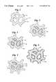
- FIG. 1is a broken-away, schematic drawing illustrating a ceramic framework useful in preparing articles of the invention
- FIG. 2is a broken-away, schematic drawing illustrating the ceramic framework of FIG. 1, the interstices of which include an osteoconductive material;
- FIG. 3is a broken-away, schematic drawing illustrating the ceramic framework of FIG. 2 showing a resilient material incorporated in the spaces between the ceramic framework and the osteoconductive material;
- FIG. 4is a broken-away, schematic drawing illustrating an embodiment of the invention.
- FIG. 5is a broken-away, schematic drawing illustrating another embodiment of the invention.
- FIG. 6is a graph of load versus strain illustrating the gradual failure mode of an article of the invention.
- FIG. 7is a broken away view of a femoral prosthesis utilizing an embodiment of the invention.
- FIG. 8is a broken away view of a tibial tray prosthesis utilizing an embodiment of the invention.
- a supportive, open frameworkhaving interstices in the size range of about 50 ⁇ m to about 1000 ⁇ m and preferably from about 200 ⁇ m to about 600 ⁇ m and having void volumes of at least about 30%, preferably at least about 50% and most preferably at least about 70%.
- the material of the frameworkmay comprise any strong, hard, biologically-compatible material such as ceramic materials, metals and composites such as zirconia, zirconia/hydroxyapatite combinations, and zirconia toughened alumina.
- the framework componentis of a ceramic material, zirconia, alumina and calcium phosphates and combinations thereof being preferred.
- a slip of ceramic materialis made by combining a ceramic powder such as zirconia with an organic binder and water to form a dispersion.
- the strut surfaces of an organic reticulated foamsuch as one of the various commercially available foams made of polyurethane, polyester, polyether, or the like are wetted and coated with the ceramic slip.
- the reticulated materialmay be immersed in the slip, and then removed and drained to remove excess slip. If desired, further excess slip can be removed by any of a variety of methods including passing the material between a pair of closely spaced rollers. By impacting the material with a jet of air, remaining slip that may fill the interstices by surface tension may be cleared.
- Varying the slip concentration, viscosity, and surface tensionprovides control over the amount of slip that is retained on the foam strut surfaces.
- Wetting agents and viscosity control agentsalso may be used for this purpose.
- a wide variety of reticulated, open cell materialscan be employed, including natural and synthetic sponge materials and woven and non-woven materials, it being necessary in this embodiment only that the open cell material enables ceramic slip material to penetrate substantially fully through the openings in the structure.
- the slip solventis removed by drying, accompanied desirably by mild heating, and the structure is then raised to sintering temperatures at which the ceramic particles at least partially sinter to form a rigid, light framework structure that mimics the configuration of the reticular struts.
- the slip-treated spongedesirably is held at a temperature at which the organic material pyrolizes or bums away, leaving behind an incompletely sintered ceramic framework structure which then is raised to the appropriate sintering temperature.
- Pyrolizing or oxidizing temperatures for most organicsare in the range of about 200° C. to about 600° C. and sintering temperatures for most ceramics of relevance to this invention are in the range of about 1100° C. to about 1600° C.
- Zirconia and alumina or composites based on zirconia and aluminaare the preferred ceramic materials for the structural elements unless the struts are also intended to be bioresorbable, in which case calcium phosphates can also be used.
- ceramic materials for the osteoconductive portioninclude calcium phosphates such as hydroxyapatite, fluorapatite, tricalcium phosphate and mixtures thereof, bioactive glasses, osteoconductive cements, and compositions containing calcium sulfate or calcium carbonate.
- FIGS. 1 through 510
- the frameworkhaving struts 12 defining open interstices 14 as shown in FIG. 1 .
- Metals which can be used to form the hard, strong, continuous framework componentinclude titanium, stainless steels, cobalt/chrome alloys, tantalum, titanium-nickel alloys such as Nitinol and other superelastic metal alloys.
- Itin, et al.“Mechanical Properties and Shape Memory of Porous Nitinol,” Materials Characterization [32] pp. 179-187 (1994); Bobyn, et al., “Bone Ingrowth Kinetics and Interface Mechanics of a Porous Tantalum Implant Material,” Transactions of the 43rd Annual Meeting, Orthopaedic Research Society, p. 758, Feb.
- Metalscan be formed into hard, strong, continuous supportive frameworks by a variety of manufacturing procedures including combustion synthesis, plating onto a “foam” substrate, chemical vapor deposition (see U.S. Pat. No. 5,282,861), lost mold techniques (see U.S. Pat. No. 3,616,841), foaming molten metal (see U.S. Pat. Nos. 5,281,251, 3,816,952 and 3,790,365) and replication of reticulated polymeric foams with a slurry of metal powder as described for ceramic powders.
- the osteoconductive and osteoinductive materials that are appropriate for use in the present inventionare biologically acceptable and include such osteoconductive materials as collagen and the various forms of calcium phosphates including hydroxyapatite; tricalcium phosphate; and fluorapatite, and such osteoinductive substances as: bone morphogenetic proteins (e.g.. rhBMP-2); demineralized bone matrix; transforming growth factors (e.g.. TGF- ⁇ ); osteoblast cells, and various other organic species known to induce bone formation.
- bone morphogenetic proteinse.g.. rhBMP-2
- demineralized bone matrixe.g.. TGF- ⁇
- osteoblast cellse.g.. TGF- ⁇
- various other organic species known to induce bone formatione.g. TGF- ⁇
- the osteoconductive and osteoinductive propertiesmay be provided by bone marrow, blood plasma, or morselized bone of the patient, or commercially available materials.
- Osteoinductive materialssuch as BMP may be applied to articles of the invention, for example, by immersing the article in an aqueous solution of this material in a dilute suspension of type I collagen.
- Osteoinductive materialssuch as TGF-p may be applied to an article of the invention from a saline solution containing an effective concentration of TGF- ⁇ , or may be carried in the resilient material.
- the continuous supporting framework having interconnecting interstices or openingsmay be considered to be the primary load bearing element, and the osteoconductive material commonly is weaker than the supporting framework.
- the supporting frameworkis preferably formed, as mentioned above, of a ceramic material such as zirconia.
- the framework structureis formed such that the interstices or openings themselves, on average, are wider than are the thicknesses of the struts which separate neighboring interstices.
- the load bearing frameworkis essentially completely continuous and self interconnected in three dimensions, and the void portion is also essentially completely continuous and self interconnected in three dimensions. These two three dimensionally interconnected parts are intercolated with one another.
- the concept of connectivityis explained at greater length in Newnham et al. “Connectivity and Piezoelectric-Pyroelectric Composites,” Materials Research Bulletin, Vol. 13 pp. 525-536 (1978), the teachings of which are incorporated herein by reference.
- the frameworkWith the supporting framework described herein, the framework itself is given a 3 as it is connected in 3 dimensions, and the void portion is treated likewise.
- partially sintered assemblages of powdersinvariably contain isolated pores or voids which are not connected to all other voids.
- a material with all isolated (that is, dead end) pores in a dense matrixwould have 3-0 connectivity.
- a material having pores that pass completely through the matrix in one dimensionwould yield 3 ⁇ 1 connectivity, and a material having pores that interconnect two perpendicular faces but not the third would have 3 ⁇ 2 connectivity.
- the voids of the frameworkinclude a three-dimensional continuous network of an osteoconductive material such as a calcium phosphate, and also a three dimensional, continuous network of a resilient, desirably bioabsorbable material between the struts of the framework and the osteoconductive material, this configuration providing 3—3 ⁇ 3 connectivity.
- an osteoconductive materialsuch as a calcium phosphate
- a resilient, desirably bioabsorbable materialbetween the struts of the framework and the osteoconductive material
- the opening sizes in the supportive frameworkpreferably are at least about 50 ⁇ m and preferably are on the order of 200 ⁇ m to about 600 ⁇ m. It is preferred that there be substantially no pores or voids less than 50 ⁇ m. It should be understood that the openings in the supportive framework are of myriad irregular shapes.
- the interconnected openings or interstices through which biological ingrowth processes can take placedefine in three dimensions a labyrinth in which bone ingrowth and vascularization can occur; that is, the openings have many junctures with other openings to thus define tortuous pathways through the framework. In general, it is believed that in order to adequately support the growth of bone into the framework openings, the openings must be capable of accommodating the passage of tissue having transverse dimensions of at least about 50 ⁇ m.
- a 50 ⁇ m opening in materials of the inventionis capable of accommodating the passage through it of a “worm” having a round cross section and a transverse diameter of 50 ⁇ m.
- a 50 ⁇ m openingshould enable passage through it of a sphere having a 50 ⁇ m diameter.
- a scanning electron micrograph of a cross section of an article of the inventionand viewing it as a planar projection of the structure, drawing several lines across the micrograph, measuring the openings that intersected by the lines, and using averaging and standard deviation techniques to permit the size of the openings to be assessed.
- Zirconia and other ceramicswhen used to form the supportive framework, are exceedingly hard and are far more rigid than is bone.
- bone substitute materials of the inventionemploying rigid materials work well. It is believed that the ultimate union of bone with such articles during the healing process occurs over a large surface area and depth as the encroaching bone penetrates into the bioabsorbable resilient material and the osteoconductive portions of the article.
- the substantial bone/ceramic interface that resultsenables forces to be readily transmitted to and from the ceramic framework with significantly less stress concentration in comparison to structure resulting from a bone/ceramic union that occurs within a small area of surface-to-surface contact and with little or no penetration of bone into the article.
- the osteoconductive material utilizedis a ceramic, e.g., a calcium phosphate
- the supportive frameworkis a ceramic such as zirconia
- several methodsmay be employed in the manufacture of the article of the invention.
- the supportive zirconia framework structurecan be fabricated as indicated above, by coating a slip of zirconia on the surface of the struts of a reticulated organic material such as a foam of polyurethane, polyester, polyether or the like, and subsequently raising the temperature of the coated foam to drive off slip solvent, to pyrolize or burn off the organic foam material, and finally to heat the ceramic to cause the ceramic particles to at least partially sinter.
- the ceramic frameworkOnce the ceramic framework has cooled, its interstices may be filled with a calcium phosphate utilizing an organic binder, and the resulting product may be sintered a second time, thus forming an included network of osteoconductive material within the interstices of the ceramic framework.
- the calcium phosphate materialAs the calcium phosphate material is heated, it shrinks so as to form an intervening space between the struts forming the ceramic framework and the included calcium phosphate network.
- the frameworkmay first be lightly coated with a release agent such as paraffin.
- FIG. 2depicts within the interstices of the supporting framework 12 the shrunken calcium phosphate material 16 and the space or gap 18 between the struts of the supporting framework and the calcium phosphate network.
- FIG. 3depicts schematically the resilient interlayer 20 formed between the framework and the calcium phosphate network.
- a second ceramic materialsuch as calcium phosphate.
- the strut surfacesmay be coated with a material such as wax to prevent the second ceramic material from bonding to the struts and to isolate the second ceramic material from the supportive framework. Since ceramic materials such as calcium phosphate shrink when they are sintered, the second material will occupy a space somewhat smaller than the space defined by the surrounding interstices of the supporting framework.
- the resulting spaces between the struts defining the interstices of the supporting framework and the calcium phosphatemay be filled with a resilient biologically acceptable material such as a copolymer of glycolic acid and L-lactic acid.
- the resulting articlethen, has a continuous strong supportive framework having struts defining a plurality of interconnecting interstices, a second framework carried within the interstices of the first framework, and a resilient interlayer between and separating the frameworks.
- the interlayerit is believed, at least partially isolates the second framework from the first and, due to its resilient nature (in comparison to the relatively rigid first and second frameworks), serves to distribute internal loads between the frameworks.
- FIG. 6illustrates a typical load-strain curve (curve A)resulting from compression testing of an article of the invention.
- curve AThe curve illustrates that the specimen did not fail catastrophically. Rather, the resilient interlayer enabled stresses within the specimen resulting from failure of portions of the framework to be distributed to other portions of the framework. Failure of the specimen was gradual, approximating the failure experienced when natural bone is similarly stressed.
- curve B in FIG. 6illustrates catastrophic failure of similar materials without resilient material present. The gradual failure mode is demonstrated also when struts are coated with resilient polymer, and there is no second framework.
- the supportive first frameworkis made of a strong material such as zirconia
- the second frameworkis of a material such as a calcium phosphate that provides osteoconductive properties, but where complete bioresorption is desired, the supportive first framework may also be a calcium phosphate composition.
- the two-part system with interconnected porescan be formed in the same manner as when the framework component is of ceramic materials, that is, the osteoconductive material may be incorporated within the struts or may be formed within the interstices of the metal struts, or foamed within the interstices and sintered, followed by infusion of the resilient interface.
- the “resilient” material referred to hereindesirably is polymeric in nature and preferably is bioresorbable.
- resilientwe refer to the ability of the material to be deformed when placed under stress without exhibiting brittle failure, the deformation tending to distribute stress within the article.
- the resilient materialalso serves to encase the struts during strut failure to provide residual compressive stiffness and to promote retention of physical integrity of the article.
- the polymeric materialis a bioresorbable polymer which may be one or a combination of: collagen, poly (lactic acid), poly (glycolic acid), copolymers of lactic acid and glycolic acid, chitin, chitosan, gelatin, or any other resorbable polymer.
- This polymer materialmay be used alone, may be reinforced with a particulate or fibrous biocompatible material, and may include one or more biological agents capable of inducing bone formation.
- Collagen and other polymeric materialsmay serve as suitable carriers of osteoinductive materials such as 13MP and various bone growth proteins.
- Bioresorbable polymeric materialswill resorb as host bone grows into the interstices to replace it.
- a hydroxyapatite slip or composite zirconia and hydroxyapatite slipmay be applied, the slip solvent driven off with heat, and the zirconia and hydroxyapatite are raised to a sintering temperature and sintered together.
- the slip of calcium phosphatemay have added to it viscosity control agents and a foaming agent such as hydrogen peroxide, or compressed gas. It may also have incorporated in it fibrous cellulosic materials.
- heatingcauses the slip to bubble and foam such that a number of smaller pores are formed in the calcium phosphate. Further heating will burn out the cellulosic materials, developing increased interconnectivity of the pores.
- the slip used to coat the polymeric reticulum and produce the ceramic reticulumcontains fractions of both the supportive framework material (such as zirconia) and the osteoconductive material (such as calcium phosphate).
- the reticulated polymeric substrateis coated with slip and the excess is allowed to drain. Further excess slip is removed by passing the article through squeeze rollers or by impacting the article with compressed air. The resulting material is heated to drive off solvent, to pyrolyze the organic constituents, and to co-sinter the two components of the composite.
- the osteoconductive material(calcium phosphate) is preferably included in a range of 10 to 90 volume percent and preferably about 10 to 25 volume percent or 75 to 90 volume percent with respect to the total zirconia/calcium phosphate volume, sufficient osteoconductive material being used so as to provide an osteoconductive surface with respect to growing bone.
- Appropriate structuresmay use, for example, 25 volume per cent of calcium phosphate and 75% of YSZ (yttria-stabilized zirconia).
- the reticulated article that resultshas struts which are comprised of an intimate mixture of the two materials.
- the calcium phosphatemay appear as very small islands on the surface of the zirconia strut.
- the osteoconductive materialremains exposed to the openings in the article so as to provide an osteoconductive effect with respect to encroaching bone.
- the supporting structurecan be 100% osteoconductive material such as a calcium phosphate.
- the bone substitute materials of the inventioncan be formed into the appropriate configurations for use as a bone substitute by several methods.
- an organic material with open intersticessuch as a reticulated polyurethane foam is simply shaped to the desired configuration using ordinary cutting instruments such as scissors, scalpels, hot wire cutters and the like.
- the configured foam materialis used in any of the foregoing methods to produce the article of the invention.
- an organic foamsuch as that referred to earlier is coated with a zirconia or other ceramic slip and is heated to drive off solvent and convert the ceramic to the “green” state, at which point it can be shaped into the desired configuration.
- a bone substitute of the invention which has been fully sinteredcan be shaped by standard machining methods such as sawing and grinding, water jet or laser cutting, etc.
- the supporting framework of the articleis of metal, it can be shaped through appropriate machining to the desired form before introducing an osteoconductive or osteoinductive material. It is contemplated that the pores of a metal material may be first filled with wax and the resulting structure frozen so that the wax supports the metal structure during machining, following which the wax is simply melted to enable the wax to escape. This procedure may have utility particularly when the metal framework component comprises a very thin walled structure with large void openings, the struts of which, accordingly, can be unintentionally easily bent.
- articles of the inventioncomprise a supporting framework with added resilient materials, the framework itself having relatively large openings and a high void volume and being attached, as by sintering to a second, denser structural element which may be of the same or different material but which has smaller openings and a smaller void volume.
- this denser portionis substantially fully dense, that is, it has a void volume less than 10%.
- the denser portionmay take the form a semitubular plate, a rod useful as a stem receivable in the intramedullary canal of a long bone for a total hip or knee replacement, or a plate useful as a tibial tray of a knee prosthesis, etc.
- the latter materialmay be formed in a thin layer relative to the first portion and the resulting structure mimics natural bone in that the second portion may be like cortical bone—the hard, dense, outer layer of a bone—whereas the first portion may be somewhat more open and porous and hence more closely resembles cancellous bone.
- FIG. 7shows a femoral hip stem prosthesis 30 made entirely of ceramic, the prosthesis having a dense stem portion 32 , an angular neck 34 terminating in an articulating ball 36 , and an angular shoulder portion 38 .
- the shoulder portionincludes a thick layer 40 of an article of the invention having a framework with relatively large openings, carried by the denser portion 42 of the prosthesis.
- the layer 40promotes bone ingrowth when the prosthesis has been implanted in the femur of a patent.
- FIG. 8depicts a tibial tray 50 having an upper plate 52 of ultra high molecular weight polyethylene having an articulating upper surface 54 .
- the ultra high molecular weight polyethylene plateis supported by a plate 56 of the dense material of the invention, the plate 56 being integrally formed with a downwardly extending stem 58 .
- the open framework material of the inventionis shown in the form of a plate 60 which is received within a downwardly open recess 62 formed in the bottom of the plate 56 , the framework 60 extending downwardly about the upper end of the stem, as shown at 64 in a relatively thick layer to promote bone ingrowth in this area.
- the dense portion of this constructcan be prepared by any of the common ceramic forming techniques such as slip casting, , tape casting, or coating and drying successive layers of slip onto a surface of a “foam” until a dense layer is formed. Dry pressing, injection molding and extrusion techniques may also be appropriate.
- the “green” dense portionis joined to the “green” low density portion through the use of a ceramic slip of substantially similar composition to the slip used in the formation of the low density portion or of a substantially similar composition to the slip used in the formation of the dense portion in the case of slip cast dense portion.
- Greenhere refers to the state of a ceramic article which has been formed and dried to a self-supporting structure but from which the organic constituents have not yet been removed.
- the dense portionmay be alternatively comprised of a resorbable polymeric material, a resorbable ceramic material, or a resorbable composite material in addition to materials enumerated above.
- the above descriptionhas centered upon completely formed bone substitute articles having a supporting, open framework, an osteoconductive material generally coextensive with and contained within the supporting framework, and a resilient, preferably bioresorbable polymer between the supporting framework and the osteoconductive material.
- the osteoconductive materialneed not be continuous within the interstices of the supporting framework.
- the osteoconductive materialmay instead be particulate, as shown at 22 in FIG. 5, and may be carried by or embedded in the resilient material 20 .
- the inventionalso relates to the embodiment illustrated in FIG. 4 in which the interstices of the supportive framework as described above, the interstices of which are coated with a resilient, desirably bioresorbable material, the coated interstices 24 opening onto surfaces of the article.
- the coated intersticesmay be filled with a calcium phosphate cement during a surgical procedure.
- the calcium phosphate cementhardens within the interstices and the resilient material separating the supportive framework from the hardened calcium phosphate cement acts to distribute forces that are generated by exterior loads on the framework.
- the supporting open frameworkmay alternatively be coated with resilient material with the interstices not being filled.
- a zirconia slipmay be prepared by combining the following ingredients and mixing them thoroughly by ball milling in a polyethylene container using zirconia media:150 grams partially stabilized zirconia powder (Zirconia Sales America)
- Pieces of reticulated polyester-polyurethane foam 10-80 pores per inchare immersed in the above slip and repeatedly compressed to remove air bubbles trapped inside.
- the foamsare removed from the slip and the excess slip is allowed to drain. Further excess slip is removed by passing the foams between a pair of stainless steel squeeze rollers several times. Passages are also cleared by blowing air through them.
- the resulting piecesare allowed to dry at room temperature followed by drying at temperatures up to 100° C. in air. When the pieces appear dry, they are heated to pyrolyze and remove organics (binder, dispersant, surfactant, anti-foam agent, and reticulated polymer foam) and then are sintered at a temperature of about 1400° C. for one hour.
- the preferred thermal cycle for the aboveinvolves raising the temperature of the pieces at the rate of 2° C. per minute to 600° C., holding the temperature at 600° C. for two hours, and then raising the temperature at the rate of 5° C. per minute to 1400° C., with a one hour hold at this temperature.
- the furnaceis then cooled to room temperature at a rate of about 10° C. per minute.
- the resulting productis a strong, light weight, porous zirconia framework or reticulum of zirconia having a void volume of about 76%.
- An injectable calcium phosphate pasteis made by combining and mixing the following:
- the pasteis injected into the interstices of the zirconia framework and allowed to dry at 60° C. in air.
- the articleis then sintered in nitrogen to 1300° C. for 1 hour.
- the resulting producthas two intertwined networks of zirconia and calcium phosphate with a space at their interface.
- a gel of collagen, type 1is made by mixing 20 parts of 50 mM acetic acid with 1 part collagen and stir blending. To this is added an equal volume of 4% chitosan solution in dilute acetic acid. This mixture is forced under pressure into the space between the intertwined networks, and upon drying forms a collagen Ichitosan resilient interlayer between these networks.
- Example Iis repeated except that the interface space is filled with a thin paste of a copolymer of glycolic acid and lactic acid (Alkermes “Medisorb” 85/15 PGA/PLLA) in ethyl acetate, mixed with an equal volume of collagen gel referred to in example I.
- the solventis allowed to evaporate to form a resilient interlayer between these networks.
- this processmay be repeated to build the polymer interface.
- Curve B in FIG. 6illustrates the brittle failure of the same product without the addition of the resilient interlayer.
- a zirconia frameworkis made as in Example I without the subsequent wax coating.
- the intersticesare filled with a paste made of a suspension of Type 1 collagen in 50 mM acetic acid in a ratio of 1 part collagen to 20 parts acid in which calcium deficient hydroxyapatite crystals are grown (according to a process described by TenHuisen et al., J. Biomed. Materials Res. Vol. 29, pp. 803-810 (1995), which is incorporated herein by reference) from precursors tetracalcium phosphate (CA 4 (PO 4 ) 2 O and monetite (CaBPO 4 ), to provide an article similar to that illustrated in FIG. 5 .
- CA 4 (PO 4 ) 2 Oprecursors tetracalcium phosphate
- CaBPO 4monetite
- a zirconia/hydroxyapatite composite frameworkwas made as in Example 1 with 25 volume percent hydroxyapatite without the subsequent wax coating.
- the strutswere coated with the solution of a copolymer of glycolic acid and lactic acid(Alkermes “Medisorb 75/25 PLLA/PGA) to provide a coating approximately 15 ⁇ m in thickness.
- the interstices of the articleare injected with calcium phosphate cement paste such as made by a process (described by Constantz, et al. SCIENCE, Vol. 267 (1995), the teachings of which are incorporated herein by reference), the paste comprising a mixture of monocalcium phosphate monohydrate, calcium phosphate and calcium carbonate in a sodium phosphate solution.
- the hardened pasteprovides a biomaterial suitable for implants that is now strengthened by the skeleton framework with its resilient interface.
- a calcium phosphate frameworkwas made by coating a reticulated polyester-polyurethane foam with a slip of calcium phosphate as described in Example I. The resulting pieces were dried at up to 100° C. in air. Following drying the pieces were heated to pyrolyze and remove organics and were sintered at a temperature of about 1300° C. in nitrogen for one hour. The resulting calcium phosphate framework was then coated with a solution of a copolymer of lactic acid and glycolic acid(Alkermes “Medisorb 75/25 PLLA/PGA) in methylene chloride. The solvent was removed by vacuum.
- a supportive frameworkwas made as in Example I but utilizing a slip of zirconia and hydroxyapatite.
- the strutswere coated, and the interstices partially filled with a solution of a copolymer of glycolic acid and lactic acid in methylene chloride. Following coating, the solvent was removed by vacuum.
- the “green” ceramic reticulum formed as in Example I before sinteringwas wetted with a slip containing zirconia of the same composition, binders and zirconia powders, and was adhered to a green zirconia ceramic of the same composition made by a conventional ceramic slip casting process by sintering, the latter material simulating curved cortical bone and the reticulum simulating attached cancellous bone.
- the struts of the reticular portionare then coated with a copolymer of glycolic acid and lactic acid as in example II.
Landscapes
- Health & Medical Sciences (AREA)
- Chemical & Material Sciences (AREA)
- Life Sciences & Earth Sciences (AREA)
- Veterinary Medicine (AREA)
- Public Health (AREA)
- General Health & Medical Sciences (AREA)
- Animal Behavior & Ethology (AREA)
- Oral & Maxillofacial Surgery (AREA)
- Transplantation (AREA)
- Engineering & Computer Science (AREA)
- Dermatology (AREA)
- Epidemiology (AREA)
- Medicinal Chemistry (AREA)
- Cardiology (AREA)
- Composite Materials (AREA)
- Inorganic Chemistry (AREA)
- Orthopedic Medicine & Surgery (AREA)
- Materials Engineering (AREA)
- Biomedical Technology (AREA)
- Heart & Thoracic Surgery (AREA)
- Vascular Medicine (AREA)
- Dispersion Chemistry (AREA)
- Materials For Medical Uses (AREA)
- Prostheses (AREA)
Abstract
Description
| 29 | grams | calcium phosphate (powder) |
| 3.5 | grams | polyethylene oxide binder |
| 2 | grams | dispersant (Darvan ™ C, R. T. Vanderbilt) |
| 3 | drops | thickening agent (Rohm and Haas T-5000) |
| 2 | drops | anti-foaming agent (Henkel Nopco ™ NXZ) |
| 30 | ml | deionized water |
Claims (32)
Priority Applications (10)
| Application Number | Priority Date | Filing Date | Title |
|---|---|---|---|
| US08/944,006US6296667B1 (en) | 1997-10-01 | 1997-10-01 | Bone substitutes |
| CN98811740ACN1280508A (en) | 1997-10-01 | 1998-09-30 | bone substitute |
| CA002305430ACA2305430C (en) | 1997-10-01 | 1998-09-30 | Bone substitutes |
| DE69825911TDE69825911T2 (en) | 1997-10-01 | 1998-09-30 | Knoch SPARE MATERIAL |
| PCT/US1998/020440WO1999016478A1 (en) | 1997-10-01 | 1998-09-30 | Bone substitutes |
| EP98950771AEP1024841B1 (en) | 1997-10-01 | 1998-09-30 | Bone substitutes |
| AU96736/98AAU754630B2 (en) | 1997-10-01 | 1998-09-30 | Bone substitutes |
| JP2000513610AJP2001518321A (en) | 1997-10-01 | 1998-09-30 | Bone substitute |
| TW087116343ATW482688B (en) | 1997-10-01 | 1998-10-01 | Strong article useful as a bone substitute material, and method of producing same |
| US09/740,807US6527810B2 (en) | 1997-10-01 | 2000-12-21 | Bone substitutes |
Applications Claiming Priority (1)
| Application Number | Priority Date | Filing Date | Title |
|---|---|---|---|
| US08/944,006US6296667B1 (en) | 1997-10-01 | 1997-10-01 | Bone substitutes |
Related Child Applications (1)
| Application Number | Title | Priority Date | Filing Date |
|---|---|---|---|
| US09/740,807DivisionUS6527810B2 (en) | 1997-10-01 | 2000-12-21 | Bone substitutes |
Publications (1)
| Publication Number | Publication Date |
|---|---|
| US6296667B1true US6296667B1 (en) | 2001-10-02 |
Family
ID=25480617
Family Applications (2)
| Application Number | Title | Priority Date | Filing Date |
|---|---|---|---|
| US08/944,006Expired - LifetimeUS6296667B1 (en) | 1997-10-01 | 1997-10-01 | Bone substitutes |
| US09/740,807Expired - LifetimeUS6527810B2 (en) | 1997-10-01 | 2000-12-21 | Bone substitutes |
Family Applications After (1)
| Application Number | Title | Priority Date | Filing Date |
|---|---|---|---|
| US09/740,807Expired - LifetimeUS6527810B2 (en) | 1997-10-01 | 2000-12-21 | Bone substitutes |
Country Status (9)
| Country | Link |
|---|---|
| US (2) | US6296667B1 (en) |
| EP (1) | EP1024841B1 (en) |
| JP (1) | JP2001518321A (en) |
| CN (1) | CN1280508A (en) |
| AU (1) | AU754630B2 (en) |
| CA (1) | CA2305430C (en) |
| DE (1) | DE69825911T2 (en) |
| TW (1) | TW482688B (en) |
| WO (1) | WO1999016478A1 (en) |
Cited By (115)
| Publication number | Priority date | Publication date | Assignee | Title |
|---|---|---|---|---|
| EP1161257A2 (en) | 1999-03-17 | 2001-12-12 | Novartis AG | Pharmaceutical compositions comprising tgf-beta |
| WO2002083188A2 (en) | 2001-04-16 | 2002-10-24 | Cassidy James J | Dense/porous structures for use as bone substitutes |
| US20020160109A1 (en)* | 2000-12-13 | 2002-10-31 | Yoon Yeo | Microencapsulation of drugs by solvent exchange |
| US20020173850A1 (en)* | 2001-05-01 | 2002-11-21 | Amedica Corporation | Radiolucent spinal fusion cage |
| US20030065400A1 (en)* | 2001-04-12 | 2003-04-03 | Beam Heather Ann | Method and apparatus for engineered regenrative biostructures such as hydroxyapatite substrates for bone healing applications |
| US20030114936A1 (en)* | 1998-10-12 | 2003-06-19 | Therics, Inc. | Complex three-dimensional composite scaffold resistant to delimination |
| US20030213090A1 (en)* | 2002-05-14 | 2003-11-20 | Emerson Electric Co. | Detachable accessory holder |
| US20030236573A1 (en)* | 2002-06-13 | 2003-12-25 | Evans Douglas G. | Devices and methods for treating defects in the tissue of a living being |
| US20040013556A1 (en)* | 2000-12-08 | 2004-01-22 | Jean-Francois Silvain | Method for making thin films in metal/ceramic composite |
| US20040019132A1 (en)* | 2001-02-23 | 2004-01-29 | Marc Long | Bone graft substitutes |
| US6743256B2 (en) | 2000-10-11 | 2004-06-01 | Michael D. Mason | Graftless spinal fusion device |
| US20040236429A1 (en)* | 2002-03-13 | 2004-11-25 | Ortho Development Corporation | Method of forming a temporary prosthetic joint using a disposable mold |
| US20040243246A1 (en)* | 2003-05-27 | 2004-12-02 | Lyren Philip S. | Hip implant with porous body |
| US20050031578A1 (en)* | 2002-03-22 | 2005-02-10 | Doctor's Research Group, Inc. | Methods of performing medical procedures which promote bone growth, compositions which promote bone growth, and methods of making such compositions |
| US20050048193A1 (en)* | 2001-02-19 | 2005-03-03 | Isotis N.V. | Porous metals and metal coatings for implants |
| US20050075408A1 (en)* | 1994-05-13 | 2005-04-07 | Ringeisen Timothy A. | Method for making a porous polymeric material |
| US20050158535A1 (en)* | 2003-05-15 | 2005-07-21 | Miqin Zhang | Methods for making porous ceramic structures |
| US20050161120A1 (en)* | 2003-06-13 | 2005-07-28 | Yukiharu Hasegawa | Orthopedic and dental endosseous implants and their preparation method |
| US20050177237A1 (en)* | 2001-04-12 | 2005-08-11 | Ben Shappley | Spinal cage insert, filler piece and method of manufacturing |
| US20050177118A1 (en)* | 1994-05-13 | 2005-08-11 | Hoganson David M. | Resorbable polymeric device for localized drug delivery |
| US6946013B2 (en) | 2002-10-28 | 2005-09-20 | Geo2 Technologies, Inc. | Ceramic exhaust filter |
| US6949251B2 (en) | 2001-03-02 | 2005-09-27 | Stryker Corporation | Porous β-tricalcium phosphate granules for regeneration of bone tissue |
| US20050220771A1 (en)* | 2002-03-22 | 2005-10-06 | Doctor's Research Group, Inc. | Methods of performing medical procedures that promote bone growth, methods of making compositions that promote bone growth, and apparatus for use in such methods |
| US20050228503A1 (en)* | 2002-04-11 | 2005-10-13 | Ferdinand Gundolf | Device for stimulating bone growth, especially for the osteosynthesis of bone fragments and/or for fixing bone fractures |
| US20050240281A1 (en)* | 1997-05-30 | 2005-10-27 | Slivka Michael A | Fiber-reinforced, porous, biodegradable implant device |
| US20050273176A1 (en)* | 2001-05-01 | 2005-12-08 | Amedica Corporation | Hip prosthesis with monoblock ceramic acetabular cup |
| US20060004168A1 (en)* | 2004-06-30 | 2006-01-05 | Keith Greer | Low crystalline polymeric material for orthopaedic implants and an associated method |
| EP1623685A1 (en)* | 2004-08-06 | 2006-02-08 | WALDEMAR LINK GmbH & Co. KG | Hip prosthesis with stem for implantation into the femur |
| US20060064070A1 (en)* | 2004-09-07 | 2006-03-23 | Jeffrey Martin | Methods and devices for sterile field transfer |
| US7045141B2 (en) | 1998-02-27 | 2006-05-16 | Musculoskeletal Transplant Foundation | Allograft bone composition having a gelatin binder |
| US20060121084A1 (en)* | 2004-12-08 | 2006-06-08 | Borden Mark D | Continuous phase composite for musculoskeletal repair |
| US20060142867A1 (en)* | 2001-02-23 | 2006-06-29 | Robert G. Metzger Et Al. | Knee joint prosthesis |
| US20060142869A1 (en)* | 2004-12-23 | 2006-06-29 | Gross Thomas P | Knee prosthesis |
| US20060198939A1 (en)* | 2001-09-24 | 2006-09-07 | Smith Timothy J | Porous ceramic composite bone grafts |
| US20070078554A1 (en)* | 1995-02-14 | 2007-04-05 | St Ville James A | Method and apparatus for manufacturing objects having optimized response characteristics |
| US20070075450A1 (en)* | 2005-10-04 | 2007-04-05 | Aztec Ip Company Llc | Parametrized material and performance properties based on virtual testing |
| US7203628B1 (en)* | 1999-08-23 | 2007-04-10 | St Ville James A | Manufacturing system and method |
| US7208222B2 (en) | 2003-07-24 | 2007-04-24 | Viasys Healthcare Inc. | Assembled non-random foams |
| US7211232B1 (en) | 2005-11-07 | 2007-05-01 | Geo2 Technologies, Inc. | Refractory exhaust filtering method and apparatus |
| US7250550B2 (en) | 2004-10-22 | 2007-07-31 | Wright Medical Technology, Inc. | Synthetic bone substitute material |
| US20070190101A1 (en)* | 2004-03-31 | 2007-08-16 | Chunlin Yang | Flowable bone grafts |
| US20070255392A1 (en)* | 2006-04-27 | 2007-11-01 | Phillips Plastics Corporation | Composite stent |
| US20080077222A1 (en)* | 2006-04-27 | 2008-03-27 | Phillips Plastics Corporation | Componsite stent |
| US7444805B2 (en) | 2005-12-30 | 2008-11-04 | Geo2 Technologies, Inc. | Substantially fibrous refractory device for cleaning a fluid |
| US7497874B1 (en) | 2001-02-23 | 2009-03-03 | Biomet Manufacturing Corp. | Knee joint prosthesis |
| US7563415B2 (en) | 2006-03-03 | 2009-07-21 | Geo2 Technologies, Inc | Catalytic exhaust filter device |
| US7572311B2 (en) | 2002-10-28 | 2009-08-11 | Geo2 Technologies, Inc. | Highly porous mullite particulate filter substrate |
| US7574796B2 (en) | 2002-10-28 | 2009-08-18 | Geo2 Technologies, Inc. | Nonwoven composites and related products and methods |
| US20090216325A1 (en)* | 2008-02-27 | 2009-08-27 | Biomet Manufacturing Corp. | Method And Apparatus For Providing Resorbable Fixation Of Press-Fit Implants |
| US7582270B2 (en) | 2002-10-28 | 2009-09-01 | Geo2 Technologies, Inc. | Multi-functional substantially fibrous mullite filtration substrates and devices |
| US7597715B2 (en) | 2005-04-21 | 2009-10-06 | Biomet Manufacturing Corp. | Method and apparatus for use of porous implants |
| US20090269388A1 (en)* | 2002-05-20 | 2009-10-29 | Musculoskeletal Transplant Foundation | Allograft bone composition having a gelatin binder |
| US7635447B2 (en) | 2006-02-17 | 2009-12-22 | Biomet Manufacturing Corp. | Method and apparatus for forming porous metal implants |
| US20100016985A1 (en)* | 2008-07-18 | 2010-01-21 | North Carolina State University | Processing of biocompatible coating on polymeric implants |
| US7666229B2 (en) | 2001-06-14 | 2010-02-23 | Amedica Corporation | Ceramic-ceramic articulation surface implants |
| US7682577B2 (en) | 2005-11-07 | 2010-03-23 | Geo2 Technologies, Inc. | Catalytic exhaust device for simplified installation or replacement |
| US7682578B2 (en) | 2005-11-07 | 2010-03-23 | Geo2 Technologies, Inc. | Device for catalytically reducing exhaust |
| US20100098673A1 (en)* | 2006-08-03 | 2010-04-22 | Ebi, Llc | Bone graft composites and methods of treating bone defects |
| US20100125303A1 (en)* | 2008-11-20 | 2010-05-20 | Daley Robert J | Methods and apparatus for replacing biological joints using bone mineral substance in a suspended state |
| US20100125335A1 (en)* | 2008-11-20 | 2010-05-20 | Daley Robert J | Methods and apparatus for replacing biological joints using bone cement in a suspended state |
| US7722828B2 (en) | 2005-12-30 | 2010-05-25 | Geo2 Technologies, Inc. | Catalytic fibrous exhaust system and method for catalyzing an exhaust gas |
| US7740794B1 (en) | 2005-04-18 | 2010-06-22 | Biomet Sports Medicine, Llc | Methods of making a polymer and ceramic composite |
| US7754246B2 (en) | 2005-09-09 | 2010-07-13 | Wright Medical Technology, Inc. | Composite bone graft substitute cement and articles produced therefrom |
| US7758646B2 (en) | 2002-12-17 | 2010-07-20 | Amedica Corporation | Total disc implant |
| US7776085B2 (en) | 2001-05-01 | 2010-08-17 | Amedica Corporation | Knee prosthesis with ceramic tibial component |
| US20100211183A1 (en)* | 2005-08-26 | 2010-08-19 | Charles Chi | Chemical Mechanical Vapor Deposition Device for Production of Bone Substitute Material |
| US7815826B2 (en) | 2004-05-12 | 2010-10-19 | Massachusetts Institute Of Technology | Manufacturing process, such as three-dimensional printing, including solvent vapor filming and the like |
| US20110021427A1 (en)* | 2009-07-27 | 2011-01-27 | Warsaw Orthopedic, Inc. | Biphasic calcium phosphate cement for drug delivery |
| US7879275B2 (en) | 2004-12-30 | 2011-02-01 | Depuy Products, Inc. | Orthopaedic bearing and method for making the same |
| US7883653B2 (en) | 2004-12-30 | 2011-02-08 | Depuy Products, Inc. | Method of making an implantable orthopaedic bearing |
| US7896921B2 (en) | 2004-12-30 | 2011-03-01 | Depuy Products, Inc. | Orthopaedic bearing and method for making the same |
| US20110106272A1 (en)* | 2009-07-10 | 2011-05-05 | Bio2 Technologies, Inc. | Devices and Methods for Tissue Engineering |
| US20110112655A1 (en)* | 1994-05-13 | 2011-05-12 | Brekke John H | Device for regeneration of articular cartilage and other tissue |
| US20110202141A1 (en)* | 2010-02-18 | 2011-08-18 | Biomet Manufacturing Corp. | Method and apparatus for augmenting bone defects |
| US20110206828A1 (en)* | 2009-07-10 | 2011-08-25 | Bio2 Technologies, Inc. | Devices and Methods for Tissue Engineering |
| US8021432B2 (en) | 2005-12-05 | 2011-09-20 | Biomet Manufacturing Corp. | Apparatus for use of porous implants |
| US8025903B2 (en) | 2005-09-09 | 2011-09-27 | Wright Medical Technology, Inc. | Composite bone graft substitute cement and articles produced therefrom |
| US8066778B2 (en) | 2005-04-21 | 2011-11-29 | Biomet Manufacturing Corp. | Porous metal cup with cobalt bearing surface |
| US8123814B2 (en) | 2001-02-23 | 2012-02-28 | Biomet Manufacturing Corp. | Method and appartus for acetabular reconstruction |
| US8137293B2 (en) | 2009-11-17 | 2012-03-20 | Boston Scientific Scimed, Inc. | Guidewires including a porous nickel-titanium alloy |
| US8157869B2 (en) | 2007-01-10 | 2012-04-17 | Biomet Manufacturing Corp. | Knee joint prosthesis system and method for implantation |
| US8163028B2 (en) | 2007-01-10 | 2012-04-24 | Biomet Manufacturing Corp. | Knee joint prosthesis system and method for implantation |
| US8187280B2 (en) | 2007-10-10 | 2012-05-29 | Biomet Manufacturing Corp. | Knee joint prosthesis system and method for implantation |
| US8221504B2 (en) | 2005-02-23 | 2012-07-17 | Wright Medical Technology, Inc. | Coating an implant for increased bone in-growth |
| US8252058B2 (en) | 2006-02-16 | 2012-08-28 | Amedica Corporation | Spinal implant with elliptical articulatory interface |
| US8266780B2 (en) | 2005-04-21 | 2012-09-18 | Biomet Manufacturing Corp. | Method and apparatus for use of porous implants |
| US8292967B2 (en) | 2005-04-21 | 2012-10-23 | Biomet Manufacturing Corp. | Method and apparatus for use of porous implants |
| US8308340B2 (en) | 2004-11-23 | 2012-11-13 | Smith & Nephew, Inc. | Composite mixer |
| US8328873B2 (en) | 2007-01-10 | 2012-12-11 | Biomet Manufacturing Corp. | Knee joint prosthesis system and method for implantation |
| US20130006354A1 (en)* | 2010-02-26 | 2013-01-03 | Limacorporate Spa | Integrated prosthetic element |
| US8535357B2 (en) | 2004-12-09 | 2013-09-17 | Biomet Sports Medicine, Llc | Continuous phase compositions for ACL repair |
| US8562616B2 (en) | 2007-10-10 | 2013-10-22 | Biomet Manufacturing, Llc | Knee joint prosthesis system and method for implantation |
| US8613938B2 (en) | 2010-11-15 | 2013-12-24 | Zimmer Orthobiologics, Inc. | Bone void fillers |
| US20140018907A1 (en)* | 2007-06-29 | 2014-01-16 | Abbott Cardiovascular Systems Inc. | Polymer-bioceramic composite implantable medical device with different types of bioceramic particles |
| US20140025179A1 (en)* | 2012-07-20 | 2014-01-23 | Ultramet | Brittle biocompatible composites and methods |
| US8690874B2 (en) | 2000-12-22 | 2014-04-08 | Zimmer Orthobiologics, Inc. | Composition and process for bone growth and repair |
| US8742072B2 (en) | 2006-12-21 | 2014-06-03 | Zimmer Orthobiologics, Inc. | Bone growth particles and osteoinductive composition thereof |
| US8765189B2 (en) | 2011-05-13 | 2014-07-01 | Howmedica Osteonic Corp. | Organophosphorous and multivalent metal compound compositions and methods |
| US8821582B1 (en)* | 2003-05-27 | 2014-09-02 | Philip Scott Lyren | Hip implant with porous body |
| US20140309746A1 (en)* | 2007-10-29 | 2014-10-16 | Zimmer, Inc | Medical implants and methods for delivering biologically active agents |
| WO2015089373A1 (en) | 2013-12-13 | 2015-06-18 | Wright Medical Technology, Inc. | Multiphasic bone graft substitute material |
| US20150190969A1 (en)* | 2012-08-03 | 2015-07-09 | Nobel Biocare Services Ag | Bone regeneration material |
| US9265857B2 (en) | 2010-05-11 | 2016-02-23 | Howmedica Osteonics Corp. | Organophosphorous, multivalent metal compounds, and polymer adhesive interpenetrating network compositions and methods |
| US20160199532A1 (en)* | 2012-01-31 | 2016-07-14 | The University Of Toledo | Injectable, Biodegradable Bone Cements and Methods of Making and Using Same |
| US9550012B2 (en) | 2013-08-05 | 2017-01-24 | University Of Notre Dame Du Lac | Tissue scaffolds having bone growth factors |
| US9775721B2 (en) | 2009-07-10 | 2017-10-03 | Bio2 Technologies, Inc. | Resorbable interbody device |
| US9782260B1 (en)* | 2014-01-29 | 2017-10-10 | Lucas Anissian | Materials and methods for prevention of cold welding, corrosion and tissue overgrowth between medical implant components |
| US10213310B2 (en)* | 2012-06-18 | 2019-02-26 | DePuy Synthes Products, Inc. | Dual modulus hip stem and method of making the same |
| US10213314B2 (en) | 2013-03-14 | 2019-02-26 | DePuy Synthes Products, Inc. | Femoral component for an implantable hip prosthesis |
| CN109865157A (en)* | 2017-12-05 | 2019-06-11 | 辽宁法库陶瓷工程技术研究中心 | A kind of preparation method based on photocuring 3D printing ceramics bone frame |
| US10433947B2 (en) | 2011-02-11 | 2019-10-08 | Khourigotti, Llc | Method and devices for tissue expansion |
| US20220109096A1 (en)* | 2019-06-28 | 2022-04-07 | Fujifilm Corporation | Piezoelectric film |
| US12016977B2 (en)* | 2017-10-06 | 2024-06-25 | Dsm Ip Assets B.V. | Method of making an osteoconductive fibrous article and a medical implant comprising such osteoconductive fibrous article |
| US12186000B2 (en) | 2020-09-04 | 2025-01-07 | DePuy Synthes Products, Inc. | Temporary antimicrobial cement spacer, assembly, kit, and method of manufacture |
| US12343449B2 (en) | 2018-03-06 | 2025-07-01 | Dsm Ip Assets B.V. | Osteoconductive fibers, medical implant comprising such osteoconductive fibers, and methods of making |
Families Citing this family (73)
| Publication number | Priority date | Publication date | Assignee | Title |
|---|---|---|---|---|
| US6977095B1 (en) | 1997-10-01 | 2005-12-20 | Wright Medical Technology Inc. | Process for producing rigid reticulated articles |
| US6736849B2 (en)* | 1998-03-11 | 2004-05-18 | Depuy Products, Inc. | Surface-mineralized spinal implants |
| SE515227C2 (en)* | 1999-04-28 | 2001-07-02 | Bruce Medical Ab | Body for providing and growing bone and / or connective tissue and methods for making the body |
| AU783249B2 (en)* | 1999-11-15 | 2005-10-06 | Cerabio, Llc | Process for producing rigid reticulated articles |
| AU2001275715B2 (en)* | 2000-08-04 | 2006-08-03 | Orthogem Limited | Porous synthetic bone graft and method of manufacture thereof |
| GB0020610D0 (en)* | 2000-08-21 | 2000-10-11 | Dytech Corp Ltd | Uses of porous carriers |
| NL1016040C2 (en)* | 2000-08-29 | 2002-03-01 | Giles William Melsom | Porous cell attachment material, method for its manufacture, and applications. |
| JP2002306518A (en)* | 2000-12-21 | 2002-10-22 | Yuichi Mori | Indwelling implement |
| US6743232B2 (en) | 2001-02-26 | 2004-06-01 | David W. Overaker | Tissue scaffold anchor for cartilage repair |
| US6575986B2 (en) | 2001-02-26 | 2003-06-10 | Ethicon, Inc. | Scaffold fixation device for use in articular cartilage repair |
| US20050177238A1 (en)* | 2001-05-01 | 2005-08-11 | Khandkar Ashok C. | Radiolucent bone graft |
| US6626950B2 (en) | 2001-06-28 | 2003-09-30 | Ethicon, Inc. | Composite scaffold with post anchor for the repair and regeneration of tissue |
| SE519564C2 (en)* | 2001-07-04 | 2003-03-11 | Nobel Biocare Ab | Implants, for example dental implants, coated with bone growth stimulants |
| US6494916B1 (en)* | 2001-07-30 | 2002-12-17 | Biomed Solutions, Llc | Apparatus for replacing musculo-skeletal parts |
| US20030045942A1 (en)* | 2001-09-05 | 2003-03-06 | Lai Wen-Fu T. | Regenerative bone implants |
| WO2003070084A2 (en)* | 2002-02-19 | 2003-08-28 | Bioarttis, Inc. | Artificial vessel scaffold and artificial organs therefrom |
| DE10243132B4 (en)* | 2002-09-17 | 2006-09-14 | Biocer Entwicklungs Gmbh | Anti-infective, biocompatible titanium oxide coatings for implants and methods of making them |
| US7309361B2 (en)* | 2002-10-23 | 2007-12-18 | Wasielewski Ray C | Biologic modular tibial and femoral component augments for use with total knee arthroplasty |
| US20050251267A1 (en)* | 2004-05-04 | 2005-11-10 | John Winterbottom | Cell permeable structural implant |
| US20060122708A1 (en)* | 2003-01-10 | 2006-06-08 | Takashi Nakamura | Osteoinductive artificial bone and manufacturing method thereof |
| US6993406B1 (en) | 2003-04-24 | 2006-01-31 | Sandia Corporation | Method for making a bio-compatible scaffold |
| WO2005034818A1 (en)* | 2003-10-09 | 2005-04-21 | B.I.Tec Ltd. | Cementless artificial joint system using composite material |
| GB0325647D0 (en)* | 2003-11-03 | 2003-12-10 | Finsbury Dev Ltd | Prosthetic implant |
| US20080234833A1 (en)* | 2004-03-23 | 2008-09-25 | B.I. Tec Ltd | Method of Designing and Manufacturing Artificial Joint Stem with Use of Composite Material |
| US20060110422A1 (en)* | 2004-11-19 | 2006-05-25 | Tas Ahmet C | Conversion of calcite powders into macro- and microporous calcium phosphate scaffolds for medical applications |
| US20070038303A1 (en)* | 2006-08-15 | 2007-02-15 | Ebi, L.P. | Foot/ankle implant and associated method |
| US20090216334A1 (en)* | 2005-02-23 | 2009-08-27 | Small Bone Innovations, Inc. | Bone Implants |
| MX2008000131A (en)* | 2005-07-01 | 2008-04-04 | Cinv Ag | Medical devices comprising a reticulated composite material. |
| DE102005037141A1 (en)* | 2005-08-06 | 2007-02-08 | Syntan Gbr(vertretungsberechtigter Gesellschafter Hr. Dr. Dieter Girlich, 01309 Dresden) | Spongy-metallic implant and method for its production |
| EP1951156A2 (en)* | 2005-11-18 | 2008-08-06 | Ceramatec, Inc. | Porous, load-bearing, ceramic or metal implant |
| CA2637616A1 (en) | 2006-01-19 | 2007-07-26 | Osteotech, Inc. | Injectable and moldable bone substitute materials |
| US20070198093A1 (en)* | 2006-02-17 | 2007-08-23 | Amedica Corporation | Spinal implant with offset keels |
| WO2007130648A2 (en)* | 2006-05-05 | 2007-11-15 | Ceramatec, Inc. | Fully or partially bioresorbable orthopedic implant |
| US20070299520A1 (en)* | 2006-06-26 | 2007-12-27 | Warsaw Orthopedic, Inc. | Surface treatment of implantable devices |
| US20100016981A1 (en)* | 2006-07-24 | 2010-01-21 | Advanced Surgical Design & Manufacture Limited | Bone reinforcement device |
| AU2008206953A1 (en)* | 2007-01-19 | 2008-07-24 | Cinvention Ag | Porous, non-degradable implant made by powder molding |
| US20080221688A1 (en)* | 2007-03-09 | 2008-09-11 | Warsaw Orthopedic, Inc. | Method of Maintaining Fatigue Performance In A Bone-Engaging Implant |
| US20080221681A1 (en)* | 2007-03-09 | 2008-09-11 | Warsaw Orthopedic, Inc. | Methods for Improving Fatigue Performance of Implants With Osteointegrating Coatings |
| US20080233203A1 (en) | 2007-03-21 | 2008-09-25 | Jennifer Woodell-May | Porous orthapedic materials coated with demineralized bone matrix |
| US8062378B2 (en) | 2007-03-26 | 2011-11-22 | Mx Orthopedics Corp. | Proximal self-locking long bone prosthesis |
| US8062364B1 (en) | 2007-04-27 | 2011-11-22 | Knee Creations, Llc | Osteoarthritis treatment and device |
| CN101842062B (en)* | 2007-09-25 | 2013-04-03 | 拜欧米特制造公司 | Method for manufacturing cementless tibial tray |
| CA2711847C (en) | 2008-01-09 | 2018-05-08 | Innovative Health Technologies, Llc | Implant pellets and methods for performing bone augmentation and preservation |
| US20090248162A1 (en)* | 2008-03-25 | 2009-10-01 | Warsaw Orthopedic, Inc. | Microparticle delivery syringe and needle for placing suspensions and removing vehicle fluid |
| US8470040B2 (en)* | 2008-04-02 | 2013-06-25 | Pioneer Surgical Technology, Inc. | Intervertebral implant devices for supporting vertebrae and devices and methods for insertion thereof |
| US20090304775A1 (en)* | 2008-06-04 | 2009-12-10 | Joshi Ashok V | Drug-Exuding Orthopedic Implant |
| JP5774989B2 (en) | 2008-08-13 | 2015-09-09 | スメド−ティーエイ/ティーディー・エルエルシー | Orthopedic screw |
| US10842645B2 (en) | 2008-08-13 | 2020-11-24 | Smed-Ta/Td, Llc | Orthopaedic implant with porous structural member |
| US9700431B2 (en) | 2008-08-13 | 2017-07-11 | Smed-Ta/Td, Llc | Orthopaedic implant with porous structural member |
| US9616205B2 (en) | 2008-08-13 | 2017-04-11 | Smed-Ta/Td, Llc | Drug delivery implants |
| US20100042213A1 (en) | 2008-08-13 | 2010-02-18 | Nebosky Paul S | Drug delivery implants |
| US20100047434A1 (en)* | 2008-08-21 | 2010-02-25 | Biomet Manufacturing Corp. | Fabrication of monolithic zones on porous scaffold |
| WO2010025386A1 (en) | 2008-08-29 | 2010-03-04 | Smed-Ta/Td, Llc | Orthopaedic implant |
| CN102573938B (en)* | 2009-07-23 | 2015-08-05 | 布若贞提克斯骨生物学有限公司 | injectable and mouldable osteoinductive ceramic material |
| US9399086B2 (en)* | 2009-07-24 | 2016-07-26 | Warsaw Orthopedic, Inc | Implantable medical devices |
| AU2011239742B2 (en) | 2010-04-16 | 2014-11-27 | Apatech Limited | Biomaterial |
| US9034048B2 (en)* | 2011-01-26 | 2015-05-19 | John A. Choren | Orthopaedic implants and methods of forming implant structures |
| GB201119966D0 (en)* | 2011-11-18 | 2012-01-04 | Biocomposites Ltd | Mould mat for producing bone cement pellets |
| US9636229B2 (en) | 2012-09-20 | 2017-05-02 | Conformis, Inc. | Solid freeform fabrication of implant components |
| US9408699B2 (en) | 2013-03-15 | 2016-08-09 | Smed-Ta/Td, Llc | Removable augment for medical implant |
| US9681966B2 (en) | 2013-03-15 | 2017-06-20 | Smed-Ta/Td, Llc | Method of manufacturing a tubular medical implant |
| US9724203B2 (en) | 2013-03-15 | 2017-08-08 | Smed-Ta/Td, Llc | Porous tissue ingrowth structure |
| US10420597B2 (en) | 2014-12-16 | 2019-09-24 | Arthrex, Inc. | Surgical implant with porous region |
| CH711817A1 (en) | 2015-11-27 | 2017-05-31 | Lakeview Innovation Ltd | Method and device for producing ceramic parts. |
| CH711815A1 (en) | 2015-11-27 | 2017-05-31 | Lakeview Innovation Ltd | Method and device for the production of ceramic components. |
| WO2017192632A1 (en) | 2016-05-03 | 2017-11-09 | Additive Orthopaedics, LLC | Bone fixation device and method of use |
| WO2017210695A1 (en) | 2016-06-03 | 2017-12-07 | Additive Orthopaedics, LLC | Bone fixation devices |
| AU2017204355B2 (en) | 2016-07-08 | 2021-09-09 | Mako Surgical Corp. | Scaffold for alloprosthetic composite implant |
| US11058468B2 (en) | 2016-07-29 | 2021-07-13 | Additive Orthopaedics, LLC | Bone fixation device and method of use |
| CN109701081B (en)* | 2017-07-15 | 2019-12-06 | 深圳市立心科学有限公司 | Absorbable biomedical composite material and preparation method thereof |
| WO2019051260A1 (en) | 2017-09-08 | 2019-03-14 | Pioneer Surgical Technology, Inc. | Intervertebral implants, instruments, and methods |
| USD907771S1 (en) | 2017-10-09 | 2021-01-12 | Pioneer Surgical Technology, Inc. | Intervertebral implant |
| US11147679B2 (en) | 2018-02-05 | 2021-10-19 | Paragon Advanced Technologies, Inc. | Bone fixation device |
Citations (42)
| Publication number | Priority date | Publication date | Assignee | Title |
|---|---|---|---|---|
| US4000525A (en) | 1975-08-21 | 1977-01-04 | The United States Of America As Represented By The Secretary Of The Navy | Ceramic prosthetic implant suitable for a knee joint plateau |
| US4237559A (en) | 1979-05-11 | 1980-12-09 | General Electric Company | Bone implant embodying a composite high and low density fired ceramic construction |
| US4343704A (en) | 1981-01-22 | 1982-08-10 | Swiss Aluminium Ltd. | Ceramic foam filter |
| US4599085A (en) | 1979-07-11 | 1986-07-08 | Neodontics, Inc. | Bone implant member for prostheses and bone connecting elements and process for the production thereof |
| US4626392A (en) | 1984-03-28 | 1986-12-02 | Ngk Spark Plug Co., Ltd. | Process for producing ceramic body for surgical implantation |
| US4722870A (en) | 1985-01-22 | 1988-02-02 | Interpore International | Metal-ceramic composite material useful for implant devices |
| EP0299342A1 (en) | 1987-07-10 | 1989-01-18 | Takeda Chemical Industries, Ltd. | Bioceramic materials |
| US4957509A (en) | 1986-02-28 | 1990-09-18 | Agency Of Industrial Science And Technology | Ceramic implant materials |
| US4976736A (en) | 1989-04-28 | 1990-12-11 | Interpore International | Coated biomaterials and methods for making same |
| US4983182A (en) | 1988-02-08 | 1991-01-08 | Mitsubishi Kasei Corporation | Ceramic implant and process for its production |
| US5001169A (en) | 1984-10-24 | 1991-03-19 | Collagen Corporation | Inductive collagen-based bone repair preparations |
| US5007930A (en) | 1985-02-19 | 1991-04-16 | The Dow Chemical Company | Composites of unsintered calcium phosphates and synthetic biodegradable polymers useful as hard tissue prosthetics |
| US5037438A (en) | 1989-07-25 | 1991-08-06 | Richards Medical Company | Zirconium oxide coated prosthesis for wear and corrosion resistance |
| JPH0415062A (en) | 1990-05-07 | 1992-01-20 | Asahi Optical Co Ltd | Living body material with multiphase structure and its manufacture |
| US5084051A (en) | 1986-11-03 | 1992-01-28 | Toermaelae Pertti | Layered surgical biocomposite material |
| US5152791A (en) | 1989-12-07 | 1992-10-06 | Olympus Optical Co., Ltd. | Prosthetic artificial bone having ceramic layers of different porosity |
| US5185177A (en) | 1988-02-08 | 1993-02-09 | Mitsubishi Kasei Corporation | Producing a ceramic implant by coating a powder mixture of zirconia and either tricalcium phosphate or hydroxyapatite on a molded unsintered body of partially stabilized zirconia and then sintering the article |
| US5192325A (en) | 1988-02-08 | 1993-03-09 | Mitsubishi Kasei Corporation | Ceramic implant |
| US5205921A (en) | 1991-02-04 | 1993-04-27 | Queen's University At Kingston | Method for depositing bioactive coatings on conductive substrates |
| US5231169A (en) | 1990-10-17 | 1993-07-27 | Norian Corporation | Mineralized collagen |
| US5273964A (en)* | 1985-03-20 | 1993-12-28 | Lemons J E | Inorganic and organic composition for treatment of bone lesions |
| US5282861A (en) | 1992-03-11 | 1994-02-01 | Ultramet | Open cell tantalum structures for cancellous bone implants and cell and tissue receptors |
| EP0585978A2 (en) | 1989-06-30 | 1994-03-09 | TDK Corporation | Living hard tissue replacement, its preparation, and preparation of integral body |
| US5356436A (en) | 1989-06-06 | 1994-10-18 | Tdk Corporation | Materials for living hard tissue replacements |
| US5366508A (en) | 1986-01-28 | 1994-11-22 | Thm Biomedical, Inc | Apparatus for biodegradable, osteogenic, bone graft substitute device |
| US5458643A (en) | 1991-03-29 | 1995-10-17 | Kyocera Corporation | Artificial intervertebral disc |
| WO1995028973A1 (en) | 1994-04-27 | 1995-11-02 | Board Of Regents, The University Of Texas System | Porous prosthesis with biodegradable material impregnated intersticial spaces |
| US5464440A (en) | 1992-01-13 | 1995-11-07 | Lucocer Aktiebolag | Porous implant with two sets of pores |
| US5522894A (en) | 1984-12-14 | 1996-06-04 | Draenert; Klaus | Bone replacement material made of absorbable beads |
| EP0714666A1 (en) | 1994-11-30 | 1996-06-05 | Ethicon, Inc. | Hard tissue bone cements and substitutes |
| US5549123A (en) | 1993-12-29 | 1996-08-27 | Ngk Spark Plug Co., Ltd. | Process for producing biocompatible implant material by firing a mixture of a granulated powder and a combustible substance |
| US5549685A (en) | 1994-02-23 | 1996-08-27 | Zimmer, Inc. | Augmentation for an orthopaedic implant |
| US5626861A (en) | 1994-04-01 | 1997-05-06 | Massachusetts Institute Of Technology | Polymeric-hydroxyapatite bone composite |
| US5629186A (en)* | 1994-04-28 | 1997-05-13 | Lockheed Martin Corporation | Porous matrix and method of its production |
| DE19610715C1 (en) | 1996-03-19 | 1997-06-26 | Axel Kirsch | Manufacture of bone replacement material |
| US5716414A (en) | 1994-04-28 | 1998-02-10 | Johnson & Johnson Professional, Inc. | Ceramic/metallic articulation component and prothesis |
| US5725813A (en) | 1995-02-15 | 1998-03-10 | Merck Patent Gesellschaft Mit Beschrankter Haftung | Process for the production of spongiosa bone ceramic shaped articles |
| US5769897A (en)* | 1991-12-13 | 1998-06-23 | Haerle; Anton | Synthetic bone |
| US5776193A (en)* | 1995-10-16 | 1998-07-07 | Orquest, Inc. | Bone grafting matrix |
| US5783248A (en) | 1995-08-28 | 1998-07-21 | National Science Council Of R.O.C. | Process for producing a bioceramic composite material containing natural bone material on an alumina substrate |
| US5830493A (en)* | 1994-09-30 | 1998-11-03 | Yamanouchi Pharmaceutical Co., Ltd. | Bone-forming graft |
| US5899939A (en)* | 1998-01-21 | 1999-05-04 | Osteotech, Inc. | Bone-derived implant for load-supporting applications |
Family Cites Families (33)
| Publication number | Priority date | Publication date | Assignee | Title |
|---|---|---|---|---|
| US3090094A (en) | 1961-02-21 | 1963-05-21 | Gen Motors Corp | Method of making porous ceramic articles |
| US3662405A (en) | 1969-03-12 | 1972-05-16 | Iit Res Inst | Reinforced porous ceramic bone prosthesis |
| US3905047A (en) | 1973-06-27 | 1975-09-16 | Posta Jr John J | Implantable ceramic bone prosthesis |
| DE2546824C2 (en) | 1975-10-18 | 1986-05-07 | Ernst Leitz Wetzlar Gmbh, 6330 Wetzlar | Coated endoprosthesis and process for their manufacture |
| JPS53144194A (en) | 1977-05-20 | 1978-12-15 | Kureha Chemical Ind Co Ltd | Compound implanted material and making method thereof |
| JPS577856A (en) | 1980-06-13 | 1982-01-16 | Mitsubishi Mining & Cement Co | Manufacture of calcium phosphate porous body |
| US4596574A (en) | 1984-05-14 | 1986-06-24 | The Regents Of The University Of California | Biodegradable porous ceramic delivery system for bone morphogenetic protein |
| JPS6131163A (en) | 1984-07-23 | 1986-02-13 | 京セラ株式会社 | Ceramic bioprosthetic material and its manufacturing method |
| US4629464A (en) | 1984-09-25 | 1986-12-16 | Tdk Corporation | Porous hydroxyapatite material for artificial bone substitute |
| JPS61201683A (en) | 1985-03-06 | 1986-09-06 | オリンパス光学工業株式会社 | Composite material for artificial aggregate use |
| JPS6456056A (en) | 1987-08-26 | 1989-03-02 | Dental Chem Co Ltd | Hydroxyapatite bone filling material |
| EP0335359A2 (en) | 1988-03-31 | 1989-11-29 | Asahi Kogaku Kogyo Kabushiki Kaisha | Porous ceramic material and production process thereof |
| JP2706467B2 (en) | 1988-05-27 | 1998-01-28 | 住友大阪セメント株式会社 | Artificial bone structure for bone transplantation |
| US5171720A (en) | 1988-09-20 | 1992-12-15 | Asahi Kogaku Kogyo K.K. | Porous ceramic sinter and process for producing same |
| JP2858126B2 (en) | 1989-06-30 | 1999-02-17 | 京セラ株式会社 | Biological implant material and its manufacturing method |
| EP0475077B1 (en) | 1990-09-10 | 1996-06-12 | Synthes AG, Chur | Bone regeneration membrane |
| US5681572A (en) | 1991-10-18 | 1997-10-28 | Seare, Jr.; William J. | Porous material product and process |
| US5211664A (en) | 1992-01-14 | 1993-05-18 | Forschungsinstitut, Davos Laboratorium Fur Experimentelle Chirugie | Shell structure for bone replacement |
| US5531794A (en) | 1993-09-13 | 1996-07-02 | Asahi Kogaku Kogyo Kabushiki Kaisha | Ceramic device providing an environment for the promotion and formation of new bone |
| GB2288537B (en) | 1994-04-12 | 1997-11-12 | Corin Medical Ltd | A prosthesis component |
| CA2189335A1 (en) | 1994-05-24 | 1995-11-30 | Ugo Ripamonti | A biomaterial and bone implant for bone repair and replacement |
| US6376573B1 (en)* | 1994-12-21 | 2002-04-23 | Interpore International | Porous biomaterials and methods for their manufacture |
| JP3450920B2 (en) | 1994-12-26 | 2003-09-29 | 京セラ株式会社 | Method for manufacturing bioprosthesis member |
| US6039762A (en) | 1995-06-07 | 2000-03-21 | Sdgi Holdings, Inc. | Reinforced bone graft substitutes |
| US5702449A (en) | 1995-06-07 | 1997-12-30 | Danek Medical, Inc. | Reinforced porous spinal implants |
| US6037519A (en) | 1997-10-20 | 2000-03-14 | Sdgi Holdings, Inc. | Ceramic fusion implants and compositions |
| US5972368A (en) | 1997-06-11 | 1999-10-26 | Sdgi Holdings, Inc. | Bone graft composites and spacers |
| SE513556C2 (en) | 1997-11-11 | 2000-10-02 | Nobel Biocare Ab | Implant element with thin surface applied by hot isostatic pressing |
| US6214049B1 (en) | 1999-01-14 | 2001-04-10 | Comfort Biomedical, Inc. | Method and apparatus for augmentating osteointegration of prosthetic implant devices |
| US6123731A (en) | 1998-02-06 | 2000-09-26 | Osteotech, Inc. | Osteoimplant and method for its manufacture |
| WO2000054821A1 (en) | 1999-03-16 | 2000-09-21 | Regeneration Technologies, Inc. | Molded implants for orthopedic applications |
| JP3400740B2 (en) | 1999-04-13 | 2003-04-28 | 東芝セラミックス株式会社 | Calcium phosphate porous sintered body and method for producing the same |
| US6458162B1 (en) | 1999-08-13 | 2002-10-01 | Vita Special Purpose Corporation | Composite shaped bodies and methods for their production and use |
- 1997
- 1997-10-01USUS08/944,006patent/US6296667B1/ennot_activeExpired - Lifetime
- 1998
- 1998-09-30CNCN98811740Apatent/CN1280508A/enactivePending
- 1998-09-30EPEP98950771Apatent/EP1024841B1/ennot_activeExpired - Lifetime
- 1998-09-30WOPCT/US1998/020440patent/WO1999016478A1/enactiveIP Right Grant
- 1998-09-30DEDE69825911Tpatent/DE69825911T2/ennot_activeExpired - Lifetime
- 1998-09-30JPJP2000513610Apatent/JP2001518321A/ennot_activeWithdrawn
- 1998-09-30CACA002305430Apatent/CA2305430C/ennot_activeExpired - Fee Related
- 1998-09-30AUAU96736/98Apatent/AU754630B2/ennot_activeCeased
- 1998-10-01TWTW087116343Apatent/TW482688B/ennot_activeIP Right Cessation
- 2000
- 2000-12-21USUS09/740,807patent/US6527810B2/ennot_activeExpired - Lifetime
Patent Citations (43)
| Publication number | Priority date | Publication date | Assignee | Title |
|---|---|---|---|---|
| US4000525A (en) | 1975-08-21 | 1977-01-04 | The United States Of America As Represented By The Secretary Of The Navy | Ceramic prosthetic implant suitable for a knee joint plateau |
| US4237559A (en) | 1979-05-11 | 1980-12-09 | General Electric Company | Bone implant embodying a composite high and low density fired ceramic construction |
| US4599085A (en) | 1979-07-11 | 1986-07-08 | Neodontics, Inc. | Bone implant member for prostheses and bone connecting elements and process for the production thereof |
| US4343704A (en) | 1981-01-22 | 1982-08-10 | Swiss Aluminium Ltd. | Ceramic foam filter |
| US4626392A (en) | 1984-03-28 | 1986-12-02 | Ngk Spark Plug Co., Ltd. | Process for producing ceramic body for surgical implantation |
| US5001169A (en) | 1984-10-24 | 1991-03-19 | Collagen Corporation | Inductive collagen-based bone repair preparations |
| US5522894A (en) | 1984-12-14 | 1996-06-04 | Draenert; Klaus | Bone replacement material made of absorbable beads |
| US4722870A (en) | 1985-01-22 | 1988-02-02 | Interpore International | Metal-ceramic composite material useful for implant devices |
| US5007930A (en) | 1985-02-19 | 1991-04-16 | The Dow Chemical Company | Composites of unsintered calcium phosphates and synthetic biodegradable polymers useful as hard tissue prosthetics |
| US5273964A (en)* | 1985-03-20 | 1993-12-28 | Lemons J E | Inorganic and organic composition for treatment of bone lesions |
| US5366508A (en) | 1986-01-28 | 1994-11-22 | Thm Biomedical, Inc | Apparatus for biodegradable, osteogenic, bone graft substitute device |
| US4957509A (en) | 1986-02-28 | 1990-09-18 | Agency Of Industrial Science And Technology | Ceramic implant materials |
| US5084051A (en) | 1986-11-03 | 1992-01-28 | Toermaelae Pertti | Layered surgical biocomposite material |
| EP0299342A1 (en) | 1987-07-10 | 1989-01-18 | Takeda Chemical Industries, Ltd. | Bioceramic materials |
| US4983182A (en) | 1988-02-08 | 1991-01-08 | Mitsubishi Kasei Corporation | Ceramic implant and process for its production |
| US5185177A (en) | 1988-02-08 | 1993-02-09 | Mitsubishi Kasei Corporation | Producing a ceramic implant by coating a powder mixture of zirconia and either tricalcium phosphate or hydroxyapatite on a molded unsintered body of partially stabilized zirconia and then sintering the article |
| US5192325A (en) | 1988-02-08 | 1993-03-09 | Mitsubishi Kasei Corporation | Ceramic implant |
| US4976736A (en) | 1989-04-28 | 1990-12-11 | Interpore International | Coated biomaterials and methods for making same |
| US5356436A (en) | 1989-06-06 | 1994-10-18 | Tdk Corporation | Materials for living hard tissue replacements |
| EP0585978A2 (en) | 1989-06-30 | 1994-03-09 | TDK Corporation | Living hard tissue replacement, its preparation, and preparation of integral body |
| US5037438A (en) | 1989-07-25 | 1991-08-06 | Richards Medical Company | Zirconium oxide coated prosthesis for wear and corrosion resistance |
| US5152791A (en) | 1989-12-07 | 1992-10-06 | Olympus Optical Co., Ltd. | Prosthetic artificial bone having ceramic layers of different porosity |
| JPH0415062A (en) | 1990-05-07 | 1992-01-20 | Asahi Optical Co Ltd | Living body material with multiphase structure and its manufacture |
| US5231169A (en) | 1990-10-17 | 1993-07-27 | Norian Corporation | Mineralized collagen |
| US5205921A (en) | 1991-02-04 | 1993-04-27 | Queen's University At Kingston | Method for depositing bioactive coatings on conductive substrates |
| US5458643A (en) | 1991-03-29 | 1995-10-17 | Kyocera Corporation | Artificial intervertebral disc |
| US5769897A (en)* | 1991-12-13 | 1998-06-23 | Haerle; Anton | Synthetic bone |
| US5464440A (en) | 1992-01-13 | 1995-11-07 | Lucocer Aktiebolag | Porous implant with two sets of pores |
| US5282861A (en) | 1992-03-11 | 1994-02-01 | Ultramet | Open cell tantalum structures for cancellous bone implants and cell and tissue receptors |
| US5549123A (en) | 1993-12-29 | 1996-08-27 | Ngk Spark Plug Co., Ltd. | Process for producing biocompatible implant material by firing a mixture of a granulated powder and a combustible substance |
| US5549685A (en) | 1994-02-23 | 1996-08-27 | Zimmer, Inc. | Augmentation for an orthopaedic implant |
| US5626861A (en) | 1994-04-01 | 1997-05-06 | Massachusetts Institute Of Technology | Polymeric-hydroxyapatite bone composite |
| WO1995028973A1 (en) | 1994-04-27 | 1995-11-02 | Board Of Regents, The University Of Texas System | Porous prosthesis with biodegradable material impregnated intersticial spaces |
| US5629186A (en)* | 1994-04-28 | 1997-05-13 | Lockheed Martin Corporation | Porous matrix and method of its production |
| US5716414A (en) | 1994-04-28 | 1998-02-10 | Johnson & Johnson Professional, Inc. | Ceramic/metallic articulation component and prothesis |
| US5830493A (en)* | 1994-09-30 | 1998-11-03 | Yamanouchi Pharmaceutical Co., Ltd. | Bone-forming graft |
| US5876446A (en)* | 1994-10-31 | 1999-03-02 | Board Of Regents, The University Of Texas System | Porous prosthesis with biodegradable material impregnated intersticial spaces |
| EP0714666A1 (en) | 1994-11-30 | 1996-06-05 | Ethicon, Inc. | Hard tissue bone cements and substitutes |
| US5725813A (en) | 1995-02-15 | 1998-03-10 | Merck Patent Gesellschaft Mit Beschrankter Haftung | Process for the production of spongiosa bone ceramic shaped articles |
| US5783248A (en) | 1995-08-28 | 1998-07-21 | National Science Council Of R.O.C. | Process for producing a bioceramic composite material containing natural bone material on an alumina substrate |
| US5776193A (en)* | 1995-10-16 | 1998-07-07 | Orquest, Inc. | Bone grafting matrix |
| DE19610715C1 (en) | 1996-03-19 | 1997-06-26 | Axel Kirsch | Manufacture of bone replacement material |
| US5899939A (en)* | 1998-01-21 | 1999-05-04 | Osteotech, Inc. | Bone-derived implant for load-supporting applications |
Non-Patent Citations (22)
Cited By (220)
| Publication number | Priority date | Publication date | Assignee | Title |
|---|---|---|---|---|
| US20050177118A1 (en)* | 1994-05-13 | 2005-08-11 | Hoganson David M. | Resorbable polymeric device for localized drug delivery |
| US20050075408A1 (en)* | 1994-05-13 | 2005-04-07 | Ringeisen Timothy A. | Method for making a porous polymeric material |
| US8697108B2 (en) | 1994-05-13 | 2014-04-15 | Kensey Nash Corporation | Method for making a porous polymeric material |
| US8403996B2 (en)* | 1994-05-13 | 2013-03-26 | Kensey Nash Corporation | Device for regeneration of articular cartilage and other tissue |
| US20110112655A1 (en)* | 1994-05-13 | 2011-05-12 | Brekke John H | Device for regeneration of articular cartilage and other tissue |
| US8795242B2 (en) | 1994-05-13 | 2014-08-05 | Kensey Nash Corporation | Resorbable polymeric device for localized drug delivery |
| US20070078554A1 (en)* | 1995-02-14 | 2007-04-05 | St Ville James A | Method and apparatus for manufacturing objects having optimized response characteristics |
| US7991496B2 (en) | 1995-02-14 | 2011-08-02 | Aztec Ip Company, L.L.C. | Method and apparatus for manufacturing objects having optimized response characteristics |
| US7542817B2 (en) | 1995-02-14 | 2009-06-02 | Aztec Ip Company, L.L.C. | Method and apparatus for manufacturing objects having optimized response characteristics |
| US20050240281A1 (en)* | 1997-05-30 | 2005-10-27 | Slivka Michael A | Fiber-reinforced, porous, biodegradable implant device |
| US7524335B2 (en)* | 1997-05-30 | 2009-04-28 | Smith & Nephew, Inc. | Fiber-reinforced, porous, biodegradable implant device |
| US7045141B2 (en) | 1998-02-27 | 2006-05-16 | Musculoskeletal Transplant Foundation | Allograft bone composition having a gelatin binder |
| US20030114936A1 (en)* | 1998-10-12 | 2003-06-19 | Therics, Inc. | Complex three-dimensional composite scaffold resistant to delimination |
| EP1161257A2 (en) | 1999-03-17 | 2001-12-12 | Novartis AG | Pharmaceutical compositions comprising tgf-beta |
| US7203628B1 (en)* | 1999-08-23 | 2007-04-10 | St Ville James A | Manufacturing system and method |
| US20040186578A1 (en)* | 2000-10-11 | 2004-09-23 | Mason Michael D. | Method for graftless spinal fusion |
| US7163560B2 (en) | 2000-10-11 | 2007-01-16 | Mason Michael D | Method for graftless spinal fusion |
| US6743256B2 (en) | 2000-10-11 | 2004-06-01 | Michael D. Mason | Graftless spinal fusion device |
| US20040013556A1 (en)* | 2000-12-08 | 2004-01-22 | Jean-Francois Silvain | Method for making thin films in metal/ceramic composite |
| US7871562B2 (en) | 2000-12-08 | 2011-01-18 | Centre National De La Recherche Scientifique | Manufacturing process for thin films made of metal /ceramic composite |
| US7585456B2 (en)* | 2000-12-08 | 2009-09-08 | Centre National De La Recherche Scientifique | Manufacturing process for thin films made of metal/ceramic composite |
| US20090208645A1 (en)* | 2000-12-08 | 2009-08-20 | Centre National De La Recherche Scientifique | Manufacturing Process for Thin Films Made of Metal /Ceramic Composite |
| WO2003053325A3 (en)* | 2000-12-13 | 2003-10-23 | Purdue Research Foundation | Microencapsulation of drugs by solvent exchange |
| US20020160109A1 (en)* | 2000-12-13 | 2002-10-31 | Yoon Yeo | Microencapsulation of drugs by solvent exchange |
| US8690874B2 (en) | 2000-12-22 | 2014-04-08 | Zimmer Orthobiologics, Inc. | Composition and process for bone growth and repair |
| US20050048193A1 (en)* | 2001-02-19 | 2005-03-03 | Isotis N.V. | Porous metals and metal coatings for implants |
| US9375316B2 (en) | 2001-02-23 | 2016-06-28 | Biomet Manufacturing, Llc. | Method and apparatus for acetabular reconstruction |
| US7497874B1 (en) | 2001-02-23 | 2009-03-03 | Biomet Manufacturing Corp. | Knee joint prosthesis |
| US8123814B2 (en) | 2001-02-23 | 2012-02-28 | Biomet Manufacturing Corp. | Method and appartus for acetabular reconstruction |
| US8551181B2 (en) | 2001-02-23 | 2013-10-08 | Biomet Manufacturing, Llc | Method and apparatus for acetabular reconstruction |
| US20040019132A1 (en)* | 2001-02-23 | 2004-01-29 | Marc Long | Bone graft substitutes |
| US20060142867A1 (en)* | 2001-02-23 | 2006-06-29 | Robert G. Metzger Et Al. | Knee joint prosthesis |
| US7445639B2 (en) | 2001-02-23 | 2008-11-04 | Biomet Manufacturing Corp. | Knee joint prosthesis |
| US6949251B2 (en) | 2001-03-02 | 2005-09-27 | Stryker Corporation | Porous β-tricalcium phosphate granules for regeneration of bone tissue |
| US7390498B2 (en) | 2001-03-02 | 2008-06-24 | Stryker Corporation | Method of forming bone with porous βtricalcium phosphate granules |
| US8173149B2 (en) | 2001-03-02 | 2012-05-08 | Stryker Corporation | Method for producing porous β-tricalcium phosphate granules |
| US7357941B2 (en) | 2001-03-02 | 2008-04-15 | Stryker Corporation | Method of delivering a bioactive agent with porous β-tricalcium phosphate granules |
| US20050177237A1 (en)* | 2001-04-12 | 2005-08-11 | Ben Shappley | Spinal cage insert, filler piece and method of manufacturing |
| US7122057B2 (en) | 2001-04-12 | 2006-10-17 | Therics, Llc | Method and apparatus for engineered regenerative biostructures such as hydroxyapatite substrates for bone healing applications |
| US20030065400A1 (en)* | 2001-04-12 | 2003-04-03 | Beam Heather Ann | Method and apparatus for engineered regenrative biostructures such as hydroxyapatite substrates for bone healing applications |
| WO2002083188A2 (en) | 2001-04-16 | 2002-10-24 | Cassidy James J | Dense/porous structures for use as bone substitutes |
| WO2002083188A3 (en)* | 2001-04-16 | 2004-05-27 | James J Cassidy | Dense/porous structures for use as bone substitutes |
| US6790233B2 (en) | 2001-05-01 | 2004-09-14 | Amedica Corporation | Radiolucent spinal fusion cage |
| US20050049706A1 (en)* | 2001-05-01 | 2005-03-03 | Amedica Corporation, A Delaware Corporation | Radiolucent spinal fusion cage |
| US20050273176A1 (en)* | 2001-05-01 | 2005-12-08 | Amedica Corporation | Hip prosthesis with monoblock ceramic acetabular cup |
| US7776085B2 (en) | 2001-05-01 | 2010-08-17 | Amedica Corporation | Knee prosthesis with ceramic tibial component |
| US7695521B2 (en) | 2001-05-01 | 2010-04-13 | Amedica Corporation | Hip prosthesis with monoblock ceramic acetabular cup |
| US20020173850A1 (en)* | 2001-05-01 | 2002-11-21 | Amedica Corporation | Radiolucent spinal fusion cage |
| US7780738B2 (en) | 2001-06-14 | 2010-08-24 | Amedica Corporation | Polymer-ceramic articulation |
| US8123812B2 (en) | 2001-06-14 | 2012-02-28 | Amedica Corporation | Ceramic-ceramic articulation surface implants |
| US7666229B2 (en) | 2001-06-14 | 2010-02-23 | Amedica Corporation | Ceramic-ceramic articulation surface implants |
| US20060198939A1 (en)* | 2001-09-24 | 2006-09-07 | Smith Timothy J | Porous ceramic composite bone grafts |
| US7875342B2 (en)* | 2001-09-24 | 2011-01-25 | Warsaw Orthopedic, Inc. | Porous ceramic composite bone grafts |
| US6942475B2 (en)* | 2002-03-13 | 2005-09-13 | Ortho Development Corporation | Disposable knee mold |
| US7429346B2 (en) | 2002-03-13 | 2008-09-30 | Ortho Development Corporation | Method of forming a temporary prosthetic joint using a disposable mold |
| US20040236429A1 (en)* | 2002-03-13 | 2004-11-25 | Ortho Development Corporation | Method of forming a temporary prosthetic joint using a disposable mold |
| US7964207B2 (en) | 2002-03-22 | 2011-06-21 | Doctor's Research Group, Inc. | Methods of performing medical procedures that promote bone growth, method of making compositions that promote bone growth, and apparatus for use in such methods |
| US8211458B2 (en) | 2002-03-22 | 2012-07-03 | Doctor's Research Group, Inc. | Methods of performing medical procedures that promote bone growth, methods of making compositions that promote bone growth, and apparatus for use in such methods |
| US20050031578A1 (en)* | 2002-03-22 | 2005-02-10 | Doctor's Research Group, Inc. | Methods of performing medical procedures which promote bone growth, compositions which promote bone growth, and methods of making such compositions |
| US20050220771A1 (en)* | 2002-03-22 | 2005-10-06 | Doctor's Research Group, Inc. | Methods of performing medical procedures that promote bone growth, methods of making compositions that promote bone growth, and apparatus for use in such methods |
| US20050228503A1 (en)* | 2002-04-11 | 2005-10-13 | Ferdinand Gundolf | Device for stimulating bone growth, especially for the osteosynthesis of bone fragments and/or for fixing bone fractures |
| US20030213090A1 (en)* | 2002-05-14 | 2003-11-20 | Emerson Electric Co. | Detachable accessory holder |
| US20090269388A1 (en)* | 2002-05-20 | 2009-10-29 | Musculoskeletal Transplant Foundation | Allograft bone composition having a gelatin binder |
| US20090030528A1 (en)* | 2002-06-13 | 2009-01-29 | Evans Douglas G | Devices and methods for treating defects in the tissue of a living being |
| US20040064193A1 (en)* | 2002-06-13 | 2004-04-01 | Evans Douglas G. | Devices and methods for treating defects in the tissue of a living being |
| US7235107B2 (en) | 2002-06-13 | 2007-06-26 | Evans Douglas G | Devices and methods for treating defects in the tissue of a living being |
| US7166133B2 (en)* | 2002-06-13 | 2007-01-23 | Kensey Nash Corporation | Devices and methods for treating defects in the tissue of a living being |
| US20030236573A1 (en)* | 2002-06-13 | 2003-12-25 | Evans Douglas G. | Devices and methods for treating defects in the tissue of a living being |
| US8425619B2 (en) | 2002-06-13 | 2013-04-23 | Kensey Nash Bvf Technology, Llc | Devices and methods for treating defects in the tissue of a living being |
| US7156880B2 (en) | 2002-06-13 | 2007-01-02 | Kensey Nash Corporation | Devices and methods for treating defects in the tissue of a living being |
| US7241316B2 (en) | 2002-06-13 | 2007-07-10 | Douglas G Evans | Devices and methods for treating defects in the tissue of a living being |
| US20040034434A1 (en)* | 2002-06-13 | 2004-02-19 | Evans Douglas G. | Devices and methods for treating defects in the tissue of a living being |
| US9283074B2 (en) | 2002-06-13 | 2016-03-15 | Kensey Nash Bvf Technology, Llc | Devices and methods for treating defects in the tissue of a living being |
| US20090110710A1 (en)* | 2002-06-13 | 2009-04-30 | Evans Douglas G | Devices and methods for treating defects in the tissue of a living being |
| US8163032B2 (en) | 2002-06-13 | 2012-04-24 | Kensey Nash Bvf Technology, Llc | Devices and methods for treating defects in the tissue of a living being |
| US20040127987A1 (en)* | 2002-06-13 | 2004-07-01 | Evans Douglas G. | Devices and methods for treating defects in the tissue of a living being |
| US8435306B2 (en) | 2002-06-13 | 2013-05-07 | Kensey Nash Bvf Technology Llc | Devices and methods for treating defects in the tissue of a living being |
| US8419802B2 (en) | 2002-06-13 | 2013-04-16 | Kensey Nash Bvf Technology, Llc | Devices and methods for treating defects in the tissue of a living being |
| US7892291B2 (en) | 2002-06-13 | 2011-02-22 | Kensey Nash Bvf Technology, Llc | Devices and methods for treating defects in the tissue of a living being |
| US8623094B2 (en) | 2002-06-13 | 2014-01-07 | Kensey Nash Bvf Technology Llc | Devices and methods for treating defects in the tissue of a living being |
| US20080015709A1 (en)* | 2002-06-13 | 2008-01-17 | Evans Douglas G | Devices and methods for treating defects in the tissue of a living being |
| US7887598B2 (en) | 2002-06-13 | 2011-02-15 | Kensey Nash Bvf Technology, Llc | Devices and methods for treating defects in the tissue of a living being |
| US20040138758A1 (en)* | 2002-06-13 | 2004-07-15 | Evans Douglas G. | Devices and methods for treating defects in the tissue of a living being |
| US7582270B2 (en) | 2002-10-28 | 2009-09-01 | Geo2 Technologies, Inc. | Multi-functional substantially fibrous mullite filtration substrates and devices |
| US7574796B2 (en) | 2002-10-28 | 2009-08-18 | Geo2 Technologies, Inc. | Nonwoven composites and related products and methods |
| US7572311B2 (en) | 2002-10-28 | 2009-08-11 | Geo2 Technologies, Inc. | Highly porous mullite particulate filter substrate |
| US6946013B2 (en) | 2002-10-28 | 2005-09-20 | Geo2 Technologies, Inc. | Ceramic exhaust filter |
| US7758646B2 (en) | 2002-12-17 | 2010-07-20 | Amedica Corporation | Total disc implant |
| US7771481B2 (en) | 2002-12-17 | 2010-08-10 | Amedica Corporation | Total disc implant |
| US20050158535A1 (en)* | 2003-05-15 | 2005-07-21 | Miqin Zhang | Methods for making porous ceramic structures |
| US8506642B1 (en)* | 2003-05-27 | 2013-08-13 | Philip Scott Lyren | Hip implant with porous body |
| US8821582B1 (en)* | 2003-05-27 | 2014-09-02 | Philip Scott Lyren | Hip implant with porous body |
| US10149765B1 (en)* | 2003-05-27 | 2018-12-11 | Four Mile Bay, Llc | Hip implant with porous body |
| US9283080B1 (en)* | 2003-05-27 | 2016-03-15 | Four Mile Bay, Llc | Hip implant with porous body |
| US20040243246A1 (en)* | 2003-05-27 | 2004-12-02 | Lyren Philip S. | Hip implant with porous body |
| US20050161120A1 (en)* | 2003-06-13 | 2005-07-28 | Yukiharu Hasegawa | Orthopedic and dental endosseous implants and their preparation method |
| US7208222B2 (en) | 2003-07-24 | 2007-04-24 | Viasys Healthcare Inc. | Assembled non-random foams |
| US20070190101A1 (en)* | 2004-03-31 | 2007-08-16 | Chunlin Yang | Flowable bone grafts |
| US7815826B2 (en) | 2004-05-12 | 2010-10-19 | Massachusetts Institute Of Technology | Manufacturing process, such as three-dimensional printing, including solvent vapor filming and the like |
| US7384430B2 (en) | 2004-06-30 | 2008-06-10 | Depuy Products, Inc. | Low crystalline polymeric material for orthopaedic implants and an associated method |
| US20060004168A1 (en)* | 2004-06-30 | 2006-01-05 | Keith Greer | Low crystalline polymeric material for orthopaedic implants and an associated method |
| US8641772B2 (en) | 2004-08-06 | 2014-02-04 | Waldemar Link Gmbh & Co. Kg | Hip-joint prosthesis with a shaft to be inserted into the femur |
| JP2008508914A (en)* | 2004-08-06 | 2008-03-27 | ヴァルデマール・リンク・ゲゼルシャフト・ミット・ベシュレンクテル・ハフツング・ウント・コムパニー・コマンディットゲゼルシャフト | Hip prosthesis with a shaft inserted into the thigh |
| AU2005270361B2 (en)* | 2004-08-06 | 2010-08-26 | Waldemar Link Gmbh & Co. Kg | Hip-joint prosthesis comprising a shaft that can be inserted into the femur |
| US7947084B2 (en) | 2004-08-06 | 2011-05-24 | Waldemar Link Gmbh & Co. Kg | Hip joint prosthesis with a shaft to be inserted into the femur |
| US20080033568A1 (en)* | 2004-08-06 | 2008-02-07 | Waldemar Link Gmbh & Co. Kg | Hip Joint Prosthesis with a Shaft to be Inserted Into the Femur |
| US20110190900A1 (en)* | 2004-08-06 | 2011-08-04 | Waldemar Link Gmbh & Co. Kg | Hip-joint prosthesis with a shaft to be inserted into the femur |
| EP1623685A1 (en)* | 2004-08-06 | 2006-02-08 | WALDEMAR LINK GmbH & Co. KG | Hip prosthesis with stem for implantation into the femur |
| WO2006015812A1 (en)* | 2004-08-06 | 2006-02-16 | Waldemar Link Gmbh & Co. Kg | Hip-joint prosthesis comprising a shaft that can be inserted into the femur |
| US20060064070A1 (en)* | 2004-09-07 | 2006-03-23 | Jeffrey Martin | Methods and devices for sterile field transfer |
| US7766972B2 (en) | 2004-10-22 | 2010-08-03 | Wright Medical Technology, Inc. | Synthetic, malleable bone graft substitute material |
| US7250550B2 (en) | 2004-10-22 | 2007-07-31 | Wright Medical Technology, Inc. | Synthetic bone substitute material |
| US8308340B2 (en) | 2004-11-23 | 2012-11-13 | Smith & Nephew, Inc. | Composite mixer |
| US7879109B2 (en) | 2004-12-08 | 2011-02-01 | Biomet Manufacturing Corp. | Continuous phase composite for musculoskeletal repair |
| US20060121084A1 (en)* | 2004-12-08 | 2006-06-08 | Borden Mark D | Continuous phase composite for musculoskeletal repair |
| US9456905B2 (en) | 2004-12-08 | 2016-10-04 | Biomet Manufacturing, Llc | Continuous phase composite for musculoskeletal repair |
| US8679191B2 (en) | 2004-12-08 | 2014-03-25 | Biomet Manufacturing, Llc | Continuous phase composite for musculoskeletal repair |
| US9211184B2 (en) | 2004-12-09 | 2015-12-15 | Biomet Sports Medicine, Llc | Continuous phase compositions for ACL repair |
| US8535357B2 (en) | 2004-12-09 | 2013-09-17 | Biomet Sports Medicine, Llc | Continuous phase compositions for ACL repair |
| US20060142869A1 (en)* | 2004-12-23 | 2006-06-29 | Gross Thomas P | Knee prosthesis |
| EP1674052A3 (en)* | 2004-12-23 | 2006-10-18 | Corin Limited | Knee prosthesis |
| US7879275B2 (en) | 2004-12-30 | 2011-02-01 | Depuy Products, Inc. | Orthopaedic bearing and method for making the same |
| US7883653B2 (en) | 2004-12-30 | 2011-02-08 | Depuy Products, Inc. | Method of making an implantable orthopaedic bearing |
| US7896921B2 (en) | 2004-12-30 | 2011-03-01 | Depuy Products, Inc. | Orthopaedic bearing and method for making the same |
| US8221504B2 (en) | 2005-02-23 | 2012-07-17 | Wright Medical Technology, Inc. | Coating an implant for increased bone in-growth |
| US7740794B1 (en) | 2005-04-18 | 2010-06-22 | Biomet Sports Medicine, Llc | Methods of making a polymer and ceramic composite |
| US8066778B2 (en) | 2005-04-21 | 2011-11-29 | Biomet Manufacturing Corp. | Porous metal cup with cobalt bearing surface |
| US8197550B2 (en) | 2005-04-21 | 2012-06-12 | Biomet Manufacturing Corp. | Method and apparatus for use of porous implants |
| US8266780B2 (en) | 2005-04-21 | 2012-09-18 | Biomet Manufacturing Corp. | Method and apparatus for use of porous implants |
| US8292967B2 (en) | 2005-04-21 | 2012-10-23 | Biomet Manufacturing Corp. | Method and apparatus for use of porous implants |
| US7597715B2 (en) | 2005-04-21 | 2009-10-06 | Biomet Manufacturing Corp. | Method and apparatus for use of porous implants |
| US8192835B2 (en)* | 2005-08-26 | 2012-06-05 | Charlie W. Chi | Bone substitute material |
| US20100206228A1 (en)* | 2005-08-26 | 2010-08-19 | Charles Chi | Chemical Mechanical Vapor Deposition Device for Production of Bone Substitute Material |
| US8156888B2 (en) | 2005-08-26 | 2012-04-17 | Charlie W. Chi | Chemical mechanical vapor deposition device for production of bone substitute material |
| US20100211183A1 (en)* | 2005-08-26 | 2010-08-19 | Charles Chi | Chemical Mechanical Vapor Deposition Device for Production of Bone Substitute Material |
| US8685465B2 (en) | 2005-09-09 | 2014-04-01 | Agnovos Healthcare, Llc | Composite bone graft substitute cement and articles produced therefrom |
| US8025903B2 (en) | 2005-09-09 | 2011-09-27 | Wright Medical Technology, Inc. | Composite bone graft substitute cement and articles produced therefrom |
| US9180224B2 (en) | 2005-09-09 | 2015-11-10 | Agnovos Healthcare, Llc | Composite bone graft substitute cement and articles produced therefrom |
| US8685464B2 (en) | 2005-09-09 | 2014-04-01 | Agnovos Healthcare, Llc | Composite bone graft substitute cement and articles produced therefrom |
| US7754246B2 (en) | 2005-09-09 | 2010-07-13 | Wright Medical Technology, Inc. | Composite bone graft substitute cement and articles produced therefrom |
| US20070075450A1 (en)* | 2005-10-04 | 2007-04-05 | Aztec Ip Company Llc | Parametrized material and performance properties based on virtual testing |
| US7682577B2 (en) | 2005-11-07 | 2010-03-23 | Geo2 Technologies, Inc. | Catalytic exhaust device for simplified installation or replacement |
| US7682578B2 (en) | 2005-11-07 | 2010-03-23 | Geo2 Technologies, Inc. | Device for catalytically reducing exhaust |
| US7211232B1 (en) | 2005-11-07 | 2007-05-01 | Geo2 Technologies, Inc. | Refractory exhaust filtering method and apparatus |
| US8021432B2 (en) | 2005-12-05 | 2011-09-20 | Biomet Manufacturing Corp. | Apparatus for use of porous implants |
| US7444805B2 (en) | 2005-12-30 | 2008-11-04 | Geo2 Technologies, Inc. | Substantially fibrous refractory device for cleaning a fluid |
| US7722828B2 (en) | 2005-12-30 | 2010-05-25 | Geo2 Technologies, Inc. | Catalytic fibrous exhaust system and method for catalyzing an exhaust gas |
| US8252058B2 (en) | 2006-02-16 | 2012-08-28 | Amedica Corporation | Spinal implant with elliptical articulatory interface |
| US7635447B2 (en) | 2006-02-17 | 2009-12-22 | Biomet Manufacturing Corp. | Method and apparatus for forming porous metal implants |
| US7563415B2 (en) | 2006-03-03 | 2009-07-21 | Geo2 Technologies, Inc | Catalytic exhaust filter device |
| US9155646B2 (en) | 2006-04-27 | 2015-10-13 | Brs Holdings, Llc | Composite stent with bioremovable ceramic flakes |
| US9101505B2 (en) | 2006-04-27 | 2015-08-11 | Brs Holdings, Llc | Composite stent |
| US20080077222A1 (en)* | 2006-04-27 | 2008-03-27 | Phillips Plastics Corporation | Componsite stent |
| US20070255392A1 (en)* | 2006-04-27 | 2007-11-01 | Phillips Plastics Corporation | Composite stent |
| US20100098673A1 (en)* | 2006-08-03 | 2010-04-22 | Ebi, Llc | Bone graft composites and methods of treating bone defects |
| US8742072B2 (en) | 2006-12-21 | 2014-06-03 | Zimmer Orthobiologics, Inc. | Bone growth particles and osteoinductive composition thereof |
| US8163028B2 (en) | 2007-01-10 | 2012-04-24 | Biomet Manufacturing Corp. | Knee joint prosthesis system and method for implantation |
| US8480751B2 (en) | 2007-01-10 | 2013-07-09 | Biomet Manufacturing, Llc | Knee joint prosthesis system and method for implantation |
| US8936648B2 (en) | 2007-01-10 | 2015-01-20 | Biomet Manufacturing, Llc | Knee joint prosthesis system and method for implantation |
| US8328873B2 (en) | 2007-01-10 | 2012-12-11 | Biomet Manufacturing Corp. | Knee joint prosthesis system and method for implantation |
| US8157869B2 (en) | 2007-01-10 | 2012-04-17 | Biomet Manufacturing Corp. | Knee joint prosthesis system and method for implantation |
| US9199004B2 (en)* | 2007-06-29 | 2015-12-01 | Abbott Cardiovascular Systems Inc. | Polymer-bioceramic composite implantable medical device with different types of bioceramic particles |
| US20140018907A1 (en)* | 2007-06-29 | 2014-01-16 | Abbott Cardiovascular Systems Inc. | Polymer-bioceramic composite implantable medical device with different types of bioceramic particles |
| US9763793B2 (en) | 2007-10-10 | 2017-09-19 | Biomet Manufacturing, Llc | Knee joint prosthesis system and method for implantation |
| US10736747B2 (en) | 2007-10-10 | 2020-08-11 | Biomet Manufacturing, Llc | Knee joint prosthesis system and method for implantation |
| US8187280B2 (en) | 2007-10-10 | 2012-05-29 | Biomet Manufacturing Corp. | Knee joint prosthesis system and method for implantation |
| US8562616B2 (en) | 2007-10-10 | 2013-10-22 | Biomet Manufacturing, Llc | Knee joint prosthesis system and method for implantation |
| US20140309746A1 (en)* | 2007-10-29 | 2014-10-16 | Zimmer, Inc | Medical implants and methods for delivering biologically active agents |
| US20090216325A1 (en)* | 2008-02-27 | 2009-08-27 | Biomet Manufacturing Corp. | Method And Apparatus For Providing Resorbable Fixation Of Press-Fit Implants |
| US7988736B2 (en) | 2008-02-27 | 2011-08-02 | Biomet Manufacturing Corp. | Method and apparatus for providing resorbable fixation of press-fit implants |
| US20100016985A1 (en)* | 2008-07-18 | 2010-01-21 | North Carolina State University | Processing of biocompatible coating on polymeric implants |
| US8323722B2 (en)* | 2008-07-18 | 2012-12-04 | North Carolina State University | Processing of biocompatible coating on polymeric implants |
| US20100125335A1 (en)* | 2008-11-20 | 2010-05-20 | Daley Robert J | Methods and apparatus for replacing biological joints using bone cement in a suspended state |
| US20100125303A1 (en)* | 2008-11-20 | 2010-05-20 | Daley Robert J | Methods and apparatus for replacing biological joints using bone mineral substance in a suspended state |
| US9775721B2 (en) | 2009-07-10 | 2017-10-03 | Bio2 Technologies, Inc. | Resorbable interbody device |
| US8673016B2 (en) | 2009-07-10 | 2014-03-18 | Bio2 Technologies, Inc. | Devices and methods for tissue engineering |
| US20110206828A1 (en)* | 2009-07-10 | 2011-08-25 | Bio2 Technologies, Inc. | Devices and Methods for Tissue Engineering |
| US8652368B2 (en) | 2009-07-10 | 2014-02-18 | Bio2 Technologies, Inc. | Devices and methods for tissue engineering |
| US20110106255A1 (en)* | 2009-07-10 | 2011-05-05 | Bio2 Technologies, Inc. | Devices and Methods for Tissue Engineering |
| US8790682B2 (en) | 2009-07-10 | 2014-07-29 | Bio2 Technologies, Inc. | Devices and methods for tissue engineering |
| US20110106272A1 (en)* | 2009-07-10 | 2011-05-05 | Bio2 Technologies, Inc. | Devices and Methods for Tissue Engineering |
| US8337876B2 (en) | 2009-07-10 | 2012-12-25 | Bio2 Technologies, Inc. | Devices and methods for tissue engineering |
| US20110021427A1 (en)* | 2009-07-27 | 2011-01-27 | Warsaw Orthopedic, Inc. | Biphasic calcium phosphate cement for drug delivery |
| US8529933B2 (en) | 2009-07-27 | 2013-09-10 | Warsaw Orthopedic, Inc. | Biphasic calcium phosphate cement for drug delivery |
| US8137293B2 (en) | 2009-11-17 | 2012-03-20 | Boston Scientific Scimed, Inc. | Guidewires including a porous nickel-titanium alloy |
| US9289299B2 (en) | 2010-02-18 | 2016-03-22 | Biomet Manufacturing, Llc | Method and apparatus for augumenting bone defects |
| US8444699B2 (en) | 2010-02-18 | 2013-05-21 | Biomet Manufacturing Corp. | Method and apparatus for augmenting bone defects |
| US20110202141A1 (en)* | 2010-02-18 | 2011-08-18 | Biomet Manufacturing Corp. | Method and apparatus for augmenting bone defects |
| US20130006354A1 (en)* | 2010-02-26 | 2013-01-03 | Limacorporate Spa | Integrated prosthetic element |
| US8864826B2 (en)* | 2010-02-26 | 2014-10-21 | Limacorporate Spa | Integrated prosthetic element |
| US9265857B2 (en) | 2010-05-11 | 2016-02-23 | Howmedica Osteonics Corp. | Organophosphorous, multivalent metal compounds, and polymer adhesive interpenetrating network compositions and methods |
| US10286102B2 (en) | 2010-05-11 | 2019-05-14 | Howmedica Osteonics Corp | Organophosphorous, multivalent metal compounds, and polymer adhesive interpenetrating network compositions and methods |
| US8613938B2 (en) | 2010-11-15 | 2013-12-24 | Zimmer Orthobiologics, Inc. | Bone void fillers |
| US10433947B2 (en) | 2011-02-11 | 2019-10-08 | Khourigotti, Llc | Method and devices for tissue expansion |
| US8765189B2 (en) | 2011-05-13 | 2014-07-01 | Howmedica Osteonic Corp. | Organophosphorous and multivalent metal compound compositions and methods |
| US20160199532A1 (en)* | 2012-01-31 | 2016-07-14 | The University Of Toledo | Injectable, Biodegradable Bone Cements and Methods of Making and Using Same |
| US10052403B2 (en)* | 2012-01-31 | 2018-08-21 | The University Of Toledo | Injectable, biodegradable bone cements and methods of making and using same |
| US10213310B2 (en)* | 2012-06-18 | 2019-02-26 | DePuy Synthes Products, Inc. | Dual modulus hip stem and method of making the same |
| US20140025179A1 (en)* | 2012-07-20 | 2014-01-23 | Ultramet | Brittle biocompatible composites and methods |
| US10035308B2 (en)* | 2012-08-03 | 2018-07-31 | Nobel Biocare Services Ag | Bone regeneration material |
| US20150190969A1 (en)* | 2012-08-03 | 2015-07-09 | Nobel Biocare Services Ag | Bone regeneration material |
| US12295851B2 (en) | 2013-03-14 | 2025-05-13 | DePuy Synthes Products, Inc. | Femoral component for an implantable hip prosthesis and method of making same |
| US10213314B2 (en) | 2013-03-14 | 2019-02-26 | DePuy Synthes Products, Inc. | Femoral component for an implantable hip prosthesis |
| US11058543B2 (en) | 2013-03-14 | 2021-07-13 | DePuy Synthes Products, Inc. | Femoral component for an implantable hip prosthesis and method of making same |
| US9550012B2 (en) | 2013-08-05 | 2017-01-24 | University Of Notre Dame Du Lac | Tissue scaffolds having bone growth factors |
| EP3269399A1 (en) | 2013-12-13 | 2018-01-17 | Agnovos Healthcare, LLC | Multiphasic bone graft substitute material |
| US9446170B2 (en) | 2013-12-13 | 2016-09-20 | Agnovos Healthcare, Llc | Multiphasic bone graft substitute material |
| WO2015089373A1 (en) | 2013-12-13 | 2015-06-18 | Wright Medical Technology, Inc. | Multiphasic bone graft substitute material |
| US10973949B2 (en) | 2013-12-13 | 2021-04-13 | Agnovos Healthcare, Llc | Multiphasic bone graft substitute material |
| US10485667B1 (en) | 2014-01-29 | 2019-11-26 | Lucas Anissian | Materials and methods for prevention of cold welding, corrosion and tissue overgrowth between medical implant components |
| US10478306B1 (en) | 2014-01-29 | 2019-11-19 | Lucas Anissian | Materials and methods for prevention of cold welding, corrosion and tissue overgrowth between medical implant components |
| US9782260B1 (en)* | 2014-01-29 | 2017-10-10 | Lucas Anissian | Materials and methods for prevention of cold welding, corrosion and tissue overgrowth between medical implant components |
| US12016977B2 (en)* | 2017-10-06 | 2024-06-25 | Dsm Ip Assets B.V. | Method of making an osteoconductive fibrous article and a medical implant comprising such osteoconductive fibrous article |
| US12128158B2 (en) | 2017-10-06 | 2024-10-29 | Dsm Ip Assets B.V. | Method of making an osteoconductive fibrous article and a medical implant comprising such osteoconductive fibrous article |
| CN109865157A (en)* | 2017-12-05 | 2019-06-11 | 辽宁法库陶瓷工程技术研究中心 | A kind of preparation method based on photocuring 3D printing ceramics bone frame |
| CN109865157B (en)* | 2017-12-05 | 2022-06-24 | 辽宁省轻工科学研究院有限公司 | Preparation method of ceramic bone scaffold based on photocuring 3D printing |
| US12343449B2 (en) | 2018-03-06 | 2025-07-01 | Dsm Ip Assets B.V. | Osteoconductive fibers, medical implant comprising such osteoconductive fibers, and methods of making |
| US20220109096A1 (en)* | 2019-06-28 | 2022-04-07 | Fujifilm Corporation | Piezoelectric film |
| US11930714B2 (en)* | 2019-06-28 | 2024-03-12 | Fujifilm Corporation | Piezoelectric film |
| US12186000B2 (en) | 2020-09-04 | 2025-01-07 | DePuy Synthes Products, Inc. | Temporary antimicrobial cement spacer, assembly, kit, and method of manufacture |
Also Published As
| Publication number | Publication date |
|---|---|
| EP1024841A1 (en) | 2000-08-09 |
| AU9673698A (en) | 1999-04-23 |
| CA2305430A1 (en) | 1999-04-08 |
| WO1999016478A1 (en) | 1999-04-08 |
| TW482688B (en) | 2002-04-11 |
| DE69825911T2 (en) | 2005-09-15 |
| JP2001518321A (en) | 2001-10-16 |
| US6527810B2 (en) | 2003-03-04 |
| CN1280508A (en) | 2001-01-17 |
| US20010053937A1 (en) | 2001-12-20 |
| EP1024841B1 (en) | 2004-08-25 |
| CA2305430C (en) | 2009-09-08 |
| AU754630B2 (en) | 2002-11-21 |
| DE69825911D1 (en) | 2004-09-30 |
Similar Documents
| Publication | Publication Date | Title |
|---|---|---|
| US6296667B1 (en) | Bone substitutes | |
| EP1024840B1 (en) | Bone substitute materials | |
| US12048633B2 (en) | Porous composite biomaterials and related methods | |
| US11179243B2 (en) | Implantable devices | |
| Hing et al. | Mediation of bone ingrowth in porous hydroxyapatite bone graft substitutes | |
| EP1492475B1 (en) | Dense/porous structures for use as bone substitutes | |
| US6993406B1 (en) | Method for making a bio-compatible scaffold | |
| US7740897B2 (en) | Process for producing rigid reticulated articles | |
| Dorozhkin | Calcium orthophosphate (CaPO4) scaffolds for bone tissue engineering applications | |
| Blom | (V) Which scaffold for which application? | |
| WO2020061176A1 (en) | Implantable devices | |
| Dorozhkin | Calcium orthophosphate-based bioceramics and its clinical applications | |
| Li | Porous titanium for biomedical applications: development, characterization and biological evaluation | |
| Bloemers et al. | The use of calcium phosphates as a bone substitute material in trauma surgery | |
| Dorozhkin | JOURNAL OF BIOTECHNOLOGY AND BIOMEDICAL SCIENCE | |
| Toni et al. | 3-D Porous Alumina-Bioglass Composite Coating (Poral®) for Cementless Ceramic Hip Prosthetic Cup |
Legal Events
| Date | Code | Title | Description |
|---|---|---|---|
| AS | Assignment | Owner name:METAGEN, LLC, WISCONSIN Free format text:ASSIGNMENT OF ASSIGNORS INTEREST;ASSIGNOR:JOHNSON, JAMES R.;REEL/FRAME:009091/0529 Effective date:19980113 Owner name:METAGEN, LLC, WISCONSIN Free format text:ASSIGNMENT OF ASSIGNORS INTEREST;ASSIGNORS:JOHNSON, WESLEY D.;MARX, JEFFREY G.;REEL/FRAME:009085/0624 Effective date:19980121 | |
| AS | Assignment | Owner name:PHILLIPS-ORIGEN CERAMIC TECHNOLOGY, LLC, WISCONSIN Free format text:CHANGE OF NAME;ASSIGNOR:PHILLIPS-ORIGEN CERAMIC TECHNOLOGIES, LLC;REEL/FRAME:010333/0200 Effective date:19991004 Owner name:PHILLIPS-ORIGEN CERAMIC TECHNOLOGIES, LLC, WISCONS Free format text:CHANGE OF NAME;ASSIGNOR:METAGEN, LLC;REEL/FRAME:010333/0210 Effective date:19990508 | |
| AS | Assignment | Owner name:CERABIO, LLC, WISCONSIN Free format text:CHANGE OF NAME;ASSIGNOR:PHILLIPS-ORIGEN CERAMIC TECHNOLOGY LLC.;REEL/FRAME:011482/0400 Effective date:20001026 | |
| STCF | Information on status: patent grant | Free format text:PATENTED CASE | |
| AS | Assignment | Owner name:WRIGHT MEDICAL TECHNOLOGY, INC., TENNESSEE Free format text:ASSIGNMENT OF ASSIGNORS INTEREST;ASSIGNOR:CERABIO, LLC;REEL/FRAME:013184/0551 Effective date:20020805 | |
| FPAY | Fee payment | Year of fee payment:4 | |
| FPAY | Fee payment | Year of fee payment:8 | |
| FPAY | Fee payment | Year of fee payment:12 | |
| AS | Assignment | Owner name:MIDCAP FINANCIAL TRUST, AS AGENT, MARYLAND Free format text:SECURITY INTEREST;ASSIGNOR:WRIGHT MEDICAL TECHNOLOGY, INC.;REEL/FRAME:041257/0126 Effective date:20161223 |