US6284284B1 - Compositions and methods for production and use of an injectable naturally secreted extracellular matrix - Google Patents
Compositions and methods for production and use of an injectable naturally secreted extracellular matrixDownload PDFInfo
- Publication number
- US6284284B1 US6284284B1US09/182,822US18282298AUS6284284B1US 6284284 B1US6284284 B1US 6284284B1US 18282298 AUS18282298 AUS 18282298AUS 6284284 B1US6284284 B1US 6284284B1
- Authority
- US
- United States
- Prior art keywords
- cells
- stromal
- framework
- tissue
- extracellular matrix
- Prior art date
- Legal status (The legal status is an assumption and is not a legal conclusion. Google has not performed a legal analysis and makes no representation as to the accuracy of the status listed.)
- Expired - Lifetime
Links
- 102000010834Extracellular Matrix ProteinsHuman genes0.000titleclaimsabstractdescription88
- 108010037362Extracellular Matrix ProteinsProteins0.000titleclaimsabstractdescription88
- 210000002744extracellular matrixAnatomy0.000titleclaimsabstractdescription81
- 238000000034methodMethods0.000titleclaimsabstractdescription43
- 239000000203mixtureSubstances0.000titleclaimsabstractdescription28
- 238000004519manufacturing processMethods0.000titledescription9
- 238000002347injectionMethods0.000claimsabstractdescription20
- 239000007924injectionSubstances0.000claimsabstractdescription20
- 230000007547defectEffects0.000claimsabstractdescription17
- 230000008439repair processEffects0.000claimsabstractdescription10
- 210000004027cellAnatomy0.000claimsdescription113
- 102000008186CollagenHuman genes0.000claimsdescription81
- 108010035532CollagenProteins0.000claimsdescription81
- 210000001519tissueAnatomy0.000claimsdescription81
- 229920001436collagenPolymers0.000claimsdescription80
- 210000002536stromal cellAnatomy0.000claimsdescription59
- 210000002950fibroblastAnatomy0.000claimsdescription51
- 239000000463materialSubstances0.000claimsdescription45
- 108090000623proteins and genesProteins0.000claimsdescription31
- 102000004169proteins and genesHuman genes0.000claimsdescription23
- 239000003102growth factorSubstances0.000claimsdescription21
- 210000003491skinAnatomy0.000claimsdescription20
- 230000001413cellular effectEffects0.000claimsdescription18
- 238000000338in vitroMethods0.000claimsdescription17
- 210000002808connective tissueAnatomy0.000claimsdescription16
- 239000000835fiberSubstances0.000claimsdescription11
- 210000002889endothelial cellAnatomy0.000claimsdescription10
- 238000001727in vivoMethods0.000claimsdescription10
- -1polypropylenePolymers0.000claimsdescription10
- 239000000314lubricantSubstances0.000claimsdescription8
- 210000001185bone marrowAnatomy0.000claimsdescription7
- 239000003937drug carrierSubstances0.000claimsdescription7
- 210000001612chondrocyteAnatomy0.000claimsdescription6
- 238000009472formulationMethods0.000claimsdescription6
- 210000002540macrophageAnatomy0.000claimsdescription5
- 210000001616monocyteAnatomy0.000claimsdescription5
- 229920000954PolyglycolidePolymers0.000claimsdescription4
- 239000004793PolystyreneSubstances0.000claimsdescription4
- 210000001789adipocyteAnatomy0.000claimsdescription4
- 238000004132cross linkingMethods0.000claimsdescription4
- 239000004633polyglycolic acidSubstances0.000claimsdescription4
- 229920002223polystyrenePolymers0.000claimsdescription4
- 210000000329smooth muscle myocyteAnatomy0.000claimsdescription4
- 229920002307DextranPolymers0.000claimsdescription3
- 108010010803GelatinProteins0.000claimsdescription3
- 229920000159gelatinPolymers0.000claimsdescription3
- 235000019322gelatineNutrition0.000claimsdescription3
- 235000011852gelatine dessertsNutrition0.000claimsdescription3
- 210000003630histaminocyteAnatomy0.000claimsdescription3
- 210000004498neuroglial cellAnatomy0.000claimsdescription3
- 210000003668pericyteAnatomy0.000claimsdescription3
- 210000004180plasmocyteAnatomy0.000claimsdescription3
- 229920001343polytetrafluoroethylenePolymers0.000claimsdescription3
- 229920000742CottonPolymers0.000claimsdescription2
- 239000000020NitrocelluloseSubstances0.000claimsdescription2
- 239000004952PolyamideSubstances0.000claimsdescription2
- 239000004743PolypropyleneSubstances0.000claimsdescription2
- 239000002729catgutSubstances0.000claimsdescription2
- 239000001913celluloseSubstances0.000claimsdescription2
- 229920002678cellulosePolymers0.000claimsdescription2
- 239000008273gelatinSubstances0.000claimsdescription2
- 210000003958hematopoietic stem cellAnatomy0.000claimsdescription2
- 229920001220nitrocellulosPolymers0.000claimsdescription2
- 210000004416odontoblastAnatomy0.000claimsdescription2
- 210000000963osteoblastAnatomy0.000claimsdescription2
- 229920000058polyacrylatePolymers0.000claimsdescription2
- 229920002647polyamidePolymers0.000claimsdescription2
- 239000004417polycarbonateSubstances0.000claimsdescription2
- 229920000515polycarbonatePolymers0.000claimsdescription2
- 229920000728polyesterPolymers0.000claimsdescription2
- 229920001155polypropylenePolymers0.000claimsdescription2
- 239000004810polytetrafluoroethyleneSubstances0.000claimsdescription2
- 239000011148porous materialSubstances0.000claimsdescription2
- 230000002207retinal effectEffects0.000claimsdescription2
- 210000004927skin cellAnatomy0.000claimsdescription2
- 230000008467tissue growthEffects0.000claimsdescription2
- 229920002554vinyl polymerPolymers0.000claimsdescription2
- 210000000695crystalline lenAnatomy0.000claims1
- 239000003193general anesthetic agentSubstances0.000claims1
- 210000000265leukocyteAnatomy0.000claims1
- 210000005229liver cellAnatomy0.000claims1
- 239000011159matrix materialSubstances0.000description29
- 238000002360preparation methodMethods0.000description22
- 230000012010growthEffects0.000description16
- 241000283690Bos taurusSpecies0.000description15
- 239000002609mediumSubstances0.000description15
- 235000018102proteinsNutrition0.000description15
- 108091003079Bovine Serum AlbuminProteins0.000description13
- 239000007943implantSubstances0.000description13
- 230000002500effect on skinEffects0.000description12
- 229920001778nylonPolymers0.000description12
- 210000004872soft tissueAnatomy0.000description12
- QTBSBXVTEAMEQO-UHFFFAOYSA-NAcetic acidChemical compoundCC(O)=OQTBSBXVTEAMEQO-UHFFFAOYSA-N0.000description11
- 239000004677NylonSubstances0.000description11
- 229920002683GlycosaminoglycanPolymers0.000description10
- 238000000151depositionMethods0.000description9
- 239000002245particleSubstances0.000description9
- 239000000047productSubstances0.000description9
- 239000000243solutionSubstances0.000description9
- 239000006144Dulbecco’s modified Eagle's mediumSubstances0.000description8
- 239000000501collagen implantSubstances0.000description8
- 230000008021depositionEffects0.000description8
- 239000003599detergentSubstances0.000description8
- 239000012091fetal bovine serumSubstances0.000description8
- 239000003226mitogenSubstances0.000description8
- 230000035755proliferationEffects0.000description8
- 239000000725suspensionSubstances0.000description8
- 102000016359FibronectinsHuman genes0.000description7
- 108010067306FibronectinsProteins0.000description7
- 108090000631TrypsinProteins0.000description7
- 102000004142TrypsinHuman genes0.000description7
- 230000003416augmentationEffects0.000description7
- 238000004113cell cultureMethods0.000description7
- 108060002894fibrillar collagenProteins0.000description7
- 102000013373fibrillar collagenHuman genes0.000description7
- 239000010410layerSubstances0.000description7
- 210000000056organAnatomy0.000description7
- 239000002356single layerSubstances0.000description7
- 239000012588trypsinSubstances0.000description7
- 229960001322trypsinDrugs0.000description7
- 102000004887Transforming Growth Factor betaHuman genes0.000description6
- 108090001012Transforming Growth Factor betaProteins0.000description6
- 238000003556assayMethods0.000description6
- 210000000845cartilageAnatomy0.000description6
- 238000009826distributionMethods0.000description6
- 238000001914filtrationMethods0.000description6
- 239000012530fluidSubstances0.000description6
- 238000011534incubationMethods0.000description6
- 238000011081inoculationMethods0.000description6
- 210000004185liverAnatomy0.000description6
- 239000007787solidSubstances0.000description6
- UCSJYZPVAKXKNQ-HZYVHMACSA-NstreptomycinChemical compoundCN[C@H]1[C@H](O)[C@@H](O)[C@H](CO)O[C@H]1O[C@@H]1[C@](C=O)(O)[C@H](C)O[C@H]1O[C@@H]1[C@@H](NC(N)=N)[C@H](O)[C@@H](NC(N)=N)[C@H](O)[C@H]1OUCSJYZPVAKXKNQ-HZYVHMACSA-N0.000description6
- KCXVZYZYPLLWCC-UHFFFAOYSA-NEDTAChemical compoundOC(=O)CN(CC(O)=O)CCN(CC(O)=O)CC(O)=OKCXVZYZYPLLWCC-UHFFFAOYSA-N0.000description5
- 230000008901benefitEffects0.000description5
- 230000010261cell growthEffects0.000description5
- 238000005119centrifugationMethods0.000description5
- 210000004207dermisAnatomy0.000description5
- 239000006185dispersionSubstances0.000description5
- 230000001976improved effectEffects0.000description5
- 210000004379membraneAnatomy0.000description5
- 239000012528membraneSubstances0.000description5
- 239000002953phosphate buffered salineSubstances0.000description5
- 229920001296polysiloxanePolymers0.000description5
- 238000000926separation methodMethods0.000description5
- ZRKFYGHZFMAOKI-QMGMOQQFSA-NtgfbetaChemical compoundC([C@H](NC(=O)[C@H](C(C)C)NC(=O)CNC(=O)[C@H](CCC(O)=O)NC(=O)[C@H](CCCNC(N)=N)NC(=O)[C@H](CC(N)=O)NC(=O)[C@H](CC(C)C)NC(=O)[C@H]([C@@H](C)O)NC(=O)[C@H](CCC(O)=O)NC(=O)[C@H]([C@@H](C)O)NC(=O)[C@H](CC(C)C)NC(=O)CNC(=O)[C@H](C)NC(=O)[C@H](CO)NC(=O)[C@H](CCC(N)=O)NC(=O)[C@@H](NC(=O)[C@H](C)NC(=O)[C@H](C)NC(=O)[C@@H](NC(=O)[C@H](CC(C)C)NC(=O)[C@@H](N)CCSC)C(C)C)[C@@H](C)CC)C(=O)N[C@@H]([C@@H](C)O)C(=O)N[C@@H](C(C)C)C(=O)N[C@@H](CC=1C=CC=CC=1)C(=O)N[C@@H](C)C(=O)N1[C@@H](CCC1)C(=O)N[C@@H]([C@@H](C)O)C(=O)N[C@@H](CC(N)=O)C(=O)N[C@@H](CCC(O)=O)C(=O)N[C@@H](C)C(=O)N[C@@H](CC=1C=CC=CC=1)C(=O)N[C@@H](CCCNC(N)=N)C(=O)N[C@@H](C)C(=O)N[C@@H](CC(C)C)C(=O)N1[C@@H](CCC1)C(=O)N1[C@@H](CCC1)C(=O)N[C@@H](CCCNC(N)=N)C(=O)N[C@@H](CCC(O)=O)C(=O)N[C@@H](CCCNC(N)=N)C(=O)N[C@@H](CO)C(=O)N[C@@H](CCCNC(N)=N)C(=O)N[C@@H](CC(C)C)C(=O)N[C@@H](CC(C)C)C(O)=O)C1=CC=C(O)C=C1ZRKFYGHZFMAOKI-QMGMOQQFSA-N0.000description5
- XLYOFNOQVPJJNP-UHFFFAOYSA-NwaterChemical compoundOXLYOFNOQVPJJNP-UHFFFAOYSA-N0.000description5
- KIUKXJAPPMFGSW-DNGZLQJQSA-N(2S,3S,4S,5R,6R)-6-[(2S,3R,4R,5S,6R)-3-Acetamido-2-[(2S,3S,4R,5R,6R)-6-[(2R,3R,4R,5S,6R)-3-acetamido-2,5-dihydroxy-6-(hydroxymethyl)oxan-4-yl]oxy-2-carboxy-4,5-dihydroxyoxan-3-yl]oxy-5-hydroxy-6-(hydroxymethyl)oxan-4-yl]oxy-3,4,5-trihydroxyoxane-2-carboxylic acidChemical compoundCC(=O)N[C@H]1[C@H](O)O[C@H](CO)[C@@H](O)[C@@H]1O[C@H]1[C@H](O)[C@@H](O)[C@H](O[C@H]2[C@@H]([C@@H](O[C@H]3[C@@H]([C@@H](O)[C@H](O)[C@H](O3)C(O)=O)O)[C@H](O)[C@@H](CO)O2)NC(C)=O)[C@@H](C(O)=O)O1KIUKXJAPPMFGSW-DNGZLQJQSA-N0.000description4
- 108091032973(ribonucleotides)n+mProteins0.000description4
- IJGRMHOSHXDMSA-UHFFFAOYSA-NAtomic nitrogenChemical compoundN#NIJGRMHOSHXDMSA-UHFFFAOYSA-N0.000description4
- 102000004237DecorinHuman genes0.000description4
- 108090000738DecorinProteins0.000description4
- 102000004190EnzymesHuman genes0.000description4
- 108090000790EnzymesProteins0.000description4
- 102100028071Fibroblast growth factor 7Human genes0.000description4
- 108090000385Fibroblast growth factor 7Proteins0.000description4
- DHMQDGOQFOQNFH-UHFFFAOYSA-NGlycineChemical compoundNCC(O)=ODHMQDGOQFOQNFH-UHFFFAOYSA-N0.000description4
- 108060003393GranulinProteins0.000description4
- 239000012981Hank's balanced salt solutionSubstances0.000description4
- 206010020751HypersensitivityDiseases0.000description4
- ZDXPYRJPNDTMRX-VKHMYHEASA-NL-glutamineChemical compoundOC(=O)[C@@H](N)CCC(N)=OZDXPYRJPNDTMRX-VKHMYHEASA-N0.000description4
- 239000012980RPMI-1640 mediumSubstances0.000description4
- QAOWNCQODCNURD-UHFFFAOYSA-LSulfateChemical compound[O-]S([O-])(=O)=OQAOWNCQODCNURD-UHFFFAOYSA-L0.000description4
- 102000007000TenascinHuman genes0.000description4
- 108010008125TenascinProteins0.000description4
- 229960000583acetic acidDrugs0.000description4
- 210000000988bone and boneAnatomy0.000description4
- 230000007423decreaseEffects0.000description4
- 229940088598enzymeDrugs0.000description4
- 239000012737fresh mediumSubstances0.000description4
- 229920002674hyaluronanPolymers0.000description4
- 229960003160hyaluronic acidDrugs0.000description4
- 238000002513implantationMethods0.000description4
- 210000002510keratinocyteAnatomy0.000description4
- 230000007774longtermEffects0.000description4
- 230000008569processEffects0.000description4
- 102000007299AmphiregulinHuman genes0.000description3
- 108010033760AmphiregulinProteins0.000description3
- 229920002567ChondroitinPolymers0.000description3
- 102000029816CollagenaseHuman genes0.000description3
- 108060005980CollagenaseProteins0.000description3
- IAZDPXIOMUYVGZ-UHFFFAOYSA-NDimethylsulphoxideChemical compoundCS(C)=OIAZDPXIOMUYVGZ-UHFFFAOYSA-N0.000description3
- 229930182566GentamicinNatural products0.000description3
- CEAZRRDELHUEMR-URQXQFDESA-NGentamicinChemical compoundO1[C@H](C(C)NC)CC[C@@H](N)[C@H]1O[C@H]1[C@H](O)[C@@H](O[C@@H]2[C@@H]([C@@H](NC)[C@@](C)(O)CO2)O)[C@H](N)C[C@@H]1NCEAZRRDELHUEMR-URQXQFDESA-N0.000description3
- SXRSQZLOMIGNAQ-UHFFFAOYSA-NGlutaraldehydeChemical compoundO=CCCCC=OSXRSQZLOMIGNAQ-UHFFFAOYSA-N0.000description3
- PEDCQBHIVMGVHV-UHFFFAOYSA-NGlycerineChemical compoundOCC(O)COPEDCQBHIVMGVHV-UHFFFAOYSA-N0.000description3
- 229920002971Heparan sulfatePolymers0.000description3
- 101800001649Heparin-binding EGF-like growth factorProteins0.000description3
- 108010039918PolylysineProteins0.000description3
- 102100033762Proheparin-binding EGF-like growth factorHuman genes0.000description3
- DNIAPMSPPWPWGF-UHFFFAOYSA-NPropylene glycolChemical compoundCC(O)CODNIAPMSPPWPWGF-UHFFFAOYSA-N0.000description3
- 206010040954Skin wrinklingDiseases0.000description3
- 101800004564Transforming growth factor alphaProteins0.000description3
- 102400001320Transforming growth factor alphaHuman genes0.000description3
- 239000007983Tris bufferSubstances0.000description3
- 102000005789Vascular Endothelial Growth FactorsHuman genes0.000description3
- 108010019530Vascular Endothelial Growth FactorsProteins0.000description3
- 230000002378acidificating effectEffects0.000description3
- 239000013543active substanceSubstances0.000description3
- 230000001464adherent effectEffects0.000description3
- 230000002491angiogenic effectEffects0.000description3
- 239000003242anti bacterial agentSubstances0.000description3
- 229940088710antibiotic agentDrugs0.000description3
- 230000032823cell divisionEffects0.000description3
- DLGJWSVWTWEWBJ-HGGSSLSASA-NchondroitinChemical compoundCC(O)=N[C@@H]1[C@H](O)O[C@H](CO)[C@H](O)[C@@H]1OC1[C@H](O)[C@H](O)C=C(C(O)=O)O1DLGJWSVWTWEWBJ-HGGSSLSASA-N0.000description3
- 229960002424collagenaseDrugs0.000description3
- 230000000295complement effectEffects0.000description3
- 239000002537cosmeticSubstances0.000description3
- 239000002270dispersing agentSubstances0.000description3
- 230000000694effectsEffects0.000description3
- 210000002919epithelial cellAnatomy0.000description3
- 239000012894fetal calf serumSubstances0.000description3
- 239000013020final formulationSubstances0.000description3
- 229960002518gentamicinDrugs0.000description3
- ZDXPYRJPNDTMRX-UHFFFAOYSA-NglutamineNatural productsOC(=O)C(N)CCC(N)=OZDXPYRJPNDTMRX-UHFFFAOYSA-N0.000description3
- 230000005847immunogenicityEffects0.000description3
- 239000007972injectable compositionSubstances0.000description3
- 239000007788liquidSubstances0.000description3
- 239000003589local anesthetic agentSubstances0.000description3
- 210000004698lymphocyteAnatomy0.000description3
- 239000008188pelletSubstances0.000description3
- 229920000656polylysinePolymers0.000description3
- 230000002062proliferating effectEffects0.000description3
- 230000001105regulatory effectEffects0.000description3
- 102000037983regulatory factorsHuman genes0.000description3
- 108091008025regulatory factorsProteins0.000description3
- 230000002441reversible effectEffects0.000description3
- 238000004626scanning electron microscopyMethods0.000description3
- 238000000527sonicationMethods0.000description3
- 238000012421spikingMethods0.000description3
- 229960005322streptomycinDrugs0.000description3
- 239000000758substrateSubstances0.000description3
- 230000017423tissue regenerationEffects0.000description3
- 230000001131transforming effectEffects0.000description3
- LENZDBCJOHFCAS-UHFFFAOYSA-NtrisChemical compoundOCC(N)(CO)COLENZDBCJOHFCAS-UHFFFAOYSA-N0.000description3
- 230000002792vascularEffects0.000description3
- OWEGMIWEEQEYGQ-UHFFFAOYSA-N100676-05-9Natural productsOC1C(O)C(O)C(CO)OC1OCC1C(O)C(O)C(O)C(OC2C(OC(O)C(O)C2O)CO)O1OWEGMIWEEQEYGQ-UHFFFAOYSA-N0.000description2
- CIWBSHSKHKDKBQ-JLAZNSOCSA-NAscorbic acidChemical compoundOC[C@H](O)[C@H]1OC(=O)C(O)=C1OCIWBSHSKHKDKBQ-JLAZNSOCSA-N0.000description2
- 206010003694AtrophyDiseases0.000description2
- 229920001287Chondroitin sulfatePolymers0.000description2
- 208000032544CicatrixDiseases0.000description2
- 206010009269Cleft palateDiseases0.000description2
- 102000012422Collagen Type IHuman genes0.000description2
- 108010022452Collagen Type IProteins0.000description2
- 206010010356Congenital anomalyDiseases0.000description2
- QNAYBMKLOCPYGJ-UHFFFAOYSA-ND-alpha-AlaNatural productsCC([NH3+])C([O-])=OQNAYBMKLOCPYGJ-UHFFFAOYSA-N0.000description2
- 102000016911DeoxyribonucleasesHuman genes0.000description2
- 108010053770DeoxyribonucleasesProteins0.000description2
- 229920000045Dermatan sulfatePolymers0.000description2
- 102000016942ElastinHuman genes0.000description2
- 108010014258ElastinProteins0.000description2
- 101150021185FGF geneProteins0.000description2
- 239000004471GlycineSubstances0.000description2
- 102000003886GlycoproteinsHuman genes0.000description2
- 108090000288GlycoproteinsProteins0.000description2
- 108090000723Insulin-Like Growth Factor IProteins0.000description2
- 102000004218Insulin-Like Growth Factor IHuman genes0.000description2
- 102000048143Insulin-Like Growth Factor IIHuman genes0.000description2
- 108090001117Insulin-Like Growth Factor IIProteins0.000description2
- QNAYBMKLOCPYGJ-UWTATZPHSA-NL-AlanineNatural productsC[C@@H](N)C(O)=OQNAYBMKLOCPYGJ-UWTATZPHSA-N0.000description2
- QNAYBMKLOCPYGJ-REOHCLBHSA-NL-alanineChemical compoundC[C@H](N)C(O)=OQNAYBMKLOCPYGJ-REOHCLBHSA-N0.000description2
- GUBGYTABKSRVRQ-PICCSMPSSA-NMaltoseNatural productsO[C@@H]1[C@@H](O)[C@H](O)[C@@H](CO)O[C@@H]1O[C@@H]1[C@@H](CO)OC(O)[C@H](O)[C@H]1OGUBGYTABKSRVRQ-PICCSMPSSA-N0.000description2
- 229930182555PenicillinNatural products0.000description2
- JGSARLDLIJGVTE-MBNYWOFBSA-NPenicillin GChemical compoundN([C@H]1[C@H]2SC([C@@H](N2C1=O)C(O)=O)(C)C)C(=O)CC1=CC=CC=C1JGSARLDLIJGVTE-MBNYWOFBSA-N0.000description2
- 108020004511Recombinant DNAProteins0.000description2
- 108010081750ReticulinProteins0.000description2
- DBMJMQXJHONAFJ-UHFFFAOYSA-MSodium laurylsulphateChemical compound[Na+].CCCCCCCCCCCCOS([O-])(=O)=ODBMJMQXJHONAFJ-UHFFFAOYSA-M0.000description2
- QAOWNCQODCNURD-UHFFFAOYSA-NSulfuric acidChemical compoundOS(O)(=O)=OQAOWNCQODCNURD-UHFFFAOYSA-N0.000description2
- 102000046299Transforming Growth Factor beta1Human genes0.000description2
- 101800002279Transforming growth factor beta-1Proteins0.000description2
- 102000056172Transforming growth factor beta-3Human genes0.000description2
- 108090000097Transforming growth factor beta-3Proteins0.000description2
- 239000002253acidSubstances0.000description2
- 239000000853adhesiveSubstances0.000description2
- 230000001070adhesive effectEffects0.000description2
- 229960003767alanineDrugs0.000description2
- 150000001299aldehydesChemical class0.000description2
- 150000001413amino acidsChemical class0.000description2
- APKFDSVGJQXUKY-INPOYWNPSA-Namphotericin BChemical compoundO[C@H]1[C@@H](N)[C@H](O)[C@@H](C)O[C@H]1O[C@H]1/C=C/C=C/C=C/C=C/C=C/C=C/C=C/[C@H](C)[C@@H](O)[C@@H](C)[C@H](C)OC(=O)C[C@H](O)C[C@H](O)CC[C@@H](O)[C@H](O)C[C@H](O)C[C@](O)(C[C@H](O)[C@H]2C(O)=O)O[C@H]2C1APKFDSVGJQXUKY-INPOYWNPSA-N0.000description2
- 210000004102animal cellAnatomy0.000description2
- 239000008365aqueous carrierSubstances0.000description2
- 239000012298atmosphereSubstances0.000description2
- 230000037444atrophyEffects0.000description2
- 238000011888autopsyMethods0.000description2
- 210000002469basement membraneAnatomy0.000description2
- 239000012620biological materialSubstances0.000description2
- 238000001574biopsyMethods0.000description2
- 210000004556brainAnatomy0.000description2
- 239000006285cell suspensionSubstances0.000description2
- 230000033077cellular processEffects0.000description2
- 230000003399chemotactic effectEffects0.000description2
- 230000030944contact inhibitionEffects0.000description2
- 238000012258culturingMethods0.000description2
- 230000006378damageEffects0.000description2
- 230000004069differentiationEffects0.000description2
- 230000029087digestionEffects0.000description2
- 102000038379digestive enzymesHuman genes0.000description2
- 108091007734digestive enzymesProteins0.000description2
- 229920002549elastinPolymers0.000description2
- 230000003511endothelial effectEffects0.000description2
- 239000003797essential amino acidSubstances0.000description2
- 235000020776essential amino acidNutrition0.000description2
- 238000002474experimental methodMethods0.000description2
- 230000001605fetal effectEffects0.000description2
- 150000004676glycansPolymers0.000description2
- 238000005469granulationMethods0.000description2
- 230000003179granulationEffects0.000description2
- 230000003394haemopoietic effectEffects0.000description2
- 238000000265homogenisationMethods0.000description2
- JYGXADMDTFJGBT-VWUMJDOOSA-NhydrocortisoneChemical compoundO=C1CC[C@]2(C)[C@H]3[C@@H](O)C[C@](C)([C@@](CC4)(O)C(=O)CO)[C@@H]4[C@@H]3CCC2=C1JYGXADMDTFJGBT-VWUMJDOOSA-N0.000description2
- 230000028993immune responseEffects0.000description2
- 238000003119immunoblotMethods0.000description2
- 230000002163immunogenEffects0.000description2
- 230000002608insulinlikeEffects0.000description2
- 238000002955isolationMethods0.000description2
- 238000012423maintenanceMethods0.000description2
- 230000007246mechanismEffects0.000description2
- 108020004999messenger RNAProteins0.000description2
- 210000001724microfibrilAnatomy0.000description2
- 238000002156mixingMethods0.000description2
- 238000012986modificationMethods0.000description2
- 230000004048modificationEffects0.000description2
- 229910052757nitrogenInorganic materials0.000description2
- 210000000496pancreasAnatomy0.000description2
- 229940049954penicillinDrugs0.000description2
- 230000002688persistenceEffects0.000description2
- 229920000915polyvinyl chloridePolymers0.000description2
- 239000004800polyvinyl chlorideSubstances0.000description2
- 102000004196processed proteins & peptidesHuman genes0.000description2
- 108090000765processed proteins & peptidesProteins0.000description2
- 230000001737promoting effectEffects0.000description2
- 108020003175receptorsProteins0.000description2
- 238000012552reviewMethods0.000description2
- 238000001878scanning electron micrographMethods0.000description2
- 231100000241scarToxicity0.000description2
- 230000037387scarsEffects0.000description2
- 238000007790scrapingMethods0.000description2
- 230000003248secreting effectEffects0.000description2
- 210000002966serumAnatomy0.000description2
- 210000001626skin fibroblastAnatomy0.000description2
- 235000019333sodium laurylsulphateNutrition0.000description2
- DAEPDZWVDSPTHF-UHFFFAOYSA-Msodium pyruvateChemical compound[Na+].CC(=O)C([O-])=ODAEPDZWVDSPTHF-UHFFFAOYSA-M0.000description2
- 238000007920subcutaneous administrationMethods0.000description2
- 230000029305taxisEffects0.000description2
- 210000002435tendonAnatomy0.000description2
- 231100000331toxicToxicity0.000description2
- 230000002588toxic effectEffects0.000description2
- 238000012546transferMethods0.000description2
- 238000004627transmission electron microscopyMethods0.000description2
- 230000037303wrinklesEffects0.000description2
- OEANUJAFZLQYOD-CXAZCLJRSA-N(2r,3s,4r,5r,6r)-6-[(2r,3r,4r,5r,6r)-5-acetamido-3-hydroxy-2-(hydroxymethyl)-6-methoxyoxan-4-yl]oxy-4,5-dihydroxy-3-methoxyoxane-2-carboxylic acidChemical compoundCC(=O)N[C@H]1[C@H](OC)O[C@H](CO)[C@H](O)[C@@H]1O[C@H]1[C@H](O)[C@@H](O)[C@H](OC)[C@H](C(O)=O)O1OEANUJAFZLQYOD-CXAZCLJRSA-N0.000description1
- DNIAPMSPPWPWGF-GSVOUGTGSA-N(R)-(-)-Propylene glycolChemical compoundC[C@@H](O)CODNIAPMSPPWPWGF-GSVOUGTGSA-N0.000description1
- CMCBDXRRFKYBDG-UHFFFAOYSA-N1-dodecoxydodecaneChemical compoundCCCCCCCCCCCCOCCCCCCCCCCCCCMCBDXRRFKYBDG-UHFFFAOYSA-N0.000description1
- IEQAICDLOKRSRL-UHFFFAOYSA-N2-[2-[2-[2-[2-[2-[2-[2-[2-[2-[2-[2-[2-[2-[2-[2-[2-[2-[2-[2-[2-[2-(2-dodecoxyethoxy)ethoxy]ethoxy]ethoxy]ethoxy]ethoxy]ethoxy]ethoxy]ethoxy]ethoxy]ethoxy]ethoxy]ethoxy]ethoxy]ethoxy]ethoxy]ethoxy]ethoxy]ethoxy]ethoxy]ethoxy]ethoxy]ethanolChemical compoundCCCCCCCCCCCCOCCOCCOCCOCCOCCOCCOCCOCCOCCOCCOCCOCCOCCOCCOCCOCCOCCOCCOCCOCCOCCOCCOCCOIEQAICDLOKRSRL-UHFFFAOYSA-N0.000description1
- KZMAWJRXKGLWGS-UHFFFAOYSA-N2-chloro-n-[4-(4-methoxyphenyl)-1,3-thiazol-2-yl]-n-(3-methoxypropyl)acetamideChemical compoundS1C(N(C(=O)CCl)CCCOC)=NC(C=2C=CC(OC)=CC=2)=C1KZMAWJRXKGLWGS-UHFFFAOYSA-N0.000description1
- UMCMPZBLKLEWAF-BCTGSCMUSA-N3-[(3-cholamidopropyl)dimethylammonio]propane-1-sulfonateChemical compoundC([C@H]1C[C@H]2O)[C@H](O)CC[C@]1(C)[C@@H]1[C@@H]2[C@@H]2CC[C@H]([C@@H](CCC(=O)NCCC[N+](C)(C)CCCS([O-])(=O)=O)C)[C@@]2(C)[C@@H](O)C1UMCMPZBLKLEWAF-BCTGSCMUSA-N0.000description1
- SQDAZGGFXASXDW-UHFFFAOYSA-N5-bromo-2-(trifluoromethoxy)pyridineChemical compoundFC(F)(F)OC1=CC=C(Br)C=N1SQDAZGGFXASXDW-UHFFFAOYSA-N0.000description1
- 208000002874Acne VulgarisDiseases0.000description1
- 206010067484Adverse reactionDiseases0.000description1
- 208000002109ArgyriaDiseases0.000description1
- 241000894006BacteriaSpecies0.000description1
- 206010007882CellulitisDiseases0.000description1
- 229920001661ChitosanPolymers0.000description1
- 108090000317ChymotrypsinProteins0.000description1
- 102000004266Collagen Type IVHuman genes0.000description1
- 108010042086Collagen Type IVProteins0.000description1
- MNQZXJOMYWMBOU-VKHMYHEASA-ND-glyceraldehydeChemical compoundOC[C@@H](O)C=OMNQZXJOMYWMBOU-VKHMYHEASA-N0.000description1
- 229920004934Dacron®Polymers0.000description1
- 101100447432Danio rerio gapdh-2 geneProteins0.000description1
- 101710088194DehydrogenaseProteins0.000description1
- 208000002021EnophthalmosDiseases0.000description1
- IAYPIBMASNFSPL-UHFFFAOYSA-NEthylene oxideChemical compoundC1CO1IAYPIBMASNFSPL-UHFFFAOYSA-N0.000description1
- 206010016654FibrosisDiseases0.000description1
- 241000233866FungiSpecies0.000description1
- 208000002325Funnel ChestDiseases0.000description1
- 101150112014Gapdh geneProteins0.000description1
- 229920002527GlycogenPolymers0.000description1
- 201000003200Goldenhar SyndromeDiseases0.000description1
- 206010061199Head deformityDiseases0.000description1
- HTTJABKRGRZYRN-UHFFFAOYSA-NHeparinChemical compoundOC1C(NC(=O)C)C(O)OC(COS(O)(=O)=O)C1OC1C(OS(O)(=O)=O)C(O)C(OC2C(C(OS(O)(=O)=O)C(OC3C(C(O)C(O)C(O3)C(O)=O)OS(O)(=O)=O)C(CO)O2)NS(O)(=O)=O)C(C(O)=O)O1HTTJABKRGRZYRN-UHFFFAOYSA-N0.000description1
- 101000599951Homo sapiens Insulin-like growth factor IProteins0.000description1
- 102000003839Human ProteinsHuman genes0.000description1
- 108090000144Human ProteinsProteins0.000description1
- 108010003272Hyaluronate lyaseProteins0.000description1
- 102000001974HyaluronidasesHuman genes0.000description1
- PMMYEEVYMWASQN-DMTCNVIQSA-NHydroxyprolineChemical compoundO[C@H]1CN[C@H](C(O)=O)C1PMMYEEVYMWASQN-DMTCNVIQSA-N0.000description1
- 102100037852Insulin-like growth factor IHuman genes0.000description1
- 102100034343IntegraseHuman genes0.000description1
- 102000015271Intercellular Adhesion Molecule-1Human genes0.000description1
- 108010064593Intercellular Adhesion Molecule-1Proteins0.000description1
- 108010002352Interleukin-1Proteins0.000description1
- 102000000589Interleukin-1Human genes0.000description1
- 229920000288Keratan sulfatePolymers0.000description1
- 229930182816L-glutamineNatural products0.000description1
- 239000004166LanolinSubstances0.000description1
- NNJVILVZKWQKPM-UHFFFAOYSA-NLidocaineChemical compoundCCN(CC)CC(=O)NC1=C(C)C=CC=C1CNNJVILVZKWQKPM-UHFFFAOYSA-N0.000description1
- 102000004882LipaseHuman genes0.000description1
- 108090001060LipaseProteins0.000description1
- 239000004367LipaseSubstances0.000description1
- 208000000185Localized sclerodermaDiseases0.000description1
- 241001465754MetazoaSpecies0.000description1
- 206010027982MorphoeaDiseases0.000description1
- 101001055320Myxine glutinosa Insulin-like growth factorProteins0.000description1
- 206010061875Nose deformityDiseases0.000description1
- 101710163270NucleaseProteins0.000description1
- 229910019142PO4Inorganic materials0.000description1
- 108010067372Pancreatic elastaseProteins0.000description1
- 102000016387Pancreatic elastaseHuman genes0.000description1
- 206010034204Pectus excavatumDiseases0.000description1
- 239000004264PetrolatumSubstances0.000description1
- 102000015439PhospholipasesHuman genes0.000description1
- 108010064785PhospholipasesProteins0.000description1
- 102100037596Platelet-derived growth factor subunit AHuman genes0.000description1
- 208000019222Poland syndromeDiseases0.000description1
- 239000002202Polyethylene glycolSubstances0.000description1
- 229920001213Polysorbate 20Polymers0.000description1
- 108010059712PronaseProteins0.000description1
- 102000016611ProteoglycansHuman genes0.000description1
- 108010067787ProteoglycansProteins0.000description1
- 108010092799RNA-directed DNA polymeraseProteins0.000description1
- MUPFEKGTMRGPLJ-RMMQSMQOSA-NRaffinoseNatural productsO(C[C@H]1[C@@H](O)[C@H](O)[C@@H](O)[C@@H](O[C@@]2(CO)[C@H](O)[C@@H](O)[C@@H](CO)O2)O1)[C@@H]1[C@H](O)[C@@H](O)[C@@H](O)[C@@H](CO)O1MUPFEKGTMRGPLJ-RMMQSMQOSA-N0.000description1
- 102000006382RibonucleasesHuman genes0.000description1
- 108010083644RibonucleasesProteins0.000description1
- 239000006146Roswell Park Memorial Institute mediumSubstances0.000description1
- 108010071390Serum AlbuminProteins0.000description1
- 102000007562Serum AlbuminHuman genes0.000description1
- YIQKLZYTHXTDDT-UHFFFAOYSA-HSirius red F3BChemical compoundC1=CC(=CC=C1N=NC2=CC(=C(C=C2)N=NC3=C(C=C4C=C(C=CC4=C3[O-])NC(=O)NC5=CC6=CC(=C(C(=C6C=C5)[O-])N=NC7=C(C=C(C=C7)N=NC8=CC=C(C=C8)S(=O)(=O)[O-])S(=O)(=O)[O-])S(=O)(=O)O)S(=O)(=O)O)S(=O)(=O)[O-])S(=O)(=O)[O-].[Na+].[Na+].[Na+].[Na+].[Na+].[Na+]YIQKLZYTHXTDDT-UHFFFAOYSA-H0.000description1
- 206010040943Skin UlcerDiseases0.000description1
- FAPWRFPIFSIZLT-UHFFFAOYSA-MSodium chlorideChemical compound[Na+].[Cl-]FAPWRFPIFSIZLT-UHFFFAOYSA-M0.000description1
- 101710172711Structural proteinProteins0.000description1
- 229930006000SucroseNatural products0.000description1
- CZMRCDWAGMRECN-UGDNZRGBSA-NSucroseChemical compoundO[C@H]1[C@H](O)[C@@H](CO)O[C@@]1(CO)O[C@@H]1[C@H](O)[C@@H](O)[C@H](O)[C@@H](CO)O1CZMRCDWAGMRECN-UGDNZRGBSA-N0.000description1
- 102000019361SyndecanHuman genes0.000description1
- 108050006774SyndecanProteins0.000description1
- 238000003917TEM imageMethods0.000description1
- 239000004809TeflonSubstances0.000description1
- 229920006362Teflon®Polymers0.000description1
- 102100033663Transforming growth factor beta receptor type 3Human genes0.000description1
- GLNADSQYFUSGOU-GPTZEZBUSA-JTrypan blueChemical compound[Na+].[Na+].[Na+].[Na+].C1=C(S([O-])(=O)=O)C=C2C=C(S([O-])(=O)=O)C(/N=N/C3=CC=C(C=C3C)C=3C=C(C(=CC=3)\N=N\C=3C(=CC4=CC(=CC(N)=C4C=3O)S([O-])(=O)=O)S([O-])(=O)=O)C)=C(O)C2=C1NGLNADSQYFUSGOU-GPTZEZBUSA-J0.000description1
- MUPFEKGTMRGPLJ-UHFFFAOYSA-NUNPD196149Natural productsOC1C(O)C(CO)OC1(CO)OC1C(O)C(O)C(O)C(COC2C(C(O)C(O)C(CO)O2)O)O1MUPFEKGTMRGPLJ-UHFFFAOYSA-N0.000description1
- 208000006812Velopharyngeal InsufficiencyDiseases0.000description1
- 206010066790Velopharyngeal incompetenceDiseases0.000description1
- 241000700605VirusesSpecies0.000description1
- 208000005248Vocal Cord ParalysisDiseases0.000description1
- 238000010521absorption reactionMethods0.000description1
- 206010000496acneDiseases0.000description1
- 230000003213activating effectEffects0.000description1
- 230000006838adverse reactionEffects0.000description1
- 150000001335aliphatic alkanesChemical class0.000description1
- 208000026935allergic diseaseDiseases0.000description1
- 230000000172allergic effectEffects0.000description1
- 208000030961allergic reactionDiseases0.000description1
- 230000007815allergyEffects0.000description1
- 229940024606amino acidDrugs0.000description1
- 235000001014amino acidNutrition0.000description1
- 230000003321amplificationEffects0.000description1
- 239000012491analyteSubstances0.000description1
- 238000004458analytical methodMethods0.000description1
- 230000033115angiogenesisEffects0.000description1
- 230000001857anti-mycotic effectEffects0.000description1
- 239000000427antigenSubstances0.000description1
- 230000000890antigenic effectEffects0.000description1
- 108091007433antigensProteins0.000description1
- 102000036639antigensHuman genes0.000description1
- 239000002543antimycoticSubstances0.000description1
- 238000013459approachMethods0.000description1
- 239000012736aqueous mediumSubstances0.000description1
- 239000007900aqueous suspensionSubstances0.000description1
- 238000003491arrayMethods0.000description1
- 229940072107ascorbateDrugs0.000description1
- 235000010323ascorbic acidNutrition0.000description1
- 239000011668ascorbic acidSubstances0.000description1
- 108010045569atelocollagenProteins0.000description1
- 208000010668atopic eczemaDiseases0.000description1
- 230000003190augmentative effectEffects0.000description1
- 239000011324beadSubstances0.000description1
- 235000013871bee waxNutrition0.000description1
- 229940092738beeswaxDrugs0.000description1
- 239000012166beeswaxSubstances0.000description1
- 108010079292betaglycanProteins0.000description1
- 210000000013bile ductAnatomy0.000description1
- 230000003115biocidal effectEffects0.000description1
- 230000015572biosynthetic processEffects0.000description1
- 210000004204blood vesselAnatomy0.000description1
- 229940098773bovine serum albuminDrugs0.000description1
- 239000000872bufferSubstances0.000description1
- CDQSJQSWAWPGKG-UHFFFAOYSA-Nbutane-1,1-diolChemical compoundCCCC(O)OCDQSJQSWAWPGKG-UHFFFAOYSA-N0.000description1
- 239000002775capsuleSubstances0.000description1
- 125000004432carbon atomChemical groupC*0.000description1
- 230000024245cell differentiationEffects0.000description1
- 230000004663cell proliferationEffects0.000description1
- 230000008614cellular interactionEffects0.000description1
- 239000000919ceramicSubstances0.000description1
- 239000002738chelating agentSubstances0.000description1
- 239000013043chemical agentSubstances0.000description1
- 238000010382chemical cross-linkingMethods0.000description1
- 238000006243chemical reactionMethods0.000description1
- 239000003153chemical reaction reagentSubstances0.000description1
- 239000003795chemical substances by applicationSubstances0.000description1
- 230000001587cholestatic effectEffects0.000description1
- 229940094517chondroitin 4-sulfateDrugs0.000description1
- KXKPYJOVDUMHGS-OSRGNVMNSA-Nchondroitin sulfateChemical compoundCC(=O)N[C@H]1[C@H](O)O[C@H](OS(O)(=O)=O)[C@H](O)[C@@H]1O[C@H]1[C@H](O)[C@@H](O)[C@H](O)[C@@H](C(O)=O)O1KXKPYJOVDUMHGS-OSRGNVMNSA-N0.000description1
- 229940059329chondroitin sulfateDrugs0.000description1
- 229960002376chymotrypsinDrugs0.000description1
- 238000010367cloningMethods0.000description1
- 239000011248coating agentSubstances0.000description1
- 238000000576coating methodMethods0.000description1
- 239000000515collagen spongeSubstances0.000description1
- 150000001875compoundsChemical class0.000description1
- 230000001143conditioned effectEffects0.000description1
- 238000013270controlled releaseMethods0.000description1
- 238000012937correctionMethods0.000description1
- 239000003431cross linking reagentSubstances0.000description1
- 230000002338cryopreservative effectEffects0.000description1
- 239000002577cryoprotective agentSubstances0.000description1
- 235000018417cysteineNutrition0.000description1
- XUJNEKJLAYXESH-UHFFFAOYSA-NcysteineNatural productsSCC(N)C(O)=OXUJNEKJLAYXESH-UHFFFAOYSA-N0.000description1
- 210000004268dentinAnatomy0.000description1
- 230000000994depressogenic effectEffects0.000description1
- AVJBPWGFOQAPRH-FWMKGIEWSA-Ldermatan sulfateChemical compoundCC(=O)N[C@H]1[C@H](O)O[C@H](CO)[C@H](OS([O-])(=O)=O)[C@@H]1O[C@H]1[C@H](O)[C@@H](O)[C@H](O)[C@H](C([O-])=O)O1AVJBPWGFOQAPRH-FWMKGIEWSA-L0.000description1
- 229940051593dermatan sulfateDrugs0.000description1
- 238000001514detection methodMethods0.000description1
- 238000011161developmentMethods0.000description1
- 230000018109developmental processEffects0.000description1
- LOKCTEFSRHRXRJ-UHFFFAOYSA-Idipotassium trisodium dihydrogen phosphate hydrogen phosphate dichlorideChemical compoundP(=O)(O)(O)[O-].[K+].P(=O)(O)([O-])[O-].[Na+].[Na+].[Cl-].[K+].[Cl-].[Na+]LOKCTEFSRHRXRJ-UHFFFAOYSA-I0.000description1
- 108010007093dispaseProteins0.000description1
- 238000010494dissociation reactionMethods0.000description1
- 230000005593dissociationsEffects0.000description1
- 239000012153distilled waterSubstances0.000description1
- PMMYEEVYMWASQN-UHFFFAOYSA-Ndl-hydroxyprolineNatural productsOC1C[NH2+]C(C([O-])=O)C1PMMYEEVYMWASQN-UHFFFAOYSA-N0.000description1
- 229940079593drugDrugs0.000description1
- 239000003814drugSubstances0.000description1
- 229960001484edetic acidDrugs0.000description1
- 210000004177elastic tissueAnatomy0.000description1
- 238000010894electron beam technologyMethods0.000description1
- 238000001493electron microscopyMethods0.000description1
- 238000001962electrophoresisMethods0.000description1
- 210000003038endotheliumAnatomy0.000description1
- 230000006862enzymatic digestionEffects0.000description1
- 230000002255enzymatic effectEffects0.000description1
- 210000005175epidermal keratinocyteAnatomy0.000description1
- 210000003527eukaryotic cellAnatomy0.000description1
- 230000007717exclusionEffects0.000description1
- 210000001723extracellular spaceAnatomy0.000description1
- 210000002219extraembryonic membraneAnatomy0.000description1
- 230000001815facial effectEffects0.000description1
- 208000002980facial hemiatrophyDiseases0.000description1
- 230000002349favourable effectEffects0.000description1
- 210000003754fetusAnatomy0.000description1
- 210000000968fibrocartilageAnatomy0.000description1
- 230000004761fibrosisEffects0.000description1
- 230000003176fibrotic effectEffects0.000description1
- 239000010419fine particleSubstances0.000description1
- 238000001943fluorescence-activated cell sortingMethods0.000description1
- 210000003953foreskinAnatomy0.000description1
- 238000007710freezingMethods0.000description1
- ZZUFCTLCJUWOSV-UHFFFAOYSA-NfurosemideChemical compoundC1=C(Cl)C(S(=O)(=O)N)=CC(C(O)=O)=C1NCC1=CC=CO1ZZUFCTLCJUWOSV-UHFFFAOYSA-N0.000description1
- 239000000499gelSubstances0.000description1
- 239000012362glacial acetic acidSubstances0.000description1
- 230000002518glial effectEffects0.000description1
- 229940096919glycogenDrugs0.000description1
- 230000002414glycolytic effectEffects0.000description1
- 210000002288golgi apparatusAnatomy0.000description1
- 230000005484gravityEffects0.000description1
- 230000035876healingEffects0.000description1
- 208000017918hemifacial microsomiaDiseases0.000description1
- 210000004276hyalinAnatomy0.000description1
- 229960002773hyaluronidaseDrugs0.000description1
- 229960000890hydrocortisoneDrugs0.000description1
- 229960002591hydroxyprolineDrugs0.000description1
- 210000002865immune cellAnatomy0.000description1
- 238000010166immunofluorescenceMethods0.000description1
- 230000006872improvementEffects0.000description1
- 238000011065in-situ storageMethods0.000description1
- 230000001939inductive effectEffects0.000description1
- 208000015181infectious diseaseDiseases0.000description1
- 230000008611intercellular interactionEffects0.000description1
- FZWBNHMXJMCXLU-BLAUPYHCSA-NisomaltotrioseChemical compoundO[C@@H]1[C@@H](O)[C@H](O)[C@@H](CO)O[C@@H]1OC[C@@H]1[C@@H](O)[C@H](O)[C@@H](O)[C@@H](OC[C@@H](O)[C@@H](O)[C@H](O)[C@@H](O)C=O)O1FZWBNHMXJMCXLU-BLAUPYHCSA-N0.000description1
- YWXYYJSYQOXTPL-SLPGGIOYSA-Nisosorbide mononitrateChemical compound[O-][N+](=O)O[C@@H]1CO[C@@H]2[C@@H](O)CO[C@@H]21YWXYYJSYQOXTPL-SLPGGIOYSA-N0.000description1
- KXCLCNHUUKTANI-RBIYJLQWSA-NkeratanChemical compoundCC(=O)N[C@@H]1[C@@H](O)C[C@@H](COS(O)(=O)=O)O[C@H]1O[C@@H]1[C@@H](O)[C@H](O[C@@H]2[C@H](O[C@@H](O[C@H]3[C@H]([C@@H](COS(O)(=O)=O)O[C@@H](O)[C@@H]3O)O)[C@H](NC(C)=O)[C@H]2O)COS(O)(=O)=O)O[C@H](COS(O)(=O)=O)[C@@H]1OKXCLCNHUUKTANI-RBIYJLQWSA-N0.000description1
- 210000001865kupffer cellAnatomy0.000description1
- 235000019388lanolinNutrition0.000description1
- 229940039717lanolinDrugs0.000description1
- 230000003902lesionEffects0.000description1
- 229960004194lidocaineDrugs0.000description1
- 208000016809linear sclerodermaDiseases0.000description1
- 235000019421lipaseNutrition0.000description1
- 229960005015local anestheticsDrugs0.000description1
- 230000033001locomotionEffects0.000description1
- 230000014759maintenance of locationEffects0.000description1
- 230000035800maturationEffects0.000description1
- 230000001394metastastic effectEffects0.000description1
- 206010061289metastatic neoplasmDiseases0.000description1
- 230000001617migratory effectEffects0.000description1
- 230000011278mitosisEffects0.000description1
- 238000004264monolayer cultureMethods0.000description1
- 210000004400mucous membraneAnatomy0.000description1
- DIOQZVSQGTUSAI-UHFFFAOYSA-Nn-butylhexaneNatural productsCCCCCCCCCCDIOQZVSQGTUSAI-UHFFFAOYSA-N0.000description1
- 210000004788neurological cellAnatomy0.000description1
- 238000010899nucleationMethods0.000description1
- 238000003199nucleic acid amplification methodMethods0.000description1
- 235000015097nutrientsNutrition0.000description1
- UYDLBVPAAFVANX-UHFFFAOYSA-Noctylphenoxy polyethoxyethanolChemical compoundCC(C)(C)CC(C)(C)C1=CC=C(OCCOCCOCCOCCO)C=C1UYDLBVPAAFVANX-UHFFFAOYSA-N0.000description1
- 229920000620organic polymerPolymers0.000description1
- 239000003960organic solventSubstances0.000description1
- 230000003204osmotic effectEffects0.000description1
- 230000002188osteogenic effectEffects0.000description1
- 239000012188paraffin waxSubstances0.000description1
- 238000005192partitionMethods0.000description1
- 229940066842petrolatumDrugs0.000description1
- 235000019271petrolatumNutrition0.000description1
- 238000002135phase contrast microscopyMethods0.000description1
- 239000010452phosphateSubstances0.000description1
- 239000008363phosphate bufferSubstances0.000description1
- OXNIZHLAWKMVMX-UHFFFAOYSA-Npicric acidChemical classOC1=C([N+]([O-])=O)C=C([N+]([O-])=O)C=C1[N+]([O-])=OOXNIZHLAWKMVMX-UHFFFAOYSA-N0.000description1
- 210000002826placentaAnatomy0.000description1
- 108010017843platelet-derived growth factor AProteins0.000description1
- 229920001223polyethylene glycolPolymers0.000description1
- 239000005020polyethylene terephthalateSubstances0.000description1
- 238000003752polymerase chain reactionMethods0.000description1
- 239000000256polyoxyethylene sorbitan monolaurateSubstances0.000description1
- 235000010486polyoxyethylene sorbitan monolaurateNutrition0.000description1
- 229920001184polypeptidePolymers0.000description1
- 150000004804polysaccharidesPolymers0.000description1
- 229920000036polyvinylpyrrolidonePolymers0.000description1
- 239000001267polyvinylpyrrolidoneSubstances0.000description1
- 235000013855polyvinylpyrrolidoneNutrition0.000description1
- 238000001556precipitationMethods0.000description1
- 230000002035prolonged effectEffects0.000description1
- 230000005855radiationEffects0.000description1
- MUPFEKGTMRGPLJ-ZQSKZDJDSA-NraffinoseChemical compoundO[C@H]1[C@H](O)[C@@H](CO)O[C@@]1(CO)O[C@@H]1[C@H](O)[C@@H](O)[C@H](O)[C@@H](CO[C@@H]2[C@@H]([C@@H](O)[C@@H](O)[C@@H](CO)O2)O)O1MUPFEKGTMRGPLJ-ZQSKZDJDSA-N0.000description1
- 238000011084recoveryMethods0.000description1
- 230000025053regulation of cell proliferationEffects0.000description1
- 230000003362replicative effectEffects0.000description1
- 238000011160researchMethods0.000description1
- 230000004044responseEffects0.000description1
- 238000003757reverse transcription PCRMethods0.000description1
- 206010039073rheumatoid arthritisDiseases0.000description1
- 210000003935rough endoplasmic reticulumAnatomy0.000description1
- 230000037390scarringEffects0.000description1
- 230000028327secretionEffects0.000description1
- 238000005204segregationMethods0.000description1
- 230000035945sensitivityEffects0.000description1
- 231100000019skin ulcerToxicity0.000description1
- 210000002460smooth muscleAnatomy0.000description1
- 239000011780sodium chlorideSubstances0.000description1
- 229940054269sodium pyruvateDrugs0.000description1
- 239000001590sorbitan monolaureateSubstances0.000description1
- 235000011067sorbitan monolaureateNutrition0.000description1
- 210000005070sphincterAnatomy0.000description1
- 238000010186stainingMethods0.000description1
- 238000010561standard procedureMethods0.000description1
- 239000008223sterile waterSubstances0.000description1
- 230000004936stimulating effectEffects0.000description1
- 239000000126substanceSubstances0.000description1
- 239000005720sucroseSubstances0.000description1
- 239000006228supernatantSubstances0.000description1
- 230000000153supplemental effectEffects0.000description1
- 230000003746surface roughnessEffects0.000description1
- 230000008961swellingEffects0.000description1
- 208000024891symptomDiseases0.000description1
- 208000011580syndromic diseaseDiseases0.000description1
- FGMPLJWBKKVCDB-UHFFFAOYSA-Ntrans-L-hydroxy-prolineNatural productsON1CCCC1C(O)=OFGMPLJWBKKVCDB-UHFFFAOYSA-N0.000description1
- 230000002103transcriptional effectEffects0.000description1
- GPRLSGONYQIRFK-MNYXATJNSA-NtritonChemical compound[3H+]GPRLSGONYQIRFK-MNYXATJNSA-N0.000description1
- 210000004881tumor cellAnatomy0.000description1
- 235000015112vegetable and seed oilNutrition0.000description1
- 239000008158vegetable oilSubstances0.000description1
- 239000003981vehicleSubstances0.000description1
- 230000035899viabilityEffects0.000description1
- 210000004127vitreous bodyAnatomy0.000description1
- 108010047303von Willebrand FactorProteins0.000description1
- 102100036537von Willebrand factorHuman genes0.000description1
- 230000029663wound healingEffects0.000description1
- 210000000707wristAnatomy0.000description1
Images


Classifications
- A—HUMAN NECESSITIES
- A61—MEDICAL OR VETERINARY SCIENCE; HYGIENE
- A61L—METHODS OR APPARATUS FOR STERILISING MATERIALS OR OBJECTS IN GENERAL; DISINFECTION, STERILISATION OR DEODORISATION OF AIR; CHEMICAL ASPECTS OF BANDAGES, DRESSINGS, ABSORBENT PADS OR SURGICAL ARTICLES; MATERIALS FOR BANDAGES, DRESSINGS, ABSORBENT PADS OR SURGICAL ARTICLES
- A61L27/00—Materials for grafts or prostheses or for coating grafts or prostheses
- A61L27/36—Materials for grafts or prostheses or for coating grafts or prostheses containing ingredients of undetermined constitution or reaction products thereof, e.g. transplant tissue, natural bone, extracellular matrix
- A61L27/3604—Materials for grafts or prostheses or for coating grafts or prostheses containing ingredients of undetermined constitution or reaction products thereof, e.g. transplant tissue, natural bone, extracellular matrix characterised by the human or animal origin of the biological material, e.g. hair, fascia, fish scales, silk, shellac, pericardium, pleura, renal tissue, amniotic membrane, parenchymal tissue, fetal tissue, muscle tissue, fat tissue, enamel
- A61L27/3633—Extracellular matrix [ECM]
- A—HUMAN NECESSITIES
- A61—MEDICAL OR VETERINARY SCIENCE; HYGIENE
- A61L—METHODS OR APPARATUS FOR STERILISING MATERIALS OR OBJECTS IN GENERAL; DISINFECTION, STERILISATION OR DEODORISATION OF AIR; CHEMICAL ASPECTS OF BANDAGES, DRESSINGS, ABSORBENT PADS OR SURGICAL ARTICLES; MATERIALS FOR BANDAGES, DRESSINGS, ABSORBENT PADS OR SURGICAL ARTICLES
- A61L27/00—Materials for grafts or prostheses or for coating grafts or prostheses
- A61L27/36—Materials for grafts or prostheses or for coating grafts or prostheses containing ingredients of undetermined constitution or reaction products thereof, e.g. transplant tissue, natural bone, extracellular matrix
- A61L27/3683—Materials for grafts or prostheses or for coating grafts or prostheses containing ingredients of undetermined constitution or reaction products thereof, e.g. transplant tissue, natural bone, extracellular matrix subjected to a specific treatment prior to implantation, e.g. decellularising, demineralising, grinding, cellular disruption/non-collagenous protein removal, anti-calcification, crosslinking, supercritical fluid extraction, enzyme treatment
- A—HUMAN NECESSITIES
- A61—MEDICAL OR VETERINARY SCIENCE; HYGIENE
- A61L—METHODS OR APPARATUS FOR STERILISING MATERIALS OR OBJECTS IN GENERAL; DISINFECTION, STERILISATION OR DEODORISATION OF AIR; CHEMICAL ASPECTS OF BANDAGES, DRESSINGS, ABSORBENT PADS OR SURGICAL ARTICLES; MATERIALS FOR BANDAGES, DRESSINGS, ABSORBENT PADS OR SURGICAL ARTICLES
- A61L27/00—Materials for grafts or prostheses or for coating grafts or prostheses
- A61L27/36—Materials for grafts or prostheses or for coating grafts or prostheses containing ingredients of undetermined constitution or reaction products thereof, e.g. transplant tissue, natural bone, extracellular matrix
- A61L27/38—Materials for grafts or prostheses or for coating grafts or prostheses containing ingredients of undetermined constitution or reaction products thereof, e.g. transplant tissue, natural bone, extracellular matrix containing added animal cells
- A61L27/3804—Materials for grafts or prostheses or for coating grafts or prostheses containing ingredients of undetermined constitution or reaction products thereof, e.g. transplant tissue, natural bone, extracellular matrix containing added animal cells characterised by specific cells or progenitors thereof, e.g. fibroblasts, connective tissue cells, kidney cells
- A—HUMAN NECESSITIES
- A61—MEDICAL OR VETERINARY SCIENCE; HYGIENE
- A61L—METHODS OR APPARATUS FOR STERILISING MATERIALS OR OBJECTS IN GENERAL; DISINFECTION, STERILISATION OR DEODORISATION OF AIR; CHEMICAL ASPECTS OF BANDAGES, DRESSINGS, ABSORBENT PADS OR SURGICAL ARTICLES; MATERIALS FOR BANDAGES, DRESSINGS, ABSORBENT PADS OR SURGICAL ARTICLES
- A61L27/00—Materials for grafts or prostheses or for coating grafts or prostheses
- A61L27/36—Materials for grafts or prostheses or for coating grafts or prostheses containing ingredients of undetermined constitution or reaction products thereof, e.g. transplant tissue, natural bone, extracellular matrix
- A61L27/38—Materials for grafts or prostheses or for coating grafts or prostheses containing ingredients of undetermined constitution or reaction products thereof, e.g. transplant tissue, natural bone, extracellular matrix containing added animal cells
- A61L27/3839—Materials for grafts or prostheses or for coating grafts or prostheses containing ingredients of undetermined constitution or reaction products thereof, e.g. transplant tissue, natural bone, extracellular matrix containing added animal cells characterised by the site of application in the body
- A—HUMAN NECESSITIES
- A61—MEDICAL OR VETERINARY SCIENCE; HYGIENE
- A61L—METHODS OR APPARATUS FOR STERILISING MATERIALS OR OBJECTS IN GENERAL; DISINFECTION, STERILISATION OR DEODORISATION OF AIR; CHEMICAL ASPECTS OF BANDAGES, DRESSINGS, ABSORBENT PADS OR SURGICAL ARTICLES; MATERIALS FOR BANDAGES, DRESSINGS, ABSORBENT PADS OR SURGICAL ARTICLES
- A61L27/00—Materials for grafts or prostheses or for coating grafts or prostheses
- A61L27/50—Materials characterised by their function or physical properties, e.g. injectable or lubricating compositions, shape-memory materials, surface modified materials
- A61L27/60—Materials for use in artificial skin
- A—HUMAN NECESSITIES
- A61—MEDICAL OR VETERINARY SCIENCE; HYGIENE
- A61Q—SPECIFIC USE OF COSMETICS OR SIMILAR TOILETRY PREPARATIONS
- A61Q19/00—Preparations for care of the skin
- A61Q19/08—Anti-ageing preparations
- A—HUMAN NECESSITIES
- A61—MEDICAL OR VETERINARY SCIENCE; HYGIENE
- A61F—FILTERS IMPLANTABLE INTO BLOOD VESSELS; PROSTHESES; DEVICES PROVIDING PATENCY TO, OR PREVENTING COLLAPSING OF, TUBULAR STRUCTURES OF THE BODY, e.g. STENTS; ORTHOPAEDIC, NURSING OR CONTRACEPTIVE DEVICES; FOMENTATION; TREATMENT OR PROTECTION OF EYES OR EARS; BANDAGES, DRESSINGS OR ABSORBENT PADS; FIRST-AID KITS
- A61F2/00—Filters implantable into blood vessels; Prostheses, i.e. artificial substitutes or replacements for parts of the body; Appliances for connecting them with the body; Devices providing patency to, or preventing collapsing of, tubular structures of the body, e.g. stents
- A61F2/0059—Cosmetic or alloplastic implants
- A—HUMAN NECESSITIES
- A61—MEDICAL OR VETERINARY SCIENCE; HYGIENE
- A61F—FILTERS IMPLANTABLE INTO BLOOD VESSELS; PROSTHESES; DEVICES PROVIDING PATENCY TO, OR PREVENTING COLLAPSING OF, TUBULAR STRUCTURES OF THE BODY, e.g. STENTS; ORTHOPAEDIC, NURSING OR CONTRACEPTIVE DEVICES; FOMENTATION; TREATMENT OR PROTECTION OF EYES OR EARS; BANDAGES, DRESSINGS OR ABSORBENT PADS; FIRST-AID KITS
- A61F2/00—Filters implantable into blood vessels; Prostheses, i.e. artificial substitutes or replacements for parts of the body; Appliances for connecting them with the body; Devices providing patency to, or preventing collapsing of, tubular structures of the body, e.g. stents
- A61F2/02—Prostheses implantable into the body
- A61F2/10—Hair or skin implants
- A61F2/105—Skin implants, e.g. artificial skin
- Y—GENERAL TAGGING OF NEW TECHNOLOGICAL DEVELOPMENTS; GENERAL TAGGING OF CROSS-SECTIONAL TECHNOLOGIES SPANNING OVER SEVERAL SECTIONS OF THE IPC; TECHNICAL SUBJECTS COVERED BY FORMER USPC CROSS-REFERENCE ART COLLECTIONS [XRACs] AND DIGESTS
- Y10—TECHNICAL SUBJECTS COVERED BY FORMER USPC
- Y10T—TECHNICAL SUBJECTS COVERED BY FORMER US CLASSIFICATION
- Y10T442/00—Fabric [woven, knitted, or nonwoven textile or cloth, etc.]
- Y10T442/20—Coated or impregnated woven, knit, or nonwoven fabric which is not [a] associated with another preformed layer or fiber layer or, [b] with respect to woven and knit, characterized, respectively, by a particular or differential weave or knit, wherein the coating or impregnation is neither a foamed material nor a free metal or alloy layer
- Y10T442/2525—Coating or impregnation functions biologically [e.g., insect repellent, antiseptic, insecticide, bactericide, etc.]
Definitions
- the present inventionrelates to compositions and methods for the treatment and repair of soft tissue and skin defects such as wrinkles and scars. More particularly, the invention relates to an injectable composition of human extracellular matrix components and methods of preparing and using same.
- the injectable preparationis obtained from three-dimensional living stromal tissues that are prepared in vitro.
- Collagenis the principal extracellular structural protein of the animal body. At least fourteen types of mammalian collagen have been described. The common characteristic amongst them is a three stranded helix, consisting of three polypeptide chains, called alpha-chains. All alpha-chains have the same configuration, but differ in the composition and sequence of their amino acids. Although this leads to different types of alpha-chains, however, they all have glycine at every third position in the amino acid sequence. The glycine at every third position allows for the helical structure of the alpha-chains.
- Type I collagenis composed of two alpha 1 -chains and one alpha 2 -chain and is the principal extracellular material of skin, tendon and bone. When clinicians mention “collagen”, they are usually referring to type I collage n. See Table I, infra, for a detailed listing of collagen types I-V and in which tissues they are found.
- Collagenhas been used as an implant material to replace or augment hard or soft connective tissue, such as skin, tendon, cartilage, bone and interstitium. Additionally, collagen implants have been used for cosmetic purposes for a number of years since collagen can help cellular ingrowth at the placement site. Early collagen implants were often solid collagen masses which were cross-linked with chemical agents, radiation or other means to improve mechanical properties, decrease immunogenicity and/or increase resistance to resorption. The collagen utilized was in a variety of forms, including cross-linked and non-cross-linked fibrillar collagens, gelatins, and the like and sometimes was combined with various other components, such as lubricants, osteogenic factors and the like, depending on use. A major disadvantage of solid cross-linked collagen implants is the requirement for surgical implantation by means of incision. In addition, lack of deformability and flexibility are other disadvantages of solid collagen implants.
- U.S. Pat. Nos. 3,949,073describes the use of atelopeptide solutions of bovine collagen as an injectable implant material for augmenting soft tissue.
- the bovine collagenis reconstituted before implantation and forms a fibrous mass of tissue when implanted.
- the patentsuggests adding particles of insoluble bovine collagen microfibrils to control the shrinkage of the fibrous mass formed at the augmentation site.
- the commercial embodiment of the material described in the patentis composed of reconstituted atelopeptide bovine collagen in saline that contains a small amount of local anesthetic.
- the implantshrinks in volume after implantation due primarily to absorption of its fluid component by the body.
- volume consistencyis essential, an additional injection or injections of supplemental implant material is required.
- This specific compositionhas many serious drawbacks, e.g., the collagen is from a bovine source, not human, and the preparation process is not only lengthy and expensive but also requires the addition of microfibrils.
- U.S. Pat. No. 4,424,208describes an injectable dispersion of cross-linked atelopeptide bovine collagen and reconstituted atelopeptide bovine collagen fibers in an aqueous carrier which exhibited improved volume consistency over the material of U.S. Pat. No. 3,949,073.
- U.S. Pat. No. 4,582,640discloses an improved injectable implant over U.S. Pat. Nos. 3,949,073 and 4,424,208 in which the improvement consists of improved volume consistency and resistance to physical deformation, improved injectability as compared to the dispersion of U.S. Pat. No. 4,424,208 and that the bovine collagen contains only a single physical form of collagen as compared to the two physical forms found in U.S. Pat. No. 4,424,208.
- U.S. Pat. No. 4,803,075describes bovine collagen compositions including a lubricant material to enhance injectability through narrow diameter needles for soft tissue repair.
- the present inventionrelates to injectable materials for soft tissue augmentation and methods for use and manufacture of the same, which overcome the shortcomings of bovine injectable collagen and other injectable materials, including silicone, of the prior art.
- the injectable materials used in accordance with the present inventioncomprise naturally secreted extracellular matrix preparations as well as preparations derived from naturally secreted extracellular matrix. These preparations are biocompatible, biodegradable and are capable of promoting connective tissue deposition, angiogenesis, reepithilialization and fibroplasia, which is useful in the repair of skin and other tissue defects. These extracellular matrix preparations may be used to repair tissue defects by injection at the site of the defect.
- the injectable preparations of the present inventionhave many advantages over conventional injectable collagen preparations used for the repair of skin defects.
- the extracellular matrix preparations of the present inventioncontain only human proteins, therefore, there is a reduced risk of an immune response due to foreign proteins or peptides, especially the type of immune response seen with bovine collagen found in conventional injectable collagen preparations.
- the injected preparations of the present inventionshould persist longer and even if multiple injections are required, the injections should not be subject to the “no more than three injections per year” rule of bovine collagen-based preparations due to the lack of immunogenicity.
- Another advantage provided by the present inventionis that the preparations of native extracellular matrix contain a mixture of extracellular matrix proteins which closely mimics the compositions of physiologically normal conditions, for example, in an extracellular matrix derived from dermal cells, type I and III collagens, hyaluronic acid as well as various glycosaminoglycans and natural growth factors are present. Many of these extracellular matrix proteins and growth factors have been studied extensively and have been shown to be critical for wound healing and tissue restoration.
- the preparationscan be used in highly improved systems for in vitro tissue culture.
- naturally secreted extracellular matrix coated three-dimensional frameworkscan be used to culture cells which require attachment to a support in order to grow but do not attach to conventional tissue culture vessels.
- the extracellular matrix secreted by the cells onto the frameworkcan be collected and used to coat vessels for use in tissue culture.
- the extracellular matrix, acting as a base substrate,may allow cells normally unable to attach to conventional tissue culture dish base substrates to attach and subsequently grow.
- Yet another embodiment of the present inventionis directed to a novel method for determining the ability for cellular taxis of a particular cell.
- the methodinvolves inoculating one end of a native extracellular matrix coated three-dimensional framework with the cell type in question and over time measure the distance traversed across the framework by the cell. Because the extracellular matrix is secreted naturally by the cells onto the framework, it is an excellent in vitro equivalent of extracellular matrix found in the body.
- Such an assaymay inform whether isolated tumor cells are metastatic or whether certain immune cells can migrate across or even chemotact across the framework, thus, indicating that the cell has such cellular taxis ability.
- an aqueous mediumat physiological isotonicity and pH and may contain other elements such as local anesthetics and/or fluid lubricants.
- fibroblastswith or without other cells and/or elements found in loose connective tissue, including but not limited to, endothelial cells, pericytes, macrophages, monocytes, plasma cells, mast cells, adipocytes, chondrocytes, etc.
- a three dimensional supportcomposed of any material and/or shape that (a) allows cells to attach to it (or can be modified to allow cells to attach to it); and (b) allows cells to grow in more than one layer.
- This supportis inoculated with stromal cells to form the living stromal matrix.
- stromal cellsWhether confluent or subconfluent, stromal cells according to the invention continue to grow and divide.
- the living stromal tissue prepared in vitrois the source of the extracellular matrix proteins used in the injectable formulations of the invention.
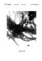
- FIG. 1is a scanning electron micrograph depicting fibroblast attachment to the three-dimensional matrix and extension of cellular processes across the mesh opening. Fibroblasts are actively secreting matrix proteins and are at the appropriate stage of subconfluency which should be obtained prior to inoculation with tissue-specific cells.
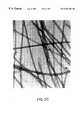
- FIGS. 2A-Dare transmission electron micrographs of collagen isolated from extracellular matrix prepared from dermal tissue grown in vitro (FIG. 2A-B) or from a normal adult human dermal sample (FIG. 2 C-D).
- One embodiment of the present inventioninvolves the preparation and use of an injectable extracellular matrix composition for the treatment of skin defects.
- the extracellular matrix proteinsare derived from a living stromal tissue prepared in vitro by growing stromal cells on a three-dimensional framework resulting in a multi-layer cell culture system. In conventional tissue culture systems, the cells were grown in a monolayer. Cells grown on a three-dimensional framework support, in accordance with the present invention, grow in multiple layers, forming a cellular matrix. This matrix system approaches physiologic conditions found in vivo to a greater degree than previously described monolayer tissue culture systems.
- the three-dimensional cell culture systemis applicable to the proliferation of different types of stromal cells and formation of a number of different stromal tissues, including but not limited to dermis, bone marrow stroma, glial tissue, cartilage, to name but a few.
- the pre-established living stromal tissuecomprises stromal cells grown on a three-dimensional framework or network.
- the stromal cellscan comprise fibroblasts with or without additional cells and/or elements described more fully herein.
- the fibroblasts and other cells and/or elements that comprise the stromacan be fetal or adult in origin, and can be derived from convenient sources such as skin, liver, pancreas, etc.
- Such tissues and/or organscan be obtained by appropriate biopsy or upon autopsy.
- cadaver organsmay be used to provide a generous supply of stromal cells and elements.
- the stromal cellswill proliferate on the framework, and elaborate growth factors, regulatory factors and extracellular matrix proteins that are deposited on the support.
- the living stromal tissuewill sustain active proliferation of the culture for long periods of time. Growth and regulatory factors can be added to the culture, but are not necessary since they are elaborated by the stromal support matrix.
- the naturally secreted extracellular matrixis collected from the three-dimensional framework and is processed further with a pharmaceutically acceptable aqueous carrier and placed in a syringe for precise placement of the biomaterial into tissues, such as the facial dermis.
- the present inventionis based, in part, on the discovery that during the growth of human stromal cells on a biodegradable or non-biodegradable three-dimensional support framework, the cells synthesize and deposit on the three-dimensional support framework a human extracellular matrix as produced in normal human tissue.
- the extracellular matrixis secreted locally by cells and not only binds cells and tissues together but also influences the development and behavior of the cells it contacts.
- the extracellular matrixcontains various fiber-forming proteins interwoven in a hydrated gel composed of a network of glycosaminoglycan chains.
- the glycosaminoglycansare a heterogeneous group of long, negatively charged polysaccharide chains, which (except for hyaluronic acid) are covalently linked to protein to form proteoglycan molecules.
- the fiber-forming proteinsare of two functional types: mainly structural (collagens and elastin) and mainly adhesive (such as fibronectin and laminin).
- the fibrillar collagens(types I, II, and III) are rope-like, triple-stranded helical molecules that aggregate into long cable-like fibrils in the extracellular space; these in turn can assemble into a variety of highly ordered arrays.
- Type IV collagen moleculesassemble into a sheetlike meshwork that forms the core of all basal laminae.
- Elastin moleculesform an extensive cross-linked network of fibers and sheets that can stretch and recoil, imparting elasticity to the matrix.
- Fibronectin and lamininare examples of large adhesive glycoproteins in the matrix; fibronectin is widely distributed in connective tissues, whereas laminin is found mainly in basal laminae. By means of their multiple binding domains, such proteins help cells adhere to and become organized by the extracellular matrix.
- a naturally secreted human dermal extracellular matrixcontains type I and type III collagens, fibronectin, tenascin, glycosaminoglycans, acidic and basic FGF, TGF- ⁇ and TGF- ⁇ , KGF, decorin and various other secreted human dermal matrix proteins.
- the various extracellular matrix proteinsare produced in the quantities and ratios similar to that existing in vivo.
- growth of the stromal cells in three dimensionswill sustain active proliferation of cells in culture for much longer time periods than will monolayer systems.
- the three-dimensional systemsupports the maturation, differentiation, and segregation of cells in culture in vitro to form components of adult tissues analogous to counterparts found in vivo.
- the extracellular matrix created by the cells in cultureis more analogous to native tissues.
- the three-dimensional frameworkprovides a greater surface area for protein attachment, and consequently, for the adherence of stromal cells.
- stromal cellsBecause of the three-dimensionality of the framework, stromal cells continue to actively grow in contrast to cells in monolayer cultures, which grow to confluence, exhibit contact inhibition, and cease to grow and divide.
- the elaboration of growth and regulatory factors by replicating stromal cellsmay be partially responsible for stimulating proliferation and regulating differentiation of cells in culture.
- the increase in potential volume for cell growth in the three-dimensional systemmay allow the establishment of localized microenvironments analogous to native counterparts found in vivo.
- the three-dimensional matrixmaximizes cell-cell interactions by allowing greater potential for movement of migratory cells, such as macrophages, monocytes and possibly lymphocytes in the adherent layer.
- the three-dimensional stromal support, the culture system itself, and its maintenance, as well as various uses of the three-dimensional cultures and of the naturally secreted extracellular matrixare described in greater detail in the subsections below. Solely for ease of explanation, the detailed description of the invention is divided into the three sections, (i) growth of the three-dimensional stromal cell culture, (ii) isolation of the naturally secreted human extracellular matrix, and (iii) formulation of the isolated extracellular matrix into preparations for injection at the site of soft tissue defects.
- the three-dimensional support used to culture stromal tissuemay be of any material and/or shape that:
- (b)allows cells to grow in more than one layer.
- non-biodegradable materialsinclude but are not limited to: nylon (polyamides), dacron (polyesters), polystyrene, polypropylene, polyacrylates, polyvinyl compounds (e.g., polyvinylchloride), polycarbonate (PVC), polytetrafluorethylene (PTFE; teflon), thermanox (TPX), etc.
- biodegradable materialmay also be utilized, including but not limited to: nitrocellulose, cotton, polyglycolic acid (PGA), cat gut sutures, cellulose, gelatin, dextran, collagen, chitosan, hyaluronic acid, etc. Any of these materials, bio- or non-biodegradable, can be woven into a mesh to form a three-dimensional framework. Alternatively, the materials can be used to form other types of three-dimensional frameworks, for example, sponges, such as collagen sponges.
- nylonpolystyrene, etc.
- nylon frameworkscan be treated with 0.1 M acetic acid, and incubated in polylysine, FBS, and/or collagen to coat the nylon.
- Polystyrenecan be similarly treated using sulfuric acid.
- a convenient nylon mesh which can be used in accordance with the inventionis Nitex, a nylon filtration mesh having an average pore size of 210 ⁇ m and an average nylon fiber diameter of 90 ⁇ m (#3-210/36, Tetko, Inc., N.Y.).
- fibroblastsderived from adult or fetal tissue, with or without other cells and elements described below, are inoculated onto the framework.
- fibroblastsmay be derived from organs, such as skin, liver, pancreas, etc. which can be obtained by biopsy, where appropriate, or upon autopsy.
- fibroblastscan be obtained in quantity rather conveniently from any appropriate cadaver organ.
- fetal fibroblastscan be obtained in high quantity from foreskin.
- Fibroblastsmay be readily isolated by disaggregating an appropriate organ or tissue which is to serve as the source of the fibroblasts. This can be readily accomplished using techniques known to those skilled in the art.
- the tissue or organcan be disaggregated mechanically and/or treated with digestive enzymes and/or chelating agents that-weaken the connections between neighboring cells making it possible to disperse the tissue into a suspension of individual cells without appreciable cell breakage.
- Enzymatic dissociationcan be accomplished by mincing the tissue and treating the minced tissue with any of a number of digestive enzymes either alone or in combination.
- the suspensioncan be fractionated into subpopulations from which the fibroblasts and/or other stromal cells and/or elements can be obtained.
- Thisalso may be accomplished using standard techniques for cell separation including, but not limited to, cloning and selection of specific cell types, selective destruction of unwanted cells (negative selection), separation based upon differential cell agglutinability in the mixed population, freeze-thaw procedures, differential adherence properties of the cells in the mixed population, filtration, conventional and zonal centrifugation, centrifugal elutriation (counter-streaming centrifugation), unit gravity separation, countercurrent distribution, electrophoresis and fluorescence-activated cell sorting.
- fibroblastsfor example, can be carried out as follows: fresh tissue samples are thoroughly washed and minced in Hanks' balanced salt solution (HBSS) in order to remove serum. The minced tissue is incubated from 1-12 hours in a freshly prepared solution of a dissociating enzyme such as trypsin. After such incubation, the dissociated cells are suspended, pelleted by centrifugation and plated onto culture dishes. All fibroblasts will attach before other cells, therefore, appropriate stromal cells can be selectively isolated and grown. The isolated fibroblasts can then be grown to confluency, lifted from the confluent culture and inoculated onto the three-dimensional framework, see Naughton et al., 1987, J. Med.
- HBSSHanks' balanced salt solution
- stromal cell culture-producing extracellular matrixcan be added to form the three-dimensional stromal cell culture-producing extracellular matrix.
- other cells found in loose connective tissuemay be inoculated onto the three-dimensional support framework along with fibroblasts.
- Such cellsinclude, but are not limited to, endothelial cells, pericytes, macrophages, monocytes, plasma cells, mast cells, adipocytes, chondrocytes, etc.
- These stromal cellscan be readily derived from appropriate organs such as skin, liver, etc., using methods known, such as those discussed above.
- stromal cellswhich are specialized for the particular tissue to be cultured can be added to the fibroblast stroma for the production of a tissue type specific extracellular matrix.
- dermal fibroblastscan be used to form the three-dimensional subconfluent stroma for the production of skin-specific extracellular matrix in vitro.
- stromal cells of hematopoietic tissueincluding, but not limited to, fibroblast endothelial cells, macrophages/monocytes, adipocytes and reticular cells, can be used to form the three-dimensional subconfluent stroma for the production of a bone marrow-specific extracellular matrix in vitro, see infra.
- Hematopoietic stromal cellscan be readily obtained from the “buffy coat” formed in bone marrow suspensions by centrifugation at low forces, e.g., 3000 ⁇ g.
- Stromal cells of livermay include fibroblasts, Kupffer cells, and vascular and bile duct endothelial cells.
- glial cellscan be used as the stroma to support the proliferation of neurological cells and tissues. Glial cells for this purpose can be obtained by trypsinization or collagenase digestion of embryonic or adult brain. Ponten and Westermark, 1980, In Federof, S. Hertz, L., eds, “Advances in Cellular Neurobiology,” Vol.1, New York, Academic Press, pp.209-227.
- the stromal cellsFor certain uses in vivo it is preferable to obtain the stromal cells from the patient's own tissues.
- the growth of cells in the presence of the three-dimensional stromal support frameworkcan be further enhanced by adding to the framework, or coating the framework support with proteins, e.g., collagens, elastic fibers, reticular fibers, glycoproteins; glycosaminoglycans, e.g., heparin sulfate, chondroitin-4-sulfate, chondroitin-6-sulfate, dermatan sulfate, keratan sulfate, etc.; a cellular matrix, and/or other materials.
- proteinse.g., collagens, elastic fibers, reticular fibers, glycoproteins
- glycosaminoglycanse.g., heparin sulfate, chondroitin-4-sulfate, chondroitin-6-sulfate, dermatan sulf
- the three-dimensional frameworkis incubated in an appropriate nutrient medium under physiologic conditions favorable for cell growth, i.e., promoting mitosis, i.e., cell division.
- an appropriate nutrient mediumi.e., promoting mitosis, i.e., cell division.
- Many commercially available mediasuch as RPMI 1640, Fisher's, Iscove's, McCoy's, and the like may be suitable for use. It is important that the three-dimensional stromal culture be suspended or floated in the medium during the incubation period in order to maximize proliferative activity.
- the cultureshould be “fed” periodically to remove the spent media, depopulate released cells, and to add fresh media.
- the stromal cellswill grow linearly along and envelop the three-dimensional framework before beginning to grow into the openings of the framework.
- the cellsare grown to an appropriate degree to allow for adequate deposition of extracellular matrix proteins.
- the openings of the frameworkshould be of an appropriate size to allow the stromal cells to stretch across the openings. Maintaining actively growing stromal cells which stretch across the framework enhances the production of growth factors which are elaborated by the stromal cells, and hence, will support long term cultures. For example, if the openings are too small, the stromal cells may rapidly achieve confluence but be unable to easily exit from the mesh. Trapped cells can exhibit contact inhibition and cease production of the appropriate factors necessary to support proliferation and maintain long term cultures. If the openings are too large, the stromal cells are unable to stretch across the opening. This will also decrease stromal cell production of the appropriate factors necessary to support proliferation and maintain long term cultures.
- openings ranging from about 150 ⁇ m to about 220 ⁇ mwill work satisfactorily.
- other sizesmay work equally well.
- any shape or structure that allows the stromal cells to stretch and continue to replicate and grow for lengthy time periodswill work in accordance with the present invention.
- the matrixshould preferably contain collagen types III, IV and I in an approximate ratio of 6:3:1 in the initial matrix.
- collagen types I and IIIare preferably deposited in the initial matrix.
- the proportions of collagen types depositedcan be manipulated or enhanced by selecting fibroblasts which elaborate the appropriate extracellular matrix proteins. This can be accomplished using monoclonal antibodies of an appropriate isotype or subclass which are capable of activating complement, and which define particular collagen types. These antibodies in combination with complement can be used to negatively select the fibroblasts which express the desired collagen type.
- the stroma used to inoculate the frameworkcan be a mixture of cells which synthesize the appropriate collagen types desired. The distribution and origins of the five types of collagen is shown in Table I.
- the appropriate stromal cell(s)can be selected to inoculate the three-dimensional framework.
- the three-dimensional extracellular matrix producing culture of the present inventionaffords a vehicle for introducing gene products in vivo.
- the cellsmay be genetically engineered to express the gene product, or altered forms of the gene product that are immobilized in the extracellular matrix laid down by the stromal cells.
- a gene of interestcan be placed under the control of an inducible promoter.
- the recombinant DNA construct containing the genecan be used to transform or transfect a host cell which is cloned and then clonally expanded in the three-dimensional culture system.
- the use of the three-dimensional culture in this regardhas a number of advantages.
- the number of transfected cellscan be substantially enhanced to be of clinical value, relevance, and utility.
- the three-dimensional cultures of the present inventionallow for expansion of the number of transfected cells and amplification (via cell division) of transfected cells.
- the expression control elements usedshould allow for the regulated expression of the gene so that the product can be over synthesized in culture.
- the transcriptional promoter chosen, generally, and promoter elements specifically,depends, in part, upon the type of tissue and cells cultured. Cells and tissues which are capable of secreting proteins (e.g., those characterized by abundant rough endoplasmic reticulum and golgi complex) are preferable.
- proliferating cellsare released from the framework. These released cells can stick to the walls of the culture vessel where they can continue to proliferate and form a confluent monolayer. This should be prevented or minimized, for example, by removal of the released cells during feeding, or by transferring the three-dimensional framework to a new culture vessel. The presence of a confluent monolayer in the vessel will “shut down” the growth of cells in the three-dimensional framework and/or culture. Removal of the confluent monolayer or transfer of the stromal culture to fresh media in a new vessel will restore proliferative activity of the three-dimensional culture system. Such removal or transfers should be done in any culture vessel which has a stromal monolayer exceeding 25% confluency.
- the culture systemcan be agitated to prevent the released cells from sticking, or instead of periodically feeding the cultures, the culture system could be set up so that fresh media continuously flows through the system.
- the flow ratecan be adjusted to both maximize proliferation within the three-dimensional culture, and to wash out and remove cells released from the matrix, so that they will not stick to the walls of the vessel and grow to confluence.
- the released stromal cellscan be collected and crypreserved for future use.
- fibroblastsOnce inoculated onto the three-dimensional framework, adherence of the fibroblasts is seen quickly (e.g., within hours) and the fibroblasts begin to stretch across the framework openings within days. These fibroblasts are metabolically active, secrete extracellular matrix and rapidly form a dermal equivalent consisting of active fibroblasts and collagen.
- FIG. 1illustrates the ability of the fibroblasts to arrange themselves into parallel layers between the naturally-secreted collagen bundles. These fibroblasts exhibit a rapid rate of cell division and protein secretion.
- the cellsAfter the cells have been inoculated onto the framework and cultured under conditions favoring cellular growth, such that a desired amount of extracellular matrix is secreted on to the three-dimensional framework, the cells are killed and the naturally secreted extracellular matrix is processed further.
- the cellscan be killed by flash-freezing the living stromal tissue prepared in vitro in liquid nitrogen without a cryopreservative.
- Another way to kill the cellsis to irrigate the inoculated three-dimensional framework with sterile water, such that the cells burst in response to osmotic pressure.
- the frameworkcan be subjected to enzymatic digestion and/or extracting with reagents that break down the cellular membranes and allow removal of cell contents.
- detergentsinclude non-ionic detergents (for example, TRITON X-100, octylphenoxy polyethoxyethanol, (Rohm and Haas); BRIJ-35, a polyethoxyethanol lauryl ether (Atlas Chemical Co.), TWEEN 20, a polyethoxyethanol sorbitan monolaureate (Rohm and Haas), LUBROL-PX, or polyethylene lauryl ether (Rohm and Haas)); and ionic detergents (for example, sodium dodecyl sulphate, sulfated higher aliphatic alcohol, sulfonated alkane and sulfonated alkylarene containing 7 to 22 carbon atoms in a branched or unbranched chain).
- non-ionic detergentsfor example, TRITON X-100, octylphenoxy polyethoxyethanol, (Rohm and Haas); BRIJ-35, a
- Enzymescan be used also and can include nucleases (for example, deoxyribonuclease and ribonuclease), phospholipases and lipases.
- nucleasesfor example, deoxyribonuclease and ribonuclease
- phospholipasesfor example, phospholipases and lipases.
- the collection of the naturally secreted human extracellular matrixcan be accomplished in a variety of ways which depends on whether the three-dimensional framework is composed of material that is biodegradable or non-biodegradable. For example, if the framework is composed of non-biodegradable material, one can remove the extracellular matrix from a non-biodegradable support by subjecting the three-dimensional framework to sonication and/or to high pressure water jets and/or to mechanical scraping and/or to a mild treatment with detergents and/or enzymes to remove the attached extracellular matrix from the framework.
- the extracellular matrixis deposited on a biodegradable three-dimensional framework, after killing and removing the cells and cellular debris, the extracellular matrix can be recovered, for example, by simply allowing the framework to degrade in solution, i.e., allow the framework to dissolve, thus freeing the extracellular matrix.
- the biodegradable supportis composed of a material which can be injected, like the extracellular matrix itself, one can process the entire extracellular matrix coated framework into syringes for injection.
- the matrixcan be removed by the same methods as if the matrix had been deposited on a non-biodegradable support, i.e., by subjecting the three-dimensional framework to sonication and/or to high pressure water jets and/or to mechanical scraping and/or to a mild treatment with detergents and/or enzymes to remove the attached extracellular matrix from the framework. None of the removal processes are designed to damage and/or denature the naturally secreted human extracellular matrix produced by the cells.
- the extracellular matrixcan be homogenized to fine particles, such that it can pass through a surgical needle. Homogenization is well known in the art, for example, by sonication. Further, the extracellular matrix can be cross-linked by gamma irradiation without the use of chemical cross-linking agents, such as glutaraldehyde, which are toxic.
- the gamma irradiationshould be a minimum of 20 M rads to sterilize the material since all bacteria, fungi, and viruses are destroyed at 0.2 M rads.
- the extracellular matrixcan be irradiated from 0.25 to 2 M rads to sterilize and cross-link the extracellular matrix.
- the amounts and/or ratios of the collagens and other proteinsmay be adjusted by mixing extracellular matrices secreted by other cell types prior to placing the material in a syringe.
- biologically active substancessuch as proteins and drugs
- exemplary biologically active substancescan include tissue growth factors, such as TGF- ⁇ , and the like which promote healing and tissue repair at the site of the injection.
- Final formulation of the aqueous suspension of naturally secreted human extracellular matrixwill typically involve adjusting the ionic strength of the suspension to isotonicity (i.e., about 0.1 to 0.2) and to physiological pH (i.e., about pH 6.8 to 7.5) and adding a local anesthetic, such as lidocaine, (usually at a concentration of about 0.3% by weight) to reduce local pain upon injection.
- the final formulationwill also typically contain a fluid lubricant, such as maltose, which must be tolerated by the body.
- Exemplary lubricant componentsinclude glycerol, glycogen, maltose and the like.
- Organic polymer base materialssuch as polyethylene glycol and hyaluronic acid as well as non-fibrillar collagen, preferably succinylated collagen, can also act as lubricants.
- Such lubricantsare generally used to improve the injectability, intrudability and dispersion of the injected biomaterial at the site of injection and to decrease the amount of spiking by modifying the viscosity of the compositions.
- This final formulationis by definition the processed extracellular matrix in a pharmaceutically acceptable carrier.
- the matrixis subsequently placed in a syringe or other injection apparatus for precise placement of the matrix at the site of the tissue defect.
- injectablemeans the formulation can be dispensed from syringes having a gauge as low as 25 under normal conditions under normal pressure without substantial spiking. Spiking can cause the composition to ooze from the syringe rather than be injected into the tissue.
- needlesas fine as 27 gauge (200 ⁇ I.D.) or even 30 gauge (150 ⁇ I.D.) are desirable.
- the maximum particle size that can be extruded through such needleswill be a complex function of at least the following: particle maximum dimension, particle aspect ratio (length:width), particle rigidity, surface roughness of particles and related factors affecting particle:particle adhesion, the viscoelastic properties of the suspending fluid, and the rate of flow through the needle.
- particle maximum dimensionparticle aspect ratio (length:width)
- particle rigidityparticle rigidity
- surface roughness of particles and related factors affecting particle:particle adhesionthe viscoelastic properties of the suspending fluid
- the rate of flow through the needleRigid spherical beads suspended in a Newtonian fluid represent the simplest case, while fibrous or branched particles in a viscoelastic fluid are likely to be more complex.
- the above described steps in the process for preparing injectable naturally secreted human extracellular matrixare preferably carried out under sterile conditions using sterile materials.
- the processed extracellular matrix in a pharmaceutically acceptable carriercan be injected intradermally or subcutaneously to augment soft tissue, to repair or correct congenital anomalies, acquired defects or cosmetic defects.
- congenital anomaliesas hemifacial microsomia, malar and zygomatic hypoplasia, unilateral mammary hypoplasia, pectus excavatum, pectoralis agenesis (Poland's anomaly) and velopharyngeal incompetence secondary to cleft palate repair or submucous cleft palate (as a retropharyngeal implant); acquired defects (post-traumatic, post-surgical, post-infectious) such as depressed scars, subcutaneous atrophy (e.g., secondary to discoid lupis erythematosus), keratotic lesions, enophthalmos in the unucleated eye (also superior sulcus syndrome), acne pitting of the face, linear scleroderma with subcutaneous atrophy, saddle-nose deformity, Romberg's disease and unilateral vocal cord paralysis; and cosmetic defects such as glabellar frown lines, deep nasolabial creases, circum-oral geographical
- the three-dimensional culture system of the inventionis described based upon the type of tissue and cells used in various systems. These descriptions specifically include but are not limited to bone marrow, skin, epithelial cells, and cartilage but it is expressly understood that the three-dimensional culture system can be used with other types of cells and tissues.
- the inventionis also illustrated by way of examples, which demonstrate characteristic data generated for each system described.
- Skin fibroblastswere isolated by mincing dermal tissue, trypsinization for 2 hours, and separation of cells into a suspension by physical means. Fibroblasts were grown to confluency in 25 cm 2 Falcon tissue culture dishes and fed with RPMI 1640 (Sigma, MO) supplemented with 10% fetal bovine serum (FBS), fungizone, gentamicin, and penicillin/streptomycin. Fibroblasts were lifted by mild trypsinization and cells were plated onto nylon filtration mesh, the fibers of which are approximately 90 ⁇ m in diameter and are assembled into a square weave with a mesh opening of 210 ⁇ m (Tetko, Inc., N.Y.).
- FIG. 1is a scanning electron micrograph depicting fibroblast attachment and extension of cellular processes across the mesh opening.
- Bone marrowwas aspirated from multiple sites on the posterior iliac crest of hematologically normal adult volunteers after informed consent was obtained. Specimens were collected into heparinized tubes and suspended in 8 ml of RPMI 1640 medium which was conditioned with 10% FBS and 20 5-10% HS and supplemented with hydrocortisone, fungizone, and streptomycin. The cell clumps were disaggregated and divided into aliquots of 5 ⁇ 10 6 nucleated cells.
- Nylon filtration screen(#3-210/36, Tetko Inc., N.Y.) was used as a three-dimensional framework to support all stromal cell cultures described in the examples below.
- the screenconsisted of fibers, which were 90 ⁇ m in diameter, assembled into a square weave pattern with sieve openings of 210 ⁇ m.
- Stromal cellswere inoculated using the protocols described in Section 6.1. Adherence and subsequent growth of the stromal elements was monitored using inverted phase contrast microscopy and scanning electron microscopy (SEM).
- Samples of oral mucosal tissuewere obtained from orthodontic surgical specimens. Tissue was washed three times with fresh MEM containing antibiotics (2 ml of antibiotic antimycotic solution from GIBCO, Cat. #600-5240 AG; and 0.01 ml of gentamicin solution from GIBCO Cat. #600-5710 AD per 100 cc MEM), cut into small pieces, then washed with 0.02% EDTA (w/v). 0.25% trypsin (in PBS without Ca ++ or Mg ++ ) was added; after a few seconds, the tissue pieces were removed and placed in fresh trypsin (in PBS without Ca ++ or Mg ++ ) and refrigerated at 4° C. overnight.
- MEMantibiotic antimycotic solution from GIBCO, Cat. #600-5240 AG
- gentamicin solutionfrom GIBCO Cat. #600-5710 AD per 100 cc MEM
- Tissueswere removed and placed in fresh trypsin solution, and gently agitated until cell appeared to form a single-cell suspension.
- the single-cell suspensionwas then diluted in MEM containing 10% heat inactivated fetal bovine serum and centrifuged at 1400 ⁇ g for 7 minutes. The supernatant was decanted and the pellet containing mucosal epithelial cells was placed into seeding medium.
- Mediumconsisted of DMEM with 2% Ultrosen G, 1 ⁇ L-glutamine, 1 ⁇ non-essential amino acids, penicillin and streptomycin. The cells were seeded onto a three-dimensional framework.
- the three-dimensional stromal culturewas generated using oral fibroblasts and 8 mm ⁇ 45 mm pieces of nylon filtration screen (#3-210/36, Tetko Inc., N.Y.). The mesh was soaked in 0.1 M acetic acid for 30 minutes and treated with 10 mM polylysine suspension for 1 hour. The meshes were placed in a sterile petri dish and inoculated with 1 ⁇ 10 6 oral fibroblasts collected as described above in DMEM complete medium. After 1-2 hours of incubation at 5% CO 2 the meshes were placed in a Corning 25 cm 2 tissue culture flask, floated with an additional 5 ml of medium, and allowed to reach subconfluence, being fed at 3 day intervals. Cultures were maintained in DMEM complete medium at 37° C. and 5% CO 2 in a humidified atmosphere and were fed with fresh medium every 3 days.
- Small vessel endothelial cells isolated from the brain according to the method of Larson et al., 1987, Microvasc. Res. 34:184were cultured in vitro using T-75 tissue culture flasks.
- the cellswere maintained in Dulbecco's Modified Eagle Medium/Hams-F-12 medium combination (the solution is available as a 1:1 mixture).
- the mediumwas supplemented with 20% heat-inactivated fetal calf serum (FCS), glutamine, and antibiotics.
- FCSheat-inactivated fetal calf serum
- the cellswere seeded at a concentration of 1 ⁇ 10 6 cells per flask, and reached a confluent state within one week.
- the cellswere passaged once a week, and, in addition, were fed once a week with DMEM/Hams-F-12 containing FCS, glutamine, and antibiotics as described.
- flaskswere rinsed twice with 5 ml of PBS (without Ca ++ or Mg ++ ) and trypsinized with 3 ml of 0.05% Trypsin and 0.53 mM EDTA.
- the cellswere pelleted, resuspended, and tested for viability by trypan blue exclusion, seeded and fed with 25 ml of the above mentioned DMEM/Hams-F-12 supplemented medium.
- a factor VIII related antigen assayGrulnick et al., 1977, Ann. Int. Med. 86:598-616, is used to positively identify endothelial cells, and silver staining was used to identify tight junctional complexes, specific to only small vessel endothelium.
- Nylon filtration screen mesh(#3-210/36, Tetko, Inc., N.Y.) was prepared essentially as described above. The mesh was soaked in an acetic acid solution (1 ml glacial acetic acid plus 99 ml distilled H 2 O) for thirty minutes, was rinsed with copious amounts of distilled water and then autoclaved. Meshes were coated with 6 ml fetal bovine serum per 8 ⁇ 8 cm mesh and incubated overnight. The meshes were then stacked, three high, and 3 ⁇ 10 7 small vessel endothelial cells (cultured as described) were seeded onto the stack, and incubated for three hours at 37° C. under 5% CO 2 in a humidified atmosphere. The inoculated meshes were fed with 10 ml of DMEM/Hams-F-12 medium every 3-4 days until complete confluence was reached (in approximately two weeks).
- Cartilagewas harvested from articular surfaces of human joints.
- the cartilage pieceswere digested with collagenase (0.2% w/v) in complete medium (DMEM with 10% fetal bovine serum, glutamine, non-essential amino acids, sodium pyruvate, 50 ⁇ g/ml ascorbate and 35 ⁇ g/ml gentamicin) for 20 hours at 37° C.
- DMEMcomplete medium
- Liberated chondrocyteswere spun, resuspended in complete medium, counted and plated at 1 ⁇ 10 6 cells per T-150 flask. Cells were routinely passed at confluence (every 5-7 days).
- Polyglycolic acid mesh(1 mm diameter ⁇ 2 mm thick) was sterilized by ethylene oxide or electron beam treatment and presoaked overnight in complete medium. The mesh was seeded in 6 well plates with 3-4 ⁇ 10 6 cells per mesh in a total volume of 10 ⁇ l and incubated for 3-4 hours at 37° C. in a tissue culture incubator. At this time, 1.5 ml of media was added. The seeded mesh was incubated overnight. 5 ml of media was added the next day. Media was changed three times per week until confluence is reached.
- the extracellular matrixhas been characterized by a number of analytic methods to determine its content of matrix proteins, each value is the average of at least two independent determinations.
- the matrixcontained type I and type III collagens, fibronectin, tenascin, sulfated glycosaminoglycans, decorin and various other secreted human extracellular matrix proteins. Additionally, the secreted matrix proteins were found throughout the three-dimensional support framework.
- the extracellular matrixcontained a total protein amount of 292 mg/cm 2 ⁇ 0.06; fibronectin was present at 3.4 mg/cm 2 ⁇ 1.2; and tenascin at 1.7 mg/cm 2 ⁇ 0.6. Both fibronectin and tenascin showed the expected molecular weight distributions on immunoblots.
- Collagen content of the extracellular matrixwas determined using the Sirius Red assay.
- the binding of Sirius Red F3BA in saturated picric acid solutionhas been used widely to estimate fibrotic collagen deposition.
- the specificity of Sirius Red binding to collagenis based largely on its use as a histological stain. In rat liver with various degrees of cholestatic fibrosis, collagen content measured by Sirius Red binding shows strong correlation with hydroxyproline content. Walsh et al., 1992, Analyt. Biochem.
- Sirius Redis birefringent, indicating directional binding related to the orientation of the collagen strands. Sirius Red is known to bind to proteins other than the classical collagens that contain collagen-like triple helices, such as the complement component C1. Some minor binding to serum albumin has also been found, although control experiments using bovine serum albumin standard showed no interference with the assay. The interference is estimated to represent less than 2% of the collagen signal in the extracellular matrix and the use of Sirius Red assay gives a reproducible method for measuring collagens.
- the extracellular matrixcontained a collagen content of 0.61 mg/cm 2 ⁇ 0.09. Further, collagens I and III showed the expected molecular weight distributions on immunoblots.
- Collagen derived from the dermal tissue grown in vitro and collagen derived from a normal adult human dermal samplewere processed and visualized by transmission electron microscopy (TEM). Briefly, the respective collagens were weighed and placed in a sterile 50 ml centrifuge tube with 30 ml 0.05 M Tris buffer, pH 8.0. After mixing for two hours on a wrist shaker, the Tris buffer was removed and the specimen placed in a homogenization cylinder along with 30 ml fresh 0.05 M Tris buffer. The sample was homogenized for 30 seconds in buffer alone and then for two 30 second bursts following the addition of a dispersing agent as described in U.S. Pat. Nos. 4,969,912 and 5,332,802. The temperature was maintained at 5-10° C.
- the dispersing agentwas added at a concentration of 0.05% (wet weight of the collagen).
- the homogenized preparationwas centrifuged at 3500 rpm for 6 minutes to separate the dispersed collagenous material from the yet undispersed material.
- the undispersed residuewas again treated with dispersing agent at 0.05% (wet weight of the collagen) and homogenized for two 30 second bursts. The dispersion was again centrifuged to recover dispersed collagenous material which was added to the first recovery.
- the collagenous dispersionwas filtered through a 100 micron filter, centrifuged at 3500 rpm and the pellet was washed 3 times with 0.004 M phosphate buffer, pH 7.4. The last centrifugation was conducted at 10,000 rpm to pack the collagenous pellet. Samples were then collected for TEM. As shown in FIGS. 2A-D, the collagen fibers isolated from either the extracellular matrix prepared from dermal tissue grown in vitro (FIGS. 2A-B) or from normal adult human dermis (FIGS. 2C-D) appeared identical in that intact collagen fibers with typical collagen banding and normal periodicity in both preparations.
- Glycosaminoglycanshave been shown to play a variety of structural and functional roles in the body and their presence in the secreted extracellular matrix is important. Table II lists a number of examples of glycosaminoglycans which have been determined to be found in the extracellular matrix as well as their functional importance in normal dermis.
- the extracellular matrixwas found to contain a total of 2.8 mg/cm 2 ⁇ 0.1 sulfated glycosaminoglycans.
- the cells producing and depositing the extracellular matrixalso expressed a number of different growth factors. Growth factors are important in the extracellular matrix for two reasons. During the growth of and deposition of the extracellular matrix, naturally seeded growth factors help to control cell proliferation and activity. Further, growth factors remain attached to the extracellular matrix. A variety of growth factors have been determined to be expressed during the deposition of the matrix.
- acidic and basic FGF, TGF- ⁇ and TGF- ⁇ , and KGF mRNA transcriptswere present as were several others as shown in Table III, including PDGF, amphiregulin, HBEGF, IGF, SPARC and VEGF.
- PDGF and TGF- ⁇ 3are thought to be involved in regulation of cell proliferation and matrix deposition in culture, while TGF- ⁇ 1, HBEGF, KGF, SPARC, VEGF and decorin are deposited in the matrix.
- Amphiregulin, IGF-1, IGF-2 and IL-1were not expressed at the sensitivity used in these experiments.
Landscapes
- Health & Medical Sciences (AREA)
- Life Sciences & Earth Sciences (AREA)
- Chemical & Material Sciences (AREA)
- Biomedical Technology (AREA)
- Engineering & Computer Science (AREA)
- Dermatology (AREA)
- Animal Behavior & Ethology (AREA)
- General Health & Medical Sciences (AREA)
- Public Health (AREA)
- Veterinary Medicine (AREA)
- Transplantation (AREA)
- Medicinal Chemistry (AREA)
- Epidemiology (AREA)
- Oral & Maxillofacial Surgery (AREA)
- Botany (AREA)
- Chemical Kinetics & Catalysis (AREA)
- Zoology (AREA)
- Cell Biology (AREA)
- Molecular Biology (AREA)
- Urology & Nephrology (AREA)
- Vascular Medicine (AREA)
- Gerontology & Geriatric Medicine (AREA)
- Biophysics (AREA)
- Materials For Medical Uses (AREA)
- Micro-Organisms Or Cultivation Processes Thereof (AREA)
- Medicinal Preparation (AREA)
Abstract
Description
| TABLE I |
| DISTRIBUTIONS AND ORIGINS OF THE FIVE TYPES OF |
| COLLAGEN |
| Collagen | Principal Tissue | |
| Type | Distribution | Cells of Origin |
| I | Loose and dense ordinary | Fibroblasts and |
| connective tissue; collagen | reticular cells; | |
| fibers | smooth muscle cells | |
| Fibrocartilage | ||
| Bone | Osteoblast | |
| Dentin | Odontoblasts | |
| II | Hyaline and elastic | Chondrocytes |
| cartilage | Retinal Cells | |
| Vitreous body of eye | ||
| III | Loose connective tissue; | Fibroblasts and |
| reticular fibers | reticular cells | |
| Papillary layer of dermis | Smooth muscle cells; | |
| Blood vessels | endothelial cells | |
| IV | Basement membranes | Epithelial and |
| endothelial cells | ||
| Lens capsule of eye | Lens fibers | |
| V | Fetal membranes; placenta | Fibroblasts |
| Basement membranes | ||
| Bone | ||
| Smooth muscle | Smooth muscle cells | |
| TABLE II | ||||
| NAME | LOCATION | GLYCAN | FUNCTION | MECHANISM |
| Versican | Matrix | 12-15 | Structural | Binds hyaluronic |
| Chondroitin sulfate | acid and collagen | |||
| Decorin | Matrix | 1 Chondroitin/dermatan | Binding TGF-β | Inactivates |
| sulfate | and other | growth factors | ||
| growth factors; | ||||
| binds to | ||||
| collagen | ||||
| Betaglycan | Cell | 1-4 | TGF-β Type III | Adjunct receptor |
| membrane | Chondroitin/heparan | receptor | for TGFβ | |
| sulfate | ||||
| Syndecan | Cell | 1-3 Chondroitin | Growth factor | |
| membrane | sulfate, 1-2 heparan | binding | ||
| sulfate | ||||
| TABLE III | |||
| Messenger | |||
| RNA | Full Name | Function | Expression |
| PDGF-A Chain | Platelet- | Mitogen for | ++ |
| derived growth | fibroblasts, | ||
| factor, A | granulation | ||
| chain | tissue, | ||
| chemotactic | |||
| PDGF-B Chain | Platelet- | Mitogen for | 0 − (+)* |
| derived growth | fibroblasts, | ||
| factor, B | granulation | ||
| chain | tissue, | ||
| chemotactic | |||
| IGF-1 | Insulin-like | Mitogen for | 0 |
| growth factor- | fibroblasts | ||
| 1 | |||
| IGF-2 | Insulin-like | Mitogen for | (+) |
| growth factor- | fibroblasts | ||
| 2 | |||
| TGF-α | Transforming | Mitogen for | + |
| growth factor- | fibroblasts, | ||
| α | keratinocytes | ||
| Amphiregulin | Amphiregulin | Mitogen for | 0 |
| fibroblasts, | |||
| keratinocytes | |||
| KGF | Keratinocyte | Mitogen for | ++ |
| growth factor | keratinocytes | ||
| HBEGF | Heparin- | Mitogen for | +++ |
| binding | fibroblasts, | ||
| epidermal | keratinocytes | ||
| growth factor- | |||
| like growth | |||
| factor | |||
| TGF-β1 | Transforming | Stimulates | + |
| growth factor- | matrix | ||
| β1 | deposition | ||
| TGF-β3 | Transforming | Stimulates | ++ − ++ |
| growth factor- | matrix | ||
| β3 | deposition | ||
| VEGF | Vascular | Angiogenic | ++ |
| endothelial | factor | ||
| growth factor | |||
| SPARC | Secreted | Complex anti- | ++++ |
| protein acidic | angiogenic, | ||
| and rich in | angiogenic | ||
| cysteine | |||
| ICAM-1 | Intercellular | Lymphocyte | + |
| adhesion | adhesion, | ||
| molecule-1 | mobility | ||
| VCAM | Vascular | Lymphocyte | +++ |
| cellular | adhesion, | ||
| adhesion | mobility | ||
| molecule | |||
| GAPDh | Glyceraldehyde | Glycolytic | +++ |
| 3-phosphate | housekeeping | ||
| dehydrogenase | gene | ||
| β2- | β2- | Antigen | +++ |
| microglobulin | microglobulin | presentation | |
| °A small amount of PDGF B chain is seen in some preparations. | |||
Claims (16)
Priority Applications (2)
| Application Number | Priority Date | Filing Date | Title |
|---|---|---|---|
| US09/182,822US6284284B1 (en) | 1995-06-06 | 1998-10-29 | Compositions and methods for production and use of an injectable naturally secreted extracellular matrix |
| US09/947,131US20020019339A1 (en) | 1995-06-06 | 2001-09-04 | Compositions and methods for production and use of an injectable naturally secreted extracellular matrix |
Applications Claiming Priority (3)
| Application Number | Priority Date | Filing Date | Title |
|---|---|---|---|
| US08/470,101US5830708A (en) | 1995-06-06 | 1995-06-06 | Methods for production of a naturally secreted extracellular matrix |
| US66078796A | 1996-06-06 | 1996-06-06 | |
| US09/182,822US6284284B1 (en) | 1995-06-06 | 1998-10-29 | Compositions and methods for production and use of an injectable naturally secreted extracellular matrix |
Related Parent Applications (1)
| Application Number | Title | Priority Date | Filing Date |
|---|---|---|---|
| US66078796ADivision | 1995-06-06 | 1996-06-06 |
Related Child Applications (1)
| Application Number | Title | Priority Date | Filing Date |
|---|---|---|---|
| US09/947,131ContinuationUS20020019339A1 (en) | 1995-06-06 | 2001-09-04 | Compositions and methods for production and use of an injectable naturally secreted extracellular matrix |
Publications (1)
| Publication Number | Publication Date |
|---|---|
| US6284284B1true US6284284B1 (en) | 2001-09-04 |
Family
ID=27042969
Family Applications (5)
| Application Number | Title | Priority Date | Filing Date |
|---|---|---|---|
| US09/182,822Expired - LifetimeUS6284284B1 (en) | 1995-06-06 | 1998-10-29 | Compositions and methods for production and use of an injectable naturally secreted extracellular matrix |
| US09/947,131AbandonedUS20020019339A1 (en) | 1995-06-06 | 2001-09-04 | Compositions and methods for production and use of an injectable naturally secreted extracellular matrix |
| US09/948,379AbandonedUS20020038152A1 (en) | 1995-06-06 | 2001-09-07 | Compositions and methods for production and use of an injectable naturally secreted extracellular matrix |
| US10/805,774AbandonedUS20040180431A1 (en) | 1995-06-06 | 2004-03-22 | Compositions and methods for production and use of an injectable naturally secreted extracellular matrix |
| US10/851,773AbandonedUS20040259190A1 (en) | 1995-06-06 | 2004-05-21 | Compositions and methods for production and use of an injectable naturally secreted extracellular matrix |
Family Applications After (4)
| Application Number | Title | Priority Date | Filing Date |
|---|---|---|---|
| US09/947,131AbandonedUS20020019339A1 (en) | 1995-06-06 | 2001-09-04 | Compositions and methods for production and use of an injectable naturally secreted extracellular matrix |
| US09/948,379AbandonedUS20020038152A1 (en) | 1995-06-06 | 2001-09-07 | Compositions and methods for production and use of an injectable naturally secreted extracellular matrix |
| US10/805,774AbandonedUS20040180431A1 (en) | 1995-06-06 | 2004-03-22 | Compositions and methods for production and use of an injectable naturally secreted extracellular matrix |
| US10/851,773AbandonedUS20040259190A1 (en) | 1995-06-06 | 2004-05-21 | Compositions and methods for production and use of an injectable naturally secreted extracellular matrix |
Country Status (1)
| Country | Link |
|---|---|
| US (5) | US6284284B1 (en) |
Cited By (54)
| Publication number | Priority date | Publication date | Assignee | Title |
|---|---|---|---|---|
| US6485489B2 (en) | 1999-10-02 | 2002-11-26 | Quantum Cor, Inc. | Catheter system for repairing a mitral valve annulus |
| US20030069570A1 (en)* | 1999-10-02 | 2003-04-10 | Witzel Thomas H. | Methods for repairing mitral valve annulus percutaneously |
| US20040180431A1 (en)* | 1995-06-06 | 2004-09-16 | Naughton Gail K. | Compositions and methods for production and use of an injectable naturally secreted extracellular matrix |
| US20040210230A1 (en)* | 2003-04-02 | 2004-10-21 | Furlow Leonard T. | Materials and methods for soft tissue augmentation |
| US20040219134A1 (en)* | 1999-04-12 | 2004-11-04 | Advanced Tissue Sciences, Inc. | Methods for using a three-dimensional stromal tissue to promote angiogenesis |
| US20050013805A1 (en)* | 2003-07-15 | 2005-01-20 | Itzhak Tavori | Device and method for delivery of rapidly separating body of fluid, forming bone reconstruction medium |
| KR100527623B1 (en)* | 2002-06-01 | 2005-11-15 | 라이프코드인터내셔날 주식회사 | Biodegradable polymer scaffold containing extracellular matrix used for artificial organs and method for preparing same |
| US20060035861A1 (en)* | 1997-06-17 | 2006-02-16 | Fziomed, Inc. | Compositions of polyacids and polyethers and methods for their use as dermal fillers |
| US7064187B2 (en) | 2002-11-26 | 2006-06-20 | Crosscart, Inc. | Substantially non-immunogenic injectable collagen |
| US20060292125A1 (en)* | 2005-06-17 | 2006-12-28 | Theregen Company | Methods for treating ischemic tissue |
| US20070077265A1 (en)* | 2003-11-07 | 2007-04-05 | Klueh Ulrike W | Article tissue systems and uses thereof |
| US7229469B1 (en) | 1999-10-02 | 2007-06-12 | Quantumcor, Inc. | Methods for treating and repairing mitral valve annulus |
| US20070178137A1 (en)* | 2006-02-01 | 2007-08-02 | Toby Freyman | Local control of inflammation |
| US20070190101A1 (en)* | 2004-03-31 | 2007-08-16 | Chunlin Yang | Flowable bone grafts |
| US20070276509A1 (en)* | 2004-05-11 | 2007-11-29 | Anthony Ratcliffe | Tissue scaffold |
| WO2008041909A1 (en)* | 2006-10-02 | 2008-04-10 | Norrfors Searl | A method of producing native components, such as growth factors or extracellular matrix proteins, through cell culturing of tissue samples for tissue repair |
| US20090092674A1 (en)* | 2007-09-25 | 2009-04-09 | Integra Lifesciences Corporation | Flowable wound matrix and its preparation and use |
| US20090138074A1 (en)* | 2003-07-17 | 2009-05-28 | Boston Scientific Scimed, Inc. | Decellularized extracellular matrix of conditioned body tissues and uses thereof |
| WO2009097559A1 (en) | 2008-01-30 | 2009-08-06 | Histogen, Inc. | Extracellular matrix compositions |
| US20090202642A1 (en)* | 2008-02-08 | 2009-08-13 | Xiao Huang | Drug Delivery System Comprising Microparticles and Gelation System |
| US20100297182A1 (en)* | 2009-05-20 | 2010-11-25 | Tozzi Donna M | Injectable Amino-acid Composition |
| US20110059059A1 (en)* | 2004-08-30 | 2011-03-10 | Theregen, Inc. | Methods for Treating Ischemic Tissue |
| US20110150846A1 (en)* | 2008-07-02 | 2011-06-23 | Allergan, Inc. | Compositions and methods for tissue filling and regeneration |
| US8318209B2 (en) | 2004-10-25 | 2012-11-27 | Celonova Biosciences Germany Gmbh | Loadable polymeric particles for therapeutic and/or diagnostic applications and methods of preparing and using the same |
| US8377484B1 (en) | 2004-05-06 | 2013-02-19 | Maria V. Tsiper | Tumor encapsulation for prevention and treatment of metastatic cancer disease |
| US8440440B2 (en) | 2010-12-27 | 2013-05-14 | Intellicell Biosciences Inc. | Ultrasonic cavitation derived stromal or mesenchymal vascular extracts and cells derived therefrom obtained from adipose tissue and use thereof |
| WO2013096650A2 (en) | 2011-12-23 | 2013-06-27 | Pioneer Surgical Technology | Continuous matrix with osteoconductive particles dispersed therein, method of forming thereof, and method of regenerating bone therewith |
| US20130195814A1 (en)* | 2010-09-07 | 2013-08-01 | The Board Of Regents Of The University Of Texas System | Tissue-specific differentiation matrices and uses thereof |
| US8834928B1 (en) | 2011-05-16 | 2014-09-16 | Musculoskeletal Transplant Foundation | Tissue-derived tissugenic implants, and methods of fabricating and using same |
| US8883210B1 (en) | 2010-05-14 | 2014-11-11 | Musculoskeletal Transplant Foundation | Tissue-derived tissuegenic implants, and methods of fabricating and using same |
| US20140335046A1 (en)* | 2013-05-13 | 2014-11-13 | Robert G. Matheny | Compositions, Methods and Systems for Regenerative Cosmetics |
| US9080146B2 (en) | 2001-01-11 | 2015-07-14 | Celonova Biosciences, Inc. | Substrates containing polyphosphazene as matrices and substrates containing polyphosphazene with a micro-structured surface |
| US9101538B2 (en) | 2009-05-20 | 2015-08-11 | Donna M. Tozzi | Injectable amino-acid composition |
| US9107850B2 (en) | 2004-10-25 | 2015-08-18 | Celonova Biosciences, Inc. | Color-coded and sized loadable polymeric particles for therapeutic and/or diagnostic applications and methods of preparing and using the same |
| US9114162B2 (en) | 2004-10-25 | 2015-08-25 | Celonova Biosciences, Inc. | Loadable polymeric particles for enhanced imaging in clinical applications and methods of preparing and using the same |
| US9352003B1 (en) | 2010-05-14 | 2016-05-31 | Musculoskeletal Transplant Foundation | Tissue-derived tissuegenic implants, and methods of fabricating and using same |
| US9873862B2 (en) | 2007-01-22 | 2018-01-23 | The Board Of Trustees Of The University Of Arkansas | Maintenance and propagation of mesenchymal stem cells |
| EP3338549A1 (en) | 2007-11-20 | 2018-06-27 | Pioneer Surgical Orthobiologics, Inc. | Cryopreservation of cells using cross-linked bioactive hydrogel matrix particles |
| US10092600B2 (en) | 2013-07-30 | 2018-10-09 | Musculoskeletal Transplant Foundation | Method of preparing an adipose tissue derived matrix |
| US10130736B1 (en) | 2010-05-14 | 2018-11-20 | Musculoskeletal Transplant Foundation | Tissue-derived tissuegenic implants, and methods of fabricating and using same |
| US10130288B2 (en) | 2013-03-14 | 2018-11-20 | Cell and Molecular Tissue Engineering, LLC | Coated sensors, and corresponding systems and methods |
| WO2019152522A1 (en) | 2018-01-30 | 2019-08-08 | Histogen, Inc. | Extracellular vesicles derived from cells cultured under hypoxic conditions and uses thereof |
| US10405961B2 (en) | 2013-03-14 | 2019-09-10 | Cell and Molecular Tissue Engineering, LLC | Coated surgical mesh, and corresponding systems and methods |
| US10531957B2 (en) | 2015-05-21 | 2020-01-14 | Musculoskeletal Transplant Foundation | Modified demineralized cortical bone fibers |
| WO2020117647A1 (en) | 2018-12-03 | 2020-06-11 | Ross Ward | Indirect ultrasonic cavitation-derived perivascular cells and methods of use thereof |
| US10912864B2 (en) | 2015-07-24 | 2021-02-09 | Musculoskeletal Transplant Foundation | Acellular soft tissue-derived matrices and methods for preparing same |
| US10973770B2 (en) | 2004-10-25 | 2021-04-13 | Varian Medical Systems, Inc. | Color-coded and sized loadable polymeric particles for therapeutic and/or diagnostic applications and methods of preparing and using the same |
| US11052175B2 (en) | 2015-08-19 | 2021-07-06 | Musculoskeletal Transplant Foundation | Cartilage-derived implants and methods of making and using same |
| US11111470B2 (en) | 2019-02-05 | 2021-09-07 | Corning Incorporated | Packed-bed bioreactor systems and methods of using the same |
| US11118151B2 (en) | 2019-11-05 | 2021-09-14 | Corning Incorporated | Fixed bed bioreactor and methods of using the same |
| US20220135923A1 (en)* | 2020-10-30 | 2022-05-05 | University Of Georgia Research Foundation, Inc. | Dynamic polymer surfaces for screening, enrichment, and harvesting of cells and other soft colloidal particles |
| US11827873B2 (en) | 2016-11-09 | 2023-11-28 | Univercells Technologies S.A. | Cell growth matrix |
| US11980699B2 (en) | 2021-09-01 | 2024-05-14 | Shanghai Qisheng Biological Preparation Co., Ltd. | Cartilage regeneration using injectable, in situ polymerizable collagen compositions containing chondrocytes or stem cells |
| US12304974B2 (en) | 2021-12-28 | 2025-05-20 | Shanghai Qisheng Biological Preparation Co., Ltd. | Hyaluronic acid-collagen copolymer compositions and medical applications thereof |
Families Citing this family (33)
| Publication number | Priority date | Publication date | Assignee | Title |
|---|---|---|---|---|
| AU6662698A (en)* | 1997-02-20 | 1998-09-09 | Gregory S. Keller | Augmentation and repair of dermal, subcutaneous, and vocal cord tissue defects |
| CN102178692A (en)* | 2002-07-03 | 2011-09-14 | 派瑞克科学公司 | Compositions of hyaluronic acid and methods of use |
| WO2007064305A1 (en)* | 2005-12-01 | 2007-06-07 | Agency For Science, Technology And Research | Three-dimensional reconstituted extracellular matrices as scaffolds for tissue engineering |
| AU2007224066B2 (en)* | 2006-03-08 | 2011-10-27 | Kyorin Pharmaceutical Co., Ltd. | Method for producing aminoacetylpyrrolidinecarbonitrile derivative and production intermediate thereof |
| WO2008114857A1 (en) | 2007-03-22 | 2008-09-25 | Kyorin Pharmaceutical Co., Ltd. | Method for producing aminoacetylpyrrolidinecarbonitrile derivative |
| US8357795B2 (en) | 2008-08-04 | 2013-01-22 | Allergan, Inc. | Hyaluronic acid-based gels including lidocaine |
| CN102119139A (en)* | 2008-08-07 | 2011-07-06 | 杏林制药株式会社 | Process for production of bicyclo[2.2.2]octylamine derivative |
| EP2327406A4 (en)* | 2008-08-14 | 2014-04-09 | Kyorin Seiyaku Kk | STABILIZED PHARMACEUTICAL COMPOSITION |
| AU2009201915C1 (en) | 2008-08-22 | 2015-02-26 | Regeneus Ltd | Therapeutic methods |
| FR2938187B1 (en)* | 2008-11-07 | 2012-08-17 | Anteis Sa | INJECTABLE COMPOSITION BASED ON HYALURONIC ACID OR ONE OF ITS HEAT-STERILIZED SALTS, POLYOLS AND LIDOCAINE |
| US20130178874A1 (en)* | 2011-07-19 | 2013-07-11 | Hilton Becker | Composite implant |
| JP2014531908A (en) | 2011-10-14 | 2014-12-04 | プレジデント アンド フェローズ オブ ハーバード カレッジ | Sequencing by structural assembly |
| EP4249605B1 (en) | 2011-12-22 | 2024-08-28 | President And Fellows Of Harvard College | Methods for analyte detection |
| ES2991004T3 (en) | 2011-12-22 | 2024-12-02 | Harvard College | Methods for the detection of analytes |
| US9914967B2 (en) | 2012-06-05 | 2018-03-13 | President And Fellows Of Harvard College | Spatial sequencing of nucleic acids using DNA origami probes |
| EP3578666A1 (en) | 2013-03-12 | 2019-12-11 | President and Fellows of Harvard College | Method of generating a three-dimensional nucleic acid containing matrix |
| EP4159243A1 (en) | 2013-06-04 | 2023-04-05 | President and Fellows of Harvard College | Rna-guided transcriptional regulation |
| WO2015119642A1 (en) | 2014-02-10 | 2015-08-13 | The Johns Hopkins University | Low oxygen tension enhances endothelial fate of human pluripotent stem cells |
| US10179932B2 (en) | 2014-07-11 | 2019-01-15 | President And Fellows Of Harvard College | Methods for high-throughput labelling and detection of biological features in situ using microscopy |
| US10004824B2 (en) | 2015-05-11 | 2018-06-26 | Laboratoires Vivacy | Compositions comprising at least one polyol and at least one anesthetic |
| HK1252878A1 (en) | 2015-11-03 | 2019-06-06 | President And Fellows Of Harvard College | Method and apparatus for volumetric imaging of a three-dimensional nucleic acid containing matrix |
| CA3022290A1 (en) | 2016-04-25 | 2017-11-02 | President And Fellows Of Harvard College | Hybridization chain reaction methods for in situ molecular detection |
| JP2019528314A (en)* | 2016-08-19 | 2019-10-10 | リーゲンティス コーポレイション | Extracellular matrix for tissue reconstruction of mucosal tissue |
| EP3507364A4 (en) | 2016-08-31 | 2020-05-20 | President and Fellows of Harvard College | METHOD FOR CREATING LIBRARIES OF NUCLEIC ACID SEQUENCES FOR DETECTION BY MEANS OF IN-SITU FLUORESCENCE SEQUENCING |
| CN109923216B (en) | 2016-08-31 | 2024-08-02 | 哈佛学院董事及会员团体 | Methods for combining detection of biomolecules into a single assay using fluorescent in situ sequencing |
| JP2020516593A (en)* | 2017-04-14 | 2020-06-11 | プレジデント アンド フェローズ オブ ハーバード カレッジ | Method for producing cell-derived microfilament network |
| EP3836967A4 (en) | 2018-07-30 | 2022-06-15 | ReadCoor, LLC | Methods and systems for sample processing or analysis |
| WO2020076976A1 (en) | 2018-10-10 | 2020-04-16 | Readcoor, Inc. | Three-dimensional spatial molecular indexing |
| CN110638684A (en)* | 2019-09-02 | 2020-01-03 | 深圳兰度生物材料有限公司 | Injectable collagen composition and preparation method thereof |
| US12077780B2 (en) | 2020-02-14 | 2024-09-03 | Allergan Sales, Llc | Conditioned medium from cells cultured under hypoxic conditions and uses thereof |
| CN111282022A (en)* | 2020-02-14 | 2020-06-16 | 昆明医科大学 | A method of assisting the construction of collagen ultra-thin film tissue engineering bone |
| US20220049303A1 (en) | 2020-08-17 | 2022-02-17 | Readcoor, Llc | Methods and systems for spatial mapping of genetic variants |
| CN112503731B (en)* | 2020-11-25 | 2022-02-08 | 珠海格力电器股份有限公司 | Method and device for controlling air conditioning equipment and air conditioning equipment |
Citations (25)
| Publication number | Priority date | Publication date | Assignee | Title |
|---|---|---|---|---|
| US3949073A (en) | 1974-11-18 | 1976-04-06 | The Board Of Trustees Of Leland Stanford Junior University | Process for augmenting connective mammalian tissue with in situ polymerizable native collagen solution |
| US4424208A (en) | 1982-01-11 | 1984-01-03 | Collagen Corporation | Collagen implant material and method for augmenting soft tissue |
| US4582640A (en) | 1982-03-08 | 1986-04-15 | Collagen Corporation | Injectable cross-linked collagen implant material |
| US4721096A (en) | 1986-04-18 | 1988-01-26 | Marrow-Tech Incorporated | Process for replicating bone marrow in vitro and using the same |
| US4801299A (en) | 1983-06-10 | 1989-01-31 | University Patents, Inc. | Body implants of extracellular matrix and means and methods of making and using such implants |
| US4803075A (en) | 1986-06-25 | 1989-02-07 | Collagen Corporation | Injectable implant composition having improved intrudability |
| US4837285A (en) | 1984-03-27 | 1989-06-06 | Medimatrix | Collagen matrix beads for soft tissue repair |
| US4963489A (en) | 1987-04-14 | 1990-10-16 | Marrow-Tech, Inc. | Three-dimensional cell and tissue culture system |
| US5002071A (en) | 1987-04-03 | 1991-03-26 | Research Development Foundation | Injectable soft tissue augmentation materials from the placenta and their method of manufacture |
| US5032508A (en) | 1988-09-08 | 1991-07-16 | Marrow-Tech, Inc. | Three-dimensional cell and tissue culture system |
| EP0466105A2 (en) | 1990-07-12 | 1992-01-15 | Corvita Corporation | Composite biosynthetic graft |
| US5160490A (en) | 1986-04-18 | 1992-11-03 | Marrow-Tech Incorporated | Three-dimensional cell and tissue culture apparatus |
| US5204382A (en) | 1992-02-28 | 1993-04-20 | Collagen Corporation | Injectable ceramic compositions and methods for their preparation and use |
| US5266480A (en) | 1986-04-18 | 1993-11-30 | Advanced Tissue Sciences, Inc. | Three-dimensional skin culture system |
| US5288617A (en) | 1984-10-31 | 1994-02-22 | Commonwealth Scientific And Industrial Research Organization | Method of producing an antigenic preparation |
| US5342765A (en) | 1991-08-01 | 1994-08-30 | Occidental Chemical Corporation | Method of producing extracellular products from aerobic microorganisms |
| US5397352A (en) | 1993-08-27 | 1995-03-14 | Burres; Steven | Method of recollagenation |
| US5460939A (en) | 1986-04-18 | 1995-10-24 | Advanced Tissue Sciences, Inc. | Temporary living skin replacement |
| WO1995031944A1 (en) | 1994-05-20 | 1995-11-30 | Vec Tec, Inc. | Methods rendering grafts nonthrombogenic and substantially nonimmunogenic |
| US5478739A (en) | 1992-10-23 | 1995-12-26 | Advanced Tissue Sciences, Inc. | Three-dimensional stromal cell and tissue culture system |
| WO1996008213A1 (en) | 1994-09-12 | 1996-03-21 | Advanced Tissue Sciences, Inc. | Three-dimensional human cell cultures on cardiac valve frameworks and their uses |
| US5516532A (en) | 1994-08-05 | 1996-05-14 | Children's Medical Center Corporation | Injectable non-immunogenic cartilage and bone preparation |
| US5541106A (en) | 1993-04-05 | 1996-07-30 | Desmos, Inc. | Cell matrix stimulated attachment and hemidesmosome assembly |
| WO1996039101A1 (en) | 1995-06-06 | 1996-12-12 | Advanced Tissue Sciences, Inc. | Compositions and methods for naturally secreted extracellular matrix |
| US5716394A (en) | 1994-04-29 | 1998-02-10 | W. L. Gore & Associates, Inc. | Blood contact surfaces using extracellular matrix synthesized in vitro |
Family Cites Families (5)
| Publication number | Priority date | Publication date | Assignee | Title |
|---|---|---|---|---|
| JPH041118Y2 (en)* | 1986-07-31 | 1992-01-14 | ||
| US4969912A (en)* | 1988-02-18 | 1990-11-13 | Kelman Charles D | Human collagen processing and autoimplant use |
| US5428022A (en)* | 1992-07-29 | 1995-06-27 | Collagen Corporation | Composition of low type III content human placental collagen |
| US5656478A (en)* | 1994-02-25 | 1997-08-12 | The Regents Of The University Of California | Smooth muscle tissue formation in vivo using cultured smooth muscle cells combined with an extracellular matrix |
| US6284284B1 (en)* | 1995-06-06 | 2001-09-04 | Advanced Tissue Sciences, Inc. | Compositions and methods for production and use of an injectable naturally secreted extracellular matrix |
- 1998
- 1998-10-29USUS09/182,822patent/US6284284B1/ennot_activeExpired - Lifetime
- 2001
- 2001-09-04USUS09/947,131patent/US20020019339A1/ennot_activeAbandoned
- 2001-09-07USUS09/948,379patent/US20020038152A1/ennot_activeAbandoned
- 2004
- 2004-03-22USUS10/805,774patent/US20040180431A1/ennot_activeAbandoned
- 2004-05-21USUS10/851,773patent/US20040259190A1/ennot_activeAbandoned
Patent Citations (26)
| Publication number | Priority date | Publication date | Assignee | Title |
|---|---|---|---|---|
| US3949073A (en) | 1974-11-18 | 1976-04-06 | The Board Of Trustees Of Leland Stanford Junior University | Process for augmenting connective mammalian tissue with in situ polymerizable native collagen solution |
| US4424208A (en) | 1982-01-11 | 1984-01-03 | Collagen Corporation | Collagen implant material and method for augmenting soft tissue |
| US4582640A (en) | 1982-03-08 | 1986-04-15 | Collagen Corporation | Injectable cross-linked collagen implant material |
| US4801299A (en) | 1983-06-10 | 1989-01-31 | University Patents, Inc. | Body implants of extracellular matrix and means and methods of making and using such implants |
| US4837285A (en) | 1984-03-27 | 1989-06-06 | Medimatrix | Collagen matrix beads for soft tissue repair |
| US5288617A (en) | 1984-10-31 | 1994-02-22 | Commonwealth Scientific And Industrial Research Organization | Method of producing an antigenic preparation |
| US5460939A (en) | 1986-04-18 | 1995-10-24 | Advanced Tissue Sciences, Inc. | Temporary living skin replacement |
| US5443950A (en) | 1986-04-18 | 1995-08-22 | Advanced Tissue Sciences, Inc. | Three-dimensional cell and tissue culture system |
| US5160490A (en) | 1986-04-18 | 1992-11-03 | Marrow-Tech Incorporated | Three-dimensional cell and tissue culture apparatus |
| US4721096A (en) | 1986-04-18 | 1988-01-26 | Marrow-Tech Incorporated | Process for replicating bone marrow in vitro and using the same |
| US5266480A (en) | 1986-04-18 | 1993-11-30 | Advanced Tissue Sciences, Inc. | Three-dimensional skin culture system |
| US4803075A (en) | 1986-06-25 | 1989-02-07 | Collagen Corporation | Injectable implant composition having improved intrudability |
| US5002071A (en) | 1987-04-03 | 1991-03-26 | Research Development Foundation | Injectable soft tissue augmentation materials from the placenta and their method of manufacture |
| US4963489A (en) | 1987-04-14 | 1990-10-16 | Marrow-Tech, Inc. | Three-dimensional cell and tissue culture system |
| US5032508A (en) | 1988-09-08 | 1991-07-16 | Marrow-Tech, Inc. | Three-dimensional cell and tissue culture system |
| EP0466105A2 (en) | 1990-07-12 | 1992-01-15 | Corvita Corporation | Composite biosynthetic graft |
| US5342765A (en) | 1991-08-01 | 1994-08-30 | Occidental Chemical Corporation | Method of producing extracellular products from aerobic microorganisms |
| US5204382A (en) | 1992-02-28 | 1993-04-20 | Collagen Corporation | Injectable ceramic compositions and methods for their preparation and use |
| US5478739A (en) | 1992-10-23 | 1995-12-26 | Advanced Tissue Sciences, Inc. | Three-dimensional stromal cell and tissue culture system |
| US5541106A (en) | 1993-04-05 | 1996-07-30 | Desmos, Inc. | Cell matrix stimulated attachment and hemidesmosome assembly |
| US5397352A (en) | 1993-08-27 | 1995-03-14 | Burres; Steven | Method of recollagenation |
| US5716394A (en) | 1994-04-29 | 1998-02-10 | W. L. Gore & Associates, Inc. | Blood contact surfaces using extracellular matrix synthesized in vitro |
| WO1995031944A1 (en) | 1994-05-20 | 1995-11-30 | Vec Tec, Inc. | Methods rendering grafts nonthrombogenic and substantially nonimmunogenic |
| US5516532A (en) | 1994-08-05 | 1996-05-14 | Children's Medical Center Corporation | Injectable non-immunogenic cartilage and bone preparation |
| WO1996008213A1 (en) | 1994-09-12 | 1996-03-21 | Advanced Tissue Sciences, Inc. | Three-dimensional human cell cultures on cardiac valve frameworks and their uses |
| WO1996039101A1 (en) | 1995-06-06 | 1996-12-12 | Advanced Tissue Sciences, Inc. | Compositions and methods for naturally secreted extracellular matrix |
Non-Patent Citations (12)
Cited By (97)
| Publication number | Priority date | Publication date | Assignee | Title |
|---|---|---|---|---|
| US20040259190A1 (en)* | 1995-06-06 | 2004-12-23 | Naughton Gail K. | Compositions and methods for production and use of an injectable naturally secreted extracellular matrix |
| US20040180431A1 (en)* | 1995-06-06 | 2004-09-16 | Naughton Gail K. | Compositions and methods for production and use of an injectable naturally secreted extracellular matrix |
| US7192984B2 (en) | 1997-06-17 | 2007-03-20 | Fziomed, Inc. | Compositions of polyacids and polyethers and methods for their use as dermal fillers |
| US20060035861A1 (en)* | 1997-06-17 | 2006-02-16 | Fziomed, Inc. | Compositions of polyacids and polyethers and methods for their use as dermal fillers |
| US20040219134A1 (en)* | 1999-04-12 | 2004-11-04 | Advanced Tissue Sciences, Inc. | Methods for using a three-dimensional stromal tissue to promote angiogenesis |
| US8128924B2 (en) | 1999-04-12 | 2012-03-06 | Theregen, Inc. | Methods for using a three-dimensional stromal tissue to promote angiogenesis |
| US6485489B2 (en) | 1999-10-02 | 2002-11-26 | Quantum Cor, Inc. | Catheter system for repairing a mitral valve annulus |
| US20030069570A1 (en)* | 1999-10-02 | 2003-04-10 | Witzel Thomas H. | Methods for repairing mitral valve annulus percutaneously |
| US7229469B1 (en) | 1999-10-02 | 2007-06-12 | Quantumcor, Inc. | Methods for treating and repairing mitral valve annulus |
| US8328798B2 (en) | 1999-10-02 | 2012-12-11 | Quantumcor, Inc | Method for treating and repairing mitral valve annulus |
| US9080146B2 (en) | 2001-01-11 | 2015-07-14 | Celonova Biosciences, Inc. | Substrates containing polyphosphazene as matrices and substrates containing polyphosphazene with a micro-structured surface |
| KR100527623B1 (en)* | 2002-06-01 | 2005-11-15 | 라이프코드인터내셔날 주식회사 | Biodegradable polymer scaffold containing extracellular matrix used for artificial organs and method for preparing same |
| US7064187B2 (en) | 2002-11-26 | 2006-06-20 | Crosscart, Inc. | Substantially non-immunogenic injectable collagen |
| US20040210230A1 (en)* | 2003-04-02 | 2004-10-21 | Furlow Leonard T. | Materials and methods for soft tissue augmentation |
| US8002845B2 (en) | 2003-04-02 | 2011-08-23 | Furlow Jr Leonard T | Materials and methods for soft tissue augmentation |
| US20100063600A1 (en)* | 2003-04-02 | 2010-03-11 | Furlow Jr Leonard T | Materials and Methods for Soft Tissue Augmentation |
| US7655048B2 (en) | 2003-04-02 | 2010-02-02 | Furlow Jr Leonard T | Materials and methods for soft tissue augmentation |
| US20050013805A1 (en)* | 2003-07-15 | 2005-01-20 | Itzhak Tavori | Device and method for delivery of rapidly separating body of fluid, forming bone reconstruction medium |
| US20090138074A1 (en)* | 2003-07-17 | 2009-05-28 | Boston Scientific Scimed, Inc. | Decellularized extracellular matrix of conditioned body tissues and uses thereof |
| US20070077265A1 (en)* | 2003-11-07 | 2007-04-05 | Klueh Ulrike W | Article tissue systems and uses thereof |
| US8916184B2 (en) | 2003-11-07 | 2014-12-23 | University Of Connecticut | Artificial tissue systems and uses thereof |
| US9247910B2 (en) | 2003-11-07 | 2016-02-02 | University Of Connecticut | Artificial tissue systems and uses thereof |
| US20070190101A1 (en)* | 2004-03-31 | 2007-08-16 | Chunlin Yang | Flowable bone grafts |
| US8377484B1 (en) | 2004-05-06 | 2013-02-19 | Maria V. Tsiper | Tumor encapsulation for prevention and treatment of metastatic cancer disease |
| US8864844B2 (en) | 2004-05-11 | 2014-10-21 | Synthasome, Inc. | Tissue scaffold |
| US20070276509A1 (en)* | 2004-05-11 | 2007-11-29 | Anthony Ratcliffe | Tissue scaffold |
| US20110059059A1 (en)* | 2004-08-30 | 2011-03-10 | Theregen, Inc. | Methods for Treating Ischemic Tissue |
| US9114162B2 (en) | 2004-10-25 | 2015-08-25 | Celonova Biosciences, Inc. | Loadable polymeric particles for enhanced imaging in clinical applications and methods of preparing and using the same |
| US10973770B2 (en) | 2004-10-25 | 2021-04-13 | Varian Medical Systems, Inc. | Color-coded and sized loadable polymeric particles for therapeutic and/or diagnostic applications and methods of preparing and using the same |
| US9107850B2 (en) | 2004-10-25 | 2015-08-18 | Celonova Biosciences, Inc. | Color-coded and sized loadable polymeric particles for therapeutic and/or diagnostic applications and methods of preparing and using the same |
| US9511153B2 (en) | 2004-10-25 | 2016-12-06 | Celonova Biosciences Germany Gmbh | Loadable polymeric particles for therapeutic and/or diagnostic applications and methods of preparing and using the same |
| US9597419B2 (en) | 2004-10-25 | 2017-03-21 | Boston Scientific Limited | Loadable polymeric particles for enhanced imaging in clinical applications and methods of preparing and using the same |
| US11052050B2 (en) | 2004-10-25 | 2021-07-06 | Varian Medical Systems, Inc. | Loadable polymeric particles for therapeutic and/or diagnostic applications and methods of preparing and using the same |
| US8318209B2 (en) | 2004-10-25 | 2012-11-27 | Celonova Biosciences Germany Gmbh | Loadable polymeric particles for therapeutic and/or diagnostic applications and methods of preparing and using the same |
| US20060292125A1 (en)* | 2005-06-17 | 2006-12-28 | Theregen Company | Methods for treating ischemic tissue |
| US20080226604A9 (en)* | 2005-06-17 | 2008-09-18 | Theregen Company | Methods for treating ischemic tissue |
| US20100092448A1 (en)* | 2006-02-01 | 2010-04-15 | Boston Scientific Scimed, Inc. | Local control of inflammation |
| US20070178137A1 (en)* | 2006-02-01 | 2007-08-02 | Toby Freyman | Local control of inflammation |
| US9889233B2 (en) | 2006-10-02 | 2018-02-13 | Orthocell Limited | Method of producing native components, such as growth factors or extracellular matrix proteins, through cell culturing of tissue samples for tissue repair |
| WO2008041909A1 (en)* | 2006-10-02 | 2008-04-10 | Norrfors Searl | A method of producing native components, such as growth factors or extracellular matrix proteins, through cell culturing of tissue samples for tissue repair |
| US9220803B2 (en) | 2006-10-02 | 2015-12-29 | Lars Lidgren | Method of producing native components, such as growth factors or extracellular matrix proteins, through cell culturing of tissue samples for tissue repair |
| US20100093622A1 (en)* | 2006-10-02 | 2010-04-15 | Lars Lidgren | method of producing native components, such as growth factors or extracellular matrix proteins, through cell culturing of tissue samples for tissue repair |
| US9873862B2 (en) | 2007-01-22 | 2018-01-23 | The Board Of Trustees Of The University Of Arkansas | Maintenance and propagation of mesenchymal stem cells |
| US10676718B2 (en) | 2007-01-22 | 2020-06-09 | The Board Of Trustees Of The University Of Arkansas | Maintenance and propagation of mesenchymal stem cells |
| US8357402B2 (en) | 2007-09-25 | 2013-01-22 | Integra Lifesciences Corporation | Flowable wound matrix and its preparation and use |
| US20090092674A1 (en)* | 2007-09-25 | 2009-04-09 | Integra Lifesciences Corporation | Flowable wound matrix and its preparation and use |
| US7993679B2 (en) | 2007-09-25 | 2011-08-09 | Integra Lifesciences Corporation | Flowable wound matrix and its preparation and use |
| EP3338549A1 (en) | 2007-11-20 | 2018-06-27 | Pioneer Surgical Orthobiologics, Inc. | Cryopreservation of cells using cross-linked bioactive hydrogel matrix particles |
| WO2009097559A1 (en) | 2008-01-30 | 2009-08-06 | Histogen, Inc. | Extracellular matrix compositions |
| US20090202642A1 (en)* | 2008-02-08 | 2009-08-13 | Xiao Huang | Drug Delivery System Comprising Microparticles and Gelation System |
| US20110150846A1 (en)* | 2008-07-02 | 2011-06-23 | Allergan, Inc. | Compositions and methods for tissue filling and regeneration |
| US20100297182A1 (en)* | 2009-05-20 | 2010-11-25 | Tozzi Donna M | Injectable Amino-acid Composition |
| US9101538B2 (en) | 2009-05-20 | 2015-08-11 | Donna M. Tozzi | Injectable amino-acid composition |
| US11305035B2 (en) | 2010-05-14 | 2022-04-19 | Musculoskeletal Transplant Foundatiaon | Tissue-derived tissuegenic implants, and methods of fabricating and using same |
| US9352003B1 (en) | 2010-05-14 | 2016-05-31 | Musculoskeletal Transplant Foundation | Tissue-derived tissuegenic implants, and methods of fabricating and using same |
| US8883210B1 (en) | 2010-05-14 | 2014-11-11 | Musculoskeletal Transplant Foundation | Tissue-derived tissuegenic implants, and methods of fabricating and using same |
| US10130736B1 (en) | 2010-05-14 | 2018-11-20 | Musculoskeletal Transplant Foundation | Tissue-derived tissuegenic implants, and methods of fabricating and using same |
| US9617511B2 (en)* | 2010-09-07 | 2017-04-11 | The Board Of Regents Of The University Of Texas System | Tissue-specific differentiation matrices and uses thereof |
| US20130195814A1 (en)* | 2010-09-07 | 2013-08-01 | The Board Of Regents Of The University Of Texas System | Tissue-specific differentiation matrices and uses thereof |
| AU2011299327B2 (en)* | 2010-09-07 | 2015-07-16 | The Board Of Regents Of The University Of Texas System | Tissue-specific differentiation matrices and uses thereof |
| US8440440B2 (en) | 2010-12-27 | 2013-05-14 | Intellicell Biosciences Inc. | Ultrasonic cavitation derived stromal or mesenchymal vascular extracts and cells derived therefrom obtained from adipose tissue and use thereof |
| US8834928B1 (en) | 2011-05-16 | 2014-09-16 | Musculoskeletal Transplant Foundation | Tissue-derived tissugenic implants, and methods of fabricating and using same |
| WO2013096650A2 (en) | 2011-12-23 | 2013-06-27 | Pioneer Surgical Technology | Continuous matrix with osteoconductive particles dispersed therein, method of forming thereof, and method of regenerating bone therewith |
| US10405961B2 (en) | 2013-03-14 | 2019-09-10 | Cell and Molecular Tissue Engineering, LLC | Coated surgical mesh, and corresponding systems and methods |
| US11491001B2 (en) | 2013-03-14 | 2022-11-08 | Cell and Molecular Tissue Engineering, LLC | Implantable devices coated with extracellular matrix |
| US10130288B2 (en) | 2013-03-14 | 2018-11-20 | Cell and Molecular Tissue Engineering, LLC | Coated sensors, and corresponding systems and methods |
| US20140335046A1 (en)* | 2013-05-13 | 2014-11-13 | Robert G. Matheny | Compositions, Methods and Systems for Regenerative Cosmetics |
| US11779610B2 (en) | 2013-07-30 | 2023-10-10 | Musculoskeletal Transplant Foundation | Acellular soft tissue-derived matrices and methods for using same |
| US10596201B2 (en) | 2013-07-30 | 2020-03-24 | Musculoskeletal Transplant Foundation | Delipidated, decellularized adipose tissue matrix |
| US10092600B2 (en) | 2013-07-30 | 2018-10-09 | Musculoskeletal Transplant Foundation | Method of preparing an adipose tissue derived matrix |
| US11191788B2 (en) | 2013-07-30 | 2021-12-07 | Musculoskeletal Transplant Foundation | Acellular soft tissue-derived matrices and methods for preparing same |
| US12295848B2 (en) | 2015-05-21 | 2025-05-13 | Musculoskeletal Transplant Foundation | Implants including modified demineralized cortical bone fibers and methods of making same |
| US10531957B2 (en) | 2015-05-21 | 2020-01-14 | Musculoskeletal Transplant Foundation | Modified demineralized cortical bone fibers |
| US11596517B2 (en) | 2015-05-21 | 2023-03-07 | Musculoskeletal Transplant Foundation | Modified demineralized cortical bone fibers |
| US10912864B2 (en) | 2015-07-24 | 2021-02-09 | Musculoskeletal Transplant Foundation | Acellular soft tissue-derived matrices and methods for preparing same |
| US11524093B2 (en) | 2015-07-24 | 2022-12-13 | Musculoskeletal Transplant Foundation | Acellular soft tissue-derived matrices and methods for preparing same |
| US11938245B2 (en) | 2015-08-19 | 2024-03-26 | Musculoskeletal Transplant Foundation | Cartilage-derived implants and methods of making and using same |
| US11806443B2 (en) | 2015-08-19 | 2023-11-07 | Musculoskeletal Transplant Foundation | Cartilage-derived implants and methods of making and using same |
| US11052175B2 (en) | 2015-08-19 | 2021-07-06 | Musculoskeletal Transplant Foundation | Cartilage-derived implants and methods of making and using same |
| US11827873B2 (en) | 2016-11-09 | 2023-11-28 | Univercells Technologies S.A. | Cell growth matrix |
| WO2019152522A1 (en) | 2018-01-30 | 2019-08-08 | Histogen, Inc. | Extracellular vesicles derived from cells cultured under hypoxic conditions and uses thereof |
| WO2020117647A1 (en) | 2018-12-03 | 2020-06-11 | Ross Ward | Indirect ultrasonic cavitation-derived perivascular cells and methods of use thereof |
| US11111470B2 (en) | 2019-02-05 | 2021-09-07 | Corning Incorporated | Packed-bed bioreactor systems and methods of using the same |
| US12037572B2 (en) | 2019-02-05 | 2024-07-16 | Corning Incorporated | Packed-bed bioreactor systems and methods of using the same |
| US11692161B2 (en) | 2019-02-05 | 2023-07-04 | Corning Incorporated | Packed-bed bioreactor systems and methods of using the same |
| US11401493B2 (en) | 2019-02-05 | 2022-08-02 | Corning Incorporated | Packed-bed bioreactor systems and methods of using the same |
| US12180451B2 (en) | 2019-02-05 | 2024-12-31 | Corning Incorporated | Packed-bed bioreactor systems and methods of using the same |
| US11661576B2 (en) | 2019-02-05 | 2023-05-30 | Corning Incorporated | Packed-bed bioreactor systems and methods of using the same |
| US11434460B2 (en) | 2019-02-05 | 2022-09-06 | Corning Incorporated | Woven cell culture substrates, bioreactor systems using the same, and related methods |
| US11920117B2 (en) | 2019-02-05 | 2024-03-05 | Corning Incorporated | Woven cell culture substrates, bioreactor systems using the same, and related methods |
| US11639489B2 (en) | 2019-02-05 | 2023-05-02 | Corning Incorporated | Packed-bed bioreactor systems and methods of using the same |
| US11118151B2 (en) | 2019-11-05 | 2021-09-14 | Corning Incorporated | Fixed bed bioreactor and methods of using the same |
| US12116556B2 (en) | 2019-11-05 | 2024-10-15 | Corning Incorporated | Fixed bed bioreactor and methods of using the same |
| US11795426B2 (en) | 2019-11-05 | 2023-10-24 | Corning Incorporated | Fixed bed bioreactor and methods of using the same |
| US20220135923A1 (en)* | 2020-10-30 | 2022-05-05 | University Of Georgia Research Foundation, Inc. | Dynamic polymer surfaces for screening, enrichment, and harvesting of cells and other soft colloidal particles |
| US11980699B2 (en) | 2021-09-01 | 2024-05-14 | Shanghai Qisheng Biological Preparation Co., Ltd. | Cartilage regeneration using injectable, in situ polymerizable collagen compositions containing chondrocytes or stem cells |
| US12304974B2 (en) | 2021-12-28 | 2025-05-20 | Shanghai Qisheng Biological Preparation Co., Ltd. | Hyaluronic acid-collagen copolymer compositions and medical applications thereof |
Also Published As
| Publication number | Publication date |
|---|---|
| US20040259190A1 (en) | 2004-12-23 |
| US20020038152A1 (en) | 2002-03-28 |
| US20020019339A1 (en) | 2002-02-14 |
| US20040180431A1 (en) | 2004-09-16 |
Similar Documents
| Publication | Publication Date | Title |
|---|---|---|
| US6284284B1 (en) | Compositions and methods for production and use of an injectable naturally secreted extracellular matrix | |
| AU708090B2 (en) | Compositions and methods for naturally secreted extracellular matrix | |
| US5919702A (en) | Production of cartilage tissue using cells isolated from Wharton's jelly | |
| Rotter et al. | Cartilage reconstruction in head and neck surgery: comparison of resorbable polymer scaffolds for tissue engineering of human septal cartilage | |
| US5041138A (en) | Neomorphogenesis of cartilage in vivo from cell culture | |
| DE69330562T2 (en) | BIOTHERAPEUTIC MICROSPHERES COATED WITH CELLS | |
| US5736372A (en) | Biodegradable synthetic polymeric fibrous matrix containing chondrocyte for in vivo production of a cartilaginous structure | |
| AU715282B2 (en) | Method for making and/or repairing cartilage | |
| US7767452B2 (en) | Tissue treatments with adipocyte cells | |
| US5902741A (en) | Three-dimensional cartilage cultures | |
| US20050271633A1 (en) | Issue defect augmentation and repair with in vitro cultured fibroblasts | |
| EP1196206A1 (en) | Human naturally secreted extracellular matrix-coated device | |
| AU740113B2 (en) | Augmentation and repair of dermal, subcutaneous, and vocal cord tissue defects | |
| US20240400994A1 (en) | Conditioned medium from cells cultured under hypoxic conditions and uses thereof | |
| Armour | Evaluation of porcine acellularized dermal matrix as a biomaterial, in vitro fibroplasia |
Legal Events
| Date | Code | Title | Description |
|---|---|---|---|
| STCF | Information on status: patent grant | Free format text:PATENTED CASE | |
| CC | Certificate of correction | ||
| AS | Assignment | Owner name:SMITH & NEPHEW WOUND MANAGEMENT (LA JOLLA), CALIFO Free format text:ASSIGNMENT OF ASSIGNORS INTEREST;ASSIGNOR:ADVANCED TISSUE SCIENCES, INC.;REEL/FRAME:014146/0383 Effective date:20030528 | |
| FPAY | Fee payment | Year of fee payment:4 | |
| AS | Assignment | Owner name:ADVANCED BIOHEALING, INC., NEW YORK Free format text:ASSIGNMENT OF ASSIGNORS INTEREST;ASSIGNOR:SMITH & NEPHEW WOUND MANAGEMENT (LA JOLLA);REEL/FRAME:017730/0160 Effective date:20060525 | |
| AS | Assignment | Owner name:SMITH & NEPHEW, INC., TENNESSEE Free format text:SECURITY AGREEMENT;ASSIGNOR:ADVANCED BIOHEALING, INC.;REEL/FRAME:017882/0698 Effective date:20060525 Owner name:SMITH & NEPHEW WOUND MANGEMENT (LA JOLLA), ENGLAND Free format text:SECURITY AGREEMENT;ASSIGNOR:ADVANCED BIOHEALING, INC.;REEL/FRAME:017882/0698 Effective date:20060525 Owner name:SMITH & NEPHEW, INC. (A DELAWARE CORPORATION), TEN Free format text:SECURITY AGREEMENT;ASSIGNOR:ADVANCED BIOHEALING, INC.;REEL/FRAME:017882/0698 Effective date:20060525 | |
| AS | Assignment | Owner name:CANAAN VII L.P., AS ADMINISTRATIVE AGENT, CONNECTI Free format text:SECURITY AGREEMENT;ASSIGNOR:ADVANCED BIOHEALING, INC.;REEL/FRAME:018313/0385 Effective date:20060914 | |
| AS | Assignment | Owner name:HORIZON TECHNOLOGY FUNDING COMPANY LLC, CONNECTICU Free format text:SECURITY AGREEMENT;ASSIGNOR:ADVANCED BIOHEALING, INC.;REEL/FRAME:018806/0123 Effective date:20061221 | |
| AS | Assignment | Owner name:ADVANCED BIOHEALING, INC., CALIFORNIA Free format text:RELEASE BY SECURED PARTY;ASSIGNOR:HORIZON TECHNOLOGY FUNDING COMPANY LLC;REEL/FRAME:019193/0646 Effective date:20070321 | |
| AS | Assignment | Owner name:CANAAN VII L.P., AS ADMINISTRATION AGENT, CONNECTI Free format text:RELEASE OF SECURITY;ASSIGNOR:ADVANCED BIOHEALING, INC.;REEL/FRAME:019489/0951 Effective date:20070619 | |
| FEPP | Fee payment procedure | Free format text:PAYOR NUMBER ASSIGNED (ORIGINAL EVENT CODE: ASPN); ENTITY STATUS OF PATENT OWNER: LARGE ENTITY | |
| FPAY | Fee payment | Year of fee payment:8 | |
| FPAY | Fee payment | Year of fee payment:12 | |
| AS | Assignment | Owner name:SHIRE REGENERATIVE MEDICINE, INC., CALIFORNIA Free format text:CHANGE OF NAME;ASSIGNOR:ADVANCED BIOHEALING, INC.;REEL/FRAME:030130/0385 Effective date:20120718 | |
| AS | Assignment | Owner name:ORGANOGENESIS INC., MASSACHUSETTS Free format text:ASSIGNMENT OF ASSIGNORS INTEREST;ASSIGNOR:SHIRE REGENERATIVE MEDICINE, INC.;REEL/FRAME:037860/0073 Effective date:20140116 | |
| AS | Assignment | Owner name:MASSACHUSETTS CAPITAL RESOURCE COMPANY, MASSACHUSE Free format text:SECURITY AGREEMENT;ASSIGNOR:ORGANOGENESIS, INC.;REEL/FRAME:038601/0492 Effective date:20160412 Owner name:LIFE INSURANCE COMMUNITY INVESTMENT INITIATIVE, LL Free format text:SECURITY AGREEMENT;ASSIGNOR:ORGANOGENESIS, INC.;REEL/FRAME:038601/0492 Effective date:20160412 |