US6270464B1 - Biopsy localization method and device - Google Patents
Biopsy localization method and deviceDownload PDFInfo
- Publication number
- US6270464B1 US6270464B1US09/336,360US33636099AUS6270464B1US 6270464 B1US6270464 B1US 6270464B1US 33636099 AUS33636099 AUS 33636099AUS 6270464 B1US6270464 B1US 6270464B1
- Authority
- US
- United States
- Prior art keywords
- bioabsorbable element
- bioabsorbable
- biopsy
- delivery state
- marker
- Prior art date
- Legal status (The legal status is an assumption and is not a legal conclusion. Google has not performed a legal analysis and makes no representation as to the accuracy of the status listed.)
- Expired - Lifetime
Links
Images


Classifications
- A—HUMAN NECESSITIES
- A61—MEDICAL OR VETERINARY SCIENCE; HYGIENE
- A61B—DIAGNOSIS; SURGERY; IDENTIFICATION
- A61B90/00—Instruments, implements or accessories specially adapted for surgery or diagnosis and not covered by any of the groups A61B1/00 - A61B50/00, e.g. for luxation treatment or for protecting wound edges
- A61B90/39—Markers, e.g. radio-opaque or breast lesions markers
- A—HUMAN NECESSITIES
- A61—MEDICAL OR VETERINARY SCIENCE; HYGIENE
- A61B—DIAGNOSIS; SURGERY; IDENTIFICATION
- A61B10/00—Instruments for taking body samples for diagnostic purposes; Other methods or instruments for diagnosis, e.g. for vaccination diagnosis, sex determination or ovulation-period determination; Throat striking implements
- A61B10/02—Instruments for taking cell samples or for biopsy
- A—HUMAN NECESSITIES
- A61—MEDICAL OR VETERINARY SCIENCE; HYGIENE
- A61B—DIAGNOSIS; SURGERY; IDENTIFICATION
- A61B17/00—Surgical instruments, devices or methods
- A61B17/34—Trocars; Puncturing needles
- A61B17/3417—Details of tips or shafts, e.g. grooves, expandable, bendable; Multiple coaxial sliding cannulas, e.g. for dilating
- A61B17/3421—Cannulas
- A—HUMAN NECESSITIES
- A61—MEDICAL OR VETERINARY SCIENCE; HYGIENE
- A61B—DIAGNOSIS; SURGERY; IDENTIFICATION
- A61B17/00—Surgical instruments, devices or methods
- A61B17/34—Trocars; Puncturing needles
- A61B17/3468—Trocars; Puncturing needles for implanting or removing devices, e.g. prostheses, implants, seeds, wires
- A—HUMAN NECESSITIES
- A61—MEDICAL OR VETERINARY SCIENCE; HYGIENE
- A61B—DIAGNOSIS; SURGERY; IDENTIFICATION
- A61B17/00—Surgical instruments, devices or methods
- A61B2017/00004—(bio)absorbable, (bio)resorbable or resorptive
- A—HUMAN NECESSITIES
- A61—MEDICAL OR VETERINARY SCIENCE; HYGIENE
- A61B—DIAGNOSIS; SURGERY; IDENTIFICATION
- A61B90/00—Instruments, implements or accessories specially adapted for surgery or diagnosis and not covered by any of the groups A61B1/00 - A61B50/00, e.g. for luxation treatment or for protecting wound edges
- A61B90/39—Markers, e.g. radio-opaque or breast lesions markers
- A61B2090/3904—Markers, e.g. radio-opaque or breast lesions markers specially adapted for marking specified tissue
- A61B2090/3908—Soft tissue, e.g. breast tissue
- A—HUMAN NECESSITIES
- A61—MEDICAL OR VETERINARY SCIENCE; HYGIENE
- A61B—DIAGNOSIS; SURGERY; IDENTIFICATION
- A61B90/00—Instruments, implements or accessories specially adapted for surgery or diagnosis and not covered by any of the groups A61B1/00 - A61B50/00, e.g. for luxation treatment or for protecting wound edges
- A61B90/39—Markers, e.g. radio-opaque or breast lesions markers
- A61B2090/3962—Markers, e.g. radio-opaque or breast lesions markers palpable
- A—HUMAN NECESSITIES
- A61—MEDICAL OR VETERINARY SCIENCE; HYGIENE
- A61B—DIAGNOSIS; SURGERY; IDENTIFICATION
- A61B90/00—Instruments, implements or accessories specially adapted for surgery or diagnosis and not covered by any of the groups A61B1/00 - A61B50/00, e.g. for luxation treatment or for protecting wound edges
- A61B90/39—Markers, e.g. radio-opaque or breast lesions markers
- A61B2090/3987—Applicators for implanting markers
- A—HUMAN NECESSITIES
- A61—MEDICAL OR VETERINARY SCIENCE; HYGIENE
- A61B—DIAGNOSIS; SURGERY; IDENTIFICATION
- A61B6/00—Apparatus or devices for radiation diagnosis; Apparatus or devices for radiation diagnosis combined with radiation therapy equipment
- A61B6/50—Apparatus or devices for radiation diagnosis; Apparatus or devices for radiation diagnosis combined with radiation therapy equipment specially adapted for specific body parts; specially adapted for specific clinical applications
- A61B6/502—Apparatus or devices for radiation diagnosis; Apparatus or devices for radiation diagnosis combined with radiation therapy equipment specially adapted for specific body parts; specially adapted for specific clinical applications for diagnosis of breast, i.e. mammography
Definitions
- Biopsy Localization Deviceapplication Ser. No. 60/090,243, filed Jun. 22, 1998
- Biopsy Localization and Hemostasis Deviceapplication Ser. No. 60/092,734, filed Jul. 14, 1998
- Device and Method of Biopsy Localization and Hemostasisapplication Ser. No. 60/114,863, filed Jan. 6, 1999
- Device and Method of Biopsy Localization, Hemostasis & Cancer Therapyapplication Ser. No. 60/117,421, filed Jan. 27, 1999.
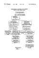
- FIG. 1diagrams the current treatment algorithm for non-palpable breast lesions.
- Biopsiescan be done in a number of different ways for non-palpable lesions, including surgical excisional biopsies and stereotactic and ultrasound guided needle breast biopsies.
- image directed biopsythe radiologist or other physician takes a small sample of the irregular tissue for laboratory analysis. If the biopsy proves to be malignant, additional surgery (typically a lumpectomy or a mastectomy) is required. The patient then returns to the radiologist a day or two later where the biopsy site (the site of the lesion) is relocated by method called needle localization, a preoperative localization in preparation for the surgery.
- Locating the previously biopsied area after surgical excision type of biopsyis usually not a problem because of the deformity caused by the surgery.
- help in relocating the biopsy siteis usually needed.
- One procedure to permit the biopsy site to be relocated by the radiologist during preoperative localizationis to leave some of the suspicious calcifications; this has its drawbacks.
- a small metallic surgical clipsuch as those made by Biopsys.
- the metallic clipcan be deployed through the biopsy needle, and is left at the biopsy site at the time of the original biopsy.
- the radiologisttypically inserts a barbed or hooked wire, such as the Hawkins, Kopans, Homer, Sadowski, and other needles, back into the patient's breast and positions the tip of the wire at the biopsy site using mammography to document the placement.
- the patientis then taken to the operating room with the needle apparatus sticking out of the patient's breast.
- the clipprovides a good indication of the biopsy site to the radiologist during preoperative localization, the clip remains permanently within the 80% of patients with benign diagnoses.
- the clipis necessarily attached to a single position at the periphery of the biopsy site, rather than the center of the biopsy site, its location may provide a misleading indication of the location of diseased tissue during any subsequent medical intervention.
- the soft nature of breast tissuepermits the tip of the barbed or hooked needle to be relatively easily dislodged from the biopsy site.
- the clipis also relatively expensive.
- Another localization methodinvolves the use of laser light from the tip of a optical fiber connected to a laser.
- a pair of hooks at the tip of the optical fibersecures the tip at the biopsy site; the glow indicates the position of the tip through several centimeters of breast tissue.
- This proceduresuffers from some of the same problems associated with the use of barbed or hooked wires.
- Another preoperative localization procedureinjects medical-grade powdered carbon suspension from the lesion to the skin surface. This procedure also has certain problems, including the creation of discontinuities along the carbon trail.
- the present inventionis directed to a biopsy localization method and device which uses a locatable bioabsorbable element left at the biopsy site so that if testing of the biopsy sample indicates a need to do so, the biopsy site can be relocated by finding the bioabsorbable element.
- Thiseliminates the need to use of metallic clips during biopsies and often eliminates the need for a return to the radiologist for preoperative needle localization.
- the bioabsorbable elementcan be used as a therapeutic tool for treatment of the diseased lesion and for hemostasis.
- a biopsy localization device made according to the inventionincludes a bioabsorbable element delivered in a pre-delivery state to a soft tissue biopsy site of a patient by an element delivery device.
- the bioabsorbable elementmay be palpably harder than the surrounding soft tissue at the biopsy site when in the post-delivery state.
- One preferred material used as the bioabsorbable elementis a dehydrated collagen plug. This type of plug may swell and is palpable for subsequent location by the surgeon. The collagen plug may not swell at all. In some situations, such as with small breasted women or where the biopsy site is close to the surface, a non-swellable bioabsorbable material, such as a round pellet of PGA, can be used instead of a swellable bioabsorbable material.
- the bioabsorbable materialcan also be made so that it is absorbed quickly to produce a local tissue inflammation; such a localized inflammation can be used to locate the biopsy site instead of location by palpation.
- a length of bioabsorbable suture material, a collagen filament, or other bioabsorbable material extending from the biopsy site out through the skincan be used.
- the surgeoncan follow the bioabsorbable suture material to the biopsy site in a manner similar to that used with Hawkins needles.
- the bioabsorbable materialmay need to be located by the radiologist, by for example, ultrasound or mammography. In any event the bioabsorbable material will typically be absorbed within about a month of placement.
- the inventionthus eliminates the use of metal clips during biopsies and usually eliminates the need for return to the radiologist for preoperative localization.
- the devicemay also be useful in marking the site of surgical excisional biopsies.
- surgeonsfrequently have difficulty in determining the precise relationship of the previously excised tissue to the surgical wound. Therefore, more tissue is removed than might have been removed had the exact location of the previous lesion been more definite.
- a bioabsorbable elementmay be inserted into the biopsy site during a surgical excisional biopsy before the wound is closed to mark the site for potential wide excision should the biopsy reveal cancer.
- a bioabsorbable elementmay be placed at the biopsy site using a delivery device by partially or completely closing the wound and then depositing the bioabsorbable element through the delivery device and removing the delivery device through the closed incision.
- the presence of the palpable marker within the previous excisional biopsy sitewould allow the surgeon to more easily and confidently remove tissue around this site, and preserve more normal breast tissue.
- the palpable markermay be inserted into the suspicious area of the breast under mammographic or ultrasonic guidance immediately prior to the surgical excisional biopsy. This would provide a palpable locator for the surgeon as described above. In this instance, the marker would only need to be palpable, and not necessarily bioresorbable, since the intent would be to remove it in all cases.
- the inventionin addition to permitting the biopsy site to be located by subsequent palpation or other means, the invention also can provide hemostasis and therapeutic benefits. Since the bioabsorbability can be varied from a day or two to a year or more, the material may be used to treat the diseased tissue and not just locate it. Some current therapies include radiation, chemotherapy, gene therapy as well as other technologies and therapies. Because the bioabsorbability can be easily varied, a medium can be place into the bioabsorbable element and be externally excited or triggered in those cases where the biopsy results are malignant. Further, the bioabsorbability concept can be used for future implantation of a therapeutic agent.
- the bioabsorbable elementis a dehydrated collagen
- this materialcould be used as a reservoir for, for example, delivery of materials that effect chemotherapy, brachytherapy, etc.
- the physicianmay inject, for example, a radiation pellet, a chemotherapeutic agent or a gene therapeutic agent into or adjacent to the bioabsorbable element for direct treatment of the diseased tissue.
- the change in the bioabsorbable elementcan be via one of several ways, such as hydration or desiccation, change in temperature, electrical stimulation, magnetic stimulation, chemical or physical reaction with another material, additives, enzymatic reactions, ionization, electrical charges, absorption, as well as other means.
- the inventionmay employ one or more of these techniques or measures or others, to change the consistency, hardness and or size of the bioabsorbable element between its deployed and non-deployed states.
- the visual detectability of the bioabsorbable elementmay be aided by the use of a coloring agent, such as methylene blue or some other dye.
- the radiographic detectability of the elementmay be enhanced by a radiopaque marker.
- ultrasonic detectabilitymay be enhance by special treatment of the bioresorbable element.
- the bioresorbable elementmay have margins which are roughened so as to prevent migration within the tissues. Filaments extending from the margins of the bioresorbable element may be utilized also to stabilize the position of the device within the cavity.
- the filamentsmay or may not be composed of the same material as the bioresorbable element.
- hemostasishelps to lessen the bleeding and swelling within and about the biopsy site. This can be accomplished by physical or chemical means. That is, the device may swell so that it essential fills the biopsy cavity or the device may have a chemical reaction with blood or blood products to cause effective blood clotting, or both. Other methods for causing local hemostasis are also possible with the invention.
- FIG. 1is a flow diagram of a conventional treatment algorithm for non-palpable breast lesions
- FIG. 2is a flow diagram of a treatment algorithm according to the present invention.
- FIG. 3is a simplified view illustrating a biopsy needle assembly obtaining a tissue sample of an abnormality at a target site
- FIG. 4illustrates the main housing and sheath of the needle biopsy assembly left in place after the tissue sample has been removed leaving a biopsied open region at the target site;
- FIG. 5illustrates the barrel of the delivery device of FIG. 4 inserted into the main housing of the biopsy needle assembly and the plunger depressed injecting the bioabsorbable element into the biopsied open region, thus effectively filling the biopsied open region at the target site;
- FIG. 6illustrates the location of the bioabsorbable element of FIG. 5 with the surgeon using his or her fingers
- FIG. 7illustrates a bioabsorbable thread extending from the bioabsorbable element of FIG. 5 up through the patient's skin, the thread being delivered to the bioabsorbable element using the delivery device of FIGS. 4 and 5.
- FIG. 2illustrates a treatment algorithm 2 according to the present invention.
- a tumor or other abnormalitymay be detected as at 6 .
- the typical responsewill often include additional magnification mammograms or a follow-up mammogram scheduled for some time in the future, such as six months. This is indicated at 8 .
- an image guided needle biopsy by a breast radiologistis typically conducted as at 10 .
- Image guided needle biopsiescan be done in a number of ways. Presently, stereotactic (x-ray) and ultrasound guided needle biopsies are commonly used, primarily because of their accuracy, speed and minimal trauma to the patient.
- Stereotactic needle biopsiestypically use a stereotactic table, such as made by Fisher or Lorad, which provides mammography (x-ray) guidance to a biopsy needle assembly. Ultrasound guided biopsies can be conducted with any one of a number of commercially available instruments.
- An exemplary biopsy needle assembly 14illustrated in FIG. 3, includes a biopsy needle 13 passing through a sheath 20 extending from a hollow main housing 22 . The tip 12 of biopsy needle 13 of biopsy needle assembly 14 is automatically inserted to the abnormality 16 at the target site 18 . Biopsy needle 13 has a laterally directed side opening 24 adjacent to tip 12 used to capture a tissue sample of abnormality 16 .
- Bioabsorbable element 34is, in this preferred embodiment, a plug of dehydrated collagen, such as that sold by several companies such as Davol, Datascope, Integra Life Sciences, Collagen Matrix, Vascular Solutions, et al.
- Bioabsorbable element 34may swell on contact with an aqueous liquid within biopsied open region 26 and substantially fills the biopsied open region as suggested in FIG. 5 .
- bioabsorbable element 34is transformed from its pre-delivery state within barrel 30 to its post-delivery state at region 26 and in the process swells and becomes somewhat softer in its post-delivery state than in its pre-delivery state.
- bioabsorbable element 34is palpably harder, preferably at least about 1.5 times harder, than the surrounding soft tissue, typically breast tissue 36 . This permits bioabsorbable element 34 at the target site 18 to be relocated by palpation of the patient by the physician, see FIG. 6, to find the bioabsorbable element 6 and as discussed in more detail below.
- a bioabsorbable elementcould be made of materials other than collagen and could be in a form other than a solid, relatively hard plug in its pre-delivery state.
- bioabsorbable element 34 in its pre-delivery state within barrel 30could be in a liquid or otherwise flowable form; after being deposited at open region 26 at target site 18 , the bioabsorbable element could change to become palpably harder than the surrounding tissue 36 to permit subsequent relocation of target site 18 by palpation.
- transformation of bioabsorbable element 34is by contact with an aqueous liquid.
- transformation of the bioabsorbable elementwhich can be in terms of, for example, hardness, texture, shape, size, or a combination thereof, can be due to other factors, such as application of thermal energy, radiation, magnetic energy, etc.
- the biopsy sampleis sent to pathology for evaluation at 36 . If the pathology report, which is available a day or two after the biopsy, is benign, the patient is so informed and the bioabsorbable element simply is absorbed by the patient within, for example, a month as at 38 . If the pathology report is positive, so that cancer is found, the biopsied open region 26 at the target site 18 is located by the surgeon by palpation as suggested by FIG. 6 . After finding the target site by palpation, which eliminates the need for preoperative localization by the radiologist, appropriate medical treatment, such as excisional surgery, can be performed.
- bioabsorbable delivery device 32could be used to place bioabsorbable element 34 at the site of the incisional biopsy. After removal of delivery device 32 , the incision would be closed, the biopsy sample would be sent to pathology and the patient would go home with the procedure preceding as discussed above, starting with item 36 .
- bioabsorbable element 34also act as a hemostatic agent to stop bleeding at site 18 by virtue of physical means, by filling or substantially filling open region 26 , as well as chemical means through the chemical interaction, such as coagulation, with blood components.
- bioabsorbable element 34could be covered by a non-hemostatic degradable outer layer so that hemostasis or other action is delayed until the outer layer has been eroded. In some situations, it may be necessary or at least desirable to shield the bioabsorbable element from the blood or other body fluids until after the bioabsorbable element is in place at target site 18 .
- bioabsorbable elementmay be changed from its pre-delivery state to its post-delivery state in a variety of manners including hydration, changing the temperature, electrical stimulation, magnetic stimulation, chemical reaction with a stimulating agent, physically interaction with an activating member (such as a knife blade which could be used to slice open a capsule containing the bioabsorbable element), by ionizing the bioabsorbable element, or by absorption or adsorption of a fluid by the bioabsorbable element.
- an activating membersuch as a knife blade which could be used to slice open a capsule containing the bioabsorbable element
- the inventionmay also be used to medically treat the patient. That is, the bioabsorbable element could include a therapeutic element which would be activated only if the pathology report indicated the need for the medical treatment.
- a bioabsorbable elementcould include a therapeutic element which would be activated only if the pathology report indicated the need for the medical treatment.
- Various ways of activating an agent in a bioabsorbable elementcould be used, such as injecting a radiation-emitting element at the vicinity of the target site, externally irradiating the target site, providing a triggering substance to the target site, manual pressure, photodynamic therapy, sclerosing chemistry, vibrational therapy, ultrasound, and the like.
- the bioabsorbable elementcould be made so that it includes no such activating agent; rather, medical treatment could be provided by, for example, delivery of a chemotherapy agent, a radiation emitting element, thermal energy, electrical energy, vibrational energy, gene therapy, vector therapy, anti-angiogenesis therapy.
- medical treatmentcould be provided by, for example, delivery of a chemotherapy agent, a radiation emitting element, thermal energy, electrical energy, vibrational energy, gene therapy, vector therapy, anti-angiogenesis therapy.
- the bioabsorbable elementmay contain a radiopaque marker or may have properties to aid in detecting it by ultrasound, in addition to being palpable.
- bioabsorbable element 34 in its post-delivery statehave a hardness of at least about one and a half times that of breast tissue so that it is palpably harder than the surrounding tissue.
- bioabsorbable element 34in one embodiment, swells from its pre-delivery state to its post-delivery state so to fill or at least substantially fills open region 26 .
- bioabsorbable element 34swells about 50 to 1500%, and more preferably about 100 to 300%, from the pre-delivery state to the post delivery state, typically when placed in contact with an aqueous liquid. It is preferred that the bioabsorbable element has a longest dimension of at least about 0.5 cm in its post-delivery state to aid its location by palpation.
- the bioabsorbable elementis preferably made of collagen in one embodiment, the bioabsorbable element can include, for example, one or more of the following materials; polyactic and polyglycolic acids, polyorthoesters, resorbable silicones and urethanes, lipids, polysaccharides, starches, ceramics, polyamino acids, proteins, hydrogels and other gels, gelatins, polymers, cellulose, elastin, and the like.
- bioabsorbable filament 44extending from bioabsorbable element 34 through the patient's skin 46 as shown in FIG. 7 . This can be accomplished by delivering bioabsorbable filament 44 through sheath 20 as bioabsorbable element 34 is injected into region 26 at target site 18 . In some situations it may not be possible or desirable to use bioabsorbable element 34 ; in those situations it may be useful to provide for only bioabsorbable filament 44 extending from target site 18 to above the patient's skin 46 .
- bioabsorbable element delivery device 32be guided through a portion of needle assembly 14 , that is sheath 20 and main housing 22 , in some situations it may be useful to cover sheath 20 with an outer sheath which would be left in place after the biopsy sample has been removed and the entire biopsy needle assembly 14 has been removed. The sheath left in place would then be used to guide barrel 30 of delivery device 32 to target site 18 .
- delivery device 32could take a number of different forms such as a syringe containing fluid or paste that is injected through a needle or through the housing 22 and sheath 20 or through an outer sheath.
- other delivery devicescould be employed for delivery of bioresorbable element 34 .
- the inventionhas applicability toward the correction of a defect that is caused by breast tissue removal for biopsy or diseased tissue removal. Collagen is often placed in the body where it is eventually replaced by human autogenous tissue. Hence, the invention could be used for the repair of tissue that has been damaged due to tissue removal.
- the delivery device described heretoforecould be used for installing a material (synthetic or mammalian) into the cavity for such a cosmetic or reconstructive repair.
- the materialwould typically be an effectively non-bioabsorbable material, such as a silicon gel-filled capsule or bag.
Landscapes
- Health & Medical Sciences (AREA)
- Surgery (AREA)
- Life Sciences & Earth Sciences (AREA)
- General Health & Medical Sciences (AREA)
- Veterinary Medicine (AREA)
- Oral & Maxillofacial Surgery (AREA)
- Engineering & Computer Science (AREA)
- Biomedical Technology (AREA)
- Heart & Thoracic Surgery (AREA)
- Medical Informatics (AREA)
- Molecular Biology (AREA)
- Animal Behavior & Ethology (AREA)
- Nuclear Medicine, Radiotherapy & Molecular Imaging (AREA)
- Public Health (AREA)
- Pathology (AREA)
- Materials For Medical Uses (AREA)
- Magnetic Resonance Imaging Apparatus (AREA)
- Surgical Instruments (AREA)
- Radiation-Therapy Devices (AREA)
- Apparatus For Radiation Diagnosis (AREA)
- Media Introduction/Drainage Providing Device (AREA)
- Analysing Materials By The Use Of Radiation (AREA)
- Prostheses (AREA)
- Eye Examination Apparatus (AREA)
Abstract
Description
Claims (57)
Priority Applications (23)
| Application Number | Priority Date | Filing Date | Title |
|---|---|---|---|
| US09/336,360US6270464B1 (en) | 1998-06-22 | 1999-06-18 | Biopsy localization method and device |
| EP10179734.8AEP2258258B1 (en) | 1998-06-22 | 1999-06-21 | Biopsy localization device |
| EP99935335AEP1096875B1 (en) | 1998-06-22 | 1999-06-21 | Biopsy localization device |
| DE69942833TDE69942833D1 (en) | 1998-06-22 | 1999-06-21 | DEVICE FOR BIOPSY LOCATION |
| AT99935335TATE483416T1 (en) | 1998-06-22 | 1999-06-21 | BIOPSY LOCATION DEVICE |
| PCT/US1999/013909WO1999066834A1 (en) | 1998-06-22 | 1999-06-21 | Biopsy localization method and device |
| JP2000555526AJP4472176B2 (en) | 1998-06-22 | 1999-06-21 | Biopsy position confirmation method and instrument |
| PCT/US2000/015495WO2000074561A1 (en) | 1999-06-04 | 2000-06-05 | Tissue removal methods and apparatus |
| JP2001501101AJP2003517346A (en) | 1999-06-04 | 2000-06-05 | Tissue removal method and device |
| US09/588,278US6530923B1 (en) | 1998-02-10 | 2000-06-05 | Tissue removal methods and apparatus |
| EP00942681AEP1191876A4 (en) | 1999-06-04 | 2000-06-05 | Tissue removal methods and apparatus |
| US09/844,661US6602204B2 (en) | 1998-02-10 | 2001-04-27 | Intraoperative tissue treatment methods |
| US09/900,801US6699205B2 (en) | 1998-06-22 | 2001-07-06 | Biopsy localization method and device |
| US10/027,157US6730042B2 (en) | 1998-06-22 | 2001-12-20 | Biopsy localization method and device |
| US10/334,476US20030109896A1 (en) | 1998-02-10 | 2002-12-31 | Tissue removal methods and apparatus |
| US10/463,026US20040010206A1 (en) | 1998-02-10 | 2003-06-17 | Intraoperative tissue treatment methods |
| US10/628,090US20040267155A1 (en) | 1998-06-22 | 2003-07-25 | Biopsy localization method and device |
| US10/747,813US20040199202A1 (en) | 1997-11-12 | 2003-12-29 | Biological passageway occlusion removal |
| US10/839,112US8292822B2 (en) | 1998-06-22 | 2004-05-04 | Biopsy localization method and device |
| US10/839,226US20040204660A1 (en) | 1998-06-22 | 2004-05-04 | Biopsy localization method and device |
| US10/943,434US20050045192A1 (en) | 1998-06-22 | 2004-09-16 | Biopsy localization method and device |
| US10/943,433US10010380B2 (en) | 1998-06-22 | 2004-09-16 | Biopsy localization method and device |
| US11/283,235US20060079829A1 (en) | 1998-06-22 | 2005-11-18 | Biopsy localization method and device |
Applications Claiming Priority (5)
| Application Number | Priority Date | Filing Date | Title |
|---|---|---|---|
| US9024398P | 1998-06-22 | 1998-06-22 | |
| US9273498P | 1998-07-14 | 1998-07-14 | |
| US11486399P | 1999-01-06 | 1999-01-06 | |
| US11742199P | 1999-01-27 | 1999-01-27 | |
| US09/336,360US6270464B1 (en) | 1998-06-22 | 1999-06-18 | Biopsy localization method and device |
Related Parent Applications (1)
| Application Number | Title | Priority Date | Filing Date |
|---|---|---|---|
| US09/248,088Continuation-In-PartUS6221006B1 (en) | 1997-11-12 | 1999-02-09 | Entrapping apparatus and method for use |
Related Child Applications (6)
| Application Number | Title | Priority Date | Filing Date |
|---|---|---|---|
| US09/588,278Continuation-In-PartUS6530923B1 (en) | 1997-11-12 | 2000-06-05 | Tissue removal methods and apparatus |
| US09/844,661Continuation-In-PartUS6602204B2 (en) | 1997-11-12 | 2001-04-27 | Intraoperative tissue treatment methods |
| US09/900,801ContinuationUS6699205B2 (en) | 1998-06-22 | 2001-07-06 | Biopsy localization method and device |
| US10/334,476DivisionUS20030109896A1 (en) | 1998-02-10 | 2002-12-31 | Tissue removal methods and apparatus |
| US10/463,026ContinuationUS20040010206A1 (en) | 1997-11-12 | 2003-06-17 | Intraoperative tissue treatment methods |
| US10/463,026Continuation-In-PartUS20040010206A1 (en) | 1997-11-12 | 2003-06-17 | Intraoperative tissue treatment methods |
Publications (1)
| Publication Number | Publication Date |
|---|---|
| US6270464B1true US6270464B1 (en) | 2001-08-07 |
Family
ID=27536563
Family Applications (8)
| Application Number | Title | Priority Date | Filing Date |
|---|---|---|---|
| US09/336,360Expired - LifetimeUS6270464B1 (en) | 1997-11-12 | 1999-06-18 | Biopsy localization method and device |
| US09/900,801Expired - LifetimeUS6699205B2 (en) | 1998-06-22 | 2001-07-06 | Biopsy localization method and device |
| US10/027,157Expired - LifetimeUS6730042B2 (en) | 1998-06-22 | 2001-12-20 | Biopsy localization method and device |
| US10/839,226AbandonedUS20040204660A1 (en) | 1998-06-22 | 2004-05-04 | Biopsy localization method and device |
| US10/839,112Expired - Fee RelatedUS8292822B2 (en) | 1998-06-22 | 2004-05-04 | Biopsy localization method and device |
| US10/943,434AbandonedUS20050045192A1 (en) | 1998-06-22 | 2004-09-16 | Biopsy localization method and device |
| US10/943,433Expired - Fee RelatedUS10010380B2 (en) | 1998-06-22 | 2004-09-16 | Biopsy localization method and device |
| US11/283,235AbandonedUS20060079829A1 (en) | 1998-06-22 | 2005-11-18 | Biopsy localization method and device |
Family Applications After (7)
| Application Number | Title | Priority Date | Filing Date |
|---|---|---|---|
| US09/900,801Expired - LifetimeUS6699205B2 (en) | 1998-06-22 | 2001-07-06 | Biopsy localization method and device |
| US10/027,157Expired - LifetimeUS6730042B2 (en) | 1998-06-22 | 2001-12-20 | Biopsy localization method and device |
| US10/839,226AbandonedUS20040204660A1 (en) | 1998-06-22 | 2004-05-04 | Biopsy localization method and device |
| US10/839,112Expired - Fee RelatedUS8292822B2 (en) | 1998-06-22 | 2004-05-04 | Biopsy localization method and device |
| US10/943,434AbandonedUS20050045192A1 (en) | 1998-06-22 | 2004-09-16 | Biopsy localization method and device |
| US10/943,433Expired - Fee RelatedUS10010380B2 (en) | 1998-06-22 | 2004-09-16 | Biopsy localization method and device |
| US11/283,235AbandonedUS20060079829A1 (en) | 1998-06-22 | 2005-11-18 | Biopsy localization method and device |
Country Status (6)
| Country | Link |
|---|---|
| US (8) | US6270464B1 (en) |
| EP (2) | EP1096875B1 (en) |
| JP (1) | JP4472176B2 (en) |
| AT (1) | ATE483416T1 (en) |
| DE (1) | DE69942833D1 (en) |
| WO (1) | WO1999066834A1 (en) |
Cited By (111)
| Publication number | Priority date | Publication date | Assignee | Title |
|---|---|---|---|---|
| US6356782B1 (en) | 1998-12-24 | 2002-03-12 | Vivant Medical, Inc. | Subcutaneous cavity marking device and method |
| US20030013989A1 (en)* | 2001-06-29 | 2003-01-16 | Joseph Obermiller | Porous sponge matrix medical devices and methods |
| US6544185B2 (en) | 2000-10-23 | 2003-04-08 | Valentino Montegrande | Ultrasound imaging marker and method of use |
| US6567689B2 (en) | 1999-02-02 | 2003-05-20 | Senorx, Inc. | Methods and chemical preparations for time-limited marking of biopsy sites |
| US20030204137A1 (en)* | 1999-06-17 | 2003-10-30 | Inrad, Inc. | Apparatus for the percutaneous marking of a lesion |
| US20030210810A1 (en)* | 2002-05-08 | 2003-11-13 | Gee, James W. | Method and apparatus for detecting structures of interest |
| US6654629B2 (en) | 2002-01-23 | 2003-11-25 | Valentino Montegrande | Implantable biomarker and method of use |
| US20030225420A1 (en)* | 2002-03-11 | 2003-12-04 | Wardle John L. | Surgical coils and methods of deploying |
| US6662041B2 (en) | 1999-02-02 | 2003-12-09 | Senorx, Inc. | Imageable biopsy site marker |
| US20030233101A1 (en)* | 2002-06-17 | 2003-12-18 | Senorx, Inc. | Plugged tip delivery tube for marker placement |
| US20040030263A1 (en)* | 2001-08-29 | 2004-02-12 | Artemis Medical, Inc. | Undamaged tissue collection assembly and method |
| US6699205B2 (en)* | 1998-06-22 | 2004-03-02 | Artemis Medical, Inc. | Biopsy localization method and device |
| US6725083B1 (en) | 1999-02-02 | 2004-04-20 | Senorx, Inc. | Tissue site markers for in VIVO imaging |
| US20040122350A1 (en)* | 2002-12-20 | 2004-06-24 | Sheng-Ping Zhong | Puncture hole sealing device |
| US20040122349A1 (en)* | 2002-12-20 | 2004-06-24 | Lafontaine Daniel M. | Closure device with textured surface |
| US20040150819A1 (en)* | 2003-02-03 | 2004-08-05 | Inberg Steven Bradley | Phase shift measurement for luminescent light |
| US20040236212A1 (en)* | 2003-05-23 | 2004-11-25 | Senorx, Inc. | Fibrous marker and intracorporeal delivery thereof |
| US20040236211A1 (en)* | 2003-05-23 | 2004-11-25 | Senorx, Inc. | Marker or filler forming fluid |
| US20050010248A1 (en)* | 2003-07-10 | 2005-01-13 | Scimed Life Systems, Inc. | System for closing an opening in a body cavity |
| US20050020899A1 (en)* | 2003-07-25 | 2005-01-27 | Rubicor Medical, Inc. | Post-biopsy cavity treatmetn implants and methods |
| US20050019262A1 (en)* | 2003-07-25 | 2005-01-27 | Rubicor Medical, Inc. | Post-biopsy cavity treatment implants and methods |
| US20050033157A1 (en)* | 2003-07-25 | 2005-02-10 | Klein Dean A. | Multi-modality marking material and method |
| US6862470B2 (en) | 1999-02-02 | 2005-03-01 | Senorx, Inc. | Cavity-filling biopsy site markers |
| US20050070753A1 (en)* | 2003-09-25 | 2005-03-31 | Forman Michael R. | Brachytherapy applicator |
| US20050119562A1 (en)* | 2003-05-23 | 2005-06-02 | Senorx, Inc. | Fibrous marker formed of synthetic polymer strands |
| US20050234336A1 (en)* | 2004-03-26 | 2005-10-20 | Beckman Andrew T | Apparatus and method for marking tissue |
| US7044957B2 (en) | 1994-09-16 | 2006-05-16 | Ethicon Endo-Surgery, Inc. | Devices for defining and marking tissue |
| US20060111646A1 (en)* | 2003-08-13 | 2006-05-25 | Gellman Barry N | Marking biopsy sites |
| US20060116573A1 (en)* | 2003-11-17 | 2006-06-01 | Inrad, Inc. | Self Contained, Self Piercing, Side-Expelling Marking Apparatus |
| US20060151460A1 (en)* | 2005-01-10 | 2006-07-13 | Wardle John L | Eluting coils and methods of deploying and retrieving |
| US20060173296A1 (en)* | 2004-10-13 | 2006-08-03 | Miller Michael E | Site marker visable under multiple modalities |
| US20060235298A1 (en)* | 2005-03-31 | 2006-10-19 | Robert Kotmel | Internal biopsy marking |
| US20070038145A1 (en)* | 2004-11-22 | 2007-02-15 | Inrad, Inc. | Post Decompression Marker Introducer System |
| US20070110665A1 (en)* | 2005-11-17 | 2007-05-17 | Bolan Patrick J | Tissue marker for multimodality radiographic imaging |
| US20080039819A1 (en)* | 2006-08-04 | 2008-02-14 | Senorx, Inc. | Marker formed of starch or other suitable polysaccharide |
| US20080091120A1 (en)* | 2002-05-03 | 2008-04-17 | Biopsy Sciences, Llc | Biodegradable polymer for marking tissue and sealing tracts |
| US20080269603A1 (en)* | 2004-10-13 | 2008-10-30 | Nicoson Zachary R | Site marker visible under multiple modalities |
| US20090216150A1 (en)* | 2008-02-25 | 2009-08-27 | Lee Reichel | Method and Apparatus For Inserting Biopsy Site Marker In Marker Body |
| US20090216181A1 (en)* | 2008-02-25 | 2009-08-27 | Speeg Trevor W V | Biopsy Site Marker Deployment Instrument |
| US7744852B2 (en) | 2003-07-25 | 2010-06-29 | Rubicor Medical, Llc | Methods and systems for marking post biopsy cavity sites |
| US20100280374A1 (en)* | 2007-09-19 | 2010-11-04 | Roberts Walter A | Direct Visualization Robotic Intra-Operative Radiation Therapy Applicator Device |
| EP2324771A1 (en) | 2003-06-06 | 2011-05-25 | Devicor Medical Products, Inc. | Subcutaneous biopsy cavity marker device |
| US20110133730A1 (en)* | 2009-12-04 | 2011-06-09 | Simon Richard Hattersley | Magnetic Probe Apparatus |
| US20110137154A1 (en)* | 2009-12-04 | 2011-06-09 | Simon Richard Hattersley | Magnetic probe apparatus |
| US8064987B2 (en) | 2006-10-23 | 2011-11-22 | C. R. Bard, Inc. | Breast marker |
| US8157862B2 (en) | 1997-10-10 | 2012-04-17 | Senorx, Inc. | Tissue marking implant |
| US8311610B2 (en) | 2008-01-31 | 2012-11-13 | C. R. Bard, Inc. | Biopsy tissue marker |
| US8361082B2 (en) | 1999-02-02 | 2013-01-29 | Senorx, Inc. | Marker delivery device with releasable plug |
| US20130046200A1 (en)* | 2011-08-18 | 2013-02-21 | Marshall Ephraim Stauber | Instrument For Concurrent Injection Of Anesthesia And Removal Of Specimens From A Body |
| US8401622B2 (en) | 2006-12-18 | 2013-03-19 | C. R. Bard, Inc. | Biopsy marker with in situ-generated imaging properties |
| US8486028B2 (en) | 2005-10-07 | 2013-07-16 | Bard Peripheral Vascular, Inc. | Tissue marking apparatus having drug-eluting tissue marker |
| US8498693B2 (en) | 1999-02-02 | 2013-07-30 | Senorx, Inc. | Intracorporeal marker and marker delivery device |
| WO2013192606A1 (en) | 2012-06-22 | 2013-12-27 | Leica Biosystems Nussloch Gmbh | Biopsy tissue sample transport device and method of using thereof |
| US8634899B2 (en) | 2003-11-17 | 2014-01-21 | Bard Peripheral Vascular, Inc. | Multi mode imaging marker |
| US8668737B2 (en) | 1997-10-10 | 2014-03-11 | Senorx, Inc. | Tissue marking implant |
| US8670818B2 (en) | 2008-12-30 | 2014-03-11 | C. R. Bard, Inc. | Marker delivery device for tissue marker placement |
| US8718745B2 (en) | 2000-11-20 | 2014-05-06 | Senorx, Inc. | Tissue site markers for in vivo imaging |
| US20140156003A1 (en)* | 1997-10-10 | 2014-06-05 | Senorx, Inc. | Method of utilizing an implant in a human breast |
| USD715442S1 (en) | 2013-09-24 | 2014-10-14 | C. R. Bard, Inc. | Tissue marker for intracorporeal site identification |
| USD715942S1 (en) | 2013-09-24 | 2014-10-21 | C. R. Bard, Inc. | Tissue marker for intracorporeal site identification |
| USD716451S1 (en) | 2013-09-24 | 2014-10-28 | C. R. Bard, Inc. | Tissue marker for intracorporeal site identification |
| USD716450S1 (en) | 2013-09-24 | 2014-10-28 | C. R. Bard, Inc. | Tissue marker for intracorporeal site identification |
| US9149341B2 (en) | 1999-02-02 | 2015-10-06 | Senorx, Inc | Deployment of polysaccharide markers for treating a site within a patient |
| US9179999B2 (en) | 2013-06-06 | 2015-11-10 | Med-Genesis, Llc | Apparatus and method for installing a stent |
| US9234877B2 (en) | 2013-03-13 | 2016-01-12 | Endomagnetics Ltd. | Magnetic detector |
| US9239314B2 (en) | 2013-03-13 | 2016-01-19 | Endomagnetics Ltd. | Magnetic detector |
| US9327061B2 (en) | 2008-09-23 | 2016-05-03 | Senorx, Inc. | Porous bioabsorbable implant |
| US9492570B2 (en) | 1998-12-24 | 2016-11-15 | Devicor Medical Products, Inc. | Device and method for safe location and marking of a biopsy cavity |
| US9579077B2 (en) | 2006-12-12 | 2017-02-28 | C.R. Bard, Inc. | Multiple imaging mode tissue marker |
| US9615915B2 (en) | 2014-07-25 | 2017-04-11 | Focal Therapeutics, Inc. | Implantable devices and techniques for oncoplastic surgery |
| WO2017083412A1 (en) | 2015-11-11 | 2017-05-18 | Devicor Medical Products, Inc. | Marker delivery device and method of deploying a marker |
| US9669113B1 (en) | 1998-12-24 | 2017-06-06 | Devicor Medical Products, Inc. | Device and method for safe location and marking of a biopsy cavity |
| US9795455B2 (en) | 2014-08-22 | 2017-10-24 | Breast-Med, Inc. | Tissue marker for multimodality radiographic imaging |
| US9808539B2 (en) | 2013-03-11 | 2017-11-07 | Endomagnetics Ltd. | Hypoosmotic solutions for lymph node detection |
| US9820824B2 (en) | 1999-02-02 | 2017-11-21 | Senorx, Inc. | Deployment of polysaccharide markers for treating a site within a patent |
| US20170333154A1 (en)* | 2016-05-20 | 2017-11-23 | David LeBeau | Stabilization device and method for surgical localization wire |
| US20180140288A1 (en)* | 2015-05-06 | 2018-05-24 | Devicor Medical Products, Inc. | Marker delivery device for use with mri breast biopsy system |
| US10092905B2 (en) | 2012-06-22 | 2018-10-09 | Leica Biosystems Nussloch Gmbh | Tissue sample container and methods |
| WO2019067441A1 (en) | 2017-09-26 | 2019-04-04 | Devicor Medical Products, Inc. | Biopsy site marker with microsphere coating |
| US10335124B1 (en) | 2016-02-29 | 2019-07-02 | Devicor Medical Products, Inc. | Marker delivery device with adaptor for biopsy site marking and method of use thereof |
| US10342635B2 (en) | 2005-04-20 | 2019-07-09 | Bard Peripheral Vascular, Inc. | Marking device with retractable cannula |
| US10413381B2 (en) | 2012-04-26 | 2019-09-17 | Focal Therapeutics, Inc. | Surgical implant for marking soft tissue |
| US10595957B2 (en) | 2015-06-04 | 2020-03-24 | Endomagnetics Ltd | Marker materials and forms for magnetic marker localization (MML) |
| US10610841B1 (en) | 2016-06-30 | 2020-04-07 | Devicor Medical Products, Inc. | Marker having enhanced ultrasound visibility and method of manufacturing the same |
| US10643371B2 (en) | 2014-08-11 | 2020-05-05 | Covidien Lp | Treatment procedure planning system and method |
| US10683119B2 (en) | 2014-05-23 | 2020-06-16 | Merit Medical Systems, Inc. | Marker element, device for making a marker element, and method for making a marker element |
| WO2020123350A1 (en) | 2018-12-10 | 2020-06-18 | Devicor Medical Products, Inc. | Biopsy system with end deploy needle |
| WO2020131654A1 (en) | 2018-12-17 | 2020-06-25 | Devicor Medical Products, Inc. | Apparatus for delivering biopsy cavity marker |
| WO2020168026A1 (en) | 2019-02-15 | 2020-08-20 | Devicor Medical Products, Inc. | Marker delivery device with sterile guide |
| US10820825B2 (en) | 2008-10-22 | 2020-11-03 | Cornell University | Method and device for evaluation of local tissue's biological or biomechanical character |
| WO2020243474A1 (en) | 2019-05-30 | 2020-12-03 | Devicor Medical Products, Inc. | Biopsy site marker for limited migration |
| WO2020243386A1 (en) | 2019-05-30 | 2020-12-03 | Devicor Medical Products, Inc. | Shape memory marker deployment device |
| WO2020243470A1 (en) | 2019-05-30 | 2020-12-03 | Devicor Medical Products, Inc. | Methods and apparatus for direct marking |
| WO2021146367A2 (en) | 2020-01-15 | 2021-07-22 | Devicor Medical Products, Inc. | Marker delivery device with push rod having actuation features |
| US11090132B2 (en) | 2017-09-15 | 2021-08-17 | Devicor Medical Products, Inc. | Method for manufacturing marker with aerated hydrogel |
| WO2021188481A2 (en) | 2020-03-17 | 2021-09-23 | Devicor Medical Products, Inc. | Biopsy site markers with non-migration features |
| US11219502B2 (en) | 2017-09-11 | 2022-01-11 | Medtronic Advanced Energy, Llc | Transformative shape-memory polymer tissue cavity marker devices, systems and deployment methods |
| US11241296B2 (en) | 2005-11-17 | 2022-02-08 | Breast-Med, Inc. | Imaging fiducial markers and methods |
| US11324567B2 (en) | 2018-02-01 | 2022-05-10 | Medtronic Advanced Energy, Llc | Expandable tissue cavity marker devices, systems and deployment methods |
| WO2022119911A1 (en) | 2020-12-02 | 2022-06-09 | Devicor Medical Products, Inc. | A marker delivery device configured to decouple plunger and push rod |
| US11752361B2 (en) | 2009-06-01 | 2023-09-12 | Hologic, Inc. | Diagnostic or therapeutic procedure using implantable targets |
| WO2023215090A1 (en) | 2022-05-03 | 2023-11-09 | Devicor Medical Products, Inc. | Biopsy site marker with increased visualization and non-migration features |
| WO2023249760A1 (en) | 2022-06-23 | 2023-12-28 | Devicor Medical Products, Inc. | Biopsy site marker with expandable mesh |
| WO2024039561A1 (en) | 2022-08-16 | 2024-02-22 | Devicor Medical Products, Inc. | Biopsy site marker having movable portions |
| WO2024039560A1 (en) | 2022-08-16 | 2024-02-22 | Devicor Medical Products, Inc. | Biopsy site marker having expandable portion |
| WO2024086055A1 (en) | 2022-10-21 | 2024-04-25 | Devicor Medical Products, Inc. | Biopsy device with end deployment for marker delivery |
| DE102023128960A1 (en) | 2022-10-21 | 2024-05-02 | Devicor Medical Products, Inc. | FLUID DEPLOYMENT MECHANISM FOR BIOPSY SITE MARKER |
| WO2024206458A1 (en) | 2023-03-28 | 2024-10-03 | Devicor Medical Products, Inc. | Biopsy site marker with light emission |
| WO2025072139A1 (en) | 2023-09-28 | 2025-04-03 | Devicor Medical Products, Inc. | Biopsy cavity irrigation |
| US12402867B2 (en) | 2019-02-15 | 2025-09-02 | C.R. Bard, Inc. | Hemostatic biopsy tract article |
| US12426860B2 (en) | 2018-04-18 | 2025-09-30 | C.R. Bard, Inc. | Dual lumen coaxial introducer having integrated tissue marker delivery |
Families Citing this family (81)
| Publication number | Priority date | Publication date | Assignee | Title |
|---|---|---|---|---|
| US6071300A (en) | 1995-09-15 | 2000-06-06 | Sub-Q Inc. | Apparatus and method for percutaneous sealing of blood vessel punctures |
| US6162192A (en) | 1998-05-01 | 2000-12-19 | Sub Q, Inc. | System and method for facilitating hemostasis of blood vessel punctures with absorbable sponge |
| US6183497B1 (en) | 1998-05-01 | 2001-02-06 | Sub-Q, Inc. | Absorbable sponge with contrasting agent |
| US20010045575A1 (en) | 1998-05-01 | 2001-11-29 | Mark Ashby | Device and method for facilitating hemostasis of a biopsy tract |
| US6315753B1 (en) | 1998-05-01 | 2001-11-13 | Sub-Q, Inc. | System and method for facilitating hemostasis of blood vessel punctures with absorbable sponge |
| US20020058882A1 (en)* | 1998-06-22 | 2002-05-16 | Artemis Medical, Incorporated | Biopsy localization method and device |
| US6036698A (en) | 1998-10-30 | 2000-03-14 | Vivant Medical, Inc. | Expandable ring percutaneous tissue removal device |
| US6306132B1 (en) | 1999-06-17 | 2001-10-23 | Vivant Medical | Modular biopsy and microwave ablation needle delivery apparatus adapted to in situ assembly and method of use |
| US6984219B2 (en) | 1999-09-23 | 2006-01-10 | Mark Ashby | Depth and puncture control for blood vessel hemostasis system |
| WO2001060235A2 (en) | 2000-02-18 | 2001-08-23 | Fogarty Thomas J M D | Improved device for accurately marking tissue |
| US6564806B1 (en) | 2000-02-18 | 2003-05-20 | Thomas J. Fogarty | Device for accurately marking tissue |
| US6722371B1 (en) | 2000-02-18 | 2004-04-20 | Thomas J. Fogarty | Device for accurately marking tissue |
| US6540735B1 (en) | 2000-05-12 | 2003-04-01 | Sub-Q, Inc. | System and method for facilitating hemostasis of blood vessel punctures with absorbable sponge |
| US7201725B1 (en) | 2000-09-25 | 2007-04-10 | Sub-Q, Inc. | Device and method for determining a depth of an incision |
| US8187625B2 (en) | 2001-03-12 | 2012-05-29 | Boston Scientific Scimed, Inc. | Cross-linked gelatin composition comprising a wetting agent |
| WO2002087636A1 (en) | 2001-03-12 | 2002-11-07 | Sub-Q, Inc. | Methods for sterilizing cross-linked gelatin compositions |
| US6863680B2 (en) | 2001-11-08 | 2005-03-08 | Sub-Q, Inc. | System and method for delivering hemostasis promoting material to a blood vessel puncture site by fluid pressure |
| US7029489B1 (en) | 2001-05-18 | 2006-04-18 | Sub-Q, Inc. | System and method for delivering hemostasis promoting material to a blood vessel puncture site |
| US7008440B2 (en) | 2001-11-08 | 2006-03-07 | Sub-Q, Inc. | System and method for delivering hemostasis promoting material to a blood vessel puncture site by fluid pressure |
| US6878147B2 (en) | 2001-11-02 | 2005-04-12 | Vivant Medical, Inc. | High-strength microwave antenna assemblies |
| US7192436B2 (en) | 2001-11-08 | 2007-03-20 | Sub-Q, Inc. | Pledget-handling system and method for delivering hemostasis promoting material to a blood vessel puncture site by fluid pressure |
| US7025748B2 (en) | 2001-11-08 | 2006-04-11 | Boston Scientific Scimed, Inc. | Sheath based blood vessel puncture locator and depth indicator |
| US7037322B1 (en) | 2001-11-08 | 2006-05-02 | Sub-Q, Inc. | System and method for delivering hemostasis promoting material to a blood vessel puncture with a staging tube |
| US7037323B2 (en) | 2001-11-08 | 2006-05-02 | Sub-Q, Inc. | Pledget-handling system and method for delivering hemostasis promoting material to a blood vessel puncture site by fluid pressure |
| US7197363B2 (en) | 2002-04-16 | 2007-03-27 | Vivant Medical, Inc. | Microwave antenna having a curved configuration |
| US6752767B2 (en) | 2002-04-16 | 2004-06-22 | Vivant Medical, Inc. | Localization element with energized tip |
| US7455680B1 (en) | 2002-11-04 | 2008-11-25 | Boston Scientific Scimed, Inc. | Apparatus and method for inhibiting blood loss |
| US7169114B2 (en)* | 2003-06-04 | 2007-01-30 | Krause William R | Biopsy and delivery device |
| US7311703B2 (en) | 2003-07-18 | 2007-12-25 | Vivant Medical, Inc. | Devices and methods for cooling microwave antennas |
| JP4673305B2 (en)* | 2003-08-11 | 2011-04-20 | ウィルソン−クック・メディカル・インコーポレーテッド | Surgical graft |
| US8172770B2 (en)* | 2005-09-28 | 2012-05-08 | Suros Surgical Systems, Inc. | System and method for minimally invasive disease therapy |
| US8349443B2 (en)* | 2006-02-23 | 2013-01-08 | Meadwestvaco Corporation | Method for treating a substrate |
| US20080294039A1 (en)* | 2006-08-04 | 2008-11-27 | Senorx, Inc. | Assembly with hemostatic and radiographically detectable pellets |
| CN100408170C (en)* | 2006-08-16 | 2008-08-06 | 哈尔滨工业大学 | Process for preparing CuO/gamma-Al2O3 used as catalyst for catalytic oxidation process by inducing ClO2 with microwave |
| US8068921B2 (en) | 2006-09-29 | 2011-11-29 | Vivant Medical, Inc. | Microwave antenna assembly and method of using the same |
| US9622813B2 (en) | 2007-11-01 | 2017-04-18 | Covidien Lp | Method for volume determination and geometric reconstruction |
| US8292880B2 (en) | 2007-11-27 | 2012-10-23 | Vivant Medical, Inc. | Targeted cooling of deployable microwave antenna |
| US9332973B2 (en) | 2008-10-01 | 2016-05-10 | Covidien Lp | Needle biopsy device with exchangeable needle and integrated needle protection |
| US9186128B2 (en) | 2008-10-01 | 2015-11-17 | Covidien Lp | Needle biopsy device |
| US11298113B2 (en) | 2008-10-01 | 2022-04-12 | Covidien Lp | Device for needle biopsy with integrated needle protection |
| US9782565B2 (en) | 2008-10-01 | 2017-10-10 | Covidien Lp | Endoscopic ultrasound-guided biliary access system |
| US8968210B2 (en) | 2008-10-01 | 2015-03-03 | Covidien LLP | Device for needle biopsy with integrated needle protection |
| US8323249B2 (en) | 2009-08-14 | 2012-12-04 | The Regents Of The University Of Michigan | Integrated vascular delivery system |
| US8814833B2 (en)* | 2010-05-19 | 2014-08-26 | Tangent Medical Technologies Llc | Safety needle system operable with a medical device |
| WO2011146769A2 (en) | 2010-05-19 | 2011-11-24 | Tangent Medical Technologies Llc | Integrated vascular delivery system |
| US20110301456A1 (en)* | 2010-06-07 | 2011-12-08 | Malignext Targeting Technologies, Inc. | Tissue Marking for Lesion Removal |
| US10959769B2 (en) | 2010-11-05 | 2021-03-30 | Ethicon Llc | Surgical instrument with slip ring assembly to power ultrasonic transducer |
| US9039720B2 (en) | 2010-11-05 | 2015-05-26 | Ethicon Endo-Surgery, Inc. | Surgical instrument with ratcheting rotatable shaft |
| US9247986B2 (en) | 2010-11-05 | 2016-02-02 | Ethicon Endo-Surgery, Llc | Surgical instrument with ultrasonic transducer having integral switches |
| US20120116381A1 (en) | 2010-11-05 | 2012-05-10 | Houser Kevin L | Surgical instrument with charging station and wireless communication |
| US10881448B2 (en) | 2010-11-05 | 2021-01-05 | Ethicon Llc | Cam driven coupling between ultrasonic transducer and waveguide in surgical instrument |
| US10660695B2 (en) | 2010-11-05 | 2020-05-26 | Ethicon Llc | Sterile medical instrument charging device |
| US9011471B2 (en) | 2010-11-05 | 2015-04-21 | Ethicon Endo-Surgery, Inc. | Surgical instrument with pivoting coupling to modular shaft and end effector |
| US9510895B2 (en) | 2010-11-05 | 2016-12-06 | Ethicon Endo-Surgery, Llc | Surgical instrument with modular shaft and end effector |
| US9000720B2 (en) | 2010-11-05 | 2015-04-07 | Ethicon Endo-Surgery, Inc. | Medical device packaging with charging interface |
| US9072523B2 (en) | 2010-11-05 | 2015-07-07 | Ethicon Endo-Surgery, Inc. | Medical device with feature for sterile acceptance of non-sterile reusable component |
| US9782215B2 (en) | 2010-11-05 | 2017-10-10 | Ethicon Endo-Surgery, Llc | Surgical instrument with ultrasonic transducer having integral switches |
| US9649150B2 (en) | 2010-11-05 | 2017-05-16 | Ethicon Endo-Surgery, Llc | Selective activation of electronic components in medical device |
| US9597143B2 (en) | 2010-11-05 | 2017-03-21 | Ethicon Endo-Surgery, Llc | Sterile medical instrument charging device |
| US9089338B2 (en) | 2010-11-05 | 2015-07-28 | Ethicon Endo-Surgery, Inc. | Medical device packaging with window for insertion of reusable component |
| US9421062B2 (en) | 2010-11-05 | 2016-08-23 | Ethicon Endo-Surgery, Llc | Surgical instrument shaft with resiliently biased coupling to handpiece |
| US9381058B2 (en) | 2010-11-05 | 2016-07-05 | Ethicon Endo-Surgery, Llc | Recharge system for medical devices |
| US9161803B2 (en) | 2010-11-05 | 2015-10-20 | Ethicon Endo-Surgery, Inc. | Motor driven electrosurgical device with mechanical and electrical feedback |
| US20120116265A1 (en) | 2010-11-05 | 2012-05-10 | Houser Kevin L | Surgical instrument with charging devices |
| US9017851B2 (en) | 2010-11-05 | 2015-04-28 | Ethicon Endo-Surgery, Inc. | Sterile housing for non-sterile medical device component |
| US9375255B2 (en) | 2010-11-05 | 2016-06-28 | Ethicon Endo-Surgery, Llc | Surgical instrument handpiece with resiliently biased coupling to modular shaft and end effector |
| US10085792B2 (en) | 2010-11-05 | 2018-10-02 | Ethicon Llc | Surgical instrument with motorized attachment feature |
| US9017849B2 (en) | 2010-11-05 | 2015-04-28 | Ethicon Endo-Surgery, Inc. | Power source management for medical device |
| US9782214B2 (en) | 2010-11-05 | 2017-10-10 | Ethicon Llc | Surgical instrument with sensor and powered control |
| US9526921B2 (en)* | 2010-11-05 | 2016-12-27 | Ethicon Endo-Surgery, Llc | User feedback through end effector of surgical instrument |
| US20190060028A1 (en)* | 2010-12-16 | 2019-02-28 | Devicor Medical Products, Inc. | Method for identifying a site for surgical removal under magnetic guidance |
| US9414816B2 (en) | 2011-06-23 | 2016-08-16 | Devicor Medical Products, Inc. | Introducer for biopsy device |
| KR102354675B1 (en)* | 2013-08-15 | 2022-01-24 | 인튜어티브 서지컬 오퍼레이션즈 인코포레이티드 | Systems and methods for medical procedure confirmation |
| CA2937744C (en) | 2014-02-04 | 2022-08-09 | Icu Medical, Inc. | Self-priming systems and methods |
| US10136938B2 (en) | 2014-10-29 | 2018-11-27 | Ethicon Llc | Electrosurgical instrument with sensor |
| EP4147649A1 (en)* | 2015-02-10 | 2023-03-15 | Teleflex Life Sciences Limited | Closure device for sealing percutaneous opening in a vessel |
| AU2016306667B2 (en) | 2015-08-13 | 2020-12-03 | Covidien Ag | Electrosurgical method and apparatus with varying stiffness capture components |
| US20160206296A1 (en)* | 2016-03-25 | 2016-07-21 | Hamid Ehsani-Nia | Biopsy Syringe Device |
| EP3458029A1 (en)* | 2016-05-20 | 2019-03-27 | Technical University of Denmark | Palpable marker composition |
| WO2021126213A1 (en)* | 2019-12-19 | 2021-06-24 | Bard Peripheral Vascular, Inc. | Introducer cannula having a pleural access liner for use in crossing pleural layers |
| WO2021158685A1 (en)* | 2020-02-04 | 2021-08-12 | Oncosec Medical Incorporated | Hemostatic combination therapy with low voltage electroporation |
Citations (37)
| Publication number | Priority date | Publication date | Assignee | Title |
|---|---|---|---|---|
| US4248214A (en) | 1979-05-22 | 1981-02-03 | Robert S. Kish | Illuminated urethral catheter |
| US4541438A (en) | 1983-06-02 | 1985-09-17 | The Johns Hopkins University | Localization of cancerous tissue by monitoring infrared fluorescence emitted by intravenously injected porphyrin tumor-specific markers excited by long wavelength light |
| US4592356A (en) | 1984-09-28 | 1986-06-03 | Pedro Gutierrez | Localizing device |
| US4774948A (en) | 1986-11-24 | 1988-10-04 | Markham Charles W | Marking and retraction needle having retrievable stylet |
| US4799495A (en) | 1987-03-20 | 1989-01-24 | National Standard Company | Localization needle assembly |
| US4813422A (en) | 1987-03-06 | 1989-03-21 | Healthcare Technological Resources, Inc. | Bowel control probe and method for controlling bowel incontinence |
| US4817622A (en) | 1986-07-22 | 1989-04-04 | Carl Pennypacker | Infrared imager for viewing subcutaneous location of vascular structures and method of use |
| US4852568A (en) | 1987-02-17 | 1989-08-01 | Kensey Nash Corporation | Method and apparatus for sealing an opening in tissue of a living being |
| US4966583A (en) | 1989-02-03 | 1990-10-30 | Elie Debbas | Apparatus for locating a breast mass |
| US4986279A (en) | 1989-03-01 | 1991-01-22 | National-Standard Company | Localization needle assembly with reinforced needle assembly |
| US5014713A (en) | 1989-12-05 | 1991-05-14 | Tarris Enterprises, Inc. | Method and apparatus for measuring thickness of fat using infrared light |
| US5018530A (en) | 1989-06-15 | 1991-05-28 | Research Corporation Technologies, Inc. | Helical-tipped lesion localization needle device and method of using the same |
| US5080655A (en) | 1988-05-26 | 1992-01-14 | Haaga John R | Medical biopsy needle |
| US5083570A (en) | 1990-06-18 | 1992-01-28 | Mosby Richard A | Volumetric localization/biopsy/surgical device |
| US5158084A (en) | 1989-11-22 | 1992-10-27 | Board Of Regents, The University Of Texas System | Modified localization wire for excisional biopsy |
| US5195988A (en) | 1988-05-26 | 1993-03-23 | Haaga John R | Medical needle with removable sheath |
| US5197482A (en) | 1989-06-15 | 1993-03-30 | Research Corporation Technologies, Inc. | Helical-tipped lesion localization needle device and method of using the same |
| US5221269A (en) | 1990-10-15 | 1993-06-22 | Cook Incorporated | Guide for localizing a nonpalpable breast lesion |
| US5325857A (en) | 1993-07-09 | 1994-07-05 | Hossein Nabai | Skin biopsy device and method |
| US5334216A (en) | 1992-12-10 | 1994-08-02 | Howmedica Inc. | Hemostatic plug |
| US5353804A (en) | 1990-09-18 | 1994-10-11 | Peb Biopsy Corporation | Method and device for percutaneous exisional breast biopsy |
| US5388588A (en) | 1993-05-04 | 1995-02-14 | Nabai; Hossein | Biopsy wound closure device and method |
| US5394886A (en) | 1993-09-20 | 1995-03-07 | Nabai; Hossein | Skin biopsy plug and method |
| US5409004A (en) | 1993-06-11 | 1995-04-25 | Cook Incorporated | Localization device with radiopaque markings |
| US5423321A (en) | 1993-02-11 | 1995-06-13 | Fontenot; Mark G. | Detection of anatomic passages using infrared emitting catheter |
| US5487392A (en) | 1993-11-15 | 1996-01-30 | Haaga; John R. | Biopxy system with hemostatic insert |
| US5517997A (en) | 1994-09-15 | 1996-05-21 | Gabriel Medical, Inc. | Transillumination of body members for protection during body invasive procedures |
| US5526822A (en) | 1994-03-24 | 1996-06-18 | Biopsys Medical, Inc. | Method and apparatus for automated biopsy and collection of soft tissue |
| US5556410A (en) | 1993-09-27 | 1996-09-17 | M3 Systems, Inc. | Surgical needle with stress-relocation means |
| US5647374A (en) | 1994-12-30 | 1997-07-15 | North American Scientific | Needle for imaging and sampling |
| US5660185A (en) | 1995-04-13 | 1997-08-26 | Neovision Corporation | Image-guided biopsy apparatus with enhanced imaging and methods |
| US5795308A (en) | 1995-03-09 | 1998-08-18 | Russin; Lincoln D. | Apparatus for coaxial breast biopsy |
| US5807276A (en) | 1995-03-09 | 1998-09-15 | Russin; Lincoln David | Biopsy device and method |
| EP0894503A2 (en) | 1997-08-01 | 1999-02-03 | Schneider (Usa) Inc. | Bioabsorbable marker having radiopaque constituents and method of using same |
| WO2000038579A2 (en) | 1998-12-24 | 2000-07-06 | Vivant Medical, Inc. | Device and method for safe location and marking of a cavity and sentinel lymph nodes |
| US6161034A (en) | 1999-02-02 | 2000-12-12 | Senorx, Inc. | Methods and chemical preparations for time-limited marking of biopsy sites |
| US6183497B1 (en) | 1998-05-01 | 2001-02-06 | Sub-Q, Inc. | Absorbable sponge with contrasting agent |
Family Cites Families (175)
| Publication number | Priority date | Publication date | Assignee | Title |
|---|---|---|---|---|
| US26201A (en)* | 1859-11-22 | Improvement in sewing-machines | ||
| US2899362A (en)* | 1959-08-11 | Hemostatic sponges and method of | ||
| DE935625C (en) | 1952-10-18 | 1955-11-24 | Guenther Bodendieck | Excision device |
| US3001522A (en) | 1957-12-26 | 1961-09-26 | Silverman Irving | Biopsy device |
| US3194239A (en)* | 1963-01-16 | 1965-07-13 | Cornelius J P Sullivan | Suture provided with radiopaque free metal |
| US3823212A (en) | 1968-11-27 | 1974-07-09 | Freudenberg C Fa | Process for the production of collagen fiber fabrics in the form of felt-like membranes or sponge-like layers |
| CS151338B1 (en)* | 1971-01-22 | 1973-10-19 | ||
| US3976071A (en) | 1974-01-07 | 1976-08-24 | Dynatech Corporation | Methods of improving control of release rates and products useful in same |
| US4197846A (en)* | 1974-10-09 | 1980-04-15 | Louis Bucalo | Method for structure for situating in a living body agents for treating the body |
| US3996938A (en) | 1975-07-10 | 1976-12-14 | Clark Iii William T | Expanding mesh catheter |
| US4034759A (en)* | 1975-08-27 | 1977-07-12 | Xomed, Inc. | Moisture-expandable prosthesis |
| US4007732A (en)* | 1975-09-02 | 1977-02-15 | Robert Carl Kvavle | Method for location and removal of soft tissue in human biopsy operations |
| DE2821048C2 (en) | 1978-05-13 | 1980-07-17 | Willy Ruesch Gmbh & Co Kg, 7053 Kernen | Medical instrument |
| US4230123A (en) | 1978-10-31 | 1980-10-28 | Hawkins Jr Irvin F | Needle sheath complex and process for decompression and biopsy |
| FR2460657A1 (en) | 1979-07-12 | 1981-01-30 | Anvar | BIODEGRADABLE IMPLANT FOR USE AS A BONE PROSTHESIS PIECE |
| US4331577A (en) | 1979-09-28 | 1982-05-25 | Union Carbide Corporation | Latex polymerization process |
| DE2943520C2 (en)* | 1979-10-27 | 1982-05-19 | Fa. Carl Freudenberg, 6940 Weinheim | Process for the production of collagen sponge for medical or cosmetic purposes |
| US4331654A (en)* | 1980-06-13 | 1982-05-25 | Eli Lilly And Company | Magnetically-localizable, biodegradable lipid microspheres |
| US4298998A (en) | 1980-12-08 | 1981-11-10 | Naficy Sadeque S | Breast prosthesis with biologically absorbable outer container |
| US4425908A (en)* | 1981-10-22 | 1984-01-17 | Beth Israel Hospital | Blood clot filter |
| US4545367A (en) | 1982-07-16 | 1985-10-08 | Cordis Corporation | Detachable balloon catheter and method of use |
| US4438253A (en) | 1982-11-12 | 1984-03-20 | American Cyanamid Company | Poly(glycolic acid)/poly(alkylene glycol) block copolymers and method of manufacturing the same |
| US4531933A (en) | 1982-12-07 | 1985-07-30 | C. R. Bard, Inc. | Helical ureteral stent |
| US4647480A (en)* | 1983-07-25 | 1987-03-03 | Amchem Products, Inc. | Use of additive in aqueous cure of autodeposited coatings |
| CH661199A5 (en) | 1983-12-22 | 1987-07-15 | Sulzer Ag | MARKING IMPLANT. |
| MX163953B (en) | 1984-03-27 | 1992-07-03 | Univ New Jersey Med | PROCEDURE FOR PREPARING A BIODEGRADABLE COLLAGEN MATRIX |
| US4611594A (en) | 1984-04-11 | 1986-09-16 | Northwestern University | Medical instrument for containment and removal of calculi |
| JPS6144825A (en) | 1984-08-09 | 1986-03-04 | Unitika Ltd | Hemostatic agent |
| JPS6176147A (en)* | 1984-09-21 | 1986-04-18 | オリンパス光学工業株式会社 | High frequency incision appliance |
| US5186922A (en) | 1985-03-15 | 1993-02-16 | See/Shell Biotechnology, Inc. | Use of biodegradable microspheres labeled with imaging energy constrast materials |
| US4608965A (en) | 1985-03-27 | 1986-09-02 | Anspach Jr William E | Endoscope retainer and tissue retracting device |
| US4787391A (en) | 1985-06-17 | 1988-11-29 | Elefteriades John A | Anastomotic marking device and related method |
| DE3522626A1 (en) | 1985-06-25 | 1987-01-08 | Merz & Co Gmbh & Co | SOLUBLE COLLAGEN SPONGE |
| US4650466A (en)* | 1985-11-01 | 1987-03-17 | Angiobrade Partners | Angioplasty device |
| US4847049A (en) | 1985-12-18 | 1989-07-11 | Vitaphore Corporation | Method of forming chelated collagen having bactericidal properties |
| US4693237A (en) | 1986-01-21 | 1987-09-15 | Hoffman Richard B | Radiopaque coded ring markers for use in identifying surgical grafts |
| US4682606A (en) | 1986-02-03 | 1987-07-28 | Decaprio Vincent H | Localizing biopsy apparatus |
| US4763642A (en) | 1986-04-07 | 1988-08-16 | Horowitz Bruce S | Intracavitational brachytherapy |
| US4832686A (en)* | 1986-06-24 | 1989-05-23 | Anderson Mark E | Method for administering interleukin-2 |
| DE3789320T2 (en) | 1986-07-30 | 1994-06-09 | Sumitomo Pharma | Administration instrument for the introduction of solid medication. |
| US4787384A (en)* | 1986-10-06 | 1988-11-29 | Bio Medic Data System, Inc. | Animal marker implanting system |
| US5002548A (en)* | 1986-10-06 | 1991-03-26 | Bio Medic Data Systems, Inc. | Animal marker implanting system |
| US4744364A (en)* | 1987-02-17 | 1988-05-17 | Intravascular Surgical Instruments, Inc. | Device for sealing percutaneous puncture in a vessel |
| US4817600A (en) | 1987-05-22 | 1989-04-04 | Medi-Tech, Inc. | Implantable filter |
| US5120802A (en)* | 1987-12-17 | 1992-06-09 | Allied-Signal Inc. | Polycarbonate-based block copolymers and devices |
| US4907589A (en)* | 1988-04-29 | 1990-03-13 | Cosman Eric R | Automatic over-temperature control apparatus for a therapeutic heating device |
| US4838280A (en) | 1988-05-26 | 1989-06-13 | Haaga John R | Hemostatic sheath for a biopsy needle and method of use |
| US4832055A (en) | 1988-07-08 | 1989-05-23 | Palestrant Aubrey M | Mechanically locking blood clot filter |
| US5074840A (en)* | 1990-07-24 | 1991-12-24 | Inbae Yoon | Packing device and method of packing for endoscopic procedures |
| US5085629A (en)* | 1988-10-06 | 1992-02-04 | Medical Engineering Corporation | Biodegradable stent |
| US4909250A (en)* | 1988-11-14 | 1990-03-20 | Smith Joseph R | Implant system for animal identification |
| US5207705A (en)* | 1988-12-08 | 1993-05-04 | Brigham And Women's Hospital | Prosthesis of foam polyurethane and collagen and uses thereof |
| US5258028A (en) | 1988-12-12 | 1993-11-02 | Ersek Robert A | Textured micro implants |
| US5183463A (en)* | 1989-02-03 | 1993-02-02 | Elie Debbas | Apparatus for locating a breast mass |
| DE3913935A1 (en) | 1989-04-27 | 1990-10-31 | Wiedeck Joerg Guenter Dr Med | Catheter for removing stones from the ureter - consists of plastics tube with retractable mandrel |
| USRE34056E (en)* | 1989-07-31 | 1992-09-08 | C.R. Bard, Inc. | Tissue sampling device |
| DE8910603U1 (en)* | 1989-09-06 | 1989-12-07 | Günther, Rolf W., Prof. Dr. | Device for removing blood clots from arteries and veins |
| US5030201A (en) | 1989-11-24 | 1991-07-09 | Aubrey Palestrant | Expandable atherectomy catheter device |
| US5334381A (en) | 1989-12-22 | 1994-08-02 | Unger Evan C | Liposomes as contrast agents for ultrasonic imaging and methods for preparing the same |
| US5469854A (en) | 1989-12-22 | 1995-11-28 | Imarx Pharmaceutical Corp. | Methods of preparing gas-filled liposomes |
| US5236410A (en) | 1990-08-02 | 1993-08-17 | Ferrotherm International, Inc. | Tumor treatment method |
| US5342283A (en) | 1990-08-13 | 1994-08-30 | Good Roger R | Endocurietherapy |
| US5100423A (en)* | 1990-08-21 | 1992-03-31 | Medical Engineering & Development Institute, Inc. | Ablation catheter |
| US5391183A (en) | 1990-09-21 | 1995-02-21 | Datascope Investment Corp | Device and method sealing puncture wounds |
| US5108421A (en)* | 1990-10-01 | 1992-04-28 | Quinton Instrument Company | Insertion assembly and method of inserting a vessel plug into the body of a patient |
| US5192300A (en)* | 1990-10-01 | 1993-03-09 | Quinton Instrument Company | Insertion assembly and method of inserting a vessel plug into the body of a patient |
| US5282781A (en)* | 1990-10-25 | 1994-02-01 | Omnitron International Inc. | Source wire for localized radiation treatment of tumors |
| US5148813A (en) | 1990-11-20 | 1992-09-22 | Bucalo Brian D | Biopsy instrument with tissue specimen retaining and retrieval device |
| US5127916A (en) | 1991-01-22 | 1992-07-07 | Medical Device Technologies, Inc. | Localization needle assembly |
| US5370901A (en) | 1991-02-15 | 1994-12-06 | Bracco International B.V. | Compositions for increasing the image contrast in diagnostic investigations of the digestive tract of patients |
| US5205290A (en)* | 1991-04-05 | 1993-04-27 | Unger Evan C | Low density microspheres and their use as contrast agents for computed tomography |
| US5183464A (en)* | 1991-05-17 | 1993-02-02 | Interventional Thermodynamics, Inc. | Radially expandable dilator |
| US5803901A (en) | 1991-05-29 | 1998-09-08 | Origin Medsystems, Inc. | Inflatable devices for separating layers of tissue and methods of using |
| US5370134A (en)* | 1991-05-29 | 1994-12-06 | Orgin Medsystems, Inc. | Method and apparatus for body structure manipulation and dissection |
| US5195540A (en)* | 1991-08-12 | 1993-03-23 | Samuel Shiber | Lesion marking process |
| CA2078530A1 (en) | 1991-09-23 | 1993-03-24 | Jay Erlebacher | Percutaneous arterial puncture seal device and insertion tool therefore |
| US5282827A (en) | 1991-11-08 | 1994-02-01 | Kensey Nash Corporation | Hemostatic puncture closure system and method of use |
| US5411520A (en)* | 1991-11-08 | 1995-05-02 | Kensey Nash Corporation | Hemostatic vessel puncture closure system utilizing a plug located within the puncture tract spaced from the vessel, and method of use |
| FR2686499A1 (en) | 1992-01-28 | 1993-07-30 | Technomed Int Sa | APPARATUS FOR TREATING A TARGET, SUCH AS A DAMAGE WITHIN THE BODY OF A MAMMAL, PARTICULARLY A HUMAN BEING, USING A MARKING ELEMENT IMPLANTED IN OR IN THE VICINITY OF THE TARGET TO CONTROL THERAPY OF THE SAME TARGET. |
| US5204382A (en)* | 1992-02-28 | 1993-04-20 | Collagen Corporation | Injectable ceramic compositions and methods for their preparation and use |
| US5674468A (en) | 1992-03-06 | 1997-10-07 | Nycomed Imaging As | Contrast agents comprising gas-containing or gas-generating polymer microparticles or microballoons |
| US5656297A (en) | 1992-03-12 | 1997-08-12 | Alkermes Controlled Therapeutics, Incorporated | Modulated release from biocompatible polymers |
| EP0633798B1 (en) | 1992-03-31 | 2003-05-07 | Boston Scientific Corporation | Vascular filter |
| FR2689400B1 (en) | 1992-04-03 | 1995-06-23 | Inoteb | BONE PROSTHESIS MATERIAL CONTAINING CALCIUM CARBONATE PARTICLES DISPERSED IN A BIORESORBABLE POLYMER MATRIX. |
| JPH0616985A (en)* | 1992-04-22 | 1994-01-25 | Lexmark Internatl Inc | Jet ink that does not form solids |
| US5326350A (en) | 1992-05-11 | 1994-07-05 | Li Shu Tung | Soft tissue closure systems |
| NL9200844A (en) | 1992-05-13 | 1993-12-01 | De Wijdeven Gijsbertus G P Van | DEVICE AND METHOD FOR INJECTING WITH A SOLID SUBSTANCE. |
| US5518730A (en) | 1992-06-03 | 1996-05-21 | Fuisz Technologies Ltd. | Biodegradable controlled release flash flow melt-spun delivery system |
| US5514379A (en)* | 1992-08-07 | 1996-05-07 | The General Hospital Corporation | Hydrogel compositions and methods of use |
| US5716407A (en)* | 1992-08-24 | 1998-02-10 | Lipomatrix, Incorporated | Method of rendering identifiable a living tissue implant using an electrical transponder marker |
| US5792157A (en) | 1992-11-13 | 1998-08-11 | Scimed Life Systems, Inc. | Expandable intravascular occlusion material removal devices and methods of use |
| US5330483A (en) | 1992-12-18 | 1994-07-19 | Advanced Surgical Inc. | Specimen reduction device |
| DK12293D0 (en) | 1993-02-02 | 1993-02-02 | Novo Nordisk As | HETEROCYCLIC COMPOUNDS AND THEIR PREPARATION AND USE |
| US5431676A (en) | 1993-03-05 | 1995-07-11 | Innerdyne Medical, Inc. | Trocar system having expandable port |
| US5576016A (en) | 1993-05-18 | 1996-11-19 | Pharmos Corporation | Solid fat nanoemulsions as drug delivery vehicles |
| US5417697A (en)* | 1993-07-07 | 1995-05-23 | Wilk; Peter J. | Polyp retrieval assembly with cauterization loop and suction web |
| DE69431956T2 (en) | 1993-07-12 | 2003-09-04 | The Regents Of The University Of California, Oakland | REINFORCEMENT DEVICE FOR SOFT TISSUE |
| US5494030A (en)* | 1993-08-12 | 1996-02-27 | Trustees Of Dartmouth College | Apparatus and methodology for determining oxygen in biological systems |
| US5445128A (en)* | 1993-08-27 | 1995-08-29 | Detroit Diesel Corporation | Method for engine control |
| US5676698A (en) | 1993-09-07 | 1997-10-14 | Datascope Investment Corp. | Soft tissue implant |
| DE4330958A1 (en) | 1993-09-09 | 1995-03-16 | Schering Ag | Novel microparticles containing active compound, media containing these, their use for the ultrasonically controlled release of active compounds and process for the production thereof |
| US5415656A (en)* | 1993-09-28 | 1995-05-16 | American Medical Systems, Inc. | Electrosurgical apparatus |
| US5507813A (en)* | 1993-12-09 | 1996-04-16 | Osteotech, Inc. | Shaped materials derived from elongate bone particles |
| FR2714284B1 (en) | 1993-12-23 | 1996-03-08 | Hubert Petitier | Prosthesis for the closure of ruptures of the cardiac walls, in particular interventricular septal ruptures. |
| US5443515A (en) | 1994-01-26 | 1995-08-22 | Implex Corporation | Vertebral body prosthetic implant with slidably positionable stabilizing member |
| DE4403789A1 (en) | 1994-02-03 | 1995-08-10 | Schering Ag | Means for visually marking body tissues |
| US5626611A (en)* | 1994-02-10 | 1997-05-06 | United States Surgical Corporation | Composite bioabsorbable materials and surgical articles made therefrom |
| US5649547A (en)* | 1994-03-24 | 1997-07-22 | Biopsys Medical, Inc. | Methods and devices for automated biopsy and collection of soft tissue |
| US5422730A (en)* | 1994-03-25 | 1995-06-06 | Barlow; Clyde H. | Automated optical detection of tissue perfusion by microspheres |
| US5454790A (en) | 1994-05-09 | 1995-10-03 | Innerdyne, Inc. | Method and apparatus for catheterization access |
| US5794626A (en) | 1994-08-18 | 1998-08-18 | Kieturakis; Maciej J. | Excisional stereotactic apparatus |
| US5643282A (en) | 1994-08-22 | 1997-07-01 | Kieturakis; Maciej J. | Surgical instrument and method for removing tissue from an endoscopic workspace |
| JPH10508504A (en)* | 1994-09-16 | 1998-08-25 | バイオプシス メディカル インコーポレイテッド | Method and apparatus for identifying and marking tissue |
| US5716404A (en)* | 1994-12-16 | 1998-02-10 | Massachusetts Institute Of Technology | Breast tissue engineering |
| US5643246A (en) | 1995-02-24 | 1997-07-01 | Gel Sciences, Inc. | Electromagnetically triggered, responsive gel based drug delivery device |
| GB2301362B (en)* | 1995-05-30 | 1999-01-06 | Johnson & Johnson Medical | Absorbable implant materials having controlled porosity |
| US6231834B1 (en)* | 1995-06-07 | 2001-05-15 | Imarx Pharmaceutical Corp. | Methods for ultrasound imaging involving the use of a contrast agent and multiple images and processing of same |
| US5609629A (en) | 1995-06-07 | 1997-03-11 | Med Institute, Inc. | Coated implantable medical device |
| US6774278B1 (en) | 1995-06-07 | 2004-08-10 | Cook Incorporated | Coated implantable medical device |
| CA2178541C (en)* | 1995-06-07 | 2009-11-24 | Neal E. Fearnot | Implantable medical device |
| US6162192A (en) | 1998-05-01 | 2000-12-19 | Sub Q, Inc. | System and method for facilitating hemostasis of blood vessel punctures with absorbable sponge |
| US6071301A (en)* | 1998-05-01 | 2000-06-06 | Sub Q., Inc. | Device and method for facilitating hemostasis of a biopsy tract |
| US5645566A (en) | 1995-09-15 | 1997-07-08 | Sub Q Inc. | Apparatus and method for percutaneous sealing of blood vessel punctures |
| CA2187975C (en) | 1995-10-20 | 2001-05-01 | Lisa W. Heaton | Surgical apparatus and method for marking tissue location |
| US6589502B1 (en) | 1995-11-27 | 2003-07-08 | International Brachytherapy S.A. | Radioisotope dispersed in a matrix for brachytherapy |
| US5752974A (en)* | 1995-12-18 | 1998-05-19 | Collagen Corporation | Injectable or implantable biomaterials for filling or blocking lumens and voids of the body |
| US5636255A (en)* | 1996-03-05 | 1997-06-03 | Queen's University At Kingston | Method and apparatus for CT image registration |
| BR9708046A (en) | 1996-03-11 | 2000-01-04 | Focal Inc | Method for local radiotherapy in a patient and its composition. |
| US5670161A (en) | 1996-05-28 | 1997-09-23 | Healy; Kevin E. | Biodegradable stent |
| US5735289A (en)* | 1996-08-08 | 1998-04-07 | Pfeffer; Herbert G. | Method and apparatus for organic specimen retrieval |
| US5902310A (en)* | 1996-08-12 | 1999-05-11 | Ethicon Endo-Surgery, Inc. | Apparatus and method for marking tissue |
| US7049346B1 (en) | 1996-08-20 | 2006-05-23 | Menlo Care Div Of Ethicon, Inc. | Swollen hydrogel for sphincter augmentation |
| US6066325A (en)* | 1996-08-27 | 2000-05-23 | Fusion Medical Technologies, Inc. | Fragmented polymeric compositions and methods for their use |
| US5676146B1 (en) | 1996-09-13 | 2000-04-18 | Osteotech Inc | Surgical implant containing a resorbable radiopaque marker and method of locating such within a body |
| US6083522A (en) | 1997-01-09 | 2000-07-04 | Neucoll, Inc. | Devices for tissue repair and methods for preparation and use thereof |
| US5827324A (en) | 1997-03-06 | 1998-10-27 | Scimed Life Systems, Inc. | Distal protection device |
| AU6784598A (en) | 1997-04-23 | 1998-11-13 | Vascular Science Inc. | Medical plug |
| US5868708A (en)* | 1997-05-07 | 1999-02-09 | Applied Medical Resources Corporation | Balloon catheter apparatus and method |
| US5911734A (en)* | 1997-05-08 | 1999-06-15 | Embol-X, Inc. | Percutaneous catheter and guidewire having filter and medical device deployment capabilities |
| US6271278B1 (en) | 1997-05-13 | 2001-08-07 | Purdue Research Foundation | Hydrogel composites and superporous hydrogel composites having fast swelling, high mechanical strength, and superabsorbent properties |
| US5928260A (en) | 1997-07-10 | 1999-07-27 | Scimed Life Systems, Inc. | Removable occlusion system for aneurysm neck |
| JP4255208B2 (en) | 1997-07-24 | 2009-04-15 | レックス メディカル リミテッド パートナーシップ | Device for resecting subcutaneous target tissue mass |
| US6340367B1 (en) | 1997-08-01 | 2002-01-22 | Boston Scientific Scimed, Inc. | Radiopaque markers and methods of using the same |
| US5980564A (en) | 1997-08-01 | 1999-11-09 | Schneider (Usa) Inc. | Bioabsorbable implantable endoprosthesis with reservoir |
| US5899935A (en)* | 1997-08-04 | 1999-05-04 | Schneider (Usa) Inc. | Balloon expandable braided stent with restraint |
| US5947836A (en)* | 1997-08-26 | 1999-09-07 | Callaway Golf Company | Integral molded grip and shaft |
| US6638308B2 (en) | 1997-10-10 | 2003-10-28 | John D. Corbitt, Jr. | Bioabsorbable breast implant |
| WO1999018886A1 (en) | 1997-10-10 | 1999-04-22 | Corbitt John D Jr | Breast implant |
| US6309420B1 (en) | 1997-10-14 | 2001-10-30 | Parallax Medical, Inc. | Enhanced visibility materials for implantation in hard tissue |
| US6015541A (en)* | 1997-11-03 | 2000-01-18 | Micro Therapeutics, Inc. | Radioactive embolizing compositions |
| US6270464B1 (en)* | 1998-06-22 | 2001-08-07 | Artemis Medical, Inc. | Biopsy localization method and device |
| US6331166B1 (en) | 1998-03-03 | 2001-12-18 | Senorx, Inc. | Breast biopsy system and method |
| US6540693B2 (en)* | 1998-03-03 | 2003-04-01 | Senorx, Inc. | Methods and apparatus for securing medical instruments to desired locations in a patients body |
| US6261241B1 (en) | 1998-03-03 | 2001-07-17 | Senorx, Inc. | Electrosurgical biopsy device and method |
| US6312429B1 (en) | 1998-09-01 | 2001-11-06 | Senorx, Inc. | Electrosurgical lesion location device |
| WO1999044643A1 (en)* | 1998-03-06 | 1999-09-10 | Biosepra Medical Inc. | Implantable particles for tissue bulking and the treatment of gastroesophageal reflux disease, urinary incontinence, and skin wrinkles |
| US6347241B2 (en)* | 1999-02-02 | 2002-02-12 | Senorx, Inc. | Ultrasonic and x-ray detectable biopsy site marker and apparatus for applying it |
| US6113629A (en)* | 1998-05-01 | 2000-09-05 | Micrus Corporation | Hydrogel for the therapeutic treatment of aneurysms |
| US20020058882A1 (en) | 1998-06-22 | 2002-05-16 | Artemis Medical, Incorporated | Biopsy localization method and device |
| US6086543A (en) | 1998-06-24 | 2000-07-11 | Rubicor Medical, Inc. | Fine needle and core biopsy devices and methods |
| WO2000006243A2 (en)* | 1998-07-28 | 2000-02-10 | Innerdyne, Inc. | Absorbable brachytherapy and chemotherapy delivery devices and methods |
| US6136014A (en) | 1998-09-01 | 2000-10-24 | Vivant Medical, Inc. | Percutaneous tissue removal device |
| US6022362A (en) | 1998-09-03 | 2000-02-08 | Rubicor Medical, Inc. | Excisional biopsy devices and methods |
| US6056700A (en)* | 1998-10-13 | 2000-05-02 | Emx, Inc. | Biopsy marker assembly and method of use |
| CA2319443C (en)* | 1998-12-01 | 2009-09-29 | Cook Biotech, Inc. | Collagenous biomaterials formed with submucosal tissue |
| US6356782B1 (en)* | 1998-12-24 | 2002-03-12 | Vivant Medical, Inc. | Subcutaneous cavity marking device and method |
| US6376742B1 (en)* | 1999-02-17 | 2002-04-23 | Richard J. Zdrahala | In vivo tissue engineering with biodegradable polymers |
| EP1155418B1 (en) | 1999-02-25 | 2007-04-11 | GE Healthcare Limited | Medical tools and devices with improved ultrasound visibility |
| US6053876A (en) | 1999-06-09 | 2000-04-25 | Fisher; John | Apparatus and method for marking non-palpable lesions |
| US6277083B1 (en) | 1999-12-27 | 2001-08-21 | Neothermia Corporation | Minimally invasive intact recovery of tissue |
| EP1142535B1 (en) | 2000-04-07 | 2012-10-03 | Collagen Matrix, Inc. | Embolization device |
| WO2002005717A1 (en) | 2000-07-18 | 2002-01-24 | Senorx, Inc. | Apparatus and method for tissue capture |
| WO2002020571A2 (en) | 2000-09-08 | 2002-03-14 | Biomerieux B.V.A. | Attenuated hiv strains and use thereof |
| US6605047B2 (en)* | 2001-09-10 | 2003-08-12 | Vivant Medical, Inc. | Biopsy marker delivery system |
| US7783336B2 (en)* | 2003-06-06 | 2010-08-24 | Ethicon Endo-Surgery, Inc. | Subcutaneous biopsy cavity marker device |
- 1999
- 1999-06-18USUS09/336,360patent/US6270464B1/ennot_activeExpired - Lifetime
- 1999-06-21WOPCT/US1999/013909patent/WO1999066834A1/enactiveApplication Filing
- 1999-06-21EPEP99935335Apatent/EP1096875B1/ennot_activeExpired - Lifetime
- 1999-06-21ATAT99935335Tpatent/ATE483416T1/ennot_activeIP Right Cessation
- 1999-06-21JPJP2000555526Apatent/JP4472176B2/ennot_activeExpired - Fee Related
- 1999-06-21DEDE69942833Tpatent/DE69942833D1/ennot_activeExpired - Lifetime
- 1999-06-21EPEP10179734.8Apatent/EP2258258B1/ennot_activeExpired - Lifetime
- 2001
- 2001-07-06USUS09/900,801patent/US6699205B2/ennot_activeExpired - Lifetime
- 2001-12-20USUS10/027,157patent/US6730042B2/ennot_activeExpired - Lifetime
- 2004
- 2004-05-04USUS10/839,226patent/US20040204660A1/ennot_activeAbandoned
- 2004-05-04USUS10/839,112patent/US8292822B2/ennot_activeExpired - Fee Related
- 2004-09-16USUS10/943,434patent/US20050045192A1/ennot_activeAbandoned
- 2004-09-16USUS10/943,433patent/US10010380B2/ennot_activeExpired - Fee Related
- 2005
- 2005-11-18USUS11/283,235patent/US20060079829A1/ennot_activeAbandoned
Patent Citations (40)
| Publication number | Priority date | Publication date | Assignee | Title |
|---|---|---|---|---|
| US4248214A (en) | 1979-05-22 | 1981-02-03 | Robert S. Kish | Illuminated urethral catheter |
| US4541438A (en) | 1983-06-02 | 1985-09-17 | The Johns Hopkins University | Localization of cancerous tissue by monitoring infrared fluorescence emitted by intravenously injected porphyrin tumor-specific markers excited by long wavelength light |
| US4592356A (en) | 1984-09-28 | 1986-06-03 | Pedro Gutierrez | Localizing device |
| US4817622A (en) | 1986-07-22 | 1989-04-04 | Carl Pennypacker | Infrared imager for viewing subcutaneous location of vascular structures and method of use |
| US4774948A (en) | 1986-11-24 | 1988-10-04 | Markham Charles W | Marking and retraction needle having retrievable stylet |
| US4852568A (en) | 1987-02-17 | 1989-08-01 | Kensey Nash Corporation | Method and apparatus for sealing an opening in tissue of a living being |
| US4813422A (en) | 1987-03-06 | 1989-03-21 | Healthcare Technological Resources, Inc. | Bowel control probe and method for controlling bowel incontinence |
| US4799495A (en) | 1987-03-20 | 1989-01-24 | National Standard Company | Localization needle assembly |
| US5080655A (en) | 1988-05-26 | 1992-01-14 | Haaga John R | Medical biopsy needle |
| US5195988A (en) | 1988-05-26 | 1993-03-23 | Haaga John R | Medical needle with removable sheath |
| US4966583A (en) | 1989-02-03 | 1990-10-30 | Elie Debbas | Apparatus for locating a breast mass |
| US4986279A (en) | 1989-03-01 | 1991-01-22 | National-Standard Company | Localization needle assembly with reinforced needle assembly |
| US5018530A (en) | 1989-06-15 | 1991-05-28 | Research Corporation Technologies, Inc. | Helical-tipped lesion localization needle device and method of using the same |
| US5197482A (en) | 1989-06-15 | 1993-03-30 | Research Corporation Technologies, Inc. | Helical-tipped lesion localization needle device and method of using the same |
| US5158084A (en) | 1989-11-22 | 1992-10-27 | Board Of Regents, The University Of Texas System | Modified localization wire for excisional biopsy |
| US5014713A (en) | 1989-12-05 | 1991-05-14 | Tarris Enterprises, Inc. | Method and apparatus for measuring thickness of fat using infrared light |
| US5083570A (en) | 1990-06-18 | 1992-01-28 | Mosby Richard A | Volumetric localization/biopsy/surgical device |
| US5353804A (en) | 1990-09-18 | 1994-10-11 | Peb Biopsy Corporation | Method and device for percutaneous exisional breast biopsy |
| US5221269A (en) | 1990-10-15 | 1993-06-22 | Cook Incorporated | Guide for localizing a nonpalpable breast lesion |
| US5334216A (en) | 1992-12-10 | 1994-08-02 | Howmedica Inc. | Hemostatic plug |
| US5423321A (en) | 1993-02-11 | 1995-06-13 | Fontenot; Mark G. | Detection of anatomic passages using infrared emitting catheter |
| US5388588A (en) | 1993-05-04 | 1995-02-14 | Nabai; Hossein | Biopsy wound closure device and method |
| US5467780A (en) | 1993-05-04 | 1995-11-21 | Nabai; Hossein | Biopsy wound closure device and method |
| US5479936A (en) | 1993-05-04 | 1996-01-02 | Nabai; Hossein | Biopsy wound closure device and method |
| US5483972A (en) | 1993-05-04 | 1996-01-16 | Nabai; Hossein | Biopsy wound closure device |
| US5409004A (en) | 1993-06-11 | 1995-04-25 | Cook Incorporated | Localization device with radiopaque markings |
| US5325857A (en) | 1993-07-09 | 1994-07-05 | Hossein Nabai | Skin biopsy device and method |
| US5394886A (en) | 1993-09-20 | 1995-03-07 | Nabai; Hossein | Skin biopsy plug and method |
| US5556410A (en) | 1993-09-27 | 1996-09-17 | M3 Systems, Inc. | Surgical needle with stress-relocation means |
| US5487392A (en) | 1993-11-15 | 1996-01-30 | Haaga; John R. | Biopxy system with hemostatic insert |
| US5526822A (en) | 1994-03-24 | 1996-06-18 | Biopsys Medical, Inc. | Method and apparatus for automated biopsy and collection of soft tissue |
| US5517997A (en) | 1994-09-15 | 1996-05-21 | Gabriel Medical, Inc. | Transillumination of body members for protection during body invasive procedures |
| US5647374A (en) | 1994-12-30 | 1997-07-15 | North American Scientific | Needle for imaging and sampling |
| US5795308A (en) | 1995-03-09 | 1998-08-18 | Russin; Lincoln D. | Apparatus for coaxial breast biopsy |
| US5807276A (en) | 1995-03-09 | 1998-09-15 | Russin; Lincoln David | Biopsy device and method |
| US5660185A (en) | 1995-04-13 | 1997-08-26 | Neovision Corporation | Image-guided biopsy apparatus with enhanced imaging and methods |
| EP0894503A2 (en) | 1997-08-01 | 1999-02-03 | Schneider (Usa) Inc. | Bioabsorbable marker having radiopaque constituents and method of using same |
| US6183497B1 (en) | 1998-05-01 | 2001-02-06 | Sub-Q, Inc. | Absorbable sponge with contrasting agent |
| WO2000038579A2 (en) | 1998-12-24 | 2000-07-06 | Vivant Medical, Inc. | Device and method for safe location and marking of a cavity and sentinel lymph nodes |
| US6161034A (en) | 1999-02-02 | 2000-12-12 | Senorx, Inc. | Methods and chemical preparations for time-limited marking of biopsy sites |
Non-Patent Citations (4)
| Title |
|---|
| "Surgical Treatment of Breast Disease, Making the Diagnosis of Breast Cancer: Non-Palpable Breast Masses," BreastDoctor.com, http://www.breastdoctor.com/breast/surgery/biopsy.htm. |
| "Surgical Treatment of Breast Disease: What's New? Diagnosing Breast Cancer: The ABBI Breast Biopsy System," BreastDoctor.com, http://breastdoctor.com/breast/surgery/abbi.htm. |
| Hussman et al., "MR Mammographic Localization Work in Progress," Radiology, 189:915-917 (1993). |
| Hussman et al., "Optical Breast Lesion Localization Fiber: Preclinical Testing of a New Device," Radiology, 200:865-866 (Sep. 1996). |
Cited By (262)
| Publication number | Priority date | Publication date | Assignee | Title |
|---|---|---|---|---|
| US7044957B2 (en) | 1994-09-16 | 2006-05-16 | Ethicon Endo-Surgery, Inc. | Devices for defining and marking tissue |
| US7229417B2 (en) | 1994-09-16 | 2007-06-12 | Ethicon Endo-Surgery, Inc. | Methods for marking a biopsy site |
| US7625397B2 (en) | 1994-09-16 | 2009-12-01 | Ethicon Endo-Surgery, Inc. | Methods for defining and marking tissue |
| US8277391B2 (en) | 1994-09-16 | 2012-10-02 | Devicor Medical Products, Inc. | Methods and devices for defining and marking tissue |
| US10058416B2 (en) | 1997-10-10 | 2018-08-28 | Senorx, Inc. | Tissue marking implant |
| US8157862B2 (en) | 1997-10-10 | 2012-04-17 | Senorx, Inc. | Tissue marking implant |
| US8668737B2 (en) | 1997-10-10 | 2014-03-11 | Senorx, Inc. | Tissue marking implant |
| US9480554B2 (en) | 1997-10-10 | 2016-11-01 | Senorx, Inc. | Tissue marking implant |
| US20140156003A1 (en)* | 1997-10-10 | 2014-06-05 | Senorx, Inc. | Method of utilizing an implant in a human breast |
| US9039763B2 (en) | 1997-10-10 | 2015-05-26 | Senorx, Inc. | Tissue marking implant |
| US20050033195A1 (en)* | 1998-06-22 | 2005-02-10 | Fulton Richard E. | Biopsy localization method and device |
| US10010380B2 (en) | 1998-06-22 | 2018-07-03 | Devicor Medical Products, Inc. | Biopsy localization method and device |
| US20050045192A1 (en)* | 1998-06-22 | 2005-03-03 | Artemis Medical, Inc. | Biopsy localization method and device |
| US6699205B2 (en)* | 1998-06-22 | 2004-03-02 | Artemis Medical, Inc. | Biopsy localization method and device |
| US6730042B2 (en)* | 1998-06-22 | 2004-05-04 | Artemis Medical, Inc. | Biopsy localization method and device |
| US8292822B2 (en) | 1998-06-22 | 2012-10-23 | Devicor Medical Products, Inc. | Biopsy localization method and device |
| US8600481B2 (en) | 1998-12-24 | 2013-12-03 | Devicor Medical Products, Inc. | Subcutaneous cavity marking device |
| US9380998B2 (en) | 1998-12-24 | 2016-07-05 | Devicor Medical Products, Inc. | Subcutaneous cavity marking device and method |
| US9492570B2 (en) | 1998-12-24 | 2016-11-15 | Devicor Medical Products, Inc. | Device and method for safe location and marking of a biopsy cavity |
| US6356782B1 (en) | 1998-12-24 | 2002-03-12 | Vivant Medical, Inc. | Subcutaneous cavity marking device and method |
| US9669113B1 (en) | 1998-12-24 | 2017-06-06 | Devicor Medical Products, Inc. | Device and method for safe location and marking of a biopsy cavity |
| US9986974B2 (en)* | 1998-12-24 | 2018-06-05 | Devicor Medical Products, Inc. | Biopsy cavity marking device |
| US7668582B2 (en) | 1998-12-24 | 2010-02-23 | Ethicon Endo-Surgery, Inc. | Biopsy site marker |
| US8306602B2 (en) | 1998-12-24 | 2012-11-06 | Devicor Medical Products, Inc. | Biopsy cavity marking device |
| US8320993B2 (en) | 1998-12-24 | 2012-11-27 | Devicor Medical Products, Inc. | Subcutaneous cavity marking device |
| US20050080339A1 (en)* | 1998-12-24 | 2005-04-14 | Vivant Medical, Inc. | Biopsy cavity marking device |
| US8320994B2 (en)* | 1998-12-24 | 2012-11-27 | Devicor Medical Products, Inc. | Biopsy cavity marking device and method |
| US7792569B2 (en)* | 1999-02-02 | 2010-09-07 | Senorx, Inc. | Cavity-filling biopsy site markers |
| US9044162B2 (en) | 1999-02-02 | 2015-06-02 | Senorx, Inc. | Marker delivery device with releasable plug |
| US6862470B2 (en) | 1999-02-02 | 2005-03-01 | Senorx, Inc. | Cavity-filling biopsy site markers |
| US6567689B2 (en) | 1999-02-02 | 2003-05-20 | Senorx, Inc. | Methods and chemical preparations for time-limited marking of biopsy sites |
| US20050063908A1 (en)* | 1999-02-02 | 2005-03-24 | Senorx, Inc. | Tissue site markers for in vivo imaging |
| US10172674B2 (en) | 1999-02-02 | 2019-01-08 | Senorx, Inc. | Intracorporeal marker and marker delivery device |
| US8224424B2 (en) | 1999-02-02 | 2012-07-17 | Senorx, Inc. | Tissue site markers for in vivo imaging |
| US8219182B2 (en) | 1999-02-02 | 2012-07-10 | Senorx, Inc. | Cavity-filling biopsy site markers |
| US8361082B2 (en) | 1999-02-02 | 2013-01-29 | Senorx, Inc. | Marker delivery device with releasable plug |
| US20050143656A1 (en)* | 1999-02-02 | 2005-06-30 | Senorx, Inc. | Cavity-filling biopsy site markers |
| US8498693B2 (en) | 1999-02-02 | 2013-07-30 | Senorx, Inc. | Intracorporeal marker and marker delivery device |
| US8626270B2 (en) | 1999-02-02 | 2014-01-07 | Senorx, Inc. | Cavity-filling biopsy site markers |
| US6993375B2 (en)* | 1999-02-02 | 2006-01-31 | Senorx, Inc. | Tissue site markers for in vivo imaging |
| US6996433B2 (en) | 1999-02-02 | 2006-02-07 | Senorx, Inc. | Imageable biopsy site marker |
| US20060084865A1 (en)* | 1999-02-02 | 2006-04-20 | Burbank Fred H | Imageable biopsy site marker |
| US7047063B2 (en) | 1999-02-02 | 2006-05-16 | Senorx, Inc. | Tissue site markers for in vivo imaging |
| US6662041B2 (en) | 1999-02-02 | 2003-12-09 | Senorx, Inc. | Imageable biopsy site marker |
| US8965486B2 (en) | 1999-02-02 | 2015-02-24 | Senorx, Inc. | Cavity filling biopsy site markers |
| US20100010342A1 (en)* | 1999-02-02 | 2010-01-14 | Senorx, Inc. | Tissue site markers for in vivo imaging |
| US20060122503A1 (en)* | 1999-02-02 | 2006-06-08 | Senorx, Inc. | Imageable biopsy site marker |
| US20060155190A1 (en)* | 1999-02-02 | 2006-07-13 | Senorx, Inc. | Imageable biopsy site marker |
| US9861294B2 (en) | 1999-02-02 | 2018-01-09 | Senorx, Inc. | Marker delivery device with releasable plug |
| US6725083B1 (en) | 1999-02-02 | 2004-04-20 | Senorx, Inc. | Tissue site markers for in VIVO imaging |
| US9820824B2 (en) | 1999-02-02 | 2017-11-21 | Senorx, Inc. | Deployment of polysaccharide markers for treating a site within a patent |
| US7565191B2 (en) | 1999-02-02 | 2009-07-21 | Senorx, Inc. | Tissue site markers for in vivo imaging |
| US20040101479A1 (en)* | 1999-02-02 | 2004-05-27 | Senorx, Inc. | Biopsy site marker and process and apparatus for applying it |
| US20040193044A1 (en)* | 1999-02-02 | 2004-09-30 | Senorx, Inc. | Tissue site markers for in vivo imaging |
| US20090131825A1 (en)* | 1999-02-02 | 2009-05-21 | Senorx, Inc. | Imageable biopsy site marker |
| US9649093B2 (en) | 1999-02-02 | 2017-05-16 | Senorx, Inc. | Cavity-filling biopsy site markers |
| US9149341B2 (en) | 1999-02-02 | 2015-10-06 | Senorx, Inc | Deployment of polysaccharide markers for treating a site within a patient |
| US9237937B2 (en) | 1999-02-02 | 2016-01-19 | Senorx, Inc. | Cavity-filling biopsy site markers |
| US20040116806A1 (en)* | 1999-02-02 | 2004-06-17 | Senorx, Inc. | Biopsy site marker and process and apparatus for applying it |
| US20030204137A1 (en)* | 1999-06-17 | 2003-10-30 | Inrad, Inc. | Apparatus for the percutaneous marking of a lesion |
| US7569065B2 (en) | 1999-06-17 | 2009-08-04 | Bard Peripheral Vascular, Inc. | Apparatus for the percutaneous marking of a lesion |
| US8579931B2 (en) | 1999-06-17 | 2013-11-12 | Bard Peripheral Vascular, Inc. | Apparatus for the percutaneous marking of a lesion |
| US8052708B2 (en) | 1999-06-17 | 2011-11-08 | Bard Peripheral Vascular, Inc. | Apparatus for the percutaneous marking of a lesion |
| US10463446B2 (en) | 1999-06-17 | 2019-11-05 | Bard Peripheral Vascular, Inc. | Apparatus for the percutaneous marking of a lesion |
| US9579159B2 (en) | 1999-06-17 | 2017-02-28 | Bard Peripheral Vascular, Inc. | Apparatus for the percutaneous marking of a lesion |
| US6544185B2 (en) | 2000-10-23 | 2003-04-08 | Valentino Montegrande | Ultrasound imaging marker and method of use |
| US8718745B2 (en) | 2000-11-20 | 2014-05-06 | Senorx, Inc. | Tissue site markers for in vivo imaging |
| US20030013989A1 (en)* | 2001-06-29 | 2003-01-16 | Joseph Obermiller | Porous sponge matrix medical devices and methods |
| US8877233B2 (en) | 2001-06-29 | 2014-11-04 | Cook Biotech Incorporated | Porous sponge matrix medical devices and methods |
| US20040030263A1 (en)* | 2001-08-29 | 2004-02-12 | Artemis Medical, Inc. | Undamaged tissue collection assembly and method |
| WO2003034894A2 (en) | 2001-10-23 | 2003-05-01 | Valentino Montegrande | Ultrasound imaging marker and method of use |
| US6654629B2 (en) | 2002-01-23 | 2003-11-25 | Valentino Montegrande | Implantable biomarker and method of use |
| US8128641B2 (en) | 2002-03-11 | 2012-03-06 | Wardle John L | Surgical coils and methods of deploying |
| US9301756B2 (en) | 2002-03-11 | 2016-04-05 | John L. Wardle | Surgical coils and methods of deploying |
| US20030225420A1 (en)* | 2002-03-11 | 2003-12-04 | Wardle John L. | Surgical coils and methods of deploying |
| US20080091120A1 (en)* | 2002-05-03 | 2008-04-17 | Biopsy Sciences, Llc | Biodegradable polymer for marking tissue and sealing tracts |
| US7158660B2 (en) | 2002-05-08 | 2007-01-02 | Gee Jr James W | Method and apparatus for detecting structures of interest |
| US20030210810A1 (en)* | 2002-05-08 | 2003-11-13 | Gee, James W. | Method and apparatus for detecting structures of interest |
| US20030233101A1 (en)* | 2002-06-17 | 2003-12-18 | Senorx, Inc. | Plugged tip delivery tube for marker placement |
| US8784433B2 (en) | 2002-06-17 | 2014-07-22 | Senorx, Inc. | Plugged tip delivery tube for marker placement |
| US7651505B2 (en) | 2002-06-17 | 2010-01-26 | Senorx, Inc. | Plugged tip delivery for marker placement |
| US8177792B2 (en) | 2002-06-17 | 2012-05-15 | Senorx, Inc. | Plugged tip delivery tube for marker placement |
| US9848956B2 (en) | 2002-11-18 | 2017-12-26 | Bard Peripheral Vascular, Inc. | Self-contained, self-piercing, side-expelling marking apparatus |
| US10813716B2 (en) | 2002-11-18 | 2020-10-27 | Bard Peripheral Vascular, Inc. | Self-contained, self-piercing, side-expelling marking apparatus |
| US20100234884A1 (en)* | 2002-12-20 | 2010-09-16 | Boston Scientific Scimed, Inc. | Closure device with textured surface |
| US8709038B2 (en) | 2002-12-20 | 2014-04-29 | Boston Scientific Scimed, Inc. | Puncture hole sealing device |
| US20040122350A1 (en)* | 2002-12-20 | 2004-06-24 | Sheng-Ping Zhong | Puncture hole sealing device |
| US20040122349A1 (en)* | 2002-12-20 | 2004-06-24 | Lafontaine Daniel M. | Closure device with textured surface |
| US8398677B2 (en) | 2002-12-20 | 2013-03-19 | Boston Scientific Scimed, Inc. | Closure device with textured surface |
| US20040150819A1 (en)* | 2003-02-03 | 2004-08-05 | Inberg Steven Bradley | Phase shift measurement for luminescent light |
| US6912050B2 (en) | 2003-02-03 | 2005-06-28 | Hach Company | Phase shift measurement for luminescent light |
| US20040236213A1 (en)* | 2003-05-23 | 2004-11-25 | Senorx, Inc. | Marker delivery device with releasable plug |
| US10045832B2 (en) | 2003-05-23 | 2018-08-14 | Senorx, Inc. | Marker or filler forming fluid |
| US7970454B2 (en) | 2003-05-23 | 2011-06-28 | Senorx, Inc. | Marker delivery device with releasable plug |
| US7983734B2 (en) | 2003-05-23 | 2011-07-19 | Senorx, Inc. | Fibrous marker and intracorporeal delivery thereof |
| US8626269B2 (en) | 2003-05-23 | 2014-01-07 | Senorx, Inc. | Fibrous marker and intracorporeal delivery thereof |
| US20040236212A1 (en)* | 2003-05-23 | 2004-11-25 | Senorx, Inc. | Fibrous marker and intracorporeal delivery thereof |
| US20040236211A1 (en)* | 2003-05-23 | 2004-11-25 | Senorx, Inc. | Marker or filler forming fluid |
| US8447386B2 (en) | 2003-05-23 | 2013-05-21 | Senorx, Inc. | Marker or filler forming fluid |
| US8639315B2 (en) | 2003-05-23 | 2014-01-28 | Senorx, Inc. | Marker or filler forming fluid |
| US8880154B2 (en) | 2003-05-23 | 2014-11-04 | Senorx, Inc. | Fibrous marker and intracorporeal delivery thereof |
| US20090287078A1 (en)* | 2003-05-23 | 2009-11-19 | Senorx, Inc. | Marker or filler forming fluid |
| US10299881B2 (en) | 2003-05-23 | 2019-05-28 | Senorx, Inc. | Marker or filler forming fluid |
| US9801688B2 (en) | 2003-05-23 | 2017-10-31 | Senorx, Inc. | Fibrous marker and intracorporeal delivery thereof |
| US20050119562A1 (en)* | 2003-05-23 | 2005-06-02 | Senorx, Inc. | Fibrous marker formed of synthetic polymer strands |
| US7877133B2 (en) | 2003-05-23 | 2011-01-25 | Senorx, Inc. | Marker or filler forming fluid |
| EP2324771A1 (en) | 2003-06-06 | 2011-05-25 | Devicor Medical Products, Inc. | Subcutaneous biopsy cavity marker device |
| US9456878B2 (en) | 2003-06-06 | 2016-10-04 | Devicor Medical Products, Inc. | Subcutaneous biopsy cavity marker device |
| US7942897B2 (en) | 2003-07-10 | 2011-05-17 | Boston Scientific Scimed, Inc. | System for closing an opening in a body cavity |
| US20050010248A1 (en)* | 2003-07-10 | 2005-01-13 | Scimed Life Systems, Inc. | System for closing an opening in a body cavity |
| US7537788B2 (en) | 2003-07-25 | 2009-05-26 | Rubicor Medical, Inc. | Post-biopsy cavity treatment implants and methods |
| US20050020899A1 (en)* | 2003-07-25 | 2005-01-27 | Rubicor Medical, Inc. | Post-biopsy cavity treatmetn implants and methods |
| US20070299541A1 (en)* | 2003-07-25 | 2007-12-27 | Rubicor Medical, Inc. | Post-biopsy cavity treatment implants and methods |
| US7780948B2 (en) | 2003-07-25 | 2010-08-24 | Rubicor Medical, Llc | Post biopsy cavity treatment implants and methods |
| US7744852B2 (en) | 2003-07-25 | 2010-06-29 | Rubicor Medical, Llc | Methods and systems for marking post biopsy cavity sites |
| US20070299339A1 (en)* | 2003-07-25 | 2007-12-27 | Rubicor Medical, Inc. | Post-biopsy cavity treatment implants and methods |
| US20050019262A1 (en)* | 2003-07-25 | 2005-01-27 | Rubicor Medical, Inc. | Post-biopsy cavity treatment implants and methods |
| US8092779B2 (en) | 2003-07-25 | 2012-01-10 | Rubicor Medical, Llc | Post-biopsy cavity treatment implants and methods |
| WO2005016112A3 (en)* | 2003-07-25 | 2005-09-01 | Rubicor Medical Inc | Post-biopsy cavity treatment implants and methods |
| US8491630B2 (en) | 2003-07-25 | 2013-07-23 | Encapsule Medical, LLC. | Post-biopsy cavity treatment implants and methods |
| US20050033157A1 (en)* | 2003-07-25 | 2005-02-10 | Klein Dean A. | Multi-modality marking material and method |
| US7416533B2 (en) | 2003-08-13 | 2008-08-26 | Scimed Life Systems, Inc. | Marking biopsy sites |
| US20090054806A1 (en)* | 2003-08-13 | 2009-02-26 | Boston Scientific Scimed, Inc. | Marking biopsy sites |
| US20060111646A1 (en)* | 2003-08-13 | 2006-05-25 | Gellman Barry N | Marking biopsy sites |
| US7946998B2 (en) | 2003-08-13 | 2011-05-24 | Boston Scientific Scimed, Inc. | Marking biopsy sites |
| US20050070753A1 (en)* | 2003-09-25 | 2005-03-31 | Forman Michael R. | Brachytherapy applicator |
| US7815561B2 (en) | 2003-09-25 | 2010-10-19 | Xoft, Inc. | Brachytherapy applicator |
| US7534452B2 (en) | 2003-10-16 | 2009-05-19 | Rubicor Medical, Inc. | Post-biopsy cavity treatment implants and methods |
| US20080114329A1 (en)* | 2003-10-16 | 2008-05-15 | Rubicor Medical, Inc. | Post-biopsy cavity treatment implants and methods |
| US7819820B2 (en) | 2003-11-17 | 2010-10-26 | Bard Peripheral Vascular, Inc. | Self contained, self piercing, side-expelling marking apparatus |
| US20060116573A1 (en)* | 2003-11-17 | 2006-06-01 | Inrad, Inc. | Self Contained, Self Piercing, Side-Expelling Marking Apparatus |
| US8634899B2 (en) | 2003-11-17 | 2014-01-21 | Bard Peripheral Vascular, Inc. | Multi mode imaging marker |
| US20050234336A1 (en)* | 2004-03-26 | 2005-10-20 | Beckman Andrew T | Apparatus and method for marking tissue |
| US20060173296A1 (en)* | 2004-10-13 | 2006-08-03 | Miller Michael E | Site marker visable under multiple modalities |
| US8442623B2 (en) | 2004-10-13 | 2013-05-14 | Suros Surgical Systems, Inc. | Site marker visible under multiple modalities |
| US20080269603A1 (en)* | 2004-10-13 | 2008-10-30 | Nicoson Zachary R | Site marker visible under multiple modalities |
| US8280486B2 (en) | 2004-10-13 | 2012-10-02 | Suros Surgical Systems, Inc. | Site marker visable under multiple modalities |
| US20070038145A1 (en)* | 2004-11-22 | 2007-02-15 | Inrad, Inc. | Post Decompression Marker Introducer System |
| US8419656B2 (en) | 2004-11-22 | 2013-04-16 | Bard Peripheral Vascular, Inc. | Post decompression marker introducer system |
| US20060151460A1 (en)* | 2005-01-10 | 2006-07-13 | Wardle John L | Eluting coils and methods of deploying and retrieving |
| US20100204709A1 (en)* | 2005-01-10 | 2010-08-12 | Wardle John L | Eluting coils and methods of deploying and retrieving |
| US8287553B2 (en) | 2005-01-10 | 2012-10-16 | Wardle John L | Eluting coils and methods of deploying and retrieving |
| US8608725B2 (en) | 2005-01-10 | 2013-12-17 | John L. Wardle | Eluting coils and methods of deploying and retrieving |
| US7731705B2 (en) | 2005-01-10 | 2010-06-08 | Wardle John L | Eluting coils and methods of deploying and retrieving |
| US20060235298A1 (en)* | 2005-03-31 | 2006-10-19 | Robert Kotmel | Internal biopsy marking |
| US10342635B2 (en) | 2005-04-20 | 2019-07-09 | Bard Peripheral Vascular, Inc. | Marking device with retractable cannula |
| US10357328B2 (en) | 2005-04-20 | 2019-07-23 | Bard Peripheral Vascular, Inc. and Bard Shannon Limited | Marking device with retractable cannula |
| US11278370B2 (en) | 2005-04-20 | 2022-03-22 | Bard Peripheral Vascular, Inc. | Marking device with retractable cannula |
| US8486028B2 (en) | 2005-10-07 | 2013-07-16 | Bard Peripheral Vascular, Inc. | Tissue marking apparatus having drug-eluting tissue marker |
| US8544162B2 (en) | 2005-11-17 | 2013-10-01 | Breast-Med, Inc. | Tissue marker for multimodality radiographic imaging |
| US11241296B2 (en) | 2005-11-17 | 2022-02-08 | Breast-Med, Inc. | Imaging fiducial markers and methods |
| US20100287887A1 (en)* | 2005-11-17 | 2010-11-18 | Breast-Med, Inc. | Tissue marker for multimodality radiographic imaging |
| US7702378B2 (en) | 2005-11-17 | 2010-04-20 | Breast-Med, Inc. | Tissue marker for multimodality radiographic imaging |
| US9861450B2 (en) | 2005-11-17 | 2018-01-09 | Breast-Med, Inc. | Imaging fiducial markers and methods |
| US20070110665A1 (en)* | 2005-11-17 | 2007-05-17 | Bolan Patrick J | Tissue marker for multimodality radiographic imaging |
| US8966735B2 (en) | 2005-11-17 | 2015-03-03 | Breast-Med, Inc. | Tissue marker for multimodality radiographic imaging |
| US9241773B2 (en) | 2005-11-17 | 2016-01-26 | Breast-Med, Inc. | Imaging fiducial markers and methods |
| US20080058640A1 (en)* | 2006-08-04 | 2008-03-06 | Senoxrx, Inc. | Marker formed of starch or other suitable polysaccharide |
| US20080039819A1 (en)* | 2006-08-04 | 2008-02-14 | Senorx, Inc. | Marker formed of starch or other suitable polysaccharide |
| US8437834B2 (en) | 2006-10-23 | 2013-05-07 | C. R. Bard, Inc. | Breast marker |
| US8064987B2 (en) | 2006-10-23 | 2011-11-22 | C. R. Bard, Inc. | Breast marker |
| US10682200B2 (en) | 2006-12-12 | 2020-06-16 | C. R. Bard, Inc. | Multiple imaging mode tissue marker |
| US9901415B2 (en) | 2006-12-12 | 2018-02-27 | C. R. Bard, Inc. | Multiple imaging mode tissue marker |
| US9579077B2 (en) | 2006-12-12 | 2017-02-28 | C.R. Bard, Inc. | Multiple imaging mode tissue marker |
| US11471244B2 (en) | 2006-12-12 | 2022-10-18 | C.R. Bard, Inc. | Multiple imaging mode tissue marker |
| US9042965B2 (en) | 2006-12-18 | 2015-05-26 | C. R. Bard, Inc. | Biopsy marker with in situ-generated imaging properties |
| US8401622B2 (en) | 2006-12-18 | 2013-03-19 | C. R. Bard, Inc. | Biopsy marker with in situ-generated imaging properties |
| US20100280374A1 (en)* | 2007-09-19 | 2010-11-04 | Roberts Walter A | Direct Visualization Robotic Intra-Operative Radiation Therapy Applicator Device |
| US8092370B2 (en) | 2007-09-19 | 2012-01-10 | Sriort, LLC | Direct visualization robotic intra-operative radiation therapy applicator device |
| US8311610B2 (en) | 2008-01-31 | 2012-11-13 | C. R. Bard, Inc. | Biopsy tissue marker |
| US8068895B2 (en) | 2008-02-25 | 2011-11-29 | Devicor Medical Products, Inc. | Biopsy site marker deployment instrument |
| US8079964B2 (en) | 2008-02-25 | 2011-12-20 | Devicor Medical Products, Inc. | Method and apparatus for inserting biopsy site marker in marker body |
| US20090216181A1 (en)* | 2008-02-25 | 2009-08-27 | Speeg Trevor W V | Biopsy Site Marker Deployment Instrument |
| US20090216150A1 (en)* | 2008-02-25 | 2009-08-27 | Lee Reichel | Method and Apparatus For Inserting Biopsy Site Marker In Marker Body |
| US10786604B2 (en) | 2008-09-23 | 2020-09-29 | Senorx, Inc. | Porous bioabsorbable implant |
| US9327061B2 (en) | 2008-09-23 | 2016-05-03 | Senorx, Inc. | Porous bioabsorbable implant |
| US11833275B2 (en) | 2008-09-23 | 2023-12-05 | Senorx, Inc. | Porous bioabsorbable implant |
| US10820825B2 (en) | 2008-10-22 | 2020-11-03 | Cornell University | Method and device for evaluation of local tissue's biological or biomechanical character |
| US11779431B2 (en) | 2008-12-30 | 2023-10-10 | C. R. Bard, Inc. | Marker delivery device for tissue marker placement |
| US8670818B2 (en) | 2008-12-30 | 2014-03-11 | C. R. Bard, Inc. | Marker delivery device for tissue marker placement |
| US10258428B2 (en) | 2008-12-30 | 2019-04-16 | C. R. Bard, Inc. | Marker delivery device for tissue marker placement |
| US11752361B2 (en) | 2009-06-01 | 2023-09-12 | Hologic, Inc. | Diagnostic or therapeutic procedure using implantable targets |
| US20110133730A1 (en)* | 2009-12-04 | 2011-06-09 | Simon Richard Hattersley | Magnetic Probe Apparatus |
| US12092708B2 (en) | 2009-12-04 | 2024-09-17 | Endomagnetics Ltd. | Magnetic probe apparatus |
| US9427186B2 (en) | 2009-12-04 | 2016-08-30 | Endomagnetics Ltd. | Magnetic probe apparatus |
| US10634741B2 (en) | 2009-12-04 | 2020-04-28 | Endomagnetics Ltd. | Magnetic probe apparatus |
| US20110137154A1 (en)* | 2009-12-04 | 2011-06-09 | Simon Richard Hattersley | Magnetic probe apparatus |
| US11592501B2 (en) | 2009-12-04 | 2023-02-28 | Endomagnetics Ltd. | Magnetic probe apparatus |
| US20130046200A1 (en)* | 2011-08-18 | 2013-02-21 | Marshall Ephraim Stauber | Instrument For Concurrent Injection Of Anesthesia And Removal Of Specimens From A Body |
| US11529211B2 (en) | 2012-04-26 | 2022-12-20 | Hologic, Inc. | Surgical implant for marking soft tissue |
| US11944509B2 (en) | 2012-04-26 | 2024-04-02 | Hologic, Inc. | Surgical implant for marking soft tissue |
| US10500014B2 (en) | 2012-04-26 | 2019-12-10 | Focal Therapeutics, Inc. | Surgical implant for marking soft tissue |
| US12357418B2 (en) | 2012-04-26 | 2025-07-15 | Hologic, Inc. | Surgical implant for marking soft tissue |
| US10413381B2 (en) | 2012-04-26 | 2019-09-17 | Focal Therapeutics, Inc. | Surgical implant for marking soft tissue |
| US10201331B2 (en) | 2012-06-22 | 2019-02-12 | Leica Biosystems Nussloch Gmbh | Biopsy tissue sample transport device and method of using thereof |
| US11241220B2 (en) | 2012-06-22 | 2022-02-08 | Leica Biosystems Nussloch Gmbh | Biopsy tissue sample transport device and method of using thereof |
| US10092905B2 (en) | 2012-06-22 | 2018-10-09 | Leica Biosystems Nussloch Gmbh | Tissue sample container and methods |
| WO2013192606A1 (en) | 2012-06-22 | 2013-12-27 | Leica Biosystems Nussloch Gmbh | Biopsy tissue sample transport device and method of using thereof |
| US9808539B2 (en) | 2013-03-11 | 2017-11-07 | Endomagnetics Ltd. | Hypoosmotic solutions for lymph node detection |
| US9234877B2 (en) | 2013-03-13 | 2016-01-12 | Endomagnetics Ltd. | Magnetic detector |
| US9523748B2 (en) | 2013-03-13 | 2016-12-20 | Endomagnetics Ltd | Magnetic detector |
| US9239314B2 (en) | 2013-03-13 | 2016-01-19 | Endomagnetics Ltd. | Magnetic detector |
| US9179999B2 (en) | 2013-06-06 | 2015-11-10 | Med-Genesis, Llc | Apparatus and method for installing a stent |
| USD716450S1 (en) | 2013-09-24 | 2014-10-28 | C. R. Bard, Inc. | Tissue marker for intracorporeal site identification |
| USD715442S1 (en) | 2013-09-24 | 2014-10-14 | C. R. Bard, Inc. | Tissue marker for intracorporeal site identification |
| USD716451S1 (en) | 2013-09-24 | 2014-10-28 | C. R. Bard, Inc. | Tissue marker for intracorporeal site identification |
| USD715942S1 (en) | 2013-09-24 | 2014-10-21 | C. R. Bard, Inc. | Tissue marker for intracorporeal site identification |
| US10683119B2 (en) | 2014-05-23 | 2020-06-16 | Merit Medical Systems, Inc. | Marker element, device for making a marker element, and method for making a marker element |
| US9980809B2 (en) | 2014-07-25 | 2018-05-29 | Focal Therapeutics, Inc. | Implantable devices and techniques for oncoplastic surgery |
| US9615915B2 (en) | 2014-07-25 | 2017-04-11 | Focal Therapeutics, Inc. | Implantable devices and techniques for oncoplastic surgery |
| US11534289B2 (en) | 2014-07-25 | 2022-12-27 | Hologic, Inc. | Implantable devices and techniques for oncoplastic surgery |
| US11769292B2 (en) | 2014-08-11 | 2023-09-26 | Covidien Lp | Treatment procedure planning system and method |
| US10643371B2 (en) | 2014-08-11 | 2020-05-05 | Covidien Lp | Treatment procedure planning system and method |
| US11238642B2 (en) | 2014-08-11 | 2022-02-01 | Covidien Lp | Treatment procedure planning system and method |
| US11227427B2 (en) | 2014-08-11 | 2022-01-18 | Covidien Lp | Treatment procedure planning system and method |
| US9795455B2 (en) | 2014-08-22 | 2017-10-24 | Breast-Med, Inc. | Tissue marker for multimodality radiographic imaging |
| US20180140288A1 (en)* | 2015-05-06 | 2018-05-24 | Devicor Medical Products, Inc. | Marker delivery device for use with mri breast biopsy system |
| US10646208B2 (en)* | 2015-05-06 | 2020-05-12 | Devicor Medical Products, Inc. | Marker delivery device for use with MRI breast biopsy system |
| US10595957B2 (en) | 2015-06-04 | 2020-03-24 | Endomagnetics Ltd | Marker materials and forms for magnetic marker localization (MML) |
| US12161513B2 (en) | 2015-06-04 | 2024-12-10 | Endomagnetics Ltd | Marker materials and forms for magnetic marker localization (MML) |
| US11504207B2 (en) | 2015-06-04 | 2022-11-22 | Endomagnetics Ltd | Marker materials and forms for magnetic marker localization (MML) |
| US11571273B2 (en) | 2015-11-11 | 2023-02-07 | Devicor Medical Products, Inc. | Marker delivery device and method of deploying a marker |
| WO2017083412A1 (en) | 2015-11-11 | 2017-05-18 | Devicor Medical Products, Inc. | Marker delivery device and method of deploying a marker |
| US10335124B1 (en) | 2016-02-29 | 2019-07-02 | Devicor Medical Products, Inc. | Marker delivery device with adaptor for biopsy site marking and method of use thereof |
| US20170333154A1 (en)* | 2016-05-20 | 2017-11-23 | David LeBeau | Stabilization device and method for surgical localization wire |
| US10070938B2 (en)* | 2016-05-20 | 2018-09-11 | David LeBeau | Stabilization device and method for surgical localization wire |
| US10426569B2 (en) | 2016-05-20 | 2019-10-01 | David LeBeau | Stabilization device and method for surgical localization wire |
| US11794156B2 (en) | 2016-06-30 | 2023-10-24 | Devicor Medical Products, Inc. | Marker having enhanced ultrasound visibility and method of manufacturing the same |
| US10610841B1 (en) | 2016-06-30 | 2020-04-07 | Devicor Medical Products, Inc. | Marker having enhanced ultrasound visibility and method of manufacturing the same |
| US11219502B2 (en) | 2017-09-11 | 2022-01-11 | Medtronic Advanced Energy, Llc | Transformative shape-memory polymer tissue cavity marker devices, systems and deployment methods |
| US11090132B2 (en) | 2017-09-15 | 2021-08-17 | Devicor Medical Products, Inc. | Method for manufacturing marker with aerated hydrogel |
| WO2019067441A1 (en) | 2017-09-26 | 2019-04-04 | Devicor Medical Products, Inc. | Biopsy site marker with microsphere coating |
| US11191610B2 (en) | 2017-09-26 | 2021-12-07 | Devicor Medical Products, Inc. | Biopsy site marker with microsphere coating |
| US11324567B2 (en) | 2018-02-01 | 2022-05-10 | Medtronic Advanced Energy, Llc | Expandable tissue cavity marker devices, systems and deployment methods |
| US12426860B2 (en) | 2018-04-18 | 2025-09-30 | C.R. Bard, Inc. | Dual lumen coaxial introducer having integrated tissue marker delivery |
| WO2020123350A1 (en) | 2018-12-10 | 2020-06-18 | Devicor Medical Products, Inc. | Biopsy system with end deploy needle |
| EP4193932A1 (en) | 2018-12-10 | 2023-06-14 | Devicor Medical Products, Inc. | Biopsy system with end deploy needle |
| WO2020131654A1 (en) | 2018-12-17 | 2020-06-25 | Devicor Medical Products, Inc. | Apparatus for delivering biopsy cavity marker |
| US12310801B2 (en) | 2018-12-17 | 2025-05-27 | Devicor Medical Products, Inc. | Apparatus for delivering biopsy cavity marker |
| US12402867B2 (en) | 2019-02-15 | 2025-09-02 | C.R. Bard, Inc. | Hemostatic biopsy tract article |
| US12029399B2 (en) | 2019-02-15 | 2024-07-09 | Devicor Medical Products, Inc. | Marker delivery device with sterile guide |
| WO2020168026A1 (en) | 2019-02-15 | 2020-08-20 | Devicor Medical Products, Inc. | Marker delivery device with sterile guide |
| CN113873963A (en)* | 2019-05-30 | 2021-12-31 | Devicor医疗产业收购公司 | Biopsy site markers for limited migration |
| EP4278985A2 (en) | 2019-05-30 | 2023-11-22 | Devicor Medical Products, Inc. | Biopsy site marker for limited migration |
| EP4279015A2 (en) | 2019-05-30 | 2023-11-22 | Devicor Medical Products, Inc. | Apparatus for direct marking |
| WO2020243470A1 (en) | 2019-05-30 | 2020-12-03 | Devicor Medical Products, Inc. | Methods and apparatus for direct marking |
| WO2020243386A1 (en) | 2019-05-30 | 2020-12-03 | Devicor Medical Products, Inc. | Shape memory marker deployment device |
| WO2020243474A1 (en) | 2019-05-30 | 2020-12-03 | Devicor Medical Products, Inc. | Biopsy site marker for limited migration |
| WO2021146367A2 (en) | 2020-01-15 | 2021-07-22 | Devicor Medical Products, Inc. | Marker delivery device with push rod having actuation features |
| US12402978B2 (en) | 2020-01-15 | 2025-09-02 | Devicor Medical Products, Inc. | Marker delivery device with push rod having actuation features |
| WO2021188481A2 (en) | 2020-03-17 | 2021-09-23 | Devicor Medical Products, Inc. | Biopsy site markers with non-migration features |
| US12336867B2 (en) | 2020-03-17 | 2025-06-24 | Devicor Medical Products, Inc. | Biopsy site markers with non-migration features |
| WO2021188479A1 (en) | 2020-03-17 | 2021-09-23 | Devicor Medical Products, Inc. | Non-migrating biopsy site identifiers |
| WO2022119911A1 (en) | 2020-12-02 | 2022-06-09 | Devicor Medical Products, Inc. | A marker delivery device configured to decouple plunger and push rod |
| WO2023215090A1 (en) | 2022-05-03 | 2023-11-09 | Devicor Medical Products, Inc. | Biopsy site marker with increased visualization and non-migration features |
| WO2023249760A1 (en) | 2022-06-23 | 2023-12-28 | Devicor Medical Products, Inc. | Biopsy site marker with expandable mesh |
| WO2024039560A1 (en) | 2022-08-16 | 2024-02-22 | Devicor Medical Products, Inc. | Biopsy site marker having expandable portion |
| WO2024039561A1 (en) | 2022-08-16 | 2024-02-22 | Devicor Medical Products, Inc. | Biopsy site marker having movable portions |
| DE102023128960A1 (en) | 2022-10-21 | 2024-05-02 | Devicor Medical Products, Inc. | FLUID DEPLOYMENT MECHANISM FOR BIOPSY SITE MARKER |
| WO2024086055A1 (en) | 2022-10-21 | 2024-04-25 | Devicor Medical Products, Inc. | Biopsy device with end deployment for marker delivery |
| WO2024206458A1 (en) | 2023-03-28 | 2024-10-03 | Devicor Medical Products, Inc. | Biopsy site marker with light emission |
| WO2025072139A1 (en) | 2023-09-28 | 2025-04-03 | Devicor Medical Products, Inc. | Biopsy cavity irrigation |
Also Published As
| Publication number | Publication date |
|---|---|
| US6730042B2 (en) | 2004-05-04 |
| EP2258258B1 (en) | 2014-08-13 |
| US20050033195A1 (en) | 2005-02-10 |
| JP4472176B2 (en) | 2010-06-02 |
| EP2258258A2 (en) | 2010-12-08 |
| EP2258258A3 (en) | 2013-04-17 |
| US20060079829A1 (en) | 2006-04-13 |
| EP1096875B1 (en) | 2010-10-06 |
| US6699205B2 (en) | 2004-03-02 |
| US20010049481A1 (en) | 2001-12-06 |
| US20020058883A1 (en) | 2002-05-16 |
| EP1096875A1 (en) | 2001-05-09 |
| WO1999066834A1 (en) | 1999-12-29 |
| US20040204660A1 (en) | 2004-10-14 |
| US20050045192A1 (en) | 2005-03-03 |
| US20040210160A1 (en) | 2004-10-21 |
| EP1096875A4 (en) | 2005-12-21 |
| US10010380B2 (en) | 2018-07-03 |
| ATE483416T1 (en) | 2010-10-15 |
| US8292822B2 (en) | 2012-10-23 |
| JP2002518121A (en) | 2002-06-25 |
| DE69942833D1 (en) | 2010-11-18 |
Similar Documents
| Publication | Publication Date | Title |
|---|---|---|
| US6270464B1 (en) | Biopsy localization method and device | |
| US20020058882A1 (en) | Biopsy localization method and device | |
| US9801688B2 (en) | Fibrous marker and intracorporeal delivery thereof | |
| CA2356890C (en) | Device and method for safe location and marking of a cavity and sentinel lymph nodes | |
| US8114006B2 (en) | Radio guided seed localization of imaged lesions | |
| WO2001008578A1 (en) | Device and method for safe location and marking of a cavity and sentinel lymph nodes | |
| US20090171198A1 (en) | Powdered marker | |
| EP2303174A1 (en) | Assembly with hemostatic and radiographically detectable pellets | |
| ES2358879T3 (en) | BIOPSY LOCATION DEVICE. | |
| Fornage | Biopsy markers |
Legal Events
| Date | Code | Title | Description |
|---|---|---|---|
| AS | Assignment | Owner name:GENESIS MEDICAL TECHNOLOGIES, INC., CALIFORNIA Free format text:ASSIGNMENT OF ASSIGNORS INTEREST;ASSIGNORS:FULTON, RICHARD EUSTIS, III;DUBRUL, WILLIAM R.;REEL/FRAME:010261/0594;SIGNING DATES FROM 19990810 TO 19990812 | |
| AS | Assignment | Owner name:ARTEMIS MEDICAL, INC., CALIFORNIA Free format text:CHANGE OF NAME;ASSIGNOR:GENESIS MEDICAL TECHNOLOGIES, INC.;REEL/FRAME:011439/0045 Effective date:20000530 | |
| STCF | Information on status: patent grant | Free format text:PATENTED CASE | |
| CC | Certificate of correction | ||
| FEPP | Fee payment procedure | Free format text:PAT HOLDER NO LONGER CLAIMS SMALL ENTITY STATUS, ENTITY STATUS SET TO UNDISCOUNTED (ORIGINAL EVENT CODE: STOL); ENTITY STATUS OF PATENT OWNER: LARGE ENTITY | |
| REFU | Refund | Free format text:REFUND - SURCHARGE, PETITION TO ACCEPT PYMT AFTER EXP, UNINTENTIONAL (ORIGINAL EVENT CODE: R2551); ENTITY STATUS OF PATENT OWNER: LARGE ENTITY | |
| FPAY | Fee payment | Year of fee payment:4 | |
| FEPP | Fee payment procedure | Free format text:PAYOR NUMBER ASSIGNED (ORIGINAL EVENT CODE: ASPN); ENTITY STATUS OF PATENT OWNER: LARGE ENTITY Free format text:PAYER NUMBER DE-ASSIGNED (ORIGINAL EVENT CODE: RMPN); ENTITY STATUS OF PATENT OWNER: LARGE ENTITY | |
| FPAY | Fee payment | Year of fee payment:8 | |
| AS | Assignment | Owner name:GENERAL ELECTRIC CAPITAL CORPORATION, AS AGENT, MA Free format text:SECURITY AGREEMENT;ASSIGNOR:ARTEMIS MEDICAL, INC.;REEL/FRAME:024672/0377 Effective date:20100709 | |
| AS | Assignment | Owner name:DEVICOR MEDICAL PRODUCTS, INC., OHIO Free format text:ASSIGNMENT OF ASSIGNORS INTEREST;ASSIGNOR:ARTEMIS MEDICAL, INC.;REEL/FRAME:025456/0694 Effective date:20101206 | |
| FPAY | Fee payment | Year of fee payment:12 |