US6259945B1 - Method and device for locating a nerve - Google Patents
Method and device for locating a nerveDownload PDFInfo
- Publication number
- US6259945B1 US6259945B1US09/303,501US30350199AUS6259945B1US 6259945 B1US6259945 B1US 6259945B1US 30350199 AUS30350199 AUS 30350199AUS 6259945 B1US6259945 B1US 6259945B1
- Authority
- US
- United States
- Prior art keywords
- signal
- stimulus
- nerve
- electro
- tumescence
- Prior art date
- Legal status (The legal status is an assumption and is not a legal conclusion. Google has not performed a legal analysis and makes no representation as to the accuracy of the status listed.)
- Expired - Fee Related
Links
Images


Classifications
- A—HUMAN NECESSITIES
- A61—MEDICAL OR VETERINARY SCIENCE; HYGIENE
- A61N—ELECTROTHERAPY; MAGNETOTHERAPY; RADIATION THERAPY; ULTRASOUND THERAPY
- A61N1/00—Electrotherapy; Circuits therefor
- A61N1/18—Applying electric currents by contact electrodes
- A61N1/32—Applying electric currents by contact electrodes alternating or intermittent currents
- A61N1/36—Applying electric currents by contact electrodes alternating or intermittent currents for stimulation
- A61N1/36014—External stimulators, e.g. with patch electrodes
- A—HUMAN NECESSITIES
- A61—MEDICAL OR VETERINARY SCIENCE; HYGIENE
- A61B—DIAGNOSIS; SURGERY; IDENTIFICATION
- A61B5/00—Measuring for diagnostic purposes; Identification of persons
- A61B5/48—Other medical applications
- A61B5/4887—Locating particular structures in or on the body
- A61B5/4893—Nerves
- A—HUMAN NECESSITIES
- A61—MEDICAL OR VETERINARY SCIENCE; HYGIENE
- A61N—ELECTROTHERAPY; MAGNETOTHERAPY; RADIATION THERAPY; ULTRASOUND THERAPY
- A61N1/00—Electrotherapy; Circuits therefor
- A61N1/02—Details
- A61N1/04—Electrodes
- A61N1/05—Electrodes for implantation or insertion into the body, e.g. heart electrode
- A61N1/0502—Skin piercing electrodes
- A—HUMAN NECESSITIES
- A61—MEDICAL OR VETERINARY SCIENCE; HYGIENE
- A61N—ELECTROTHERAPY; MAGNETOTHERAPY; RADIATION THERAPY; ULTRASOUND THERAPY
- A61N1/00—Electrotherapy; Circuits therefor
- A61N1/18—Applying electric currents by contact electrodes
- A61N1/32—Applying electric currents by contact electrodes alternating or intermittent currents
- A61N1/36—Applying electric currents by contact electrodes alternating or intermittent currents for stimulation
- A61N1/36014—External stimulators, e.g. with patch electrodes
- A61N1/36017—External stimulators, e.g. with patch electrodes with leads or electrodes penetrating the skin
Definitions
- the present inventionrelates to a method and device for locating a nerve. More particularly, the present invention is a method and device for locating the carvernosal nerve having means for determining the stability of a tumescence signal to prevent misinterpretation of a response to application of an electro-stimulus.
- the operating physicianAfter evaluating the effectiveness of a pulse of electrical current, the operating physician repositioned the needle and modified the intensity of the next pulse to be applied to the tissue based on his evaluation of the response to the previous pulse. The steps were continued until the operating physician believed the needle to be proximate to the nerve to be located (typically, when a low intensity pulse evoked a strong, immediate response in the associated organ or muscle). While such a device was effective at applying a pulse of electrical current to evoke some type of response, actual localization of the nerve using the afore-described technique was slow and imprecise because the success of the technique was totally dependent on the skill of the operating physician who was responsible for performing each one of the steps.
- Localization of a nerve by conventional electro-stimulationis particularly complicated if the nerve comprises multiple, microscopic branches or if the nerve is disposed in a region of the body difficult to reach in light of the surrounding anatomy. Still other nerves, such as autonomic nerves, can be difficult to locate due to the fact that such nerves evoke response patterns which cannot be immediately observed or interpreted by the operating physician administering the electro-stimulus to the tissue site. For example, stimulation of the carvernosal nerve evokes a multi-stage response comprising 1) relaxation of the smooth muscles of the arterioles supplying the penis, 2) dilation of the arteries leading to the penis, 3) constriction of the veins carrying blood away from the penis, and 4) accumulation of blood in the cavernosa.
- Such a multi-stage responseis especially difficult to interpret because the response might not occur until some time after application of the stimulus, perhaps as long as two or more seconds. Given the afore-described factors, it is difficult for an operating physician to determine the location of a nerve via observation of the response pattern alone.
- a partially automated device specifically structured to assist in locating the carvernosal nerveis disclosed and claimed in U.S. Pat. Nos. 5,284,153 and 5,284,154 to Raymond et al.
- the devicecomprises a stimulating probe, a response detecting means for detecting and measuring tumescence, and a control means comprising means for automatically modulating the intensity of a stimulus to be applied to the tissue believed to contain the carvernosal nerve.
- the operating physicianpositions the stimulating probe beneath the tissue to be stimulated.
- a stimulus of a pre-determined intensityis applied to the tissue and the response detecting means detects and measures a tumescence response.
- the stimulating probeis re-positioned by the operating physician and the intensity of the next stimulus to be applied to the tissue is modulated by the automatic modulating means based on an evaluation of the intensity of the tumescence response by the control means.
- the stepsare repeated until the automatic stimulus modulating means of the device converges to a stimulus intensity known to successfully stimulate the carvernosal nerve when the stimulating probe is within 0.5 mm of the nerve.
- U.S. Pat. No. 5,775,331discloses an apparatus and method for locating the carvernosal nerve comprising a stimulating probe having an electrode array, an automatic control means, and a tumescence response detecting means.
- a stimulusis applied to a target area of tissue by the electrode array of the stimulating probe.
- the response detecting meansrecords the tumescence response and the control means automatically modifies the stimulus application site and the intensity of the next stimulus to be applied to the target tissue area based on the evaluation of the tumescence response. The method is repeated until the nerve is located.
- a device for locating a nervewhich is closed-loop (and, thus, independent of the skill of the operator) and comprises means for determining if a signal from a nerve fiber, organ or muscle is stable prior to application of an electro-stimulus to avoid misinterpretation of a response to electro-stimulation.
- the present inventionis an improvement to the Apparatus and Method for Locating a Nerve disclosed and claimed in U.S. Pat. No. 5,775,331 to Raymond el al.
- the improvementresides in a means for analyzing if a signal (particularly, a tumescence signal) is stable prior to application of electro-stimulation to prevent misinterpretation of a change in the signal.
- the analysis for determining if the signal is stableis based on a mathematical multi-order analysis.
- the present inventionis a device for stimulating and locating the carvernosal nerve comprising means for detecting and measuring a tumescence signal and a change in the tumescence signal, means for determining the stability of the tumescence signal provided by the signal detecting and measuring means, means for applying an electro-stimulus to a plurality of sites within an area of tissue likely to contain the carvernosal nerve, means for interpreting the change in the tumescence signal evoked by application of an electro-stimulus to determine the location of the nerve, means for automatically modifying the stimulus application site, and means for indicating the location of the carvernosal nerve to the user.
- the determining meanscharacterizes the tumescence signal as unstable if the signal exhibits a trend value which is greater than a predetermined threshold value.
- the devicefurther comprises means to prevent the applying means from applying an electro-stimulus when the determining means has characterized the tumescence signal as unstable.
- the devicefurther comprises means for indicating to the user that the determining means has determined that the tumescence signal is unstable.
- the devicefurther comprises memory for storing a library of predetermined threshold values for determining the stability of the tumescence signal by the determining means.
- the automatic modifying meansautomatically modifies the stimulus application site based on an interpretation of the change in the tumescence signal evoked by application of the electro-stimulus.
- the applying meanscomprises an array of electrodes.
- the automatic modifying meansmodifies the stimulus application site in accordance with an electrode activating algorithm and a current intensity variation algorithm.
- the tumescence signal detecting and measuring meanscomprises a tumescence monitor.
- the inventionis a method for locating the carvernosal nerve, comprising the steps of detecting and measuring a tumescence signal, determining the stability of the tumescence signal and characterizing the signal as stable or unstable, applying an electro-stimulus to a tissue site likely to contain the nerve if it is determined that the tumescence signal is stable, detecting and measuring a change in the tumescence signal evoked by application of the electro-stimulus, and interpreting the change in the tumescence signal evoked by application of the electro-stimulus.
- the methodfurther comprises the step of characterizing the tumescence signal as unstable if the signal exhibits a trend value which is greater than a predetermined threshold value.
- the step for determining if the tumescence signal is stable or unstablecomprises a mathematical, multi-order analysis.
- the methodfurther comprises the step of automatically modifying the stimulus application site based on the interpretation of the change in the tumescence signal.
- the methodfurther comprises the step of indicating when the determining step has determined that the tumescence is unstable.
- the inventionis a method for determining the stability of a change in a signal from a nerve fiber, organ or muscle, comprising the step of recording and analyzing a contrast value of the signal at discrete intervals.
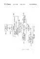
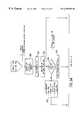
- FIG. 1is a schematic of the components of the device of the present invention
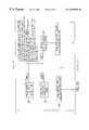
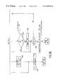
- FIG. 2is a flowchart illustrating the steps of the method for locating a nerve using the device of the present invention with particular reference to a method for determining the stability of a signal;
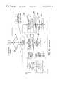
- FIG. 3is a flowchart illustrating a first method for stimulating the nerve known as the “coarse” stimulation mode
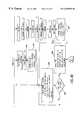
- FIG. 4is a flowchart illustrating a second method for stimulating the nerve known as the “fine” stimulation mode
- FIG. 5is a flowchart illustrating the “finestim” step of the method shown in FIG. 4;
- FIG. 6illustrates the mathematical equations for determining the stability of a signal.
- the present inventionis an improvement of the device and method for locating a nerve disclosed and claimed in U.S. Pat. No. 5,775,331 to Raymond et al.
- the structure of the devicewill be described with particular reference to the accompanying figures and to the disclosure of U.S. Pat. No. 5,775,331 to Raymond el al.
- the disclosure of the '331 patentis incorporated herein, in its entirety, by reference.
- device 100comprises stimulus applying means 102 , control means 104 , a stimulating current circuit 106 , and a signal and response detection means 108 .
- stimulus applying means 102is a probe 110 having a stimulating tip 112 , a handle 114 , a switch panel 116 , and a cable 118 for connecting the probe to control means 104 .
- Stimulating tip 112comprises an array of electrodes 120 which delivers a current pulse (an electro-stimulus) to the area of tissue believed to contain the nerve to be located.
- the arraypreferably comprises eight electrodes; however, it should be realized by those skilled in the art that the stimulating tip of the probe could be provided with any number of electrodes (including only one) depending on the nerve to be located and the associated tissue area.
- Handle 114is provided with light emitting diodes (LEDs) 122 which correspond in number and position to the array of electrodes disposed on stimulating tip 112 .
- Switch panel 116comprises switches 124 and 126 for activating and terminating the stimulating modes of the device to be discussed in more detail below.
- the electrodes of the probeare activated in accordance with an electrode activating algorithm discussed in more detail below with respect to FIGS. 4 and 5.
- probe 110is preferred to stimulate and locate the carvernosal nerve, others devices capable of delivering an electro-stimulus to a target tissue area would be suitable.
- Control means 104comprises a computer having a central processing unit (CPU), logic circuitry, memory, peripheral controllers and drivers which utiliz data acquisition hardware and software. Control means 104 is structured and programmed to perform and control all operations of the device in accordance with a number of algorithms. Specifically, the hardware and software of control means 104 performs the signal analyzing and interpreting functions for locating the nerve discussed in more detail below. Control means 104 further comprises a visual display 128 with LEDs and an audible tone module 130 for visually displaying and audibly communicating data (such as the intensity of the electro-stimulus or information concerning the stability of a tumescence signal) to the user in an intelligible format. An “up” and “down” switch 134 is also provided to manually increase or decrease the intensity of the stimulating current, if desired.
- CPUcentral processing unit
- Stimulating current circuit 106generates and delivers a biphasic square to electrodes 120 of probe 110 in response to a trigger by control means 104 . Delivery of a current pulse to a particular electrode of the array is accomplished by a set of relays activated in accordance with the electrode activation algorithm of the stimulating methods to be discussed in more detail below. A complete description of the stimulating current circuit of the device appears at column 9, lines 22-55 of the '331 patent.
- a signal and response detection means 108is provided to detect and measure a signal from a nerve fiber, organ or muscle and a change in the signal evoked by application of an electro-stimulus (also referred to as a response). Selection of a device to detect and measure a signal and response to application of an electro-stimulus is dependent upon the nerve to be located.
- a tumescence monitor 132comprising distensible tubing filled with a conductive fluid is preferable.
- any other device capable of detecting and measuring penile tumescencewould accomplish the objectives of the present invention.
- Other devices for detecting and measuring penile tumescence or for detecting and measuring a signal and response in other nervesare disclosed in FIG. 4 of the drawings of the '331 patent and at column 11, lines 22-56.
- the method for locating a nervecomprises the steps of:
- an electro-stimuluswill not be applied until it is determined that the signal is stable. If the signal is unstable, the signal is continuously monitored until stability is established.
- the method for determining if the signal is stable prior to application of an electro-stimulusis based on a mathematical, multi-order analysis. More particularly, the signal is analyzed for linear, cubic and quadratic “trends” indicative of an unstable signal (that is, an indication that a factor other an application of an electro-stimulus has caused a change in the signal which could result in an inaccurate assessment of the location of the nerve).
- the mathematical equations for determining the stability of the signalappear in FIG. 6 and a further discussion of the equations appears below.
- FIG. 2 of the accompanying drawingsis a flowchart summarizing the method of operation of the device for locating the nerve.
- block 200powers up the device and the “watchdog” program resets the CPU of control means 104 .
- the next step, shown as block 202is an “initialize and self-check” step which initializes all of the device parameters and performs a diagnostic on the control means and remaining components of the device.
- a housekeeping step shown as block 206follows the initialize and self-check step.
- the front panel of visual display 128is updated, data is stored in a datafile if a PCMCIA (Personal Computer Memory Card International Association) card is present, watchdog program is serviced, and the device is checked for error conditions.
- the tumescence signal detected and measured by tumescence monitor 132is analyzed for stability as shown by block 208 .
- the mathematical multi-order analysis of FIG. 6is performed to determine if the signal is stable. More particularly, the tumescence signal is continuously monitored in discrete 5 second segments for the existence of a “trend” determined by analyzing the linear contrast, the cubic contrast, and the quadratic contrast of the signal.
- the signalis considered to be unstable (that is, a trend is considered to be present) if a contrast value is greater than its corresponding predetermined critical contrast value defined, respectively, as CRIT-L (the linear critical Chi square statistic), CRIT-C (the cubic critical Chi square statistic), and CRIT-Q (the quadratic critical Chi square statistic).
- the signalis monitored its value is recorded and its linear, cubic, and quadratic contrasts values are recorded and analyzed against the CRIT-L, CRIT-C, and CRIT-Q parameters, respectively at intervals of 0.25 seconds for 5 seconds. If any one of the contrast values falls above its corresponding critical parameter (for example, if the cubic contrast value is greater than CRIT-C), the signal is considered to be unstable.
- the signalis continuously monitored and mathematically analyzed until it is determined that the signal is stable and that it is appropriate to proceed with application of electro-stimulation.
- decision block 210determines that a trend is present, the intensity of the preset stimulating current flashes on visual display 128 of control means 104 , indicating to the user that the signal is unstable and that it is not appropriate to proceed with application of electro-stimulation (block 212 ). If a trend is not present (that is, if the signal is determined to be stable), the intensity of the stimulating current is shown on the visual display of the control means by steady illumination, indicating that it is appropriate to proceed with application of electro-stimulation.
- Decision block 216determines if the user has pressed a switch on switch panel 116 of stimulating probe 110 .
- the choicesinclude pressing the up/down switch 134 on control means 104 to increase the intensity of the stimulating current by 1 mA (but not to an intensity greater than 20 mA) as shown by block 218 , pressing the up/down switch on the control means to decrease the stimulating current by 1 mA (but not to an intensity less than 1 mA) as shown by block 220 , pressing the course stimulation switch 124 on probe 110 as shown by block 222 , or pressing the fine stimulation switch 126 on the probe as shown by block 224 . If the answer is no, the method returns to step 206 .
- the stability of the tumescence signalis checked at block 226 . If it is determined that the signal is stable (answer “no” to decision block 228 ) either coarse mode stimulation is activated as shown by block 230 or fine mode stimulation is activated as shown by block 246 , depending on whether the coarse stimulation switch was pressed (block 222 ) or the fine stimulation switch was pressed (block 224 ). If it is determined that a trend is present and that the signal is unstable, decision block 250 determines if the coarse stimulation switch has been pressed to override or bypass the tumescence signal stability check. If the coarse stimulation switch has been pressed, the method proceeds to coarse mode stimulation (block 230 ).
- decision block 232determines if the tumescence signal has been unstable for 80 seconds. If the answer to decision block 232 is no, and if the coarse stimulation mode is not terminated by pressing the course stimulation switch (that is, the answer to decision block 234 is also “no”), the visual display flashes “00” on the visual display of the control means to indicate to the user that the signal is not stable (block 236 ). If the answer to decision block 234 is yes (that is, coarse mode stimulation has been terminated by pressing the coarse mode stimulation switch), the method returns to housekeeping block 206 .
- the usercan override or bypass the signal stability check and proceed to application of electro-stimulation by pressing the course or fine mode switch located on the probe handle. If the user chooses to heed the warning provided by the signal stability check, the method returns to block 206 and cycles through that portion of the method until the signal is stable.
- fine mode stimulation switchhas been pressed to bypass the tumescence signal stability check (that is, the answer to decision block 238 is “yes”, the method proceeds to fine mode stimulation (block 246 ). If the answer to decision block 238 is no, decision block 240 determines if the tumescence signal has been unstable 80 seconds. If the answer to decision block 240 is no, and if the fine stimulation mode is not terminated by pressing the fine mode stimulation switch (that is, the answer to decision block 242 is no), the visual display flashes “00” on the visual display of the control means to indicate to the user that the signal is not stable (block 244 ).
- the methodreturns to housekeeping block 206 .
- the methodBy continuously monitoring the stability of the tumescence signal prior to application of an electro-stimulus, interpretation of a change in the signal (a response) to electro-stimulation is enhanced and, therefore, improves the utility and accuracy of the device.
- coarse mode stimulationentails application of an electro-stimulus by all eight electrodes of probe 110 at a frequency of 16 Hz.
- the devicesequences through “steps” of electro-stimulation, each step having a duration of 20 seconds and an intensity of no more than 20 mA.
- the methodstarts at decision block 300 which determines if a valid probe 110 is present. If a valid probe is present, the response flag is cleared at block 302 and the STEP 1 electro-stimulation sequence is called.
- Block 304defaults the intensity I of the stimulating current pulse to 8 mA. However, the user has the option of requesting another intensity value, if desired as shown at 218 and 220 in FIG. 2.
- a checkis then made of the tumescence signal range to determine if the current tumescence signal is within an acceptable range (preferably, greater than 0 V and but less than approximately 4.5 V). If the tumescence signal is too high or too low to allow proper operation of the device, an error code is displayed on visual display 128 of the device.
- block 306establishes a baseline value for the tumescence signal by averaging the last 5 tumescence signal readings at a rate of 4 signal readings per second.
- Block 308then displays the tumescence signal baseline value on visual display 128 of the control means, illuminates LEDs 122 of handle 114 , and starts the timer of the control means by resetting it to 0. If decision block 310 determines that the coarse mode switch has been pressed again (returning the switch to the up position), stimulation is terminated at block 312 and the method returns to block 202 of method illustrated in FIG. 2 . If decision block 310 determines that the coarse stimulation switch is down (that is, coarse mode stimulation is active), block 314 determines if 80 seconds has elapsed. If the answer to decision block 314 is yes, coarse mode stimulation is terminated at block 312 .
- decision block 316determines if power is being supplied to stimulating current circuit 106 . If the answer to block 316 is no, block 318 displays an error code on the visual display and the device enters an endless loop which can be exited only by turning the power off. If power is being supplied to the stimulating current circuit 106 , the patient connections to tumescence monitor 132 are checked at block 320 . Next, block 322 determines if the up/down switch on control means 104 has been pressed to increase or decrease the intensity of the electro-stimulus. If the answer to decision block 322 is yes, block 324 determines the intensity of the stimulating current as adjusted by the user.
- Block 326stimulates through all 8 electrodes of probe 110 in a predetermined sequence at a frequency of 16 Hz and at intensity I (as determined by block 324 ) or at the original default intensity of 8 mA.
- Block 328performs another tumescence signal range check at a rate of 4 tumescence signal readings per second.
- Block 330performs an update to the visual and audible display of the control means to indicate the baseline tumescence signal value.
- block 332determines if a response flag R has been set. If the answer is yes, the method returns to decision block 310 .
- decision block 334determines if the tumescence response has increased at least 5% from baseline. If the answer to block 334 is yes, a response tone is generated by audible tone module 130 and a response flag R is set at block 336 . Once flag R is set, the current is maintained at a constant intensity through the rest of the stimulation mode. The method returns to decision block 310 and repeats. If the answer to decision block 334 is “no” decision block 338 determines if 20 seconds has elapsed. If the answer to the 20 second decision block 338 is no, the method returns to decision block 310 and repeats. If the answer to block 338 is yes, block 340 calls for the INCREMENT STEP step.
- Decision block 342determines if the intensity I has been increased over 4 stimulating steps or cycles ofthe coarse stimulation mode. If the device has already cycled through 4 stimulating steps (increasing the stimulus intensity by 2 mA per each successive step), block 344 establishes that no response has been evoked and it extinguishes LEDs 122 on probe 110 . Audible tone module 130 generates a no-response tone and the device reverts to the starting stimulating current intensity I established at block 304 . If the coarse stimulation button is pressed (coarse mode stimulation active) at decision block 346 , the method returns to block 344 .
- block 348silences the audible tone and extinguishes the data on visual display 128 .
- decision block 350determines if the device has cycled through 2 steps. If the answer is yes, block 352 increases the intensity I by 2 mA and the method updates the visual display, stores the data in the PCMCIA card and restarts the 20 second timer at block 354 . If the answer to decision block 350 is no, block 356 determines if the device has cycled through 3 steps of coarse stimulation. If the answer is yes, block 358 increases the intensity I of the stimulating current by 4 mA and block 354 updates the visual display, stores the data in the PCMCIA card and restarts the 20 second timer.
- block 360determines that the method is cycling through the fourth step of coarse mode stimulation.
- Block 362increases the intensity I of the stimulating current by 6 mA and block 354 updates the visual display, stores the data in the PCMCIA card and restarts the 20 second timer. It should be noted that at block 362 the intensity as increased at STEP 4 cannot be increased above 20 mA. After block 354 , the method returns to block 310 and repeats.
- the useris able to quickly determine if the target tissue area contains the carvernosal nerve to avoid cutting the nerve during the excision or sectioning of tissue.
- the coarse mode stimulation methodassists in the priming step disclosed in the '331 patent.
- fine mode stimulationentails applying an electro-stimulus of a constant intensity through 1) each electrode of the probe, 2) four pairs of electrodes, or 3) two sets of four electrodes.
- the methodstarts at block 400 which determines if a valid stimulating tip 112 is present. If a valid tip is not present, the method returns to the method of FIG. 2 . If the tip is valid, block 402 assesses the user-selected resolution.
- the methodestablishes whether the method will apply an electro-stimulus through each one of the probe electrodes (resolution 1 ), through four pairs of electrodes (resolution 2 ), or through two sets of four electrodes (resolution 4 ). If resolution 1 is selected, block 404 sets the stimulating current intensity at I. The intensity of the fine mode stimulus current for resolution 1 is 1.5 ⁇ greater than the coarse mode stimulus current, but not greater than 20 mA. If a PCMCIA datacard is present, the intensity of the stimulus current is stored in the datafile.
- Block 406establishes an electro-stimulus for electrode 1 (electrode 1 active).
- Block 408establishes a local baseline by averaging the last 5 tumescence signal readings from tumescence monitor 132 .
- Block 410performs the “finestim” method illustrated in FIG. 5 and described in greater detail below.
- the finestim methodanalyzes percent change in a tumescence response evoked by application of the electro-stimulus (and measured and recorded by tumescence monitor 132 ) to establish a peak percent change value.
- Block 412stores the peak percent change from finestim for electrode 1 as peak % value 1.
- Block 414establishes an electro-stimulus for electrode 3 (electrode 3 active), while block 416 establishes a tumescence baseline and block 418 performs the finestim method for the electro-stimulus applied by electrode 3 .
- Block 420stores the peak percent change value from finestim for electrode 3 as peak % value 3 .
- each peak % valueis compared against the other to determine the “winning” electrode (that is, the electrode responsible for evoking the greatest peak % value).
- the comparison stepgenerally comprises the step of determining which peak % value represents the greatest peak % value.
- block 426illuminates the LED(s) on handle 114 which correspond to the winning electrode(s) to indicate the location of the nerve to the user.
- block 428determines if the fine button is still down (that is, active). If the answer is yes, the method returns to block 426 and repeats. If the answer is no, block 430 extinguishes all LEDs on the visual display and probe, the audible tone is silenced, and the method returns to block 200 of the method of FIG. 1 .
- block 432sets the stimulating current intensity at I which is equal to 1.5 ⁇ the coarse mode stimulus current but is not greater than 20 mA.
- Block 434sets a pattern for electrodes 1 and 2 , while block 436 establishes a tumescence baseline value and block 438 performs the finestim method for the electro-stimulus applied by electrodes 1 and 2 .
- Block 440stores the peak percent change value from finestim for electrodes 1 and 2 as peak % value 1 . The same three steps are performed for electrodes 5 and 6 , 3 and 4 , and 7 and 8 .
- the peak percent change valuesare stored as peak % value 2 , peak % value 3 , and peak % value 4 , respectively as shown in FIG. 4 .
- Block 442After establishing the peak % value for all four pairs of electrodes, stimulation is terminated at block 442 .
- each peak % valueis compared against the other to determine the “winning” pair of electrodes in accordance with the comparison method discussed above.
- Block 426displays the winning pair or pairs (in the event of a tie) and the method for fine mode stimulation by resolution 2 is completed by blocks 428 and 430 .
- block 446sets the stimulating current intensity at I which is 1.25 ⁇ greater than the stimulus intensity for coarse mode stimulation.
- Block 448sets a pattern to apply an electro-stimulus through the first set of four electrodes (electrodes 1 , 2 , 3 , 4 ).
- Block 450establishes a tumescence baseline value and block 452 performs the “finestim” method to analyze percent changes in the tumescence response to establish a peak percent change value.
- Block 454stores the peak percent change from finestim as peak % value 1 .
- Block 456sets a pattern to apply an electro-stimulus through the second set of four electrodes (electrodes 5 , 6 , 7 , 8 ).
- Block 458establishes the tumescence baseline value and block 460 performs the finestim method to analyze percent changes in the tumescence response to establish a peak percent change value for the second set of four electrodes.
- Block 462stores the peak percent change from block 460 as peak % value 2 and block 464 terminates stimulation.
- Block 466compares peak % value 1 and peak % value 2 to determine the winning set of four electrodes.
- Block 426displays the winning set on the probe LEDs corresponding to the winning electrodes.
- Blocks 428 and 430terminate the fine mode stimulation method in the manner discussed above.
- the finestim method of the fine mode stimulation of FIG. 4will be described.
- the local tumescence baseline and stimulation patternis brought into the method at block 500 and the LEDs of the probe corresponding to the stimulating pattern from fine mode stimulation are illuminated.
- the peak % valueis set at 0 at block 502 .
- the electro-stimulusis delivered to the target tissue area by probe 110 and a 15 second timer is started (or re-started).
- Decision block 506determines if the fine stimulation switch is still down (that is, active). If the answer is no, block 508 terminates the stimulation and extinguishes all LEDs on the display and probe.
- block 510performs a check of stimulating current circuit 106 .
- Decision block 512determines if the power is on. If the answer is no, block 514 terminates stimulation and displays an error code. If the answer to block 512 is yes, block 516 checks for a proper patient connection to tumescence monitor 132 . Decision block 518 determines if the up/down current intensity switch has been pressed. If the answer is yes, block 520 adjusts the current intensity by 1 mA to a value not greater than 20 mA and not less than 1 mA. Block 522 performs a tumescence signal reading at four readings per second and it performs a tumescence signal range check.
- Decision block 524determines if the tumescence signal is in range. If the answer is no, block 526 terminates stimulation, displays an error message and extinguishes the LEDs on the probe and display. If the tumescence signal is in range, the visual and audible displays are updated by block 528 .
- Block 530calculates the change (differential) in the tumescence signal characterized as a response to the electro-stimulus of block 504 . The differential is calculated by subtracting the local baseline tumescence signal reading from the current tumescence response. The step of block 504 is performed 4 times per second. If decision block 532 determines that the differential is positive, the percentage differential is calculated by block 534 .
- block 540(If the differential is not positive, the method advances to block 540 ).
- the equation for determining the percentage differentialis illustrated in block 534 . If block 536 determines that the percentage differential is greater than 0.5% , block 538 stores the percentage differential as the peak % value. If block 540 determines that 15 seconds has elapsed, block 542 extinguishes illumination ofthe LED pattern on probe 110 and the peak % value is returned to the relevant finestim block of FIG. 4 . If the answer to block 540 is no, the method returns to decision block 506 and repeats.
- block 544determines if the fine mode stimulation switch is still down. If the answer is yes, the method returns to block 526 . If the answer is no, block 546 silences the audible tone and clears the error message. Block 548 displays the intensity of the next electro-stimulus and the method returns to the method of FIG. 1 .
- the usercan determine the location of the carvernosal with improved accuracy and specificity, to avoid cutting the nerve during the excision or sectioning of tissue.
- the deviceadvantageously indicates the stability of the tumescence signal to the user to provide the option of waiting for the signal to stabilize or to proceed with electro-stimulation, if desired.
- the tumescence signalcould be shown graphically on a video monitor with an indication of the history of the tumescence signal.
- the displaycould be designed to show the order of change of the tumescence signal, such as a single down arrow to show first order detumescence or two down arrows to show second order detumescence.
Landscapes
- Health & Medical Sciences (AREA)
- Life Sciences & Earth Sciences (AREA)
- Animal Behavior & Ethology (AREA)
- Veterinary Medicine (AREA)
- Public Health (AREA)
- Biophysics (AREA)
- Engineering & Computer Science (AREA)
- Biomedical Technology (AREA)
- Heart & Thoracic Surgery (AREA)
- General Health & Medical Sciences (AREA)
- Pathology (AREA)
- Surgery (AREA)
- Molecular Biology (AREA)
- Medical Informatics (AREA)
- Physics & Mathematics (AREA)
- Neurology (AREA)
- Nuclear Medicine, Radiotherapy & Molecular Imaging (AREA)
- Radiology & Medical Imaging (AREA)
- Measurement And Recording Of Electrical Phenomena And Electrical Characteristics Of The Living Body (AREA)
- Radiation-Therapy Devices (AREA)
Abstract
Description
Claims (37)
Priority Applications (4)
| Application Number | Priority Date | Filing Date | Title |
|---|---|---|---|
| US09/303,501US6259945B1 (en) | 1999-04-30 | 1999-04-30 | Method and device for locating a nerve |
| PCT/US2000/011669WO2000066217A1 (en) | 1999-04-30 | 2000-04-28 | Method and device for locating a nerve |
| AU48098/00AAU4809800A (en) | 1999-04-30 | 2000-04-28 | Method and device for locating a nerve |
| US09/692,418US6535759B1 (en) | 1999-04-30 | 2000-10-20 | Method and device for locating and mapping nerves |
Applications Claiming Priority (1)
| Application Number | Priority Date | Filing Date | Title |
|---|---|---|---|
| US09/303,501US6259945B1 (en) | 1999-04-30 | 1999-04-30 | Method and device for locating a nerve |
Related Child Applications (1)
| Application Number | Title | Priority Date | Filing Date |
|---|---|---|---|
| US09/692,418Continuation-In-PartUS6535759B1 (en) | 1999-04-30 | 2000-10-20 | Method and device for locating and mapping nerves |
Publications (1)
| Publication Number | Publication Date |
|---|---|
| US6259945B1true US6259945B1 (en) | 2001-07-10 |
Family
ID=23172408
Family Applications (2)
| Application Number | Title | Priority Date | Filing Date |
|---|---|---|---|
| US09/303,501Expired - Fee RelatedUS6259945B1 (en) | 1999-04-30 | 1999-04-30 | Method and device for locating a nerve |
| US09/692,418Expired - Fee RelatedUS6535759B1 (en) | 1999-04-30 | 2000-10-20 | Method and device for locating and mapping nerves |
Family Applications After (1)
| Application Number | Title | Priority Date | Filing Date |
|---|---|---|---|
| US09/692,418Expired - Fee RelatedUS6535759B1 (en) | 1999-04-30 | 2000-10-20 | Method and device for locating and mapping nerves |
Country Status (3)
| Country | Link |
|---|---|
| US (2) | US6259945B1 (en) |
| AU (1) | AU4809800A (en) |
| WO (1) | WO2000066217A1 (en) |
Cited By (143)
| Publication number | Priority date | Publication date | Assignee | Title |
|---|---|---|---|---|
| US6466817B1 (en)* | 1999-11-24 | 2002-10-15 | Nuvasive, Inc. | Nerve proximity and status detection system and method |
| US20020198568A1 (en)* | 2000-03-13 | 2002-12-26 | Fred Hafer | Instrument and method for delivery of anaesthetic drug |
| US6500128B2 (en) | 2000-06-08 | 2002-12-31 | Nuvasive, Inc. | Nerve movement and status detection system and method |
| US20030105503A1 (en)* | 2001-06-08 | 2003-06-05 | Nuvasive, Inc. | Relative nerve movement and status detection system and method |
| US20040074556A1 (en)* | 1999-12-03 | 2004-04-22 | O'connell Patrick R. | Fuel tank filler neck and method of manufacturing same |
| US20040172114A1 (en)* | 2003-02-27 | 2004-09-02 | Moscosta Medical U.S.A., L.L.C. | Nerve stimulation functionality indicator apparatus and method |
| US20040199084A1 (en)* | 1999-11-24 | 2004-10-07 | Nuvasive, Inc. | Electromyography system |
| US20040225228A1 (en)* | 2003-05-08 | 2004-11-11 | Ferree Bret A. | Neurophysiological apparatus and procedures |
| US20040267243A1 (en)* | 2003-06-30 | 2004-12-30 | Klotz Conrad Lee | Surgical scalpel and system particularly for use in a transverse carpal ligament surgical procedure |
| US20050033189A1 (en)* | 2003-08-08 | 2005-02-10 | Mccraty Rollin I. | Electrophysiological intuition indicator |
| US20050036752A1 (en)* | 2003-08-13 | 2005-02-17 | Burke James P. | Dispersion compensated optical fiber transmission system and module including micro-structured optical fiber |
| US20050075578A1 (en)* | 2001-09-25 | 2005-04-07 | James Gharib | System and methods for performing surgical procedures and assessments |
| US20050245969A1 (en)* | 2004-04-09 | 2005-11-03 | Alfred E. Mann Institute For Biomedical Engineering At The University Of Southern Ca. | Identification of target site for implantation of a microstimulator |
| US20050283148A1 (en)* | 2004-06-17 | 2005-12-22 | Janssen William M | Ablation apparatus and system to limit nerve conduction |
| US20060025703A1 (en)* | 2003-08-05 | 2006-02-02 | Nuvasive, Inc. | System and methods for performing dynamic pedicle integrity assessments |
| US20060111767A1 (en)* | 2003-05-30 | 2006-05-25 | Medi-Screw, Inc. | Medical implant systems |
| US7079883B2 (en) | 1998-12-23 | 2006-07-18 | Nuvaslve, Inc. | Nerve surveillance cannulae systems |
| WO2006042075A3 (en)* | 2004-10-07 | 2006-07-27 | Nuvasive Inc | System and methods for assessing the neuromuscular pathway prior to nerve testing |
| US20060173521A1 (en)* | 2005-01-31 | 2006-08-03 | Pond John D Jr | Electrically insulated surgical needle assembly |
| US20060173374A1 (en)* | 2005-01-31 | 2006-08-03 | Neubardt Seth L | Electrically insulated surgical probing tool |
| US20060178594A1 (en)* | 2005-02-07 | 2006-08-10 | Neubardt Seth L | Apparatus and method for locating defects in bone tissue |
| US20060178593A1 (en)* | 2005-02-07 | 2006-08-10 | Neubardt Seth L | Device and method for operating a tool relative to bone tissue and detecting neural elements |
| US20060200023A1 (en)* | 2005-03-04 | 2006-09-07 | Sdgi Holdings, Inc. | Instruments and methods for nerve monitoring in spinal surgical procedures |
| US20060217655A1 (en)* | 2000-03-13 | 2006-09-28 | Vitullo Jeffrey M | Pre-loaded lockable stimulating catheter for delivery of anaesthetic drugs |
| US20060224078A1 (en)* | 2000-05-18 | 2006-10-05 | Nuvasive, Inc. | Tissue discrimination and applications in medical procedures |
| US7207949B2 (en) | 2003-09-25 | 2007-04-24 | Nuvasive, Inc. | Surgical access system and related methods |
| US20070213616A1 (en)* | 2005-10-20 | 2007-09-13 | Thomas Anderson | Systems and methods for arteriotomy localization |
| US20080097164A1 (en)* | 2003-01-16 | 2008-04-24 | Nuvasive, Inc. | Surgical access system and related methods |
| US7386341B2 (en) | 2000-03-13 | 2008-06-10 | Arrow International, Inc. | Instrument and method for delivery of anaesthetic drugs |
| US20080312660A1 (en)* | 2007-06-15 | 2008-12-18 | Baxano, Inc. | Devices and methods for measuring the space around a nerve root |
| US20090124860A1 (en)* | 2003-02-27 | 2009-05-14 | Nuvasive, Inc. | Surgical access system and related methods |
| US7555343B2 (en) | 2004-10-15 | 2009-06-30 | Baxano, Inc. | Devices and methods for selective surgical removal of tissue |
| US20090177112A1 (en)* | 2005-02-02 | 2009-07-09 | James Gharib | System and Methods for Performing Neurophysiologic Assessments During Spine Surgery |
| US20090182478A1 (en)* | 2008-01-15 | 2009-07-16 | Gm Global Technology Operations, Inc. | Axle torque based cruise control |
| US7578819B2 (en) | 2005-05-16 | 2009-08-25 | Baxano, Inc. | Spinal access and neural localization |
| US7582058B1 (en) | 2002-06-26 | 2009-09-01 | Nuvasive, Inc. | Surgical access system and related methods |
| US20090299439A1 (en)* | 2008-06-02 | 2009-12-03 | Warsaw Orthopedic, Inc. | Method, system and tool for surgical procedures |
| US20090299214A1 (en)* | 2007-05-11 | 2009-12-03 | Changwang Wu | Method and apparatus for quantitative nerve localization |
| US7664544B2 (en) | 2002-10-30 | 2010-02-16 | Nuvasive, Inc. | System and methods for performing percutaneous pedicle integrity assessments |
| US7738969B2 (en) | 2004-10-15 | 2010-06-15 | Baxano, Inc. | Devices and methods for selective surgical removal of tissue |
| US7785253B1 (en) | 2005-01-31 | 2010-08-31 | Nuvasive, Inc. | Surgical access system and related methods |
| US20100317989A1 (en)* | 2005-07-20 | 2010-12-16 | Nuvasive Inc. | Systems and Methods for Performing Neurophysiologic Assesments With Pressure Monitoring |
| US7857813B2 (en) | 2006-08-29 | 2010-12-28 | Baxano, Inc. | Tissue access guidewire system and method |
| US7887538B2 (en) | 2005-10-15 | 2011-02-15 | Baxano, Inc. | Methods and apparatus for tissue modification |
| US7905840B2 (en) | 2003-10-17 | 2011-03-15 | Nuvasive, Inc. | Surgical access system and related methods |
| US7918849B2 (en) | 2004-10-15 | 2011-04-05 | Baxano, Inc. | Devices and methods for tissue access |
| US7920922B2 (en) | 2001-07-11 | 2011-04-05 | Nuvasive, Inc. | System and methods for determining nerve proximity, direction, and pathology during surgery |
| US20110092880A1 (en)* | 2009-10-12 | 2011-04-21 | Michael Gertner | Energetic modulation of nerves |
| US7938830B2 (en) | 2004-10-15 | 2011-05-10 | Baxano, Inc. | Powered tissue modification devices and methods |
| US7959577B2 (en) | 2007-09-06 | 2011-06-14 | Baxano, Inc. | Method, system, and apparatus for neural localization |
| US20110172528A1 (en)* | 2009-10-12 | 2011-07-14 | Michael Gertner | Systems and methods for treatment using ultrasonic energy |
| US7987001B2 (en) | 2007-01-25 | 2011-07-26 | Warsaw Orthopedic, Inc. | Surgical navigational and neuromonitoring instrument |
| WO2011046880A3 (en)* | 2009-10-12 | 2011-08-25 | Kona Medical, Inc. | Energetic modulation of nerves |
| US20110230785A1 (en)* | 2010-03-16 | 2011-09-22 | ProNerve, LLC | Somatosensory Evoked Potential (SSEP) Automated Alert System |
| US8048080B2 (en) | 2004-10-15 | 2011-11-01 | Baxano, Inc. | Flexible tissue rasp |
| US20110270119A1 (en)* | 2010-04-30 | 2011-11-03 | Jann Rasmussen | Devices And Methods For Nerve Mapping |
| US8062298B2 (en) | 2005-10-15 | 2011-11-22 | Baxano, Inc. | Flexible tissue removal devices and methods |
| US8062300B2 (en) | 2006-05-04 | 2011-11-22 | Baxano, Inc. | Tissue removal with at least partially flexible devices |
| US8092456B2 (en) | 2005-10-15 | 2012-01-10 | Baxano, Inc. | Multiple pathways for spinal nerve root decompression from a single access point |
| US8137284B2 (en) | 2002-10-08 | 2012-03-20 | Nuvasive, Inc. | Surgical access system and related methods |
| US8147421B2 (en) | 2003-01-15 | 2012-04-03 | Nuvasive, Inc. | System and methods for determining nerve direction to a surgical instrument |
| US8192436B2 (en) | 2007-12-07 | 2012-06-05 | Baxano, Inc. | Tissue modification devices |
| US8206312B2 (en) | 2005-09-22 | 2012-06-26 | Nuvasive, Inc. | Multi-channel stimulation threshold detection algorithm for use in neurophysiology monitoring |
| US8221397B2 (en) | 2004-10-15 | 2012-07-17 | Baxano, Inc. | Devices and methods for tissue modification |
| US8255045B2 (en)* | 2007-04-03 | 2012-08-28 | Nuvasive, Inc. | Neurophysiologic monitoring system |
| US8257356B2 (en) | 2004-10-15 | 2012-09-04 | Baxano, Inc. | Guidewire exchange systems to treat spinal stenosis |
| US8287597B1 (en) | 2009-04-16 | 2012-10-16 | Nuvasive, Inc. | Method and apparatus for performing spine surgery |
| US8295912B2 (en) | 2009-10-12 | 2012-10-23 | Kona Medical, Inc. | Method and system to inhibit a function of a nerve traveling with an artery |
| US20120277621A1 (en)* | 2011-04-29 | 2012-11-01 | Medtronic, Inc. | Determining nerve location relative to electrodes |
| US8313430B1 (en) | 2006-01-11 | 2012-11-20 | Nuvasive, Inc. | Surgical access system and related methods |
| US8328851B2 (en) | 2005-07-28 | 2012-12-11 | Nuvasive, Inc. | Total disc replacement system and related methods |
| CN102883659A (en)* | 2010-01-19 | 2013-01-16 | 美敦力阿迪安卢森堡有限公司 | Methods and apparatus for renal neuromodulation via stereotactic radiotherapy |
| US8366712B2 (en) | 2005-10-15 | 2013-02-05 | Baxano, Inc. | Multiple pathways for spinal nerve root decompression from a single access point |
| US8374673B2 (en) | 2007-01-25 | 2013-02-12 | Warsaw Orthopedic, Inc. | Integrated surgical navigational and neuromonitoring system having automated surgical assistance and control |
| US8388535B2 (en) | 1999-10-25 | 2013-03-05 | Kona Medical, Inc. | Methods and apparatus for focused ultrasound application |
| US20130060315A1 (en)* | 2011-09-01 | 2013-03-07 | Zoll Medical Corporation | Medical equipment electrodes |
| US8394102B2 (en) | 2009-06-25 | 2013-03-12 | Baxano, Inc. | Surgical tools for treatment of spinal stenosis |
| US8398641B2 (en) | 2008-07-01 | 2013-03-19 | Baxano, Inc. | Tissue modification devices and methods |
| US8409206B2 (en) | 2008-07-01 | 2013-04-02 | Baxano, Inc. | Tissue modification devices and methods |
| US8430881B2 (en) | 2004-10-15 | 2013-04-30 | Baxano, Inc. | Mechanical tissue modification devices and methods |
| US8469904B2 (en) | 2009-10-12 | 2013-06-25 | Kona Medical, Inc. | Energetic modulation of nerves |
| US8517962B2 (en) | 2009-10-12 | 2013-08-27 | Kona Medical, Inc. | Energetic modulation of nerves |
| US8568331B2 (en) | 2005-02-02 | 2013-10-29 | Nuvasive, Inc. | System and methods for monitoring during anterior surgery |
| US8568317B1 (en) | 2005-09-27 | 2013-10-29 | Nuvasive, Inc. | System and methods for nerve monitoring |
| US8568416B2 (en) | 2004-10-15 | 2013-10-29 | Baxano Surgical, Inc. | Access and tissue modification systems and methods |
| US8613745B2 (en) | 2004-10-15 | 2013-12-24 | Baxano Surgical, Inc. | Methods, systems and devices for carpal tunnel release |
| US8622937B2 (en) | 1999-11-26 | 2014-01-07 | Kona Medical, Inc. | Controlled high efficiency lesion formation using high intensity ultrasound |
| US8790406B1 (en) | 2011-04-01 | 2014-07-29 | William D. Smith | Systems and methods for performing spine surgery |
| US8801626B2 (en) | 2004-10-15 | 2014-08-12 | Baxano Surgical, Inc. | Flexible neural localization devices and methods |
| US8845639B2 (en) | 2008-07-14 | 2014-09-30 | Baxano Surgical, Inc. | Tissue modification devices |
| US8986231B2 (en) | 2009-10-12 | 2015-03-24 | Kona Medical, Inc. | Energetic modulation of nerves |
| US8986211B2 (en) | 2009-10-12 | 2015-03-24 | Kona Medical, Inc. | Energetic modulation of nerves |
| US8992447B2 (en) | 2009-10-12 | 2015-03-31 | Kona Medical, Inc. | Energetic modulation of nerves |
| US9005143B2 (en) | 2009-10-12 | 2015-04-14 | Kona Medical, Inc. | External autonomic modulation |
| US9066701B1 (en) | 2012-02-06 | 2015-06-30 | Nuvasive, Inc. | Systems and methods for performing neurophysiologic monitoring during spine surgery |
| US9101386B2 (en) | 2004-10-15 | 2015-08-11 | Amendia, Inc. | Devices and methods for treating tissue |
| US9113912B1 (en) | 2015-01-21 | 2015-08-25 | Serene Medical, Inc. | Systems and devices to identify and limit nerve conduction |
| US9119628B1 (en) | 2015-01-21 | 2015-09-01 | Serene Medical, Inc. | Systems and devices to identify and limit nerve conduction |
| US9155503B2 (en) | 2010-10-27 | 2015-10-13 | Cadwell Labs | Apparatus, system, and method for mapping the location of a nerve |
| US9198765B1 (en) | 2011-10-31 | 2015-12-01 | Nuvasive, Inc. | Expandable spinal fusion implants and related methods |
| US9247952B2 (en) | 2004-10-15 | 2016-02-02 | Amendia, Inc. | Devices and methods for tissue access |
| US9295401B2 (en) | 2012-11-27 | 2016-03-29 | Cadwell Laboratories, Inc. | Neuromonitoring systems and methods |
| US9314253B2 (en) | 2008-07-01 | 2016-04-19 | Amendia, Inc. | Tissue modification devices and methods |
| US9351845B1 (en) | 2009-04-16 | 2016-05-31 | Nuvasive, Inc. | Method and apparatus for performing spine surgery |
| US9392953B1 (en) | 2010-09-17 | 2016-07-19 | Nuvasive, Inc. | Neurophysiologic monitoring |
| US9456829B2 (en) | 2004-10-15 | 2016-10-04 | Amendia, Inc. | Powered tissue modification devices and methods |
| US9622732B2 (en) | 2004-10-08 | 2017-04-18 | Nuvasive, Inc. | Surgical access system and related methods |
| US9649494B2 (en) | 2011-04-29 | 2017-05-16 | Medtronic, Inc. | Electrical stimulation therapy based on head position |
| US9655505B1 (en) | 2012-02-06 | 2017-05-23 | Nuvasive, Inc. | Systems and methods for performing neurophysiologic monitoring during spine surgery |
| US9757067B1 (en) | 2012-11-09 | 2017-09-12 | Nuvasive, Inc. | Systems and methods for performing neurophysiologic monitoring during spine surgery |
| US9757072B1 (en) | 2013-02-11 | 2017-09-12 | Nuvasive, Inc. | Waveform marker placement algorithm for use in neurophysiologic monitoring |
| US9775530B2 (en) | 2013-11-01 | 2017-10-03 | Medtronic Xomed, Inc. | Foley catheter with ring electrodes |
| US9827109B2 (en) | 1999-03-07 | 2017-11-28 | Nuvasive, Inc. | Methods and apparatus for performing spine surgery |
| US10022090B2 (en) | 2013-10-18 | 2018-07-17 | Atlantic Health System, Inc. | Nerve protecting dissection device |
| US10098585B2 (en) | 2013-03-15 | 2018-10-16 | Cadwell Laboratories, Inc. | Neuromonitoring systems and methods |
| CN109330591A (en)* | 2018-09-17 | 2019-02-15 | 江苏省人民医院(南京医科大学第附属医院) | Evoked potential monitor for real-time monitoring cavernous nerve injury of penis in laparoscopic surgery |
| US10420480B1 (en) | 2014-09-16 | 2019-09-24 | Nuvasive, Inc. | Systems and methods for performing neurophysiologic monitoring |
| US10433793B1 (en) | 2015-03-27 | 2019-10-08 | Cadwell Laboratories, Inc. | Methods and systems for simultaneous review of brain activity and physical manifestations of users |
| US10772681B2 (en) | 2009-10-12 | 2020-09-15 | Utsuka Medical Devices Co., Ltd. | Energy delivery to intraparenchymal regions of the kidney |
| CN112041020A (en)* | 2018-02-22 | 2020-12-04 | 首尔大学医院 | Intraoperative mapping of cavernous nerves |
| US10925579B2 (en) | 2014-11-05 | 2021-02-23 | Otsuka Medical Devices Co., Ltd. | Systems and methods for real-time tracking of a target tissue using imaging before and during therapy delivery |
| US10959860B2 (en) | 2008-12-26 | 2021-03-30 | Pantheon Spinal, Llc | Method of retroperitoneal lateral insertion of spinal implants |
| US11071861B2 (en) | 2011-04-29 | 2021-07-27 | Medtronic, Inc. | Dual prophylactic and abortive electrical stimulation |
| US11128076B2 (en) | 2019-01-21 | 2021-09-21 | Cadwell Laboratories, Inc. | Connector receptacle |
| US11166672B2 (en) | 2013-10-18 | 2021-11-09 | Atlantic Health System, Inc. | Nerve protecting dissection device |
| US11177610B2 (en) | 2017-01-23 | 2021-11-16 | Cadwell Laboratories, ino. | Neuromonitoring connection system |
| US11185684B2 (en) | 2018-09-18 | 2021-11-30 | Cadwell Laboratories, Inc. | Minimally invasive two-dimensional grid electrode |
| US11253182B2 (en) | 2018-05-04 | 2022-02-22 | Cadwell Laboratories, Inc. | Apparatus and method for polyphasic multi-output constant-current and constant-voltage neurophysiological stimulation |
| US11259737B2 (en) | 2012-11-06 | 2022-03-01 | Nuvasive, Inc. | Systems and methods for performing neurophysiologic monitoring during spine surgery |
| US11317841B2 (en) | 2018-11-14 | 2022-05-03 | Cadwell Laboratories, Inc. | Method and system for electrode verification |
| US11443649B2 (en) | 2018-06-29 | 2022-09-13 | Cadwell Laboratories, Inc. | Neurophysiological monitoring training simulator |
| US11452874B2 (en) | 2020-02-03 | 2022-09-27 | Medtronic, Inc. | Shape control for electrical stimulation therapy |
| US11471087B2 (en) | 2018-11-09 | 2022-10-18 | Cadwell Laboratories, Inc. | Integrity verification system for testing high channel count neuromonitoring recording equipment |
| US11517239B2 (en) | 2018-04-05 | 2022-12-06 | Cadwell Laboratories, Inc. | Systems and methods for processing and displaying electromyographic signals |
| US11517245B2 (en) | 2018-10-30 | 2022-12-06 | Cadwell Laboratories, Inc. | Method and system for data synchronization |
| US11529107B2 (en) | 2018-11-27 | 2022-12-20 | Cadwell Laboratories, Inc. | Methods for automatic generation of EEG montages |
| US11554264B2 (en) | 2020-04-24 | 2023-01-17 | Medtronic, Inc. | Electrode position detection |
| US11596337B2 (en) | 2018-04-24 | 2023-03-07 | Cadwell Laboratories, Inc | Methods and systems for operating an intraoperative neurophysiological monitoring system in conjunction with electrocautery procedures |
| US11793504B2 (en) | 2011-08-19 | 2023-10-24 | Nuvasive, Inc. | Surgical retractor system and methods of use |
| US11877860B2 (en) | 2012-11-06 | 2024-01-23 | Nuvasive, Inc. | Systems and methods for performing neurophysiologic monitoring during spine surgery |
| US11950972B2 (en) | 2016-12-12 | 2024-04-09 | Cadwell Laboratories, Inc. | Controller, adapter and connector systems for high density electrode management |
| US11992339B2 (en) | 2018-05-04 | 2024-05-28 | Cadwell Laboratories, Inc. | Systems and methods for dynamic neurophysiological stimulation |
| US11998266B2 (en) | 2009-10-12 | 2024-06-04 | Otsuka Medical Devices Co., Ltd | Intravascular energy delivery |
Families Citing this family (36)
| Publication number | Priority date | Publication date | Assignee | Title |
|---|---|---|---|---|
| US6564078B1 (en) | 1998-12-23 | 2003-05-13 | Nuvasive, Inc. | Nerve surveillance cannula systems |
| AU2015246103B2 (en)* | 2001-07-11 | 2017-06-29 | Nuvasive, Inc. | System and methods for determining nerve proximity, direction, and pathology during surgery |
| US20090112089A1 (en)* | 2007-10-27 | 2009-04-30 | Bill Barnard | System and method for measuring bladder wall thickness and presenting a bladder virtual image |
| US20060025689A1 (en)* | 2002-06-07 | 2006-02-02 | Vikram Chalana | System and method to measure cardiac ejection fraction |
| US8221322B2 (en)* | 2002-06-07 | 2012-07-17 | Verathon Inc. | Systems and methods to improve clarity in ultrasound images |
| US8221321B2 (en) | 2002-06-07 | 2012-07-17 | Verathon Inc. | Systems and methods for quantification and classification of fluids in human cavities in ultrasound images |
| GB2391625A (en)* | 2002-08-09 | 2004-02-11 | Diagnostic Ultrasound Europ B | Instantaneous ultrasonic echo measurement of bladder urine volume with a limited number of ultrasound beams |
| US20100036252A1 (en)* | 2002-06-07 | 2010-02-11 | Vikram Chalana | Ultrasound system and method for measuring bladder wall thickness and mass |
| US20090062644A1 (en)* | 2002-06-07 | 2009-03-05 | Mcmorrow Gerald | System and method for ultrasound harmonic imaging |
| US7819806B2 (en)* | 2002-06-07 | 2010-10-26 | Verathon Inc. | System and method to identify and measure organ wall boundaries |
| US20040127797A1 (en)* | 2002-06-07 | 2004-07-01 | Bill Barnard | System and method for measuring bladder wall thickness and presenting a bladder virtual image |
| US20080262356A1 (en)* | 2002-06-07 | 2008-10-23 | Vikram Chalana | Systems and methods for ultrasound imaging using an inertial reference unit |
| US7520857B2 (en)* | 2002-06-07 | 2009-04-21 | Verathon Inc. | 3D ultrasound-based instrument for non-invasive measurement of amniotic fluid volume |
| US20040122482A1 (en)* | 2002-12-20 | 2004-06-24 | James Tung | Nerve proximity method and device |
| US7104965B1 (en)* | 2003-06-06 | 2006-09-12 | The General Hospital Corporation | Interactive system and method for peripheral nerve mapping and blocking |
| US20080161809A1 (en)* | 2006-10-03 | 2008-07-03 | Baxano, Inc. | Articulating Tissue Cutting Device |
| US20060122458A1 (en)* | 2004-10-15 | 2006-06-08 | Baxano, Inc. | Devices and methods for tissue access |
| US10154792B2 (en)* | 2005-03-01 | 2018-12-18 | Checkpoint Surgical, Inc. | Stimulation device adapter |
| KR20080042840A (en)* | 2005-08-08 | 2008-05-15 | 사이틱 코포레이션 | How to space the swollen skin |
| US20080033465A1 (en)* | 2006-08-01 | 2008-02-07 | Baxano, Inc. | Multi-Wire Tissue Cutter |
| RU2318548C2 (en)* | 2006-04-13 | 2008-03-10 | Государственное общеобразовательное учреждение ВПО "Самарский государственный медицинский Университет Росздрава" | Method for applying upper extremity nerves and muscles electrostimulation in distal forearm bone fracture cases |
| EP2241274B1 (en)* | 2006-12-07 | 2012-02-01 | Baxano, Inc. | Tissue removal devices |
| US20080183068A1 (en)* | 2007-01-25 | 2008-07-31 | Warsaw Orthopedic, Inc. | Integrated Visualization of Surgical Navigational and Neural Monitoring Information |
| US20080183074A1 (en)* | 2007-01-25 | 2008-07-31 | Warsaw Orthopedic, Inc. | Method and apparatus for coordinated display of anatomical and neuromonitoring information |
| US20080183188A1 (en)* | 2007-01-25 | 2008-07-31 | Warsaw Orthopedic, Inc. | Integrated Surgical Navigational and Neuromonitoring System |
| US8167803B2 (en)* | 2007-05-16 | 2012-05-01 | Verathon Inc. | System and method for bladder detection using harmonic imaging |
| WO2009032778A2 (en)* | 2007-08-29 | 2009-03-12 | Verathon Inc. | System and methods for nerve response mapping |
| US20100016710A1 (en)* | 2008-07-11 | 2010-01-21 | Dinesh Kumar | Prostate treatment apparatus |
| US8225998B2 (en)* | 2008-07-11 | 2012-07-24 | Es&S Innovations Llc | Secure ballot box |
| CA2732997C (en)* | 2008-08-07 | 2017-03-14 | Verathon Inc. | Device, system, and method to measure abdominal aortic aneurysm diameter |
| EP2331201B1 (en)* | 2008-10-01 | 2020-04-29 | Inspire Medical Systems, Inc. | System for treating sleep apnea transvenously |
| US8876851B1 (en) | 2008-10-15 | 2014-11-04 | Nuvasive, Inc. | Systems and methods for performing spinal fusion surgery |
| JP2012521864A (en) | 2009-03-31 | 2012-09-20 | インスパイア・メディカル・システムズ・インコーポレイテッド | Percutaneous access method in a system for treating sleep-related abnormal breathing |
| US9888864B2 (en) | 2010-03-12 | 2018-02-13 | Inspire Medical Systems, Inc. | Method and system for identifying a location for nerve stimulation |
| US9060815B1 (en) | 2012-03-08 | 2015-06-23 | Nuvasive, Inc. | Systems and methods for performing spine surgery |
| US9888859B1 (en) | 2013-03-14 | 2018-02-13 | Nuvasive, Inc. | Directional dilator for intraoperative monitoring |
Citations (8)
| Publication number | Priority date | Publication date | Assignee | Title |
|---|---|---|---|---|
| DE298268C (en) | ||||
| US5284154A (en) | 1992-04-14 | 1994-02-08 | Brigham And Women's Hospital | Apparatus for locating a nerve and for protecting nerves from injury during surgery |
| WO1997049452A1 (en) | 1996-06-26 | 1997-12-31 | Medtronic, Inc. | Method and apparatus for operating therapy system |
| US5775331A (en) | 1995-06-07 | 1998-07-07 | Uromed Corporation | Apparatus and method for locating a nerve |
| US5797854A (en)* | 1995-08-01 | 1998-08-25 | Hedgecock; James L. | Method and apparatus for testing and measuring current perception threshold and motor nerve junction performance |
| US5830151A (en)* | 1995-04-10 | 1998-11-03 | Innovative Design Associates | Apparatus for locating and anesthetizing peripheral nerves a method therefor |
| EP0876791A1 (en) | 1995-11-06 | 1998-11-11 | Colin Corporation | Electrocardiographic waveform detection system |
| US5928158A (en)* | 1997-03-25 | 1999-07-27 | Aristides; Arellano | Medical instrument with nerve sensor |
Family Cites Families (5)
| Publication number | Priority date | Publication date | Assignee | Title |
|---|---|---|---|---|
| US3682162A (en) | 1968-12-13 | 1972-08-08 | Wellcome Found | Combined electrode and hypodermic syringe needle |
| US4515168A (en) | 1983-07-22 | 1985-05-07 | Chester Martin H | Clamp-on nerve stimulator and locator |
| US5081990A (en)* | 1990-05-11 | 1992-01-21 | New York University | Catheter for spinal epidural injection of drugs and measurement of evoked potentials |
| US5713828A (en) | 1995-11-27 | 1998-02-03 | International Brachytherapy S.A | Hollow-tube brachytherapy device |
| US5860909A (en) | 1996-10-18 | 1999-01-19 | Mick Radio Nuclear Instruments, Inc. | Seed applicator for use in radiation therapy |
- 1999
- 1999-04-30USUS09/303,501patent/US6259945B1/ennot_activeExpired - Fee Related
- 2000
- 2000-04-28WOPCT/US2000/011669patent/WO2000066217A1/enactiveApplication Filing
- 2000-04-28AUAU48098/00Apatent/AU4809800A/ennot_activeAbandoned
- 2000-10-20USUS09/692,418patent/US6535759B1/ennot_activeExpired - Fee Related
Patent Citations (9)
| Publication number | Priority date | Publication date | Assignee | Title |
|---|---|---|---|---|
| DE298268C (en) | ||||
| US5284154A (en) | 1992-04-14 | 1994-02-08 | Brigham And Women's Hospital | Apparatus for locating a nerve and for protecting nerves from injury during surgery |
| US5284153A (en) | 1992-04-14 | 1994-02-08 | Brigham And Women's Hospital | Method for locating a nerve and for protecting nerves from injury during surgery |
| US5830151A (en)* | 1995-04-10 | 1998-11-03 | Innovative Design Associates | Apparatus for locating and anesthetizing peripheral nerves a method therefor |
| US5775331A (en) | 1995-06-07 | 1998-07-07 | Uromed Corporation | Apparatus and method for locating a nerve |
| US5797854A (en)* | 1995-08-01 | 1998-08-25 | Hedgecock; James L. | Method and apparatus for testing and measuring current perception threshold and motor nerve junction performance |
| EP0876791A1 (en) | 1995-11-06 | 1998-11-11 | Colin Corporation | Electrocardiographic waveform detection system |
| WO1997049452A1 (en) | 1996-06-26 | 1997-12-31 | Medtronic, Inc. | Method and apparatus for operating therapy system |
| US5928158A (en)* | 1997-03-25 | 1999-07-27 | Aristides; Arellano | Medical instrument with nerve sensor |
Cited By (373)
| Publication number | Priority date | Publication date | Assignee | Title |
|---|---|---|---|---|
| US7079883B2 (en) | 1998-12-23 | 2006-07-18 | Nuvaslve, Inc. | Nerve surveillance cannulae systems |
| US9014776B2 (en) | 1998-12-23 | 2015-04-21 | Nuvasive, Inc. | Surgical access and nerve surveillance |
| US7693562B2 (en) | 1998-12-23 | 2010-04-06 | Nuvasive, Inc. | Nerve surveillance cannulae systems |
| US8165653B2 (en) | 1998-12-23 | 2012-04-24 | Nuvasive, Inc. | Surgical access and nerve surveillance |
| US7962191B2 (en) | 1998-12-23 | 2011-06-14 | Nuvasive, Inc. | Nerve surveillance cannulae systems |
| US9827109B2 (en) | 1999-03-07 | 2017-11-28 | Nuvasive, Inc. | Methods and apparatus for performing spine surgery |
| US8388535B2 (en) | 1999-10-25 | 2013-03-05 | Kona Medical, Inc. | Methods and apparatus for focused ultrasound application |
| US20120029382A1 (en)* | 1999-11-24 | 2012-02-02 | Kelleher Brian S | Electromyography System |
| US20030045808A1 (en)* | 1999-11-24 | 2003-03-06 | Nuvasive, Inc. | Nerve proximity and status detection system and method |
| US20070293782A1 (en)* | 1999-11-24 | 2007-12-20 | Nu Vasive, Inc. | Electromyography system |
| US7963927B2 (en) | 1999-11-24 | 2011-06-21 | Nuvasive, Inc. | Electromyography system |
| US8562539B2 (en) | 1999-11-24 | 2013-10-22 | Nuvasive, Inc. | Electromyography system |
| US6466817B1 (en)* | 1999-11-24 | 2002-10-15 | Nuvasive, Inc. | Nerve proximity and status detection system and method |
| US9743853B2 (en)* | 1999-11-24 | 2017-08-29 | Nuvasive, Inc. | Electromyography system |
| US20040199084A1 (en)* | 1999-11-24 | 2004-10-07 | Nuvasive, Inc. | Electromyography system |
| US7177677B2 (en) | 1999-11-24 | 2007-02-13 | Nuvasive, Inc. | Nerve proximity and status detection system and method |
| US7470236B1 (en) | 1999-11-24 | 2008-12-30 | Nuvasive, Inc. | Electromyography system |
| US20080065178A1 (en)* | 1999-11-24 | 2008-03-13 | Nuvasive, Inc. | Electromyography system |
| US20150157227A1 (en)* | 1999-11-24 | 2015-06-11 | Nuvasive, Inc. | Electromyography System |
| US8622937B2 (en) | 1999-11-26 | 2014-01-07 | Kona Medical, Inc. | Controlled high efficiency lesion formation using high intensity ultrasound |
| US20040074556A1 (en)* | 1999-12-03 | 2004-04-22 | O'connell Patrick R. | Fuel tank filler neck and method of manufacturing same |
| US6973346B2 (en) | 2000-03-13 | 2005-12-06 | Arrow International, Inc. | Instrument and method for delivery of anaesthetic drug |
| US20020198568A1 (en)* | 2000-03-13 | 2002-12-26 | Fred Hafer | Instrument and method for delivery of anaesthetic drug |
| US8611993B2 (en) | 2000-03-13 | 2013-12-17 | Arrow International, Inc. | Pre-loaded lockable stimulating catheter for delivery of anaesthetic drugs |
| US20060217655A1 (en)* | 2000-03-13 | 2006-09-28 | Vitullo Jeffrey M | Pre-loaded lockable stimulating catheter for delivery of anaesthetic drugs |
| US7386341B2 (en) | 2000-03-13 | 2008-06-10 | Arrow International, Inc. | Instrument and method for delivery of anaesthetic drugs |
| US20100049081A1 (en)* | 2000-05-18 | 2010-02-25 | Nuvasive, Inc. | Tissue Discrimination and Applications in Medical Procedures |
| US20060224078A1 (en)* | 2000-05-18 | 2006-10-05 | Nuvasive, Inc. | Tissue discrimination and applications in medical procedures |
| US8090436B2 (en) | 2000-05-18 | 2012-01-03 | Nuvasive, Inc. | Tissue discrimination and applications in medical procedures |
| US6500128B2 (en) | 2000-06-08 | 2002-12-31 | Nuvasive, Inc. | Nerve movement and status detection system and method |
| US20030105503A1 (en)* | 2001-06-08 | 2003-06-05 | Nuvasive, Inc. | Relative nerve movement and status detection system and method |
| US9037250B2 (en) | 2001-07-11 | 2015-05-19 | Nuvasive, Inc. | System and methods for determining nerve proximity, direction and pathology during surgery |
| US9931077B2 (en) | 2001-07-11 | 2018-04-03 | Nuvasive, Inc. | System and methods for determining nerve proximity, direction and pathology during surgery |
| US10716509B2 (en) | 2001-07-11 | 2020-07-21 | Nuvasive, Inc. | System and methods for determining nerve proximity, direction and pathology during surgery |
| US8050769B2 (en) | 2001-07-11 | 2011-11-01 | Nuvasive, Inc. | System and methods for determining nerve proximity, direction, and pathology during surgery |
| US8812116B2 (en) | 2001-07-11 | 2014-08-19 | Nuvasive, Inc. | System and methods for determining nerve proximity, direction, and pathology during surgery |
| US8068912B2 (en) | 2001-07-11 | 2011-11-29 | Nuvasive, Inc. | System and methods for determining nerve proximity, direction, and pathology during surgery |
| US8634904B2 (en) | 2001-07-11 | 2014-01-21 | Nuvasive, Inc. | System and methods for determining nerve proximity, direction, and pathology during surgery |
| US9456783B2 (en) | 2001-07-11 | 2016-10-04 | Nuvasive, Inc. | System and methods for determining nerve proximity, direction and pathology during surgery |
| US7920922B2 (en) | 2001-07-11 | 2011-04-05 | Nuvasive, Inc. | System and methods for determining nerve proximity, direction, and pathology during surgery |
| US8000782B2 (en) | 2001-09-25 | 2011-08-16 | Nuvasive, Inc. | System and methods for performing surgical procedures and assessments |
| US8548579B2 (en) | 2001-09-25 | 2013-10-01 | Nuvasive, Inc. | System and methods for performing surgical procedures and assessments |
| US8265744B2 (en) | 2001-09-25 | 2012-09-11 | Nuvasive, Inc. | Systems and methods for performing surgical procedures and assessments |
| US8768450B2 (en) | 2001-09-25 | 2014-07-01 | Nuvasive, Inc. | System and methods for performing surgical procedures and assessments |
| US8244343B2 (en) | 2001-09-25 | 2012-08-14 | Nuvasive, Inc. | System and methods for performing surgical procedures and assessments |
| US20050075578A1 (en)* | 2001-09-25 | 2005-04-07 | James Gharib | System and methods for performing surgical procedures and assessments |
| US8977352B2 (en) | 2001-09-25 | 2015-03-10 | Nuvasive, Inc. | Systems and methods for performing surgical procedures and assessments |
| US7522953B2 (en) | 2001-09-25 | 2009-04-21 | Nuvasive, Inc. | System and methods for performing surgical procedures and assessments |
| US8005535B2 (en) | 2001-09-25 | 2011-08-23 | Nuvasive, Inc. | System and methods for performing surgical procedures and assessments |
| US8738123B2 (en) | 2001-09-25 | 2014-05-27 | Nuvasive, Inc. | System and methods for performing surgical procedures and assessments |
| US8027716B2 (en) | 2001-09-25 | 2011-09-27 | Nuvasive, Inc. | System and methods for performing surgical procedures and assessments |
| US10507120B2 (en) | 2001-09-25 | 2019-12-17 | Nuvasive, Inc. | Systems and methods for performing surgical procedures and assessments |
| US10470707B2 (en) | 2001-10-30 | 2019-11-12 | Nuvasive, Inc. | System and methods for performing percutaneous pedicle integrity assessments |
| US10251633B2 (en) | 2002-06-26 | 2019-04-09 | Nuvasive, Inc. | Surgical access system and related methods |
| US8187179B2 (en) | 2002-06-26 | 2012-05-29 | Nuvasive, Inc. | Surgical access system and related methods |
| US8192356B2 (en) | 2002-06-26 | 2012-06-05 | Nuvasive, Inc. | Surgical access system and related methods |
| US8182423B2 (en) | 2002-06-26 | 2012-05-22 | Nuvasive, Inc. | Surgical access system and related methods |
| US10980524B2 (en) | 2002-06-26 | 2021-04-20 | Nuvasive, Inc. | Surgical access system and related methods |
| US8672840B2 (en) | 2002-06-26 | 2014-03-18 | Nuvasive, Inc. | Surgical access system and related methods |
| US8708899B2 (en) | 2002-06-26 | 2014-04-29 | Nuvasive, Inc. | Surgical access system and related methods |
| US9848861B2 (en) | 2002-06-26 | 2017-12-26 | Nuvasive, Inc. | Surgical access system and related methods |
| US9833227B2 (en) | 2002-06-26 | 2017-12-05 | Nuvasive, Inc. | Surgical access system and related methods |
| US9826968B2 (en) | 2002-06-26 | 2017-11-28 | Nuvasive, Inc. | Surgical access system and related methods |
| US7935051B2 (en) | 2002-06-26 | 2011-05-03 | Nuvasive, Inc. | Surgical access system and related methods |
| US8915846B2 (en) | 2002-06-26 | 2014-12-23 | Nuvasive, Inc. | Surgical access system and related methods |
| US9750490B2 (en) | 2002-06-26 | 2017-09-05 | Nuvasive, Inc. | Surgical access system and related methods |
| US7582058B1 (en) | 2002-06-26 | 2009-09-01 | Nuvasive, Inc. | Surgical access system and related methods |
| US9820729B2 (en) | 2002-10-08 | 2017-11-21 | Nuvasive, Inc. | Surgical access system and related methods |
| US8956283B2 (en) | 2002-10-08 | 2015-02-17 | Nuvasive, Inc. | Surgical access system and related methods |
| US8512235B2 (en) | 2002-10-08 | 2013-08-20 | Nuvasive, Inc. | Surgical access system and related methods |
| US9572562B2 (en) | 2002-10-08 | 2017-02-21 | Nuvasive, Inc. | Surgical access system and related methods |
| US8192357B2 (en) | 2002-10-08 | 2012-06-05 | Nuvasive, Inc. | Surgical access system and related methods |
| US9204871B2 (en) | 2002-10-08 | 2015-12-08 | Nuvasive, Inc. | Surgical access system and related methods |
| US8137284B2 (en) | 2002-10-08 | 2012-03-20 | Nuvasive, Inc. | Surgical access system and related methods |
| US10695044B2 (en) | 2002-10-08 | 2020-06-30 | Nuvasive, Inc. | Surgical access system and related methods |
| US8663100B2 (en) | 2002-10-08 | 2014-03-04 | Nuvasive, Inc. | Surgical access system and related methods |
| US8679006B2 (en) | 2002-10-08 | 2014-03-25 | Nuvasive, Inc. | Surgical access system and related methods |
| US7664544B2 (en) | 2002-10-30 | 2010-02-16 | Nuvasive, Inc. | System and methods for performing percutaneous pedicle integrity assessments |
| US8147421B2 (en) | 2003-01-15 | 2012-04-03 | Nuvasive, Inc. | System and methods for determining nerve direction to a surgical instrument |
| US10993650B2 (en) | 2003-01-15 | 2021-05-04 | Nuvasive, Inc. | System for determining nerve direction to a surgical instrument |
| US8562521B2 (en) | 2003-01-16 | 2013-10-22 | Nuvasive, Inc. | Surgical access system and related methods |
| US11219440B2 (en) | 2003-01-16 | 2022-01-11 | Nuvasive, Inc. | Surgical access system and related methods |
| US20080097164A1 (en)* | 2003-01-16 | 2008-04-24 | Nuvasive, Inc. | Surgical access system and related methods |
| US8403841B2 (en) | 2003-01-16 | 2013-03-26 | Nuvasive, Inc. | Surgical access system and related methods |
| US10357238B2 (en) | 2003-01-16 | 2019-07-23 | Nuvasive, Inc. | Surgical access system and related methods |
| US8439832B2 (en) | 2003-01-16 | 2013-05-14 | Nuvasive, Inc. | Surgical access system and related methods |
| US8523768B2 (en) | 2003-01-16 | 2013-09-03 | Nuvasive, Inc. | Surgical access system and related methods |
| US9301743B2 (en) | 2003-01-16 | 2016-04-05 | Nuvasive, Inc. | Surgical access system and related methods |
| US8747307B2 (en) | 2003-01-16 | 2014-06-10 | Nuvasive, Inc. | Surgical access system and related methods |
| US8602982B2 (en) | 2003-01-16 | 2013-12-10 | Nuvasive, Inc. | Surgical access system and related methods |
| US8753270B2 (en) | 2003-01-16 | 2014-06-17 | Nuvasive, Inc. | Surgical access system and related methods |
| US7691057B2 (en) | 2003-01-16 | 2010-04-06 | Nuvasive, Inc. | Surgical access system and related methods |
| US8172750B2 (en) | 2003-01-16 | 2012-05-08 | Nuvasive, Inc. | Surgical access system and related methods |
| US8343046B2 (en) | 2003-01-16 | 2013-01-01 | Nuvasive, Inc. | Surgical access system and related methods |
| US9795371B2 (en) | 2003-01-16 | 2017-10-24 | Nuvasive, Inc. | Surgical access system and related methods |
| US8114019B2 (en) | 2003-01-16 | 2012-02-14 | Nuvasive, Inc. | Surgical access system and related methods |
| US8133173B2 (en) | 2003-01-16 | 2012-03-13 | Nuvasive, Inc. | Surgical access system and related methods |
| US7892173B2 (en) | 2003-02-27 | 2011-02-22 | Nuvasive, Inc. | Surgical access system and related methods |
| US7689292B2 (en)* | 2003-02-27 | 2010-03-30 | Macosta Medical U.S.A., L.L.C. | Nerve stimulation functionality indicator apparatus and method |
| US8303498B2 (en) | 2003-02-27 | 2012-11-06 | Nuvasive, Inc. | Surgical access system and related methods |
| US20040172114A1 (en)* | 2003-02-27 | 2004-09-02 | Moscosta Medical U.S.A., L.L.C. | Nerve stimulation functionality indicator apparatus and method |
| US9468405B2 (en) | 2003-02-27 | 2016-10-18 | Nuvasive, Inc. | Surgical access system and related methods |
| US20090124860A1 (en)* | 2003-02-27 | 2009-05-14 | Nuvasive, Inc. | Surgical access system and related methods |
| US7819801B2 (en) | 2003-02-27 | 2010-10-26 | Nuvasive, Inc. | Surgical access system and related methods |
| US8696559B2 (en) | 2003-02-27 | 2014-04-15 | Nuvasive, Inc. | Surgical access system and related methods |
| US8550994B2 (en) | 2003-02-27 | 2013-10-08 | Nuvasive, Inc. | Surgical access system and related methods |
| US10695108B1 (en) | 2003-05-08 | 2020-06-30 | Nuvasive, Inc. | Neurophysiological apparatus and procedures |
| US20040225228A1 (en)* | 2003-05-08 | 2004-11-11 | Ferree Bret A. | Neurophysiological apparatus and procedures |
| US9131947B2 (en) | 2003-05-08 | 2015-09-15 | Nuvasive, Inc. | Neurophysiological apparatus and procedures |
| US20060111767A1 (en)* | 2003-05-30 | 2006-05-25 | Medi-Screw, Inc. | Medical implant systems |
| US20040267243A1 (en)* | 2003-06-30 | 2004-12-30 | Klotz Conrad Lee | Surgical scalpel and system particularly for use in a transverse carpal ligament surgical procedure |
| US10398467B2 (en) | 2003-06-30 | 2019-09-03 | DePuy Synthes Products, Inc. | Surgical scalpel and system particularly for use in a transverse carpal ligament surgical procedure |
| US9636140B2 (en) | 2003-06-30 | 2017-05-02 | DePuy Synthes Products, Inc. | Surgical scalpel and system particularly for use in a transverse carpal ligament surgical procedure |
| US8419728B2 (en) | 2003-06-30 | 2013-04-16 | Depuy Products, Inc. | Surgical scalpel and system particularly for use in a transverse carpal ligament surgical procedure |
| US8255044B2 (en) | 2003-08-05 | 2012-08-28 | Nuvasive, Inc. | System and methods for performing dynamic pedicle integrity assessments |
| US20100249644A1 (en)* | 2003-08-05 | 2010-09-30 | Patrick Miles | System and Methods for Performing Dynamic Pedicle Integrity Assessements |
| US7657308B2 (en) | 2003-08-05 | 2010-02-02 | Nuvasive, Inc. | System and methods for performing dynamic pedicle integrity assessments |
| US20060025703A1 (en)* | 2003-08-05 | 2006-02-02 | Nuvasive, Inc. | System and methods for performing dynamic pedicle integrity assessments |
| US20050033189A1 (en)* | 2003-08-08 | 2005-02-10 | Mccraty Rollin I. | Electrophysiological intuition indicator |
| US20050036752A1 (en)* | 2003-08-13 | 2005-02-17 | Burke James P. | Dispersion compensated optical fiber transmission system and module including micro-structured optical fiber |
| US8764649B2 (en) | 2003-09-25 | 2014-07-01 | Nuvasive, Inc. | Surgical access system and related methods |
| US8388527B2 (en) | 2003-09-25 | 2013-03-05 | Nuvasive, Inc. | Surgical access system and related method |
| US8628469B2 (en) | 2003-09-25 | 2014-01-14 | Nuvasive, Inc. | Surgical access system and related methods |
| US8016767B2 (en) | 2003-09-25 | 2011-09-13 | Nuvasive, Inc. | Surgical access system and related methods |
| US8355780B2 (en) | 2003-09-25 | 2013-01-15 | Nuvasive, Inc. | Surgical access system and related methods |
| US11064934B2 (en) | 2003-09-25 | 2021-07-20 | Nuvasive, Inc. | Surgical access system and related methods |
| US8556808B2 (en) | 2003-09-25 | 2013-10-15 | Nuvasive, Inc. | Surgical access system and related methods |
| US8753271B1 (en) | 2003-09-25 | 2014-06-17 | Nuvasive, Inc. | Surgical access system and related methods |
| US8821396B1 (en) | 2003-09-25 | 2014-09-02 | Nuvasive, Inc. | Surgical access system and related methods |
| US8942801B2 (en) | 2003-09-25 | 2015-01-27 | Nuvasive, Inc. | Surgical access system and related methods |
| US8303515B2 (en) | 2003-09-25 | 2012-11-06 | Nuvasive, Inc. | Surgical access system and related methods |
| US9314152B2 (en) | 2003-09-25 | 2016-04-19 | Nuvasive, Inc. | Surgical access system and related methods |
| US8945004B2 (en) | 2003-09-25 | 2015-02-03 | Nuvasive, Inc. | Surgical access system and related methods |
| US10357233B2 (en) | 2003-09-25 | 2019-07-23 | Nuvasive, Inc. | Surgical access system and related methods |
| US7207949B2 (en) | 2003-09-25 | 2007-04-24 | Nuvasive, Inc. | Surgical access system and related methods |
| US8591432B2 (en) | 2003-09-25 | 2013-11-26 | Nuvasive, Inc. | Surgical access system and related methods |
| US9974531B2 (en) | 2003-09-25 | 2018-05-22 | Nuvasive, Inc. | Surgical access system and related methods |
| US9265493B2 (en) | 2003-09-25 | 2016-02-23 | Nuvasive, Inc. | Surgical access system and related methods |
| US9788822B2 (en) | 2003-09-25 | 2017-10-17 | Nuvasive, Inc. | Surgical access system and related methods |
| US8500634B2 (en) | 2003-09-25 | 2013-08-06 | Nuvasive, Inc. | Surgical access system and related methods |
| US9610071B2 (en) | 2003-09-25 | 2017-04-04 | Nuvasive, Inc. | Surgical access system and related methods |
| US7905840B2 (en) | 2003-10-17 | 2011-03-15 | Nuvasive, Inc. | Surgical access system and related methods |
| US10653308B2 (en) | 2003-10-17 | 2020-05-19 | Nuvasive, Inc. | Surgical access system and related methods |
| US7555347B2 (en) | 2004-04-09 | 2009-06-30 | Alfred E. Mann Institute For Biomedical Engineering At The University Of Southern California | Identification of target site for implantation of a microstimulator |
| US20050245969A1 (en)* | 2004-04-09 | 2005-11-03 | Alfred E. Mann Institute For Biomedical Engineering At The University Of Southern Ca. | Identification of target site for implantation of a microstimulator |
| US20070167943A1 (en)* | 2004-06-17 | 2007-07-19 | Jnj Technology Holdings Llc | Ablation apparatus and system to limit nerve conduction |
| US9168091B2 (en) | 2004-06-17 | 2015-10-27 | Serene Medical, Inc. | Ablation apparatus and system to limit nerve conduction |
| US20100114095A1 (en)* | 2004-06-17 | 2010-05-06 | Bioform Medical, Inc. | Ablation Apparatus and System to Limit Nerve Conduction |
| US20070060921A1 (en)* | 2004-06-17 | 2007-03-15 | Jnj Technology Holdings Llc | Ablation apparatus and system to limit nerve conduction |
| US10548660B2 (en) | 2004-06-17 | 2020-02-04 | Serene Medical, Inc. | Ablation apparatus and system to limit nerve conduction |
| US9283031B2 (en) | 2004-06-17 | 2016-03-15 | Serene Medical, Inc. | Ablation apparatus and system to limit nerve conduction |
| US20050283148A1 (en)* | 2004-06-17 | 2005-12-22 | Janssen William M | Ablation apparatus and system to limit nerve conduction |
| US8989866B2 (en) | 2004-10-07 | 2015-03-24 | Nuvasive, Inc. | System and methods for assessing the neuromuscular pathway prior to nerve testing |
| US8538539B2 (en) | 2004-10-07 | 2013-09-17 | Nu Vasive, Inc. | System and methods for assessing the neuromuscular pathway prior to nerve testing |
| WO2006042075A3 (en)* | 2004-10-07 | 2006-07-27 | Nuvasive Inc | System and methods for assessing the neuromuscular pathway prior to nerve testing |
| US9622732B2 (en) | 2004-10-08 | 2017-04-18 | Nuvasive, Inc. | Surgical access system and related methods |
| US11723644B2 (en) | 2004-10-08 | 2023-08-15 | Nuvasive, Inc. | Surgical access system and related methods |
| US7738969B2 (en) | 2004-10-15 | 2010-06-15 | Baxano, Inc. | Devices and methods for selective surgical removal of tissue |
| US7918849B2 (en) | 2004-10-15 | 2011-04-05 | Baxano, Inc. | Devices and methods for tissue access |
| US8568416B2 (en) | 2004-10-15 | 2013-10-29 | Baxano Surgical, Inc. | Access and tissue modification systems and methods |
| US8579902B2 (en) | 2004-10-15 | 2013-11-12 | Baxano Signal, Inc. | Devices and methods for tissue modification |
| US9101386B2 (en) | 2004-10-15 | 2015-08-11 | Amendia, Inc. | Devices and methods for treating tissue |
| US7740631B2 (en) | 2004-10-15 | 2010-06-22 | Baxano, Inc. | Devices and methods for tissue modification |
| US9247952B2 (en) | 2004-10-15 | 2016-02-02 | Amendia, Inc. | Devices and methods for tissue access |
| US7738968B2 (en) | 2004-10-15 | 2010-06-15 | Baxano, Inc. | Devices and methods for selective surgical removal of tissue |
| US8613745B2 (en) | 2004-10-15 | 2013-12-24 | Baxano Surgical, Inc. | Methods, systems and devices for carpal tunnel release |
| US8617163B2 (en) | 2004-10-15 | 2013-12-31 | Baxano Surgical, Inc. | Methods, systems and devices for carpal tunnel release |
| US9320618B2 (en) | 2004-10-15 | 2016-04-26 | Amendia, Inc. | Access and tissue modification systems and methods |
| US8192435B2 (en) | 2004-10-15 | 2012-06-05 | Baxano, Inc. | Devices and methods for tissue modification |
| US8257356B2 (en) | 2004-10-15 | 2012-09-04 | Baxano, Inc. | Guidewire exchange systems to treat spinal stenosis |
| US8647346B2 (en) | 2004-10-15 | 2014-02-11 | Baxano Surgical, Inc. | Devices and methods for tissue modification |
| US9345491B2 (en) | 2004-10-15 | 2016-05-24 | Amendia, Inc. | Flexible tissue rasp |
| US8652138B2 (en) | 2004-10-15 | 2014-02-18 | Baxano Surgical, Inc. | Flexible tissue rasp |
| US9456829B2 (en) | 2004-10-15 | 2016-10-04 | Amendia, Inc. | Powered tissue modification devices and methods |
| US8221397B2 (en) | 2004-10-15 | 2012-07-17 | Baxano, Inc. | Devices and methods for tissue modification |
| US10052116B2 (en) | 2004-10-15 | 2018-08-21 | Amendia, Inc. | Devices and methods for treating tissue |
| US8048080B2 (en) | 2004-10-15 | 2011-11-01 | Baxano, Inc. | Flexible tissue rasp |
| US7555343B2 (en) | 2004-10-15 | 2009-06-30 | Baxano, Inc. | Devices and methods for selective surgical removal of tissue |
| US9463041B2 (en) | 2004-10-15 | 2016-10-11 | Amendia, Inc. | Devices and methods for tissue access |
| US7553307B2 (en) | 2004-10-15 | 2009-06-30 | Baxano, Inc. | Devices and methods for tissue modification |
| US8430881B2 (en) | 2004-10-15 | 2013-04-30 | Baxano, Inc. | Mechanical tissue modification devices and methods |
| US7938830B2 (en) | 2004-10-15 | 2011-05-10 | Baxano, Inc. | Powered tissue modification devices and methods |
| US8801626B2 (en) | 2004-10-15 | 2014-08-12 | Baxano Surgical, Inc. | Flexible neural localization devices and methods |
| US11382647B2 (en) | 2004-10-15 | 2022-07-12 | Spinal Elements, Inc. | Devices and methods for treating tissue |
| US7963915B2 (en) | 2004-10-15 | 2011-06-21 | Baxano, Inc. | Devices and methods for tissue access |
| US20060173374A1 (en)* | 2005-01-31 | 2006-08-03 | Neubardt Seth L | Electrically insulated surgical probing tool |
| US8425430B2 (en) | 2005-01-31 | 2013-04-23 | Warsaw Orthopedic, Inc. | Electrically insulated surgical needle assembly |
| US7785253B1 (en) | 2005-01-31 | 2010-08-31 | Nuvasive, Inc. | Surgical access system and related methods |
| US20060173521A1 (en)* | 2005-01-31 | 2006-08-03 | Pond John D Jr | Electrically insulated surgical needle assembly |
| US7643884B2 (en) | 2005-01-31 | 2010-01-05 | Warsaw Orthopedic, Inc. | Electrically insulated surgical needle assembly |
| US8568331B2 (en) | 2005-02-02 | 2013-10-29 | Nuvasive, Inc. | System and methods for monitoring during anterior surgery |
| US11033218B2 (en) | 2005-02-02 | 2021-06-15 | Nuvasive, Inc. | System and methods for performing neurophysiologic assessments during spine surgery |
| US20090177112A1 (en)* | 2005-02-02 | 2009-07-09 | James Gharib | System and Methods for Performing Neurophysiologic Assessments During Spine Surgery |
| US11793447B2 (en) | 2005-02-02 | 2023-10-24 | Nuvasive, Inc. | System and methods for performing neurophysiologic assessments during spine surgery |
| US9700228B2 (en) | 2005-02-02 | 2017-07-11 | Nuvasive, Inc. | System and methods for performing neurophysiologic assessments during spine surgery |
| US10362957B2 (en) | 2005-02-02 | 2019-07-30 | Nuvasive, Inc. | System and methods for performing neurophysiologic assessments during spine surgery |
| US20060178594A1 (en)* | 2005-02-07 | 2006-08-10 | Neubardt Seth L | Apparatus and method for locating defects in bone tissue |
| US8652140B2 (en) | 2005-02-07 | 2014-02-18 | Warsaw Orthopedic, Inc. | Device and method for operating a tool relative to bone tissue and detecting neural elements |
| US20060178593A1 (en)* | 2005-02-07 | 2006-08-10 | Neubardt Seth L | Device and method for operating a tool relative to bone tissue and detecting neural elements |
| US9681880B2 (en) | 2005-02-07 | 2017-06-20 | Warsaw Orthopedic, Inc. | Device and method for operating a tool relative to bone tissue and detecting neural elements |
| US8092455B2 (en) | 2005-02-07 | 2012-01-10 | Warsaw Orthopedic, Inc. | Device and method for operating a tool relative to bone tissue and detecting neural elements |
| US20060200023A1 (en)* | 2005-03-04 | 2006-09-07 | Sdgi Holdings, Inc. | Instruments and methods for nerve monitoring in spinal surgical procedures |
| US8419653B2 (en) | 2005-05-16 | 2013-04-16 | Baxano, Inc. | Spinal access and neural localization |
| US7578819B2 (en) | 2005-05-16 | 2009-08-25 | Baxano, Inc. | Spinal access and neural localization |
| US20100317989A1 (en)* | 2005-07-20 | 2010-12-16 | Nuvasive Inc. | Systems and Methods for Performing Neurophysiologic Assesments With Pressure Monitoring |
| US8740783B2 (en)* | 2005-07-20 | 2014-06-03 | Nuvasive, Inc. | System and methods for performing neurophysiologic assessments with pressure monitoring |
| US8870960B2 (en) | 2005-07-28 | 2014-10-28 | Nuvasive, Inc. | Total disc replacement system and related methods |
| US9168149B2 (en) | 2005-07-28 | 2015-10-27 | NaVasive, Inc. | Total disc replacement system and related methods |
| US8328851B2 (en) | 2005-07-28 | 2012-12-11 | Nuvasive, Inc. | Total disc replacement system and related methods |
| US9610171B2 (en) | 2005-07-28 | 2017-04-04 | Nuvasive, Inc. | Total disc replacement system and related methods |
| US8206312B2 (en) | 2005-09-22 | 2012-06-26 | Nuvasive, Inc. | Multi-channel stimulation threshold detection algorithm for use in neurophysiology monitoring |
| US8500653B2 (en) | 2005-09-22 | 2013-08-06 | Nuvasive, Inc. | Neurophysiology monitoring system configured for rapid stimulation threshold acquisition |
| US11457857B2 (en) | 2005-09-22 | 2022-10-04 | Nuvasive, Inc. | Multi-channel stimulation threshold detection algorithm for use with neurophysiology monitoring systems |
| US10441183B2 (en) | 2005-09-22 | 2019-10-15 | Nuvasive, Inc. | Multi-channel stimulation threshold detection algorithm for use with neurophysiology monitoring systems |
| US11653894B2 (en) | 2005-09-27 | 2023-05-23 | Nuvasive, Inc. | System and methods for nerve monitoring |
| US11617562B2 (en) | 2005-09-27 | 2023-04-04 | Nuvasive, Inc. | System and methods for nerve monitoring |
| US11540804B2 (en) | 2005-09-27 | 2023-01-03 | Nuvasive, Inc. | System and methods for nerve monitoring |
| US12127877B2 (en) | 2005-09-27 | 2024-10-29 | Nuvasive Inc. | System and methods for nerve monitoring |
| US8568317B1 (en) | 2005-09-27 | 2013-10-29 | Nuvasive, Inc. | System and methods for nerve monitoring |
| US11712218B2 (en) | 2005-09-27 | 2023-08-01 | Nuvasive, Inc. | System and methods for nerve monitoring |
| US10299756B1 (en) | 2005-09-27 | 2019-05-28 | Nuvasive, Inc. | System and methods for nerve monitoring |
| US7887538B2 (en) | 2005-10-15 | 2011-02-15 | Baxano, Inc. | Methods and apparatus for tissue modification |
| US8366712B2 (en) | 2005-10-15 | 2013-02-05 | Baxano, Inc. | Multiple pathways for spinal nerve root decompression from a single access point |
| US9492151B2 (en) | 2005-10-15 | 2016-11-15 | Amendia, Inc. | Multiple pathways for spinal nerve root decompression from a single access point |
| US8092456B2 (en) | 2005-10-15 | 2012-01-10 | Baxano, Inc. | Multiple pathways for spinal nerve root decompression from a single access point |
| US9125682B2 (en) | 2005-10-15 | 2015-09-08 | Amendia, Inc. | Multiple pathways for spinal nerve root decompression from a single access point |
| US8062298B2 (en) | 2005-10-15 | 2011-11-22 | Baxano, Inc. | Flexible tissue removal devices and methods |
| US8372009B2 (en) | 2005-10-20 | 2013-02-12 | Kona Medical, Inc. | System and method for treating a therapeutic site |
| US9220488B2 (en) | 2005-10-20 | 2015-12-29 | Kona Medical, Inc. | System and method for treating a therapeutic site |
| US20070213616A1 (en)* | 2005-10-20 | 2007-09-13 | Thomas Anderson | Systems and methods for arteriotomy localization |
| US8827900B1 (en) | 2006-01-11 | 2014-09-09 | Nuvasive, Inc. | Surgical access system and related methods |
| US8313430B1 (en) | 2006-01-11 | 2012-11-20 | Nuvasive, Inc. | Surgical access system and related methods |
| US8062300B2 (en) | 2006-05-04 | 2011-11-22 | Baxano, Inc. | Tissue removal with at least partially flexible devices |
| US8585704B2 (en) | 2006-05-04 | 2013-11-19 | Baxano Surgical, Inc. | Flexible tissue removal devices and methods |
| US9351741B2 (en) | 2006-05-04 | 2016-05-31 | Amendia, Inc. | Flexible tissue removal devices and methods |
| US8845637B2 (en) | 2006-08-29 | 2014-09-30 | Baxano Surgical, Inc. | Tissue access guidewire system and method |
| US8551097B2 (en) | 2006-08-29 | 2013-10-08 | Baxano Surgical, Inc. | Tissue access guidewire system and method |
| US7857813B2 (en) | 2006-08-29 | 2010-12-28 | Baxano, Inc. | Tissue access guidewire system and method |
| US7987001B2 (en) | 2007-01-25 | 2011-07-26 | Warsaw Orthopedic, Inc. | Surgical navigational and neuromonitoring instrument |
| US8374673B2 (en) | 2007-01-25 | 2013-02-12 | Warsaw Orthopedic, Inc. | Integrated surgical navigational and neuromonitoring system having automated surgical assistance and control |
| US9295396B2 (en) | 2007-04-03 | 2016-03-29 | Nuvasive, Inc. | Neurophysiologic monitoring system |
| US20220265189A1 (en)* | 2007-04-03 | 2022-08-25 | Nuvasive, Inc. | Neurophysiologic monitoring system |
| US8255045B2 (en)* | 2007-04-03 | 2012-08-28 | Nuvasive, Inc. | Neurophysiologic monitoring system |
| US20090299214A1 (en)* | 2007-05-11 | 2009-12-03 | Changwang Wu | Method and apparatus for quantitative nerve localization |
| US9042978B2 (en) | 2007-05-11 | 2015-05-26 | Neurometrix, Inc. | Method and apparatus for quantitative nerve localization |
| US20080312660A1 (en)* | 2007-06-15 | 2008-12-18 | Baxano, Inc. | Devices and methods for measuring the space around a nerve root |
| US7959577B2 (en) | 2007-09-06 | 2011-06-14 | Baxano, Inc. | Method, system, and apparatus for neural localization |
| US8303516B2 (en) | 2007-09-06 | 2012-11-06 | Baxano, Inc. | Method, system and apparatus for neural localization |
| US9463029B2 (en) | 2007-12-07 | 2016-10-11 | Amendia, Inc. | Tissue modification devices |
| US8192436B2 (en) | 2007-12-07 | 2012-06-05 | Baxano, Inc. | Tissue modification devices |
| US8663228B2 (en) | 2007-12-07 | 2014-03-04 | Baxano Surgical, Inc. | Tissue modification devices |
| US20090182478A1 (en)* | 2008-01-15 | 2009-07-16 | Gm Global Technology Operations, Inc. | Axle torque based cruise control |
| US20090299439A1 (en)* | 2008-06-02 | 2009-12-03 | Warsaw Orthopedic, Inc. | Method, system and tool for surgical procedures |
| US9314253B2 (en) | 2008-07-01 | 2016-04-19 | Amendia, Inc. | Tissue modification devices and methods |
| US8409206B2 (en) | 2008-07-01 | 2013-04-02 | Baxano, Inc. | Tissue modification devices and methods |
| US8398641B2 (en) | 2008-07-01 | 2013-03-19 | Baxano, Inc. | Tissue modification devices and methods |
| US8845639B2 (en) | 2008-07-14 | 2014-09-30 | Baxano Surgical, Inc. | Tissue modification devices |
| US12383411B2 (en) | 2008-12-26 | 2025-08-12 | Pantheon Spinal, Llc | Spinal surgery methods and devices |
| US11969359B2 (en) | 2008-12-26 | 2024-04-30 | Pantheon Spinal, Llc | Method of retroperitoneal lateral insertion of spinal implants |
| US10959860B2 (en) | 2008-12-26 | 2021-03-30 | Pantheon Spinal, Llc | Method of retroperitoneal lateral insertion of spinal implants |
| US11246713B2 (en) | 2009-04-16 | 2022-02-15 | Nuvasive, Inc. | Methods and apparatus for performing spine surgery |
| US8920500B1 (en) | 2009-04-16 | 2014-12-30 | Nuvasive, Inc. | Methods and apparatus for performing spine surgery |
| US10327750B1 (en) | 2009-04-16 | 2019-06-25 | Nuvasive, Inc. | Method and apparatus for performing spine surgery |
| US11647999B1 (en) | 2009-04-16 | 2023-05-16 | Nuvasive, Inc. | Method and apparatus for performing spine surgery |
| US9351845B1 (en) | 2009-04-16 | 2016-05-31 | Nuvasive, Inc. | Method and apparatus for performing spine surgery |
| US9192482B1 (en) | 2009-04-16 | 2015-11-24 | Nuvasive, Inc. | Methods and apparatus for performing spine surgery |
| US12396863B2 (en) | 2009-04-16 | 2025-08-26 | Globus Medical Inc. | Methods and apparatus for performing spine surgery |
| US10426627B2 (en) | 2009-04-16 | 2019-10-01 | Nuvasive, Inc. | Methods and apparatus for performing spine surgery |
| US9757246B1 (en) | 2009-04-16 | 2017-09-12 | Nuvasive, Inc. | Methods and apparatus for performing spine surgery |
| US8287597B1 (en) | 2009-04-16 | 2012-10-16 | Nuvasive, Inc. | Method and apparatus for performing spine surgery |
| US8394102B2 (en) | 2009-06-25 | 2013-03-12 | Baxano, Inc. | Surgical tools for treatment of spinal stenosis |
| US11154356B2 (en) | 2009-10-12 | 2021-10-26 | Otsuka Medical Devices Co., Ltd. | Intravascular energy delivery |
| US10772681B2 (en) | 2009-10-12 | 2020-09-15 | Utsuka Medical Devices Co., Ltd. | Energy delivery to intraparenchymal regions of the kidney |
| US8469904B2 (en) | 2009-10-12 | 2013-06-25 | Kona Medical, Inc. | Energetic modulation of nerves |
| US8986231B2 (en) | 2009-10-12 | 2015-03-24 | Kona Medical, Inc. | Energetic modulation of nerves |
| WO2011046880A3 (en)* | 2009-10-12 | 2011-08-25 | Kona Medical, Inc. | Energetic modulation of nerves |
| US8295912B2 (en) | 2009-10-12 | 2012-10-23 | Kona Medical, Inc. | Method and system to inhibit a function of a nerve traveling with an artery |
| US8715209B2 (en) | 2009-10-12 | 2014-05-06 | Kona Medical, Inc. | Methods and devices to modulate the autonomic nervous system with ultrasound |
| US9119951B2 (en) | 2009-10-12 | 2015-09-01 | Kona Medical, Inc. | Energetic modulation of nerves |
| US8556834B2 (en) | 2009-10-12 | 2013-10-15 | Kona Medical, Inc. | Flow directed heating of nervous structures |
| US8986211B2 (en) | 2009-10-12 | 2015-03-24 | Kona Medical, Inc. | Energetic modulation of nerves |
| CN102740925B (en)* | 2009-10-12 | 2015-02-18 | 科纳医药股份有限公司 | Neural Energy Regulation |
| US8992447B2 (en) | 2009-10-12 | 2015-03-31 | Kona Medical, Inc. | Energetic modulation of nerves |
| US9579518B2 (en) | 2009-10-12 | 2017-02-28 | Kona Medical, Inc. | Nerve treatment system |
| US8517962B2 (en) | 2009-10-12 | 2013-08-27 | Kona Medical, Inc. | Energetic modulation of nerves |
| US20110172528A1 (en)* | 2009-10-12 | 2011-07-14 | Michael Gertner | Systems and methods for treatment using ultrasonic energy |
| US9005143B2 (en) | 2009-10-12 | 2015-04-14 | Kona Medical, Inc. | External autonomic modulation |
| US9199097B2 (en) | 2009-10-12 | 2015-12-01 | Kona Medical, Inc. | Energetic modulation of nerves |
| US9352171B2 (en) | 2009-10-12 | 2016-05-31 | Kona Medical, Inc. | Nerve treatment system |
| US9174065B2 (en) | 2009-10-12 | 2015-11-03 | Kona Medical, Inc. | Energetic modulation of nerves |
| US9358401B2 (en) | 2009-10-12 | 2016-06-07 | Kona Medical, Inc. | Intravascular catheter to deliver unfocused energy to nerves surrounding a blood vessel |
| US8512262B2 (en) | 2009-10-12 | 2013-08-20 | Kona Medical, Inc. | Energetic modulation of nerves |
| RU2523129C2 (en)* | 2009-10-12 | 2014-07-20 | Кона Медикал, Инк. | Energy neuromodulation |
| US11998266B2 (en) | 2009-10-12 | 2024-06-04 | Otsuka Medical Devices Co., Ltd | Intravascular energy delivery |
| US9125642B2 (en) | 2009-10-12 | 2015-09-08 | Kona Medical, Inc. | External autonomic modulation |
| US8374674B2 (en) | 2009-10-12 | 2013-02-12 | Kona Medical, Inc. | Nerve treatment system |
| CN102740925A (en)* | 2009-10-12 | 2012-10-17 | 科纳医药股份有限公司 | Neural Energy Regulation |
| US20110092880A1 (en)* | 2009-10-12 | 2011-04-21 | Michael Gertner | Energetic modulation of nerves |
| US9119952B2 (en) | 2009-10-12 | 2015-09-01 | Kona Medical, Inc. | Methods and devices to modulate the autonomic nervous system via the carotid body or carotid sinus |
| CN102883659A (en)* | 2010-01-19 | 2013-01-16 | 美敦力阿迪安卢森堡有限公司 | Methods and apparatus for renal neuromodulation via stereotactic radiotherapy |
| US20110230785A1 (en)* | 2010-03-16 | 2011-09-22 | ProNerve, LLC | Somatosensory Evoked Potential (SSEP) Automated Alert System |
| US11684310B2 (en) | 2010-04-30 | 2023-06-27 | J3G Spine, Llc | Devices and methods for nerve mapping |
| US10631782B2 (en) | 2010-04-30 | 2020-04-28 | J3G Spine, Llc | Devices and methods for nerve mapping |
| US9743884B2 (en)* | 2010-04-30 | 2017-08-29 | J3G Spine, Llc | Devices and methods for nerve mapping |
| US20110270119A1 (en)* | 2010-04-30 | 2011-11-03 | Jann Rasmussen | Devices And Methods For Nerve Mapping |
| US9392953B1 (en) | 2010-09-17 | 2016-07-19 | Nuvasive, Inc. | Neurophysiologic monitoring |
| US9730634B2 (en) | 2010-10-27 | 2017-08-15 | Cadwell Labs | Apparatus, system, and method for mapping the location of a nerve |
| US9155503B2 (en) | 2010-10-27 | 2015-10-13 | Cadwell Labs | Apparatus, system, and method for mapping the location of a nerve |
| US8790406B1 (en) | 2011-04-01 | 2014-07-29 | William D. Smith | Systems and methods for performing spine surgery |
| US9949840B1 (en) | 2011-04-01 | 2018-04-24 | William D. Smith | Systems and methods for performing spine surgery |
| US20120277621A1 (en)* | 2011-04-29 | 2012-11-01 | Medtronic, Inc. | Determining nerve location relative to electrodes |
| US11589810B2 (en) | 2011-04-29 | 2023-02-28 | Medtronic, Inc. | Determining nerve location relative to electrodes |
| US9649494B2 (en) | 2011-04-29 | 2017-05-16 | Medtronic, Inc. | Electrical stimulation therapy based on head position |
| US11071861B2 (en) | 2011-04-29 | 2021-07-27 | Medtronic, Inc. | Dual prophylactic and abortive electrical stimulation |
| US10448889B2 (en)* | 2011-04-29 | 2019-10-22 | Medtronic, Inc. | Determining nerve location relative to electrodes |
| US12256916B2 (en) | 2011-08-19 | 2025-03-25 | Nuvasive, Inc. | Surgical retractor system and methods of use |
| US11793504B2 (en) | 2011-08-19 | 2023-10-24 | Nuvasive, Inc. | Surgical retractor system and methods of use |
| US12214185B2 (en) | 2011-09-01 | 2025-02-04 | Zoll Medical Corporation | Medical equipment electrodes |
| US10363409B2 (en) | 2011-09-01 | 2019-07-30 | Zoll Medical Corporation | Medical equipment electrodes |
| US9180288B2 (en)* | 2011-09-01 | 2015-11-10 | Zoll Medical Corporation | Medical equipment electrodes |
| US9802034B2 (en) | 2011-09-01 | 2017-10-31 | Zoll Medical Corporation | Medical equipment electrodes |
| US11224738B2 (en) | 2011-09-01 | 2022-01-18 | Zoll Medical Corporation | Medical equipment electrodes |
| US20130060315A1 (en)* | 2011-09-01 | 2013-03-07 | Zoll Medical Corporation | Medical equipment electrodes |
| US9198765B1 (en) | 2011-10-31 | 2015-12-01 | Nuvasive, Inc. | Expandable spinal fusion implants and related methods |
| US9655744B1 (en) | 2011-10-31 | 2017-05-23 | Nuvasive, Inc. | Expandable spinal fusion implants and related methods |
| US9655505B1 (en) | 2012-02-06 | 2017-05-23 | Nuvasive, Inc. | Systems and methods for performing neurophysiologic monitoring during spine surgery |
| US9066701B1 (en) | 2012-02-06 | 2015-06-30 | Nuvasive, Inc. | Systems and methods for performing neurophysiologic monitoring during spine surgery |
| US11877860B2 (en) | 2012-11-06 | 2024-01-23 | Nuvasive, Inc. | Systems and methods for performing neurophysiologic monitoring during spine surgery |
| US11259737B2 (en) | 2012-11-06 | 2022-03-01 | Nuvasive, Inc. | Systems and methods for performing neurophysiologic monitoring during spine surgery |
| US9757067B1 (en) | 2012-11-09 | 2017-09-12 | Nuvasive, Inc. | Systems and methods for performing neurophysiologic monitoring during spine surgery |
| US9295401B2 (en) | 2012-11-27 | 2016-03-29 | Cadwell Laboratories, Inc. | Neuromonitoring systems and methods |
| US10039461B2 (en) | 2012-11-27 | 2018-08-07 | Cadwell Laboratories, Inc. | Neuromonitoring systems and methods |
| US10945621B2 (en) | 2012-11-27 | 2021-03-16 | Cadwell Laboratories, Inc. | Neuromonitoring systems and methods |
| US9757072B1 (en) | 2013-02-11 | 2017-09-12 | Nuvasive, Inc. | Waveform marker placement algorithm for use in neurophysiologic monitoring |
| US10098585B2 (en) | 2013-03-15 | 2018-10-16 | Cadwell Laboratories, Inc. | Neuromonitoring systems and methods |
| US12178606B2 (en) | 2013-03-15 | 2024-12-31 | Cadwell Laboratories, Inc. | Neuromonitoring systems and methods |
| US11026627B2 (en) | 2013-03-15 | 2021-06-08 | Cadwell Laboratories, Inc. | Surgical instruments for determining a location of a nerve during a procedure |
| US11166672B2 (en) | 2013-10-18 | 2021-11-09 | Atlantic Health System, Inc. | Nerve protecting dissection device |
| US10022090B2 (en) | 2013-10-18 | 2018-07-17 | Atlantic Health System, Inc. | Nerve protecting dissection device |
| US9775530B2 (en) | 2013-11-01 | 2017-10-03 | Medtronic Xomed, Inc. | Foley catheter with ring electrodes |
| US11471086B2 (en) | 2014-09-16 | 2022-10-18 | Nuvasive, Inc. | Systems and methods for performing neurophysiologic monitoring |
| US10420480B1 (en) | 2014-09-16 | 2019-09-24 | Nuvasive, Inc. | Systems and methods for performing neurophysiologic monitoring |
| US12133765B2 (en) | 2014-11-05 | 2024-11-05 | Otsuka Medical Devices Co., Ltd. | Systems and methods for real-time tracking of a target tissue using imaging before and during therapy delivery |
| US10925579B2 (en) | 2014-11-05 | 2021-02-23 | Otsuka Medical Devices Co., Ltd. | Systems and methods for real-time tracking of a target tissue using imaging before and during therapy delivery |
| US9693817B2 (en) | 2015-01-21 | 2017-07-04 | Serene Medical, Inc. | Systems and devices to identify and limit nerve conduction |
| US9119628B1 (en) | 2015-01-21 | 2015-09-01 | Serene Medical, Inc. | Systems and devices to identify and limit nerve conduction |
| US9113912B1 (en) | 2015-01-21 | 2015-08-25 | Serene Medical, Inc. | Systems and devices to identify and limit nerve conduction |
| US10433793B1 (en) | 2015-03-27 | 2019-10-08 | Cadwell Laboratories, Inc. | Methods and systems for simultaneous review of brain activity and physical manifestations of users |
| US12364574B2 (en) | 2016-12-12 | 2025-07-22 | Cadwell Laboratories, Inc. | System and method for high density electrode management |
| US11950972B2 (en) | 2016-12-12 | 2024-04-09 | Cadwell Laboratories, Inc. | Controller, adapter and connector systems for high density electrode management |
| US11177610B2 (en) | 2017-01-23 | 2021-11-16 | Cadwell Laboratories, ino. | Neuromonitoring connection system |
| US11949188B2 (en) | 2017-01-23 | 2024-04-02 | Cadwell Laboratories, Inc. | Methods for concurrently forming multiple electrical connections in a neuro-monitoring system |
| CN112041020A (en)* | 2018-02-22 | 2020-12-04 | 首尔大学医院 | Intraoperative mapping of cavernous nerves |
| US11517239B2 (en) | 2018-04-05 | 2022-12-06 | Cadwell Laboratories, Inc. | Systems and methods for processing and displaying electromyographic signals |
| US11596337B2 (en) | 2018-04-24 | 2023-03-07 | Cadwell Laboratories, Inc | Methods and systems for operating an intraoperative neurophysiological monitoring system in conjunction with electrocautery procedures |
| US11992339B2 (en) | 2018-05-04 | 2024-05-28 | Cadwell Laboratories, Inc. | Systems and methods for dynamic neurophysiological stimulation |
| US11998338B2 (en) | 2018-05-04 | 2024-06-04 | Cadwell Laboratories, Inc. | Systems and methods for dynamically switching output port cathode and anode designations |
| US11253182B2 (en) | 2018-05-04 | 2022-02-22 | Cadwell Laboratories, Inc. | Apparatus and method for polyphasic multi-output constant-current and constant-voltage neurophysiological stimulation |
| US11978360B2 (en) | 2018-06-29 | 2024-05-07 | Cadwell Laboratories, Inc. | Systems and methods for neurophysiological simulation |
| US11443649B2 (en) | 2018-06-29 | 2022-09-13 | Cadwell Laboratories, Inc. | Neurophysiological monitoring training simulator |
| CN109330591A (en)* | 2018-09-17 | 2019-02-15 | 江苏省人民医院(南京医科大学第附属医院) | Evoked potential monitor for real-time monitoring cavernous nerve injury of penis in laparoscopic surgery |
| US11938313B2 (en) | 2018-09-18 | 2024-03-26 | Cadwell Laboratories, Inc. | Methods and systems for deploying an electrode array at a target location and verifying the location thereof |
| US11185684B2 (en) | 2018-09-18 | 2021-11-30 | Cadwell Laboratories, Inc. | Minimally invasive two-dimensional grid electrode |
| US11517245B2 (en) | 2018-10-30 | 2022-12-06 | Cadwell Laboratories, Inc. | Method and system for data synchronization |
| US11471087B2 (en) | 2018-11-09 | 2022-10-18 | Cadwell Laboratories, Inc. | Integrity verification system for testing high channel count neuromonitoring recording equipment |
| US11896378B2 (en) | 2018-11-09 | 2024-02-13 | Cadwell Laboratories, Inc. | Integrity verification system for testing high channel count neuromonitoring recording equipment |
| US11317841B2 (en) | 2018-11-14 | 2022-05-03 | Cadwell Laboratories, Inc. | Method and system for electrode verification |
| US11529107B2 (en) | 2018-11-27 | 2022-12-20 | Cadwell Laboratories, Inc. | Methods for automatic generation of EEG montages |
| US11128076B2 (en) | 2019-01-21 | 2021-09-21 | Cadwell Laboratories, Inc. | Connector receptacle |
| US11777243B2 (en) | 2019-01-21 | 2023-10-03 | Cadwell Laboratories, Inc. | Connector receptacle with improved mating retention and release |
| US11452874B2 (en) | 2020-02-03 | 2022-09-27 | Medtronic, Inc. | Shape control for electrical stimulation therapy |
| US11554264B2 (en) | 2020-04-24 | 2023-01-17 | Medtronic, Inc. | Electrode position detection |
| US12138459B2 (en) | 2020-04-24 | 2024-11-12 | Medtronic, Inc. | Electrode position detection |
Also Published As
| Publication number | Publication date |
|---|---|
| US6535759B1 (en) | 2003-03-18 |
| WO2000066217A1 (en) | 2000-11-09 |
| AU4809800A (en) | 2000-11-17 |
Similar Documents
| Publication | Publication Date | Title |
|---|---|---|
| US6259945B1 (en) | Method and device for locating a nerve | |
| JP4854900B2 (en) | EMG measurement method | |
| US5836990A (en) | Method and apparatus for determining electrode/tissue contact | |
| US10945621B2 (en) | Neuromonitoring systems and methods | |
| US7310546B2 (en) | Artifact detection electrode | |
| US6306100B1 (en) | Intraoperative neurophysiological monitoring system | |
| US8818503B2 (en) | Method and apparatus for diagnosing and treating neural dysfunction | |
| Glimcher et al. | Representation of averaging saccades in the superior colliculus of the monkey | |
| US10220180B2 (en) | Method and apparatus for performing a peripheral nerve block | |
| JPH07505800A (en) | Nerve identification method and device | |
| JPH11506956A (en) | Apparatus and method for detecting a nerve | |
| JP2017503620A (en) | System and method for performing neurophysiological monitoring during spinal surgery | |
| JP2004522497A (en) | Neurostimulator output control needle with depth determination function and method of using same | |
| US20080109049A1 (en) | Device & method for relieving pain | |
| KR20120004398A (en) | A method and apparatus for liposuction | |
| JP2004016333A (en) | Catheter for extradural anesthesia, and electrostimulator using the catheter for extradural anesthesia | |
| CN113727645A (en) | Intraoperative nerve monitoring device for patient pelvic region nerves | |
| US7399281B2 (en) | Pain measurement system and method of measuring pain | |
| US10052257B2 (en) | Method and apparatus for stimulative electrotherapy | |
| WO2014200488A1 (en) | Method and apparatus for stimulative electrotherapy | |
| KR102656525B1 (en) | Method and device for subcutaneous injection with electrical nerve stimulation | |
| US20070043405A1 (en) | Method and apparatus for diagnosing and treating neural dysfunction | |
| JP2003126112A (en) | Surgical equipment | |
| US20240099632A1 (en) | Nerve stimulation patterns and device for detectability of nerve function and health | |
| CN111991073B (en) | Method and apparatus for quantifying neuromuscular stimulation due to RF current |
Legal Events
| Date | Code | Title | Description |
|---|---|---|---|
| AS | Assignment | Owner name:UROMED CORPORATION, MASSACHUSETTS Free format text:ASSIGNMENT OF ASSIGNORS INTEREST;ASSIGNORS:EPSTEIN, RICHARD;ABICHAKER, DAVID G.;REEL/FRAME:010105/0683;SIGNING DATES FROM 19990706 TO 19990707 | |
| FEPP | Fee payment procedure | Free format text:PAT HOLDER CLAIMS SMALL ENTITY STATUS, ENTITY STATUS SET TO SMALL (ORIGINAL EVENT CODE: LTOS); ENTITY STATUS OF PATENT OWNER: SMALL ENTITY | |
| AS | Assignment | Owner name:BLUE TORCH MEDICAL TECHNOLOGIES, INC., MASSACHUSET Free format text:ASSIGNMENT OF ASSIGNORS INTEREST;ASSIGNOR:UROMED CORPORATION;REEL/FRAME:015642/0074 Effective date:20021016 | |
| FPAY | Fee payment | Year of fee payment:4 | |
| SULP | Surcharge for late payment | ||
| FPAY | Fee payment | Year of fee payment:8 | |
| SULP | Surcharge for late payment | Year of fee payment:7 | |
| AS | Assignment | Owner name:CAPITAL BANK, NA, MARYLAND Free format text:LOAN SECURITY INTEREST;ASSIGNOR:BLUE TORCH MEDICAL TECHNOLOGIES, INC.;REEL/FRAME:025528/0367 Effective date:20101220 | |
| AS | Assignment | Owner name:CAPITAL BANK, NA, MARYLAND Free format text:RELEASE BY SECURED PARTY;ASSIGNOR:BLUE TORCH MEDICAL TECHNOLOGIES, INC.;REEL/FRAME:025803/0914 Effective date:20101220 | |
| REMI | Maintenance fee reminder mailed | ||
| LAPS | Lapse for failure to pay maintenance fees | ||
| STCH | Information on status: patent discontinuation | Free format text:PATENT EXPIRED DUE TO NONPAYMENT OF MAINTENANCE FEES UNDER 37 CFR 1.362 | |
| FP | Lapsed due to failure to pay maintenance fee | Effective date:20130710 |