US6241687B1 - Method of use for a biopsy instrument with breakable sample segments - Google Patents
Method of use for a biopsy instrument with breakable sample segmentsDownload PDFInfo
- Publication number
- US6241687B1 US6241687B1US09/506,916US50691600AUS6241687B1US 6241687 B1US6241687 B1US 6241687B1US 50691600 AUS50691600 AUS 50691600AUS 6241687 B1US6241687 B1US 6241687B1
- Authority
- US
- United States
- Prior art keywords
- tissue
- sampling
- cutting member
- tissue sample
- segment
- Prior art date
- Legal status (The legal status is an assumption and is not a legal conclusion. Google has not performed a legal analysis and makes no representation as to the accuracy of the status listed.)
- Expired - Fee Related
Links
- 238000001574biopsyMethods0.000titleclaimsabstractdescription64
- 238000000034methodMethods0.000titleclaimsdescription33
- 238000005070samplingMethods0.000claimsabstractdescription70
- 238000000605extractionMethods0.000abstractdescription2
- 230000000149penetrating effectEffects0.000abstract1
- 210000001519tissueAnatomy0.000description120
- 210000000481breastAnatomy0.000description13
- 230000012010growthEffects0.000description4
- 206010028980NeoplasmDiseases0.000description3
- 238000003780insertionMethods0.000description3
- 230000037431insertionEffects0.000description3
- 230000035515penetrationEffects0.000description3
- 208000012287ProlapseDiseases0.000description2
- 238000011109contaminationMethods0.000description2
- 238000003745diagnosisMethods0.000description2
- 208000014674injuryDiseases0.000description2
- 230000014759maintenance of locationEffects0.000description2
- 230000007246mechanismEffects0.000description2
- 230000007170pathologyEffects0.000description2
- 230000004044responseEffects0.000description2
- 238000007920subcutaneous administrationMethods0.000description2
- 238000001356surgical procedureMethods0.000description2
- 230000008733traumaEffects0.000description2
- 206010006187Breast cancerDiseases0.000description1
- 208000026310Breast neoplasmDiseases0.000description1
- 206010061619DeformityDiseases0.000description1
- 206010033557PalpitationsDiseases0.000description1
- 241001422033ThestylusSpecies0.000description1
- 206010052428WoundDiseases0.000description1
- 239000000853adhesiveSubstances0.000description1
- 230000001070adhesive effectEffects0.000description1
- 201000011510cancerDiseases0.000description1
- 239000011248coating agentSubstances0.000description1
- 238000000576coating methodMethods0.000description1
- 239000003086colorantSubstances0.000description1
- 230000007812deficiencyEffects0.000description1
- 238000001514detection methodMethods0.000description1
- 238000007387excisional biopsyMethods0.000description1
- 239000003292glueSubstances0.000description1
- 238000002372labellingMethods0.000description1
- 239000003589local anesthetic agentSubstances0.000description1
- 230000003211malignant effectEffects0.000description1
- 230000005012migrationEffects0.000description1
- 238000013508migrationMethods0.000description1
- 238000002156mixingMethods0.000description1
- 239000002991molded plasticSubstances0.000description1
- 210000000056organAnatomy0.000description1
- 230000008520organizationEffects0.000description1
- 239000003973paintSubstances0.000description1
- 206010033675panniculitisDiseases0.000description1
- 238000007747platingMethods0.000description1
- 238000003825pressingMethods0.000description1
- 230000008569processEffects0.000description1
- 230000000717retained effectEffects0.000description1
- 230000037390scarringEffects0.000description1
- 229910000679solderInorganic materials0.000description1
- 210000004304subcutaneous tissueAnatomy0.000description1
- 238000006467substitution reactionMethods0.000description1
- 230000004083survival effectEffects0.000description1
- 238000012285ultrasound imagingMethods0.000description1
- 230000000007visual effectEffects0.000description1
Images


Classifications
- A—HUMAN NECESSITIES
- A61—MEDICAL OR VETERINARY SCIENCE; HYGIENE
- A61B—DIAGNOSIS; SURGERY; IDENTIFICATION
- A61B10/00—Instruments for taking body samples for diagnostic purposes; Other methods or instruments for diagnosis, e.g. for vaccination diagnosis, sex determination or ovulation-period determination; Throat striking implements
- A61B10/02—Instruments for taking cell samples or for biopsy
- A61B10/0233—Pointed or sharp biopsy instruments
- A61B10/0266—Pointed or sharp biopsy instruments means for severing sample
- A61B10/0275—Pointed or sharp biopsy instruments means for severing sample with sample notch, e.g. on the side of inner stylet
- A—HUMAN NECESSITIES
- A61—MEDICAL OR VETERINARY SCIENCE; HYGIENE
- A61B—DIAGNOSIS; SURGERY; IDENTIFICATION
- A61B10/00—Instruments for taking body samples for diagnostic purposes; Other methods or instruments for diagnosis, e.g. for vaccination diagnosis, sex determination or ovulation-period determination; Throat striking implements
- A61B10/02—Instruments for taking cell samples or for biopsy
- A61B2010/0225—Instruments for taking cell samples or for biopsy for taking multiple samples
- A—HUMAN NECESSITIES
- A61—MEDICAL OR VETERINARY SCIENCE; HYGIENE
- A61B—DIAGNOSIS; SURGERY; IDENTIFICATION
- A61B17/00—Surgical instruments, devices or methods
- A61B17/30—Surgical pincettes, i.e. surgical tweezers without pivotal connections
- A61B2017/306—Surgical pincettes, i.e. surgical tweezers without pivotal connections holding by means of suction
- A—HUMAN NECESSITIES
- A61—MEDICAL OR VETERINARY SCIENCE; HYGIENE
- A61B—DIAGNOSIS; SURGERY; IDENTIFICATION
- A61B90/00—Instruments, implements or accessories specially adapted for surgery or diagnosis and not covered by any of the groups A61B1/00 - A61B50/00, e.g. for luxation treatment or for protecting wound edges
- A61B90/03—Automatic limiting or abutting means, e.g. for safety
- A61B2090/037—Automatic limiting or abutting means, e.g. for safety with a frangible part, e.g. by reduced diameter
Definitions
- the present inventionrelates, in general, to biopsy instruments and methods of taking biopsies and, more particularly, to instruments and methods for acquiring multiple subcutaneous biopsies in a minimally invasive manner.
- Biopsiesare performed on human patients in order to determine the nature of a suspicious growth, tumor or mass found on or inside the body.
- the growthcan be initially identified by several methods, including visual examination, palpitation, x-ray, MRI, ultrasound imaging, or other detection means. Once identified, there is a pressing need to rapidly evaluate the tumor as to whether it is malignant (cancerous) or benign.
- a sample of suspect tissueis removed by biopsy, and then evaluated, generally under a microscope. If cancer is detected and treated in the early stages of growth, there is a significant increase in the survival rate of the patient.
- the original core biopsy devicesconsisted of a coring needle, i.e. a hollow tube with a sharpened edge, to obtain a plug of tissue. Such a device was inserted into the suspect region and withdrawn. Once the coring device was removed from the body, the plug of tissue was pushed out of the coring needle.
- a coring needlei.e. a hollow tube with a sharpened edge
- coring needle devicesprovided a tissue core, they also removed a large section of healthy tissue in the process. They were also slow to use, and multiple puncture wounds were created in the breast when more than one sample was taken.
- the TRUE CUT® needle(sold by Travenol Laboratories, Deerfield, Ill.) provides the following advantages over the original core biopsy device or the use of hollow needles: 1) the TRUE CUT® device has a pointed stylus that enables the device to penetrate the body to the surgical site without removing a core of healthy tissue; 2) the device uses an exterior sliding cutter tube that covers or shields the biopsy or tissue sample within the device as it is being withdrawn. To obtain a tissue sample, the TRUE CUT® needle depends on the passive prolapse of tissue into a tissue receiving notch within the stylus. Once the tissue is prolapsed into the notch, the cutting tube is advanced to sever the tissue sample.
- the above devicewas revolutionary in the field at the time because it removed tissue samples from the desired surgical site without affecting surrounding healthy tissue, and the instrument removed the sample from the body intact.
- the devicestill required multiple insertions and removals from the surgical site and subjected the tissue at the point of insertion to repeated tissue trauma.
- its usewas time consuming as the instrument required disassembly to remove each tissue sample.
- the size and shape of the tissue samplestended to be inconsistent. This may have been caused by the need for the operator to wait for the tissue to passively prolapse into the device and by the forced migration of the tissue away from the cutter as it advanced.
- the Muller devicedid address the need for vacuum to effectively hold the tissue in place to provide consistent samples, it did require an insertion and removal of the device from the body for each tissue sample and accompanying tissue trauma.
- an improved surgical biopsy devicewas developed and disclosed in U.S. Pat. No. 5,526,822 to Burbank et al.
- the deviceis embodied in the MAMMATOME® biopsy device. (manufactured and sold by Ethicon Endo-Surgery, Inc., Cincinnati, Ohio) It is an automated surgical biopsy device that is inserted into a surgical site and multiple tissue samples are removed from the instrument without withdrawing the instrument from the body. Additionally, vacuum is used to draw tissue into the instrument and to hold the tissue while it is cut. This provides the pathologist with elongated, intact tissue samples.
- This deviceincludes an outer piercing needle with a single aperture port.
- a hollow tubular cutter having a single cutting edgeis slidably and rotatably mounted within the outer piercing needle and cuts tissue drawn into the single aperture port.
- the cut tissue sampleis captured within the hollow of the tubular cutter and is withdrawn from the surgical site within the tubular cutter.
- the tissue sampleis then ejected from the tubular cutter into a biopsy cage or cartridge that holds multiple tissue samples for storage.
- the MAMMATOME® deviceutilizes a biopsy cage or cartridge that stores multiple tissue samples. Placement of the samples into the cage can be time consuming and care must be taken to place the samples in some sequence of order to ensure proper analysis by pathology.
- the present inventionis a method for extracting at least one tissue sample from a body.
- the methodcomprises initially inserting a surgical biopsy instrument into the body.
- the surgical biopsy instrumenthas a piercing needle having a piercing tip and a tissue receiving port.
- a cutting memberis provided, the cutting member having a distal sampling segment.
- the sampling segmenthas a cutting edge and a bore therein and the sampling segment is breakable from the cutting member.
- a tissue sampleis acquired within the sampling segment of the cutting member.
- the tissue sampleis taken at the tissue receiving port of the piercing needle and the sampling segment containing the tissue sample is removed from the piercing needle.
- the sampling segment containing the tissue sampleis broken from the cutting member.
- the novel method of use of the novel surgical biopsy instrument for extracting at least one tissue sample from a bodyenhances the surgeon's capabilities during an operative procedure.
- the breakable distal sampling segments of the cutting memberenables the surgeon to acquire multiple subcutaneous biopsies, each of which is retained in a separate sampling segment. This is especially significant, as the retention of each sample in a separate sampling segment protects the tissue samples from contact contamination with other tissue samples.
- the tissue receiving port of the piercing needleis sized shorter than the sampling segment so that when a tissue sample is taken, the tissue sample is shorter than the sampling segment to reduce possible contact contamination with other tissue.
- sampling segmentsprovide an opportunity to uniquely label or identify each tissue sample from all others. This is accomplished by placing a unique identifier on each of the sampling segments. That is, the addition of a unique number, letter, or a bar code to each sampling segment uniquely identifies each tissue sample. This is especially significant as the location of each tissue sample taken from the patient can be logged, and the sample checked to determine if all unwanted tissue was removed from the patient. If additional surgery is required, the location of the unwanted tissue can easily be determined from the log.
- the breakable sampling segmentsprovide the surgeon with a fresh cutting edge for each tissue sample obtained.
- the fresh cutting edgeensures that each tissue sample will be cut in a clean and consistent manner, and that a dull blade will not smear the tissue samples. Consequently, the surgeon is provided with improved tissue samples and improved analysis of the tissue samples.
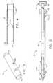
- FIG. 1is an isometric view of a surgical biopsy instrument of the present invention operably coupled to a vacuum source;
- FIG. 2is an exploded isometric view of the piercing needle of the surgical biopsy instrument of FIG. 1 and an assembled view of a cutting member of the surgical biopsy instrument;
- FIG. 3is an isometric view of a single sample segment of the cutting member of the surgical biopsy instrument shown in FIG. 2, the sample segment for the reception of a tissue sample therein;
- FIG. 4is a fragmentary cross-sectional side view of the distal end of the assembled cutting member of FIG. 2 showing the assembly of the individual sample segments;
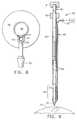
- FIG. 5is a cross-sectional side view of the surgical biopsy instrument of FIG. 1;
- FIG. 6is an enlarged cross-sectional side view of the distal end of the surgical biopsy instrument of FIG. 5 with the cutting member retracted out of the view;
- FIG. 7is an enlarged cross-sectional side view of the distal end of the surgical biopsy instrument of FIG. 6 with the cutting member moved to the fully inserted position;
- FIG. 8is a cross sectional view of the distal end of the surgical biopsy instrument of FIG. 6, with the cross section taken perpendicular to the longitudinal axis of the surgical biopsy instrument;
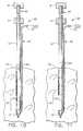
- FIG. 9is a cross-sectional side view of the surgical biopsy instrument of FIG. 5 poised above a breast of a patient;
- FIG. 10is a cross-sectional side view of the surgical biopsy instrument of FIG. 9 with the surgical biopsy instrument inserted into the breast of a patient;
- FIG. 11is a cross-sectional side view of the surgical biopsy instrument of FIG. 10 with the cutting member retracted and the vacuum source actuated to draw tissue into a tissue receiving port of the surgical biopsy instrument;
- FIG. 12is a cross-sectional side view of the surgical biopsy instrument of FIG. 11 with the cutting member extended to sever a tissue sample therein;
- FIG. 13is a cross-sectional side view of the cutting member of the surgical biopsy instrument with the distal-most sample segment and containing the tissue sample broken off of the cutting member;
- FIG. 14is a cross-sectional side view of the surgical biopsy instrument of FIG. 12 with the cutting member removed.
- the present inventionrelates, in general, to surgical biopsy instruments and methods of acquiring multiple subcutaneous tissue biopsies in a minimally invasive manner.
- the present inventionis an improved surgical instrument that provides multiple cutting edges to ensure blade sharpness and provides a novel method of tissue sample storage and identification.
- the present inventionis a surgical biopsy instrument 20 which has a piercing needle 40 for the penetration of tissue, such as breast tissue, and a cutting member 60 moveable within a passageway 44 (FIG. 2) of the piercing needle 40 for the cutting and removal of a plurality of tissue samples 30 (FIG. 12) therefrom.
- a vacuum source 25is shown operably coupled to the hollow piercing needle 40 to draw tissue samples into the surgical biopsy instrument 20 .
- the piercing needle 40has a distal piercing tip 41 for piercing tissue and a proximal knob 42 for the operator to hold.
- a tissue receiving port 43is located adjacent to the piercing tip 41 for the reception of tissue samples therein.
- the tissue receiving port 43communicates with the passageway 44 —extending from the piercing tip 41 and open at a proximal end of the knob 42 (FIG. 5) for the reception of the cutting member 60 .
- Tissue receiving port 43also has a proximal port edge 45 and a distal port edge 46 .
- a plurality of manifold ports 47are located within the base of the tissue receiving port 43 and are operably coupled to a vacuum line 50 extending along the exterior of the piercing needle 40 .
- Notches 51are located within the vacuum line 50 to couple the vacuum line 50 to the manifold ports 47 of the piercing needle 40 .
- a vacuum connector 52is provided at the distal end of the vacuum line 50 .
- the vacuum sourceis operatively coupled to the piercing needle 40 at the vacuum connector 52 .
- the vacuum line 50 and a line cover 53are fixedly attached to the piercing needle 40 by solder, glue, braze or any other airtight attachment means.
- Cutting member 60is slidably and removably received within the passageway 44 of the piercing needle 40 , and has a proximal grip 61 for the operator to grasp and a distal cutting edge 62 for the cutting of tissue samples.
- Cutting member 60is hollow and has a bore 63 extending distally from the grip 61 to the cutting edge 62 for the reception of cut tissue samples therein.
- the cutting member 60 of the preferred inventionis rigid and is assembled from a plurality of sampling segments 64 and a shaft 67 .
- sampling segments 64are formed as separate discreet members, for instance cylinders. Each sampling segment has a proximal tapered end 65 and the distal cutting edge 62 .
- the longitudinal length of the sampling segments 64is slightly longer than the longitudinal length of the tissue receiving port 43 of the piercing needle 40 (FIG. 5) and this will be explained in greater detail below.
- the angle of the distal tapered end 65 and the distal cutting edge 62are generally the same so that the sampling segments 64 can nest together to form a stack of sampling segments 64 . It is an object of the preferred invention to frangibly or breakably assemble the sampling segments 64 together with the proximal shaft 67 to form the cutting member 60 (FIG. 2 ).
- the frangible assembly of the cutting member 60provides the operator with a cutting member 60 that is rigid enough to cut a tissue sample, yet frangible so that the operator can break off the distal-most sampling segment 64 once the tissue sample is taken.
- breaking off the sampling segment 64 of the present inventionthe operator is guaranteed a new and ready cutting edge 62 for each tissue sample taken such that each tissue sample is held within its own sampling segment 64 for analysis later.
- sampling segments 64 of the preferred inventionare preferably held together by an external frangible layer 67 formed from a portion of medical grade shrink tubing.
- a plurality of perforations 68are placed within the shrink tubing of the external frangible layer 67 at the junction of two sampling segments 64 to reduce the force to break the frangible layer 67 .
- the sampling segments 64can be breakably attached in a variety of ways including but not limited to: a thin plating, a coating of paint, adhesives, molded plastics, or any one of a number of other frangible attachment methods. These frangible attachment methods can be used in combination with perforations 68 .
- the sampling segments 64have at least one sample retainer 68 a extending into the bore 63 for the retention of tissue samples therein. As shown in FIG. 4, the sample retainers 68 a are angled inwardly and provide a ramped surface as tissue enters the sampling segment 64 . The sample retainers 68 a effectively act as a one way tissue passage feature within the sampling segments 64 and resist the extraction of the tissue sample in the opposite direction.
- An identifier (indicia) 69(FIG. 3) is located on the exterior of each sampling segment 64 to provide identification so that each sample can be properly accounted for. This enables the surgical team to uniquely identify and record (or log) the identifier 69 of each sampling segment 64 along with the site from which the tissue sample was taken. This reduces the possibility of mixing unmarked tissue samples.
- the identifiers 69are most preferably sequential numbers, with a different number on each sampling segment. Whereas numbers are the preferred embodiment, letters, symbols, colors, bar codes, or any other identification system can be used.
- FIGS. 5-8are cross-sections of the surgical biopsy instrument 20 .
- FIG. 5shows a cross section of the assembled surgical biopsy instrument 20 with the cutting member 60 fully inserted into the piercing needle 40 .
- FIGS. 6-7are of the distal end of the proximal end of the surgical biopsy instrument 20 and show the movement of the cutting edge 62 relative to the tissue receiving port 43 .
- the cutting member 60is shown fully inserted into the piercing needle 40 to show how the cutting edge 62 of the sampling segment 64 has traveled past the distal port edge 46 of the tissue receiving port 43 . This final portion of the travel ensures complete severance of a tissue sample 30 (FIG. 12 ).
- FIG. 6also shows that the sampling segment 64 is longer than the tissue receiving port 43 .
- a cross section of the piercing needle 40is shown in FIG. 8, with the cross section taken across the notches 51 of the tubular vacuum line 50 and the manifold ports 47 of the piercing needle 40 .
- FIGS. 9-14best illustrate the method of use according to the present invention.
- the surgical biopsy device 20(FIG. 1) is used to obtain multiple biopsy samples from a surgical site within a patient's tissue, such as breast 75 .
- the patientis generally positioned upon a surgical table (not shown) and is given a local anesthetic to anesthetize the desired penetration site.
- the preferred surgical biopsy instrument 20is shown poised above the breast 75 of the patient just prior to penetration for the taking of multiple tissue samples 30 therefrom.
- the piercing tip 41is shown placed just above the breast 75 and will facilitate the passage of the surgical biopsy 20 into the breast.
- the cutting member 60is fully extended into the piercing needle 40 to close the tissue receiving port 43 so that tissue cannot migrate therein until desired.
- the surgical biopsy instrument 20is placed into the breast 75 by the surgeon.
- the surgical biopsy instrument 20is carefully positioned at the suspect tissue region and the cutting member 60 is retracted to open the tissue receiving port 43 .
- the vacuum source 25is activated to draw tissue into the tissue receiving port 43 and against the manifold ports 47 .
- the tissueis held in this position prior to the cutting of the tissue sample.
- the manifold ports 47are placed within the tissue receiving port to draw tissue into the receiving port 43 and not into the passageway 64 . This limits the length of the tissue sample that will be acquired.
- the cutting member 60has been advanced downward to sever the tissue sample 30 from the breast 75 .
- the tissue sample 30resides within the bore 63 of the distal-most sampling segment 64 and does not extend the proximal and distal end of the sampling segment 64 .
- the tissue retainers 66retain the tissue sample 30 within the sampling segment 64 .
- the vacuum from the vacuum source 25has been turned off and the tissue sample 30 is ready for removal from the patient.
- FIG. 13shows the cutting member 60 after removal from the piercing needle 40 and from the patient.
- the distal-most sampling segment 64has been broken and detached from the cutting member 60 and contains the tissue sample 30 therein. Once the sample is detached, it is placed into a container for delivery to a laboratory such as the pathology department for analysis.
- the sampling segment 64numbered with its identifier (not shown) identifies the sequence of the tissue sample 30 . Accordingly, new cutting edge 62 is exposed at the distal end of the cutting member 60 whenever a sampling segment 64 and sample 30 are detached.
- FIG. 14depicts the piercing needle 40 still embedded in the breast 25 after the cutting member 60 has been removed for further sampling.
- Cutting member 60may be reinserted into passageway 44 of the piercing needle 40 if further sampling is necessary.
- Piercing needle 40may be repositioned and the biopsy procedure can be repeated according to the steps detailed above until all of the sampling segments 64 have been used.
Landscapes
- Health & Medical Sciences (AREA)
- Life Sciences & Earth Sciences (AREA)
- Medical Informatics (AREA)
- Engineering & Computer Science (AREA)
- Biomedical Technology (AREA)
- Heart & Thoracic Surgery (AREA)
- Pathology (AREA)
- Molecular Biology (AREA)
- Surgery (AREA)
- Animal Behavior & Ethology (AREA)
- General Health & Medical Sciences (AREA)
- Public Health (AREA)
- Veterinary Medicine (AREA)
- Sampling And Sample Adjustment (AREA)
Abstract
Description
Claims (9)
Priority Applications (1)
| Application Number | Priority Date | Filing Date | Title |
|---|---|---|---|
| US09/506,916US6241687B1 (en) | 2000-02-18 | 2000-02-18 | Method of use for a biopsy instrument with breakable sample segments |
Applications Claiming Priority (1)
| Application Number | Priority Date | Filing Date | Title |
|---|---|---|---|
| US09/506,916US6241687B1 (en) | 2000-02-18 | 2000-02-18 | Method of use for a biopsy instrument with breakable sample segments |
Publications (1)
| Publication Number | Publication Date |
|---|---|
| US6241687B1true US6241687B1 (en) | 2001-06-05 |
Family
ID=24016469
Family Applications (1)
| Application Number | Title | Priority Date | Filing Date |
|---|---|---|---|
| US09/506,916Expired - Fee RelatedUS6241687B1 (en) | 2000-02-18 | 2000-02-18 | Method of use for a biopsy instrument with breakable sample segments |
Country Status (1)
| Country | Link |
|---|---|
| US (1) | US6241687B1 (en) |
Cited By (89)
| Publication number | Priority date | Publication date | Assignee | Title |
|---|---|---|---|---|
| US20030050574A1 (en)* | 2000-04-18 | 2003-03-13 | John Krueger | Bone biopsy instrument having improved sample retention |
| US6575991B1 (en)* | 1999-06-17 | 2003-06-10 | Inrad, Inc. | Apparatus for the percutaneous marking of a lesion |
| US20030212342A1 (en)* | 2002-03-26 | 2003-11-13 | Rudnick James J. | System and method for biopsy management |
| US20040034310A1 (en)* | 2002-08-15 | 2004-02-19 | Mcalister Gary | Multiple biopsy apparatus and related method of use |
| US20050054945A1 (en)* | 2003-09-10 | 2005-03-10 | Scimed Life Systems, Inc | Forceps and collection assembly with accompanying mechanisms and related methods of use |
| US20050113867A1 (en)* | 2003-11-25 | 2005-05-26 | Scimed Life Systems, Inc. | Forceps and collection assembly and related methods of use and manufacture |
| US20050228311A1 (en)* | 2004-03-31 | 2005-10-13 | Beckman Andrew T | Marker device and method of deploying a cavity marker using a surgical biopsy device |
| US20060111663A1 (en)* | 2002-07-12 | 2006-05-25 | Andreas Pein | Surgical device for removing tissue cells form a biological structure especially for liposuction |
| US20060116573A1 (en)* | 2003-11-17 | 2006-06-01 | Inrad, Inc. | Self Contained, Self Piercing, Side-Expelling Marking Apparatus |
| US20060129185A1 (en)* | 1999-10-25 | 2006-06-15 | Boston Scientific Scimed, Inc. | Forceps for medical use |
| US20060173377A1 (en)* | 2005-01-31 | 2006-08-03 | Mccullough Adam B | Quick cycle biopsy system |
| US20060229528A1 (en)* | 2003-03-29 | 2006-10-12 | C. R. Brad, Inc. | Coaxial cannula provided with a sealing element |
| US20070038145A1 (en)* | 2004-11-22 | 2007-02-15 | Inrad, Inc. | Post Decompression Marker Introducer System |
| US20070149893A1 (en)* | 2002-03-19 | 2007-06-28 | C.R. Bard, Inc. | Biopsy device and biopsy needle module that can be inserted into the biopsy device |
| US20070213631A1 (en)* | 2004-09-14 | 2007-09-13 | Olympus Corporation | Living-tissue sampling instrument, endoscope system and method of sampling living tissues |
| US20070270714A1 (en)* | 2006-05-19 | 2007-11-22 | E-Z-Em, Inc. | System and method for tissue specimen collection |
| US20080234697A1 (en)* | 2007-03-19 | 2008-09-25 | Restoration Robotics, Inc. | Device and method for harvesting and implanting follicular units |
| US20080235055A1 (en)* | 2003-07-17 | 2008-09-25 | Scott Mattingly | Laboratory instrumentation information management and control network |
| EP2026070A1 (en) | 2002-09-09 | 2009-02-18 | Arbor Vita Corporation | Methods of diagnosing cervical cancer |
| US20100030234A1 (en)* | 2005-09-30 | 2010-02-04 | Mohan Bodduluri | Tool Assembly for Harvesting and Implanting Follicular Units |
| US20100106055A1 (en)* | 2002-03-19 | 2010-04-29 | C.R. Bard, Inc. | Biopsy device having a vacuum pump |
| US7762961B2 (en) | 2003-03-29 | 2010-07-27 | C. R. Bard, Inc. | Pressure generating unit |
| US7762960B2 (en) | 2005-05-13 | 2010-07-27 | Boston Scientific Scimed, Inc. | Biopsy forceps assemblies |
| US20110015542A1 (en)* | 2002-04-23 | 2011-01-20 | Devicor Medical Products, Inc. | Mri compatible biopsy device with detachable probe |
| US8052615B2 (en) | 2004-07-09 | 2011-11-08 | Bard Peripheral Vascular, Inc. | Length detection system for biopsy device |
| US8064987B2 (en) | 2006-10-23 | 2011-11-22 | C. R. Bard, Inc. | Breast marker |
| US8157862B2 (en) | 1997-10-10 | 2012-04-17 | Senorx, Inc. | Tissue marking implant |
| US8177792B2 (en) | 2002-06-17 | 2012-05-15 | Senorx, Inc. | Plugged tip delivery tube for marker placement |
| US8219182B2 (en) | 1999-02-02 | 2012-07-10 | Senorx, Inc. | Cavity-filling biopsy site markers |
| US8224424B2 (en) | 1999-02-02 | 2012-07-17 | Senorx, Inc. | Tissue site markers for in vivo imaging |
| US8251917B2 (en) | 2006-08-21 | 2012-08-28 | C. R. Bard, Inc. | Self-contained handheld biopsy needle |
| US8262585B2 (en) | 2005-08-10 | 2012-09-11 | C. R. Bard, Inc. | Single-insertion, multiple sampling biopsy device with linear drive |
| US8262586B2 (en) | 2006-10-24 | 2012-09-11 | C. R. Bard, Inc. | Large sample low aspect ratio biopsy needle |
| US8267868B2 (en) | 2005-08-10 | 2012-09-18 | C. R. Bard, Inc. | Single-insertion, multiple sample biopsy device with integrated markers |
| US8282574B2 (en) | 2005-08-10 | 2012-10-09 | C. R. Bard, Inc. | Single-insertion, multiple sampling biopsy device usable with various transport systems and integrated markers |
| US8311610B2 (en) | 2008-01-31 | 2012-11-13 | C. R. Bard, Inc. | Biopsy tissue marker |
| US8361082B2 (en) | 1999-02-02 | 2013-01-29 | Senorx, Inc. | Marker delivery device with releasable plug |
| US8401622B2 (en) | 2006-12-18 | 2013-03-19 | C. R. Bard, Inc. | Biopsy marker with in situ-generated imaging properties |
| US8430824B2 (en) | 2009-10-29 | 2013-04-30 | Bard Peripheral Vascular, Inc. | Biopsy driver assembly having a control circuit for conserving battery power |
| US8447386B2 (en) | 2003-05-23 | 2013-05-21 | Senorx, Inc. | Marker or filler forming fluid |
| US8454532B2 (en) | 2007-12-27 | 2013-06-04 | Devicor Medical Products, Inc. | Clutch and valving system for tetherless biopsy device |
| US8485989B2 (en) | 2009-09-01 | 2013-07-16 | Bard Peripheral Vascular, Inc. | Biopsy apparatus having a tissue sample retrieval mechanism |
| US8486028B2 (en) | 2005-10-07 | 2013-07-16 | Bard Peripheral Vascular, Inc. | Tissue marking apparatus having drug-eluting tissue marker |
| US8485987B2 (en) | 2006-10-06 | 2013-07-16 | Bard Peripheral Vascular, Inc. | Tissue handling system with reduced operator exposure |
| US8498693B2 (en) | 1999-02-02 | 2013-07-30 | Senorx, Inc. | Intracorporeal marker and marker delivery device |
| US8597205B2 (en) | 2007-12-20 | 2013-12-03 | C. R. Bard, Inc. | Biopsy device |
| US8597206B2 (en) | 2009-10-12 | 2013-12-03 | Bard Peripheral Vascular, Inc. | Biopsy probe assembly having a mechanism to prevent misalignment of components prior to installation |
| US8626269B2 (en) | 2003-05-23 | 2014-01-07 | Senorx, Inc. | Fibrous marker and intracorporeal delivery thereof |
| US8634899B2 (en) | 2003-11-17 | 2014-01-21 | Bard Peripheral Vascular, Inc. | Multi mode imaging marker |
| US8668737B2 (en) | 1997-10-10 | 2014-03-11 | Senorx, Inc. | Tissue marking implant |
| US8670818B2 (en) | 2008-12-30 | 2014-03-11 | C. R. Bard, Inc. | Marker delivery device for tissue marker placement |
| US8690793B2 (en) | 2009-03-16 | 2014-04-08 | C. R. Bard, Inc. | Biopsy device having rotational cutting |
| US8708930B2 (en) | 2009-04-15 | 2014-04-29 | Bard Peripheral Vascular, Inc. | Biopsy apparatus having integrated fluid management |
| US8718745B2 (en) | 2000-11-20 | 2014-05-06 | Senorx, Inc. | Tissue site markers for in vivo imaging |
| US20140200484A1 (en)* | 2011-01-28 | 2014-07-17 | The General Hospital Corporation | Apparatus and method for tissue biopsy |
| US8845548B2 (en) | 2009-06-12 | 2014-09-30 | Devicor Medical Products, Inc. | Cutter drive assembly for biopsy device |
| USD715442S1 (en) | 2013-09-24 | 2014-10-14 | C. R. Bard, Inc. | Tissue marker for intracorporeal site identification |
| USD715942S1 (en) | 2013-09-24 | 2014-10-21 | C. R. Bard, Inc. | Tissue marker for intracorporeal site identification |
| USD716450S1 (en) | 2013-09-24 | 2014-10-28 | C. R. Bard, Inc. | Tissue marker for intracorporeal site identification |
| USD716451S1 (en) | 2013-09-24 | 2014-10-28 | C. R. Bard, Inc. | Tissue marker for intracorporeal site identification |
| US20150148703A1 (en)* | 2013-11-25 | 2015-05-28 | Devicor Medical Products, Inc. | Biopsy device with translating valve assembly |
| US9149341B2 (en) | 1999-02-02 | 2015-10-06 | Senorx, Inc | Deployment of polysaccharide markers for treating a site within a patient |
| US9173641B2 (en) | 2009-08-12 | 2015-11-03 | C. R. Bard, Inc. | Biopsy apparatus having integrated thumbwheel mechanism for manual rotation of biopsy cannula |
| US9327061B2 (en) | 2008-09-23 | 2016-05-03 | Senorx, Inc. | Porous bioabsorbable implant |
| US20160363512A1 (en)* | 2015-06-13 | 2016-12-15 | Sampling Systems Ltd. | Sampling pod system and method |
| US9579077B2 (en) | 2006-12-12 | 2017-02-28 | C.R. Bard, Inc. | Multiple imaging mode tissue marker |
| WO2017088013A1 (en)* | 2015-11-27 | 2017-06-01 | The University Of Western Australia | A kit for the detection of urease |
| US9820824B2 (en) | 1999-02-02 | 2017-11-21 | Senorx, Inc. | Deployment of polysaccharide markers for treating a site within a patent |
| US9907542B2 (en)* | 2005-08-05 | 2018-03-06 | Devicor Medical Products, Inc. | Biopsy device with translating valve member |
| US10251792B2 (en) | 2013-02-20 | 2019-04-09 | Cytrellis Biosystems, Inc. | Methods and devices for skin tightening |
| US10285673B2 (en) | 2013-03-20 | 2019-05-14 | Bard Peripheral Vascular, Inc. | Biopsy device |
| US10327800B2 (en) | 2011-01-28 | 2019-06-25 | The General Hospital Corporation | Method and apparatus for skin resurfacing |
| US10342635B2 (en) | 2005-04-20 | 2019-07-09 | Bard Peripheral Vascular, Inc. | Marking device with retractable cannula |
| US10456120B2 (en) | 2013-11-05 | 2019-10-29 | C. R. Bard, Inc. | Biopsy device having integrated vacuum |
| US10463350B2 (en) | 2015-05-01 | 2019-11-05 | C. R. Bard, Inc. | Biopsy device |
| CN110495931A (en)* | 2019-09-09 | 2019-11-26 | 武汉微新坦医疗科技有限公司 | An Electric Intracardiac Cardiac Cutter |
| US10555754B2 (en) | 2013-08-09 | 2020-02-11 | Cytrellis Biosystems, Inc. | Methods and apparatuses for skin treatment using non-thermal tissue ablation |
| CN111956268A (en)* | 2020-08-11 | 2020-11-20 | 王新铮 | Full-automatic biopsy puncture device for mammary gland |
| US10953143B2 (en) | 2013-12-19 | 2021-03-23 | Cytrellis Biosystems, Inc. | Methods and devices for manipulating subdermal fat |
| US11116483B2 (en) | 2017-05-19 | 2021-09-14 | Merit Medical Systems, Inc. | Rotating biopsy needle |
| US11166743B2 (en) | 2016-03-29 | 2021-11-09 | Cytrellis Biosystems, Inc. | Devices and methods for cosmetic skin resurfacing |
| US11324534B2 (en) | 2014-11-14 | 2022-05-10 | Cytrellis Biosystems, Inc. | Devices and methods for ablation of the skin |
| US11337720B2 (en) | 2011-07-21 | 2022-05-24 | The General Hospital Corporation | Method and apparatus for damage and removal of fat |
| US11464954B2 (en) | 2016-09-21 | 2022-10-11 | Cytrellis Biosystems, Inc. | Devices and methods for cosmetic skin resurfacing |
| US11793498B2 (en) | 2017-05-19 | 2023-10-24 | Merit Medical Systems, Inc. | Biopsy needle devices and methods of use |
| US11844500B2 (en) | 2017-05-19 | 2023-12-19 | Merit Medical Systems, Inc. | Semi-automatic biopsy needle device and methods of use |
| US12150627B2 (en) | 2019-12-11 | 2024-11-26 | Merit Medical Systems, Inc. | Bone biopsy device and related methods |
| US12178513B2 (en) | 2010-12-21 | 2024-12-31 | Venus Concept Inc. | Methods and systems for directing movement of a tool in hair transplantation procedures |
| US12295556B2 (en) | 2019-09-27 | 2025-05-13 | Merit Medical Systems, Inc. | Rotation biopsy system and handle |
Citations (4)
| Publication number | Priority date | Publication date | Assignee | Title |
|---|---|---|---|---|
| US3590808A (en) | 1968-09-04 | 1971-07-06 | Us Catheter & Instr Corp | Biopsy tool |
| US5045067A (en) | 1987-03-19 | 1991-09-03 | Terumo Kabushiki Kaisha | Breakaway tube assembly |
| US5524634A (en) | 1994-07-19 | 1996-06-11 | Symbiois Corporation | Methods and apparatus for recording the source and orientation of as well as for storing and identifying multiple biopsy samples |
| US5526822A (en) | 1994-03-24 | 1996-06-18 | Biopsys Medical, Inc. | Method and apparatus for automated biopsy and collection of soft tissue |
- 2000
- 2000-02-18USUS09/506,916patent/US6241687B1/ennot_activeExpired - Fee Related
Patent Citations (4)
| Publication number | Priority date | Publication date | Assignee | Title |
|---|---|---|---|---|
| US3590808A (en) | 1968-09-04 | 1971-07-06 | Us Catheter & Instr Corp | Biopsy tool |
| US5045067A (en) | 1987-03-19 | 1991-09-03 | Terumo Kabushiki Kaisha | Breakaway tube assembly |
| US5526822A (en) | 1994-03-24 | 1996-06-18 | Biopsys Medical, Inc. | Method and apparatus for automated biopsy and collection of soft tissue |
| US5524634A (en) | 1994-07-19 | 1996-06-11 | Symbiois Corporation | Methods and apparatus for recording the source and orientation of as well as for storing and identifying multiple biopsy samples |
Cited By (238)
| Publication number | Priority date | Publication date | Assignee | Title |
|---|---|---|---|---|
| US8668737B2 (en) | 1997-10-10 | 2014-03-11 | Senorx, Inc. | Tissue marking implant |
| US9039763B2 (en) | 1997-10-10 | 2015-05-26 | Senorx, Inc. | Tissue marking implant |
| US8157862B2 (en) | 1997-10-10 | 2012-04-17 | Senorx, Inc. | Tissue marking implant |
| US9044162B2 (en) | 1999-02-02 | 2015-06-02 | Senorx, Inc. | Marker delivery device with releasable plug |
| US8626270B2 (en) | 1999-02-02 | 2014-01-07 | Senorx, Inc. | Cavity-filling biopsy site markers |
| US10172674B2 (en) | 1999-02-02 | 2019-01-08 | Senorx, Inc. | Intracorporeal marker and marker delivery device |
| US8361082B2 (en) | 1999-02-02 | 2013-01-29 | Senorx, Inc. | Marker delivery device with releasable plug |
| US9649093B2 (en) | 1999-02-02 | 2017-05-16 | Senorx, Inc. | Cavity-filling biopsy site markers |
| US9237937B2 (en) | 1999-02-02 | 2016-01-19 | Senorx, Inc. | Cavity-filling biopsy site markers |
| US8224424B2 (en) | 1999-02-02 | 2012-07-17 | Senorx, Inc. | Tissue site markers for in vivo imaging |
| US8219182B2 (en) | 1999-02-02 | 2012-07-10 | Senorx, Inc. | Cavity-filling biopsy site markers |
| US8498693B2 (en) | 1999-02-02 | 2013-07-30 | Senorx, Inc. | Intracorporeal marker and marker delivery device |
| US9149341B2 (en) | 1999-02-02 | 2015-10-06 | Senorx, Inc | Deployment of polysaccharide markers for treating a site within a patient |
| US9820824B2 (en) | 1999-02-02 | 2017-11-21 | Senorx, Inc. | Deployment of polysaccharide markers for treating a site within a patent |
| US9861294B2 (en) | 1999-02-02 | 2018-01-09 | Senorx, Inc. | Marker delivery device with releasable plug |
| US8965486B2 (en) | 1999-02-02 | 2015-02-24 | Senorx, Inc. | Cavity filling biopsy site markers |
| US20030204137A1 (en)* | 1999-06-17 | 2003-10-30 | Inrad, Inc. | Apparatus for the percutaneous marking of a lesion |
| US9579159B2 (en) | 1999-06-17 | 2017-02-28 | Bard Peripheral Vascular, Inc. | Apparatus for the percutaneous marking of a lesion |
| US6575991B1 (en)* | 1999-06-17 | 2003-06-10 | Inrad, Inc. | Apparatus for the percutaneous marking of a lesion |
| US10463446B2 (en) | 1999-06-17 | 2019-11-05 | Bard Peripheral Vascular, Inc. | Apparatus for the percutaneous marking of a lesion |
| US7569065B2 (en) | 1999-06-17 | 2009-08-04 | Bard Peripheral Vascular, Inc. | Apparatus for the percutaneous marking of a lesion |
| US20090093714A1 (en)* | 1999-06-17 | 2009-04-09 | Bard Peripheral Vascular, Inc | Apparatus for the percutaneous marking of a lesion |
| US8052708B2 (en) | 1999-06-17 | 2011-11-08 | Bard Peripheral Vascular, Inc. | Apparatus for the percutaneous marking of a lesion |
| US20060025795A1 (en)* | 1999-06-17 | 2006-02-02 | Inrad, Inc. | Apparatus for the percutaneous marking of a lesion |
| US8579931B2 (en) | 1999-06-17 | 2013-11-12 | Bard Peripheral Vascular, Inc. | Apparatus for the percutaneous marking of a lesion |
| US20060129185A1 (en)* | 1999-10-25 | 2006-06-15 | Boston Scientific Scimed, Inc. | Forceps for medical use |
| US7909850B2 (en) | 1999-10-25 | 2011-03-22 | Boston Scientific Scimed, Inc. | Forceps for medical use |
| US20030050574A1 (en)* | 2000-04-18 | 2003-03-13 | John Krueger | Bone biopsy instrument having improved sample retention |
| US7201722B2 (en) | 2000-04-18 | 2007-04-10 | Allegiance Corporation | Bone biopsy instrument having improved sample retention |
| US8718745B2 (en) | 2000-11-20 | 2014-05-06 | Senorx, Inc. | Tissue site markers for in vivo imaging |
| US8172773B2 (en) | 2002-03-19 | 2012-05-08 | C. R. Bard, Inc. | Biopsy device and biopsy needle module that can be inserted into the biopsy device |
| US10335128B2 (en) | 2002-03-19 | 2019-07-02 | C. R. Bard, Inc. | Biopsy device and insertable biopsy needle module |
| US9439631B2 (en) | 2002-03-19 | 2016-09-13 | C. R. Bard, Inc. | Biopsy device and insertable biopsy needle module |
| US20070149893A1 (en)* | 2002-03-19 | 2007-06-28 | C.R. Bard, Inc. | Biopsy device and biopsy needle module that can be inserted into the biopsy device |
| US11382608B2 (en) | 2002-03-19 | 2022-07-12 | C. R. Bard, Inc. | Disposable biopsy unit |
| US8016772B2 (en) | 2002-03-19 | 2011-09-13 | C. R. Bard, Inc. | Biopsy device for removing tissue specimens using a vacuum |
| US9421002B2 (en) | 2002-03-19 | 2016-08-23 | C. R. Bard, Inc. | Disposable biopsy unit |
| US20100106055A1 (en)* | 2002-03-19 | 2010-04-29 | C.R. Bard, Inc. | Biopsy device having a vacuum pump |
| US9072502B2 (en) | 2002-03-19 | 2015-07-07 | C. R. Bard, Inc. | Disposable biopsy unit |
| US8002713B2 (en) | 2002-03-19 | 2011-08-23 | C. R. Bard, Inc. | Biopsy device and insertable biopsy needle module |
| US8951209B2 (en) | 2002-03-19 | 2015-02-10 | C. R. Bard, Inc. | Biopsy device and insertable biopsy needle module |
| US8052614B2 (en) | 2002-03-19 | 2011-11-08 | C. R. Bard, Inc. | Biopsy device having a vacuum pump |
| US10271827B2 (en) | 2002-03-19 | 2019-04-30 | C. R. Bard, Inc. | Disposable biopsy unit |
| US8109885B2 (en) | 2002-03-19 | 2012-02-07 | C. R. Bard, Inc. | Biopsy device for removing tissue specimens using a vacuum |
| US6986780B2 (en) | 2002-03-26 | 2006-01-17 | Ethicon, Inc. | Surgical element delivery system and method |
| US6835180B2 (en)* | 2002-03-26 | 2004-12-28 | Ethicon, Inc. | System and method for biopsy management |
| US20030212417A1 (en)* | 2002-03-26 | 2003-11-13 | Rudnick James J. | Surgical element delivery system and method |
| US20030212342A1 (en)* | 2002-03-26 | 2003-11-13 | Rudnick James J. | System and method for biopsy management |
| US20110015542A1 (en)* | 2002-04-23 | 2011-01-20 | Devicor Medical Products, Inc. | Mri compatible biopsy device with detachable probe |
| US8808198B2 (en)* | 2002-04-23 | 2014-08-19 | Devicor Medical Products, Inc. | MRI compatible biopsy device with detachable probe |
| US8177792B2 (en) | 2002-06-17 | 2012-05-15 | Senorx, Inc. | Plugged tip delivery tube for marker placement |
| US8784433B2 (en) | 2002-06-17 | 2014-07-22 | Senorx, Inc. | Plugged tip delivery tube for marker placement |
| US20060111663A1 (en)* | 2002-07-12 | 2006-05-25 | Andreas Pein | Surgical device for removing tissue cells form a biological structure especially for liposuction |
| US8262587B2 (en) | 2002-08-15 | 2012-09-11 | Boston Scientific Scimed, Inc. | Multiple biopsy apparatus and related method of use |
| US20040034310A1 (en)* | 2002-08-15 | 2004-02-19 | Mcalister Gary | Multiple biopsy apparatus and related method of use |
| US20060282012A1 (en)* | 2002-08-15 | 2006-12-14 | Boston Scientific Scimed, Inc. | Multiple biopsy apparatus and related method of use |
| US7828745B2 (en) | 2002-08-15 | 2010-11-09 | Boston Scientific Scimed, Inc. | Multiple biopsy apparatus and related method of use |
| US20110021949A1 (en)* | 2002-08-15 | 2011-01-27 | Boston Scientific Scimed, Inc. | Multiple Biopsy Apparatus and Related Method of Use |
| US6986748B2 (en)* | 2002-08-15 | 2006-01-17 | Scimed Life Systems, Inc. | Multiple biopsy apparatus and related method of use |
| EP2026070A1 (en) | 2002-09-09 | 2009-02-18 | Arbor Vita Corporation | Methods of diagnosing cervical cancer |
| US10813716B2 (en) | 2002-11-18 | 2020-10-27 | Bard Peripheral Vascular, Inc. | Self-contained, self-piercing, side-expelling marking apparatus |
| US9848956B2 (en) | 2002-11-18 | 2017-12-26 | Bard Peripheral Vascular, Inc. | Self-contained, self-piercing, side-expelling marking apparatus |
| US11071529B2 (en) | 2003-03-29 | 2021-07-27 | C.R. Bard, Inc. | Cannula provided with a sealing element for use in a medical procedure |
| US8162851B2 (en) | 2003-03-29 | 2012-04-24 | C. R. Bard, Inc. | Biopsy needle system having a pressure generating unit |
| US20070179403A1 (en)* | 2003-03-29 | 2007-08-02 | C.R. Bard, Inc. | Coaxial cannula provided with a sealing element |
| US8728004B2 (en) | 2003-03-29 | 2014-05-20 | C.R. Bard, Inc. | Biopsy needle system having a pressure generating unit |
| US7828747B2 (en) | 2003-03-29 | 2010-11-09 | C. R. Bard, Inc. | Pressure generating unit |
| US7740598B2 (en) | 2003-03-29 | 2010-06-22 | C. R. Bard, Inc. | Coaxial cannula provided with a sealing element |
| US9980706B2 (en) | 2003-03-29 | 2018-05-29 | C. R. Bard, Inc. | Cannula provided with a sealing element for use in a medical procedure |
| US8845547B2 (en) | 2003-03-29 | 2014-09-30 | C. R. Bard, Inc. | Cannula provided with a sealing element for use in a medical procedure |
| US20060229528A1 (en)* | 2003-03-29 | 2006-10-12 | C. R. Brad, Inc. | Coaxial cannula provided with a sealing element |
| US9706980B2 (en) | 2003-03-29 | 2017-07-18 | C. R. Bard, Inc. | Cannula provided with a sealing element for use in a medical procedure |
| US7762961B2 (en) | 2003-03-29 | 2010-07-27 | C. R. Bard, Inc. | Pressure generating unit |
| US7645239B2 (en) | 2003-03-29 | 2010-01-12 | C. R. Bard, Inc. | Coaxial cannula provided with a sealing element |
| US10299881B2 (en) | 2003-05-23 | 2019-05-28 | Senorx, Inc. | Marker or filler forming fluid |
| US10045832B2 (en) | 2003-05-23 | 2018-08-14 | Senorx, Inc. | Marker or filler forming fluid |
| US9801688B2 (en) | 2003-05-23 | 2017-10-31 | Senorx, Inc. | Fibrous marker and intracorporeal delivery thereof |
| US8639315B2 (en) | 2003-05-23 | 2014-01-28 | Senorx, Inc. | Marker or filler forming fluid |
| US8626269B2 (en) | 2003-05-23 | 2014-01-07 | Senorx, Inc. | Fibrous marker and intracorporeal delivery thereof |
| US8447386B2 (en) | 2003-05-23 | 2013-05-21 | Senorx, Inc. | Marker or filler forming fluid |
| US8880154B2 (en) | 2003-05-23 | 2014-11-04 | Senorx, Inc. | Fibrous marker and intracorporeal delivery thereof |
| US20080235055A1 (en)* | 2003-07-17 | 2008-09-25 | Scott Mattingly | Laboratory instrumentation information management and control network |
| US20050054945A1 (en)* | 2003-09-10 | 2005-03-10 | Scimed Life Systems, Inc | Forceps and collection assembly with accompanying mechanisms and related methods of use |
| US8083686B2 (en) | 2003-09-10 | 2011-12-27 | Boston Scientific Scimed, Inc. | Forceps and collection assembly with accompanying mechanisms and related methods of use |
| US7588545B2 (en) | 2003-09-10 | 2009-09-15 | Boston Scientific Scimed, Inc. | Forceps and collection assembly with accompanying mechanisms and related methods of use |
| US8460205B2 (en) | 2003-09-10 | 2013-06-11 | Boston Scientific Scimed, Inc. | Forceps and collection assembly with accompanying mechanisms and related methods of use |
| US7819820B2 (en) | 2003-11-17 | 2010-10-26 | Bard Peripheral Vascular, Inc. | Self contained, self piercing, side-expelling marking apparatus |
| US8634899B2 (en) | 2003-11-17 | 2014-01-21 | Bard Peripheral Vascular, Inc. | Multi mode imaging marker |
| US20060116573A1 (en)* | 2003-11-17 | 2006-06-01 | Inrad, Inc. | Self Contained, Self Piercing, Side-Expelling Marking Apparatus |
| US20050113867A1 (en)* | 2003-11-25 | 2005-05-26 | Scimed Life Systems, Inc. | Forceps and collection assembly and related methods of use and manufacture |
| US7942896B2 (en) | 2003-11-25 | 2011-05-17 | Scimed Life Systems, Inc. | Forceps and collection assembly and related methods of use and manufacture |
| US20050228311A1 (en)* | 2004-03-31 | 2005-10-13 | Beckman Andrew T | Marker device and method of deploying a cavity marker using a surgical biopsy device |
| US20090088665A1 (en)* | 2004-03-31 | 2009-04-02 | Beckman Andrew T | Marker device and method of deploying a cavity marker using a surgical biopsy device |
| US7465279B2 (en) | 2004-03-31 | 2008-12-16 | Ethicon Endo-Surgery, Inc. | Marker device and method of deploying a cavity marker using a surgical biopsy device |
| US9345458B2 (en) | 2004-07-09 | 2016-05-24 | Bard Peripheral Vascular, Inc. | Transport system for biopsy device |
| US8864680B2 (en) | 2004-07-09 | 2014-10-21 | Bard Peripheral Vascular, Inc. | Transport system for biopsy device |
| US8366636B2 (en) | 2004-07-09 | 2013-02-05 | Bard Peripheral Vascular, Inc. | Firing system for biopsy device |
| US8926527B2 (en) | 2004-07-09 | 2015-01-06 | Bard Peripheral Vascular, Inc. | Tissue sample flushing system for biopsy device |
| US8052615B2 (en) | 2004-07-09 | 2011-11-08 | Bard Peripheral Vascular, Inc. | Length detection system for biopsy device |
| US8157744B2 (en) | 2004-07-09 | 2012-04-17 | Bard Peripheral Vascular, Inc. | Tissue sample flushing system for biopsy device |
| US10499888B2 (en) | 2004-07-09 | 2019-12-10 | Bard Peripheral Vascular, Inc. | Tissue sample flushing system for biopsy device |
| US10166011B2 (en) | 2004-07-09 | 2019-01-01 | Bard Peripheral Vascular, Inc. | Transport system for biopsy device |
| US8992440B2 (en) | 2004-07-09 | 2015-03-31 | Bard Peripheral Vascular, Inc. | Length detection system for biopsy device |
| US9456809B2 (en) | 2004-07-09 | 2016-10-04 | Bard Peripheral Vascular, Inc. | Tissue sample flushing system for biopsy device |
| US9872672B2 (en) | 2004-07-09 | 2018-01-23 | Bard Peripheral Vascular, Inc. | Length detection system for biopsy device |
| US20070213631A1 (en)* | 2004-09-14 | 2007-09-13 | Olympus Corporation | Living-tissue sampling instrument, endoscope system and method of sampling living tissues |
| US8419656B2 (en) | 2004-11-22 | 2013-04-16 | Bard Peripheral Vascular, Inc. | Post decompression marker introducer system |
| US20070038145A1 (en)* | 2004-11-22 | 2007-02-15 | Inrad, Inc. | Post Decompression Marker Introducer System |
| US20060173377A1 (en)* | 2005-01-31 | 2006-08-03 | Mccullough Adam B | Quick cycle biopsy system |
| US11166702B2 (en) | 2005-01-31 | 2021-11-09 | C.R. Bard, Inc. | Quick cycle biopsy system |
| US10058308B2 (en) | 2005-01-31 | 2018-08-28 | C. R. Bard, Inc. | Method for operating a biopsy apparatus |
| US7517321B2 (en) | 2005-01-31 | 2009-04-14 | C. R. Bard, Inc. | Quick cycle biopsy system |
| US20070149895A1 (en)* | 2005-01-31 | 2007-06-28 | C.R. Bard, Inc. | Quick cycle biopsy system |
| US8702621B2 (en) | 2005-01-31 | 2014-04-22 | C.R. Bard, Inc. | Quick cycle biopsy system |
| US9161743B2 (en) | 2005-01-31 | 2015-10-20 | C. R. Bard, Inc. | Quick cycle biopsy system |
| US7959580B2 (en) | 2005-01-31 | 2011-06-14 | C.R. Bard, Inc. | Quick cycle biopsy system |
| US8012102B2 (en) | 2005-01-31 | 2011-09-06 | C. R. Bard, Inc. | Quick cycle biopsy system |
| US8702622B2 (en) | 2005-01-31 | 2014-04-22 | C.R. Bard, Inc. | Quick cycle biopsy system |
| US11278370B2 (en) | 2005-04-20 | 2022-03-22 | Bard Peripheral Vascular, Inc. | Marking device with retractable cannula |
| US10357328B2 (en) | 2005-04-20 | 2019-07-23 | Bard Peripheral Vascular, Inc. and Bard Shannon Limited | Marking device with retractable cannula |
| US10342635B2 (en) | 2005-04-20 | 2019-07-09 | Bard Peripheral Vascular, Inc. | Marking device with retractable cannula |
| US7762960B2 (en) | 2005-05-13 | 2010-07-27 | Boston Scientific Scimed, Inc. | Biopsy forceps assemblies |
| US8672859B2 (en) | 2005-05-13 | 2014-03-18 | Boston Scientific Scimed, Inc. | Biopsy forceps assemblies |
| US8317726B2 (en) | 2005-05-13 | 2012-11-27 | Boston Scientific Scimed, Inc. | Biopsy forceps assemblies |
| US11224412B2 (en) | 2005-08-05 | 2022-01-18 | Devicor Medical Products, Inc. | Biopsy device with translating valve member |
| US9907542B2 (en)* | 2005-08-05 | 2018-03-06 | Devicor Medical Products, Inc. | Biopsy device with translating valve member |
| US8721563B2 (en) | 2005-08-10 | 2014-05-13 | C. R. Bard, Inc. | Single-insertion, multiple sample biopsy device with integrated markers |
| US8771200B2 (en) | 2005-08-10 | 2014-07-08 | C.R. Bard, Inc. | Single insertion, multiple sampling biopsy device with linear drive |
| US8961430B2 (en) | 2005-08-10 | 2015-02-24 | C.R. Bard, Inc. | Single-insertion, multiple sampling biopsy device usable with various transport systems and integrated markers |
| US8267868B2 (en) | 2005-08-10 | 2012-09-18 | C. R. Bard, Inc. | Single-insertion, multiple sample biopsy device with integrated markers |
| US11219431B2 (en) | 2005-08-10 | 2022-01-11 | C.R. Bard, Inc. | Single-insertion, multiple sampling biopsy device with linear drive |
| US8728003B2 (en) | 2005-08-10 | 2014-05-20 | C.R. Bard Inc. | Single insertion, multiple sample biopsy device with integrated markers |
| US10010307B2 (en) | 2005-08-10 | 2018-07-03 | C. R. Bard, Inc. | Single-insertion, multiple sampling biopsy device with linear drive |
| US8282574B2 (en) | 2005-08-10 | 2012-10-09 | C. R. Bard, Inc. | Single-insertion, multiple sampling biopsy device usable with various transport systems and integrated markers |
| US10368849B2 (en) | 2005-08-10 | 2019-08-06 | C. R. Bard, Inc. | Single-insertion, multiple sampling biopsy device usable with various transport systems and integrated markers |
| US11849928B2 (en) | 2005-08-10 | 2023-12-26 | C. R. Bard, Inc. | Single-insertion, multiple sampling biopsy device usable with various transport systems and integrated markers |
| US8262585B2 (en) | 2005-08-10 | 2012-09-11 | C. R. Bard, Inc. | Single-insertion, multiple sampling biopsy device with linear drive |
| US20100030234A1 (en)* | 2005-09-30 | 2010-02-04 | Mohan Bodduluri | Tool Assembly for Harvesting and Implanting Follicular Units |
| US8133247B2 (en) | 2005-09-30 | 2012-03-13 | Restoration Robotics, Inc. | Tool assembly for harvesting and implanting follicular units |
| US8486028B2 (en) | 2005-10-07 | 2013-07-16 | Bard Peripheral Vascular, Inc. | Tissue marking apparatus having drug-eluting tissue marker |
| CN101472526B (en)* | 2006-05-19 | 2012-01-25 | 布拉蔻诊断公司 | System and method for tissue specimen collection |
| US20070270714A1 (en)* | 2006-05-19 | 2007-11-22 | E-Z-Em, Inc. | System and method for tissue specimen collection |
| US10617399B2 (en) | 2006-08-21 | 2020-04-14 | C.R. Bard, Inc. | Self-contained handheld biopsy needle |
| US8251917B2 (en) | 2006-08-21 | 2012-08-28 | C. R. Bard, Inc. | Self-contained handheld biopsy needle |
| US8951208B2 (en) | 2006-08-21 | 2015-02-10 | C. R. Bard, Inc. | Self-contained handheld biopsy needle |
| US8485987B2 (en) | 2006-10-06 | 2013-07-16 | Bard Peripheral Vascular, Inc. | Tissue handling system with reduced operator exposure |
| US11559289B2 (en) | 2006-10-06 | 2023-01-24 | Bard Peripheral Vascular, Inc. | Tissue handling system with reduced operator exposure |
| US10172594B2 (en) | 2006-10-06 | 2019-01-08 | Bard Peripheral Vascular, Inc. | Tissue handling system with reduced operator exposure |
| US9566045B2 (en) | 2006-10-06 | 2017-02-14 | Bard Peripheral Vascular, Inc. | Tissue handling system with reduced operator exposure |
| US8064987B2 (en) | 2006-10-23 | 2011-11-22 | C. R. Bard, Inc. | Breast marker |
| US8437834B2 (en) | 2006-10-23 | 2013-05-07 | C. R. Bard, Inc. | Breast marker |
| US8262586B2 (en) | 2006-10-24 | 2012-09-11 | C. R. Bard, Inc. | Large sample low aspect ratio biopsy needle |
| US11583261B2 (en) | 2006-10-24 | 2023-02-21 | C. R. Bard, Inc. | Large sample low aspect ratio biopsy needle |
| US10149664B2 (en) | 2006-10-24 | 2018-12-11 | C. R. Bard, Inc. | Large sample low aspect ratio biopsy needle |
| US9579077B2 (en) | 2006-12-12 | 2017-02-28 | C.R. Bard, Inc. | Multiple imaging mode tissue marker |
| US11471244B2 (en) | 2006-12-12 | 2022-10-18 | C.R. Bard, Inc. | Multiple imaging mode tissue marker |
| US9901415B2 (en) | 2006-12-12 | 2018-02-27 | C. R. Bard, Inc. | Multiple imaging mode tissue marker |
| US10682200B2 (en) | 2006-12-12 | 2020-06-16 | C. R. Bard, Inc. | Multiple imaging mode tissue marker |
| US8401622B2 (en) | 2006-12-18 | 2013-03-19 | C. R. Bard, Inc. | Biopsy marker with in situ-generated imaging properties |
| US9042965B2 (en) | 2006-12-18 | 2015-05-26 | C. R. Bard, Inc. | Biopsy marker with in situ-generated imaging properties |
| US20080234697A1 (en)* | 2007-03-19 | 2008-09-25 | Restoration Robotics, Inc. | Device and method for harvesting and implanting follicular units |
| US8562627B2 (en) | 2007-03-19 | 2013-10-22 | Restoration Robotics, Inc | Device and method for harvesting and implanting follicular units |
| US8066717B2 (en)* | 2007-03-19 | 2011-11-29 | Restoration Robotics, Inc. | Device and method for harvesting and implanting follicular units |
| US10687791B2 (en) | 2007-12-20 | 2020-06-23 | C. R. Bard, Inc. | Biopsy device |
| US8858463B2 (en) | 2007-12-20 | 2014-10-14 | C. R. Bard, Inc. | Biopsy device |
| US8597205B2 (en) | 2007-12-20 | 2013-12-03 | C. R. Bard, Inc. | Biopsy device |
| US9775588B2 (en) | 2007-12-20 | 2017-10-03 | C. R. Bard, Inc. | Biopsy device |
| US8864682B2 (en) | 2007-12-27 | 2014-10-21 | Devicor Medical Products, Inc. | Clutch and valving system for tetherless biopsy device |
| US8454532B2 (en) | 2007-12-27 | 2013-06-04 | Devicor Medical Products, Inc. | Clutch and valving system for tetherless biopsy device |
| US8311610B2 (en) | 2008-01-31 | 2012-11-13 | C. R. Bard, Inc. | Biopsy tissue marker |
| US9327061B2 (en) | 2008-09-23 | 2016-05-03 | Senorx, Inc. | Porous bioabsorbable implant |
| US10786604B2 (en) | 2008-09-23 | 2020-09-29 | Senorx, Inc. | Porous bioabsorbable implant |
| US11833275B2 (en) | 2008-09-23 | 2023-12-05 | Senorx, Inc. | Porous bioabsorbable implant |
| US8670818B2 (en) | 2008-12-30 | 2014-03-11 | C. R. Bard, Inc. | Marker delivery device for tissue marker placement |
| US11779431B2 (en) | 2008-12-30 | 2023-10-10 | C. R. Bard, Inc. | Marker delivery device for tissue marker placement |
| US10258428B2 (en) | 2008-12-30 | 2019-04-16 | C. R. Bard, Inc. | Marker delivery device for tissue marker placement |
| US8690793B2 (en) | 2009-03-16 | 2014-04-08 | C. R. Bard, Inc. | Biopsy device having rotational cutting |
| US8708930B2 (en) | 2009-04-15 | 2014-04-29 | Bard Peripheral Vascular, Inc. | Biopsy apparatus having integrated fluid management |
| US8708929B2 (en) | 2009-04-15 | 2014-04-29 | Bard Peripheral Vascular, Inc. | Biopsy apparatus having integrated fluid management |
| US8708928B2 (en) | 2009-04-15 | 2014-04-29 | Bard Peripheral Vascular, Inc. | Biopsy apparatus having integrated fluid management |
| US8845548B2 (en) | 2009-06-12 | 2014-09-30 | Devicor Medical Products, Inc. | Cutter drive assembly for biopsy device |
| US9468424B2 (en) | 2009-06-12 | 2016-10-18 | Devicor Medical Products, Inc. | Cutter drive assembly for biopsy device |
| US9173641B2 (en) | 2009-08-12 | 2015-11-03 | C. R. Bard, Inc. | Biopsy apparatus having integrated thumbwheel mechanism for manual rotation of biopsy cannula |
| US9655599B2 (en) | 2009-08-12 | 2017-05-23 | C. R. Bard, Inc. | Biopsy apparatus having integrated thumbwheel mechanism for manual rotation of biopsy cannula |
| US10575833B2 (en) | 2009-08-12 | 2020-03-03 | C. R. Bard, Inc. | Biopsy apparatus having integrated thumbwheel mechanism for manual rotation of biopsy cannula |
| US8485989B2 (en) | 2009-09-01 | 2013-07-16 | Bard Peripheral Vascular, Inc. | Biopsy apparatus having a tissue sample retrieval mechanism |
| US9949726B2 (en) | 2009-09-01 | 2018-04-24 | Bard Peripheral Vscular, Inc. | Biopsy driver assembly having a control circuit for conserving battery power |
| US8597206B2 (en) | 2009-10-12 | 2013-12-03 | Bard Peripheral Vascular, Inc. | Biopsy probe assembly having a mechanism to prevent misalignment of components prior to installation |
| US8808197B2 (en) | 2009-10-29 | 2014-08-19 | Bard Peripheral Vascular, Inc. | Biopsy driver assembly having a control circuit for conserving battery power |
| US8430824B2 (en) | 2009-10-29 | 2013-04-30 | Bard Peripheral Vascular, Inc. | Biopsy driver assembly having a control circuit for conserving battery power |
| US12178513B2 (en) | 2010-12-21 | 2024-12-31 | Venus Concept Inc. | Methods and systems for directing movement of a tool in hair transplantation procedures |
| US11419588B2 (en) | 2011-01-28 | 2022-08-23 | The General Hospital Corporation | Apparatus and method for tissue biopsy |
| US20140200484A1 (en)* | 2011-01-28 | 2014-07-17 | The General Hospital Corporation | Apparatus and method for tissue biopsy |
| US10327800B2 (en) | 2011-01-28 | 2019-06-25 | The General Hospital Corporation | Method and apparatus for skin resurfacing |
| US11364049B2 (en) | 2011-01-28 | 2022-06-21 | The General Hospital Corporation | Method and apparatus for skin resurfacing |
| US10278677B2 (en)* | 2011-01-28 | 2019-05-07 | The General Hospital Corporation | Apparatus and method for tissue biopsy |
| US11337720B2 (en) | 2011-07-21 | 2022-05-24 | The General Hospital Corporation | Method and apparatus for damage and removal of fat |
| US12171455B2 (en) | 2011-07-21 | 2024-12-24 | The General Hospital Corporation | Method and apparatus for damage and removal of fat |
| US10251792B2 (en) | 2013-02-20 | 2019-04-09 | Cytrellis Biosystems, Inc. | Methods and devices for skin tightening |
| US12023226B2 (en) | 2013-02-20 | 2024-07-02 | Cytrellis Biosystems, Inc. | Methods and devices for skin tightening |
| US11534344B2 (en) | 2013-02-20 | 2022-12-27 | Cytrellis Biosystems, Inc. | Methods and devices for skin tightening |
| US10543127B2 (en) | 2013-02-20 | 2020-01-28 | Cytrellis Biosystems, Inc. | Methods and devices for skin tightening |
| US10285673B2 (en) | 2013-03-20 | 2019-05-14 | Bard Peripheral Vascular, Inc. | Biopsy device |
| US11779316B2 (en) | 2013-03-20 | 2023-10-10 | Bard Peripheral Vascular, Inc. | Biopsy device |
| US12150671B2 (en) | 2013-08-09 | 2024-11-26 | Cytrellis Biosystems, Inc. | Methods and apparatuses for skin treatment using non-thermal tissue ablation |
| US10555754B2 (en) | 2013-08-09 | 2020-02-11 | Cytrellis Biosystems, Inc. | Methods and apparatuses for skin treatment using non-thermal tissue ablation |
| USD716451S1 (en) | 2013-09-24 | 2014-10-28 | C. R. Bard, Inc. | Tissue marker for intracorporeal site identification |
| USD716450S1 (en) | 2013-09-24 | 2014-10-28 | C. R. Bard, Inc. | Tissue marker for intracorporeal site identification |
| USD715442S1 (en) | 2013-09-24 | 2014-10-14 | C. R. Bard, Inc. | Tissue marker for intracorporeal site identification |
| USD715942S1 (en) | 2013-09-24 | 2014-10-21 | C. R. Bard, Inc. | Tissue marker for intracorporeal site identification |
| US11534148B2 (en) | 2013-11-05 | 2022-12-27 | C. R. Bard, Inc. | Biopsy device having integrated vacuum |
| US10456120B2 (en) | 2013-11-05 | 2019-10-29 | C. R. Bard, Inc. | Biopsy device having integrated vacuum |
| US10206665B2 (en)* | 2013-11-25 | 2019-02-19 | Devicor Medical Products, Inc. | Biopsy device with translating valve assembly |
| US9724074B2 (en)* | 2013-11-25 | 2017-08-08 | Devicor Medical Products, Inc. | Biopsy device with translating valve assembly |
| US20150148703A1 (en)* | 2013-11-25 | 2015-05-28 | Devicor Medical Products, Inc. | Biopsy device with translating valve assembly |
| US20160287221A1 (en)* | 2013-11-25 | 2016-10-06 | Devicor Medical Products, Inc. | Biopsy device with translating valve assembly |
| KR20160070837A (en)* | 2013-11-25 | 2016-06-20 | 데비코어 메디컬 프로덕츠, 인코포레이티드 | Biopsy device with translating valve assembly |
| KR101863807B1 (en) | 2013-11-25 | 2018-06-04 | 데비코어 메디컬 프로덕츠, 인코포레이티드 | Biopsy device with translating valve assembly |
| US10953143B2 (en) | 2013-12-19 | 2021-03-23 | Cytrellis Biosystems, Inc. | Methods and devices for manipulating subdermal fat |
| US12256957B2 (en) | 2014-11-14 | 2025-03-25 | Cytrellis Biosystems, Inc. | Devices and methods for ablation of the skin |
| US11896261B2 (en) | 2014-11-14 | 2024-02-13 | Cytrellis Biosystems, Inc. | Devices and methods for ablation of the skin |
| US11324534B2 (en) | 2014-11-14 | 2022-05-10 | Cytrellis Biosystems, Inc. | Devices and methods for ablation of the skin |
| US10463350B2 (en) | 2015-05-01 | 2019-11-05 | C. R. Bard, Inc. | Biopsy device |
| US11179142B2 (en) | 2015-05-01 | 2021-11-23 | C.R. Bard, Inc. | Biopsy device |
| US10379009B2 (en)* | 2015-06-13 | 2019-08-13 | Sampling Systems Ltd. | Sampling pod system having removable sampling pod with lid |
| US20160363512A1 (en)* | 2015-06-13 | 2016-12-15 | Sampling Systems Ltd. | Sampling pod system and method |
| CN108779485A (en)* | 2015-11-27 | 2018-11-09 | Infagen(香港)有限公司 | Kit for detecting urease |
| WO2017088013A1 (en)* | 2015-11-27 | 2017-06-01 | The University Of Western Australia | A kit for the detection of urease |
| US11480564B2 (en) | 2015-11-27 | 2022-10-25 | Infagen (Hong Kong) Limited | Kit for the detection of urease |
| US11166743B2 (en) | 2016-03-29 | 2021-11-09 | Cytrellis Biosystems, Inc. | Devices and methods for cosmetic skin resurfacing |
| US11464954B2 (en) | 2016-09-21 | 2022-10-11 | Cytrellis Biosystems, Inc. | Devices and methods for cosmetic skin resurfacing |
| US11844500B2 (en) | 2017-05-19 | 2023-12-19 | Merit Medical Systems, Inc. | Semi-automatic biopsy needle device and methods of use |
| US11793498B2 (en) | 2017-05-19 | 2023-10-24 | Merit Medical Systems, Inc. | Biopsy needle devices and methods of use |
| US11116483B2 (en) | 2017-05-19 | 2021-09-14 | Merit Medical Systems, Inc. | Rotating biopsy needle |
| CN110495931A (en)* | 2019-09-09 | 2019-11-26 | 武汉微新坦医疗科技有限公司 | An Electric Intracardiac Cardiac Cutter |
| US12295556B2 (en) | 2019-09-27 | 2025-05-13 | Merit Medical Systems, Inc. | Rotation biopsy system and handle |
| US12150627B2 (en) | 2019-12-11 | 2024-11-26 | Merit Medical Systems, Inc. | Bone biopsy device and related methods |
| CN111956268A (en)* | 2020-08-11 | 2020-11-20 | 王新铮 | Full-automatic biopsy puncture device for mammary gland |
Similar Documents
| Publication | Publication Date | Title |
|---|---|---|
| US6241687B1 (en) | Method of use for a biopsy instrument with breakable sample segments | |
| US6231522B1 (en) | Biopsy instrument with breakable sample segments | |
| US5964716A (en) | Method of use for a multi-port biopsy instrument | |
| US6077230A (en) | Biopsy instrument with removable extractor | |
| US6050955A (en) | Biopsy apparatus and method | |
| US5944673A (en) | Biopsy instrument with multi-port needle | |
| US6019733A (en) | Biopsy apparatus and method | |
| EP0751744B1 (en) | Apparatus for automated biopsy | |
| US7311673B2 (en) | Biopsy device | |
| US5823970A (en) | Biopsy needle set | |
| US6280398B1 (en) | Methods and devices for collection of soft tissue | |
| EP1026992B1 (en) | Biopsy apparatus | |
| CA2512490C (en) | Self-contained, self-piercing, side-expelling marking apparatus | |
| US4781202A (en) | Biopsy cannula | |
| US20050165329A1 (en) | Multiple biopsy collection device | |
| US7914463B2 (en) | Double core biopsy instrumentation kit | |
| EP0755225A1 (en) | Biopsy needle | |
| US20100280408A1 (en) | Fine needle biopsy system and method of use | |
| WO2002062231A2 (en) | Biopsy apparatus and method | |
| WO2002062228A1 (en) | Biopsy apparatus and method | |
| WO2002062232A1 (en) | Biopsy apparatus and method | |
| WO2002062227A1 (en) | Biopsy apparatus and method | |
| EP0919192A2 (en) | Biopsy instrument including tip for tissue dilation | |
| WO2002062230A1 (en) | Biopsy apparatus and method |
Legal Events
| Date | Code | Title | Description |
|---|---|---|---|
| AS | Assignment | Owner name:ETHICON ENDO-SURGERY, INC., OHIO Free format text:ASSIGNMENT OF ASSIGNORS INTEREST;ASSIGNOR:BROWN, DAVID C.;REEL/FRAME:010624/0041 Effective date:20000209 Owner name:ETHICON ENDO-SURGERY, INC., OHIO Free format text:ASSIGNMENT OF ASSIGNORS INTEREST;ASSIGNORS:VOEGELE, JAMES WALDEN;ERICKSON, PAUL LAWRENCE;REEL/FRAME:010624/0088;SIGNING DATES FROM 20000210 TO 20000215 | |
| FPAY | Fee payment | Year of fee payment:4 | |
| FPAY | Fee payment | Year of fee payment:8 | |
| AS | Assignment | Owner name:DEVICOR MEDICAL PRODUCTS, INC., WISCONSIN Free format text:ASSIGNMENT OF ASSIGNORS INTEREST;ASSIGNOR:ETHICON ENDO-SURGERY, INC.;REEL/FRAME:024656/0606 Effective date:20100709 | |
| AS | Assignment | Owner name:GENERAL ELECTRIC CAPITAL CORPORATION, AS AGENT, MA Free format text:SECURITY AGREEMENT;ASSIGNOR:DEVICOR MEDICAL PRODUCTS, INC.;REEL/FRAME:024672/0088 Effective date:20100709 | |
| REMI | Maintenance fee reminder mailed | ||
| LAPS | Lapse for failure to pay maintenance fees | ||
| STCH | Information on status: patent discontinuation | Free format text:PATENT EXPIRED DUE TO NONPAYMENT OF MAINTENANCE FEES UNDER 37 CFR 1.362 | |
| FP | Lapsed due to failure to pay maintenance fee | Effective date:20130605 |