US6241681B1 - Methods of measuring cardiac output using a non-invasively estimated intrapulmonary shunt fraction - Google Patents
Methods of measuring cardiac output using a non-invasively estimated intrapulmonary shunt fractionDownload PDFInfo
- Publication number
- US6241681B1 US6241681B1US09/464,589US46458999AUS6241681B1US 6241681 B1US6241681 B1US 6241681B1US 46458999 AUS46458999 AUS 46458999AUS 6241681 B1US6241681 B1US 6241681B1
- Authority
- US
- United States
- Prior art keywords
- patient
- arterial blood
- oxygen saturation
- determining
- blood oxygen
- Prior art date
- Legal status (The legal status is an assumption and is not a legal conclusion. Google has not performed a legal analysis and makes no representation as to the accuracy of the status listed.)
- Expired - Lifetime
Links
- 238000000034methodMethods0.000titleclaimsabstractdescription95
- 206010069675Ventilation perfusion mismatchDiseases0.000titleclaimsabstractdescription42
- 230000000747cardiac effectEffects0.000titleclaimsabstractdescription41
- CURLTUGMZLYLDI-UHFFFAOYSA-NCarbon dioxideChemical compoundO=C=OCURLTUGMZLYLDI-UHFFFAOYSA-N0.000claimsabstractdescription164
- 229910002092carbon dioxideInorganic materials0.000claimsabstractdescription117
- 239000001569carbon dioxideSubstances0.000claimsabstractdescription109
- QVGXLLKOCUKJST-UHFFFAOYSA-Natomic oxygenChemical compound[O]QVGXLLKOCUKJST-UHFFFAOYSA-N0.000claimsabstractdescription98
- 229910052760oxygenInorganic materials0.000claimsabstractdescription98
- 239000001301oxygenSubstances0.000claimsabstractdescription98
- 210000004369bloodAnatomy0.000claimsabstractdescription94
- 239000008280bloodSubstances0.000claimsabstractdescription94
- 230000002685pulmonary effectEffects0.000claimsabstractdescription47
- 230000008822capillary blood flowEffects0.000claimsabstractdescription39
- 238000005259measurementMethods0.000claimsabstractdescription33
- 230000000241respiratory effectEffects0.000claimsabstractdescription29
- 230000029058respiratory gaseous exchangeEffects0.000claimsdescription18
- 230000008030eliminationEffects0.000claimsdescription14
- 238000003379elimination reactionMethods0.000claimsdescription14
- 238000002106pulse oximetryMethods0.000claimsdescription14
- 238000012937correctionMethods0.000claimsdescription11
- 238000009423ventilationMethods0.000claimsdescription8
- 230000036387respiratory rateEffects0.000claimsdescription2
- 230000008569processEffects0.000abstractdescription9
- 239000007789gasSubstances0.000description23
- 238000010586diagramMethods0.000description12
- 210000004072lungAnatomy0.000description10
- 102000001554HemoglobinsHuman genes0.000description8
- 108010054147HemoglobinsProteins0.000description8
- 230000008859changeEffects0.000description7
- 230000003434inspiratory effectEffects0.000description5
- 239000000203mixtureSubstances0.000description5
- 230000017531blood circulationEffects0.000description3
- 230000007423decreaseEffects0.000description3
- 210000003437tracheaAnatomy0.000description3
- XLYOFNOQVPJJNP-UHFFFAOYSA-NwaterChemical classOXLYOFNOQVPJJNP-UHFFFAOYSA-N0.000description3
- 208000028399Critical IllnessDiseases0.000description2
- 210000000621bronchiAnatomy0.000description2
- 238000006243chemical reactionMethods0.000description2
- 238000004891communicationMethods0.000description2
- 230000010412perfusionEffects0.000description2
- 239000000126substanceSubstances0.000description2
- 238000007792additionMethods0.000description1
- 238000009530blood pressure measurementMethods0.000description1
- 230000037396body weightEffects0.000description1
- 238000004364calculation methodMethods0.000description1
- 238000007675cardiac surgeryMethods0.000description1
- 230000000295complement effectEffects0.000description1
- 230000003247decreasing effectEffects0.000description1
- 238000012217deletionMethods0.000description1
- 230000037430deletionEffects0.000description1
- 238000009795derivationMethods0.000description1
- 238000010494dissociation reactionMethods0.000description1
- 230000005593dissociationsEffects0.000description1
- 210000000624ear auricleAnatomy0.000description1
- 210000003743erythrocyteAnatomy0.000description1
- 230000000004hemodynamic effectEffects0.000description1
- 238000012986modificationMethods0.000description1
- 230000004048modificationEffects0.000description1
- 238000012544monitoring processMethods0.000description1
- 238000002640oxygen therapyMethods0.000description1
- 238000006213oxygenation reactionMethods0.000description1
- 210000003456pulmonary alveoliAnatomy0.000description1
- 230000036412respiratory physiologyEffects0.000description1
- 238000009531respiratory rate measurementMethods0.000description1
- 230000004044responseEffects0.000description1
- 229920006395saturated elastomerPolymers0.000description1
- 238000006467substitution reactionMethods0.000description1
- 238000002627tracheal intubationMethods0.000description1
- 238000010200validation analysisMethods0.000description1
Images


Classifications
- A—HUMAN NECESSITIES
- A61—MEDICAL OR VETERINARY SCIENCE; HYGIENE
- A61B—DIAGNOSIS; SURGERY; IDENTIFICATION
- A61B5/00—Measuring for diagnostic purposes; Identification of persons
- A61B5/145—Measuring characteristics of blood in vivo, e.g. gas concentration or pH-value ; Measuring characteristics of body fluids or tissues, e.g. interstitial fluid or cerebral tissue
- A61B5/1455—Measuring characteristics of blood in vivo, e.g. gas concentration or pH-value ; Measuring characteristics of body fluids or tissues, e.g. interstitial fluid or cerebral tissue using optical sensors, e.g. spectral photometrical oximeters
- A61B5/14551—Measuring characteristics of blood in vivo, e.g. gas concentration or pH-value ; Measuring characteristics of body fluids or tissues, e.g. interstitial fluid or cerebral tissue using optical sensors, e.g. spectral photometrical oximeters for measuring blood gases
- A—HUMAN NECESSITIES
- A61—MEDICAL OR VETERINARY SCIENCE; HYGIENE
- A61B—DIAGNOSIS; SURGERY; IDENTIFICATION
- A61B5/00—Measuring for diagnostic purposes; Identification of persons
- A61B5/08—Measuring devices for evaluating the respiratory organs
- A61B5/083—Measuring rate of metabolism by using breath test, e.g. measuring rate of oxygen consumption
- A61B5/0836—Measuring rate of CO2 production
- A—HUMAN NECESSITIES
- A61—MEDICAL OR VETERINARY SCIENCE; HYGIENE
- A61B—DIAGNOSIS; SURGERY; IDENTIFICATION
- A61B5/00—Measuring for diagnostic purposes; Identification of persons
- A61B5/02—Detecting, measuring or recording for evaluating the cardiovascular system, e.g. pulse, heart rate, blood pressure or blood flow
- A61B5/026—Measuring blood flow
- A61B5/029—Measuring blood output from the heart, e.g. minute volume
- A—HUMAN NECESSITIES
- A61—MEDICAL OR VETERINARY SCIENCE; HYGIENE
- A61B—DIAGNOSIS; SURGERY; IDENTIFICATION
- A61B5/00—Measuring for diagnostic purposes; Identification of persons
- A61B5/08—Measuring devices for evaluating the respiratory organs
- A61B5/083—Measuring rate of metabolism by using breath test, e.g. measuring rate of oxygen consumption
Definitions
- the present inventionrelates to a method of non-invasively measuring the cardiac output of a patient.
- the present inventionrelates to a method of measuring cardiac output which accounts for the amount of intrapulmonary shunted blood. More particularly, the present invention relates to a method of non-invasively estimating intrapulmonary shunt and considering the intrapulmonary shunt with re-breathing pulmonary capillary blood flow measurements in measuring the cardiac output.
- Cardiac outputis one of various hemodynamic parameters that may be monitored in critically ill patients.
- cardiac outputhas been measured by direct, invasive techniques, such as by thermodilution using a Swan-Ganz catheter. Invasive measurement of cardiac output is undesirable because of the potential for harming the patient that is typically associated with the use of such a catheter.
- Cardiac outputis the sum of blood flow through the lungs that participates in gas exchange, which is typically referred to as pulmonary capillary blood flow, and the blood flow that does not participate in gas exchange, which is typically referred to as intrapulmonary shunt flow or venous admixture.
- the pulmonary capillary blood flow of a patienthas been non-invasively determined by employing various respiratory, blood, and blood gas profile parameters in a derivation of the Fick equation (typically either the O 2 Fick equation or the CO 2 Fick equation), such as by the use of partial and total re-breathing techniques.
- the Fick equationtypically either the O 2 Fick equation or the CO 2 Fick equation
- the carbon dioxide Fick equationwhich may be employed to determine cardiac output, follows:
- V CO 2is the carbon dioxide elimination of the patient
- C V CO 2is the carbon dioxide content of the venous blood of the patient
- CaCO 2is the carbon dioxide content of the arterial blood of the patient.
- the carbon dioxide elimination of the patientmay be non-invasively measured as the difference per breath between the volume of carbon dioxide inhaled during inspiration and the volume of carbon dioxide exhaled during expiration, and is typically calculated as the integral of the carbon dioxide signal times the rate of flow over an entire breath.
- the volume of carbon dioxide inhaled and exhaledmay each be corrected for any deadspace or for any intrapulmonary shunt.
- the partial pressure of end tidal carbon dioxide(PetCO 2 or etCO 2 ) is also measured in re-breathing processes.
- the partial pressure of end-tidal carbon dioxideafter correcting for any deadspace, is typically assumed to be approximately equal to the partial pressure of carbon dioxide in the alveoli (P A CO 2 ) of the patient or, if there is no intrapulmonary shunt, the partial pressure of carbon dioxide in the arterial blood of the patient (PaCO 2 ).
- Conventionally employed Fick methods of determining cardiac outputtypically include a direct, invasive determination of C V CO 2 by analyzing a sample of the patient's mixed venous blood.
- the re-breathing processis typically employed to either estimate the carbon dioxide content of mixed venous blood (in total re-breathing) or to obviate the need to know the carbon dioxide content of the mixed venous blood (by partial re-breathing) or determine the partial pressure of carbon dioxide in the patient's venous blood (P V CO 2 ).
- Re-breathing processestypically include the inhalation of a gas mixture which includes carbon dioxide. During re-breathing, the carbon dioxide elimination typically decreases. In total re-breathing, carbon dioxide elimination decreases to near zero. In partial re-breathing, carbon dioxide elimination does not cease. Thus, in partial re-breathing, the decrease in carbon dioxide elimination is not as large as that of total re-breathing.
- FIG. 1schematically illustrates an exemplary re-breathing circuit 50 that includes a tubular airway 52 that communicates air flow to and from the lungs of a patient.
- Tubular airway 52may be placed in communication with the trachea of the patient by known intubation processes, or by connection to a breathing mask positioned over the nose and/or mouth of the patient.
- a flow meter 72such as a pneumotachometer, and a carbon dioxide sensor 74 , which is typically referred to as a capnometer, are disposed between tubular airway 52 and a length of hose 60 , and are exposed to any air that flows through re-breathing circuit 50 .
- Both ends of another length of hose, which is referred to as deadspace 70communicate with hose 60 .
- the two ends of deadspace 70are separated from one another by a two-way valve 68 , which may be positioned to direct the flow of air through deadspace 70 .
- Deadspace 70may also include an expandable section 62 .
- a Y-piece 58disposed on hose 60 opposite flow meter 72 and carbon dioxide sensor 74 , facilitates the connection of an inspiratory hose 54 and an expiratory hose 56 to re-breathing circuit 50 and the flow communication of the inspiratory hose 54 and expiratory hose 56 with hose 60 .
- gasflows into inspiratory hose 54 from the atmosphere or a ventilator (not shown).
- valve 68is positioned to prevent inhaled and exhaled air from flowing through deadspace 70 .
- valve 68is positioned to direct the flow of exhaled and inhaled gases through deadspace 70 .
- the partial pressure of end-tidal carbon dioxideis typically assumed to be equal to the partial pressure of carbon dioxide in the venous blood (P V CO 2 ) of the patient, as well as to the partial pressure of carbon dioxide in the arterial blood (PaCO 2 ) of the patient and to the partial pressure of carbon dioxide in the alveolar blood (P A CO 2 ) of the patient.
- the partial pressure of carbon dioxide in bloodmay be converted to the content of carbon dioxide in blood by means of a carbon dioxide dissociation curve.
- ⁇ sterlundnotes that while pulse oximetry measurements provide accurate shunt estimates when FiO 2 is close to 0.21 (approximately the fraction of oxygen in the air), when the fraction of inspired oxygen (FiO 2 ) exceeds 0.5, as it typically does when a patient is artificially ventilated, the arterial oxygen tension of a patient should be measured directly (i.e., invasively).
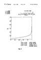
- FIG. 2illustrates, as the blood becomes about 95-100% saturated with oxygen, due to the steepness of the oxygen tension-saturation curve of FIG. 2, precise and accurate arterial blood oxygen saturation measurements (SaO 2 ) are necessary to accurately determine the partial pressure of oxygen in a patient's arterial blood.
- the present inventionincludes a method of non-invasively estimating the intrapulmonary shunt, pulmonary capillary blood flow and cardiac output of a patient.
- the shunt-estimating method according to the present inventionincludes non-invasively measuring the pulmonary capillary blood flow of the patient, measuring a volume of carbon dioxide exhaled by the patient, determining the difference between the end capillary oxygen content and the arterial oxygen content of the patient's blood, dividing the difference by the volume of carbon dioxide exhaled by the patient, and multiplying the difference by the patient's pulmonary capillary blood flow and by the patient's respiratory quotient (RQ).
- the respiratory quotientis the volume of carbon dioxide exhaled by the patient divided by the volume of oxygen exhaled by the patient.
- the pulmonary capillary blood flow of the patientmay be determined by known techniques, such as partial or total re-breathing techniques.
- the patient's cardiac output (Q t )includes a portion, which is typically identified as pulmonary capillary blood flow Q pcbf , that flows through pulmonary capillaries 164 (FIG. 5) and participates in gas exchange in the lungs 150 , and a portion that does not participate in blood gas exchange, which is referred to as the intrapulmonary shunt 165 , venous admixture, shunted blood, or simply as “shunt”.
- the corrected cardiac outputmay be determined by adding the non-invasively measured volume rate of pulmonary capillary blood flow (Q pCbf ) and the volume rate of flow of the intrapulmonary shunt flow of the patient (Q s ) by the following equation:
- a patient's corrected cardiac outputmay be determined as follows:
- the uncorrected volume/rate of the patient's pulmonary capillary blood flow(Q pcbf ) is preferably measured by non-invasive techniques, such as known partial or total re-breathing techniques, and may be employed with a variety of carbon dioxide, respiratory flow and pulse oximetry apparatus.
- the shunt fraction of the patient's pulmonary capillary blood flowmay be derived from various respiratory profile parameters, many of which may also be measured by non-invasive techniques.
- Cc′O 2is the end-capillary oxygen content
- CaO 2is the arterial oxygen content
- C V O 2is the mixed venous oxygen content.
- the denominator of the preceding formula (Cc′O 2 ⁇ C V O 2 )can be derived from the Fick oxygen equation that is typically employed in known re-breathing techniques for determining pulmonary capillary blood flow:
- the respiratory quotientis the ratio of the carbon dioxide elimination (V CO 2 ) to the amount of oxygen consumed (V O 2 ) by the patient, as defined by the following equation:
- V CO 2 /RQmay be substituted for V O 2 .
- Such substitution of V CO 2 /RQ for V O 2is preferred because it is difficult to accurately measure V O 2 , especially in patients who require an elevated fraction of inspired oxygen.
- RQcan be assumed with accuracy (typically about 0.7 to 1.0, and more particularly about 0.8 to 0.9 or 0.86).
- the V CO 2 measurementis preferably based on the alveolar CO 2 output of the patient, and may be measured by known re-breathing techniques.
- the end-capillary oxygen content, Cc′O 2may be calculated by the following equation:
- alveolar oxygen tension of the patientmay be calculated by the following formula:
- p baris the barometric pressure
- P H 2 Ois the saturated water vapor pressure of a sample at ambient temperature
- PaCO 2is the partial pressure of CO 2 in the patient's arterial blood, which may be assumed, calculated as known in the art from non-invasively obtained arterial blood gas data, or obtained by direct measurement.
- the blood oxygen solubility coefficient ( ⁇ ), the end-capillary blood saturation (Sc′O 2 ), hemoglobin concentration (Hb conc ), and hemoglobin capacity (Hb capacity ) valuesmay each be assumed values or determined by known techniques.
- the arterial oxygen content, CaO 2may be calculated by the following equation:
- CaO 2(PaO 2 ⁇ )+(SaO 2 ⁇ Hb capacity ⁇ Hb conc ).
- PaO 2is a function of SaO 2 , which may be approximated by SpO 2
- the partial pressure of the patient's arterial oxygen, PaO 2may be calculated from the oxygen saturation (SaO 2 ) of the patient's arterial blood using an invertible version of the blood oxygen tension-saturation relationship, as represented by the Lobdell equation.
- Lobdell, D. D.An invertible simple equation for computation of blood O 2 dissociation relations, J Appl. Physiol . (1981) 971-973.
- SaO 2Arterial blood oxygen saturation, SaO 2 , may be determined by known pulse oximetry (SpO 2 ) techniques. Pulse oximetry techniques may provide somewhat inaccurate blood oxygen saturation data (i.e., SaO 2 values). As the inverted tension-saturation curve is relatively steep at about 95-100% blood oxygen saturation, it is, therefore, difficult to accurately derive PaO 2 from SaO 2 . Thus, the SpO 2 measurement is corrected to provide a more accurate PaO 2 value.
- SpO 2pulse oximetry
- the measured SaO 2 or PaO 2 and FiO 2 of the patientmay be employed with an iso-shunt diagram or one or more equations that may be employed to generate an iso-shunt diagram to non-invasively estimate the intrapulmonary shunt fraction of the cardiac output of a patient.
- the patient's cardiac outputQ t .
- FIG. 1is a schematic representation of an exemplary re-breathing circuit that may be employed with the methods of the present invention
- FIG. 2is a line graph of an inverse Lobdell relation, which illustrates the relatively large difference in PaO 2 values derived from relatively close SpO 2 values—in the 95-100% range—and, thus, the potential for error when inaccurate SpO 2 measurements are made;
- FIG. 3is a schematic representation which illustrates the various componentry that may be utilized to measure respiratory profile parameters that are employed in the methods of the present invention
- FIG. 4is a schematic representation which illustrates a pulse oximetry sensor and associated monitor, which may be employed in association with the methods of the present invention
- FIG. 5is a schematic representation of the lungs of a patient
- FIG. 6is a flow diagram of a preferred embodiment of the method of the present invention.
- FIG. 7is an iso-shunt diagram that is useful in determining the intrapulmonary shunt fractions of the cardiac output of a patient in accordance with another embodiment of the methods.
- FIG. 8is another iso-shunt diagram that may be employed to determine the intrapulmonary shunt fraction of the cardiac output of a patient.
- the present inventionincludes a method of estimating the flow, or fraction, of blood that does not participate in the exchange of oxygen (O 2 ) and carbon dioxide (CO 2 ) in the pulmonary capillaries, which is referred to as “intrapulmonary shunt”, “venous admixture”, “shunted blood”, or simply as “shunt”.
- the present inventionalso includes a method of calculating cardiac output based on the shunt estimate.
- V O 2Due to the difficulty of measuring the amount of oxygen consumed by a patient (V O 2 ), especially in patients who require an elevated fraction of inspired oxygen, pulmonary capillary blood flow (Q pcbf ), cardiac output (CO), and the estimated shunt fraction are preferably measured in terms of the amount of carbon dioxide excreted into the lungs of the patient, which is typically measured in terms of carbon dioxide elimination (V CO 2 ).
- the Fick equation for measurement of cardiac output, in terms of CO 2is:
- calculating pulmonary capillary blood flowin accordance with the method of the present invention includes measuring the flow rates and CO 2 fraction of gas mixtures that are inhaled and exhaled by a patient 10 over the course of the patient's breathing, at 110 .
- a flow sensor 12 of a known typesuch as the differential-pressure type respiratory flow sensors manufactured by Novametrix Medical Systems Inc. (“Novametrix”) of Wallingford, Conn. (e.g, the Pediatric/Adult Flow Sensor (Catalog No. 6717) or the Neonatal Flow Sensor (Catalog No.
- a CO 2 sensor 14such as the CAPNOSTAT® CO 2 sensor and a complementary airway adapter (e.g., the Pediatric/Adult Single Patient Use Airway Adapter (Catalog No. 6063), the Pediatric/Adult Reusable Airway Adapter (Catalog No. 7007), or the Neonatal/Pediatric Reusable Airway Adapter (Catalog No.
- Flow sensor 12 and CO 2 sensor 14are connected to a flow monitor 16 and a CO 2 monitor 18 , respectively, each of which may be operatively associated with a computer 20 so that data from the flow and CO 2 monitors 16 and 18 representative of the signals from each of flow sensor 12 and CO 2 sensor 14 may be detected by the computer 20 and processed according to programming (e.g., by software) thereof
- raw flow and CO 2 signals from the flow monitor and CO 2 sensorare filtered, as known in the art, to remove any significant artifacts.
- the respiratory flow and CO 2 pressure datamay be stored by computer 20 .
- pulmonary capillary blood flowmay be calculated, in accordance with the foregoing equation or by any other equation known in the art, by computer 20 .
- Each breath, or breathing cycle, of patient 10may be delineated as known in the art, such as by continually monitoring the flow rate of the breathing of patient 10 .
- blood oxygen measurementsmay be made, at 122 of FIG. 6, by non-invasive means, such as by a pulse oximetry sensor 30 of a type known in the art, such as the OXYSNAPTM or Y-SENSORTM, both of which are manufactured by Novametrix.
- Pulse oximetry sensor 30includes a light emitting diode (LED) assembly 32 and a photodiode 34 which are positionable on opposite sides of an appendage of the body of a patient, such as a finger 11 , hand, toe, heel, foot, ear lobe, nose, or tongue.
- SpO 2 signalswhich may be conveyed from pulse oximetry sensor 30 to computer 20 , as known in the art, such as by a cable connector 36 , are subsequently employed in the methods of the present invention.
- end-tidal CO 2the partial pressure of end-tidal CO 2 , carbon dioxide elimination (V CO 2 ), the fraction of inspired, or “mixed inspired”, CO 2 and the airway deadspace are calculated.
- End-tidal CO 2is measured as known in the art.
- Carbon dioxide eliminationis typically calculated as the integral of the respiratory flow over a breathing cycle (in milliliters) multiplied by fraction of CO 2 over the entire breath.
- the fraction of inspired CO 2is the integral of CO 2 fraction times the air flow during inspiration, divided by the volume (in milliliters) of inspired gas.
- V CO 2 and PetCO 2may be filtered by employing a three-point median filter, which uses a median value from the most recent value of recorded V CO 2 and PetCO 2 values and the two values that precede the most recent measured value, as known in the art.
- the V CO 2 valueis corrected to account for anatomic deadspace and alveolar deadspace.
- the lungs 150 of a patientmay be described as including a trachea 152 , two bronchi 154 and numerous alveoli 160 , 162 .
- the anatomic, or “serial”, deadspace of lungs 150includes the volume of the trachea 152 , bronchi 154 , and other components of lungs 150 which hold gases, but do not participate in gas exchange.
- the anatomic deadspaceexists approximately in the region located between arrows A and B.
- the so-called “shunted” bloodbypasses pulmonary capillaries by way of an intrapulmonary shunt 165 .
- Lungs 150typically include alveoli 160 that are in contact with blood flow and which can facilitate oxygenation of the blood, which are referred to as “perfused” alveoli, as well as unperfised alveoli 162 . Both perfused alveoli 160 and unperfused alveoli 162 may be ventilated. The volume of unperfused alveoli is the alveolar deadspace.
- Perfused alveoli 160are surrounded by and in contact with pulmonary capillaries 164 .
- oxygenated blood 166enters pulmonary capillaries 164
- oxygenated blood 168Blood that exits pulmonary capillaries 164 in the direction of arrow 170 is referred to as oxygenated blood 168 .
- a volume of gas known as the functional residual capacity (FRC) 171remains following exhalation.
- the alveolar CO 2is expired from a portion 172 of each of the alveoli 160 that is evacuated, or ventilated, during exhalation.
- the ventilated portion 178 of each of the unperfused alveoli 162may also include CO 2 .
- the CO 2 of ventilated portion 178 of each of the unperfused alveoli 162is not the result of O 2 and CO 2 exchange in that alveolus. Since the ventilated portion 178 of each of the unperfused alveoli 162 is ventilated in parallel with the perfused alveoli, ventilated portion 178 is typically referred to as “parallel” deadspace (PDS).
- Unperfused alveoli 162also include a FRC 176 , which includes a volume of gas that is not evacuated during a breath.
- FRCmay be estimated as a function of body weight and the airway deadspace volume by the following equation:
- FRC-factoris either an experimentally determined value or is based on published data (e.g., “experiential” data) known in the art
- offset valueis a fixed constant which compensates for breathing masks or other equipment components that may add deadspace to the breathing circuit and thereby unacceptably skew the relationship between FRC and deadspace.
- the partial pressure of CO 2 in the parallel deadspacemay be calculated from the mixed inspired CO 2 (Vi CO2 ) added to the product of the serial deadspace multiplied by the end tidal CO 2 of the previous breath (PetCO 2 (n ⁇ 1)). Because the average partial pressure of CO 2 in the parallel deadspace is equal to the partial pressure of CO 2 in the parallel deadspace divided by the tidal volume (V t ) (i.e., the total volume of one respiratory cycle, or breath), the partial pressure of CO 2 in the parallel deadspace may be calculated on a breath-by-breath basis, as follows:
- PCO 2 PDS (n)[FRC/(FRC+V t )] ⁇ PCO 2 PDS (n ⁇ 1)+(P bar ⁇ (([Vi CO2 +deadspace ⁇ (PetCO 2 (n ⁇ 1)/P bar )]/V t ) ⁇ [V t /(V t +FRC)])),
- (n)indicates a respiratory profile parameter (in this case, the partial pressure of CO 2 in the parallel deadspace) from the most recent breath and (n ⁇ 1) indicates a respiratory profile parameter from the previous breath.
- the partial pressure of end-tidal CO 2which is assumed to be substantially equal to a weighted average of the partial pressure of CO 2 in all of the perfused and unperfused alveoli of a patient, may then be calculated as follows:
- PetCO 2® ⁇ P A CO 2 )+(1 ⁇ r)PCO 2 PDS ,
- ris the perfusion ratio, which is calculated as the ratio of perfused alveolar ventilation to the total alveolar ventilation, or (V A ⁇ V PDS )/V A .
- the perfusion ratiomay be assumed to be about 0.95 or estimated as known in the art.
- alveolar CO 2 partial pressure of the patientmay be calculated.
- alveolar CO 2 partial pressureis calculated from the end-tidal CO 2 and the CO 2 in the parallel deadspace, as follows:
- P A CO 2[PetCO 2 ⁇ (1 ⁇ r)PCO 2PDS ]/r.
- the alveolar CO 2 partial pressuremay then be converted to alveolar blood CO 2 content (C A CO 2 ) using an equation, such as the following:
- C A CO 2is the content of CO 2 in the alveolar blood and Hb is the concentration of hemoglobin in the blood of the pulmonary capillaries.
- Hbis the concentration of hemoglobin in the blood of the pulmonary capillaries.
- the FRC and alveolar deadspace of the lungs of a patientmay be accounted for by multiplying the FRC by the change in end tidal partial pressure, such as by the following equation:
- V CO 2 correctedV CO 2 +FRC ⁇ PetCO 2 /P bar ,
- ⁇ PetCO 2is the breath-to-breath change in PetCO 2 .
- Baseline PetCO 2 and V CO 2 valueswhich are also referred to as “before re-breathing PetCO 2 ” and “before re-breathing V CO 2 ”, respectively, occur during normal breathing and may be calculated as the average of a group of samples taken before the re-breathing process (e.g., the average of all samples between about 27 and 0 seconds before the start of a known re-breathing process).
- a V CO 2 valuewhich is typically referred to as “during re-breathing V CO 2 ”, is calculated during the re-breathing process.
- “During re-breathing V CO 2 ”may be calculated as the average V CO 2 during the interval of 25 to 30 seconds into the re-breathing period.
- the content of CO 2 in the alveolar blood during the re-breathing processmay then be calculated by employing a regression line, which facilitates prediction of the stable, or unchanging, content of alveolar CO 2 .
- P A CO 2is plotted against the breath-to-breath change in content of alveolar CO 2 ( ⁇ P A CO 2 ).
- a graph line that is defined by the plotted pointsis regressed, and the intersection between P A CO 2 and zero ⁇ P A CO 2 is the predicted stable content of alveolar CO 2 .
- Alternative differential Fick methods of measuring pulmonary capillary blood flow or cardiac outputmay be employed in place of the embodiment of the re-breathing method disclosed herein.
- Such alternative differential Fick methodstypically require a brief change of PetCO 2 and V CO 2 in response to a change in effective ventilation. This brief change can be accomplished by adjusting the respiratory rate, inspiratory and/or expiratory times, or tidal volume.
- a brief change in effective ventilationmay also be effected by adding CO 2 , either directly or by re-breathing.
- An exemplary differential Fick method that may be employed with the present inventionwhich is disclosed in Gedeon, A. et al. in 18 Med . & Biol. Eng . & Comput . 411-418 (1980), employs a period of increased ventilation followed immediately by a period of decreased ventilation.
- the intrapulmonary shunt fraction of the cardiac output of the patientmay be estimated.
- the method of estimating intrapulmonary shunt according to the present inventionalso includes non-invasively determining the difference between the end capillary oxygen content and the arterial oxygen content of the patient's blood, dividing the difference by the volume of carbon dioxide exhaled by the patient, and multiplying the difference by the patient's pulmonary capillary blood flow (Q pcbf ) and by the patient's respiratory quotient (RQ).
- the shunt fraction of the patient's cardiac outputmay be derived from various respiratory profile parameters, many of which may also be measured by non-invasive techniques.
- Cc′O 2is the end-capillary oxygen content
- CaO 2is the arterial oxygen content
- C V O 2is the mixed venous oxygen content.
- RQthe respiratory quotient
- V CO 2 /RQmay be substituted for V O 2 .
- V CO 2which was determined above in the calculation of the pulmonary capillary blood flow of the patient, is the CO 2 elimination of the patient.
- VO 2may be measured as known in the art, and RQ can be calculated rather than assumed.
- the VO 2 measurementmay be divided by Q pcbf to directly determine Cc′O 2 ⁇ C V O 2 , in which case the following formula may be employed to estimate the shunt fraction:
- Q s / Q tCc ′ ⁇ O 2 - CaO 2 V ⁇ O 2 Q pcbf
- the end-capillary oxygen content, Cc′O 2may be calculated, at 114 of FIG. 6, by the following equation:
- ⁇blood oxygen solubility coefficient
- Sc′O 2end capillary blood saturation
- Hb conchemoglobin concentration
- Hb capacityhemoglobin capacity
- the alveolar oxygen tension of the patientmay be calculated by the following formula:
- P A O 2(FiO 2 ⁇ (P bar ⁇ P H 2 O )) ⁇ (PaCO 2 /RQ ⁇ (1 ⁇ (FiO 2 ⁇ (1 ⁇ RQ)))),
- P baris barometric pressure
- P H 2 Ois the saturated water vapor pressure of a sample at ambient temperature
- PaCO 2is the partial pressure of CO 2 in the patient's arterial blood, which may be assumed, calculated as known in the art from non-invasively obtained arterial blood gas data, or obtained by direct measurement.
- the oxygen content of the patient's arterial blood, CaO 2may be calculated, at 126 of FIG. 6, by the following equation:
- CaO 2(PaO 2 ⁇ )+(SaO 2 ⁇ Hb capacity ⁇ Hb conc ).
- PaO 2is a function of SaO 2 , which may be non-invasively estimated by measuring SpO 2 (see FIG. 6, at 124 ), the partial pressure of O 2 in the patient's arterial blood, PaO 2 , may be calculated from the oxygen saturation (SaO 2 ) of the patient's arterial blood by employing an invertable version of a blood oxygen tension-saturation curve.
- Arterial blood oxygen saturationis determined non-invasively by known techniques, such as by pulse oximetry (SpO 2 ), as discussed previously in reference to FIG. 4 .
- a correction summandwhich is also referred to as a correction factor, is employed in determining the oxygen saturation and partial pressure of oxygen in the arterial blood.
- the correction summandmay be an assumed value (e.g., 2 or 3%) based on experiential error of a known degree when a specific type of pulse oximeter or a particular model of pulse oximeter of a particular manufacturer is employed to measure SpO 2 .
- the correction summandmay be determined by comparing a direct SaO 2 measurement from blood gas chemical analysis with an SpO 2 measurement taken by a pulse oximeter.
- the correction summandmay then be employed in combination with subsequent pulse oximetry measurements to modify these pulse oximetry measurements and more accurately determine the partial pressure of oxygen in the patient's arterial blood. This may be done by adding a correction summand to the SpO 2 measurement or subtracting a correction summand from the SpO 2 measurement, by generating an equation to convert the SpO 2 measurement to a more accurate value, or by generating a special function in which the non-invasively measured, possibly somewhat inaccurate, SpO 2 measurement is employed to accurately determine SaO 2 or PaO 2 . The SaO 2 or PaO 2 value may then be employed in the preceding equation to facilitate an accurate, non-invasive determination of the patient's intrapulmonary shunt.
- the SaO 2 or PaO 2 of a patientmay be non-invasively determined and corrected as described above.
- the patient's FiO 2is also determined, as known in the art, such as by a respiratory measurement or from a set fraction, or value, of oxygen in a gas mixture with which the patient is artificially ventilated.
- An iso-shunt diagramwhich is also referred to as an iso-shunt plot, such as that disclosed in S. R. Benatar et al., The use of iso-shunt lines for control of oxygen therapy, Brit. J Anaesth . (1973) 45:711, and in N UNN , J.
- N UNN'S A PPLIED R ESPIRATORY P HYSIOLOGY 184, FIG. 8.10 (4th ed.) and shown in FIG. 7,may then be employed with the SaO 2 or PaO 2 and FiO 2 measurements to determine the intrapulmonary shunt fraction of the cardiac output of the patient.
- the intrapulmonary shunt fractionmay be similarly estimated by incorporating the measured SaO 2 or PaO 2 and FiO 2 values into the following series of equations that Nunn used to generate the iso-shunt diagram of FIG. 7 from FiO 2 and PaO 2 measurements:
- P A O 2((P bar ⁇ P H 2 O ) ⁇ FiO 2 ) ⁇ (PaCO 2 /RQ)(1 ⁇ FiO 2 ⁇ (1 ⁇ RQ)),
- PaO 2is the partial pressure of oxygen in the alveoli of the patient
- P barthe barometric pressure
- P H 2 Oor water pressure
- PaCO 2the partial pressure of carbon dioxide in the arterial blood of the patient, or the arterial carbon dioxide tension
- RQis assumed to be 0.8 (alternatively, each of these parameters may be assumed to be equal to a different value or measured by techniques known in the art);
- Hb capis assumed to be 1.31 (ml/g) and Hb conc is assumed to be 14 g/dl;
- CaO 2(PaO 2 ⁇ )+(SaO 2 ⁇ Hb cap ⁇ Hb conc ),
- Hb capis assumed to be 1.31 (mlg) and Hb conc is assumed to be 14 g/dl;
- iso-shunt equations or diagramsare also useful with the corrected SaO 2 and PaO 2 values of the present invention to estimate the intrapulmonary shunt of a patient, such as the equation and graphs disclosed in Dean, J. M., Wetzel, R. C., and Rogers, M. C., Arterial blood gas derived variables as estimates of intrapulmonary shunt in critically ill children, Crit. Care Med . 13(12):1029-1033 (1985)(“Dean”).
- Hgbis the hemoglobin concentration of the patient
- SaO 2may be employed with the graph of FIG. 8, which is depicted in Dean, to estimate the intrapulmonary shunt fraction.
- the patient's cardiac output(Q t ) may be determined.
- the patient's cardiac outputmay be determined by adding the non-invasively measured volumetric rate of pulmonary capillary blood flow (Q pcbf ) and the volumetric rate of flow of the patient's shunted blood (Q s ) by the following equation:
- a patient's total cardiac outputmay also be determined from the shunt fraction (Q s /Q t ) and pulmonary capillary blood flow as follows:
Landscapes
- Health & Medical Sciences (AREA)
- Life Sciences & Earth Sciences (AREA)
- Physics & Mathematics (AREA)
- Biomedical Technology (AREA)
- Medical Informatics (AREA)
- Veterinary Medicine (AREA)
- Public Health (AREA)
- Biophysics (AREA)
- Pathology (AREA)
- Engineering & Computer Science (AREA)
- General Health & Medical Sciences (AREA)
- Heart & Thoracic Surgery (AREA)
- Animal Behavior & Ethology (AREA)
- Molecular Biology (AREA)
- Surgery (AREA)
- Pulmonology (AREA)
- Emergency Medicine (AREA)
- Obesity (AREA)
- Physiology (AREA)
- Spectroscopy & Molecular Physics (AREA)
- Optics & Photonics (AREA)
- Measuring Pulse, Heart Rate, Blood Pressure Or Blood Flow (AREA)
- Measurement Of The Respiration, Hearing Ability, Form, And Blood Characteristics Of Living Organisms (AREA)
Abstract
Description


Claims (31)
Priority Applications (1)
| Application Number | Priority Date | Filing Date | Title |
|---|---|---|---|
| US09/464,589US6241681B1 (en) | 1998-09-09 | 1999-12-16 | Methods of measuring cardiac output using a non-invasively estimated intrapulmonary shunt fraction |
Applications Claiming Priority (2)
| Application Number | Priority Date | Filing Date | Title |
|---|---|---|---|
| US09/150,450US6042550A (en) | 1998-09-09 | 1998-09-09 | Methods of non-invasively estimating intrapulmonary shunt fraction and measuring cardiac output |
| US09/464,589US6241681B1 (en) | 1998-09-09 | 1999-12-16 | Methods of measuring cardiac output using a non-invasively estimated intrapulmonary shunt fraction |
Related Parent Applications (1)
| Application Number | Title | Priority Date | Filing Date |
|---|---|---|---|
| US09/150,450ContinuationUS6042550A (en) | 1998-09-09 | 1998-09-09 | Methods of non-invasively estimating intrapulmonary shunt fraction and measuring cardiac output |
Publications (1)
| Publication Number | Publication Date |
|---|---|
| US6241681B1true US6241681B1 (en) | 2001-06-05 |
Family
ID=22534587
Family Applications (3)
| Application Number | Title | Priority Date | Filing Date |
|---|---|---|---|
| US09/150,450Expired - LifetimeUS6042550A (en) | 1998-09-09 | 1998-09-09 | Methods of non-invasively estimating intrapulmonary shunt fraction and measuring cardiac output |
| US09/465,059Expired - LifetimeUS6258038B1 (en) | 1998-09-09 | 1999-12-16 | Methods of non-invasively estimating intrapulmonary shunt fraction and measuring cardiac output |
| US09/464,589Expired - LifetimeUS6241681B1 (en) | 1998-09-09 | 1999-12-16 | Methods of measuring cardiac output using a non-invasively estimated intrapulmonary shunt fraction |
Family Applications Before (2)
| Application Number | Title | Priority Date | Filing Date |
|---|---|---|---|
| US09/150,450Expired - LifetimeUS6042550A (en) | 1998-09-09 | 1998-09-09 | Methods of non-invasively estimating intrapulmonary shunt fraction and measuring cardiac output |
| US09/465,059Expired - LifetimeUS6258038B1 (en) | 1998-09-09 | 1999-12-16 | Methods of non-invasively estimating intrapulmonary shunt fraction and measuring cardiac output |
Country Status (2)
| Country | Link |
|---|---|
| US (3) | US6042550A (en) |
| WO (1) | WO2000013581A1 (en) |
Cited By (13)
| Publication number | Priority date | Publication date | Assignee | Title |
|---|---|---|---|---|
| US6413226B1 (en) | 1999-10-22 | 2002-07-02 | Respironics, Inc. | Method and apparatus for determining cardiac output |
| US20030214409A1 (en)* | 2002-05-13 | 2003-11-20 | Scott Laboratories, Inc. | System and method for transparent early detection, warning, and intervention during a medical procedure |
| US8844526B2 (en) | 2012-03-30 | 2014-09-30 | Covidien Lp | Methods and systems for triggering with unknown base flow |
| US9022031B2 (en) | 2012-01-31 | 2015-05-05 | Covidien Lp | Using estimated carinal pressure for feedback control of carinal pressure during ventilation |
| US20150168182A1 (en)* | 1999-10-26 | 2015-06-18 | Sony Corporation | Searching system, searching unit, searching method, displaying method for search results, terminal unit, inputting unit, and record medium |
| US9364624B2 (en) | 2011-12-07 | 2016-06-14 | Covidien Lp | Methods and systems for adaptive base flow |
| US9492629B2 (en) | 2013-02-14 | 2016-11-15 | Covidien Lp | Methods and systems for ventilation with unknown exhalation flow and exhalation pressure |
| US9498589B2 (en) | 2011-12-31 | 2016-11-22 | Covidien Lp | Methods and systems for adaptive base flow and leak compensation |
| US9649458B2 (en) | 2008-09-30 | 2017-05-16 | Covidien Lp | Breathing assistance system with multiple pressure sensors |
| US9925346B2 (en) | 2015-01-20 | 2018-03-27 | Covidien Lp | Systems and methods for ventilation with unknown exhalation flow |
| US9981096B2 (en) | 2013-03-13 | 2018-05-29 | Covidien Lp | Methods and systems for triggering with unknown inspiratory flow |
| US20180153440A1 (en)* | 2016-12-05 | 2018-06-07 | Medipines Corporation & The Regents Of The University Of California | System And Methods For Respiratory Measurements Using Breathing Gas Samples |
| CN111407280A (en)* | 2020-03-10 | 2020-07-14 | 山东大学 | End-tidal CO of noninvasive ventilator2Monitoring device and method |
Families Citing this family (44)
| Publication number | Priority date | Publication date | Assignee | Title |
|---|---|---|---|---|
| US8409846B2 (en) | 1997-09-23 | 2013-04-02 | The United States Of America As Represented By The Department Of Veteran Affairs | Compositions, methods and devices for maintaining an organ |
| US6186956B1 (en)* | 1998-05-28 | 2001-02-13 | University Of South Carolina | Method and system for continuously monitoring cardiac output |
| US6575164B1 (en)* | 1998-10-15 | 2003-06-10 | Ntc Technology, Inc. | Reliability-enhanced apparatus operation for re-breathing and methods of effecting same |
| US6210342B1 (en)* | 1999-09-08 | 2001-04-03 | Ntc Technology, Inc. | Bi-directional partial re-breathing method |
| US6254546B1 (en)* | 1999-12-07 | 2001-07-03 | Instrumentarium Corporation | Method to determine ventilation-perfusion and ventilation-volume distributions of the lungs |
| DE60137191D1 (en) | 2001-03-05 | 2009-02-12 | Instrumentarium Corp | Method for the non-invasive determination of the condition of the circulation of an individual |
| US7135001B2 (en)* | 2001-03-20 | 2006-11-14 | Ric Investments, Llc | Rebreathing methods including oscillating, substantially equal rebreathing and nonrebreathing periods |
| US6575918B2 (en) | 2001-09-27 | 2003-06-10 | Charlotte-Mecklenburg Hospital | Non-invasive device and method for the diagnosis of pulmonary vascular occlusions |
| US6951216B2 (en)* | 2002-12-19 | 2005-10-04 | Instrumentarium Corp. | Apparatus and method for use in non-invasively determining conditions in the circulatory system of a subject |
| US7225022B2 (en)* | 2003-03-12 | 2007-05-29 | Cra Associates, Ltd. | Method of optimizing patient outcome from cardiac resynchronization therapy |
| US6884222B1 (en)* | 2003-11-20 | 2005-04-26 | James R. Braig | Method and apparatus for estimation of resting respiratory quotient |
| US12010987B2 (en) | 2004-10-07 | 2024-06-18 | Transmedics, Inc. | Systems and methods for ex-vivo organ care and for using lactate as an indication of donor organ status |
| US9301519B2 (en)* | 2004-10-07 | 2016-04-05 | Transmedics, Inc. | Systems and methods for ex-vivo organ care |
| US8304181B2 (en) | 2004-10-07 | 2012-11-06 | Transmedics, Inc. | Method for ex-vivo organ care and for using lactate as an indication of donor organ status |
| JP5113522B2 (en) | 2004-10-07 | 2013-01-09 | トランスメディクス, インク. | System and method for organ management ex-vivo |
| US9078428B2 (en) | 2005-06-28 | 2015-07-14 | Transmedics, Inc. | Systems, methods, compositions and solutions for perfusing an organ |
| DK1942726T3 (en)* | 2006-04-19 | 2017-04-10 | Transmedics Inc | METHODS FOR EX VIVO ORGANIC CARE |
| US8123695B2 (en)* | 2006-09-27 | 2012-02-28 | Nellcor Puritan Bennett Llc | Method and apparatus for detection of venous pulsation |
| KR100874111B1 (en)* | 2007-03-05 | 2008-12-15 | 한국과학기술원 | Noninvasive Respiratory Characteristic Prediction Method and Indicator by Respiratory Analysis and Arterial Blood |
| US8109882B2 (en) | 2007-03-09 | 2012-02-07 | Nellcor Puritan Bennett Llc | System and method for venous pulsation detection using near infrared wavelengths |
| US8221326B2 (en)* | 2007-03-09 | 2012-07-17 | Nellcor Puritan Bennett Llc | Detection of oximetry sensor sites based on waveform characteristics |
| US8229530B2 (en)* | 2007-03-09 | 2012-07-24 | Nellcor Puritan Bennett Llc | System and method for detection of venous pulsation |
| US9457179B2 (en) | 2007-03-20 | 2016-10-04 | Transmedics, Inc. | Systems for monitoring and applying electrical currents in an organ perfusion system |
| US8794235B2 (en)* | 2007-06-08 | 2014-08-05 | Ric Investments, Llc | System and method for treating ventilatory instability |
| US10750738B2 (en) | 2008-01-31 | 2020-08-25 | Transmedics, Inc. | Systems and methods for ex vivo lung care |
| KR100868808B1 (en)* | 2008-03-04 | 2008-11-17 | 한국과학기술원 | Non-invasive respiratory characteristic prediction method and display device by measuring respiratory gas and blood gas |
| US8425428B2 (en) | 2008-03-31 | 2013-04-23 | Covidien Lp | Nitric oxide measurements in patients using flowfeedback |
| US8652064B2 (en)* | 2008-09-30 | 2014-02-18 | Covidien Lp | Sampling circuit for measuring analytes |
| US10390711B2 (en) | 2010-11-26 | 2019-08-27 | Mermaid Care A/S | Automatic lung parameter estimator for measuring oxygen and carbon dioxide gas exchange |
| JP6029650B2 (en) | 2011-04-14 | 2016-11-24 | トランスメディクス,インコーポレイテッド | Organ protection solution for mechanical perfusion in ex-vivo of donor lung |
| CN102423263B (en)* | 2011-09-23 | 2014-09-24 | 深圳市纽泰克电子有限公司 | Monitoring method and device for partial pressure of carbon dioxide |
| US20170027451A1 (en)* | 2013-12-20 | 2017-02-02 | Maquet Critical Care Ab | Method and apparatus for estimating shunt |
| CA3185937A1 (en) | 2014-06-02 | 2015-12-10 | Transmedics, Inc. | Ex vivo organ care system |
| US20160058346A1 (en)* | 2014-09-02 | 2016-03-03 | General Electric Company | Determination of arterial co2 partial pressure |
| EP3229588B1 (en) | 2014-12-12 | 2025-04-23 | TransMedics, Inc. | Apparatus and method for organ perfusion |
| DK3347084T3 (en) | 2015-09-09 | 2021-02-15 | Transmedics Inc | AORTIC NEEDLE FOR EX VIVO ORGAN CARE SYSTEM |
| WO2017205967A1 (en) | 2016-05-30 | 2017-12-07 | Freed Darren | Apparatus and method for ex vivo lung ventilation with a varying exterior pressure |
| US20210219882A1 (en)* | 2017-01-19 | 2021-07-22 | General Electric Company | Methods and systems for non-invasive measurement and monitoring of physiological parameters |
| SG11202001113SA (en)* | 2017-08-08 | 2020-03-30 | Rostrum Medical Innovations Inc | Method and system for estimating the efficiency of the lungs of a patient |
| CN113439310B (en)* | 2019-02-26 | 2024-07-30 | Obi股份有限公司 | Method for providing decision support related to patients receiving oxygen therapy |
| US11324954B2 (en) | 2019-06-28 | 2022-05-10 | Covidien Lp | Achieving smooth breathing by modified bilateral phrenic nerve pacing |
| US11844610B2 (en) | 2019-12-23 | 2023-12-19 | Koninklijke Philips N.V. | System and method for monitoring gas exchange |
| US20240050676A1 (en)* | 2020-12-16 | 2024-02-15 | Koninklijke Philips N.V. | Visualizing and simulating changes in oxygenation |
| EP4014861A1 (en)* | 2020-12-16 | 2022-06-22 | Koninklijke Philips N.V. | Graphical representation of oxygenation |
Citations (11)
| Publication number | Priority date | Publication date | Assignee | Title |
|---|---|---|---|---|
| US4221224A (en) | 1978-06-29 | 1980-09-09 | Intermountain Health Care | Non-airtight pulmonary measuring device |
| US4463764A (en) | 1981-09-29 | 1984-08-07 | Medical Graphics Corporation | Cardiopulmonary exercise system |
| US5060656A (en) | 1990-05-22 | 1991-10-29 | Aerosport, Inc. | Metabolic rate analyzer |
| US5069220A (en) | 1989-05-26 | 1991-12-03 | Bear Medical Systems, Inc. | Measurement of gas concentration in exhaled breath |
| US5117674A (en) | 1990-05-22 | 1992-06-02 | Aerosport, Inc. | Metabolic rate analyzer |
| US5178155A (en) | 1988-06-29 | 1993-01-12 | Mault James R | Respiratory calorimeter with bidirectional flow monitors for calculating of oxygen consumption and carbon dioxide production |
| US5285794A (en) | 1992-12-14 | 1994-02-15 | Temple University Of The Commonwealth System Of Higher Education | Respiratory gas monitor |
| US5299579A (en) | 1989-11-24 | 1994-04-05 | Minco Ab | Apparatus for examining a patient's pulmonary function |
| US5402796A (en) | 1990-09-19 | 1995-04-04 | University Of Melbourne | Arterial CO2 Monitor and closed loop controller |
| US5595181A (en) | 1994-03-24 | 1997-01-21 | Hubbard; A. Robert | System for providing cardiac output and shunt quantitation |
| WO1998012963A1 (en) | 1996-09-28 | 1998-04-02 | Technische Universität Dresden | Device to determine effective pulmonary blood flow |
- 1998
- 1998-09-09USUS09/150,450patent/US6042550A/ennot_activeExpired - Lifetime
- 1999
- 1999-09-09WOPCT/US1999/020843patent/WO2000013581A1/enactiveApplication Filing
- 1999-12-16USUS09/465,059patent/US6258038B1/ennot_activeExpired - Lifetime
- 1999-12-16USUS09/464,589patent/US6241681B1/ennot_activeExpired - Lifetime
Patent Citations (11)
| Publication number | Priority date | Publication date | Assignee | Title |
|---|---|---|---|---|
| US4221224A (en) | 1978-06-29 | 1980-09-09 | Intermountain Health Care | Non-airtight pulmonary measuring device |
| US4463764A (en) | 1981-09-29 | 1984-08-07 | Medical Graphics Corporation | Cardiopulmonary exercise system |
| US5178155A (en) | 1988-06-29 | 1993-01-12 | Mault James R | Respiratory calorimeter with bidirectional flow monitors for calculating of oxygen consumption and carbon dioxide production |
| US5069220A (en) | 1989-05-26 | 1991-12-03 | Bear Medical Systems, Inc. | Measurement of gas concentration in exhaled breath |
| US5299579A (en) | 1989-11-24 | 1994-04-05 | Minco Ab | Apparatus for examining a patient's pulmonary function |
| US5060656A (en) | 1990-05-22 | 1991-10-29 | Aerosport, Inc. | Metabolic rate analyzer |
| US5117674A (en) | 1990-05-22 | 1992-06-02 | Aerosport, Inc. | Metabolic rate analyzer |
| US5402796A (en) | 1990-09-19 | 1995-04-04 | University Of Melbourne | Arterial CO2 Monitor and closed loop controller |
| US5285794A (en) | 1992-12-14 | 1994-02-15 | Temple University Of The Commonwealth System Of Higher Education | Respiratory gas monitor |
| US5595181A (en) | 1994-03-24 | 1997-01-21 | Hubbard; A. Robert | System for providing cardiac output and shunt quantitation |
| WO1998012963A1 (en) | 1996-09-28 | 1998-04-02 | Technische Universität Dresden | Device to determine effective pulmonary blood flow |
Non-Patent Citations (32)
| Title |
|---|
| A. Gedeon, Non-Invasive Pulmonary Blood Flow for Optimal Peep, ICOR AB, Ulvsundavagen 178 B, S-161 30 Bromma, Sweden, pp. 49-58. |
| Cane, Roy D., et al., Unreliability of oxygen tension-based indices in reflecting intrapulmonary shunting in critically ill patients, Critical Care Medicine (1988) 16(12): 1243-45. |
| Capek, J.M., et al., Noninvasive Measurement of Cardiac Output Using Partial CO2 Rebreathing, IEEE Trans. Biomed. Eng. (1988) 35(9):653-61. |
| Capek, J.M.,Noninvasive Measurement of Cardiac Output Using Partial CO2 Rebreathing [Dissertation], Rensselaer Polytechnic Institute (1988) 28:351 p. (due to large number of pages, only table of contents and abstract have been copied). |
| Cruz, J. C., et al., Understanding the Meaning of the Shunt Fraction Calculation, Journal of Clinical Monitoring (1987) 3(2): 124-34. |
| Davies, Gerald G., et al., Continuous Fick cardiac output compared to the thermodilution cardiac output, Critical Care Medicine (1986) 14(10):881-85. |
| Dean, J. Michael, et al., Arterial blood gas derived variables as estimates of intrapulmonary shunt in critically ill children, Critical Care Medicine (1985) 13(12): 1029-33. |
| Elliott, C. Gregory, et al., Complications of Pulmonary Artery Catheterization in the Care of Critically Ill Patients, Chest (1979) 76:6, 647-52. |
| Fick, A., Über die Messung des Blutquantums in den Herzventrikeln, Sitzungsbericht der Physikalisch-Medizinischen Gesellschaft zu Wüzburg (1870) 36 (2 pages). of a letter summarizing the article. |
| Gama de Abreu, et al., Partial carbon dioxide rebreathing: A reliable technique for noninvasive measurement of nonshunted pulmonary capillary blood flow, Crit. Care Med. (1997) 25(4):675-83. |
| Gama de Abreu, Marcelo, et al., Is the Partial CO2 Rebreathing Technique a Useful Tool for Trending Pulmonary Capillary Blood Flow During Adjustments of Peep?, Crit. Care Med. (1998) vol. 26, No. 1 (Suppl.), A106, Abstract #237, (1 page). |
| Gama de Abreu, Marcelo, et al., Measurement of Pulmonary Capillary Blood Flow for Trending Mixed Venous Blood Oxygen Saturation and Oxygen Delivery, Crit. Care Med. (1998), vol. 26, No. 1 (Suppl.) A106, Abstract #238 (1 page). |
| Gedeon, A., et al., A new method for noninvasive bedside determination of pulmonary blood flow, Med. & Biol. Eng. & Comput. (1980) 18:411-418. |
| Gedeon, A., et al., Noninvasive Cardiac Output Determined with a New Method Based on Gas Exchange Measurements and Carbon Dioxide Rebreathing: A Study in Animals/Pigs, J. Clin. Monit. (1992) 8(4):267-78. |
| Guyton, A.E., et al., Measurement of cardiac output by the direct Fick method, In: Cardiac output and its regulation, W. B. Saunders Company (1973) 21-39. |
| H. Blomquist et al., A Non-Invasive Technique for Measurement of Lung Perfusion, Intensive Care Medicine 1986; 12:172. |
| Hope, D.A., et al., Non-invasive estimation of venous admixture: validation of a new formula, British Journal of Anaesthesia (1995) 74:538-43. |
| Kyoku, I., et al., Measurement of cardiac output by Fick method using CO2 analyzer Servo, Kyobu Geka. Japanese Journal of Thoracic Surgery (1988) 41(12):966-70. |
| Lobdell, Donn D., An ivertible simple equation for computation of blood O2 dissociation relations, American Physiological Society: (1981) 971-73. Including English translation. (5 pages). |
| Lynch, J., et al., Comparison of a modified Fick method with thermodilution for determining cardiac output in critically ill patients on mechanical ventilation, Intensive Care Med. (1990) 16:248-51. |
| Mahutte, C. Kees, et al., Relationship of Thermodilution Cardiac Output to Metabolic Measurements and Mixed Venous Oxygen Saturation, Chest (1993) 104(4):1236-42. |
| Miller, D.M., et al., A Simple Method for the Continuous Noninvasive Estimate of Cardiac Output Using the Maxima Breathing System. A Pilot Study, Anaesth. Intens. Care (1997) 25(1):23-28. |
| Österlund, B., et al., A new method of using gas exchange measurements for the noninvasive determination of cardiac output: clinical experiences in adults following cardiac surgery, Acta Anaesthesiol Scand (1995) 39:727-32. |
| R.J. Bosman et al, Non-Invasive Pulimonary Blood Flow Measurement by Means of CO2 Analysis Of Expiratory Gases, Intensive Care Medicine 1991, 17:98-102. |
| Sackner, Marvin A., Measurement of cardiac output by alveolar gas exchange, Handbook of Physiology-The Respiratory System IV, Chapter 13, 233-55. |
| Sapsford, D.J., et al., The PIO2 vs. SpO2 diagram: a non-invasive measure of pulmonary oxygen exchange, European Journal of Anaesthesiology (1995) 12: 375-86. |
| Serveringhaus, John W., Simple, accurate equations for human blood O2 dissociation computations, American Physiological Society: (1979) 599-602. |
| Shepherd, A. P., et al., Role of Oximeter Error in the Diagnosis of Shunts, Cath. and Cardio. Diagnosis (1996) 37:435-46. |
| Spalding, H. K., et al., Carbon Dioxide (CO2) Elimination Rate Accurately Predicts Cardiac Output, Anesthesiology (1997) 87(3A) (1 page). |
| Sprung, Charles L., et al., Ventricular Arrhythmias During Swan-Ganz Catheteriztion of the Critically Ill, Chest (1981) 79:4, 413-15. |
| Taskar, V., et al., Dynamics of Carbon Dioxide Elimination Following Ventilator Resetting, Chest (1995) 108:1, 196-202. |
| Winkler, Tilo, et al., Pulmonary Capillary Blood Flow by Partial CO2 Rebreathing: A Simulation Study Using a Bicompartmental Model of Gas Exchange, Crit. Care Med. (1998), vol. 26, No. 1 (Suppl.), A105, Abstract #234, (1 page). |
Cited By (29)
| Publication number | Priority date | Publication date | Assignee | Title |
|---|---|---|---|---|
| US6413226B1 (en) | 1999-10-22 | 2002-07-02 | Respironics, Inc. | Method and apparatus for determining cardiac output |
| US6699203B2 (en) | 1999-10-22 | 2004-03-02 | Respironics, Inc. | Method and apparatus for determining cardiac output |
| US20040171950A1 (en)* | 1999-10-22 | 2004-09-02 | Respironics, Inc. | Method and apparatus for determining cardiac output |
| US7367954B2 (en) | 1999-10-22 | 2008-05-06 | Ric Investments, Llc | Method and apparatus for determining cardiac output |
| US9482561B2 (en)* | 1999-10-26 | 2016-11-01 | Sony Corporation | Searching system, searching unit, searching method, displaying method for search results, terminal unit, inputting unit, and record medium |
| US10133794B2 (en) | 1999-10-26 | 2018-11-20 | Sony Corporation | Searching system, searching unit, searching method, displaying method for search results, terminal unit, inputting unit, and record medium |
| US20150168182A1 (en)* | 1999-10-26 | 2015-06-18 | Sony Corporation | Searching system, searching unit, searching method, displaying method for search results, terminal unit, inputting unit, and record medium |
| US7034692B2 (en) | 2002-05-13 | 2006-04-25 | Scott Laboratories, Inc. | System and method for transparent early detection, warning, and intervention during a medical procedure |
| US20030214409A1 (en)* | 2002-05-13 | 2003-11-20 | Scott Laboratories, Inc. | System and method for transparent early detection, warning, and intervention during a medical procedure |
| US9649458B2 (en) | 2008-09-30 | 2017-05-16 | Covidien Lp | Breathing assistance system with multiple pressure sensors |
| US9364624B2 (en) | 2011-12-07 | 2016-06-14 | Covidien Lp | Methods and systems for adaptive base flow |
| US11497869B2 (en) | 2011-12-07 | 2022-11-15 | Covidien Lp | Methods and systems for adaptive base flow |
| US10543327B2 (en) | 2011-12-07 | 2020-01-28 | Covidien Lp | Methods and systems for adaptive base flow |
| US11833297B2 (en) | 2011-12-31 | 2023-12-05 | Covidien Lp | Methods and systems for adaptive base flow and leak compensation |
| US9498589B2 (en) | 2011-12-31 | 2016-11-22 | Covidien Lp | Methods and systems for adaptive base flow and leak compensation |
| US10709854B2 (en) | 2011-12-31 | 2020-07-14 | Covidien Lp | Methods and systems for adaptive base flow and leak compensation |
| US9022031B2 (en) | 2012-01-31 | 2015-05-05 | Covidien Lp | Using estimated carinal pressure for feedback control of carinal pressure during ventilation |
| US10029057B2 (en) | 2012-03-30 | 2018-07-24 | Covidien Lp | Methods and systems for triggering with unknown base flow |
| US8844526B2 (en) | 2012-03-30 | 2014-09-30 | Covidien Lp | Methods and systems for triggering with unknown base flow |
| US9492629B2 (en) | 2013-02-14 | 2016-11-15 | Covidien Lp | Methods and systems for ventilation with unknown exhalation flow and exhalation pressure |
| US9981096B2 (en) | 2013-03-13 | 2018-05-29 | Covidien Lp | Methods and systems for triggering with unknown inspiratory flow |
| US9925346B2 (en) | 2015-01-20 | 2018-03-27 | Covidien Lp | Systems and methods for ventilation with unknown exhalation flow |
| WO2018106424A1 (en)* | 2016-12-05 | 2018-06-14 | Medipines Corporation | System and methods for respiratory measurements using breathing gas samples |
| US20180153440A1 (en)* | 2016-12-05 | 2018-06-07 | Medipines Corporation & The Regents Of The University Of California | System And Methods For Respiratory Measurements Using Breathing Gas Samples |
| CN110520043A (en)* | 2016-12-05 | 2019-11-29 | 梅迪平斯公司 | The system and method for carrying out respiration measurement using breathing gas sample |
| JP2020513290A (en)* | 2016-12-05 | 2020-05-14 | メディパインズ コーポレイションMedipines Corporation | System and method for respiratory measurements using respiratory gas samples |
| EP3838139A1 (en)* | 2016-12-05 | 2021-06-23 | Medipines Corporation | Device for respiratory measurements using breathing gas samples |
| US11154215B2 (en) | 2016-12-05 | 2021-10-26 | Medipines Corporation | System and methods for respiratory measurements using breathing gas samples |
| CN111407280A (en)* | 2020-03-10 | 2020-07-14 | 山东大学 | End-tidal CO of noninvasive ventilator2Monitoring device and method |
Also Published As
| Publication number | Publication date |
|---|---|
| US6042550A (en) | 2000-03-28 |
| US6258038B1 (en) | 2001-07-10 |
| WO2000013581A1 (en) | 2000-03-16 |
Similar Documents
| Publication | Publication Date | Title |
|---|---|---|
| US6241681B1 (en) | Methods of measuring cardiac output using a non-invasively estimated intrapulmonary shunt fraction | |
| US6217524B1 (en) | Method of continuously, non-invasively monitoring pulmonary capillary blood flow and cardiac output | |
| US6238351B1 (en) | Method for compensating for non-metabolic changes in respiratory or blood gas profile parameters | |
| US7367954B2 (en) | Method and apparatus for determining cardiac output | |
| US6200271B1 (en) | Bi-directional partial re-breathing method | |
| US7018340B2 (en) | Apparatus and method for non-invasively measuring cardiac output | |
| EP1257201B1 (en) | Noninvasive determination of cardiac output, pulmonary blood flow, and blood gas content | |
| US6210342B1 (en) | Bi-directional partial re-breathing method | |
| US7135001B2 (en) | Rebreathing methods including oscillating, substantially equal rebreathing and nonrebreathing periods | |
| US8613707B2 (en) | System and method for monitoring cardiac output | |
| US6059732A (en) | ISO-volumetric method of measuring carbon dioxide elimination | |
| US7070569B2 (en) | Non-invasive determination of conditions in the circulatory system of a subject | |
| WO2006119546A1 (en) | Pulmonary capnodynamic method for continuous non-invasive measurement of cardiac output | |
| US20060004297A1 (en) | Lung model-based cardiopulmonary performance determination | |
| AU2005232306A1 (en) | Method and apparatus for determining cardiac output |
Legal Events
| Date | Code | Title | Description |
|---|---|---|---|
| STCF | Information on status: patent grant | Free format text:PATENTED CASE | |
| CC | Certificate of correction | ||
| FEPP | Fee payment procedure | Free format text:PAT HOLDER NO LONGER CLAIMS SMALL ENTITY STATUS, ENTITY STATUS SET TO UNDISCOUNTED (ORIGINAL EVENT CODE: STOL); ENTITY STATUS OF PATENT OWNER: LARGE ENTITY | |
| REFU | Refund | Free format text:REFUND - SURCHARGE, PETITION TO ACCEPT PYMT AFTER EXP, UNINTENTIONAL (ORIGINAL EVENT CODE: R2551); ENTITY STATUS OF PATENT OWNER: LARGE ENTITY | |
| AS | Assignment | Owner name:RESPIRONICS NOVAMETRIX, INC., CONNECTICUT Free format text:MERGER;ASSIGNOR:NTC TECHNOLOGIES, INC.;REEL/FRAME:015348/0259 Effective date:20021219 Owner name:RESPIRONICS NOVAMETRIX, INC.,CONNECTICUT Free format text:MERGER;ASSIGNOR:NTC TECHNOLOGIES, INC.;REEL/FRAME:015348/0259 Effective date:20021219 | |
| FPAY | Fee payment | Year of fee payment:4 | |
| AS | Assignment | Owner name:RESPIRONICS NOVAMETRIX, LLC.,CONNECTICUT Free format text:MERGER;ASSIGNOR:RESPIRONICS NOVAMETRIX, INC.;REEL/FRAME:016301/0886 Effective date:20021216 Owner name:RESPIRONICS NOVAMETRIX, LLC., CONNECTICUT Free format text:MERGER;ASSIGNOR:RESPIRONICS NOVAMETRIX, INC.;REEL/FRAME:016301/0886 Effective date:20021216 | |
| AS | Assignment | Owner name:RESPIRONICS, INC.,PENNSYLVANIA Free format text:DIVIDEND FROM SUBSIDIARY TO PARENT;ASSIGNOR:RESPIRONICS NOVAMETRIX, LLC.;REEL/FRAME:016637/0931 Effective date:20030101 Owner name:RESPIRONICS, INC., PENNSYLVANIA Free format text:DIVIDEND FROM SUBSIDIARY TO PARENT;ASSIGNOR:RESPIRONICS NOVAMETRIX, LLC.;REEL/FRAME:016637/0931 Effective date:20030101 | |
| AS | Assignment | Owner name:RIC INVESTMENTS, INC.,DELAWARE Free format text:ASSIGNMENT OF ASSIGNORS INTEREST;ASSIGNOR:RESPIRONICS, INC.;REEL/FRAME:016649/0763 Effective date:20020627 Owner name:RIC INVESTMENTS, LLC.,DELAWARE Free format text:CHANGE OF NAME;ASSIGNOR:RIC INVESTMENTS, INC.;REEL/FRAME:016653/0709 Effective date:20040317 Owner name:RIC INVESTMENTS, LLC., DELAWARE Free format text:CHANGE OF NAME;ASSIGNOR:RIC INVESTMENTS, INC.;REEL/FRAME:016653/0709 Effective date:20040317 Owner name:RIC INVESTMENTS, INC., DELAWARE Free format text:ASSIGNMENT OF ASSIGNORS INTEREST;ASSIGNOR:RESPIRONICS, INC.;REEL/FRAME:016649/0763 Effective date:20020627 | |
| FPAY | Fee payment | Year of fee payment:8 | |
| FPAY | Fee payment | Year of fee payment:12 |