US6238336B1 - Ultrasonic endoscope including radial scanning and linear scanning ultrasonic transducers - Google Patents
Ultrasonic endoscope including radial scanning and linear scanning ultrasonic transducersDownload PDFInfo
- Publication number
- US6238336B1 US6238336B1US09/258,313US25831399AUS6238336B1US 6238336 B1US6238336 B1US 6238336B1US 25831399 AUS25831399 AUS 25831399AUS 6238336 B1US6238336 B1US 6238336B1
- Authority
- US
- United States
- Prior art keywords
- scanning
- ultrasonic transducer
- radial
- ultrasonic
- plane section
- Prior art date
- Legal status (The legal status is an assumption and is not a legal conclusion. Google has not performed a legal analysis and makes no representation as to the accuracy of the status listed.)
- Expired - Lifetime
Links
Images

Classifications
- A—HUMAN NECESSITIES
- A61—MEDICAL OR VETERINARY SCIENCE; HYGIENE
- A61B—DIAGNOSIS; SURGERY; IDENTIFICATION
- A61B8/00—Diagnosis using ultrasonic, sonic or infrasonic waves
- A61B8/08—Clinical applications
- A61B8/0833—Clinical applications involving detecting or locating foreign bodies or organic structures
- A—HUMAN NECESSITIES
- A61—MEDICAL OR VETERINARY SCIENCE; HYGIENE
- A61B—DIAGNOSIS; SURGERY; IDENTIFICATION
- A61B1/00—Instruments for performing medical examinations of the interior of cavities or tubes of the body by visual or photographical inspection, e.g. endoscopes; Illuminating arrangements therefor
- A61B1/00064—Constructional details of the endoscope body
- A61B1/00071—Insertion part of the endoscope body
- A61B1/0008—Insertion part of the endoscope body characterised by distal tip features
- A61B1/00098—Deflecting means for inserted tools
- A—HUMAN NECESSITIES
- A61—MEDICAL OR VETERINARY SCIENCE; HYGIENE
- A61B—DIAGNOSIS; SURGERY; IDENTIFICATION
- A61B1/00—Instruments for performing medical examinations of the interior of cavities or tubes of the body by visual or photographical inspection, e.g. endoscopes; Illuminating arrangements therefor
- A61B1/00163—Optical arrangements
- A61B1/00165—Optical arrangements with light-conductive means, e.g. fibre optics
- A—HUMAN NECESSITIES
- A61—MEDICAL OR VETERINARY SCIENCE; HYGIENE
- A61B—DIAGNOSIS; SURGERY; IDENTIFICATION
- A61B1/00—Instruments for performing medical examinations of the interior of cavities or tubes of the body by visual or photographical inspection, e.g. endoscopes; Illuminating arrangements therefor
- A61B1/012—Instruments for performing medical examinations of the interior of cavities or tubes of the body by visual or photographical inspection, e.g. endoscopes; Illuminating arrangements therefor characterised by internal passages or accessories therefor
- A61B1/018—Instruments for performing medical examinations of the interior of cavities or tubes of the body by visual or photographical inspection, e.g. endoscopes; Illuminating arrangements therefor characterised by internal passages or accessories therefor for receiving instruments
- A—HUMAN NECESSITIES
- A61—MEDICAL OR VETERINARY SCIENCE; HYGIENE
- A61B—DIAGNOSIS; SURGERY; IDENTIFICATION
- A61B8/00—Diagnosis using ultrasonic, sonic or infrasonic waves
- A61B8/08—Clinical applications
- A61B8/0833—Clinical applications involving detecting or locating foreign bodies or organic structures
- A61B8/0841—Clinical applications involving detecting or locating foreign bodies or organic structures for locating instruments
- A—HUMAN NECESSITIES
- A61—MEDICAL OR VETERINARY SCIENCE; HYGIENE
- A61B—DIAGNOSIS; SURGERY; IDENTIFICATION
- A61B8/00—Diagnosis using ultrasonic, sonic or infrasonic waves
- A61B8/12—Diagnosis using ultrasonic, sonic or infrasonic waves in body cavities or body tracts, e.g. by using catheters
- A—HUMAN NECESSITIES
- A61—MEDICAL OR VETERINARY SCIENCE; HYGIENE
- A61B—DIAGNOSIS; SURGERY; IDENTIFICATION
- A61B8/00—Diagnosis using ultrasonic, sonic or infrasonic waves
- A61B8/13—Tomography
- A61B8/14—Echo-tomography
- A61B8/145—Echo-tomography characterised by scanning multiple planes
- A—HUMAN NECESSITIES
- A61—MEDICAL OR VETERINARY SCIENCE; HYGIENE
- A61B—DIAGNOSIS; SURGERY; IDENTIFICATION
- A61B8/00—Diagnosis using ultrasonic, sonic or infrasonic waves
- A61B8/44—Constructional features of the ultrasonic, sonic or infrasonic diagnostic device
- A61B8/4444—Constructional features of the ultrasonic, sonic or infrasonic diagnostic device related to the probe
- A61B8/4461—Features of the scanning mechanism, e.g. for moving the transducer within the housing of the probe
- A—HUMAN NECESSITIES
- A61—MEDICAL OR VETERINARY SCIENCE; HYGIENE
- A61B—DIAGNOSIS; SURGERY; IDENTIFICATION
- A61B8/00—Diagnosis using ultrasonic, sonic or infrasonic waves
- A61B8/44—Constructional features of the ultrasonic, sonic or infrasonic diagnostic device
- A61B8/4483—Constructional features of the ultrasonic, sonic or infrasonic diagnostic device characterised by features of the ultrasound transducer
- A61B8/4488—Constructional features of the ultrasonic, sonic or infrasonic diagnostic device characterised by features of the ultrasound transducer the transducer being a phased array
- A—HUMAN NECESSITIES
- A61—MEDICAL OR VETERINARY SCIENCE; HYGIENE
- A61B—DIAGNOSIS; SURGERY; IDENTIFICATION
- A61B17/00—Surgical instruments, devices or methods
- A61B17/34—Trocars; Puncturing needles
- A61B17/3478—Endoscopic needles, e.g. for infusion
- A—HUMAN NECESSITIES
- A61—MEDICAL OR VETERINARY SCIENCE; HYGIENE
- A61B—DIAGNOSIS; SURGERY; IDENTIFICATION
- A61B17/00—Surgical instruments, devices or methods
- A61B17/00234—Surgical instruments, devices or methods for minimally invasive surgery
- A61B2017/00292—Surgical instruments, devices or methods for minimally invasive surgery mounted on or guided by flexible, e.g. catheter-like, means
- A61B2017/003—Steerable
- A—HUMAN NECESSITIES
- A61—MEDICAL OR VETERINARY SCIENCE; HYGIENE
- A61B—DIAGNOSIS; SURGERY; IDENTIFICATION
- A61B17/00—Surgical instruments, devices or methods
- A61B17/34—Trocars; Puncturing needles
- A61B17/3403—Needle locating or guiding means
- A61B2017/3405—Needle locating or guiding means using mechanical guide means
- A—HUMAN NECESSITIES
- A61—MEDICAL OR VETERINARY SCIENCE; HYGIENE
- A61B—DIAGNOSIS; SURGERY; IDENTIFICATION
- A61B90/00—Instruments, implements or accessories specially adapted for surgery or diagnosis and not covered by any of the groups A61B1/00 - A61B50/00, e.g. for luxation treatment or for protecting wound edges
- A61B90/36—Image-producing devices or illumination devices not otherwise provided for
- A61B90/37—Surgical systems with images on a monitor during operation
- A61B2090/378—Surgical systems with images on a monitor during operation using ultrasound
- A61B2090/3782—Surgical systems with images on a monitor during operation using ultrasound transmitter or receiver in catheter or minimal invasive instrument
- A61B2090/3784—Surgical systems with images on a monitor during operation using ultrasound transmitter or receiver in catheter or minimal invasive instrument both receiver and transmitter being in the instrument or receiver being also transmitter
- A—HUMAN NECESSITIES
- A61—MEDICAL OR VETERINARY SCIENCE; HYGIENE
- A61B—DIAGNOSIS; SURGERY; IDENTIFICATION
- A61B8/00—Diagnosis using ultrasonic, sonic or infrasonic waves
- A61B8/44—Constructional features of the ultrasonic, sonic or infrasonic diagnostic device
- A61B8/4444—Constructional features of the ultrasonic, sonic or infrasonic diagnostic device related to the probe
- A61B8/445—Details of catheter construction
Definitions
- the present inventionrelates to an ultrasonic endoscope, both an objective optical system for optical observation and an ultrasonic transducer being incorporated into the tip of the endoscope.
- Endoscopic ultrasonographyis a combination of endoscopy and ultrasonography, a small ultrasonic transducer (ultrasonic probe) being incorporated into the tip of an endoscope.
- EUSEndoscopic ultrasonography
- Two different instrument types of EUSare currently available: a linear-type echo endoscope (providing an ultrasonic view which extends parallel to the shaft axis of the instrument) and a radial-type echo endoscope (providing a 360° ultrasonic panoramic view which extends perpendicular to the shaft axis of the instrument). These types each have merits and demerits and are thus selectively used depending on the circumstances.
- EUSis used for taking ultrasonic tomograms or plane sectional images of, e.g., a viscera or part of a viscera which is located at the back or behind a mucous membrane.
- a needlean aspiration needle or an injection needle
- the course of the needlecannot be monitored since only a small part of the whole image of the needle appears in any plane sectional images, so that the penetration depth cannot be monitored. Therefore, an operation using the radial-type echo endoscope cannot be said to be performed with safety.
- the course of the needlecan be monitored since the needle is guided in an ultrasonic scanning surface therealong, so that the penetration depth can be clearly monitored.
- any other plane sectional images of the surroundings of the penetrated portioncannot be monitored at all, while the plane sectional image of the penetrated portion is monitored, it is difficult to make sure if the penetrated portion is actually an appropriate portion to be penetrated by the needle.
- the primary object of the present inventionis to provide an ultrasonic endoscope with which the needle can be properly and accurately penetrated into a target part while accurately monitoring the state of penetration in a plane sectional image.
- an ultrasonic endoscopethat includes: an objective optical system provided at a tip portion of the ultrasonic endoscope; a treatment tool insertion channel provided along the ultrasonic endoscope, the treatment tool insertion channel having an exit opening at the tip portion; a linear-scanning ultrasonic transducer, provided at the tip portion, for scanning a first scanning plane section which lies on a plane including a shaft axis of the tip portion; and a radial-scanning ultrasonic transducer, provided at the tip portion, for scanning a second scanning plane section which lies on a rotational plane about the shaft axis, wherein the linear-scanning ultrasonic transducer and the radial-scanning ultrasonic transducer are positioned so that the first scanning plane section intersects the second scanning plane section within an optical field of the objective optical system, and wherein the treatment tool insertion channel is formed so that the tip of an tubular instrument, inserted into the treatment tool
- the ultrasonic endoscopefurther includes a display (e.g., a TV monitor) for simultaneously displaying a first image obtained through the linear-scanning ultrasonic transducer and a second image obtained through the radial-scanning ultrasonic transducer.
- a displaye.g., a TV monitor
- the ultrasonic endoscopefurther includes a device for moving the tip of the tubular instrument, which projects outwards from the exit opening, in a direction along the intersection.
- the moving deviceincludes a rotatable member which is positioned in the vicinity of the exit opening to abut against the tip of the tubular instrument.
- the linear-scanning ultrasonic transducer and the radial-scanning ultrasonic transducerare arranged adjacent to each other so that the radial-scanning ultrasonic transducer is positioned closer to the top of the tip of the ultrasonic endoscope than the linear-scanning ultrasonic transducer.
- the radial-scanning ultrasonic transduceris positioned so that the second scanning plane section is inclined rearwardly by a predetermined angle with respect to the shaft axis, wherein the objective optical system is positioned so that the optical field of the objective optical system covers both the first scanning plane section and the second scanning plane section.
- the linear-scanning ultrasonic transduceris positioned between the radial-scanning ultrasonic transducer and the objective optical system, the objective optical system being positioned to extend an optical axis thereof forwardly towards the intersection.
- the objective optical systemincludes a shield glass which is fixed to the tip portion adjacent to the exit opening.
- the shield glassis inclined by a predetermined angle with respect to the shaft axis to face the intersection.
- an ultrasonic endoscopethat includes: an objective optical system provided at a tip portion of the ultrasonic endoscope; a treatment tool insertion channel provided along the ultrasonic endoscope, the treatment tool insertion channel having an exit opening at the tip portion; a radial-scanning ultrasonic transducer, provided at the tip portion, for scanning one scanning plane section which lies on a rotational plane about a shaft axis of the tip portion; and a linear-scanning ultrasonic transducer, provided at the tip portion to be positioned between the exit opening and the radial-scanning ultrasonic transducer, for scanning another scanning plane section which lies on a plane including the shaft axis, wherein the linear-scanning ultrasonic transducer and the radial-scanning ultrasonic transducer are positioned so that the one scanning plane section intersects the another scanning plane section within an optical field of the objective optical system, and wherein the treatment tool insertion channel is formed so that the tip
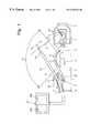
- FIG. 1is a cross sectional view of the tip of an ultrasonic endoscope to which the present invention is applied;
- FIG. 2is a perspective view of the tip of the ultrasonic endoscope shown in FIG. 1 .
- a tip portion (distal end) 1 of an ultrasonic endoscope of the EUSis provided with a linear scanning portion 1 a for scanning a scanning plane section “A” which lies on a plane including a shaft axis O (see FIG. 2) of the tip portion 1 .
- the linear scanning portion 1 ais provided inside the tip portion 1 with a linear-scanning ultrasonic transducer (linear-scanning ultrasonic probe) 2 which is made of an array of ultrasonic-generating oscillators. Accordingly, the linear-scanning ultrasonic transducer 2 is arranged so as to provide an ultrasonic view parallel to the shaft axis O.
- the tip portion 1is further provided with a radial scanning portion 1 b for scanning a scanning plane section “B” which lies on a rotational plane “B” about the shaft axis O.
- the linear scanning portion 1 a and the radial scanning portion 1 bare arranged adjacent to each other so that the radial scanning portion 1 b is closer to the top of the tip portion 1 than the linear scanning portion 1 a .
- the radial scanning portion 1 bis provided inside the tip portion 1 with a radial-scanning ultrasonic transducer (radial-scanning ultrasonic probe) 3 which is made of an array of ultrasonic-generating oscillators.
- the radial-scanning ultrasonic transducer 3is arranged so as to provide a 360° ultrasonic panoramic view perpendicular to the shaft axis O.
- the ultrasonic endoscope of EUS of the present embodimentis a hybrid type ultrasonic endoscope: a combination of a linear type and a radial type.
- the radial-scanning ultrasonic transducer 3is fixed onto a rotatable base 4 provided inside the tip portion 1 .
- the rotatable base 4is rotated about the shaft axis O.
- the radial-scanning ultrasonic transducer 3is fixed to the rotatable base 4 to be inclined rearwardly by a predetermined angle with respect to the shaft axis O so that the scanning plane section “B” intersects the scanning plane section “A”.
- “X”represents an intersection between the scanning plane section “A” and the scanning plane section “B”.
- the intersection “X”is in the shape of a straight line.
- the tip portion 1is provided around the shaft axis O with front and rear annular grooves 5 and 6 to which a rubber band (not shown) for fixing a balloon is secured.
- the front groove 5is formed at the top of the tip portion 1
- the rear groove 6is formed around a portion of the tip portion 1 at the back of the linear scanning portion 1 a with respect to the tip portion 1 (see FIG. 2 ).
- the tip portion 1is further provided at the back of the rear groove 6 with an objective optical viewing portion (objective optical system) 1 c which includes a shield glass 11 and an objective optical lens (not shown) provided inside the shield glass 11 .
- the shield glass 11is fixed to the tip portion 1 to be inclined by a predetermined angle with respect to the shaft axis O so as to face the intersection “X”.
- the objective optical viewing portion 1 cis positioned to extend an optical axis thereof forwardly towards the intersection “X”.
- the optical field of the shield glass 11is shown by dotted lines in FIG. 2 . It can be appreciated from FIG. 2 that the optical field OF covers both the scanning plane section “A” of the linear-scanning ultrasonic transducer 2 and the scanning plane section “B” of the radial-scanning ultrasonic transducer 3 .
- the tip portion 1is further provided, in the vicinity of the shield glass 11 with another shield glass 12 from which illumination light is emitted towards a viewing area.
- the tip portion 1is further provided, between the two shield glasses 11 and 12 , with an exit opening 14 of a treatment tool insertion channel 13 .
- the tip portion of a tubular instrument 100 having a needle (an aspiration needle or an injection needle) 101 at its pointprojects outwards from the exit opening 14 .
- the tip portion 1is further provided inside the exit opening 14 with a rotatable member 16 which is pivoted at a shaft 17 extending in a direction substantially perpendicular to the shaft axis O.
- the rotatable member 16abuts against the instrument 100 and is manipulated to rotate about the shaft 17 by a manipulating device (not shown) to swing the tip (needle 101 ) of the instrument 100 .
- the tip of the instrument 100is oriented so that the projecting direction thereof lies in a plane which includes the scanning plane section “A” of the linear-scanning ultrasonic transducer 2 and passes the intersection “X”.
- the projecting direction of the tip of the instrument 100can be varied so that the tip of the instrument 100 moves along the intersection “X”. In other words, rotation of the rotatable member 16 causes the needle 101 to swing along the intersection “X”.
- optical imagesare viewed through an eyepiece (not shown) via the shield glass 11 and the aforementioned objective optical lens (not shown), which is positioned inside the shield glass 11 .
- signals which are output from the linear-scanning ultrasonic transducer 2 and the radial-scanning ultrasonic transducer 3are transmitted to an external ultrasonic signals processor (not shown) via cables 21 and 22 , respectively.
- the external ultrasonic signals processorconverts the input signals into corresponding image signals to input the same to a TV monitor 23 .
- the TV monitor 23displays an ultrasonic tomogram 24 a obtained through the linear-scanning ultrasonic transducer 2 and an ultrasonic tomogram 24 b obtained through the radial-scanning ultrasonic transducer 3 side by side at the same time, as shown in FIG. 1 .
- the tip of the instrument 100When the instrument 100 is operated to penetrate the needle 101 into a viscera or part of viscera which is located at the back or behind a mucous membrane, the tip of the instrument 100 passes the intersection “X” between the scanning plane section “A” and the scanning plane section “B”. Therefore, the image of the tip of the instrument 100 appears in each of the ultrasonic tomograms 24 a and 24 b , which makes it possible to monitor the variation in penetration depth of the needle 101 on the ultrasonic tomogram 24 a while monitoring the surroundings of the penetrated portion. Hence, an operation using the EUS of the present embodiment can be performed with safety.
- the linear-scanning ultrasonic transducer 2 and the radial-scanning ultrasonic transducer 3can be replaced by a biplane type ultrasonic transducer or probe, that is provided with a linear-scanning ultrasonic transducer and a radial-scanning ultrasonic transducer which are arranged overlapping each other. With such type of transducer, the correlation between the ultrasonic tomograms 24 a and 24 b can be easily recognized.
Landscapes
- Health & Medical Sciences (AREA)
- Life Sciences & Earth Sciences (AREA)
- Surgery (AREA)
- Physics & Mathematics (AREA)
- Medical Informatics (AREA)
- Animal Behavior & Ethology (AREA)
- Radiology & Medical Imaging (AREA)
- Engineering & Computer Science (AREA)
- Biomedical Technology (AREA)
- Heart & Thoracic Surgery (AREA)
- Nuclear Medicine, Radiotherapy & Molecular Imaging (AREA)
- Molecular Biology (AREA)
- Biophysics (AREA)
- Pathology (AREA)
- General Health & Medical Sciences (AREA)
- Public Health (AREA)
- Veterinary Medicine (AREA)
- Optics & Photonics (AREA)
- Gynecology & Obstetrics (AREA)
- Endoscopes (AREA)
- Ultra Sonic Daignosis Equipment (AREA)
Abstract
Description
Claims (20)
Applications Claiming Priority (2)
| Application Number | Priority Date | Filing Date | Title |
|---|---|---|---|
| JP10-051582 | 1998-03-04 | ||
| JP05158298AJP4125814B2 (en) | 1998-03-04 | 1998-03-04 | Ultrasound endoscope |
Publications (1)
| Publication Number | Publication Date |
|---|---|
| US6238336B1true US6238336B1 (en) | 2001-05-29 |
Family
ID=12890944
Family Applications (1)
| Application Number | Title | Priority Date | Filing Date |
|---|---|---|---|
| US09/258,313Expired - LifetimeUS6238336B1 (en) | 1998-03-04 | 1999-02-26 | Ultrasonic endoscope including radial scanning and linear scanning ultrasonic transducers |
Country Status (2)
| Country | Link |
|---|---|
| US (1) | US6238336B1 (en) |
| JP (1) | JP4125814B2 (en) |
Cited By (74)
| Publication number | Priority date | Publication date | Assignee | Title |
|---|---|---|---|---|
| WO2002045588A1 (en)* | 2000-11-24 | 2002-06-13 | Innovacell Biotechnologie Gmbh | Ultrasonic probe comprising a positioning device for examination devices and operation devices |
| US20020089257A1 (en)* | 2001-01-09 | 2002-07-11 | Asahi Kogaku Kogyo Kabushiki Kaisha | Ultrasonic-motor control system |
| US6554801B1 (en)* | 2000-10-26 | 2003-04-29 | Advanced Cardiovascular Systems, Inc. | Directional needle injection drug delivery device and method of use |
| US6589164B1 (en)* | 2000-02-15 | 2003-07-08 | Transvascular, Inc. | Sterility barriers for insertion of non-sterile apparatus into catheters or other medical devices |
| US20030225332A1 (en)* | 2002-05-31 | 2003-12-04 | Olympus Optical Co., Ltd. | Ultrasonic therapeutic apparatus |
| US20040082883A1 (en)* | 2002-10-18 | 2004-04-29 | Fuji Photo Optical Co., Ltd. | Ultrasound endoscope |
| US20040111138A1 (en)* | 2002-10-18 | 2004-06-10 | Radiant Medical, Inc. | Valved connector assembly and sterility barriers for heat exchange catheters and other closed loop catheters |
| US20040249288A1 (en)* | 2002-12-05 | 2004-12-09 | Olympus Corporation | Ultrasonic puncture needle |
| US20050085730A1 (en)* | 2003-10-21 | 2005-04-21 | Aime Flesch | Bi-plane ultrasonic probe |
| US20050222493A1 (en)* | 2004-03-31 | 2005-10-06 | Shinichi Kohno | Endoscope |
| US20050228289A1 (en)* | 2004-03-31 | 2005-10-13 | Fujinon Corporation | Ultrasonic endoscope |
| US20060064118A1 (en)* | 2000-08-11 | 2006-03-23 | Kimblad Per O | Device and a method for treatment of atrioventricular regurgitation |
| US7087024B1 (en)* | 1999-04-08 | 2006-08-08 | Pruter Rick L | Method and apparatus for guiding needles |
| US20060282092A1 (en)* | 2005-06-13 | 2006-12-14 | Stokes Michael J | Surgical suturing apparatus with needle position indicator |
| US20060282093A1 (en)* | 2005-06-13 | 2006-12-14 | Shelton Frederick E Iv | Surgical suturing apparatus with anti-backup system |
| US20060282091A1 (en)* | 2005-06-13 | 2006-12-14 | Shelton Frederick E Iv | Adjustable vacuum chamber for a surgical suturing apparatus |
| US20060282089A1 (en)* | 2005-06-13 | 2006-12-14 | Ethicon Endo-Surgery, Inc. | Endoscopic suturing device |
| US20060281970A1 (en)* | 2005-06-13 | 2006-12-14 | Stokes Michael J | Attachment apparatus for coupling with an endoscope |
| US20060282099A1 (en)* | 2005-06-13 | 2006-12-14 | Stokes Michael J | Method for suture lacing |
| US20060287657A1 (en)* | 2002-09-03 | 2006-12-21 | Bachman Alan B | Single catheter mitral valve repair device and method for use |
| US20070038112A1 (en)* | 2001-10-16 | 2007-02-15 | Taylor James D | Scanning probe with integrated electronics |
| WO2007050941A1 (en)* | 2005-10-27 | 2007-05-03 | Edwards Lifesciences Corporation | System, apparatus, and method for imaging and treating tissue |
| EP1836966A1 (en)* | 2006-03-22 | 2007-09-26 | Fujinon Corporation | Ultrasonic endoscope |
| US20070249936A1 (en)* | 2006-04-20 | 2007-10-25 | Gynesonics, Inc. | Devices and methods for treatment of tissue |
| US20070249939A1 (en)* | 2006-04-20 | 2007-10-25 | Gynesonics, Inc. | Rigid delivery systems having inclined ultrasound and curved needle |
| US20070293787A1 (en)* | 2003-08-13 | 2007-12-20 | Taylor James D | Targeted biopsy delivery system |
| US20080114203A1 (en)* | 2006-11-09 | 2008-05-15 | Crank Justin M | Orientation Adapter for Injection Tube in Flexible Endoscope |
| US20080125709A1 (en)* | 2003-12-31 | 2008-05-29 | Gregory Waimong Chang | Needle catheter |
| EP1977681A1 (en)* | 2007-04-05 | 2008-10-08 | Olympus Medical Systems Corp. | Treatment instrument system |
| US20080281356A1 (en)* | 2007-05-08 | 2008-11-13 | Mark Chau | Suture-fastening clip |
| US7452331B1 (en) | 1999-04-08 | 2008-11-18 | Rick L Pruter | Vascular adjustable multi-gauge tilt-out method and apparatus for guiding needles |
| US20090054773A1 (en)* | 2005-04-11 | 2009-02-26 | Toshihiro Shizuka | Medical Treatment Device |
| EP2036500A1 (en) | 2007-09-11 | 2009-03-18 | Olympus Medical Systems Corporation | Ultrasound diagnostic apparatus |
| US20090099544A1 (en)* | 2007-10-12 | 2009-04-16 | Gynesonics, Inc. | Methods and systems for controlled deployment of needles in tissue |
| US20090287081A1 (en)* | 2008-04-29 | 2009-11-19 | Gynesonics , Inc | Submucosal fibroid ablation for the treatment of menorrhagia |
| US20090318831A1 (en)* | 2008-06-24 | 2009-12-24 | Olympus Medical Systems Corp. | Endoscope apparatus |
| US20100056926A1 (en)* | 2008-08-26 | 2010-03-04 | Gynesonics, Inc. | Ablation device with articulated imaging transducer |
| US20110015614A1 (en)* | 2008-12-16 | 2011-01-20 | Rykhus Jr Robert L | Needleless injection device components, systems, and methods |
| US20110046600A1 (en)* | 2008-12-05 | 2011-02-24 | Crank Justin M | Devices, systems, and related methods for delivery of fluid to tissue |
| US20110172631A1 (en)* | 2008-12-05 | 2011-07-14 | Crank Justin M | Needleless injection device components, systems, and methods |
| US20110238006A1 (en)* | 2008-12-16 | 2011-09-29 | Crank Justin M | Needleless injection device components, systems, and methods |
| US8057386B2 (en) | 2002-09-06 | 2011-11-15 | C.R. Bard, Inc. | Integrated endoscope and accessory treatment device |
| US8206300B2 (en) | 2008-08-26 | 2012-06-26 | Gynesonics, Inc. | Ablation device with articulated imaging transducer |
| US8226666B2 (en) | 2003-03-14 | 2012-07-24 | Edwards Lifesciences Corporation | Mitral valve repair system and method for use |
| US8262574B2 (en) | 2009-02-27 | 2012-09-11 | Gynesonics, Inc. | Needle and tine deployment mechanism |
| EP2070480A4 (en)* | 2006-10-03 | 2012-11-21 | Olympus Medical Systems Corp | ULTRASONIC IMAGE PROCESSING DEVICE AND ULTRASONIC DIAGNOSTIC DEVICE |
| WO2013112887A1 (en)* | 2012-01-25 | 2013-08-01 | Boston Scientific Scimed, Inc. | Endoscopic instrument having movable distal tool |
| EP2671514A1 (en)* | 2012-06-07 | 2013-12-11 | Fujifilm Corporation | Ultrasonic endoscope |
| US8628494B2 (en) | 2009-07-20 | 2014-01-14 | Ams Research Corporation | Devices, systems, and methods for delivering fluid to tissue |
| US8758256B2 (en) | 2010-07-12 | 2014-06-24 | Best Medical International, Inc. | Apparatus for brachytherapy that uses a scanning probe for treatment of malignant tissue |
| US8876759B2 (en) | 2008-12-05 | 2014-11-04 | Ams Research Corporation | Devices, systems and methods for delivering fluid to tissue |
| US8945045B2 (en) | 2009-07-20 | 2015-02-03 | Ams Research Corporation | Needleless injection device components, systems, and methods |
| US8979797B2 (en) | 2010-12-16 | 2015-03-17 | Ams Research Corporation | High pressure delivery system and method for treating pelvic disorder using large molecule therapeutics |
| US9044216B2 (en) | 2010-07-12 | 2015-06-02 | Best Medical International, Inc. | Biopsy needle assembly |
| US9138535B2 (en) | 2009-07-20 | 2015-09-22 | Ams Research Corporation | High pressure injection catheter systems |
| US9283353B2 (en) | 2008-12-05 | 2016-03-15 | Justin M. Crank | Devices, systems and related methods for delivery of fluid to tissue |
| US9370646B2 (en) | 2008-12-05 | 2016-06-21 | Justin M. Crank | Devices, systems and methods for delivering fluid to tissue |
| US9421326B2 (en) | 2008-12-29 | 2016-08-23 | Robert L. Rykhus | Method and apparatus for compensating for injection media viscosity in a pressurized drug injection system |
| US9456733B2 (en) | 2011-10-31 | 2016-10-04 | Boston Scientific Scimed, Inc. | Endoscopic instrument having a deflectable distal tool |
| US10058342B2 (en) | 2006-01-12 | 2018-08-28 | Gynesonics, Inc. | Devices and methods for treatment of tissue |
| US10182862B2 (en) | 2005-02-02 | 2019-01-22 | Gynesonics, Inc. | Method and device for uterine fibroid treatment |
| GB2572860A (en)* | 2018-03-14 | 2019-10-16 | Spiration Inc | Catheter assembly with offset device for tissue sampling |
| GB2572861A (en)* | 2018-03-14 | 2019-10-16 | Spiration Inc | Catheter assembly with offset device for tissue sampling |
| US10456128B2 (en) | 2005-06-13 | 2019-10-29 | Ethicon Llc | Method for suture lacing |
| US10595819B2 (en) | 2006-04-20 | 2020-03-24 | Gynesonics, Inc. | Ablation device with articulated imaging transducer |
| US10772600B2 (en) | 2015-09-25 | 2020-09-15 | Perceptive Navigation Llc | Image guided catheters and methods of use |
| CN111938694A (en)* | 2020-08-07 | 2020-11-17 | 深圳北芯生命科技有限公司 | Transmission device of ultrasonic transducer and manufacturing method thereof |
| US10993770B2 (en) | 2016-11-11 | 2021-05-04 | Gynesonics, Inc. | Controlled treatment of tissue and dynamic interaction with, and comparison of, tissue and/or treatment data |
| US20210169312A1 (en)* | 2018-09-10 | 2021-06-10 | Fujifilm Corporation | Endoscope |
| US11234581B2 (en)* | 2014-05-02 | 2022-02-01 | Endochoice, Inc. | Elevator for directing medical tool |
| US11259825B2 (en) | 2006-01-12 | 2022-03-01 | Gynesonics, Inc. | Devices and methods for treatment of tissue |
| US11330965B2 (en) | 2017-03-03 | 2022-05-17 | Boston Scientific Scimed, Inc. | Device tip |
| US11850006B2 (en) | 2017-06-28 | 2023-12-26 | Innoscion Llc | Devices and methods for image-guided percutaneous cardiac valve implantation and repair |
| US12390654B2 (en) | 2015-09-25 | 2025-08-19 | Innoscion Llc | Pericardial implantable cardioverter defibrillator |
Families Citing this family (2)
| Publication number | Priority date | Publication date | Assignee | Title |
|---|---|---|---|---|
| JP5235706B2 (en)* | 2009-02-03 | 2013-07-10 | Hoya株式会社 | Treatment endoscope |
| JP5719683B2 (en)* | 2010-06-07 | 2015-05-20 | 富士フイルム株式会社 | Endoscope |
Citations (10)
| Publication number | Priority date | Publication date | Assignee | Title |
|---|---|---|---|---|
| US4433692A (en) | 1981-05-20 | 1984-02-28 | Olympus Optical Co., Ltd. | Ultrasonic diagnosis device |
| US4605009A (en)* | 1983-04-06 | 1986-08-12 | Universite Francois Rabelais | Ultrasonic sweep echography and display endoscopic probe |
| US4757819A (en)* | 1986-05-21 | 1988-07-19 | Olympus Optical Co., Ltd. | Ultrasonic endoscope |
| US4974590A (en)* | 1988-05-18 | 1990-12-04 | Olympus Optical Co., Ltd. | Ultrasonic probe for use in ultrasonic endoscope |
| US5471988A (en)* | 1993-12-24 | 1995-12-05 | Olympus Optical Co., Ltd. | Ultrasonic diagnosis and therapy system in which focusing point of therapeutic ultrasonic wave is locked at predetermined position within observation ultrasonic scanning range |
| US5492126A (en)* | 1994-05-02 | 1996-02-20 | Focal Surgery | Probe for medical imaging and therapy using ultrasound |
| JPH08126643A (en) | 1994-11-01 | 1996-05-21 | Asahi Optical Co Ltd | Ultrasound endoscope |
| US5596989A (en) | 1993-12-28 | 1997-01-28 | Olympus Optical Co., Ltd. | Ultrasonic probe |
| US5873828A (en)* | 1994-02-18 | 1999-02-23 | Olympus Optical Co., Ltd. | Ultrasonic diagnosis and treatment system |
| US5980454A (en)* | 1997-12-01 | 1999-11-09 | Endonetics, Inc. | Endoscopic imaging system employing diffractive optical elements |
- 1998
- 1998-03-04JPJP05158298Apatent/JP4125814B2/ennot_activeExpired - Fee Related
- 1999
- 1999-02-26USUS09/258,313patent/US6238336B1/ennot_activeExpired - Lifetime
Patent Citations (10)
| Publication number | Priority date | Publication date | Assignee | Title |
|---|---|---|---|---|
| US4433692A (en) | 1981-05-20 | 1984-02-28 | Olympus Optical Co., Ltd. | Ultrasonic diagnosis device |
| US4605009A (en)* | 1983-04-06 | 1986-08-12 | Universite Francois Rabelais | Ultrasonic sweep echography and display endoscopic probe |
| US4757819A (en)* | 1986-05-21 | 1988-07-19 | Olympus Optical Co., Ltd. | Ultrasonic endoscope |
| US4974590A (en)* | 1988-05-18 | 1990-12-04 | Olympus Optical Co., Ltd. | Ultrasonic probe for use in ultrasonic endoscope |
| US5471988A (en)* | 1993-12-24 | 1995-12-05 | Olympus Optical Co., Ltd. | Ultrasonic diagnosis and therapy system in which focusing point of therapeutic ultrasonic wave is locked at predetermined position within observation ultrasonic scanning range |
| US5596989A (en) | 1993-12-28 | 1997-01-28 | Olympus Optical Co., Ltd. | Ultrasonic probe |
| US5873828A (en)* | 1994-02-18 | 1999-02-23 | Olympus Optical Co., Ltd. | Ultrasonic diagnosis and treatment system |
| US5492126A (en)* | 1994-05-02 | 1996-02-20 | Focal Surgery | Probe for medical imaging and therapy using ultrasound |
| JPH08126643A (en) | 1994-11-01 | 1996-05-21 | Asahi Optical Co Ltd | Ultrasound endoscope |
| US5980454A (en)* | 1997-12-01 | 1999-11-09 | Endonetics, Inc. | Endoscopic imaging system employing diffractive optical elements |
Cited By (169)
| Publication number | Priority date | Publication date | Assignee | Title |
|---|---|---|---|---|
| US7087024B1 (en)* | 1999-04-08 | 2006-08-08 | Pruter Rick L | Method and apparatus for guiding needles |
| US7452331B1 (en) | 1999-04-08 | 2008-11-18 | Rick L Pruter | Vascular adjustable multi-gauge tilt-out method and apparatus for guiding needles |
| US6589164B1 (en)* | 2000-02-15 | 2003-07-08 | Transvascular, Inc. | Sterility barriers for insertion of non-sterile apparatus into catheters or other medical devices |
| US9999419B2 (en) | 2000-05-01 | 2018-06-19 | Edwards Lifesciences Corporation | Single catheter heart repair device and method for use |
| US9314242B2 (en) | 2000-05-01 | 2016-04-19 | Edwards Lifesciences Corporation | Single catheter heart repair device and method for use |
| US20060064118A1 (en)* | 2000-08-11 | 2006-03-23 | Kimblad Per O | Device and a method for treatment of atrioventricular regurgitation |
| US8062313B2 (en) | 2000-08-11 | 2011-11-22 | Edwards Lifesciences Corporation | Device and a method for treatment of atrioventricular regurgitation |
| US20070135714A1 (en)* | 2000-10-26 | 2007-06-14 | Jeffrey Steward | Directional needle injection drug delivery device and method use |
| US6554801B1 (en)* | 2000-10-26 | 2003-04-29 | Advanced Cardiovascular Systems, Inc. | Directional needle injection drug delivery device and method of use |
| US20030233065A1 (en)* | 2000-10-26 | 2003-12-18 | Jeffrey Steward | Directional needle injection drug delivery device and method of use |
| US7179249B2 (en)* | 2000-10-26 | 2007-02-20 | Advanced Cardiovascular Systems, Inc. | Directional needle injection drug delivery device and method of use |
| US7811265B2 (en)* | 2000-11-24 | 2010-10-12 | Innovacell Biotechnologie Gmbh | Ultrasonic probe with positioning device for examination devices and operation devices |
| EA005391B1 (en)* | 2000-11-24 | 2005-02-24 | Инноваселл Биотехнологи Гмбх | Ultrasonic probe comprising a positioning device for examination devices and operation devices |
| WO2002045588A1 (en)* | 2000-11-24 | 2002-06-13 | Innovacell Biotechnologie Gmbh | Ultrasonic probe comprising a positioning device for examination devices and operation devices |
| US20040092821A1 (en)* | 2000-11-24 | 2004-05-13 | Steffen Hering | Ultrasonic probe with positioning device for examination devices and operation devices |
| US6894422B2 (en)* | 2001-01-09 | 2005-05-17 | Pentax Corporation | Ultrasonic-motor control system |
| US20020089257A1 (en)* | 2001-01-09 | 2002-07-11 | Asahi Kogaku Kogyo Kabushiki Kaisha | Ultrasonic-motor control system |
| US8137279B2 (en) | 2001-10-16 | 2012-03-20 | Envisioneering, Llc | Scanning probe |
| US20070038112A1 (en)* | 2001-10-16 | 2007-02-15 | Taylor James D | Scanning probe with integrated electronics |
| US20030225332A1 (en)* | 2002-05-31 | 2003-12-04 | Olympus Optical Co., Ltd. | Ultrasonic therapeutic apparatus |
| US7887552B2 (en) | 2002-09-03 | 2011-02-15 | Edwards Lifesciences Corporation | Single catheter mitral valve repair device and method for use |
| US20060287657A1 (en)* | 2002-09-03 | 2006-12-21 | Bachman Alan B | Single catheter mitral valve repair device and method for use |
| US8057386B2 (en) | 2002-09-06 | 2011-11-15 | C.R. Bard, Inc. | Integrated endoscope and accessory treatment device |
| US8206284B2 (en) | 2002-09-06 | 2012-06-26 | C.R. Bard, Inc. | Integrated endoscope and accessory treatment device |
| US8262716B2 (en) | 2002-10-18 | 2012-09-11 | Zoll Circulation, Inc. | Valved connector assembly and sterility barriers for heat exchange catheters and other closed loop catheters |
| US20040111138A1 (en)* | 2002-10-18 | 2004-06-10 | Radiant Medical, Inc. | Valved connector assembly and sterility barriers for heat exchange catheters and other closed loop catheters |
| US20040082883A1 (en)* | 2002-10-18 | 2004-04-29 | Fuji Photo Optical Co., Ltd. | Ultrasound endoscope |
| US7318806B2 (en)* | 2002-10-18 | 2008-01-15 | Fujinon Corporation | Ultrasound endoscope |
| US7510568B2 (en) | 2002-10-18 | 2009-03-31 | Zoll Circulation, Inc. | Valved connector assembly and sterility barriers for heat exchange catheters and other closed looped catheters |
| US20050143798A1 (en)* | 2002-10-18 | 2005-06-30 | Radiant Medical, Inc. | Valved connector assembly and sterility barriers for heat exchange catheters and other closed loop catheters |
| US20090247963A1 (en)* | 2002-10-18 | 2009-10-01 | Zoll Circulation, Inc. | Valved connector assembly and sterility barriers for heat exchange catheters and other closed loop catheters |
| US20040249288A1 (en)* | 2002-12-05 | 2004-12-09 | Olympus Corporation | Ultrasonic puncture needle |
| US8777991B2 (en) | 2003-03-14 | 2014-07-15 | David Zarbatany | Mitral valve repair system and method for use |
| US8226666B2 (en) | 2003-03-14 | 2012-07-24 | Edwards Lifesciences Corporation | Mitral valve repair system and method for use |
| US8317724B2 (en) | 2003-08-13 | 2012-11-27 | Envisioneering, Llc | Targeted treatment delivery system |
| US7833168B2 (en) | 2003-08-13 | 2010-11-16 | Envisioneering Medical Technologies, Llc | Targeted biopsy delivery system |
| US20090054807A1 (en)* | 2003-08-13 | 2009-02-26 | Taylor James D | Targeted biopsy delivery system |
| US20110144492A1 (en)* | 2003-08-13 | 2011-06-16 | Taylor James D | Targeted Treatment Delivery System |
| US20070293787A1 (en)* | 2003-08-13 | 2007-12-20 | Taylor James D | Targeted biopsy delivery system |
| US20050085730A1 (en)* | 2003-10-21 | 2005-04-21 | Aime Flesch | Bi-plane ultrasonic probe |
| US7066887B2 (en) | 2003-10-21 | 2006-06-27 | Vermon | Bi-plane ultrasonic probe |
| US20080125709A1 (en)* | 2003-12-31 | 2008-05-29 | Gregory Waimong Chang | Needle catheter |
| US8152758B2 (en) | 2003-12-31 | 2012-04-10 | Advanced Cardiovascular Systems, Inc. | Needle catheter |
| US7946993B2 (en)* | 2004-03-31 | 2011-05-24 | Fujinon Corporation | Ultrasonic endoscope |
| US20050228289A1 (en)* | 2004-03-31 | 2005-10-13 | Fujinon Corporation | Ultrasonic endoscope |
| US20050222493A1 (en)* | 2004-03-31 | 2005-10-06 | Shinichi Kohno | Endoscope |
| US7771349B2 (en)* | 2004-03-31 | 2010-08-10 | Fujinon Corporation | Endoscope |
| US10182862B2 (en) | 2005-02-02 | 2019-01-22 | Gynesonics, Inc. | Method and device for uterine fibroid treatment |
| US11419668B2 (en) | 2005-02-02 | 2022-08-23 | Gynesonics, Inc. | Method and device for uterine fibroid treatment |
| US11950837B2 (en) | 2005-02-02 | 2024-04-09 | Gynesonics, Inc. | Method and device for uterine fibroid treatment |
| US12414813B2 (en) | 2005-02-02 | 2025-09-16 | Gynesonics, Inc. | Method and device for uterine fibroid treatment |
| US8162939B2 (en) | 2005-04-11 | 2012-04-24 | Olympus Medical Systems Corp. | Medical treatment device |
| US20090054773A1 (en)* | 2005-04-11 | 2009-02-26 | Toshihiro Shizuka | Medical Treatment Device |
| EP2397097A1 (en)* | 2005-04-11 | 2011-12-21 | Olympus Medical Systems Corp. | Medical treatment device |
| US20060282093A1 (en)* | 2005-06-13 | 2006-12-14 | Shelton Frederick E Iv | Surgical suturing apparatus with anti-backup system |
| US7846169B2 (en) | 2005-06-13 | 2010-12-07 | Ethicon Endo-Surgery, Inc. | Adjustable vacuum chamber for a surgical suturing apparatus |
| US10456128B2 (en) | 2005-06-13 | 2019-10-29 | Ethicon Llc | Method for suture lacing |
| US20060282091A1 (en)* | 2005-06-13 | 2006-12-14 | Shelton Frederick E Iv | Adjustable vacuum chamber for a surgical suturing apparatus |
| US20060282097A1 (en)* | 2005-06-13 | 2006-12-14 | Ortiz Mark S | Surgical suturing apparatus with a non-visible spectrum sensing member |
| US20060282089A1 (en)* | 2005-06-13 | 2006-12-14 | Ethicon Endo-Surgery, Inc. | Endoscopic suturing device |
| US20060282096A1 (en)* | 2005-06-13 | 2006-12-14 | Papa Christopher A | Quick load mechanism for a surgical suturing apparatus |
| US20060282095A1 (en)* | 2005-06-13 | 2006-12-14 | Stokes Michael J | Surgical suturing apparatus with collapsible vacuum chamber |
| US7828812B2 (en) | 2005-06-13 | 2010-11-09 | Ethicon Endo-Surgery, Inc. | Surgical suturing apparatus with needle release system |
| US7976553B2 (en) | 2005-06-13 | 2011-07-12 | Ethicon Endo-Surgery, Inc. | Surgical suturing apparatus with detachable handle |
| US9545191B2 (en) | 2005-06-13 | 2017-01-17 | Ethicon Endo-Surgery, Inc. | Method for suture lacing |
| US8641728B2 (en) | 2005-06-13 | 2014-02-04 | Ethicon Endo-Surgery, Inc. | Attachment apparatus for coupling with an endoscope |
| US20060282092A1 (en)* | 2005-06-13 | 2006-12-14 | Stokes Michael J | Surgical suturing apparatus with needle position indicator |
| US8500756B2 (en) | 2005-06-13 | 2013-08-06 | Ethicon Endo. Surgery, Inc. | Quick load mechanism for a surgical suturing apparatus |
| US7887554B2 (en) | 2005-06-13 | 2011-02-15 | Ethicon Endo-Surgery, Inc. | Surgical suturing apparatus with needle position indicator |
| US20060282090A1 (en)* | 2005-06-13 | 2006-12-14 | Stokes Michael J | Surgical suturing apparatus with needle release system |
| US20060281970A1 (en)* | 2005-06-13 | 2006-12-14 | Stokes Michael J | Attachment apparatus for coupling with an endoscope |
| US20060282099A1 (en)* | 2005-06-13 | 2006-12-14 | Stokes Michael J | Method for suture lacing |
| US7628796B2 (en) | 2005-06-13 | 2009-12-08 | Ethicon Endo-Surgery, Inc. | Surgical suturing apparatus with anti-backup system |
| US7615060B2 (en) | 2005-06-13 | 2009-11-10 | Ethicon-Endo Surgery, Inc. | Endoscopic suturing device |
| US7833236B2 (en) | 2005-06-13 | 2010-11-16 | Ethicon Endo-Surgery, Inc. | Surgical suturing apparatus with collapsible vacuum chamber |
| US20070232941A1 (en)* | 2005-10-27 | 2007-10-04 | Stan Rabinovich | System, apparatus, and method for imaging and treating tissue |
| WO2007050941A1 (en)* | 2005-10-27 | 2007-05-03 | Edwards Lifesciences Corporation | System, apparatus, and method for imaging and treating tissue |
| US11259825B2 (en) | 2006-01-12 | 2022-03-01 | Gynesonics, Inc. | Devices and methods for treatment of tissue |
| US10058342B2 (en) | 2006-01-12 | 2018-08-28 | Gynesonics, Inc. | Devices and methods for treatment of tissue |
| EP1836966A1 (en)* | 2006-03-22 | 2007-09-26 | Fujinon Corporation | Ultrasonic endoscope |
| US20070249940A1 (en)* | 2006-03-22 | 2007-10-25 | Fujinon Corporation | Ultrasonic endoscope |
| US8177717B2 (en) | 2006-03-22 | 2012-05-15 | Fujinon Corporation | Ultrasonic endoscope |
| EP1839590A1 (en)* | 2006-03-31 | 2007-10-03 | Ethicon Endo-Surgery, Inc. | Surgical suturing apparatus with a non-visible spectrum sensing |
| AU2007201320B2 (en)* | 2006-03-31 | 2013-05-23 | Ethicon Endo-Surgery, Inc. | Surgical suturing apparatus with a non-visible spectrum sensing |
| US20070249939A1 (en)* | 2006-04-20 | 2007-10-25 | Gynesonics, Inc. | Rigid delivery systems having inclined ultrasound and curved needle |
| US7874986B2 (en)* | 2006-04-20 | 2011-01-25 | Gynesonics, Inc. | Methods and devices for visualization and ablation of tissue |
| US10610197B2 (en) | 2006-04-20 | 2020-04-07 | Gynesonics, Inc. | Ablation device with articulated imaging transducer |
| US12048583B2 (en) | 2006-04-20 | 2024-07-30 | Gynesonics, Inc. | Ablation device with articulated imaging transducer |
| US20070249936A1 (en)* | 2006-04-20 | 2007-10-25 | Gynesonics, Inc. | Devices and methods for treatment of tissue |
| US7815571B2 (en) | 2006-04-20 | 2010-10-19 | Gynesonics, Inc. | Rigid delivery systems having inclined ultrasound and needle |
| US10595819B2 (en) | 2006-04-20 | 2020-03-24 | Gynesonics, Inc. | Ablation device with articulated imaging transducer |
| US8506485B2 (en) | 2006-04-20 | 2013-08-13 | Gynesonics, Inc | Devices and methods for treatment of tissue |
| EP2070480A4 (en)* | 2006-10-03 | 2012-11-21 | Olympus Medical Systems Corp | ULTRASONIC IMAGE PROCESSING DEVICE AND ULTRASONIC DIAGNOSTIC DEVICE |
| US10945704B2 (en) | 2006-10-12 | 2021-03-16 | Perceptive Navigation Llc | Image guided catheters and methods of use |
| US10945703B2 (en) | 2006-10-12 | 2021-03-16 | Perceptive Navigation Llc | Image guided catheters and method of use |
| US11660067B2 (en) | 2006-10-12 | 2023-05-30 | Perceptive Navigation Llc | Image guided catheters and methods of use |
| US20080114203A1 (en)* | 2006-11-09 | 2008-05-15 | Crank Justin M | Orientation Adapter for Injection Tube in Flexible Endoscope |
| US8852084B2 (en) | 2006-11-09 | 2014-10-07 | Ams Research Corporation | Orientation adapter for injection tube in flexible endoscope |
| US7993264B2 (en)* | 2006-11-09 | 2011-08-09 | Ams Research Corporation | Orientation adapter for injection tube in flexible endoscope |
| EP1977681A1 (en)* | 2007-04-05 | 2008-10-08 | Olympus Medical Systems Corp. | Treatment instrument system |
| US8167808B2 (en) | 2007-04-05 | 2012-05-01 | Olympus Medical Systems Corp. | Treatment instrument system |
| US20080281356A1 (en)* | 2007-05-08 | 2008-11-13 | Mark Chau | Suture-fastening clip |
| US8753373B2 (en) | 2007-05-08 | 2014-06-17 | Edwards Lifesciences Corporation | Suture-fastening clip |
| CN101385654B (en)* | 2007-09-11 | 2011-04-27 | 奥林巴斯医疗株式会社 | Ultrasonic diagnostic device |
| EP2036500A1 (en) | 2007-09-11 | 2009-03-18 | Olympus Medical Systems Corporation | Ultrasound diagnostic apparatus |
| US8088072B2 (en) | 2007-10-12 | 2012-01-03 | Gynesonics, Inc. | Methods and systems for controlled deployment of needles in tissue |
| US20090099544A1 (en)* | 2007-10-12 | 2009-04-16 | Gynesonics, Inc. | Methods and systems for controlled deployment of needles in tissue |
| US11925512B2 (en) | 2007-10-12 | 2024-03-12 | Gynesonics, Inc. | Methods and systems for controlled deployment of needles in tissue |
| US11096760B2 (en) | 2007-10-12 | 2021-08-24 | Gynesonics, Inc. | Methods and systems for controlled deployment of needles in tissue |
| US11826207B2 (en) | 2007-10-12 | 2023-11-28 | Gynesonics, Inc | Methods and systems for controlled deployment of needles in tissue |
| US11096761B2 (en) | 2007-10-12 | 2021-08-24 | Gynesonics, Inc. | Methods and systems for controlled deployment of needles in tissue |
| US8262577B2 (en) | 2007-10-12 | 2012-09-11 | Gynesonics, Inc. | Methods and systems for controlled deployment of needles in tissue |
| US20090287081A1 (en)* | 2008-04-29 | 2009-11-19 | Gynesonics , Inc | Submucosal fibroid ablation for the treatment of menorrhagia |
| US8827922B2 (en) | 2008-06-24 | 2014-09-09 | Olympus Medical Systems Corp. | Endoscope apparatus |
| EP2138092A1 (en)* | 2008-06-24 | 2009-12-30 | Olympus Medical Systems Corporation | Endoscope apparatus |
| US20090318831A1 (en)* | 2008-06-24 | 2009-12-24 | Olympus Medical Systems Corp. | Endoscope apparatus |
| US20100056926A1 (en)* | 2008-08-26 | 2010-03-04 | Gynesonics, Inc. | Ablation device with articulated imaging transducer |
| US8206300B2 (en) | 2008-08-26 | 2012-06-26 | Gynesonics, Inc. | Ablation device with articulated imaging transducer |
| US8876759B2 (en) | 2008-12-05 | 2014-11-04 | Ams Research Corporation | Devices, systems and methods for delivering fluid to tissue |
| US9370646B2 (en) | 2008-12-05 | 2016-06-21 | Justin M. Crank | Devices, systems and methods for delivering fluid to tissue |
| US20110046600A1 (en)* | 2008-12-05 | 2011-02-24 | Crank Justin M | Devices, systems, and related methods for delivery of fluid to tissue |
| US8366657B2 (en) | 2008-12-05 | 2013-02-05 | Ams Research Corporation | Needleless injection device components, systems, and methods |
| US9283353B2 (en) | 2008-12-05 | 2016-03-15 | Justin M. Crank | Devices, systems and related methods for delivery of fluid to tissue |
| US20110172631A1 (en)* | 2008-12-05 | 2011-07-14 | Crank Justin M | Needleless injection device components, systems, and methods |
| US9017282B2 (en) | 2008-12-05 | 2015-04-28 | Ams Research Corporation | Needleless injection device components, systems, and methods |
| US20110015614A1 (en)* | 2008-12-16 | 2011-01-20 | Rykhus Jr Robert L | Needleless injection device components, systems, and methods |
| US9295823B2 (en) | 2008-12-16 | 2016-03-29 | Robert L. Rykhus, Jr. | Needleless injection device components, systems, and methods |
| US9579462B2 (en) | 2008-12-16 | 2017-02-28 | Astora Women's Health Holdings, Llc | Needleless injection device components, systems, and methods |
| US8852142B2 (en) | 2008-12-16 | 2014-10-07 | Ams Research Corporation | Needleless injection device components, systems, and methods |
| US20110238006A1 (en)* | 2008-12-16 | 2011-09-29 | Crank Justin M | Needleless injection device components, systems, and methods |
| US9795733B2 (en) | 2008-12-29 | 2017-10-24 | Astora Women's Health Holdings, Llc | Method and apparatus for compensating for injection media viscosity in a pressurized drug injection system |
| US9421326B2 (en) | 2008-12-29 | 2016-08-23 | Robert L. Rykhus | Method and apparatus for compensating for injection media viscosity in a pressurized drug injection system |
| US10321951B2 (en) | 2009-02-27 | 2019-06-18 | Gynesonics, Inc. | Needle and tine deployment mechanism |
| US8262574B2 (en) | 2009-02-27 | 2012-09-11 | Gynesonics, Inc. | Needle and tine deployment mechanism |
| US11564735B2 (en) | 2009-02-27 | 2023-01-31 | Gynesonics, Inc. | Needle and fine deployment mechanism |
| US11992258B2 (en) | 2009-02-27 | 2024-05-28 | Gynesonics, Inc. | Needle and tine deployment mechanism |
| US8945045B2 (en) | 2009-07-20 | 2015-02-03 | Ams Research Corporation | Needleless injection device components, systems, and methods |
| US8628494B2 (en) | 2009-07-20 | 2014-01-14 | Ams Research Corporation | Devices, systems, and methods for delivering fluid to tissue |
| US9138535B2 (en) | 2009-07-20 | 2015-09-22 | Ams Research Corporation | High pressure injection catheter systems |
| US9364615B2 (en) | 2009-07-20 | 2016-06-14 | Justin M. Crank | Devices, systems, and methods for delivering fluid to tissue |
| US9675759B2 (en) | 2009-07-20 | 2017-06-13 | Astora Women's Health Holdings, Llc | Devices, systems, and methods for delivering fluid to tissue |
| US9044216B2 (en) | 2010-07-12 | 2015-06-02 | Best Medical International, Inc. | Biopsy needle assembly |
| US8758256B2 (en) | 2010-07-12 | 2014-06-24 | Best Medical International, Inc. | Apparatus for brachytherapy that uses a scanning probe for treatment of malignant tissue |
| US9827375B2 (en) | 2010-12-16 | 2017-11-28 | Astora Women's Health Holdings, Llc | High pressure delivery system and method for treating pelvic disorder using large molecule therapeutics |
| US8979797B2 (en) | 2010-12-16 | 2015-03-17 | Ams Research Corporation | High pressure delivery system and method for treating pelvic disorder using large molecule therapeutics |
| US9456733B2 (en) | 2011-10-31 | 2016-10-04 | Boston Scientific Scimed, Inc. | Endoscopic instrument having a deflectable distal tool |
| WO2013112887A1 (en)* | 2012-01-25 | 2013-08-01 | Boston Scientific Scimed, Inc. | Endoscopic instrument having movable distal tool |
| US10687691B2 (en) | 2012-01-25 | 2020-06-23 | Boston Scientific Scimed, Inc. | Endoscopic instrument having movable distal tool |
| US9408529B2 (en) | 2012-01-25 | 2016-08-09 | Boston Scientific Scimed, Inc. | Endoscopic instrument having movable distal tool |
| EP2671514A1 (en)* | 2012-06-07 | 2013-12-11 | Fujifilm Corporation | Ultrasonic endoscope |
| US12053155B2 (en)* | 2014-05-02 | 2024-08-06 | Endochoice, Inc. | Elevator for directing medical tool |
| US11234581B2 (en)* | 2014-05-02 | 2022-02-01 | Endochoice, Inc. | Elevator for directing medical tool |
| US10772600B2 (en) | 2015-09-25 | 2020-09-15 | Perceptive Navigation Llc | Image guided catheters and methods of use |
| US12390654B2 (en) | 2015-09-25 | 2025-08-19 | Innoscion Llc | Pericardial implantable cardioverter defibrillator |
| US10993770B2 (en) | 2016-11-11 | 2021-05-04 | Gynesonics, Inc. | Controlled treatment of tissue and dynamic interaction with, and comparison of, tissue and/or treatment data |
| US11419682B2 (en) | 2016-11-11 | 2022-08-23 | Gynesonics, Inc. | Controlled treatment of tissue and dynamic interaction with, and comparison of, tissue and/or treatment data |
| US12239382B2 (en) | 2016-11-11 | 2025-03-04 | Gynesonics, Inc. | Controlled treatment of tissue and dynamic interaction with, and comparison of, tissue and/or treatment data |
| US11751751B2 (en) | 2017-03-03 | 2023-09-12 | Boston Scientific Scimed, Inc. | Device tip |
| US12402780B2 (en) | 2017-03-03 | 2025-09-02 | Boston Scientific Scimed, Inc. | Device tip |
| US11330965B2 (en) | 2017-03-03 | 2022-05-17 | Boston Scientific Scimed, Inc. | Device tip |
| US11850006B2 (en) | 2017-06-28 | 2023-12-26 | Innoscion Llc | Devices and methods for image-guided percutaneous cardiac valve implantation and repair |
| GB2572860B (en)* | 2018-03-14 | 2022-12-07 | Gyrus Acmi Inc | Catheter assembly with offset device for tissue sampling |
| GB2572860A (en)* | 2018-03-14 | 2019-10-16 | Spiration Inc | Catheter assembly with offset device for tissue sampling |
| GB2572861B (en)* | 2018-03-14 | 2022-09-21 | Gyrus Acmi Inc | Catheter assembly with offset device for tissue sampling |
| GB2572861A (en)* | 2018-03-14 | 2019-10-16 | Spiration Inc | Catheter assembly with offset device for tissue sampling |
| US12310554B2 (en)* | 2018-09-10 | 2025-05-27 | Fujifilm Corporation | Endoscope having elevator support member in distal end portion body for heat dissipation |
| US20210169312A1 (en)* | 2018-09-10 | 2021-06-10 | Fujifilm Corporation | Endoscope |
| CN111938694B (en)* | 2020-08-07 | 2022-10-11 | 深圳北芯生命科技股份有限公司 | Transmission device of ultrasonic transducer and manufacturing method thereof |
| CN111938694A (en)* | 2020-08-07 | 2020-11-17 | 深圳北芯生命科技有限公司 | Transmission device of ultrasonic transducer and manufacturing method thereof |
Also Published As
| Publication number | Publication date |
|---|---|
| JPH11244289A (en) | 1999-09-14 |
| JP4125814B2 (en) | 2008-07-30 |
Similar Documents
| Publication | Publication Date | Title |
|---|---|---|
| US6238336B1 (en) | Ultrasonic endoscope including radial scanning and linear scanning ultrasonic transducers | |
| US6390973B1 (en) | Endoscope for ultrasonic examination and surgical treatment associated thereto | |
| US4489727A (en) | Device for diagnosing body cavity interior with supersonic waves | |
| US6149598A (en) | Ultrasound endoscope | |
| US5499630A (en) | Catheter type ultrasound probe | |
| US4489728A (en) | Device for diagnosing body cavity interior with supersonic waves | |
| JP2002263055A (en) | Tip hood for endoscope | |
| US6689066B1 (en) | Ultrasonic probe | |
| US6193666B1 (en) | Tip of ultrasonic endoscope | |
| JP3179184B2 (en) | Ultrasound probe with observation function | |
| JPH0919403A (en) | Endoscope device and endoscope apparatus | |
| JPH0876028A (en) | Leading edge structure for side-view type electronic endoscope | |
| JPH09220192A (en) | Endoscope | |
| JP2018110741A (en) | Ultrasound endoscope | |
| JPH08140976A (en) | Ultrasonic endoscope | |
| JP2785252B2 (en) | Endoscope | |
| JPH08126644A (en) | Ultrasonic endoscope | |
| JP4981373B2 (en) | Magnifying endoscope | |
| JP4827636B2 (en) | Endoscope device for magnification observation | |
| JPS6258257B2 (en) | ||
| JPH08117233A (en) | Ultrasonic endoscope device | |
| JP2837708B2 (en) | Ultrasonic diagnostic device in body cavity | |
| JPH0919432A (en) | Ultrasonic inspection device to be oral-endoscopically inserted | |
| JPH0523337A (en) | Endoscope | |
| JPH08280686A (en) | Ultrasonic endoscope |
Legal Events
| Date | Code | Title | Description |
|---|---|---|---|
| AS | Assignment | Owner name:ASAHI KOGAKU KOGYO KABUSHIKI KAISHA, JAPAN Free format text:ASSIGNMENT OF ASSIGNORS INTEREST;ASSIGNOR:OUCHI, TERUO;REEL/FRAME:009810/0593 Effective date:19990223 | |
| STCF | Information on status: patent grant | Free format text:PATENTED CASE | |
| FEPP | Fee payment procedure | Free format text:PAYOR NUMBER ASSIGNED (ORIGINAL EVENT CODE: ASPN); ENTITY STATUS OF PATENT OWNER: LARGE ENTITY | |
| FPAY | Fee payment | Year of fee payment:4 | |
| FPAY | Fee payment | Year of fee payment:8 | |
| FPAY | Fee payment | Year of fee payment:12 | |
| AS | Assignment | Owner name:PENTAX CORPORATION, JAPAN Free format text:CHANGE OF NAME;ASSIGNOR:ASAHI KOGAKU KOGYO KABUSHIKI KAISHA;REEL/FRAME:041758/0285 Effective date:20021001 | |
| AS | Assignment | Owner name:HOYA CORPORATION, JAPAN Free format text:MERGER;ASSIGNOR:PENTAX CORPORATION;REEL/FRAME:042114/0463 Effective date:20080407 | |
| AS | Assignment | Owner name:HOYA CORPORATION, JAPAN Free format text:CHANGE OF ADDRESS OF ASSIGNEE;ASSIGNOR:HOYA CORPORATION;REEL/FRAME:042424/0318 Effective date:20160401 |