US6226543B1 - System and method of recording and displaying in context of an image a location of at least one point-of-interest in a body during an intra-body medical procedure - Google Patents
System and method of recording and displaying in context of an image a location of at least one point-of-interest in a body during an intra-body medical procedureDownload PDFInfo
- Publication number
- US6226543B1 US6226543B1US09/179,827US17982798AUS6226543B1US 6226543 B1US6226543 B1US 6226543B1US 17982798 AUS17982798 AUS 17982798AUS 6226543 B1US6226543 B1US 6226543B1
- Authority
- US
- United States
- Prior art keywords
- location
- imaging instrument
- catheter
- interest
- locating
- Prior art date
- Legal status (The legal status is an assumption and is not a legal conclusion. Google has not performed a legal analysis and makes no representation as to the accuracy of the status listed.)
- Expired - Lifetime
Links
Images

Classifications
- A—HUMAN NECESSITIES
- A61—MEDICAL OR VETERINARY SCIENCE; HYGIENE
- A61B—DIAGNOSIS; SURGERY; IDENTIFICATION
- A61B5/00—Measuring for diagnostic purposes; Identification of persons
- A61B5/06—Devices, other than using radiation, for detecting or locating foreign bodies ; Determining position of diagnostic devices within or on the body of the patient
- A—HUMAN NECESSITIES
- A61—MEDICAL OR VETERINARY SCIENCE; HYGIENE
- A61B—DIAGNOSIS; SURGERY; IDENTIFICATION
- A61B34/00—Computer-aided surgery; Manipulators or robots specially adapted for use in surgery
- A61B34/20—Surgical navigation systems; Devices for tracking or guiding surgical instruments, e.g. for frameless stereotaxis
- A—HUMAN NECESSITIES
- A61—MEDICAL OR VETERINARY SCIENCE; HYGIENE
- A61B—DIAGNOSIS; SURGERY; IDENTIFICATION
- A61B5/00—Measuring for diagnostic purposes; Identification of persons
- A61B5/06—Devices, other than using radiation, for detecting or locating foreign bodies ; Determining position of diagnostic devices within or on the body of the patient
- A61B5/061—Determining position of a probe within the body employing means separate from the probe, e.g. sensing internal probe position employing impedance electrodes on the surface of the body
- A61B5/064—Determining position of a probe within the body employing means separate from the probe, e.g. sensing internal probe position employing impedance electrodes on the surface of the body using markers
- A—HUMAN NECESSITIES
- A61—MEDICAL OR VETERINARY SCIENCE; HYGIENE
- A61B—DIAGNOSIS; SURGERY; IDENTIFICATION
- A61B17/00—Surgical instruments, devices or methods
- A61B2017/00681—Aspects not otherwise provided for
- A61B2017/00694—Aspects not otherwise provided for with means correcting for movement of or for synchronisation with the body
- A61B2017/00703—Aspects not otherwise provided for with means correcting for movement of or for synchronisation with the body correcting for movement of heart, e.g. ECG-triggered
- A—HUMAN NECESSITIES
- A61—MEDICAL OR VETERINARY SCIENCE; HYGIENE
- A61B—DIAGNOSIS; SURGERY; IDENTIFICATION
- A61B34/00—Computer-aided surgery; Manipulators or robots specially adapted for use in surgery
- A61B34/20—Surgical navigation systems; Devices for tracking or guiding surgical instruments, e.g. for frameless stereotaxis
- A61B2034/2046—Tracking techniques
- A61B2034/2051—Electromagnetic tracking systems
- A—HUMAN NECESSITIES
- A61—MEDICAL OR VETERINARY SCIENCE; HYGIENE
- A61B—DIAGNOSIS; SURGERY; IDENTIFICATION
- A61B34/00—Computer-aided surgery; Manipulators or robots specially adapted for use in surgery
- A61B34/20—Surgical navigation systems; Devices for tracking or guiding surgical instruments, e.g. for frameless stereotaxis
- A61B2034/2046—Tracking techniques
- A61B2034/2063—Acoustic tracking systems, e.g. using ultrasound
- A—HUMAN NECESSITIES
- A61—MEDICAL OR VETERINARY SCIENCE; HYGIENE
- A61B—DIAGNOSIS; SURGERY; IDENTIFICATION
- A61B90/00—Instruments, implements or accessories specially adapted for surgery or diagnosis and not covered by any of the groups A61B1/00 - A61B50/00, e.g. for luxation treatment or for protecting wound edges
- A61B90/36—Image-producing devices or illumination devices not otherwise provided for
- A61B2090/364—Correlation of different images or relation of image positions in respect to the body
- A—HUMAN NECESSITIES
- A61—MEDICAL OR VETERINARY SCIENCE; HYGIENE
- A61B—DIAGNOSIS; SURGERY; IDENTIFICATION
- A61B90/00—Instruments, implements or accessories specially adapted for surgery or diagnosis and not covered by any of the groups A61B1/00 - A61B50/00, e.g. for luxation treatment or for protecting wound edges
- A61B90/36—Image-producing devices or illumination devices not otherwise provided for
- A61B90/37—Surgical systems with images on a monitor during operation
- A61B2090/376—Surgical systems with images on a monitor during operation using X-rays, e.g. fluoroscopy
- A—HUMAN NECESSITIES
- A61—MEDICAL OR VETERINARY SCIENCE; HYGIENE
- A61B—DIAGNOSIS; SURGERY; IDENTIFICATION
- A61B90/00—Instruments, implements or accessories specially adapted for surgery or diagnosis and not covered by any of the groups A61B1/00 - A61B50/00, e.g. for luxation treatment or for protecting wound edges
- A61B90/36—Image-producing devices or illumination devices not otherwise provided for
- A61B90/37—Surgical systems with images on a monitor during operation
- A61B2090/378—Surgical systems with images on a monitor during operation using ultrasound
- A—HUMAN NECESSITIES
- A61—MEDICAL OR VETERINARY SCIENCE; HYGIENE
- A61B—DIAGNOSIS; SURGERY; IDENTIFICATION
- A61B90/00—Instruments, implements or accessories specially adapted for surgery or diagnosis and not covered by any of the groups A61B1/00 - A61B50/00, e.g. for luxation treatment or for protecting wound edges
- A61B90/36—Image-producing devices or illumination devices not otherwise provided for
Definitions
- the present inventionrelates to a system and method of recording and displaying in context of an image a location of at least one point-of-interest in a body during an intra-body medical procedure, and, more particularly, to a system and method which enable to simultaneously obtain location data of the body, of a catheter inserted into the body and of an imaging instrument used to image the catheter and the body, to thereby record and display in context of the image the location of the at least one point-of-interest in a body even when the relative location between any of the above locatable items is changed.
- a catheterIn many cases patients undergo procedures in which a catheter is inserted into their body (e.g., into a body cavity, such as, but not limited to, heart, lung, kidney, bladder and brain cavities). It is in many cases desirable to follow the location of the catheter within the body. This is especially the case when the catheter is a probe designed to collected local information from within the body (e.g., record electrical activity) and/or to perform a local treatment within the body (e.g., ablation). In such cases, it is important to precisely locate the catheter within the body, such that the local information collected has value and/or the treatment is appropriately locally applied.
- a body cavitysuch as, but not limited to, heart, lung, kidney, bladder and brain cavities.
- an imaging apparatusis employed to provide an image of the body
- a locating implement combined with location implementse.g., transmitters or receivers of electromagnetic or acoustic waves
- location implementse.g., transmitters or receivers of electromagnetic or acoustic waves
- the prior artfails to teach the co-establishment of the location of the imaging apparatus or the image coordinates, such that points-of-interest in the body are recordable, displayable and most importantly projectable onto an image of the body of the patient taken from another angle during the same procedure or during another, later procedure.
- Cardiac arrhythmiais the result of improper progression of electrical signals for contraction across the heart tissue.
- the common cases of cardiac arrhythmiaare accessory pathways, ventricular tachycardia, supra ventricular tachycardia, AV node reentry and atrial tachycardia.
- Atrial fibrillation symptomsincluding typical anti clockwise and clockwise flutter, are also treated by ablation.
- a typical EP laboratoryincludes the following equipment: A steerable X-ray transillumination device, typically a C-mount transluminance fluoroscope; an electrocardiogram unit for recording electric signals obtained by ECG and by electrodes inserted into the heart via catheters to record inner heart electric signals; a radio-frequency unit to effect ablation via RF electrode also engaged with one of the catheters; a pacemaking unit, also operable via one of the catheter; and a computer and display unit for recording and presenting in real-time the electric signals derived from the heart of the patient.
- a steerable X-ray transillumination devicetypically a C-mount transluminance fluoroscope
- an electrocardiogram unitfor recording electric signals obtained by ECG and by electrodes inserted into the heart via catheters to record inner heart electric signals
- a radio-frequency unitto effect ablation via RF electrode also engaged with one of the catheters
- a pacemaking unitalso operable via one of the catheter
- a computer and display unitfor recording and presenting in real-time the electric signals
- Each procedureinvolves a staff including at least two physicians and a nurse.
- One of the physiciansinserts, advances and steers the catheters within the body of the patient, while the other operates the computer and the other equipment.
- the tips of one or more (typically two) reference cathetersare inserted into acceptable reference locations within the heart, typically the coronary sinus (CS) and/or to the right ventricular apical (RVA).
- the reference cathetersinclude electrodes which measure reference electric signals from the inner surface of the heart tissue.
- the RVA cathetertypically also serves to measure signals of the His boundle.
- a steerable mapping/ablation/pacemaking catheterin also inserted into the heart and serves to collect electric signals for mapping the electrical activity within the heart, for pacemaking and, in some cases, for ablation of selected locations in the heart. These data may be used as an electrophysiology real time imaging of the heart.
- the heart regionis transilluminated via the transillumination device and the catheters described are inserted into the heart from the inferior vena cava or the superior vena cava to the right atrium and, if so required, through the tricuspid valve to the right ventricular.
- Operation in the left portion of the heartis performed via Fossa ovalis to the left atrium and further through the Miteral valve to the left ventricle.
- the problem causing cardiac arrhythmiais known and the procedure is pre-planned. Accordingly, electric signals mapping of the region of interest is effected to locate the precise point to be ablated.
- the heartis triggered by the pacemaking unit to a series of contractions to see if the ablation solved the problem. In many cases the ablation procedure is repeated a number of times until a desired result is achieved.
- knowing the three dimensional location of the steerable catheter tip within the heart cavitydepends on a large number of data parameters and visual memorization and is therefore highly subjective. It is clear that movements of the catheter along the transillumination lines (Z axis) are at all not detectable since the image is two dimensional. In addition, the heart tissue itself is transparent to X-rays and it is therefore hardly or not imageable.
- the reference cathetersserve an important function in this respect. While the position of the mapping/ablation/pacemaking catheter along the X and Y axes is provided by the transillumination image, the position of that catheter along the Z axis is evaluated by the steering physician according to the electrical signals recorded therefrom as compared to those signals recorded by the reference electrodes.
- mapping/ablation/pacemaking catheteris subjectively established by experience, memorization and analysis of a large number of data parameters as opposed to objective criteria. These difficulties are more critical when it is required to return accurately to a location already mapped for further treatment. It is furthermore critical, when it is required to return to a location ablated before since while the catheter is in its ablation mode, its electric signals mapping function must be turned off. As a result, completely undetectable and undesirable location shifts, especially along the Z axis are sometimes experienced.
- a catheter which can be located in a patient using an ultrasound transmitter allocated to the catheteris disclosed in U.S. Pat. No. 4,697,595 and in the technical note “Ultrasonically marked catheter, a method for positive echographic catheter position identification.” Breyer et al., Medical and Biological Engineering and Computing. May, 1985, pp. 268-271. Also, U.S. Pat. No. 5,042,486 discloses a catheter which can be located in a patient using non-ionizing fields and superimposing catheter location on a previously obtained radiological image of a blood vessel.
- U.S. Pat. No. 5,443,489teaches an apparatus and method for the treatment of cardiac arrhythmias directed to a method for ablating a portion of an organ or bodily structure of a patient, which comprises obtaining a perspective image of the organ or structure to be mapped; advancing one or more catheters having distal tips to sites adjacent to or within the organ or structure, at least one of the catheters having ablation ability; sensing the location of each catheter's distal tip using a non-ionizing field; at the distal tip of one or more catheters, sensing local information of the organ or structure; processing the sensed information to create one or more data points; superimposing the one or more data points on the perspective image of the organ or structure; and ablating a portion of the organ or structure.
- U.S. Pat. No. 5,409,000teaches endocardial mapping and ablation system for introduction into a chamber of the heart formed by a wall and having a passage leading thereto comprising a catheter probe having a distal extremity adapted to be positioned in the chamber of the heart.
- the catheter probeis comprised of a plurality of flexible longitudinally extending circumferentially spaced-apart arms adapted to be disposed within the chamber of the heart. Electrodes are carried by the arms and are adapted to be moved into engagement with the wall of the heart. Markers visible ultrasonically are carried by the arms for encoding the arms so that the one arm can be distinguished from another.
- An ablation catheteris carried by and is slidably mounted in the catheter probe and has a distal extremity movable into the chamber of the heart while the catheter probe is disposed therein.
- the ablation catheterhas control means whereby the distal extremity can be moved independently of movement of the catheter probe while the distal extremity of the catheter probe is in the chamber of the heart.
- An ablation electrodeis carried by the distal extremity of the ablation catheter.
- Ultrasonic viewing meansis carried by the distal extremity of the ablation catheter.
- the distal extremity of the ablation catheteris movable into positions to view ultrasonically the markers carried by the arms of the catheter probe so that the arms can be identified and the spacing of the arms can be ascertained.
- the ability to record points-of interestwill also find benefits in percutanious myocardial revascularization (PMR) in which holes are drilled into the heart muscle to provide for blood into the muscle.
- PMRpercutanious myocardial revascularization
- the exact spacing and positioning of the holesis crucial and can be monitored using the method and system according to the present invention in a better way as compared with the prior art.
- the present inventionalso finds uses and advantages in flexible catheter (as opposed to solid instruments) based neurosurgeries combined with imaging. In particular the present invention is advantageous when corrective procedures are applied to the same patient at a later date.
- a method of recording and displaying in context of an image a location of at least one point-of-interest in a body during an intra-body medical procedurecomprising the steps of (a) establishing a location of the body; (b) inserting at least one catheter into a portion of the body, the at least one catheter including a first location implement; (c) using an imaging instrument for imaging the portion of the body; (d) establishing a location of the imaging instrument; (e) advancing the at least one catheter to at least one point-of-interest in the portion of the body and via a locating implement recording a location of the at least one point-of-interest; and (f) displaying and highlighting the at least one point-of-interest in context of an image of the portion of the body, the image being generated by the imaging instrument; such that, in course of the procedure, the locations of the body, the at least one catheter and the imaging instrument are known, thereby the at least one point-of-interest is projectable and displayable in
- a system of recording and displaying in context of an image a location of at least one point-of-interest in a body during an intra-body medical procedurecomprising (a) a first mechanism for establishing a location of the body; (b) at least one catheter insertable into a portion of the body, the at least one catheter being supplemented with a first location implement; (c) an imaging instrument for imaging the portion of the body; (d) a locating implement for locating the first location implement and for establishing a location of the at least one catheter; and (e) a second mechanism for establishing a location of the imaging instrument; such that, by inserting the at least one catheter into the portion of the body; using the imaging instrument for imaging the portion of the body; establishing a location of the imaging instrument; advancing the at least one catheter to at least one point-of-interest in the portion of the body and recording a location of the at least one point-of-interest; so that in course of the procedure, the locations of the body
- the first mechanismincludes a second location implement attachable onto the body, whereas establishing the location of the body is effected via the locating implement.
- the second location implement and the locating implementform a locating system selected from the group consisting of electromagnetic locating system, magnetic locating system, acoustic locating system, and stereopair optical system.
- the first mechanismis effected by ensuring that the body is fixed at a known location during the procedure.
- the first mechanismis effected by image processing of features in the image.
- the featuresare imageable markers made is contact with the body.
- the first mechanismis synchronized with physiological activity of the body.
- the at least one catheterincludes a probing catheter.
- the at least one catheterhaving an ablation ability.
- the at least one catheterincludes a sensor for sensing local information within the body.
- the at least one catheterincludes a plurality of electrodes simultaneously collecting local electric information from inner walls of a heart cavity.
- the catheterincludes a plurality of flexible longitudinally expanding circumferentially spaced-apart arms adapted to be disposed within a chamber of a heart.
- itincludes an inflatable balloon supplemented with such electrodes.
- the at least one catheterincludes a strain gauge.
- the at least one catheterincludes a plurality of first location implements along at least a part of its length, each of the plurality of first location implements is locationable via the locating implement.
- the first location implement and the locating implementform a locating system selected from the group consisting of electromagnetic locating system, magnetic locating system and acoustic locating system.
- the imaging instrumentis a real-time imaging instrument.
- the real-time imaging instrumentis selected from the group consisting of ultrasound, fluoroscope and electrophysiology imaging.
- the imaging instrumentis a non-real-time imaging instrument.
- the imaging instrumentprovides a primary image of the portion of the body.
- the imaging instrumentprovides a secondary image of the portion of the body.
- the imaging instrumentis an electro physiological imaging system.
- the imaging instrumentis designed to provide an image which corresponds to a vitality map of a tissue.
- the imaging instrumentis adapted for simultaneously generating at least two images each of a different plane.
- the non-real-time imaging instrumentis selected from the group consisting of computer aided tomography (CT), magnetic resonance imaging (MRI), proton emission tomography (PET) and three dimensional ultrasound.
- CTcomputer aided tomography
- MRImagnetic resonance imaging
- PETproton emission tomography
- three dimensional ultrasoundthree dimensional ultrasound.
- the second mechanismis effected by attaching a second location implement onto the imaging instrument and establishing the location of the imaging instrument via the locating implement.
- the second location implement and the locating implementform a locating system selected from the group consisting of electromagnetic locating system, magnetic locating system, acoustic locating system, and stereopair optical system.
- the second mechanismis effected by image processing of features in the image and by location information regarding the features.
- the featuresare imageable markers made is contact with the body.
- the featuresare imageable markers on the at least one catheter.
- the second mechanismis effected by a positioning implement inherent to the imaging instrument.
- the at least one point-of-interestis within a heart in the body.
- the at least one catheterhas treatment ability, whereas the at least one point-of-interest is at least one point treated by the at least one catheter.
- the treatmentis ablation or percutanious myocardial revascularization (PMR).
- PMRpercutanious myocardial revascularization
- the at least one point-of-interestis at least one point located at a displacement relative to the at least one point treated by the at least one catheter.
- the at least one catheterincludes a sensor for sensing local information within the body, whereas the at least one point-of-interest is established in accordance with the local information.
- the portion of the bodyis a cavity within the body.
- the portion of the bodyis selected from the group consisting of heart, lung, kidney, bladder, brain, colon and blood vessels.
- At least one of the locationsis determined in at least three degrees of freedom.
- At least one of the locationsis determined in at least four degrees of freedom.
- At least one of the locationsis determined in at least five degrees of freedom.
- At least one of the locationsis determined in at least six degrees of freedom.
- the at least one point-of-interestis highlighted in a distinctive fashion indicative of its nature or properties.
- the at least one point-of-interestincludes a plurality of points-of-interest all having a common nature or property and are highlighted by a line connecting thereamongst.
- systemfurther comprising (f) at least one additional imaging instrument for imaging the portion of the body; and (g) a third mechanism for establishing a location of the at least one additional imaging instrument, so as to enable displaying and highlighting the at least one point-of-interest in context of at least one additional image of the portion of the body, the at least one additional image being generated by the at least one additional imaging instrument; such that, in course of the procedure, the locations of the body, the at least one catheter and the at least one additional imaging instrument are known, thereby the at least one point-of-interest is projectable and displayable in context of the at least one additional image even in cases whereby a relative location of the body and the at least one additional imaging instrument is changed.
- displaying and highlighting the at least one point-of-interestis effected in a context of at least two images of the portion of the body, the at least two images being generated by the imaging instrument, each is of a different plane of the portion of the body.
- the at least two imagesare displayed simultaneously.
- the at least two imagesare of at least two orthogonal planes.
- systemfurther comprising a memory module for receiving and storing in memory the image data and/or the at least one point-of-interest data.
- the locating implementis connected to the imaging instrument.
- the present inventionsuccessfully addresses the shortcomings of the presently known configurations by providing a system and method which enable the co-locating of a body of a patient, of a catheter inserted into a portion therein and of an imaging instrument imaging that portion, such that points-of-interest are projectable among images of different planes or sources.
- FIG. 1is a schematic cross-sectional depiction of a preferred embodiment of a system according to the present invention
- FIG. 2is a schematic cross-sectional depiction of another preferred embodiment of a system according to the present invention.
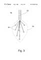
- FIG. 3is a schematic depiction of a catheter including an expandable carrier and a plurality of electrodes according to the present invention.
- the present inventionis of a system and method which enable to simultaneously obtain location data of the body, of a catheter inserted into the body and of an imaging instrument used to image the catheter and the body which can be used to simultaneously obtain location data of the body, of a catheter inserted into the body and of an imaging instrument used to image the catheter and the body.
- the present inventioncan be used to record and display in context of the image the location of the at least one point-of-interest in a body even when the relative location between any of the above locatable items is changed.
- FIGS. 1 and 2illustrate the present invention in a non-limiting fashion.
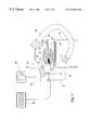
- a system for recording and displaying in context of an image a location of at least one point-of-interest in a body during an intra-body medical procedurewhich system is referred to herein as system 20 .
- System 20includes an imaging instrument 22 for imaging a portion of a body of a patient, indicated by 24 .
- System 20further includes a catheter 26 insertable into in body 24 , e.g., into a cavity 28 present in body 24 .
- locationrefers to a position of a point relative to a reference frame of coordinates, in three-dimension, in at least three degrees of freedom.
- the gist of the present inventionis the ability to determine the relative locations among body 24 , catheter 26 and imaging instrument 22 , such that (i) points-of-interest within body 24 can be presented (highlighted) in context of an image provided by instrument 22 ; (ii) such points-of-interest are presentable in context of images of different projections, obtained by one or more imaging instruments, at one or more time points before or after the logging of a point-of-interest, in other words, such points-of-interest are projectable among all such images and allow a physician to, for example, go back to a point-of-interest logged in or recorder earlier, in context of a plane image no longer presented; (iii) such points-of-interest are recordable in a memory and can be used in following procedures of the same patient performed, for example, in a different time or place; and (iv) in cases where the cavity itself is non-imageable, such as the heart chambers using a fluoroscope, such points of interest can be used to mark some reference cavity coordinates
- the locating systemincludes a locating implement 30 (typically a transmitter or receiver of electromagnetic or acoustic waves and location implement or implements 32 (typically receiver(s) or transmitter(s) of electromagnetic or acoustic waves).
- Implement or implements 32are engaged at one or plurality of locations along catheter 26 , typically close to or at a tip thereof and provide location data in three or more (say four, preferably five, more preferably six) degrees of freedom of catheter 26 with respect to implement 30 .
- Implement 30can be located in a variety of locations. It can be anywhere within an effective distance with respect to implement(s) 32 . As shown in FIG. 1, it can be implemented on imaging instrument 22 .
- catheter 26can be determined in relation to instrument 22 . As shown in FIG. 2, it can be implemented onto an operation platform 34 on which the patient lies during the medical procedure.
- U.S. Pat. No. 5,443,489provides examples for receivers/transmitters which function as hereindescribed.
- a location implement 38is attached to or within body 24 of the patient, such that the location of body 24 with respect to implement 30 is establishable in three or more (say four, preferably five, more preferably six) degrees of freedom. Attaching the location implement according to one embodiment is to one or more reference catheters inserted, for example, during cardiac procedures into the heart cavity of the patient and left unmoved therein, all as further detailed in the Background section above.
- the location of body 24can alternatively be determined by image processing of features in the body image obtained via the imaging instrument using, for example, pattern recognition, edge enhancement, edge detection, shape detection and the like techniques of image recognition or processing.
- These featurescan be imageable markers 44 (e.g., two or more, two are shown in FIGS. 1-2) attached thereto in known positions.
- the location of body 24can be fixed at a known location during the procedure and therefore be known.
- establishing the location of body 24can be synchronized with physiological activity of the body which causes the body or portions thereof to rhythmically move, such as breathing and heart beating.
- the marks and/or location implements employedcan be relocated on the body of the patient in their exact former position by permanently or transiently marking the positions thereof on the body of the patient with, for example, durable ink or tattoo.
- Image processing or recognition techniquesare well known in the art and require no further description herein.
- instrument 22can include a location implement 40 , such that the location of instrument 22 with respect to implement 30 is establishable in three or more (say four, preferably five, more preferably six) degrees of freedom.
- Establishing the location of instrument 22can also be effected according to the present invention by marking catheter 26 with imageable markers 46 combined with data of its own location and image processing.
- Establishing the location of the imaging instrumentcan alternatively be effected by a positioning implement inherent to the imaging instrument.
- a positioning implementinherent to the imaging instrument.
- magnetic resonance imaging systemsinclude such inherent positioning implement.
- Such implementsrecord movements of parts of the instrument relative to a fixed reference coordinate system.
- an additional imaging instrument 52can be employed along with instrument 22 to obtain additional images of body 24 .
- the location of instrument 52is established in a fashion similar to that of instrument 22 , such that points-of-interest can be projected onto such additional images.
- a location implement 40 a similar to implement 40can be employed to establish the location of instrument 52 .
- image processing as described above with respect to instrument 22can be employed for establishing the location of instrument 52 .
- locating implement 30 and any of the above location implements 32 , 38 and/or 40form a locating system selected from the group consisting of electromagnetic locating system, magnetic locating system and acoustic locating system.
- a stereopair optical systemis also applicable.
- the relative locations of the body, catheter inserted therein and the imaging instrumentare established.
- points-of-interest to which the catheter points can be recordedare recorded.
- Such pointscan thereafter be presented in context of an image taken from any orientation, because the orientation is known.
- the catheterby inserting the catheter into a portion of the body of the patient, using the imaging instrument for imaging that portion of the body; establishing a location of the imaging instrument; advancing the catheter (e.g., the tip thereof) to a point-of-interest in the portion of the body and recording a location of that point, so that in course of the procedure, the locations of the body, the catheter and the imaging instrument are known, as well as the magnification mode employed by the imaging, instrument, the point-of-interest is projectable and displayable in a highlighted fashion in context of an image of the portion of the body generated by the imaging instrument even and especially in cases where a relative location of the body and the imaging instrument are changed.
- a method of recording and displaying in context of an image a location of at least one point-of-interest in a body during an intra-body medical procedureis effected by implementing the following method steps, in which, in a first step, the location of the body is established. In a second step of the method, at least one catheter including a location implement is inserted into a portion of the body. In a third step of the method, an imaging instrument is used for imaging the portion of the body. In a fourth step the location of the imaging instrument is established. In a fifth step, the catheter is advanced to a point-of-interest in the portion of the body and via a locating implement a location of the point-of-interest is recorded.
- the point-of-interestis displayed and highlighted in context of an image of the portion of the body, the image is generated by the imaging instrument.
- the locations of the body, the catheter and the imaging instrumentare known, thereby the point-of-interest is projectable and displayable in context of the image of the portion of the body even in cases whereby a relative location of the body and the imaging instrument are changed.
- the catheter according to the present inventioncan be of any type.
- itcan be what is known in the art as probing catheter.
- probing catheterrefers to a catheter equipped with a sensor for sensing biological activities (or geometry e.g., by intravascular or intracardiac ultrasound), such as, for example, electrophysiological activities.
- the catheteris preferably designed to provide a treatment within the body.
- ablatione.g., radio frequency (RF) ablation
- RFradio frequency
- Anotheris the intra-body local application of a drug.
- Ablating catheters, as well as other preferred features used in context of the present invention,are described in U.S. Pat. No.
- the catheterincludes local sensors for sensing local information within the body.
- One exampleinclude electrode sensors to record electric activity within the body.
- Such sensorsas well as other preferred features used in context of the present invention, are described in U.S. Pat. Nos. 5,662,108 and 5,409,000, both are incorporated by reference as if fully set forth herein.
- the catheter according to one embodiment of the present inventionincludes a plurality of flexible longitudinally expanding circumferentially spaced-apart arms adapted to be disposed within a chamber of a heart, to thereby simultaneously record electric activity in a plurality of locations within the heart.
- FIG. 3shows a catheter 70 including a location implement 72 , an expandable carrier 74 implemented at a tip of catheter 70 and a plurality of electrodes 76 carried by carrier 74 .
- the catheterincludes a strain gauge which enables to obtain location information of the tip of the catheter with relation to its body.
- the catheterpreferably further includes a pacemaking ability (a pacemaking electrode).
- a pacemaking abilitya pacemaking electrode.
- Catheters effective in cardiac applications according to the present inventionare distributed by EP Technology, Sunnyvale, Calif., U.S.; Cordis Webster Inc., Miami, Fla., U.S.; Cardiac Pathways Corp., Sunnyvale, Calif., U.S.; and Endocardial Solutions Inc., St. Paul, Minn. U.S.
- the imaging instrumentcan be of any type.
- itcan be a real-time imaging instrument, such as, but not limited to, ultrasound, fluoroscope (X-ray transillumination, e.g., a C-mount fluoroscope) and electrophysiology imaging instrument.
- the imaging instrumentis a non-real-time imaging instrument, such as, but not limited to, computer aided tomography (CT), magnetic resonance imaging (MRI), proton emission tomography (PET) and three dimensional ultrasound (a software therefore is obtainable from EchoTech, Kunststoff, Germany).
- CTcomputer aided tomography
- MRImagnetic resonance imaging
- PETproton emission tomography
- three dimensional ultrasounda software therefore is obtainable from EchoTech, Kunststoff, Germany.
- the imaging instrumentprovides a primary image of a portion of the body of the treated patient.
- primary imagerefers to a 2D image of a 3D tissue, where each picture element is achieved by an integral of some characteristic of the tissue along a line.
- the imaging instrumentprovides a secondary image of said portion of the body.
- secondary imagerefers to an image map of activity of a tissue, such as spatial physiological activity obtained by electro-physiology (EP) mapping achieved with a physiological imaging system, tissue vitality mapping, etc.
- EPelectro-physiology
- the imaging instrumentis adapted for simultaneously generating at least two images each of a different plane.
- Bi-plane fluoroscopes having two spaced apart X ray sourcesare well known in the art, so are multiple plane ultrasound transducers.
- the term “point-of-interest”refers to any point within the body, e.g., a point on an inner side of a heart wall.
- the point-of-interestcan reflect a point featuring local information such as specific type of electric activity.
- the point-of-interestcan reflect a point to which treatment, e.g., ablation treatment, has been applied.
- a point-of-interestcan also be displaced in known displacement magnitude and orientation from another point-of-interest.
- a point-of-interestcan be displaced relative to a point previously treated or a point featuring specific local information previously recorded.
- the points-of-interestare highlighted and displayed on a display 48 .
- each of the points-of-interestis highlighted in a distinctive fashion indicative of its nature or properties.
- Distinctively highlighting points-of-interest according to the present inventioncan involve application of numero-alphabet symbols, shapes, colors, etc. Some or all of the points-of-interest having a common nature or property can be highlighted by a line connecting thereamongst.
- a computer 50receives all the data, for example, via wires 51 (although wireless communication is also applicable), e.g., the image data, the data relating to the locations of the catheter, imaging instrument and the body of the patient, as well as the locations of points-of-interest which are defined by the user by pointing thereon with the catheter and activating a process for their definition as “points-of-interest”, and displays the points-of-interest in context of a present or old image on display 48 .
- Computer 50preferably includes a memory module for receiving and storing in memory the image and/or points-of-interest data for later retrieval. The points-of-interest can be highlighted superimposed on the image in a single display 48 , or alternatively, the points-of-interest and the image can be displayed separately in two different displays.
- Displaying and highlighting the points-of-interest according to the present inventioncan be effected in context of two or more images of the portion of the body. These images are generated by one or more imaging instruments and each can represent a different plane (e.g., orthogonal planes) of the portion of the body. Such images can be displayed simultaneously or independently.
- points-of-interest within the body, pointed at by the cathetercan be logged in and projected onto the image. Furthermore, old points-of-interest can be projected onto a present or later image, even if taken from a different orientation, therefore presenting a different plane of the body, or taken by a different imaging instrument.
- the three dimensional numerical description of any one or more of the points-of-interest according to the present inventionis also displayable.
- the co-localization of the catheter with a displayed point-of-interestcan be made recognizable by a special display effect (e.g., blinking) or sound effect. Automatic steering of the catheter is also envisaged.
- ECGelectrocardiogram
- the ⁇ X,Y,Z ⁇ systemis rotated with respect to the ⁇ K,L,F ⁇ system.
- the rotation operator, Tis a matrix of 3 ⁇ 3 terms which satisfies the orthonormality condition.
- the location implement implemented in the catheteris at ⁇ x,y,z ⁇ as measured in the ⁇ X,Y,Z ⁇ system.
- the location implementis imageable and therefore will be reflected on the image plane of the imaging instrument.
- the location of its reflection thereonis ⁇ k,l,f ⁇ , wherein f is the distance between the radiation source and the image plane, which defines the magnification achieved while imaging.
- [ k l f ][ T 11 T 12 T 13 T 21 T 22 T 23 T 31 T 32 T 33 ] ⁇ [ x y z ] + [ k 0 l 0 f 0 ] ( 1 )
- kf ⁇ T 11 ⁇ x + T 12 ⁇ y + T 13 ⁇ z + k 0 T 31 ⁇ x + T 32 ⁇ y + T 33 ⁇ z + f 0 ( 2 )
- lf ⁇ T 21 ⁇ x + T 22 ⁇ y + T 23 ⁇ z + l 0 T 31 ⁇ x + T 32 ⁇ y + T 33 ⁇ z + f 0 ( 3 )
- the reflection of the tip of the catheteris calculable.
- the location of the imaging instrumentcan be established, as further described hereinabove, via, for example, a location implement.
- fis, for example, measurable using an additional sensor implemented at the imaging plane.
Landscapes
- Health & Medical Sciences (AREA)
- Life Sciences & Earth Sciences (AREA)
- Engineering & Computer Science (AREA)
- Surgery (AREA)
- Veterinary Medicine (AREA)
- General Health & Medical Sciences (AREA)
- Public Health (AREA)
- Biomedical Technology (AREA)
- Heart & Thoracic Surgery (AREA)
- Medical Informatics (AREA)
- Molecular Biology (AREA)
- Animal Behavior & Ethology (AREA)
- Physics & Mathematics (AREA)
- Biophysics (AREA)
- Pathology (AREA)
- Human Computer Interaction (AREA)
- Nuclear Medicine, Radiotherapy & Molecular Imaging (AREA)
- Robotics (AREA)
- Surgical Instruments (AREA)
- Ultra Sonic Daignosis Equipment (AREA)
- Magnetic Resonance Imaging Apparatus (AREA)
- Media Introduction/Drainage Providing Device (AREA)
Abstract
Description


| TABLE | ||
| n | known parameters | required parameter |
| 1 | k, l, x, y, z, T, k0, l0and f0 | f |
| 3 | k, l, x, y, z, k0, l0and f0 | T |
| 4 | k, l, x, y, z and f | T, k0, l0and f0 |
| 5 | k, l, x, y and z | T, k0, l0, f0and f |
Claims (194)
Priority Applications (8)
| Application Number | Priority Date | Filing Date | Title |
|---|---|---|---|
| EP09160580.8AEP2085026B1 (en) | 1998-09-24 | 1999-09-23 | System for Determining the Location of a Catheter during an Intra-Body Medical Procedure |
| EP99946419AEP1115328A4 (en) | 1998-09-24 | 1999-09-23 | System and method for determining the location of a catheter during an intra-body medical procedure |
| AU58825/99AAU5882599A (en) | 1998-09-24 | 1999-09-23 | System and method for determining the location of a catheter during an intra-body medical procedure |
| PCT/IL1999/000512WO2000016684A1 (en) | 1998-09-24 | 1999-09-23 | System and method for determining the location of a catheter during an intra-body medical procedure |
| JP2000573647AJP2002526188A (en) | 1998-09-24 | 1999-09-23 | System and method for determining the position of a catheter during a medical procedure inside the body |
| US09/463,176US6711429B1 (en) | 1998-09-24 | 1999-09-24 | System and method for determining the location of a catheter during an intra-body medical procedure |
| US09/838,238US6558333B2 (en) | 1998-09-24 | 2001-04-20 | System and method of recording and displaying in context of an image a location of at least one point-of-interest in a body during an intra-body medical procedure |
| US11/427,353US20070232896A1 (en) | 1998-09-24 | 2006-06-29 | System and method of recording and displaying in context of an image a location of at least one point-of-interest in a body during an intra-body medical procedure |
Applications Claiming Priority (2)
| Application Number | Priority Date | Filing Date | Title |
|---|---|---|---|
| IL12633398AIL126333A0 (en) | 1998-09-24 | 1998-09-24 | System and method of recording and displaying in context of an image a location of at least one point-of-interest in body during an intra-body medical procedure |
| IL126333 | 1998-09-24 |
Related Child Applications (2)
| Application Number | Title | Priority Date | Filing Date |
|---|---|---|---|
| US09/463,176Continuation-In-PartUS6711429B1 (en) | 1998-09-24 | 1999-09-24 | System and method for determining the location of a catheter during an intra-body medical procedure |
| US09/838,238ContinuationUS6558333B2 (en) | 1998-09-24 | 2001-04-20 | System and method of recording and displaying in context of an image a location of at least one point-of-interest in a body during an intra-body medical procedure |
Publications (1)
| Publication Number | Publication Date |
|---|---|
| US6226543B1true US6226543B1 (en) | 2001-05-01 |
Family
ID=11071990
Family Applications (2)
| Application Number | Title | Priority Date | Filing Date |
|---|---|---|---|
| US09/179,827Expired - LifetimeUS6226543B1 (en) | 1998-09-24 | 1998-10-28 | System and method of recording and displaying in context of an image a location of at least one point-of-interest in a body during an intra-body medical procedure |
| US09/838,238Expired - LifetimeUS6558333B2 (en) | 1998-09-24 | 2001-04-20 | System and method of recording and displaying in context of an image a location of at least one point-of-interest in a body during an intra-body medical procedure |
Family Applications After (1)
| Application Number | Title | Priority Date | Filing Date |
|---|---|---|---|
| US09/838,238Expired - LifetimeUS6558333B2 (en) | 1998-09-24 | 2001-04-20 | System and method of recording and displaying in context of an image a location of at least one point-of-interest in a body during an intra-body medical procedure |
Country Status (3)
| Country | Link |
|---|---|
| US (2) | US6226543B1 (en) |
| EP (1) | EP2085026B1 (en) |
| IL (1) | IL126333A0 (en) |
Cited By (157)
| Publication number | Priority date | Publication date | Assignee | Title |
|---|---|---|---|---|
| WO2002024049A3 (en)* | 2000-09-21 | 2002-06-13 | Super Dimension Ltd | Method and system for archiving medical images |
| US6544178B1 (en)* | 1999-11-05 | 2003-04-08 | Volumetrics Medical Imaging | Methods and systems for volume rendering using ultrasound data |
| US6558333B2 (en)* | 1998-09-24 | 2003-05-06 | Super Dimension Ltd | System and method of recording and displaying in context of an image a location of at least one point-of-interest in a body during an intra-body medical procedure |
| US20030088178A1 (en)* | 2001-11-02 | 2003-05-08 | Owens Timothy R | Method and apparatus for computer modified magnetic resonance imaging |
| US20030092995A1 (en)* | 2001-11-13 | 2003-05-15 | Medtronic, Inc. | System and method of positioning implantable medical devices |
| US20030135115A1 (en)* | 1997-11-24 | 2003-07-17 | Burdette Everette C. | Method and apparatus for spatial registration and mapping of a biopsy needle during a tissue biopsy |
| US20030135102A1 (en)* | 2000-05-18 | 2003-07-17 | Burdette Everette C. | Method and system for registration and guidance of intravascular treatment |
| US20030158477A1 (en)* | 2001-11-09 | 2003-08-21 | Dorin Panescu | Systems and methods for guiding catheters using registered images |
| US20030176778A1 (en)* | 2002-03-15 | 2003-09-18 | Scimed Life Systems, Inc. | Medical device control systems |
| US6640126B2 (en)* | 2001-02-26 | 2003-10-28 | Toshiba America Mri, Inc. | Acoustic gating monitor for magnetic resonance imaging system |
| WO2003092488A1 (en)* | 2002-05-02 | 2003-11-13 | Philips Intellectual Property & Standards Gmbh | Method for transcutaneous catheter guiding |
| US20040022438A1 (en)* | 2002-08-02 | 2004-02-05 | Hibbard Lyndon S. | Method and apparatus for image segmentation using Jensen-Shannon divergence and Jensen-Renyi divergence |
| US20040034300A1 (en)* | 2002-08-19 | 2004-02-19 | Laurent Verard | Method and apparatus for virtual endoscopy |
| US6711429B1 (en)* | 1998-09-24 | 2004-03-23 | Super Dimension Ltd. | System and method for determining the location of a catheter during an intra-body medical procedure |
| WO2004036230A3 (en)* | 2002-10-21 | 2004-06-10 | Bbms Ltd | Method and apparatus for magnetic resonance analysis |
| US20050038337A1 (en)* | 2003-08-11 | 2005-02-17 | Edwards Jerome R. | Methods, apparatuses, and systems useful in conducting image guided interventions |
| US20050090746A1 (en)* | 2003-10-14 | 2005-04-28 | Aloka Co., Ltd. | Ultrasound diagnosis apparatus |
| US6895267B2 (en) | 2001-10-24 | 2005-05-17 | Scimed Life Systems, Inc. | Systems and methods for guiding and locating functional elements on medical devices positioned in a body |
| US20050103333A1 (en)* | 2000-12-02 | 2005-05-19 | Bonutti Peter M. | Medical device positioning system and method |
| US20050154282A1 (en)* | 2003-12-31 | 2005-07-14 | Wenguang Li | System and method for registering an image with a representation of a probe |
| US20050154285A1 (en)* | 2004-01-02 | 2005-07-14 | Neason Curtis G. | System and method for receiving and displaying information pertaining to a patient |
| US20050154279A1 (en)* | 2003-12-31 | 2005-07-14 | Wenguang Li | System and method for registering an image with a representation of a probe |
| US20050154286A1 (en)* | 2004-01-02 | 2005-07-14 | Neason Curtis G. | System and method for receiving and displaying information pertaining to a patient |
| US20050154281A1 (en)* | 2003-12-31 | 2005-07-14 | Xue Joel Q. | System and method for registering an image with a representation of a probe |
| US20050182316A1 (en)* | 2002-08-29 | 2005-08-18 | Burdette Everette C. | Method and system for localizing a medical tool |
| US20050182319A1 (en)* | 2004-02-17 | 2005-08-18 | Glossop Neil D. | Method and apparatus for registration, verification, and referencing of internal organs |
| US20050209524A1 (en)* | 2004-03-10 | 2005-09-22 | General Electric Company | System and method for receiving and storing information pertaining to a patient |
| US20050222554A1 (en)* | 2004-03-05 | 2005-10-06 | Wallace Daniel T | Robotic catheter system |
| US20050222509A1 (en)* | 2004-04-02 | 2005-10-06 | General Electric Company | Electrophysiology system and method |
| US20050228252A1 (en)* | 2004-04-02 | 2005-10-13 | General Electric Company | Electrophysiology system and method |
| US20050228251A1 (en)* | 2004-03-30 | 2005-10-13 | General Electric Company | System and method for displaying a three-dimensional image of an organ or structure inside the body |
| US20050288586A1 (en)* | 2004-06-28 | 2005-12-29 | Bozidar Ferek-Petric | Electrode location mapping system and method |
| US6996430B1 (en)* | 1999-08-16 | 2006-02-07 | Super Dimension Ltd | Method and system for displaying cross-sectional images of a body |
| US20060057560A1 (en)* | 2004-03-05 | 2006-03-16 | Hansen Medical, Inc. | System and method for denaturing and fixing collagenous tissue |
| US20060122497A1 (en)* | 2004-11-12 | 2006-06-08 | Glossop Neil D | Device and method for ensuring the accuracy of a tracking device in a volume |
| US20060173291A1 (en)* | 2005-01-18 | 2006-08-03 | Glossop Neil D | Electromagnetically tracked K-wire device |
| US20060173269A1 (en)* | 2004-11-12 | 2006-08-03 | Glossop Neil D | Integrated skin-mounted multifunction device for use in image-guided surgery |
| US20060184016A1 (en)* | 2005-01-18 | 2006-08-17 | Glossop Neil D | Method and apparatus for guiding an instrument to a target in the lung |
| US20060247683A1 (en)* | 2005-04-21 | 2006-11-02 | Asthmatx, Inc. | Control systems for delivering energy |
| US20060258947A1 (en)* | 2005-04-25 | 2006-11-16 | Charles Olson | Display for ECG diagnostics |
| US20060262631A1 (en)* | 2004-11-12 | 2006-11-23 | Samsung Electronics Co., Ltd. | Bank selection signal control circuit for use in semiconductor memory device, and bank selection control method |
| US20070000257A1 (en)* | 2003-03-26 | 2007-01-04 | Aisin Seiki Kabushiki Kaisha | Pulse tube refrigerating machine |
| US20070032723A1 (en)* | 2005-06-21 | 2007-02-08 | Glossop Neil D | System, method and apparatus for navigated therapy and diagnosis |
| US20070055128A1 (en)* | 2005-08-24 | 2007-03-08 | Glossop Neil D | System, method and devices for navigated flexible endoscopy |
| US20070066881A1 (en)* | 2005-09-13 | 2007-03-22 | Edwards Jerome R | Apparatus and method for image guided accuracy verification |
| US20070167787A1 (en)* | 2005-06-21 | 2007-07-19 | Glossop Neil D | Device and method for a trackable ultrasound |
| US20070299468A1 (en)* | 2006-06-22 | 2007-12-27 | Viola Frank J | Tissue vitality comparator with light pipe with fiber optic imaging bundle |
| US20080009760A1 (en)* | 2006-06-30 | 2008-01-10 | Broncus Technologies, Inc. | Airway bypass site selection and treatment planning |
| US20080009674A1 (en)* | 2006-02-24 | 2008-01-10 | Visionsense Ltd. | Method and system for navigating within a flexible organ of the body of a patient |
| US20080021297A1 (en)* | 2004-02-10 | 2008-01-24 | Koninklijke Philips Electronic, N.V. | Method,a System for Generating a Spatial Roadmap for an Interventional Device and Quality Control System for Guarding the Spatial Accuracy Thereof |
| US20080071215A1 (en)* | 2004-11-05 | 2008-03-20 | Traxtal Technologies Inc. | Access System |
| US20080118116A1 (en)* | 2006-11-20 | 2008-05-22 | General Electric Company | Systems and methods for tracking a surgical instrument and for conveying tracking information via a network |
| US20080118135A1 (en)* | 2006-11-10 | 2008-05-22 | Superdimension, Ltd. | Adaptive Navigation Technique For Navigating A Catheter Through A Body Channel Or Cavity |
| US20080132757A1 (en)* | 2006-12-01 | 2008-06-05 | General Electric Company | System and Method for Performing Minimally Invasive Surgery Using a Multi-Channel Catheter |
| US20080139929A1 (en)* | 2006-12-06 | 2008-06-12 | General Electric Company | System and method for tracking an invasive surgical instrument while imaging a patient |
| US7438685B2 (en) | 2001-11-05 | 2008-10-21 | Computerized Medical Systems, Inc. | Apparatus and method for registration, guidance and targeting of external beam radiation therapy |
| US20090134334A1 (en)* | 2005-04-01 | 2009-05-28 | San Diego State University Research Foundation | Edge-on sar scintillator devices and systems for enhanced spect, pet, and compton gamma cameras |
| US20090171187A1 (en)* | 2007-12-26 | 2009-07-02 | Gerhart John P | Catheter electrode that can simultaneously emit electrical energy and facilitate visualization by magnetic resonance imaging |
| US20090171188A1 (en)* | 2007-12-28 | 2009-07-02 | Saurav Paul | Flexible polymer electrode for mri-guided positioning and radio frequency ablation |
| US7599730B2 (en) | 2002-11-19 | 2009-10-06 | Medtronic Navigation, Inc. | Navigation system for cardiac therapies |
| US20090281566A1 (en)* | 2003-08-11 | 2009-11-12 | Edwards Jerome R | Bodily sealants and methods and apparatus for image-guided delivery of same |
| WO2009147671A1 (en) | 2008-06-03 | 2009-12-10 | Superdimension Ltd. | Feature-based registration method |
| US20100008555A1 (en)* | 2008-05-15 | 2010-01-14 | Superdimension, Ltd. | Automatic Pathway And Waypoint Generation And Navigation Method |
| US20100034449A1 (en)* | 2008-06-06 | 2010-02-11 | Superdimension, Ltd. | Hybrid Registration Method |
| US20100041949A1 (en)* | 2007-03-12 | 2010-02-18 | David Tolkowsky | Devices and methods for performing medical procedures in tree-like luminal structures |
| US7697972B2 (en) | 2002-11-19 | 2010-04-13 | Medtronic Navigation, Inc. | Navigation system for cardiac therapies |
| WO2010071895A1 (en) | 2008-12-19 | 2010-06-24 | Superdimension, Ltd. | Navigable tissue treatment tools |
| US20100228257A1 (en)* | 2000-01-14 | 2010-09-09 | Bonutti Peter M | Joint replacement component |
| US20100249622A1 (en)* | 2005-04-25 | 2010-09-30 | Charles Olson | Location and displaying an ischemic region for ecg diagnostics |
| EP2253287A2 (en) | 2009-05-14 | 2010-11-24 | superDimension Ltd. | Automatic registration technique |
| WO2011027107A1 (en)* | 2009-09-01 | 2011-03-10 | Ucl Business Plc | Apparatus and method for determining a location in a target image |
| US20110130649A1 (en)* | 2003-01-13 | 2011-06-02 | Gera Strommer | Method and system for registering a medical situation associated with a first coordinate system, in a second coordinate system using an mps system |
| EP2329786A2 (en) | 2009-10-01 | 2011-06-08 | Navotek Medical Ltd. | Guided surgery |
| US7998062B2 (en) | 2004-03-29 | 2011-08-16 | Superdimension, Ltd. | Endoscope structures and techniques for navigating to a target in branched structure |
| US20110206253A1 (en)* | 2010-02-01 | 2011-08-25 | Superdimension, Ltd. | Region-Growing Algorithm |
| EP2401956A1 (en) | 2008-07-10 | 2012-01-04 | Superdimension Ltd. | Integrated multi-functional endoscopic tool |
| US8175681B2 (en) | 2008-12-16 | 2012-05-08 | Medtronic Navigation Inc. | Combination of electromagnetic and electropotential localization |
| US8333204B2 (en) | 1999-06-25 | 2012-12-18 | Hansen Medical, Inc. | Apparatus and methods for treating tissue |
| US8364242B2 (en) | 2007-05-17 | 2013-01-29 | General Electric Company | System and method of combining ultrasound image acquisition with fluoroscopic image acquisition |
| US8428690B2 (en) | 2007-05-16 | 2013-04-23 | General Electric Company | Intracardiac echocardiography image reconstruction in combination with position tracking system |
| US8489192B1 (en) | 2008-02-15 | 2013-07-16 | Holaira, Inc. | System and method for bronchial dilation |
| US8494614B2 (en) | 2009-08-31 | 2013-07-23 | Regents Of The University Of Minnesota | Combination localization system |
| US8494613B2 (en) | 2009-08-31 | 2013-07-23 | Medtronic, Inc. | Combination localization system |
| US8527032B2 (en) | 2007-05-16 | 2013-09-03 | General Electric Company | Imaging system and method of delivery of an instrument to an imaged subject |
| US8608724B2 (en) | 2004-07-19 | 2013-12-17 | Broncus Medical Inc. | Devices for delivering substances through an extra-anatomic opening created in an airway |
| US8611984B2 (en) | 2009-04-08 | 2013-12-17 | Covidien Lp | Locatable catheter |
| US8623030B2 (en) | 2001-08-28 | 2014-01-07 | Bonutti Skeletal Innovations Llc | Robotic arthroplasty system including navigation |
| US8663088B2 (en) | 2003-09-15 | 2014-03-04 | Covidien Lp | System of accessories for use with bronchoscopes |
| US8696549B2 (en) | 2010-08-20 | 2014-04-15 | Veran Medical Technologies, Inc. | Apparatus and method for four dimensional soft tissue navigation in endoscopic applications |
| US8709034B2 (en) | 2011-05-13 | 2014-04-29 | Broncus Medical Inc. | Methods and devices for diagnosing, monitoring, or treating medical conditions through an opening through an airway wall |
| US8740895B2 (en) | 2009-10-27 | 2014-06-03 | Holaira, Inc. | Delivery devices with coolable energy emitting assemblies |
| US8764725B2 (en) | 2004-02-09 | 2014-07-01 | Covidien Lp | Directional anchoring mechanism, method and applications thereof |
| US8781186B2 (en) | 2010-05-04 | 2014-07-15 | Pathfinder Therapeutics, Inc. | System and method for abdominal surface matching using pseudo-features |
| US20140228858A1 (en)* | 2013-02-08 | 2014-08-14 | Covidien Lp | Methods for lung denervation |
| US8808280B2 (en) | 2008-05-09 | 2014-08-19 | Holaira, Inc. | Systems, assemblies, and methods for treating a bronchial tree |
| US8905920B2 (en) | 2007-09-27 | 2014-12-09 | Covidien Lp | Bronchoscope adapter and method |
| US8911439B2 (en) | 2009-11-11 | 2014-12-16 | Holaira, Inc. | Non-invasive and minimally invasive denervation methods and systems for performing the same |
| US8989842B2 (en) | 2007-05-16 | 2015-03-24 | General Electric Company | System and method to register a tracking system with intracardiac echocardiography (ICE) imaging system |
| US9014851B2 (en) | 2013-03-15 | 2015-04-21 | Hansen Medical, Inc. | Systems and methods for tracking robotically controlled medical instruments |
| US9055881B2 (en) | 2004-04-26 | 2015-06-16 | Super Dimension Ltd. | System and method for image-based alignment of an endoscope |
| US9057600B2 (en) | 2013-03-13 | 2015-06-16 | Hansen Medical, Inc. | Reducing incremental measurement sensor error |
| US9138166B2 (en) | 2011-07-29 | 2015-09-22 | Hansen Medical, Inc. | Apparatus and methods for fiber integration and registration |
| US9138165B2 (en) | 2012-02-22 | 2015-09-22 | Veran Medical Technologies, Inc. | Systems, methods and devices for forming respiratory-gated point cloud for four dimensional soft tissue navigation |
| US9149328B2 (en) | 2009-11-11 | 2015-10-06 | Holaira, Inc. | Systems, apparatuses, and methods for treating tissue and controlling stenosis |
| US9173713B2 (en) | 2013-03-14 | 2015-11-03 | Hansen Medical, Inc. | Torque-based catheter articulation |
| US9271663B2 (en) | 2013-03-15 | 2016-03-01 | Hansen Medical, Inc. | Flexible instrument localization from both remote and elongation sensors |
| US9283046B2 (en) | 2013-03-15 | 2016-03-15 | Hansen Medical, Inc. | User interface for active drive apparatus with finite range of motion |
| US9326822B2 (en) | 2013-03-14 | 2016-05-03 | Hansen Medical, Inc. | Active drives for robotic catheter manipulators |
| US9339618B2 (en) | 2003-05-13 | 2016-05-17 | Holaira, Inc. | Method and apparatus for controlling narrowing of at least one airway |
| US9345532B2 (en) | 2011-05-13 | 2016-05-24 | Broncus Medical Inc. | Methods and devices for ablation of tissue |
| US9358076B2 (en) | 2011-01-20 | 2016-06-07 | Hansen Medical, Inc. | System and method for endoluminal and translumenal therapy |
| US9398933B2 (en) | 2012-12-27 | 2016-07-26 | Holaira, Inc. | Methods for improving drug efficacy including a combination of drug administration and nerve modulation |
| US9408669B2 (en) | 2013-03-15 | 2016-08-09 | Hansen Medical, Inc. | Active drive mechanism with finite range of motion |
| US9452018B2 (en) | 2013-03-15 | 2016-09-27 | Hansen Medical, Inc. | Rotational support for an elongate member |
| US9457168B2 (en) | 2005-07-01 | 2016-10-04 | Hansen Medical, Inc. | Robotic catheter system and methods |
| US9498601B2 (en) | 2013-03-14 | 2016-11-22 | Hansen Medical, Inc. | Catheter tension sensing |
| US9498291B2 (en) | 2013-03-15 | 2016-11-22 | Hansen Medical, Inc. | Touch-free catheter user interface controller |
| US9532840B2 (en) | 2013-03-08 | 2017-01-03 | Hansen Medical, Inc. | Slider control of catheters and wires |
| US9533128B2 (en) | 2003-07-18 | 2017-01-03 | Broncus Medical Inc. | Devices for maintaining patency of surgically created channels in tissue |
| US9566414B2 (en) | 2013-03-13 | 2017-02-14 | Hansen Medical, Inc. | Integrated catheter and guide wire controller |
| US9576107B2 (en) | 2013-07-09 | 2017-02-21 | Biosense Webster (Israel) Ltd. | Model based reconstruction of the heart from sparse samples |
| US9575140B2 (en) | 2008-04-03 | 2017-02-21 | Covidien Lp | Magnetic interference detection system and method |
| US9629595B2 (en) | 2013-03-15 | 2017-04-25 | Hansen Medical, Inc. | Systems and methods for localizing, tracking and/or controlling medical instruments |
| US9668814B2 (en) | 2013-03-07 | 2017-06-06 | Hansen Medical, Inc. | Infinitely rotatable tool with finite rotating drive shafts |
| US10046140B2 (en) | 2014-04-21 | 2018-08-14 | Hansen Medical, Inc. | Devices, systems, and methods for controlling active drive systems |
| US10123843B2 (en) | 2013-03-15 | 2018-11-13 | Auris Health, Inc. | Input device for controlling a catheter |
| US10272260B2 (en) | 2011-11-23 | 2019-04-30 | Broncus Medical Inc. | Methods and devices for diagnosing, monitoring, or treating medical conditions through an opening through an airway wall |
| US10363103B2 (en) | 2009-04-29 | 2019-07-30 | Auris Health, Inc. | Flexible and steerable elongate instruments with shape control and support elements |
| US10376672B2 (en) | 2013-03-15 | 2019-08-13 | Auris Health, Inc. | Catheter insertion system and method of fabrication |
| US10418705B2 (en) | 2016-10-28 | 2019-09-17 | Covidien Lp | Electromagnetic navigation antenna assembly and electromagnetic navigation system including the same |
| US10426555B2 (en) | 2015-06-03 | 2019-10-01 | Covidien Lp | Medical instrument with sensor for use in a system and method for electromagnetic navigation |
| US10446931B2 (en) | 2016-10-28 | 2019-10-15 | Covidien Lp | Electromagnetic navigation antenna assembly and electromagnetic navigation system including the same |
| US10463439B2 (en) | 2016-08-26 | 2019-11-05 | Auris Health, Inc. | Steerable catheter with shaft load distributions |
| US10478254B2 (en) | 2016-05-16 | 2019-11-19 | Covidien Lp | System and method to access lung tissue |
| US10517505B2 (en) | 2016-10-28 | 2019-12-31 | Covidien Lp | Systems, methods, and computer-readable media for optimizing an electromagnetic navigation system |
| US10524867B2 (en) | 2013-03-15 | 2020-01-07 | Auris Health, Inc. | Active drive mechanism for simultaneous rotation and translation |
| US10543047B2 (en) | 2013-03-15 | 2020-01-28 | Auris Health, Inc. | Remote catheter manipulator |
| US10556092B2 (en) | 2013-03-14 | 2020-02-11 | Auris Health, Inc. | Active drives for robotic catheter manipulators |
| US10583271B2 (en) | 2012-11-28 | 2020-03-10 | Auris Health, Inc. | Method of anchoring pullwire directly articulatable region in catheter |
| US10582834B2 (en) | 2010-06-15 | 2020-03-10 | Covidien Lp | Locatable expandable working channel and method |
| US10615500B2 (en) | 2016-10-28 | 2020-04-07 | Covidien Lp | System and method for designing electromagnetic navigation antenna assemblies |
| US10617324B2 (en) | 2014-04-23 | 2020-04-14 | Veran Medical Technologies, Inc | Apparatuses and methods for endobronchial navigation to and confirmation of the location of a target tissue and percutaneous interception of the target tissue |
| US10624701B2 (en) | 2014-04-23 | 2020-04-21 | Veran Medical Technologies, Inc. | Apparatuses and methods for registering a real-time image feed from an imaging device to a steerable catheter |
| US10631797B2 (en) | 2011-07-22 | 2020-04-28 | Canon Medical Systems Corporation | X-ray diagnosis apparatus and control method |
| US10638952B2 (en) | 2016-10-28 | 2020-05-05 | Covidien Lp | Methods, systems, and computer-readable media for calibrating an electromagnetic navigation system |
| US10722311B2 (en) | 2016-10-28 | 2020-07-28 | Covidien Lp | System and method for identifying a location and/or an orientation of an electromagnetic sensor based on a map |
| US10751126B2 (en) | 2016-10-28 | 2020-08-25 | Covidien Lp | System and method for generating a map for electromagnetic navigation |
| US10792106B2 (en) | 2016-10-28 | 2020-10-06 | Covidien Lp | System for calibrating an electromagnetic navigation system |
| US10849702B2 (en) | 2013-03-15 | 2020-12-01 | Auris Health, Inc. | User input devices for controlling manipulation of guidewires and catheters |
| US10925628B2 (en) | 2017-09-18 | 2021-02-23 | Novuson Surgical, Inc. | Tissue engagement apparatus for theapeutic ultrasound apparatus and method |
| US10952593B2 (en) | 2014-06-10 | 2021-03-23 | Covidien Lp | Bronchoscope adapter |
| US11213363B2 (en) | 2013-03-14 | 2022-01-04 | Auris Health, Inc. | Catheter tension sensing |
| US11219489B2 (en) | 2017-10-31 | 2022-01-11 | Covidien Lp | Devices and systems for providing sensors in parallel with medical tools |
| US11241559B2 (en) | 2016-08-29 | 2022-02-08 | Auris Health, Inc. | Active drive for guidewire manipulation |
| US11304629B2 (en) | 2005-09-13 | 2022-04-19 | Veran Medical Technologies, Inc. | Apparatus and method for image guided accuracy verification |
| US12089902B2 (en) | 2019-07-30 | 2024-09-17 | Coviden Lp | Cone beam and 3D fluoroscope lung navigation |
| US12369884B2 (en) | 2021-03-04 | 2025-07-29 | Covidien Lp | Endoluminal shafts including ultrasound coupling capability |
Families Citing this family (48)
| Publication number | Priority date | Publication date | Assignee | Title |
|---|---|---|---|---|
| US8251070B2 (en) | 2000-03-27 | 2012-08-28 | Asthmatx, Inc. | Methods for treating airways |
| US7627145B2 (en)* | 2000-09-06 | 2009-12-01 | Hitachi, Ltd. | Personal identification device and method |
| US6755790B2 (en)* | 2002-10-14 | 2004-06-29 | Medtronic, Inc. | Transseptal access tissue thickness sensing dilator devices and methods for fabricating and using same |
| WO2005063125A1 (en)* | 2003-12-22 | 2005-07-14 | Koninklijke Philips Electronics N.V. | System for guiding a medical instrument in a patient body |
| US20080234570A1 (en)* | 2004-03-05 | 2008-09-25 | Koninklijke Philips Electronics, N.V. | System For Guiding a Medical Instrument in a Patient Body |
| JP4681602B2 (en)* | 2004-03-29 | 2011-05-11 | ケーシーアイ ライセンシング インコーポレイテッド | Method and apparatus for controlling at least one ventilation parameter of an artificial ventilator that ventilates a patient's lungs according to a plurality of lung positions |
| US20050267453A1 (en)* | 2004-05-27 | 2005-12-01 | Wong Serena H | High intensity focused ultrasound for imaging and treatment of arrhythmias |
| US8409282B2 (en) | 2004-10-20 | 2013-04-02 | Vertiflex, Inc. | Systems and methods for posterior dynamic stabilization of the spine |
| US8123807B2 (en) | 2004-10-20 | 2012-02-28 | Vertiflex, Inc. | Systems and methods for posterior dynamic stabilization of the spine |
| US8123782B2 (en)* | 2004-10-20 | 2012-02-28 | Vertiflex, Inc. | Interspinous spacer |
| US8277488B2 (en) | 2004-10-20 | 2012-10-02 | Vertiflex, Inc. | Interspinous spacer |
| US8152837B2 (en)* | 2004-10-20 | 2012-04-10 | The Board Of Trustees Of The Leland Stanford Junior University | Systems and methods for posterior dynamic stabilization of the spine |
| US9119680B2 (en) | 2004-10-20 | 2015-09-01 | Vertiflex, Inc. | Interspinous spacer |
| US8317864B2 (en) | 2004-10-20 | 2012-11-27 | The Board Of Trustees Of The Leland Stanford Junior University | Systems and methods for posterior dynamic stabilization of the spine |
| US8128662B2 (en) | 2004-10-20 | 2012-03-06 | Vertiflex, Inc. | Minimally invasive tooling for delivery of interspinous spacer |
| US7763074B2 (en)* | 2004-10-20 | 2010-07-27 | The Board Of Trustees Of The Leland Stanford Junior University | Systems and methods for posterior dynamic stabilization of the spine |
| US9023084B2 (en) | 2004-10-20 | 2015-05-05 | The Board Of Trustees Of The Leland Stanford Junior University | Systems and methods for stabilizing the motion or adjusting the position of the spine |
| US9161783B2 (en) | 2004-10-20 | 2015-10-20 | Vertiflex, Inc. | Interspinous spacer |
| US8012207B2 (en)* | 2004-10-20 | 2011-09-06 | Vertiflex, Inc. | Systems and methods for posterior dynamic stabilization of the spine |
| US8945183B2 (en) | 2004-10-20 | 2015-02-03 | Vertiflex, Inc. | Interspinous process spacer instrument system with deployment indicator |
| US8167944B2 (en) | 2004-10-20 | 2012-05-01 | The Board Of Trustees Of The Leland Stanford Junior University | Systems and methods for posterior dynamic stabilization of the spine |
| US8425559B2 (en) | 2004-10-20 | 2013-04-23 | Vertiflex, Inc. | Systems and methods for posterior dynamic stabilization of the spine |
| US8273108B2 (en) | 2004-10-20 | 2012-09-25 | Vertiflex, Inc. | Interspinous spacer |
| US8613747B2 (en) | 2004-10-20 | 2013-12-24 | Vertiflex, Inc. | Spacer insertion instrument |
| EP2219538B1 (en) | 2004-12-06 | 2022-07-06 | Vertiflex, Inc. | Spacer insertion instrument |
| US8845726B2 (en) | 2006-10-18 | 2014-09-30 | Vertiflex, Inc. | Dilator |
| AU2008241447B2 (en) | 2007-04-16 | 2014-03-27 | Vertiflex, Inc. | Interspinous spacer |
| AU2009206098B2 (en) | 2008-01-15 | 2014-10-30 | Vertiflex, Inc. | Interspinous spacer |
| US8219179B2 (en) | 2008-03-06 | 2012-07-10 | Vida Diagnostics, Inc. | Systems and methods for navigation within a branched structure of a body |
| US10039527B2 (en) | 2009-05-20 | 2018-08-07 | Analogic Canada Corporation | Ultrasound systems incorporating spatial position sensors and associated methods |
| US8556815B2 (en)* | 2009-05-20 | 2013-10-15 | Laurent Pelissier | Freehand ultrasound imaging systems and methods for guiding fine elongate instruments |
| WO2010144402A2 (en) | 2009-06-08 | 2010-12-16 | Surgivision, Inc. | Mri-guided surgical systems with preset scan planes |
| JP5795576B2 (en) | 2009-06-12 | 2015-10-14 | バード・アクセス・システムズ,インコーポレーテッド | Method of operating a computer-based medical device that uses an electrocardiogram (ECG) signal to position an intravascular device in or near the heart |
| CN102625670B (en) | 2009-06-16 | 2015-07-15 | 核磁共振成像介入技术有限公司 | MRI-guided devices and MRI-guided interventional systems that can track and generate dynamic visualizations of the devices in near real time |
| US8740948B2 (en) | 2009-12-15 | 2014-06-03 | Vertiflex, Inc. | Spinal spacer for cervical and other vertebra, and associated systems and methods |
| US9295449B2 (en) | 2012-01-23 | 2016-03-29 | Ultrasonix Medical Corporation | Landmarks for ultrasound imaging |
| US8900225B2 (en) | 2012-05-07 | 2014-12-02 | Biosense Webster (Israel) Ltd. | Automatic ablation tracking |
| US10441236B2 (en)† | 2012-10-19 | 2019-10-15 | Biosense Webster (Israel) Ltd. | Integration between 3D maps and fluoroscopic images |
| US9675303B2 (en) | 2013-03-15 | 2017-06-13 | Vertiflex, Inc. | Visualization systems, instruments and methods of using the same in spinal decompression procedures |
| US9326702B2 (en) | 2013-03-15 | 2016-05-03 | Mediguide Ltd. | Medical device navigation system |
| AU2015256024B2 (en) | 2014-05-07 | 2020-03-05 | Vertiflex, Inc. | Spinal nerve decompression systems, dilation systems, and methods of using the same |
| WO2019198061A1 (en) | 2018-04-13 | 2019-10-17 | Universidade Do Minho | Guidance system, method and devices thereof |
| WO2020097425A2 (en) | 2018-11-09 | 2020-05-14 | Vida Diagnostics, Inc. | Cut-surface display of tubular structures |
| WO2021207289A1 (en) | 2020-04-07 | 2021-10-14 | Vida Diagnostics, Inc. | Subject specific coordinatization and virtual navigation systems and methods |
| US12266111B2 (en) | 2020-11-27 | 2025-04-01 | Vida Diagnostics, Inc. | Visualization of sub-pleural regions |
| WO2023158581A1 (en) | 2022-02-15 | 2023-08-24 | Boston Scientific Neuromodulation Corporation | Interspinous spacer and systems utilizing the interspinous spacer |
| US12433646B2 (en) | 2023-02-21 | 2025-10-07 | Boston Scientific Neuromodulation Corporation | Interspinous spacer with actuator locking arrangements and methods and systems |
| US12390340B2 (en) | 2023-03-15 | 2025-08-19 | Boston Scientific Neuromodulation Corporation | Interspinous spacer with a range of deployment positions and methods and systems |
Citations (14)
| Publication number | Priority date | Publication date | Assignee | Title |
|---|---|---|---|---|
| US5398684A (en)* | 1988-12-23 | 1995-03-21 | Hardy; Tyrone L. | Method and apparatus for video presentation from scanner imaging sources |
| US5409000A (en) | 1993-09-14 | 1995-04-25 | Cardiac Pathways Corporation | Endocardial mapping and ablation system utilizing separately controlled steerable ablation catheter with ultrasonic imaging capabilities and method |
| US5443489A (en) | 1993-07-20 | 1995-08-22 | Biosense, Inc. | Apparatus and method for ablation |
| US5485849A (en)* | 1994-01-31 | 1996-01-23 | Ep Technologies, Inc. | System and methods for matching electrical characteristics and propagation velocities in cardiac tissue |
| WO1997025101A2 (en) | 1996-01-08 | 1997-07-17 | Biosense Inc. | Methods and apparatus for myocardial revascularization |
| US5651047A (en)* | 1993-01-25 | 1997-07-22 | Cardiac Mariners, Incorporated | Maneuverable and locateable catheters |
| WO1997029685A1 (en) | 1996-02-15 | 1997-08-21 | Biosense, Inc. | Independently positionable transducers for location system |
| WO1997029701A1 (en) | 1996-02-15 | 1997-08-21 | Biosense Inc. | Catheter based surgery |
| WO1997029682A1 (en) | 1996-02-15 | 1997-08-21 | Biosense Inc. | Locatable biopsy needle |
| US5662108A (en) | 1992-09-23 | 1997-09-02 | Endocardial Solutions, Inc. | Electrophysiology mapping system |
| WO1998011840A1 (en) | 1996-09-17 | 1998-03-26 | Biosense Inc. | Position confirmation with learn and test functions |
| WO1998035720A2 (en) | 1997-02-14 | 1998-08-20 | Biosense Inc. | X-ray guided surgical location system with extended mapping volume |
| US5803084A (en)* | 1996-12-05 | 1998-09-08 | Olson; Charles | Three dimensional vector cardiographic display and method for displaying same |
| US6019728A (en)* | 1996-05-08 | 2000-02-01 | Kabushiki Kaisha Tokai Rika Denki Seisakusho | Catheter and sensor having pressure detecting function |
Family Cites Families (7)
| Publication number | Priority date | Publication date | Assignee | Title |
|---|---|---|---|---|
| US4697595A (en) | 1984-07-24 | 1987-10-06 | Telectronics N.V. | Ultrasonically marked cardiac catheters |
| EP0419729A1 (en) | 1989-09-29 | 1991-04-03 | Siemens Aktiengesellschaft | Position finding of a catheter by means of non-ionising fields |
| US5738096A (en) | 1993-07-20 | 1998-04-14 | Biosense, Inc. | Cardiac electromechanics |
| AU709081B2 (en)* | 1996-02-15 | 1999-08-19 | Biosense, Inc. | Medical procedures and apparatus using intrabody probes |
| US5944022A (en)* | 1997-04-28 | 1999-08-31 | American Cardiac Ablation Co. Inc. | Catheter positioning system |
| US6086532A (en)* | 1997-09-26 | 2000-07-11 | Ep Technologies, Inc. | Systems for recording use of structures deployed in association with heart tissue |
| IL126333A0 (en)* | 1998-09-24 | 1999-05-09 | Super Dimension Ltd | System and method of recording and displaying in context of an image a location of at least one point-of-interest in body during an intra-body medical procedure |
- 1998
- 1998-09-24ILIL12633398Apatent/IL126333A0/enunknown
- 1998-10-28USUS09/179,827patent/US6226543B1/ennot_activeExpired - Lifetime
- 1999
- 1999-09-23EPEP09160580.8Apatent/EP2085026B1/ennot_activeExpired - Lifetime
- 2001
- 2001-04-20USUS09/838,238patent/US6558333B2/ennot_activeExpired - Lifetime
Patent Citations (14)
| Publication number | Priority date | Publication date | Assignee | Title |
|---|---|---|---|---|
| US5398684A (en)* | 1988-12-23 | 1995-03-21 | Hardy; Tyrone L. | Method and apparatus for video presentation from scanner imaging sources |
| US5662108A (en) | 1992-09-23 | 1997-09-02 | Endocardial Solutions, Inc. | Electrophysiology mapping system |
| US5651047A (en)* | 1993-01-25 | 1997-07-22 | Cardiac Mariners, Incorporated | Maneuverable and locateable catheters |
| US5443489A (en) | 1993-07-20 | 1995-08-22 | Biosense, Inc. | Apparatus and method for ablation |
| US5409000A (en) | 1993-09-14 | 1995-04-25 | Cardiac Pathways Corporation | Endocardial mapping and ablation system utilizing separately controlled steerable ablation catheter with ultrasonic imaging capabilities and method |
| US5485849A (en)* | 1994-01-31 | 1996-01-23 | Ep Technologies, Inc. | System and methods for matching electrical characteristics and propagation velocities in cardiac tissue |
| WO1997025101A2 (en) | 1996-01-08 | 1997-07-17 | Biosense Inc. | Methods and apparatus for myocardial revascularization |
| WO1997029685A1 (en) | 1996-02-15 | 1997-08-21 | Biosense, Inc. | Independently positionable transducers for location system |
| WO1997029701A1 (en) | 1996-02-15 | 1997-08-21 | Biosense Inc. | Catheter based surgery |
| WO1997029682A1 (en) | 1996-02-15 | 1997-08-21 | Biosense Inc. | Locatable biopsy needle |
| US6019728A (en)* | 1996-05-08 | 2000-02-01 | Kabushiki Kaisha Tokai Rika Denki Seisakusho | Catheter and sensor having pressure detecting function |
| WO1998011840A1 (en) | 1996-09-17 | 1998-03-26 | Biosense Inc. | Position confirmation with learn and test functions |
| US5803084A (en)* | 1996-12-05 | 1998-09-08 | Olson; Charles | Three dimensional vector cardiographic display and method for displaying same |
| WO1998035720A2 (en) | 1997-02-14 | 1998-08-20 | Biosense Inc. | X-ray guided surgical location system with extended mapping volume |
Cited By (409)
| Publication number | Priority date | Publication date | Assignee | Title |
|---|---|---|---|---|
| US20030135115A1 (en)* | 1997-11-24 | 2003-07-17 | Burdette Everette C. | Method and apparatus for spatial registration and mapping of a biopsy needle during a tissue biopsy |
| US6558333B2 (en)* | 1998-09-24 | 2003-05-06 | Super Dimension Ltd | System and method of recording and displaying in context of an image a location of at least one point-of-interest in a body during an intra-body medical procedure |
| US6711429B1 (en)* | 1998-09-24 | 2004-03-23 | Super Dimension Ltd. | System and method for determining the location of a catheter during an intra-body medical procedure |
| US8333204B2 (en) | 1999-06-25 | 2012-12-18 | Hansen Medical, Inc. | Apparatus and methods for treating tissue |
| US8523883B2 (en) | 1999-06-25 | 2013-09-03 | Hansen Medical, Inc. | Apparatus and methods for treating tissue |
| US6996430B1 (en)* | 1999-08-16 | 2006-02-07 | Super Dimension Ltd | Method and system for displaying cross-sectional images of a body |
| US6544178B1 (en)* | 1999-11-05 | 2003-04-08 | Volumetrics Medical Imaging | Methods and systems for volume rendering using ultrasound data |
| US8632552B2 (en) | 2000-01-14 | 2014-01-21 | Bonutti Skeletal Innovations Llc | Method of preparing a femur and tibia in knee arthroplasty |
| US8425522B2 (en) | 2000-01-14 | 2013-04-23 | Bonutti Skeletal Innovations Llc | Joint replacement method |
| US9795394B2 (en) | 2000-01-14 | 2017-10-24 | Bonutti Skeletal Innovations Llc | Method for placing implant using robotic system |
| US20100228257A1 (en)* | 2000-01-14 | 2010-09-09 | Bonutti Peter M | Joint replacement component |
| US8784495B2 (en) | 2000-01-14 | 2014-07-22 | Bonutti Skeletal Innovations Llc | Segmental knee arthroplasty |
| US9101443B2 (en) | 2000-01-14 | 2015-08-11 | Bonutti Skeletal Innovations Llc | Methods for robotic arthroplasty |
| US9192459B2 (en) | 2000-01-14 | 2015-11-24 | Bonutti Skeletal Innovations Llc | Method of performing total knee arthroplasty |
| US20030135102A1 (en)* | 2000-05-18 | 2003-07-17 | Burdette Everette C. | Method and system for registration and guidance of intravascular treatment |
| WO2002024049A3 (en)* | 2000-09-21 | 2002-06-13 | Super Dimension Ltd | Method and system for archiving medical images |
| US10071215B2 (en) | 2000-12-02 | 2018-09-11 | P Tech, Llc | Medical device system including guide rod illumination |
| US20110224539A1 (en)* | 2000-12-02 | 2011-09-15 | Bonutti Peter M | Methods for positioning an ultrasonic catheter |
| US7320319B2 (en) | 2000-12-02 | 2008-01-22 | Marctec, Llc | Medicant delivery system and method |
| US20090216066A1 (en)* | 2000-12-02 | 2009-08-27 | Bonutti Peter M | Magnetically assisted medication delivery method |
| US20050103333A1 (en)* | 2000-12-02 | 2005-05-19 | Bonutti Peter M. | Medical device positioning system and method |
| US20050103334A1 (en)* | 2000-12-02 | 2005-05-19 | Bonutti Peter M. | Medicant delivery system and method |
| US10065009B2 (en) | 2000-12-02 | 2018-09-04 | P Tech, Llc | Methods for positioning a medical device in a respiratory system |
| US6640126B2 (en)* | 2001-02-26 | 2003-10-28 | Toshiba America Mri, Inc. | Acoustic gating monitor for magnetic resonance imaging system |
| US10321918B2 (en) | 2001-08-28 | 2019-06-18 | Bonutti Skeletal Innovations Llc | Methods for robotic surgery using a cannula |
| US8858557B2 (en) | 2001-08-28 | 2014-10-14 | Bonutti Skeletal Innovations Llc | Method of preparing a femur and tibia in knee arthroplasty |
| US8623030B2 (en) | 2001-08-28 | 2014-01-07 | Bonutti Skeletal Innovations Llc | Robotic arthroplasty system including navigation |
| US10470780B2 (en) | 2001-08-28 | 2019-11-12 | Bonutti Skeletal Innovations Llc | Systems and methods for ligament balancing in robotic surgery |
| US8834490B2 (en) | 2001-08-28 | 2014-09-16 | Bonutti Skeletal Innovations Llc | Method for robotic arthroplasty using navigation |
| US8641726B2 (en) | 2001-08-28 | 2014-02-04 | Bonutti Skeletal Innovations Llc | Method for robotic arthroplasty using navigation |
| US10231739B1 (en) | 2001-08-28 | 2019-03-19 | Bonutti Skeletal Innovations Llc | System and method for robotic surgery |
| US9763683B2 (en) | 2001-08-28 | 2017-09-19 | Bonutti Skeletal Innovations Llc | Method for performing surgical procedures using optical cutting guides |
| US9060797B2 (en) | 2001-08-28 | 2015-06-23 | Bonutti Skeletal Innovations Llc | Method of preparing a femur and tibia in knee arthroplasty |
| US8840629B2 (en) | 2001-08-28 | 2014-09-23 | Bonutti Skeletal Innovations Llc | Robotic arthroplasty system including navigation |
| US6895267B2 (en) | 2001-10-24 | 2005-05-17 | Scimed Life Systems, Inc. | Systems and methods for guiding and locating functional elements on medical devices positioned in a body |
| US20030088178A1 (en)* | 2001-11-02 | 2003-05-08 | Owens Timothy R | Method and apparatus for computer modified magnetic resonance imaging |
| US7587234B2 (en) | 2001-11-02 | 2009-09-08 | Abbott Cardiovascular Systems Inc. | Method and apparatus for computer modified magnetic resonance imaging |
| US7438685B2 (en) | 2001-11-05 | 2008-10-21 | Computerized Medical Systems, Inc. | Apparatus and method for registration, guidance and targeting of external beam radiation therapy |
| US8175680B2 (en) | 2001-11-09 | 2012-05-08 | Boston Scientific Scimed, Inc. | Systems and methods for guiding catheters using registered images |
| US20030158477A1 (en)* | 2001-11-09 | 2003-08-21 | Dorin Panescu | Systems and methods for guiding catheters using registered images |
| US20030092995A1 (en)* | 2001-11-13 | 2003-05-15 | Medtronic, Inc. | System and method of positioning implantable medical devices |
| US20030176778A1 (en)* | 2002-03-15 | 2003-09-18 | Scimed Life Systems, Inc. | Medical device control systems |
| US7285117B2 (en) | 2002-03-15 | 2007-10-23 | Boston Scientific Scimed, Inc. | Medical device control systems |
| US8696548B2 (en) | 2002-04-17 | 2014-04-15 | Covidien Lp | Endoscope structures and techniques for navigating to a target in branched structure |
| US8696685B2 (en) | 2002-04-17 | 2014-04-15 | Covidien Lp | Endoscope structures and techniques for navigating to a target in branched structure |
| US9642514B2 (en) | 2002-04-17 | 2017-05-09 | Covidien Lp | Endoscope structures and techniques for navigating to a target in a branched structure |
| US10743748B2 (en) | 2002-04-17 | 2020-08-18 | Covidien Lp | Endoscope structures and techniques for navigating to a target in branched structure |
| US7603159B2 (en)* | 2002-05-02 | 2009-10-13 | Koninklijke Philips Electronics N.V. | Method for transcutaneous catheter guiding |
| WO2003092488A1 (en)* | 2002-05-02 | 2003-11-13 | Philips Intellectual Property & Standards Gmbh | Method for transcutaneous catheter guiding |
| US20050171426A1 (en)* | 2002-05-02 | 2005-08-04 | Kononklijke Philips Electronics N.V. | Method for transcutaneous catheter guiding |
| US7187800B2 (en) | 2002-08-02 | 2007-03-06 | Computerized Medical Systems, Inc. | Method and apparatus for image segmentation using Jensen-Shannon divergence and Jensen-Renyi divergence |
| US20040022438A1 (en)* | 2002-08-02 | 2004-02-05 | Hibbard Lyndon S. | Method and apparatus for image segmentation using Jensen-Shannon divergence and Jensen-Renyi divergence |
| EP1391181A1 (en)* | 2002-08-19 | 2004-02-25 | Surgical Navigation Technologies, Inc. | Apparatus for virtual endoscopy |
| US20040034300A1 (en)* | 2002-08-19 | 2004-02-19 | Laurent Verard | Method and apparatus for virtual endoscopy |
| US6892090B2 (en) | 2002-08-19 | 2005-05-10 | Surgical Navigation Technologies, Inc. | Method and apparatus for virtual endoscopy |
| US20050182316A1 (en)* | 2002-08-29 | 2005-08-18 | Burdette Everette C. | Method and system for localizing a medical tool |
| US20060047196A1 (en)* | 2002-10-21 | 2006-03-02 | Bbms Ltd. | Method and apparatus for magnetic resonance analysis |
| WO2004036230A3 (en)* | 2002-10-21 | 2004-06-10 | Bbms Ltd | Method and apparatus for magnetic resonance analysis |
| US7697972B2 (en) | 2002-11-19 | 2010-04-13 | Medtronic Navigation, Inc. | Navigation system for cardiac therapies |
| US8046052B2 (en) | 2002-11-19 | 2011-10-25 | Medtronic Navigation, Inc. | Navigation system for cardiac therapies |
| US8060185B2 (en) | 2002-11-19 | 2011-11-15 | Medtronic Navigation, Inc. | Navigation system for cardiac therapies |
| US8401616B2 (en) | 2002-11-19 | 2013-03-19 | Medtronic Navigation, Inc. | Navigation system for cardiac therapies |
| US8467853B2 (en) | 2002-11-19 | 2013-06-18 | Medtronic Navigation, Inc. | Navigation system for cardiac therapies |
| US7599730B2 (en) | 2002-11-19 | 2009-10-06 | Medtronic Navigation, Inc. | Navigation system for cardiac therapies |
| US10398345B2 (en)* | 2003-01-13 | 2019-09-03 | St. Jude Medical Internation Holding S.à.r.l. | Method and system to reposition an imager based on the orientation of a medical intervention device |
| US20110130649A1 (en)* | 2003-01-13 | 2011-06-02 | Gera Strommer | Method and system for registering a medical situation associated with a first coordinate system, in a second coordinate system using an mps system |
| US20070000257A1 (en)* | 2003-03-26 | 2007-01-04 | Aisin Seiki Kabushiki Kaisha | Pulse tube refrigerating machine |
| US10953170B2 (en) | 2003-05-13 | 2021-03-23 | Nuvaira, Inc. | Apparatus for treating asthma using neurotoxin |
| US9339618B2 (en) | 2003-05-13 | 2016-05-17 | Holaira, Inc. | Method and apparatus for controlling narrowing of at least one airway |
| US9533128B2 (en) | 2003-07-18 | 2017-01-03 | Broncus Medical Inc. | Devices for maintaining patency of surgically created channels in tissue |
| US20110054309A1 (en)* | 2003-08-11 | 2011-03-03 | Edwards Jerome R | Methods, apparatuses, and systems useful in conductng image guided interventions |
| US11154283B2 (en) | 2003-08-11 | 2021-10-26 | Veran Medical Technologies, Inc. | Bodily sealants and methods and apparatus for image-guided delivery of same |
| US7853307B2 (en) | 2003-08-11 | 2010-12-14 | Veran Medical Technologies, Inc. | Methods, apparatuses, and systems useful in conducting image guided interventions |
| US20050038337A1 (en)* | 2003-08-11 | 2005-02-17 | Edwards Jerome R. | Methods, apparatuses, and systems useful in conducting image guided interventions |
| US11426134B2 (en) | 2003-08-11 | 2022-08-30 | Veran Medical Technologies, Inc. | Methods, apparatuses and systems useful in conducting image guided interventions |
| US20090281566A1 (en)* | 2003-08-11 | 2009-11-12 | Edwards Jerome R | Bodily sealants and methods and apparatus for image-guided delivery of same |
| US10470725B2 (en) | 2003-08-11 | 2019-11-12 | Veran Medical Technologies, Inc. | Method, apparatuses, and systems useful in conducting image guided interventions |
| US8483801B2 (en) | 2003-08-11 | 2013-07-09 | Veran Medical Technologies, Inc. | Methods, apparatuses, and systems useful in conducting image guided interventions |
| US8150495B2 (en) | 2003-08-11 | 2012-04-03 | Veran Medical Technologies, Inc. | Bodily sealants and methods and apparatus for image-guided delivery of same |
| US7398116B2 (en) | 2003-08-11 | 2008-07-08 | Veran Medical Technologies, Inc. | Methods, apparatuses, and systems useful in conducting image guided interventions |
| US8663088B2 (en) | 2003-09-15 | 2014-03-04 | Covidien Lp | System of accessories for use with bronchoscopes |
| US9089261B2 (en) | 2003-09-15 | 2015-07-28 | Covidien Lp | System of accessories for use with bronchoscopes |
| US10383509B2 (en) | 2003-09-15 | 2019-08-20 | Covidien Lp | System of accessories for use with bronchoscopes |
| US20050090746A1 (en)* | 2003-10-14 | 2005-04-28 | Aloka Co., Ltd. | Ultrasound diagnosis apparatus |
| US20050154279A1 (en)* | 2003-12-31 | 2005-07-14 | Wenguang Li | System and method for registering an image with a representation of a probe |
| US20050154282A1 (en)* | 2003-12-31 | 2005-07-14 | Wenguang Li | System and method for registering an image with a representation of a probe |
| US7966058B2 (en) | 2003-12-31 | 2011-06-21 | General Electric Company | System and method for registering an image with a representation of a probe |
| US20050154281A1 (en)* | 2003-12-31 | 2005-07-14 | Xue Joel Q. | System and method for registering an image with a representation of a probe |
| US20050154285A1 (en)* | 2004-01-02 | 2005-07-14 | Neason Curtis G. | System and method for receiving and displaying information pertaining to a patient |
| US20050154286A1 (en)* | 2004-01-02 | 2005-07-14 | Neason Curtis G. | System and method for receiving and displaying information pertaining to a patient |
| US8764725B2 (en) | 2004-02-09 | 2014-07-01 | Covidien Lp | Directional anchoring mechanism, method and applications thereof |
| US20080021297A1 (en)* | 2004-02-10 | 2008-01-24 | Koninklijke Philips Electronic, N.V. | Method,a System for Generating a Spatial Roadmap for an Interventional Device and Quality Control System for Guarding the Spatial Accuracy Thereof |
| US20050182319A1 (en)* | 2004-02-17 | 2005-08-18 | Glossop Neil D. | Method and apparatus for registration, verification, and referencing of internal organs |
| US10582879B2 (en) | 2004-02-17 | 2020-03-10 | Philips Electronics Ltd | Method and apparatus for registration, verification and referencing of internal organs |
| US20110230896A1 (en)* | 2004-03-05 | 2011-09-22 | Hansen Medical, Inc. | Robotic catheter system |
| US8394054B2 (en) | 2004-03-05 | 2013-03-12 | Hansen Medical, Inc. | Robotic catheter system |
| US9629682B2 (en) | 2004-03-05 | 2017-04-25 | Hansen Medical, Inc. | Robotic catheter system |
| US20060057560A1 (en)* | 2004-03-05 | 2006-03-16 | Hansen Medical, Inc. | System and method for denaturing and fixing collagenous tissue |
| US8409136B2 (en) | 2004-03-05 | 2013-04-02 | Hansen Medical, Inc. | Robotic catheter system |
| US20050222554A1 (en)* | 2004-03-05 | 2005-10-06 | Wallace Daniel T | Robotic catheter system |
| US8974408B2 (en) | 2004-03-05 | 2015-03-10 | Hansen Medical, Inc. | Robotic catheter system |
| US10874468B2 (en) | 2004-03-05 | 2020-12-29 | Auris Health, Inc. | Robotic catheter system |
| US7972298B2 (en) | 2004-03-05 | 2011-07-05 | Hansen Medical, Inc. | Robotic catheter system |
| US7976539B2 (en) | 2004-03-05 | 2011-07-12 | Hansen Medical, Inc. | System and method for denaturing and fixing collagenous tissue |
| US8926603B2 (en) | 2004-03-05 | 2015-01-06 | Hansen Medical, Inc. | System and method for denaturing and fixing collagenous tissue |
| US11883121B2 (en) | 2004-03-05 | 2024-01-30 | Auris Health, Inc. | Robotic catheter system |
| US20050209524A1 (en)* | 2004-03-10 | 2005-09-22 | General Electric Company | System and method for receiving and storing information pertaining to a patient |
| US7998062B2 (en) | 2004-03-29 | 2011-08-16 | Superdimension, Ltd. | Endoscope structures and techniques for navigating to a target in branched structure |
| US20050228251A1 (en)* | 2004-03-30 | 2005-10-13 | General Electric Company | System and method for displaying a three-dimensional image of an organ or structure inside the body |
| US20050222509A1 (en)* | 2004-04-02 | 2005-10-06 | General Electric Company | Electrophysiology system and method |
| US20050228252A1 (en)* | 2004-04-02 | 2005-10-13 | General Electric Company | Electrophysiology system and method |
| US9055881B2 (en) | 2004-04-26 | 2015-06-16 | Super Dimension Ltd. | System and method for image-based alignment of an endoscope |
| US10321803B2 (en) | 2004-04-26 | 2019-06-18 | Covidien Lp | System and method for image-based alignment of an endoscope |
| US20050288586A1 (en)* | 2004-06-28 | 2005-12-29 | Bozidar Ferek-Petric | Electrode location mapping system and method |
| US7850610B2 (en) | 2004-06-28 | 2010-12-14 | Medtronic, Inc. | Electrode location mapping system and method |
| US8608724B2 (en) | 2004-07-19 | 2013-12-17 | Broncus Medical Inc. | Devices for delivering substances through an extra-anatomic opening created in an airway |
| US8784400B2 (en) | 2004-07-19 | 2014-07-22 | Broncus Medical Inc. | Devices for delivering substances through an extra-anatomic opening created in an airway |
| US10369339B2 (en) | 2004-07-19 | 2019-08-06 | Broncus Medical Inc. | Devices for delivering substances through an extra-anatomic opening created in an airway |
| US11357960B2 (en) | 2004-07-19 | 2022-06-14 | Broncus Medical Inc. | Devices for delivering substances through an extra-anatomic opening created in an airway |
| US7722565B2 (en) | 2004-11-05 | 2010-05-25 | Traxtal, Inc. | Access system |
| US20080071215A1 (en)* | 2004-11-05 | 2008-03-20 | Traxtal Technologies Inc. | Access System |
| US7751868B2 (en) | 2004-11-12 | 2010-07-06 | Philips Electronics Ltd | Integrated skin-mounted multifunction device for use in image-guided surgery |
| US20060173269A1 (en)* | 2004-11-12 | 2006-08-03 | Glossop Neil D | Integrated skin-mounted multifunction device for use in image-guided surgery |
| US20060122497A1 (en)* | 2004-11-12 | 2006-06-08 | Glossop Neil D | Device and method for ensuring the accuracy of a tracking device in a volume |
| US20060262631A1 (en)* | 2004-11-12 | 2006-11-23 | Samsung Electronics Co., Ltd. | Bank selection signal control circuit for use in semiconductor memory device, and bank selection control method |
| US7805269B2 (en) | 2004-11-12 | 2010-09-28 | Philips Electronics Ltd | Device and method for ensuring the accuracy of a tracking device in a volume |
| US20060184016A1 (en)* | 2005-01-18 | 2006-08-17 | Glossop Neil D | Method and apparatus for guiding an instrument to a target in the lung |
| US20060173291A1 (en)* | 2005-01-18 | 2006-08-03 | Glossop Neil D | Electromagnetically tracked K-wire device |
| US8611983B2 (en) | 2005-01-18 | 2013-12-17 | Philips Electronics Ltd | Method and apparatus for guiding an instrument to a target in the lung |
| US7840254B2 (en) | 2005-01-18 | 2010-11-23 | Philips Electronics Ltd | Electromagnetically tracked K-wire device |
| US10368951B2 (en) | 2005-03-04 | 2019-08-06 | Auris Health, Inc. | Robotic catheter system and methods |
| US20090134334A1 (en)* | 2005-04-01 | 2009-05-28 | San Diego State University Research Foundation | Edge-on sar scintillator devices and systems for enhanced spect, pet, and compton gamma cameras |
| US7635848B2 (en) | 2005-04-01 | 2009-12-22 | San Diego State University Research Foundation | Edge-on SAR scintillator devices and systems for enhanced SPECT, PET, and compton gamma cameras |
| US9199091B2 (en) | 2005-04-21 | 2015-12-01 | Asthmatx, Inc. | Devices and methods for tracking an energy device |
| US20060247683A1 (en)* | 2005-04-21 | 2006-11-02 | Asthmatx, Inc. | Control systems for delivering energy |
| US9808312B2 (en) | 2005-04-21 | 2017-11-07 | Boston Scientific Scimed, Inc. | Devices and methods for tracking an energy delivery device |
| US20070265639A1 (en)* | 2005-04-21 | 2007-11-15 | Asthmatx, Inc. | Devices and methods for tracking an energy delivery device which treats asthma |
| US7594925B2 (en) | 2005-04-21 | 2009-09-29 | Asthmatx, Inc. | Control systems for delivering energy |
| US7708768B2 (en) | 2005-04-21 | 2010-05-04 | Asthmatx, Inc. | Devices and methods for tracking an energy delivery device which treats asthma |
| US7751874B2 (en) | 2005-04-25 | 2010-07-06 | Charles Olson | Display for ECG diagnostics |
| US20100249622A1 (en)* | 2005-04-25 | 2010-09-30 | Charles Olson | Location and displaying an ischemic region for ecg diagnostics |
| US8412314B2 (en) | 2005-04-25 | 2013-04-02 | Charles Olson | Location and displaying an ischemic region for ECG diagnostics |
| US20060258947A1 (en)* | 2005-04-25 | 2006-11-16 | Charles Olson | Display for ECG diagnostics |
| US9398892B2 (en) | 2005-06-21 | 2016-07-26 | Koninklijke Philips N.V. | Device and method for a trackable ultrasound |
| US20070167787A1 (en)* | 2005-06-21 | 2007-07-19 | Glossop Neil D | Device and method for a trackable ultrasound |
| US8632461B2 (en) | 2005-06-21 | 2014-01-21 | Koninklijke Philips N.V. | System, method and apparatus for navigated therapy and diagnosis |
| US20070032723A1 (en)* | 2005-06-21 | 2007-02-08 | Glossop Neil D | System, method and apparatus for navigated therapy and diagnosis |
| US12251176B2 (en) | 2005-07-01 | 2025-03-18 | Auris Health, Inc. | Robotic catheter system and methods |
| US9457168B2 (en) | 2005-07-01 | 2016-10-04 | Hansen Medical, Inc. | Robotic catheter system and methods |
| US9661991B2 (en) | 2005-08-24 | 2017-05-30 | Koninklijke Philips N.V. | System, method and devices for navigated flexible endoscopy |
| US20070055128A1 (en)* | 2005-08-24 | 2007-03-08 | Glossop Neil D | System, method and devices for navigated flexible endoscopy |
| US7920909B2 (en) | 2005-09-13 | 2011-04-05 | Veran Medical Technologies, Inc. | Apparatus and method for automatic image guided accuracy verification |
| US9218664B2 (en) | 2005-09-13 | 2015-12-22 | Veran Medical Technologies, Inc. | Apparatus and method for image guided accuracy verification |
| US11304630B2 (en) | 2005-09-13 | 2022-04-19 | Veran Medical Technologies, Inc. | Apparatus and method for image guided accuracy verification |
| US10617332B2 (en) | 2005-09-13 | 2020-04-14 | Veran Medical Technologies, Inc. | Apparatus and method for image guided accuracy verification |
| US20070066881A1 (en)* | 2005-09-13 | 2007-03-22 | Edwards Jerome R | Apparatus and method for image guided accuracy verification |
| US20110184276A1 (en)* | 2005-09-13 | 2011-07-28 | Lyon Torsten M | Apparatus and method for automatic image guided accuracy verification |
| US9218663B2 (en) | 2005-09-13 | 2015-12-22 | Veran Medical Technologies, Inc. | Apparatus and method for automatic image guided accuracy verification |
| US11304629B2 (en) | 2005-09-13 | 2022-04-19 | Veran Medical Technologies, Inc. | Apparatus and method for image guided accuracy verification |
| US20110208044A1 (en)* | 2005-09-13 | 2011-08-25 | Edwards Jerome R | Apparatus and method for image guided accuracy verification |
| US20080009674A1 (en)* | 2006-02-24 | 2008-01-10 | Visionsense Ltd. | Method and system for navigating within a flexible organ of the body of a patient |
| US7935048B2 (en) | 2006-02-24 | 2011-05-03 | Visionsense Ltd. | Method and system for navigating within a flexible organ of the body of a patient |
| US8758224B2 (en) | 2006-06-22 | 2014-06-24 | Covidien Lp | Tissue vitality comparator with light pipe with fiber optic imaging bundle |
| US20070299468A1 (en)* | 2006-06-22 | 2007-12-27 | Viola Frank J | Tissue vitality comparator with light pipe with fiber optic imaging bundle |
| US8114121B2 (en) | 2006-06-22 | 2012-02-14 | Tyco Healthcare Group Lp | Tissue vitality comparator with light pipe with fiber optic imaging bundle |
| US20080009760A1 (en)* | 2006-06-30 | 2008-01-10 | Broncus Technologies, Inc. | Airway bypass site selection and treatment planning |
| US7517320B2 (en) | 2006-06-30 | 2009-04-14 | Broncus Technologies, Inc. | Airway bypass site selection and treatment planning |
| US20100305463A1 (en)* | 2006-06-30 | 2010-12-02 | Macklem Peter J | Airway bypass site selection and treatment planning |
| US7985187B2 (en) | 2006-06-30 | 2011-07-26 | Broncus Technologies, Inc. | Airway bypass site selection and treatment planning |
| US8668652B2 (en) | 2006-06-30 | 2014-03-11 | Broncus Medical Inc. | Airway bypass site selection and treatment planning |
| US9913969B2 (en) | 2006-10-05 | 2018-03-13 | Broncus Medical Inc. | Devices for delivering substances through an extra-anatomic opening created in an airway |
| US11631174B2 (en) | 2006-11-10 | 2023-04-18 | Covidien Lp | Adaptive navigation technique for navigating a catheter through a body channel or cavity |
| US9129359B2 (en) | 2006-11-10 | 2015-09-08 | Covidien Lp | Adaptive navigation technique for navigating a catheter through a body channel or cavity |
| US10346976B2 (en) | 2006-11-10 | 2019-07-09 | Covidien Lp | Adaptive navigation technique for navigating a catheter through a body channel or cavity |
| EP3260042A1 (en) | 2006-11-10 | 2017-12-27 | Covidien LP | Adaptive navigation technique for navigating a catheter through a body channel or cavity |
| US11024026B2 (en) | 2006-11-10 | 2021-06-01 | Covidien Lp | Adaptive navigation technique for navigating a catheter through a body channel or cavity |
| US20080118135A1 (en)* | 2006-11-10 | 2008-05-22 | Superdimension, Ltd. | Adaptive Navigation Technique For Navigating A Catheter Through A Body Channel Or Cavity |
| US20080118116A1 (en)* | 2006-11-20 | 2008-05-22 | General Electric Company | Systems and methods for tracking a surgical instrument and for conveying tracking information via a network |
| US20080132757A1 (en)* | 2006-12-01 | 2008-06-05 | General Electric Company | System and Method for Performing Minimally Invasive Surgery Using a Multi-Channel Catheter |
| US20080139929A1 (en)* | 2006-12-06 | 2008-06-12 | General Electric Company | System and method for tracking an invasive surgical instrument while imaging a patient |
| US20100041949A1 (en)* | 2007-03-12 | 2010-02-18 | David Tolkowsky | Devices and methods for performing medical procedures in tree-like luminal structures |
| US8821376B2 (en) | 2007-03-12 | 2014-09-02 | David Tolkowsky | Devices and methods for performing medical procedures in tree-like luminal structures |
| US8428690B2 (en) | 2007-05-16 | 2013-04-23 | General Electric Company | Intracardiac echocardiography image reconstruction in combination with position tracking system |
| US8989842B2 (en) | 2007-05-16 | 2015-03-24 | General Electric Company | System and method to register a tracking system with intracardiac echocardiography (ICE) imaging system |
| US8527032B2 (en) | 2007-05-16 | 2013-09-03 | General Electric Company | Imaging system and method of delivery of an instrument to an imaged subject |
| US8364242B2 (en) | 2007-05-17 | 2013-01-29 | General Electric Company | System and method of combining ultrasound image acquisition with fluoroscopic image acquisition |
| US8905920B2 (en) | 2007-09-27 | 2014-12-09 | Covidien Lp | Bronchoscope adapter and method |
| US9668639B2 (en) | 2007-09-27 | 2017-06-06 | Covidien Lp | Bronchoscope adapter and method |
| US10390686B2 (en) | 2007-09-27 | 2019-08-27 | Covidien Lp | Bronchoscope adapter and method |
| US9986895B2 (en) | 2007-09-27 | 2018-06-05 | Covidien Lp | Bronchoscope adapter and method |
| US10980400B2 (en) | 2007-09-27 | 2021-04-20 | Covidien Lp | Bronchoscope adapter and method |
| US8473029B2 (en) | 2007-12-26 | 2013-06-25 | St. Jude Medical, Atrial Fibrillation Division, Inc. | Catheter electrode that can simultaneously emit electrical energy and facilitate visualization by magnetic resonance imaging |
| US20090171187A1 (en)* | 2007-12-26 | 2009-07-02 | Gerhart John P | Catheter electrode that can simultaneously emit electrical energy and facilitate visualization by magnetic resonance imaging |
| US8175679B2 (en) | 2007-12-26 | 2012-05-08 | St. Jude Medical, Atrial Fibrillation Division, Inc. | Catheter electrode that can simultaneously emit electrical energy and facilitate visualization by magnetic resonance imaging |
| US20090171188A1 (en)* | 2007-12-28 | 2009-07-02 | Saurav Paul | Flexible polymer electrode for mri-guided positioning and radio frequency ablation |
| US11331136B2 (en) | 2007-12-28 | 2022-05-17 | St. Jude Medical, Atrial Fibrillation Division, Inc. | Flexible polymer electrode for MRI-guided positioning and radio frequency ablation |
| US9675410B2 (en) | 2007-12-28 | 2017-06-13 | St. Jude Medical, Atrial Fibrillation Division, Inc. | Flexible polymer electrode for MRI-guided positioning and radio frequency ablation |
| US12357827B2 (en) | 2008-02-15 | 2025-07-15 | Nuvaira, Inc. | System and method for bronchial dilation |
| US8731672B2 (en) | 2008-02-15 | 2014-05-20 | Holaira, Inc. | System and method for bronchial dilation |
| US9125643B2 (en) | 2008-02-15 | 2015-09-08 | Holaira, Inc. | System and method for bronchial dilation |
| US8489192B1 (en) | 2008-02-15 | 2013-07-16 | Holaira, Inc. | System and method for bronchial dilation |
| US11058879B2 (en) | 2008-02-15 | 2021-07-13 | Nuvaira, Inc. | System and method for bronchial dilation |
| US9575140B2 (en) | 2008-04-03 | 2017-02-21 | Covidien Lp | Magnetic interference detection system and method |
| US8961507B2 (en) | 2008-05-09 | 2015-02-24 | Holaira, Inc. | Systems, assemblies, and methods for treating a bronchial tree |
| US11937868B2 (en) | 2008-05-09 | 2024-03-26 | Nuvaira, Inc. | Systems, assemblies, and methods for treating a bronchial tree |
| US10149714B2 (en) | 2008-05-09 | 2018-12-11 | Nuvaira, Inc. | Systems, assemblies, and methods for treating a bronchial tree |
| US8821489B2 (en) | 2008-05-09 | 2014-09-02 | Holaira, Inc. | Systems, assemblies, and methods for treating a bronchial tree |
| US8808280B2 (en) | 2008-05-09 | 2014-08-19 | Holaira, Inc. | Systems, assemblies, and methods for treating a bronchial tree |
| US9668809B2 (en) | 2008-05-09 | 2017-06-06 | Holaira, Inc. | Systems, assemblies, and methods for treating a bronchial tree |
| US8961508B2 (en) | 2008-05-09 | 2015-02-24 | Holaira, Inc. | Systems, assemblies, and methods for treating a bronchial tree |
| US8494246B2 (en) | 2008-05-15 | 2013-07-23 | Covidien Lp | Automatic pathway and waypoint generation and navigation method |
| US9439564B2 (en) | 2008-05-15 | 2016-09-13 | Covidien Lp | Automatic pathway and waypoint generation and navigation method |
| US20100008555A1 (en)* | 2008-05-15 | 2010-01-14 | Superdimension, Ltd. | Automatic Pathway And Waypoint Generation And Navigation Method |
| US10136814B2 (en) | 2008-05-15 | 2018-11-27 | Covidien Lp | Automatic pathway and waypoint generation and navigation method |
| US8218846B2 (en) | 2008-05-15 | 2012-07-10 | Superdimension, Ltd. | Automatic pathway and waypoint generation and navigation method |
| US9375141B2 (en) | 2008-05-15 | 2016-06-28 | Covidien Lp | Automatic pathway and waypoint generation and navigation method |
| EP3603559A1 (en) | 2008-05-15 | 2020-02-05 | Covidien LP | Automatic pathway and waypoint generation and navigation method |
| US9659374B2 (en) | 2008-06-03 | 2017-05-23 | Covidien Lp | Feature-based registration method |
| WO2009147671A1 (en) | 2008-06-03 | 2009-12-10 | Superdimension Ltd. | Feature-based registration method |
| US8473032B2 (en) | 2008-06-03 | 2013-06-25 | Superdimension, Ltd. | Feature-based registration method |
| US11783498B2 (en) | 2008-06-03 | 2023-10-10 | Covidien Lp | Feature-based registration method |
| US11074702B2 (en) | 2008-06-03 | 2021-07-27 | Covidien Lp | Feature-based registration method |
| US9117258B2 (en) | 2008-06-03 | 2015-08-25 | Covidien Lp | Feature-based registration method |
| US10096126B2 (en) | 2008-06-03 | 2018-10-09 | Covidien Lp | Feature-based registration method |
| US10478092B2 (en) | 2008-06-06 | 2019-11-19 | Covidien Lp | Hybrid registration method |
| US9271803B2 (en) | 2008-06-06 | 2016-03-01 | Covidien Lp | Hybrid registration method |
| EP3485798A2 (en) | 2008-06-06 | 2019-05-22 | Covidien LP | Hybrid registration method |
| US10285623B2 (en) | 2008-06-06 | 2019-05-14 | Covidien Lp | Hybrid registration method |
| US10674936B2 (en) | 2008-06-06 | 2020-06-09 | Covidien Lp | Hybrid registration method |
| US8467589B2 (en) | 2008-06-06 | 2013-06-18 | Covidien Lp | Hybrid registration method |
| US20100034449A1 (en)* | 2008-06-06 | 2010-02-11 | Superdimension, Ltd. | Hybrid Registration Method |
| US8452068B2 (en) | 2008-06-06 | 2013-05-28 | Covidien Lp | Hybrid registration method |
| US8218847B2 (en) | 2008-06-06 | 2012-07-10 | Superdimension, Ltd. | Hybrid registration method |
| US11931141B2 (en) | 2008-06-06 | 2024-03-19 | Covidien Lp | Hybrid registration method |
| EP2401956A1 (en) | 2008-07-10 | 2012-01-04 | Superdimension Ltd. | Integrated multi-functional endoscopic tool |
| US10070801B2 (en) | 2008-07-10 | 2018-09-11 | Covidien Lp | Integrated multi-functional endoscopic tool |
| US8932207B2 (en) | 2008-07-10 | 2015-01-13 | Covidien Lp | Integrated multi-functional endoscopic tool |
| US11241164B2 (en) | 2008-07-10 | 2022-02-08 | Covidien Lp | Integrated multi-functional endoscopic tool |
| US10912487B2 (en) | 2008-07-10 | 2021-02-09 | Covidien Lp | Integrated multi-function endoscopic tool |
| EP2926719A1 (en) | 2008-07-10 | 2015-10-07 | Covidien LP | Integrated multi-functional endoscopic tool |
| US11234611B2 (en) | 2008-07-10 | 2022-02-01 | Covidien Lp | Integrated multi-functional endoscopic tool |
| US8175681B2 (en) | 2008-12-16 | 2012-05-08 | Medtronic Navigation Inc. | Combination of electromagnetic and electropotential localization |
| US8731641B2 (en) | 2008-12-16 | 2014-05-20 | Medtronic Navigation, Inc. | Combination of electromagnetic and electropotential localization |
| WO2010071895A1 (en) | 2008-12-19 | 2010-06-24 | Superdimension, Ltd. | Navigable tissue treatment tools |
| US9113813B2 (en) | 2009-04-08 | 2015-08-25 | Covidien Lp | Locatable catheter |
| US8611984B2 (en) | 2009-04-08 | 2013-12-17 | Covidien Lp | Locatable catheter |
| US10154798B2 (en) | 2009-04-08 | 2018-12-18 | Covidien Lp | Locatable catheter |
| US11464586B2 (en) | 2009-04-29 | 2022-10-11 | Auris Health, Inc. | Flexible and steerable elongate instruments with shape control and support elements |
| US10363103B2 (en) | 2009-04-29 | 2019-07-30 | Auris Health, Inc. | Flexible and steerable elongate instruments with shape control and support elements |
| EP3427687A1 (en) | 2009-05-14 | 2019-01-16 | Covidien LP | Automatic registration technique |
| EP2253287A2 (en) | 2009-05-14 | 2010-11-24 | superDimension Ltd. | Automatic registration technique |
| US8494613B2 (en) | 2009-08-31 | 2013-07-23 | Medtronic, Inc. | Combination localization system |
| US8494614B2 (en) | 2009-08-31 | 2013-07-23 | Regents Of The University Of Minnesota | Combination localization system |
| US20120219185A1 (en)* | 2009-09-01 | 2012-08-30 | Ucl Business Plc | Apparatus and method for determining a location in a target image |
| WO2011027107A1 (en)* | 2009-09-01 | 2011-03-10 | Ucl Business Plc | Apparatus and method for determining a location in a target image |
| US8781167B2 (en)* | 2009-09-01 | 2014-07-15 | Ucl Business Plc | Apparatus and method for determining a location in a target image |
| EP2329786A2 (en) | 2009-10-01 | 2011-06-08 | Navotek Medical Ltd. | Guided surgery |
| US9017324B2 (en) | 2009-10-27 | 2015-04-28 | Holaira, Inc. | Delivery devices with coolable energy emitting assemblies |
| US8932289B2 (en) | 2009-10-27 | 2015-01-13 | Holaira, Inc. | Delivery devices with coolable energy emitting assemblies |
| US9675412B2 (en) | 2009-10-27 | 2017-06-13 | Holaira, Inc. | Delivery devices with coolable energy emitting assemblies |
| US8777943B2 (en) | 2009-10-27 | 2014-07-15 | Holaira, Inc. | Delivery devices with coolable energy emitting assemblies |
| US9931162B2 (en) | 2009-10-27 | 2018-04-03 | Nuvaira, Inc. | Delivery devices with coolable energy emitting assemblies |
| US9649153B2 (en) | 2009-10-27 | 2017-05-16 | Holaira, Inc. | Delivery devices with coolable energy emitting assemblies |
| US8740895B2 (en) | 2009-10-27 | 2014-06-03 | Holaira, Inc. | Delivery devices with coolable energy emitting assemblies |
| US9005195B2 (en) | 2009-10-27 | 2015-04-14 | Holaira, Inc. | Delivery devices with coolable energy emitting assemblies |
| US9649154B2 (en) | 2009-11-11 | 2017-05-16 | Holaira, Inc. | Non-invasive and minimally invasive denervation methods and systems for performing the same |
| US8911439B2 (en) | 2009-11-11 | 2014-12-16 | Holaira, Inc. | Non-invasive and minimally invasive denervation methods and systems for performing the same |
| US10610283B2 (en) | 2009-11-11 | 2020-04-07 | Nuvaira, Inc. | Non-invasive and minimally invasive denervation methods and systems for performing the same |
| US9149328B2 (en) | 2009-11-11 | 2015-10-06 | Holaira, Inc. | Systems, apparatuses, and methods for treating tissue and controlling stenosis |
| US12290309B2 (en) | 2009-11-11 | 2025-05-06 | Nuvaira, Inc. | Systems, apparatuses, and methods for treating tissue and controlling stenosis |
| US12343060B2 (en) | 2009-11-11 | 2025-07-01 | Nuvaira, Inc. | Non-invasive and minimally invasive denervation methods and systems for performing the same |
| US11389233B2 (en) | 2009-11-11 | 2022-07-19 | Nuvaira, Inc. | Systems, apparatuses, and methods for treating tissue and controlling stenosis |
| US11712283B2 (en) | 2009-11-11 | 2023-08-01 | Nuvaira, Inc. | Non-invasive and minimally invasive denervation methods and systems for performing the same |
| US9836850B2 (en) | 2010-02-01 | 2017-12-05 | Covidien Lp | Region-growing algorithm |
| US8428328B2 (en) | 2010-02-01 | 2013-04-23 | Superdimension, Ltd | Region-growing algorithm |
| US10249045B2 (en) | 2010-02-01 | 2019-04-02 | Covidien Lp | Region-growing algorithm |
| US8842898B2 (en) | 2010-02-01 | 2014-09-23 | Covidien Lp | Region-growing algorithm |
| US9042625B2 (en) | 2010-02-01 | 2015-05-26 | Covidien Lp | Region-growing algorithm |
| US9595111B2 (en) | 2010-02-01 | 2017-03-14 | Covidien Lp | Region-growing algorithm |
| US20110206253A1 (en)* | 2010-02-01 | 2011-08-25 | Superdimension, Ltd. | Region-Growing Algorithm |
| US8781186B2 (en) | 2010-05-04 | 2014-07-15 | Pathfinder Therapeutics, Inc. | System and method for abdominal surface matching using pseudo-features |
| US10582834B2 (en) | 2010-06-15 | 2020-03-10 | Covidien Lp | Locatable expandable working channel and method |
| US11109740B2 (en) | 2010-08-20 | 2021-09-07 | Veran Medical Technologies, Inc. | Apparatus and method for four dimensional soft tissue navigation in endoscopic applications |
| US10264947B2 (en) | 2010-08-20 | 2019-04-23 | Veran Medical Technologies, Inc. | Apparatus and method for airway registration and navigation |
| US11690527B2 (en) | 2010-08-20 | 2023-07-04 | Veran Medical Technologies, Inc. | Apparatus and method for four dimensional soft tissue navigation in endoscopic applications |
| US10165928B2 (en) | 2010-08-20 | 2019-01-01 | Mark Hunter | Systems, instruments, and methods for four dimensional soft tissue navigation |
| US8696549B2 (en) | 2010-08-20 | 2014-04-15 | Veran Medical Technologies, Inc. | Apparatus and method for four dimensional soft tissue navigation in endoscopic applications |
| US10898057B2 (en) | 2010-08-20 | 2021-01-26 | Veran Medical Technologies, Inc. | Apparatus and method for airway registration and navigation |
| US10350390B2 (en) | 2011-01-20 | 2019-07-16 | Auris Health, Inc. | System and method for endoluminal and translumenal therapy |
| US9358076B2 (en) | 2011-01-20 | 2016-06-07 | Hansen Medical, Inc. | System and method for endoluminal and translumenal therapy |
| US9486229B2 (en) | 2011-05-13 | 2016-11-08 | Broncus Medical Inc. | Methods and devices for excision of tissue |
| US12016640B2 (en) | 2011-05-13 | 2024-06-25 | Broncus Medical Inc. | Methods and devices for diagnosing, monitoring, or treating medical conditions through an opening through an airway wall |
| US9421070B2 (en) | 2011-05-13 | 2016-08-23 | Broncus Medical Inc. | Methods and devices for diagnosing, monitoring, or treating medical conditions through an opening through an airway wall |
| US10631938B2 (en) | 2011-05-13 | 2020-04-28 | Broncus Medical Inc. | Methods and devices for diagnosing, monitoring, or treating medical conditions through an opening through an airway wall |
| US8932316B2 (en) | 2011-05-13 | 2015-01-13 | Broncus Medical Inc. | Methods and devices for diagnosing, monitoring, or treating medical conditions through an opening through an airway wall |
| US9345532B2 (en) | 2011-05-13 | 2016-05-24 | Broncus Medical Inc. | Methods and devices for ablation of tissue |
| US8709034B2 (en) | 2011-05-13 | 2014-04-29 | Broncus Medical Inc. | Methods and devices for diagnosing, monitoring, or treating medical conditions through an opening through an airway wall |
| US9993306B2 (en) | 2011-05-13 | 2018-06-12 | Broncus Medical Inc. | Methods and devices for diagnosing, monitoring, or treating medical conditions through an opening through an airway wall |
| US10631797B2 (en) | 2011-07-22 | 2020-04-28 | Canon Medical Systems Corporation | X-ray diagnosis apparatus and control method |
| US11419518B2 (en) | 2011-07-29 | 2022-08-23 | Auris Health, Inc. | Apparatus and methods for fiber integration and registration |
| US10667720B2 (en) | 2011-07-29 | 2020-06-02 | Auris Health, Inc. | Apparatus and methods for fiber integration and registration |
| US9138166B2 (en) | 2011-07-29 | 2015-09-22 | Hansen Medical, Inc. | Apparatus and methods for fiber integration and registration |
| US10272260B2 (en) | 2011-11-23 | 2019-04-30 | Broncus Medical Inc. | Methods and devices for diagnosing, monitoring, or treating medical conditions through an opening through an airway wall |
| US10460437B2 (en) | 2012-02-22 | 2019-10-29 | Veran Medical Technologies, Inc. | Method for placing a localization element in an organ of a patient for four dimensional soft tissue navigation |
| US10140704B2 (en) | 2012-02-22 | 2018-11-27 | Veran Medical Technologies, Inc. | Systems, methods and devices for forming respiratory-gated point cloud for four dimensional soft tissue navigation |
| US11830198B2 (en) | 2012-02-22 | 2023-11-28 | Veran Medical Technologies, Inc. | Systems, methods and devices for forming respiratory-gated point cloud for four dimensional soft tissue navigation |
| US10977789B2 (en) | 2012-02-22 | 2021-04-13 | Veran Medical Technologies, Inc. | Systems, methods and devices for forming respiratory-gated point cloud for four dimensional soft tissue navigation |
| US10249036B2 (en) | 2012-02-22 | 2019-04-02 | Veran Medical Technologies, Inc. | Surgical catheter having side exiting medical instrument and related systems and methods for four dimensional soft tissue navigation |
| US11551359B2 (en) | 2012-02-22 | 2023-01-10 | Veran Medical Technologies, Inc | Systems, methods and devices for forming respiratory-gated point cloud for four dimensional soft tissue navigation |
| US11403753B2 (en) | 2012-02-22 | 2022-08-02 | Veran Medical Technologies, Inc. | Surgical catheter having side exiting medical instrument and related systems and methods for four dimensional soft tissue navigation |
| US9138165B2 (en) | 2012-02-22 | 2015-09-22 | Veran Medical Technologies, Inc. | Systems, methods and devices for forming respiratory-gated point cloud for four dimensional soft tissue navigation |
| US9972082B2 (en) | 2012-02-22 | 2018-05-15 | Veran Medical Technologies, Inc. | Steerable surgical catheter having biopsy devices and related systems and methods for four dimensional soft tissue navigation |
| US11925774B2 (en) | 2012-11-28 | 2024-03-12 | Auris Health, Inc. | Method of anchoring pullwire directly articulatable region in catheter |
| US10583271B2 (en) | 2012-11-28 | 2020-03-10 | Auris Health, Inc. | Method of anchoring pullwire directly articulatable region in catheter |
| US12350449B2 (en) | 2012-11-28 | 2025-07-08 | Auris Health, Inc. | Method of anchoring pullwire directly articulatable region in catheter |
| US9398933B2 (en) | 2012-12-27 | 2016-07-26 | Holaira, Inc. | Methods for improving drug efficacy including a combination of drug administration and nerve modulation |
| US10328280B2 (en)* | 2013-02-08 | 2019-06-25 | Covidien Lp | System and method for lung denervation |
| US20140228858A1 (en)* | 2013-02-08 | 2014-08-14 | Covidien Lp | Methods for lung denervation |
| US11547872B2 (en) | 2013-02-08 | 2023-01-10 | Covidien Lp | System and method for lung denervation |
| US10328281B2 (en)* | 2013-02-08 | 2019-06-25 | Covidien Lp | System and method for lung denervation |
| US20140228614A1 (en)* | 2013-02-08 | 2014-08-14 | Covidien Lp | System and method for lung denervation |
| US9668814B2 (en) | 2013-03-07 | 2017-06-06 | Hansen Medical, Inc. | Infinitely rotatable tool with finite rotating drive shafts |
| US10478595B2 (en) | 2013-03-07 | 2019-11-19 | Auris Health, Inc. | Infinitely rotatable tool with finite rotating drive shafts |
| US9532840B2 (en) | 2013-03-08 | 2017-01-03 | Hansen Medical, Inc. | Slider control of catheters and wires |
| US12156755B2 (en) | 2013-03-13 | 2024-12-03 | Auris Health, Inc. | Reducing measurement sensor error |
| US11241203B2 (en) | 2013-03-13 | 2022-02-08 | Auris Health, Inc. | Reducing measurement sensor error |
| US11992626B2 (en) | 2013-03-13 | 2024-05-28 | Auris Health, Inc. | Integrated catheter and guide wire controller |
| US10123755B2 (en) | 2013-03-13 | 2018-11-13 | Auris Health, Inc. | Reducing incremental measurement sensor error |
| US10688283B2 (en) | 2013-03-13 | 2020-06-23 | Auris Health, Inc. | Integrated catheter and guide wire controller |
| US9057600B2 (en) | 2013-03-13 | 2015-06-16 | Hansen Medical, Inc. | Reducing incremental measurement sensor error |
| US9566414B2 (en) | 2013-03-13 | 2017-02-14 | Hansen Medical, Inc. | Integrated catheter and guide wire controller |
| US9844353B2 (en) | 2013-03-13 | 2017-12-19 | Hansen Medical, Inc. | Reducing incremental measurement sensor error |
| US9289578B2 (en) | 2013-03-13 | 2016-03-22 | Hansen Medical, Inc. | Reducing incremental measurement sensor error |
| US10492741B2 (en) | 2013-03-13 | 2019-12-03 | Auris Health, Inc. | Reducing incremental measurement sensor error |
| US10213264B2 (en) | 2013-03-14 | 2019-02-26 | Auris Health, Inc. | Catheter tension sensing |
| US9498601B2 (en) | 2013-03-14 | 2016-11-22 | Hansen Medical, Inc. | Catheter tension sensing |
| US11517717B2 (en) | 2013-03-14 | 2022-12-06 | Auris Health, Inc. | Active drives for robotic catheter manipulators |
| US10493239B2 (en) | 2013-03-14 | 2019-12-03 | Auris Health, Inc. | Torque-based catheter articulation |
| US11452844B2 (en) | 2013-03-14 | 2022-09-27 | Auris Health, Inc. | Torque-based catheter articulation |
| US11213363B2 (en) | 2013-03-14 | 2022-01-04 | Auris Health, Inc. | Catheter tension sensing |
| US11779414B2 (en) | 2013-03-14 | 2023-10-10 | Auris Health, Inc. | Active drive for robotic catheter manipulators |
| US10687903B2 (en) | 2013-03-14 | 2020-06-23 | Auris Health, Inc. | Active drive for robotic catheter manipulators |
| US9636483B2 (en) | 2013-03-14 | 2017-05-02 | Hansen Medical, Inc. | Torque-based catheter articulation |
| US9173713B2 (en) | 2013-03-14 | 2015-11-03 | Hansen Medical, Inc. | Torque-based catheter articulation |
| US9326822B2 (en) | 2013-03-14 | 2016-05-03 | Hansen Medical, Inc. | Active drives for robotic catheter manipulators |
| US10556092B2 (en) | 2013-03-14 | 2020-02-11 | Auris Health, Inc. | Active drives for robotic catheter manipulators |
| US12420063B2 (en) | 2013-03-14 | 2025-09-23 | Auris Health, Inc. | Torque-based catheter articulation |
| US10206746B2 (en) | 2013-03-15 | 2019-02-19 | Auris Health, Inc. | User interface for active drive apparatus with finite range of motion |
| US10792112B2 (en) | 2013-03-15 | 2020-10-06 | Auris Health, Inc. | Active drive mechanism with finite range of motion |
| US11129602B2 (en) | 2013-03-15 | 2021-09-28 | Auris Health, Inc. | Systems and methods for tracking robotically controlled medical instruments |
| US9827061B2 (en) | 2013-03-15 | 2017-11-28 | Hansen Medical, Inc. | Touch-free catheter user interface controller |
| US9710921B2 (en) | 2013-03-15 | 2017-07-18 | Hansen Medical, Inc. | System and methods for tracking robotically controlled medical instruments |
| US10543047B2 (en) | 2013-03-15 | 2020-01-28 | Auris Health, Inc. | Remote catheter manipulator |
| US12114943B2 (en) | 2013-03-15 | 2024-10-15 | Auris Health, Inc. | Remote catheter manipulator |
| US12089912B2 (en) | 2013-03-15 | 2024-09-17 | Auris Health, Inc. | User input devices for controlling manipulation of guidewires and catheters |
| US9283046B2 (en) | 2013-03-15 | 2016-03-15 | Hansen Medical, Inc. | User interface for active drive apparatus with finite range of motion |
| US9271663B2 (en) | 2013-03-15 | 2016-03-01 | Hansen Medical, Inc. | Flexible instrument localization from both remote and elongation sensors |
| US10531864B2 (en) | 2013-03-15 | 2020-01-14 | Auris Health, Inc. | System and methods for tracking robotically controlled medical instruments |
| US10675101B2 (en) | 2013-03-15 | 2020-06-09 | Auris Health, Inc. | User interface for active drive apparatus with finite range of motion |
| US10524867B2 (en) | 2013-03-15 | 2020-01-07 | Auris Health, Inc. | Active drive mechanism for simultaneous rotation and translation |
| US11007021B2 (en) | 2013-03-15 | 2021-05-18 | Auris Health, Inc. | User interface for active drive apparatus with finite range of motion |
| US9014851B2 (en) | 2013-03-15 | 2015-04-21 | Hansen Medical, Inc. | Systems and methods for tracking robotically controlled medical instruments |
| US10123843B2 (en) | 2013-03-15 | 2018-11-13 | Auris Health, Inc. | Input device for controlling a catheter |
| US10376672B2 (en) | 2013-03-15 | 2019-08-13 | Auris Health, Inc. | Catheter insertion system and method of fabrication |
| US11376085B2 (en) | 2013-03-15 | 2022-07-05 | Auris Health, Inc. | Remote catheter manipulator |
| US9408669B2 (en) | 2013-03-15 | 2016-08-09 | Hansen Medical, Inc. | Active drive mechanism with finite range of motion |
| US10130345B2 (en) | 2013-03-15 | 2018-11-20 | Auris Health, Inc. | System and methods for tracking robotically controlled medical instruments |
| US11413428B2 (en) | 2013-03-15 | 2022-08-16 | Auris Health, Inc. | Catheter insertion system and method of fabrication |
| US9629595B2 (en) | 2013-03-15 | 2017-04-25 | Hansen Medical, Inc. | Systems and methods for localizing, tracking and/or controlling medical instruments |
| US9452018B2 (en) | 2013-03-15 | 2016-09-27 | Hansen Medical, Inc. | Rotational support for an elongate member |
| US11426095B2 (en) | 2013-03-15 | 2022-08-30 | Auris Health, Inc. | Flexible instrument localization from both remote and elongation sensors |
| US11660153B2 (en) | 2013-03-15 | 2023-05-30 | Auris Health, Inc. | Active drive mechanism with finite range of motion |
| US10849702B2 (en) | 2013-03-15 | 2020-12-01 | Auris Health, Inc. | User input devices for controlling manipulation of guidewires and catheters |
| US11504195B2 (en) | 2013-03-15 | 2022-11-22 | Auris Health, Inc. | Active drive mechanism for simultaneous rotation and translation |
| US11504187B2 (en) | 2013-03-15 | 2022-11-22 | Auris Health, Inc. | Systems and methods for localizing, tracking and/or controlling medical instruments |
| US10820952B2 (en) | 2013-03-15 | 2020-11-03 | Auris Heath, Inc. | Rotational support for an elongate member |
| US9498291B2 (en) | 2013-03-15 | 2016-11-22 | Hansen Medical, Inc. | Touch-free catheter user interface controller |
| US9576107B2 (en) | 2013-07-09 | 2017-02-21 | Biosense Webster (Israel) Ltd. | Model based reconstruction of the heart from sparse samples |
| US10046140B2 (en) | 2014-04-21 | 2018-08-14 | Hansen Medical, Inc. | Devices, systems, and methods for controlling active drive systems |
| US11278703B2 (en) | 2014-04-21 | 2022-03-22 | Auris Health, Inc. | Devices, systems, and methods for controlling active drive systems |
| US11553968B2 (en) | 2014-04-23 | 2023-01-17 | Veran Medical Technologies, Inc. | Apparatuses and methods for registering a real-time image feed from an imaging device to a steerable catheter |
| US10617324B2 (en) | 2014-04-23 | 2020-04-14 | Veran Medical Technologies, Inc | Apparatuses and methods for endobronchial navigation to and confirmation of the location of a target tissue and percutaneous interception of the target tissue |
| US10624701B2 (en) | 2014-04-23 | 2020-04-21 | Veran Medical Technologies, Inc. | Apparatuses and methods for registering a real-time image feed from an imaging device to a steerable catheter |
| US10952593B2 (en) | 2014-06-10 | 2021-03-23 | Covidien Lp | Bronchoscope adapter |
| US10426555B2 (en) | 2015-06-03 | 2019-10-01 | Covidien Lp | Medical instrument with sensor for use in a system and method for electromagnetic navigation |
| US11786317B2 (en) | 2016-05-16 | 2023-10-17 | Covidien Lp | System and method to access lung tissue |
| US11160617B2 (en) | 2016-05-16 | 2021-11-02 | Covidien Lp | System and method to access lung tissue |
| US10478254B2 (en) | 2016-05-16 | 2019-11-19 | Covidien Lp | System and method to access lung tissue |
| US10463439B2 (en) | 2016-08-26 | 2019-11-05 | Auris Health, Inc. | Steerable catheter with shaft load distributions |
| US12295692B2 (en) | 2016-08-26 | 2025-05-13 | Auris Health, Inc. | Steerable catheter with shaft load distributions |
| US11701192B2 (en) | 2016-08-26 | 2023-07-18 | Auris Health, Inc. | Steerable catheter with shaft load distributions |
| US11241559B2 (en) | 2016-08-29 | 2022-02-08 | Auris Health, Inc. | Active drive for guidewire manipulation |
| US10751126B2 (en) | 2016-10-28 | 2020-08-25 | Covidien Lp | System and method for generating a map for electromagnetic navigation |
| US10792106B2 (en) | 2016-10-28 | 2020-10-06 | Covidien Lp | System for calibrating an electromagnetic navigation system |
| US10615500B2 (en) | 2016-10-28 | 2020-04-07 | Covidien Lp | System and method for designing electromagnetic navigation antenna assemblies |
| US10446931B2 (en) | 2016-10-28 | 2019-10-15 | Covidien Lp | Electromagnetic navigation antenna assembly and electromagnetic navigation system including the same |
| US10517505B2 (en) | 2016-10-28 | 2019-12-31 | Covidien Lp | Systems, methods, and computer-readable media for optimizing an electromagnetic navigation system |
| US10418705B2 (en) | 2016-10-28 | 2019-09-17 | Covidien Lp | Electromagnetic navigation antenna assembly and electromagnetic navigation system including the same |
| US11672604B2 (en) | 2016-10-28 | 2023-06-13 | Covidien Lp | System and method for generating a map for electromagnetic navigation |
| US11786314B2 (en) | 2016-10-28 | 2023-10-17 | Covidien Lp | System for calibrating an electromagnetic navigation system |
| US10638952B2 (en) | 2016-10-28 | 2020-05-05 | Covidien Lp | Methods, systems, and computer-readable media for calibrating an electromagnetic navigation system |
| US11759264B2 (en) | 2016-10-28 | 2023-09-19 | Covidien Lp | System and method for identifying a location and/or an orientation of an electromagnetic sensor based on a map |
| US10722311B2 (en) | 2016-10-28 | 2020-07-28 | Covidien Lp | System and method for identifying a location and/or an orientation of an electromagnetic sensor based on a map |
| US10925629B2 (en) | 2017-09-18 | 2021-02-23 | Novuson Surgical, Inc. | Transducer for therapeutic ultrasound apparatus and method |
| US10925628B2 (en) | 2017-09-18 | 2021-02-23 | Novuson Surgical, Inc. | Tissue engagement apparatus for theapeutic ultrasound apparatus and method |
| US11259831B2 (en) | 2017-09-18 | 2022-03-01 | Novuson Surgical, Inc. | Therapeutic ultrasound apparatus and method |
| US11219489B2 (en) | 2017-10-31 | 2022-01-11 | Covidien Lp | Devices and systems for providing sensors in parallel with medical tools |
| US12089902B2 (en) | 2019-07-30 | 2024-09-17 | Coviden Lp | Cone beam and 3D fluoroscope lung navigation |
| US12369884B2 (en) | 2021-03-04 | 2025-07-29 | Covidien Lp | Endoluminal shafts including ultrasound coupling capability |
Also Published As
| Publication number | Publication date |
|---|---|
| IL126333A0 (en) | 1999-05-09 |
| EP2085026B1 (en) | 2018-07-04 |
| US20010031985A1 (en) | 2001-10-18 |
| EP2085026A1 (en) | 2009-08-05 |
| US6558333B2 (en) | 2003-05-06 |
Similar Documents
| Publication | Publication Date | Title |
|---|---|---|
| US6226543B1 (en) | System and method of recording and displaying in context of an image a location of at least one point-of-interest in a body during an intra-body medical procedure | |
| US6711429B1 (en) | System and method for determining the location of a catheter during an intra-body medical procedure | |
| US20040006268A1 (en) | System and method of recording and displaying in context of an image a location of at least one point-of-interest in a body during an intra-body medical procedure | |
| US20030074011A1 (en) | System and method of recording and displaying in context of an image a location of at least one point-of-interest in a body during an intra-body medical procedure | |
| US7720520B2 (en) | Method and system for registering an image with a navigation reference catheter | |
| JP6301398B2 (en) | System for arrhythmia diagnosis and catheter therapy | |
| US9757036B2 (en) | Method for producing an electrophysiological map of the heart | |
| US8706195B2 (en) | Method for producing an electrophysiological map of the heart | |
| US20060116576A1 (en) | System and use thereof to provide indication of proximity between catheter and location of interest in 3-D space | |
| EP1760661B1 (en) | Segmentation and registration of multimodal images using physiological data | |
| EP1472975B1 (en) | Device for performing a medical procedure | |
| EP2737869B1 (en) | Device for location sensing using a local coordinate system | |
| US20080200913A1 (en) | Single Catheter Navigation for Diagnosis and Treatment of Arrhythmias | |
| KR20040093452A (en) | Method and device for transseptal facilitation based on injury patterns | |
| JP2002306435A (en) | Instrument and method for measuring a plurality of electric signals from body of patient | |
| KR20040093451A (en) | Method and device for transseptal facilitation using location system | |
| JP2008535560A (en) | 3D imaging for guided interventional medical devices in body volume | |
| JP2007083050A (en) | Visual support method for invasive examination or treatment of heart | |
| US20050228251A1 (en) | System and method for displaying a three-dimensional image of an organ or structure inside the body | |
| EP3673945A1 (en) | Adjustable balloon fixation for a sheath | |
| AU2019219814A1 (en) | Radiofrequency (RF) transmission system to find tissue proximity | |
| HK1069523A (en) | Device for performing a medical procedure | |
| HK1069520A (en) | Device for performing a medical procedure |
Legal Events
| Date | Code | Title | Description |
|---|---|---|---|
| AS | Assignment | Owner name:SUPER DIMENSION LTD., ISRAEL Free format text:ASSIGNMENT OF ASSIGNORS INTEREST;ASSIGNORS:GILBOA, PINHAS;TOLKOWSKY, DAVID;HOLLANDER, DAVID;REEL/FRAME:009552/0487 Effective date:19981022 | |
| STCF | Information on status: patent grant | Free format text:PATENTED CASE | |
| FPAY | Fee payment | Year of fee payment:4 | |
| FPAY | Fee payment | Year of fee payment:8 | |
| AS | Assignment | Owner name:OXFORD FINANCE CORPORATION, VIRGINIA Free format text:SECURITY AGREEMENT;ASSIGNOR:SUPERDIMENSION LTD.;REEL/FRAME:026572/0849 Effective date:20100331 | |
| FEPP | Fee payment procedure | Free format text:PAT HOLDER NO LONGER CLAIMS SMALL ENTITY STATUS, ENTITY STATUS SET TO UNDISCOUNTED (ORIGINAL EVENT CODE: STOL); ENTITY STATUS OF PATENT OWNER: LARGE ENTITY | |
| AS | Assignment | Owner name:SUPERDIMENSION LTD., ISRAEL Free format text:RELEASE BY SECURED PARTY;ASSIGNOR:OXFORD FINANCE CORPORATION;REEL/FRAME:028363/0016 Effective date:20120608 | |
| FPAY | Fee payment | Year of fee payment:12 | |
| AS | Assignment | Owner name:COVIDIEN GROUP S.A.R.L., LUXEMBOURG Free format text:ASSIGNMENT OF ASSIGNORS INTEREST;ASSIGNOR:SUPERDIMENSION, LTD.;REEL/FRAME:031505/0358 Effective date:20121112 Owner name:COVIDIEN LP, MASSACHUSETTS Free format text:ASSIGNMENT OF ASSIGNORS INTEREST;ASSIGNOR:COVIDIEN GROUP S.A.R.L.;REEL/FRAME:031505/0701 Effective date:20121113 | |
| AS | Assignment | Owner name:SUPERDIMENSION LTD., ISRAEL Free format text:CORRECTIVE ASSIGNMENT TO CORRECT THE SUPPORTING LEGAL DOCUMENTATION OF THE ORIGINAL SUBMISSION PREVIOUSLY RECORDED AT REEL: 028363 FRAME: 0016. ASSIGNOR(S) HEREBY CONFIRMS THE ASSIGNMENT;ASSIGNOR:OXFORD FINANCE CORPORATION;REEL/FRAME:055305/0943 Effective date:20120514 |