US6221622B1 - Method and kit for obtaining fluids and cellular material from breast ducts - Google Patents
Method and kit for obtaining fluids and cellular material from breast ductsDownload PDFInfo
- Publication number
- US6221622B1 US6221622B1US09/067,661US6766198AUS6221622B1US 6221622 B1US6221622 B1US 6221622B1US 6766198 AUS6766198 AUS 6766198AUS 6221622 B1US6221622 B1US 6221622B1
- Authority
- US
- United States
- Prior art keywords
- ductal
- catheter
- washing fluid
- lumen
- passage
- Prior art date
- Legal status (The legal status is an assumption and is not a legal conclusion. Google has not performed a legal analysis and makes no representation as to the accuracy of the status listed.)
- Expired - Lifetime
Links
- 239000012530fluidSubstances0.000titleclaimsabstractdescription116
- 239000000463materialSubstances0.000titleclaimsabstractdescription68
- 238000000034methodMethods0.000titleclaimsabstractdescription68
- 210000000481breastAnatomy0.000titleclaimsabstractdescription66
- 230000001413cellular effectEffects0.000titleclaimsabstractdescription30
- 238000005406washingMethods0.000claimsabstractdescription73
- 239000008267milkSubstances0.000claimsabstractdescription19
- 210000004080milkAnatomy0.000claimsabstractdescription19
- 210000002445nippleAnatomy0.000claimsdescription27
- 206010028980NeoplasmDiseases0.000claimsdescription16
- 201000011510cancerDiseases0.000claimsdescription16
- 210000002919epithelial cellAnatomy0.000claimsdescription15
- 230000000916dilatatory effectEffects0.000claims3
- 239000003550markerSubstances0.000abstractdescription20
- 239000000126substanceSubstances0.000abstractdescription3
- 210000004027cellAnatomy0.000description26
- 206010006187Breast cancerDiseases0.000description10
- 208000026310Breast neoplasmDiseases0.000description10
- FAPWRFPIFSIZLT-UHFFFAOYSA-MSodium chlorideChemical compound[Na+].[Cl-]FAPWRFPIFSIZLT-UHFFFAOYSA-M0.000description9
- 239000011780sodium chlorideSubstances0.000description9
- 201000009030CarcinomaDiseases0.000description8
- 201000010099diseaseDiseases0.000description7
- 208000037265diseases, disorders, signs and symptomsDiseases0.000description7
- 208000037396Intraductal Noninfiltrating CarcinomaDiseases0.000description6
- 208000006994Precancerous ConditionsDiseases0.000description6
- 208000028715ductal breast carcinoma in situDiseases0.000description6
- 239000002771cell markerSubstances0.000description5
- 238000012631diagnostic techniqueMethods0.000description4
- 238000002372labellingMethods0.000description4
- 239000002609mediumSubstances0.000description4
- 102000004169proteins and genesHuman genes0.000description4
- 108090000623proteins and genesProteins0.000description4
- 206010006223Breast dischargeDiseases0.000description3
- 230000002159abnormal effectEffects0.000description3
- 235000014633carbohydratesNutrition0.000description3
- 150000001720carbohydratesChemical class0.000description3
- 238000003745diagnosisMethods0.000description3
- 238000012216screeningMethods0.000description3
- 238000001356surgical procedureMethods0.000description3
- 208000006402Ductal CarcinomaDiseases0.000description2
- 239000000090biomarkerSubstances0.000description2
- 238000001574biopsyMethods0.000description2
- 239000002872contrast mediaSubstances0.000description2
- 238000001514detection methodMethods0.000description2
- 238000001839endoscopyMethods0.000description2
- 239000006260foamSubstances0.000description2
- 229920002521macromoleculePolymers0.000description2
- 238000012423maintenanceMethods0.000description2
- 230000003211malignant effectEffects0.000description2
- 238000004806packaging method and processMethods0.000description2
- 230000007170pathologyEffects0.000description2
- 238000011160researchMethods0.000description2
- 150000003384small moleculesChemical class0.000description2
- 208000003200AdenomaDiseases0.000description1
- 206010073094Intraductal proliferative breast lesionDiseases0.000description1
- 239000012979RPMI mediumSubstances0.000description1
- 239000006146Roswell Park Memorial Institute mediumSubstances0.000description1
- 230000005856abnormalityEffects0.000description1
- 206010000496acneDiseases0.000description1
- 239000008186active pharmaceutical agentSubstances0.000description1
- 238000002583angiographyMethods0.000description1
- 201000008275breast carcinomaDiseases0.000description1
- 208000030270breast diseaseDiseases0.000description1
- 230000010261cell growthEffects0.000description1
- 239000013626chemical specieSubstances0.000description1
- 239000003795chemical substances by applicationSubstances0.000description1
- 238000012790confirmationMethods0.000description1
- 229940039231contrast mediaDrugs0.000description1
- 238000007796conventional methodMethods0.000description1
- 230000002596correlated effectEffects0.000description1
- 238000011161developmentMethods0.000description1
- 238000002405diagnostic procedureMethods0.000description1
- 201000007273ductal carcinoma in situDiseases0.000description1
- 238000011156evaluationMethods0.000description1
- 238000007387excisional biopsyMethods0.000description1
- 238000002695general anesthesiaMethods0.000description1
- 230000012010growthEffects0.000description1
- 230000001744histochemical effectEffects0.000description1
- 235000020256human milkNutrition0.000description1
- 210000004251human milkAnatomy0.000description1
- 238000003384imaging methodMethods0.000description1
- 230000000984immunochemical effectEffects0.000description1
- 238000011065in-situ storageMethods0.000description1
- 208000014674injuryDiseases0.000description1
- 201000003159intraductal papillomaDiseases0.000description1
- 238000012332laboratory investigationMethods0.000description1
- 230000003902lesionEffects0.000description1
- 230000004807localizationEffects0.000description1
- 210000002540macrophageAnatomy0.000description1
- 238000000968medical method and processMethods0.000description1
- 239000002184metalSubstances0.000description1
- 238000012986modificationMethods0.000description1
- 230000004048modificationEffects0.000description1
- 238000012544monitoring processMethods0.000description1
- 238000010827pathological analysisMethods0.000description1
- 102000004196processed proteins & peptidesHuman genes0.000description1
- 108090000765processed proteins & peptidesProteins0.000description1
- 241000894007speciesSpecies0.000description1
- 239000012128staining reagentSubstances0.000description1
- 208000024891symptomDiseases0.000description1
- 238000012360testing methodMethods0.000description1
- 230000008733traumaEffects0.000description1
- 230000000472traumatic effectEffects0.000description1
Images


Classifications
- A—HUMAN NECESSITIES
- A61—MEDICAL OR VETERINARY SCIENCE; HYGIENE
- A61B—DIAGNOSIS; SURGERY; IDENTIFICATION
- A61B10/00—Instruments for taking body samples for diagnostic purposes; Other methods or instruments for diagnosis, e.g. for vaccination diagnosis, sex determination or ovulation-period determination; Throat striking implements
- A61B10/0045—Devices for taking samples of body liquids
Definitions
- the present inventionrelates generally to medical methods and apparatus for obtaining fluids and cellular materials from a patient. More particularly, the present invention relates to methods and apparatus for obtaining epithelial cells from the lining of a breast milk duct.
- FNAfine needle aspiration
- fluids from the breast ductshave been externally collected, analyzed, and correlated to some extent with the risk of breast cancer.
- Such fluid collectionis generally taken from the surface of the nipple and includes material from all of the ductal structures.
- Information on the condition of an individual ductis generally not provided.
- Information on individual ductscan be obtained through cannulation and endoscopic or fluoroscopic examination, but such examinations have been primarily in women with nipple discharge or for research purposes and have generally not examined each individual duct in the breast.
- breast cancerusually arises from a single ductal system and exists in a precancerous state for a number of years, endoscopy in and fluid collection from individual breast ducts holds great diagnostic promise for the identification of intermediate markers.
- ductal fluids and cellular and non-cellular marker materialse.g. epithelial and other cells as well as proteins, carbohydrates, and other non-cellular marker materials
- the present inventionprovides improved methods, kits, and other apparatus for obtaining fluids, marker substances, cellular material, and the like (referred to hereinafter as “marker materials”) from single milk ducts in the breasts of human female patients.
- the methods of the present inventionpermit reliable washing and retrieval of marker materials from an entire network of a single milk duct to enable screening, diagnosis, and monitoring of diseases associated with the lining of the milk duct, particularly for identifying cancer and pre-cancerous conditions.
- diagnosiscan be made on a duct-by-duct basis.
- a method for obtaining marker materials from a milk duct of a breastcomprises locating a single milk duct, typically by labeling a ductal orifice present in the nipple of the breast.
- a washing fluidtypically saline
- a washing fluidis introduced into the duct so that it passes substantially throughout the entirely ductal network, preferably without rupturing the duct.
- At least a portion of the washing fluidis then collected from the duct, and marker materials which may be present in the collected fluid (including fluids which might otherwise be secreted) are identified.
- Cellular marker materialsmay comprise epithelial cells from the lining of the duct while the fluids will comprise normally secreted and non-secreted fluids present in the ducts.
- the epithelial and other cells obtained by the methodwill usually be morphologically histochemically, and/or immunohistochemically examined to determine if they are abnormal and to assess the likelihood of a cancer or pre-cancerous condition present in the cellular lining of the duct.
- Non-cellular marker materialsinclude proteins, peptides, and other chemical species which may be secreted or otherwise released into a duct in response to a disease or other condition to be identified.
- a preferred method for obtaining marker materials from a milk duct of a breastcomprises locating at least one of the ductal orifices on the breast nipple.
- a dual-lumen catheteris then introduced through the orifice and into the ductal passage, usually over a guidewire.
- a washing fluidis then introduced through one of the lumens into the duct.
- Sufficient fluidis introduced so that the fluid will substantially fill the ductal volume and will then pass outwardly through the other of the catheter lumens so that it may be collected externally to the breast.
- Marker materials, such as epithelial and other cells, present in the collected washing fluidmay then be isolated, detected and/or examined, as generally described above.
- the ductal fluids present in the ducts prior to introduction of the washing fluidwill be diluted and collected and may be examined for the presence of both small molecules and macromolecules, including proteins, carbohydrates, and other potential disease markers.
- the volume of washing fluid introduced into the ductal networkwill usually be at least 5 ml, preferably being from 5 ml to 25 ml, usually being about 10 ml.
- the washing fluidwill typically be introduced through the catheter lumen using a syringe at a generally low pressure which will not result in rupture of the ductal network.
- the washing fluidwill be introduced over a relatively short time period, typically from 1 minute to 5 minutes, and will continue to be introduced even after the initial portions of the fluid begin to emerge from the second catheter lumen. As before, the method will usually be repeated for each of the ductal networks present in the breast.
- a kit for obtaining marker materials from a breast ductcomprises a dual-lumen catheter together with instructions setting forth a method for use as described above.
- the kitwill usually further comprise a package, such as a pouch, tray, tube, box, or the like.
- the instructions for usemay be printed on a separate piece of paper, or optionally may be printed in whole or in part on a portion of the packaging.
- the dual-lumen catheterwill be sterilized and maintained in a sterile condition within the packaging.
- other system componentssuch as guidewires, saline or other washing fluid(s), cell growth and maintenance media, cell fixation media, cell collection trays, or the like, could be provided as part of the kit.
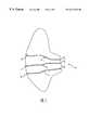
- FIG. 1is an anterior view of a human female breast, shown in section, and illustrating three of the six to nine ductal networks extending inwardly from the nipple.
- FIG. 2is an enlarged view of the nipple of FIG. 1 illustrating the orifices leading to each of the three ductal networks.
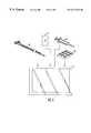
- FIG. 3is a perspective view of a dual-lumen catheter which is useful in performing the methods of the present invention.
- FIG. 4is a detailed view of the distal end of the catheter of FIG. 3, shown in section.
- FIG. 5illustrates use of the catheter of FIG. 3 in performing the method of the present invention in a single ductal network.
- FIG. 6illustrates a kit comprising a dual-lumen catheter and other system components, including instructions for use.
- FIGS. 7A-7Cillustrate the catheter utilized in the working examples hereinafter.
- the present inventioncomprises methods and kits for obtaining marker materials from one or more ductal networks in a human female breast.
- a typical breast Bas illustrated in FIG. 1, includes a nipple N and from six to nine ducts D.
- Three ductal networks D 1-3extending inwardly from the nipple N into the breast tissue are illustrated.
- each ductal network D 1-3begins with an orifice O 1-3 which lies at the surface of the nipple N and extends inwardly through a ductal sinus S 1-3 and then into a branching network.
- Each network Dcomprises a series of successively smaller lumens which are arranged in complex, three-dimensional patterns. The networks of each duct will overlap within the breast tissue but will not be interconnected.
- each networkis usually in the range from 0.1 ml to 0.5 ml, but the walls are somewhat compliant so the internal volume may increase as fluid is introduced.
- the present inventionrelies on accessing the ductal network(s) through the orifice O of the duct D within the nipple N. Usually, there will be from six to nine orifices which open into a like number of ductal networks. Confirmation of the number and location of the ductal orifices can be made by labelling the nipple as described below.
- the present inventionrelies on collecting endogenous ductal fluids and cellular and non-cellular marker materials from the individual ductal networks on a duct-by-duct basis. That is, fluids and marker materials are obtained from a single duct without obtaining material from any other ducts. This is in contrast to prior techniques which, in some instances, are able to obtain cellular and other materials from all milk ducts at once by applying a mild vacuum to the nipple. It should be noted, however, that in some instances such screening of all ducts in a single step may be appropriate in order to identify patients showing abnormalities for whom further, duct-specific testing according to the present invention is appropriate.
- a location of at least one ductwill be determined, typically by labeling all ductal orifices as described in co-pending application Ser. No. 08/931,786, the full disclosure of which has previously been incorporated herein by reference.
- a portion of the epithelial lining present exposed at the ductal orificemay be labeled with a visible marker which allows the treating professional to identify the entry orifice for each of the ductal networks in the breast.
- a washing fluidwill be introduced into the duct in order to loosen and mobilize cellular material from the ductal lining, primarily epithelial cells from the lining.
- the washing fluidis introduced in an amount and a manner such that substantially the entire volume of the duct will be washed with the fluid in order to obtain a sample which is representative of the entire ductal network.
- Cellular components from the samplewill usually be of the most interest, but ductal fluids and secreted molecular species (both small molecules and more usually biological macromolecules such as proteins and carbohydrates) may also be analyzed.
- the washing fluid carrying the cells and other materialsis then collected, and the materials morphologically, histologically, immunohistologically, chemically, immunologically, enzymatically, or otherwise examined in order to determine any abnormal or disease conditions within the ductal network, particularly cancer or a pre-cancerous condition.
- the washing fluidis introduced using a dual-lumen catheter which permits simultaneous introduction of the washing fluid and collection of excess washing fluid as it flushes back outwardly from the ductal network.
- the fluid being collectedis usually not aspirated (since aspiration could collapse the duct), and instead the pressure of the introduced fluid is relied on to both flush the entire ductal network and expel the excess fluid through the other lumen of the cannula.
- external pressuremay be applied to the breast to enhance or expedite fluid collection.
- the fluidis introduced using a syringe, with the fluid being introduced at a relatively low rate, typically in the range from 0.1 ml/sec to 5 ml/sec, preferably from 0.5 ml/sec to 1 ml/sec.
- the total introduced volume of the washing fluidis typically at least 5 ml, typically being in the range from 5 ml to 25 ml, usually being about 10 ml, and optionally being greater.
- a preferred washing fluidis physiologic saline but contrast media and other physiologically acceptable, sterile fluids may also be used.
- Also contemplated is a method for obtaining material from a milk duct in a breast of a patientcomprising locating at least one of the ductal orifices on a nipple of the breast, introducing a catheter having at least one lumen through one of the dutcal orifices and into the ductal passage, introducing a washing fluid through a lumen into the ductal passage, collecting the washing fluid from the ductal passage through a lumen of the catheter, and identifying materials present in the collected washing fluid.
- FIGS. 3 and 4An exemplary catheter 50 useful for performing the methods of the present invention is illustrated in FIGS. 3 and 4.
- the cathetercomprises a catheter body 52 , typically having a length in the range from 3 cm to 50 cm usually from 10 cm to 25 cm.
- the catheter body 52includes at least a first lumen 54 and a second lumen 56 .
- the first lumen 54terminates in a distal port 58 , as best seen in FIG. 4, while the second lumen terminates in a proximally located port 60 , typically being located by a distance d which is approximately 0.1 cm to 1 cm, usually from 0.1 cm to 0.25 cm, proximal of the distal port 58 .
- Catheter body 52will have a relatively narrow diameter, typically having a maximum diameter in the dual-lumen region in the range from 0.8 mm to 2.5 mm, preferably being in the range from 0.8 mm to 1.2 mm.
- the diameter of the distal, single-lumen regionmay be less, as in the range from 0.5 mm to 1.5 mm, preferably from 0.6 mm to 1 mm.
- Proximal hub 62includes a port 64 which is fluidly coupled to the second lumen 56 for delivering the washing fluid into the ductal network. Second port 66 is provided both for introducing the catheter over a guidewire and for collecting the washing fluid from the ductal network via port 58 .
- the catheter 50for collecting marker materials from a ductal network D 2 will be described.
- the ductal network D 2will first be accessed with a guidewire, such as a conventional 0.014 inch guidewire (not shown).
- a guidewiresuch as a conventional 0.014 inch guidewire (not shown).
- the catheter 50will be introduced over the guidewire by passing distal port 58 of the first lumen 54 over the external end of the guidewire.
- the distal port 58is introduced into the ductal network D 2 typically to a depth of about 0.25 cm to 2.5 cm, usually about 0.75 cm to 1.5 cm.
- the second port 60will be located proximally from the first port by a distance in the range from 0.1 cm to 1 cm, and will thus be closer to the orifice O 2 .
- the guidewirewill typically be withdrawn and the washing fluid introduced through the second lumen 56 via port 64 and opening 60 .
- the washing fluidwill flow into the ductal network and will generally reach most of the ductal volume, typically reaching at least 75% of the ductal volume, preferably at least 85%, and sometimes as much as 95%.
- the guidewire and catheter 50may be introduced simultaneously, typically with the distal tip of the guidewire extending a short distance ahead of the distal end of the catheter, usually about 0.1 cm to 1 cm.
- the guidewireis used to steer and the catheter 50 follows to the desired target location in the duct.
- the volume of fluid introduced into the ductal network D 2will be sufficiently large so that substantially the entire volume of the ductal network may be filled with the washing fluid and excess fluid will flow from the network as it is displaced by additional fluid input.
- Usually, only a small portion in the amount of washing fluid being introducedwill be necessary to fill the ductal network, usually less than 1 ml, often less than 0.5 ml.
- the remaining fluidwill continue to be introduced and will thus flush the cellular and other marker materials from the ductal network into the opening 58 in the first lumen 54 .
- that fluidwill pass outwardly through the catheter and may be collected from port 66 in the catheter.
- no vacuum or other aspiration pressurewill be applied to the catheter. Instead, the fluid will flow outwardly in response to the positive pressure created by the inflow of washing fluid, optionally with external pressure applied to the breast.
- the collected fluidmay be treated or analyzed in conventional ways to identify the presence, amount(s), identities, and/or other characteristics of any marker materials that may be present in the collected fluids.
- cellular materialsmay be transferred to a suitable medium, such as RPMI or other growth or maintenance medium.
- the cellsmay then be examined morphologically under a microscope and/or histologically using suitable histochemical and immunochemical staining reagents.
- Chemical and molecular markersmay be identified and/or examined chemically, immunologically, enzymatically, or by other conventional techniques. Such analysis techniques are well described in the art.
- Kits according to the present inventionwill comprise at least a catheter 50 (which may be any dual- or multiple-lumen catheter capable of accessing an individual ductal network) and instructions for use (IFU) 80 which are combined together in a conventional manner, typically within a container 82 , which may be in the form of a pouch, tray, box, tube, or the like. Kits will usually also include at least a guidewire, and other kit components may also be provided.
- a syringe 84may be provided, usually pre-filled with saline or other suitable washing medium for washing the ductal network.
- a collection tray 86 for receiving and maintaining the cellular material and the washing fluids collected from the cathetermay also be provided.
- the traymay include a suitable collection medium, such as RPMI medium.
- the kitsmay include materials for assaying non-cellular markers as well as components for identification of the ductal orifice, such as described in co-pending application Ser. No. 08/931,786, the full disclosure of which was previously incorporated herein by reference.
- a double lumen catheter which would allow a continuous flow of saline throughout the ductal systemwas prepared, as illustrated in FIG. 7 .
- the catheterwas a 3 French double lumen catheter with the proximal lumen smaller in diameter and the distal larger to allow aspiration.
- the catheter 100had a length from the hub 102 to the distal tip 104 of 31 cm, an outer diameter of D O of 0.041 in, a guidewire lumen D GW of 0.019 in, and a crescent-shaped lumen 110 .
- the outer tip diameter D OTwas 0.033 in and the lumenal tip diameter D LT was 0.017 in, reflecting a tapered distal end 116 .
- the double lumen catheterwas threaded into the duct. Saline was instilled, setting up a continuous flow until 10 cc was collected. The procedure took about 15 minutes. If 10 cc of fluid was not collected within the 15 minute limit, the procedure stopped prematurely. The washings were then sent to cytology for analysis.
- iorated aerolar ductal cells carcinoma 3R RUOQ 3:00 3 tubular deter- car- iorated cinoma, cells micro- papillary 4 L infra- 9:00 2 invasive deter- aerolar ductal iorated carcinoma cells 5 R N/A central 1 lactiferous Acellular (ab- duct scess) 6 L LUOQ 3-4:00 1 fiber- Benign adenoma mammary epithelial cells 7 R 3:00 fi- A- broad bund enoma epithelial cells, benign mammary epithelial cells 8 R 12:00 12:00 2 Invasive Acellular, ductal rare, car- ductal cinoma cells 9 R 11:00 11:00 3 Invasive acellular lob- ular 10 L UOQ central 1 Invasive Mod lobular macro- phage, no ductal cells 11 L LUOQ central 1 Adeno Foam carcinoma cells ductal cells 12 R RUOQ 3:00 2 Atyp
Landscapes
- Health & Medical Sciences (AREA)
- Life Sciences & Earth Sciences (AREA)
- Animal Behavior & Ethology (AREA)
- General Health & Medical Sciences (AREA)
- Engineering & Computer Science (AREA)
- Biomedical Technology (AREA)
- Heart & Thoracic Surgery (AREA)
- Medical Informatics (AREA)
- Molecular Biology (AREA)
- Surgery (AREA)
- Hematology (AREA)
- Pathology (AREA)
- Public Health (AREA)
- Veterinary Medicine (AREA)
- Medicines Containing Material From Animals Or Micro-Organisms (AREA)
- Infusion, Injection, And Reservoir Apparatuses (AREA)
- External Artificial Organs (AREA)
- Media Introduction/Drainage Providing Device (AREA)
- Investigating Or Analysing Biological Materials (AREA)
- Medicines Containing Antibodies Or Antigens For Use As Internal Diagnostic Agents (AREA)
- Surgical Instruments (AREA)
- Prostheses (AREA)
Abstract
Description
| TABLE 1 | ||||||
| mass | ||||||
| patient # | breast | location | duct location | #duct | pathology | cytology |
| Detached | ||||||
| breasts | ||||||
| 1 | R | RLQ | central | 1 | Ductal | acellular |
| carcinoma | ||||||
| comedo | ||||||
| 2 | R | No | 6:00 | 3 | ductal | |
| cancer | cells | |||||
| L | L breast | 9:00, 7:00 | 3 | Micro- | ductal | |
| papillary | cells | |||||
| DCIS | ||||||
| 3 | L | LLQ | 12:00 | 3 | infiltrating | ductal |
| ductal | cells | |||||
| carcinoma | ||||||
| 4 | R | RUOQ | 8:00 | 4 | DCIS | ductal |
| pagetoid | cells | |||||
| 5 | L | LUOQ | 11:00 | 5 | Intraductal | ductal |
| carcinoma | cells | |||||
| in situ | ||||||
| 6 | R | no | 9:00 | 2 | acelluar | |
| cancer | ||||||
| L | 3:00, | 6:00 | 4 | Invasive | acelular | |
| 9:00 | ductal | |||||
| car- | ||||||
| cinoma, | ||||||
| DCIS | ||||||
| 7 | L | LUOQ | 6:00 | 4 | Infiltrating | (1) |
| ductal | acellular, | |||||
| car- | (2) | |||||
| cinoma, | ductal | |||||
| DCIS | cells | |||||
| 8 | L | LOQ, | central | 2 | Infiltrating | ductal |
| 3:00 | lobular | cells | ||||
| car- | ||||||
| cinoma, | ||||||
| infiltrating | ||||||
| ductal | ||||||
| Attached | ||||||
| breasts | ||||||
| 1 | R | RUOQ | 12:00 | 2 | adeno ca | carcinoma |
| cells | ||||||
| 2 | L | retro- | central | 1 | poorly | deter- |
| diff. | iorated | |||||
| aerolar | ductal | cells | ||||
| carcinoma | ||||||
| 3 | R | RUOQ | 3:00 | 3 | tubular | deter- |
| car- | iorated | |||||
| cinoma, | cells | |||||
| micro- | ||||||
| papillary | ||||||
| 4 | L | infra- | 9:00 | 2 | invasive | deter- |
| aerolar | ductal | iorated | ||||
| carcinoma | cells | |||||
| 5 | R | N/A | central | 1 | lactiferous | Acellular |
| (ab- | duct | |||||
| scess) | ||||||
| 6 | L | LUOQ | 3-4:00 | 1 | fiber- | Benign |
| adenoma | mammary | |||||
| epithelial | ||||||
| cells | ||||||
| 7 | R | 3:00 | fi- | A- | ||
| broad | bund | |||||
| enoma | epithelial | |||||
| cells, | ||||||
| benign | ||||||
| mammary | ||||||
| epithelial | ||||||
| cells | ||||||
| 8 | R | 12:00 | 12:00 | 2 | Invasive | Acellular, |
| ductal | rare, | |||||
| car- | ductal | |||||
| cinoma | cells | |||||
| 9 | R | 11:00 | 11:00 | 3 | Invasive | acellular |
| lob- | ||||||
| ular | ||||||
| 10 | L | UOQ | central | 1 | Invasive | Mod |
| lobular | macro- | |||||
| phage, | ||||||
| no ductal | ||||||
| cells | ||||||
| 11 | L | LUOQ | central | 1 | Adeno | Foam |
| carcinoma | cells | |||||
| ductal | ||||||
| cells | ||||||
| 12 | R | RUOQ | 3:00 | 2 | Atyp med | Foam |
| carcinoma | cells, | |||||
| rare ductal | ||||||
| cells | ||||||
| R: right | ||||||
| L: left | ||||||
| RLQ: right lower quadrant | ||||||
| LLQ: left lower quadrant | ||||||
| LUOQ: left upper outer quadrant | ||||||
| LOQ: left outer quadrant | ||||||
| RUOQ: right upper outer quadrant | ||||||
| UOQ: upper outer quadrant | ||||||
| DCIS: ductal carcinoma in situ | ||||||
Claims (33)
Priority Applications (18)
| Application Number | Priority Date | Filing Date | Title |
|---|---|---|---|
| US09/067,661US6221622B1 (en) | 1998-04-28 | 1998-04-28 | Method and kit for obtaining fluids and cellular material from breast ducts |
| DE69929433TDE69929433T2 (en) | 1998-04-28 | 1999-04-28 | CATHETER FOR THE REMOVAL OF CELLULAR MATERIAL FROM THE BREAST GESTURE |
| EP99920083AEP1073476B1 (en) | 1998-04-28 | 1999-04-28 | Catheter for obtaining cellular material from breast ducts |
| EP05027586AEP1656953A1 (en) | 1998-04-28 | 1999-04-28 | Method for obtaining cellular material from breast ducts |
| US09/301,058US6494859B2 (en) | 1998-04-28 | 1999-04-28 | Methods using pressure to obtain fluids and cellular material from breast ducts |
| IL13877399AIL138773A0 (en) | 1998-04-28 | 1999-04-28 | Method and kit for obtaining fluids and cellular material from breast ducts |
| JP2000545580AJP2002512853A (en) | 1998-04-28 | 1999-04-28 | Methods and kits for obtaining fluid and cellular material from breast ducts |
| NZ507070ANZ507070A (en) | 1998-04-28 | 1999-04-28 | Method and kit for obtaining fluids and cellular material from breast ducts |
| PCT/US1999/009141WO1999055384A1 (en) | 1998-04-28 | 1999-04-28 | Method and kit for obtaining fluids and cellular material from breast ducts |
| CA002327514ACA2327514C (en) | 1998-04-28 | 1999-04-28 | Method and kit for obtaining fluids and cellular material from breast ducts |
| ES99920083TES2258330T3 (en) | 1998-04-28 | 1999-04-28 | CATHETER TO OBTAIN CELLULAR MATERIAL MATERIALS. |
| AU37663/99AAU753939B2 (en) | 1998-04-28 | 1999-04-28 | Method and kit for obtaining fluids and cellular material from breast ducts |
| AT99920083TATE315418T1 (en) | 1998-04-28 | 1999-04-28 | CATHETER FOR EXTRACTION OF CELLULAR MATERIAL FROM THE MAMMARY GLAND |
| ZA200005020AZA200005020B (en) | 1998-04-28 | 2000-09-20 | Method and kit for obtaining fluids and cellular material from breast ducts. |
| IL138773AIL138773A (en) | 1998-04-28 | 2000-09-28 | Method and kit for obtaining fluids and cellular material from breast ducts |
| US09/740,561US20010001059A1 (en) | 1998-04-28 | 2000-12-19 | Method and kit for obtaining fluids and cellular material from breast ducts |
| US10/099,439US6890311B2 (en) | 1997-09-16 | 2002-03-15 | Methods for performing medical procedures within a breast duct |
| US10/175,022US20020164286A1 (en) | 1997-09-16 | 2002-06-19 | Methods for obtaining fluid and cellular material from a breast duct |
Applications Claiming Priority (1)
| Application Number | Priority Date | Filing Date | Title |
|---|---|---|---|
| US09/067,661US6221622B1 (en) | 1998-04-28 | 1998-04-28 | Method and kit for obtaining fluids and cellular material from breast ducts |
Related Child Applications (2)
| Application Number | Title | Priority Date | Filing Date |
|---|---|---|---|
| US09/301,058Continuation-In-PartUS6494859B2 (en) | 1998-04-28 | 1999-04-28 | Methods using pressure to obtain fluids and cellular material from breast ducts |
| US09/740,561ContinuationUS20010001059A1 (en) | 1997-09-16 | 2000-12-19 | Method and kit for obtaining fluids and cellular material from breast ducts |
Publications (1)
| Publication Number | Publication Date |
|---|---|
| US6221622B1true US6221622B1 (en) | 2001-04-24 |
Family
ID=22077527
Family Applications (3)
| Application Number | Title | Priority Date | Filing Date |
|---|---|---|---|
| US09/067,661Expired - LifetimeUS6221622B1 (en) | 1997-09-16 | 1998-04-28 | Method and kit for obtaining fluids and cellular material from breast ducts |
| US09/301,058Expired - LifetimeUS6494859B2 (en) | 1998-04-28 | 1999-04-28 | Methods using pressure to obtain fluids and cellular material from breast ducts |
| US09/740,561AbandonedUS20010001059A1 (en) | 1997-09-16 | 2000-12-19 | Method and kit for obtaining fluids and cellular material from breast ducts |
Family Applications After (2)
| Application Number | Title | Priority Date | Filing Date |
|---|---|---|---|
| US09/301,058Expired - LifetimeUS6494859B2 (en) | 1998-04-28 | 1999-04-28 | Methods using pressure to obtain fluids and cellular material from breast ducts |
| US09/740,561AbandonedUS20010001059A1 (en) | 1997-09-16 | 2000-12-19 | Method and kit for obtaining fluids and cellular material from breast ducts |
Country Status (12)
| Country | Link |
|---|---|
| US (3) | US6221622B1 (en) |
| EP (2) | EP1073476B1 (en) |
| JP (1) | JP2002512853A (en) |
| AT (1) | ATE315418T1 (en) |
| AU (1) | AU753939B2 (en) |
| CA (1) | CA2327514C (en) |
| DE (1) | DE69929433T2 (en) |
| ES (1) | ES2258330T3 (en) |
| IL (2) | IL138773A0 (en) |
| NZ (1) | NZ507070A (en) |
| WO (1) | WO1999055384A1 (en) |
| ZA (1) | ZA200005020B (en) |
Cited By (43)
| Publication number | Priority date | Publication date | Assignee | Title |
|---|---|---|---|---|
| WO2001085023A2 (en) | 2000-05-10 | 2001-11-15 | Cytyc Health Corporation | Method for differentiating breast ducts for cancer risk status |
| US20020002343A1 (en)* | 1998-12-28 | 2002-01-03 | David Hung | Devices, methods and systems for collecting material from a breast duct |
| WO2002009589A1 (en) | 2000-07-28 | 2002-02-07 | Cytyc Health Corporation | Methods and devices for diagnosis of precancer and cancer in breast milk ducts |
| US6398765B1 (en)* | 1999-03-01 | 2002-06-04 | Pro Duct Health, Inc. | Apparatus, methods and kits for simultaneous delivery of a substance to multiple breast milk ducts |
| US20020110609A1 (en)* | 1998-12-28 | 2002-08-15 | David Hung | Increasing retrievable fluid from a breast duct |
| US6455027B1 (en)* | 1997-09-16 | 2002-09-24 | The Regents Of The University Of California | Methods and kits for identifying ductal orifices in a nipple |
| US20020183717A1 (en)* | 2001-05-30 | 2002-12-05 | Morton Kevin B. | Method and apparatus for noninvasive intraductal fluid diagnostic screen |
| US20020193822A1 (en)* | 2001-04-16 | 2002-12-19 | Pro Duct Health, Inc. | Externally positioned medical dilator |
| US6500114B1 (en) | 1993-11-23 | 2002-12-31 | Dofi Technologies, Inc. | Method of extracting biopsy cells from the breast |
| US6517513B1 (en) | 1999-01-21 | 2003-02-11 | Neomatrix, Llc | Intraductal breast fluid aspiration device |
| WO2002096267A3 (en)* | 2001-05-30 | 2003-02-20 | Neomatrix Llc | Noninvasive intraductal fluid diagnostic screen |
| US20030181823A1 (en)* | 2002-03-25 | 2003-09-25 | Gatto Dominick L. | Apparatus and method for intraductal cytology |
| US20030187427A1 (en)* | 2002-04-02 | 2003-10-02 | Gatto Dominick L. | Method and apparatus for in VIVO treatment of mammary ducts by light induced fluorescence |
| US6642009B2 (en) | 1999-05-17 | 2003-11-04 | Cytyc Health Corporation | Isolated ductal fluid sample |
| US6652442B2 (en) | 2002-04-23 | 2003-11-25 | Acueity, Inc. | Micro-endoscope assembly for intraductal brachytherapy of a mammary duct and method of using same |
| US20040029202A1 (en)* | 1999-01-26 | 2004-02-12 | Cytyc Health Corporation | Identifying, Monitoring and treating women with breast precancer or cancer |
| US20040153001A1 (en)* | 1999-12-28 | 2004-08-05 | David Hung | Devices, methods and systems for collecting material from a breast duct |
| GB2400037A (en)* | 2003-03-31 | 2004-10-06 | Psimedica Ltd | Device made of silicon and method for collecting mammary fluid |
| US6878149B2 (en) | 2002-03-25 | 2005-04-12 | Acueity, Inc. | Apparatus and method for intraductal abalation |
| US6890311B2 (en)* | 1997-09-16 | 2005-05-10 | The Regents Of The University Of California | Methods for performing medical procedures within a breast duct |
| US20050165288A1 (en)* | 2004-01-27 | 2005-07-28 | Scimed Life Systems, Inc. | Systems and methods for treating breast tissue |
| US20050234497A1 (en)* | 2001-04-16 | 2005-10-20 | David Hung | Externally positioned medical dilator |
| US20060089567A1 (en)* | 2004-10-22 | 2006-04-27 | Goldenberg Alec S | Aspiration needle with venting feature |
| AU2003204883B2 (en)* | 1999-03-01 | 2006-05-04 | Atossa Genetics, Inc. | Apparatus, Methods and Kits for Simultaneous Delivery of a Substance to Multiple Breast Milk Ducts |
| US20060184098A1 (en)* | 2002-04-19 | 2006-08-17 | Neuron Therapeutic, Inc. | Subarachnoid spinal catheter for transporting cerebrospinal fluid |
| US7132232B2 (en) | 2000-08-08 | 2006-11-07 | Cytyc Corporation | Identification of viral agents in breast ducts and antiviral therapy therefore |
| US20070142744A1 (en)* | 2005-12-16 | 2007-06-21 | Provencher Kevin M | Tissue sample needle and method of using same |
| US20070142743A1 (en)* | 2005-12-16 | 2007-06-21 | Provencher Kevin M | Tissue sample needle actuator system and apparatus and method of using same |
| US20070189968A1 (en)* | 1999-06-11 | 2007-08-16 | Annette Bianchi | Gel composition for filling a breast milk duct prior to surgical excision of the duct or other breast tissue |
| US7274809B2 (en) | 2002-08-29 | 2007-09-25 | Perceptronix Medical, Inc. And British Columbia Cancer Agency | Computerized methods and systems related to the detection of malignancy-associated changes (MAC) to detect cancer |
| US20080015469A1 (en)* | 2001-05-30 | 2008-01-17 | Neomatrix, Llc | Disposable patient interface for intraductal fluid aspiration system |
| US20080096283A1 (en)* | 2002-10-11 | 2008-04-24 | Sentina Biotechnology Incorporated | Methods for detection of breast cancer |
| US20080166733A1 (en)* | 1999-07-09 | 2008-07-10 | The Burnham Institute | Method for determining the prognosis of cancer patients by measuring levels of bag expression |
| US20090250074A1 (en)* | 2001-02-27 | 2009-10-08 | Dr. Susan Love Research Foundation | Nipple covering system |
| US20100256464A1 (en)* | 2007-05-14 | 2010-10-07 | Dr. Susan Love Research Foundation | Device for determining risk of developing breast cancer and method thereof |
| US20110190662A1 (en)* | 2008-10-01 | 2011-08-04 | Beacon Endoscopic Corporation | Rapid exchange fna biopsy device with diagnostic and therapeutic capabilities |
| US20120034644A1 (en)* | 1999-01-26 | 2012-02-09 | Atossa Genetics, Inc. | Identifying material from a breast duct |
| US8968210B2 (en) | 2008-10-01 | 2015-03-03 | Covidien LLP | Device for needle biopsy with integrated needle protection |
| US9186128B2 (en) | 2008-10-01 | 2015-11-17 | Covidien Lp | Needle biopsy device |
| US9332973B2 (en) | 2008-10-01 | 2016-05-10 | Covidien Lp | Needle biopsy device with exchangeable needle and integrated needle protection |
| US9782565B2 (en) | 2008-10-01 | 2017-10-10 | Covidien Lp | Endoscopic ultrasound-guided biliary access system |
| WO2021016117A1 (en)* | 2019-07-19 | 2021-01-28 | The Board Of Regents Of The University Of Oklahoma | Endoscope biopsy system and method for ductoscopy |
| US11298113B2 (en) | 2008-10-01 | 2022-04-12 | Covidien Lp | Device for needle biopsy with integrated needle protection |
Families Citing this family (19)
| Publication number | Priority date | Publication date | Assignee | Title |
|---|---|---|---|---|
| JP2003529318A (en)* | 1998-12-28 | 2003-10-07 | プロ ダクト ヘルス インコーポレーティッド | Apparatus, method, and system for harvesting material from a milk duct |
| JP2004505278A (en)* | 2000-07-28 | 2004-02-19 | シーワイティーワイシー ヘルス コーポレイション | Cytological evaluation of ductal epithelial cells recovered by duct washing |
| EP1785727A3 (en)* | 2000-08-08 | 2007-08-29 | Cytyc Corporation | Identification of viral agents in breast ducts and antiviral therapy therefore |
| AU2003220138A1 (en)* | 2002-03-19 | 2003-10-08 | Cytyc Corporation | Method and apparatus for analyzing mammary gland fluid |
| US7534245B2 (en)* | 2002-12-02 | 2009-05-19 | Chappuis James L | Flexible tap apparatus and method of use |
| CA2575675A1 (en)* | 2004-07-30 | 2006-03-09 | Adeza Biomedical Corporation | Oncofetal fibronectin as a marker for disease and other conditions and methods for detection of oncofetal fibronectin |
| US20060149141A1 (en)* | 2004-12-30 | 2006-07-06 | Ellen Sheets | Method of using pressure to determine the positioning of a catheter within a breast duct |
| DE102007014634B3 (en)* | 2007-03-23 | 2008-12-11 | Karl-Heinz Bachmann | Instrument for the medical examination of narrow body canals |
| EP2060295A1 (en)* | 2007-11-13 | 2009-05-20 | Tyco Healthcare Group LP | Dual lumen catheter and method for minimally invasive endoluminal surgery |
| US8814846B2 (en) | 2007-11-13 | 2014-08-26 | Covidien Lp | Dual lumen catheter and method for minimally invasive endoluminal surgery |
| US9119926B2 (en) | 2009-07-31 | 2015-09-01 | Avent, Inc. | Subglottic suctioning system |
| WO2013165813A1 (en)* | 2012-04-30 | 2013-11-07 | The Regents Of The University Of Colorado, A Body Corporate | Cell collecting apparatus and method of use |
| DE102013216476A1 (en)* | 2013-08-20 | 2015-02-26 | Carl Zeiss Meditec Ag | Surgical microscope with optical interfaces |
| US11589846B2 (en)* | 2014-09-12 | 2023-02-28 | Robert K Ackroyd | Dual needle core biopsy instrument |
| US11701174B2 (en)* | 2016-01-29 | 2023-07-18 | Boston Scientific Scimed, Inc. | Medical device having a plurality of lumens and a port |
| US10755676B2 (en) | 2018-03-15 | 2020-08-25 | Magic Leap, Inc. | Image correction due to deformation of components of a viewing device |
| US20220323112A1 (en)* | 2019-09-25 | 2022-10-13 | Oxford University Innovation Limited | Needle and uses thereof |
| US20230190242A1 (en)* | 2020-05-18 | 2023-06-22 | Thomas Jefferson University | Viral sample collection |
| CN119604324A (en)* | 2022-06-16 | 2025-03-11 | H·N·克拉弗 | Multi-lumen central lines |
Citations (3)
| Publication number | Priority date | Publication date | Assignee | Title |
|---|---|---|---|---|
| US5413558A (en)* | 1991-09-09 | 1995-05-09 | New York University | Selective aortic perfusion system for use during CPR |
| WO1997005898A1 (en) | 1995-08-03 | 1997-02-20 | The Johns Hopkins University School Of Medicine | Delivery of an agent to the ductal epithelium in the prophylactic and therapeutic treatment of cancer |
| WO1999013917A1 (en) | 1997-09-16 | 1999-03-25 | The Regents Of The University Of California | Methods and kits for identifying ductal orifices in a nipple |
Family Cites Families (6)
| Publication number | Priority date | Publication date | Assignee | Title |
|---|---|---|---|---|
| EP0131166B1 (en)* | 1983-06-14 | 1988-09-07 | Fertility And Genetics Research, Inc. | Non-surgical apparatus for human embryo transfer |
| JPH02280061A (en)* | 1989-04-20 | 1990-11-16 | Mochida Pharmaceut Co Ltd | Method and implement for measuring antigen associated with tumor in slight amount of papilla secretion |
| US5167622A (en)* | 1990-12-07 | 1992-12-01 | Smiths Industries Medical Systems, Inc. | Triple conduit suction catheter |
| US5443454A (en)* | 1992-12-09 | 1995-08-22 | Terumo Kabushiki Kaisha | Catheter for embolectomy |
| US5348536A (en)* | 1993-08-02 | 1994-09-20 | Quinton Instrument Company | Coextruded catheter and method of forming |
| BR9601849A (en)* | 1996-06-18 | 1998-09-01 | Newton Paes | Improvement in double-way catheter for washing drainage monitoring and control of intracranial pressure |
- 1998
- 1998-04-28USUS09/067,661patent/US6221622B1/ennot_activeExpired - Lifetime
- 1999
- 1999-04-28CACA002327514Apatent/CA2327514C/ennot_activeExpired - Fee Related
- 1999-04-28EPEP99920083Apatent/EP1073476B1/ennot_activeExpired - Lifetime
- 1999-04-28USUS09/301,058patent/US6494859B2/ennot_activeExpired - Lifetime
- 1999-04-28AUAU37663/99Apatent/AU753939B2/ennot_activeCeased
- 1999-04-28DEDE69929433Tpatent/DE69929433T2/ennot_activeExpired - Fee Related
- 1999-04-28NZNZ507070Apatent/NZ507070A/enunknown
- 1999-04-28ILIL13877399Apatent/IL138773A0/enunknown
- 1999-04-28JPJP2000545580Apatent/JP2002512853A/ennot_activeCeased
- 1999-04-28ATAT99920083Tpatent/ATE315418T1/ennot_activeIP Right Cessation
- 1999-04-28ESES99920083Tpatent/ES2258330T3/ennot_activeExpired - Lifetime
- 1999-04-28EPEP05027586Apatent/EP1656953A1/ennot_activeWithdrawn
- 1999-04-28WOPCT/US1999/009141patent/WO1999055384A1/enactiveIP Right Grant
- 2000
- 2000-09-20ZAZA200005020Apatent/ZA200005020B/enunknown
- 2000-09-28ILIL138773Apatent/IL138773A/ennot_activeIP Right Cessation
- 2000-12-19USUS09/740,561patent/US20010001059A1/ennot_activeAbandoned
Patent Citations (3)
| Publication number | Priority date | Publication date | Assignee | Title |
|---|---|---|---|---|
| US5413558A (en)* | 1991-09-09 | 1995-05-09 | New York University | Selective aortic perfusion system for use during CPR |
| WO1997005898A1 (en) | 1995-08-03 | 1997-02-20 | The Johns Hopkins University School Of Medicine | Delivery of an agent to the ductal epithelium in the prophylactic and therapeutic treatment of cancer |
| WO1999013917A1 (en) | 1997-09-16 | 1999-03-25 | The Regents Of The University Of California | Methods and kits for identifying ductal orifices in a nipple |
Non-Patent Citations (31)
Cited By (81)
| Publication number | Priority date | Publication date | Assignee | Title |
|---|---|---|---|---|
| US6500114B1 (en) | 1993-11-23 | 2002-12-31 | Dofi Technologies, Inc. | Method of extracting biopsy cells from the breast |
| US6890311B2 (en)* | 1997-09-16 | 2005-05-10 | The Regents Of The University Of California | Methods for performing medical procedures within a breast duct |
| US6455027B1 (en)* | 1997-09-16 | 2002-09-24 | The Regents Of The University Of California | Methods and kits for identifying ductal orifices in a nipple |
| US20020002343A1 (en)* | 1998-12-28 | 2002-01-03 | David Hung | Devices, methods and systems for collecting material from a breast duct |
| US6413228B1 (en)* | 1998-12-28 | 2002-07-02 | Pro Duct Health, Inc. | Devices, methods and systems for collecting material from a breast duct |
| US20020110609A1 (en)* | 1998-12-28 | 2002-08-15 | David Hung | Increasing retrievable fluid from a breast duct |
| US20030149421A1 (en)* | 1999-01-21 | 2003-08-07 | Chandice Covington | Method and apparatus for influencing the transport of blood carotenoids into intraductal breast fluid |
| US6517513B1 (en) | 1999-01-21 | 2003-02-11 | Neomatrix, Llc | Intraductal breast fluid aspiration device |
| US20040091423A1 (en)* | 1999-01-26 | 2004-05-13 | Cytyc Health Corporation | Methods for identifying treating or monitoring asymptomatic patients for risk reduction or therapeutic treatment of breast cancer |
| US20040029202A1 (en)* | 1999-01-26 | 2004-02-12 | Cytyc Health Corporation | Identifying, Monitoring and treating women with breast precancer or cancer |
| US20120034644A1 (en)* | 1999-01-26 | 2012-02-09 | Atossa Genetics, Inc. | Identifying material from a breast duct |
| US20070161063A1 (en)* | 1999-01-26 | 2007-07-12 | Susan Love | Identifying, monitoring, and treating women for breast precancer or cancer |
| AU2003204883B9 (en)* | 1999-03-01 | 2006-09-28 | Atossa Genetics, Inc. | Apparatus, methods and kits for simultaneous delivery of a substance to multiple breast milk ducts |
| US6585706B2 (en)* | 1999-03-01 | 2003-07-01 | Cytyc Health Corporation | Apparatus, methods and kits for simultaneous delivery of a substance to multiple breast milk ducts |
| AU2003204883B2 (en)* | 1999-03-01 | 2006-05-04 | Atossa Genetics, Inc. | Apparatus, Methods and Kits for Simultaneous Delivery of a Substance to Multiple Breast Milk Ducts |
| US7029462B2 (en) | 1999-03-01 | 2006-04-18 | Cytyc Corporation | Apparatus, methods and kits for simultaneous delivery of a substance to multiple breast milk ducts |
| US6398765B1 (en)* | 1999-03-01 | 2002-06-04 | Pro Duct Health, Inc. | Apparatus, methods and kits for simultaneous delivery of a substance to multiple breast milk ducts |
| US6642009B2 (en) | 1999-05-17 | 2003-11-04 | Cytyc Health Corporation | Isolated ductal fluid sample |
| US20040038281A1 (en)* | 1999-05-17 | 2004-02-26 | Cytyc Health Corporation | Isolated ductal fluid sample |
| US20110200695A1 (en)* | 1999-06-11 | 2011-08-18 | Annette Bianchi | Gel composition for filling a breast milk duct prior to surgical excision of the duct or other breast tissue |
| US20070189968A1 (en)* | 1999-06-11 | 2007-08-16 | Annette Bianchi | Gel composition for filling a breast milk duct prior to surgical excision of the duct or other breast tissue |
| US7951544B1 (en) | 1999-07-09 | 2011-05-31 | Sanford-Burnham Medical Research Institute | Method for determining the prognosis of cancer patients by measuring levels of bag expression |
| US20080166733A1 (en)* | 1999-07-09 | 2008-07-10 | The Burnham Institute | Method for determining the prognosis of cancer patients by measuring levels of bag expression |
| US20040153001A1 (en)* | 1999-12-28 | 2004-08-05 | David Hung | Devices, methods and systems for collecting material from a breast duct |
| WO2001085023A2 (en) | 2000-05-10 | 2001-11-15 | Cytyc Health Corporation | Method for differentiating breast ducts for cancer risk status |
| US20040054300A1 (en)* | 2000-05-10 | 2004-03-18 | Cytyc Health Corporation | Method for differentiating breast ducts for cancer risk status |
| US6629936B2 (en)* | 2000-05-10 | 2003-10-07 | Cytyc Health Corporation | Method and kits for differentiating breast ducts for cancer risk status |
| WO2002009589A1 (en) | 2000-07-28 | 2002-02-07 | Cytyc Health Corporation | Methods and devices for diagnosis of precancer and cancer in breast milk ducts |
| US7132232B2 (en) | 2000-08-08 | 2006-11-07 | Cytyc Corporation | Identification of viral agents in breast ducts and antiviral therapy therefore |
| US7405045B2 (en)* | 2000-08-08 | 2008-07-29 | Cytyc Corporation | Identification of viral agents in breast ducts and antiviral therapy therefore |
| US20070009946A1 (en)* | 2000-08-08 | 2007-01-11 | David Hung | Identification of viral agents in breast ducts and antiviral therapy therefore |
| US8844539B2 (en) | 2001-02-27 | 2014-09-30 | Dr. Susan Love Research Foundation | Nipple cover |
| US8191554B2 (en) | 2001-02-27 | 2012-06-05 | Dr. Susan Love Research Foundation | Nipple covering system |
| US20090250074A1 (en)* | 2001-02-27 | 2009-10-08 | Dr. Susan Love Research Foundation | Nipple covering system |
| US7921851B2 (en) | 2001-02-27 | 2011-04-12 | Dr. Susan Love Research Foundation | Nipple covering system |
| US20040249317A1 (en)* | 2001-04-06 | 2004-12-09 | Cytyc Corporation | Increasing retrievable cells from a breast duct |
| US7628765B2 (en)* | 2001-04-06 | 2009-12-08 | Cytyc Corporation | Increasing retrievable cells from a breast duct |
| US20020193822A1 (en)* | 2001-04-16 | 2002-12-19 | Pro Duct Health, Inc. | Externally positioned medical dilator |
| US20050234497A1 (en)* | 2001-04-16 | 2005-10-20 | David Hung | Externally positioned medical dilator |
| US6875184B2 (en) | 2001-05-30 | 2005-04-05 | Neomatrix, Llc | Method and apparatus for noninvasive intraductal fluid diagnostic screen |
| US20020183718A1 (en)* | 2001-05-30 | 2002-12-05 | Morton Kevin B. | Disposable fluid loop for intraductal fluid aspiration system |
| US7468043B2 (en) | 2001-05-30 | 2008-12-23 | Neomatrix, Llc | Method and apparatus for noninvasive intraductal fluid diagnositc screen |
| WO2002096267A3 (en)* | 2001-05-30 | 2003-02-20 | Neomatrix Llc | Noninvasive intraductal fluid diagnostic screen |
| US7575557B2 (en) | 2001-05-30 | 2009-08-18 | Neo Matrix, Llc | Disposable fluid loop for intraductal fluid aspiration system |
| US20050171471A1 (en)* | 2001-05-30 | 2005-08-04 | Neomatrix, Llc | Method and apparatus for noninvasive intraductal fluid diagnostic screen |
| US6676610B2 (en) | 2001-05-30 | 2004-01-13 | Neomatrix, Llc | Disposable patient interface for intraductal fluid aspiration system |
| US20080015495A1 (en)* | 2001-05-30 | 2008-01-17 | Neomatrix, Llc | Method and apparatus for noninvasive intraductal fluid diagnostic screen |
| US6712785B2 (en) | 2001-05-30 | 2004-03-30 | Neomatrix, Llc | Method of noninvasively obtaining intraductal fluid |
| US20080015469A1 (en)* | 2001-05-30 | 2008-01-17 | Neomatrix, Llc | Disposable patient interface for intraductal fluid aspiration system |
| US20020183717A1 (en)* | 2001-05-30 | 2002-12-05 | Morton Kevin B. | Method and apparatus for noninvasive intraductal fluid diagnostic screen |
| US6866994B2 (en) | 2001-05-30 | 2005-03-15 | Neomatrix, Llc | Noninvasive intraductal fluid diagnostic screen |
| US20030181823A1 (en)* | 2002-03-25 | 2003-09-25 | Gatto Dominick L. | Apparatus and method for intraductal cytology |
| US6878149B2 (en) | 2002-03-25 | 2005-04-12 | Acueity, Inc. | Apparatus and method for intraductal abalation |
| US6840909B2 (en)* | 2002-03-25 | 2005-01-11 | Acueity, Inc. | Apparatus and method for intraductal cytology |
| US20030187427A1 (en)* | 2002-04-02 | 2003-10-02 | Gatto Dominick L. | Method and apparatus for in VIVO treatment of mammary ducts by light induced fluorescence |
| US6846311B2 (en) | 2002-04-02 | 2005-01-25 | Acueity, Inc. | Method and apparatus for in VIVO treatment of mammary ducts by light induced fluorescence |
| US20060184098A1 (en)* | 2002-04-19 | 2006-08-17 | Neuron Therapeutic, Inc. | Subarachnoid spinal catheter for transporting cerebrospinal fluid |
| US6652442B2 (en) | 2002-04-23 | 2003-11-25 | Acueity, Inc. | Micro-endoscope assembly for intraductal brachytherapy of a mammary duct and method of using same |
| US7274809B2 (en) | 2002-08-29 | 2007-09-25 | Perceptronix Medical, Inc. And British Columbia Cancer Agency | Computerized methods and systems related to the detection of malignancy-associated changes (MAC) to detect cancer |
| US20110124110A1 (en)* | 2002-10-11 | 2011-05-26 | Dr. Susan Love Research Foundation | Systems and methods for breast cancer detection and risk assessment |
| US20080096283A1 (en)* | 2002-10-11 | 2008-04-24 | Sentina Biotechnology Incorporated | Methods for detection of breast cancer |
| US7879614B2 (en) | 2002-10-11 | 2011-02-01 | Dr. Susan Love Research Foundation | Methods for detection of breast cancer |
| US8133737B2 (en) | 2002-10-11 | 2012-03-13 | Atossa Genetics, Inc. | Systems and methods for breast cancer detection and risk assessment |
| GB2400037A (en)* | 2003-03-31 | 2004-10-06 | Psimedica Ltd | Device made of silicon and method for collecting mammary fluid |
| US20050165288A1 (en)* | 2004-01-27 | 2005-07-28 | Scimed Life Systems, Inc. | Systems and methods for treating breast tissue |
| US7226423B2 (en) | 2004-10-22 | 2007-06-05 | Goldenberg Alec S | Aspiration needle with venting feature |
| US20060089567A1 (en)* | 2004-10-22 | 2006-04-27 | Goldenberg Alec S | Aspiration needle with venting feature |
| US20070142743A1 (en)* | 2005-12-16 | 2007-06-21 | Provencher Kevin M | Tissue sample needle actuator system and apparatus and method of using same |
| US20070142744A1 (en)* | 2005-12-16 | 2007-06-21 | Provencher Kevin M | Tissue sample needle and method of using same |
| US20100256464A1 (en)* | 2007-05-14 | 2010-10-07 | Dr. Susan Love Research Foundation | Device for determining risk of developing breast cancer and method thereof |
| US9186128B2 (en) | 2008-10-01 | 2015-11-17 | Covidien Lp | Needle biopsy device |
| US8968210B2 (en) | 2008-10-01 | 2015-03-03 | Covidien LLP | Device for needle biopsy with integrated needle protection |
| US20110190662A1 (en)* | 2008-10-01 | 2011-08-04 | Beacon Endoscopic Corporation | Rapid exchange fna biopsy device with diagnostic and therapeutic capabilities |
| US9332973B2 (en) | 2008-10-01 | 2016-05-10 | Covidien Lp | Needle biopsy device with exchangeable needle and integrated needle protection |
| US9782565B2 (en) | 2008-10-01 | 2017-10-10 | Covidien Lp | Endoscopic ultrasound-guided biliary access system |
| US9913630B2 (en) | 2008-10-01 | 2018-03-13 | Covidien Lp | Device for needle biopsy with integrated needle protection |
| US10076316B2 (en) | 2008-10-01 | 2018-09-18 | Covidien Lp | Needle biopsy device |
| US10888689B2 (en) | 2008-10-01 | 2021-01-12 | Covidien Lp | Endoscopic ultrasound-guided biliary access system |
| US11039816B2 (en) | 2008-10-01 | 2021-06-22 | Covidien Lp | Needle biopsy device with exchangeable needle and integrated needle protection |
| US11298113B2 (en) | 2008-10-01 | 2022-04-12 | Covidien Lp | Device for needle biopsy with integrated needle protection |
| WO2021016117A1 (en)* | 2019-07-19 | 2021-01-28 | The Board Of Regents Of The University Of Oklahoma | Endoscope biopsy system and method for ductoscopy |
Also Published As
| Publication number | Publication date |
|---|---|
| US6494859B2 (en) | 2002-12-17 |
| ZA200005020B (en) | 2002-06-20 |
| ATE315418T1 (en) | 2006-02-15 |
| DE69929433T2 (en) | 2006-08-24 |
| CA2327514A1 (en) | 1999-11-04 |
| DE69929433D1 (en) | 2006-04-06 |
| US20020019017A1 (en) | 2002-02-14 |
| ES2258330T3 (en) | 2006-08-16 |
| EP1073476B1 (en) | 2006-01-11 |
| JP2002512853A (en) | 2002-05-08 |
| IL138773A (en) | 2009-07-20 |
| IL138773A0 (en) | 2001-10-31 |
| AU753939B2 (en) | 2002-10-31 |
| EP1073476A1 (en) | 2001-02-07 |
| WO1999055384A1 (en) | 1999-11-04 |
| US20010001059A1 (en) | 2001-05-10 |
| AU3766399A (en) | 1999-11-16 |
| NZ507070A (en) | 2007-02-23 |
| CA2327514C (en) | 2008-08-26 |
| EP1656953A1 (en) | 2006-05-17 |
Similar Documents
| Publication | Publication Date | Title |
|---|---|---|
| US6221622B1 (en) | Method and kit for obtaining fluids and cellular material from breast ducts | |
| Matsunaga et al. | Mammary ductoscopy for diagnosis and treatment of intraductal lesions of the breast | |
| US12185925B2 (en) | Endoscopic tri-point biopsy needle | |
| ROBERTSON et al. | Destructive spine lesions: diagnosis by needle biopsy | |
| US6328709B1 (en) | Devices and methods to identify ductal orifices during nipple aspiration | |
| Isler et al. | Tissue core biopsy of abdominal tumors with a 22 gauge cutting needle | |
| Makita et al. | Duct endoscopy and endoscopic biopsy in the evaluation of nipple discharge | |
| JP2005508488A (en) | Isolated ductal fluid sample | |
| Gardecki et al. | Aspiration cytology in the preoperative management of breast cancer | |
| US4697600A (en) | Method for obtaining tissue and cells by fine needle aspiration for cytology/biopsy and kit relating to the same | |
| US6689070B2 (en) | Devices, methods and systems for collecting material from a breast duct | |
| Ehrhart | Principles of tumor biopsy | |
| Yamamoto et al. | Histocytologic diagnosis of pancreatic cancer by percutaneous aspiration biopsy under ultrasonic guidance | |
| Zhu et al. | A randomized controlled study of selective microdochectomy guided by ductoscopic wire marking or methylene blue injection | |
| US20040153001A1 (en) | Devices, methods and systems for collecting material from a breast duct | |
| Ihre et al. | Percutaneous Fine-Needle Aspiration Biopsy during Endoscopic Retrograde Cholangio-pancreatography | |
| AU2002258642B2 (en) | Devices, methods and systems for collecting material from a breast duct | |
| CN112603386B (en) | A kind of brain tumor surgery cavity interstitial fluid sampling monitoring and diagnosis device | |
| Chalas et al. | The gynecologist and surgical procedures for breast disease | |
| Galea et al. | Diagnosis by team work: an approach to conservatism | |
| AU2002258642A1 (en) | Devices, methods and systems for collecting material from a breast duct | |
| Petersen et al. | Fine-needle aspiration biopsy of the thyroid gland | |
| Ivanovic | Overview of cytopathology procedures and techniques | |
| Coleman et al. | Sentinel lymph node biopsy | |
| Ζερβούδης et al. | Nipple discharge screening |
Legal Events
| Date | Code | Title | Description |
|---|---|---|---|
| AS | Assignment | Owner name:REGENTS OF THE UNIVERSITY OF CALIFORNIA, THE, CALI Free format text:ASSIGNMENT OF ASSIGNORS INTEREST;ASSIGNOR:LOVE, SUSAN M.;REEL/FRAME:009484/0334 Effective date:19980827 | |
| AS | Assignment | Owner name:UNITED STATES GOVERNMENT, THE, MARYLAND Free format text:CONFIRMATORY LICENSE;ASSIGNOR:REGENTS OF THE UNIVERSITY OF CALIFORNIA, THE;REEL/FRAME:011705/0142 Effective date:19990223 | |
| STCF | Information on status: patent grant | Free format text:PATENTED CASE | |
| AS | Assignment | Owner name:ARMY MEDICAL RESEARCH AND MATERIEL COMMAND, UNITED Free format text:CONFIRMATORY LICENSE;ASSIGNOR:CALIFORNIA, UNIVERSITY OF;REEL/FRAME:012219/0992 Effective date:20010701 | |
| FEPP | Fee payment procedure | Free format text:PAT HOLDER NO LONGER CLAIMS SMALL ENTITY STATUS, ENTITY STATUS SET TO UNDISCOUNTED (ORIGINAL EVENT CODE: STOL); ENTITY STATUS OF PATENT OWNER: LARGE ENTITY | |
| AS | Assignment | Owner name:UNITED STATES GOVERNMENT, THE, MARYLAND Free format text:CONFIRMATORY LICENSE;ASSIGNOR:REGENTS OF THE UNIVERSITY OF CALIFORNIA, THE;REEL/FRAME:013071/0007 Effective date:20000309 | |
| AS | Assignment | Owner name:REGENTS OF THE UNIVERSITY OF CALIFORNIA, THE, CALI Free format text:ASSIGNMENT OF ASSIGNORS INTEREST;ASSIGNOR:BARSKSY, SANFORD H.;REEL/FRAME:013117/0635 Effective date:20020719 | |
| CC | Certificate of correction | ||
| FPAY | Fee payment | Year of fee payment:4 | |
| CC | Certificate of correction | ||
| FPAY | Fee payment | Year of fee payment:8 | |
| FPAY | Fee payment | Year of fee payment:12 |