US6173201B1 - Stereotactic diagnosis and treatment with reference to a combined image - Google Patents
Stereotactic diagnosis and treatment with reference to a combined imageDownload PDFInfo
- Publication number
- US6173201B1 US6173201B1US09/253,779US25377999AUS6173201B1US 6173201 B1US6173201 B1US 6173201B1US 25377999 AUS25377999 AUS 25377999AUS 6173201 B1US6173201 B1US 6173201B1
- Authority
- US
- United States
- Prior art keywords
- image
- patient
- imaging
- target
- structural
- Prior art date
- Legal status (The legal status is an assumption and is not a legal conclusion. Google has not performed a legal analysis and makes no representation as to the accuracy of the status listed.)
- Expired - Lifetime
Links
- 238000003745diagnosisMethods0.000titledescription8
- 238000003384imaging methodMethods0.000claimsabstractdescription30
- 238000000034methodMethods0.000claimsabstractdescription18
- 238000002603single-photon emission computed tomographyMethods0.000claimsabstractdescription10
- 210000000056organAnatomy0.000claimsdescription15
- 230000002792vascularEffects0.000claimsdescription11
- 230000001225therapeutic effectEffects0.000claimsdescription10
- 238000012633nuclear imagingMethods0.000claimsdescription6
- 239000012217radiopharmaceuticalSubstances0.000claimsdescription6
- 229940121896radiopharmaceuticalDrugs0.000claimsdescription6
- 230000002799radiopharmaceutical effectEffects0.000claimsdescription6
- 230000005855radiationEffects0.000claimsdescription5
- 210000003743erythrocyteAnatomy0.000claimsdescription3
- LJJFNFYPZOHRHM-UHFFFAOYSA-N1-isocyano-2-methoxy-2-methylpropaneChemical compoundCOC(C)(C)C[N+]#[C-]LJJFNFYPZOHRHM-UHFFFAOYSA-N0.000claimsdescription2
- AOYNUTHNTBLRMT-SLPGGIOYSA-N2-deoxy-2-fluoro-aldehydo-D-glucoseChemical compoundOC[C@@H](O)[C@@H](O)[C@H](O)[C@@H](F)C=OAOYNUTHNTBLRMT-SLPGGIOYSA-N0.000claimsdescription2
- 108700040425indium-111-octreotideProteins0.000claimsdescription2
- ACTRVOBWPAIOHC-XIXRPRMCSA-NsuccimerChemical compoundOC(=O)[C@@H](S)[C@@H](S)C(O)=OACTRVOBWPAIOHC-XIXRPRMCSA-N0.000claimsdescription2
- 238000001574biopsyMethods0.000abstractdescription9
- 238000002725brachytherapyMethods0.000abstractdescription7
- 238000002560therapeutic procedureMethods0.000abstractdescription4
- 206010028980NeoplasmDiseases0.000description25
- 201000011510cancerDiseases0.000description6
- 230000004807localizationEffects0.000description5
- 206010025323LymphomasDiseases0.000description4
- 210000004204blood vesselAnatomy0.000description4
- 210000004556brainAnatomy0.000description4
- 210000000038chestAnatomy0.000description3
- 201000010099diseaseDiseases0.000description3
- 208000037265diseases, disorders, signs and symptomsDiseases0.000description3
- 238000003780insertionMethods0.000description3
- 230000037431insertionEffects0.000description3
- 210000003734kidneyAnatomy0.000description3
- 230000035515penetrationEffects0.000description3
- 210000001015abdomenAnatomy0.000description2
- 210000000481breastAnatomy0.000description2
- 238000009206nuclear medicineMethods0.000description2
- 230000033912thigmotaxisEffects0.000description2
- 206010009944Colon cancerDiseases0.000description1
- 208000001333Colorectal NeoplasmsDiseases0.000description1
- 206010033128Ovarian cancerDiseases0.000description1
- -1Tl-201Chemical compound0.000description1
- 239000002246antineoplastic agentSubstances0.000description1
- 230000005540biological transmissionEffects0.000description1
- 210000004369bloodAnatomy0.000description1
- 239000008280bloodSubstances0.000description1
- 238000002512chemotherapyMethods0.000description1
- 201000010989colorectal carcinomaDiseases0.000description1
- 238000002591computed tomographyMethods0.000description1
- 238000013170computed tomography imagingMethods0.000description1
- 229940127089cytotoxic agentDrugs0.000description1
- 210000004013groinAnatomy0.000description1
- 210000001624hipAnatomy0.000description1
- 239000003550markerSubstances0.000description1
- 239000000463materialSubstances0.000description1
- 238000012986modificationMethods0.000description1
- 230000004048modificationEffects0.000description1
- 210000000496pancreasAnatomy0.000description1
- 210000004197pelvisAnatomy0.000description1
- 210000002832shoulderAnatomy0.000description1
- 238000002719stereotactic radiosurgeryMethods0.000description1
- 238000003325tomographyMethods0.000description1
- 238000002604ultrasonographyMethods0.000description1
- 238000010200validation analysisMethods0.000description1
- 210000001835visceraAnatomy0.000description1
- 230000000007visual effectEffects0.000description1
Images


Classifications
- A—HUMAN NECESSITIES
- A61—MEDICAL OR VETERINARY SCIENCE; HYGIENE
- A61B—DIAGNOSIS; SURGERY; IDENTIFICATION
- A61B6/00—Apparatus or devices for radiation diagnosis; Apparatus or devices for radiation diagnosis combined with radiation therapy equipment
- A61B6/02—Arrangements for diagnosis sequentially in different planes; Stereoscopic radiation diagnosis
- A61B6/03—Computed tomography [CT]
- A61B6/032—Transmission computed tomography [CT]
- A—HUMAN NECESSITIES
- A61—MEDICAL OR VETERINARY SCIENCE; HYGIENE
- A61B—DIAGNOSIS; SURGERY; IDENTIFICATION
- A61B34/00—Computer-aided surgery; Manipulators or robots specially adapted for use in surgery
- A61B34/20—Surgical navigation systems; Devices for tracking or guiding surgical instruments, e.g. for frameless stereotaxis
- A—HUMAN NECESSITIES
- A61—MEDICAL OR VETERINARY SCIENCE; HYGIENE
- A61B—DIAGNOSIS; SURGERY; IDENTIFICATION
- A61B6/00—Apparatus or devices for radiation diagnosis; Apparatus or devices for radiation diagnosis combined with radiation therapy equipment
- A61B6/02—Arrangements for diagnosis sequentially in different planes; Stereoscopic radiation diagnosis
- A61B6/03—Computed tomography [CT]
- A61B6/037—Emission tomography
- A—HUMAN NECESSITIES
- A61—MEDICAL OR VETERINARY SCIENCE; HYGIENE
- A61B—DIAGNOSIS; SURGERY; IDENTIFICATION
- A61B6/00—Apparatus or devices for radiation diagnosis; Apparatus or devices for radiation diagnosis combined with radiation therapy equipment
- A61B6/12—Arrangements for detecting or locating foreign bodies
- A—HUMAN NECESSITIES
- A61—MEDICAL OR VETERINARY SCIENCE; HYGIENE
- A61B—DIAGNOSIS; SURGERY; IDENTIFICATION
- A61B6/00—Apparatus or devices for radiation diagnosis; Apparatus or devices for radiation diagnosis combined with radiation therapy equipment
- A61B6/52—Devices using data or image processing specially adapted for radiation diagnosis
- A61B6/5211—Devices using data or image processing specially adapted for radiation diagnosis involving processing of medical diagnostic data
- A61B6/5229—Devices using data or image processing specially adapted for radiation diagnosis involving processing of medical diagnostic data combining image data of a patient, e.g. combining a functional image with an anatomical image
- A61B6/5235—Devices using data or image processing specially adapted for radiation diagnosis involving processing of medical diagnostic data combining image data of a patient, e.g. combining a functional image with an anatomical image combining images from the same or different ionising radiation imaging techniques, e.g. PET and CT
- A—HUMAN NECESSITIES
- A61—MEDICAL OR VETERINARY SCIENCE; HYGIENE
- A61B—DIAGNOSIS; SURGERY; IDENTIFICATION
- A61B6/00—Apparatus or devices for radiation diagnosis; Apparatus or devices for radiation diagnosis combined with radiation therapy equipment
- A61B6/52—Devices using data or image processing specially adapted for radiation diagnosis
- A61B6/5211—Devices using data or image processing specially adapted for radiation diagnosis involving processing of medical diagnostic data
- A61B6/5229—Devices using data or image processing specially adapted for radiation diagnosis involving processing of medical diagnostic data combining image data of a patient, e.g. combining a functional image with an anatomical image
- A61B6/5247—Devices using data or image processing specially adapted for radiation diagnosis involving processing of medical diagnostic data combining image data of a patient, e.g. combining a functional image with an anatomical image combining images from an ionising-radiation diagnostic technique and a non-ionising radiation diagnostic technique, e.g. X-ray and ultrasound
- A—HUMAN NECESSITIES
- A61—MEDICAL OR VETERINARY SCIENCE; HYGIENE
- A61B—DIAGNOSIS; SURGERY; IDENTIFICATION
- A61B90/00—Instruments, implements or accessories specially adapted for surgery or diagnosis and not covered by any of the groups A61B1/00 - A61B50/00, e.g. for luxation treatment or for protecting wound edges
- A61B90/36—Image-producing devices or illumination devices not otherwise provided for
- A—HUMAN NECESSITIES
- A61—MEDICAL OR VETERINARY SCIENCE; HYGIENE
- A61B—DIAGNOSIS; SURGERY; IDENTIFICATION
- A61B34/00—Computer-aided surgery; Manipulators or robots specially adapted for use in surgery
- A61B34/10—Computer-aided planning, simulation or modelling of surgical operations
- A61B2034/107—Visualisation of planned trajectories or target regions
- A—HUMAN NECESSITIES
- A61—MEDICAL OR VETERINARY SCIENCE; HYGIENE
- A61B—DIAGNOSIS; SURGERY; IDENTIFICATION
- A61B34/00—Computer-aided surgery; Manipulators or robots specially adapted for use in surgery
- A61B34/20—Surgical navigation systems; Devices for tracking or guiding surgical instruments, e.g. for frameless stereotaxis
- A61B2034/2072—Reference field transducer attached to an instrument or patient
- A—HUMAN NECESSITIES
- A61—MEDICAL OR VETERINARY SCIENCE; HYGIENE
- A61B—DIAGNOSIS; SURGERY; IDENTIFICATION
- A61B90/00—Instruments, implements or accessories specially adapted for surgery or diagnosis and not covered by any of the groups A61B1/00 - A61B50/00, e.g. for luxation treatment or for protecting wound edges
- A61B90/10—Instruments, implements or accessories specially adapted for surgery or diagnosis and not covered by any of the groups A61B1/00 - A61B50/00, e.g. for luxation treatment or for protecting wound edges for stereotaxic surgery, e.g. frame-based stereotaxis
- A61B90/11—Instruments, implements or accessories specially adapted for surgery or diagnosis and not covered by any of the groups A61B1/00 - A61B50/00, e.g. for luxation treatment or for protecting wound edges for stereotaxic surgery, e.g. frame-based stereotaxis with guides for needles or instruments, e.g. arcuate slides or ball joints
- A—HUMAN NECESSITIES
- A61—MEDICAL OR VETERINARY SCIENCE; HYGIENE
- A61B—DIAGNOSIS; SURGERY; IDENTIFICATION
- A61B90/00—Instruments, implements or accessories specially adapted for surgery or diagnosis and not covered by any of the groups A61B1/00 - A61B50/00, e.g. for luxation treatment or for protecting wound edges
- A61B90/39—Markers, e.g. radio-opaque or breast lesions markers
- A—HUMAN NECESSITIES
- A61—MEDICAL OR VETERINARY SCIENCE; HYGIENE
- A61N—ELECTROTHERAPY; MAGNETOTHERAPY; RADIATION THERAPY; ULTRASOUND THERAPY
- A61N5/00—Radiation therapy
- A61N5/10—X-ray therapy; Gamma-ray therapy; Particle-irradiation therapy
- A61N5/1048—Monitoring, verifying, controlling systems and methods
- A61N5/1049—Monitoring, verifying, controlling systems and methods for verifying the position of the patient with respect to the radiation beam
Definitions
- the present inventionrelates to the diagnosis and treatment of ailments such as cancer and, more particularly, to a method of stereotactic diagnosis and treatment with reference to an image of the target region of the patient that combines a high-resolution structural image such as a CT or MRI image with a lower resolution functional image such as a nuclear image.
- a biopsy needlecommonly is inserted into the tumor to withdraw a tissue sample for biopsy.
- a radioisotopeis placed at the tip of a needle, and the tip of the needle is inserted into the tumor to deliver radiation to the tumor with minimal irradiation of the surrounding healthy tissue.
- Chemotherapeutic agentsalso may be injected into the tumor using a needle.
- the needlesoften are inserted into the patient with reference to a series of structural images of the patient, such as ultrasound images or CT images. These images are acquired before and during the insertion of the instrument, to verify that the instrument is being directed towards the correct target and is bypassing organs such as blood vessels which should not be penetrated by the instrument.
- a tumormay include both cancerous tissue and non-cancerous tissue. It would be useful to be able to direct an instrument specifically to a cancerous portion of a tumor, and to avoid an obviously non-cancerous portion of a tumor.
- the needleIn the case of a biopsy, the needle should be directed towards the portion of the tumor that is most likely to be cancerous.
- the needleIn the case of brachytherapy, the needle should be directed towards the portion of the tumor that is known to be cancerous.
- Structural imaging modalitiessuch as CT and MRI, that have enough spatial accuracy and resolution to distinguish resolve tumors, are nevertheless unable to differentiate cancerous tissue from noncancerous tissue because they resolve only structure.
- Functional imaging modalitiesincluding tomographic nuclear imaging modalities such as SPECT and PET, can distinguish cancerous tissue from noncancerous tissue but lack the spatial accuracy and resolution that is needed for the accurate positioning of instruments such as biopsy needles and brachytherapy needles.
- Stereotaxisis a known technique for localizing a region inside the body of a patient from outside the body of the patient, and for directing an instrument such as a needle, or a narrow beam of therapeutic radiation, to the target region.
- the degree of accuracy needed for stereotaxisis relatively high, so that the technique has been considered useful mainly on the brain and the breast.
- a CT localization frameis attached rigidly to the head of a patient and a CT image of the patient's brain is acquired with the CT localization frame in place.
- the position of the CT localization frame in the CT imageis used to position the patient with respect to the source of the beam of radiation so that the beam intersects the desired target. Note that the CT localization frame is removed from the patient's head before the therapeutic irradiation commences.
- targetrefers to a structurally discrete portion of the patient, such as an organ or a tumor, that is suspected of including diseased tissue but that also may include healthy or noncancerous tissue.
- therapeutic instrumentrefers to an instrument that is inserted in the target from outside the patient for the purpose of either diagnosis of disease or treatment of disease.
- Typical examples of therapeutic instrumentsinclude biopsy needles and needles for brachytherapy and chemotherapy.
- a method for diagnosing and accessing a target in a patientincluding the steps of: (a) rigidly securing a frame to the patient, the frame including at least three markers; (b) imaging at least a portion of the patient including the target, using a structural imaging modality, to produce a structural image of the at least portion of the patient and of the markers; (c) imaging the at least portion of the patient using a functional imaging modality to produce a functional image of the at least portion of the patient; and (d) if a presence of a diseased portion of the target is indicated in the functional image: (i) registering the functional image with the structural image to produce a first combined image, (ii) rigidly attaching a stereotactic guide to the frame, and (iii) directing a therapeutic instrument to the diseased portion of the target, using the stereotactic guide, with reference to the diseased portion and to the markers as imaged on the first combined image.
- the scope of the present inventionincludes the diagnosis and treatment of any diseased tissue, the present invention is described below in terms of the primary application thereof, the diagnosis and treatment of cancer.
- a frameis rigidly attached to the patient and a portion of the patient including the target is imaged using a high-resolution structural imaging modality such as CT or MRI.
- the frameincludes markers that are recorded in the structural image so that the position of any other point in the image relative to the frame can be determined.
- a suitable radiopharmaceuticalthat is taken up preferentially by cancerous tissue, is injected into the patient, and the portion of the patient that was imaged in the first phase is again imaged using a lower-resolution, functional imaging modality that records the radiation emitted by the radiopharmaceutical to give an image of the cancerous part of the target.
- the structural image and the functional imageare registered to provide a combined image that shows which part of the tumor mass contains cancer and where the cancerous areas are on the high-resolution image.
- a stereotactic guideis rigidly attached to the frame. Because the position of the cancerous tissue relative to the frame is known, the position of the cancerous tissue relative to the stereotactic guide also is known.
- the stereotactic guidethen is used, under computer control, to guide an instrument such as a biopsy needle or a brachytherapy needle to the cancerous tissue, with reference to the pixels of the combined image that represent the markers and the cancerous tissue.
- the trajectory of the instrumentis programmed in advance so that the instrument does not penetrate organs, such as vascular structures, that should not be penetrated.
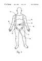
- FIG. 1shows a patient wearing a frame
- FIGS. 2A, 2 B and 2 Care axial sections of a Ga-67, a CT and a combined image of a chest of a patient;
- FIG. 3shows the patient of FIG. 1 with a stereotactic guide attached to the frame.
- the present inventionis of a method of stereotactic diagnosis and treatment of diseased tissue. Specifically, the present invention can be used to diagnose and treat cancerous tissue in tumors.
- FIG. 1shows a patient 10 wearing a rigid frame 12 .
- frame 12is rigidly secured to patient 10 .
- Frame 12bears thereon three markers 14 that include a material that shows up well in a high-resolution structural image of patient 10 and frame 12 .
- markers 14are lead blocks.
- markers 14are hollow plastic blocks filled with a solution of Gd-DTPA.
- Frame 12also bears three attachment points 16 for a stereotactic guide.
- a high-resolution structural image of the portion of patient 10 that contains the targetis acquired while patient 10 wears frame 12 as shown in FIG. 1 .
- markers 14are shown in FIG. 1 as three blocks only for illustrational clarity. Any suitable marker geometry or form may be used, for example, vertical and diagonal rods, as are used in the BRW CT localization frame sold by Radionics. Inc., of Burlington Mass. and by Sofamor Danek, of Elektra, Sweden, for stereotactic treatment of the brain.
- the positioning of markers 14 and attachment points 16 on frame 12 in FIG. 1also is only illustrative, as an example of appropriate positions of markers 14 and attachment points 16 for treatment of the abdomen of patient 10 .
- a lower resolution functional (e.g., nuclear medicine) image of the same portion of patient 10is acquired for the purpose of imaging the diseased tissue in the target.
- the preferred imaging modalityis nuclear imaging, and the patient is injected before imaging with a radiopharmaceutical that is preferentially taken up by cancerous tissue.
- radiopharmaceuticalsinclude Ga-67, Tc-99m MIBI, Tl-201, F-18 fluorodeoxyglucose and In-111 octreotide.
- the most preferred nuclear imaging modalitiesare tomographic modalities such as SPECT and PET.
- the functional and structural imagesthen are registered, for example as described in the paper by Weber and Ivanovic cited above, to produce a combined image.
- FIG. 2Ais an axial section of a Ga-67 image of a chest of a cancer patient, showing a tumor (yellow) containing a lymphoma (red).
- FIG. 2Bis an axial section of a CT image of the same patient's chest. The portion of FIG. 2B that corresponds to the lymphoma of FIG. 2A is outlined in yellow in FIG. 2 B. It can be seen that there is no visual contrast in FIG. 2B between the lymphoma and the rest of the tumor.
- FIG. 2Cis an axial section of a combined image, showing the functional image of FIG. 2A registered on the structural image of FIG. 2 B. FIG. 2C was produced by registering the image of FIG. 2A with the image of FIG. 2B as described in the paper by Weber and Ivanovic cited above. Biopsy and therapy directed towards the tumor should be aimed specifically at the lymphoma.
- FIG. 3shows patient 10 with a stereotactic guide 18 firmly and rigidly attached to frame 12 at attachment points 16 , and with an instrument 20 , such as a biopsy needle or a brachytherapy needle, mounted in stereotactic guide 18 and ready for insertion into the abdomen of patient 10 under the control of a computer 24 .
- Computer 24sends control signals, including the relevant Cartesian coordinates, to stereotactic guide 18 via a suitable electrical connection such as a cable 22 .
- Computer 24also includes a suitable display mechanism, such as a video terminal, for displaying the combined image.
- a physicianprograms the trajectory of instrument 22 in computer 24 in advance, with reference to the position of the target and the other internal organs of patient 10 , with reference to the diseased tissue in the target, and with reference to markers 14 , as seen in the combined image.
- Computer 24includes software that transforms the coordinate system of the combined image, as defined by markers 14 , into the coordinate system of stereotactic guide 18 .
- This softwareis used to transform the coordinates of the trajectory, which is defined by the physician in the coordinate system of the combined image, into the coordinate system of stereotactic guide 18 , to enable stereotactic guide 18 to steer instrument 20 along the desired trajectory.
- the physiciandesigns the trajectory to avoid vascular structures and other organs that should not be penetrated by instrument 20 .
- vascular structuressuch as blood vessels
- Another functional image of the portion of patient 10 that includes the targetis acquired, for the purpose of imaging the blood vessels.
- the preferred imaging modality for imaging vascular structuresis Tc-99m labeled red blood cells SPECT.
- patient 10is first injected with Tc-99m, to label the red blood cells of patient 10 .
- the SPECT image of the vascular structuresis registered with the structural image, and the resulting second combined image is displayed by computer 24 and is used by the physician in planning the trajectory of instrument 20 to avoid penetration of the imaged vascular structures.
- this second functional imageis a SPECT image obtained using Tc-99 DMSA.
- the SPECT image of the organ or organsis registered with the structural image, and the resulting second combined image is displayed by computer 24 and is used by the physician in planning the trajectory of instrument 20 to avoid penetration of the imaged organs.
- computer 24is programmed to avoid penetration of blood vessels and organs with high blood pool, and other organs that should not be penetrated, such as the gut, the kidneys, the pancreas and the heart.
Landscapes
- Health & Medical Sciences (AREA)
- Life Sciences & Earth Sciences (AREA)
- Engineering & Computer Science (AREA)
- Medical Informatics (AREA)
- Surgery (AREA)
- Veterinary Medicine (AREA)
- Nuclear Medicine, Radiotherapy & Molecular Imaging (AREA)
- Biomedical Technology (AREA)
- Heart & Thoracic Surgery (AREA)
- Molecular Biology (AREA)
- Animal Behavior & Ethology (AREA)
- General Health & Medical Sciences (AREA)
- Public Health (AREA)
- Pathology (AREA)
- Physics & Mathematics (AREA)
- Radiology & Medical Imaging (AREA)
- Biophysics (AREA)
- High Energy & Nuclear Physics (AREA)
- Optics & Photonics (AREA)
- Computer Vision & Pattern Recognition (AREA)
- Robotics (AREA)
- Oral & Maxillofacial Surgery (AREA)
- Pulmonology (AREA)
- Theoretical Computer Science (AREA)
- Magnetic Resonance Imaging Apparatus (AREA)
- Apparatus For Radiation Diagnosis (AREA)
- Measuring And Recording Apparatus For Diagnosis (AREA)
- Ultra Sonic Daignosis Equipment (AREA)
Abstract
Description
Claims (10)
Priority Applications (9)
| Application Number | Priority Date | Filing Date | Title |
|---|---|---|---|
| US09/253,779US6173201B1 (en) | 1999-02-22 | 1999-02-22 | Stereotactic diagnosis and treatment with reference to a combined image |
| AU28232/00AAU766301B2 (en) | 1999-02-22 | 2000-02-22 | A method and system for guiding a diagnostic or therapeutic instrument towards a target region inside the patient's body |
| PCT/IL2000/000113WO2000049958A1 (en) | 1999-02-22 | 2000-02-22 | A method and system for guiding a diagnostic or therapeutic instrument towards a target region inside the patient's body |
| EP00906576AEP1162921A1 (en) | 1999-02-22 | 2000-02-22 | A method and system for guiding a diagnostic or therapeutic instrument towards a target region inside the patient's body |
| IL14478600AIL144786A0 (en) | 1999-02-22 | 2000-02-22 | A method and system for guiding a diagnostic or therapeutic instrument towards a target region inside a patient's copy |
| CA002362981ACA2362981C (en) | 1999-02-22 | 2000-02-22 | A method and system for guiding a diagnostic or therapeutic instrument towards a target region inside the patient's body |
| JP2000600573AJP2002541885A (en) | 1999-02-22 | 2000-02-22 | Method and system for guiding a diagnostic or therapeutic instrument towards a target area inside a patient's body |
| US09/512,294US6368331B1 (en) | 1999-02-22 | 2000-02-24 | Method and system for guiding a diagnostic or therapeutic instrument towards a target region inside the patient's body |
| US09/900,042US6567687B2 (en) | 1999-02-22 | 2001-07-09 | Method and system for guiding a diagnostic or therapeutic instrument towards a target region inside the patient's body |
Applications Claiming Priority (1)
| Application Number | Priority Date | Filing Date | Title |
|---|---|---|---|
| US09/253,779US6173201B1 (en) | 1999-02-22 | 1999-02-22 | Stereotactic diagnosis and treatment with reference to a combined image |
Related Child Applications (1)
| Application Number | Title | Priority Date | Filing Date |
|---|---|---|---|
| US09/512,294Continuation-In-PartUS6368331B1 (en) | 1999-02-22 | 2000-02-24 | Method and system for guiding a diagnostic or therapeutic instrument towards a target region inside the patient's body |
Publications (1)
| Publication Number | Publication Date |
|---|---|
| US6173201B1true US6173201B1 (en) | 2001-01-09 |
Family
ID=22961669
Family Applications (1)
| Application Number | Title | Priority Date | Filing Date |
|---|---|---|---|
| US09/253,779Expired - LifetimeUS6173201B1 (en) | 1999-02-22 | 1999-02-22 | Stereotactic diagnosis and treatment with reference to a combined image |
Country Status (7)
| Country | Link |
|---|---|
| US (1) | US6173201B1 (en) |
| EP (1) | EP1162921A1 (en) |
| JP (1) | JP2002541885A (en) |
| AU (1) | AU766301B2 (en) |
| CA (1) | CA2362981C (en) |
| IL (1) | IL144786A0 (en) |
| WO (1) | WO2000049958A1 (en) |
Cited By (62)
| Publication number | Priority date | Publication date | Assignee | Title |
|---|---|---|---|---|
| US20030139661A1 (en)* | 2001-01-22 | 2003-07-24 | Yoav Kimchy | Ingestible device |
| US6640130B1 (en)* | 1999-07-02 | 2003-10-28 | Hypermed, Inc. | Integrated imaging apparatus |
| US20040015075A1 (en)* | 2000-08-21 | 2004-01-22 | Yoav Kimchy | Radioactive emission detector equipped with a position tracking system and utilization thereof with medical systems and in medical procedures |
| US20040054248A1 (en)* | 2000-08-21 | 2004-03-18 | Yoav Kimchy | Radioactive emission detector equipped with a position tracking system |
| US20040054278A1 (en)* | 2001-01-22 | 2004-03-18 | Yoav Kimchy | Ingestible pill |
| US20040057607A1 (en)* | 2002-09-19 | 2004-03-25 | Marcel Breeuwer | Display of image data information |
| US20040064148A1 (en)* | 2001-09-14 | 2004-04-01 | Wolfgang Daum | Navigation of medical instrument |
| US20040116807A1 (en)* | 2002-10-17 | 2004-06-17 | Roni Amrami | Blood vessels wall imaging catheter |
| US6799569B2 (en)* | 1999-12-20 | 2004-10-05 | Barbro Danielsson | Device for compression of the neck spine for medical imaging purposes |
| US20040204646A1 (en)* | 2002-11-04 | 2004-10-14 | V-Target Technologies Ltd. | Intracorporeal-imaging head |
| US20050038337A1 (en)* | 2003-08-11 | 2005-02-17 | Edwards Jerome R. | Methods, apparatuses, and systems useful in conducting image guided interventions |
| US20050055174A1 (en)* | 2000-08-21 | 2005-03-10 | V Target Ltd. | Radioactive emission detector equipped with a position tracking system and utilization thereof with medical systems and in medical procedures |
| US20050205792A1 (en)* | 2004-01-13 | 2005-09-22 | Benny Rousso | Multi-dimensional image reconstruction |
| US20050277823A1 (en)* | 2002-06-10 | 2005-12-15 | Robert Sutherland | Angiogram display overlay technique for tracking vascular intervention sites |
| US20060258933A1 (en)* | 2005-05-10 | 2006-11-16 | Advanced Clinical Solutions, Inc. | Method of defining a biological target for treatment |
| WO2007010534A2 (en) | 2005-07-19 | 2007-01-25 | Spectrum Dynamics Llc | Imaging protocols |
| US20070060799A1 (en)* | 2005-09-13 | 2007-03-15 | Lyon Torsten M | Apparatus and method for automatic image guided accuracy verification |
| US20070091428A1 (en)* | 2005-10-20 | 2007-04-26 | Wilson David L | Imaging system |
| US20070156047A1 (en)* | 2000-08-21 | 2007-07-05 | Michael Nagler | Radioactive-emission-measurement optimization to specific body structures |
| US20070161885A1 (en)* | 2003-12-17 | 2007-07-12 | Check-Cap Ltd. | Intra-lumen polyp detection |
| US20070265230A1 (en)* | 2006-05-11 | 2007-11-15 | Benny Rousso | Radiopharmaceuticals For Diagnosis And Therapy |
| US20080042067A1 (en)* | 2004-11-09 | 2008-02-21 | Spectrum Dynamics Llc | Radioimaging |
| US20080086059A1 (en)* | 2006-10-04 | 2008-04-10 | Cynthia Keppel | Method and apparatus for lesion localization using a dual modality x-ray/gamma biopsy system |
| US20080131362A1 (en)* | 2004-11-09 | 2008-06-05 | Spectrum Dynamics Llc | Radiopharmaceutical dispensing, administration, and imaging |
| US20080128626A1 (en)* | 2004-11-09 | 2008-06-05 | Spectrum Dynamics Llc | Radioimaging |
| US7397934B2 (en) | 2002-04-03 | 2008-07-08 | Segami S.A.R.L. | Registration of thoracic and abdominal imaging modalities |
| US20080221444A1 (en)* | 2007-03-07 | 2008-09-11 | Ritchie Paul G | Integrated Imaging and Biopsy System with Integrated Surgical, Therapy, and Diagnostic Devices |
| US20080260637A1 (en)* | 2004-11-17 | 2008-10-23 | Dalia Dickman | Methods of Detecting Prostate Cancer |
| US20090078875A1 (en)* | 2004-11-09 | 2009-03-26 | Spectrum Dynamics Llc | Radioimaging |
| US20090112086A1 (en)* | 2007-10-29 | 2009-04-30 | Spectrum Dynamics Llc | Prostate imaging |
| US20090152471A1 (en)* | 2005-11-09 | 2009-06-18 | Spectrum Dynamics Llc | Dynamic Spect Camera |
| US20090190807A1 (en)* | 2005-07-19 | 2009-07-30 | Spectrum Dynamics Llc | Reconstruction Stabilizer and Active Vision |
| US20090201291A1 (en)* | 2004-01-13 | 2009-08-13 | Spectrum Dynamics Llc | Gating With Anatomically Varying Durations |
| US7601966B2 (en) | 2006-06-28 | 2009-10-13 | Spectrum Dynamics Llc | Imaging techniques for reducing blind spots |
| US20090281566A1 (en)* | 2003-08-11 | 2009-11-12 | Edwards Jerome R | Bodily sealants and methods and apparatus for image-guided delivery of same |
| US7628957B1 (en)* | 2005-06-27 | 2009-12-08 | Moseley Patrick T | Carbon dioxide sensor |
| US20100012847A1 (en)* | 2008-07-16 | 2010-01-21 | Dilon Technologies, Inc. | Dual-capillary obturator for real-time verification in gamma guided stereotactic localization |
| US7652259B2 (en) | 2000-08-21 | 2010-01-26 | Spectrum Dynamics Llc | Apparatus and methods for imaging and attenuation correction |
| US20100021378A1 (en)* | 2004-01-13 | 2010-01-28 | Spectrum Dynamics Llc | Imaging protocols |
| US20100142774A1 (en)* | 2006-12-20 | 2010-06-10 | Spectrum Dynamics Llc | method, a system, and an apparatus for using and processing multidimensional data |
| US20100140483A1 (en)* | 2006-11-13 | 2010-06-10 | Benny Rousso | Radioimaging applications of and novel formulations of teboroxime |
| US20100245354A1 (en)* | 2004-01-13 | 2010-09-30 | Spectrum Dynamics Llc | Dynamic spect camera |
| US7853311B1 (en)* | 1999-04-23 | 2010-12-14 | 3M Innovative Properties Company | Surgical targeting system |
| US7872235B2 (en) | 2005-01-13 | 2011-01-18 | Spectrum Dynamics Llc | Multi-dimensional image reconstruction and analysis for expert-system diagnosis |
| US8204500B2 (en) | 2005-12-28 | 2012-06-19 | Starhome Gmbh | Optimal voicemail deposit for roaming cellular telephony |
| US8280124B2 (en) | 2004-06-01 | 2012-10-02 | Spectrum Dynamics Llc | Methods of view selection for radioactive emission measurements |
| US8338788B2 (en) | 2009-07-29 | 2012-12-25 | Spectrum Dynamics Llc | Method and system of optimized volumetric imaging |
| US8489176B1 (en)* | 2000-08-21 | 2013-07-16 | Spectrum Dynamics Llc | Radioactive emission detector equipped with a position tracking system and utilization thereof with medical systems and in medical procedures |
| WO2013168111A2 (en) | 2012-05-08 | 2013-11-14 | Spectrum Dynamics Llc | Nuclear medicine tomography systems, detectors and methods |
| US8615405B2 (en) | 2004-11-09 | 2013-12-24 | Biosensors International Group, Ltd. | Imaging system customization using data from radiopharmaceutical-associated data carrier |
| US8696549B2 (en) | 2010-08-20 | 2014-04-15 | Veran Medical Technologies, Inc. | Apparatus and method for four dimensional soft tissue navigation in endoscopic applications |
| US8781186B2 (en) | 2010-05-04 | 2014-07-15 | Pathfinder Therapeutics, Inc. | System and method for abdominal surface matching using pseudo-features |
| US8837793B2 (en) | 2005-07-19 | 2014-09-16 | Biosensors International Group, Ltd. | Reconstruction stabilizer and active vision |
| US9138165B2 (en) | 2012-02-22 | 2015-09-22 | Veran Medical Technologies, Inc. | Systems, methods and devices for forming respiratory-gated point cloud for four dimensional soft tissue navigation |
| US9316743B2 (en) | 2004-11-09 | 2016-04-19 | Biosensors International Group, Ltd. | System and method for radioactive emission measurement |
| US9392961B2 (en) | 2003-12-17 | 2016-07-19 | Check-Cap Ltd. | Intra-lumen polyp detection |
| US9844354B2 (en) | 2007-02-06 | 2017-12-19 | Check-Cap Ltd. | Intra-lumen polyp detection |
| US10136865B2 (en) | 2004-11-09 | 2018-11-27 | Spectrum Dynamics Medical Limited | Radioimaging using low dose isotope |
| US10617324B2 (en) | 2014-04-23 | 2020-04-14 | Veran Medical Technologies, Inc | Apparatuses and methods for endobronchial navigation to and confirmation of the location of a target tissue and percutaneous interception of the target tissue |
| US10624701B2 (en) | 2014-04-23 | 2020-04-21 | Veran Medical Technologies, Inc. | Apparatuses and methods for registering a real-time image feed from an imaging device to a steerable catheter |
| US11304630B2 (en) | 2005-09-13 | 2022-04-19 | Veran Medical Technologies, Inc. | Apparatus and method for image guided accuracy verification |
| US11684428B2 (en) | 2015-12-28 | 2023-06-27 | Xact Robotics Ltd. | Adjustable registration frame |
Families Citing this family (1)
| Publication number | Priority date | Publication date | Assignee | Title |
|---|---|---|---|---|
| CN113075599B (en)* | 2020-01-03 | 2023-05-16 | 上海联影医疗科技股份有限公司 | Magnetic resonance signal acquisition method, magnetic resonance system and medium |
Citations (1)
| Publication number | Priority date | Publication date | Assignee | Title |
|---|---|---|---|---|
| US5376795A (en) | 1990-07-09 | 1994-12-27 | Regents Of The University Of California | Emission-transmission imaging system using single energy and dual energy transmission and radionuclide emission data |
Family Cites Families (4)
| Publication number | Priority date | Publication date | Assignee | Title |
|---|---|---|---|---|
| US5517990A (en)* | 1992-11-30 | 1996-05-21 | The Cleveland Clinic Foundation | Stereotaxy wand and tool guide |
| US5682890A (en)* | 1995-01-26 | 1997-11-04 | Picker International, Inc. | Magnetic resonance stereotactic surgery with exoskeleton tissue stabilization |
| CA2250961C (en)* | 1997-02-14 | 2012-09-04 | Biosense, Inc. | X-ray guided surgical location system with extended mapping volume |
| US6021342A (en)* | 1997-06-30 | 2000-02-01 | Neorad A/S | Apparatus for assisting percutaneous computed tomography-guided surgical activity |
- 1999
- 1999-02-22USUS09/253,779patent/US6173201B1/ennot_activeExpired - Lifetime
- 2000
- 2000-02-22CACA002362981Apatent/CA2362981C/ennot_activeExpired - Lifetime
- 2000-02-22ILIL14478600Apatent/IL144786A0/ennot_activeIP Right Cessation
- 2000-02-22AUAU28232/00Apatent/AU766301B2/ennot_activeExpired
- 2000-02-22EPEP00906576Apatent/EP1162921A1/ennot_activeWithdrawn
- 2000-02-22WOPCT/IL2000/000113patent/WO2000049958A1/ennot_activeApplication Discontinuation
- 2000-02-22JPJP2000600573Apatent/JP2002541885A/enactivePending
Patent Citations (1)
| Publication number | Priority date | Publication date | Assignee | Title |
|---|---|---|---|---|
| US5376795A (en) | 1990-07-09 | 1994-12-27 | Regents Of The University Of California | Emission-transmission imaging system using single energy and dual energy transmission and radionuclide emission data |
Non-Patent Citations (14)
Cited By (149)
| Publication number | Priority date | Publication date | Assignee | Title |
|---|---|---|---|---|
| US7853311B1 (en)* | 1999-04-23 | 2010-12-14 | 3M Innovative Properties Company | Surgical targeting system |
| US6640130B1 (en)* | 1999-07-02 | 2003-10-28 | Hypermed, Inc. | Integrated imaging apparatus |
| US6799569B2 (en)* | 1999-12-20 | 2004-10-05 | Barbro Danielsson | Device for compression of the neck spine for medical imaging purposes |
| US20040054248A1 (en)* | 2000-08-21 | 2004-03-18 | Yoav Kimchy | Radioactive emission detector equipped with a position tracking system |
| US8489176B1 (en)* | 2000-08-21 | 2013-07-16 | Spectrum Dynamics Llc | Radioactive emission detector equipped with a position tracking system and utilization thereof with medical systems and in medical procedures |
| US8565860B2 (en) | 2000-08-21 | 2013-10-22 | Biosensors International Group, Ltd. | Radioactive emission detector equipped with a position tracking system |
| US8094894B2 (en) | 2000-08-21 | 2012-01-10 | Spectrum Dynamics Llc | Radioactive-emission-measurement optimization to specific body structures |
| US20070156047A1 (en)* | 2000-08-21 | 2007-07-05 | Michael Nagler | Radioactive-emission-measurement optimization to specific body structures |
| US8909325B2 (en) | 2000-08-21 | 2014-12-09 | Biosensors International Group, Ltd. | Radioactive emission detector equipped with a position tracking system and utilization thereof with medical systems and in medical procedures |
| US20040015075A1 (en)* | 2000-08-21 | 2004-01-22 | Yoav Kimchy | Radioactive emission detector equipped with a position tracking system and utilization thereof with medical systems and in medical procedures |
| US8620046B2 (en) | 2000-08-21 | 2013-12-31 | Biosensors International Group, Ltd. | Radioactive-emission-measurement optimization to specific body structures |
| US7826889B2 (en) | 2000-08-21 | 2010-11-02 | Spectrum Dynamics Llc | Radioactive emission detector equipped with a position tracking system and utilization thereof with medical systems and in medical procedures |
| US20050055174A1 (en)* | 2000-08-21 | 2005-03-10 | V Target Ltd. | Radioactive emission detector equipped with a position tracking system and utilization thereof with medical systems and in medical procedures |
| US7652259B2 (en) | 2000-08-21 | 2010-01-26 | Spectrum Dynamics Llc | Apparatus and methods for imaging and attenuation correction |
| US9370333B2 (en) | 2000-08-21 | 2016-06-21 | Biosensors International Group, Ltd. | Radioactive-emission-measurement optimization to specific body structures |
| US20030139661A1 (en)* | 2001-01-22 | 2003-07-24 | Yoav Kimchy | Ingestible device |
| US8036731B2 (en) | 2001-01-22 | 2011-10-11 | Spectrum Dynamics Llc | Ingestible pill for diagnosing a gastrointestinal tract |
| US8055329B2 (en) | 2001-01-22 | 2011-11-08 | Spectrum Dynamics Llc | Ingestible device for radioimaging of the gastrointestinal tract |
| US20040054278A1 (en)* | 2001-01-22 | 2004-03-18 | Yoav Kimchy | Ingestible pill |
| US20060122630A1 (en)* | 2001-09-14 | 2006-06-08 | Wolfgang Daum | Navigation of medical instrument |
| US6989015B2 (en) | 2001-09-14 | 2006-01-24 | Invivo Germany Gmbh | Navigation of medical instrument |
| US20040064148A1 (en)* | 2001-09-14 | 2004-04-01 | Wolfgang Daum | Navigation of medical instrument |
| US7397934B2 (en) | 2002-04-03 | 2008-07-08 | Segami S.A.R.L. | Registration of thoracic and abdominal imaging modalities |
| US20050277823A1 (en)* | 2002-06-10 | 2005-12-15 | Robert Sutherland | Angiogram display overlay technique for tracking vascular intervention sites |
| US7106892B2 (en) | 2002-09-19 | 2006-09-12 | Koninklijke Philips Electronics, N.V. | Display of image data information |
| WO2004026140A3 (en)* | 2002-09-19 | 2004-06-03 | Koninkl Philips Electronics Nv | Display of image data information |
| US20040057607A1 (en)* | 2002-09-19 | 2004-03-25 | Marcel Breeuwer | Display of image data information |
| US20040116807A1 (en)* | 2002-10-17 | 2004-06-17 | Roni Amrami | Blood vessels wall imaging catheter |
| US20040204646A1 (en)* | 2002-11-04 | 2004-10-14 | V-Target Technologies Ltd. | Intracorporeal-imaging head |
| US8150495B2 (en) | 2003-08-11 | 2012-04-03 | Veran Medical Technologies, Inc. | Bodily sealants and methods and apparatus for image-guided delivery of same |
| US20080298655A1 (en)* | 2003-08-11 | 2008-12-04 | Edwards Jerome R | Methods, apparatuses, and systems useful in conducting image guided interventions |
| US20050038337A1 (en)* | 2003-08-11 | 2005-02-17 | Edwards Jerome R. | Methods, apparatuses, and systems useful in conducting image guided interventions |
| US7398116B2 (en) | 2003-08-11 | 2008-07-08 | Veran Medical Technologies, Inc. | Methods, apparatuses, and systems useful in conducting image guided interventions |
| US7853307B2 (en) | 2003-08-11 | 2010-12-14 | Veran Medical Technologies, Inc. | Methods, apparatuses, and systems useful in conducting image guided interventions |
| US11426134B2 (en) | 2003-08-11 | 2022-08-30 | Veran Medical Technologies, Inc. | Methods, apparatuses and systems useful in conducting image guided interventions |
| US20110054309A1 (en)* | 2003-08-11 | 2011-03-03 | Edwards Jerome R | Methods, apparatuses, and systems useful in conductng image guided interventions |
| US20090281566A1 (en)* | 2003-08-11 | 2009-11-12 | Edwards Jerome R | Bodily sealants and methods and apparatus for image-guided delivery of same |
| US11154283B2 (en) | 2003-08-11 | 2021-10-26 | Veran Medical Technologies, Inc. | Bodily sealants and methods and apparatus for image-guided delivery of same |
| US10470725B2 (en) | 2003-08-11 | 2019-11-12 | Veran Medical Technologies, Inc. | Method, apparatuses, and systems useful in conducting image guided interventions |
| US8483801B2 (en) | 2003-08-11 | 2013-07-09 | Veran Medical Technologies, Inc. | Methods, apparatuses, and systems useful in conducting image guided interventions |
| US7787926B2 (en) | 2003-12-17 | 2010-08-31 | Check-Cap LLC | Intra-lumen polyp detection |
| US20070161885A1 (en)* | 2003-12-17 | 2007-07-12 | Check-Cap Ltd. | Intra-lumen polyp detection |
| US9392961B2 (en) | 2003-12-17 | 2016-07-19 | Check-Cap Ltd. | Intra-lumen polyp detection |
| US7176466B2 (en) | 2004-01-13 | 2007-02-13 | Spectrum Dynamics Llc | Multi-dimensional image reconstruction |
| US20090201291A1 (en)* | 2004-01-13 | 2009-08-13 | Spectrum Dynamics Llc | Gating With Anatomically Varying Durations |
| US10964075B2 (en) | 2004-01-13 | 2021-03-30 | Spectrum Dynamics Llc | Gating with anatomically varying durations |
| US7968851B2 (en) | 2004-01-13 | 2011-06-28 | Spectrum Dynamics Llc | Dynamic spect camera |
| US8676292B2 (en) | 2004-01-13 | 2014-03-18 | Biosensors International Group, Ltd. | Multi-dimensional image reconstruction |
| US20070194241A1 (en)* | 2004-01-13 | 2007-08-23 | Spectrum Dynamics Llc | Multi-dimensional image reconstruction |
| US20050205792A1 (en)* | 2004-01-13 | 2005-09-22 | Benny Rousso | Multi-dimensional image reconstruction |
| US20100021378A1 (en)* | 2004-01-13 | 2010-01-28 | Spectrum Dynamics Llc | Imaging protocols |
| US9470801B2 (en) | 2004-01-13 | 2016-10-18 | Spectrum Dynamics Llc | Gating with anatomically varying durations |
| US9040016B2 (en) | 2004-01-13 | 2015-05-26 | Biosensors International Group, Ltd. | Diagnostic kit and methods for radioimaging myocardial perfusion |
| US20100245354A1 (en)* | 2004-01-13 | 2010-09-30 | Spectrum Dynamics Llc | Dynamic spect camera |
| US8280124B2 (en) | 2004-06-01 | 2012-10-02 | Spectrum Dynamics Llc | Methods of view selection for radioactive emission measurements |
| US9943278B2 (en) | 2004-06-01 | 2018-04-17 | Spectrum Dynamics Medical Limited | Radioactive-emission-measurement optimization to specific body structures |
| US9316743B2 (en) | 2004-11-09 | 2016-04-19 | Biosensors International Group, Ltd. | System and method for radioactive emission measurement |
| US8571881B2 (en) | 2004-11-09 | 2013-10-29 | Spectrum Dynamics, Llc | Radiopharmaceutical dispensing, administration, and imaging |
| US20090078875A1 (en)* | 2004-11-09 | 2009-03-26 | Spectrum Dynamics Llc | Radioimaging |
| US8606349B2 (en) | 2004-11-09 | 2013-12-10 | Biosensors International Group, Ltd. | Radioimaging using low dose isotope |
| US8586932B2 (en) | 2004-11-09 | 2013-11-19 | Spectrum Dynamics Llc | System and method for radioactive emission measurement |
| US8620679B2 (en) | 2004-11-09 | 2013-12-31 | Biosensors International Group, Ltd. | Radiopharmaceutical dispensing, administration, and imaging |
| US20080128626A1 (en)* | 2004-11-09 | 2008-06-05 | Spectrum Dynamics Llc | Radioimaging |
| US20080131362A1 (en)* | 2004-11-09 | 2008-06-05 | Spectrum Dynamics Llc | Radiopharmaceutical dispensing, administration, and imaging |
| US8000773B2 (en) | 2004-11-09 | 2011-08-16 | Spectrum Dynamics Llc | Radioimaging |
| US8445851B2 (en) | 2004-11-09 | 2013-05-21 | Spectrum Dynamics Llc | Radioimaging |
| US20080042067A1 (en)* | 2004-11-09 | 2008-02-21 | Spectrum Dynamics Llc | Radioimaging |
| US10136865B2 (en) | 2004-11-09 | 2018-11-27 | Spectrum Dynamics Medical Limited | Radioimaging using low dose isotope |
| US8615405B2 (en) | 2004-11-09 | 2013-12-24 | Biosensors International Group, Ltd. | Imaging system customization using data from radiopharmaceutical-associated data carrier |
| US8423125B2 (en) | 2004-11-09 | 2013-04-16 | Spectrum Dynamics Llc | Radioimaging |
| US8748826B2 (en) | 2004-11-17 | 2014-06-10 | Biosensor International Group, Ltd. | Radioimaging methods using teboroxime and thallium |
| US20080260637A1 (en)* | 2004-11-17 | 2008-10-23 | Dalia Dickman | Methods of Detecting Prostate Cancer |
| US7872235B2 (en) | 2005-01-13 | 2011-01-18 | Spectrum Dynamics Llc | Multi-dimensional image reconstruction and analysis for expert-system diagnosis |
| US7831293B2 (en)* | 2005-05-10 | 2010-11-09 | Advanced Clinical Solutions, Inc. | Method of defining a biological target for treatment |
| US20060258933A1 (en)* | 2005-05-10 | 2006-11-16 | Advanced Clinical Solutions, Inc. | Method of defining a biological target for treatment |
| US7628957B1 (en)* | 2005-06-27 | 2009-12-08 | Moseley Patrick T | Carbon dioxide sensor |
| US8644910B2 (en) | 2005-07-19 | 2014-02-04 | Biosensors International Group, Ltd. | Imaging protocols |
| US20090190807A1 (en)* | 2005-07-19 | 2009-07-30 | Spectrum Dynamics Llc | Reconstruction Stabilizer and Active Vision |
| US8111886B2 (en) | 2005-07-19 | 2012-02-07 | Spectrum Dynamics Llc | Reconstruction stabilizer and active vision |
| US8837793B2 (en) | 2005-07-19 | 2014-09-16 | Biosensors International Group, Ltd. | Reconstruction stabilizer and active vision |
| US20090304582A1 (en)* | 2005-07-19 | 2009-12-10 | Spectrum Dynamics Llc | Imaging Protocols |
| WO2007010534A2 (en) | 2005-07-19 | 2007-01-25 | Spectrum Dynamics Llc | Imaging protocols |
| US10617332B2 (en) | 2005-09-13 | 2020-04-14 | Veran Medical Technologies, Inc. | Apparatus and method for image guided accuracy verification |
| US20070060799A1 (en)* | 2005-09-13 | 2007-03-15 | Lyon Torsten M | Apparatus and method for automatic image guided accuracy verification |
| US20110208044A1 (en)* | 2005-09-13 | 2011-08-25 | Edwards Jerome R | Apparatus and method for image guided accuracy verification |
| US20070066881A1 (en)* | 2005-09-13 | 2007-03-22 | Edwards Jerome R | Apparatus and method for image guided accuracy verification |
| US20110184276A1 (en)* | 2005-09-13 | 2011-07-28 | Lyon Torsten M | Apparatus and method for automatic image guided accuracy verification |
| US7962193B2 (en) | 2005-09-13 | 2011-06-14 | Veran Medical Technologies, Inc. | Apparatus and method for image guided accuracy verification |
| US7920909B2 (en) | 2005-09-13 | 2011-04-05 | Veran Medical Technologies, Inc. | Apparatus and method for automatic image guided accuracy verification |
| US9218664B2 (en) | 2005-09-13 | 2015-12-22 | Veran Medical Technologies, Inc. | Apparatus and method for image guided accuracy verification |
| US11304630B2 (en) | 2005-09-13 | 2022-04-19 | Veran Medical Technologies, Inc. | Apparatus and method for image guided accuracy verification |
| US9218663B2 (en) | 2005-09-13 | 2015-12-22 | Veran Medical Technologies, Inc. | Apparatus and method for automatic image guided accuracy verification |
| US11304629B2 (en) | 2005-09-13 | 2022-04-19 | Veran Medical Technologies, Inc. | Apparatus and method for image guided accuracy verification |
| US20110091088A1 (en)* | 2005-10-20 | 2011-04-21 | Wilson David L | Imaging system |
| US20070091428A1 (en)* | 2005-10-20 | 2007-04-26 | Wilson David L | Imaging system |
| US8238632B2 (en)* | 2005-10-20 | 2012-08-07 | Wilson David L | Imaging system |
| US7831075B2 (en)* | 2005-10-20 | 2010-11-09 | Case Western Reserve University | Imaging system |
| US20090152471A1 (en)* | 2005-11-09 | 2009-06-18 | Spectrum Dynamics Llc | Dynamic Spect Camera |
| US7705316B2 (en) | 2005-11-09 | 2010-04-27 | Spectrum Dynamics Llc | Dynamic SPECT camera |
| US8204500B2 (en) | 2005-12-28 | 2012-06-19 | Starhome Gmbh | Optimal voicemail deposit for roaming cellular telephony |
| US8894974B2 (en) | 2006-05-11 | 2014-11-25 | Spectrum Dynamics Llc | Radiopharmaceuticals for diagnosis and therapy |
| US20070265230A1 (en)* | 2006-05-11 | 2007-11-15 | Benny Rousso | Radiopharmaceuticals For Diagnosis And Therapy |
| US7601966B2 (en) | 2006-06-28 | 2009-10-13 | Spectrum Dynamics Llc | Imaging techniques for reducing blind spots |
| US20080086059A1 (en)* | 2006-10-04 | 2008-04-10 | Cynthia Keppel | Method and apparatus for lesion localization using a dual modality x-ray/gamma biopsy system |
| US8610075B2 (en) | 2006-11-13 | 2013-12-17 | Biosensors International Group Ltd. | Radioimaging applications of and novel formulations of teboroxime |
| US20100140483A1 (en)* | 2006-11-13 | 2010-06-10 | Benny Rousso | Radioimaging applications of and novel formulations of teboroxime |
| US20100142774A1 (en)* | 2006-12-20 | 2010-06-10 | Spectrum Dynamics Llc | method, a system, and an apparatus for using and processing multidimensional data |
| US9275451B2 (en) | 2006-12-20 | 2016-03-01 | Biosensors International Group, Ltd. | Method, a system, and an apparatus for using and processing multidimensional data |
| US9844354B2 (en) | 2007-02-06 | 2017-12-19 | Check-Cap Ltd. | Intra-lumen polyp detection |
| US20080221444A1 (en)* | 2007-03-07 | 2008-09-11 | Ritchie Paul G | Integrated Imaging and Biopsy System with Integrated Surgical, Therapy, and Diagnostic Devices |
| US20080221478A1 (en)* | 2007-03-07 | 2008-09-11 | Ritchie Paul G | Integrated Imaging and Biopsy System with Integrated Control Interface |
| US20080221479A1 (en)* | 2007-03-07 | 2008-09-11 | Ritchie Paul G | Integrated Imaging and Biopsy System with Integrated Utilities |
| US20080221443A1 (en)* | 2007-03-07 | 2008-09-11 | Ritchie Paul G | Integrated Imaging and Biopsy System with Ancillary Device Authentication |
| WO2008109247A1 (en)* | 2007-03-07 | 2008-09-12 | Ethicon Endo-Surgery, Inc | Integrated imaging and biopsy system |
| US8521253B2 (en) | 2007-10-29 | 2013-08-27 | Spectrum Dynamics Llc | Prostate imaging |
| US20090112086A1 (en)* | 2007-10-29 | 2009-04-30 | Spectrum Dynamics Llc | Prostate imaging |
| US20100012847A1 (en)* | 2008-07-16 | 2010-01-21 | Dilon Technologies, Inc. | Dual-capillary obturator for real-time verification in gamma guided stereotactic localization |
| US7795591B2 (en)* | 2008-07-16 | 2010-09-14 | Dilon Technologies, Inc. | Dual-capillary obturator for real-time verification in gamma guided stereotactic localization |
| US8338788B2 (en) | 2009-07-29 | 2012-12-25 | Spectrum Dynamics Llc | Method and system of optimized volumetric imaging |
| US8748827B2 (en) | 2009-07-29 | 2014-06-10 | Biosensors International Group, Ltd. | Method and system of optimized volumetric imaging |
| US8492725B2 (en) | 2009-07-29 | 2013-07-23 | Biosensors International Group Ltd. | Method and system of optimized volumetric imaging |
| US8781186B2 (en) | 2010-05-04 | 2014-07-15 | Pathfinder Therapeutics, Inc. | System and method for abdominal surface matching using pseudo-features |
| US10898057B2 (en) | 2010-08-20 | 2021-01-26 | Veran Medical Technologies, Inc. | Apparatus and method for airway registration and navigation |
| US10165928B2 (en) | 2010-08-20 | 2019-01-01 | Mark Hunter | Systems, instruments, and methods for four dimensional soft tissue navigation |
| US8696549B2 (en) | 2010-08-20 | 2014-04-15 | Veran Medical Technologies, Inc. | Apparatus and method for four dimensional soft tissue navigation in endoscopic applications |
| US10264947B2 (en) | 2010-08-20 | 2019-04-23 | Veran Medical Technologies, Inc. | Apparatus and method for airway registration and navigation |
| US11109740B2 (en) | 2010-08-20 | 2021-09-07 | Veran Medical Technologies, Inc. | Apparatus and method for four dimensional soft tissue navigation in endoscopic applications |
| US11690527B2 (en) | 2010-08-20 | 2023-07-04 | Veran Medical Technologies, Inc. | Apparatus and method for four dimensional soft tissue navigation in endoscopic applications |
| US10460437B2 (en) | 2012-02-22 | 2019-10-29 | Veran Medical Technologies, Inc. | Method for placing a localization element in an organ of a patient for four dimensional soft tissue navigation |
| US11551359B2 (en) | 2012-02-22 | 2023-01-10 | Veran Medical Technologies, Inc | Systems, methods and devices for forming respiratory-gated point cloud for four dimensional soft tissue navigation |
| US9972082B2 (en) | 2012-02-22 | 2018-05-15 | Veran Medical Technologies, Inc. | Steerable surgical catheter having biopsy devices and related systems and methods for four dimensional soft tissue navigation |
| US10977789B2 (en) | 2012-02-22 | 2021-04-13 | Veran Medical Technologies, Inc. | Systems, methods and devices for forming respiratory-gated point cloud for four dimensional soft tissue navigation |
| US11830198B2 (en) | 2012-02-22 | 2023-11-28 | Veran Medical Technologies, Inc. | Systems, methods and devices for forming respiratory-gated point cloud for four dimensional soft tissue navigation |
| US10140704B2 (en) | 2012-02-22 | 2018-11-27 | Veran Medical Technologies, Inc. | Systems, methods and devices for forming respiratory-gated point cloud for four dimensional soft tissue navigation |
| US11403753B2 (en) | 2012-02-22 | 2022-08-02 | Veran Medical Technologies, Inc. | Surgical catheter having side exiting medical instrument and related systems and methods for four dimensional soft tissue navigation |
| US9138165B2 (en) | 2012-02-22 | 2015-09-22 | Veran Medical Technologies, Inc. | Systems, methods and devices for forming respiratory-gated point cloud for four dimensional soft tissue navigation |
| US10249036B2 (en) | 2012-02-22 | 2019-04-02 | Veran Medical Technologies, Inc. | Surgical catheter having side exiting medical instrument and related systems and methods for four dimensional soft tissue navigation |
| US11534115B2 (en) | 2012-05-08 | 2022-12-27 | Speetrum Dynamics Medical Limited | Counterbalancing of detectors for nuclear medicine tomography systems |
| US11317877B2 (en) | 2012-05-08 | 2022-05-03 | Spectrum Dynamics Medical Limited | Collimator |
| WO2013168111A2 (en) | 2012-05-08 | 2013-11-14 | Spectrum Dynamics Llc | Nuclear medicine tomography systems, detectors and methods |
| US10987069B2 (en) | 2012-05-08 | 2021-04-27 | Spectrum Dynamics Medical Limited | Nuclear medicine tomography systems, detectors and methods |
| EP3647822A2 (en) | 2012-05-08 | 2020-05-06 | Spectrum Dynamics Medical Limited | Nuclear medicine tomography systems, detectors and methods |
| US11806176B2 (en) | 2012-05-08 | 2023-11-07 | Spectrum Dynamics Medical Limited | Proximity detection |
| US11857353B2 (en) | 2012-05-08 | 2024-01-02 | Spectrum Dynamics Medical Limited | Gantry rotation |
| US12251249B2 (en) | 2012-05-08 | 2025-03-18 | Spectrum Dynamics Medical Limited | Gantry rotation |
| US11553968B2 (en) | 2014-04-23 | 2023-01-17 | Veran Medical Technologies, Inc. | Apparatuses and methods for registering a real-time image feed from an imaging device to a steerable catheter |
| US10624701B2 (en) | 2014-04-23 | 2020-04-21 | Veran Medical Technologies, Inc. | Apparatuses and methods for registering a real-time image feed from an imaging device to a steerable catheter |
| US10617324B2 (en) | 2014-04-23 | 2020-04-14 | Veran Medical Technologies, Inc | Apparatuses and methods for endobronchial navigation to and confirmation of the location of a target tissue and percutaneous interception of the target tissue |
| US11684428B2 (en) | 2015-12-28 | 2023-06-27 | Xact Robotics Ltd. | Adjustable registration frame |
Also Published As
| Publication number | Publication date |
|---|---|
| CA2362981A1 (en) | 2000-08-31 |
| WO2000049958A1 (en) | 2000-08-31 |
| JP2002541885A (en) | 2002-12-10 |
| EP1162921A1 (en) | 2001-12-19 |
| IL144786A0 (en) | 2002-12-01 |
| CA2362981C (en) | 2009-01-06 |
| AU2823200A (en) | 2000-09-14 |
| AU766301B2 (en) | 2003-10-16 |
Similar Documents
| Publication | Publication Date | Title |
|---|---|---|
| US6173201B1 (en) | Stereotactic diagnosis and treatment with reference to a combined image | |
| US6368331B1 (en) | Method and system for guiding a diagnostic or therapeutic instrument towards a target region inside the patient's body | |
| Lecchi et al. | Current concepts on imaging in radiotherapy | |
| EP1795142B1 (en) | Medical tracking system using a gamma camera | |
| US7110587B1 (en) | Registration of nuclear medicine images | |
| US6473634B1 (en) | Medical imaging at two temporal resolutions for tumor treatment planning | |
| US5871013A (en) | Registration of nuclear medicine images | |
| US8082023B2 (en) | Tissue interventions using nuclear-emission image guidance | |
| EP3021940B1 (en) | Portal imaging for brachytherapy | |
| US20070153969A1 (en) | Radiotherapeutic device | |
| US20120281898A1 (en) | Method of identification of an element in two or more images | |
| EP3962367A1 (en) | Systems, methods, and devices for registering and tracking organs during interventional procedures | |
| Julow et al. | The application of image fusion in stereotactic brachytherapy of brain tumours | |
| US20070167700A1 (en) | Method for accurate in vivo delivery of a therapeutic agent to a target area of an organ | |
| CN109770902A (en) | An automatic tumor localization system for nuclear magnetic resonance environment | |
| Bolcaen et al. | PET and MRI guided irradiation of a glioblastoma rat model using a micro-irradiator | |
| Yin et al. | Physics and imaging for targeting of oligometastases | |
| US20230090468A1 (en) | Systems, methods, and devices for registering and tracking organs during interventional procedures | |
| Walter et al. | Technical issues of [18f] FET-PET imaging for radiation therapy planning in malignant glioma patients–a review | |
| US20130158388A1 (en) | Needle guidance for molecular imaging | |
| Mountz et al. | Validation of a reference method for correlation of anatomic and functional brain images | |
| Choi et al. | Electromagnetic tracking-based ultrasound/computed tomography fusion imaging in dogs: preliminary application to ocular and periocular regions | |
| Liang et al. | Reconstruction of brachytherapy catheters and needles using EM sensor-based navigation system | |
| Wang et al. | Treatment of liver metastases using an internal target volume method for stereotactic body radiotherapy | |
| Wong | Imaging modalities |
Legal Events
| Date | Code | Title | Description |
|---|---|---|---|
| AS | Assignment | Owner name:V-TARGET LTD., ISRAEL Free format text:ASSIGNMENT OF ASSIGNORS INTEREST;ASSIGNOR:FRONT, YARON;REEL/FRAME:011228/0218 Effective date:20000926 | |
| FEPP | Fee payment procedure | Free format text:PAYOR NUMBER ASSIGNED (ORIGINAL EVENT CODE: ASPN); ENTITY STATUS OF PATENT OWNER: SMALL ENTITY | |
| STCF | Information on status: patent grant | Free format text:PATENTED CASE | |
| AS | Assignment | Owner name:V-TARGET TECHNOLOGIES, ISRAEL Free format text:RE-RECORD TO CORRECT THE EXECUTION DATE AND THE RECEIVING PARTY'S NAME, PREVIOUSLY RECORD AT REEL 011228, FRAME 0218.;ASSIGNOR:FRONT, YARON;REEL/FRAME:014186/0757 Effective date:20030525 | |
| FPAY | Fee payment | Year of fee payment:4 | |
| FEPP | Fee payment procedure | Free format text:PAYOR NUMBER ASSIGNED (ORIGINAL EVENT CODE: ASPN); ENTITY STATUS OF PATENT OWNER: SMALL ENTITY Free format text:PAYER NUMBER DE-ASSIGNED (ORIGINAL EVENT CODE: RMPN); ENTITY STATUS OF PATENT OWNER: SMALL ENTITY | |
| FPAY | Fee payment | Year of fee payment:8 | |
| AS | Assignment | Owner name:SPECTRUM DYNAMICS (ISRAEL) LTD., ISRAEL Free format text:CHANGE OF NAME;ASSIGNOR:V-TARGET TECHNOLOGIES LTD.;REEL/FRAME:022012/0062 Effective date:20050606 | |
| AS | Assignment | Owner name:SPECTRUM DYNAMICS LLC, NEW YORK Free format text:ASSIGNMENT OF ASSIGNORS INTEREST;ASSIGNOR:SPECTRUM DYNAMICS (ISRAEL) LTD.;REEL/FRAME:022012/0619 Effective date:20081223 | |
| AS | Assignment | Owner name:MEDINVEST CAPITAL S.A R.L., LUXEMBOURG Free format text:SECURITY AGREEMENT;ASSIGNOR:SPECTRUM DYNAMICS LLC;REEL/FRAME:022408/0833 Effective date:20090309 Owner name:MEDINVEST CAPITAL S.A R.L.,LUXEMBOURG Free format text:SECURITY AGREEMENT;ASSIGNOR:SPECTRUM DYNAMICS LLC;REEL/FRAME:022408/0833 Effective date:20090309 | |
| FPAY | Fee payment | Year of fee payment:12 | |
| AS | Assignment | Owner name:BIOSENSORS GROUP INTERNATIONAL, LTD., BERMUDA Free format text:ASSIGNMENT OF ASSIGNORS INTEREST;ASSIGNOR:SPECTRUM DYNAMICS LLC;REEL/FRAME:030869/0299 Effective date:20130517 | |
| AS | Assignment | Owner name:BIOSENSORS INTERNATIONAL GROUP, LTD., BERMUDA Free format text:CORRECTIVE ASSIGNMENT TO CORRECT THE NAME OF ASSIGNEE TO BIOSENSORS INTERNATIONAL GROUP, LTD. PREVIOUSLY RECORDED ON REEL 030869 FRAME 0299. ASSIGNOR(S) HEREBY CONFIRMS THE BIOSENSORS SIGNATURE PAGE, SCHEDULE A HEADER AND EDITED 8/2/2013 CORRECTION.;ASSIGNOR:SPECTRUM DYNAMICS LLC;REEL/FRAME:030954/0949 Effective date:20130517 | |
| AS | Assignment | Owner name:SPECTRUM DYNAMICS LLC, NEW YORK Free format text:RELEASE OF INTELLECTUAL PROPERTY SECURITY AGREEMENT;ASSIGNORS:MEDINVEST CAPITAL S.A.R.L.;MORGAN STANLEY BANK INTERNATIONAL LIMITED;REEL/FRAME:035387/0005 Effective date:20150331 | |
| AS | Assignment | Owner name:SPECTRUM DYNAMICS MEDICAL LIMITED, VIRGIN ISLANDS, BRITISH Free format text:ASSIGNMENT OF ASSIGNORS INTEREST;ASSIGNOR:BIOSENSORS INTERNATIONAL GROUP, LTD.;REEL/FRAME:045186/0004 Effective date:20170531 Owner name:SPECTRUM DYNAMICS MEDICAL LIMITED, VIRGIN ISLANDS, Free format text:ASSIGNMENT OF ASSIGNORS INTEREST;ASSIGNOR:BIOSENSORS INTERNATIONAL GROUP, LTD.;REEL/FRAME:045186/0004 Effective date:20170531 |