US6172088B1 - Testosterone compounds and use for the protection of neurons - Google Patents
Testosterone compounds and use for the protection of neuronsDownload PDFInfo
- Publication number
- US6172088B1 US6172088B1US09/198,416US19841698AUS6172088B1US 6172088 B1US6172088 B1US 6172088B1US 19841698 AUS19841698 AUS 19841698AUS 6172088 B1US6172088 B1US 6172088B1
- Authority
- US
- United States
- Prior art keywords
- inhibitor
- testosterone
- estrogen
- compounds
- androgen
- Prior art date
- Legal status (The legal status is an assumption and is not a legal conclusion. Google has not performed a legal analysis and makes no representation as to the accuracy of the status listed.)
- Expired - Fee Related
Links
- 210000002569neuronAnatomy0.000titledescription10
- 150000003515testosteronesChemical class0.000title1
- MUMGGOZAMZWBJJ-DYKIIFRCSA-NTestostosteroneChemical compoundO=C1CC[C@]2(C)[C@H]3CC[C@](C)([C@H](CC4)O)[C@@H]4[C@@H]3CCC2=C1MUMGGOZAMZWBJJ-DYKIIFRCSA-N0.000claimsabstractdescription140
- 229960003604testosteroneDrugs0.000claimsabstractdescription71
- 150000001875compoundsChemical class0.000claimsabstractdescription30
- 229940011871estrogenDrugs0.000claimsabstractdescription23
- 239000000262estrogenSubstances0.000claimsabstractdescription23
- 239000003098androgenSubstances0.000claimsabstractdescription11
- 239000003112inhibitorSubstances0.000claimsdescription40
- 238000000034methodMethods0.000claimsdescription21
- 230000000694effectsEffects0.000claimsdescription15
- 208000015122neurodegenerative diseaseDiseases0.000claimsdescription13
- 230000004112neuroprotectionEffects0.000claimsdescription11
- 239000000556agonistSubstances0.000claimsdescription8
- 239000008194pharmaceutical compositionSubstances0.000claimsdescription8
- 208000037265diseases, disorders, signs and symptomsDiseases0.000claimsdescription7
- 229940088597hormoneDrugs0.000claimsdescription7
- 239000005556hormoneSubstances0.000claimsdescription7
- 230000015572biosynthetic processEffects0.000claimsdescription6
- 238000003786synthesis reactionMethods0.000claimsdescription6
- 2390000026775-alpha reductase inhibitorSubstances0.000claimsdescription5
- 230000001592luteinising effectEffects0.000claimsdescription5
- 239000000203mixtureSubstances0.000claimsdescription5
- 2299401131785 Alpha reductase inhibitorDrugs0.000claimsdescription4
- 230000002411adverseEffects0.000claimsdescription4
- 239000003956nonsteroidal anti androgenSubstances0.000claimsdescription3
- 230000003637steroidlikeEffects0.000claimsdescription3
- 238000009472formulationMethods0.000claimsdescription2
- 238000007918intramuscular administrationMethods0.000claimsdescription2
- 238000001990intravenous administrationMethods0.000claimsdescription2
- 238000007920subcutaneous administrationMethods0.000claimsdescription2
- NMJREATYWWNIKX-UHFFFAOYSA-NGnRHChemical compoundC1CCC(C(=O)NCC(N)=O)N1C(=O)C(CC(C)C)NC(=O)C(CC=1C2=CC=CC=C2NC=1)NC(=O)CNC(=O)C(NC(=O)C(CO)NC(=O)C(CC=1C2=CC=CC=C2NC=1)NC(=O)C(CC=1NC=NC=1)NC(=O)C1NC(=O)CC1)CC1=CC=C(O)C=C1NMJREATYWWNIKX-UHFFFAOYSA-N0.000claims5
- 101000857870Squalus acanthias GonadoliberinProteins0.000claims5
- 239000002474gonadorelin antagonistSubstances0.000claims3
- 229940121381gonadotrophin releasing hormone (gnrh) antagonistsDrugs0.000claims3
- 108090000765processed proteins & peptidesProteins0.000claims3
- 239000003488releasing hormoneSubstances0.000claims3
- 125000001792phenanthrenyl groupChemical classC1(=CC=CC=2C3=CC=CC=C3C=CC12)*0.000claims2
- 230000000302ischemic effectEffects0.000abstractdescription24
- 208000024827Alzheimer diseaseDiseases0.000abstractdescription5
- 206010028980NeoplasmDiseases0.000abstractdescription3
- 208000018737Parkinson diseaseDiseases0.000abstractdescription3
- 239000003795chemical substances by applicationSubstances0.000abstractdescription2
- 230000035939shockEffects0.000abstractdescription2
- 230000001225therapeutic effectEffects0.000abstractdescription2
- 239000003263anabolic agentSubstances0.000abstract2
- 229940124325anabolic agentDrugs0.000abstract2
- 230000001419dependent effectEffects0.000abstract2
- 210000002307prostateAnatomy0.000abstract2
- 206010048962Brain oedemaDiseases0.000abstract1
- 206010020880HypertrophyDiseases0.000abstract1
- 208000001132OsteoporosisDiseases0.000abstract1
- 229940030486androgensDrugs0.000abstract1
- 208000006673asthmaDiseases0.000abstract1
- 230000000740bleeding effectEffects0.000abstract1
- 230000002490cerebral effectEffects0.000abstract1
- 230000009826neoplastic cell growthEffects0.000abstract1
- 210000000056organAnatomy0.000abstract1
- 208000001685postmenopausal osteoporosisDiseases0.000abstract1
- 239000000583progesterone congenerSubstances0.000abstract1
- 241000700159RattusSpecies0.000description29
- 230000004770neurodegenerationEffects0.000description23
- 210000004556brainAnatomy0.000description20
- 210000003657middle cerebral arteryAnatomy0.000description18
- 238000011282treatmentMethods0.000description15
- 230000003902lesionEffects0.000description13
- VOXZDWNPVJITMN-ZBRFXRBCSA-N17β-estradiolChemical compoundOC1=CC=C2[C@H]3CC[C@](C)([C@H](CC4)O)[C@@H]4[C@@H]3CCC2=C1VOXZDWNPVJITMN-ZBRFXRBCSA-N0.000description12
- 241001465754MetazoaSpecies0.000description12
- 210000004027cellAnatomy0.000description10
- 108010025020Nerve Growth FactorProteins0.000description9
- 230000032683agingEffects0.000description9
- 210000004004carotid artery internalAnatomy0.000description9
- 239000003814drugSubstances0.000description9
- 229960005309estradiolDrugs0.000description9
- 230000007654ischemic lesionEffects0.000description9
- 229940124597therapeutic agentDrugs0.000description9
- 102000015336Nerve Growth FactorHuman genes0.000description8
- 208000014674injuryDiseases0.000description8
- 229940053128nerve growth factorDrugs0.000description8
- 210000000269carotid artery externalAnatomy0.000description7
- 230000008499blood brain barrier functionEffects0.000description6
- 210000001218blood-brain barrierAnatomy0.000description6
- 230000006378damageEffects0.000description6
- 229930182833estradiolNatural products0.000description6
- 230000004060metabolic processEffects0.000description6
- 230000002829reductive effectEffects0.000description6
- 230000008733traumaEffects0.000description6
- 206010008089Cerebral artery occlusionDiseases0.000description5
- 108700012941GNRH1Proteins0.000description5
- 210000001367arteryAnatomy0.000description5
- 210000003169central nervous systemAnatomy0.000description5
- 201000010099diseaseDiseases0.000description5
- 201000007309middle cerebral artery infarctionDiseases0.000description5
- 230000009467reductionEffects0.000description5
- 208000006011StrokeDiseases0.000description4
- 239000005557antagonistSubstances0.000description4
- 239000008280bloodSubstances0.000description4
- 210000004369bloodAnatomy0.000description4
- 210000001168carotid artery commonAnatomy0.000description4
- 230000001537neural effectEffects0.000description4
- 230000000324neuroprotective effectEffects0.000description4
- 210000000956olfactory bulbAnatomy0.000description4
- 239000008188pelletSubstances0.000description4
- 238000003127radioimmunoassayMethods0.000description4
- 229920000260silasticPolymers0.000description4
- 150000003431steroidsChemical class0.000description4
- 208000013016HypoglycemiaDiseases0.000description3
- OKKJLVBELUTLKV-UHFFFAOYSA-NMethanolChemical compoundOCOKKJLVBELUTLKV-UHFFFAOYSA-N0.000description3
- HOKKHZGPKSLGJE-GSVOUGTGSA-NN-Methyl-D-aspartic acidChemical compoundCN[C@@H](C(O)=O)CC(O)=OHOKKHZGPKSLGJE-GSVOUGTGSA-N0.000description3
- 230000002280anti-androgenic effectEffects0.000description3
- 239000000051antiandrogenSubstances0.000description3
- 230000001684chronic effectEffects0.000description3
- 230000002596correlated effectEffects0.000description3
- MKXKFYHWDHIYRV-UHFFFAOYSA-NflutamideChemical compoundCC(C)C(=O)NC1=CC=C([N+]([O-])=O)C(C(F)(F)F)=C1MKXKFYHWDHIYRV-UHFFFAOYSA-N0.000description3
- 230000002218hypoglycaemic effectEffects0.000description3
- 230000016273neuron deathEffects0.000description3
- 230000000979retarding effectEffects0.000description3
- 238000001356surgical procedureMethods0.000description3
- 208000024891symptomDiseases0.000description3
- 210000001519tissueAnatomy0.000description3
- 231100000331toxicToxicity0.000description3
- 230000002588toxic effectEffects0.000description3
- 201000006474Brain IschemiaDiseases0.000description2
- 206010061818Disease progressionDiseases0.000description2
- 239000000579Gonadotropin-Releasing HormoneSubstances0.000description2
- 206010019196Head injuryDiseases0.000description2
- 206010021143HypoxiaDiseases0.000description2
- 206010060860Neurological symptomDiseases0.000description2
- 208000027418Wounds and injuryDiseases0.000description2
- 230000009471actionEffects0.000description2
- 235000001014amino acidNutrition0.000description2
- 229940024606amino acidDrugs0.000description2
- 150000001413amino acidsChemical class0.000description2
- 238000010171animal modelMethods0.000description2
- 210000002551anterior cerebral arteryAnatomy0.000description2
- 229940030495antiandrogen sex hormone and modulator of the genital systemDrugs0.000description2
- YZXBAPSDXZZRGB-DOFZRALJSA-Narachidonic acidChemical compoundCCCCC\C=C/C\C=C/C\C=C/C\C=C/CCCC(O)=OYZXBAPSDXZZRGB-DOFZRALJSA-N0.000description2
- 238000003556assayMethods0.000description2
- 210000003050axonAnatomy0.000description2
- 210000004227basal gangliaAnatomy0.000description2
- 230000006931brain damageEffects0.000description2
- 231100000874brain damageToxicity0.000description2
- 208000029028brain injuryDiseases0.000description2
- 201000011510cancerDiseases0.000description2
- 210000005056cell bodyAnatomy0.000description2
- -1cymetidineChemical compound0.000description2
- 230000007423decreaseEffects0.000description2
- 230000006735deficitEffects0.000description2
- 230000005750disease progressionEffects0.000description2
- 230000002124endocrineEffects0.000description2
- 229960002074flutamideDrugs0.000description2
- 239000007943implantSubstances0.000description2
- 238000002513implantationMethods0.000description2
- 238000001727in vivoMethods0.000description2
- RGLRXNKKBLIBQS-XNHQSDQCSA-Nleuprolide acetateChemical compoundCC(O)=O.CCNC(=O)[C@@H]1CCCN1C(=O)[C@H](CCCNC(N)=N)NC(=O)[C@H](CC(C)C)NC(=O)[C@@H](CC(C)C)NC(=O)[C@@H](NC(=O)[C@H](CO)NC(=O)[C@H](CC=1C2=CC=CC=C2NC=1)NC(=O)[C@H](CC=1N=CNC=1)NC(=O)[C@H]1NC(=O)CC1)CC1=CC=C(O)C=C1RGLRXNKKBLIBQS-XNHQSDQCSA-N0.000description2
- RQZAXGRLVPAYTJ-GQFGMJRRSA-Nmegestrol acetateChemical compoundC1=C(C)C2=CC(=O)CC[C@]2(C)[C@@H]2[C@@H]1[C@@H]1CC[C@@](C(C)=O)(OC(=O)C)[C@@]1(C)CC2RQZAXGRLVPAYTJ-GQFGMJRRSA-N0.000description2
- RWHUEXWOYVBUCI-ITQXDASVSA-NnafarelinChemical compoundC([C@@H](C(=O)N[C@H](CC=1C=C2C=CC=CC2=CC=1)C(=O)N[C@@H](CC(C)C)C(=O)N[C@@H](CCCN=C(N)N)C(=O)N1[C@@H](CCC1)C(=O)NCC(N)=O)NC(=O)[C@H](CO)NC(=O)[C@H](CC=1C2=CC=CC=C2NC=1)NC(=O)[C@H](CC=1NC=NC=1)NC(=O)[C@H]1NC(=O)CC1)C1=CC=C(O)C=C1RWHUEXWOYVBUCI-ITQXDASVSA-N0.000description2
- 239000002858neurotransmitter agentSubstances0.000description2
- 231100000252nontoxicToxicity0.000description2
- 230000003000nontoxic effectEffects0.000description2
- 230000001936parietal effectEffects0.000description2
- 239000002504physiological saline solutionSubstances0.000description2
- 235000018102proteinsNutrition0.000description2
- 102000004169proteins and genesHuman genes0.000description2
- 108090000623proteins and genesProteins0.000description2
- 238000011552rat modelMethods0.000description2
- 102000005962receptorsHuman genes0.000description2
- 108020003175receptorsProteins0.000description2
- 201000000980schizophreniaDiseases0.000description2
- 210000003625skullAnatomy0.000description2
- 230000001228trophic effectEffects0.000description2
- 210000001186vagus nerveAnatomy0.000description2
- XMAYWYJOQHXEEK-OZXSUGGESA-N(2R,4S)-ketoconazoleChemical compoundC1CN(C(=O)C)CCN1C(C=C1)=CC=C1OC[C@@H]1O[C@@](CN2C=NC=C2)(C=2C(=CC(Cl)=CC=2)Cl)OC1XMAYWYJOQHXEEK-OZXSUGGESA-N0.000description1
- LKJPYSCBVHEWIU-KRWDZBQOSA-N(R)-bicalutamideChemical compoundC([C@@](O)(C)C(=O)NC=1C=C(C(C#N)=CC=1)C(F)(F)F)S(=O)(=O)C1=CC=C(F)C=C1LKJPYSCBVHEWIU-KRWDZBQOSA-N0.000description1
- PKDBCJSWQUOKDO-UHFFFAOYSA-M2,3,5-triphenyltetrazolium chlorideChemical compound[Cl-].C1=CC=CC=C1C(N=[N+]1C=2C=CC=CC=2)=NN1C1=CC=CC=C1PKDBCJSWQUOKDO-UHFFFAOYSA-M0.000description1
- 208000030507AIDSDiseases0.000description1
- QTBSBXVTEAMEQO-UHFFFAOYSA-MAcetateChemical compoundCC([O-])=OQTBSBXVTEAMEQO-UHFFFAOYSA-M0.000description1
- 206010001497AgitationDiseases0.000description1
- 208000000044AmnesiaDiseases0.000description1
- 206010002091AnaesthesiaDiseases0.000description1
- 229940127512Androgen Synthesis InhibitorsDrugs0.000description1
- 229940123407Androgen receptor antagonistDrugs0.000description1
- 206010002660AnoxiaDiseases0.000description1
- 241000976983AnoxiaSpecies0.000description1
- 201000001320AtherosclerosisDiseases0.000description1
- WDPFQABQVGJEBZ-MAKOZQESSA-NBothermonChemical compoundOC1=CC=C2[C@H]3CC[C@](C)([C@H](CC4)O)[C@@H]4[C@@H]3CCC2=C1.O=C1CC[C@]2(C)[C@H]3CC[C@](C)([C@H](CC4)O)[C@@H]4[C@@H]3CCC2=C1WDPFQABQVGJEBZ-MAKOZQESSA-N0.000description1
- 108010037003BuserelinProteins0.000description1
- 206010008120Cerebral ischaemiaDiseases0.000description1
- 206010012289DementiaDiseases0.000description1
- 208000032131Diabetic NeuropathiesDiseases0.000description1
- WQZGKKKJIJFFOK-GASJEMHNSA-NGlucoseNatural productsOC[C@H]1OC(O)[C@H](O)[C@@H](O)[C@@H]1OWQZGKKKJIJFFOK-GASJEMHNSA-N0.000description1
- WHUUTDBJXJRKMK-UHFFFAOYSA-NGlutamic acidNatural productsOC(=O)C(N)CCC(O)=OWHUUTDBJXJRKMK-UHFFFAOYSA-N0.000description1
- 108010069236GoserelinProteins0.000description1
- 208000032843HemorrhageDiseases0.000description1
- 229940123486Hormone receptor agonistDrugs0.000description1
- 229940123502Hormone receptor antagonistDrugs0.000description1
- 206010022998IrritabilityDiseases0.000description1
- YQEZLKZALYSWHR-UHFFFAOYSA-NKetamineChemical compoundC=1C=CC=C(Cl)C=1C1(NC)CCCCC1=OYQEZLKZALYSWHR-UHFFFAOYSA-N0.000description1
- CKLJMWTZIZZHCS-REOHCLBHSA-NL-aspartic acidChemical compoundOC(=O)[C@@H](N)CC(O)=OCKLJMWTZIZZHCS-REOHCLBHSA-N0.000description1
- WHUUTDBJXJRKMK-VKHMYHEASA-NL-glutamic acidChemical compoundOC(=O)[C@@H](N)CCC(O)=OWHUUTDBJXJRKMK-VKHMYHEASA-N0.000description1
- 108010000817LeuprolideProteins0.000description1
- 101710151321MelanostatinProteins0.000description1
- 208000026139Memory diseaseDiseases0.000description1
- 108010021717NafarelinProteins0.000description1
- 102000007072Nerve Growth FactorsHuman genes0.000description1
- 102400000064Neuropeptide YHuman genes0.000description1
- 239000004677NylonSubstances0.000description1
- 206010060862Prostate cancerDiseases0.000description1
- 208000000236Prostatic NeoplasmsDiseases0.000description1
- 208000001431Psychomotor AgitationDiseases0.000description1
- 102000007056Recombinant Fusion ProteinsHuman genes0.000description1
- 108010008281Recombinant Fusion ProteinsProteins0.000description1
- 206010038743RestlessnessDiseases0.000description1
- 208000037169Retrograde DegenerationDiseases0.000description1
- 208000007536ThrombosisDiseases0.000description1
- 102000003938Thromboxane ReceptorsHuman genes0.000description1
- 108090000300Thromboxane ReceptorsProteins0.000description1
- 108010050144Triptorelin PamoateProteins0.000description1
- 210000001015abdomenAnatomy0.000description1
- 230000001154acute effectEffects0.000description1
- 239000000853adhesiveSubstances0.000description1
- 230000001070adhesive effectEffects0.000description1
- 230000016571aggressive behaviorEffects0.000description1
- 238000013019agitationMethods0.000description1
- 206010002026amyotrophic lateral sclerosisDiseases0.000description1
- 230000037005anaesthesiaEffects0.000description1
- 238000000540analysis of varianceMethods0.000description1
- 238000004458analytical methodMethods0.000description1
- 239000003936androgen receptor antagonistSubstances0.000description1
- 102000001307androgen receptorsHuman genes0.000description1
- 108010080146androgen receptorsProteins0.000description1
- 230000007953anoxiaEffects0.000description1
- 210000000709aortaAnatomy0.000description1
- 238000013459approachMethods0.000description1
- 229940114079arachidonic acidDrugs0.000description1
- 235000021342arachidonic acidNutrition0.000description1
- 235000003704aspartic acidNutrition0.000description1
- QVGXLLKOCUKJST-UHFFFAOYSA-Natomic oxygenChemical compound[O]QVGXLLKOCUKJST-UHFFFAOYSA-N0.000description1
- 230000006741behavioral dysfunctionEffects0.000description1
- 208000013404behavioral symptomDiseases0.000description1
- 230000009286beneficial effectEffects0.000description1
- WQZGKKKJIJFFOK-VFUOTHLCSA-Nbeta-D-glucoseChemical compoundOC[C@H]1O[C@@H](O)[C@H](O)[C@@H](O)[C@@H]1OWQZGKKKJIJFFOK-VFUOTHLCSA-N0.000description1
- OQFSQFPPLPISGP-UHFFFAOYSA-Nbeta-carboxyaspartic acidNatural productsOC(=O)C(N)C(C(O)=O)C(O)=OOQFSQFPPLPISGP-UHFFFAOYSA-N0.000description1
- 229960000997bicalutamideDrugs0.000description1
- 230000002146bilateral effectEffects0.000description1
- 210000000988bone and boneAnatomy0.000description1
- CUWODFFVMXJOKD-UVLQAERKSA-NbuserelinChemical compoundCCNC(=O)[C@@H]1CCCN1C(=O)[C@H](CCCN=C(N)N)NC(=O)[C@H](CC(C)C)NC(=O)[C@@H](COC(C)(C)C)NC(=O)[C@@H](NC(=O)[C@H](CO)NC(=O)[C@H](CC=1C2=CC=CC=C2NC=1)NC(=O)[C@H](CC=1NC=NC=1)NC(=O)[C@H]1NC(=O)CC1)CC1=CC=C(O)C=C1CUWODFFVMXJOKD-UVLQAERKSA-N0.000description1
- 229960002719buserelinDrugs0.000description1
- 238000011088calibration curveMethods0.000description1
- 230000000747cardiac effectEffects0.000description1
- 238000012754cardiac punctureMethods0.000description1
- 210000001715carotid arteryAnatomy0.000description1
- 230000015556catabolic processEffects0.000description1
- 230000005779cell damageEffects0.000description1
- 230000030833cell deathEffects0.000description1
- 208000037887cell injuryDiseases0.000description1
- 230000006727cell lossEffects0.000description1
- 238000002659cell therapyMethods0.000description1
- 230000001413cellular effectEffects0.000description1
- 238000005119centrifugationMethods0.000description1
- 206010008118cerebral infarctionDiseases0.000description1
- 230000008859changeEffects0.000description1
- 238000000546chi-square testMethods0.000description1
- 230000001713cholinergic effectEffects0.000description1
- 210000002932cholinergic neuronAnatomy0.000description1
- 208000037516chromosome inversion diseaseDiseases0.000description1
- 230000007278cognition impairmentEffects0.000description1
- 230000001447compensatory effectEffects0.000description1
- 210000004351coronary vesselAnatomy0.000description1
- 230000009260cross reactivityEffects0.000description1
- 230000034994deathEffects0.000description1
- 231100000517deathToxicity0.000description1
- 230000007812deficiencyEffects0.000description1
- 230000007850degenerationEffects0.000description1
- 238000006731degradation reactionMethods0.000description1
- 210000001787dendriteAnatomy0.000description1
- 238000009792diffusion processMethods0.000description1
- 208000035475disorderDiseases0.000description1
- 230000002222downregulating effectEffects0.000description1
- 230000002255enzymatic effectEffects0.000description1
- 206010015037epilepsyDiseases0.000description1
- 230000002964excitative effectEffects0.000description1
- 230000003631expected effectEffects0.000description1
- 230000006870functionEffects0.000description1
- 210000001932glossopharyngeal nerveAnatomy0.000description1
- 239000008103glucoseSubstances0.000description1
- 235000013922glutamic acidNutrition0.000description1
- 239000004220glutamic acidSubstances0.000description1
- 150000002306glutamic acid derivativesChemical class0.000description1
- 210000002149gonadAnatomy0.000description1
- 230000002710gonadal effectEffects0.000description1
- 239000003102growth factorSubstances0.000description1
- 230000009931harmful effectEffects0.000description1
- 238000010438heat treatmentMethods0.000description1
- 210000001320hippocampusAnatomy0.000description1
- 239000003689hormone receptor blocking agentSubstances0.000description1
- 239000000698hormone receptor stimulating agentSubstances0.000description1
- 230000002267hypothalamic effectEffects0.000description1
- 230000007954hypoxiaEffects0.000description1
- 238000000338in vitroMethods0.000description1
- 230000002452interceptive effectEffects0.000description1
- 208000028867ischemiaDiseases0.000description1
- 229960003299ketamineDrugs0.000description1
- 229960004125ketoconazoleDrugs0.000description1
- 150000002605large moleculesChemical class0.000description1
- 229960004338leuprorelinDrugs0.000description1
- 230000000670limiting effectEffects0.000description1
- 229920002521macromoleculePolymers0.000description1
- 238000012423maintenanceMethods0.000description1
- 239000011159matrix materialSubstances0.000description1
- 229960004296megestrol acetateDrugs0.000description1
- 230000006984memory degenerationEffects0.000description1
- 208000023060memory lossDiseases0.000description1
- 230000004048modificationEffects0.000description1
- 238000012986modificationMethods0.000description1
- 230000000877morphologic effectEffects0.000description1
- 201000006417multiple sclerosisDiseases0.000description1
- 210000003205muscleAnatomy0.000description1
- 229960002333nafarelinDrugs0.000description1
- 230000032405negative regulation of neuron apoptotic processEffects0.000description1
- 210000005036nerveAnatomy0.000description1
- 230000003961neuronal insultEffects0.000description1
- 239000004090neuroprotective agentSubstances0.000description1
- 239000003900neurotrophic factorSubstances0.000description1
- 229960002653nilutamideDrugs0.000description1
- XWXYUMMDTVBTOU-UHFFFAOYSA-NnilutamideChemical compoundO=C1C(C)(C)NC(=O)N1C1=CC=C([N+]([O-])=O)C(C(F)(F)F)=C1XWXYUMMDTVBTOU-UHFFFAOYSA-N0.000description1
- 230000004942nuclear accumulationEffects0.000description1
- URPYMXQQVHTUDU-OFGSCBOVSA-Nnucleopeptide yChemical compoundC([C@@H](C(=O)N[C@@H]([C@@H](C)CC)C(=O)N[C@@H](CC(N)=O)C(=O)N[C@@H](CC(C)C)C(=O)N[C@@H]([C@@H](C)CC)C(=O)N[C@@H]([C@@H](C)O)C(=O)N[C@@H](CCCNC(N)=N)C(=O)N[C@@H](CCC(N)=O)C(=O)N[C@@H](CCCNC(N)=N)C(=O)N[C@@H](CC=1C=CC(O)=CC=1)C(N)=O)NC(=O)[C@H](CC=1NC=NC=1)NC(=O)[C@H](CCCNC(N)=N)NC(=O)[C@H](CC(C)C)NC(=O)[C@H](C)NC(=O)[C@H](CO)NC(=O)[C@H](CC=1C=CC(O)=CC=1)NC(=O)[C@H](CC=1C=CC(O)=CC=1)NC(=O)[C@H](CCCNC(N)=N)NC(=O)[C@H](C)NC(=O)[C@H](CC(C)C)NC(=O)[C@H](CC(O)=O)NC(=O)[C@H](CCC(O)=O)NC(=O)[C@H](C)NC(=O)[C@H]1N(CCC1)C(=O)[C@H](C)NC(=O)[C@H](CC(O)=O)NC(=O)[C@H](CCC(O)=O)NC(=O)CNC(=O)[C@H]1N(CCC1)C(=O)[C@H](CC(N)=O)NC(=O)[C@H](CC(O)=O)NC(=O)[C@H]1N(CCC1)C(=O)[C@H](CCCCN)NC(=O)[C@H](CO)NC(=O)[C@H]1N(CCC1)C(=O)[C@@H](N)CC=1C=CC(O)=CC=1)C1=CC=C(O)C=C1URPYMXQQVHTUDU-OFGSCBOVSA-N0.000description1
- 229920001778nylonPolymers0.000description1
- 238000009806oophorectomyMethods0.000description1
- 230000003204osmotic effectEffects0.000description1
- 229910052760oxygenInorganic materials0.000description1
- 239000001301oxygenSubstances0.000description1
- 229940094443oxytocics prostaglandinsDrugs0.000description1
- 230000001575pathological effectEffects0.000description1
- 210000000578peripheral nerveAnatomy0.000description1
- 210000001428peripheral nervous systemAnatomy0.000description1
- 230000035699permeabilityEffects0.000description1
- 230000017363positive regulation of growthEffects0.000description1
- 238000013105post hoc analysisMethods0.000description1
- 210000003388posterior cerebral arteryAnatomy0.000description1
- 230000003389potentiating effectEffects0.000description1
- 230000000750progressive effectEffects0.000description1
- 150000003180prostaglandinsChemical class0.000description1
- 230000001681protective effectEffects0.000description1
- 239000000700radioactive tracerSubstances0.000description1
- VMXUWOKSQNHOCA-LCYFTJDESA-NranitidineChemical compound[O-][N+](=O)/C=C(/NC)NCCSCC1=CC=C(CN(C)C)O1VMXUWOKSQNHOCA-LCYFTJDESA-N0.000description1
- 229960000620ranitidineDrugs0.000description1
- 238000000611regression analysisMethods0.000description1
- 238000009256replacement therapyMethods0.000description1
- 238000011160researchMethods0.000description1
- 230000003938response to stressEffects0.000description1
- 230000002441reversible effectEffects0.000description1
- 230000003248secreting effectEffects0.000description1
- 210000001044sensory neuronAnatomy0.000description1
- 230000006403short-term memoryEffects0.000description1
- 150000003384small moleculesChemical class0.000description1
- 239000000243solutionSubstances0.000description1
- 210000000278spinal cordAnatomy0.000description1
- LXMSZDCAJNLERA-ZHYRCANASA-NspironolactoneChemical compoundC([C@@H]1[C@]2(C)CC[C@@H]3[C@@]4(C)CCC(=O)C=C4C[C@H]([C@@H]13)SC(=O)C)C[C@@]21CCC(=O)O1LXMSZDCAJNLERA-ZHYRCANASA-N0.000description1
- 229960002256spironolactoneDrugs0.000description1
- 208000010110spontaneous platelet aggregationDiseases0.000description1
- 238000010972statistical evaluationMethods0.000description1
- 230000004936stimulating effectEffects0.000description1
- 239000000126substanceSubstances0.000description1
- 230000002889sympathetic effectEffects0.000description1
- 230000008685targetingEffects0.000description1
- 238000012360testing methodMethods0.000description1
- 238000011287therapeutic doseMethods0.000description1
- DSNBHJFQCNUKMA-SCKDECHMSA-Nthromboxane A2Chemical compoundOC(=O)CCC\C=C/C[C@@H]1[C@@H](/C=C/[C@@H](O)CCCCC)O[C@@H]2O[C@H]1C2DSNBHJFQCNUKMA-SCKDECHMSA-N0.000description1
- 210000001685thyroid glandAnatomy0.000description1
- 230000001052transient effectEffects0.000description1
- VXKHXGOKWPXYNA-PGBVPBMZSA-NtriptorelinChemical compoundC([C@@H](C(=O)N[C@H](CC=1C2=CC=CC=C2NC=1)C(=O)N[C@@H](CC(C)C)C(=O)N[C@@H](CCCNC(N)=N)C(=O)N1[C@@H](CCC1)C(=O)NCC(N)=O)NC(=O)[C@H](CO)NC(=O)[C@H](CC=1C2=CC=CC=C2NC=1)NC(=O)[C@H](CC=1N=CNC=1)NC(=O)[C@H]1NC(=O)CC1)C1=CC=C(O)C=C1VXKHXGOKWPXYNA-PGBVPBMZSA-N0.000description1
- 229960004824triptorelinDrugs0.000description1
- 239000005526vasoconstrictor agentSubstances0.000description1
- 229940124549vasodilatorDrugs0.000description1
- 239000003071vasodilator agentSubstances0.000description1
- 238000005303weighingMethods0.000description1
Images


Classifications
- A—HUMAN NECESSITIES
- A61—MEDICAL OR VETERINARY SCIENCE; HYGIENE
- A61K—PREPARATIONS FOR MEDICAL, DENTAL OR TOILETRY PURPOSES
- A61K31/00—Medicinal preparations containing organic active ingredients
- A—HUMAN NECESSITIES
- A61—MEDICAL OR VETERINARY SCIENCE; HYGIENE
- A61K—PREPARATIONS FOR MEDICAL, DENTAL OR TOILETRY PURPOSES
- A61K31/00—Medicinal preparations containing organic active ingredients
- A61K31/33—Heterocyclic compounds
- A61K31/395—Heterocyclic compounds having nitrogen as a ring hetero atom, e.g. guanethidine or rifamycins
- A61K31/435—Heterocyclic compounds having nitrogen as a ring hetero atom, e.g. guanethidine or rifamycins having six-membered rings with one nitrogen as the only ring hetero atom
- A61K31/44—Non condensed pyridines; Hydrogenated derivatives thereof
- A—HUMAN NECESSITIES
- A61—MEDICAL OR VETERINARY SCIENCE; HYGIENE
- A61K—PREPARATIONS FOR MEDICAL, DENTAL OR TOILETRY PURPOSES
- A61K31/00—Medicinal preparations containing organic active ingredients
- A61K31/56—Compounds containing cyclopenta[a]hydrophenanthrene ring systems; Derivatives thereof, e.g. steroids
- A61K31/565—Compounds containing cyclopenta[a]hydrophenanthrene ring systems; Derivatives thereof, e.g. steroids not substituted in position 17 beta by a carbon atom, e.g. estrane, estradiol
- A—HUMAN NECESSITIES
- A61—MEDICAL OR VETERINARY SCIENCE; HYGIENE
- A61P—SPECIFIC THERAPEUTIC ACTIVITY OF CHEMICAL COMPOUNDS OR MEDICINAL PREPARATIONS
- A61P25/00—Drugs for disorders of the nervous system
- A—HUMAN NECESSITIES
- A61—MEDICAL OR VETERINARY SCIENCE; HYGIENE
- A61P—SPECIFIC THERAPEUTIC ACTIVITY OF CHEMICAL COMPOUNDS OR MEDICINAL PREPARATIONS
- A61P43/00—Drugs for specific purposes, not provided for in groups A61P1/00-A61P41/00
Definitions
- the present inventionrelates to novel methods for conferring neuroprotection on a subject that relies on administering an effective dose of at least one testosterone inhibitor.
- Neurodegenerative diseaseshave a major impact on society. For example, approximately 3 to 4 million Americans are afflicted with a chronic neurodegenerative disease known as Alzheimer's disease. Other examples of chronic neurodegenerative diseases include diabetic peripheral neuropathy, multiple sclerosis, amyotrophic lateral sclerosis, Huntingdon's disease and Parkinson's disease. Not all neurodegenerative diseases are chronic. Some acute conditions arise from stroke, schizophrenia, cerebral ischemia resulting from surgery and epilepsy as well as hypoglycemia and trauma resulting in injury of the brain, peripheral nerves or spinal cord. There is a need for improved therapeutic agents and methods for reversing or retarding neuronal damage associated with each of these conditions.
- Neurodegenerative diseases and agingare characterized by a wide range of symptoms which vary in severity and range from individual to individual.
- Alzheimer's diseaseis characterized by symptoms such as depression, aggression, impairment in short-term memory, impairment in intellectual ability, agitation, irritability and restlessness.
- a common feature of neurodegenerative disorders and the process of aging in animalsis the progressive cell damage of neurons within the central nervous system (CNS) leading to loss of neuronal activity and cell death. This loss of activity has been correlated with adverse behavioral symptoms including memory loss and cognitive deficits.
- Therapeutic agents that have been developed to retard loss of neuronal activityeither have toxic side effects or are prevented from reaching their target site because of their inability to cross the blood-brain barrier.
- the blood-brain barrieris a complex of morphological and enzymatic components that retards the passage of both large and charged small molecules thereby limiting access to cells of the brain.
- NGFnerve growth factor
- NGFis a large molecule and as such cannot normally pass across the blood-brain barrier and therefore has very limited access to the cells of the brain.
- Current methods for administering nerve growth factor across the blood-brain barrierinclude: polymeric implants, osmotic minipumps, cell therapy using genetically engineered autologous or heterologous cells secreting NGF for implantation into the brain, and methods of increasing the permeability of the blood-brain barrier thereby allowing diffusion of these molecules to cells in the brain. Where exogenous NGF is used, a relatively large amount of relatively costly recombinant protein is required. Non-localized targeting not only decreases the amount of protein available at the target site but also results in stimulation of growth of neurons at inappropriate sites resulting in potential harmful effects for the subject.
- NMDAN-methyl D-aspartate
- Estrogen compoundshave been found to have a neuroprotective effect (Simpkins et al., U.S. Pat. No. 5,554,601 herein incorporated by reference). Furthermore, the class of compounds identified as four ring cyclopentanophenanthrene compounds have been shown to have a neuroprotective effect. (Simpkins et al., U.S. Ser. No. 08/685,574, herein incorporated by reference). These observations have been confirmed in a variety of in vitro and in vivo models for neurodegeneration [C. Behl, et al., Biochem. Biophys. Res. Comm ., Vol. 216, (1995), pp. 473-482; J.
- a method for conferring neuroprotection on a population of cells in a subjectthat includes in a preferred embodiment, the steps of providing an effective dose of a non-estrogen inhibitor of testosterone metabolism in a pharmaceutical formulation, wherein the inhibitor further excludes four ring cyclopentanophenanthrene compounds; and administering the inhibitor to the subject so as to confer neuroprotection.
- the subjectis a male subject.
- the methodfurther includes an inhibitor of testosterone metabolism that is selected from the group consisting of: a hormone, a steroidal anti-androgen, a non-steroidal anti-androgen, an androgen synthesis inhibitor, a luteinising hormone receptor antagonist, a luteinising hormone receptor agonist and a 5-alpha-reductase inhibitor, the compound being preferably administered by a route selected from oral, intramuscular, transdermal, buccal, intravenous and subcutaneous.
- an inhibitor of testosterone metabolismthat is selected from the group consisting of: a hormone, a steroidal anti-androgen, a non-steroidal anti-androgen, an androgen synthesis inhibitor, a luteinising hormone receptor antagonist, a luteinising hormone receptor agonist and a 5-alpha-reductase inhibitor, the compound being preferably administered by a route selected from oral, intramuscular, transdermal, buccal, intravenous and subcutaneous.
- a method of conferring neuroprotection on a population of cells in a subjectincludes the steps of providing a mixture containing a plurality of non-estrogen inhibitors of testosterone metabolism at an effective dose and in a pharmaceutical formulation, wherein the inhibitor further excludes four ring cyclopentanophenanthrene compounds; and administering the inhibitor to the subject so as to confer neuroprotection.
- a method of treating a neurodegenerative disorder in a subjectincludes providing an effective dose of a non-estrogen inhibitor of testosterone metabolism in a pharmaceutical formulation wherein the inhibitor further excludes four ring cyclopentanophenanthrene compounds; and administering the formulation to the subject so as to retard the adverse effects of the disorder.
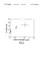
- FIG. 1shows the effects of androgen environment on ischemic lesion size in male rats. Depicted are the percentage of the cross-sectional areas of the brain (mean percent ischemic area ⁇ SEM) in various slices taken at increasing distances caudal to the olfactory bulb from gonad intact (Intact), castrated (Castrate) and castrated animals with testosterone replacement (Castrate+T). * indicates p ⁇ 0.05 versus intact rats for individual brain sections.
- FIG. 2shows the effects of endocrine manipulation on mean percent ischemic area (mean ⁇ SEM) in male rats. * indicates p ⁇ 0.05 versus Intact. ** indicates p ⁇ 0.05 versus Castrate and Castrate+E2 groups. Data is presented from intact rats, castrated rats, castrated rats treated with testosterone, intact rats treated with estradiol, castrated rats treated with estrogen and castrated rats treated with estradiol and testosterone.
- FIG. 3shows the relationship between plasma testosterone concentration and mean percent ischemic area in male rats. Depicted are the mean ⁇ SEM for both plasma testosterone concentration and percent mean ischemic area for the six treatment groups evaluated. The r 2 value for the relationship is 0.922.
- Neuron lossis associated with disease progression and therefore methods to retard neuron loss are desirable for disease management. Whereas certain classes of compounds have shown efficacy in retarding neuron loss, it is desirable to identify additional compounds that may prove effective at retarding neurodegenerative disease progression and aging as well as the sequelac of trauma. In aging populations, there is a particular need for methods of protecting neurons from cell death caused by neurodegenerative diseases, aging and trauma.
- Testosteroneenhances the release of the vasoconstrictor substance, neuropeptide Y, in response to stress [Z. Sukowska-Grnjec, Ann, N. Y. Acad. Sci ., Vol. 771, (1995), pp. 219-33]. Additionally, testosterone has been shown to inhibit synthesis of the vasodilator, protacyclin, in aortic tissue [J. Nakao, et al., Atherosclerosis , Vol. 39, (1981), pp. 203-209], to enhance thromboxane A2-induced constriction of coronary arteries [K. Schor, et al., Euro. J. Clin. Inves ., Vol. 24 Supp., (1994), pp.
- Testosteronehas also been shown to enhance platelet aggregation [W. I. Rosenblum, et al., Thromb. Res ., Vol. 45, (1987), pp. 719-728]and arachidonic acid-induced thrombosis [A. D. Uzunova, et al., Prostaglandins , Vol. 13, (1977), pp. 995-1002].
- Aroonsakulspecifically included testosterone in a mixture for treating symptoms of patients with Alzheimer's disease, suggesting that testosterone might have a beneficial effect as a therapeutic agent.
- MCAOmiddle cerebral artery occlusion
- testosterone inhibitorsto reverse the negative effect of testosterone on neuron loss so as to enhance neuroprotection.
- Testosterone inhibitors known in the artinclude compounds for treating prostate cancer. In a preferred embodiment of the invention, a new use is provided for these compounds and other testosterone inhibitors as defined below.
- testosterone inhibitorsmay be used singly or in combination to prevent neuron loss which occurs following an injury or disease.
- Testosterone inhibitorsmay be used to treat neuron loss in conditions that include but are not limited to: stroke, transient ischemic events, subarrachinoid hemorrhage, neuron loss secondary to cardiac or neural surgery, shock, head trauma, Alzheimer's disease, Parkinson's disease, Huntingdon's disease, AIDS, dementia, aging and schizophrenia.
- testosterone inhibitorsare defined here and in the claims as compounds which decrease the concentration or activity of testosterone or inactivate or otherwise antagonize or inhibit the activity or metabolism of testosterone which would otherwise lead to neuron loss. According to the definition, this class of compounds does not include four ring cyclopentanophenanthrene compounds or estrogen compounds.
- the testosterone inhibitor moleculesinclude antagonists and agonists of testosterone that counteract the action of testosterone. Without wishing to be limited to scientific theories, testosterone inhibitors used in the invention, may act for example by binding to the androgen receptor, interfering with nuclear accumulation of active receptor-hormone complexes, by down-regulating the synthesis testosterone or acting on the metabolism of testosterone.
- the testosterone inhibitor molecules used according to the inventionmay include compounds found to be effective in treating prostrate cancer.
- Testosterone inhibitorsare not limited to but are exemplified by: (1) Hormones which inhibit hypothalamic release of gonadotrophin releasing hormone (GnRH); (2) GnRH analogues (e.g., goserelin Zoladex), leuprorelin (Prostap), buserelin (Suprelfact), triptorelin (De-capeptyl), (nafarelin); (3) steroidal anti-androgens (eg., cytoproterone acetate, megestrol acetate); (4) pure anti-androgens (eg., flutamide (Drogenil), nilutamide, bicalutamide (Cadodex); (5) androgen synthesis inhibitors (eg., ketoconazole); (6) 5-alpha-reductase inhibitors, e.g., finasteroid or Proscen and including inhibitors for treating prostrate cancer; (7) androgen receptor antagonists including cytopro
- Polycyclic phenolic compoundsare defined here and in the claims by the compounds enumerated in Simpkins et al. U.S. Ser. No. 08/685,574, herein incorporated by reference.
- a preferred embodiment of the current inventionis directed to the observation that the amounts of testosterone in a male animal is correlated with the extent of ischemic damage following a stroke.
- the correlation of testosterone as a negative risk factor in the outcome of cerebrovascular ischemia demonstrated in the accompanying example,is novel.
- the datashows that plasma testosterone concentrations are highly correlated with increased ischemic brain damage from middle cerebral artery occlusion.
- the datais obtained using a rat model in which middle cerebral artery (MCA) occlusion has been used to produce focal ischemic lesions in the rat.
- MCAmiddle cerebral artery
- This modelis the preferred experimental model for studying neuron loss in the human brain.
- Reduction in plasma testosterone in the experimental MCA rat modeleither through castration or treatment with estradiol, is associated with approximately a 50% reduction in ischemic lesion size.
- the lesions produced by the MCA occlusion in male ratsoccurred in the frontal and parietal cortex and the basal ganglia, with the maximal extent of the lesion seen at 7 and 9 mm posterior to the olfactory bulb. Reduced ischemic damage was further detected in the rostral and caudal to these areas, where the anterior and posterior cerebral arteries, respectively, also supply the tissue [S. A. Menzies, et al, Neurosurgery , Vol. 31, (1992), pp. 100-106]. The brain samples taken from the expected region of the MCA lesion in control rats demonstrated the expected effects of the MCA occlusion.
- 17 ⁇ -estradiolexerted a profound protective effect in intact male rats that were associated with a marked reduction in plasma testosterone concentrations.
- estradiolwas only partially effective in reducing lesion size.
- Testosterone AssayThe blood from rats for radioimmunoassay (RIA) of testosterone was collected in heparinized tubes by intracardiac puncture just before sacrifice. The plasma was separated by centrifugation and stored at 80° C. until RIA. Coat-A-Count RIA kit was purchased from Diagnostics Products Corporation, Los Angeles, Calif. The tracer had high specific activity with approximately 30 to 40% maximum binding. The antiserum used was highly specific for testosterone with little cross reactivity to other compounds. The Coat-A-Count total testosterone assay had a broad reportable range of 4 to 1600 ng/dl, 50 ⁇ l of the same was used in duplicate tubes for RIA. The concentration of the unknown samples was read from the standard calibration curve whose correlation coefficient was 0.9989.
- Middle Cerebral Artery OcclusionAt 7 days after gonadectomy and steroid implantation, animals were anesthetized with ketamine (60 mg/kg, ip) and zylazine (10 mg/kg, ip). During surgery, rectal temperature was maintained between 36.5 and 37.0° C. by a heating lamp. During an operating microscope, the left carotid artery was exposed through a midline incision of the neck. The sternohyloid, digastric (posterior belly) and the omohyloid muscles were divided and retracted. Then the greater hom of the hyloid bone was removed for exposure of the distal external carotid artery (ECA).
- ECAdistal external carotid artery
- the common carotid arterywas dissected from the vagus nerve and the ECA and its branches (occipital and superior thyroid arteries) were dissected distally.
- the internal carotid arterywas carefully separated from the vagus and glossopharyngeal nerves just below the ECA. Near the base of the skull, the ICA has an extracranial branch, the pterygopalatine artery. Beyond this bifurcation, the ICA enters the cranium medially. After the arteries and their branches were dissected, the distal ECA and its branches, the CCA and the Pterygopalatine arteries were cauterized completely. The ECA and the occipital arteries were cut, then a microvascular clip was placed on the internal carotid artery (ICA) near the base of the skull.
- ICAinternal carotid artery
- the tip of 2.5 cm long 3-0 monofilment nylon suturewas heated to create a globule for easy movement and blockade of the lumen of the vessel.
- the suturewas introduced into the ECA lumen through a puncture and was gently advanced to the distal ICA until it reached the clipped position.
- the microvascular slipwas then removed and the suture was inserted until resistance was felt.
- the distance between the CCA bifurcation and the resistive pointwas 1.8 cm.
- the operative procedurewas completed with 10 min. with minimal blood loss. After 40 minutes of occlusion time, the suture was withdrawn from the ICA and the distal ICA was immediately cauterized.
- FIG. 1The effects of castration and testosterone replacement on ischemic damage following MCA occlusion is shown in FIG. 1 .
- Intact ratsshowed the expected rostral to caudal extent of ischemic damage with peak ischemic lesions observed at 7 and 9 mm caudal to the olfactory bulb. Lesion size was small at more rostral and caudal brain sections.
- the MCA occlusion lesionoccupies the expected brain regions, i.e., the frontal and parietal cortex and basal ganglia, supplied by the MCA [S. A. Menzies, et al, Neurosurgery , Vol. 31, (1992), pp. 100-106]. Castration of adult male rats reduced ischemic lesion size in each section evaluated (FIG.
- Estrogensare neuroprotective against MCA occlusion-induced ischemic brain damage in female rats (U.S. Ser. No. 08/749,703 incorporated by reference).
- Estrogenswere neuroprotective against MCA occlusion-induced ischemic brain damage in female rats (U.S. Ser. No. 08/749,703 incorporated by reference).
- Treatment of intact male rats with 17 ⁇ -estradiolreduced overall mean ischemic area from 17 ⁇ 3% to 8 ⁇ 2% (FIG. 2 ).
- Treatment of castrated males with estradioldid not change the already reduced size of the MCA occlusion-induced lesion (FIG. 2 ).
- Castrate +E2 treatmentprofoundly reduced plasma testosterone concentration from 1.556 ⁇ 0.409 nglml in intact male rats to 0.069 ⁇ 0.029 ng/ml and 0.054 ⁇ 0.010 ng/ml in castrate and intact +E2-treated animals, respectively (Table 1).
- Testosterone replacement in castrate or castrate +E2 ratsincreased plasma testosterone concentration to 0.668 ⁇ 0.067 ng/ml and 0.590 ⁇ 0.055 ng/ml, respectively (Table 1).
Landscapes
- Health & Medical Sciences (AREA)
- Life Sciences & Earth Sciences (AREA)
- Chemical & Material Sciences (AREA)
- Veterinary Medicine (AREA)
- Public Health (AREA)
- Medicinal Chemistry (AREA)
- General Health & Medical Sciences (AREA)
- Animal Behavior & Ethology (AREA)
- Pharmacology & Pharmacy (AREA)
- Epidemiology (AREA)
- General Chemical & Material Sciences (AREA)
- Organic Chemistry (AREA)
- Nuclear Medicine, Radiotherapy & Molecular Imaging (AREA)
- Engineering & Computer Science (AREA)
- Chemical Kinetics & Catalysis (AREA)
- Bioinformatics & Cheminformatics (AREA)
- Biomedical Technology (AREA)
- Neurology (AREA)
- Neurosurgery (AREA)
- Pharmaceuticals Containing Other Organic And Inorganic Compounds (AREA)
- Medicines That Contain Protein Lipid Enzymes And Other Medicines (AREA)
Abstract
Description
| TABLE 1 |
| Effects of Gonadal Steroid Modification on Mortality and |
| Plasma Testosterone Concentration Following Middle Cerebral |
| Artery Occlusion. |
| Plasma Testosterone | Mortality | ||
| Treatment Group | N | ng/ml mean ± sem | (%) |
| 1. Intact | 5 | 1.57 + 0.41* | 46 |
| 2. Castrate | 8 | 0.07 + 0.03 | 15 |
| 3. Castrate + | 11 | 0.67 + 0.07** | 12 |
| 4. Intact + | 9 | 0.05 + 0.01 | 30 |
| 5. Castrate + | 6 | 0.04 + 0.01 | 9 |
| 6. Castrate + E2 + | 7 | 0.59 + 0.06** | 13 |
| N = Number of rats in each treatment group. | |||
| *p < 0.05 when compared with all other treatment groups. | |||
| **p < 0.05 when compared with Intact + E2, Castrate and Castrate + E2 treatment groups. | |||
Claims (5)
Priority Applications (1)
| Application Number | Priority Date | Filing Date | Title |
|---|---|---|---|
| US09/198,416US6172088B1 (en) | 1997-11-24 | 1998-11-24 | Testosterone compounds and use for the protection of neurons |
Applications Claiming Priority (2)
| Application Number | Priority Date | Filing Date | Title |
|---|---|---|---|
| US6669497P | 1997-11-24 | 1997-11-24 | |
| US09/198,416US6172088B1 (en) | 1997-11-24 | 1998-11-24 | Testosterone compounds and use for the protection of neurons |
Publications (1)
| Publication Number | Publication Date |
|---|---|
| US6172088B1true US6172088B1 (en) | 2001-01-09 |
Family
ID=22071097
Family Applications (1)
| Application Number | Title | Priority Date | Filing Date |
|---|---|---|---|
| US09/198,416Expired - Fee RelatedUS6172088B1 (en) | 1997-11-24 | 1998-11-24 | Testosterone compounds and use for the protection of neurons |
Country Status (6)
| Country | Link |
|---|---|
| US (1) | US6172088B1 (en) |
| EP (1) | EP1032397A1 (en) |
| JP (1) | JP2004515446A (en) |
| AU (1) | AU747807B2 (en) |
| CA (1) | CA2309855A1 (en) |
| WO (1) | WO1999026630A1 (en) |
Cited By (14)
| Publication number | Priority date | Publication date | Assignee | Title |
|---|---|---|---|---|
| US20020035100A1 (en)* | 2000-06-27 | 2002-03-21 | Laszlo Prokai | Alkyl ether modified polycyclic compounds having a terminal phenol and uses for protection of cells |
| US20030022877A1 (en)* | 2000-08-30 | 2003-01-30 | Dudley Robert E. | Method of increasing testosterone and related steroid concentrations in women |
| US20030105167A1 (en)* | 2001-12-05 | 2003-06-05 | Dykens James Alan | Treatment of opthalmic diseases |
| US20030166626A1 (en)* | 2000-06-29 | 2003-09-04 | Ernest Wulfert | 7-Hydroxyepiandrosterone having neuroprotective activity |
| US20030186953A1 (en)* | 2000-06-29 | 2003-10-02 | Wuelfert Ernst | Neuroprotective 7-beta-hydroxysteroids |
| US20030220504A1 (en)* | 2002-05-15 | 2003-11-27 | Genzyme Corporation | Synthesis of benzonitriles from substituted benzaldehyde |
| US20040077615A1 (en)* | 2000-08-17 | 2004-04-22 | Gordon Katherine D. | Novel formulations for administering therapeutic lipophilic molecules |
| US20050014731A1 (en)* | 2001-11-30 | 2005-01-20 | Solvay Pharmaceuticals Gmbh | Treatment of Th1 dominated immunological disease states with non-endogenous gestagen compounds |
| US20050118242A1 (en)* | 2000-08-30 | 2005-06-02 | Dudley Robert E. | Androgen pharmaceutical composition and method for treating depression |
| US20050267086A1 (en)* | 2004-05-27 | 2005-12-01 | Migenix Corp. | Compounds and methods for cytoprotection |
| US20070088012A1 (en)* | 2005-04-08 | 2007-04-19 | Woun Seo | Method of treating or preventing type-2 diabetes |
| US20080125403A1 (en)* | 2004-04-02 | 2008-05-29 | Merck & Co., Inc. | Method of Treating Men with Metabolic and Anthropometric Disorders |
| US20110172196A1 (en)* | 2000-08-30 | 2011-07-14 | Dudley Robert E | Pharmaceutical composition and method for treating hypogonadism |
| US8466137B2 (en) | 2005-10-12 | 2013-06-18 | Unimed Pharmaceuticals, Llc | Testosterone gel and method of use |
Families Citing this family (4)
| Publication number | Priority date | Publication date | Assignee | Title |
|---|---|---|---|---|
| AU2002232509A1 (en) | 2000-11-03 | 2002-05-15 | Washington University | Estrone-derivatives having cytoprotective activity |
| DE10137174A1 (en)* | 2001-07-31 | 2003-02-13 | Zentaris Ag | Use of LHRH antagonists in non-castrating doses to improve T cell-mediated immunity |
| PT1919290E (en)* | 2005-07-12 | 2014-03-20 | Ampio Pharmaceuticals Inc | Methods and products for treatment of diseases |
| CA2726247C (en)* | 2008-05-29 | 2018-06-26 | Isr Immune System Regulation Ab | Method and means for treating viral disease, in particular hiv/aids |
Citations (6)
| Publication number | Priority date | Publication date | Assignee | Title |
|---|---|---|---|---|
| DE613140C (en) | 1935-05-13 | Francesco Serra | Playback device for optical sound films | |
| US4897389A (en) | 1984-10-29 | 1990-01-30 | Chaovanee Aroonsakul | Treating central nervous system diseases |
| WO1994024146A1 (en) | 1993-04-13 | 1994-10-27 | Jenapharm Gmbh | Novel androgens and anabolic agents |
| US5453428A (en) | 1991-02-14 | 1995-09-26 | The Mount Sinai School Of Medicine Of The City Of New York | Method and composition for the treatment of apathy-amotivation syndrome |
| EP0679642A1 (en) | 1994-04-28 | 1995-11-02 | Takeda Chemical Industries, Ltd. | Condensed heterocyclic compounds, their production and use |
| EP0792642A1 (en) | 1996-02-28 | 1997-09-03 | Pfizer Inc. | Method of treating conditions with estrogen agonists |
Family Cites Families (2)
| Publication number | Priority date | Publication date | Assignee | Title |
|---|---|---|---|---|
| JPH06100466A (en)* | 1992-02-14 | 1994-04-12 | Tsumura & Co | Protective agent for stress-induced neurological disorders |
| DE4320896A1 (en)* | 1993-06-24 | 1995-01-05 | Denecke Rainer Dr Med Vet | Product for the therapy and prophylaxis of dementias |
- 1998
- 1998-11-24USUS09/198,416patent/US6172088B1/ennot_activeExpired - Fee Related
- 1998-11-24JPJP2000521832Apatent/JP2004515446A/ennot_activeWithdrawn
- 1998-11-24EPEP98960463Apatent/EP1032397A1/ennot_activeWithdrawn
- 1998-11-24CACA002309855Apatent/CA2309855A1/ennot_activeAbandoned
- 1998-11-24WOPCT/US1998/025140patent/WO1999026630A1/ennot_activeApplication Discontinuation
- 1998-11-24AUAU16048/99Apatent/AU747807B2/ennot_activeCeased
Patent Citations (6)
| Publication number | Priority date | Publication date | Assignee | Title |
|---|---|---|---|---|
| DE613140C (en) | 1935-05-13 | Francesco Serra | Playback device for optical sound films | |
| US4897389A (en) | 1984-10-29 | 1990-01-30 | Chaovanee Aroonsakul | Treating central nervous system diseases |
| US5453428A (en) | 1991-02-14 | 1995-09-26 | The Mount Sinai School Of Medicine Of The City Of New York | Method and composition for the treatment of apathy-amotivation syndrome |
| WO1994024146A1 (en) | 1993-04-13 | 1994-10-27 | Jenapharm Gmbh | Novel androgens and anabolic agents |
| EP0679642A1 (en) | 1994-04-28 | 1995-11-02 | Takeda Chemical Industries, Ltd. | Condensed heterocyclic compounds, their production and use |
| EP0792642A1 (en) | 1996-02-28 | 1997-09-03 | Pfizer Inc. | Method of treating conditions with estrogen agonists |
Non-Patent Citations (17)
| Title |
|---|
| Bishop, et al., Molecular and Cellular Neuroscience, (1994), vol. 5, pp. 303-308. |
| Georgiou et al., Movement Disorders, (1995), vol. 10, No. 4, pp. 472-481. |
| Goodman, et al., J. Neurochem, (1996), vol. 66, pp. 1836-1844. |
| Green, et al., J. Neuroscience, (1997), vol. 17, pp. 511-515. |
| Matsuda, et al., Amer J. Physiologic., (1994), vol. 267, pp. H887-H893. |
| Menzies, et al, Neurosurgery, (1992), vol. 31, pp. 100-106. |
| Nakao, et al., Atherosclerosis, (1981), vol. 39, pp. 203-209. |
| Rich and Ovsiew, Movement Disorders, (1994), vol. 9, No. 3, pp. 353-357. |
| Rich et al. Mov. Disord. 9(3): 353-357, 1994.* |
| Rosenblum, et al. Thromb. Res., (1987), vol. 45, pp.719-728. |
| Schor, et al., Euro J. Clin. Inves., (1994), vol. 24 Supp., pp. 50-52. |
| Simpkins, et al., Neurobiology of Aging, (1994), pp. S195-S197. |
| Singer, et al., Neurosci. Let., (1996), vol. 212, pp. 13-16. |
| Singh, et al., Brain Res., (1994), vol. 644, pp. 305-312. |
| Toung et al., Stroke, (1998), vol. 29, pp. 1666-1670. |
| Uzunova, et al., Prostaglandins, (1977), vol. 13, pp. 995-1002. |
| Zukowska-Grojec, Ann, N.Y. Acad. Sci., (1995), vol. 771, pp. 219-233. |
Cited By (32)
| Publication number | Priority date | Publication date | Assignee | Title |
|---|---|---|---|---|
| US20020035100A1 (en)* | 2000-06-27 | 2002-03-21 | Laszlo Prokai | Alkyl ether modified polycyclic compounds having a terminal phenol and uses for protection of cells |
| US7718639B2 (en)* | 2000-06-29 | 2010-05-18 | Hunter-Fleming Limited | 7-hydroxyepiandrosterone having neuroprotective activity |
| US20100130459A1 (en)* | 2000-06-29 | 2010-05-27 | Hunter-Fleming Limited | Neuroprotective 7-beta-hydroxysteroids |
| US20100173883A1 (en)* | 2000-06-29 | 2010-07-08 | Hunter-Fleming Limited | 7-hydroxyepiandrosterone having neuroprotective activity |
| US20030166626A1 (en)* | 2000-06-29 | 2003-09-04 | Ernest Wulfert | 7-Hydroxyepiandrosterone having neuroprotective activity |
| US20030186953A1 (en)* | 2000-06-29 | 2003-10-02 | Wuelfert Ernst | Neuroprotective 7-beta-hydroxysteroids |
| US20040077615A1 (en)* | 2000-08-17 | 2004-04-22 | Gordon Katherine D. | Novel formulations for administering therapeutic lipophilic molecules |
| US20040092494A9 (en)* | 2000-08-30 | 2004-05-13 | Dudley Robert E. | Method of increasing testosterone and related steroid concentrations in women |
| US9125816B2 (en) | 2000-08-30 | 2015-09-08 | Besins Healthcare Inc. | Pharmaceutical composition and method for treating hypogonadism |
| US20050118242A1 (en)* | 2000-08-30 | 2005-06-02 | Dudley Robert E. | Androgen pharmaceutical composition and method for treating depression |
| US20050152956A1 (en)* | 2000-08-30 | 2005-07-14 | Dudley Robert E. | Method of increasing testosterone and related steroid concentrations in women |
| US20110201586A1 (en)* | 2000-08-30 | 2011-08-18 | Dudley Robert E | Pharmaceutical composition and method for treating hypogonadism |
| US20110172196A1 (en)* | 2000-08-30 | 2011-07-14 | Dudley Robert E | Pharmaceutical composition and method for treating hypogonadism |
| US9132089B2 (en) | 2000-08-30 | 2015-09-15 | Besins Healthcare Inc. | Pharmaceutical composition and method for treating hypogonadism |
| US20030022877A1 (en)* | 2000-08-30 | 2003-01-30 | Dudley Robert E. | Method of increasing testosterone and related steroid concentrations in women |
| US20050014731A1 (en)* | 2001-11-30 | 2005-01-20 | Solvay Pharmaceuticals Gmbh | Treatment of Th1 dominated immunological disease states with non-endogenous gestagen compounds |
| US20030105167A1 (en)* | 2001-12-05 | 2003-06-05 | Dykens James Alan | Treatment of opthalmic diseases |
| US20030220504A1 (en)* | 2002-05-15 | 2003-11-27 | Genzyme Corporation | Synthesis of benzonitriles from substituted benzaldehyde |
| US20080125403A1 (en)* | 2004-04-02 | 2008-05-29 | Merck & Co., Inc. | Method of Treating Men with Metabolic and Anthropometric Disorders |
| US20110015164A1 (en)* | 2004-04-02 | 2011-01-20 | Alan Meehan | Method of treating men with metabolic and anthropometric disorders |
| US20080171034A1 (en)* | 2004-05-27 | 2008-07-17 | Migenix Corp. | Compounds and methods for cytoprotection |
| US7304171B2 (en) | 2004-05-27 | 2007-12-04 | Migenix Corp. | Compounds and methods for cytoprotection |
| US20050267086A1 (en)* | 2004-05-27 | 2005-12-01 | Migenix Corp. | Compounds and methods for cytoprotection |
| US20070088012A1 (en)* | 2005-04-08 | 2007-04-19 | Woun Seo | Method of treating or preventing type-2 diabetes |
| US8466136B2 (en) | 2005-10-12 | 2013-06-18 | Unimed Pharmaceuticals, Llc | Testosterone gel and method of use |
| US8486925B2 (en) | 2005-10-12 | 2013-07-16 | Unimed Pharmaceuticals, Llc | Testosterone gel and method of use |
| US8729057B2 (en) | 2005-10-12 | 2014-05-20 | Unimed Pharmaeuticals, LLC | Testosterone gel and method of use |
| US8741881B2 (en) | 2005-10-12 | 2014-06-03 | Unimed Pharmaceuticals, Llc | Testosterone gel and method of use |
| US8754070B2 (en) | 2005-10-12 | 2014-06-17 | Unimed Pharmaceuticals, Llc | Testosterone gel and method of use |
| US8759329B2 (en) | 2005-10-12 | 2014-06-24 | Unimed Pharmaceuticals, Llc | Testosterone gel and method of use |
| US8466138B2 (en) | 2005-10-12 | 2013-06-18 | Unimed Pharmaceuticals, Llc | Testosterone gel and method of use |
| US8466137B2 (en) | 2005-10-12 | 2013-06-18 | Unimed Pharmaceuticals, Llc | Testosterone gel and method of use |
Also Published As
| Publication number | Publication date |
|---|---|
| EP1032397A1 (en) | 2000-09-06 |
| JP2004515446A (en) | 2004-05-27 |
| WO1999026630A1 (en) | 1999-06-03 |
| AU747807B2 (en) | 2002-05-23 |
| CA2309855A1 (en) | 1999-06-03 |
| AU1604899A (en) | 1999-06-15 |
Similar Documents
| Publication | Publication Date | Title |
|---|---|---|
| US6172088B1 (en) | Testosterone compounds and use for the protection of neurons | |
| Woolley et al. | Hormonal effects on the brain | |
| Schotzinger et al. | Acquisition of cholinergic and peptidergic properties by sympathetic innervation of rat sweat glands requires interaction with normal target | |
| Donjacour et al. | The effect of androgen deprivation on branching morphogenesis in the mouse prostate | |
| Woolley et al. | Roles of estradiol and progesterone in regulation of hippocampal dendritic spine density during the estrous cycle in the rat | |
| Claro et al. | Lesions in the medial posterior region of the BST impair sexual behavior in sexually experienced and inexperienced male rats | |
| Saravia et al. | Neuroprotective effects of estradiol in hippocampal neurons and glia of middle age mice | |
| Czaja | Food rejection by female rhesus monkeys during the menstrual cycle and early pregnancy | |
| Davis et al. | Independence of the differentiation of masculine and feminine sexual behavior in rats | |
| Urban et al. | Neuropeptides: Effects on paradoxical sleep and theta rhythm in rats | |
| Behan et al. | Sex hormone receptors are expressed in identified respiratory motoneurons in male and female rats | |
| Luttge et al. | Androgen-induced agonistic behavior in castrate male Swiss-Webster mice: comparison of four naturally occurring androgens | |
| GREER et al. | EVIDENCE OF SEPARATE HYPOTHALAMIC CENTERS CONTROLLING CORTICOTROPIN AND THYROTROPINSECRETION BY THE PITUITARY | |
| Hong et al. | Losartan inhibits development of spontaneous recurrent seizures by preventing astrocyte activation and attenuating blood-brain barrier permeability following pilocarpine-induced status epilepticus | |
| Al-Bishri et al. | Purslane protects against the reproductive toxicity of carbamazepine treatment in pilocarpine-induced epilepsy model | |
| Marchlewska-Koj | Pregnancy blocking by pheromones | |
| Inselman-Temkin et al. | Sex-dependent effects of gonadal and gonadotropic hormones on centrally-elicited attack in cats | |
| Ziegler et al. | Low luteinizing hormone enhances spatial memory and has protective effects on memory loss in rats | |
| Kow et al. | Effects of progesterone on female reproductive behavior in rats: Possible modes of action and role in behavioral sex differences | |
| Monaghan et al. | Brain sites projecting to the spinal nucleus of the bulbocavernosus | |
| Fryer et al. | Hypothalamic control of ACTH secretion in goldfish: II. Hypothalamic lesioning studies | |
| Doherty et al. | Increased serum prolactin levels mediate the suppressive effects of ectopic pituitary grafts on copulatory behavior in male rats | |
| Kuenzel | Central nervous system regulation of gonadal development in the avian male | |
| Havens et al. | Estrogen-dependent and estrogen-independent effects of progesterone on the electrophysiological excitability of dorsal midbrain neurons in golden hamsters | |
| MacKenzie et al. | Effect of estradiol-17ß and prostaglandins on rat myometrial gap junctions |
Legal Events
| Date | Code | Title | Description |
|---|---|---|---|
| AS | Assignment | Owner name:APOLLO BIOPHARMACEUTICS, INC., MASSACHUSETTS Free format text:ASSIGNMENT OF ASSIGNORS INTEREST;ASSIGNORS:GORDON, KATHERINE;LEONARD, ROBERT;REEL/FRAME:010591/0804 Effective date:19990301 Owner name:UNIVERSITY OF FLORIDA RESEARCH FOUNDATION, INC., F Free format text:ASSIGNMENT OF ASSIGNORS INTEREST;ASSIGNOR:SIMPKINS, JAMES W.;REEL/FRAME:010591/0821 Effective date:19990104 | |
| CC | Certificate of correction | ||
| AS | Assignment | Owner name:MITOKOR, INC., CALIFORNIA Free format text:LIQUIDATION AND DISTRIBUTION OF ASSETS;ASSIGNOR:APOLLIO BIOPHARMACEUTICALS, INC.;REEL/FRAME:014384/0990 Effective date:20020628 | |
| FEPP | Fee payment procedure | Free format text:PAT HOLDER CLAIMS SMALL ENTITY STATUS, ENTITY STATUS SET TO SMALL (ORIGINAL EVENT CODE: LTOS); ENTITY STATUS OF PATENT OWNER: SMALL ENTITY | |
| AS | Assignment | Owner name:MITOKOR, INC., CALIFORNIA Free format text:ASSIGNMENT OF ASSIGNORS INTEREST;ASSIGNOR:APOLLO BIOPHARMACEUTICS, INC.;REEL/FRAME:014953/0560 Effective date:20040121 | |
| AS | Assignment | Owner name:MITOKOR, INC., MASSACHUSETTS Free format text:CORRECTIVE ASSIGNMENT TO CORRECT THE INCORRECT PATENT NUMBER FROM 6,172,988 TO 6,172,088. DOCUMENT PREVIOUSLY RECORDED AT REEL 014394 FRAME 0682;ASSIGNOR:GORDON, KATHERINE;REEL/FRAME:015341/0674 Effective date:20021121 Owner name:MITOKOR, CALIFORNIA Free format text:CORRECTIVE TO CORRECT THE ASSIGNOR'S NAME AND PATENT NUMBER PREVIOUSLY RECORDED AT REEL 014384 FRAME 0990. (LIQUIDATION AND DISTRIBUTION OF ASSETS);ASSIGNOR:APOLLO BIOPHARMACEUTICALS, INC.;REEL/FRAME:015341/0679 Effective date:20020628 | |
| FPAY | Fee payment | Year of fee payment:4 | |
| CC | Certificate of correction | ||
| REMI | Maintenance fee reminder mailed | ||
| LAPS | Lapse for failure to pay maintenance fees | ||
| STCH | Information on status: patent discontinuation | Free format text:PATENT EXPIRED DUE TO NONPAYMENT OF MAINTENANCE FEES UNDER 37 CFR 1.362 | |
| FP | Lapsed due to failure to pay maintenance fee | Effective date:20090109 |