US6165210A - Self-expandable helical intravascular stent and stent-graft - Google Patents
Self-expandable helical intravascular stent and stent-graftDownload PDFInfo
- Publication number
- US6165210A US6165210AUS08/221,815US22181594AUS6165210AUS 6165210 AUS6165210 AUS 6165210AUS 22181594 AUS22181594 AUS 22181594AUS 6165210 AUS6165210 AUS 6165210A
- Authority
- US
- United States
- Prior art keywords
- stent
- graft
- collagen
- self
- peg
- Prior art date
- Legal status (The legal status is an assumption and is not a legal conclusion. Google has not performed a legal analysis and makes no representation as to the accuracy of the status listed.)
- Expired - Lifetime
Links
- 238000000034methodMethods0.000claimsabstractdescription55
- 229910045601alloyInorganic materials0.000claimsdescription21
- 239000000956alloySubstances0.000claimsdescription21
- 229910001000nickel titaniumInorganic materials0.000claimsdescription19
- HLXZNVUGXRDIFK-UHFFFAOYSA-Nnickel titaniumChemical group[Ti].[Ti].[Ti].[Ti].[Ti].[Ti].[Ti].[Ti].[Ti].[Ti].[Ti].[Ni].[Ni].[Ni].[Ni].[Ni].[Ni].[Ni].[Ni].[Ni].[Ni].[Ni].[Ni].[Ni].[Ni]HLXZNVUGXRDIFK-UHFFFAOYSA-N0.000claimsdescription18
- 230000002792vascularEffects0.000claimsdescription18
- 238000005452bendingMethods0.000claimsdescription5
- 241000124008MammaliaSpecies0.000claims2
- 239000000463materialSubstances0.000abstractdescription54
- 230000017531blood circulationEffects0.000abstractdescription11
- 238000001356surgical procedureMethods0.000abstractdescription6
- 238000009434installationMethods0.000abstractdescription3
- 238000004804windingMethods0.000abstractdescription3
- 108010035532CollagenProteins0.000description106
- 102000008186CollagenHuman genes0.000description106
- 229920001436collagenPolymers0.000description106
- 229920001223polyethylene glycolPolymers0.000description90
- 229920000642polymerPolymers0.000description38
- 239000000203mixtureSubstances0.000description29
- -1polytetrafluoroethylenePolymers0.000description28
- 239000000835fiberSubstances0.000description25
- 229920001477hydrophilic polymerPolymers0.000description18
- 239000003102growth factorSubstances0.000description16
- 210000001519tissueAnatomy0.000description16
- 235000018102proteinsNutrition0.000description15
- 102000004169proteins and genesHuman genes0.000description15
- 108090000623proteins and genesProteins0.000description15
- 239000002202Polyethylene glycolSubstances0.000description14
- 210000004369bloodAnatomy0.000description14
- 239000008280bloodSubstances0.000description14
- RTZKZFJDLAIYFH-UHFFFAOYSA-NetherSubstancesCCOCCRTZKZFJDLAIYFH-UHFFFAOYSA-N0.000description14
- 208000027418Wounds and injuryDiseases0.000description13
- 229910052751metalInorganic materials0.000description12
- 239000002184metalSubstances0.000description12
- 229920003023plasticPolymers0.000description12
- 239000004033plasticSubstances0.000description12
- 238000013461designMethods0.000description11
- 238000002399angioplastyMethods0.000description9
- 150000001875compoundsChemical class0.000description9
- 150000002148estersChemical class0.000description9
- BASFCYQUMIYNBI-UHFFFAOYSA-NplatinumChemical compound[Pt]BASFCYQUMIYNBI-UHFFFAOYSA-N0.000description9
- 230000015572biosynthetic processEffects0.000description8
- 239000003814drugSubstances0.000description8
- 229910001220stainless steelInorganic materials0.000description8
- 229940079593drugDrugs0.000description7
- 230000004962physiological conditionEffects0.000description7
- 239000005020polyethylene terephthalateSubstances0.000description7
- 229920001343polytetrafluoroethylenePolymers0.000description7
- 239000004810polytetrafluoroethyleneSubstances0.000description7
- 229920001059synthetic polymerPolymers0.000description7
- SXRSQZLOMIGNAQ-UHFFFAOYSA-NGlutaraldehydeChemical compoundO=CCCCC=OSXRSQZLOMIGNAQ-UHFFFAOYSA-N0.000description6
- 208000007536ThrombosisDiseases0.000description6
- 108010009583Transforming Growth FactorsProteins0.000description6
- 102000009618Transforming Growth FactorsHuman genes0.000description6
- 230000004888barrier functionEffects0.000description6
- 230000008901benefitEffects0.000description6
- 238000006243chemical reactionMethods0.000description6
- 238000007906compressionMethods0.000description6
- 230000006835compressionEffects0.000description6
- 230000000694effectsEffects0.000description6
- 125000003588lysine groupChemical group[H]N([H])C([H])([H])C([H])([H])C([H])([H])C([H])([H])C([H])(N([H])[H])C(*)=O0.000description6
- 229920000728polyesterPolymers0.000description6
- 108090000765processed proteins & peptidesProteins0.000description6
- 239000000126substanceSubstances0.000description6
- 229920004934Dacron®Polymers0.000description5
- JOYRKODLDBILNP-UHFFFAOYSA-NEthyl urethaneChemical compoundCCOC(N)=OJOYRKODLDBILNP-UHFFFAOYSA-N0.000description5
- 239000004743PolypropyleneSubstances0.000description5
- 229920001577copolymerPolymers0.000description5
- 238000012377drug deliveryMethods0.000description5
- 230000007062hydrolysisEffects0.000description5
- 238000006460hydrolysis reactionMethods0.000description5
- 125000002887hydroxy groupChemical group[H]O*0.000description5
- 230000002163immunogenEffects0.000description5
- 238000011068loading methodMethods0.000description5
- 150000002739metalsChemical class0.000description5
- 230000001453nonthrombogenic effectEffects0.000description5
- 229920001155polypropylenePolymers0.000description5
- 229920002635polyurethanePolymers0.000description5
- 239000004814polyurethaneSubstances0.000description5
- 230000002787reinforcementEffects0.000description5
- 206010002329AneurysmDiseases0.000description4
- LYCAIKOWRPUZTN-UHFFFAOYSA-NEthylene glycolChemical compoundOCCOLYCAIKOWRPUZTN-UHFFFAOYSA-N0.000description4
- XEEYBQQBJWHFJM-UHFFFAOYSA-NIronChemical compound[Fe]XEEYBQQBJWHFJM-UHFFFAOYSA-N0.000description4
- 229920000106Liquid crystal polymerPolymers0.000description4
- KDLHZDBZIXYQEI-UHFFFAOYSA-NPalladiumChemical compound[Pd]KDLHZDBZIXYQEI-UHFFFAOYSA-N0.000description4
- 229920000954PolyglycolidePolymers0.000description4
- 101150084989Speg geneProteins0.000description4
- 125000003277amino groupChemical group0.000description4
- 239000003795chemical substances by applicationSubstances0.000description4
- 210000003038endotheliumAnatomy0.000description4
- 239000007943implantSubstances0.000description4
- 230000001976improved effectEffects0.000description4
- 125000005647linker groupChemical group0.000description4
- 230000008569processEffects0.000description4
- 239000000243solutionSubstances0.000description4
- 229920000544Gore-TexPolymers0.000description3
- 229920000271Kevlar®Polymers0.000description3
- 239000004977Liquid-crystal polymers (LCPs)Substances0.000description3
- 239000004952PolyamideSubstances0.000description3
- 239000004698PolyethyleneSubstances0.000description3
- 239000004809TeflonSubstances0.000description3
- 229920006362Teflon®Polymers0.000description3
- 229910052770UraniumInorganic materials0.000description3
- 230000003213activating effectEffects0.000description3
- 210000004204blood vesselAnatomy0.000description3
- 210000000988bone and boneAnatomy0.000description3
- 239000002131composite materialSubstances0.000description3
- 229910000701elgiloys (Co-Cr-Ni Alloy)Inorganic materials0.000description3
- 239000004744fabricSubstances0.000description3
- JFCQEDHGNNZCLN-UHFFFAOYSA-Nglutaric acidChemical compoundOC(=O)CCCC(O)=OJFCQEDHGNNZCLN-UHFFFAOYSA-N0.000description3
- VANNPISTIUFMLH-UHFFFAOYSA-Nglutaric anhydrideChemical compoundO=C1CCCC(=O)O1VANNPISTIUFMLH-UHFFFAOYSA-N0.000description3
- PCHJSUWPFVWCPO-UHFFFAOYSA-NgoldChemical compound[Au]PCHJSUWPFVWCPO-UHFFFAOYSA-N0.000description3
- 229910052737goldInorganic materials0.000description3
- 239000010931goldSubstances0.000description3
- 230000012010growthEffects0.000description3
- 239000004761kevlarSubstances0.000description3
- 239000007788liquidSubstances0.000description3
- 238000003754machiningMethods0.000description3
- 238000004519manufacturing processMethods0.000description3
- 239000011159matrix materialSubstances0.000description3
- 229910052697platinumInorganic materials0.000description3
- 229920002647polyamidePolymers0.000description3
- 229920000573polyethylenePolymers0.000description3
- 239000004633polyglycolic acidSubstances0.000description3
- 102000004196processed proteins & peptidesHuman genes0.000description3
- 239000000047productSubstances0.000description3
- 230000008439repair processEffects0.000description3
- 208000037803restenosisDiseases0.000description3
- 239000000758substrateSubstances0.000description3
- 238000013268sustained releaseMethods0.000description3
- 239000012730sustained-release formSubstances0.000description3
- 229910052715tantalumInorganic materials0.000description3
- GUVRBAGPIYLISA-UHFFFAOYSA-Ntantalum atomChemical compound[Ta]GUVRBAGPIYLISA-UHFFFAOYSA-N0.000description3
- BFKJFAAPBSQJPD-UHFFFAOYSA-NtetrafluoroetheneChemical compoundFC(F)=C(F)FBFKJFAAPBSQJPD-UHFFFAOYSA-N0.000description3
- 210000005166vasculatureAnatomy0.000description3
- ZJIFDEVVTPEXDL-UHFFFAOYSA-N(2,5-dioxopyrrolidin-1-yl) hydrogen carbonateChemical compoundOC(=O)ON1C(=O)CCC1=OZJIFDEVVTPEXDL-UHFFFAOYSA-N0.000description2
- NHJVRSWLHSJWIN-UHFFFAOYSA-N2,4,6-trinitrobenzenesulfonic acidChemical compoundOS(=O)(=O)C1=C([N+]([O-])=O)C=C([N+]([O-])=O)C=C1[N+]([O-])=ONHJVRSWLHSJWIN-UHFFFAOYSA-N0.000description2
- 229920002799BoPETPolymers0.000description2
- 241000283690Bos taurusSpecies0.000description2
- KXDHJXZQYSOELW-UHFFFAOYSA-MCarbamateChemical compoundNC([O-])=OKXDHJXZQYSOELW-UHFFFAOYSA-M0.000description2
- 102000000503Collagen Type IIHuman genes0.000description2
- 108010041390Collagen Type IIProteins0.000description2
- 102000003951ErythropoietinHuman genes0.000description2
- 108090000394ErythropoietinProteins0.000description2
- 102000018233Fibroblast Growth FactorHuman genes0.000description2
- 108050007372Fibroblast Growth FactorProteins0.000description2
- MHAJPDPJQMAIIY-UHFFFAOYSA-NHydrogen peroxideChemical compoundOOMHAJPDPJQMAIIY-UHFFFAOYSA-N0.000description2
- 241001465754MetazoaSpecies0.000description2
- 239000005041Mylar™Substances0.000description2
- NQTADLQHYWFPDB-UHFFFAOYSA-NN-HydroxysuccinimideChemical compoundON1C(=O)CCC1=ONQTADLQHYWFPDB-UHFFFAOYSA-N0.000description2
- 108010025020Nerve Growth FactorProteins0.000description2
- 102000015336Nerve Growth FactorHuman genes0.000description2
- 229920001734PEG propionaldehydePolymers0.000description2
- 241000906446TherapsSpecies0.000description2
- 230000004913activationEffects0.000description2
- 230000003872anastomosisEffects0.000description2
- 238000004873anchoringMethods0.000description2
- 150000008064anhydridesChemical class0.000description2
- 229920003235aromatic polyamidePolymers0.000description2
- 230000009286beneficial effectEffects0.000description2
- 239000000560biocompatible materialSubstances0.000description2
- 230000004071biological effectEffects0.000description2
- 150000004649carbonic acid derivativesChemical class0.000description2
- 210000000845cartilageAnatomy0.000description2
- 210000004027cellAnatomy0.000description2
- 230000008859changeEffects0.000description2
- 239000007795chemical reaction productSubstances0.000description2
- 239000000501collagen implantSubstances0.000description2
- 108010035886connective tissue-activating peptideProteins0.000description2
- 238000010276constructionMethods0.000description2
- 239000003431cross linking reagentSubstances0.000description2
- 239000013078crystalSubstances0.000description2
- MGNCLNQXLYJVJD-UHFFFAOYSA-Ncyanuric chlorideChemical compoundClC1=NC(Cl)=NC(Cl)=N1MGNCLNQXLYJVJD-UHFFFAOYSA-N0.000description2
- 230000029087digestionEffects0.000description2
- 201000010099diseaseDiseases0.000description2
- 208000037265diseases, disorders, signs and symptomsDiseases0.000description2
- 238000009826distributionMethods0.000description2
- 239000003937drug carrierSubstances0.000description2
- 210000002889endothelial cellAnatomy0.000description2
- 238000005516engineering processMethods0.000description2
- 229940105423erythropoietinDrugs0.000description2
- 229940126864fibroblast growth factorDrugs0.000description2
- 239000002657fibrous materialSubstances0.000description2
- 239000012467final productSubstances0.000description2
- 238000009472formulationMethods0.000description2
- 230000006870functionEffects0.000description2
- HCDGVLDPFQMKDK-UHFFFAOYSA-NhexafluoropropyleneChemical groupFC(F)=C(F)C(F)(F)FHCDGVLDPFQMKDK-UHFFFAOYSA-N0.000description2
- WGCNASOHLSPBMP-UHFFFAOYSA-NhydroxyacetaldehydeNatural productsOCC=OWGCNASOHLSPBMP-UHFFFAOYSA-N0.000description2
- 230000005847immunogenicityEffects0.000description2
- 238000001727in vivoMethods0.000description2
- 230000001965increasing effectEffects0.000description2
- 238000003780insertionMethods0.000description2
- 230000037431insertionEffects0.000description2
- 210000000936intestineAnatomy0.000description2
- 229910052742ironInorganic materials0.000description2
- 230000001788irregularEffects0.000description2
- 230000003902lesionEffects0.000description2
- 235000018977lysineNutrition0.000description2
- 238000002595magnetic resonance imagingMethods0.000description2
- 229910000734martensiteInorganic materials0.000description2
- 230000007246mechanismEffects0.000description2
- 230000004048modificationEffects0.000description2
- 238000012986modificationMethods0.000description2
- 229940053128nerve growth factorDrugs0.000description2
- 229910052763palladiumInorganic materials0.000description2
- 229920000139polyethylene terephthalatePolymers0.000description2
- 229920002503polyoxyethylene-polyoxypropylenePolymers0.000description2
- OXCMYAYHXIHQOA-UHFFFAOYSA-Npotassium;[2-butyl-5-chloro-3-[[4-[2-(1,2,4-triaza-3-azanidacyclopenta-1,4-dien-5-yl)phenyl]phenyl]methyl]imidazol-4-yl]methanolChemical compound[K+].CCCCC1=NC(Cl)=C(CO)N1CC1=CC=C(C=2C(=CC=CC=2)C2=N[N-]N=N2)C=C1OXCMYAYHXIHQOA-UHFFFAOYSA-N0.000description2
- 230000002265preventionEffects0.000description2
- 230000005855radiationEffects0.000description2
- 238000011084recoveryMethods0.000description2
- 230000003014reinforcing effectEffects0.000description2
- 239000012783reinforcing fiberSubstances0.000description2
- 229910001285shape-memory alloyInorganic materials0.000description2
- 239000002002slurrySubstances0.000description2
- 210000000329smooth muscle myocyteAnatomy0.000description2
- 239000010935stainless steelSubstances0.000description2
- 239000007858starting materialSubstances0.000description2
- 230000008093supporting effectEffects0.000description2
- 238000003786synthesis reactionMethods0.000description2
- 230000001225therapeutic effectEffects0.000description2
- 230000002885thrombogenetic effectEffects0.000description2
- 239000010936titaniumSubstances0.000description2
- 231100000331toxicToxicity0.000description2
- 230000002588toxic effectEffects0.000description2
- 230000007704transitionEffects0.000description2
- 210000003954umbilical cordAnatomy0.000description2
- XLYOFNOQVPJJNP-UHFFFAOYSA-NwaterSubstancesOXLYOFNOQVPJJNP-UHFFFAOYSA-N0.000description2
- KIUKXJAPPMFGSW-DNGZLQJQSA-N(2S,3S,4S,5R,6R)-6-[(2S,3R,4R,5S,6R)-3-Acetamido-2-[(2S,3S,4R,5R,6R)-6-[(2R,3R,4R,5S,6R)-3-acetamido-2,5-dihydroxy-6-(hydroxymethyl)oxan-4-yl]oxy-2-carboxy-4,5-dihydroxyoxan-3-yl]oxy-5-hydroxy-6-(hydroxymethyl)oxan-4-yl]oxy-3,4,5-trihydroxyoxane-2-carboxylic acidChemical compoundCC(=O)N[C@H]1[C@H](O)O[C@H](CO)[C@@H](O)[C@@H]1O[C@H]1[C@H](O)[C@@H](O)[C@H](O[C@H]2[C@@H]([C@@H](O[C@H]3[C@@H]([C@@H](O)[C@H](O)[C@H](O3)C(O)=O)O)[C@H](O)[C@@H](CO)O2)NC(C)=O)[C@@H](C(O)=O)O1KIUKXJAPPMFGSW-DNGZLQJQSA-N0.000description1
- JPSKCQCQZUGWNM-UHFFFAOYSA-N2,7-OxepanedioneChemical compoundO=C1CCCCC(=O)O1JPSKCQCQZUGWNM-UHFFFAOYSA-N0.000description1
- FALRKNHUBBKYCC-UHFFFAOYSA-N2-(chloromethyl)pyridine-3-carbonitrileChemical compoundClCC1=NC=CC=C1C#NFALRKNHUBBKYCC-UHFFFAOYSA-N0.000description1
- FDSYTWVNUJTPMA-UHFFFAOYSA-N2-[3,9-bis(carboxymethyl)-3,6,9,15-tetrazabicyclo[9.3.1]pentadeca-1(15),11,13-trien-6-yl]acetic acidChemical compoundC1N(CC(O)=O)CCN(CC(=O)O)CCN(CC(O)=O)CC2=CC=CC1=N2FDSYTWVNUJTPMA-UHFFFAOYSA-N0.000description1
- 1250000040424-aminobutyl groupChemical group[H]C([*])([H])C([H])([H])C([H])([H])C([H])([H])N([H])[H]0.000description1
- NWAGXLBTAPTCPR-UHFFFAOYSA-N5-(2,5-dioxopyrrolidin-1-yl)oxy-5-oxopentanoic acidChemical compoundOC(=O)CCCC(=O)ON1C(=O)CCC1=ONWAGXLBTAPTCPR-UHFFFAOYSA-N0.000description1
- 241000894006BacteriaSpecies0.000description1
- 108010081589BecaplerminProteins0.000description1
- 101800003265Beta-thromboglobulinProteins0.000description1
- 102400001362Beta-thromboglobulinHuman genes0.000description1
- 229910000684Cobalt-chromeInorganic materials0.000description1
- 102000012422Collagen Type IHuman genes0.000description1
- 108010022452Collagen Type IProteins0.000description1
- 102000001187Collagen Type IIIHuman genes0.000description1
- 108010069502Collagen Type IIIProteins0.000description1
- RYGMFSIKBFXOCR-UHFFFAOYSA-NCopperChemical compound[Cu]RYGMFSIKBFXOCR-UHFFFAOYSA-N0.000description1
- 102000004190EnzymesHuman genes0.000description1
- 108090000790EnzymesProteins0.000description1
- PIICEJLVQHRZGT-UHFFFAOYSA-NEthylenediamineChemical classNCCNPIICEJLVQHRZGT-UHFFFAOYSA-N0.000description1
- 102000003971Fibroblast Growth Factor 1Human genes0.000description1
- 108090000386Fibroblast Growth Factor 1Proteins0.000description1
- 108010010803GelatinProteins0.000description1
- 208000032843HemorrhageDiseases0.000description1
- HTTJABKRGRZYRN-UHFFFAOYSA-NHeparinChemical compoundOC1C(NC(=O)C)C(O)OC(COS(O)(=O)=O)C1OC1C(OS(O)(=O)=O)C(O)C(OC2C(C(OS(O)(=O)=O)C(OC3C(C(O)C(O)C(O3)C(O)=O)OS(O)(=O)=O)C(CO)O2)NS(O)(=O)=O)C(C(O)=O)O1HTTJABKRGRZYRN-UHFFFAOYSA-N0.000description1
- 241000282412HomoSpecies0.000description1
- 108010003272Hyaluronate lyaseProteins0.000description1
- 102000001974HyaluronidasesHuman genes0.000description1
- 108090000723Insulin-Like Growth Factor IProteins0.000description1
- 229920000914Metallic fiberPolymers0.000description1
- GRSMWKLPSNHDHA-UHFFFAOYSA-NNaphthalic anhydrideChemical compoundC1=CC(C(=O)OC2=O)=C3C2=CC=CC3=C1GRSMWKLPSNHDHA-UHFFFAOYSA-N0.000description1
- 206010028980NeoplasmDiseases0.000description1
- 239000004677NylonSubstances0.000description1
- 108010038512Platelet-Derived Growth FactorProteins0.000description1
- 102000010780Platelet-Derived Growth FactorHuman genes0.000description1
- 229920003171Poly (ethylene oxide)Polymers0.000description1
- 229910001260Pt alloyInorganic materials0.000description1
- 241000220010RhodeSpecies0.000description1
- 239000008156Ringer's lactate solutionSubstances0.000description1
- 101150108015STR6 geneProteins0.000description1
- 101100386054Saccharomyces cerevisiae (strain ATCC 204508 / S288c) CYS3 geneProteins0.000description1
- 102000013275SomatomedinsHuman genes0.000description1
- 229920002472StarchPolymers0.000description1
- 239000004974Thermotropic liquid crystalSubstances0.000description1
- 229910001069Ti alloyInorganic materials0.000description1
- RTAQQCXQSZGOHL-UHFFFAOYSA-NTitaniumChemical compound[Ti]RTAQQCXQSZGOHL-UHFFFAOYSA-N0.000description1
- 206010060872Transplant failureDiseases0.000description1
- 108010006886VitrogenProteins0.000description1
- 229910001080W alloyInorganic materials0.000description1
- HZEWFHLRYVTOIW-UHFFFAOYSA-N[Ti].[Ni]Chemical compound[Ti].[Ni]HZEWFHLRYVTOIW-UHFFFAOYSA-N0.000description1
- 239000011149active materialSubstances0.000description1
- 230000004075alterationEffects0.000description1
- 230000002965anti-thrombogenic effectEffects0.000description1
- 230000002785anti-thrombosisEffects0.000description1
- 239000003146anticoagulant agentSubstances0.000description1
- 230000010100anticoagulationEffects0.000description1
- 210000000709aortaAnatomy0.000description1
- 239000007864aqueous solutionSubstances0.000description1
- 210000001367arteryAnatomy0.000description1
- 230000003143atherosclerotic effectEffects0.000description1
- QVGXLLKOCUKJST-UHFFFAOYSA-Natomic oxygenChemical compound[O]QVGXLLKOCUKJST-UHFFFAOYSA-N0.000description1
- 230000003416augmentationEffects0.000description1
- 230000003190augmentative effectEffects0.000description1
- 230000004323axial lengthEffects0.000description1
- 229920002988biodegradable polymerPolymers0.000description1
- 239000004621biodegradable polymerSubstances0.000description1
- 239000012620biological materialSubstances0.000description1
- PFYXSUNOLOJMDX-UHFFFAOYSA-Nbis(2,5-dioxopyrrolidin-1-yl) carbonateChemical compoundO=C1CCC(=O)N1OC(=O)ON1C(=O)CCC1=OPFYXSUNOLOJMDX-UHFFFAOYSA-N0.000description1
- OMAHFYGHUQSIEF-UHFFFAOYSA-Nbis(2,5-dioxopyrrolidin-1-yl) oxalateChemical compoundO=C1CCC(=O)N1OC(=O)C(=O)ON1C(=O)CCC1=OOMAHFYGHUQSIEF-UHFFFAOYSA-N0.000description1
- 208000034158bleedingDiseases0.000description1
- 230000000740bleeding effectEffects0.000description1
- 201000011510cancerDiseases0.000description1
- 125000004432carbon atomChemical groupC*0.000description1
- PFKFTWBEEFSNDU-UHFFFAOYSA-NcarbonyldiimidazoleChemical compoundC1=CN=CN1C(=O)N1C=CN=C1PFKFTWBEEFSNDU-UHFFFAOYSA-N0.000description1
- 210000000748cardiovascular systemAnatomy0.000description1
- 238000005266castingMethods0.000description1
- 230000010261cell growthEffects0.000description1
- 230000004663cell proliferationEffects0.000description1
- 229920002678cellulosePolymers0.000description1
- 239000001913celluloseSubstances0.000description1
- 235000010980celluloseNutrition0.000description1
- 239000013043chemical agentSubstances0.000description1
- 231100000481chemical toxicantToxicity0.000description1
- 239000000788chromium alloySubstances0.000description1
- 239000011248coating agentSubstances0.000description1
- 238000000576coating methodMethods0.000description1
- 239000010952cobalt-chromeSubstances0.000description1
- 230000001427coherent effectEffects0.000description1
- 230000000295complement effectEffects0.000description1
- 230000021615conjugationEffects0.000description1
- 210000002808connective tissueAnatomy0.000description1
- 239000000470constituentSubstances0.000description1
- 238000013270controlled releaseMethods0.000description1
- 229910052802copperInorganic materials0.000description1
- 239000010949copperSubstances0.000description1
- 238000012937correctionMethods0.000description1
- 238000005859coupling reactionMethods0.000description1
- 238000005520cutting processMethods0.000description1
- 230000002950deficientEffects0.000description1
- 230000001419dependent effectEffects0.000description1
- 238000000151depositionMethods0.000description1
- 210000004207dermisAnatomy0.000description1
- 238000009792diffusion processMethods0.000description1
- GYZLOYUZLJXAJU-UHFFFAOYSA-Ndiglycidyl etherChemical compoundC1OC1COCC1CO1GYZLOYUZLJXAJU-UHFFFAOYSA-N0.000description1
- 238000006073displacement reactionMethods0.000description1
- 238000002224dissectionMethods0.000description1
- 238000002651drug therapyMethods0.000description1
- 238000001035dryingMethods0.000description1
- 229920006351engineering plasticPolymers0.000description1
- 230000002708enhancing effectEffects0.000description1
- 230000002255enzymatic effectEffects0.000description1
- 229940088598enzymeDrugs0.000description1
- 210000002615epidermisAnatomy0.000description1
- 230000032050esterificationEffects0.000description1
- 238000005886esterification reactionMethods0.000description1
- 229920000295expanded polytetrafluoroethylenePolymers0.000description1
- 238000001125extrusionMethods0.000description1
- 239000013305flexible fiberSubstances0.000description1
- 239000012530fluidSubstances0.000description1
- 229920001002functional polymerPolymers0.000description1
- 238000007306functionalization reactionMethods0.000description1
- 229920000159gelatinPolymers0.000description1
- 239000008273gelatinSubstances0.000description1
- 235000019322gelatineNutrition0.000description1
- 235000011852gelatine dessertsNutrition0.000description1
- 150000002334glycolsChemical class0.000description1
- 230000035876healingEffects0.000description1
- 238000010438heat treatmentMethods0.000description1
- 230000000004hemodynamic effectEffects0.000description1
- 229920000669heparinPolymers0.000description1
- 229960002897heparinDrugs0.000description1
- 229920002674hyaluronanPolymers0.000description1
- 229960003160hyaluronic acidDrugs0.000description1
- 229960002773hyaluronidaseDrugs0.000description1
- 239000001257hydrogenSubstances0.000description1
- 229910052739hydrogenInorganic materials0.000description1
- 230000003301hydrolyzing effectEffects0.000description1
- 125000004356hydroxy functional groupChemical groupO*0.000description1
- 238000002513implantationMethods0.000description1
- 238000010348incorporationMethods0.000description1
- 230000001939inductive effectEffects0.000description1
- 230000002757inflammatory effectEffects0.000description1
- 230000000977initiatory effectEffects0.000description1
- 208000014674injuryDiseases0.000description1
- 230000010354integrationEffects0.000description1
- 230000008069intimal proliferationEffects0.000description1
- 238000002955isolationMethods0.000description1
- 238000003698laser cuttingMethods0.000description1
- 210000003041ligamentAnatomy0.000description1
- 230000007774longtermEffects0.000description1
- 229910001092metal group alloyInorganic materials0.000description1
- 125000000956methoxy groupChemical group[H]C([H])([H])O*0.000description1
- 125000002496methyl groupChemical group[H]C([H])([H])*0.000description1
- 239000000178monomerSubstances0.000description1
- 230000000921morphogenic effectEffects0.000description1
- YTVNOVQHSGMMOV-UHFFFAOYSA-Nnaphthalenetetracarboxylic dianhydrideChemical compoundC1=CC(C(=O)OC2=O)=C3C2=CC=C2C(=O)OC(=O)C1=C32YTVNOVQHSGMMOV-UHFFFAOYSA-N0.000description1
- 239000005445natural materialSubstances0.000description1
- 239000006225natural substrateSubstances0.000description1
- 230000000926neurological effectEffects0.000description1
- QJGQUHMNIGDVPM-UHFFFAOYSA-Nnitrogen groupChemical group[N]QJGQUHMNIGDVPM-UHFFFAOYSA-N0.000description1
- 231100000252nontoxicToxicity0.000description1
- 230000003000nontoxic effectEffects0.000description1
- 229920001778nylonPolymers0.000description1
- 230000000414obstructive effectEffects0.000description1
- 210000000056organAnatomy0.000description1
- 230000002188osteogenic effectEffects0.000description1
- 239000001301oxygenSubstances0.000description1
- 229910052760oxygenInorganic materials0.000description1
- 230000002093peripheral effectEffects0.000description1
- 230000000704physical effectEffects0.000description1
- 210000002826placentaAnatomy0.000description1
- 108010017843platelet-derived growth factor AProteins0.000description1
- 108010000685platelet-derived growth factor ABProteins0.000description1
- ZONODCCBXBRQEZ-UHFFFAOYSA-Nplatinum tungstenChemical compound[W].[Pt]ZONODCCBXBRQEZ-UHFFFAOYSA-N0.000description1
- HWLDNSXPUQTBOD-UHFFFAOYSA-Nplatinum-iridium alloyChemical compound[Ir].[Pt]HWLDNSXPUQTBOD-UHFFFAOYSA-N0.000description1
- 229920000747poly(lactic acid)Polymers0.000description1
- 229920001610polycaprolactonePolymers0.000description1
- 239000004632polycaprolactoneSubstances0.000description1
- 229920006324polyoxymethylenePolymers0.000description1
- 229920000036polyvinylpyrrolidonePolymers0.000description1
- 235000013855polyvinylpyrrolidoneNutrition0.000description1
- 239000011148porous materialSubstances0.000description1
- 235000020004porterNutrition0.000description1
- 125000002924primary amino groupChemical group[H]N([H])*0.000description1
- 230000002062proliferating effectEffects0.000description1
- 230000009145protein modificationEffects0.000description1
- 230000002797proteolythic effectEffects0.000description1
- 239000002296pyrolytic carbonSubstances0.000description1
- 239000000376reactantSubstances0.000description1
- 238000002278reconstructive surgeryMethods0.000description1
- 239000012779reinforcing materialSubstances0.000description1
- 230000000452restraining effectEffects0.000description1
- 229910052703rhodiumInorganic materials0.000description1
- 239000010948rhodiumSubstances0.000description1
- MHOVAHRLVXNVSD-UHFFFAOYSA-Nrhodium atomChemical compound[Rh]MHOVAHRLVXNVSD-UHFFFAOYSA-N0.000description1
- 238000007665saggingMethods0.000description1
- 238000007789sealingMethods0.000description1
- 238000000926separation methodMethods0.000description1
- 238000004904shorteningMethods0.000description1
- 210000003491skinAnatomy0.000description1
- NCGJACBPALRHNG-UHFFFAOYSA-Msodium;2,4,6-trinitrobenzenesulfonateChemical compound[Na+].[O-][N+](=O)C1=CC([N+]([O-])=O)=C(S([O-])(=O)=O)C([N+]([O-])=O)=C1NCGJACBPALRHNG-UHFFFAOYSA-M0.000description1
- 230000000087stabilizing effectEffects0.000description1
- 238000010561standard procedureMethods0.000description1
- 235000019698starchNutrition0.000description1
- 239000008107starchSubstances0.000description1
- 101150035983str1 geneProteins0.000description1
- 125000001424substituent groupChemical group0.000description1
- KDYFGRWQOYBRFD-UHFFFAOYSA-Lsuccinate(2-)Chemical compound[O-]C(=O)CCC([O-])=OKDYFGRWQOYBRFD-UHFFFAOYSA-L0.000description1
- 229940014800succinic anhydrideDrugs0.000description1
- 239000000725suspensionSubstances0.000description1
- 229920002994synthetic fiberPolymers0.000description1
- 239000012209synthetic fiberSubstances0.000description1
- 230000009885systemic effectEffects0.000description1
- 229940124597therapeutic agentDrugs0.000description1
- 230000008467tissue growthEffects0.000description1
- 229910052719titaniumInorganic materials0.000description1
- 239000003440toxic substanceSubstances0.000description1
- 231100000419toxicityToxicity0.000description1
- 230000001988toxicityEffects0.000description1
- 230000009466transformationEffects0.000description1
- 230000008733traumaEffects0.000description1
- 230000000472traumatic effectEffects0.000description1
- 229910052721tungstenInorganic materials0.000description1
- 239000010937tungstenSubstances0.000description1
- UDKYUQZDRMRDOR-UHFFFAOYSA-NtungstenChemical compound[W][W][W][W][W][W][W][W][W][W][W][W][W][W][W][W][W][W][W][W][W][W][W][W][W][W][W][W][W][W][W][W][W][W][W][W][W][W][W][W][W][W][W][W][W][W][W][W]UDKYUQZDRMRDOR-UHFFFAOYSA-N0.000description1
- VBEQCZHXXJYVRD-GACYYNSASA-NuroantheloneChemical compoundC([C@@H](C(=O)N[C@H](C(=O)N[C@@H](CS)C(=O)N[C@@H](CC(N)=O)C(=O)N[C@@H](CS)C(=O)N[C@H](C(=O)N[C@@H]([C@@H](C)CC)C(=O)NCC(=O)N[C@@H](CC=1C=CC(O)=CC=1)C(=O)N[C@@H](CO)C(=O)NCC(=O)N[C@@H](CC(O)=O)C(=O)N[C@@H](CCCNC(N)=N)C(=O)N[C@@H](CS)C(=O)N[C@@H](CCC(N)=O)C(=O)N[C@@H]([C@@H](C)O)C(=O)N[C@@H](CCCNC(N)=N)C(=O)N[C@@H](CC(O)=O)C(=O)N[C@@H](CC(C)C)C(=O)N[C@@H](CCCNC(N)=N)C(=O)N[C@@H](CC=1C2=CC=CC=C2NC=1)C(=O)N[C@@H](CC=1C2=CC=CC=C2NC=1)C(=O)N[C@@H](CCC(O)=O)C(=O)N[C@@H](CC(C)C)C(=O)N[C@@H](CCCNC(N)=N)C(O)=O)C(C)C)[C@@H](C)O)NC(=O)[C@H](CO)NC(=O)[C@H](CC(O)=O)NC(=O)[C@H](CC(C)C)NC(=O)[C@H](CO)NC(=O)[C@H](CCC(O)=O)NC(=O)[C@@H](NC(=O)[C@H](CC=1NC=NC=1)NC(=O)[C@H](CCSC)NC(=O)[C@H](CS)NC(=O)[C@@H](NC(=O)CNC(=O)CNC(=O)[C@H](CC(N)=O)NC(=O)[C@H](CC(C)C)NC(=O)[C@H](CS)NC(=O)[C@H](CC=1C=CC(O)=CC=1)NC(=O)CNC(=O)[C@H](CC(O)=O)NC(=O)[C@H](CC=1C=CC(O)=CC=1)NC(=O)[C@H](CO)NC(=O)[C@H](CO)NC(=O)[C@H]1N(CCC1)C(=O)[C@H](CS)NC(=O)CNC(=O)[C@H]1N(CCC1)C(=O)[C@H](CC=1C=CC(O)=CC=1)NC(=O)[C@H](CO)NC(=O)[C@@H](N)CC(N)=O)C(C)C)[C@@H](C)CC)C1=CC=C(O)C=C1VBEQCZHXXJYVRD-GACYYNSASA-N0.000description1
- 239000003981vehicleSubstances0.000description1
- 210000003462veinAnatomy0.000description1
- 238000012800visualizationMethods0.000description1
- 238000009941weavingMethods0.000description1
Images


Classifications
- A—HUMAN NECESSITIES
- A61—MEDICAL OR VETERINARY SCIENCE; HYGIENE
- A61F—FILTERS IMPLANTABLE INTO BLOOD VESSELS; PROSTHESES; DEVICES PROVIDING PATENCY TO, OR PREVENTING COLLAPSING OF, TUBULAR STRUCTURES OF THE BODY, e.g. STENTS; ORTHOPAEDIC, NURSING OR CONTRACEPTIVE DEVICES; FOMENTATION; TREATMENT OR PROTECTION OF EYES OR EARS; BANDAGES, DRESSINGS OR ABSORBENT PADS; FIRST-AID KITS
- A61F2/00—Filters implantable into blood vessels; Prostheses, i.e. artificial substitutes or replacements for parts of the body; Appliances for connecting them with the body; Devices providing patency to, or preventing collapsing of, tubular structures of the body, e.g. stents
- A61F2/82—Devices providing patency to, or preventing collapsing of, tubular structures of the body, e.g. stents
- A61F2/86—Stents in a form characterised by the wire-like elements; Stents in the form characterised by a net-like or mesh-like structure
- A61F2/88—Stents in a form characterised by the wire-like elements; Stents in the form characterised by a net-like or mesh-like structure the wire-like elements formed as helical or spiral coils
- A—HUMAN NECESSITIES
- A61—MEDICAL OR VETERINARY SCIENCE; HYGIENE
- A61F—FILTERS IMPLANTABLE INTO BLOOD VESSELS; PROSTHESES; DEVICES PROVIDING PATENCY TO, OR PREVENTING COLLAPSING OF, TUBULAR STRUCTURES OF THE BODY, e.g. STENTS; ORTHOPAEDIC, NURSING OR CONTRACEPTIVE DEVICES; FOMENTATION; TREATMENT OR PROTECTION OF EYES OR EARS; BANDAGES, DRESSINGS OR ABSORBENT PADS; FIRST-AID KITS
- A61F2/00—Filters implantable into blood vessels; Prostheses, i.e. artificial substitutes or replacements for parts of the body; Appliances for connecting them with the body; Devices providing patency to, or preventing collapsing of, tubular structures of the body, e.g. stents
- A61F2/02—Prostheses implantable into the body
- A61F2/04—Hollow or tubular parts of organs, e.g. bladders, tracheae, bronchi or bile ducts
- A61F2/06—Blood vessels
- A61F2/07—Stent-grafts
- A—HUMAN NECESSITIES
- A61—MEDICAL OR VETERINARY SCIENCE; HYGIENE
- A61F—FILTERS IMPLANTABLE INTO BLOOD VESSELS; PROSTHESES; DEVICES PROVIDING PATENCY TO, OR PREVENTING COLLAPSING OF, TUBULAR STRUCTURES OF THE BODY, e.g. STENTS; ORTHOPAEDIC, NURSING OR CONTRACEPTIVE DEVICES; FOMENTATION; TREATMENT OR PROTECTION OF EYES OR EARS; BANDAGES, DRESSINGS OR ABSORBENT PADS; FIRST-AID KITS
- A61F2/00—Filters implantable into blood vessels; Prostheses, i.e. artificial substitutes or replacements for parts of the body; Appliances for connecting them with the body; Devices providing patency to, or preventing collapsing of, tubular structures of the body, e.g. stents
- A61F2/82—Devices providing patency to, or preventing collapsing of, tubular structures of the body, e.g. stents
- A61F2/86—Stents in a form characterised by the wire-like elements; Stents in the form characterised by a net-like or mesh-like structure
- A61F2/90—Stents in a form characterised by the wire-like elements; Stents in the form characterised by a net-like or mesh-like structure characterised by a net-like or mesh-like structure
- A61F2/91—Stents in a form characterised by the wire-like elements; Stents in the form characterised by a net-like or mesh-like structure characterised by a net-like or mesh-like structure made from perforated sheets or tubes, e.g. perforated by laser cuts or etched holes
- A—HUMAN NECESSITIES
- A61—MEDICAL OR VETERINARY SCIENCE; HYGIENE
- A61F—FILTERS IMPLANTABLE INTO BLOOD VESSELS; PROSTHESES; DEVICES PROVIDING PATENCY TO, OR PREVENTING COLLAPSING OF, TUBULAR STRUCTURES OF THE BODY, e.g. STENTS; ORTHOPAEDIC, NURSING OR CONTRACEPTIVE DEVICES; FOMENTATION; TREATMENT OR PROTECTION OF EYES OR EARS; BANDAGES, DRESSINGS OR ABSORBENT PADS; FIRST-AID KITS
- A61F2/00—Filters implantable into blood vessels; Prostheses, i.e. artificial substitutes or replacements for parts of the body; Appliances for connecting them with the body; Devices providing patency to, or preventing collapsing of, tubular structures of the body, e.g. stents
- A61F2/82—Devices providing patency to, or preventing collapsing of, tubular structures of the body, e.g. stents
- A61F2/92—Stents in the form of a rolled-up sheet expanding after insertion into the vessel, e.g. with a spiral shape in cross-section
- A—HUMAN NECESSITIES
- A61—MEDICAL OR VETERINARY SCIENCE; HYGIENE
- A61F—FILTERS IMPLANTABLE INTO BLOOD VESSELS; PROSTHESES; DEVICES PROVIDING PATENCY TO, OR PREVENTING COLLAPSING OF, TUBULAR STRUCTURES OF THE BODY, e.g. STENTS; ORTHOPAEDIC, NURSING OR CONTRACEPTIVE DEVICES; FOMENTATION; TREATMENT OR PROTECTION OF EYES OR EARS; BANDAGES, DRESSINGS OR ABSORBENT PADS; FIRST-AID KITS
- A61F2/00—Filters implantable into blood vessels; Prostheses, i.e. artificial substitutes or replacements for parts of the body; Appliances for connecting them with the body; Devices providing patency to, or preventing collapsing of, tubular structures of the body, e.g. stents
- A61F2/82—Devices providing patency to, or preventing collapsing of, tubular structures of the body, e.g. stents
- A61F2/86—Stents in a form characterised by the wire-like elements; Stents in the form characterised by a net-like or mesh-like structure
- A61F2/89—Stents in a form characterised by the wire-like elements; Stents in the form characterised by a net-like or mesh-like structure the wire-like elements comprising two or more adjacent rings flexibly connected by separate members
- A—HUMAN NECESSITIES
- A61—MEDICAL OR VETERINARY SCIENCE; HYGIENE
- A61F—FILTERS IMPLANTABLE INTO BLOOD VESSELS; PROSTHESES; DEVICES PROVIDING PATENCY TO, OR PREVENTING COLLAPSING OF, TUBULAR STRUCTURES OF THE BODY, e.g. STENTS; ORTHOPAEDIC, NURSING OR CONTRACEPTIVE DEVICES; FOMENTATION; TREATMENT OR PROTECTION OF EYES OR EARS; BANDAGES, DRESSINGS OR ABSORBENT PADS; FIRST-AID KITS
- A61F2/00—Filters implantable into blood vessels; Prostheses, i.e. artificial substitutes or replacements for parts of the body; Appliances for connecting them with the body; Devices providing patency to, or preventing collapsing of, tubular structures of the body, e.g. stents
- A61F2/95—Instruments specially adapted for placement or removal of stents or stent-grafts
- A—HUMAN NECESSITIES
- A61—MEDICAL OR VETERINARY SCIENCE; HYGIENE
- A61F—FILTERS IMPLANTABLE INTO BLOOD VESSELS; PROSTHESES; DEVICES PROVIDING PATENCY TO, OR PREVENTING COLLAPSING OF, TUBULAR STRUCTURES OF THE BODY, e.g. STENTS; ORTHOPAEDIC, NURSING OR CONTRACEPTIVE DEVICES; FOMENTATION; TREATMENT OR PROTECTION OF EYES OR EARS; BANDAGES, DRESSINGS OR ABSORBENT PADS; FIRST-AID KITS
- A61F2/00—Filters implantable into blood vessels; Prostheses, i.e. artificial substitutes or replacements for parts of the body; Appliances for connecting them with the body; Devices providing patency to, or preventing collapsing of, tubular structures of the body, e.g. stents
- A61F2/02—Prostheses implantable into the body
- A61F2/30—Joints
- A61F2002/30001—Additional features of subject-matter classified in A61F2/28, A61F2/30 and subgroups thereof
- A61F2002/30003—Material related properties of the prosthesis or of a coating on the prosthesis
- A61F2002/3006—Properties of materials and coating materials
- A61F2002/3008—Properties of materials and coating materials radio-opaque, e.g. radio-opaque markers
- A—HUMAN NECESSITIES
- A61—MEDICAL OR VETERINARY SCIENCE; HYGIENE
- A61F—FILTERS IMPLANTABLE INTO BLOOD VESSELS; PROSTHESES; DEVICES PROVIDING PATENCY TO, OR PREVENTING COLLAPSING OF, TUBULAR STRUCTURES OF THE BODY, e.g. STENTS; ORTHOPAEDIC, NURSING OR CONTRACEPTIVE DEVICES; FOMENTATION; TREATMENT OR PROTECTION OF EYES OR EARS; BANDAGES, DRESSINGS OR ABSORBENT PADS; FIRST-AID KITS
- A61F2/00—Filters implantable into blood vessels; Prostheses, i.e. artificial substitutes or replacements for parts of the body; Appliances for connecting them with the body; Devices providing patency to, or preventing collapsing of, tubular structures of the body, e.g. stents
- A61F2/02—Prostheses implantable into the body
- A61F2/30—Joints
- A61F2002/30001—Additional features of subject-matter classified in A61F2/28, A61F2/30 and subgroups thereof
- A61F2002/30003—Material related properties of the prosthesis or of a coating on the prosthesis
- A61F2002/3006—Properties of materials and coating materials
- A61F2002/30092—Properties of materials and coating materials using shape memory or superelastic materials, e.g. nitinol
- A—HUMAN NECESSITIES
- A61—MEDICAL OR VETERINARY SCIENCE; HYGIENE
- A61F—FILTERS IMPLANTABLE INTO BLOOD VESSELS; PROSTHESES; DEVICES PROVIDING PATENCY TO, OR PREVENTING COLLAPSING OF, TUBULAR STRUCTURES OF THE BODY, e.g. STENTS; ORTHOPAEDIC, NURSING OR CONTRACEPTIVE DEVICES; FOMENTATION; TREATMENT OR PROTECTION OF EYES OR EARS; BANDAGES, DRESSINGS OR ABSORBENT PADS; FIRST-AID KITS
- A61F2/00—Filters implantable into blood vessels; Prostheses, i.e. artificial substitutes or replacements for parts of the body; Appliances for connecting them with the body; Devices providing patency to, or preventing collapsing of, tubular structures of the body, e.g. stents
- A61F2/02—Prostheses implantable into the body
- A61F2/30—Joints
- A61F2002/30001—Additional features of subject-matter classified in A61F2/28, A61F2/30 and subgroups thereof
- A61F2002/30316—The prosthesis having different structural features at different locations within the same prosthesis; Connections between prosthetic parts; Special structural features of bone or joint prostheses not otherwise provided for
- A61F2002/30329—Connections or couplings between prosthetic parts, e.g. between modular parts; Connecting elements
- A61F2002/30462—Connections or couplings between prosthetic parts, e.g. between modular parts; Connecting elements retained or tied with a rope, string, thread, wire or cable
- A—HUMAN NECESSITIES
- A61—MEDICAL OR VETERINARY SCIENCE; HYGIENE
- A61F—FILTERS IMPLANTABLE INTO BLOOD VESSELS; PROSTHESES; DEVICES PROVIDING PATENCY TO, OR PREVENTING COLLAPSING OF, TUBULAR STRUCTURES OF THE BODY, e.g. STENTS; ORTHOPAEDIC, NURSING OR CONTRACEPTIVE DEVICES; FOMENTATION; TREATMENT OR PROTECTION OF EYES OR EARS; BANDAGES, DRESSINGS OR ABSORBENT PADS; FIRST-AID KITS
- A61F2/00—Filters implantable into blood vessels; Prostheses, i.e. artificial substitutes or replacements for parts of the body; Appliances for connecting them with the body; Devices providing patency to, or preventing collapsing of, tubular structures of the body, e.g. stents
- A61F2/95—Instruments specially adapted for placement or removal of stents or stent-grafts
- A61F2002/9505—Instruments specially adapted for placement or removal of stents or stent-grafts having retaining means other than an outer sleeve, e.g. male-female connector between stent and instrument
- A61F2002/9511—Instruments specially adapted for placement or removal of stents or stent-grafts having retaining means other than an outer sleeve, e.g. male-female connector between stent and instrument the retaining means being filaments or wires
- A—HUMAN NECESSITIES
- A61—MEDICAL OR VETERINARY SCIENCE; HYGIENE
- A61F—FILTERS IMPLANTABLE INTO BLOOD VESSELS; PROSTHESES; DEVICES PROVIDING PATENCY TO, OR PREVENTING COLLAPSING OF, TUBULAR STRUCTURES OF THE BODY, e.g. STENTS; ORTHOPAEDIC, NURSING OR CONTRACEPTIVE DEVICES; FOMENTATION; TREATMENT OR PROTECTION OF EYES OR EARS; BANDAGES, DRESSINGS OR ABSORBENT PADS; FIRST-AID KITS
- A61F2210/00—Particular material properties of prostheses classified in groups A61F2/00 - A61F2/26 or A61F2/82 or A61F9/00 or A61F11/00 or subgroups thereof
- A61F2210/0014—Particular material properties of prostheses classified in groups A61F2/00 - A61F2/26 or A61F2/82 or A61F9/00 or A61F11/00 or subgroups thereof using shape memory or superelastic materials, e.g. nitinol
- A61F2210/0019—Particular material properties of prostheses classified in groups A61F2/00 - A61F2/26 or A61F2/82 or A61F9/00 or A61F11/00 or subgroups thereof using shape memory or superelastic materials, e.g. nitinol operated at only one temperature whilst inside or touching the human body, e.g. constrained in a non-operative shape during surgery, another temperature only occurring before the operation
- A—HUMAN NECESSITIES
- A61—MEDICAL OR VETERINARY SCIENCE; HYGIENE
- A61F—FILTERS IMPLANTABLE INTO BLOOD VESSELS; PROSTHESES; DEVICES PROVIDING PATENCY TO, OR PREVENTING COLLAPSING OF, TUBULAR STRUCTURES OF THE BODY, e.g. STENTS; ORTHOPAEDIC, NURSING OR CONTRACEPTIVE DEVICES; FOMENTATION; TREATMENT OR PROTECTION OF EYES OR EARS; BANDAGES, DRESSINGS OR ABSORBENT PADS; FIRST-AID KITS
- A61F2220/00—Fixations or connections for prostheses classified in groups A61F2/00 - A61F2/26 or A61F2/82 or A61F9/00 or A61F11/00 or subgroups thereof
- A61F2220/0025—Connections or couplings between prosthetic parts, e.g. between modular parts; Connecting elements
- A61F2220/0075—Connections or couplings between prosthetic parts, e.g. between modular parts; Connecting elements sutured, ligatured or stitched, retained or tied with a rope, string, thread, wire or cable
- A—HUMAN NECESSITIES
- A61—MEDICAL OR VETERINARY SCIENCE; HYGIENE
- A61F—FILTERS IMPLANTABLE INTO BLOOD VESSELS; PROSTHESES; DEVICES PROVIDING PATENCY TO, OR PREVENTING COLLAPSING OF, TUBULAR STRUCTURES OF THE BODY, e.g. STENTS; ORTHOPAEDIC, NURSING OR CONTRACEPTIVE DEVICES; FOMENTATION; TREATMENT OR PROTECTION OF EYES OR EARS; BANDAGES, DRESSINGS OR ABSORBENT PADS; FIRST-AID KITS
- A61F2230/00—Geometry of prostheses classified in groups A61F2/00 - A61F2/26 or A61F2/82 or A61F9/00 or A61F11/00 or subgroups thereof
- A61F2230/0063—Three-dimensional shapes
- A61F2230/0073—Quadric-shaped
- A61F2230/0078—Quadric-shaped hyperboloidal
- A—HUMAN NECESSITIES
- A61—MEDICAL OR VETERINARY SCIENCE; HYGIENE
- A61F—FILTERS IMPLANTABLE INTO BLOOD VESSELS; PROSTHESES; DEVICES PROVIDING PATENCY TO, OR PREVENTING COLLAPSING OF, TUBULAR STRUCTURES OF THE BODY, e.g. STENTS; ORTHOPAEDIC, NURSING OR CONTRACEPTIVE DEVICES; FOMENTATION; TREATMENT OR PROTECTION OF EYES OR EARS; BANDAGES, DRESSINGS OR ABSORBENT PADS; FIRST-AID KITS
- A61F2250/00—Special features of prostheses classified in groups A61F2/00 - A61F2/26 or A61F2/82 or A61F9/00 or A61F11/00 or subgroups thereof
- A61F2250/0058—Additional features; Implant or prostheses properties not otherwise provided for
- A61F2250/0096—Markers and sensors for detecting a position or changes of a position of an implant, e.g. RF sensors, ultrasound markers
- A61F2250/0098—Markers and sensors for detecting a position or changes of a position of an implant, e.g. RF sensors, ultrasound markers radio-opaque, e.g. radio-opaque markers
Definitions
- the deviceis a foldable stent or stent-graft which may be percutaneously delivered with (or on) an endovascular catheter or via surgical techniques or using other suitable techniques and then expanded.
- the expandable stent structureutilizes at least one torsional member having an undulating shape which is helically deployed to form the generally cylindrical shape eventually deployed as the stent.
- the helical windingdesirably is aligned to allow those undulating shapes in adjacent turns of the helix to be in phase.
- the adjacent undulating shapesmaybe held in that phased relationship using a flexible linkage, often made of a polymeric material.
- the stentmay be flared on at least one of its ends to promote smooth blood flow at that flare and to assure that the stent will remain in the chosen position within the body.
- the stent's configurationallows it to be folded or otherwise compressed to a very small diameter prior to deployment without changing the length of the stent.
- the stentmay be expanded with the use of an installation device such as a balloon but preferably is used as a self-expandable device.
- the graft component cooperating with the stentis tubular and preferably is a blood-compatible collagenous material which may, if desired, be reinforced with fibers.
- the tubular graft membermay be cast onto or otherwise attached or imbedded into the stent structure to form an integral stent-graft.
- the graftmay be used to maintain the generally cylindrical shape of the stent, e.g., without the use of the flexible linkage.
- the stent-graftmay be used to reinforce vascular irregularities, to provide a smooth nonthrombogenic interior vascular surface for diseased areas in blood vessels, or to increase blood flow past a diseased area of a vessel by mechanically improving the interior surface of the vessel.
- the inventive stent-graftis especially suitable for use within smaller vessels between 2 mm and 6 mm in diameter but is equally suitable for significantly larger vessels.
- the inventionprovides a stent which is self-expanding, kink-resistant, easily bent along its longitudinal axis, does not change its length during that expansion, and is able to provide collapsible support for otherwise frangible graft material.
- the inventioninvolves procedures for deploying stents or stent-grafts which have been folded, bound, or otherwise collapsed to significantly smaller diameters for insertion into a human or animal body.
- the stentWhen used with super-elastic alloys, the stent may be collapsed at a convenient temperature either above or, preferably, below the transition temperature of the alloy.
- the deployment proceduresmay involve the use of an outer sleeve to maintain the stent or stent-graft at a reduced diameter or may involve a "slip-line” or "tether wire” to hold and then to release the device.
- vascular aneurysms or of vessel walls which have been thinned or thickened by diseasehas traditionally been done via surgical bypassing with vascular grafts.
- Shortcomings of this procedureinclude the morbidity and mortality associated with surgery, long recovery times after surgery, and the high incidence of repeat intervention needed due to limitations of the graft or of the procedure.
- Vessels thickened by diseaseare currently sometimes treated less invasively with intraluminal stents that mechanically hold these vessels open either subsequent to or as an adjunct to a balloon angioplasty procedure.
- Shortcomings of current stentsinclude the use of highly thrombogenic materials (stainless steels, tantalum, ELGILOY) which are exposed to blood, the general failure of these materials to attract and support functional endothelium, the irregular stent/vessel surface that causes unnatural blood flow patterns, and the mismatch of compliance and flexibility between the vessel and the stent.
- Important to this inventionis the use of less invasive intraluminal delivery and, in a preferred aspect, placement of a nonthrombogenic blood-carrying conduit having a smooth inner lumen which will endothelize.
- the most preferred biologic material chosen for the inner layer of the inventive stent-graftis collagen-based and, although it will fold with ease, is otherwise fairly frangible or inelastic in that it has very little ability to stretch. Mounting a collagen tube on the outside of or as a part of a balloon-expandable stent will usually cause the tube to tear. Mounting such a tube on the inside of a balloon expandable stent will yield a torn irregular surface exposed to blood flow.
- balloon expandable devicesthat rely upon plastic deformation of the stent to achieve a deployed shape are subject to abrupt closure as a result of trauma when the devices are placed in a vessel near the skin surface or across a joint or ligament.
- Those self-expanding stentswhich rely on the shortening of the stent upon radial expansion at deployment may cause vessel tearing problems similar to those observed with the use of balloon expandable devices.
- stents which shorten during deploymentare also subject to deployment placement inaccuracies.
- stent-graftwhich is self-expanding, which does not shorten upon delivery, which has excellent longitudinal flexibility, which has high radial compliance to the vessel lumen, exposes the blood to a smooth, nonthrombogenic surface capable of supporting endothelium growth.
- the inventive devicemay be delivered in a reduced diameter and expanded to maintain the patency of any conduit or lumen in the body.
- An area in which the inventive stent and stent graft is particularly beneficialis in the scaffolding of atherosclerotic lesions in the cardiovascular system to establish vessel patency, prevention of thrombosis, and the further prevention of restenosis after angioplasty.
- the smooth, continuous surface provided by the preferred tubular collagen-based inner conduit of our inventionprovides a hemodynamically superior surface for blood flow.
- the non-thrombogenic properties of the most preferred sPEG collagen surfaceresults in a less thrombogenic device. Clinically, this allows a more moderate anti-coagulation regimen to be used. As a result, the rate of bleeding complications, a major drawback associated with stenting, may be reduced.
- the absence of gaps or holes in the graft structure between stent struts of our inventionallows the tacking of both large and small flaps and tears in the vessel wall. These flaps disrupt blood flow and attract thrombus.
- the disruption of the natural anti-thrombotic covering of endotheliumonly worsens the condition.
- the collagen-based barrier we interpose between blood and a disrupted or injured portion of the vessel wallserves to mask injured intimal or medial layers from blood, thereby preventing thrombus formation and intimal proliferation which may lead to restenosis.
- the presence of our inventive stent-graftacts as a mechanical barrier preventing tissue from proliferating into or impinging the lumen.
- the nature of the bioactivity of the collagen and the smoother flow characteristics at the blood-contacting surfaceare conducive to endothelial cell attachment and growth thereby assuring the long-term blood compatibility of the device.
- our inventive linked helical stent structureprovides a good combination of radial strength and flexibility.
- the structureis also radially resilient. It can be completely crushed or flattened and yet spring open again once the obstructive loading is removed. This ability is important for use in exposed portions of the body around the peripheral vasculature or around joints.
- the stent-graftcan sustain a crushing traumatic blow or compression from the bending of a joint and still return to the open configuration once the load is removed.
- the self-expansion mechanismeliminates the need for a balloon catheter and the associated balloon rupture problems often associated with balloons.
- the absence of the bulk of the balloonallows a smaller delivery profile to be achieved.
- this stent-graftmaintains a constant length throughout the expansion process.
- the stent-graftwould not have some of the positioning problems associated with other many self-expanding stents.
- our self-expanding designeliminates the need for special long balloons or repositioning of the balloon between inflations in order to expand the entire length of the stent.
- the collagen-based materialsupports endothelial cell growth and is incorporated into the surrounding tissue.
- the devicehas several advantages.
- the wall thicknessmay be made thinner than either of current stents and expanded PTFE or of tanned, reinforced biologic grafts.
- a thin-walled graftresults in a larger opening for blood flow resulting in improved hemodynamics.
- the impermeability of the preferred stent-graftmakes it suitable for shunting and thereby hydraulically isolating aneurysms.
- the expansile properties derived from the stent structureprovide a secure anchor to the vessel wall.
- the stentreinforces the collagen-based tubular component, much as a fiber tube would, increasing the burst strength of the stent-graft.
- the organic composition of the preferred collagen-based materials used in the inventive stent-graftprovides an excellent vehicle for localized drug delivery, certainly better than metallic stents or other synthetic grafts.
- therapeutic compoundsmay be linked, conjugated, or otherwise more easily bound to the organic graft material (or to its substituents, such as PEG) than to the surface of a metallic structure. Localized drug delivery is desirable in preventing thrombosis or restenosis.
- Therapeutically effective dosesmay be administered to the target area without systemic concentrations being raised. This capability is of great benefit in reducing side-effects and complications associated with drug therapy.
- Therapeutic agentsmay be delivered out of the collagen matrix by diffusion. Alternatively, these agents may be bound temporarily or permanently on the collagen surfaces. Different agents may be bound on the inner and outer surfaces to achieve different therapeutic ends. For example, a drug to minimize thrombus formation might be appropriate for the inside, blood-contacting surface, while a drug which would inhibit smooth muscle cell proliferation might be appropriate on the outer surface. Drugs can be chemically or physically bound to either the sPEG or the collagen molecules.
- the stents currently described in the open literatureinclude a wide variety of different shapes.
- WallstenU.S. Pat. No. 4,655,771
- a vascular prosthesis for transluminal implantationwhich is made up of a flexible tubular body having a diameter that is varied by adjusting the axial separation of the two ends of the body relative to each other.
- the bodyappears to be a woven device produced of various plastics or stainless steel.
- Porter, U.S. Pat. No. 5,064,435suggests a stent made up of two or more tubular stent segments which may be deployed together so to produce a single axial length by a provision of overlapping areas. This concept is to permit the use of segments of known length, which, when deployed, may be used together in overlapping fashion additively to provide a stent of significant length.
- U.S. Pat. No. 5,151,105discloses an implantable, collapsible tubular sleeve apparently of an outer band and an inner spring used to maintain the sleeve in a deployed condition.
- Gianturcoin U.S. Pat. Nos. 4,580,568 and 5,035,706, describes stents formed of stainless steel wire arranged in a closed zigzag pattern.
- the stentsare compressible to a reduced diameter for insertion into and removal from a body passageway.
- the stentsappear to be introduced into the selected sites by discharge of the collapsed zigzag wire configuration from the tip of a catheter.
- WiktorU.S. Pat. Nos. 4,649,992, 4,886,062, 4,969,458, and 5,133,732, shows wire stent designs using variously a zigzag design or, in the case of Wiktor '458, a helix which winds back upon itself.
- Wiktor '062suggests use of a wire component made of a low-memory metal such as copper, titanium or gold. These stents are to be implanted using a balloon and expanded radially for plastic deformation of the metal.
- Wiktor '458is similarly made of low-memory alloy and is to be plastically deformed upon its expansion on an angioplasty balloon.
- Wiktor '732teaches the use of a longitudinal wire welded to each turn of the helically wound zig-zag wire which is said to prevent the longitudinal expansion of the stent during deployment.
- a further variation of the described stentincludes a hook in each turn of the helix which loops over a turn in an adjacent turn.
- Neither variationincludes a flexible linkage between adjacent helices.
- W093/13825shows a self-expanding stent similar to the Gianturco, Wolff, and Wiktor designs, formed of stainless steel wire, which is built into an elongated zig-zag pattern, and helically wound about a central axis to form a tubular shape interconnected with a filament.
- the bends of the helixeach have small loops or "eyes" at their apexes which are inter-connected with a filament.
- the stentappears to be a design that axially expands during compression and may tear attached grafts because of the relative change in position of the arms of the zig-zag during compression of the stent.
- MacGregor, U.S. Pat. No. 5,015,253shows a tubular non-woven stent made up of a pair of helical members which appear to be wound using opposite "handedness".
- the stent helicesdesirably are joined or secured at the various points where they cross.
- Pinchukin U.S. Pat. Nos. 5,019,090, 5,092,877, and 5,163,958, suggests a spring stent which appears to circumferentially and helically wind about as it is finally deployed except, perhaps, at the very end link of the stent.
- the Pinchuk '958 patentfurther suggests the use of a pyrolytic carbon layer on the surface of the stent to present a porous surface of improved antithrombogenic properties.
- the helicesare not linked to each other, however, nor is there any suggestion that the helices be maintained in a specific relationship either as deployed or as kept in the catheter prior to deployment.
- U.S. Pat. No. 5,123,917, to Leesuggests an expandable vascular graft having a flexible cylindrical inner tubing and a number of "scaffold members" which are expandable, ring-like, and provide circumferential rigidity to the graft.
- the scaffold membersare deployed by deforming them beyond their plastic limit using, e.g., an angioplasty balloon.
- Jervisin U.S. Pat. Nos. 4,665,906 and 5,067,957, describes the use of shape memory alloys having stress-induced martensite properties in medical devices which are implantable or, at least, introduced into the human body.
- Ersekshows a system for deploying expandable, plastically deformable stents of metal mesh having an attached graft through the use of an expansion tool.
- Palmazdescribes a variety of expandable intraluminal vascular grafts in a sequence of patents: U.S. Pat. Nos. 4,733,665; 4,739,762; 4,776,337; and 5,102,417.
- the Palmaz '665 patentsuggests grafts (which also function as stents) that are expanded using angioplasty balloons.
- the graftsare variously a wire mesh tube or of a plurality of thin bars fixedly secured to each other.
- the devicesare installed, e.g., using an angioplasty balloon and consequently are not seen to be self-expanding.
- Palmaz '762 and '337 patentsappear to suggest the use of thin-walled, biologically inert materials on the outer periphery of the earlier-described stents.
- Palmaz '417 patentdescribes the use of multiple stent sections each flexibly connected to its neighbor.
- the stentis a sequence of ring-like members formed of links spaced apart along the graft.
- the graftis a sleeve of a material such as expanded a polyfluorocarbon, e.g., GORETEX or IMPRAGRAFT.
- SchatzU.S. Pat. No. 5,195,984, shows an expandable intraluminal stent and graft related in concept to the Palmaz patents discussed above.
- Schatzdiscusses, in addition, the use of flexibly-connecting vascular grafts which contain several of the Palmaz stent rings to allow flexibility of the overall structure in following curving body lumen.
- CraggEuropean Patent Application 0,556,850 discloses an intraluminal stent made up of a continuous helix of zig-zag wire and having loops at each apex of the zig-zags. Those loops on the adjacent apexes are individually tied together to form diamond-shaped openings among the wires.
- the stentmay be made of a metal such as nitinol (col. 3, lines 15-25 and col. 4, lines 42+) and may be associated with a "polytetrafluoroethylene (PTFE), dacron, or any other suitable biocompatible material". Those biocompatible materials may be inside the stent (col. 3, lines 52+) or outside the stent (col. 4, lines 6+).
- MedellU.S. Pat. No. 3,479,670, discloses a tubular prothesis adapted to be placed permanently in the human body. It is made of framework or support of a synthetic fiber such as DACRON or TEFLON. The tube is said to be made more resistant to collapse by fusing a helix of a polypropylene monofilament to its exterior. The reinforced fabric tube is then coated with a layer of collagen or gelatin to render the tubing (to be used as an esophageal graft) impermeable to bacteria or fluids.
- DACRONDACRON
- TEFLONsynthetic fiber
- the reinforced fabric tubeis then coated with a layer of collagen or gelatin to render the tubing (to be used as an esophageal graft) impermeable to bacteria or fluids.
- Braunin U.S. Pat. No. 3,562,820, shows a biological prosthesis manufactured by applying onto a support of a biological tissue (such as serosa taken from cattle intestine) a collagen fiber paste. The procedure is repeated using multiple layers of biological tissue and collagen fiber paste until a multi-layer structure of the desired wall thicknesses is produced. The prosthesis is then dried and removed prior to use.
- a biological tissuesuch as serosa taken from cattle intestine
- Dardik et alU.S. Pat. No. 3,974,526 shows a procedure for producing tubular prostheses for use in vascular reconstructive surgeries.
- the prosthesisis made from the umbilical cord of a newly born infant. It is washed with a solution of 1 percent hydrogen peroxide and rinsed with Ringer's lactate solution. It is then immersed in a hyaluronidase solution to dissolve the hyaluronic acid coating found in the umbilical cord. The vessels are then separated from the cord and their natural interior valving removed by use of a tapered mandrel. The vessels are then tanned with glutaraldehyde. A polyester mesh support is applied to the graft for added support and strength.
- KetharanathanU.S. Pat. No. 4,319,363, shows a procedure for producing a vascular prosthesis suitable for use as a surgical graft.
- the prosthesisis produced by implanting a rod or tube in a living host and allowing collagenous tissue to grow on the rod or tube in the form of coherent tubular wall.
- the collagenous implantis removed from the rod or tube and tanned in glutaraldehyde. The prosthesis is then ready for use.
- Bell, U.S. Pat. No. 4,546,500teaches a method for making a vessel prosthesis by incorporating a contractile agent such as smooth muscle cells or platelets into a collagen lattice and contracting the lattice around a inner core. After the structure has set, additional layers are applied in a similar fashion. A plastic mesh sleeve is desirably sandwiched between the layers or imbedded within the structure to provide some measure of elasticity.
- a contractile agentsuch as smooth muscle cells or platelets
- Hoffman Jr. et alU.S. Pat. No. 4,842,575, shows a collagen impregnated synthetic vascular graft. It is made of a synthetic graft substrate and a cross-linked collagen fibril. It is formed by depositing a aqueous slurry of collagen fibrils into the lumen of the graft and massaging the slurry into the pore structure of the substrate to assure intimate admixture in the interior. Repeated applications and massaging and drying is said further to reduce the porosity of the graft.
- KreamerU.S. Pat. No. 4,740,207, suggests a intraluminal graft made of a semi-rigid resilient tube, open along a seam extending from one end to the other, which is expanded within the vessel and which resulting larger diameter is maintained by use of a ledge at the longitudinal seam for catching the opposite side of the seam on the expanded graft.
- the hinge regionsinclude torsion members allowing a significant portion of the folding displacement to be re-oriented parallel to the longitudinal axis of the stent-graft.
- the act of folding, crushing, or otherwise elastically deforming the stentcreates a significant torsional component in the torsion members which component is parallel to that longitudinal axis.
- the hingesare positioned at least at each of the fold points around the circumference of the stent where folding is desired.
- the length of the torsion membersis increased to lower the amount of twist per length or strain imposed.
- the orientation of the torsion membersis such that their length does not increase the circumference of the device. None of the cited references suggest such a device.
- This inventionis a foldable stent or stent-graft which may be percutaneously delivered through or over an endovascular catheter or using surgical techniques or other appropriate methodologies.
- the expandable stent structureutilizes torsional regions which allow it to be folded to a very small diameter prior to deployment.
- the torsional membershave an undulating shape which may be helically deployed to form the stent's cylindrical shape.
- the undulating shapemay be aligned to allow the shapes in adjacent turns of the helix to be in phase.
- the undulating shapesmay be generally V-shaped, U-shaped, sinusoidal, or ovoid. Adjacent undulating shapes may be held in the phased relationship using a flexible linkage, often made of a polymeric material.
- the undulating torsional membersdo not have any means at (or near) the apex of the undulating shapes which would tend to constrict the movement of the flexible linkage during compression of the stent.
- the stentmay be expanded with the use of an installation device such as an angioplasty balloon but preferably is used as a self-expandable device.
- the graft component used to complement the stentis tubular and preferably is of a collagenous material which may, if desired, be reinforced with fibers of random, woven, roving, or wound configurations.
- the tubular memberpreferably is cast onto or otherwise attached or imbedded into the stent structure.
- the stent-graftmay be used to reinforce vascular irregularities and provide a smooth interior vascular surface, particularly within smaller vessels.
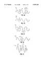
- FIGS. 1A, 1B, 1C, 1D, and 1Eare plan views of an unrolled stent form making up the invention.
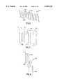
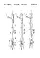
- FIG. 2is a side view of the inventive stent.
- FIG. 3is a close-up of a portion of the inventive stent shown in FIG. 2.
- FIG. 4is an abstracted portion of an inventive stent and shows the concept of torsion on a portion of the stent.
- FIG. 5is a side view of the inventive stent showing a variation having flared ends.
- FIGS. 6, 7, and 8show plan views of an unrolled stent produced from flat stock.
- FIG. 9shows a quarter view of the rolled stent using the flat stock pattern shown in FIG. 7.
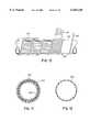
- FIG. 10shows a device for winding and heat treating a stent made according to the invention.
- FIGS. 11 and 12show end view cutaways of stent-grafts made according to the invention.
- FIGS. 13A, 13C, and 13Eshow procedures for folding the stent-grafts made according to the invention.
- FIGS. 13B, 13D, and 13Fshow the corresponding folded stent-grafts.
- FIGS. 14A-14Cshow a schematic procedure for deploying the inventive stent-grafts using an external sleeve.
- FIGS. 15A and 16Ashow front quarter views of folded stents or stent-grafts held in that folded position by a tether wire.
- FIGS. 15B, 15C, 16B, and 16Cshow end views of the folded stent and of the open stent shown respectively in FIGS. 15A and 16A.
- FIGS. 17A-17Cshow a schematic procedure for deploying the inventive stent-grafts (as shown in FIGS. 15A-15C and 16A-16C) using a tether wire.
- this inventionis variously an expandable stent, a stent-graft, and a fiber reinforced stent-graft.
- the stent-graftmay be a combination of several components: a thin-walled tube generally coaxial with the stent, the expandable stent structure, and an optional network of fibers used to reinforce the tubular component.
- the stent and the optional reinforcing fibersare desirably imbedded in the wall of the thin-walled tube.
- the expandable stent structureis a cylindrical body produced of a helically placed (wound or otherwise preformed) torsion member having an undulating or serpentine shape.
- the undulationsWhen the undulating torsion member is formed into the cylinder, the undulations may be aligned so that they are "in phase” with each other.
- the undulationsare desirably linked, typically with a flexible linkage of a suitable polymeric material, to maintain the phased relationship of the undulations during compression and deployment.
- These stent configurationsare exceptionally kink-resistant and flexible, particularly when flexed along the longitudinal axis of the stent.
- the stentWhen the stent is used in a reinforced stent-graft, that is to say: the stent is included into a thin-walled tube having reinforcing fibers, the fibers may be formed into a network, such as a tubular mesh.
- the stent-graftmay be delivered percutaneously through the vasculature after having been folded to a reduced diameter. Once reaching the intended delivery site, it is expanded to form a lining on the vessel wall.
- Methods of delivering the various devices using a percutaneous catheter either with or without expansion aidsare also an aspect of the invention.
- vascular graftsusually do not have the stiffness or strength by themselves both to stay open against the radial inward loads found in those vessels and to prevent their slippage from the chosen deployment site.
- a radially rigid stent structuremay be incorporated into the stent-graft.
- Our stentis constructed of a reasonably high strength material, i.e., one which is resistant to plastic deformation when stressed.
- the structureis typically from one of three sources: 1.) a wire form in which a wire is first formed into an undulating shape and the resulting undulating shape is helically wound to form a cylinder, 2.) an appropriate shape is formed from a flat stock and wound into a cylinder, and 3.) a length of tubing is formed into an appropriate shape.
- These stent structuresare typically oriented coaxially with the tubular graft component. The stent structures may be placed on the outer surface or the inner surface of the tubular member although it is preferable that the stent be imbedded in the graft tubing wall for ease of integration with the tubing and to prevent the stent's exposure to blood.
- the stent structurehave the strength and flexibility to tack the graft tubing firmly and conformally against the vessel wall.
- the stent materialshould have a high strength-to-volume ratio.
- a percutaneously delivered stent-graftmust expand from a reduced diameter, necessary for delivery, to a larger deployed diameter.
- the diameters of these devicesobviously vary with the size of the body lumen into which they are placed.
- the stents of this inventionmay range in size (for neurological applications) from 2.0 mm in diameter to 30 mm in diameter (for placement in the aorta). A range of about 2.0 mm to 6.5 mm (perhaps to 10.0 mm) is believed to be desirable.
- expansion ratios of 2:1 or moreare required.
- These stentsare capable of expansion ratios of up to 5:1 for larger diameter stents.
- Typical expansion ratios for use with the stents and stent-grafts of the inventiontypically are in the range of about 2:1 to about 4:1 although the invention is not so limited.
- the thickness of the stent materialsobviously varies with the size (or diameter) of the stent and the ultimate required yield strength of the folded stent. These values are further dependent upon the selected materials of construction.
- Wire used in these variationsare typically of stronger alloys, e.g., nitinol and stronger spring stainless steels, and have diameters of about 0.002 inches to 0.005 inches.
- the appropriate diameter for the stent wiremay be somewhat larger, e.g., 0.005 to 0.020 inches.
- thicknesses of about 0.002 inches to 0.005 inchesis usually sufficient.
- the appropriate thickness for the stent flat stockmay be somewhat thicker, e.g., 0.005 to 0.020 inches.
- the stent-graftis fabricated in the expanded configuration. In order to reduce its diameter for delivery the stent-graft would be folded along its length, similar to the way in which a PCTA balloon would be folded. It is desirable, when using super-elastic alloys which are also have temperature-memory characteristics, to reduce the diameter of the stent at a temperature below the transition-temperature of the alloys. Often the phase of the alloy at the lower temperature is somewhat more workable and easily formed. The temperature of deployment is desirably above the transition temperature to allow use of the super-elastic properties of the alloy.
- FIG. 1Ais a plan view of an isolated section of the inventive stent device and is intended both to identify a variation of the invention and to provide conventions for naming the components of the torsion member (100).
- FIG. 1Ashows, in plan view, an undulating torsion member (100) formed from a wire stock into a U-shape.
- a torsion pair (102)is made up of an end member (104) and two adjacent torsion lengths (106).
- each torsion length (106)will be a component to each of its adjacent torsion pairs (102).
- the U-shaped torsion pair (102)may be characterized by the fact that the adjacent torsion lengths are generally parallel to each other prior to formation into the stent.
- the stents of this inventionuse undulating torsion members which are "open” or “unconfined” at their apex or end member (104).
- "open” or “unconfined”we mean that the apex or end member (104) does not have any means in that apex which would tend to inhibit the movement of the flexible linkage (discussed below) down between the arms or torsion lengths (106) of the torsion pair (102).
- FIG. 1Bshows another variation of the invention having a sinusoidal shaped torsion member (108).
- the adjacent torsion lengths (110)are not parallel and the wire forms an approximate sine shape before being formed into a cylinder.
- FIG. 1Cshows a variation of the invention having an ovoid shaped torsion member (112).
- the adjacent torsion lengths (114)are again not parallel.
- the wireforms an approximate open-ended oval with each torsion pair (116) before being formed into a cylinder.
- FIG. 1Dshows another variation of the invention having a V-shaped torsion member (118).
- the adjacent torsion lengths (120)form a relatively sharp angle at the torsion end (122) shape before being formed into a cylinder.
- FIG. 1Eshows a variation of the invention in which adjacent torsion members on the stent (117) have differing amplitude.
- the peaks of the high amplitude torsion members (119)may be lined up “out of phase” or “peak to peak” with short amplitude (121) or high amplitude torsion members in the adjacent turn of the helix or may be positioned “in phase” similar to those discussed with regard to FIG. 2 below.
- FIGS. 1A-1Eare exceptionally kink-resistant and flexible when flexed along the longitudinal axis of the stent.
- a section of the torsion member found on one of FIGS. 1A-1Dwould be helically wound about a form of an appropriate size so that the end members (e.g., 104 in FIG. 1A) would be centered between the end members of the torsion member on an adjacent turn of the helix. This is said to be “in phase”. "Out of phase” would be the instance in which the adjacent members meet directly, i.e., end member-to-end member. In any event, once so aligned, the phasic relationship may be stabilized by weaving a flexible linkage through the end members from one turn of the helix to the next.
- FIG. 2shows a side view of a typical stent (122) made according to this invention including the phased relationship of the helical turns of the stent and the flexible linkage (124).
- FIG. 3shows a close-up of the FIG. 2 stent and depicts the phased relationship (within box A) and shows in detail a typical way in which the flexible linkage (124) is looped through the various end members (104) to maintain the phased relationship. It may be noted that the flexible linkage (124) is free to move away from the apex at the end members (104) without constraint.
- the stentmay be folded in some fashion (as will be discussed below) for deployment. During the step of folding, the stent undergoes a transformation.
- FIG. 4shows an isolated torsion pair (102).
- the end memberWhen the torsion pair (102) undergoes a flexing in the amount of ⁇ °, the end member will flex some amount ⁇ °, torsion length (130) will undertake a twist of ⁇ °, and torsion length (132) will undertake a twist opposite of that found in torsion length (130) in the amount of ⁇ °.
- the amounts of angular torsion found in the torsion lengths (130 and 132)will not necessarily be equal because the torsion lengths are not necessarily at the same angle to the longitudinal axis of the stent.
- the torsion pair (102)undergoes a significant of flexing as the stent is folded or compressed in some fashion.
- the flexingprovides a twist to the torsion lengths (103 and 132), a significant portion of which is generally parallel to the longitudinal axis of the stent. It is this significant imposed longitudinal torsion which forms an important concept of the inventive stent.
- the stentis folded longitudinally and is delivered through the lumen of the catheter in such a way that it is self-restoring once it has been introduced to the selected body lumen site.
- This stated desireis not to rule out the use of the inventive stent or stent-graft with a balloon or expander or other shape-restoring tool if so desired, but the design of the stent is meant to eliminate the need for (or, at least to minimize the need for) such expanding tools.
- the inventive stentuses concepts which can be thought of as widely distributing and storing the force necessary to fold the tubular stent into a configuration which will fit through a diameter smaller than its relaxed outside diameter without inducing plastic deformation of the constituent metal or plastic and yet allowing those distributed forces to expand the stent upon deployment.
- FIG. 5shows, in side view, a variation of the inventive stent (140) made from wire having flares (142) at one or both ends.
- the flaringprovides a secure anchoring of the stent or stent-graft (140) against the vessel wall. This prevents the implant from migrating downstream.
- the flaringprovides a tight seal against the vessel so that the blood is channelled through the lumen rather than outside the graft.
- the undulating structuremay vary in spacing to allow the helix turns to maintain its phased relationship between turns of the helix and to conform to the discussion just above.
- a flexible linkage between the contiguous helical turnsmay also be applied to at least a portion of the helices.
- the stent structuremay also be made by forming a desired structural pattern out of a flat sheet.
- the sheetmay then be rolled to form a tube.
- FIGS. 6, 7, and 8show plan views of torsion members (respectively 200, 202, and 204) which may be then rolled about an axis (206) to form a cylinder.
- the end caps (208)may be aligned so that they are "out of phase”.
- the flexible linkage (210)is then included to preserve the diameter of the stent.
- the stent shown in FIG. 9may be machined from tubing. If the chosen material in nitinol, careful control of temperature during the machining step may be had by EDM (electro-discharge-machining), laser cutting, chemical machining, or high pressure water cutting.
- EDMelectro-discharge-machining
- laser cuttinglaser cutting
- chemical machiningchemical machining
- high pressure water cuttinghigh pressure water cutting
- Nitinolis especially preferred because of its "super-elastic" or "pseudo-elastic" shape recovery properties, i.e., the ability to withstand a significant amount of bending and flexing and yet return to its original form without deformation.
- These metalsare characterized by their ability to be transformed from an austenitic crystal structure to a stress-induced martensitic structure at certain temperatures, and to return elastically to the austenitic shape when the stress is released.
- These alternating crystalline structuresprovide the alloy with its super-elastic properties. These alloys are well known but are described in U.S. Pat. Nos. 3,174,851, 3,351,463, and 3,753,700.
- nitinolwill be nominally 50.6% ( ⁇ 0.2%) Ni with the remainder Ti.
- Commercially available nitinol materialsusually will be sequentially mixed, cast, formed, and separately cold-worked to 30-40%, annealed, and stretched. Nominal ultimate yield strength values for commercial nitinol are in the range of 30 psi and for Young's modulus are about 700 kBar.
- Nitinolis further suitable because it has a relatively high strength to volume ratio. This allows the torsion members to be shorter than for less elastic metals.
- the flexibility of the stent-graftis largely dictated by the length of the torsion member components in the stent structural component. The shorter the pitch of the device, the more flexible the stent-graft structure can be made. Materials other than nitinol are suitable. Spring tempered stainless steels and cobalt-chromium alloys such as ELGILOY are also suitable as are a wide variety of other known "super-elastic" alloys.
- nitinolis preferred in this service because of its physical properties and its significant history in implantable medical devices, we also consider it also to be suitable for use as a stent because of its overall suitability with magnetic resonance imaging (MRI) technology.
- MRImagnetic resonance imaging
- thermotropic liquid crystal polymersare high molecular weight materials which can exist in a so-called “liquid crystalline state” where the material has some of the properties of a liquid (in that it can flow) but retains the long range molecular order of a crystal.
- thermotropicrefers to the class of LCP's which are formed by temperature adjustment. LCP's may be prepared from monomers such as p,p'-dihydroxy-polynuclear-aromatics or dicarboxy-polynuclear-aromatics.
- the LCP'sare easily formed and retain the necessary interpolymer attraction at room temperature to act as high strength plastic artifacts as are needed as a foldable stent. They are particularly suitable when augmented or filled with fibers such as those of the metals or alloys discussed below. It is to be noted that the fibers need not be linear but may have some preforming such as corrugations which add to the physical torsion enhancing abilities of the composite.
- FIG. 10shows a method for producing a stent of the configuration shown in FIG. 9. It may be used, of course, for producing a variety of other configurations shown herein.
- a preformed strip (211)(preferably nitinol) is rolled onto a mandrel (213) having a channel defined by a thread (215).
- the pitch of the strip (211)is selected using the criteria discussed above.
- the pitch angle (215) shownis about 20° but is not critical to the operation of the invention.
- the strip (211)is wound onto the mandrel (213) and the assembly is introduced into the outer sleeve (217), the strip (now in the shape of the stent) may then be annealed (or at least "heat set") to assist in maintaining the resulting stent in a useful shape.
- nitinol devicesmay be heat treated for five minutes or less at temperatures of 500° C. without significant lessening of the superelastic properties. Said another way, heating super-elastic alloys must carried out with some care so as not to destroy or significantly lessen the super-elastic properties.
- the flexible linkage between adjacent turns of the helixmay be of any appropriate filamentary material which is blood compatible or biocompatible and sufficiently flexible to allow the stent to flex and not deform the stent upon folding.
- the linkagemay be a single or multiple strand wire (platinum, platinum/tungsten, gold, palladium, tantalum, stainless steel, etc.), much preferred is the use of polymeric biocompatible filaments.
- Synthetic polymerssuch as polyethylene, polypropylene, polyurethane, polyglycolic acid, polyesters, polyamides, their mixtures, blends, copolymers, mixtures, blends and copolymers are suitable; preferred of this class are polyesters such as polyethylene terephthalate including DACRON and MYLAR and polyaramids such as KEVLAR, polyfluorocarbons such as polytetrafluoroethylene with and without copolymerized hexafluoropropylene (TEFLON or GORETEX), and porous or nonporous polyurethanes. Natural materials or materials based on natural sources such as collagen are especially preferred is this service.
- the tubular component or member of the stent-graftmay be made up of any material which is suitable for use as a graft in the chosen body lumen.
- Many graft materialsare known, particularly known are vascular graft materials.
- natural materialmay be introduced onto the inner surface of the stent and fastened into place.
- Synthetic polymerssuch as polyethylene, polypropylene, polyurethane, polyglycolic acid, polyesters, polyamides, their mixtures, blends, copolymers, mixtures, blends and copolymers are suitable; preferred of this class are polyesters such as polyethylene terephthalate including DACRON and MYLAR and polyaramids such as KEVLAR, polyfluorocarbons such as polytetrafluoroethylene with and without copolymerized hexafluoropropylene (TEFLON or GORETEX), and porous or nonporous polyurethanes. Highly preferred materials are certain collagen-based materials of COLLAGEN CORPORATION of Palo Alto, Calif.
- the graftmay adhere to or partially encapsulate or be cast about the stent when appropriate materials such as castable polyurethane or collagen-based materials are employed.
- appropriate materialssuch as castable polyurethane or collagen-based materials are employed.
- a highly preferred materialis a collagen-based material described in U.S. Pat. No. 5,162,430, to Rhee et al, the entirety of which is incorporated by notice, or as described below.
- Collagenis easily formed into thin-walled tubes which are limp, compliant, flexible, uniform, and have smooth surfaces.
- the tubing wallsmay have a hydrated thickness of 0.001 to 0.020 inches (or to 0.100 inches in some cases) for efficacy. Other thicknesses may be used if specific goals are to be achieved.
- the collagen tubeacts as an intravascular blood conduit to line the interior surface of the blood vessel.
- the preferred collagen composition used in this inventionis a pharmaceutically acceptable non-immunogenic composition formed by covalently binding atelopeptide collagen to pharmaceutically pure, synthetic, hydrophilic polymers via specific types of chemical bonds to provide collagen/polymer conjugates.
- Any type of collagenmay be used including extracted and purified collagen including atelopeptide collagen which can be type I, type II or type III collagen.
- the collagenmay be extracted from various sources such as bovine hide and human placenta and may be fibrillar or non-fibrillar.
- the synthetic hydrophilic polymermay be polyethylene glycol and derivatives thereof having a weight average molecular weight over a range of from about 100 to about 20,000.
- the compositionsmay incorporate other components such as biologically active materials.
- the collagen-polymer conjugatesgenerally contain large amounts of water when formed. The extruded materials may be dehydrated, resulting in a reasonably flexible material which can be readily stored.
- collagenrefers to all forms of collagen, including those which have been extracted, processed or otherwise modified. Preferred collagens are non-immunogenic and, if extracted from animals, are treated to remove the immunogenic telopeptide regions ("atelopeptide collagen"), are soluble, and may be in the fibrillar or non-fibrillar form. Type I collagen is best suited to most applications involving bone or cartilage repair. However, other forms of collagen are also useful in the practice of the invention, and are not excluded from consideration here. Collagen crosslinked using heat, radiation, or chemical agents such as glutaraldehyde may be conjugated with polymers as described herein to form particularly rigid compositions.
- Collagen crosslinked using glutaraldehyde or other (nonpolymer) linking agentsis referred to herein as "GAX", while collagen cross-linked using heat and/or radiation is termed "HRX.”
- GXCollagen crosslinked using glutaraldehyde or other (nonpolymer) linking agents
- HRXcollagen cross-linked using heat and/or radiation
- Collagen used in connection with the preferred embodiments of the inventionis in a pharmaceutically pure form such that it can be incorporated into a body, human or otherwise, for the intended purpose.
- synthetic hydrophilic polymerrefers to a synthetic polymer having an average molecular weight and composition which renders the polymer essentially water-soluble.
- Preferred polymersare highly pure or are purified to a highly pure state such that the polymer is, or is treated to become, pharmaceutically pure.
- Most hydrophilic polymerscan be rendered water-soluble by incorporating a sufficient number of oxygen (or, less frequently, nitrogen) atoms available for forming hydrogen bonds in aqueous solutions.
- Preferred polymersare hydrophilic but not soluble.
- Preferred hydrophilic polymers used hereininclude polyethylene glycol, polyoxyethylene, polymethylene glycol, polytrimethylene glycols, polyvinylpyrrolidones, and derivatives thereof.
- the polymerscan be linear or multiply branched and will not be substantially crosslinked.
- Other suitable polymersinclude polyoxyethylene-polyoxypropylene block polymers and copolymers. Polyoxyethylene-polyoxypropylene block polymers having an ethylene diamine nucleus (and thus having four ends) are also available and may be used in the practice of the invention. Naturally occurring and/or biologically active polymers such as proteins, starch, cellulose, heparin, and the like are not generally desirable in this definition although they may be used. All suitable polymers will be non-toxic, non-inflammatory and non-immunogenic when used to form the desired composition, and will preferably be essentially non-degradable in vivo over a period of at least several months.
- the hydrophilic polymermay increase the hydrophilicity of the collagen, but does not render it water-soluble.
- Presently preferred hydrophilic polymersare mono-, di-, and multi-functional polyethylene glycols (PEG).
- Monofunctional PEGhas only one reactive hydroxy group, while difunctional PEG has reactive groups at each end.
- Monofunctional PEGpreferably has a weight average molecular weight between about 100 and about 15,000, more preferably between about 200 and about 8,000, and most preferably about 4,000.
- Difunctional PEGpreferably has a molecular weight of about 400 to about 40,000, more preferably about 3,000 to about 10,000.
- Multi-functional PEGpreferably has a molecular weight between about 3,000 and 100,000.
- PEGcan be rendered monofunctional by forming an alkylene ether at one end.
- the alkylene ethermay be any suitable alkoxy radical having 1-6 carbon atoms, for example, methoxy, ethoxy, propoxy, 2-propoxy, butoxy, hexyloxy, and the like. Methoxy is presently preferred.
- Difunctional PEGis provided by allowing a reactive hydroxy group at each end of the linear molecule. The reactive groups are preferably at the ends of the polymer, but may be provided along the length thereof.
- collagen-polymerrefers to collagen chemically conjugated to a synthetic hydrophilic polymer, within the meaning of this invention.
- collagen-PEGor “PEG-collagen” denotes a composition within the most preferred aspect of the invention wherein collagen is chemically conjugated to PEG.
- Collagen-dPEGrefers to collagen chemically conjugated to difunctional PEG, wherein the collagen molecules are typically crosslinked.
- Crosslinked collagenrefers to collagen in which collagen molecules are linked by covalent bonds with polyfunctional (including difunctional) polymers.
- Terms such as “GAX-dPEG” and “HRX-dPEG”indicate collagen crosslinked by both a difunctional hydrophilic polymer and a crosslinking agent such as glutaraldehyde or heat.
- the polymermay be "chemically conjugated" to the collagen by means of a number of different types of chemical linkages. For example, the conjugation can be via an ester or urethane linkage, but is more preferably by means of an ether linkage. An ether linkage is preferred in that it can be formed without the use of toxic chemicals and is not readily susceptible to hydrolysis in vivo.
- molecular weightrefers to the weight average molecular weight of a number of molecules in any given sample, as commonly used in the art.
- a sample of PEG 2,000might contain a statistical mixture of polymer molecules ranging in weight from, for example, 1,500 to 2,500 daltons with one molecule differing slightly from the next over a range.
- Specification of a range of molecular weightindicates that the average molecular weight may be any value between the limits specified, and may include molecules outside those limits.
- a molecular weight range of about 800 to about 20,000indicates an average molecular weight of at least about 800, ranging up to about 20 kDa.
- available lysine residuerefers to lysine side chains exposed on the outer surface of collagen molecules, which are positioned in a manner to allow reaction with activated PEG.
- the number of available lysine residuesmay be determined by reaction with sodium 2,4,6-trinitrobenzenesulfonate (TNBS).
- growth factoris used to describe biologically active molecules and active peptides (which may be naturally occurring or synthetic) which aid in healing or regrowth of normal tissue.
- the function of growth factorsis two-fold: 1) they can incite local cells to produce new collagen or tissue, or 2) they can attract cells to the site in need of correction.
- growth factorsmay serve to encourage "biological anchoring" of the collagen graft implant within the host tissue.
- the growth factorsmay either be admixed with the collagen-polymer conjugate or chemically coupled to the conjugate.
- growth factorssuch as epidermal growth factor (EGF), transforming growth factor (TGF) alpha, TGF.sub. ⁇ : (including any combination of TGF.sub. ⁇ s), TGF.sub. ⁇ 1, TGF.sub. ⁇ 2, platelet derived growth factor (PDGF-AA, PDGF-AB, PDGF-BB), acidic fibroblast growth factor (FGF), basic FGF, connective tissue activating peptides (CTAP), ⁇ -thrombo-globulin, insulin-like growth factors, erythropoietin (EPO), nerve growth factor (NGF), bone morphogenic protein (BMP), osteogenic factors, and the like.
- EGFepidermal growth factor
- TGFtransforming growth factor alpha
- TGF.sub. ⁇including any combination of TGF.sub. ⁇ s), TGF.sub. ⁇ 1, TGF.sub. ⁇ 2, platelet derived growth factor (PDGF-AA, PDGF-AB, PDGF-BB), acidic fibroblast growth factor (FGF),
- Incorporation of growth factorscan facilitate regrowth when the tubes are used in the treatment of defective or damaged channels.
- onemay chemically link the growth factors to the collagen-polymer composition by employing a suitable amount of multi-functional polymer molecules during synthesis.
- the growth factorsmay then be attached to the free polymer ends by the same method used to attach PEG to collagen, or by any other suitable method.
- By tethering growth factors to the outer and/or inner surface of the graft materialthe amount of grafts needed to carry out effective treatment is substantially reduced.
- Tubes which incorporate growth factorsmay provide effective controlled-release drug delivery. By varying the chemical linkage between the collagen and the synthetic polymer, it is possible to vary the effect with respect to the release of the biologic.
- the linkagewhen an "ester” linkage is used, the linkage is more easily broken under physiological conditions, allowing for sustained release of the growth factor from the matrix.
- an "ether” linkagewhen an “ether” linkage is used, the bonds are not easily broken and the growth factor will remain in place for longer periods of time with its active sites exposed providing a biological effect on the natural substrate for the active site of the protein. It is possible to include a mixture of conjugates with different linkages so as to obtain variations in the effect with respect to the release of the biologic, e.g., the sustained release effect can be modified to obtain the desired rate of release.
- tissue growth-promoting amountof a composition containing a growth factor refers to the amount of growth factor needed in order to stimulate tissue growth to a detectable degree.
- Tissuein this context, includes connective tissue, bone, cartilage, epidermis and dermis, blood, and other tissues with particular emphasis on tissues which form channels such as veins, arteries, intestines and the like.
- the actual amount which is determined to be an effective amountwill vary depending on factors such as the size, condition, sex, and age of the patient, the type of tissue or channel, the effect desired and type of growth factor, and can be more readily determined by the caregiver.
- sufficient amountis applied to the amount of carrier used in combination with the collagen-polymer conjugates used in forming the tubes of the invention.
- a sufficient amountis that amount which, when mixed with the conjugate, renders it in the physical form desired, for example, extrudable tubes, extrudable cylinders having any desired cross-section, and so forth.
- Extrudable formulationsmay include an amount of a carrier sufficient to render the composition smoothly extrudable without significant need to interrupt the extrusion process.
- the amount of the carriercan be varied and adjusted depending on the size and shape and thickness of the wall of the tube being extruded. Such adjustments will be apparent to those skilled in the art upon reading this disclosure.
- collagenmust be chemically bound to a synthetic hydrophilic polymer.
- the synthetic hydrophilic polymeris activated and then reacted with the collagen.
- the hydroxyl or amino groups present on the collagencan be activated and the activated groups will react with the polymer to form the conjugate.
- a linking group with activated hydroxyl or amino groups thereoncan be combined with the polymer and collagen in a manner so as to concurrently react with both the polymer and collagen forming the conjugate.
- Other methods of forming the conjugateswill become apparent to those skilled in the art upon reading this disclosure.
- conjugates of the inventionare to be used in the human body it is important that all of the components, including the polymer, collagen, and linking group, if used form a conjugate that is unlikely to be rejected by the body. Accordingly, toxic and/or immunoreactive components are not preferred as starting materials. Some preferred starting materials and methods of forming conjugates are described further below.
- hydrophilic synthetic polymerscan be used in connection with forming the conjugate, such polymers must be biocompatible, relatively insoluble, but hydrophilic and is preferably one or more forms of polyethylene glycol (PEG), due to its known biocompatibility.
- PEGpolyethylene glycol
- Various forms of PEGare extensively used in the modification of biologically active molecules because PEG can be formulated to have a wide range of solubilities and because it lacks toxicity, antigenicity, immunogenicity, and does not typically interfere with the enzymatic activities and/or conformations of peptides. Further, PEG is generally non-biodegradable and is easily excreted from most living organisms including humans.
- the first step in forming the collagen-polymer conjugatesgenerally involves the functionalization of the PEG molecule.
- Various functionalized polyethylene glycolshave been used effectively in fields such as protein modification (see Abuchowski et al., Enzymes as Drugs, John Wiley & Sons: New York, N.Y. (1981) pp. 367-383; and Dreborg et al., Crit. Rev. Therap. Drug Carrier Syst. (1990) 6:315, both of which are incorporated herein by reference), peptide chemistry (see Mutter et al., The Peptides, Academic: New York, N.Y. 2:285-332; and Zalipsky et al., Int. J. Peptide Protein Res.
- polyethylene glycolwhich has been found to be particularly useful is monomethoxy-polyethylene glycol (mPEG), which can be activated by the addition of a compound such as cyanuric chloride, then coupled to a protein (see Abuchowski et al., J. Biol. Chem. (1977) 252:3578, which is incorporated herein by reference).
- mPEGmonomethoxy-polyethylene glycol
- cyanuric chloridecyanuric chloride
- Activated forms of PEGcan be made from reactants which can be purchased commercially.
- One form of activated PEG which has been found to be particularly useful in connection with the present inventionis mPEG-succinate-N-hydroxysuccinimide ester (SS-PEG) (see Abuchowski et al., Cancer Biochem. Biphys. (1984) 7:175, which is incorporated herein by reference).
- SS-PEGmPEG-succinate-N-hydroxysuccinimide ester
- ester linkagescan be used in connection with the present invention, they are not particularly preferred in that they undergo hydrolysis when subjected to physiological conditions over extended periods of time (see Dreborg et al., Crit. Rev. Therap. Drug Carrier Syst. (1990) 6:315; and Ulbrich et al., J. Makromol. Chem. (1986) 187:1131, both of which are incorporated herein by reference).
- Another means of attaching the PEG to a proteincan be by means of a carbamate linkage (see Beauchamp et al., Anal. Biochem. (1983) 131:25; and Berger et al., Blood (1988) 71:1641, both of which are incorporated herein by reference).
- the carbamate linkageis created by the use of carbonyldiimidazole-activated PEG. Although such linkages have advantages, the reactions are relatively slow and may take 2 to 3 days to complete.
- the conjugates used in forming the graftsmay be prepared by covalently binding a variety of different types of synthetic hydrophilic polymers to collagen.
- the final product or conjugate obtainedmust have a number of required characteristics such as being extrudable from a nozzle, biocompatible and non-immunogenic, it has been found useful to use polyethylene glycol as the synthetic hydrophilic polymer.
- the polyethylene glycolmust be modified in order to provide activated groups on one or preferably both ends of the molecule so that covalent binding can occur between the PEG and the collagen.
- the first functionalized PEGis difunctionalized PEG succinimidyl glutarate, referred to herein as (SG-PEG).
- the structural formula of this molecule and the reaction product obtained by reacting it with collagenis shown in Formula 1. ##STR1##
- PEG succinimidylAnother difunctionally activated form of PEG is referred to as PEG succinimidyl (S-PEG).
- S-PEGPEG succinimidyl
- the structural formula for this compound and the reaction product obtained by reacting it with collagenis shown in Formula 2.
- the subscript 3is replaced with an "n.”
- the structure in Formula 2results in a conjugate which includes an "ether” linkage which is not subject to hydrolysis. This is distinct from the first conjugate shown in Formula 1, wherein an ester linkage is provided. The ester linkage is subject to hydrolysis under physiological conditions.
- the structural formula and resulting conjugateare shown in Formula 4. It is noted that the conjugate includes both an ether and a peptide linkage. These linkages are stable under physiological conditions.
- the difunctionalized formis referred to as PEG succinimidyl carbonate (SC-PEG).
- SC-PEGPEG succinimidyl carbonate
- the structural formula of this compound and the conjugate formed by reacting SC-PEG with collagenis shown in Formula 5.
- this conjugateincludes a urethane linkage, the conjugate has been found not to have a high degree of stability under physiological conditions. The instability can be a desirable characteristic when the tubes are used in a situation where it is desirable that they dissolve over time.
- A-PEGdifunctional PEG propionaldehyde
- E-PEGdifunctional PEG glycidyl ether
- the conjugates formed using the functionalized forms of PEGvary depending on the functionalized form of PEG which is used in the reaction. Furthermore, the final product can be varied with respect to its characteristics by changing the molecular weight of the PEG. In general, the stability of the conjugate is improved by eliminating any ester linkages between the PEG and the collagen and including ether and/or urethane linkages. These stable linkages are generally used to form tubes to replace or augment a channel as may be done with a stent-graft.
- the graftsWhen the grafts are used as a temporary repair unit for a damaged channel, it may be desirable to include the weaker ester linkages so that the linkages are gradually broken by hydrolysis under physiological conditions, breaking apart the tube as it may be replaced by host tissue, or as it degrades, and releasing a component held therein, such as a growth factor.
- the rate of sustained releasecan be varied.
- Suitable collagensinclude all types of pharmaceutically useful collagen, preferably types I, II and III.
- Collagensmay be soluble (for example, commercially available Vitrogen® 100 collagen-in-solution), and may or may not have the telopeptide regions.
- the collagenwill be reconstituted fibrillar atelopeptide collagen, for example Zyderm® collagen implant (ZCI) or atelopeptide collagen in solution (CIS).
- ZCIZyderm® collagen implant
- CISatelopeptide collagen in solution
- Various forms of collagenare available commercially, or may be prepared by the processes described in, for example, U.S. Pat. Nos.
- Fibrillar, atelopeptide, reconstituted collagenis preferred in order to form tubes used for the repair or augmentation of channels.
- compositions used in forming the inventioncomprise collagen chemically conjugated to a selected synthetic hydrophilic polymer or polymers.
- Collagencontains a number of available amino and hydroxy groups which may be used to bind the synthetic hydrophilic polymer.
- the polymermay be bound using a "linking group", as the native hydroxy or amino groups in collagen and in the polymer frequently require activation before they can be linked.
- dicarboxylic anhydridese.g., glutaric or succinic anhydride
- a polymer derivativee.g., succinate
- a convenient leaving groupfor example, N-hydroxy-succinimide, N,N'-disuccinimidyl oxalate, N,N'-disuccinimidyl carbonate, and the like. See also Davis, U.S. Pat. No. 4,179,337
- Presently preferred dicarboxylic anhydrides that are used to form polymer-glutarate compositionsinclude glutaric anhydride, adipic anhydride, 1,8-naphthalene dicarboxylic anhydride, and 1,4,5,8-naphthalenetetracarboxylic dianhydride.
- the polymer thus activatedis then allowed to react with the collagen, forming a collagen-polymer composition used to make the grafts.
- a pharmaceutically pure form of monomethylpolyethylene glycol(mPEG) (mw 5,000) is reacted with glutaric anhydride (pure form) to create mPEG glutarate.
- the glutarate derivativeis then reacted with N-hydroxy-succinimide to form a succinimidyl monomethylpolyethylene glycol glutarate.
- the succinimidyl ester(mPEG*, denoting the activated PEG intermediate) is then capable of reacting with free amino groups present on collagen (lysine residues) to form a collagen-PEG conjugate wherein one end of the PEG molecule is free or nonbound.
- Other polymersmay be substituted for the monomethyl PEG, as described above.
- the coupling reactionmay be carried out using any known method for derivatizing proteins and synthetic polymers.
- the number of available lysines conjugatedmay vary from a single residue to 100% of the lysines, preferably 10-50%, and more preferably 20-30%.
- the number of reactive lysine residuesmay be determined by standard methods, for example by reaction with TNBS.
- the resulting productis a smooth, pliable, rubbery mass having a shiny appearance. It may be wetted, but is not water-soluble. It may be formulated as a suspension at any convenient concentration, preferably about 30-65 mg/mL, and may be extruded through a nozzle to form a tube. The consistency of the formulation may be adjusted by varying the amount of liquid used.
- the tubular componentmay be reinforced using a network of small diameter fibers.
- the fibersmay be random, braided, knitted, or woven.
- the fibersmay be imbedded in the tubular component wall, may be placed in a separate layer coaxial with the tubular component, or may be used in a combination of the two.
- FIG. 11shows an end view, cross-section of the configuration in which the stent (220) forms the outermost layer, a fibrous layer (222) coaxial to and inside the stent (220), and the tubular component (224) of, e.g., collagen as the inner layer.
- the fibrous materialis mixed with or imbedded into the tubular layer (226) and cast or injected around the stent (220).
- This fibrous materialmay extend for the length of the device or may be shorter.
- the fibersmay be wound or placed in any reasonable orientation within the device. Alternatively, randomly oriented short segments of fibers may also be imbedded in the wall of the tubing.
- the fibermay be any suitable fibrous blood-compatible material including polyesters such as DACRON, polyamides such as NYLON, KEVLAR, polyglycolic acids, polylactic acids, polyethylene, polypropylene, silk or other strong flexible fiber which are not detrimentally affected in the medical service in which this device is placed. Specifically, polypropylene and the like will not be dissolved in blood but polyglycolic acid will dissolve. Each are suitable but work in different ways.
- radio-opaque metallic fiberssuch as gold, platinum, platinum-tungsten, palladium, platinum-iridium, rhodium, tantalum, or alloys or composites of these metals like may be incorporated into the multi-strand reinforcement network to allow fluoroscopic visualization of the device.
- the fiberscarry much of the hoop stress and other loadings imposed by the vessel. This relieves the loading on the collagen and significantly increases the burst strength and fatigue properties of the tube. In addition, this makes the tube more effective in hydraulically isolating the vessel and as a result prevents the formation or worsening of aneurysms. This would be particularly beneficial in thinned weakened vessel walls resulting from de-bulking interventions or from medial thinning that has been seen to accompany stent placement. Another benefit of the fiber reinforcement is the increase in resistance to radially inward loading, especially if the loading is very focussed. Finally, fiber reinforcement may also impart some longitudinal stiffness to the stent-graft. This allows the stent-graft to maintain its strength and prevent it from kinking or sagging into the lumen.
- the preferred method of constructing the stent-graftis to first construct the stent and then to mold or cast the collagen tubular component about the stent.
- the stent structure and fiber reinforcementmay be molded into the wall of the collagen tube.
- a mold for such a structuredesirably is a simple annular space between two cylinders having room in the annular space for placement of the stent and would have a longitudinal axis slightly longer than the length of the stent-graft to be produced.
- the stent and fiber tubingis centered in the annular space and then the remaining space filled with collagen. If sPEG cross-linked collagen is used as the matrix material, the sPEG and collagen are mixed and introduced into the mold and allowed to cure. After curing, the mold is separated and the inventive fiber reinforced collagen tube with a stent structure produced.
- a stent-graft having torsion membersWhen a stent-graft having torsion members is folded, crushed, or otherwise collapsed, mechanical energy is stored in torsion in those torsion members. In this loaded state, the torsion members have a torque exerted about them and consequently have a tendency to untwist. Collectively, the torque exerted by the torsion members as folded down to a reduced diameter must be restrained from springing open.
- the stenttypically has at least one torsion member per fold to take advantage of the invention.
- the stent-graftis folded along its longitudinal axis and restrained from springing open. The stent-graft is then deployed by removing the restraining mechanism, thus allowing the torsion members to spring open against the vessel wall.
- inventive devices of this typeare typically selected having an expanded diameter of up to about 10% greater than the diameter of the lumen to be the site of the stent deployment.
- FIG. 13Ashows a sequence of folding the device (230) of this invention about a guidewire (232) into a loose C-shaped configuration.
- FIG. 13Bshows a front quarter view of the resulting folded stent or stent-graft.
- FIG. 13Cshows a sequence of folding the device (230) of this invention about a guidewire (232) into a rolled configuration.
- FIG. 13Dshows a front quarter view of the resulting folded stent or stent-graft.
- FIG. 13Eshows a sequence of folding the device (230) of this invention about a guidewire (232) into a triple lobed configuration.
- FIG. 13Fshows a front quarter view of the resulting folded stent or stent-graft.
- the stent-graftmay be tracked through the vasculature to the intended deployment site and then unfolded against the vessel lumen.
- the collagen tube component of the stent-graftis limp, flexible, and thus easy to fold. Folding of the stent structure in the manner discussed above allows it to return to a circular, open configuration.
- FIGS. 14A-14Cshow one desired way to place the devices of the present invention and allow them to self-expand.
- FIG. 14Ashows a target site (246) having, e.g., a narrowed vessel lumen.
- a guidewire (248) having a guide tip (250)has been directed to the site using known techniques.
- the stent-graft (252)is mounted on guidewire tubing (254) inside outer sliding sheath (256) after having folded in the manner discussed above.
- the outer sliding sheath (256)binds the compressed stent-graft (252) in place until released.
- FIG. 14Bshows placement of the stent-graft (252) at the selected site (246) by sliding the stent-graft (252) over the guidewire (248) all together with the guidewire tubing (254) and the outer sliding sheath (256).
- the stent-graft (252)is deployed by holding the guidewire tubing (254) in a stationary position while withdrawing the outer sliding sheath (256).
- the stent-graft (252)can be seen in FIG. 14B as partially deployed.
- FIG. 14Cshows the stent-graft (252) fully deployed after the guidewire tubing (254) and the outer sliding sheath (256) have been fully retracted.
- FIGS. 15A-C, 16A-C, and 17A-Cshow an inventive variation of deploying a stent or stent-graft made according to this invention. These methods involve the use of a control line or tether line which maintains the stent or stent-graft in a folded configuration until release.
- FIG. 15Ais a front-quarter view of the stent (302) or stent-graft which has been folded as shown in the Figures discussed above.
- the stent (302)is folded about guidewire (304) so that, when deployed, the guidewire (304) is within the stent (302).
- the tether wire (306)which is passed through loops (308) associated with the various helices as they wind about the stent (302).
- the loops (308)may be formed from the flexible link (124 in FIG. 2 or 3) or may be simply an alternating weave through appropriate apexes of the undulating helix, e.g., (104 in FIG. 3) or may be loops specifically installed for the purpose shown here.
- the tether wire (306)is so placed that when it is removed by sliding it axially along the stent (302) and out of the loops (308), that the stent (302) unfolds into a generally cylindrical shape within the body lumen.
- FIG. 15Bshows an end-view of a folded stent (302) or stent-graft having a guidewire (304) within the inner surface of the stent (302) and with the tether wire (306) within the loops (308).
- the end view of the folded stent (302)shows it to be folded into a form which is generally C-shaped.
- the stent (302) in FIG. 15Bassumes the form shown in end view in FIG. 15C.
- the guidewire (304) within the lumen of the stent (302) and the loops (308)which were formerly in a generally linear relationship having a tether wire passing through them.
- FIG. 16Ashows a folded stent (310) (or stent-graft) in front quarter view which is similar in configuration to the stent (302) shown in FIG. 15A except that the stent (310) is rolled somewhat tighter than the previously discussed stent.
- the guidewire (304)is also inside the stent (310) rather than outside of it. Loops (308) from generally opposing sides of the stent (310) are folded into an approximate line so that the tether wire may pass through the aligned loops (308).
- FIG. 16Bshows an end view of the stent (310), and in particular, emphasizes the tighter fold of the stent (310). When expanded by removal of the tether wire (306), the stent (310) in FIG.
- FIG. 16Bassumes the form shown in FIG. 16C.
- FIGS. 17A-Cshow a schematic procedure for deploying the stent (312) (or stent-graft) using a percutaneous catheter assembly (314).
- FIG. 17Amay be seen a percutaneous catheter assembly (314) which has been inserted to a selected site (316) within a body lumen.
- the stent (312)is folded about the guidewire (319) and guidewire tube (318) held axially in place prior to deployment by distal barrier (320) and proximal barrier (322).
- the distal barrier (320) and proximal barrier (322)typically are affixed to the guidewire tube (318).
- the tether wire (306)is shown extending through loops (308) proximally through the catheter assembly's (314) outer jacket (324) through to outside the body.
- FIG. 17Bshows the removal of the tether wire (306) from a portion of the loops (308) to partially expand the stent (312) onto the selected site (316).
- FIG. 17Cshows the final removal of the tether wire (306) from the loops (308) and the retraction of the catheter assembly (314) from the interior of the stent (312).
- the stent (312)is shown as fully expanded.
Landscapes
- Health & Medical Sciences (AREA)
- Engineering & Computer Science (AREA)
- Biomedical Technology (AREA)
- Cardiology (AREA)
- Oral & Maxillofacial Surgery (AREA)
- Transplantation (AREA)
- Heart & Thoracic Surgery (AREA)
- Vascular Medicine (AREA)
- Life Sciences & Earth Sciences (AREA)
- Animal Behavior & Ethology (AREA)
- General Health & Medical Sciences (AREA)
- Public Health (AREA)
- Veterinary Medicine (AREA)
- Gastroenterology & Hepatology (AREA)
- Pulmonology (AREA)
- Physics & Mathematics (AREA)
- Optics & Photonics (AREA)
- Prostheses (AREA)
Abstract
Description
Claims (18)
Priority Applications (14)
| Application Number | Priority Date | Filing Date | Title |
|---|---|---|---|
| US08/221,815US6165210A (en) | 1994-04-01 | 1994-04-01 | Self-expandable helical intravascular stent and stent-graft |
| US08/411,452US5876432A (en) | 1994-04-01 | 1995-03-28 | Self-expandable helical intravascular stent and stent-graft |
| CA2499888ACA2499888C (en) | 1994-04-01 | 1995-04-03 | Self-expandable stent and stent-graft and method of using them |
| CA002157575ACA2157575C (en) | 1994-04-01 | 1995-04-03 | Self-expandable stent and stent-graft and method of using them |
| AU23795/95AAU690684B2 (en) | 1994-04-01 | 1995-04-03 | Self-expandable stent and stent-graft and method of using them |
| DE69531239TDE69531239T2 (en) | 1994-04-01 | 1995-04-03 | SELF-EXPANDABLE STENT AND STENT-GRAFT TRANSPLANT |
| DE69532049TDE69532049T2 (en) | 1994-04-01 | 1995-04-03 | Self-expanding stent or stent graft and method for its preparation |
| AT00101667TATE252872T1 (en) | 1994-04-01 | 1995-04-03 | SELF-EXPANDABLE STENT OR STENT TRANSPLANT AND METHOD FOR PREPARING SAME |
| PCT/US1995/004000WO1995026695A2 (en) | 1994-04-01 | 1995-04-03 | Self-expandable stent and stent-graft and method of using them |
| EP00101667AEP0997115B1 (en) | 1994-04-01 | 1995-04-03 | Self-expandable stent and stent-graft and method of preparing them |
| CA002261941ACA2261941C (en) | 1994-04-01 | 1995-04-03 | Self-expandable stent and stent-graft and method of using them |
| EP95916920AEP0754016B1 (en) | 1994-04-01 | 1995-04-03 | Self-expandable stent and stent-graft |
| AT95916920TATE244543T1 (en) | 1994-04-01 | 1995-04-03 | SELF-EXPANDABLE STENT AND STENT-GRAFT TRANSPLANT |
| JP07524227AJP3129327B2 (en) | 1994-04-01 | 1995-04-03 | Self-expandable stents and stent-grafts and methods of using them |
Applications Claiming Priority (1)
| Application Number | Priority Date | Filing Date | Title |
|---|---|---|---|
| US08/221,815US6165210A (en) | 1994-04-01 | 1994-04-01 | Self-expandable helical intravascular stent and stent-graft |
Related Child Applications (1)
| Application Number | Title | Priority Date | Filing Date |
|---|---|---|---|
| US08/411,452DivisionUS5876432A (en) | 1994-04-01 | 1995-03-28 | Self-expandable helical intravascular stent and stent-graft |
Publications (1)
| Publication Number | Publication Date |
|---|---|
| US6165210Atrue US6165210A (en) | 2000-12-26 |
Family
ID=22829513
Family Applications (2)
| Application Number | Title | Priority Date | Filing Date |
|---|---|---|---|
| US08/221,815Expired - LifetimeUS6165210A (en) | 1994-04-01 | 1994-04-01 | Self-expandable helical intravascular stent and stent-graft |
| US08/411,452Expired - LifetimeUS5876432A (en) | 1994-04-01 | 1995-03-28 | Self-expandable helical intravascular stent and stent-graft |
Family Applications After (1)
| Application Number | Title | Priority Date | Filing Date |
|---|---|---|---|
| US08/411,452Expired - LifetimeUS5876432A (en) | 1994-04-01 | 1995-03-28 | Self-expandable helical intravascular stent and stent-graft |
Country Status (1)
| Country | Link |
|---|---|
| US (2) | US6165210A (en) |
Cited By (230)
| Publication number | Priority date | Publication date | Assignee | Title |
|---|---|---|---|---|
| US6331188B1 (en) | 1994-08-31 | 2001-12-18 | Gore Enterprise Holdings, Inc. | Exterior supported self-expanding stent-graft |
| US6352561B1 (en) | 1996-12-23 | 2002-03-05 | W. L. Gore & Associates | Implant deployment apparatus |
| US6352553B1 (en) | 1995-12-14 | 2002-03-05 | Gore Enterprise Holdings, Inc. | Stent-graft deployment apparatus and method |
| US6361637B2 (en) | 1995-12-14 | 2002-03-26 | Gore Enterprise Holdings, Inc. | Method of making a kink resistant stent-graft |
| US20020040238A1 (en)* | 1994-08-12 | 2002-04-04 | Meadox Medicals, Inc. | High strength and high density intraluminal wire stent |
| US6451050B1 (en)* | 2000-04-28 | 2002-09-17 | Cardiovasc, Inc. | Stent graft and method |
| WO2002089706A1 (en)* | 2001-05-03 | 2002-11-14 | Bionx Implants, Inc. | Method for attaching axial filaments to a self expanding stent |
| US20020177890A1 (en)* | 1996-01-05 | 2002-11-28 | Lenker Jay A. | Stent graft loading and deployment device and method |
| US6497671B2 (en)* | 1997-12-05 | 2002-12-24 | Micrus Corporation | Coated superelastic stent |
| US6533807B2 (en) | 1998-02-05 | 2003-03-18 | Medtronic, Inc. | Radially-expandable stent and delivery system |
| US6544249B1 (en)* | 1996-11-29 | 2003-04-08 | The Lions Eye Institute Of Western Australia Incorporated | Biological microfistula tube and implantation method and apparatus |
| US6551350B1 (en) | 1996-12-23 | 2003-04-22 | Gore Enterprise Holdings, Inc. | Kink resistant bifurcated prosthesis |
| WO2003057077A1 (en)* | 2001-12-28 | 2003-07-17 | Advanced Cardiovascular Systems, Inc. | Hybrid stent |
| US6599319B2 (en)* | 2001-12-14 | 2003-07-29 | Celanese Advanced Materials, Inc. | Prosthetic ligament |
| US6602280B2 (en) | 2000-02-02 | 2003-08-05 | Trivascular, Inc. | Delivery system and method for expandable intracorporeal device |
| US6613072B2 (en) | 1994-09-08 | 2003-09-02 | Gore Enterprise Holdings, Inc. | Procedures for introducing stents and stent-grafts |
| US20030181848A1 (en)* | 2000-04-14 | 2003-09-25 | Bergheim Olav B. | Implant with drug coating |
| US20040024446A1 (en)* | 1999-07-02 | 2004-02-05 | Scimed Life Systems, Inc. | Helically formed stent/graft assembly |
| US6723120B2 (en) | 1997-04-15 | 2004-04-20 | Advanced Cardiovascular Systems, Inc. | Medicated porous metal prosthesis |
| US6733521B2 (en) | 2001-04-11 | 2004-05-11 | Trivascular, Inc. | Delivery system and method for endovascular graft |
| US6758858B2 (en) | 1995-03-10 | 2004-07-06 | Bard Peripheral Vascular, Inc. | Diametrically adaptable encapsulated stent and methods for deployment thereof |
| US6761733B2 (en) | 2001-04-11 | 2004-07-13 | Trivascular, Inc. | Delivery system and method for bifurcated endovascular graft |
| US6770089B1 (en)* | 2000-12-28 | 2004-08-03 | Advanced Cardiovascular Systems, Inc. | Hybrid stent fabrication using metal rings and polymeric links |
| US6783438B2 (en) | 2002-04-18 | 2004-08-31 | Ormco Corporation | Method of manufacturing an endodontic instrument |
| US6790223B2 (en) | 2001-09-21 | 2004-09-14 | Scimed Life Systems, Inc. | Delivering a uretheral stent |
| US6790226B2 (en) | 1995-03-10 | 2004-09-14 | Bard Peripheral Vascular, Inc. | Endoluminal prosthesis with support wire |
| US6805706B2 (en) | 2002-08-15 | 2004-10-19 | Gmp Cardiac Care, Inc. | Stent-graft with rails |
| US6805898B1 (en) | 2000-09-28 | 2004-10-19 | Advanced Cardiovascular Systems, Inc. | Surface features of an implantable medical device |
| US20040215320A1 (en)* | 2003-04-25 | 2004-10-28 | James Machek | Integral stent graft |
| US20040216814A1 (en)* | 2003-05-02 | 2004-11-04 | Dooley Bret A. | Shape memory alloy articles with improved fatigue performance and methods therefore |
| US20040243215A1 (en)* | 2003-05-27 | 2004-12-02 | Kristoff Nelson | Staged deployment endograft |
| EP1518518A3 (en)* | 2003-09-25 | 2005-04-13 | Karel Volenec | Stent designed to stop esophageal variceal bleeding |
| US20050085898A1 (en)* | 2003-10-21 | 2005-04-21 | Cook Incorporated. | Natural tissue stent |
| US6887215B2 (en) | 2001-06-01 | 2005-05-03 | Boston Scientific Scimed, Inc. | Compressible ureteral stent for comfort |
| US20050118344A1 (en)* | 2003-12-01 | 2005-06-02 | Pacetti Stephen D. | Temperature controlled crimping |
| WO2005039445A3 (en)* | 2003-10-23 | 2005-07-21 | James C Peacock Iii | Stent-graft assembly formed insitu |
| US20050171595A1 (en)* | 2001-03-06 | 2005-08-04 | Feldman Marc D. | Method and apparatus for stent deployment with enhanced delivery of bioactive agents |
| US6981964B2 (en) | 2001-05-22 | 2006-01-03 | Boston Scientific Scimed, Inc. | Draining bodily fluids with a stent |
| US6981958B1 (en) | 2001-05-02 | 2006-01-03 | Glaukos Corporation | Implant with pressure sensor for glaucoma treatment |
| US20060014480A1 (en)* | 2002-04-18 | 2006-01-19 | Ormco Corporation | Method of manufacturing a dental instrument |
| US20060052861A1 (en)* | 2002-12-24 | 2006-03-09 | Novostent Corporation | Vascular prothesis having interdigitating edges and methods of use |
| US20060142840A1 (en)* | 2004-12-28 | 2006-06-29 | Scimed Life Systems, Inc. | Low profile stent-graft attachment |
| US20060149364A1 (en)* | 2004-12-31 | 2006-07-06 | Steven Walak | Low profile vascular graft |
| US7081129B2 (en) | 1998-02-09 | 2006-07-25 | Boston Scientific Santa Rosa Corp. | Endovascular graft |
| US7101392B2 (en) | 1992-03-31 | 2006-09-05 | Boston Scientific Corporation | Tubular medical endoprostheses |
| US7112226B2 (en) | 2002-10-22 | 2006-09-26 | Boston Scientific Scimed, Inc. | Male urethral stent device |
| US7147661B2 (en) | 2001-12-20 | 2006-12-12 | Boston Scientific Santa Rosa Corp. | Radially expandable stent |
| US7147660B2 (en) | 2001-12-20 | 2006-12-12 | Boston Scientific Santa Rosa Corp. | Advanced endovascular graft |
| US20070073375A1 (en)* | 2005-09-23 | 2007-03-29 | Boston Scientific Scimed, Inc. | Secured stent delivery system |
| WO2007002392A3 (en)* | 2005-06-24 | 2007-05-10 | Zimmer Spine Inc | Expandable surgical site access system |
| US7225518B2 (en) | 2004-02-23 | 2007-06-05 | Boston Scientific Scimed, Inc. | Apparatus for crimping a stent assembly |
| US20070129786A1 (en)* | 2005-10-14 | 2007-06-07 | Bradley Beach | Helical stent |
| US20070149690A1 (en)* | 2005-12-23 | 2007-06-28 | Boston Scientific Scimed, Inc. | Functionalized block copolymers |
| US7314480B2 (en) | 2003-02-27 | 2008-01-01 | Boston Scientific Scimed, Inc. | Rotating balloon expandable sheath bifurcation delivery |
| US7367989B2 (en) | 2003-02-27 | 2008-05-06 | Scimed Life Systems, Inc. | Rotating balloon expandable sheath bifurcation delivery |
| US7431710B2 (en) | 2002-04-08 | 2008-10-07 | Glaukos Corporation | Ocular implants with anchors and methods thereof |
| US20080262630A1 (en)* | 2005-10-13 | 2008-10-23 | Synthes (U.S.A. | Drug-Impregnated Encasement |
| US7488303B1 (en) | 2002-09-21 | 2009-02-10 | Glaukos Corporation | Ocular implant with anchor and multiple openings |
| US20090143760A1 (en)* | 2007-11-30 | 2009-06-04 | Jacques Van Dam | Methods, Devices, Kits and Systems for Defunctionalizing the Gallbladder |
| US7563241B2 (en) | 2001-04-07 | 2009-07-21 | Glaukos Corporation | Implant and methods thereof for treatment of ocular disorders |
| US7637936B2 (en) | 2001-04-06 | 2009-12-29 | Boston Scientific Scimed, Inc. | Stent design |
| US7651529B2 (en) | 2003-05-09 | 2010-01-26 | Boston Scientific Scimed, Inc. | Stricture retractor |
| US7678065B2 (en) | 2001-05-02 | 2010-03-16 | Glaukos Corporation | Implant with intraocular pressure sensor for glaucoma treatment |
| US7686841B2 (en) | 2003-12-29 | 2010-03-30 | Boston Scientific Scimed, Inc. | Rotating balloon expandable sheath bifurcation delivery system |
| US20100094447A1 (en)* | 2008-10-09 | 2010-04-15 | Seiko Epson Corporation | Operation sequence creating apparatus, method for controlling same, and program |
| US20100094394A1 (en)* | 2008-10-06 | 2010-04-15 | Bradley Beach | Reconstrainable stent delivery system |
| US7708711B2 (en) | 2000-04-14 | 2010-05-04 | Glaukos Corporation | Ocular implant with therapeutic agents and methods thereof |
| US20100119696A1 (en)* | 2006-06-30 | 2010-05-13 | Aquesys, Inc. | Manufacture of an organ implant |
| US7744619B2 (en) | 2004-02-24 | 2010-06-29 | Boston Scientific Scimed, Inc. | Rotatable catheter assembly |
| US7803180B2 (en) | 2005-04-04 | 2010-09-28 | Flexible Stenting Solutions, Inc. | Flexible stent |
| US7803178B2 (en) | 2004-01-30 | 2010-09-28 | Trivascular, Inc. | Inflatable porous implants and methods for drug delivery |
| US20100286760A1 (en)* | 2009-04-24 | 2010-11-11 | Bradley Beach | Flexible devices |
| US7850637B2 (en) | 1999-04-26 | 2010-12-14 | Glaukos Corporation | Shunt device and method for treating glaucoma |
| US7867186B2 (en) | 2002-04-08 | 2011-01-11 | Glaukos Corporation | Devices and methods for treatment of ocular disorders |
| US20110015721A1 (en)* | 1998-03-05 | 2011-01-20 | Boston Scientific Scimed, Inc. | Intraluminal Stent |
| US7879079B2 (en) | 2001-08-28 | 2011-02-01 | Glaukos Corporation | Implant delivery system and methods thereof for treating ocular disorders |
| US7892273B2 (en) | 2001-12-03 | 2011-02-22 | Xtent, Inc. | Custom length stent apparatus |
| US7892274B2 (en) | 2001-12-03 | 2011-02-22 | Xtent, Inc. | Apparatus and methods for deployment of vascular prostheses |
| US7918881B2 (en) | 2003-06-09 | 2011-04-05 | Xtent, Inc. | Stent deployment systems and methods |
| US7922740B2 (en) | 2004-02-24 | 2011-04-12 | Boston Scientific Scimed, Inc. | Rotatable catheter assembly |
| US7922753B2 (en) | 2004-01-13 | 2011-04-12 | Boston Scientific Scimed, Inc. | Bifurcated stent delivery system |
| US7938852B2 (en) | 2001-12-03 | 2011-05-10 | Xtent, Inc. | Apparatus and methods for delivery of braided prostheses |
| US7951155B2 (en) | 2002-03-15 | 2011-05-31 | Glaukos Corporation | Combined treatment for cataract and glaucoma treatment |
| US8012192B2 (en) | 2004-02-18 | 2011-09-06 | Boston Scientific Scimed, Inc. | Multi-stent delivery system |
| US8016870B2 (en) | 2001-12-03 | 2011-09-13 | Xtent, Inc. | Apparatus and methods for delivery of variable length stents |
| US8016871B2 (en) | 2001-12-03 | 2011-09-13 | Xtent, Inc. | Apparatus and methods for delivery of multiple distributed stents |
| US8066755B2 (en) | 2007-09-26 | 2011-11-29 | Trivascular, Inc. | System and method of pivoted stent deployment |
| US8080048B2 (en) | 2001-12-03 | 2011-12-20 | Xtent, Inc. | Stent delivery for bifurcated vessels |
| US8083788B2 (en) | 2001-12-03 | 2011-12-27 | Xtent, Inc. | Apparatus and methods for positioning prostheses for deployment from a catheter |
| US8083789B2 (en) | 2007-11-16 | 2011-12-27 | Trivascular, Inc. | Securement assembly and method for expandable endovascular device |
| US8133199B2 (en) | 2008-08-27 | 2012-03-13 | Boston Scientific Scimed, Inc. | Electroactive polymer activation system for a medical device |
| US8172897B2 (en) | 1997-04-15 | 2012-05-08 | Advanced Cardiovascular Systems, Inc. | Polymer and metal composite implantable medical devices |
| US8177831B2 (en) | 2001-12-03 | 2012-05-15 | Xtent, Inc. | Stent delivery apparatus and method |
| US8196279B2 (en) | 2008-02-27 | 2012-06-12 | C. R. Bard, Inc. | Stent-graft covering process |
| US8226701B2 (en) | 2007-09-26 | 2012-07-24 | Trivascular, Inc. | Stent and delivery system for deployment thereof |
| US8241346B2 (en) | 2001-12-20 | 2012-08-14 | Trivascular, Inc. | Endovascular graft and method of delivery |
| US8257427B2 (en) | 2001-09-11 | 2012-09-04 | J.W. Medical Systems, Ltd. | Expandable stent |
| US8282680B2 (en) | 2003-01-17 | 2012-10-09 | J. W. Medical Systems Ltd. | Multiple independent nested stent structures and methods for their preparation and deployment |
| US8308701B2 (en) | 2010-11-15 | 2012-11-13 | Aquesys, Inc. | Methods for deploying intraocular shunts |
| US8317859B2 (en) | 2004-06-28 | 2012-11-27 | J.W. Medical Systems Ltd. | Devices and methods for controlling expandable prostheses during deployment |
| US8328861B2 (en) | 2007-11-16 | 2012-12-11 | Trivascular, Inc. | Delivery system and method for bifurcated graft |
| US8333003B2 (en) | 2008-05-19 | 2012-12-18 | Boston Scientific Scimed, Inc. | Bifurcation stent crimping systems and methods |
| US8337445B2 (en) | 2001-05-03 | 2012-12-25 | Glaukos Corporation | Ocular implant with double anchor mechanism |
| US8372131B2 (en) | 2007-07-16 | 2013-02-12 | Power Ten , LLC | Surgical site access system and deployment device for same |
| US8414635B2 (en) | 1999-02-01 | 2013-04-09 | Idev Technologies, Inc. | Plain woven stents |
| US8419788B2 (en) | 2006-10-22 | 2013-04-16 | Idev Technologies, Inc. | Secured strand end devices |
| US8460358B2 (en) | 2004-03-30 | 2013-06-11 | J.W. Medical Systems, Ltd. | Rapid exchange interventional devices and methods |
| US8486132B2 (en) | 2007-03-22 | 2013-07-16 | J.W. Medical Systems Ltd. | Devices and methods for controlling expandable prostheses during deployment |
| US8500794B2 (en) | 2007-08-02 | 2013-08-06 | Flexible Stenting Solutions, Inc. | Flexible stent |
| US8506515B2 (en) | 2006-11-10 | 2013-08-13 | Glaukos Corporation | Uveoscleral shunt and methods for implanting same |
| US20130211501A1 (en)* | 2012-02-14 | 2013-08-15 | W. L. Gore & Associates, Inc. | Endoprosthesis having aligned legs for ease of cannulation |
| US8585747B2 (en) | 2003-12-23 | 2013-11-19 | J.W. Medical Systems Ltd. | Devices and methods for controlling and indicating the length of an interventional element |
| US8585629B2 (en) | 2010-11-15 | 2013-11-19 | Aquesys, Inc. | Systems for deploying intraocular shunts |
| US8586125B2 (en) | 1999-09-03 | 2013-11-19 | Advanced Cardiovascular Systems, Inc. | Thermal treatment of an implantable medical device |
| US8617337B2 (en) | 1999-02-02 | 2013-12-31 | Bard Peripheral Vascular, Inc. | Partial encapsulation of stents |
| US8617094B2 (en) | 2002-03-07 | 2013-12-31 | Glaukos Corporation | Fluid infusion methods for glaucoma treatment |
| US8617234B2 (en) | 2004-05-25 | 2013-12-31 | Covidien Lp | Flexible vascular occluding device |
| US8623067B2 (en) | 2004-05-25 | 2014-01-07 | Covidien Lp | Methods and apparatus for luminal stenting |
| US8623070B2 (en) | 2007-03-08 | 2014-01-07 | Thomas O. Bales | Tapered helical stent and method for manufacturing the stent |
| US8652193B2 (en) | 2005-05-09 | 2014-02-18 | Angiomed Gmbh & Co. Medizintechnik Kg | Implant delivery device |
| US8652198B2 (en) | 2006-03-20 | 2014-02-18 | J.W. Medical Systems Ltd. | Apparatus and methods for deployment of linked prosthetic segments |
| US8663309B2 (en) | 2007-09-26 | 2014-03-04 | Trivascular, Inc. | Asymmetric stent apparatus and method |
| US8663303B2 (en) | 2010-11-15 | 2014-03-04 | Aquesys, Inc. | Methods for deploying an intraocular shunt from a deployment device and into an eye |
| US8702781B2 (en) | 2001-12-03 | 2014-04-22 | J.W. Medical Systems Ltd. | Apparatus and methods for delivery of multiple distributed stents |
| US8721702B2 (en) | 2010-11-15 | 2014-05-13 | Aquesys, Inc. | Intraocular shunt deployment devices |
| US8758290B2 (en) | 2010-11-15 | 2014-06-24 | Aquesys, Inc. | Devices and methods for implanting a shunt in the suprachoroidal space |
| US8765210B2 (en) | 2011-12-08 | 2014-07-01 | Aquesys, Inc. | Systems and methods for making gelatin shunts |
| US8769796B2 (en) | 2008-09-25 | 2014-07-08 | Advanced Bifurcation Systems, Inc. | Selective stent crimping |
| US8784472B2 (en) | 2003-08-15 | 2014-07-22 | Boston Scientific Scimed, Inc. | Clutch driven stent delivery system |
| US8795347B2 (en) | 2008-09-25 | 2014-08-05 | Advanced Bifurcation Systems, Inc. | Methods and systems for treating a bifurcation with provisional side branch stenting |
| US8801766B2 (en) | 2010-11-15 | 2014-08-12 | Aquesys, Inc. | Devices for deploying intraocular shunts |
| US8808347B2 (en) | 2008-09-25 | 2014-08-19 | Advanced Bifurcation Systems, Inc. | Stent alignment during treatment of a bifurcation |
| US8821562B2 (en) | 2008-09-25 | 2014-09-02 | Advanced Bifurcation Systems, Inc. | Partially crimped stent |
| US8828070B2 (en) | 2010-11-15 | 2014-09-09 | Aquesys, Inc. | Devices for deploying intraocular shunts |
| US8852136B2 (en) | 2011-12-08 | 2014-10-07 | Aquesys, Inc. | Methods for placing a shunt into the intra-scleral space |
| US8852137B2 (en) | 2010-11-15 | 2014-10-07 | Aquesys, Inc. | Methods for implanting a soft gel shunt in the suprachoroidal space |
| US8852256B2 (en) | 2010-11-15 | 2014-10-07 | Aquesys, Inc. | Methods for intraocular shunt placement |
| US20140324164A1 (en)* | 2011-08-05 | 2014-10-30 | Mitraltech Ltd. | Techniques for percutaneous mitral valve replacement and sealing |
| US8926688B2 (en) | 2008-01-11 | 2015-01-06 | W. L. Gore & Assoc. Inc. | Stent having adjacent elements connected by flexible webs |
| USD723165S1 (en) | 2013-03-12 | 2015-02-24 | C. R. Bard, Inc. | Stent |
| US8974511B2 (en) | 2010-11-15 | 2015-03-10 | Aquesys, Inc. | Methods for treating closed angle glaucoma |
| US8979917B2 (en) | 2008-09-25 | 2015-03-17 | Advanced Bifurcation Systems, Inc. | System and methods for treating a bifurcation |
| US8980297B2 (en) | 2007-02-20 | 2015-03-17 | J.W. Medical Systems Ltd. | Thermo-mechanically controlled implants and methods of use |
| US8986362B2 (en) | 2004-06-28 | 2015-03-24 | J.W. Medical Systems Ltd. | Devices and methods for controlling expandable prostheses during deployment |
| US8992595B2 (en) | 2012-04-04 | 2015-03-31 | Trivascular, Inc. | Durable stent graft with tapered struts and stable delivery methods and devices |
| US9017276B2 (en) | 2010-11-15 | 2015-04-28 | Aquesys, Inc. | Shunt placement through the sclera |
| US9066825B2 (en) | 2012-05-14 | 2015-06-30 | C.R. Bard, Inc. | Uniformly expandable stent |
| US9095411B2 (en) | 2010-11-15 | 2015-08-04 | Aquesys, Inc. | Devices for deploying intraocular shunts |
| US9101503B2 (en) | 2008-03-06 | 2015-08-11 | J.W. Medical Systems Ltd. | Apparatus having variable strut length and methods of use |
| US9114001B2 (en) | 2012-10-30 | 2015-08-25 | Covidien Lp | Systems for attaining a predetermined porosity of a vascular device |
| US9125659B2 (en) | 2004-05-25 | 2015-09-08 | Covidien Lp | Flexible vascular occluding device |
| US9125723B2 (en) | 2013-02-19 | 2015-09-08 | Aquesys, Inc. | Adjustable glaucoma implant |
| US9157174B2 (en) | 2013-02-05 | 2015-10-13 | Covidien Lp | Vascular device for aneurysm treatment and providing blood flow into a perforator vessel |
| US9254210B2 (en) | 2011-02-08 | 2016-02-09 | Advanced Bifurcation Systems, Inc. | Multi-stent and multi-balloon apparatus for treating bifurcations and methods of use |
| US9301875B2 (en) | 2002-04-08 | 2016-04-05 | Glaukos Corporation | Ocular disorder treatment implants with multiple opening |
| US9320590B2 (en) | 2006-02-22 | 2016-04-26 | Covidien Lp | Stents having radiopaque mesh |
| US9364356B2 (en) | 2011-02-08 | 2016-06-14 | Advanced Bifurcation System, Inc. | System and methods for treating a bifurcation with a fully crimped stent |
| US20160184116A1 (en)* | 2014-12-29 | 2016-06-30 | Cook Medical Technologies Llc | Trigger wire arrangements for endografts |
| US9381683B2 (en) | 2011-12-28 | 2016-07-05 | DePuy Synthes Products, Inc. | Films and methods of manufacture |
| US9387078B2 (en) | 2011-08-05 | 2016-07-12 | Mitraltech Ltd. | Percutaneous mitral valve replacement and sealing |
| US9393021B2 (en) | 2004-05-25 | 2016-07-19 | Covidien Lp | Flexible vascular occluding device |
| US9452070B2 (en) | 2012-10-31 | 2016-09-27 | Covidien Lp | Methods and systems for increasing a density of a region of a vascular device |
| US9480594B2 (en) | 2013-02-27 | 2016-11-01 | Spirox, Inc. | Nasal implants and systems and methods of use |
| US9498332B2 (en) | 2012-11-13 | 2016-11-22 | Mitraltech Ltd. | Percutaneously-deliverable mechanical valve |
| US9498363B2 (en) | 2012-04-06 | 2016-11-22 | Trivascular, Inc. | Delivery catheter for endovascular device |
| US9585790B2 (en) | 2013-11-14 | 2017-03-07 | Aquesys, Inc. | Intraocular shunt inserter |
| US9592151B2 (en) | 2013-03-15 | 2017-03-14 | Glaukos Corporation | Systems and methods for delivering an ocular implant to the suprachoroidal space within an eye |
| US9597220B2 (en) | 2008-11-19 | 2017-03-21 | Spirox, Inc. | Apparatus and methods for correcting nasal valve collapse |
| US9610195B2 (en) | 2013-02-27 | 2017-04-04 | Aquesys, Inc. | Intraocular shunt implantation methods and devices |
| US9622888B2 (en) | 2006-11-16 | 2017-04-18 | W. L. Gore & Associates, Inc. | Stent having flexibly connected adjacent stent elements |
| US9681952B2 (en) | 2013-01-24 | 2017-06-20 | Mitraltech Ltd. | Anchoring of prosthetic valve supports |
| US9730638B2 (en) | 2013-03-13 | 2017-08-15 | Glaukos Corporation | Intraocular physiological sensor |
| US9737424B2 (en) | 2008-09-25 | 2017-08-22 | Advanced Bifurcation Systems, Inc. | Partially crimped stent |
| US9763657B2 (en) | 2010-07-21 | 2017-09-19 | Mitraltech Ltd. | Techniques for percutaneous mitral valve replacement and sealing |
| US9788941B2 (en) | 2010-03-10 | 2017-10-17 | Mitraltech Ltd. | Axially-shortening prosthetic valve |
| USD800908S1 (en) | 2016-08-10 | 2017-10-24 | Mitraltech Ltd. | Prosthetic valve element |
| US9808373B2 (en) | 2013-06-28 | 2017-11-07 | Aquesys, Inc. | Intraocular shunt implantation |
| US9901347B2 (en) | 2009-05-29 | 2018-02-27 | Terus Medical, Inc. | Biliary shunts, delivery systems, and methods of using the same |
| US9943427B2 (en) | 2012-11-06 | 2018-04-17 | Covidien Lp | Shaped occluding devices and methods of using the same |
| US9974651B2 (en) | 2015-02-05 | 2018-05-22 | Mitral Tech Ltd. | Prosthetic valve with axially-sliding frames |
| US10004618B2 (en) | 2004-05-25 | 2018-06-26 | Covidien Lp | Methods and apparatus for luminal stenting |
| US10028851B2 (en) | 1997-04-15 | 2018-07-24 | Advanced Cardiovascular Systems, Inc. | Coatings for controlling erosion of a substrate of an implantable medical device |
| US10080682B2 (en) | 2011-12-08 | 2018-09-25 | Aquesys, Inc. | Intrascleral shunt placement |
| US10085884B2 (en) | 2006-06-30 | 2018-10-02 | Aquesys, Inc. | Intraocular devices |
| US10159557B2 (en) | 2007-10-04 | 2018-12-25 | Trivascular, Inc. | Modular vascular graft for low profile percutaneous delivery |
| US10159600B2 (en) | 2013-02-19 | 2018-12-25 | Aquesys, Inc. | Adjustable intraocular flow regulation |
| USD841812S1 (en) | 2017-08-03 | 2019-02-26 | Cardiovalve Ltd. | Prosthetic heart valve element |
| US10271989B2 (en) | 2012-03-26 | 2019-04-30 | Glaukos Corporation | System and method for delivering multiple ocular implants |
| US10299948B2 (en) | 2014-11-26 | 2019-05-28 | W. L. Gore & Associates, Inc. | Balloon expandable endoprosthesis |
| US10335264B2 (en)* | 2017-03-10 | 2019-07-02 | Byung Choo Moon | Vascular graft |
| US10376361B2 (en) | 2011-08-05 | 2019-08-13 | Cardiovalve Ltd. | Techniques for percutaneous mitral valve replacement and sealing |
| US10390952B2 (en) | 2015-02-05 | 2019-08-27 | Cardiovalve Ltd. | Prosthetic valve with flexible tissue anchor portions |
| US10398545B2 (en) | 2014-08-26 | 2019-09-03 | Spirox, Inc. | Nasal implants and systems and method of use |
| US10463537B2 (en) | 2015-06-03 | 2019-11-05 | Aquesys Inc. | Ab externo intraocular shunt placement |
| US10492908B2 (en) | 2014-07-30 | 2019-12-03 | Cardiovalve Ltd. | Anchoring of a prosthetic valve |
| US10500304B2 (en) | 2013-06-21 | 2019-12-10 | DePuy Synthes Products, Inc. | Films and methods of manufacture |
| US10517759B2 (en) | 2013-03-15 | 2019-12-31 | Glaukos Corporation | Glaucoma stent and methods thereof for glaucoma treatment |
| US10531866B2 (en) | 2016-02-16 | 2020-01-14 | Cardiovalve Ltd. | Techniques for providing a replacement valve and transseptal communication |
| US10548726B2 (en) | 2009-12-08 | 2020-02-04 | Cardiovalve Ltd. | Rotation-based anchoring of an implant |
| US10568752B2 (en) | 2016-05-25 | 2020-02-25 | W. L. Gore & Associates, Inc. | Controlled endoprosthesis balloon expansion |
| US10575948B2 (en) | 2017-08-03 | 2020-03-03 | Cardiovalve Ltd. | Prosthetic heart valve |
| EP3622922A1 (en) | 2018-09-13 | 2020-03-18 | ETH Zürich | Self-expandable stent, method and device to produce the self-expandable stent |
| US10603163B2 (en) | 2006-09-25 | 2020-03-31 | Spirox, Inc. | Nasal implant introduced through a non-surgical injection technique |
| US10654230B2 (en) | 2017-08-28 | 2020-05-19 | The Boeing Company | Methods of forming a cored composite laminate |
| US10667947B2 (en) | 2016-06-02 | 2020-06-02 | Aquesys, Inc. | Intraocular drug delivery |
| US10842671B2 (en) | 2010-11-15 | 2020-11-24 | Aquesys, Inc. | Intraocular shunt placement in the suprachoroidal space |
| US10856975B2 (en) | 2016-08-10 | 2020-12-08 | Cardiovalve Ltd. | Prosthetic valve with concentric frames |
| US10888421B2 (en) | 2017-09-19 | 2021-01-12 | Cardiovalve Ltd. | Prosthetic heart valve with pouch |
| US10952898B2 (en) | 2018-03-09 | 2021-03-23 | Aquesys, Inc. | Intraocular shunt inserter |
| US10959941B2 (en) | 2014-05-29 | 2021-03-30 | Glaukos Corporation | Implants with controlled drug delivery features and methods of using same |
| US10987133B2 (en) | 2016-05-02 | 2021-04-27 | Entellus Medical, Inc. | Nasal valve implants and methods of implanting the same |
| US10993800B2 (en) | 2015-09-25 | 2021-05-04 | Spirox, Inc. | Nasal implants and systems and method of use |
| US11116625B2 (en) | 2017-09-28 | 2021-09-14 | Glaukos Corporation | Apparatus and method for controlling placement of intraocular implants |
| US11135089B2 (en) | 2018-03-09 | 2021-10-05 | Aquesys, Inc. | Intraocular shunt inserter |
| US11246753B2 (en) | 2017-11-08 | 2022-02-15 | Aquesys, Inc. | Manually adjustable intraocular flow regulation |
| US11246704B2 (en) | 2017-08-03 | 2022-02-15 | Cardiovalve Ltd. | Prosthetic heart valve |
| US11291547B2 (en) | 2011-08-05 | 2022-04-05 | Cardiovalve Ltd. | Leaflet clip with collars |
| US11298252B2 (en) | 2008-09-25 | 2022-04-12 | Advanced Bifurcation Systems Inc. | Stent alignment during treatment of a bifurcation |
| US20220151763A1 (en)* | 2019-08-14 | 2022-05-19 | Jotec Gmbh | Vascular prosthesis |
| US11363951B2 (en) | 2011-09-13 | 2022-06-21 | Glaukos Corporation | Intraocular physiological sensor |
| US11382746B2 (en) | 2017-12-13 | 2022-07-12 | Cardiovalve Ltd. | Prosthetic valve and delivery tool therefor |
| US11633277B2 (en) | 2018-01-10 | 2023-04-25 | Cardiovalve Ltd. | Temperature-control during crimping of an implant |
| US11653910B2 (en) | 2010-07-21 | 2023-05-23 | Cardiovalve Ltd. | Helical anchor implantation |
| US11793633B2 (en) | 2017-08-03 | 2023-10-24 | Cardiovalve Ltd. | Prosthetic heart valve |
| US11925578B2 (en) | 2015-09-02 | 2024-03-12 | Glaukos Corporation | Drug delivery implants with bi-directional delivery capacity |
| US12029646B2 (en) | 2017-08-03 | 2024-07-09 | Cardiovalve Ltd. | Prosthetic heart valve |
| US12053379B2 (en) | 2016-08-01 | 2024-08-06 | Cardiovalve Ltd. | Minimally-invasive delivery systems |
| US12076258B2 (en) | 2008-09-25 | 2024-09-03 | Advanced Bifurcation Systems Inc. | Selective stent crimping |
| US12324756B2 (en) | 2008-09-25 | 2025-06-10 | Advanced Bifurcation Systems Inc. | System and methods for treating a bifurcation |
| US12357459B2 (en) | 2020-12-03 | 2025-07-15 | Cardiovalve Ltd. | Transluminal delivery system |
Families Citing this family (213)
| Publication number | Priority date | Publication date | Assignee | Title |
|---|---|---|---|---|
| US20040019259A1 (en)* | 1992-11-17 | 2004-01-29 | Brown Stephen J. | Remote monitoring and data management platform |
| US5609627A (en)* | 1994-02-09 | 1997-03-11 | Boston Scientific Technology, Inc. | Method for delivering a bifurcated endoluminal prosthesis |
| US6051020A (en)* | 1994-02-09 | 2000-04-18 | Boston Scientific Technology, Inc. | Bifurcated endoluminal prosthesis |
| US6736843B1 (en) | 1994-07-25 | 2004-05-18 | Advanced Cardiovascular Systems, Inc. | Cylindrically-shaped balloon-expandable stent |
| US5636641A (en)* | 1994-07-25 | 1997-06-10 | Advanced Cardiovascular Systems, Inc. | High strength member for intracorporeal use |
| US5683449A (en)* | 1995-02-24 | 1997-11-04 | Marcade; Jean Paul | Modular bifurcated intraluminal grafts and methods for delivering and assembling same |
| US20070073384A1 (en)* | 1995-03-01 | 2007-03-29 | Boston Scientific Scimed, Inc. | Longitudinally flexible expandable stent |
| DE69622231T2 (en)* | 1995-03-01 | 2002-12-05 | Scimed Life Systems, Inc. | LENGTHFLEXIBLE AND EXPANDABLE STENT |
| US6818014B2 (en) | 1995-03-01 | 2004-11-16 | Scimed Life Systems, Inc. | Longitudinally flexible expandable stent |
| US7204848B1 (en) | 1995-03-01 | 2007-04-17 | Boston Scientific Scimed, Inc. | Longitudinally flexible expandable stent |
| US6981986B1 (en) | 1995-03-01 | 2006-01-03 | Boston Scientific Scimed, Inc. | Longitudinally flexible expandable stent |
| US6007573A (en)* | 1996-09-18 | 1999-12-28 | Microtherapeutics, Inc. | Intracranial stent and method of use |
| US6086610A (en)* | 1996-10-22 | 2000-07-11 | Nitinol Devices & Components | Composite self expanding stent device having a restraining element |
| US6159165A (en)* | 1997-12-05 | 2000-12-12 | Micrus Corporation | Three dimensional spherical micro-coils manufactured from radiopaque nickel-titanium microstrand |
| US7520890B2 (en)* | 1998-01-26 | 2009-04-21 | Phillips Peter W | Reinforced graft and method of deployment |
| US20050154446A1 (en)* | 1998-01-26 | 2005-07-14 | Peter Phillips | Reinforced graft |
| US5984926A (en)* | 1998-02-24 | 1999-11-16 | Jones; A. Alexander M. | Bone screw shimming and bone graft containment system and method |
| US6280467B1 (en)* | 1998-02-26 | 2001-08-28 | World Medical Manufacturing Corporation | Delivery system for deployment and endovascular assembly of a multi-stage stented graft |
| US7491232B2 (en) | 1998-09-18 | 2009-02-17 | Aptus Endosystems, Inc. | Catheter-based fastener implantation apparatus and methods with implantation force resolution |
| US6264689B1 (en)* | 1998-03-31 | 2001-07-24 | Scimed Life Systems, Incorporated | Low profile medical stent |
| US6520983B1 (en)* | 1998-03-31 | 2003-02-18 | Scimed Life Systems, Inc. | Stent delivery system |
| US7294144B1 (en)* | 1998-08-10 | 2007-11-13 | Schneider James R | Preserved implantable vessel derived from a human umbilical cord or placenta |
| US6159239A (en) | 1998-08-14 | 2000-12-12 | Prodesco, Inc. | Woven stent/graft structure |
| US6733523B2 (en)* | 1998-12-11 | 2004-05-11 | Endologix, Inc. | Implantable vascular graft |
| US6660030B2 (en)* | 1998-12-11 | 2003-12-09 | Endologix, Inc. | Bifurcation graft deployment catheter |
| AU2004200062B2 (en)* | 1999-02-01 | 2007-09-06 | Board Of Regents, The University Of Texas System | Woven intravascular devices and methods for making the same and apparatus for delivery of the same |
| CA2360168C (en) | 1999-02-01 | 2008-10-14 | Hideki Hyodoh | Woven bifurcated and trifurcated stents and methods for making the same |
| US6368346B1 (en)* | 1999-06-03 | 2002-04-09 | American Medical Systems, Inc. | Bioresorbable stent |
| US6365734B1 (en)* | 1999-10-21 | 2002-04-02 | Pohang University Of Science And Technology Foundation | Cucurbituril derivatives, their preparation methods and uses |
| US6733513B2 (en) | 1999-11-04 | 2004-05-11 | Advanced Bioprosthetic Surfaces, Ltd. | Balloon catheter having metal balloon and method of making same |
| US6379383B1 (en) | 1999-11-19 | 2002-04-30 | Advanced Bio Prosthetic Surfaces, Ltd. | Endoluminal device exhibiting improved endothelialization and method of manufacture thereof |
| US6537310B1 (en) | 1999-11-19 | 2003-03-25 | Advanced Bio Prosthetic Surfaces, Ltd. | Endoluminal implantable devices and method of making same |
| US7300457B2 (en) | 1999-11-19 | 2007-11-27 | Advanced Bio Prosthetic Surfaces, Ltd. | Self-supporting metallic implantable grafts, compliant implantable medical devices and methods of making same |
| US7736687B2 (en)* | 2006-01-31 | 2010-06-15 | Advance Bio Prosthetic Surfaces, Ltd. | Methods of making medical devices |
| US7195641B2 (en) | 1999-11-19 | 2007-03-27 | Advanced Bio Prosthetic Surfaces, Ltd. | Valvular prostheses having metal or pseudometallic construction and methods of manufacture |
| US7235092B2 (en)* | 1999-11-19 | 2007-06-26 | Advanced Bio Prosthetic Surfaces, Ltd. | Guidewires and thin film catheter-sheaths and method of making same |
| US8458879B2 (en)* | 2001-07-03 | 2013-06-11 | Advanced Bio Prosthetic Surfaces, Ltd., A Wholly Owned Subsidiary Of Palmaz Scientific, Inc. | Method of fabricating an implantable medical device |
| US10172730B2 (en)* | 1999-11-19 | 2019-01-08 | Vactronix Scientific, Llc | Stents with metallic covers and methods of making same |
| US6849085B2 (en) | 1999-11-19 | 2005-02-01 | Advanced Bio Prosthetic Surfaces, Ltd. | Self-supporting laminated films, structural materials and medical devices manufactured therefrom and method of making same |
| US6936066B2 (en)* | 1999-11-19 | 2005-08-30 | Advanced Bio Prosthetic Surfaces, Ltd. | Complaint implantable medical devices and methods of making same |
| US6423090B1 (en)* | 2000-02-11 | 2002-07-23 | Advanced Cardiovascular Systems, Inc. | Stent pattern with staged expansion |
| GB0003387D0 (en)* | 2000-02-14 | 2000-04-05 | Angiomed Ag | Stent matrix |
| WO2001067985A1 (en)* | 2000-03-10 | 2001-09-20 | Paracor Surgical, Inc. | Expandable cardiac harness for treating congestive heart failure |
| US6379382B1 (en) | 2000-03-13 | 2002-04-30 | Jun Yang | Stent having cover with drug delivery capability |
| US6613082B2 (en) | 2000-03-13 | 2003-09-02 | Jun Yang | Stent having cover with drug delivery capability |
| US6290722B1 (en) | 2000-03-13 | 2001-09-18 | Endovascular Technologies, Inc. | Tacky attachment method of covered materials on stents |
| US6695865B2 (en) | 2000-03-20 | 2004-02-24 | Advanced Bio Prosthetic Surfaces, Ltd. | Embolic protection device |
| US6632241B1 (en)* | 2000-03-22 | 2003-10-14 | Endovascular Technologies, Inc. | Self-expanding, pseudo-braided intravascular device |
| US7250058B1 (en) | 2000-03-24 | 2007-07-31 | Abbott Cardiovascular Systems Inc. | Radiopaque intraluminal stent |
| GB2369575A (en)* | 2000-04-20 | 2002-06-05 | Salviac Ltd | An embolic protection system |
| US6425856B1 (en)* | 2000-05-10 | 2002-07-30 | Acorn Cardiovascular, Inc. | Cardiac disease treatment and device |
| US9566148B2 (en) | 2000-05-12 | 2017-02-14 | Vactronix Scientific, Inc. | Self-supporting laminated films, structural materials and medical devices manufactured therefrom and methods of making same |
| US7181261B2 (en) | 2000-05-15 | 2007-02-20 | Silver James H | Implantable, retrievable, thrombus minimizing sensors |
| US7006858B2 (en)* | 2000-05-15 | 2006-02-28 | Silver James H | Implantable, retrievable sensors and immunosensors |
| US7769420B2 (en)* | 2000-05-15 | 2010-08-03 | Silver James H | Sensors for detecting substances indicative of stroke, ischemia, or myocardial infarction |
| US6442413B1 (en) | 2000-05-15 | 2002-08-27 | James H. Silver | Implantable sensor |
| US8133698B2 (en)* | 2000-05-15 | 2012-03-13 | Silver James H | Sensors for detecting substances indicative of stroke, ischemia, infection or inflammation |
| US6800089B1 (en) | 2000-05-31 | 2004-10-05 | Advanced Cardiovascular Systems, Inc. | Mechanical attachment method of cover materials on stents |
| US6805704B1 (en) | 2000-06-26 | 2004-10-19 | C. R. Bard, Inc. | Intraluminal stents |
| US20020144757A1 (en)* | 2000-07-07 | 2002-10-10 | Craig Charles Horace | Stainless steel alloy with improved radiopaque characteristics |
| US6773454B2 (en)* | 2000-08-02 | 2004-08-10 | Michael H. Wholey | Tapered endovascular stent graft and method of treating abdominal aortic aneurysms and distal iliac aneurysms |
| US20020087176A1 (en)* | 2000-10-10 | 2002-07-04 | Greenhalgh E. Skott | Anastomosis device |
| WO2002038080A2 (en)* | 2000-11-07 | 2002-05-16 | Advanced Bio Prosthetic Surfaces, Ltd. | Endoluminal stent, self-fupporting endoluminal graft and methods of making same |
| WO2002039888A2 (en)* | 2000-11-15 | 2002-05-23 | Endologix, Inc. | Implantable vascular graft |
| US6648911B1 (en) | 2000-11-20 | 2003-11-18 | Avantec Vascular Corporation | Method and device for the treatment of vulnerable tissue site |
| US20040106975A1 (en)* | 2001-03-20 | 2004-06-03 | Gmp/Cardiac Care, Inc. | Rail stent |
| JP2004524911A (en)* | 2001-03-20 | 2004-08-19 | ジーエムピー カーディアック ケア インコーポレーテッド | Rail stent |
| US6613077B2 (en)* | 2001-03-27 | 2003-09-02 | Scimed Life Systems, Inc. | Stent with controlled expansion |
| WO2002092783A2 (en)* | 2001-05-15 | 2002-11-21 | Children's Medical Center Corporation | Methods and apparatus for application of micro-mechanical forces to tissues |
| US20020188342A1 (en)* | 2001-06-01 | 2002-12-12 | Rykhus Robert L. | Short-term bioresorbable stents |
| US6579307B2 (en) | 2001-07-19 | 2003-06-17 | The Cleveland Clinic Foundation | Endovascular prosthesis having a layer of biological tissue |
| US7377938B2 (en)* | 2001-07-19 | 2008-05-27 | The Cleveland Clinic Foundation | Prosthetic cardiac value and method for making same |
| EP1424958A2 (en)* | 2001-09-10 | 2004-06-09 | Paracor Medical, Inc. | Cardiac harness |
| WO2003037217A1 (en)* | 2001-10-31 | 2003-05-08 | Paracor Medical, Inc. | Heart failure treatment device |
| US20050177180A1 (en)* | 2001-11-28 | 2005-08-11 | Aptus Endosystems, Inc. | Devices, systems, and methods for supporting tissue and/or structures within a hollow body organ |
| US9320503B2 (en)* | 2001-11-28 | 2016-04-26 | Medtronic Vascular, Inc. | Devices, system, and methods for guiding an operative tool into an interior body region |
| AU2002353807B2 (en) | 2001-11-28 | 2008-08-14 | Aptus Endosystems, Inc. | Endovascular aneurysm repair system |
| US7147657B2 (en)* | 2003-10-23 | 2006-12-12 | Aptus Endosystems, Inc. | Prosthesis delivery systems and methods |
| US20070073389A1 (en)* | 2001-11-28 | 2007-03-29 | Aptus Endosystems, Inc. | Endovascular aneurysm devices, systems, and methods |
| US20090138072A1 (en)* | 2001-11-28 | 2009-05-28 | Michael William Gendreau | Devices, systems, and methods for endovascular staple and/or prosthesis delivery and implantation |
| US20090112303A1 (en)* | 2001-11-28 | 2009-04-30 | Lee Bolduc | Devices, systems, and methods for endovascular staple and/or prosthesis delivery and implantation |
| US8231639B2 (en) | 2001-11-28 | 2012-07-31 | Aptus Endosystems, Inc. | Systems and methods for attaching a prosthesis within a body lumen or hollow organ |
| US7174896B1 (en) | 2002-01-07 | 2007-02-13 | Paracor Medical, Inc. | Method and apparatus for supporting a heart |
| US7022063B2 (en) | 2002-01-07 | 2006-04-04 | Paracor Medical, Inc. | Cardiac harness |
| US20050033405A1 (en)* | 2002-08-15 | 2005-02-10 | Gmp/Cardiac Care, Inc. | Rail stent-graft for repairing abdominal aortic aneurysm |
| EP1534173A2 (en)* | 2002-09-05 | 2005-06-01 | Paracor Medical, Inc. | Cardiac harness |
| AU2003270817B2 (en)* | 2002-09-26 | 2009-09-17 | Vactronix Scientific, Llc | High strength vacuum deposited nitionol alloy films, medical thin film graft materials and method of making same |
| US7976936B2 (en)* | 2002-10-11 | 2011-07-12 | University Of Connecticut | Endoprostheses |
| US7794494B2 (en)* | 2002-10-11 | 2010-09-14 | Boston Scientific Scimed, Inc. | Implantable medical devices |
| JP4960631B2 (en)* | 2002-10-11 | 2012-06-27 | ユニバーシティ オブ コネチカット | Shape memory polymers based on semi-crystalline thermoplastic polyurethane with nanostructured hard segments |
| US7736299B2 (en) | 2002-11-15 | 2010-06-15 | Paracor Medical, Inc. | Introducer for a cardiac harness delivery |
| JP2006506183A (en)* | 2002-11-15 | 2006-02-23 | パラコー メディカル インコーポレイテッド | Cardiac harness feeder |
| US20050059855A1 (en)* | 2002-11-15 | 2005-03-17 | Lilip Lau | Cardiac harness delivery device and method |
| US7229405B2 (en) | 2002-11-15 | 2007-06-12 | Paracor Medical, Inc. | Cardiac harness delivery device and method of use |
| US20070255093A1 (en)* | 2002-11-15 | 2007-11-01 | Lilip Lau | Cardiac harness delivery device and method |
| US20050165469A1 (en)* | 2002-12-24 | 2005-07-28 | Michael Hogendijk | Vascular prosthesis including torsional stabilizer and methods of use |
| US7846198B2 (en)* | 2002-12-24 | 2010-12-07 | Novostent Corporation | Vascular prosthesis and methods of use |
| US7455687B2 (en)* | 2002-12-30 | 2008-11-25 | Advanced Cardiovascular Systems, Inc. | Polymer link hybrid stent |
| US20040181186A1 (en)* | 2003-03-13 | 2004-09-16 | Scimed Life Systems, Inc. | Medical device |
| CA2519226C (en)* | 2003-03-19 | 2013-01-15 | Advanced Bio Prosthetic Surfaces, Ltd. | Endoluminal stent having mid-strut interconnecting members |
| US20040249242A1 (en)* | 2003-03-28 | 2004-12-09 | Lilip Lau | Multi-panel cardiac harness |
| US20050283042A1 (en)* | 2003-03-28 | 2005-12-22 | Steve Meyer | Cardiac harness having radiopaque coating and method of use |
| US20040254628A1 (en)* | 2003-06-13 | 2004-12-16 | Patrice Nazzaro | One-branch stent-graft for bifurcated lumens |
| US7316711B2 (en)* | 2003-10-29 | 2008-01-08 | Medtronic Vascular, Inc. | Intralumenal stent device for use in body lumens of various diameters |
| US20060009831A1 (en)* | 2003-11-07 | 2006-01-12 | Lilip Lau | Cardiac harness having leadless electrodes for pacing and sensing therapy |
| US20070055091A1 (en)* | 2004-12-02 | 2007-03-08 | Lilip Lau | Cardiac harness for treating congestive heart failure and for defibrillating and/or pacing/sensing |
| US20050288715A1 (en)* | 2003-11-07 | 2005-12-29 | Lilip Lau | Cardiac harness for treating congestive heart failure and for defibrillating and/or pacing/sensing |
| US20050137673A1 (en)* | 2003-11-07 | 2005-06-23 | Lilip Lau | Cardiac harness having electrodes and epicardial leads |
| US7155295B2 (en)* | 2003-11-07 | 2006-12-26 | Paracor Medical, Inc. | Cardiac harness for treating congestive heart failure and for defibrillating and/or pacing/sensing |
| US20050171589A1 (en)* | 2003-11-07 | 2005-08-04 | Lilip Lau | Cardiac harness and method of delivery by minimally invasive access |
| US7158839B2 (en)* | 2003-11-07 | 2007-01-02 | Paracor Medical, Inc. | Cardiac harness for treating heart disease |
| US20070106359A1 (en)* | 2003-11-07 | 2007-05-10 | Alan Schaer | Cardiac harness assembly for treating congestive heart failure and for pacing/sensing |
| US20070106336A1 (en)* | 2003-11-07 | 2007-05-10 | Alan Schaer | Cardiac harness assembly for treating congestive heart failure and for pacing/sensing |
| US7056286B2 (en) | 2003-11-12 | 2006-06-06 | Adrian Ravenscroft | Medical device anchor and delivery system |
| US20050131515A1 (en)* | 2003-12-16 | 2005-06-16 | Cully Edward H. | Removable stent-graft |
| US7530994B2 (en)* | 2003-12-30 | 2009-05-12 | Scimed Life Systems, Inc. | Non-porous graft with fastening elements |
| EP1703854A1 (en)* | 2004-01-12 | 2006-09-27 | Paracor Medical, Inc. | Cardiac harness having interconnected strands |
| US20080027531A1 (en)* | 2004-02-12 | 2008-01-31 | Reneker Darrell H | Stent for Use in Cardiac, Cranial, and Other Arteries |
| US20060009675A1 (en)* | 2004-07-08 | 2006-01-12 | Steven Meyer | Self-anchoring cardiac harness for treating the heart and for defibrillating and/or pacing/sensing |
| US9051411B2 (en)* | 2004-08-16 | 2015-06-09 | Lawrence Livermore National Security, Llc | Shape memory polymers |
| US11820852B2 (en) | 2004-08-16 | 2023-11-21 | Lawrence Livermore National Security, Llc | Shape memory polymers |
| US7237272B2 (en)* | 2004-10-29 | 2007-07-03 | Boetcher Ewald | Protective clothing and flexible mesh from interwoven metal rings for production of protective clothing |
| US20060116602A1 (en)* | 2004-12-01 | 2006-06-01 | Alden Dana A | Medical sensing device and system |
| US20060127442A1 (en)* | 2004-12-09 | 2006-06-15 | Helmus Michael N | Use of supercritical fluids to incorporate biologically active agents into nanoporous medical articles |
| US8043361B2 (en)* | 2004-12-10 | 2011-10-25 | Boston Scientific Scimed, Inc. | Implantable medical devices, and methods of delivering the same |
| US20060129026A1 (en)* | 2004-12-15 | 2006-06-15 | Joshua Wallin | Apparatus and method for mounting a cardiac harness on the heart |
| US20070005103A1 (en)* | 2005-06-30 | 2007-01-04 | Cook Incorporated | Emboli capturing device having a netted outer surface |
| KR100633020B1 (en)* | 2005-07-15 | 2006-10-11 | 주식회사 스텐다드싸이텍 | Stents and how to make them |
| US20070032696A1 (en)* | 2005-07-22 | 2007-02-08 | Sieu Duong | Cardiac harness delivery device |
| US20070026040A1 (en)* | 2005-07-29 | 2007-02-01 | Crawley Jerald M | Composite self-cohered web materials |
| US20070155010A1 (en)* | 2005-07-29 | 2007-07-05 | Farnsworth Ted R | Highly porous self-cohered fibrous tissue engineering scaffold |
| US7604668B2 (en)* | 2005-07-29 | 2009-10-20 | Gore Enterprise Holdings, Inc. | Composite self-cohered web materials |
| US20070027551A1 (en)* | 2005-07-29 | 2007-02-01 | Farnsworth Ted R | Composite self-cohered web materials |
| US20070026039A1 (en) | 2005-07-29 | 2007-02-01 | Drumheller Paul D | Composite self-cohered web materials |
| US7850810B2 (en)* | 2005-07-29 | 2010-12-14 | Gore Enterprise Holdings, Inc. | Method of making porous self-cohered web materials |
| US8048503B2 (en)* | 2005-07-29 | 2011-11-01 | Gore Enterprise Holdings, Inc. | Highly porous self-cohered web materials |
| US20070073218A1 (en)* | 2005-09-26 | 2007-03-29 | Lilip Lau | Inflatable cardiac device for treating and preventing ventricular remodeling |
| CN101466316B (en) | 2005-10-20 | 2012-06-27 | 阿普特斯内系统公司 | Devices systems and methods for prosthesis delivery and implantation including the use of a fastener tool |
| US8025693B2 (en)* | 2006-03-01 | 2011-09-27 | Boston Scientific Scimed, Inc. | Stent-graft having flexible geometries and methods of producing the same |
| US9155641B2 (en)* | 2006-03-09 | 2015-10-13 | Cook Medical Technologies Llc | Expandable stent grafts |
| US20070219618A1 (en)* | 2006-03-17 | 2007-09-20 | Cully Edward H | Endoprosthesis having multiple helically wound flexible framework elements |
| US20070287883A1 (en)* | 2006-06-07 | 2007-12-13 | Lilip Lau | Apparatus and method for pulling a cardiac harness onto a heart |
| US7988720B2 (en) | 2006-09-12 | 2011-08-02 | Boston Scientific Scimed, Inc. | Longitudinally flexible expandable stent |
| KR101659197B1 (en) | 2006-10-22 | 2016-09-22 | 이데브 테크놀로지스, 아이엔씨. | Devices and methods for stent advancement |
| WO2008053469A2 (en)* | 2006-10-29 | 2008-05-08 | Alon Shalev | An extra-vascular wrapping for treating aneurysmatic aorta and methods thereof |
| ES2624595T3 (en) | 2007-03-05 | 2017-07-17 | Endospan Ltd | Bifurcated, supportive, expandable endoluminal grafts with multiple components and methods for use |
| US8974514B2 (en)* | 2007-03-13 | 2015-03-10 | Abbott Cardiovascular Systems Inc. | Intravascular stent with integrated link and ring strut |
| KR100847432B1 (en)* | 2007-03-14 | 2008-07-21 | 주식회사 에스앤지바이오텍 | Lumen extension stent |
| EP2144580B1 (en)* | 2007-04-09 | 2015-08-12 | Covidien LP | Stent delivery system |
| US8057446B2 (en)* | 2007-05-01 | 2011-11-15 | The Brigham And Women's Hospital, Inc. | Wound healing device |
| DE102007022060A1 (en)* | 2007-05-11 | 2008-11-13 | Rubenstein, Nicola M., Dr. | Device for connecting two blood vessels |
| US8133268B2 (en)* | 2007-05-30 | 2012-03-13 | Cordis Corporation | Stent/fiber structural combinations |
| US8192351B2 (en) | 2007-08-13 | 2012-06-05 | Paracor Medical, Inc. | Medical device delivery system having integrated introducer |
| US20090048480A1 (en)* | 2007-08-13 | 2009-02-19 | Paracor Medical, Inc. | Cardiac harness delivery device |
| CN101965162B (en) | 2007-12-15 | 2014-12-10 | 恩多斯潘有限公司 | Extra-vascular wrapping for treating aneurysmatic aorta in conjunction with endovascular stent-graft and methods thereof |
| US8236040B2 (en) | 2008-04-11 | 2012-08-07 | Endologix, Inc. | Bifurcated graft deployment systems and methods |
| US20090281372A1 (en)* | 2008-05-06 | 2009-11-12 | Paracor Medical, Inc. | Cardiac harness assembly for treating congestive heart failure and for defibrillation and/or pacing/sensing |
| EP2520320B1 (en) | 2008-07-01 | 2016-11-02 | Endologix, Inc. | Catheter system |
| WO2010044854A1 (en)* | 2008-10-16 | 2010-04-22 | Aptus Endosystems, Inc. | Devices, systems, and methods for endovascular staple and/or prosthesis delivery and implantation |
| CA2740867C (en) | 2008-10-16 | 2018-06-12 | Aptus Endosystems, Inc. | Devices, systems, and methods for endovascular staple and/or prosthesis delivery and implantation |
| DE102009003890A1 (en) | 2009-01-02 | 2010-07-08 | Bioregeneration Gmbh | Apparatus comprising a device and a liner implantable in a vessel of the body of a patient, and methods of making same |
| US9205177B2 (en)* | 2009-03-04 | 2015-12-08 | Peytant Solutions, Inc. | Stents modified with material comprising amnion tissue and corresponding processes |
| CA3009244C (en) | 2009-06-23 | 2020-04-28 | Endospan Ltd. | Vascular prostheses for treating aneurysms |
| WO2011004374A1 (en) | 2009-07-09 | 2011-01-13 | Endospan Ltd. | Apparatus for closure of a lumen and methods of using the same |
| US20110218608A1 (en)* | 2009-09-10 | 2011-09-08 | Novostent Corporation | Vascular Prosthesis Delivery System and Method |
| US9060889B2 (en)* | 2009-09-18 | 2015-06-23 | Medtronic Vascular, Inc. | Methods for forming an orthogonal end on a helical stent |
| US20110071614A1 (en)* | 2009-09-24 | 2011-03-24 | David Christopher Majercak | Stent - graft suture locks |
| US10092427B2 (en) | 2009-11-04 | 2018-10-09 | Confluent Medical Technologies, Inc. | Alternating circumferential bridge stent design and methods for use thereof |
| CN102740807B (en) | 2009-11-30 | 2015-11-25 | 恩多斯潘有限公司 | Multi-component stent-graft system for implantation into vessels with multiple branches |
| EP2509535B1 (en) | 2009-12-08 | 2016-12-07 | Endospan Ltd | Endovascular stent-graft system with fenestrated and crossing stent-grafts |
| WO2011080738A1 (en) | 2009-12-31 | 2011-07-07 | Endospan Ltd. | Endovascular flow direction indicator |
| US9468517B2 (en) | 2010-02-08 | 2016-10-18 | Endospan Ltd. | Thermal energy application for prevention and management of endoleaks in stent-grafts |
| US11993673B2 (en) | 2010-05-06 | 2024-05-28 | Lawrence Livermore National Security, Llc | Shape memory polymers |
| US9023095B2 (en) | 2010-05-27 | 2015-05-05 | Idev Technologies, Inc. | Stent delivery system with pusher assembly |
| WO2012047308A1 (en) | 2010-10-08 | 2012-04-12 | Nitinol Devices And Components, Inc. | Alternating circumferential bridge stent design and methods for use thereof |
| US9566147B2 (en) | 2010-11-17 | 2017-02-14 | Abbott Cardiovascular Systems, Inc. | Radiopaque intraluminal stents comprising cobalt-based alloys containing one or more platinum group metals, refractory metals, or combinations thereof |
| US11298251B2 (en) | 2010-11-17 | 2022-04-12 | Abbott Cardiovascular Systems, Inc. | Radiopaque intraluminal stents comprising cobalt-based alloys with primarily single-phase supersaturated tungsten content |
| JP2014508559A (en) | 2010-12-30 | 2014-04-10 | ボストン サイエンティフィック サイムド,インコーポレイテッド | Multi-stage open stent design |
| US8833402B2 (en)* | 2010-12-30 | 2014-09-16 | Cook Medical Technologies Llc | Woven fabric having composite yarns for endoluminal devices |
| CA2826022A1 (en) | 2011-02-03 | 2012-08-09 | Endospan Ltd. | Implantable medical devices constructed of shape memory material |
| WO2012111006A1 (en) | 2011-02-17 | 2012-08-23 | Endospan Ltd. | Vascular bands and delivery systems therefor |
| US8808350B2 (en) | 2011-03-01 | 2014-08-19 | Endologix, Inc. | Catheter system and methods of using same |
| WO2012117395A1 (en) | 2011-03-02 | 2012-09-07 | Endospan Ltd. | Reduced-strain extra- vascular ring for treating aortic aneurysm |
| WO2012119037A1 (en) | 2011-03-03 | 2012-09-07 | Boston Scientific Scimed, Inc. | Stent with reduced profile |
| EP2680797B1 (en) | 2011-03-03 | 2016-10-26 | Boston Scientific Scimed, Inc. | Low strain high strength stent |
| US8574287B2 (en) | 2011-06-14 | 2013-11-05 | Endospan Ltd. | Stents incorporating a plurality of strain-distribution locations |
| US8951298B2 (en) | 2011-06-21 | 2015-02-10 | Endospan Ltd. | Endovascular system with circumferentially-overlapping stent-grafts |
| US9724494B2 (en) | 2011-06-29 | 2017-08-08 | Abbott Cardiovascular Systems, Inc. | Guide wire device including a solderable linear elastic nickel-titanium distal end section and methods of preparation therefor |
| US9254209B2 (en) | 2011-07-07 | 2016-02-09 | Endospan Ltd. | Stent fixation with reduced plastic deformation |
| US9839510B2 (en) | 2011-08-28 | 2017-12-12 | Endospan Ltd. | Stent-grafts with post-deployment variable radial displacement |
| US9427339B2 (en) | 2011-10-30 | 2016-08-30 | Endospan Ltd. | Triple-collar stent-graft |
| US9597204B2 (en) | 2011-12-04 | 2017-03-21 | Endospan Ltd. | Branched stent-graft system |
| WO2013171730A1 (en) | 2012-05-15 | 2013-11-21 | Endospan Ltd. | Stent-graft with fixation elements that are radially confined for delivery |
| US9993360B2 (en) | 2013-01-08 | 2018-06-12 | Endospan Ltd. | Minimization of stent-graft migration during implantation |
| US9668892B2 (en) | 2013-03-11 | 2017-06-06 | Endospan Ltd. | Multi-component stent-graft system for aortic dissections |
| US10271975B2 (en) | 2013-03-15 | 2019-04-30 | Atrium Medical Corporation | Stent device having reduced foreshortening and recoil and method of making same |
| CN104586536B (en) | 2013-10-31 | 2017-05-03 | 微创心脉医疗科技(上海)有限公司 | Covered stent |
| WO2015075708A1 (en) | 2013-11-19 | 2015-05-28 | Endospan Ltd. | Stent system with radial-expansion locking |
| US11839698B2 (en) | 2014-03-13 | 2023-12-12 | W. L. Gore & Associates, Inc. | Drug composition and coating |
| US10064745B2 (en)* | 2014-03-18 | 2018-09-04 | Abbott Cardiovascular Systems Inc. | Tapered scaffolds |
| US8956394B1 (en) | 2014-08-05 | 2015-02-17 | Woven Orthopedic Technologies, Llc | Woven retention devices, systems and methods |
| US9907593B2 (en) | 2014-08-05 | 2018-03-06 | Woven Orthopedic Technologies, Llc | Woven retention devices, systems and methods |
| US9943351B2 (en) | 2014-09-16 | 2018-04-17 | Woven Orthopedic Technologies, Llc | Woven retention devices, systems, packaging, and related methods |
| CN106029005B (en) | 2014-12-18 | 2018-01-19 | 恩都思潘有限公司 | The Endovascular stent-graft of horizontal conduit with tired resistance |
| WO2017004265A1 (en) | 2015-06-30 | 2017-01-05 | Endologix, Inc. | Locking assembly for coupling guidewire to delivery system |
| US10555758B2 (en) | 2015-08-05 | 2020-02-11 | Woven Orthopedic Technologies, Llc | Tapping devices, systems and methods for use in bone tissue |
| US10561766B2 (en) | 2015-09-15 | 2020-02-18 | W. L. Gore & Associates, Inc. | Drug composition and coating |
| WO2018107114A1 (en) | 2016-12-09 | 2018-06-14 | Woven Orthopedic Technologies, LLC. | Retention devices, lattices and related systems and methods |
| US11154410B2 (en)* | 2018-06-29 | 2021-10-26 | Monarch Biosciences, Inc. | Spiral-based thin-film mesh systems and related methods |
| US12151049B2 (en) | 2019-10-14 | 2024-11-26 | Abbott Cardiovascular Systems, Inc. | Methods for manufacturing radiopaque intraluminal stents comprising cobalt-based alloys with supersaturated tungsten content |
| BR202020020329U2 (en) | 2020-10-02 | 2022-04-19 | Braile Biomédica Indústria Comércio E Representações Ltda. | Constructive arrangement introduced in endoprosthesis |
| WO2022187560A1 (en)* | 2021-03-03 | 2022-09-09 | BALCOM, Alexis | Percutaneous shunt devices and related methods |
| JP2025512490A (en) | 2022-04-14 | 2025-04-17 | ダブリュ.エル.ゴア アンド アソシエイツ,インコーポレイティド | Chemicals |
Citations (144)
| Publication number | Priority date | Publication date | Assignee | Title |
|---|---|---|---|---|
| US3152618A (en)* | 1959-03-30 | 1964-10-13 | Dayco Corp | Flexible conduit |
| US3174851A (en)* | 1961-12-01 | 1965-03-23 | William J Buehler | Nickel-base alloys |
| US3351463A (en)* | 1965-08-20 | 1967-11-07 | Alexander G Rozner | High strength nickel-base alloys |
| US3479670A (en)* | 1966-10-19 | 1969-11-25 | Ethicon Inc | Tubular prosthetic implant having helical thermoplastic wrapping therearound |
| US3514791A (en)* | 1967-07-25 | 1970-06-02 | Charles H Sparks | Tissue grafts |
| US3562820A (en)* | 1966-08-22 | 1971-02-16 | Bernhard Braun | Tubular sheet and strip form prostheses on a basis of biological tissue |
| US3625198A (en)* | 1969-05-09 | 1971-12-07 | Charles H Sparks | Die and holder for implanting in a living body to grow tissue grafts |
| US3657744A (en)* | 1970-05-08 | 1972-04-25 | Univ Minnesota | Method for fixing prosthetic implants in a living body |
| US3710777A (en)* | 1970-12-23 | 1973-01-16 | C Sparks | Method and apparatus for growing graft tubes in place |
| US3753700A (en)* | 1970-07-02 | 1973-08-21 | Raychem Corp | Heat recoverable alloy |
| US3774596A (en)* | 1971-06-29 | 1973-11-27 | G Cook | Compliable cavity speculum |
| GB1355373A (en) | 1970-05-21 | 1974-06-05 | Gore & Ass | Porous materials derived from tetrafluoroethylene and process for their production |
| US3866247A (en)* | 1972-04-05 | 1975-02-18 | Charles Howard Sparks | Graft tubes |
| US3866609A (en)* | 1972-04-05 | 1975-02-18 | Charles Howard Sparks | Apparatus for growing graft tubes in place |
| US3868956A (en)* | 1972-06-05 | 1975-03-04 | Ralph J Alfidi | Vessel implantable appliance and method of implanting it |
| US3927422A (en)* | 1973-12-12 | 1975-12-23 | Philip Nicholas Sawyer | Prosthesis and method for making same |
| US3949073A (en)* | 1974-11-18 | 1976-04-06 | The Board Of Trustees Of Leland Stanford Junior University | Process for augmenting connective mammalian tissue with in situ polymerizable native collagen solution |
| US3974526A (en)* | 1973-07-06 | 1976-08-17 | Dardik Irving I | Vascular prostheses and process for producing the same |
| GB1506432A (en) | 1974-04-02 | 1978-04-05 | Gore & Ass | Prosthetic device |
| US4130904A (en)* | 1977-06-06 | 1978-12-26 | Thermo Electron Corporation | Prosthetic blood conduit |
| US4164045A (en)* | 1977-08-03 | 1979-08-14 | Carbomedics, Inc. | Artificial vascular and patch grafts |
| GB1567122A (en) | 1977-03-31 | 1980-05-08 | Gore & Ass | Tubular flixible instruments |
| US4319363A (en)* | 1978-05-23 | 1982-03-16 | Vettivetpillai Ketharanathan | Vascular prostheses |
| US4411655A (en)* | 1981-11-30 | 1983-10-25 | Schreck David M | Apparatus and method for percutaneous catheterization |
| US4424208A (en)* | 1982-01-11 | 1984-01-03 | Collagen Corporation | Collagen implant material and method for augmenting soft tissue |
| US4425908A (en)* | 1981-10-22 | 1984-01-17 | Beth Israel Hospital | Blood clot filter |
| US4488911A (en)* | 1975-10-22 | 1984-12-18 | Luck Edward E | Non-antigenic collagen and articles of manufacture |
| US4494531A (en)* | 1982-12-06 | 1985-01-22 | Cook, Incorporated | Expandable blood clot filter |
| US4503569A (en)* | 1983-03-03 | 1985-03-12 | Dotter Charles T | Transluminally placed expandable graft prosthesis |
| US4512338A (en)* | 1983-01-25 | 1985-04-23 | Balko Alexander B | Process for restoring patency to body vessels |
| US4546500A (en)* | 1981-05-08 | 1985-10-15 | Massachusetts Institute Of Technology | Fabrication of living blood vessels and glandular tissues |
| US4553545A (en)* | 1981-09-16 | 1985-11-19 | Medinvent S.A. | Device for application in blood vessels or other difficultly accessible locations and its use |
| US4557764A (en)* | 1984-09-05 | 1985-12-10 | Collagen Corporation | Process for preparing malleable collagen and the product thereof |
| US4580568A (en)* | 1984-10-01 | 1986-04-08 | Cook, Incorporated | Percutaneous endovascular stent and method for insertion thereof |
| US4582640A (en)* | 1982-03-08 | 1986-04-15 | Collagen Corporation | Injectable cross-linked collagen implant material |
| US4604762A (en)* | 1981-02-13 | 1986-08-12 | Thoratec Laboratories Corporation | Arterial graft prosthesis |
| US4629458A (en)* | 1985-02-26 | 1986-12-16 | Cordis Corporation | Reinforcing structure for cardiovascular graft |
| US4641653A (en)* | 1978-06-02 | 1987-02-10 | Rockey Arthur G | Medical sleeve |
| US4642117A (en)* | 1985-03-22 | 1987-02-10 | Collagen Corporation | Mechanically sheared collagen implant material and method |
| US4647416A (en)* | 1983-08-03 | 1987-03-03 | Shiley Incorporated | Method of preparing a vascular graft prosthesis |
| US4649922A (en)* | 1986-01-23 | 1987-03-17 | Wiktor Donimik M | Catheter arrangement having a variable diameter tip and spring prosthesis |
| US4655771A (en)* | 1982-04-30 | 1987-04-07 | Shepherd Patents S.A. | Prosthesis comprising an expansible or contractile tubular body |
| US4665906A (en)* | 1983-10-14 | 1987-05-19 | Raychem Corporation | Medical devices incorporating sim alloy elements |
| US4689399A (en)* | 1984-12-24 | 1987-08-25 | Collagen Corporation | Collagen membranes for medical use |
| US4733665A (en)* | 1985-11-07 | 1988-03-29 | Expandable Grafts Partnership | Expandable intraluminal graft, and method and apparatus for implanting an expandable intraluminal graft |
| US4738666A (en)* | 1985-06-11 | 1988-04-19 | Genus Catheter Technologies, Inc. | Variable diameter catheter |
| US4740207A (en)* | 1986-09-10 | 1988-04-26 | Kreamer Jeffry W | Intralumenal graft |
| US4760849A (en)* | 1985-04-10 | 1988-08-02 | Medinvent S.A. | Planar blank and a coil spring manufactured therefrom |
| US4787899A (en)* | 1983-12-09 | 1988-11-29 | Lazarus Harrison M | Intraluminal graft device, system and method |
| US4798606A (en)* | 1985-02-26 | 1989-01-17 | Corvita Corporation | Reinforcing structure for cardiovascular graft |
| US4816339A (en)* | 1987-04-28 | 1989-03-28 | Baxter International Inc. | Multi-layered poly(tetrafluoroethylene)/elastomer materials useful for in vivo implantation |
| US4820298A (en)* | 1987-11-20 | 1989-04-11 | Leveen Eric G | Internal vascular prosthesis |
| US4830003A (en)* | 1988-06-17 | 1989-05-16 | Wolff Rodney G | Compressive stent and delivery system |
| US4842575A (en)* | 1984-01-30 | 1989-06-27 | Meadox Medicals, Inc. | Method for forming impregnated synthetic vascular grafts |
| US4856516A (en)* | 1989-01-09 | 1989-08-15 | Cordis Corporation | Endovascular stent apparatus and method |
| US4878906A (en)* | 1986-03-25 | 1989-11-07 | Servetus Partnership | Endoprosthesis for repairing a damaged vessel |
| US4886062A (en)* | 1987-10-19 | 1989-12-12 | Medtronic, Inc. | Intravascular radially expandable stent and method of implant |
| US4886500A (en)* | 1988-11-07 | 1989-12-12 | Lazarus Harrison M | External guide wire |
| US4913141A (en)* | 1988-10-25 | 1990-04-03 | Cordis Corporation | Apparatus and method for placement of a stent within a subject vessel |
| US4957508A (en)* | 1986-10-31 | 1990-09-18 | Ube Industries, Ltd. | Medical tubes |
| US4969458A (en)* | 1987-07-06 | 1990-11-13 | Medtronic, Inc. | Intracoronary stent and method of simultaneous angioplasty and stent implant |
| US4990155A (en)* | 1989-05-19 | 1991-02-05 | Wilkoff Howard M | Surgical stent method and apparatus |
| US4990151A (en)* | 1988-09-28 | 1991-02-05 | Medinvent S.A. | Device for transluminal implantation or extraction |
| US4994071A (en)* | 1989-05-22 | 1991-02-19 | Cordis Corporation | Bifurcating stent apparatus and method |
| US5015253A (en)* | 1989-06-15 | 1991-05-14 | Cordis Corporation | Non-woven endoprosthesis |
| US5019085A (en)* | 1988-10-25 | 1991-05-28 | Cordis Corporation | Apparatus and method for placement of a stent within a subject vessel |
| US5019090A (en)* | 1988-09-01 | 1991-05-28 | Corvita Corporation | Radially expandable endoprosthesis and the like |
| US5035706A (en)* | 1989-10-17 | 1991-07-30 | Cook Incorporated | Percutaneous stent and method for retrieval thereof |
| US5037427A (en)* | 1987-03-25 | 1991-08-06 | Terumo Kabushiki Kaisha | Method of implanting a stent within a tubular organ of a living body and of removing same |
| US5037377A (en)* | 1984-11-28 | 1991-08-06 | Medtronic, Inc. | Means for improving biocompatibility of implants, particularly of vascular grafts |
| US5064435A (en)* | 1990-06-28 | 1991-11-12 | Schneider (Usa) Inc. | Self-expanding prosthesis having stable axial length |
| US5067957A (en)* | 1983-10-14 | 1991-11-26 | Raychem Corporation | Method of inserting medical devices incorporating SIM alloy elements |
| US5092877A (en)* | 1988-09-01 | 1992-03-03 | Corvita Corporation | Radially expandable endoprosthesis |
| US5100429A (en)* | 1989-04-28 | 1992-03-31 | C. R. Bard, Inc. | Endovascular stent and delivery system |
| US5102417A (en)* | 1985-11-07 | 1992-04-07 | Expandable Grafts Partnership | Expandable intraluminal graft, and method and apparatus for implanting an expandable intraluminal graft |
| US5104404A (en)* | 1989-10-02 | 1992-04-14 | Medtronic, Inc. | Articulated stent |
| US5122154A (en)* | 1990-08-15 | 1992-06-16 | Rhodes Valentine J | Endovascular bypass graft |
| US5123917A (en)* | 1990-04-27 | 1992-06-23 | Lee Peter Y | Expandable intraluminal vascular graft |
| US5133732A (en)* | 1987-10-19 | 1992-07-28 | Medtronic, Inc. | Intravascular stent |
| US5139480A (en)* | 1990-08-22 | 1992-08-18 | Biotech Laboratories, Inc. | Necking stents |
| US5147370A (en)* | 1991-06-12 | 1992-09-15 | Mcnamara Thomas O | Nitinol stent for hollow body conduits |
| US5151105A (en)* | 1991-10-07 | 1992-09-29 | Kwan Gett Clifford | Collapsible vessel sleeve implant |
| US5161547A (en)* | 1990-11-28 | 1992-11-10 | Numed, Inc. | Method of forming an intravascular radially expandable stent |
| US5162430A (en)* | 1988-11-21 | 1992-11-10 | Collagen Corporation | Collagen-polymer conjugates |
| US5163958A (en)* | 1989-02-02 | 1992-11-17 | Cordis Corporation | Carbon coated tubular endoprosthesis |
| US5171262A (en)* | 1989-06-15 | 1992-12-15 | Cordis Corporation | Non-woven endoprosthesis |
| US5192307A (en)* | 1987-12-08 | 1993-03-09 | Wall W Henry | Angioplasty stent |
| US5195984A (en)* | 1988-10-04 | 1993-03-23 | Expandable Grafts Partnership | Expandable intraluminal graft |
| US5197978A (en)* | 1991-04-26 | 1993-03-30 | Advanced Coronary Technology, Inc. | Removable heat-recoverable tissue supporting device |
| US5201757A (en)* | 1992-04-03 | 1993-04-13 | Schneider (Usa) Inc. | Medial region deployment of radially self-expanding stents |
| US5209735A (en)* | 1988-11-07 | 1993-05-11 | Lazarus Harrison M | External guide wire and enlargement means |
| US5211658A (en)* | 1991-11-05 | 1993-05-18 | New England Deaconess Hospital Corporation | Method and device for performing endovascular repair of aneurysms |
| US5213580A (en)* | 1988-08-24 | 1993-05-25 | Endoluminal Therapeutics, Inc. | Biodegradable polymeric endoluminal sealing process |
| US5217483A (en)* | 1990-11-28 | 1993-06-08 | Numed, Inc. | Intravascular radially expandable stent |
| US5221261A (en)* | 1990-04-12 | 1993-06-22 | Schneider (Usa) Inc. | Radially expandable fixation member |
| US5236447A (en)* | 1990-06-29 | 1993-08-17 | Nissho Corporation | Artificial tubular organ |
| US5242451A (en)* | 1987-09-24 | 1993-09-07 | Terumo Kabushiki Kaisha | Instrument for retaining inner diameter of tubular organ lumen |
| US5264276A (en)* | 1992-04-06 | 1993-11-23 | W. L. Gore & Associates, Inc. | Chemically protective laminate |
| US5282847A (en) | 1991-02-28 | 1994-02-01 | Medtronic, Inc. | Prosthetic vascular grafts with a pleated structure |
| US5282824A (en)* | 1990-10-09 | 1994-02-01 | Cook, Incorporated | Percutaneous stent assembly |
| EP0408245B1 (en) | 1989-07-13 | 1994-03-02 | American Medical Systems, Inc. | Stent placement instrument |
| US5306261A (en) | 1993-01-22 | 1994-04-26 | Misonix, Inc. | Catheter with collapsible wire guide |
| US5306294A (en) | 1992-08-05 | 1994-04-26 | Ultrasonic Sensing And Monitoring Systems, Inc. | Stent construction of rolled configuration |
| US5314472A (en) | 1991-10-01 | 1994-05-24 | Cook Incorporated | Vascular stent |
| US5324304A (en) | 1992-06-18 | 1994-06-28 | William Cook Europe A/S | Introduction catheter set for a collapsible self-expandable implant |
| US5342348A (en) | 1992-12-04 | 1994-08-30 | Kaplan Aaron V | Method and device for treating and enlarging body lumens |
| US5344426A (en) | 1990-04-25 | 1994-09-06 | Advanced Cardiovascular Systems, Inc. | Method and system for stent delivery |
| US5354309A (en) | 1991-10-11 | 1994-10-11 | Angiomed Ag | Apparatus for widening a stenosis in a body cavity |
| US5354308A (en) | 1992-05-01 | 1994-10-11 | Beth Israel Hospital Association | Metal wire stent |
| US5356423A (en) | 1991-01-04 | 1994-10-18 | American Medical Systems, Inc. | Resectable self-expanding stent |
| US5360443A (en) | 1990-06-11 | 1994-11-01 | Barone Hector D | Aortic graft for repairing an abdominal aortic aneurysm |
| US5366473A (en) | 1992-08-18 | 1994-11-22 | Ultrasonic Sensing And Monitoring Systems, Inc. | Method and apparatus for applying vascular grafts |
| US5372600A (en) | 1991-10-31 | 1994-12-13 | Instent Inc. | Stent delivery systems |
| EP0382014B1 (en) | 1989-01-26 | 1994-12-14 | Advanced Cardiovascular Systems, Inc. | Intravascular endoprothesis |
| US5382261A (en) | 1992-09-01 | 1995-01-17 | Expandable Grafts Partnership | Method and apparatus for occluding vessels |
| US5405377A (en) | 1992-02-21 | 1995-04-11 | Endotech Ltd. | Intraluminal stent |
| US5405378A (en) | 1992-05-20 | 1995-04-11 | Strecker; Ernst P. | Device with a prosthesis implantable in the body of a patient |
| US5421955A (en) | 1991-10-28 | 1995-06-06 | Advanced Cardiovascular Systems, Inc. | Expandable stents and method for making same |
| US5423849A (en) | 1993-01-15 | 1995-06-13 | Target Therapeutics, Inc. | Vasoocclusion device containing radiopaque fibers |
| EP0472731B1 (en) | 1990-02-15 | 1995-09-06 | Kanji Inoue | Resilient and bendable instrument inserted into human organ |
| US5449373A (en) | 1994-03-17 | 1995-09-12 | Medinol Ltd. | Articulated stent |
| US5458605A (en) | 1994-04-04 | 1995-10-17 | Advanced Cardiovascular Systems, Inc. | Coiled reinforced retractable sleeve for stent delivery catheter |
| US5480423A (en) | 1993-05-20 | 1996-01-02 | Boston Scientific Corporation | Prosthesis delivery |
| US5484444A (en) | 1992-10-31 | 1996-01-16 | Schneider (Europe) A.G. | Device for the implantation of self-expanding endoprostheses |
| US5496365A (en) | 1992-07-02 | 1996-03-05 | Sgro; Jean-Claude | Autoexpandable vascular endoprosthesis |
| US5507767A (en) | 1992-01-15 | 1996-04-16 | Cook Incorporated | Spiral stent |
| US5509902A (en) | 1994-07-25 | 1996-04-23 | Raulerson; J. Daniel | Subcutaneous catheter stabilizing devices and methods for securing a catheter using the same |
| US5522881A (en) | 1994-06-28 | 1996-06-04 | Meadox Medicals, Inc. | Implantable tubular prosthesis having integral cuffs |
| US5540701A (en) | 1994-05-20 | 1996-07-30 | Hugh Sharkey | Passive fixation anastomosis method and device |
| US5540712A (en) | 1992-05-01 | 1996-07-30 | Nitinol Medical Technologies, Inc. | Stent and method and apparatus for forming and delivering the same |
| US5545210A (en) | 1994-09-22 | 1996-08-13 | Advanced Coronary Technology, Inc. | Method of implanting a permanent shape memory alloy stent |
| US5549663A (en) | 1994-03-09 | 1996-08-27 | Cordis Corporation | Endoprosthesis having graft member and exposed welded end junctions, method and procedure |
| US5549635A (en) | 1994-01-24 | 1996-08-27 | Solar, Rita & Gaterud, Ltd. | Non-deformable self-expanding parallel flow endovascular stent and deployment apparatus therefore |
| US5554180A (en) | 1995-07-07 | 1996-09-10 | Aeroquip Corporation | Intraluminal stenting graft |
| US5556413A (en) | 1994-03-11 | 1996-09-17 | Advanced Cardiovascular Systems, Inc. | Coiled stent with locking ends |
| US5562726A (en) | 1991-10-25 | 1996-10-08 | Cook Incorporated | Expandable transluminal graft prosthesis for repair of aneurysm and method for implanting |
| US5571173A (en) | 1990-06-11 | 1996-11-05 | Parodi; Juan C. | Graft to repair a body passageway |
| US5575816A (en) | 1994-08-12 | 1996-11-19 | Meadox Medicals, Inc. | High strength and high density intraluminal wire stent |
| US5591197A (en) | 1995-03-14 | 1997-01-07 | Advanced Cardiovascular Systems, Inc. | Expandable stent forming projecting barbs and method for deploying |
| US5607442A (en) | 1995-11-13 | 1997-03-04 | Isostent, Inc. | Stent with improved radiopacity and appearance characteristics |
| US5662713A (en) | 1991-10-09 | 1997-09-02 | Boston Scientific Corporation | Medical stents for body lumens exhibiting peristaltic motion |
| DE19617823A1 (en) | 1996-05-03 | 1997-11-13 | Sitomed Medizintechnik Vertrie | Vascular prosthesis for coronary use |
| EP0565251B1 (en) | 1992-03-25 | 1998-05-20 | Cook Incorporated | Vascular stent |
| EP0686379B1 (en) | 1994-06-08 | 2000-08-09 | Cardiovascular Concepts, Inc. | Vascular graft |
- 1994
- 1994-04-01USUS08/221,815patent/US6165210A/ennot_activeExpired - Lifetime
- 1995
- 1995-03-28USUS08/411,452patent/US5876432A/ennot_activeExpired - Lifetime
Patent Citations (159)
| Publication number | Priority date | Publication date | Assignee | Title |
|---|---|---|---|---|
| US3152618A (en)* | 1959-03-30 | 1964-10-13 | Dayco Corp | Flexible conduit |
| US3174851A (en)* | 1961-12-01 | 1965-03-23 | William J Buehler | Nickel-base alloys |
| US3351463A (en)* | 1965-08-20 | 1967-11-07 | Alexander G Rozner | High strength nickel-base alloys |
| US3562820A (en)* | 1966-08-22 | 1971-02-16 | Bernhard Braun | Tubular sheet and strip form prostheses on a basis of biological tissue |
| US3479670A (en)* | 1966-10-19 | 1969-11-25 | Ethicon Inc | Tubular prosthetic implant having helical thermoplastic wrapping therearound |
| US3514791A (en)* | 1967-07-25 | 1970-06-02 | Charles H Sparks | Tissue grafts |
| US3625198A (en)* | 1969-05-09 | 1971-12-07 | Charles H Sparks | Die and holder for implanting in a living body to grow tissue grafts |
| US3657744A (en)* | 1970-05-08 | 1972-04-25 | Univ Minnesota | Method for fixing prosthetic implants in a living body |
| US4187390A (en)* | 1970-05-21 | 1980-02-05 | W. L. Gore & Associates, Inc. | Porous products and process therefor |
| GB1355373A (en) | 1970-05-21 | 1974-06-05 | Gore & Ass | Porous materials derived from tetrafluoroethylene and process for their production |
| US3953566A (en)* | 1970-05-21 | 1976-04-27 | W. L. Gore & Associates, Inc. | Process for producing porous products |
| US3753700A (en)* | 1970-07-02 | 1973-08-21 | Raychem Corp | Heat recoverable alloy |
| US3710777A (en)* | 1970-12-23 | 1973-01-16 | C Sparks | Method and apparatus for growing graft tubes in place |
| US3774596A (en)* | 1971-06-29 | 1973-11-27 | G Cook | Compliable cavity speculum |
| US3866609A (en)* | 1972-04-05 | 1975-02-18 | Charles Howard Sparks | Apparatus for growing graft tubes in place |
| US3866247A (en)* | 1972-04-05 | 1975-02-18 | Charles Howard Sparks | Graft tubes |
| US3868956A (en)* | 1972-06-05 | 1975-03-04 | Ralph J Alfidi | Vessel implantable appliance and method of implanting it |
| US3974526A (en)* | 1973-07-06 | 1976-08-17 | Dardik Irving I | Vascular prostheses and process for producing the same |
| US3927422A (en)* | 1973-12-12 | 1975-12-23 | Philip Nicholas Sawyer | Prosthesis and method for making same |
| GB1506432A (en) | 1974-04-02 | 1978-04-05 | Gore & Ass | Prosthetic device |
| US3949073A (en)* | 1974-11-18 | 1976-04-06 | The Board Of Trustees Of Leland Stanford Junior University | Process for augmenting connective mammalian tissue with in situ polymerizable native collagen solution |
| US4488911A (en)* | 1975-10-22 | 1984-12-18 | Luck Edward E | Non-antigenic collagen and articles of manufacture |
| GB1567122A (en) | 1977-03-31 | 1980-05-08 | Gore & Ass | Tubular flixible instruments |
| US4130904A (en)* | 1977-06-06 | 1978-12-26 | Thermo Electron Corporation | Prosthetic blood conduit |
| US4164045A (en)* | 1977-08-03 | 1979-08-14 | Carbomedics, Inc. | Artificial vascular and patch grafts |
| US4319363A (en)* | 1978-05-23 | 1982-03-16 | Vettivetpillai Ketharanathan | Vascular prostheses |
| US4641653A (en)* | 1978-06-02 | 1987-02-10 | Rockey Arthur G | Medical sleeve |
| US4604762A (en)* | 1981-02-13 | 1986-08-12 | Thoratec Laboratories Corporation | Arterial graft prosthesis |
| US4546500A (en)* | 1981-05-08 | 1985-10-15 | Massachusetts Institute Of Technology | Fabrication of living blood vessels and glandular tissues |
| US4553545A (en)* | 1981-09-16 | 1985-11-19 | Medinvent S.A. | Device for application in blood vessels or other difficultly accessible locations and its use |
| US4425908A (en)* | 1981-10-22 | 1984-01-17 | Beth Israel Hospital | Blood clot filter |
| US4411655A (en)* | 1981-11-30 | 1983-10-25 | Schreck David M | Apparatus and method for percutaneous catheterization |
| US4424208A (en)* | 1982-01-11 | 1984-01-03 | Collagen Corporation | Collagen implant material and method for augmenting soft tissue |
| US4582640A (en)* | 1982-03-08 | 1986-04-15 | Collagen Corporation | Injectable cross-linked collagen implant material |
| US4655771B1 (en)* | 1982-04-30 | 1996-09-10 | Medinvent Ams Sa | Prosthesis comprising an expansible or contractile tubular body |
| US4655771A (en)* | 1982-04-30 | 1987-04-07 | Shepherd Patents S.A. | Prosthesis comprising an expansible or contractile tubular body |
| US4494531A (en)* | 1982-12-06 | 1985-01-22 | Cook, Incorporated | Expandable blood clot filter |
| US4512338A (en)* | 1983-01-25 | 1985-04-23 | Balko Alexander B | Process for restoring patency to body vessels |
| US4503569A (en)* | 1983-03-03 | 1985-03-12 | Dotter Charles T | Transluminally placed expandable graft prosthesis |
| US4647416A (en)* | 1983-08-03 | 1987-03-03 | Shiley Incorporated | Method of preparing a vascular graft prosthesis |
| US4665906A (en)* | 1983-10-14 | 1987-05-19 | Raychem Corporation | Medical devices incorporating sim alloy elements |
| US5067957A (en)* | 1983-10-14 | 1991-11-26 | Raychem Corporation | Method of inserting medical devices incorporating SIM alloy elements |
| US4787899A (en)* | 1983-12-09 | 1988-11-29 | Lazarus Harrison M | Intraluminal graft device, system and method |
| US4842575A (en)* | 1984-01-30 | 1989-06-27 | Meadox Medicals, Inc. | Method for forming impregnated synthetic vascular grafts |
| US4557764A (en)* | 1984-09-05 | 1985-12-10 | Collagen Corporation | Process for preparing malleable collagen and the product thereof |
| US4580568A (en)* | 1984-10-01 | 1986-04-08 | Cook, Incorporated | Percutaneous endovascular stent and method for insertion thereof |
| US5037377A (en)* | 1984-11-28 | 1991-08-06 | Medtronic, Inc. | Means for improving biocompatibility of implants, particularly of vascular grafts |
| US4689399A (en)* | 1984-12-24 | 1987-08-25 | Collagen Corporation | Collagen membranes for medical use |
| US4798606A (en)* | 1985-02-26 | 1989-01-17 | Corvita Corporation | Reinforcing structure for cardiovascular graft |
| US4629458A (en)* | 1985-02-26 | 1986-12-16 | Cordis Corporation | Reinforcing structure for cardiovascular graft |
| US4642117A (en)* | 1985-03-22 | 1987-02-10 | Collagen Corporation | Mechanically sheared collagen implant material and method |
| US4760849A (en)* | 1985-04-10 | 1988-08-02 | Medinvent S.A. | Planar blank and a coil spring manufactured therefrom |
| US4738666A (en)* | 1985-06-11 | 1988-04-19 | Genus Catheter Technologies, Inc. | Variable diameter catheter |
| US4733665A (en)* | 1985-11-07 | 1988-03-29 | Expandable Grafts Partnership | Expandable intraluminal graft, and method and apparatus for implanting an expandable intraluminal graft |
| US5102417A (en)* | 1985-11-07 | 1992-04-07 | Expandable Grafts Partnership | Expandable intraluminal graft, and method and apparatus for implanting an expandable intraluminal graft |
| US4776337A (en)* | 1985-11-07 | 1988-10-11 | Expandable Grafts Partnership | Expandable intraluminal graft, and method and apparatus for implanting an expandable intraluminal graft |
| US4739762A (en)* | 1985-11-07 | 1988-04-26 | Expandable Grafts Partnership | Expandable intraluminal graft, and method and apparatus for implanting an expandable intraluminal graft |
| US4733665C2 (en)* | 1985-11-07 | 2002-01-29 | Expandable Grafts Partnership | Expandable intraluminal graft and method and apparatus for implanting an expandable intraluminal graft |
| US4733665B1 (en)* | 1985-11-07 | 1994-01-11 | Expandable Grafts Partnership | Expandable intraluminal graft,and method and apparatus for implanting an expandable intraluminal graft |
| US4739762B1 (en)* | 1985-11-07 | 1998-10-27 | Expandable Grafts Partnership | Expandable intraluminal graft and method and apparatus for implanting an expandable intraluminal graft |
| US4776337B1 (en)* | 1985-11-07 | 2000-12-05 | Cordis Corp | Expandable intraluminal graft and method and apparatus for implanting an expandable intraluminal graft |
| US4649922A (en)* | 1986-01-23 | 1987-03-17 | Wiktor Donimik M | Catheter arrangement having a variable diameter tip and spring prosthesis |
| US4878906A (en)* | 1986-03-25 | 1989-11-07 | Servetus Partnership | Endoprosthesis for repairing a damaged vessel |
| US4740207A (en)* | 1986-09-10 | 1988-04-26 | Kreamer Jeffry W | Intralumenal graft |
| US4957508A (en)* | 1986-10-31 | 1990-09-18 | Ube Industries, Ltd. | Medical tubes |
| US5037427A (en)* | 1987-03-25 | 1991-08-06 | Terumo Kabushiki Kaisha | Method of implanting a stent within a tubular organ of a living body and of removing same |
| US4816339A (en)* | 1987-04-28 | 1989-03-28 | Baxter International Inc. | Multi-layered poly(tetrafluoroethylene)/elastomer materials useful for in vivo implantation |
| US4969458A (en)* | 1987-07-06 | 1990-11-13 | Medtronic, Inc. | Intracoronary stent and method of simultaneous angioplasty and stent implant |
| US5242451A (en)* | 1987-09-24 | 1993-09-07 | Terumo Kabushiki Kaisha | Instrument for retaining inner diameter of tubular organ lumen |
| US5133732A (en)* | 1987-10-19 | 1992-07-28 | Medtronic, Inc. | Intravascular stent |
| US4886062A (en)* | 1987-10-19 | 1989-12-12 | Medtronic, Inc. | Intravascular radially expandable stent and method of implant |
| US4820298A (en)* | 1987-11-20 | 1989-04-11 | Leveen Eric G | Internal vascular prosthesis |
| US5192307A (en)* | 1987-12-08 | 1993-03-09 | Wall W Henry | Angioplasty stent |
| US4830003A (en)* | 1988-06-17 | 1989-05-16 | Wolff Rodney G | Compressive stent and delivery system |
| US5213580A (en)* | 1988-08-24 | 1993-05-25 | Endoluminal Therapeutics, Inc. | Biodegradable polymeric endoluminal sealing process |
| US5019090A (en)* | 1988-09-01 | 1991-05-28 | Corvita Corporation | Radially expandable endoprosthesis and the like |
| US5092877A (en)* | 1988-09-01 | 1992-03-03 | Corvita Corporation | Radially expandable endoprosthesis |
| US4990151A (en)* | 1988-09-28 | 1991-02-05 | Medinvent S.A. | Device for transluminal implantation or extraction |
| US5195984A (en)* | 1988-10-04 | 1993-03-23 | Expandable Grafts Partnership | Expandable intraluminal graft |
| US5019085A (en)* | 1988-10-25 | 1991-05-28 | Cordis Corporation | Apparatus and method for placement of a stent within a subject vessel |
| US4913141A (en)* | 1988-10-25 | 1990-04-03 | Cordis Corporation | Apparatus and method for placement of a stent within a subject vessel |
| US4886500A (en)* | 1988-11-07 | 1989-12-12 | Lazarus Harrison M | External guide wire |
| US5209735A (en)* | 1988-11-07 | 1993-05-11 | Lazarus Harrison M | External guide wire and enlargement means |
| US5162430A (en)* | 1988-11-21 | 1992-11-10 | Collagen Corporation | Collagen-polymer conjugates |
| US4856516A (en)* | 1989-01-09 | 1989-08-15 | Cordis Corporation | Endovascular stent apparatus and method |
| US5443500A (en) | 1989-01-26 | 1995-08-22 | Advanced Cardiovascular Systems, Inc. | Intravascular stent |
| EP0382014B1 (en) | 1989-01-26 | 1994-12-14 | Advanced Cardiovascular Systems, Inc. | Intravascular endoprothesis |
| US5163958A (en)* | 1989-02-02 | 1992-11-17 | Cordis Corporation | Carbon coated tubular endoprosthesis |
| US5100429A (en)* | 1989-04-28 | 1992-03-31 | C. R. Bard, Inc. | Endovascular stent and delivery system |
| US4990155A (en)* | 1989-05-19 | 1991-02-05 | Wilkoff Howard M | Surgical stent method and apparatus |
| US4994071A (en)* | 1989-05-22 | 1991-02-19 | Cordis Corporation | Bifurcating stent apparatus and method |
| US5015253A (en)* | 1989-06-15 | 1991-05-14 | Cordis Corporation | Non-woven endoprosthesis |
| US5171262A (en)* | 1989-06-15 | 1992-12-15 | Cordis Corporation | Non-woven endoprosthesis |
| EP0408245B1 (en) | 1989-07-13 | 1994-03-02 | American Medical Systems, Inc. | Stent placement instrument |
| US5104404A (en)* | 1989-10-02 | 1992-04-14 | Medtronic, Inc. | Articulated stent |
| US5035706A (en)* | 1989-10-17 | 1991-07-30 | Cook Incorporated | Percutaneous stent and method for retrieval thereof |
| EP0472731B1 (en) | 1990-02-15 | 1995-09-06 | Kanji Inoue | Resilient and bendable instrument inserted into human organ |
| US5221261A (en)* | 1990-04-12 | 1993-06-22 | Schneider (Usa) Inc. | Radially expandable fixation member |
| US5344426A (en) | 1990-04-25 | 1994-09-06 | Advanced Cardiovascular Systems, Inc. | Method and system for stent delivery |
| US5123917A (en)* | 1990-04-27 | 1992-06-23 | Lee Peter Y | Expandable intraluminal vascular graft |
| US5571173A (en) | 1990-06-11 | 1996-11-05 | Parodi; Juan C. | Graft to repair a body passageway |
| US5360443A (en) | 1990-06-11 | 1994-11-01 | Barone Hector D | Aortic graft for repairing an abdominal aortic aneurysm |
| US5064435A (en)* | 1990-06-28 | 1991-11-12 | Schneider (Usa) Inc. | Self-expanding prosthesis having stable axial length |
| US5236447A (en)* | 1990-06-29 | 1993-08-17 | Nissho Corporation | Artificial tubular organ |
| US5122154A (en)* | 1990-08-15 | 1992-06-16 | Rhodes Valentine J | Endovascular bypass graft |
| US5139480A (en)* | 1990-08-22 | 1992-08-18 | Biotech Laboratories, Inc. | Necking stents |
| US5282824A (en)* | 1990-10-09 | 1994-02-01 | Cook, Incorporated | Percutaneous stent assembly |
| US5161547A (en)* | 1990-11-28 | 1992-11-10 | Numed, Inc. | Method of forming an intravascular radially expandable stent |
| US5217483A (en)* | 1990-11-28 | 1993-06-08 | Numed, Inc. | Intravascular radially expandable stent |
| US5356423A (en) | 1991-01-04 | 1994-10-18 | American Medical Systems, Inc. | Resectable self-expanding stent |
| US5282847A (en) | 1991-02-28 | 1994-02-01 | Medtronic, Inc. | Prosthetic vascular grafts with a pleated structure |
| US5197978B1 (en)* | 1991-04-26 | 1996-05-28 | Advanced Coronary Tech | Removable heat-recoverable tissue supporting device |
| US5197978A (en)* | 1991-04-26 | 1993-03-30 | Advanced Coronary Technology, Inc. | Removable heat-recoverable tissue supporting device |
| US5147370A (en)* | 1991-06-12 | 1992-09-15 | Mcnamara Thomas O | Nitinol stent for hollow body conduits |
| US5314472A (en) | 1991-10-01 | 1994-05-24 | Cook Incorporated | Vascular stent |
| US5151105A (en)* | 1991-10-07 | 1992-09-29 | Kwan Gett Clifford | Collapsible vessel sleeve implant |
| US5662713A (en) | 1991-10-09 | 1997-09-02 | Boston Scientific Corporation | Medical stents for body lumens exhibiting peristaltic motion |
| US5354309A (en) | 1991-10-11 | 1994-10-11 | Angiomed Ag | Apparatus for widening a stenosis in a body cavity |
| US5562726A (en) | 1991-10-25 | 1996-10-08 | Cook Incorporated | Expandable transluminal graft prosthesis for repair of aneurysm and method for implanting |
| US5514154A (en) | 1991-10-28 | 1996-05-07 | Advanced Cardiovascular Systems, Inc. | Expandable stents |
| US5421955B1 (en) | 1991-10-28 | 1998-01-20 | Advanced Cardiovascular System | Expandable stents and method for making same |
| US5421955A (en) | 1991-10-28 | 1995-06-06 | Advanced Cardiovascular Systems, Inc. | Expandable stents and method for making same |
| EP0540290B1 (en) | 1991-10-28 | 1998-01-28 | Advanced Cardiovascular Systems, Inc. | Expandable stents |
| US5372600A (en) | 1991-10-31 | 1994-12-13 | Instent Inc. | Stent delivery systems |
| US5211658A (en)* | 1991-11-05 | 1993-05-18 | New England Deaconess Hospital Corporation | Method and device for performing endovascular repair of aneurysms |
| US5507767A (en) | 1992-01-15 | 1996-04-16 | Cook Incorporated | Spiral stent |
| EP0556850B1 (en) | 1992-02-21 | 1997-03-26 | Boston Scientific Ireland Limited | Intraluminal stent |
| US5405377A (en) | 1992-02-21 | 1995-04-11 | Endotech Ltd. | Intraluminal stent |
| EP0565251B1 (en) | 1992-03-25 | 1998-05-20 | Cook Incorporated | Vascular stent |
| US5201757A (en)* | 1992-04-03 | 1993-04-13 | Schneider (Usa) Inc. | Medial region deployment of radially self-expanding stents |
| US5264276A (en)* | 1992-04-06 | 1993-11-23 | W. L. Gore & Associates, Inc. | Chemically protective laminate |
| US5354308A (en) | 1992-05-01 | 1994-10-11 | Beth Israel Hospital Association | Metal wire stent |
| US5540712A (en) | 1992-05-01 | 1996-07-30 | Nitinol Medical Technologies, Inc. | Stent and method and apparatus for forming and delivering the same |
| US5405378A (en) | 1992-05-20 | 1995-04-11 | Strecker; Ernst P. | Device with a prosthesis implantable in the body of a patient |
| US5324304A (en) | 1992-06-18 | 1994-06-28 | William Cook Europe A/S | Introduction catheter set for a collapsible self-expandable implant |
| US5496365A (en) | 1992-07-02 | 1996-03-05 | Sgro; Jean-Claude | Autoexpandable vascular endoprosthesis |
| US5306294A (en) | 1992-08-05 | 1994-04-26 | Ultrasonic Sensing And Monitoring Systems, Inc. | Stent construction of rolled configuration |
| US5366473A (en) | 1992-08-18 | 1994-11-22 | Ultrasonic Sensing And Monitoring Systems, Inc. | Method and apparatus for applying vascular grafts |
| US5382261A (en) | 1992-09-01 | 1995-01-17 | Expandable Grafts Partnership | Method and apparatus for occluding vessels |
| US5484444A (en) | 1992-10-31 | 1996-01-16 | Schneider (Europe) A.G. | Device for the implantation of self-expanding endoprostheses |
| US5342348A (en) | 1992-12-04 | 1994-08-30 | Kaplan Aaron V | Method and device for treating and enlarging body lumens |
| US5423849A (en) | 1993-01-15 | 1995-06-13 | Target Therapeutics, Inc. | Vasoocclusion device containing radiopaque fibers |
| US5306261A (en) | 1993-01-22 | 1994-04-26 | Misonix, Inc. | Catheter with collapsible wire guide |
| US5480423A (en) | 1993-05-20 | 1996-01-02 | Boston Scientific Corporation | Prosthesis delivery |
| US5549635A (en) | 1994-01-24 | 1996-08-27 | Solar, Rita & Gaterud, Ltd. | Non-deformable self-expanding parallel flow endovascular stent and deployment apparatus therefore |
| US5549663A (en) | 1994-03-09 | 1996-08-27 | Cordis Corporation | Endoprosthesis having graft member and exposed welded end junctions, method and procedure |
| US5556413A (en) | 1994-03-11 | 1996-09-17 | Advanced Cardiovascular Systems, Inc. | Coiled stent with locking ends |
| US5449373A (en) | 1994-03-17 | 1995-09-12 | Medinol Ltd. | Articulated stent |
| US5458605A (en) | 1994-04-04 | 1995-10-17 | Advanced Cardiovascular Systems, Inc. | Coiled reinforced retractable sleeve for stent delivery catheter |
| US5540701A (en) | 1994-05-20 | 1996-07-30 | Hugh Sharkey | Passive fixation anastomosis method and device |
| EP0686379B1 (en) | 1994-06-08 | 2000-08-09 | Cardiovascular Concepts, Inc. | Vascular graft |
| US5522881A (en) | 1994-06-28 | 1996-06-04 | Meadox Medicals, Inc. | Implantable tubular prosthesis having integral cuffs |
| US5509902A (en) | 1994-07-25 | 1996-04-23 | Raulerson; J. Daniel | Subcutaneous catheter stabilizing devices and methods for securing a catheter using the same |
| US5575816A (en) | 1994-08-12 | 1996-11-19 | Meadox Medicals, Inc. | High strength and high density intraluminal wire stent |
| US5545210A (en) | 1994-09-22 | 1996-08-13 | Advanced Coronary Technology, Inc. | Method of implanting a permanent shape memory alloy stent |
| US5591197A (en) | 1995-03-14 | 1997-01-07 | Advanced Cardiovascular Systems, Inc. | Expandable stent forming projecting barbs and method for deploying |
| US5554180A (en) | 1995-07-07 | 1996-09-10 | Aeroquip Corporation | Intraluminal stenting graft |
| US5607442A (en) | 1995-11-13 | 1997-03-04 | Isostent, Inc. | Stent with improved radiopacity and appearance characteristics |
| DE19617823A1 (en) | 1996-05-03 | 1997-11-13 | Sitomed Medizintechnik Vertrie | Vascular prosthesis for coronary use |
Non-Patent Citations (15)
| Title |
|---|
| Cragg, A.H., et al., "Nitinol intravascular stent: Results of preclinical evaluation" Radiology (1993) 189:775-778. |
| Cragg, A.H., et al., "Percutaneous femoropopliteal graft placement" Journal of Vascular and Interventional Radiology (1993) 4:445-463. |
| Cragg, A.H., et al., "Percutaneous femoropopliteal graft placement" Radiology (1993) 187(3):643-648. |
| Cragg, A.H., et al., Nitinol intravascular stent: Results of preclinical evaluation Radiology (1993) 189:775 778.* |
| Cragg, A.H., et al., Percutaneous femoropopliteal graft placement Journal of Vascular and Interventional Radiology (1993) 4:445 463.* |
| Cragg, A.H., et al., Percutaneous femoropopliteal graft placement Radiology (1993) 187(3):643 648.* |
| Laborde et al., "Intraluminal Bypass of Abdominal Aortic Aneurysm: Feasibilty Study"; Radiology 1992; 184: 185-190. |
| Laborde et al., Intraluminal Bypass of Abdominal Aortic Aneurysm: Feasibilty Study ; Radiology 1992; 184: 185 190.* |
| MinTec Minimally Invasive Technologies Product Brochure for the Craggstent and Cragg EndoPro System 1, 4 pages total.* |
| MinTec™ Minimally Invasive Technologies Product Brochure for the Craggstent and Cragg EndoPro System 1, 4 pages total. |
| Product Brochure for Cook Z Stents, Gianturco Rosch Biliary Design, Cook , A Cook Groups Company, P.O. Box 489, Bloomington, IN, 47402, U.S.A., 4 pages total.* |
| Product Brochure for Cook-Z™ Stents, Gianturco-Rosch Biliary Design, Cook®, A Cook Groups Company, P.O. Box 489, Bloomington, IN, 47402, U.S.A., 4 pages total. |
| Product Brochure for Palmaz Balloon Expandable Stent, Johnson & Johnson Interventional Systems, 40 Technology Drive, P.O. Box 4917, Warren, NJ, 07059, 2 pages total.* |
| Product Brochure for Palmaz™ Balloon-Expandable Stent, Johnson & Johnson Interventional Systems, 40 Technology Drive, P.O. Box 4917, Warren, NJ, 07059, 2 pages total. |
| Schneider (USA) Inc., Pfizer Hospital Products Group, 5905 Nathan Lane, Minneapolis, Minnesota, 55442, Product Brochure for Catheters, Guidewires, and Stents, 2 pages total.* |
Cited By (546)
| Publication number | Priority date | Publication date | Assignee | Title |
|---|---|---|---|---|
| US7101392B2 (en) | 1992-03-31 | 2006-09-05 | Boston Scientific Corporation | Tubular medical endoprostheses |
| US20020040238A1 (en)* | 1994-08-12 | 2002-04-04 | Meadox Medicals, Inc. | High strength and high density intraluminal wire stent |
| US8092512B2 (en)* | 1994-08-12 | 2012-01-10 | Boston Scientific Scimed, Inc. | Nested stent |
| US20040193251A1 (en)* | 1994-08-12 | 2004-09-30 | Meadox Medicals, Inc. | Nested stent |
| US6331188B1 (en) | 1994-08-31 | 2001-12-18 | Gore Enterprise Holdings, Inc. | Exterior supported self-expanding stent-graft |
| US6517570B1 (en) | 1994-08-31 | 2003-02-11 | Gore Enterprise Holdings, Inc. | Exterior supported self-expanding stent-graft |
| US8623065B2 (en) | 1994-08-31 | 2014-01-07 | W. L. Gore & Associates, Inc. | Exterior supported self-expanding stent-graft |
| US6613072B2 (en) | 1994-09-08 | 2003-09-02 | Gore Enterprise Holdings, Inc. | Procedures for introducing stents and stent-grafts |
| US7939000B2 (en) | 1995-03-10 | 2011-05-10 | Bard Peripheral Vascular, Inc. | Methods for making an encapsulated stent and intraluminal delivery thereof |
| US6790226B2 (en) | 1995-03-10 | 2004-09-14 | Bard Peripheral Vascular, Inc. | Endoluminal prosthesis with support wire |
| US6758858B2 (en) | 1995-03-10 | 2004-07-06 | Bard Peripheral Vascular, Inc. | Diametrically adaptable encapsulated stent and methods for deployment thereof |
| US8337650B2 (en) | 1995-03-10 | 2012-12-25 | Bard Peripheral Vascular, Inc. | Methods for making a supported graft |
| US8617441B2 (en) | 1995-03-10 | 2013-12-31 | Bard Peripheral Vascular, Inc. | Methods for making an encapsulated stent |
| US7468071B2 (en) | 1995-03-10 | 2008-12-23 | C. R. Bard, Inc. | Diametrically adaptable encapsulated stent and methods for deployment thereof |
| US7306756B2 (en) | 1995-03-10 | 2007-12-11 | Bard Peripheral Vascular, Inc. | Methods for making encapsulated stent-grafts |
| US8647458B2 (en) | 1995-03-10 | 2014-02-11 | Bard Peripheral Vascular, Inc. | Methods for making a supported graft |
| US6797217B2 (en) | 1995-03-10 | 2004-09-28 | Bard Peripheral Vascular, Inc. | Methods for making encapsulated stent-grafts |
| US6352553B1 (en) | 1995-12-14 | 2002-03-05 | Gore Enterprise Holdings, Inc. | Stent-graft deployment apparatus and method |
| US6361637B2 (en) | 1995-12-14 | 2002-03-26 | Gore Enterprise Holdings, Inc. | Method of making a kink resistant stent-graft |
| US8323328B2 (en) | 1995-12-14 | 2012-12-04 | W. L. Gore & Associates, Inc. | Kink resistant stent-graft |
| US6520986B2 (en) | 1995-12-14 | 2003-02-18 | Gore Enterprise Holdings, Inc. | Kink resistant stent-graft |
| US6878161B2 (en)* | 1996-01-05 | 2005-04-12 | Medtronic Vascular, Inc. | Stent graft loading and deployment device and method |
| US20020177890A1 (en)* | 1996-01-05 | 2002-11-28 | Lenker Jay A. | Stent graft loading and deployment device and method |
| US6544249B1 (en)* | 1996-11-29 | 2003-04-08 | The Lions Eye Institute Of Western Australia Incorporated | Biological microfistula tube and implantation method and apparatus |
| US7682380B2 (en) | 1996-12-23 | 2010-03-23 | Gore Enterprise Holdings, Inc. | Kink-resistant bifurcated prosthesis |
| US6352561B1 (en) | 1996-12-23 | 2002-03-05 | W. L. Gore & Associates | Implant deployment apparatus |
| US6551350B1 (en) | 1996-12-23 | 2003-04-22 | Gore Enterprise Holdings, Inc. | Kink resistant bifurcated prosthesis |
| US8007529B2 (en) | 1997-04-15 | 2011-08-30 | Advanced Cardiovascular Systems, Inc. | Medicated porous metal prosthesis |
| US8172897B2 (en) | 1997-04-15 | 2012-05-08 | Advanced Cardiovascular Systems, Inc. | Polymer and metal composite implantable medical devices |
| US7699890B2 (en) | 1997-04-15 | 2010-04-20 | Advanced Cardiovascular Systems, Inc. | Medicated porous metal prosthesis and a method of making the same |
| US10028851B2 (en) | 1997-04-15 | 2018-07-24 | Advanced Cardiovascular Systems, Inc. | Coatings for controlling erosion of a substrate of an implantable medical device |
| US7931931B2 (en) | 1997-04-15 | 2011-04-26 | Advanced Cardiovascular Systems, Inc. | Medicated porous metal prosthesis and a method of making the same |
| US6723120B2 (en) | 1997-04-15 | 2004-04-20 | Advanced Cardiovascular Systems, Inc. | Medicated porous metal prosthesis |
| US20070282427A1 (en)* | 1997-04-15 | 2007-12-06 | Yan John Y | Medicated Porous Metal Prosthesis And A Method Of Making The Same |
| US6497671B2 (en)* | 1997-12-05 | 2002-12-24 | Micrus Corporation | Coated superelastic stent |
| US20030144731A1 (en)* | 1998-02-05 | 2003-07-31 | Medtronic, Inc. | Radially-expandable stent and delivery system |
| US6533807B2 (en) | 1998-02-05 | 2003-03-18 | Medtronic, Inc. | Radially-expandable stent and delivery system |
| US6613079B1 (en)* | 1998-02-05 | 2003-09-02 | Medtronic, Inc. | Radially-expandable stent with controllable force profile |
| US9867727B2 (en) | 1998-02-09 | 2018-01-16 | Trivascular, Inc. | Endovascular graft |
| US8801769B2 (en) | 1998-02-09 | 2014-08-12 | Trivascular, Inc. | Endovascular graft |
| US7081129B2 (en) | 1998-02-09 | 2006-07-25 | Boston Scientific Santa Rosa Corp. | Endovascular graft |
| US10548750B2 (en) | 1998-02-09 | 2020-02-04 | Trivascular, Inc. | Endovascular graft |
| US8361136B2 (en) | 1998-02-09 | 2013-01-29 | Trivascular, Inc. | Endovascular graft |
| US7615071B2 (en) | 1998-02-09 | 2009-11-10 | Trivascular2, Inc. | Endovascular graft |
| US8118858B2 (en) | 1998-03-05 | 2012-02-21 | Boston Scientific Scimed, Inc. | Intraluminal stent |
| US8764815B2 (en) | 1998-03-05 | 2014-07-01 | Boston Scientific Scimed, Inc. | Intraluminal stent |
| US20110015721A1 (en)* | 1998-03-05 | 2011-01-20 | Boston Scientific Scimed, Inc. | Intraluminal Stent |
| US8414635B2 (en) | 1999-02-01 | 2013-04-09 | Idev Technologies, Inc. | Plain woven stents |
| US8974516B2 (en) | 1999-02-01 | 2015-03-10 | Board Of Regents, The University Of Texas System | Plain woven stents |
| US9925074B2 (en) | 1999-02-01 | 2018-03-27 | Board Of Regents, The University Of Texas System | Plain woven stents |
| US8876880B2 (en) | 1999-02-01 | 2014-11-04 | Board Of Regents, The University Of Texas System | Plain woven stents |
| US10213328B2 (en) | 1999-02-02 | 2019-02-26 | Bard Peripheral Vascular, Inc. | Partial encapsulation of stents |
| US8617337B2 (en) | 1999-02-02 | 2013-12-31 | Bard Peripheral Vascular, Inc. | Partial encapsulation of stents |
| US8388568B2 (en) | 1999-04-26 | 2013-03-05 | Glaukos Corporation | Shunt device and method for treating ocular disorders |
| US8771217B2 (en) | 1999-04-26 | 2014-07-08 | Glaukos Corporation | Shunt device and method for treating ocular disorders |
| US7850637B2 (en) | 1999-04-26 | 2010-12-14 | Glaukos Corporation | Shunt device and method for treating glaucoma |
| US8152752B2 (en) | 1999-04-26 | 2012-04-10 | Glaukos Corporation | Shunt device and method for treating glaucoma |
| US9492320B2 (en) | 1999-04-26 | 2016-11-15 | Glaukos Corporation | Shunt device and method for treating ocular disorders |
| US10492950B2 (en) | 1999-04-26 | 2019-12-03 | Glaukos Corporation | Shunt device and method for treating ocular disorders |
| US9827143B2 (en) | 1999-04-26 | 2017-11-28 | Glaukos Corporation | Shunt device and method for treating ocular disorders |
| US10568762B2 (en) | 1999-04-26 | 2020-02-25 | Glaukos Corporation | Stent for treating ocular disorders |
| US20040024446A1 (en)* | 1999-07-02 | 2004-02-05 | Scimed Life Systems, Inc. | Helically formed stent/graft assembly |
| US8586125B2 (en) | 1999-09-03 | 2013-11-19 | Advanced Cardiovascular Systems, Inc. | Thermal treatment of an implantable medical device |
| US7066951B2 (en) | 2000-02-02 | 2006-06-27 | Trivascular, Inc. | Delivery system and method for expandable intracorporeal device |
| US6602280B2 (en) | 2000-02-02 | 2003-08-05 | Trivascular, Inc. | Delivery system and method for expandable intracorporeal device |
| US7338518B2 (en) | 2000-02-02 | 2008-03-04 | Boston Scientific Santa Rosa Corp. | Delivery system and method for expandable intracorporeal device |
| US20030181848A1 (en)* | 2000-04-14 | 2003-09-25 | Bergheim Olav B. | Implant with drug coating |
| US7708711B2 (en) | 2000-04-14 | 2010-05-04 | Glaukos Corporation | Ocular implant with therapeutic agents and methods thereof |
| US9066782B2 (en) | 2000-04-14 | 2015-06-30 | Dose Medical Corporation | Ocular implant with therapeutic agents and methods thereof |
| US8348877B2 (en) | 2000-04-14 | 2013-01-08 | Dose Medical Corporation | Ocular implant with therapeutic agents and methods thereof |
| US8808219B2 (en) | 2000-04-14 | 2014-08-19 | Glaukos Corporation | Implant delivery device and methods thereof for treatment of ocular disorders |
| US10485702B2 (en) | 2000-04-14 | 2019-11-26 | Glaukos Corporation | System and method for treating an ocular disorder |
| US9993368B2 (en) | 2000-04-14 | 2018-06-12 | Glaukos Corporation | System and method for treating an ocular disorder |
| US6780164B2 (en) | 2000-04-14 | 2004-08-24 | Glaukos Corporation | L-shaped implant with bi-directional flow |
| US7867205B2 (en) | 2000-04-14 | 2011-01-11 | Glaukos Corporation | Method of delivering an implant for treating an ocular disorder |
| US7297130B2 (en) | 2000-04-14 | 2007-11-20 | Glaukos Corporation | Implant with anchor |
| US8801648B2 (en) | 2000-04-14 | 2014-08-12 | Glaukos Corporation | Ocular implant with anchor and methods thereof |
| US9789001B2 (en) | 2000-04-14 | 2017-10-17 | Dose Medical Corporation | Ocular implant with therapeutic agents and methods thereof |
| US8333742B2 (en) | 2000-04-14 | 2012-12-18 | Glaukos Corporation | Method of delivering an implant for treating an ocular disorder |
| US8273050B2 (en) | 2000-04-14 | 2012-09-25 | Glaukos Corporation | Ocular implant with anchor and therapeutic agent |
| US6955656B2 (en) | 2000-04-14 | 2005-10-18 | Glaukos Corporation | Apparatus and method for treating glaucoma |
| US8814820B2 (en) | 2000-04-14 | 2014-08-26 | Glaukos Corporation | Ocular implant with therapeutic agent and methods thereof |
| US6451050B1 (en)* | 2000-04-28 | 2002-09-17 | Cardiovasc, Inc. | Stent graft and method |
| US7335314B2 (en) | 2000-09-28 | 2008-02-26 | Advanced Cardiovascular Systems Inc. | Method of making an implantable medical device |
| US6805898B1 (en) | 2000-09-28 | 2004-10-19 | Advanced Cardiovascular Systems, Inc. | Surface features of an implantable medical device |
| US20050033412A1 (en)* | 2000-09-28 | 2005-02-10 | Wu Steven Z. | Method of making an implantable medical device |
| US6770089B1 (en)* | 2000-12-28 | 2004-08-03 | Advanced Cardiovascular Systems, Inc. | Hybrid stent fabrication using metal rings and polymeric links |
| US20050171595A1 (en)* | 2001-03-06 | 2005-08-04 | Feldman Marc D. | Method and apparatus for stent deployment with enhanced delivery of bioactive agents |
| US7238199B2 (en) | 2001-03-06 | 2007-07-03 | The Board Of Regents Of The University Of Texas System | Method and apparatus for stent deployment with enhanced delivery of bioactive agents |
| US7637936B2 (en) | 2001-04-06 | 2009-12-29 | Boston Scientific Scimed, Inc. | Stent design |
| US8118768B2 (en) | 2001-04-07 | 2012-02-21 | Dose Medical Corporation | Drug eluting ocular implant with anchor and methods thereof |
| US8579846B2 (en) | 2001-04-07 | 2013-11-12 | Glaukos Corporation | Ocular implant systems |
| US10828473B2 (en) | 2001-04-07 | 2020-11-10 | Glaukos Corporation | Ocular implant delivery system and methods thereof |
| US9155654B2 (en) | 2001-04-07 | 2015-10-13 | Glaukos Corporation | Ocular system with anchoring implant and therapeutic agent |
| US7857782B2 (en) | 2001-04-07 | 2010-12-28 | Glaukos Corporation | Ocular implant delivery system and method thereof |
| US10406029B2 (en) | 2001-04-07 | 2019-09-10 | Glaukos Corporation | Ocular system with anchoring implant and therapeutic agent |
| US8075511B2 (en) | 2001-04-07 | 2011-12-13 | Glaukos Corporation | System for treating ocular disorders and methods thereof |
| US9987472B2 (en) | 2001-04-07 | 2018-06-05 | Glaukos Corporation | Ocular implant delivery systems |
| US7563241B2 (en) | 2001-04-07 | 2009-07-21 | Glaukos Corporation | Implant and methods thereof for treatment of ocular disorders |
| US9572963B2 (en) | 2001-04-07 | 2017-02-21 | Glaukos Corporation | Ocular disorder treatment methods and systems |
| US8062244B2 (en) | 2001-04-07 | 2011-11-22 | Glaukos Corporation | Self-trephining implant and methods thereof for treatment of ocular disorders |
| US6733521B2 (en) | 2001-04-11 | 2004-05-11 | Trivascular, Inc. | Delivery system and method for endovascular graft |
| US6761733B2 (en) | 2001-04-11 | 2004-07-13 | Trivascular, Inc. | Delivery system and method for bifurcated endovascular graft |
| US7678065B2 (en) | 2001-05-02 | 2010-03-16 | Glaukos Corporation | Implant with intraocular pressure sensor for glaucoma treatment |
| US6981958B1 (en) | 2001-05-02 | 2006-01-03 | Glaukos Corporation | Implant with pressure sensor for glaucoma treatment |
| US8142364B2 (en) | 2001-05-02 | 2012-03-27 | Dose Medical Corporation | Method of monitoring intraocular pressure and treating an ocular disorder |
| US6551352B2 (en) | 2001-05-03 | 2003-04-22 | Bionx Implants, Inc. | Method for attaching axial filaments to a self expanding stent |
| WO2002089706A1 (en)* | 2001-05-03 | 2002-11-14 | Bionx Implants, Inc. | Method for attaching axial filaments to a self expanding stent |
| US8337445B2 (en) | 2001-05-03 | 2012-12-25 | Glaukos Corporation | Ocular implant with double anchor mechanism |
| US6981964B2 (en) | 2001-05-22 | 2006-01-03 | Boston Scientific Scimed, Inc. | Draining bodily fluids with a stent |
| US7691078B2 (en) | 2001-05-22 | 2010-04-06 | Boston Scientific Scimed, Inc. | Draining bodily fluids with a stent |
| US7918815B2 (en) | 2001-05-22 | 2011-04-05 | Boston Scientific Scimed, Inc. | Draining bodily fluids with a stent |
| US6887215B2 (en) | 2001-06-01 | 2005-05-03 | Boston Scientific Scimed, Inc. | Compressible ureteral stent for comfort |
| US20050187510A1 (en)* | 2001-06-01 | 2005-08-25 | Mcweeney John O. | Compressible ureteral stent for comfort |
| US9561131B2 (en) | 2001-08-28 | 2017-02-07 | Glaukos Corporation | Implant delivery system and methods thereof for treating ocular disorders |
| US10285856B2 (en) | 2001-08-28 | 2019-05-14 | Glaukos Corporation | Implant delivery system and methods thereof for treating ocular disorders |
| US7879079B2 (en) | 2001-08-28 | 2011-02-01 | Glaukos Corporation | Implant delivery system and methods thereof for treating ocular disorders |
| US8257427B2 (en) | 2001-09-11 | 2012-09-04 | J.W. Medical Systems, Ltd. | Expandable stent |
| US6790223B2 (en) | 2001-09-21 | 2004-09-14 | Scimed Life Systems, Inc. | Delivering a uretheral stent |
| US8083788B2 (en) | 2001-12-03 | 2011-12-27 | Xtent, Inc. | Apparatus and methods for positioning prostheses for deployment from a catheter |
| US8080048B2 (en) | 2001-12-03 | 2011-12-20 | Xtent, Inc. | Stent delivery for bifurcated vessels |
| US8177831B2 (en) | 2001-12-03 | 2012-05-15 | Xtent, Inc. | Stent delivery apparatus and method |
| US8574282B2 (en) | 2001-12-03 | 2013-11-05 | J.W. Medical Systems Ltd. | Apparatus and methods for delivery of braided prostheses |
| US7892273B2 (en) | 2001-12-03 | 2011-02-22 | Xtent, Inc. | Custom length stent apparatus |
| US7892274B2 (en) | 2001-12-03 | 2011-02-22 | Xtent, Inc. | Apparatus and methods for deployment of vascular prostheses |
| US8016871B2 (en) | 2001-12-03 | 2011-09-13 | Xtent, Inc. | Apparatus and methods for delivery of multiple distributed stents |
| US8016870B2 (en) | 2001-12-03 | 2011-09-13 | Xtent, Inc. | Apparatus and methods for delivery of variable length stents |
| US9326876B2 (en) | 2001-12-03 | 2016-05-03 | J.W. Medical Systems Ltd. | Apparatus and methods for delivery of multiple distributed stents |
| US8956398B2 (en) | 2001-12-03 | 2015-02-17 | J.W. Medical Systems Ltd. | Custom length stent apparatus |
| US8070789B2 (en) | 2001-12-03 | 2011-12-06 | Xtent, Inc. | Apparatus and methods for deployment of vascular prostheses |
| US8702781B2 (en) | 2001-12-03 | 2014-04-22 | J.W. Medical Systems Ltd. | Apparatus and methods for delivery of multiple distributed stents |
| US7938852B2 (en) | 2001-12-03 | 2011-05-10 | Xtent, Inc. | Apparatus and methods for delivery of braided prostheses |
| US6599319B2 (en)* | 2001-12-14 | 2003-07-29 | Celanese Advanced Materials, Inc. | Prosthetic ligament |
| US7147661B2 (en) | 2001-12-20 | 2006-12-12 | Boston Scientific Santa Rosa Corp. | Radially expandable stent |
| US7766954B2 (en) | 2001-12-20 | 2010-08-03 | Trivascular2, Inc. | Advanced endovascular graft |
| US8864814B2 (en) | 2001-12-20 | 2014-10-21 | Trivascular, Inc. | Method of delivering advanced endovascular graft and system |
| US8241346B2 (en) | 2001-12-20 | 2012-08-14 | Trivascular, Inc. | Endovascular graft and method of delivery |
| US7147660B2 (en) | 2001-12-20 | 2006-12-12 | Boston Scientific Santa Rosa Corp. | Advanced endovascular graft |
| US20030212449A1 (en)* | 2001-12-28 | 2003-11-13 | Cox Daniel L. | Hybrid stent |
| WO2003057077A1 (en)* | 2001-12-28 | 2003-07-17 | Advanced Cardiovascular Systems, Inc. | Hybrid stent |
| US8617094B2 (en) | 2002-03-07 | 2013-12-31 | Glaukos Corporation | Fluid infusion methods for glaucoma treatment |
| US9220632B2 (en) | 2002-03-07 | 2015-12-29 | Glaukos Corporation | Fluid infusion methods for ocular disorder treatment |
| US7951155B2 (en) | 2002-03-15 | 2011-05-31 | Glaukos Corporation | Combined treatment for cataract and glaucoma treatment |
| US8882781B2 (en) | 2002-03-15 | 2014-11-11 | Glaukos Corporation | Combined treatment for cataract and glaucoma treatment |
| US10485701B2 (en) | 2002-04-08 | 2019-11-26 | Glaukos Corporation | Devices and methods for glaucoma treatment |
| US9301875B2 (en) | 2002-04-08 | 2016-04-05 | Glaukos Corporation | Ocular disorder treatment implants with multiple opening |
| US7867186B2 (en) | 2002-04-08 | 2011-01-11 | Glaukos Corporation | Devices and methods for treatment of ocular disorders |
| US9597230B2 (en) | 2002-04-08 | 2017-03-21 | Glaukos Corporation | Devices and methods for glaucoma treatment |
| US7431710B2 (en) | 2002-04-08 | 2008-10-07 | Glaukos Corporation | Ocular implants with anchors and methods thereof |
| US7879001B2 (en) | 2002-04-08 | 2011-02-01 | Glaukos Corporation | Devices and methods for treatment of ocular disorders |
| US20060014480A1 (en)* | 2002-04-18 | 2006-01-19 | Ormco Corporation | Method of manufacturing a dental instrument |
| US7207111B2 (en) | 2002-04-18 | 2007-04-24 | Ormco Corporation | Method of manufacturing an endodontic instrument |
| US7779542B2 (en) | 2002-04-18 | 2010-08-24 | Ormco Corporation | Method of manufacturing a dental instrument |
| US20040171333A1 (en)* | 2002-04-18 | 2004-09-02 | Ormco Corporaiton | Method of manufacturing an endodontic instrument |
| US6783438B2 (en) | 2002-04-18 | 2004-08-31 | Ormco Corporation | Method of manufacturing an endodontic instrument |
| US6805706B2 (en) | 2002-08-15 | 2004-10-19 | Gmp Cardiac Care, Inc. | Stent-graft with rails |
| US8007459B2 (en) | 2002-09-21 | 2011-08-30 | Glaukos Corporation | Ocular implant with anchoring mechanism and multiple outlets |
| US7488303B1 (en) | 2002-09-21 | 2009-02-10 | Glaukos Corporation | Ocular implant with anchor and multiple openings |
| US7112226B2 (en) | 2002-10-22 | 2006-09-26 | Boston Scientific Scimed, Inc. | Male urethral stent device |
| US7527651B2 (en) | 2002-10-22 | 2009-05-05 | Boston Scientific Scimed, Inc. | Male urethral stent device |
| US7901448B2 (en) | 2002-12-24 | 2011-03-08 | Novostent Corporation | Vascular prothesis having interdigitating edges and methods of use |
| US20060052861A1 (en)* | 2002-12-24 | 2006-03-09 | Novostent Corporation | Vascular prothesis having interdigitating edges and methods of use |
| US8740968B2 (en) | 2003-01-17 | 2014-06-03 | J.W. Medical Systems Ltd. | Multiple independent nested stent structures and methods for their preparation and deployment |
| US8282680B2 (en) | 2003-01-17 | 2012-10-09 | J. W. Medical Systems Ltd. | Multiple independent nested stent structures and methods for their preparation and deployment |
| US7367989B2 (en) | 2003-02-27 | 2008-05-06 | Scimed Life Systems, Inc. | Rotating balloon expandable sheath bifurcation delivery |
| US7314480B2 (en) | 2003-02-27 | 2008-01-01 | Boston Scientific Scimed, Inc. | Rotating balloon expandable sheath bifurcation delivery |
| US20040215320A1 (en)* | 2003-04-25 | 2004-10-28 | James Machek | Integral stent graft |
| US8709177B2 (en) | 2003-05-02 | 2014-04-29 | W. L. Gore & Associates, Inc. | Shape memory alloy articles with improved fatigue performance and methods therefore |
| US7789979B2 (en) | 2003-05-02 | 2010-09-07 | Gore Enterprise Holdings, Inc. | Shape memory alloy articles with improved fatigue performance and methods therefor |
| US8216396B2 (en) | 2003-05-02 | 2012-07-10 | W. L. Gore & Associates, Inc. | Shape memory alloy articles with improved fatigue performance and methods therefor |
| US20100319815A1 (en)* | 2003-05-02 | 2010-12-23 | Dooley Bret A | Method of making shape memory alloy articles with improved fatigue performance |
| US20070088426A1 (en)* | 2003-05-02 | 2007-04-19 | Dooley Bert A | Shape memory alloy articles with improved fatigue performance and methods therefore |
| US20040216814A1 (en)* | 2003-05-02 | 2004-11-04 | Dooley Bret A. | Shape memory alloy articles with improved fatigue performance and methods therefore |
| US8177927B2 (en) | 2003-05-02 | 2012-05-15 | W. L. Gore & Associates, Inc. | Method of making shape memory alloy articles with improved fatigue performance |
| US7811393B2 (en) | 2003-05-02 | 2010-10-12 | Gore Enterprise Holdings, Inc. | Shape memory alloy articles with improved fatigue performance and methods therefor |
| US7651529B2 (en) | 2003-05-09 | 2010-01-26 | Boston Scientific Scimed, Inc. | Stricture retractor |
| US20060229703A1 (en)* | 2003-05-27 | 2006-10-12 | Kristoff Nelson | Staged deployment endograft |
| US20040243215A1 (en)* | 2003-05-27 | 2004-12-02 | Kristoff Nelson | Staged deployment endograft |
| US7101390B2 (en)* | 2003-05-27 | 2006-09-05 | Scimed Life Systems, Inc. | Staged deployment endograft |
| US7297156B2 (en)* | 2003-05-27 | 2007-11-20 | Boston Scientific Corporation | Staged deployment endograft |
| US7918881B2 (en) | 2003-06-09 | 2011-04-05 | Xtent, Inc. | Stent deployment systems and methods |
| US8784472B2 (en) | 2003-08-15 | 2014-07-22 | Boston Scientific Scimed, Inc. | Clutch driven stent delivery system |
| EP1518518A3 (en)* | 2003-09-25 | 2005-04-13 | Karel Volenec | Stent designed to stop esophageal variceal bleeding |
| US20050085898A1 (en)* | 2003-10-21 | 2005-04-21 | Cook Incorporated. | Natural tissue stent |
| US7056337B2 (en) | 2003-10-21 | 2006-06-06 | Cook Incorporated | Natural tissue stent |
| WO2005039445A3 (en)* | 2003-10-23 | 2005-07-21 | James C Peacock Iii | Stent-graft assembly formed insitu |
| US20060259125A1 (en)* | 2003-10-23 | 2006-11-16 | Peacock James C Iii | System and method for forming composite stent-graft assembly in-situ |
| US8052912B2 (en) | 2003-12-01 | 2011-11-08 | Advanced Cardiovascular Systems, Inc. | Temperature controlled crimping |
| USRE45744E1 (en) | 2003-12-01 | 2015-10-13 | Abbott Cardiovascular Systems Inc. | Temperature controlled crimping |
| US20050118344A1 (en)* | 2003-12-01 | 2005-06-02 | Pacetti Stephen D. | Temperature controlled crimping |
| US8585747B2 (en) | 2003-12-23 | 2013-11-19 | J.W. Medical Systems Ltd. | Devices and methods for controlling and indicating the length of an interventional element |
| US9566179B2 (en) | 2003-12-23 | 2017-02-14 | J.W. Medical Systems Ltd. | Devices and methods for controlling and indicating the length of an interventional element |
| US7686841B2 (en) | 2003-12-29 | 2010-03-30 | Boston Scientific Scimed, Inc. | Rotating balloon expandable sheath bifurcation delivery system |
| US7922753B2 (en) | 2004-01-13 | 2011-04-12 | Boston Scientific Scimed, Inc. | Bifurcated stent delivery system |
| EP2332493A1 (en) | 2004-01-30 | 2011-06-15 | TriVascular, Inc. | Inflatable porous implants and methods for drug delivery |
| US7803178B2 (en) | 2004-01-30 | 2010-09-28 | Trivascular, Inc. | Inflatable porous implants and methods for drug delivery |
| US8267989B2 (en) | 2004-01-30 | 2012-09-18 | Trivascular, Inc. | Inflatable porous implants and methods for drug delivery |
| US8012192B2 (en) | 2004-02-18 | 2011-09-06 | Boston Scientific Scimed, Inc. | Multi-stent delivery system |
| US7225518B2 (en) | 2004-02-23 | 2007-06-05 | Boston Scientific Scimed, Inc. | Apparatus for crimping a stent assembly |
| US8333784B2 (en) | 2004-02-24 | 2012-12-18 | Boston Scientific Scimed, Inc. | Rotatable catheter assembly |
| US7744619B2 (en) | 2004-02-24 | 2010-06-29 | Boston Scientific Scimed, Inc. | Rotatable catheter assembly |
| US7922740B2 (en) | 2004-02-24 | 2011-04-12 | Boston Scientific Scimed, Inc. | Rotatable catheter assembly |
| US8460358B2 (en) | 2004-03-30 | 2013-06-11 | J.W. Medical Systems, Ltd. | Rapid exchange interventional devices and methods |
| US10918389B2 (en) | 2004-05-25 | 2021-02-16 | Covidien Lp | Flexible vascular occluding device |
| US8628564B2 (en) | 2004-05-25 | 2014-01-14 | Covidien Lp | Methods and apparatus for luminal stenting |
| US11771433B2 (en) | 2004-05-25 | 2023-10-03 | Covidien Lp | Flexible vascular occluding device |
| US9801744B2 (en) | 2004-05-25 | 2017-10-31 | Covidien Lp | Methods and apparatus for luminal stenting |
| US9125659B2 (en) | 2004-05-25 | 2015-09-08 | Covidien Lp | Flexible vascular occluding device |
| US9295568B2 (en) | 2004-05-25 | 2016-03-29 | Covidien Lp | Methods and apparatus for luminal stenting |
| US10004618B2 (en) | 2004-05-25 | 2018-06-26 | Covidien Lp | Methods and apparatus for luminal stenting |
| US9050205B2 (en) | 2004-05-25 | 2015-06-09 | Covidien Lp | Methods and apparatus for luminal stenting |
| US9855047B2 (en) | 2004-05-25 | 2018-01-02 | Covidien Lp | Flexible vascular occluding device |
| US9393021B2 (en) | 2004-05-25 | 2016-07-19 | Covidien Lp | Flexible vascular occluding device |
| US8617234B2 (en) | 2004-05-25 | 2013-12-31 | Covidien Lp | Flexible vascular occluding device |
| US8623067B2 (en) | 2004-05-25 | 2014-01-07 | Covidien Lp | Methods and apparatus for luminal stenting |
| US12042411B2 (en) | 2004-05-25 | 2024-07-23 | Covidien Lp | Methods and apparatus for luminal stenting |
| US10765542B2 (en) | 2004-05-25 | 2020-09-08 | Covidien Lp | Methods and apparatus for luminal stenting |
| US8986362B2 (en) | 2004-06-28 | 2015-03-24 | J.W. Medical Systems Ltd. | Devices and methods for controlling expandable prostheses during deployment |
| US8317859B2 (en) | 2004-06-28 | 2012-11-27 | J.W. Medical Systems Ltd. | Devices and methods for controlling expandable prostheses during deployment |
| US9700448B2 (en) | 2004-06-28 | 2017-07-11 | J.W. Medical Systems Ltd. | Devices and methods for controlling expandable prostheses during deployment |
| US20060142840A1 (en)* | 2004-12-28 | 2006-06-29 | Scimed Life Systems, Inc. | Low profile stent-graft attachment |
| US7641681B2 (en) | 2004-12-28 | 2010-01-05 | Boston Scientific Scimed, Inc. | Low profile stent-graft attachment |
| US20060149364A1 (en)* | 2004-12-31 | 2006-07-06 | Steven Walak | Low profile vascular graft |
| US7803180B2 (en) | 2005-04-04 | 2010-09-28 | Flexible Stenting Solutions, Inc. | Flexible stent |
| US9592137B2 (en) | 2005-04-04 | 2017-03-14 | Flexible Stenting Solutions, Inc. | Flexible stent |
| US8652193B2 (en) | 2005-05-09 | 2014-02-18 | Angiomed Gmbh & Co. Medizintechnik Kg | Implant delivery device |
| US20100049003A1 (en)* | 2005-06-24 | 2010-02-25 | Levy Elad I | Expandable surgical site access system |
| WO2007002392A3 (en)* | 2005-06-24 | 2007-05-10 | Zimmer Spine Inc | Expandable surgical site access system |
| US20070073375A1 (en)* | 2005-09-23 | 2007-03-29 | Boston Scientific Scimed, Inc. | Secured stent delivery system |
| US7670365B2 (en)* | 2005-09-23 | 2010-03-02 | BostonScientific Scimed, Inc. | Secured stent delivery system |
| US8900620B2 (en) | 2005-10-13 | 2014-12-02 | DePuy Synthes Products, LLC | Drug-impregnated encasement |
| US10814112B2 (en) | 2005-10-13 | 2020-10-27 | DePuy Synthes Products, Inc. | Drug-impregnated encasement |
| US20080262630A1 (en)* | 2005-10-13 | 2008-10-23 | Synthes (U.S.A. | Drug-Impregnated Encasement |
| US9579260B2 (en) | 2005-10-13 | 2017-02-28 | DePuy Synthes Products, Inc. | Drug-impregnated encasement |
| US8956400B2 (en) | 2005-10-14 | 2015-02-17 | Flexible Stenting Solutions, Inc. | Helical stent |
| US20070129786A1 (en)* | 2005-10-14 | 2007-06-07 | Bradley Beach | Helical stent |
| US20070149690A1 (en)* | 2005-12-23 | 2007-06-28 | Boston Scientific Scimed, Inc. | Functionalized block copolymers |
| US7723422B2 (en) | 2005-12-23 | 2010-05-25 | Boston Scientific Scimed, Inc. | Functionalized block copolymers |
| US11382777B2 (en) | 2006-02-22 | 2022-07-12 | Covidien Lp | Stents having radiopaque mesh |
| US10433988B2 (en) | 2006-02-22 | 2019-10-08 | Covidien Lp | Stents having radiopaque mesh |
| US9610181B2 (en) | 2006-02-22 | 2017-04-04 | Covidien Lp | Stents having radiopaque mesh |
| US9320590B2 (en) | 2006-02-22 | 2016-04-26 | Covidien Lp | Stents having radiopaque mesh |
| US9883957B2 (en) | 2006-03-20 | 2018-02-06 | J.W. Medical Systems Ltd. | Apparatus and methods for deployment of linked prosthetic segments |
| US8652198B2 (en) | 2006-03-20 | 2014-02-18 | J.W. Medical Systems Ltd. | Apparatus and methods for deployment of linked prosthetic segments |
| US10085884B2 (en) | 2006-06-30 | 2018-10-02 | Aquesys, Inc. | Intraocular devices |
| US20100121248A1 (en)* | 2006-06-30 | 2010-05-13 | Aquesys, Inc. | Apparatus for reducing pressure in an organ |
| US20100119696A1 (en)* | 2006-06-30 | 2010-05-13 | Aquesys, Inc. | Manufacture of an organ implant |
| US9636254B2 (en) | 2006-06-30 | 2017-05-02 | Aquesys, Inc. | Systems for reducing pressure in an organ |
| US12133794B2 (en) | 2006-09-25 | 2024-11-05 | Stryker Corporation | Nasal implant introduced through a non-surgical injection technique |
| US10603163B2 (en) | 2006-09-25 | 2020-03-31 | Spirox, Inc. | Nasal implant introduced through a non-surgical injection technique |
| US9408729B2 (en) | 2006-10-22 | 2016-08-09 | Idev Technologies, Inc. | Secured strand end devices |
| US10470902B2 (en) | 2006-10-22 | 2019-11-12 | Idev Technologies, Inc. | Secured strand end devices |
| US8739382B2 (en) | 2006-10-22 | 2014-06-03 | Idev Technologies, Inc. | Secured strand end devices |
| US9585776B2 (en) | 2006-10-22 | 2017-03-07 | Idev Technologies, Inc. | Secured strand end devices |
| US9149374B2 (en) | 2006-10-22 | 2015-10-06 | Idev Technologies, Inc. | Methods for manufacturing secured strand end devices |
| US9895242B2 (en) | 2006-10-22 | 2018-02-20 | Idev Technologies, Inc. | Secured strand end devices |
| US9408730B2 (en) | 2006-10-22 | 2016-08-09 | Idev Technologies, Inc. | Secured strand end devices |
| US8419788B2 (en) | 2006-10-22 | 2013-04-16 | Idev Technologies, Inc. | Secured strand end devices |
| US8966733B2 (en) | 2006-10-22 | 2015-03-03 | Idev Technologies, Inc. | Secured strand end devices |
| US9629736B2 (en) | 2006-10-22 | 2017-04-25 | Idev Technologies, Inc. | Secured strand end devices |
| US8506515B2 (en) | 2006-11-10 | 2013-08-13 | Glaukos Corporation | Uveoscleral shunt and methods for implanting same |
| US9962290B2 (en) | 2006-11-10 | 2018-05-08 | Glaukos Corporation | Uveoscleral shunt and methods for implanting same |
| US10828195B2 (en) | 2006-11-10 | 2020-11-10 | Glaukos Corporation | Uveoscleral shunt and methods for implanting same |
| US12186237B2 (en) | 2006-11-10 | 2025-01-07 | Glaukos Corporation | Uveoscleral shunt and methods for implanting same |
| US9622888B2 (en) | 2006-11-16 | 2017-04-18 | W. L. Gore & Associates, Inc. | Stent having flexibly connected adjacent stent elements |
| US10456281B2 (en) | 2006-11-16 | 2019-10-29 | W.L. Gore & Associates, Inc. | Stent having flexibly connected adjacent stent elements |
| US8980297B2 (en) | 2007-02-20 | 2015-03-17 | J.W. Medical Systems Ltd. | Thermo-mechanically controlled implants and methods of use |
| US9457133B2 (en) | 2007-02-20 | 2016-10-04 | J.W. Medical Systems Ltd. | Thermo-mechanically controlled implants and methods of use |
| US8623070B2 (en) | 2007-03-08 | 2014-01-07 | Thomas O. Bales | Tapered helical stent and method for manufacturing the stent |
| US9339404B2 (en) | 2007-03-22 | 2016-05-17 | J.W. Medical Systems Ltd. | Devices and methods for controlling expandable prostheses during deployment |
| US8486132B2 (en) | 2007-03-22 | 2013-07-16 | J.W. Medical Systems Ltd. | Devices and methods for controlling expandable prostheses during deployment |
| US8372131B2 (en) | 2007-07-16 | 2013-02-12 | Power Ten , LLC | Surgical site access system and deployment device for same |
| US8500794B2 (en) | 2007-08-02 | 2013-08-06 | Flexible Stenting Solutions, Inc. | Flexible stent |
| US8663309B2 (en) | 2007-09-26 | 2014-03-04 | Trivascular, Inc. | Asymmetric stent apparatus and method |
| US8066755B2 (en) | 2007-09-26 | 2011-11-29 | Trivascular, Inc. | System and method of pivoted stent deployment |
| US8226701B2 (en) | 2007-09-26 | 2012-07-24 | Trivascular, Inc. | Stent and delivery system for deployment thereof |
| US10682222B2 (en) | 2007-10-04 | 2020-06-16 | Trivascular, Inc. | Modular vascular graft for low profile percutaneous delivery |
| US10159557B2 (en) | 2007-10-04 | 2018-12-25 | Trivascular, Inc. | Modular vascular graft for low profile percutaneous delivery |
| US12016766B2 (en) | 2007-10-04 | 2024-06-25 | Trivascular, Inc. | Modular vascular graft for low profile percutaneous delivery |
| US8328861B2 (en) | 2007-11-16 | 2012-12-11 | Trivascular, Inc. | Delivery system and method for bifurcated graft |
| US8083789B2 (en) | 2007-11-16 | 2011-12-27 | Trivascular, Inc. | Securement assembly and method for expandable endovascular device |
| US9282968B2 (en) | 2007-11-30 | 2016-03-15 | Treus Medical, Inc. | Applicator for endoscopic treatment of biliary disease |
| US9486219B2 (en) | 2007-11-30 | 2016-11-08 | Treus Medical, Inc. | Biliary shunts, delivery systems, methods of using the same and kits therefor |
| US20090143760A1 (en)* | 2007-11-30 | 2009-06-04 | Jacques Van Dam | Methods, Devices, Kits and Systems for Defunctionalizing the Gallbladder |
| US8926688B2 (en) | 2008-01-11 | 2015-01-06 | W. L. Gore & Assoc. Inc. | Stent having adjacent elements connected by flexible webs |
| US9943428B2 (en) | 2008-01-11 | 2018-04-17 | W. L. Gore & Associates, Inc. | Stent having adjacent elements connected by flexible webs |
| US11865020B2 (en) | 2008-01-11 | 2024-01-09 | W. L. Gore & Associates, Inc. | Stent having adjacent elements connected by flexible webs |
| US12329667B2 (en) | 2008-01-11 | 2025-06-17 | W. L. Gore & Associates, Inc. | Stent having adjacent elements connected by flexible webs |
| US11103372B2 (en) | 2008-01-11 | 2021-08-31 | W. L. Gore & Associates, Inc. | Stent having adjacent elements connected by flexible webs |
| US8196279B2 (en) | 2008-02-27 | 2012-06-12 | C. R. Bard, Inc. | Stent-graft covering process |
| US9101503B2 (en) | 2008-03-06 | 2015-08-11 | J.W. Medical Systems Ltd. | Apparatus having variable strut length and methods of use |
| US8333003B2 (en) | 2008-05-19 | 2012-12-18 | Boston Scientific Scimed, Inc. | Bifurcation stent crimping systems and methods |
| US8133199B2 (en) | 2008-08-27 | 2012-03-13 | Boston Scientific Scimed, Inc. | Electroactive polymer activation system for a medical device |
| US10610391B2 (en) | 2008-09-25 | 2020-04-07 | Advanced Bifurcation Systems Inc. | Stent alignment during treatment of a bifurcation |
| US8795347B2 (en) | 2008-09-25 | 2014-08-05 | Advanced Bifurcation Systems, Inc. | Methods and systems for treating a bifurcation with provisional side branch stenting |
| US8828071B2 (en) | 2008-09-25 | 2014-09-09 | Advanced Bifurcation Systems, Inc. | Methods and systems for ostial stenting of a bifurcation |
| US9855158B2 (en) | 2008-09-25 | 2018-01-02 | Advanced Bifurcation Systems, Inc. | Stent alignment during treatment of a bifurcation |
| US8979917B2 (en) | 2008-09-25 | 2015-03-17 | Advanced Bifurcation Systems, Inc. | System and methods for treating a bifurcation |
| US12042412B2 (en) | 2008-09-25 | 2024-07-23 | Advanced Bifurcation Systems Inc. | Stent alignment during treatment of a bifurcation |
| US8821562B2 (en) | 2008-09-25 | 2014-09-02 | Advanced Bifurcation Systems, Inc. | Partially crimped stent |
| US12324756B2 (en) | 2008-09-25 | 2025-06-10 | Advanced Bifurcation Systems Inc. | System and methods for treating a bifurcation |
| US10918506B2 (en) | 2008-09-25 | 2021-02-16 | Advanced Bifurcation Systems Inc. | System and methods for treating a bifurcation |
| US11857442B2 (en) | 2008-09-25 | 2024-01-02 | Advanced Bifurcation Systems Inc. | System and methods for treating a bifurcation |
| US8769796B2 (en) | 2008-09-25 | 2014-07-08 | Advanced Bifurcation Systems, Inc. | Selective stent crimping |
| US11426297B2 (en) | 2008-09-25 | 2022-08-30 | Advanced Bifurcation Systems Inc. | Selective stent crimping |
| US11839562B2 (en) | 2008-09-25 | 2023-12-12 | Advanced Bifurcation Systems Inc. | Partially crimped stent |
| US8808347B2 (en) | 2008-09-25 | 2014-08-19 | Advanced Bifurcation Systems, Inc. | Stent alignment during treatment of a bifurcation |
| US11000392B2 (en) | 2008-09-25 | 2021-05-11 | Advanced Bifurcation Systems Inc. | Partially crimped stent |
| US9724218B2 (en) | 2008-09-25 | 2017-08-08 | Advanced Bifurcation Systems, Inc. | Methods and systems for ostial stenting of a bifurcation |
| US9730821B2 (en) | 2008-09-25 | 2017-08-15 | Advanced Bifurcation Systems, Inc. | Methods and systems for treating a bifurcation with provisional side branch stenting |
| US12076258B2 (en) | 2008-09-25 | 2024-09-03 | Advanced Bifurcation Systems Inc. | Selective stent crimping |
| US9737424B2 (en) | 2008-09-25 | 2017-08-22 | Advanced Bifurcation Systems, Inc. | Partially crimped stent |
| US10219926B2 (en) | 2008-09-25 | 2019-03-05 | Advanced Bifurcation Systems Inc. | Selective stent crimping |
| US10219927B2 (en) | 2008-09-25 | 2019-03-05 | Advanced Bifurcation Systems Inc. | System and methods for treating a bifurcation |
| US11298252B2 (en) | 2008-09-25 | 2022-04-12 | Advanced Bifurcation Systems Inc. | Stent alignment during treatment of a bifurcation |
| US20100094394A1 (en)* | 2008-10-06 | 2010-04-15 | Bradley Beach | Reconstrainable stent delivery system |
| US9149376B2 (en) | 2008-10-06 | 2015-10-06 | Cordis Corporation | Reconstrainable stent delivery system |
| US10010438B2 (en) | 2008-10-06 | 2018-07-03 | Flexible Stenting Solutions, Inc. | Reconstrainable stent delivery system |
| US20100094447A1 (en)* | 2008-10-09 | 2010-04-15 | Seiko Epson Corporation | Operation sequence creating apparatus, method for controlling same, and program |
| US10786383B2 (en) | 2008-11-19 | 2020-09-29 | Spirox, Inc. | Apparatus and methods for correcting nasal valve collapse |
| US9597220B2 (en) | 2008-11-19 | 2017-03-21 | Spirox, Inc. | Apparatus and methods for correcting nasal valve collapse |
| US11806265B2 (en) | 2008-11-19 | 2023-11-07 | Spirox, Inc. | Apparatus and methods for correcting nasal valve collapse |
| US20100286760A1 (en)* | 2009-04-24 | 2010-11-11 | Bradley Beach | Flexible devices |
| US9901347B2 (en) | 2009-05-29 | 2018-02-27 | Terus Medical, Inc. | Biliary shunts, delivery systems, and methods of using the same |
| US10548726B2 (en) | 2009-12-08 | 2020-02-04 | Cardiovalve Ltd. | Rotation-based anchoring of an implant |
| US11141268B2 (en) | 2009-12-08 | 2021-10-12 | Cardiovalve Ltd. | Prosthetic heart valve with upper and lower skirts |
| US11351026B2 (en) | 2009-12-08 | 2022-06-07 | Cardiovalve Ltd. | Rotation-based anchoring of an implant |
| US10660751B2 (en) | 2009-12-08 | 2020-05-26 | Cardiovalve Ltd. | Prosthetic heart valve with upper skirt |
| US11839541B2 (en) | 2009-12-08 | 2023-12-12 | Cardiovalve Ltd. | Prosthetic heart valve with upper skirt |
| US12167963B2 (en) | 2010-03-10 | 2024-12-17 | Cardiovalve Ltd. | Method for use at a heart valve |
| US11109964B2 (en) | 2010-03-10 | 2021-09-07 | Cardiovalve Ltd. | Axially-shortening prosthetic valve |
| US9788941B2 (en) | 2010-03-10 | 2017-10-17 | Mitraltech Ltd. | Axially-shortening prosthetic valve |
| US9763657B2 (en) | 2010-07-21 | 2017-09-19 | Mitraltech Ltd. | Techniques for percutaneous mitral valve replacement and sealing |
| US11426155B2 (en) | 2010-07-21 | 2022-08-30 | Cardiovalve Ltd. | Helical anchor implantation |
| US10531872B2 (en) | 2010-07-21 | 2020-01-14 | Cardiovalve Ltd. | Valve prosthesis configured for deployment in annular spacer |
| US12310575B2 (en) | 2010-07-21 | 2025-05-27 | Cardiovalve Ltd. | Helical anchor implantation |
| US11653910B2 (en) | 2010-07-21 | 2023-05-23 | Cardiovalve Ltd. | Helical anchor implantation |
| US11969163B2 (en) | 2010-07-21 | 2024-04-30 | Cardiovalve Ltd. | Valve prosthesis configured for deployment in annular spacer |
| US10512456B2 (en) | 2010-07-21 | 2019-12-24 | Cardiovalve Ltd. | Techniques for percutaneous mitral valve replacement and sealing |
| US10925595B2 (en) | 2010-07-21 | 2021-02-23 | Cardiovalve Ltd. | Valve prosthesis configured for deployment in annular spacer |
| US9326891B2 (en) | 2010-11-15 | 2016-05-03 | Aquesys, Inc. | Methods for deploying intraocular shunts |
| US10307293B2 (en) | 2010-11-15 | 2019-06-04 | Aquesys, Inc. | Methods for intraocular shunt placement |
| US8828070B2 (en) | 2010-11-15 | 2014-09-09 | Aquesys, Inc. | Devices for deploying intraocular shunts |
| US9095411B2 (en) | 2010-11-15 | 2015-08-04 | Aquesys, Inc. | Devices for deploying intraocular shunts |
| US8663303B2 (en) | 2010-11-15 | 2014-03-04 | Aquesys, Inc. | Methods for deploying an intraocular shunt from a deployment device and into an eye |
| US8852137B2 (en) | 2010-11-15 | 2014-10-07 | Aquesys, Inc. | Methods for implanting a soft gel shunt in the suprachoroidal space |
| US9693901B2 (en) | 2010-11-15 | 2017-07-04 | Aquesys, Inc. | Shunt placement through the sclera |
| US10004638B2 (en) | 2010-11-15 | 2018-06-26 | Aquesys, Inc. | Intraocular shunt delivery |
| US9393153B2 (en) | 2010-11-15 | 2016-07-19 | Aquesys, Inc. | Methods for intraocular shunt placement |
| US8801766B2 (en) | 2010-11-15 | 2014-08-12 | Aquesys, Inc. | Devices for deploying intraocular shunts |
| US8852256B2 (en) | 2010-11-15 | 2014-10-07 | Aquesys, Inc. | Methods for intraocular shunt placement |
| US9980854B2 (en) | 2010-11-15 | 2018-05-29 | Aquesys, Inc. | Shunt placement through the sclera |
| US10842671B2 (en) | 2010-11-15 | 2020-11-24 | Aquesys, Inc. | Intraocular shunt placement in the suprachoroidal space |
| US8308701B2 (en) | 2010-11-15 | 2012-11-13 | Aquesys, Inc. | Methods for deploying intraocular shunts |
| US9192516B2 (en) | 2010-11-15 | 2015-11-24 | Aquesys, Inc. | Intraocular shunt placement |
| US8585629B2 (en) | 2010-11-15 | 2013-11-19 | Aquesys, Inc. | Systems for deploying intraocular shunts |
| US8974511B2 (en) | 2010-11-15 | 2015-03-10 | Aquesys, Inc. | Methods for treating closed angle glaucoma |
| US10940040B2 (en) | 2010-11-15 | 2021-03-09 | Aquesys, Inc. | Intraocular shunt placement |
| US8721702B2 (en) | 2010-11-15 | 2014-05-13 | Aquesys, Inc. | Intraocular shunt deployment devices |
| US9877866B2 (en) | 2010-11-15 | 2018-01-30 | Aquesys, Inc. | Intraocular shunt placement |
| US8758290B2 (en) | 2010-11-15 | 2014-06-24 | Aquesys, Inc. | Devices and methods for implanting a shunt in the suprachoroidal space |
| US9283116B2 (en) | 2010-11-15 | 2016-03-15 | Aquesys, Inc. | Intraocular shunt deployment device |
| US9017276B2 (en) | 2010-11-15 | 2015-04-28 | Aquesys, Inc. | Shunt placement through the sclera |
| US10406010B2 (en) | 2011-02-08 | 2019-09-10 | Advanced Bifurcation Systems Inc. | Multi-stent and multi-balloon apparatus for treating bifurcations and methods of use |
| US10285832B2 (en) | 2011-02-08 | 2019-05-14 | Advanced Bifurcation Systems Inc. | System and methods for treating a bifurcation with a fully crimped stent |
| US11717428B2 (en) | 2011-02-08 | 2023-08-08 | Advanced Bifurcation Systems Inc. | System and methods for treating a bifurcation with a fully crimped stent |
| US12324757B2 (en) | 2011-02-08 | 2025-06-10 | Advanced Bifurcation Systems Inc. | Multi-stent and multi-balloon apparatus for treating bifurcations and methods of use |
| US12263107B2 (en) | 2011-02-08 | 2025-04-01 | Advanced Bifurcation Systems Inc. | System and methods for treating a bifurcation with a fully crimped stent |
| US12053400B2 (en) | 2011-02-08 | 2024-08-06 | Advanced Bifurcation Systems Inc. | Multi-stent and multi-balloon apparatus for treating bifurcations and methods of use |
| US11484424B2 (en) | 2011-02-08 | 2022-11-01 | Advanced Bifurcation Systems Inc. | Multi-stent and multi-balloon apparatus for treating bifurcations and methods of use |
| US9364356B2 (en) | 2011-02-08 | 2016-06-14 | Advanced Bifurcation System, Inc. | System and methods for treating a bifurcation with a fully crimped stent |
| US11000393B2 (en) | 2011-02-08 | 2021-05-11 | Advanced Bifurcation Systems Inc. | System and methods for treating a bifurcation with a fully crimped stent |
| US9254210B2 (en) | 2011-02-08 | 2016-02-09 | Advanced Bifurcation Systems, Inc. | Multi-stent and multi-balloon apparatus for treating bifurcations and methods of use |
| US11517429B2 (en) | 2011-08-05 | 2022-12-06 | Cardiovalve Ltd. | Apparatus for use at a heart valve |
| US10376361B2 (en) | 2011-08-05 | 2019-08-13 | Cardiovalve Ltd. | Techniques for percutaneous mitral valve replacement and sealing |
| US11344410B2 (en) | 2011-08-05 | 2022-05-31 | Cardiovalve Ltd. | Implant for heart valve |
| US12396848B2 (en) | 2011-08-05 | 2025-08-26 | Cardiovalve Ltd. | Method for use at a heart valve |
| US11369469B2 (en) | 2011-08-05 | 2022-06-28 | Cardiovalve Ltd. | Method for use at a heart valve |
| US10695173B2 (en) | 2011-08-05 | 2020-06-30 | Cardiovalve Ltd. | Techniques for percutaneous mitral valve replacement and sealing |
| US10245143B2 (en) | 2011-08-05 | 2019-04-02 | Cardiovalve Ltd. | Techniques for percutaneous mitral valve replacement and sealing |
| US11291547B2 (en) | 2011-08-05 | 2022-04-05 | Cardiovalve Ltd. | Leaflet clip with collars |
| US10226341B2 (en) | 2011-08-05 | 2019-03-12 | Cardiovalve Ltd. | Implant for heart valve |
| US11291545B2 (en) | 2011-08-05 | 2022-04-05 | Cardiovalve Ltd. | Implant for heart valve |
| US10702385B2 (en) | 2011-08-05 | 2020-07-07 | Cardiovalve Ltd. | Implant for heart valve |
| US20140324164A1 (en)* | 2011-08-05 | 2014-10-30 | Mitraltech Ltd. | Techniques for percutaneous mitral valve replacement and sealing |
| US11864995B2 (en) | 2011-08-05 | 2024-01-09 | Cardiovalve Ltd. | Implant for heart valve |
| US11291546B2 (en) | 2011-08-05 | 2022-04-05 | Cardiovalve Ltd. | Leaflet clip with collars |
| US11690712B2 (en) | 2011-08-05 | 2023-07-04 | Cardiovalve Ltd. | Clip-secured implant for heart valve |
| US11951005B2 (en) | 2011-08-05 | 2024-04-09 | Cardiovalve Ltd. | Implant for heart valve |
| US11517436B2 (en) | 2011-08-05 | 2022-12-06 | Cardiovalve Ltd. | Implant for heart valve |
| US9387078B2 (en) | 2011-08-05 | 2016-07-12 | Mitraltech Ltd. | Percutaneous mitral valve replacement and sealing |
| US11363951B2 (en) | 2011-09-13 | 2022-06-21 | Glaukos Corporation | Intraocular physiological sensor |
| US10314743B2 (en) | 2011-12-08 | 2019-06-11 | Aquesys, Inc. | Intraocular shunt manufacture |
| US8765210B2 (en) | 2011-12-08 | 2014-07-01 | Aquesys, Inc. | Systems and methods for making gelatin shunts |
| US9095413B2 (en) | 2011-12-08 | 2015-08-04 | Aquesys, Inc. | Intraocular shunt manufacture |
| US10080682B2 (en) | 2011-12-08 | 2018-09-25 | Aquesys, Inc. | Intrascleral shunt placement |
| US9271869B2 (en) | 2011-12-08 | 2016-03-01 | Aquesys, Inc. | Intrascleral shunt placement |
| US9883969B2 (en) | 2011-12-08 | 2018-02-06 | Aquesys, Inc. | Intrascleral shunt placement |
| US9592154B2 (en) | 2011-12-08 | 2017-03-14 | Aquesys, Inc. | Intraocular shunt manufacture |
| US8852136B2 (en) | 2011-12-08 | 2014-10-07 | Aquesys, Inc. | Methods for placing a shunt into the intra-scleral space |
| US9113994B2 (en) | 2011-12-08 | 2015-08-25 | Aquesys, Inc. | Intraocular shunt manufacture |
| US9381683B2 (en) | 2011-12-28 | 2016-07-05 | DePuy Synthes Products, Inc. | Films and methods of manufacture |
| US10617653B2 (en) | 2011-12-28 | 2020-04-14 | DePuy Synthes Products, Inc. | Films and methods of manufacture |
| US10695203B2 (en)* | 2012-02-14 | 2020-06-30 | W. L. Gore & Associates, Inc. | Endoprosthesis having aligned legs for ease of cannulation |
| US9687371B2 (en)* | 2012-02-14 | 2017-06-27 | W. L. Gore & Associates, Inc. | Endoprosthesis having aligned legs for ease of cannulation |
| US20130211501A1 (en)* | 2012-02-14 | 2013-08-15 | W. L. Gore & Associates, Inc. | Endoprosthesis having aligned legs for ease of cannulation |
| US11672682B2 (en) | 2012-02-14 | 2023-06-13 | W. L. Gore & Associates, Inc. | Endoprosthesis having aligned legs for ease of cannulation |
| US10195063B2 (en) | 2012-02-14 | 2019-02-05 | W. L. Gore & Associates, Inc. | Endoprosthesis having aligned legs for ease of cannulation |
| US10271989B2 (en) | 2012-03-26 | 2019-04-30 | Glaukos Corporation | System and method for delivering multiple ocular implants |
| US11197780B2 (en) | 2012-03-26 | 2021-12-14 | Glaukos Corporation | System and method for delivering multiple ocular implants |
| US12343288B2 (en) | 2012-03-26 | 2025-07-01 | Glaukos Corporation | System and method for delivering multiple ocular implants |
| US11944573B2 (en) | 2012-03-26 | 2024-04-02 | Glaukos Corporation | System and method for delivering multiple ocular implants |
| US8992595B2 (en) | 2012-04-04 | 2015-03-31 | Trivascular, Inc. | Durable stent graft with tapered struts and stable delivery methods and devices |
| US9498363B2 (en) | 2012-04-06 | 2016-11-22 | Trivascular, Inc. | Delivery catheter for endovascular device |
| US9066825B2 (en) | 2012-05-14 | 2015-06-30 | C.R. Bard, Inc. | Uniformly expandable stent |
| US10588765B2 (en) | 2012-05-14 | 2020-03-17 | C. R. Bard, Inc. | Uniformly expandable stent |
| US9907643B2 (en) | 2012-10-30 | 2018-03-06 | Covidien Lp | Systems for attaining a predetermined porosity of a vascular device |
| US9114001B2 (en) | 2012-10-30 | 2015-08-25 | Covidien Lp | Systems for attaining a predetermined porosity of a vascular device |
| US9301831B2 (en) | 2012-10-30 | 2016-04-05 | Covidien Lp | Methods for attaining a predetermined porosity of a vascular device |
| US9452070B2 (en) | 2012-10-31 | 2016-09-27 | Covidien Lp | Methods and systems for increasing a density of a region of a vascular device |
| US10952878B2 (en) | 2012-10-31 | 2021-03-23 | Covidien Lp | Methods and systems for increasing a density of a region of a vascular device |
| US10206798B2 (en) | 2012-10-31 | 2019-02-19 | Covidien Lp | Methods and systems for increasing a density of a region of a vascular device |
| US9943427B2 (en) | 2012-11-06 | 2018-04-17 | Covidien Lp | Shaped occluding devices and methods of using the same |
| US9498332B2 (en) | 2012-11-13 | 2016-11-22 | Mitraltech Ltd. | Percutaneously-deliverable mechanical valve |
| US10500040B2 (en) | 2012-11-13 | 2019-12-10 | Cardiovalve Ltd. | Percutaneously-deliverable dual-frame valve |
| US10045845B2 (en) | 2012-11-13 | 2018-08-14 | Mitraltech Ltd. | Percutaneously-deliverable mechanical valve |
| US9681952B2 (en) | 2013-01-24 | 2017-06-20 | Mitraltech Ltd. | Anchoring of prosthetic valve supports |
| US11844691B2 (en) | 2013-01-24 | 2023-12-19 | Cardiovalve Ltd. | Partially-covered prosthetic valves |
| US11135059B2 (en) | 2013-01-24 | 2021-10-05 | Cardiovalve Ltd. | Prosthetic valve and upstream support therefor |
| US10631982B2 (en) | 2013-01-24 | 2020-04-28 | Cardiovale Ltd. | Prosthetic valve and upstream support therefor |
| US10835377B2 (en) | 2013-01-24 | 2020-11-17 | Cardiovalve Ltd. | Rolled prosthetic valve support |
| US9561122B2 (en) | 2013-02-05 | 2017-02-07 | Covidien Lp | Vascular device for aneurysm treatment and providing blood flow into a perforator vessel |
| US9157174B2 (en) | 2013-02-05 | 2015-10-13 | Covidien Lp | Vascular device for aneurysm treatment and providing blood flow into a perforator vessel |
| US10195079B2 (en) | 2013-02-19 | 2019-02-05 | Aquesys, Inc. | Adjustable intraocular implant |
| US10195078B2 (en) | 2013-02-19 | 2019-02-05 | Aquesys, Inc. | Adjustable intraocular flow regulation |
| US9125723B2 (en) | 2013-02-19 | 2015-09-08 | Aquesys, Inc. | Adjustable glaucoma implant |
| US10159600B2 (en) | 2013-02-19 | 2018-12-25 | Aquesys, Inc. | Adjustable intraocular flow regulation |
| US10588740B2 (en) | 2013-02-27 | 2020-03-17 | Spirox, Inc . | Nasal implants and systems and methods of use |
| US9610195B2 (en) | 2013-02-27 | 2017-04-04 | Aquesys, Inc. | Intraocular shunt implantation methods and devices |
| US10524959B2 (en) | 2013-02-27 | 2020-01-07 | Aquesys, Inc. | Intraocular shunt implantation methods and devices |
| US11890186B2 (en) | 2013-02-27 | 2024-02-06 | Spirox, Inc. | Nasal implants and systems and methods of use |
| US9480594B2 (en) | 2013-02-27 | 2016-11-01 | Spirox, Inc. | Nasal implants and systems and methods of use |
| USD723165S1 (en) | 2013-03-12 | 2015-02-24 | C. R. Bard, Inc. | Stent |
| US10849558B2 (en) | 2013-03-13 | 2020-12-01 | Glaukos Corporation | Intraocular physiological sensor |
| US9730638B2 (en) | 2013-03-13 | 2017-08-15 | Glaukos Corporation | Intraocular physiological sensor |
| US10285853B2 (en) | 2013-03-15 | 2019-05-14 | Glaukos Corporation | Systems and methods for delivering an ocular implant to the suprachoroidal space within an eye |
| US9592151B2 (en) | 2013-03-15 | 2017-03-14 | Glaukos Corporation | Systems and methods for delivering an ocular implant to the suprachoroidal space within an eye |
| US10188551B2 (en) | 2013-03-15 | 2019-01-29 | Glaukos Corporation | Systems and methods for delivering an ocular implant to the suprachoroidal space within an eye |
| US11523938B2 (en) | 2013-03-15 | 2022-12-13 | Glaukos Corporation | Systems and methods for delivering an ocular implant to the suprachoroidal space within an eye |
| US11559430B2 (en) | 2013-03-15 | 2023-01-24 | Glaukos Corporation | Glaucoma stent and methods thereof for glaucoma treatment |
| US10517759B2 (en) | 2013-03-15 | 2019-12-31 | Glaukos Corporation | Glaucoma stent and methods thereof for glaucoma treatment |
| US10500304B2 (en) | 2013-06-21 | 2019-12-10 | DePuy Synthes Products, Inc. | Films and methods of manufacture |
| US11298264B2 (en) | 2013-06-28 | 2022-04-12 | Aquesys, Inc. | Intraocular shunt implantation |
| US10369048B2 (en) | 2013-06-28 | 2019-08-06 | Aquesys, Inc. | Intraocular shunt implantation |
| US9808373B2 (en) | 2013-06-28 | 2017-11-07 | Aquesys, Inc. | Intraocular shunt implantation |
| US11938059B2 (en) | 2013-11-14 | 2024-03-26 | Aquesys, Inc. | Intraocular shunt insertion techniques |
| US10470928B2 (en) | 2013-11-14 | 2019-11-12 | Aquesys, Inc. | Intraocular shunt inserter |
| US9585790B2 (en) | 2013-11-14 | 2017-03-07 | Aquesys, Inc. | Intraocular shunt inserter |
| US10653555B2 (en) | 2013-11-14 | 2020-05-19 | Aquesys, Inc. | Intraocular shunt insertion techniques |
| US10959941B2 (en) | 2014-05-29 | 2021-03-30 | Glaukos Corporation | Implants with controlled drug delivery features and methods of using same |
| US11992551B2 (en) | 2014-05-29 | 2024-05-28 | Glaukos Corporation | Implants with controlled drug delivery features and methods of using same |
| US10492908B2 (en) | 2014-07-30 | 2019-12-03 | Cardiovalve Ltd. | Anchoring of a prosthetic valve |
| US12053380B2 (en) | 2014-07-30 | 2024-08-06 | Cardiovalve Ltd. | Anchoring of a prosthetic valve |
| US10398545B2 (en) | 2014-08-26 | 2019-09-03 | Spirox, Inc. | Nasal implants and systems and method of use |
| US10980631B2 (en) | 2014-08-26 | 2021-04-20 | Spirox, Inc. | Nasal implants and systems and method of use |
| US12213877B2 (en) | 2014-08-26 | 2025-02-04 | Stryker Corporation | Nasal implants and systems and method of use |
| US10543116B2 (en) | 2014-11-26 | 2020-01-28 | W. L. Gore & Associates, Inc. | Balloon expandable endoprosthesis |
| US11285029B2 (en) | 2014-11-26 | 2022-03-29 | W. L. Gore & Associates, Inc. | Balloon expandable endoprosthesis |
| US12226329B2 (en) | 2014-11-26 | 2025-02-18 | W. L. Gore & Associates, Inc. | Balloon expandable endoprosthesis |
| US11857444B2 (en) | 2014-11-26 | 2024-01-02 | W. L. Gore & Associates, Inc. | Balloon expandable endoprosthesis |
| US10299948B2 (en) | 2014-11-26 | 2019-05-28 | W. L. Gore & Associates, Inc. | Balloon expandable endoprosthesis |
| US20160184116A1 (en)* | 2014-12-29 | 2016-06-30 | Cook Medical Technologies Llc | Trigger wire arrangements for endografts |
| US10206801B2 (en)* | 2014-12-29 | 2019-02-19 | Cook Medical Technologies Llc | Trigger wire arrangements for endografts |
| US10864078B2 (en) | 2015-02-05 | 2020-12-15 | Cardiovalve Ltd. | Prosthetic valve with separably-deployable valve body and tissue anchors |
| US11672658B2 (en) | 2015-02-05 | 2023-06-13 | Cardiovalve Ltd. | Prosthetic valve with aligned inner and outer frames |
| US10426610B2 (en) | 2015-02-05 | 2019-10-01 | Cardiovalve Ltd. | Prosthetic valve with radially-deflectable tissue anchors |
| US10888422B2 (en) | 2015-02-05 | 2021-01-12 | Cardiovalve Ltd. | Prosthetic valve with flexible tissue anchor portions |
| US10463487B2 (en) | 2015-02-05 | 2019-11-05 | Cardiovalve Ltd. | Prosthetic valve delivery system with independently-movable capsule portions |
| US10973636B2 (en) | 2015-02-05 | 2021-04-13 | Cardiovalve Ltd. | Prosthetic valve with tissue anchors free from lateral interconnections |
| US10357360B2 (en) | 2015-02-05 | 2019-07-23 | Cardiovalve Ltd. | Prosthetic valve with aligned inner and outer frames |
| US10849748B2 (en) | 2015-02-05 | 2020-12-01 | Cardiovalve Ltd. | Prosthetic valve delivery system with independently-movable capsule portions |
| US11534298B2 (en) | 2015-02-05 | 2022-12-27 | Cardiovalve Ltd. | Prosthetic valve with s-shaped tissue anchors |
| US12396851B2 (en) | 2015-02-05 | 2025-08-26 | Cardiovalve Ltd. | Prosthetic valve with arms and flanges |
| US10507105B2 (en) | 2015-02-05 | 2019-12-17 | Cardiovalve Ltd. | Prosthetic valve with tissue anchors free from lateral interconnections |
| US10449047B2 (en) | 2015-02-05 | 2019-10-22 | Cardiovalve Ltd. | Prosthetic heart valve with compressible frames |
| US10758344B2 (en) | 2015-02-05 | 2020-09-01 | Cardiovalve Ltd. | Prosthetic valve with angularly offset frames |
| US10736742B2 (en) | 2015-02-05 | 2020-08-11 | Cardiovalve Ltd. | Prosthetic valve with atrial arms |
| US10722360B2 (en) | 2015-02-05 | 2020-07-28 | Cardiovalve Ltd. | Prosthetic valve with radially-deflectable tissue anchors |
| US10918481B2 (en) | 2015-02-05 | 2021-02-16 | Cardiovalve Ltd. | Techniques for deployment of a prosthetic valve |
| US10524903B2 (en) | 2015-02-05 | 2020-01-07 | Cardiovalve Ltd. | Prosthetic valve with aligned inner and outer frames |
| US10695177B2 (en) | 2015-02-05 | 2020-06-30 | Cardiovalve Ltd. | Prosthetic valve with aligned inner and outer frames |
| US10682227B2 (en) | 2015-02-05 | 2020-06-16 | Cardiovalve Ltd. | Prosthetic valve with pivoting tissue anchor portions |
| US10667908B2 (en) | 2015-02-05 | 2020-06-02 | Cardiovalve Ltd. | Prosthetic valve with S-shaped tissue anchors |
| US9974651B2 (en) | 2015-02-05 | 2018-05-22 | Mitral Tech Ltd. | Prosthetic valve with axially-sliding frames |
| US10390952B2 (en) | 2015-02-05 | 2019-08-27 | Cardiovalve Ltd. | Prosthetic valve with flexible tissue anchor portions |
| US11793635B2 (en) | 2015-02-05 | 2023-10-24 | Cardiovalve Ltd. | Prosthetic valve with angularly offset frames |
| US10463488B2 (en) | 2015-02-05 | 2019-11-05 | Cardiovalve Ltd. | Prosthetic valve with separably-deployable valve body and tissue anchors |
| US11793638B2 (en) | 2015-02-05 | 2023-10-24 | Cardiovalve Ltd. | Prosthetic valve with pivoting tissue anchor portions |
| US11801135B2 (en) | 2015-02-05 | 2023-10-31 | Cardiovalve Ltd. | Techniques for deployment of a prosthetic valve |
| US11612517B2 (en) | 2015-06-03 | 2023-03-28 | Aquesys, Inc. | Ab externo intraocular shunt placement |
| US10463537B2 (en) | 2015-06-03 | 2019-11-05 | Aquesys Inc. | Ab externo intraocular shunt placement |
| US10470927B2 (en) | 2015-06-03 | 2019-11-12 | Aquesys, Inc. | AB externo intraocular shunt placement |
| US11925578B2 (en) | 2015-09-02 | 2024-03-12 | Glaukos Corporation | Drug delivery implants with bi-directional delivery capacity |
| US11737865B2 (en) | 2015-09-25 | 2023-08-29 | Spirox, Inc. | Nasal implants and systems and method of use |
| US10993800B2 (en) | 2015-09-25 | 2021-05-04 | Spirox, Inc. | Nasal implants and systems and method of use |
| US11298117B2 (en) | 2016-02-16 | 2022-04-12 | Cardiovalve Ltd. | Techniques for providing a replacement valve and transseptal communication |
| US10531866B2 (en) | 2016-02-16 | 2020-01-14 | Cardiovalve Ltd. | Techniques for providing a replacement valve and transseptal communication |
| US11937795B2 (en) | 2016-02-16 | 2024-03-26 | Cardiovalve Ltd. | Techniques for providing a replacement valve and transseptal communication |
| US10987133B2 (en) | 2016-05-02 | 2021-04-27 | Entellus Medical, Inc. | Nasal valve implants and methods of implanting the same |
| US11931071B2 (en) | 2016-05-02 | 2024-03-19 | Entellus Medical, Inc. | Nasal valve implants and methods of implanting the same |
| US12427046B2 (en) | 2016-05-25 | 2025-09-30 | W. L. Gore & Associates, Inc. | Controlled endoprosthesis balloon expansion |
| US11779481B2 (en) | 2016-05-25 | 2023-10-10 | W. L. Gore & Associates, Inc. | Controlled endoprosthesis balloon expansion |
| US10568752B2 (en) | 2016-05-25 | 2020-02-25 | W. L. Gore & Associates, Inc. | Controlled endoprosthesis balloon expansion |
| US10667947B2 (en) | 2016-06-02 | 2020-06-02 | Aquesys, Inc. | Intraocular drug delivery |
| US12053379B2 (en) | 2016-08-01 | 2024-08-06 | Cardiovalve Ltd. | Minimally-invasive delivery systems |
| USD800908S1 (en) | 2016-08-10 | 2017-10-24 | Mitraltech Ltd. | Prosthetic valve element |
| US11779458B2 (en) | 2016-08-10 | 2023-10-10 | Cardiovalve Ltd. | Prosthetic valve with leaflet connectors |
| US10856975B2 (en) | 2016-08-10 | 2020-12-08 | Cardiovalve Ltd. | Prosthetic valve with concentric frames |
| US10335264B2 (en)* | 2017-03-10 | 2019-07-02 | Byung Choo Moon | Vascular graft |
| USD841812S1 (en) | 2017-08-03 | 2019-02-26 | Cardiovalve Ltd. | Prosthetic heart valve element |
| US10575948B2 (en) | 2017-08-03 | 2020-03-03 | Cardiovalve Ltd. | Prosthetic heart valve |
| US12029646B2 (en) | 2017-08-03 | 2024-07-09 | Cardiovalve Ltd. | Prosthetic heart valve |
| US11793633B2 (en) | 2017-08-03 | 2023-10-24 | Cardiovalve Ltd. | Prosthetic heart valve |
| US12064347B2 (en) | 2017-08-03 | 2024-08-20 | Cardiovalve Ltd. | Prosthetic heart valve |
| US11246704B2 (en) | 2017-08-03 | 2022-02-15 | Cardiovalve Ltd. | Prosthetic heart valve |
| US12090048B2 (en) | 2017-08-03 | 2024-09-17 | Cardiovalve Ltd. | Prosthetic heart valve |
| US10537426B2 (en) | 2017-08-03 | 2020-01-21 | Cardiovalve Ltd. | Prosthetic heart valve |
| US12232958B2 (en) | 2017-08-03 | 2025-02-25 | Cardiovalve Ltd. | Prosthetic heart valve |
| US11571298B2 (en) | 2017-08-03 | 2023-02-07 | Cardiovalve Ltd. | Prosthetic valve with appendages |
| USD841813S1 (en) | 2017-08-03 | 2019-02-26 | Cardiovalve Ltd. | Prosthetic heart valve element |
| US10654230B2 (en) | 2017-08-28 | 2020-05-19 | The Boeing Company | Methods of forming a cored composite laminate |
| US10888421B2 (en) | 2017-09-19 | 2021-01-12 | Cardiovalve Ltd. | Prosthetic heart valve with pouch |
| US11116625B2 (en) | 2017-09-28 | 2021-09-14 | Glaukos Corporation | Apparatus and method for controlling placement of intraocular implants |
| US12226308B2 (en) | 2017-09-28 | 2025-02-18 | Glaukos Corporation | Method for controlling placement of intraocular implants |
| US11246753B2 (en) | 2017-11-08 | 2022-02-15 | Aquesys, Inc. | Manually adjustable intraocular flow regulation |
| US11382746B2 (en) | 2017-12-13 | 2022-07-12 | Cardiovalve Ltd. | Prosthetic valve and delivery tool therefor |
| US11872131B2 (en) | 2017-12-13 | 2024-01-16 | Cardiovalve Ltd. | Prosthetic valve and delivery tool therefor |
| US11633277B2 (en) | 2018-01-10 | 2023-04-25 | Cardiovalve Ltd. | Temperature-control during crimping of an implant |
| US11872124B2 (en) | 2018-01-10 | 2024-01-16 | Cardiovalve Ltd. | Temperature-control during crimping of an implant |
| US11135089B2 (en) | 2018-03-09 | 2021-10-05 | Aquesys, Inc. | Intraocular shunt inserter |
| US10952898B2 (en) | 2018-03-09 | 2021-03-23 | Aquesys, Inc. | Intraocular shunt inserter |
| US12121459B2 (en) | 2018-09-13 | 2024-10-22 | Eth Zurich | Self-expandable stent, method and device to produce the self-expandable stent |
| WO2020052870A1 (en) | 2018-09-13 | 2020-03-19 | Eth Zurich | Self-expandable stent, method and device to produce the self-expandable stent |
| EP3622922A1 (en) | 2018-09-13 | 2020-03-18 | ETH Zürich | Self-expandable stent, method and device to produce the self-expandable stent |
| US20220151763A1 (en)* | 2019-08-14 | 2022-05-19 | Jotec Gmbh | Vascular prosthesis |
| US12357459B2 (en) | 2020-12-03 | 2025-07-15 | Cardiovalve Ltd. | Transluminal delivery system |
Also Published As
| Publication number | Publication date |
|---|---|
| US5876432A (en) | 1999-03-02 |
Similar Documents
| Publication | Publication Date | Title |
|---|---|---|
| US6165210A (en) | Self-expandable helical intravascular stent and stent-graft | |
| US6017362A (en) | Folding self-expandable intravascular stent | |
| EP0754016B1 (en) | Self-expandable stent and stent-graft | |
| US6015429A (en) | Procedures for introducing stents and stent-grafts | |
| US8623065B2 (en) | Exterior supported self-expanding stent-graft | |
| AU783826B2 (en) | Spider silk covered stent | |
| US20020156523A1 (en) | Exterior supported self-expanding stent-graft | |
| US5556414A (en) | Composite intraluminal graft | |
| EP0689805B1 (en) | Bistable luminal graft endoprostheses | |
| EP1263348B1 (en) | Intraluminal prosthesis | |
| US20060122691A1 (en) | Hybrid stent | |
| WO1996021404A1 (en) | Kink-resistant stent-graft | |
| WO1996021404A9 (en) | Kink-resistant stent-graft |
Legal Events
| Date | Code | Title | Description |
|---|---|---|---|
| AS | Assignment | Owner name:PROGRAFT MEDICAL, INC., CALIFORNIA Free format text:ASSIGNMENT OF ASSIGNORS INTEREST;ASSIGNORS:RHEE, WOONZA;MCCULLOUGH, KIMBERLY A.;REEL/FRAME:007041/0234;SIGNING DATES FROM 19940603 TO 19940606 Owner name:PROGRAFT MEDICAL, INC., CALIFORNIA Free format text:ASSIGNMENT OF ASSIGNORS INTEREST;ASSIGNORS:LAU, LILIP;MARONEY, CHARLES THOMAS;HARTIGAN, WILLIAM M.;REEL/FRAME:007041/0230 Effective date:19940615 | |
| AS | Assignment | Owner name:GORE ENTERPRISE HOLDINGS, INC., DELAWARE Free format text:ASSIGNMENT OF ASSIGNORS INTEREST;ASSIGNOR:PROGRAFT MEDICAL, INC.;REEL/FRAME:010450/0097 Effective date:19980424 | |
| STCF | Information on status: patent grant | Free format text:PATENTED CASE | |
| FEPP | Fee payment procedure | Free format text:PAYOR NUMBER ASSIGNED (ORIGINAL EVENT CODE: ASPN); ENTITY STATUS OF PATENT OWNER: LARGE ENTITY | |
| FPAY | Fee payment | Year of fee payment:4 | |
| FPAY | Fee payment | Year of fee payment:8 | |
| AS | Assignment | Owner name:W. L. GORE & ASSOCIATES, INC., DELAWARE Free format text:ASSIGNMENT OF ASSIGNORS INTEREST;ASSIGNOR:GORE ENTERPRISE HOLDINGS, INC.;REEL/FRAME:027906/0508 Effective date:20120130 | |
| FPAY | Fee payment | Year of fee payment:12 |