US6156032A - Method for causing a stricture of a body passageway - Google Patents
Method for causing a stricture of a body passagewayDownload PDFInfo
- Publication number
- US6156032A US6156032AUS09/164,002US16400298AUS6156032AUS 6156032 AUS6156032 AUS 6156032AUS 16400298 AUS16400298 AUS 16400298AUS 6156032 AUS6156032 AUS 6156032A
- Authority
- US
- United States
- Prior art keywords
- wall
- energy
- body passageway
- probe
- injury
- Prior art date
- Legal status (The legal status is an assumption and is not a legal conclusion. Google has not performed a legal analysis and makes no representation as to the accuracy of the status listed.)
- Expired - Lifetime
Links
Images


Classifications
- A—HUMAN NECESSITIES
- A61—MEDICAL OR VETERINARY SCIENCE; HYGIENE
- A61B—DIAGNOSIS; SURGERY; IDENTIFICATION
- A61B18/00—Surgical instruments, devices or methods for transferring non-mechanical forms of energy to or from the body
- A61B18/04—Surgical instruments, devices or methods for transferring non-mechanical forms of energy to or from the body by heating
- A61B18/12—Surgical instruments, devices or methods for transferring non-mechanical forms of energy to or from the body by heating by passing a current through the tissue to be heated, e.g. high-frequency current
- A61B18/14—Probes or electrodes therefor
- A61B18/1492—Probes or electrodes therefor having a flexible, catheter-like structure, e.g. for heart ablation
- A—HUMAN NECESSITIES
- A61—MEDICAL OR VETERINARY SCIENCE; HYGIENE
- A61B—DIAGNOSIS; SURGERY; IDENTIFICATION
- A61B18/00—Surgical instruments, devices or methods for transferring non-mechanical forms of energy to or from the body
- A61B2018/00005—Cooling or heating of the probe or tissue immediately surrounding the probe
- A61B2018/00011—Cooling or heating of the probe or tissue immediately surrounding the probe with fluids
- A61B2018/00029—Cooling or heating of the probe or tissue immediately surrounding the probe with fluids open
- A—HUMAN NECESSITIES
- A61—MEDICAL OR VETERINARY SCIENCE; HYGIENE
- A61B—DIAGNOSIS; SURGERY; IDENTIFICATION
- A61B18/00—Surgical instruments, devices or methods for transferring non-mechanical forms of energy to or from the body
- A61B2018/00053—Mechanical features of the instrument of device
- A61B2018/00059—Material properties
- A61B2018/00089—Thermal conductivity
- A61B2018/00101—Thermal conductivity low, i.e. thermally insulating
- A—HUMAN NECESSITIES
- A61—MEDICAL OR VETERINARY SCIENCE; HYGIENE
- A61B—DIAGNOSIS; SURGERY; IDENTIFICATION
- A61B18/00—Surgical instruments, devices or methods for transferring non-mechanical forms of energy to or from the body
- A61B2018/00315—Surgical instruments, devices or methods for transferring non-mechanical forms of energy to or from the body for treatment of particular body parts
- A61B2018/00505—Urinary tract
- A—HUMAN NECESSITIES
- A61—MEDICAL OR VETERINARY SCIENCE; HYGIENE
- A61B—DIAGNOSIS; SURGERY; IDENTIFICATION
- A61B18/00—Surgical instruments, devices or methods for transferring non-mechanical forms of energy to or from the body
- A61B2018/00315—Surgical instruments, devices or methods for transferring non-mechanical forms of energy to or from the body for treatment of particular body parts
- A61B2018/00553—Sphincter
- A—HUMAN NECESSITIES
- A61—MEDICAL OR VETERINARY SCIENCE; HYGIENE
- A61B—DIAGNOSIS; SURGERY; IDENTIFICATION
- A61B18/00—Surgical instruments, devices or methods for transferring non-mechanical forms of energy to or from the body
- A61B2018/00636—Sensing and controlling the application of energy
- A61B2018/00666—Sensing and controlling the application of energy using a threshold value
- A61B2018/00678—Sensing and controlling the application of energy using a threshold value upper
- A—HUMAN NECESSITIES
- A61—MEDICAL OR VETERINARY SCIENCE; HYGIENE
- A61B—DIAGNOSIS; SURGERY; IDENTIFICATION
- A61B18/00—Surgical instruments, devices or methods for transferring non-mechanical forms of energy to or from the body
- A61B2018/00636—Sensing and controlling the application of energy
- A61B2018/00696—Controlled or regulated parameters
- A61B2018/00702—Power or energy
- A—HUMAN NECESSITIES
- A61—MEDICAL OR VETERINARY SCIENCE; HYGIENE
- A61B—DIAGNOSIS; SURGERY; IDENTIFICATION
- A61B18/00—Surgical instruments, devices or methods for transferring non-mechanical forms of energy to or from the body
- A61B2018/00636—Sensing and controlling the application of energy
- A61B2018/00773—Sensed parameters
- A61B2018/00791—Temperature
- A61B2018/00815—Temperature measured by a thermistor
Definitions
- This inventionrelates generally to the field of endoscopic medical procedures, and more specifically to the use of endoscopic probes for treating body passageway dysfunctions.
- Urinary incontinence or, more specifically, urinary stress incontinenceafflicts a large number of people. Although not life threatening, urinary incontinence has a dramatic negative impact on the quality of life. Vesicoureteral reflux is a congenital condition that afflicts one in one hundred children. In the more severe forms, reduced, or complete loss of kidney function can occur, and it can be life threatening.
- Urinary incontinenceresults from dysfunctional urinary sphincter(s).
- the dysfunction of the sphincter(s)can be caused by disease, complications from urological procedures, or result from trauma to the pelvic region.
- the dysfunction of the sphincter(s)can also be caused by neurological or sphincter muscle dysfunction. In all cases, the sphincter is unable to retain urine in the bladder as normal.
- Vesicoureteral refluxis similar to stress incontinence in that the vesicoureteric junction functions like a sphincter.
- the vesicoureteric junctionis at the junction of the ureter and the bladder.
- the ureterjoins the bladder through the muscularis mucosea of the bladder in an oblique fashion.
- the muscularis mucoseacontracts closing the ureter.
- the vesicoureteric junctionacts as a check valve and prevents urine from flowing back into the kidney during bladder contraction during micturition.
- the ureterjoins the bladder through the muscularis mucosea of the bladder in a perpendicular fashion.
- the ureterremains open allowing the urine to flow back in the ureter.
- the backwards flow of urine and the associated pressurization of the ureter and kidneyresult in dilation of the ureter and scarring of the kidney.
- Interventional treatment of this conditionconsists of surgery and injection of bulking agents. Mild cases of reflux can be managed with medication.
- the present inventionis an improvement over other modalities such as surgical implants, or injectable tissue bulking agents for treating these conditions.
- This inventionprovides for a cost effective, minimally invasive, permanent treatment for urinary incontinence and vesicoureteral reflux without the need for implantation of foreign or autologous material.
- this inventionprovides for bulking of a body passageway without introduction of bulking agents by causing disruption of epithelium lining the interior wall of the body passageway and injury to the submucosa to trigger a healing response that results in hyperplasia of the submucosa and stricture formation in the body passageway.
- Methods and devices according to the present inventionare used to form chronic stricture in a body passageway by the application of energy (e.g., RF energy) to create an injury to the wall of the body passageway and hyperplasia of the submucosa at the site of injury during the healing response.
- energye.g., RF energy
- the hyperplasia of the submucosa at the site of the woundcreates a stricture of the body passageway.
- the inventioninvolves inserting an energy-delivering probe into a body passageway and applying energy to a segment of the wall of the body passageway sufficient to create an injury to the wall, hyperplasia of the tissue adjacent the wall, and stricture of the body passageway.
- Body passageways in which creation of a stricture may be desirable according to the present inventionare the urethra, the ureters, the bile ducts, the esophagus, the rectum, veins and arteries, for example.
- An endoscopecan be used to insert energy-delivering probe according to the invention into the body passageway.
- the probecan have a focused energy emitter (pre-shaped to the desired shape of the injury) for applying the energy to the wall of the body passageway.
- the energycan be delivered to the wall in the form of, for example, heat, cryogen fluid, or mechanical abrasive action.
- the energy emittercan be a radio-frequency emitter (RF), an ultrasound emitter, a laser emitter, a cryogenic fluid emitter, or a mechanical abraser.
- RFradio-frequency emitter
- the injury caused to the wall by the probemay be a burn or an abrasion. However the injury is caused, it results in a healing response that in turn results in hyperplasia at the site of the injury and stricture of the body passageway.
- incontinenceis treated by causing a stricture of the urethra.
- the energycan be applied to a segment of the urethral wall proximal to the urethral sphincter.
- vesicoureteral refluxis treated by causing a stricture of the ureter.
- the energycan be applied to a segment of the ureteral wall at the vesicoureteric junction.
- gastro-esophageal refluxis treated by causing a stricture of the esophagus.
- FIG. 1Ais a cross-sectional side view of an embodiment of a probe according to the invention.
- FIG. 1Bis a top view of the probe of FIG. 1A;
- FIG. 2is a perspective view of the energy-delivering device of FIGS. 1A and 1B;
- FIG. 3is a longitudinal cross-sectional view showing the use of the probe (FIGS. 1A, 1B and 2) during treatment;
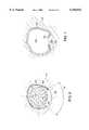
- FIG. 4is a schematic cross-sectional representation of a body passageway prior to treatment according to the invention.
- FIG. 5is a schematic cross-sectional representation illustrating the use of a probe during treatment according to the invention prior to causing injury to the wall of the urethra;
- FIG. 6is a schematic cross-sectional representation illustrating the use of a probe during treatment according to the invention after causing injury to the wall of the urethra;
- FIG. 7is a schematic cross-sectional representation illustrating the healing response following treatment according to the invention.
- FIGS. 8A and 8Bare schematic cross-sectional representations illustrating the valve action of opening and closing, respectively, of a stricture of a body passageway following treatment according to the invention.
- This inventiongenerally provides for a method and apparatus for forming a stricture in a body passageway. Stricture of a body passageway is desirable whenever there is a need to gain a better control over the passage of fluids within the body passageway. Varying degrees of stricture may be used with the methods and apparatus of the invention to obtain a gamut of treatment results.
- a stricture according to the present inventionis a narrowing of a body passageway that decreases the flow or intermittently stops the passage of fluids within it upon contraction or relaxation of the surrounding tissue or the pressure of fluid within it, but does not result in a permanent occlusion of the body passageway.
- a stricturemay be used, for example, in a body passageway of the urinary tract to cure or alleviate the symptoms of urinary incontinence and to preserve kidney function in persons (e.g., children) afflicted by vesicoureteral reflux.
- a stricturecan also be formed in body passageways other than the urinary tract such as, the bile duct, the vascular system, and the digestive system, for example.
- a stricturemay be used in veins, for example, to treat tumors by decreasing or stopping blood flow to benign or cancerous neoplasms.
- Strictures in arteriesmay be used, for example, in by-pass heart surgery, liver transplant or kidney transplant.
- Strictures in the rectummay be used to treat fecal incontinence.
- Strictures in the esophagus at the gastro-esophageal junctionmay be used to treat gastro-esophageal reflux.
- a stricture according to the inventionmay create a valve action, open and shut, within the body passageway upon contraction or relaxation of the smooth muscle cells surrounding the body passageway or by the variation of pressure within the body passageway.
- a shut positionthe supple and compliant wall of the body passageway is collapsed and pressed against the non-compliant, fibrous hyperplasic mass forming the stricture.
- an open positionthe wall of the body passageway opposite the stricture is extended and is not in contact with the hyperplasic mass.
- Preferred body passageways for treatment according to the apparatus and method of the present inventionare lined with epithelium cells.
- the epitheliumsuch as the urothelium in the urinary tract or the endothelium in the vascular system, belongs to a category of cells that are continuously replicating to replace destroyed cells. Smooth muscle cells that populate the submucosa are in normal condition quiescent, stable cells that show a low level of replication. However, in response to stimuli, such as following injury, these cells can undergo rapid division. This rapid cell division can lead to hyperplasia at the site of the injury. De-epithelization can be such an injury stimulating hyperplasia.
- Hyperplasiais an increase in the number of cells in a tissue, such as the submucosa, which results in an increase in volume of body tissue. It is a response of connective tissue cells that occurs during wound healing. Hyperplasia is stimulated by growth factors released from the injured cells. Hyperplasia is best induced when the extracellular matrix of the epithelium and the basal membrane have been destroyed sufficiently to expose the cells of the submucosa to growth factors released from injured cells.
- the epitheliumhas a modulating effect on the mitotic cycle of the muscle cells in the submucosa such that the preservation of the epithelium will prevent hyperplasia of the muscle cells underlying the intact epithelium even if adjacent tissue is de-epithelized and injured.
- the degree of stricture formationis proportional to the degree of injury, and that the stricture morphology is hyperplasia of muscle cells in the submucosa.
- Hyperplasiais generally an undesirable result of wound healing. For example, if it occurs in the skin, it would leave scars. However, it can be advantageously induced in the body passageways of the urinary tract, for example, to assist in preventing the unwanted urine flow such as in the treatment of urinary incontinence or vesicoureteral reflux.
- an injury-causing probesuch as an energy-emitting probe
- an endoscopeis introduced into the body passageway via an endoscope.
- Access to the urethrais direct, as it directly communicates with the exterior of the body.
- Access to the ureteris achieved via the urethra and bladder.
- Access to veins and arteriesis percutaneous.
- Access to the bile ductmay be percutaneous or through the upper gastro intestinal tract, i.e. through the mouth, the esophagus, the stomach and the duodenum.
- Access to the rectumis direct.
- Access to the esophagusis achieved through the mouth.
- Methods to monitor the positioning of the probe within the body passagewayare well known in the art. In preferred embodiments, positioning of the probe in the body passageway may be monitored by the use of an endoscope, fluoroscopy or other locating and positioning devices.
- the probeis positioned at the location of the desired bulking, energy is applied to create an injury in the wall of the body passageway, such that a radial segment of the epithelium covering the wall along a circumferential portion of the body passageway, i.e., radial or arcuate segment, is destroyed and removed, exposing the submucosa.
- the remaining radial segment, within the same circumferential portion of the body passageway,is preferably left un-injured by the probe.
- the preservation of the epithelium in the complementary radial segment within the same circumferential portion of the body passagewaywill insure that the hyperplasia occurring during the wound healing process does not result in an occlusion of the body passageway by adhesion of the opposite surfaces of the wall.
- the nature of the injury created at the site of the desired bulkingis of a type where the extracellular matrix is denatured and where stimulating growth factors are released from the injured cells.
- stimulating growth factorsmay be provided to the site of the injury by direct application using the cooling channel of the probe or by parenteral oral mode of administration. Injuries such as burns created by heating or freezing would be suitable. Also, injuries created by an abrasion, are suitable for the methods of the present invention.
- the injuryis created as a burn by the application of heat from an RF probe.
- the injurycan take a variety of sizes and shapes as long as the shape of the resulting hyperplasia be conducive of a valve action within the body passageway, i.e., constricting the body passageway in a relaxed or contracted state of the body passageway (as shown in FIG. 8A) and unobstructing the body passageway in a expanded state of the body passageway (as shown in FIG. 8B).
- Both the size and shape of the injuryare controlled by the size and shape of the energy emitting zone of the energy emitter on the outer surface of the probe.
- the depth of the injurycan be controlled by the amount of energy delivered to the surface of the wall of the body passageway.
- the shape of the injuryis an arcuate quadrangular segment.
- a quadrangular shapeis any polygonal shape having four sides and four corners including, but not limited to, squares, rectangles, losanges, and trapezia.
- the shape of the injuryis an arcuate rectangular segment.
- the shape of the injuryis a continuous segment.
- the shape of the injuryis a series of proximate, non-contiguous segments.
- the shape of the injuryis a series of proximate, non-contiguous segments on alternate, opposite sides of the wall of the body passageway.
- the proceduremay be repeated after the first injury has completely healed, i.e., after total epithelization of the hyperplasic mass at the site of the injury.
- a subsequent injurycan then be caused to a new portion of the body passageway adjacent to the prior site of injury, either within the same circumferential portion, or along the longitudinal axis of the body passageway.
- the subsequent injurycan also be repeated at the same location as the prior injury, or at a location overlapping the prior injury until the desired bulking is reached.
- the size of the injury in the case of the urethra or the ureters for examplecan be about 0.2-1 mm in depth, about 4-20 mm in length over about a 30 to 180 degree angle.
- the size of the woundis 0.4 to 0.8 mm in depth, 8 to 12 mm in length, and 60 to 120 degree angle.
- the size of the woundis 1 mm in depth, 10 mm in length over a 60 degree angle.
- the length of the injurycan be 3-5 cm., the depth and the width being as described above.
- the shape of the desired stricture within the body passagewaymay easily be designed and controlled by the manner in which and the location at which the wall is injured. Design of injury to attain a specific stricture pattern is well within the reach of a skilled artisan.
- the present inventionis not to be limited by the above-described specific stricture patterns.
- a probe designed to carry out the above described treatmentcan be used through the working channel of an endoscope.
- the probecan be flexible to facilitate the positioning within the body passageway, such as in the ureter, or the esophagus for example.
- the diameter of the probeis 6 to 10 French or about that size for body passageways of the urinary tract. In another preferred embodiment, the diameter of the probe is 30-40 French or about that size for body passageways of the gastrointestinal tract.
- the distal end of the probeis fitted with an energy emitter. Alternatively, the distal end of the probe may be an expandable balloon with an energy emitter fitted onto a portion of its outer surface.
- the distal endalso includes a thermistor or thermal sensor to ensure the controlled delivery of sufficient energy to create an injury to the epithelium as described above.
- the probemay also include a cooling channel for the delivery of cooling fluid to the site of injury.
- the cooling channelmay also be used for delivery of drugs, such as, but not limited to, anesthetics, antibiotics, antiseptics, growth factors and pharmaceutical compositions thereof, to the site of the injury.
- the size and shape of the energy emitter at the periphery of the probecan be preformed to correspond to the desired size and shape of the injury.
- the size of the energy emitter at the periphery of the probemay be defined and measured by two parameters: a height (h) and an arc ( ⁇ ).
- the height (h)is measured by the distance separating the two most extreme points following a line parallel to the longitudinal axis of the probe.
- the arcis measured by the angle formed by two planes intersecting along the central axis of the probe and each passing by the most extreme lateral points of the energy emitter at the peripheral surface of the probe.
- the energy emitter at the periphery of the probehas an arcuate quadrangular shape.
- energy emitter at the periphery of the probehas an arcuate rectangular shape.
- the energy emitterhas a shape of a continuous segment.
- the energy emitterhas a shape of a series of proximate, non-contiguous segments.
- the energy emitterhas a shape of a series of proximate, non-contiguous segments on alternate, opposite sides of the distal end of the probe.
- the remaining circumferential portion of the probeis made of insulating material to prevent injury to the epithelium neighboring the site of the desired injury. Synthetic or natural polymers can be suitable as insulating material for use in the manufacture of the probe.
- Suitable energy emittersinclude radio-frequency (RF) emitters, ultrasound emitters, laser emitters, electrical heat emitters, cryogenic fluid emitters, or mechanical abraser.
- FIGS. 1A and 1Bshow two views of an RF emitter suitable to carry out the methods of the present invention.
- FIG. 2shows a side view of a device featuring a RF emitter at its distal end suitable to carry out the methods of the present invention. Examples of emitters that may be suitable or use in the treatments described herein include those described in: U.S. Pat. No. 5,122,137 for temperature controlled RF delivering probe; U.S. Pat. No. 5,383,876 for a fluid cooled electrosurgical heat delivering probe; U.S. Pat. No.
- an RF electrode as shown in FIGS. 1A and 1Bcan be disposed on the distal end of the device shown in FIG. 2.
- the RF electrodecan deliver a suitable level of energy at the site of the desired injury when a temperature of about 45° C. to about 100° C. is maintained for a period of about 1 to about 60 seconds.
- the length of time for application of energyvaries inversely with the temperature applied at the site of the injury.
- suitable injuries for this inventioncan be created with the application of 100° C. for about one second. Suitable injuries can also be created with the application of 60° C. for about 10 seconds, or even 48° C. for about sixty seconds.
- the injuryis created with the application of RF energy that provides 60° C. at the desired site of injury for 10 seconds. It will be apparent to a skilled artisan that many variations as to the temperature and length of time of heat application can result in an injury of the type according to the invention.
- the apparatus for accomplishing the methods of the present inventioncan apply heat using an RF probe 10 as shown in FIG. 1A with a grounding plate (not shown).
- the probe 10is made of a shaft 70 having a cooling channel 20 located within the shaft 70, running along the longitudinal axis of the shaft 70.
- An electrode 40is located at the distal end of shaft 70.
- the electrode 40comprises a conductive material 80 having an arcuate quadrangular shape at the periphery of the shaft 70.
- the conductive material 80is in contact with a thermistor 30.
- the thermistoris connected to a RF power supply (not shown) via wires 50 running through a wire channel 90.
- the probe 10can comprise a temperature sensor (not shown) to permit indirect monitoring of the application of heat to the tissue and thus control the degree of injury to the tissue.
- the sensorwould be connected to a control circuit that modulates RF power applied to the electrode according to the signal received from the temperature sensor.
- the control circuit and an RF power supplywould alternate between two operating modes. In a first mode, the RF power supply would apply RF power to the electrode. In a second mode, the control circuit would sense a signal from the temperature sensor in the absence of RF signal. The control circuit then would compare the signal from the temperature sensor to a set value (determined by the operator) and modulate the RF power applied to the electrode in accordance with the set value.
- FIG. 1Bshows a top view of probe 10 showing the arcuate shape 60 having an arc ⁇ of the electrode 40 at the periphery of the shaft 70.
- FIG. 2shows the device with a flexible shaft 70 having a radial quadrangular electrode 40 at its distal end and a connector 72 having a port 22 for the cooling channel 20 and a port 92 for the wire channel 90.
- the wire channel 90is connected to an adapter 94 for connecting wires 50 with an RF power supply (not shown).
- Suitable conductive materials 80 for the electrode 40are materials that present little ohmic resistance.
- metalssuch as metal from group VIII and IB or alloy thereof are suitable materials.
- the conductive materialsis copper or platinum.
- Suitable materials for the shaft 70 and distal portion of the probe surrounding the electrodeare synthetic materials and insulants.
- silicone, rubber, latex, polyurethane, nylon, polyethyleneare suitable materials, as well as any other suitable catheter material.
- FIG. 3shows the probe 10 positioned within the lumen 140 of the urethra of a human female.
- the electrode 40is located in the proximity of the urethral sphincters 120.
- the probewould be inserted through the penile urethra, the bulbous urethra, and positioned in the prostatic urethra beyond the urethral junction with the prostate duct (not shown).
- an injury 130 to the epithelium 100 of the urethraexposes the submucosa 110.
- the lumen 140 of the urethrais collapsed and folded more or less loosely by the pressure of the surrounding tissues, the submucosa 110 and epithelium 100.
- FIG. 5which shows a cross-sectional view along the axis shown in FIG. 3
- the probe 10is located within the lumen 140 of the urethra prior to the application of heat.
- the lumen 140is shown expanded by the circular shape of the probe 10.
- the electrode 40is in close proximity of the epithelium 100 of the urethra.
- FIG. 6which shows a cross-sectional view along the axis shown in FIG. 3, the probe 10 is located within the lumen 140 of the urethra after the application of heat.
- the application of heathas removed a radial segment 60 of epithelium 100 and submucosa 110 at the site of the injury 130.
- the size and shape of the injury 130correspond to the size and shape of the electrode 40.
- a healing responseis triggered that stimulates the cells of the submucosa 110 to divide leading to hyperplasia of the submucosa 150. Cells division continues until the newly grown epithelium 160 has covered the site of injury 130.
- FIGS. 8A and 8Bthe hyperplasia 150 at the site of injury 130 once the new epithelium 160 has fully regrown protrudes into the lumen 140 of the urethra.
- FIGS. 8A and 8Bthe hyperplasia 150 at the site of injury 130 once the new epithelium 160 has fully regrown protrudes into the lumen 140 of the urethra.
- FIG. 8A and 8Billustrate the valve action when the urethra is expanded by the pressure of the passage of urine in the lumen 140, as in FIG. 8A, and when the urethra is collapsed by the pressure of the surrounding tissues of the submucosa 110, as in FIG. 8B closing the flow of urine in the lumen.
- the valve actionis due to the difference in texture between the hyperplasic mass and the opposite wall of the body passageway. Hyperplasic growth is fibrous in nature and thus non compliant while the remaining of body passageway is compliant. Thus, the hperplasic fibrous mass presses against the compliant opposite wall when the body passageway is collapsed as in FIG. 8B and operate as a valve.
Landscapes
- Health & Medical Sciences (AREA)
- Surgery (AREA)
- Life Sciences & Earth Sciences (AREA)
- Engineering & Computer Science (AREA)
- Heart & Thoracic Surgery (AREA)
- Animal Behavior & Ethology (AREA)
- Otolaryngology (AREA)
- Plasma & Fusion (AREA)
- Physics & Mathematics (AREA)
- Biomedical Technology (AREA)
- Cardiology (AREA)
- Medical Informatics (AREA)
- Molecular Biology (AREA)
- Nuclear Medicine, Radiotherapy & Molecular Imaging (AREA)
- General Health & Medical Sciences (AREA)
- Public Health (AREA)
- Veterinary Medicine (AREA)
- Prostheses (AREA)
- Surgical Instruments (AREA)
- Electrotherapy Devices (AREA)
- Thermotherapy And Cooling Therapy Devices (AREA)
Abstract
Description
This invention relates generally to the field of endoscopic medical procedures, and more specifically to the use of endoscopic probes for treating body passageway dysfunctions.
Urinary incontinence or, more specifically, urinary stress incontinence afflicts a large number of people. Although not life threatening, urinary incontinence has a dramatic negative impact on the quality of life. Vesicoureteral reflux is a congenital condition that afflicts one in one hundred children. In the more severe forms, reduced, or complete loss of kidney function can occur, and it can be life threatening.
Urinary incontinence results from dysfunctional urinary sphincter(s). The dysfunction of the sphincter(s) can be caused by disease, complications from urological procedures, or result from trauma to the pelvic region. The dysfunction of the sphincter(s) can also be caused by neurological or sphincter muscle dysfunction. In all cases, the sphincter is unable to retain urine in the bladder as normal.
Currently, there are several techniques and devices that are used to treat urinary incontinence. There are surgical solutions in which artificial sphincters are implanted into tissue surrounding the urethra. These artificial sphincters function as a valve and can be actuated by the patient. Other surgical implants are rubber-band like devices that are placed around the outside of the sphincter muscle and urethra and assist the sphincter in retaining urine. Some less invasive techniques involve the injection of bulking agents the submucosa of the urethra. The bulking agents can be biologically compatible materials such as Teflon, biomaterials such as bovine collagen, or autologous materials such as fat, collagen, muscle, or cartilage. Other therapies that are used include electrical stimulation of the sphincter muscle and biofeedback techniques.
Vesicoureteral reflux is similar to stress incontinence in that the vesicoureteric junction functions like a sphincter. The vesicoureteric junction is at the junction of the ureter and the bladder. In a normal vesicoureteric junction, the ureter joins the bladder through the muscularis mucosea of the bladder in an oblique fashion. During micturition, the muscularis mucosea contracts closing the ureter. Thus, the vesicoureteric junction acts as a check valve and prevents urine from flowing back into the kidney during bladder contraction during micturition. In an abnormal vesicoureteric junction, the ureter joins the bladder through the muscularis mucosea of the bladder in a perpendicular fashion. During micturition, the ureter remains open allowing the urine to flow back in the ureter. The backwards flow of urine and the associated pressurization of the ureter and kidney result in dilation of the ureter and scarring of the kidney. Interventional treatment of this condition consists of surgery and injection of bulking agents. Mild cases of reflux can be managed with medication.
It is an object of the invention to provide a treatment for both urinary incontinence and vesicoureteral reflux that is less intrusive than surgery or implantation of bulking agent, and that provides permanent relief It is another object of the present invention to provide a cure and/or relieve the symptoms of urinary incontinence and to preserve kidney function in people (e.g., children) afflicted with vesicoureteral reflux. The present invention is an improvement over other modalities such as surgical implants, or injectable tissue bulking agents for treating these conditions. This invention provides for a cost effective, minimally invasive, permanent treatment for urinary incontinence and vesicoureteral reflux without the need for implantation of foreign or autologous material.
More generally, this invention provides for bulking of a body passageway without introduction of bulking agents by causing disruption of epithelium lining the interior wall of the body passageway and injury to the submucosa to trigger a healing response that results in hyperplasia of the submucosa and stricture formation in the body passageway.
Methods and devices according to the present invention are used to form chronic stricture in a body passageway by the application of energy (e.g., RF energy) to create an injury to the wall of the body passageway and hyperplasia of the submucosa at the site of injury during the healing response. The hyperplasia of the submucosa at the site of the wound creates a stricture of the body passageway. More specifically, the invention involves inserting an energy-delivering probe into a body passageway and applying energy to a segment of the wall of the body passageway sufficient to create an injury to the wall, hyperplasia of the tissue adjacent the wall, and stricture of the body passageway.
Body passageways in which creation of a stricture may be desirable according to the present invention are the urethra, the ureters, the bile ducts, the esophagus, the rectum, veins and arteries, for example.
An endoscope can be used to insert energy-delivering probe according to the invention into the body passageway. The probe can have a focused energy emitter (pre-shaped to the desired shape of the injury) for applying the energy to the wall of the body passageway. The energy can be delivered to the wall in the form of, for example, heat, cryogen fluid, or mechanical abrasive action. Thus, the energy emitter can be a radio-frequency emitter (RF), an ultrasound emitter, a laser emitter, a cryogenic fluid emitter, or a mechanical abraser. The injury caused to the wall by the probe may be a burn or an abrasion. However the injury is caused, it results in a healing response that in turn results in hyperplasia at the site of the injury and stricture of the body passageway.
In some embodiments of the invention, incontinence is treated by causing a stricture of the urethra. The energy can be applied to a segment of the urethral wall proximal to the urethral sphincter. In other embodiments of the invention, vesicoureteral reflux is treated by causing a stricture of the ureter. The energy can be applied to a segment of the ureteral wall at the vesicoureteric junction. In yet other embodiments, gastro-esophageal reflux is treated by causing a stricture of the esophagus.
These and other objects, along with advantages and features of the invention disclosed herein, will be made more apparent from the description, drawings, and claims that follow.
The foregoing and other objects, features and advantages of the present invention, as well as the invention itself, will be more fully understood from the following description of preferred embodiments, when read together with the accompanying drawings, in which
FIG. 1A is a cross-sectional side view of an embodiment of a probe according to the invention;
FIG. 1B is a top view of the probe of FIG. 1A;
FIG. 2 is a perspective view of the energy-delivering device of FIGS. 1A and 1B;
FIG. 3 is a longitudinal cross-sectional view showing the use of the probe (FIGS. 1A, 1B and 2) during treatment;
FIG. 4 is a schematic cross-sectional representation of a body passageway prior to treatment according to the invention;
FIG. 5 is a schematic cross-sectional representation illustrating the use of a probe during treatment according to the invention prior to causing injury to the wall of the urethra;
FIG. 6 is a schematic cross-sectional representation illustrating the use of a probe during treatment according to the invention after causing injury to the wall of the urethra;
FIG. 7 is a schematic cross-sectional representation illustrating the healing response following treatment according to the invention; and
FIGS. 8A and 8B are schematic cross-sectional representations illustrating the valve action of opening and closing, respectively, of a stricture of a body passageway following treatment according to the invention.
This invention generally provides for a method and apparatus for forming a stricture in a body passageway. Stricture of a body passageway is desirable whenever there is a need to gain a better control over the passage of fluids within the body passageway. Varying degrees of stricture may be used with the methods and apparatus of the invention to obtain a gamut of treatment results. A stricture according to the present invention is a narrowing of a body passageway that decreases the flow or intermittently stops the passage of fluids within it upon contraction or relaxation of the surrounding tissue or the pressure of fluid within it, but does not result in a permanent occlusion of the body passageway.
In accordance with the invention, a stricture may be used, for example, in a body passageway of the urinary tract to cure or alleviate the symptoms of urinary incontinence and to preserve kidney function in persons (e.g., children) afflicted by vesicoureteral reflux. A stricture can also be formed in body passageways other than the urinary tract such as, the bile duct, the vascular system, and the digestive system, for example. Thus, a stricture may be used in veins, for example, to treat tumors by decreasing or stopping blood flow to benign or cancerous neoplasms. Strictures in arteries may be used, for example, in by-pass heart surgery, liver transplant or kidney transplant. Strictures in the rectum may be used to treat fecal incontinence. Strictures in the esophagus at the gastro-esophageal junction may be used to treat gastro-esophageal reflux.
A stricture according to the invention may create a valve action, open and shut, within the body passageway upon contraction or relaxation of the smooth muscle cells surrounding the body passageway or by the variation of pressure within the body passageway. In a shut position, the supple and compliant wall of the body passageway is collapsed and pressed against the non-compliant, fibrous hyperplasic mass forming the stricture. In an open position, the wall of the body passageway opposite the stricture is extended and is not in contact with the hyperplasic mass.
Preferred body passageways for treatment according to the apparatus and method of the present invention are lined with epithelium cells. The epithelium, such as the urothelium in the urinary tract or the endothelium in the vascular system, belongs to a category of cells that are continuously replicating to replace destroyed cells. Smooth muscle cells that populate the submucosa are in normal condition quiescent, stable cells that show a low level of replication. However, in response to stimuli, such as following injury, these cells can undergo rapid division. This rapid cell division can lead to hyperplasia at the site of the injury. De-epithelization can be such an injury stimulating hyperplasia.
Hyperplasia is an increase in the number of cells in a tissue, such as the submucosa, which results in an increase in volume of body tissue. It is a response of connective tissue cells that occurs during wound healing. Hyperplasia is stimulated by growth factors released from the injured cells. Hyperplasia is best induced when the extracellular matrix of the epithelium and the basal membrane have been destroyed sufficiently to expose the cells of the submucosa to growth factors released from injured cells. Therefore, it is believed that the epithelium has a modulating effect on the mitotic cycle of the muscle cells in the submucosa such that the preservation of the epithelium will prevent hyperplasia of the muscle cells underlying the intact epithelium even if adjacent tissue is de-epithelized and injured. Thus, it is believed that the degree of stricture formation is proportional to the degree of injury, and that the stricture morphology is hyperplasia of muscle cells in the submucosa.
Hyperplasia is generally an undesirable result of wound healing. For example, if it occurs in the skin, it would leave scars. However, it can be advantageously induced in the body passageways of the urinary tract, for example, to assist in preventing the unwanted urine flow such as in the treatment of urinary incontinence or vesicoureteral reflux.
In accordance with this invention, an injury-causing probe, such as an energy-emitting probe, is introduced into the body passageway via an endoscope. Access to the urethra is direct, as it directly communicates with the exterior of the body. Access to the ureter is achieved via the urethra and bladder. Access to veins and arteries is percutaneous. Access to the bile duct may be percutaneous or through the upper gastro intestinal tract, i.e. through the mouth, the esophagus, the stomach and the duodenum. Access to the rectum is direct. Access to the esophagus is achieved through the mouth. Methods to monitor the positioning of the probe within the body passageway are well known in the art. In preferred embodiments, positioning of the probe in the body passageway may be monitored by the use of an endoscope, fluoroscopy or other locating and positioning devices.
Once the probe is positioned at the location of the desired bulking, energy is applied to create an injury in the wall of the body passageway, such that a radial segment of the epithelium covering the wall along a circumferential portion of the body passageway, i.e., radial or arcuate segment, is destroyed and removed, exposing the submucosa. The remaining radial segment, within the same circumferential portion of the body passageway, is preferably left un-injured by the probe. The preservation of the epithelium in the complementary radial segment within the same circumferential portion of the body passageway will insure that the hyperplasia occurring during the wound healing process does not result in an occlusion of the body passageway by adhesion of the opposite surfaces of the wall.
The nature of the injury created at the site of the desired bulking is of a type where the extracellular matrix is denatured and where stimulating growth factors are released from the injured cells. Alternatively, stimulating growth factors may be provided to the site of the injury by direct application using the cooling channel of the probe or by parenteral oral mode of administration. Injuries such as burns created by heating or freezing would be suitable. Also, injuries created by an abrasion, are suitable for the methods of the present invention. In preferred embodiments, the injury is created as a burn by the application of heat from an RF probe.
The injury can take a variety of sizes and shapes as long as the shape of the resulting hyperplasia be conducive of a valve action within the body passageway, i.e., constricting the body passageway in a relaxed or contracted state of the body passageway (as shown in FIG. 8A) and unobstructing the body passageway in a expanded state of the body passageway (as shown in FIG. 8B). Both the size and shape of the injury are controlled by the size and shape of the energy emitting zone of the energy emitter on the outer surface of the probe. The depth of the injury can be controlled by the amount of energy delivered to the surface of the wall of the body passageway. In some embodiments, the shape of the injury is an arcuate quadrangular segment. A quadrangular shape is any polygonal shape having four sides and four corners including, but not limited to, squares, rectangles, losanges, and trapezia. In preferred embodiments, the shape of the injury is an arcuate rectangular segment. In certain embodiments, the shape of the injury is a continuous segment. In yet other embodiments, the shape of the injury is a series of proximate, non-contiguous segments. In yet other embodiments, the shape of the injury is a series of proximate, non-contiguous segments on alternate, opposite sides of the wall of the body passageway.
In cases where hyperplasia of the total circumference of the body passageway is desired to achieve a greater control of the passage of fluid, the procedure may be repeated after the first injury has completely healed, i.e., after total epithelization of the hyperplasic mass at the site of the injury. A subsequent injury can then be caused to a new portion of the body passageway adjacent to the prior site of injury, either within the same circumferential portion, or along the longitudinal axis of the body passageway. The subsequent injury can also be repeated at the same location as the prior injury, or at a location overlapping the prior injury until the desired bulking is reached. Thus, by repeating this procedure a number of times, hyperplasia to the complete circumference of the body passageway may be attained, this, however, without inducing total occlusion of the body passageway.
The size of the injury in the case of the urethra or the ureters for example, can be about 0.2-1 mm in depth, about 4-20 mm in length over about a 30 to 180 degree angle. In preferred embodiments, the size of the wound is 0.4 to 0.8 mm in depth, 8 to 12 mm in length, and 60 to 120 degree angle. In a most preferred embodiment, the size of the wound is 1 mm in depth, 10 mm in length over a 60 degree angle. In the case of the esophagus or rectum, the length of the injury can be 3-5 cm., the depth and the width being as described above.
It is understood that the shape of the desired stricture within the body passageway may easily be designed and controlled by the manner in which and the location at which the wall is injured. Design of injury to attain a specific stricture pattern is well within the reach of a skilled artisan. The present invention is not to be limited by the above-described specific stricture patterns.
A probe designed to carry out the above described treatment can be used through the working channel of an endoscope. The probe can be flexible to facilitate the positioning within the body passageway, such as in the ureter, or the esophagus for example. In a preferred embodiment, the diameter of the probe is 6 to 10 French or about that size for body passageways of the urinary tract. In another preferred embodiment, the diameter of the probe is 30-40 French or about that size for body passageways of the gastrointestinal tract. The distal end of the probe is fitted with an energy emitter. Alternatively, the distal end of the probe may be an expandable balloon with an energy emitter fitted onto a portion of its outer surface. Optionally, the distal end also includes a thermistor or thermal sensor to ensure the controlled delivery of sufficient energy to create an injury to the epithelium as described above. To this end, the probe may also include a cooling channel for the delivery of cooling fluid to the site of injury. The cooling channel may also be used for delivery of drugs, such as, but not limited to, anesthetics, antibiotics, antiseptics, growth factors and pharmaceutical compositions thereof, to the site of the injury.
The size and shape of the energy emitter at the periphery of the probe can be preformed to correspond to the desired size and shape of the injury. The size of the energy emitter at the periphery of the probe may be defined and measured by two parameters: a height (h) and an arc (θ). The height (h) is measured by the distance separating the two most extreme points following a line parallel to the longitudinal axis of the probe. The arc is measured by the angle formed by two planes intersecting along the central axis of the probe and each passing by the most extreme lateral points of the energy emitter at the peripheral surface of the probe. In some embodiments, the energy emitter at the periphery of the probe has an arcuate quadrangular shape. In preferred embodiments, energy emitter at the periphery of the probe has an arcuate rectangular shape. In certain embodiments, the energy emitter has a shape of a continuous segment. In yet other embodiments, the energy emitter has a shape of a series of proximate, non-contiguous segments. In yet other embodiments, the energy emitter has a shape of a series of proximate, non-contiguous segments on alternate, opposite sides of the distal end of the probe. The remaining circumferential portion of the probe is made of insulating material to prevent injury to the epithelium neighboring the site of the desired injury. Synthetic or natural polymers can be suitable as insulating material for use in the manufacture of the probe.
Suitable energy emitters include radio-frequency (RF) emitters, ultrasound emitters, laser emitters, electrical heat emitters, cryogenic fluid emitters, or mechanical abraser. FIGS. 1A and 1B show two views of an RF emitter suitable to carry out the methods of the present invention. FIG. 2 shows a side view of a device featuring a RF emitter at its distal end suitable to carry out the methods of the present invention. Examples of emitters that may be suitable or use in the treatments described herein include those described in: U.S. Pat. No. 5,122,137 for temperature controlled RF delivering probe; U.S. Pat. No. 5,383,876 for a fluid cooled electrosurgical heat delivering probe; U.S. Pat. No. 5,380,317 for a localized high intensity light and heat delivering probe; U.S. Pat. No. 4,662,368 for a localized heat delivering probe; and U.S. Pat. No. 4,765,331 for a focused heat delivering probe with an treatment arc of less than 360 degrees. All of these patents are hereby incorporated herein by reference. In general, the emitters described in these patents are suitable to the extent that they can be made to fit within the urinary passageways and deliver energy sufficient to create an injury as described herein.
In embodiments where the energy is delivered in the form of thermal energy, an RF electrode as shown in FIGS. 1A and 1B can be disposed on the distal end of the device shown in FIG. 2. The RF electrode can deliver a suitable level of energy at the site of the desired injury when a temperature of about 45° C. to about 100° C. is maintained for a period of about 1 to about 60 seconds. The length of time for application of energy varies inversely with the temperature applied at the site of the injury. For example, suitable injuries for this invention can be created with the application of 100° C. for about one second. Suitable injuries can also be created with the application of 60° C. for about 10 seconds, or even 48° C. for about sixty seconds. In a preferred embodiment, the injury is created with the application of RF energy that provides 60° C. at the desired site of injury for 10 seconds. It will be apparent to a skilled artisan that many variations as to the temperature and length of time of heat application can result in an injury of the type according to the invention.
In one embodiment, the apparatus for accomplishing the methods of the present invention can apply heat using anRF probe 10 as shown in FIG. 1A with a grounding plate (not shown). Theprobe 10 is made of ashaft 70 having a coolingchannel 20 located within theshaft 70, running along the longitudinal axis of theshaft 70. Anelectrode 40 is located at the distal end ofshaft 70. Theelectrode 40 comprises aconductive material 80 having an arcuate quadrangular shape at the periphery of theshaft 70. Theconductive material 80 is in contact with athermistor 30. The thermistor is connected to a RF power supply (not shown) viawires 50 running through awire channel 90.
Alternatively, theprobe 10 can comprise a temperature sensor (not shown) to permit indirect monitoring of the application of heat to the tissue and thus control the degree of injury to the tissue. The sensor would be connected to a control circuit that modulates RF power applied to the electrode according to the signal received from the temperature sensor. The control circuit and an RF power supply would alternate between two operating modes. In a first mode, the RF power supply would apply RF power to the electrode. In a second mode, the control circuit would sense a signal from the temperature sensor in the absence of RF signal. The control circuit then would compare the signal from the temperature sensor to a set value (determined by the operator) and modulate the RF power applied to the electrode in accordance with the set value.
FIG. 1B shows a top view ofprobe 10 showing thearcuate shape 60 having an arc θ of theelectrode 40 at the periphery of theshaft 70. FIG. 2 shows the device with aflexible shaft 70 having a radialquadrangular electrode 40 at its distal end and aconnector 72 having aport 22 for the coolingchannel 20 and aport 92 for thewire channel 90. Thewire channel 90 is connected to anadapter 94 for connectingwires 50 with an RF power supply (not shown).
Suitableconductive materials 80 for theelectrode 40 are materials that present little ohmic resistance. For example, metals such as metal from group VIII and IB or alloy thereof are suitable materials. Preferably, the conductive materials is copper or platinum.
Suitable materials for theshaft 70 and distal portion of the probe surrounding the electrode are synthetic materials and insulants. For example, silicone, rubber, latex, polyurethane, nylon, polyethylene are suitable materials, as well as any other suitable catheter material.
The following description explains in more details one embodiment of the methods and apparatus of the present invention for treatment of the urinary tract dysfunctions.
As an example, FIG. 3 shows theprobe 10 positioned within thelumen 140 of the urethra of a human female. Theelectrode 40 is located in the proximity of theurethral sphincters 120. In a human male, the probe would be inserted through the penile urethra, the bulbous urethra, and positioned in the prostatic urethra beyond the urethral junction with the prostate duct (not shown). Opposite the electrode, aninjury 130 to theepithelium 100 of the urethra exposes thesubmucosa 110. Prior to insertion of theprobe 10 and referring to FIG. 4, thelumen 140 of the urethra is collapsed and folded more or less loosely by the pressure of the surrounding tissues, thesubmucosa 110 andepithelium 100. Referring to FIG. 5 which shows a cross-sectional view along the axis shown in FIG. 3, theprobe 10 is located within thelumen 140 of the urethra prior to the application of heat. Thelumen 140 is shown expanded by the circular shape of theprobe 10. Theelectrode 40 is in close proximity of theepithelium 100 of the urethra. Referring to FIG. 6 which shows a cross-sectional view along the axis shown in FIG. 3, theprobe 10 is located within thelumen 140 of the urethra after the application of heat. The application of heat has removed aradial segment 60 ofepithelium 100 andsubmucosa 110 at the site of theinjury 130. The size and shape of theinjury 130 correspond to the size and shape of theelectrode 40. Following application of heat and destruction of theepithelium 100, and referring to FIG. 7, a healing response is triggered that stimulates the cells of thesubmucosa 110 to divide leading to hyperplasia of thesubmucosa 150. Cells division continues until the newly grownepithelium 160 has covered the site ofinjury 130. Referring to FIGS. 8A and 8B, thehyperplasia 150 at the site ofinjury 130 once thenew epithelium 160 has fully regrown protrudes into thelumen 140 of the urethra. FIGS. 8A and 8B illustrate the valve action when the urethra is expanded by the pressure of the passage of urine in thelumen 140, as in FIG. 8A, and when the urethra is collapsed by the pressure of the surrounding tissues of thesubmucosa 110, as in FIG. 8B closing the flow of urine in the lumen. The valve action is due to the difference in texture between the hyperplasic mass and the opposite wall of the body passageway. Hyperplasic growth is fibrous in nature and thus non compliant while the remaining of body passageway is compliant. Thus, the hperplasic fibrous mass presses against the compliant opposite wall when the body passageway is collapsed as in FIG. 8B and operate as a valve.
Having described certain embodiments of the invention, it will now become apparent to one skilled in the art that other embodiments incorporating the concepts disclosed herein may be used without departing from the spirit of the invention. Therefore, it is intended that the scope of the present invention be only limited by the following claims.
Claims (19)
1. A method for causing a stricture of a body passageway, comprising the steps of
inserting an energy-delivering probe into a body passageway, the body passageway defined by an internal tubular wall and adjacent tissue surrounding the wall; and
applying energy along a radial segment of the wall using said probe, thereby causing injury to the wall, hyperplasia of tissue adjacent the wall, and stricture of the body passageway.
2. The method of claim 1, wherein the body passageway is the urethra.
3. The method of claim 2, wherein the energy is applied proximal to the urethral sphincter.
4. The method of claim 1, wherein the body passageway is the ureter.
5. The method of claim 4, wherein the energy is applied at the vesicoureteric junction.
6. The method of claim 1, wherein the body passageway is the bile duct.
7. The method of claim 1, wherein the body passageway is a vein or an artery.
8. The method of claim 1, wherein the application of energy causes a burn.
9. The method of claim 8, wherein the energy is RF radiation.
10. The method of claim 1, wherein the energy is applied through an energy emitter near a distal end of the probe.
11. The method of claim 10, further comprising the step of regulating the temperature of the energy emitter.
12. The method of claim 1 further comprising the step of
applying energy along a segment of the wall causing a subsequent injury to the wall, hyperplasia of tissue surrounding the wall and tighter stricture of the body passageway.
13. The method of claim 12 wherein a subsequent injury is caused at a location overlapping the prior injury.
14. The method of claim 1, wherein the body passageway is the rectum.
15. The method of claim 1, wherein the body passageway is the esophagus.
16. A method for treating urinary incontinence by causing a stricture in a urethra, said method comprising the steps of
inserting an energy-delivering probe into the urethra, the urethra having a tubular internal wall and adjacent tissue surrounding the wall, said energy-delivering probe having a temperature regulated RF electrode near a distal end of said probe; and
applying energy along a radial segment of the wall through said electrode, thereby causing injury to the wall, hyperplasia of tissue adjacent the wall, and stricture of the urethra.
17. The method of claim 16, wherein the energy is applied proximal to the urethral sphincter.
18. A method for treating vesicoureteral reflux by causing a stricture in a ureter, said method comprising the steps of
inserting an energy-delivering probe into the ureter, the ureter having a tubular internal wall and adjacent tissue surrounding the wall, said energy-delivering probe having a temperature regulated RF electrode near a distal end of said probe; and
applying energy along a radial segment of the wall through said electrode, thereby causing injury to the wall, hyperplasia of tissue adjacent the wall, and stricture of the urethra.
19. The method of claim 18, wherein the energy is applied at the vesicoureteric junction.
Priority Applications (6)
| Application Number | Priority Date | Filing Date | Title |
|---|---|---|---|
| US09/164,002US6156032A (en) | 1998-09-30 | 1998-09-30 | Method for causing a stricture of a body passageway |
| JP2000571837AJP2002525159A (en) | 1998-09-30 | 1999-09-29 | Method and device for causing a narrowing of a body passage |
| DE69934702TDE69934702T2 (en) | 1998-09-30 | 1999-09-29 | Device for plugging a physical passage |
| CA002345247ACA2345247A1 (en) | 1998-09-30 | 1999-09-29 | Method and device for causing a stricture of a body passageway |
| EP99952973AEP1117339B1 (en) | 1998-09-30 | 1999-09-29 | Device for causing a stricture of a body passageway |
| PCT/US1999/022500WO2000018314A1 (en) | 1998-09-30 | 1999-09-29 | Method and device for causing a stricture of a body passageway |
Applications Claiming Priority (1)
| Application Number | Priority Date | Filing Date | Title |
|---|---|---|---|
| US09/164,002US6156032A (en) | 1998-09-30 | 1998-09-30 | Method for causing a stricture of a body passageway |
Publications (1)
| Publication Number | Publication Date |
|---|---|
| US6156032Atrue US6156032A (en) | 2000-12-05 |
Family
ID=22592550
Family Applications (1)
| Application Number | Title | Priority Date | Filing Date |
|---|---|---|---|
| US09/164,002Expired - LifetimeUS6156032A (en) | 1998-09-30 | 1998-09-30 | Method for causing a stricture of a body passageway |
Country Status (6)
| Country | Link |
|---|---|
| US (1) | US6156032A (en) |
| EP (1) | EP1117339B1 (en) |
| JP (1) | JP2002525159A (en) |
| CA (1) | CA2345247A1 (en) |
| DE (1) | DE69934702T2 (en) |
| WO (1) | WO2000018314A1 (en) |
Cited By (71)
| Publication number | Priority date | Publication date | Assignee | Title |
|---|---|---|---|---|
| US6405732B1 (en)* | 1994-06-24 | 2002-06-18 | Curon Medical, Inc. | Method to treat gastric reflux via the detection and ablation of gastro-esophageal nerves and receptors |
| US6440124B1 (en)* | 1998-07-22 | 2002-08-27 | Endovasix, Inc. | Flexible flow apparatus and method for the disruption of occlusions |
| WO2003011158A1 (en)* | 2001-08-01 | 2003-02-13 | Nicholas Franco | Apparatus and method for treating urinary stress incontinence |
| US6527763B2 (en) | 1998-07-22 | 2003-03-04 | Endovasix, Inc. | Flow apparatus for the disruption of occlusions |
| US20030173406A1 (en)* | 2001-12-24 | 2003-09-18 | Daoshen Bi | Covert variable information on identification documents and methods of making same |
| US20030181897A1 (en)* | 2000-10-02 | 2003-09-25 | Thomas Simon W.H. | Apparatus and methods for treating female urinary incontinence |
| US20040116921A1 (en)* | 2002-12-11 | 2004-06-17 | Marshall Sherman | Cold tip rf/ultrasonic ablation catheter |
| US6790207B2 (en) | 1998-06-04 | 2004-09-14 | Curon Medical, Inc. | Systems and methods for applying a selected treatment agent into contact with tissue to treat disorders of the gastrointestinal tract |
| US6802841B2 (en)* | 1998-06-04 | 2004-10-12 | Curon Medical, Inc. | Systems and methods for applying a selected treatment agent into contact with tissue to treat sphincter dysfunction |
| US20050055053A1 (en)* | 2003-08-22 | 2005-03-10 | Phalen Michael P. | Methods of delivering energy to body portions to produce a therapeutic response |
| US20050070888A1 (en)* | 2004-10-29 | 2005-03-31 | Boston Scientific Corporation | Medical device systems and methods |
| US6890307B2 (en) | 2001-03-01 | 2005-05-10 | Scimed Life Systems, Inc. | Catheters with fluorescent temperature sensors |
| US6971395B2 (en)* | 1998-01-14 | 2005-12-06 | Curon Medical, Inc. | Sphincter treatment method |
| US20060039896A1 (en)* | 1999-11-05 | 2006-02-23 | Gerigene Medical Corporation | Augmentation and repair of age-related soft tissue defects |
| US20060224157A1 (en)* | 1999-07-14 | 2006-10-05 | Curon Medical, Inc. | Method for treating fecal incontinence |
| US20070005049A1 (en)* | 2005-06-30 | 2007-01-04 | Comben Richard H | Apparatus and Method of Treating Urinary Incontinence by Heating Urethra |
| US7165551B2 (en) | 1998-02-19 | 2007-01-23 | Curon Medical, Inc. | Apparatus to detect and treat aberrant myoelectric activity |
| US7165552B2 (en) | 2003-03-27 | 2007-01-23 | Cierra, Inc. | Methods and apparatus for treatment of patent foramen ovale |
| US7186251B2 (en) | 2003-03-27 | 2007-03-06 | Cierra, Inc. | Energy based devices and methods for treatment of patent foramen ovale |
| US20070065415A1 (en)* | 2005-09-16 | 2007-03-22 | Kleinsek Donald A | Compositions and methods for the augmentation and repair of defects in tissue |
| US20070142699A1 (en)* | 2005-12-16 | 2007-06-21 | Acoustx Corporation | Methods and implantable apparatuses for treating an esophageal disorder such as gastroesophageal reflux disease |
| US20070142884A1 (en)* | 2005-12-16 | 2007-06-21 | Acoustx Corporation | Methods and apparatuses for treating an esophageal disorder such as gastroesophageal reflux disease |
| US20070154462A1 (en)* | 1997-02-20 | 2007-07-05 | Kleinsek Don A | Augmentation and repair of tissue defects with in vitro cultured fibroblasts |
| US7257450B2 (en) | 2003-02-13 | 2007-08-14 | Coaptus Medical Corporation | Systems and methods for securing cardiovascular tissue |
| US7293562B2 (en) | 2003-03-27 | 2007-11-13 | Cierra, Inc. | Energy based devices and methods for treatment of anatomic tissue defects |
| US7311701B2 (en) | 2003-06-10 | 2007-12-25 | Cierra, Inc. | Methods and apparatus for non-invasively treating atrial fibrillation using high intensity focused ultrasound |
| US20080103498A1 (en)* | 1998-02-19 | 2008-05-01 | Curon Medical, Inc. | Systems and methods for treating dysfunctions in the intestines and rectum that adapt to the anatomic form and structure of different individuals |
| US7367975B2 (en) | 2004-06-21 | 2008-05-06 | Cierra, Inc. | Energy based devices and methods for treatment of anatomic tissue defects |
| US20080112935A1 (en)* | 1999-11-05 | 2008-05-15 | Kleinsek Donald A | Augmentation and repair of spincter defects with cells including fibroblasts |
| US20080118478A1 (en)* | 1999-11-05 | 2008-05-22 | Kleinsek Donald A | Hair undifferentiated cells |
| US20080138324A1 (en)* | 1999-11-05 | 2008-06-12 | Kleinsek Donald A | Hair mesenchymal cells |
| US20080152628A1 (en)* | 1999-11-05 | 2008-06-26 | Kleinsek Donald A | Augmentation and repair of spincter defects with cells including mesenchymal cells |
| US20080249565A1 (en)* | 1996-08-22 | 2008-10-09 | The Trustees Of Columbia University In The City Of New York | Endovascular Flexible Stapling Device |
| US7473252B2 (en) | 2004-10-07 | 2009-01-06 | Coaptus Medical Corporation | Systems and methods for shrinking and/or securing cardiovascular tissue |
| US20090130066A1 (en)* | 2000-11-06 | 2009-05-21 | Gerigene Medical Corporation | Augmentation and repair of sphincter defects with cells including muscle cells |
| US20090156981A1 (en)* | 2006-10-04 | 2009-06-18 | Celon Ag Medical Instruments | Flexible Catheter for High-Frequency Therapy of Biological Tissue and Method of Using Same |
| US7637924B2 (en) | 2003-03-27 | 2009-12-29 | Terumo Kabushiki Kaisha | Methods and apparatus for treatment of patent foramen ovale |
| US20100004644A1 (en)* | 2008-07-01 | 2010-01-07 | Ralph Zipper | Method for Decreasing the Size and/or Changing the Shape of Pelvic Tissues |
| US7694887B2 (en) | 2001-12-24 | 2010-04-13 | L-1 Secure Credentialing, Inc. | Optically variable personalized indicia for identification documents |
| US7789311B2 (en) | 2003-04-16 | 2010-09-07 | L-1 Secure Credentialing, Inc. | Three dimensional data storage |
| US7804982B2 (en) | 2002-11-26 | 2010-09-28 | L-1 Secure Credentialing, Inc. | Systems and methods for managing and detecting fraud in image databases used with identification documents |
| US7914527B2 (en) | 2003-03-27 | 2011-03-29 | Terumo Kabushiki Kaisha | Energy based devices and methods for treatment of patent foramen ovale |
| US7972330B2 (en) | 2003-03-27 | 2011-07-05 | Terumo Kabushiki Kaisha | Methods and apparatus for closing a layered tissue defect |
| US8021362B2 (en) | 2003-03-27 | 2011-09-20 | Terumo Kabushiki Kaisha | Methods and apparatus for closing a layered tissue defect |
| US8021359B2 (en) | 2003-02-13 | 2011-09-20 | Coaptus Medical Corporation | Transseptal closure of a patent foramen ovale and other cardiac defects |
| US8083738B2 (en) | 2004-08-05 | 2011-12-27 | Tyco Healthcare Group Lp | Method and apparatus for coagulating and/or constricting hollow anatomical structures |
| US8083152B2 (en) | 2001-12-24 | 2011-12-27 | L-1 Secure Credentialing, Inc. | Laser etched security features for identification documents and methods of making same |
| US8109274B2 (en) | 2005-04-11 | 2012-02-07 | Terumo Kabushiki Kaisha | Methods and electrode apparatus to achieve a closure of a layered tissue defect |
| US8291915B2 (en) | 1997-03-04 | 2012-10-23 | Tyco Healthcare Group Lp | Method and apparatus for treating venous insufficiency using directionally applied energy |
| WO2012167266A1 (en)* | 2011-06-03 | 2012-12-06 | Children's Hospital Los Angeles | Electrophysiological diagnosis and treatment for asthma |
| US8361061B2 (en) | 2005-02-23 | 2013-01-29 | Covidien Lp | Methods and apparatus for coagulating and/or constricting hollow anatomical structures |
| US8403927B1 (en) | 2012-04-05 | 2013-03-26 | William Bruce Shingleton | Vasectomy devices and methods |
| US8435235B2 (en) | 2007-04-27 | 2013-05-07 | Covidien Lp | Systems and methods for treating hollow anatomical structures |
| US8597290B2 (en) | 1999-07-14 | 2013-12-03 | Mederi Therapeutics | Method for treating fecal incontinence |
| EP2726008A4 (en)* | 2011-07-01 | 2015-01-07 | Ellman International Inc | Electrosurgical systems and methods |
| US8936631B2 (en) | 2010-01-04 | 2015-01-20 | Covidien Lp | Apparatus and methods for treating hollow anatomical structures |
| US8998897B2 (en) | 2011-08-19 | 2015-04-07 | Cook Medical Technologies Llc | Ablation cap |
| US9023031B2 (en) | 1997-08-13 | 2015-05-05 | Verathon Inc. | Noninvasive devices, methods, and systems for modifying tissues |
| US20150217100A1 (en)* | 2014-02-03 | 2015-08-06 | Terumo Kabushiki Kaisha | Urethral stricture treatment method |
| CN104970881A (en)* | 2015-07-02 | 2015-10-14 | 安隽医疗科技(南京)有限公司 | Radiofrequency ablation catheter and treatment device containing the same |
| CN104983462A (en)* | 2015-07-02 | 2015-10-21 | 安隽医疗科技(南京)有限公司 | Radiofrequency ablation bipolar catheter and treatment device with same |
| US9526570B2 (en) | 2012-10-04 | 2016-12-27 | Cook Medical Technologies Llc | Tissue cutting cap |
| US9770297B2 (en) | 2008-06-04 | 2017-09-26 | Covidien Lp | Energy devices and methods for treating hollow anatomical structures |
| US10492849B2 (en) | 2013-03-15 | 2019-12-03 | Cynosure, Llc | Surgical instruments and systems with multimodes of treatments and electrosurgical operation |
| US20220257969A1 (en)* | 2008-07-01 | 2022-08-18 | Ralph Zipper | Method for treating pelvic pain, chronic prostatitis, and or overactive bladder symptoms |
| US20220273367A1 (en)* | 2008-07-01 | 2022-09-01 | Ralph Zipper | System and method for applying controlled dosage light therapy for treatment of body tissue |
| USD1005484S1 (en) | 2019-07-19 | 2023-11-21 | Cynosure, Llc | Handheld medical instrument and docking base |
| US11819259B2 (en) | 2018-02-07 | 2023-11-21 | Cynosure, Inc. | Methods and apparatus for controlled RF treatments and RF generator system |
| US11918182B2 (en) | 2011-08-19 | 2024-03-05 | Cook Medical Technologies Llc | Cap for attachment to an endoscope |
| US20240156517A1 (en)* | 2017-11-17 | 2024-05-16 | Sherry Thomas | Method and apparatus for treating female urinary incontinence and fecal incontinence |
| US12376898B2 (en) | 2016-07-01 | 2025-08-05 | Cynosure, Llc | Non-invasive, uniform and non-uniform RF methods and systems related applications |
Families Citing this family (8)
| Publication number | Priority date | Publication date | Assignee | Title |
|---|---|---|---|---|
| US7317949B2 (en) | 1996-11-08 | 2008-01-08 | Ams Research Corporation | Energy induced bulking and buttressing of tissues for incontinence |
| US6292700B1 (en) | 1999-09-10 | 2001-09-18 | Surx, Inc. | Endopelvic fascia treatment for incontinence |
| US7097644B2 (en) | 2001-03-30 | 2006-08-29 | Ethicon Endo-Surgery, Inc. | Medical device with improved wall construction |
| US6918906B2 (en) | 2001-03-30 | 2005-07-19 | Gary L. Long | Endoscopic ablation system with improved electrode geometry |
| US7137981B2 (en) | 2002-03-25 | 2006-11-21 | Ethicon Endo-Surgery, Inc. | Endoscopic ablation system with a distally mounted image sensor |
| US7232438B2 (en) | 2004-07-09 | 2007-06-19 | Ethicon Endo-Surgery, Inc. | Ablation device with clear probe |
| JP6080368B2 (en)* | 2012-02-29 | 2017-02-15 | フクダ電子株式会社 | catheter |
| US11896823B2 (en) | 2017-04-04 | 2024-02-13 | Btl Healthcare Technologies A.S. | Method and device for pelvic floor tissue treatment |
Citations (7)
| Publication number | Priority date | Publication date | Assignee | Title |
|---|---|---|---|---|
| WO1995010978A1 (en)* | 1993-10-19 | 1995-04-27 | Ep Technologies, Inc. | Segmented electrode assemblies for ablation of tissue |
| WO1995017131A1 (en)* | 1993-12-22 | 1995-06-29 | Monamed Medizintechnik Gmbh | Ultrasonic marked cardiac ablation catheter |
| US5520684A (en)* | 1993-06-10 | 1996-05-28 | Imran; Mir A. | Transurethral radio frequency apparatus for ablation of the prostate gland and method |
| US5607422A (en)* | 1993-05-07 | 1997-03-04 | Cordis Corporation | Catheter with elongated side electrode |
| US5626576A (en)* | 1989-01-06 | 1997-05-06 | Advanced Coronary Intervention, Inc. | Electrosurgical catheter for resolving atherosclerotic plaque by radio frequency sparking |
| US5681282A (en)* | 1992-01-07 | 1997-10-28 | Arthrocare Corporation | Methods and apparatus for ablation of luminal tissues |
| US6056744A (en)* | 1994-06-24 | 2000-05-02 | Conway Stuart Medical, Inc. | Sphincter treatment apparatus |
Family Cites Families (5)
| Publication number | Priority date | Publication date | Assignee | Title |
|---|---|---|---|---|
| US4662368A (en) | 1983-06-13 | 1987-05-05 | Trimedyne Laser Systems, Inc. | Localized heat applying medical device |
| US4765331A (en) | 1987-02-10 | 1988-08-23 | Circon Corporation | Electrosurgical device with treatment arc of less than 360 degrees |
| US5242437A (en) | 1988-06-10 | 1993-09-07 | Trimedyne Laser Systems, Inc. | Medical device applying localized high intensity light and heat, particularly for destruction of the endometrium |
| US5122137A (en) | 1990-04-27 | 1992-06-16 | Boston Scientific Corporation | Temperature controlled rf coagulation |
| EP0719113A1 (en) | 1992-11-13 | 1996-07-03 | American Cardiac Ablation Co., Inc. | Fluid cooled electrosurgical probe |
- 1998
- 1998-09-30USUS09/164,002patent/US6156032A/ennot_activeExpired - Lifetime
- 1999
- 1999-09-29WOPCT/US1999/022500patent/WO2000018314A1/enactiveIP Right Grant
- 1999-09-29DEDE69934702Tpatent/DE69934702T2/ennot_activeExpired - Lifetime
- 1999-09-29CACA002345247Apatent/CA2345247A1/ennot_activeAbandoned
- 1999-09-29EPEP99952973Apatent/EP1117339B1/ennot_activeExpired - Lifetime
- 1999-09-29JPJP2000571837Apatent/JP2002525159A/enactivePending
Patent Citations (7)
| Publication number | Priority date | Publication date | Assignee | Title |
|---|---|---|---|---|
| US5626576A (en)* | 1989-01-06 | 1997-05-06 | Advanced Coronary Intervention, Inc. | Electrosurgical catheter for resolving atherosclerotic plaque by radio frequency sparking |
| US5681282A (en)* | 1992-01-07 | 1997-10-28 | Arthrocare Corporation | Methods and apparatus for ablation of luminal tissues |
| US5607422A (en)* | 1993-05-07 | 1997-03-04 | Cordis Corporation | Catheter with elongated side electrode |
| US5520684A (en)* | 1993-06-10 | 1996-05-28 | Imran; Mir A. | Transurethral radio frequency apparatus for ablation of the prostate gland and method |
| WO1995010978A1 (en)* | 1993-10-19 | 1995-04-27 | Ep Technologies, Inc. | Segmented electrode assemblies for ablation of tissue |
| WO1995017131A1 (en)* | 1993-12-22 | 1995-06-29 | Monamed Medizintechnik Gmbh | Ultrasonic marked cardiac ablation catheter |
| US6056744A (en)* | 1994-06-24 | 2000-05-02 | Conway Stuart Medical, Inc. | Sphincter treatment apparatus |
Cited By (129)
| Publication number | Priority date | Publication date | Assignee | Title |
|---|---|---|---|---|
| US6405732B1 (en)* | 1994-06-24 | 2002-06-18 | Curon Medical, Inc. | Method to treat gastric reflux via the detection and ablation of gastro-esophageal nerves and receptors |
| US7794474B2 (en) | 1996-08-22 | 2010-09-14 | The Trustees Of Columbia University In The City Of New York | Endovascular flexible stapling device |
| US20080249565A1 (en)* | 1996-08-22 | 2008-10-09 | The Trustees Of Columbia University In The City Of New York | Endovascular Flexible Stapling Device |
| US8167905B2 (en) | 1996-08-22 | 2012-05-01 | The Trustees Of Columbia University In The City Of New York | Endovascular flexible stapling device |
| US9468437B2 (en) | 1996-08-22 | 2016-10-18 | The Trustees Of Columbia University In The City Of New York | Endovascular flexible stapling device |
| US8048110B2 (en) | 1996-08-22 | 2011-11-01 | The Trustees Of Columbia University In The City Of New York | Endovascular flexible stapling device |
| US20070154462A1 (en)* | 1997-02-20 | 2007-07-05 | Kleinsek Don A | Augmentation and repair of tissue defects with in vitro cultured fibroblasts |
| US7767452B2 (en) | 1997-02-20 | 2010-08-03 | Kleinsek Don A | Tissue treatments with adipocyte cells |
| US8291915B2 (en) | 1997-03-04 | 2012-10-23 | Tyco Healthcare Group Lp | Method and apparatus for treating venous insufficiency using directionally applied energy |
| US9023031B2 (en) | 1997-08-13 | 2015-05-05 | Verathon Inc. | Noninvasive devices, methods, and systems for modifying tissues |
| US8313484B2 (en)* | 1998-01-14 | 2012-11-20 | Mederi Therapeutics Inc. | GERD treatment apparatus and method |
| US6971395B2 (en)* | 1998-01-14 | 2005-12-06 | Curon Medical, Inc. | Sphincter treatment method |
| US9204926B2 (en) | 1998-02-19 | 2015-12-08 | Mederi Therapeutics, Inc. | Systems and methods for treating dysfunctions in the intestines and rectum that adapt to the anatomic form and structure of different individuals |
| US8696661B2 (en) | 1998-02-19 | 2014-04-15 | Mederi Therapeutics Inc. | Systems and methods for treating dysfunctions in the intestines and rectum that adapt to the anatomic form and structure of different individuals |
| US8790339B2 (en) | 1998-02-19 | 2014-07-29 | Mederi Therapeutics Inc. | Apparatus to detect and treat aberrant myoelectric activity |
| US6974456B2 (en)* | 1998-02-19 | 2005-12-13 | Curon Medical, Inc. | Method to treat gastric reflux via the detection and ablation of gastro-esophageal nerves and receptors |
| US8870862B2 (en) | 1998-02-19 | 2014-10-28 | Mederi Therapeutics, Inc. | Method to treat gastric reflux via the detection and ablation of gastro-esophageal nerves and receptors |
| US7585296B2 (en) | 1998-02-19 | 2009-09-08 | Mederi Therapeutics, Inc. | Method to treat gastric reflux via the detection and ablation of gastro-esophageal nerves and receptors |
| US8152803B2 (en) | 1998-02-19 | 2012-04-10 | Mederi Therapeutics, Inc. | Apparatus to detect and treat aberrant myoelectric activity |
| US7165551B2 (en) | 1998-02-19 | 2007-01-23 | Curon Medical, Inc. | Apparatus to detect and treat aberrant myoelectric activity |
| US20080103498A1 (en)* | 1998-02-19 | 2008-05-01 | Curon Medical, Inc. | Systems and methods for treating dysfunctions in the intestines and rectum that adapt to the anatomic form and structure of different individuals |
| US20020138075A1 (en)* | 1998-02-19 | 2002-09-26 | Curon Medical, Inc. | Method to treat gastric reflux via the detection and ablation of gastro-esophageal nerves and receptors |
| US6802841B2 (en)* | 1998-06-04 | 2004-10-12 | Curon Medical, Inc. | Systems and methods for applying a selected treatment agent into contact with tissue to treat sphincter dysfunction |
| US7458378B2 (en)* | 1998-06-04 | 2008-12-02 | Respiratory Diagnostics, Inc. | Systems methods for applying a selected treatment agent into contact with tissue to treat sphincter dysfunction |
| US8161976B2 (en) | 1998-06-04 | 2012-04-24 | Mederi Therapeutics, Inc. | Systems and methods for applying a selected treatment agent into contact with tissue |
| US6790207B2 (en) | 1998-06-04 | 2004-09-14 | Curon Medical, Inc. | Systems and methods for applying a selected treatment agent into contact with tissue to treat disorders of the gastrointestinal tract |
| US7293563B2 (en)* | 1998-06-04 | 2007-11-13 | Curon Medical, Inc. | Systems and methods for applying a selected treatment agent into contact with tissue to treat disorders of the gastrointestinal tract |
| US6527763B2 (en) | 1998-07-22 | 2003-03-04 | Endovasix, Inc. | Flow apparatus for the disruption of occlusions |
| US6440124B1 (en)* | 1998-07-22 | 2002-08-27 | Endovasix, Inc. | Flexible flow apparatus and method for the disruption of occlusions |
| US20060224157A1 (en)* | 1999-07-14 | 2006-10-05 | Curon Medical, Inc. | Method for treating fecal incontinence |
| US7699844B2 (en)* | 1999-07-14 | 2010-04-20 | Mederi Therapeutics, Inc. | Method for treating fecal incontinence |
| US8597290B2 (en) | 1999-07-14 | 2013-12-03 | Mederi Therapeutics | Method for treating fecal incontinence |
| US9125665B2 (en) | 1999-07-14 | 2015-09-08 | Mederi Therapeutics, Inc | Method for treating fecal incontinence |
| US20090074729A2 (en)* | 1999-11-05 | 2009-03-19 | Donald Kleinsek | Augmentation and repair of spincter defects with cells including fibroblasts |
| US7799325B2 (en) | 1999-11-05 | 2010-09-21 | Kleinsek Donald A | Removal of hypertrophic scars |
| US20080112935A1 (en)* | 1999-11-05 | 2008-05-15 | Kleinsek Donald A | Augmentation and repair of spincter defects with cells including fibroblasts |
| US20080118478A1 (en)* | 1999-11-05 | 2008-05-22 | Kleinsek Donald A | Hair undifferentiated cells |
| US20090016996A2 (en)* | 1999-11-05 | 2009-01-15 | Donald Kleinsek | Hair mesenchymal cells |
| US20080138324A1 (en)* | 1999-11-05 | 2008-06-12 | Kleinsek Donald A | Hair mesenchymal cells |
| US20080152628A1 (en)* | 1999-11-05 | 2008-06-26 | Kleinsek Donald A | Augmentation and repair of spincter defects with cells including mesenchymal cells |
| US20060039896A1 (en)* | 1999-11-05 | 2006-02-23 | Gerigene Medical Corporation | Augmentation and repair of age-related soft tissue defects |
| US20080267923A2 (en)* | 1999-11-05 | 2008-10-30 | Donald Kleinsek | Hair undifferentiated cells |
| US20080286242A2 (en)* | 1999-11-05 | 2008-11-20 | Donald Kleinsek | Augmentation and repair of spincter defects with cells including mesenchymal cells |
| US7306591B2 (en) | 2000-10-02 | 2007-12-11 | Novasys Medical, Inc. | Apparatus and methods for treating female urinary incontinence |
| US20070112340A1 (en)* | 2000-10-02 | 2007-05-17 | Novasys Medical, Inc. | Apparatus and methods for treating female urinary incontinence |
| US20030181897A1 (en)* | 2000-10-02 | 2003-09-25 | Thomas Simon W.H. | Apparatus and methods for treating female urinary incontinence |
| US8968284B2 (en) | 2000-10-02 | 2015-03-03 | Verathon Inc. | Apparatus and methods for treating female urinary incontinence |
| US8177781B2 (en) | 2000-10-02 | 2012-05-15 | Novasys Medical, Inc. | Apparatus and methods for treating female urinary incontinence |
| US8465482B2 (en) | 2000-10-02 | 2013-06-18 | Verathon, Inc. | Apparatus and methods for treating female urinary incontinence |
| US20090130066A1 (en)* | 2000-11-06 | 2009-05-21 | Gerigene Medical Corporation | Augmentation and repair of sphincter defects with cells including muscle cells |
| US6890307B2 (en) | 2001-03-01 | 2005-05-10 | Scimed Life Systems, Inc. | Catheters with fluorescent temperature sensors |
| WO2003011158A1 (en)* | 2001-08-01 | 2003-02-13 | Nicholas Franco | Apparatus and method for treating urinary stress incontinence |
| US6607525B2 (en)* | 2001-08-01 | 2003-08-19 | Nicolas Franco | Apparatus and method for treating urinary stress incontinence |
| US20030173406A1 (en)* | 2001-12-24 | 2003-09-18 | Daoshen Bi | Covert variable information on identification documents and methods of making same |
| US7694887B2 (en) | 2001-12-24 | 2010-04-13 | L-1 Secure Credentialing, Inc. | Optically variable personalized indicia for identification documents |
| US7798413B2 (en) | 2001-12-24 | 2010-09-21 | L-1 Secure Credentialing, Inc. | Covert variable information on ID documents and methods of making same |
| US8083152B2 (en) | 2001-12-24 | 2011-12-27 | L-1 Secure Credentialing, Inc. | Laser etched security features for identification documents and methods of making same |
| US7804982B2 (en) | 2002-11-26 | 2010-09-28 | L-1 Secure Credentialing, Inc. | Systems and methods for managing and detecting fraud in image databases used with identification documents |
| US20040116921A1 (en)* | 2002-12-11 | 2004-06-17 | Marshall Sherman | Cold tip rf/ultrasonic ablation catheter |
| US8021359B2 (en) | 2003-02-13 | 2011-09-20 | Coaptus Medical Corporation | Transseptal closure of a patent foramen ovale and other cardiac defects |
| US7257450B2 (en) | 2003-02-13 | 2007-08-14 | Coaptus Medical Corporation | Systems and methods for securing cardiovascular tissue |
| US8052677B2 (en) | 2003-02-13 | 2011-11-08 | Coaptus Medical Corporation | Transseptal left atrial access and septal closure |
| US8021362B2 (en) | 2003-03-27 | 2011-09-20 | Terumo Kabushiki Kaisha | Methods and apparatus for closing a layered tissue defect |
| US8070747B2 (en) | 2003-03-27 | 2011-12-06 | Terumo Kabushiki Kaisha | Energy based devices and methods for treatment of patent foramen ovale |
| US7293562B2 (en) | 2003-03-27 | 2007-11-13 | Cierra, Inc. | Energy based devices and methods for treatment of anatomic tissue defects |
| US7922716B2 (en) | 2003-03-27 | 2011-04-12 | Terumo Kabushiki Kaisha | Energy based devices and methods for treatment of anatomic tissue defects |
| US8038669B2 (en) | 2003-03-27 | 2011-10-18 | Terumo Kabushiki Kaisha | Energy based devices and methods for treatment of patent foramen ovale |
| US8038672B2 (en) | 2003-03-27 | 2011-10-18 | Terumo Kabushiki Kaisha | Energy based devices and methods for treatment of patent foramen ovale |
| US8038671B2 (en) | 2003-03-27 | 2011-10-18 | Terumo Kabushiki Kaisha | Energy based devices and methods for treatment of patent foramen ovale |
| US8038673B2 (en) | 2003-03-27 | 2011-10-18 | Terumo Kabushiki Kaisha | Energy based devices and methods for treatment of patent foramen ovale |
| US7972330B2 (en) | 2003-03-27 | 2011-07-05 | Terumo Kabushiki Kaisha | Methods and apparatus for closing a layered tissue defect |
| US8052678B2 (en) | 2003-03-27 | 2011-11-08 | Terumo Kabushiki Kaisha | Energy based devices and methods for treatment of patent foramen ovale |
| US7914527B2 (en) | 2003-03-27 | 2011-03-29 | Terumo Kabushiki Kaisha | Energy based devices and methods for treatment of patent foramen ovale |
| US8057469B2 (en) | 2003-03-27 | 2011-11-15 | Terumo Kabushiki Kaisha | Methods and apparatus for treatment of patent foramen ovale |
| US8066701B2 (en) | 2003-03-27 | 2011-11-29 | Terumo Kabushiki Kaisha | Energy based devices and methods for treatment of patent foramen ovale |
| US8465485B2 (en) | 2003-03-27 | 2013-06-18 | Terumo Kabushiki Kaisha | Energy based devices and methods for treatment of patent foramen ovale |
| US8075554B2 (en) | 2003-03-27 | 2011-12-13 | Terumo Kabushiki Kaisha | Energy based devices and methods for treatment of patent foramen ovale |
| US7165552B2 (en) | 2003-03-27 | 2007-01-23 | Cierra, Inc. | Methods and apparatus for treatment of patent foramen ovale |
| US7637924B2 (en) | 2003-03-27 | 2009-12-29 | Terumo Kabushiki Kaisha | Methods and apparatus for treatment of patent foramen ovale |
| US8852181B2 (en) | 2003-03-27 | 2014-10-07 | Terumo Kabushiki Kaisha | Energy based devices and methods for treatment of anatomic tissue defects |
| US7186251B2 (en) | 2003-03-27 | 2007-03-06 | Cierra, Inc. | Energy based devices and methods for treatment of patent foramen ovale |
| US7789311B2 (en) | 2003-04-16 | 2010-09-07 | L-1 Secure Credentialing, Inc. | Three dimensional data storage |
| US7311701B2 (en) | 2003-06-10 | 2007-12-25 | Cierra, Inc. | Methods and apparatus for non-invasively treating atrial fibrillation using high intensity focused ultrasound |
| US20050055053A1 (en)* | 2003-08-22 | 2005-03-10 | Phalen Michael P. | Methods of delivering energy to body portions to produce a therapeutic response |
| US7294125B2 (en) | 2003-08-22 | 2007-11-13 | Scimed Life Systems, Inc. | Methods of delivering energy to body portions to produce a therapeutic response |
| US20080132809A1 (en)* | 2003-08-22 | 2008-06-05 | Boston Scientific Scimed, Inc. | Methods of delivering energy to body portions to produce a therapeutic response |
| US8133221B2 (en) | 2004-06-21 | 2012-03-13 | Terumo Kabushiki Kaisha | Energy based devices and methods for treatment of anatomic tissue defects |
| US7367975B2 (en) | 2004-06-21 | 2008-05-06 | Cierra, Inc. | Energy based devices and methods for treatment of anatomic tissue defects |
| US8721639B2 (en) | 2004-08-05 | 2014-05-13 | Covidien Lp | Methods and apparatus for coagulating and/or constricting hollow anatomical structures |
| US8357157B2 (en) | 2004-08-05 | 2013-01-22 | Covidien Lp | Methods and apparatus for coagulating and/or constricting hollow anatomical structures |
| US8083738B2 (en) | 2004-08-05 | 2011-12-27 | Tyco Healthcare Group Lp | Method and apparatus for coagulating and/or constricting hollow anatomical structures |
| US7473252B2 (en) | 2004-10-07 | 2009-01-06 | Coaptus Medical Corporation | Systems and methods for shrinking and/or securing cardiovascular tissue |
| US20050070888A1 (en)* | 2004-10-29 | 2005-03-31 | Boston Scientific Corporation | Medical device systems and methods |
| US7753907B2 (en) | 2004-10-29 | 2010-07-13 | Boston Scientific Scimed, Inc. | Medical device systems and methods |
| US8795266B2 (en) | 2005-02-23 | 2014-08-05 | Covidien Lp | Methods and apparatus for coagulating and/or constricting hollow anatomical structures |
| US8361061B2 (en) | 2005-02-23 | 2013-01-29 | Covidien Lp | Methods and apparatus for coagulating and/or constricting hollow anatomical structures |
| US8109274B2 (en) | 2005-04-11 | 2012-02-07 | Terumo Kabushiki Kaisha | Methods and electrode apparatus to achieve a closure of a layered tissue defect |
| US20070005049A1 (en)* | 2005-06-30 | 2007-01-04 | Comben Richard H | Apparatus and Method of Treating Urinary Incontinence by Heating Urethra |
| US20070065415A1 (en)* | 2005-09-16 | 2007-03-22 | Kleinsek Donald A | Compositions and methods for the augmentation and repair of defects in tissue |
| US20070142699A1 (en)* | 2005-12-16 | 2007-06-21 | Acoustx Corporation | Methods and implantable apparatuses for treating an esophageal disorder such as gastroesophageal reflux disease |
| US20070142884A1 (en)* | 2005-12-16 | 2007-06-21 | Acoustx Corporation | Methods and apparatuses for treating an esophageal disorder such as gastroesophageal reflux disease |
| US9750564B2 (en)* | 2006-10-04 | 2017-09-05 | Celon Ag Medical Instruments | Flexible catheter for high-frequency therapy of biological tissue and method of using same |
| US20090156981A1 (en)* | 2006-10-04 | 2009-06-18 | Celon Ag Medical Instruments | Flexible Catheter for High-Frequency Therapy of Biological Tissue and Method of Using Same |
| US8435235B2 (en) | 2007-04-27 | 2013-05-07 | Covidien Lp | Systems and methods for treating hollow anatomical structures |
| US9547123B2 (en) | 2007-04-27 | 2017-01-17 | Covidien Lp | Systems and methods for treating hollow anatomical structures |
| US9770297B2 (en) | 2008-06-04 | 2017-09-26 | Covidien Lp | Energy devices and methods for treating hollow anatomical structures |
| US20220257969A1 (en)* | 2008-07-01 | 2022-08-18 | Ralph Zipper | Method for treating pelvic pain, chronic prostatitis, and or overactive bladder symptoms |
| US20220273367A1 (en)* | 2008-07-01 | 2022-09-01 | Ralph Zipper | System and method for applying controlled dosage light therapy for treatment of body tissue |
| US20100004644A1 (en)* | 2008-07-01 | 2010-01-07 | Ralph Zipper | Method for Decreasing the Size and/or Changing the Shape of Pelvic Tissues |
| US8795264B2 (en)* | 2008-07-01 | 2014-08-05 | Ralph Zipper | Method for decreasing the size and/or changing the shape of pelvic tissues |
| US8936631B2 (en) | 2010-01-04 | 2015-01-20 | Covidien Lp | Apparatus and methods for treating hollow anatomical structures |
| US9616246B2 (en) | 2010-01-04 | 2017-04-11 | Covidien Lp | Apparatus and methods for treating hollow anatomical structures |
| WO2012167266A1 (en)* | 2011-06-03 | 2012-12-06 | Children's Hospital Los Angeles | Electrophysiological diagnosis and treatment for asthma |
| EP2726008A4 (en)* | 2011-07-01 | 2015-01-07 | Ellman International Inc | Electrosurgical systems and methods |
| US11918182B2 (en) | 2011-08-19 | 2024-03-05 | Cook Medical Technologies Llc | Cap for attachment to an endoscope |
| US8998897B2 (en) | 2011-08-19 | 2015-04-07 | Cook Medical Technologies Llc | Ablation cap |
| US8403927B1 (en) | 2012-04-05 | 2013-03-26 | William Bruce Shingleton | Vasectomy devices and methods |
| US9526570B2 (en) | 2012-10-04 | 2016-12-27 | Cook Medical Technologies Llc | Tissue cutting cap |
| US11389226B2 (en) | 2013-03-15 | 2022-07-19 | Cynosure, Llc | Surgical instruments and systems with multimodes of treatments and electrosurgical operation |
| US10492849B2 (en) | 2013-03-15 | 2019-12-03 | Cynosure, Llc | Surgical instruments and systems with multimodes of treatments and electrosurgical operation |
| US20150217100A1 (en)* | 2014-02-03 | 2015-08-06 | Terumo Kabushiki Kaisha | Urethral stricture treatment method |
| CN104983462A (en)* | 2015-07-02 | 2015-10-21 | 安隽医疗科技(南京)有限公司 | Radiofrequency ablation bipolar catheter and treatment device with same |
| CN104970881A (en)* | 2015-07-02 | 2015-10-14 | 安隽医疗科技(南京)有限公司 | Radiofrequency ablation catheter and treatment device containing the same |
| US12376898B2 (en) | 2016-07-01 | 2025-08-05 | Cynosure, Llc | Non-invasive, uniform and non-uniform RF methods and systems related applications |
| US20240156517A1 (en)* | 2017-11-17 | 2024-05-16 | Sherry Thomas | Method and apparatus for treating female urinary incontinence and fecal incontinence |
| US11819259B2 (en) | 2018-02-07 | 2023-11-21 | Cynosure, Inc. | Methods and apparatus for controlled RF treatments and RF generator system |
| USD1005484S1 (en) | 2019-07-19 | 2023-11-21 | Cynosure, Llc | Handheld medical instrument and docking base |
| USD1025356S1 (en) | 2019-07-19 | 2024-04-30 | Cynosure, Llc | Handheld medical instrument and optional docking base |
| USD1097149S1 (en) | 2019-07-19 | 2025-10-07 | Cynosure, Llc | Handheld medical instrument |
Also Published As
| Publication number | Publication date |
|---|---|
| EP1117339B1 (en) | 2007-01-03 |
| WO2000018314A9 (en) | 2000-08-24 |
| WO2000018314A1 (en) | 2000-04-06 |
| DE69934702T2 (en) | 2007-11-15 |
| JP2002525159A (en) | 2002-08-13 |
| CA2345247A1 (en) | 2000-04-06 |
| DE69934702D1 (en) | 2007-02-15 |
| EP1117339A1 (en) | 2001-07-25 |
Similar Documents
| Publication | Publication Date | Title |
|---|---|---|
| US6156032A (en) | Method for causing a stricture of a body passageway | |
| CA2384866C (en) | Treatment of tissue by application of energy and drugs | |
| US10463426B2 (en) | Method for treating a tubular anatomical structure | |
| US6692490B1 (en) | Treatment of urinary incontinence and other disorders by application of energy and drugs | |
| JP4571358B2 (en) | Biological tissue treatment device | |
| US7294125B2 (en) | Methods of delivering energy to body portions to produce a therapeutic response | |
| JP4568283B2 (en) | Apparatus for treatment of tissue adjacent to a body tube by a compression balloon | |
| US6447505B2 (en) | Balloon catheter method for intra-urethral radio-frequency urethral enlargement | |
| US5391196A (en) | Method for therapeutic treatment of an obstructed natural canal | |
| US20030181897A1 (en) | Apparatus and methods for treating female urinary incontinence | |
| WO1993004727A1 (en) | Balloon-catheter | |
| WO2000062696A1 (en) | Application of energy and substances in the treatment of uro-genital disorders | |
| US20070185478A1 (en) | Device for local ablation of tissue | |
| EP1281366B1 (en) | Treatment of sphincters with electrosurgery and active substances | |
| Kabalin | Transurethral radio-frequency ablation of the prostate | |
| Zlotta | of the prostate (TUNATM): a new | |
| HARADA et al. | TRANSURETHRAL MICROWAVE COAGULATION FOR PROSTATIC CANCER |
Legal Events
| Date | Code | Title | Description |
|---|---|---|---|
| AS | Assignment | Owner name:SCIMED LIFE SYSTEMS, INC., MINNESOTA Free format text:ASSIGNMENT OF ASSIGNORS INTEREST;ASSIGNOR:LENNOX, CHARLES D.;REEL/FRAME:009651/0877 Effective date:19981211 | |
| STCF | Information on status: patent grant | Free format text:PATENTED CASE | |
| FEPP | Fee payment procedure | Free format text:PAYOR NUMBER ASSIGNED (ORIGINAL EVENT CODE: ASPN); ENTITY STATUS OF PATENT OWNER: LARGE ENTITY | |
| FPAY | Fee payment | Year of fee payment:4 | |
| AS | Assignment | Owner name:BOSTON SCIENTIFIC SCIMED, INC., MINNESOTA Free format text:CHANGE OF NAME;ASSIGNOR:SCIMED LIFE SYSTEMS, INC.;REEL/FRAME:018505/0868 Effective date:20050101 Owner name:BOSTON SCIENTIFIC SCIMED, INC.,MINNESOTA Free format text:CHANGE OF NAME;ASSIGNOR:SCIMED LIFE SYSTEMS, INC.;REEL/FRAME:018505/0868 Effective date:20050101 | |
| FPAY | Fee payment | Year of fee payment:8 | |
| FPAY | Fee payment | Year of fee payment:12 |