US6143293A - Assembled scaffolds for three dimensional cell culturing and tissue generation - Google Patents
Assembled scaffolds for three dimensional cell culturing and tissue generationDownload PDFInfo
- Publication number
- US6143293A US6143293AUS09/048,944US4894498AUS6143293AUS 6143293 AUS6143293 AUS 6143293AUS 4894498 AUS4894498 AUS 4894498AUS 6143293 AUS6143293 AUS 6143293A
- Authority
- US
- United States
- Prior art keywords
- scaffold
- cells
- dimensional
- layers
- layer
- Prior art date
- Legal status (The legal status is an assumption and is not a legal conclusion. Google has not performed a legal analysis and makes no representation as to the accuracy of the status listed.)
- Expired - Lifetime
Links
- 238000012258culturingMethods0.000titledescription4
- 239000003102growth factorSubstances0.000claimsabstractdescription26
- 238000000034methodMethods0.000claimsdescription60
- 230000010261cell growthEffects0.000claimsdescription15
- 210000004204blood vesselAnatomy0.000claimsdescription12
- 210000002798bone marrow cellAnatomy0.000claimsdescription10
- 238000010899nucleationMethods0.000claimsdescription10
- 125000001475halogen functional groupChemical group0.000claimsdescription9
- 210000002950fibroblastAnatomy0.000claimsdescription7
- 210000002901mesenchymal stem cellAnatomy0.000claimsdescription7
- 210000000963osteoblastAnatomy0.000claimsdescription7
- 210000001057smooth muscle myoblastAnatomy0.000claimsdescription7
- 230000001413cellular effectEffects0.000claimsdescription5
- 238000007373indentationMethods0.000claimsdescription3
- 230000035899viabilityEffects0.000claimsdescription2
- 238000002513implantationMethods0.000abstractdescription4
- 210000004027cellAnatomy0.000description63
- 239000010410layerSubstances0.000description44
- 210000001519tissueAnatomy0.000description41
- 239000000463materialSubstances0.000description35
- 238000004519manufacturing processMethods0.000description19
- 238000013459approachMethods0.000description17
- 229920000642polymerPolymers0.000description12
- 239000000843powderSubstances0.000description12
- 230000008569processEffects0.000description12
- 2380000101463D printingMethods0.000description11
- XYJRXVWERLGGKC-UHFFFAOYSA-Dpentacalcium;hydroxide;triphosphateChemical compound[OH-].[Ca+2].[Ca+2].[Ca+2].[Ca+2].[Ca+2].[O-]P([O-])([O-])=O.[O-]P([O-])([O-])=O.[O-]P([O-])([O-])=OXYJRXVWERLGGKC-UHFFFAOYSA-D0.000description10
- 102000008186CollagenHuman genes0.000description9
- 108010035532CollagenProteins0.000description9
- 229920001436collagenPolymers0.000description9
- 238000010100freeform fabricationMethods0.000description8
- 229910052588hydroxylapatiteInorganic materials0.000description8
- 238000001727in vivoMethods0.000description8
- 210000000988bone and boneAnatomy0.000description7
- 239000000835fiberSubstances0.000description7
- 229920001432poly(L-lactide)Polymers0.000description7
- HEDRZPFGACZZDS-UHFFFAOYSA-NChloroformChemical compoundClC(Cl)ClHEDRZPFGACZZDS-UHFFFAOYSA-N0.000description6
- JVTAAEKCZFNVCJ-REOHCLBHSA-NL-lactic acidChemical compoundC[C@H](O)C(O)=OJVTAAEKCZFNVCJ-REOHCLBHSA-N0.000description6
- 239000002131composite materialSubstances0.000description6
- 239000012528membraneSubstances0.000description6
- 238000002054transplantationMethods0.000description6
- 229920000954PolyglycolidePolymers0.000description5
- 230000033115angiogenesisEffects0.000description5
- 239000000203mixtureSubstances0.000description5
- 238000000465mouldingMethods0.000description5
- 210000003205muscleAnatomy0.000description5
- 239000004633polyglycolic acidSubstances0.000description5
- 150000003839saltsChemical class0.000description5
- 239000002904solventSubstances0.000description5
- 210000002435tendonAnatomy0.000description5
- 238000004113cell cultureMethods0.000description4
- 230000008021depositionEffects0.000description4
- 238000003754machiningMethods0.000description4
- 239000002609mediumSubstances0.000description4
- 238000000110selective laser sinteringMethods0.000description4
- 239000007787solidSubstances0.000description4
- 238000012360testing methodMethods0.000description4
- YMWUJEATGCHHMB-UHFFFAOYSA-NDichloromethaneChemical compoundClCClYMWUJEATGCHHMB-UHFFFAOYSA-N0.000description3
- 241000283973Oryctolagus cuniculusSpecies0.000description3
- 239000011230binding agentSubstances0.000description3
- 210000002449bone cellAnatomy0.000description3
- 210000001185bone marrowAnatomy0.000description3
- 239000006285cell suspensionSubstances0.000description3
- 239000000919ceramicSubstances0.000description3
- 238000001816coolingMethods0.000description3
- 238000000151depositionMethods0.000description3
- 238000004090dissolutionMethods0.000description3
- 239000008273gelatinSubstances0.000description3
- 229920000159gelatinPolymers0.000description3
- 230000012010growthEffects0.000description3
- 230000006698inductionEffects0.000description3
- 210000004185liverAnatomy0.000description3
- 238000002360preparation methodMethods0.000description3
- 239000000126substanceSubstances0.000description3
- 239000000758substrateSubstances0.000description3
- 238000001356surgical procedureMethods0.000description3
- 230000002792vascularEffects0.000description3
- 108010010803GelatinProteins0.000description2
- 241001465754MetazoaSpecies0.000description2
- FAPWRFPIFSIZLT-UHFFFAOYSA-MSodium chlorideChemical compound[Na+].[Cl-]FAPWRFPIFSIZLT-UHFFFAOYSA-M0.000description2
- RTAQQCXQSZGOHL-UHFFFAOYSA-NTitaniumChemical compound[Ti]RTAQQCXQSZGOHL-UHFFFAOYSA-N0.000description2
- 238000003491arrayMethods0.000description2
- 239000000560biocompatible materialSubstances0.000description2
- 210000000845cartilageAnatomy0.000description2
- 230000000295complement effectEffects0.000description2
- 210000004748cultured cellAnatomy0.000description2
- 238000005137deposition processMethods0.000description2
- 238000009792diffusion processMethods0.000description2
- 230000000694effectsEffects0.000description2
- 239000006260foamSubstances0.000description2
- 230000004927fusionEffects0.000description2
- 238000007499fusion processingMethods0.000description2
- 235000019322gelatineNutrition0.000description2
- 235000011852gelatine dessertsNutrition0.000description2
- 239000001963growth mediumSubstances0.000description2
- 238000003306harvestingMethods0.000description2
- 238000010438heat treatmentMethods0.000description2
- 239000007943implantSubstances0.000description2
- 238000000338in vitroMethods0.000description2
- 238000001802infusionMethods0.000description2
- 238000007641inkjet printingMethods0.000description2
- 238000003780insertionMethods0.000description2
- 230000037431insertionEffects0.000description2
- 238000009630liquid cultureMethods0.000description2
- 230000013011matingEffects0.000description2
- 239000011159matrix materialSubstances0.000description2
- 238000002844meltingMethods0.000description2
- 230000008018meltingEffects0.000description2
- 229910052751metalInorganic materials0.000description2
- 239000002184metalSubstances0.000description2
- 150000002739metalsChemical class0.000description2
- 238000001053micromouldingMethods0.000description2
- 239000004005microsphereSubstances0.000description2
- 235000015097nutrientsNutrition0.000description2
- 239000002245particleSubstances0.000description2
- 229920000747poly(lactic acid)Polymers0.000description2
- 229920001606poly(lactic acid-co-glycolic acid)Polymers0.000description2
- 239000011148porous materialSubstances0.000description2
- 238000011160researchMethods0.000description2
- 238000010079rubber tappingMethods0.000description2
- 238000000807solvent castingMethods0.000description2
- 238000000072solvent casting and particulate leachingMethods0.000description2
- 239000003104tissue culture mediaSubstances0.000description2
- 239000010936titaniumSubstances0.000description2
- 229910052719titaniumInorganic materials0.000description2
- 210000000689upper legAnatomy0.000description2
- 210000005166vasculatureAnatomy0.000description2
- 241000283690Bos taurusSpecies0.000description1
- 241000484025CuniculusSpecies0.000description1
- 102000004127CytokinesHuman genes0.000description1
- 108090000695CytokinesProteins0.000description1
- 239000004677NylonSubstances0.000description1
- 229920005689PLLA-PGAPolymers0.000description1
- 229920003171Poly (ethylene oxide)Polymers0.000description1
- 239000002202Polyethylene glycolSubstances0.000description1
- 229920006362Teflon®Polymers0.000description1
- 210000003815abdominal wallAnatomy0.000description1
- 238000010521absorption reactionMethods0.000description1
- 238000004026adhesive bondingMethods0.000description1
- 230000002411adverseEffects0.000description1
- 230000006907apoptotic processEffects0.000description1
- 239000012620biological materialSubstances0.000description1
- 229920001222biopolymerPolymers0.000description1
- 210000004369bloodAnatomy0.000description1
- 239000008280bloodSubstances0.000description1
- 230000017531blood circulationEffects0.000description1
- 230000010478bone regenerationEffects0.000description1
- 239000001506calcium phosphateSubstances0.000description1
- 229910000389calcium phosphateInorganic materials0.000description1
- 235000011010calcium phosphatesNutrition0.000description1
- 230000021164cell adhesionEffects0.000description1
- 230000011712cell developmentEffects0.000description1
- 230000004663cell proliferationEffects0.000description1
- 230000003833cell viabilityEffects0.000description1
- 239000011248coating agentSubstances0.000description1
- 238000000576coating methodMethods0.000description1
- 239000000512collagen gelSubstances0.000description1
- 230000001010compromised effectEffects0.000description1
- 238000011960computer-aided designMethods0.000description1
- 239000012141concentrateSubstances0.000description1
- 239000000470constituentSubstances0.000description1
- 238000010276constructionMethods0.000description1
- 239000013078crystalSubstances0.000description1
- 238000005520cutting processMethods0.000description1
- 230000007547defectEffects0.000description1
- 239000008367deionised waterSubstances0.000description1
- 238000013461designMethods0.000description1
- 230000001066destructive effectEffects0.000description1
- 238000009826distributionMethods0.000description1
- 238000005516engineering processMethods0.000description1
- 238000001704evaporationMethods0.000description1
- 210000002744extracellular matrixAnatomy0.000description1
- 210000003414extremityAnatomy0.000description1
- 238000007429general methodMethods0.000description1
- 230000009477glass transitionEffects0.000description1
- 239000003292glueSubstances0.000description1
- 230000028993immune responseEffects0.000description1
- 238000010348incorporationMethods0.000description1
- 229910052500inorganic mineralInorganic materials0.000description1
- 210000003734kidneyAnatomy0.000description1
- 238000010030laminatingMethods0.000description1
- 238000003475laminationMethods0.000description1
- 210000002414legAnatomy0.000description1
- 230000007246mechanismEffects0.000description1
- 239000011707mineralSubstances0.000description1
- 238000012986modificationMethods0.000description1
- 230000004048modificationEffects0.000description1
- 230000037257muscle growthEffects0.000description1
- 230000003387muscularEffects0.000description1
- 238000011587new zealand white rabbitMethods0.000description1
- 229920001778nylonPolymers0.000description1
- 210000000056organAnatomy0.000description1
- 230000000399orthopedic effectEffects0.000description1
- 238000009931pascalizationMethods0.000description1
- 238000005191phase separationMethods0.000description1
- 239000004417polycarbonateSubstances0.000description1
- 229920000515polycarbonatePolymers0.000description1
- 239000003361porogenSubstances0.000description1
- 238000012805post-processingMethods0.000description1
- 239000012254powdered materialSubstances0.000description1
- 238000007639printingMethods0.000description1
- 238000012545processingMethods0.000description1
- 102000004169proteins and genesHuman genes0.000description1
- 108090000623proteins and genesProteins0.000description1
- 230000001172regenerating effectEffects0.000description1
- 230000008929regenerationEffects0.000description1
- 238000011069regeneration methodMethods0.000description1
- 230000001105regulatory effectEffects0.000description1
- 230000008439repair processEffects0.000description1
- 230000004044responseEffects0.000description1
- 210000002966serumAnatomy0.000description1
- 238000007493shaping processMethods0.000description1
- 239000002356single layerSubstances0.000description1
- 238000005245sinteringMethods0.000description1
- 239000011780sodium chlorideSubstances0.000description1
- 230000004936stimulating effectEffects0.000description1
- 239000006228supernatantSubstances0.000description1
- 239000000725suspensionSubstances0.000description1
- 230000002194synthesizing effectEffects0.000description1
- 230000008467tissue growthEffects0.000description1
- 230000001988toxicityEffects0.000description1
- 231100000419toxicityToxicity0.000description1
- QORWJWZARLRLPR-UHFFFAOYSA-Htricalcium bis(phosphate)Chemical compound[Ca+2].[Ca+2].[Ca+2].[O-]P([O-])([O-])=O.[O-]P([O-])([O-])=OQORWJWZARLRLPR-UHFFFAOYSA-H0.000description1
- 210000005239tubuleAnatomy0.000description1
- 238000009827uniform distributionMethods0.000description1
- XLYOFNOQVPJJNP-UHFFFAOYSA-NwaterSubstancesOXLYOFNOQVPJJNP-UHFFFAOYSA-N0.000description1
Images


Classifications
- A—HUMAN NECESSITIES
- A61—MEDICAL OR VETERINARY SCIENCE; HYGIENE
- A61K—PREPARATIONS FOR MEDICAL, DENTAL OR TOILETRY PURPOSES
- A61K35/00—Medicinal preparations containing materials or reaction products thereof with undetermined constitution
- A61K35/12—Materials from mammals; Compositions comprising non-specified tissues or cells; Compositions comprising non-embryonic stem cells; Genetically modified cells
- A—HUMAN NECESSITIES
- A61—MEDICAL OR VETERINARY SCIENCE; HYGIENE
- A61L—METHODS OR APPARATUS FOR STERILISING MATERIALS OR OBJECTS IN GENERAL; DISINFECTION, STERILISATION OR DEODORISATION OF AIR; CHEMICAL ASPECTS OF BANDAGES, DRESSINGS, ABSORBENT PADS OR SURGICAL ARTICLES; MATERIALS FOR BANDAGES, DRESSINGS, ABSORBENT PADS OR SURGICAL ARTICLES
- A61L27/00—Materials for grafts or prostheses or for coating grafts or prostheses
- A61L27/14—Macromolecular materials
- A61L27/18—Macromolecular materials obtained otherwise than by reactions only involving carbon-to-carbon unsaturated bonds
- A—HUMAN NECESSITIES
- A61—MEDICAL OR VETERINARY SCIENCE; HYGIENE
- A61L—METHODS OR APPARATUS FOR STERILISING MATERIALS OR OBJECTS IN GENERAL; DISINFECTION, STERILISATION OR DEODORISATION OF AIR; CHEMICAL ASPECTS OF BANDAGES, DRESSINGS, ABSORBENT PADS OR SURGICAL ARTICLES; MATERIALS FOR BANDAGES, DRESSINGS, ABSORBENT PADS OR SURGICAL ARTICLES
- A61L27/00—Materials for grafts or prostheses or for coating grafts or prostheses
- A61L27/36—Materials for grafts or prostheses or for coating grafts or prostheses containing ingredients of undetermined constitution or reaction products thereof, e.g. transplant tissue, natural bone, extracellular matrix
- A61L27/3604—Materials for grafts or prostheses or for coating grafts or prostheses containing ingredients of undetermined constitution or reaction products thereof, e.g. transplant tissue, natural bone, extracellular matrix characterised by the human or animal origin of the biological material, e.g. hair, fascia, fish scales, silk, shellac, pericardium, pleura, renal tissue, amniotic membrane, parenchymal tissue, fetal tissue, muscle tissue, fat tissue, enamel
- A61L27/3625—Vascular tissue, e.g. heart valves
- A—HUMAN NECESSITIES
- A61—MEDICAL OR VETERINARY SCIENCE; HYGIENE
- A61L—METHODS OR APPARATUS FOR STERILISING MATERIALS OR OBJECTS IN GENERAL; DISINFECTION, STERILISATION OR DEODORISATION OF AIR; CHEMICAL ASPECTS OF BANDAGES, DRESSINGS, ABSORBENT PADS OR SURGICAL ARTICLES; MATERIALS FOR BANDAGES, DRESSINGS, ABSORBENT PADS OR SURGICAL ARTICLES
- A61L27/00—Materials for grafts or prostheses or for coating grafts or prostheses
- A61L27/36—Materials for grafts or prostheses or for coating grafts or prostheses containing ingredients of undetermined constitution or reaction products thereof, e.g. transplant tissue, natural bone, extracellular matrix
- A61L27/38—Materials for grafts or prostheses or for coating grafts or prostheses containing ingredients of undetermined constitution or reaction products thereof, e.g. transplant tissue, natural bone, extracellular matrix containing added animal cells
- C—CHEMISTRY; METALLURGY
- C12—BIOCHEMISTRY; BEER; SPIRITS; WINE; VINEGAR; MICROBIOLOGY; ENZYMOLOGY; MUTATION OR GENETIC ENGINEERING
- C12N—MICROORGANISMS OR ENZYMES; COMPOSITIONS THEREOF; PROPAGATING, PRESERVING, OR MAINTAINING MICROORGANISMS; MUTATION OR GENETIC ENGINEERING; CULTURE MEDIA
- C12N5/00—Undifferentiated human, animal or plant cells, e.g. cell lines; Tissues; Cultivation or maintenance thereof; Culture media therefor
- C12N5/0062—General methods for three-dimensional culture
- A—HUMAN NECESSITIES
- A61—MEDICAL OR VETERINARY SCIENCE; HYGIENE
- A61F—FILTERS IMPLANTABLE INTO BLOOD VESSELS; PROSTHESES; DEVICES PROVIDING PATENCY TO, OR PREVENTING COLLAPSING OF, TUBULAR STRUCTURES OF THE BODY, e.g. STENTS; ORTHOPAEDIC, NURSING OR CONTRACEPTIVE DEVICES; FOMENTATION; TREATMENT OR PROTECTION OF EYES OR EARS; BANDAGES, DRESSINGS OR ABSORBENT PADS; FIRST-AID KITS
- A61F2/00—Filters implantable into blood vessels; Prostheses, i.e. artificial substitutes or replacements for parts of the body; Appliances for connecting them with the body; Devices providing patency to, or preventing collapsing of, tubular structures of the body, e.g. stents
- A61F2/0059—Cosmetic or alloplastic implants
- A—HUMAN NECESSITIES
- A61—MEDICAL OR VETERINARY SCIENCE; HYGIENE
- A61F—FILTERS IMPLANTABLE INTO BLOOD VESSELS; PROSTHESES; DEVICES PROVIDING PATENCY TO, OR PREVENTING COLLAPSING OF, TUBULAR STRUCTURES OF THE BODY, e.g. STENTS; ORTHOPAEDIC, NURSING OR CONTRACEPTIVE DEVICES; FOMENTATION; TREATMENT OR PROTECTION OF EYES OR EARS; BANDAGES, DRESSINGS OR ABSORBENT PADS; FIRST-AID KITS
- A61F2/00—Filters implantable into blood vessels; Prostheses, i.e. artificial substitutes or replacements for parts of the body; Appliances for connecting them with the body; Devices providing patency to, or preventing collapsing of, tubular structures of the body, e.g. stents
- A61F2/02—Prostheses implantable into the body
- A61F2/022—Artificial gland structures using bioreactors
- A—HUMAN NECESSITIES
- A61—MEDICAL OR VETERINARY SCIENCE; HYGIENE
- A61L—METHODS OR APPARATUS FOR STERILISING MATERIALS OR OBJECTS IN GENERAL; DISINFECTION, STERILISATION OR DEODORISATION OF AIR; CHEMICAL ASPECTS OF BANDAGES, DRESSINGS, ABSORBENT PADS OR SURGICAL ARTICLES; MATERIALS FOR BANDAGES, DRESSINGS, ABSORBENT PADS OR SURGICAL ARTICLES
- A61L2430/00—Materials or treatment for tissue regeneration
- A61L2430/22—Materials or treatment for tissue regeneration for reconstruction of hollow organs, e.g. bladder, esophagus, urether, uterus
Definitions
- the present inventionrelates to apparatus and techniques for performing tissue generation.
- the inventionrelates to scaffolds which may be prepared in three dimensions in order to support 3-dimensional cell cultures and promote guided tissue generation.
- the scaffoldis preferably, but need not be, bio-absorbable.
- Tissue engineersseek to repair, replace, or regenerate damaged or diseased tissues by manipulating cells, creating artificial implants, or synthesizing laboratory-grown substitutes.
- One regenerative tissue engineering approachinvolves a process known as "tissue induction,” whereby 21/2 and 3-dimensional polymer or mineral scaffolds without cells are implanted in a patient. With tissue induction, tissue generation occurs through ingrowth of surrounding tissue into the scaffold.
- cell transplantationinvolves seeding scaffolds with cells, cytokines, and other growth-related molecules, then culturing and implanting these constructs to induce the growth of new tissue.
- Cultured cellsare infused in a biodegradable or non-biodegradable scaffold, which may be implanted directly in the patient, or may be placed in a bioreactor (in-vitro) to allow the cells to proliferate before the tissue is implanted in the patient.
- the cell-seeded scaffoldmay be directly implanted, in which case the patient's body acts as an in-vivo bioreactor.
- the scaffoldIn both types of tissue engineering, i.e., tissue induction and cell transplantation, the scaffold, whether or not bioabsorbable, must be biocompatible, such that it does not invoke an adverse immune response from, or result in toxicity to, the patient.
- Fiber bondinginvolves preparing a mold in the shape of the desired scaffold and placing fibers, such as polyglycolic acid (PGA) into the mold and embedding the PGA fibers in a poly (L-lactic acid) (PLLA)/methylene chloride solution. The solvent is evaporated, and the PLLA-PGA composite is heated above the melting temperatures of both polymers. The PLLA is then removed by selective dissolution after cooling, leaving the PGA fibers physically joined at their cross-points without any surface or bulk of modifications and maintaining their initial diameter. Fiber bonding is useful for fabrication of thin scaffolds.
- fiberssuch as polyglycolic acid (PGA) into the mold and embedding the PGA fibers in a poly (L-lactic acid) (PLLA)/methylene chloride solution.
- PGApolyglycolic acid
- PLLApoly (L-lactic acid)/methylene chloride solution.
- the solventis evaporated, and the PLLA-PGA composite is heated above the melting temperatures of both polymers.
- the PLLAis
- PLLA/chloroform solutiona PLLA/chloroform solution which is then used to cast a membrane.
- the PLLA/salt composite membranesare heated above the PLLA melting temperature and then quenched or annealed by cooling at controlled rates to yield amorphous or semi-crystalline forms with regulated crystallinity.
- the salt particlesare eventually leached out by selective dissolution to produce a porous polymer matrix.
- Yet another technique used for constructing three-dimensional scaffoldsis known as "melt molding", wherein a mixture of fine PLGA powder and gelatin microspheres is loaded in a Teflon® mold and heated above the glass-transition temperature of the polymer. The PLGA-gelatin composite is removed from the mold and gelatin microspheres are leached out by selective dissolution in distilled de-ionized water.
- Other scaffold manufacturing techniquesinclude polymer/ceramic fiber composite foam processing, phase separation, and high-pressure processing.
- the scaffold's purposeis to support cells, which, after being seeded into the scaffold, cling to the interstices of the scaffold and replicate, produce their own extra-cellular matrices, and organize into the target tissue.
- the cell transplantation approachhas been used to produce bone, cartilage, liver, muscle, vessels, and skin analogues.
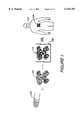
- cultured cells 100are seeded into a three-dimensional, biodegradable scaffold 102.
- the resulting cellular construct, 104is either cultured in-vivo, in a bioreactor 106, such as a broth medium placed in an incubator prior to implantation, 108, or is directly implanted in an animal or the patient 110.
- a bioreactor 106such as a broth medium placed in an incubator prior to implantation, 108, or is directly implanted in an animal or the patient 110.
- These synthesized tissuesreplicate the histological composition and function of the desired tissue with varying degrees of accuracy.
- Complete organs, such as livers, and entire functioning groups, such as a vascularized bone with attached tendon and muscle,have yet to be demonstrated.
- scaffoldshave not been successfully developed for supporting heterogeneous selective cell seeding within the same scaffold.
- a limbis comprised of bone, muscle and tendon.
- Scaffoldssuch as hydroxyapatite, useful to support bone cells, are too brittle and non-pliable to act as scaffolding for muscle or tendons.
- Other heterogeneous tissuessuch as liver and kidney, are even more complex.
- Most current scaffolds and tissue engineering techniquesfail to permit heterogeneous tissues to be grown or provided with blood vessels.
- cell growth factorsshould be present in concentration gradients in order to maximize cell development.
- Most current scaffold fabrication methodshave no direct means of directly creating controlled gradients of growth factor, with the possible exception of 3-D printing.
- the capability to create heterogeneous scaffold seeding systemswould help to enable the regeneration of tissues, and collections of tissues, which exhibit more accurate histological structure and function than can be achieved with homogeneous constructs alone.
- This capabilitywould permit different cells to be strategically placed in different regions of the scaffold, and each region could be composed of the optimal scaffold material and microstructure for organizing and stimulating the growth of cells in that region.
- Solid freeform fabricationrefers to computer-aided-design and computer-aided-manufacturing (CAD/CAM) methodologies which have been used in industrial applications to quickly and automatically fabricate arbitrarily complex shapes. SFF approaches to creating scaffolds for tissue engineering are also being investigated.
- SFF processesconstruct shapes by incremental material buildup and fusion of cross-sectional layers.
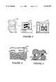
- a three-dimensional (3D) CAD model 112is first decomposed, or "sliced", via an automatic process planner 114, into thin cross-sectional layer representations which are typically 0.004 to 0.020 inches thick.
- an automatic process planner 114To build the physical shape, each layer is then selectively added or deposited and fused to the previous layer in an automated fabrication machine 116.
- each physical layer 118which consists of the cross-section and a complementary shaped sacrificial layer, is deposited and fused to the previous layer as illustrated in FIG. 3, using one of several available deposition and fusion technologies.
- the sacrificial material 120has two primary roles. First, it holds the part, analogous to a fixture in traditional fabrication techniques.
- the unconnected regionsrequire this support since they are not joined with the main body until subsequent layers are deposited.
- Another use of sacrificial materialis to form blind cavities 126 in the part. The sacrificial material is removed when the part is completely built up.
- other building approachesonly use support structures 128 where required, i.e., for the unconnected regions and steep overhanging features.
- These explicit support structuresare typically deposited with the same material as the object being formed, but are drawn out in a semisolid fashion so that it is easy to remove these supports when the part is completed. For example, they may be deposited as thin wall structures.
- FIGS. 5 and 6There are several deposition and fusion processes currently in use or being developed for SFF. Some representative examples of SFF processes, which have also been investigated for tissue engineering applications, are illustrated in FIGS. 5 and 6.
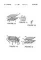
- a layer of powdered material 130is spread over the top surface of the growing structure 131.
- a CO 2 laser 132is then used to selectively scan the layer to fuse those areas defined by the geometry of the cross-section; this also fuses subsequent layers together.
- the laser beamis directed using computer-controlled mirrors 134 directed by the CAD data.
- the unfused material 136remains in place as the support structure.
- an elevator platform 138lowers the part 131 by the thickness of the layer and the next layer of powder is deposited.
- the partWhen the shape is completely built up, the part is separated from the loose supporting powder. Subsequent heat treatment might also be required.
- Several types of materialshave been investigated, including metals, ceramics, polymers, and polymer-coated metals and ceramics. While the materials which have been identified are primarily for industrial applications, the fabrication of hydroxyapatite scaffolds using selective laser sintering have been investigated using polymer-coated calcium phosphate powder. Additional post-processing, such as high temperature heating which bums out the binder, and then higher temperature sintering which fuses the powder together, is required to strengthen the part.
- the three-dimensional printing (3D printing) processis another powder-based SFF approach used in industrial applications, but with potential use in forming scaffolds for engineered tissue.
- An ink-jet printing mechanism 140scans the powder surface 142 and selectively injects a binder into the powder, which joins the powder together, into those areas defined by the geometry of the cross-section.
- an elevator platform 144lowers the part 141 by the thickness of the layer and the next layer of powder is applied by the ink jet. When the shape 141 is completely built up, the part is separated from the loose supporting powder.
- 3D printingfor fabricating biomaterial structures out of bovine bone and biopolymers have also been used.
- microchannels to help support angiogenesiscan be created in the scaffold using this technique. It would also be feasible to use the same or different microchannels to support cell growth via infused cells, harvest medium, growth factors, blood, etc.
- a modified building strategyis required to fabricate highly porous, small diameter microstructures.
- saltis used as the powder and the polymer is used as the binder. The salt, which acts as a porogen, is leached out of the completed shape by dissolving the completed shape in water, leaving a porous polymer scaffold.
- Membrane laminationis another SFF-like technique used for constructing three-dimensional biodegradable polymeric foam scaffolds with precise anatomical shapes.
- a contour plot of the particular three-dimensional shapeis prepared.
- Highly porous PLLA or PLGA membranes having the shapes of the contourare then manufactured using the solvent-casting and particulate-leaching technique.
- Adjacent membranesare bonded together by coating chloroform on their contacting surfaces.
- the final scaffoldis thus formed by laminating the constituent membranes in the proper order to create the desired three-dimensional shape.
- FIG. 7depicts such a heterogeneous structure 150, with embedded components 152, multi-materials 154, and support materials 156.
- SDMShape Deposition Manufacturing
- scaffold fabrication methodstypically involve heat or chemical actions which would destroy living cells or compromise the growth factors. With these methods, cells can only be added to the scaffolds after they have been prefabricated. Growth factors can also be added at or prior to this point. For a discussion of incorporating growth factors into scaffold materials, see Saltsman, "Growth-Factor Delivery in Tissue Engineering,” MRS Bulletin, Nov. 1996, p. 62-65. Completed scaffolds are impregnated with cells by exposing them to cells suspended in liquid culture media; the cells then diffuse into and attach to the scaffolds.
- a three-dimensional scaffoldis achieved by using mechanical fasteners, such as screws, sutures, and microbarbs in order to assemble layers and/or sections of scaffold material.
- mechanical fastenerssuch as screws, sutures, and microbarbs
- cellshave already been incorporated into each subsection of the scaffold prior to assembly.
- different scaffold structuresfor example, those having different porosities for supporting differentiated cells.
- the mechanical assembly techniques of the present inventionallow for both different types of cells to be seeded, as well as for different types of scaffolds to be used to fabricate heterogeneous generated tissue.
- the present inventionprovides a method to build up scaffold constructs by mechanical assembly of individual layer or volume elements.
- These individual elementscan be prefabricated using existing scaffold manufacturing processes such as solvent casting, shaping sections with machining, 3D printing, or molded collagen/cell constructs. These sections can then be mechanically mated using biodegradable or non-biodegradable barbs, pins, screws, clamps, staples, wires, string, or sutures.
- each prefabricated sectioncan first be seeded with cells before assembly, and different scaffold materials, scaffold microstructure, and different cells can be placed in different sections of the scaffold.
- each scaffold subsectionwhich are readily fabricated, become part of the internal microstructure (e.g., molded surface channels become conduits for cell infusion, or for blood flow to stimulate angiogenesis).
- prefabricated vessel constructscan be embedded and assembled into the scaffold as it is being built up.
- the proposed methodologyis based, in part, on the solid freeform fabrication (SFF) manufacturing paradigm, described herein.
- scaffoldsare manufactured by mechanically assembling individual prefabricated layers (or, in general, volumetric elements) of scaffolding with fasteners.
- the prefabricated sectionscan first be manufactured using techniques such as those just described, including solvent casting, fiber bonding, melt molding, 3D printing, SFF, machining hydroxyapatite (HA), and molding collagen.
- each prefabricated sectionis seeded with cells, before final assembly. In this way, cell viability is not compromised, as destructive heat or chemicals are not involved in the scaffold assembly process.
- different materials, microstructure, and cellsare used for making different sections of the scaffold.
- FIG. 1is a schematic representation of three dimensional tissue culturing and cell transplantation.
- FIG. 2is a schematic representation of a solid freeform fabrication technique.
- FIG. 3is a schematic representation of complementary support structures for freeform fabrication.
- FIG. 4is a schematic representation of explicit support structures for freeform fabrication.
- FIG. 5is a schematic representation of a selective laser sintering freeform fabrication processes.
- FIG. 6is a schematic representation of a 3D printing freeform fabrication process.
- FIG. 7is a schematic representation of a heterogeneous structure.
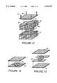
- FIG. 8is an exploded isometric view of a heterogeneous scaffold system of the present invention.
- FIG. 9is a schematic representation of a scaffold of the present invention composed of 3D subsections.
- FIG. 10is a schematic representation of 3D scaffolds assembled according to the present invention using microbarbs.
- FIG. 11is a schematic representation of a double-sided, single-hooked microbarb array useful in practicing the present invention.
- FIG. 12is a schematic representation of a double-hooked barb useful in practicing the present invention.
- FIG. 13is a schematic representation of multiple scaffold layers assembled with screws and nuts according to the present invention.
- FIG. 14is a schematic representation of assembling multiple layers of scaffold with miniature self-tapping screws according to the present invention.
- FIG. 15is a schematic representation of multiple layers of scaffold being assembled with sutures according to the present invention.
- FIG. 16is a schematic representation of multiple scaffold layers being assembled with pins according to the present invention.
- FIG. 17is a schematic representation of three dimensional scaffold sections being assembled in biodegradable containers according to the present invention.
- FIGS. 18 and 19are schematic representations of scaffold systems incorporating channels for angiogenesis according to the present invention.
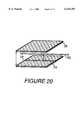
- FIG. 20is a schematic representation of incorporating embedded components, such as vessels, into scaffolds according to the present invention.
- FIG. 21is an isometric exploded view of a three dimensional, multiple layer scaffold of the present invention, incorporating blood vessels into preformed channels.
- FIG. 22is an isometric view of a vessel fastener useful in practicing the present invention.
- a multiple-sectioned scaffoldincludes a subsection of scaffold for supporting bone cell growth A, a subsection for supporting tendon cell growth B, and a subsection for supporting muscle growth C.
- each section, and even different layers within a sectioncan be made up of different materials and cells (e.g., osteoblasts or mesenchymal stem cells impregnated into machined hydroxyapatite for the bone sections A, satellite cells molded into collagen for the muscle sections C, and fibroblasts impregnated into yet another type of scaffold for tendon sections B).
- Individual segments 1 and 2 within a sectioncan be manufactured with different porosities (P), as illustrated by porosities P 1 and P 2 for section A.
- Conduits 3 for embedding blood vessels and/or infusing cellscan be molded into the surfaces of selected layers as illustrated.
- the subsections A, B and Care preferably first seeded with cells and then the subsections are joined together prior to implantation, using fasteners according to the present invention.
- Each subsection A, B and/or Ccould also have growth factors, different concentrations of growth factors, and different growth factors.
- a preferred embodiment of the present inventionuses seeded scaffolds mechanically fixed in close proximity in-vivo (or in a suitable bioreactor).
- the prefabricated elementsneed not be 21/2 D structures (i.e., thin, three-dimensional shapes of uniform thickness).
- Most SFF processeshave used 21/2 D layers for two reasons: it is geometrically straight forward to decompose arbitrarily complex CAD models into layers, and the deposition and fusion processes which they use lend themselves to layering.
- scaffold shapes for the present inventioncan be built up with three-dimensional volumetric elements of varying sizes and shapes, as illustrated in FIG. 9.
- three-dimensionalalso includes planer structures of uniform thickness, but further includes non-planar three-dimensional structures and planar structures of non-uniform thickness, i.e., any structure with three dimensions.
- the surfaces of the volume elementsneed not be planar as depicted in FIG. 9; they could, for example, be curved surfaces.
- scaffold sectionscan be mechanically assembled, preferably with biodegradable fasteners.
- the fastenersfabricated, for example, from PLA/PGA, PEO, or polycarbonate, can be molded or machined.
- These fastenerswhich can be used independently or in combination, include (but are not limited to), microbarbs, screws, sutures, pins, staples, wires, strings, and containers.
- microbarbs 10can be positioned at intervals and used to penetrate and lock into porous scaffold materials 12.
- Polymer barbscan be fabricated using micromolding techniques such as described by Whitesides in "Making Polymeric Microstructures: Capillary Micromolding," technical note available from Department of Chemistry, Harvard University, Cambridge, Mass. 02138, incorporated in its entirety by reference herein.
- the scale of the barbsis preferably from about 25 ⁇ m to 250 ⁇ m or greater in height, depending upon the dimensions of the scaffold's porous microstructure.
- Each barbcan have single (FIG. 11) or multiple (FIG. 12) hooks 14.
- Barbscan be used to mate with not only rigid or stiff scaffold materials, but also with compliant or elastic sections such as collagen. Another use of these barbs is to mate scaffolds to existing tissues in-vivo.
- FIG. 11Another preferred embodiment uses double-sided barb arrays (FIG. 11) such that one side is first attached to one section of scaffold, then the second scaffold section is pressed onto the first section. As illustrated in FIG. 10, several barb fasteners 10 can be placed throughout the entire surface of a scaffold section to distribute the loads. Another approach is to use single-sided barb arrays, and attach the flat side to the first scaffold section with solvent (before seeding the first side with cells). Various combinations, including single-sided, single barbed, double-sided, double-barbed, or multiple barbs (in excess of two) can also be used.
- scaffold sections 26, 28, and 30are screwed together with self-tapping screws 20, (FIG. 14) or with screw and nut combinations 22, 24 (FIG. 13).
- screws 22are first inserted up through predrilled holes 32 in the lower scaffold section 26, then the screw heads are bonded to the scaffold with solvents such as chloroform (before the lower section 26 is seeded with cells).
- solventssuch as chloroform
- layers 28, 30, also with predrilled holes 32are then stacked onto the lower section 26 and firmly assembled with nuts 24.
- Another preferred embodimentuses miniature self-taping screws 20 on a layer-by-layer basis as illustrated in FIG. 14.
- Screwsare advantageously used when the scaffold is fabricated of a rigid or semi-rigid material, such as hydroxyapatite.
- a rigid or semi-rigid materialsuch as hydroxyapatite.
- scaffold layers or sectionsare fastened together using sutures, 33, which may be threaded through pre-existing holes 35, or sewn through unperforated sections of the scaffold layers. Since non-rigid sutures do not provide for accurate alignment between the sections, alignment can be provided using matched pairs of indentations or grooves 37 and mating protrusions 39 which can be easily molded into the scaffold sections. Sutures 33 are especially useful for fastening thin and/or pliable sections of scaffold material together.
- thin layers of scaffold sectionsmay be threaded onto a single suture, wire, or string, and suspended in a bioreactor with weights or clamping devices used to hold the layers in proximity while the cell cultures grow.
- FIG. 16Yet another embodiment of the invention is illustrated in FIG. 16.
- pins 32are pushed into pre-drilled holes 34 in the scaffold 36.
- the holes 34can be slightly undersized to obtain a friction fit.
- insertion of the pinscan be facilitated by using a compliant scaffold material and a rigid pin, by cooling the pins prior to insertion to reduce their diameter, or using split pins (as depicted in the FIG. 16).
- the outside surface of the pins 32may be etched to improve friction/gripping properties.
- FIG. 17Another preferred embodiment of the invention is illustrated in FIG. 17.
- scaffold sectionsare stacked into prefabricated, biodegradable containers.
- the individual scaffold sections 40, 42, 44, 46 and 48are not joined to each other, rather are held in place within the container 50.
- the containercan be porous and/or have inlet/outlet ports to attach vessels to. Similarly, clamps and cable tie-straps can be used to hold and to fix sections together.
- the container 50includes a biodegradable cap 52, which encloses the container 50 and may use fasteners such as screws 54 or other fasteners described herein to close the container.
- biodegradable or non-biodegradable, non-reactive (e.g., titanium) surgical staples carried in and fired by stapling instrumentssuch as those manufactured by Ethicon Endo-Surgery, Cincinnati, Ohio, can be used to fasten subsections of scaffolding together.
- a surface feature on an individual scaffold segmentwill become an internal feature when another segment is assembled over it.
- surface featuressuch as channels can be produced by molding, machining, or by 3D printing (e.g., layer 60 with channels 61 in FIG. 18).
- the next scaffold sectione.g., layer 62 in FIG. 18
- the surface featurebecomes an internal scaffold feature.
- One applicationis to create an internal matrix of tubules for cell infusion and/or angiogenesis.
- Another strategy for producing surface features, illustrated in FIG. 19,is to place individual segments of scaffold 66, separated from each other, between layers of scaffold 67, 68, to explicitly form channels.
- one possibility for creating vasculatureis to first place a natural or synthetic vessel 70 into a surface channel 72 of a scaffold section 74.
- a molding technique for fabricating synthetic collagen-based vesselsis, for example, disclosed in Okano and Matsuda, "Hybrid Muscular Tissues: Preparation of Skeletal Muscle-Incorporated Collagen Gels," Cell Transplantation, Vol. 6, No. 2, 109-118 (1997). Then, the vessel becomes embedded within the entire scaffold when the subsequent scaffold section 76 is mated over the other section 74.
- FIG. 21illustrates the approach for embedding synthetic vessels within three dimensional scaffold material.
- an intact, in-vivo blood vessel 80is sectioned, in order for placement of a three dimensional scaffold generally 82 between the sectioned vessels 80.
- the scaffold 82is comprised of multiple subsections 83-88.
- the subsections 85 and 86are closest to the synthetic blood vessel 90 which, as illustrated, may have a textured outer surface to assist in retaining the vessel 90 within the scaffold 82.
- the synthetic vessel 90has been placed within the scaffold 82 in a bioreactor prior to implantation.
- the existing vessels 80are secured to either end of the synthetic vessel 90 using known microsurgical techniques.
- the subsections 83-88 of the scaffold 82have been assembled with fasteners and seeded with cells in the manner previously described.
- a barbed halo 92shown in greater detail in FIG. 22, is used to secure the scaffold 82 to the existing vessels 80. This is accomplished, for example, by positioning the halo 92 around the outer surface of the vessel 80 in order to create a "lock washer" for precluding the axial movement of the scaffold 82 with respect to the vessels 80.
- the halo 92can be first fastened to the scaffold 82, for example, with solvent or glue, prior to assembling the scaffold 82 around the blood vessel 80.
- a barbed halo 92can be positioned at every juncture of the scaffold 82 with a blood vessel 80. Further support could be provided, for example, by suturing the halo 92 to both the vessel 80 and the synthetic vessel 90 and/or the scaffold 82 using known microsurgical techniques.
- the halo 92may comprise two semicircular sections. One such section is illustrated in detail in FIG. 22. As illustrated, the halo 92 includes a plurality of barbs which may comprise spike-shaped elements, or may be shaped similar to the single and double-headed barbs illustrated in FIGS. 11 and 12.
- the halois preferably fabricated of a biodegradable/biocompatible material, and can be molded.
- fasteners described hereinbe fabricated of biodegradable/biocompatible materials. It is, of course, possible to use non-biodegradable materials, provided they are biocompatible. For example, titanium screws and/or staples can be used as fasteners according to the present invention.
- Support structuresmight be needed for several instances, e.g., for ⁇ unconnected ⁇ regions, for supporting steep overhanging features made out of highly compliant materials, and for substrates upon which to start assembling the scaffold.
- the scaffold sectionscan be attached to the support structures using the same mating strategies described above.
- the support sectionscan be passive and therefore not be seeded with cells.
- the microstructure and material composition of support structuresis preferably designed to inhibit ingrowth.
- Growth factorscan, according to the present invention, be incorporated into subsections either with or without cells.
- Other combinationsare, of course, possible.
- gradients of growth factorcan be achieved in the scaffold of the present invention, for example, by providing layers or subsections of scaffolding, each having homogeneous, but different, concentrations of growth factor relative to adjacent layers or subsections.
- different types of growth factorscan be used in different layers or subsections relative to those used in other layers or subsections.
- Seeding of cells and scaffold preparationwere performed in the following manner.
- Subsections of hydroxyapatite/polymer scaffoldwere steam sterilized in an autoclave and pre-soaked for 24 hours in a tissue culture medium, such as Dilbecco's Modified Eagle's Medium.
- a male New Zealand White Rabbit(Orycytolagus cuniculus) was anesthetized intramuscularly, and positioned in the supine position.
- the lower abdominal wall, inguinal region, and lateral surfaces of both thighs and legswere shaved, depilated, and prepared for aseptic surgery.
- Bone marrowwas harvested by injecting several cc's of harvest medium into the medullary canal to displace the marrow.
- the harvested bone marrowwas mixed with 4 cc of heparinized tissue culture medium in a test tube, and centrifuged for three minutes. Some of the supernatant was discarded to concentrate the cell number, the cell count was checked to verify that the number of cells was greater than 1 ⁇ 10 8 /ml.
- hydrophilic scaffold materialsuch as hydroxyapatite
- the individual scaffold subsectionswere permitted to soak in the suspension of bone marrow cells for several minutes. Two subsections were then sewn together using monofilament nylon sutures as fasteners. The first suture was passed through central region of one disc, and through the central region of a second disc, then brought back through both discs about 1 mm from the first hole to create a button-hole effect. The suture ends were tied and cut short. Four equally spaced sutures were then placed about the perimeter of the discs, tied, and cut short. The sutures were tied to create a snug relationship between adjoining layers of scaffold. The joined scaffold subsections were again immersed in the cell suspension, which was again vacuum drawn. This procedure was repeated until a three dimensional scaffold having five 1 mm thick subsections was seeded with bone marrow cells and stabilized with sutures.
- the three-dimensional scaffoldwas then implanted in the same rabbit from which the bone marrow was harvested, using the rabbit, in effect, as a bioreactor to support growth of bone cells seeded into the scaffold as a result of the bone marrow cell seeding.
- the seeded three dimensional scaffoldwas implanted intramuscularly adjacent and superficial to the deep inferior epigastric right vascular bundle.
- the incisionswere closed, and the implanted scaffold permitted to support cell growth for at least six weeks.
- the animalswill be monitored at six, eight, and twelve week intervals to assess the degree of tissue generation.
Landscapes
- Health & Medical Sciences (AREA)
- Life Sciences & Earth Sciences (AREA)
- Chemical & Material Sciences (AREA)
- Engineering & Computer Science (AREA)
- Biomedical Technology (AREA)
- Zoology (AREA)
- General Health & Medical Sciences (AREA)
- Medicinal Chemistry (AREA)
- Epidemiology (AREA)
- Animal Behavior & Ethology (AREA)
- Public Health (AREA)
- Veterinary Medicine (AREA)
- Transplantation (AREA)
- Oral & Maxillofacial Surgery (AREA)
- Dermatology (AREA)
- Chemical Kinetics & Catalysis (AREA)
- Biotechnology (AREA)
- Cell Biology (AREA)
- Organic Chemistry (AREA)
- Bioinformatics & Cheminformatics (AREA)
- Genetics & Genomics (AREA)
- Botany (AREA)
- Wood Science & Technology (AREA)
- Heart & Thoracic Surgery (AREA)
- Urology & Nephrology (AREA)
- Biochemistry (AREA)
- Cardiology (AREA)
- Microbiology (AREA)
- Vascular Medicine (AREA)
- Molecular Biology (AREA)
- General Engineering & Computer Science (AREA)
- Developmental Biology & Embryology (AREA)
- Immunology (AREA)
- Virology (AREA)
- Pharmacology & Pharmacy (AREA)
- Materials For Medical Uses (AREA)
- Prostheses (AREA)
Abstract
Description
Claims (19)
Priority Applications (2)
| Application Number | Priority Date | Filing Date | Title |
|---|---|---|---|
| US09/048,944US6143293A (en) | 1998-03-26 | 1998-03-26 | Assembled scaffolds for three dimensional cell culturing and tissue generation |
| PCT/US1999/006481WO1999048541A1 (en) | 1998-03-26 | 1999-03-26 | Assembled scaffolds for three-dimensional cell culturing and tissue generation |
Applications Claiming Priority (1)
| Application Number | Priority Date | Filing Date | Title |
|---|---|---|---|
| US09/048,944US6143293A (en) | 1998-03-26 | 1998-03-26 | Assembled scaffolds for three dimensional cell culturing and tissue generation |
Publications (1)
| Publication Number | Publication Date |
|---|---|
| US6143293Atrue US6143293A (en) | 2000-11-07 |
Family
ID=21957284
Family Applications (1)
| Application Number | Title | Priority Date | Filing Date |
|---|---|---|---|
| US09/048,944Expired - LifetimeUS6143293A (en) | 1998-03-26 | 1998-03-26 | Assembled scaffolds for three dimensional cell culturing and tissue generation |
Country Status (2)
| Country | Link |
|---|---|
| US (1) | US6143293A (en) |
| WO (1) | WO1999048541A1 (en) |
Cited By (155)
| Publication number | Priority date | Publication date | Assignee | Title |
|---|---|---|---|---|
| US6379740B1 (en)* | 1997-12-10 | 2002-04-30 | Sorin Biomedica Cardio S.P.A. | Method for treating a prosthesis having an apertured structure and associated devices |
| US20020119177A1 (en)* | 2000-12-21 | 2002-08-29 | Bowman Steven M. | Reinforced foam implants with enhanced integrity for soft tissue repair and regeneration |
| US20020127265A1 (en)* | 2000-12-21 | 2002-09-12 | Bowman Steven M. | Use of reinforced foam implants with enhanced integrity for soft tissue repair and regeneration |
| WO2002053193A3 (en)* | 2001-01-02 | 2002-09-26 | Draper Lab Charles S | Tissue engineering of three-dimensional vascularized using microfabricated polymer assembly technology |
| US20030003575A1 (en)* | 1999-04-30 | 2003-01-02 | Vacanti Joseph P. | Fabrication of vascularized tissue using microfabricated two-dimensional molds |
| US20030006534A1 (en)* | 2001-06-22 | 2003-01-09 | Taboas Juan M. | Controlled local/global and micro/macro-porous 3D plastic, polymer and ceramic/cement composite scaffold fabrication and applications thereof |
| WO2003004254A1 (en)* | 2001-07-03 | 2003-01-16 | The Regents Of The University Of California | Microfabricated biopolymer scaffolds and method of making same |
| US20030069718A1 (en)* | 2001-06-22 | 2003-04-10 | Hollister Scott J. | Design methodology for tissue engineering scaffolds and biomaterial implants |
| US20030068817A1 (en)* | 2001-04-30 | 2003-04-10 | Dan Gazit | Vascular tissue engineering |
| US20030074096A1 (en)* | 2001-10-15 | 2003-04-17 | Suman Das | Solid freeform fabrication of structurally engineered multifunctional devices |
| US6586246B1 (en)* | 1999-03-18 | 2003-07-01 | Innotech Medical, Inc. | Preparing porous biodegradable polymeric scaffolds for tissue engineering using effervescent salts |
| US20030128832A1 (en)* | 2002-01-04 | 2003-07-10 | Telefonaktiebolaget Lm Ericsson (Publ) | Message transfer part point code mapping method and node |
| US20030147935A1 (en)* | 2000-12-21 | 2003-08-07 | Ethicon, Inc. | Use of reinforced foam implants with enhanced integrity for soft tissue repair and regeneration |
| US20030170285A1 (en)* | 2001-11-13 | 2003-09-11 | Veazey William S. | Delivery of tissue engineering media |
| US20030175410A1 (en)* | 2002-03-18 | 2003-09-18 | Campbell Phil G. | Method and apparatus for preparing biomimetic scaffold |
| US20030181978A1 (en)* | 2002-03-25 | 2003-09-25 | Brown Kelly R. | Channeled biomedical foams and method for producing same |
| US20030191107A1 (en)* | 2002-01-22 | 2003-10-09 | Pfizer Inc. | 3-(Imidazolyl)-2-aminopropanoic acids |
| WO2003082145A2 (en) | 2002-03-25 | 2003-10-09 | The General Hospital Corporation | Fabrication of vascularized tissue using microfabricated two-dimensional molds |
| US20030193104A1 (en)* | 2000-12-21 | 2003-10-16 | Melican Mora Carolynne | Reinforced tissue implants and methods of manufacture and use |
| US6637437B1 (en) | 1998-04-08 | 2003-10-28 | Johns Hopkins University | Cell-culture and polymer constructs |
| US20030225459A1 (en)* | 2002-05-31 | 2003-12-04 | Hammer Joseph J. | Attachment of absorbable tissue scaffolds to fixation devices |
| JP2004505747A (en)* | 2000-08-22 | 2004-02-26 | ジンテーズ アクチエンゲゼルシャフト クール | Bone substitute material |
| US20040213767A1 (en)* | 2003-04-23 | 2004-10-28 | Marc Hendriks | Methods for using adipose-derived cells for healing of aortic aneurysmal tissue |
| US20040254640A1 (en)* | 2002-03-01 | 2004-12-16 | Children's Medical Center Corporation | Needle punched textile for use in growing anatomical elements |
| US20050038520A1 (en)* | 2003-08-11 | 2005-02-17 | Francois Binette | Method and apparatus for resurfacing an articular surface |
| US20050112186A1 (en)* | 2003-10-29 | 2005-05-26 | David Devore | Polymerizable emulsions for tissue engineering |
| WO2005057436A1 (en)* | 2003-11-14 | 2005-06-23 | Drexel University | Method and apparatus for computer-aided tissue engineering for modeling, design and freeform fabrication of tissue scaffolds, constructs, and devices |
| WO2005034624A3 (en)* | 2003-05-21 | 2005-08-25 | Gen Hospital Corp | Microfabricated compositions and processes for engineering tissues containing multiple cell types |
| US20050202557A1 (en)* | 2000-04-28 | 2005-09-15 | Jeffrey Borenstein | Micromachined bilayer unit of engineered tissues |
| WO2005060396A3 (en)* | 2003-08-18 | 2005-11-17 | Gen Hospital Corp | Nanotopographic compositions and methods for cellular organization in tissue engineered structures |
| US20050276864A1 (en)* | 2004-05-27 | 2005-12-15 | Medtronic Vascular, Inc. | Cellular therapy to heal vascular tissue |
| US20050276791A1 (en)* | 2004-02-20 | 2005-12-15 | The Ohio State University | Multi-layer polymer scaffolds |
| US20060036331A1 (en)* | 2004-03-05 | 2006-02-16 | Lu Helen H | Polymer-ceramic-hydrogel composite scaffold for osteochondral repair |
| US20060067969A1 (en)* | 2004-03-05 | 2006-03-30 | Lu Helen H | Multi-phased, biodegradable and osteointegrative composite scaffold for biological fixation of musculoskeletal soft tissue to bone |
| US20060094112A1 (en)* | 2001-03-07 | 2006-05-04 | Omotunde Babalola | Biological scaffold |
| US20060153001A1 (en)* | 2003-08-08 | 2006-07-13 | Flavio Hoerger | Method to impregnate a porous bone replacement material |
| WO2006042287A3 (en)* | 2004-10-12 | 2006-07-27 | Tufts College | Method for producing biomaterial scaffolds |
| US20060204445A1 (en)* | 2005-03-11 | 2006-09-14 | Anthony Atala | Cell scaffold matrices with image contrast agents |
| US20060240061A1 (en)* | 2005-03-11 | 2006-10-26 | Wake Forest University Health Services | Tissue engineered blood vessels |
| US20060249875A1 (en)* | 2005-02-14 | 2006-11-09 | Robb Richard A | Tissue support structure |
| US20060253192A1 (en)* | 2005-03-11 | 2006-11-09 | Wake Forest University Health Sciences | Production of tissue engineered heart valves |
| US20060257377A1 (en)* | 2005-03-11 | 2006-11-16 | Wake Forest University Health Services | Production of tissue engineered digits and limbs |
| US20060263335A1 (en)* | 2003-03-27 | 2006-11-23 | Regentec Ltd. | Porous matrix |
| US20070118156A1 (en)* | 2005-11-22 | 2007-05-24 | Yi-You Huang | Bioartificial guidance conduit and method for forming the same |
| US20070221742A1 (en)* | 2004-06-03 | 2007-09-27 | Flavio Hoerger | Device for impregnating a porous bone replacement material |
| US20070276509A1 (en)* | 2004-05-11 | 2007-11-29 | Anthony Ratcliffe | Tissue scaffold |
| US7316822B2 (en) | 2003-11-26 | 2008-01-08 | Ethicon, Inc. | Conformable tissue repair implant capable of injection delivery |
| US20080081362A1 (en)* | 2006-09-29 | 2008-04-03 | Daniel Keeley | Multilayered Composite for Organ Augmentation and Repair |
| US20080111272A1 (en)* | 2006-10-17 | 2008-05-15 | Burgess James E | Method and apparatus for manufacturing plasma based plastics and bioplastics produced therefrom |
| US20080112998A1 (en)* | 2006-11-14 | 2008-05-15 | Hongjun Wang | Innovative bottom-up cell assembly approach to three-dimensional tissue formation using nano-or micro-fibers |
| US20080145639A1 (en)* | 2005-02-25 | 2008-06-19 | Drexel University | Layered Manufacturing Utilizing Foam As A Support And Multifunctional Material For The Creation Of Parts And For Tissue Engineering |
| US20080214998A1 (en)* | 2005-10-20 | 2008-09-04 | Ed Kurek | Perfusion Device and Method |
| US20080246180A1 (en)* | 2001-06-05 | 2008-10-09 | Appleby Michael P | Methods for Manufacturing Three-Dimensional Devices and Devices Created Thereby |
| WO2008133362A1 (en)* | 2007-04-28 | 2008-11-06 | Hyunjin Yang | Proliferation culture methods using micro-scaffolds for regulations of cell-to-cell signals |
| US7488348B2 (en) | 2003-05-16 | 2009-02-10 | Musculoskeletal Transplant Foundation | Cartilage allograft plug |
| US20090113687A1 (en)* | 2006-04-14 | 2009-05-07 | Akintunde Ibitayo Akinwande | Precise hand-assembly of microfabricated components |
| US20090155340A1 (en)* | 2001-09-14 | 2009-06-18 | Research Foundaton At State University Of New York | Method of cell storage in a delivery system |
| US20090181200A1 (en)* | 2007-09-19 | 2009-07-16 | Borenstein Jeffrey T | Microfluidic Structures for Biomedical Applications |
| US20090202378A1 (en)* | 2008-02-13 | 2009-08-13 | Materials Solution | Method of forming an article |
| EP2093256A2 (en) | 2005-07-28 | 2009-08-26 | Carnegie Mellon University | Biocompatible polymers and methods of use |
| US20090233362A1 (en)* | 2005-09-20 | 2009-09-17 | Chen Guoping | Porous Scaffold, Method of Producing the Same and Method of Using the Porous Scaffold |
| US7597715B2 (en) | 2005-04-21 | 2009-10-06 | Biomet Manufacturing Corp. | Method and apparatus for use of porous implants |
| US7635447B2 (en) | 2006-02-17 | 2009-12-22 | Biomet Manufacturing Corp. | Method and apparatus for forming porous metal implants |
| US20100047309A1 (en)* | 2006-12-06 | 2010-02-25 | Lu Helen H | Graft collar and scaffold apparatuses for musculoskeletal tissue engineering and related methods |
| US7713542B2 (en) | 2005-01-14 | 2010-05-11 | Ada Foundation | Three dimensional cell protector/pore architecture formation for bone and tissue constructs |
| US20100252528A1 (en)* | 2006-07-03 | 2010-10-07 | Fuji Xerox Co., Ltd. | Liquid droplet ejection head, apparatus for ejecting liquid droplet, and method of producing liquid droplet ejection head |
| US7815926B2 (en) | 2005-07-11 | 2010-10-19 | Musculoskeletal Transplant Foundation | Implant for articular cartilage repair |
| US7824701B2 (en) | 2002-10-18 | 2010-11-02 | Ethicon, Inc. | Biocompatible scaffold for ligament or tendon repair |
| US20100292791A1 (en)* | 2007-02-12 | 2010-11-18 | Lu Helen H | Fully synthetic implantable multi-phased scaffold |
| US7837740B2 (en) | 2007-01-24 | 2010-11-23 | Musculoskeletal Transplant Foundation | Two piece cancellous construct for cartilage repair |
| US7901461B2 (en) | 2003-12-05 | 2011-03-08 | Ethicon, Inc. | Viable tissue repair implants and methods of use |
| USRE42208E1 (en) | 2003-04-29 | 2011-03-08 | Musculoskeletal Transplant Foundation | Glue for cartilage repair |
| US7901457B2 (en) | 2003-05-16 | 2011-03-08 | Musculoskeletal Transplant Foundation | Cartilage allograft plug |
| US20110177590A1 (en)* | 2009-12-11 | 2011-07-21 | Drexel University | Bioprinted Nanoparticles and Methods of Use |
| US8016867B2 (en) | 1999-07-23 | 2011-09-13 | Depuy Mitek, Inc. | Graft fixation device and method |
| US8021432B2 (en) | 2005-12-05 | 2011-09-20 | Biomet Manufacturing Corp. | Apparatus for use of porous implants |
| US8049193B1 (en) | 2001-06-05 | 2011-11-01 | Mikro Systems, Inc. | Systems, devices, and methods for large area micro mechanical systems |
| US8066778B2 (en) | 2005-04-21 | 2011-11-29 | Biomet Manufacturing Corp. | Porous metal cup with cobalt bearing surface |
| US8123814B2 (en) | 2001-02-23 | 2012-02-28 | Biomet Manufacturing Corp. | Method and appartus for acetabular reconstruction |
| US8137686B2 (en) | 2004-04-20 | 2012-03-20 | Depuy Mitek, Inc. | Nonwoven tissue scaffold |
| US8173361B2 (en) | 2003-01-16 | 2012-05-08 | The General Hospital Corporation | Method of determining metabolism of a test agent |
| US8187326B2 (en) | 2002-05-22 | 2012-05-29 | Advanced Technologies And Regenerative Medicine, Llc. | Attachment of absorbable tissue scaffolds to fixation devices |
| US8221780B2 (en) | 2004-04-20 | 2012-07-17 | Depuy Mitek, Inc. | Nonwoven tissue scaffold |
| US8226715B2 (en) | 2003-06-30 | 2012-07-24 | Depuy Mitek, Inc. | Scaffold for connective tissue repair |
| WO2012071578A3 (en)* | 2010-11-24 | 2012-08-23 | Bc Genesis Llc | Pharmacology bioassays for drug discovery, toxicity evaluation and in vitro cancer research using a 3d nano-cellulose scaffold and living tissue |
| US8266780B2 (en) | 2005-04-21 | 2012-09-18 | Biomet Manufacturing Corp. | Method and apparatus for use of porous implants |
| US8292967B2 (en) | 2005-04-21 | 2012-10-23 | Biomet Manufacturing Corp. | Method and apparatus for use of porous implants |
| US8292968B2 (en) | 2004-10-12 | 2012-10-23 | Musculoskeletal Transplant Foundation | Cancellous constructs, cartilage particles and combinations of cancellous constructs and cartilage particles |
| US8435551B2 (en) | 2007-03-06 | 2013-05-07 | Musculoskeletal Transplant Foundation | Cancellous construct with support ring for repair of osteochondral defects |
| US8449561B2 (en) | 1999-07-23 | 2013-05-28 | Depuy Mitek, Llc | Graft fixation device combination |
| EP2617427A1 (en) | 2004-03-19 | 2013-07-24 | Cytori Therapeutics, Inc. | Cell carrier and cell carrier containment devices containing regenerative cells |
| US8529960B2 (en) | 2002-03-18 | 2013-09-10 | Carnell Therapeutics Corporation | Methods and apparatus for manufacturing plasma based plastics and bioplastics produced therefrom |
| US8529958B2 (en) | 2006-10-17 | 2013-09-10 | Carmell Therapeutics Corporation | Methods and apparatus for manufacturing plasma based plastics and bioplastics produced therefrom |
| US8657881B2 (en) | 2004-04-20 | 2014-02-25 | Depuy Mitek, Llc | Meniscal repair scaffold |
| US8669086B2 (en) | 2010-04-29 | 2014-03-11 | The United States Of America, As Represented By The Secretary Of The Navy | Cell and biofactor printable biopapers |
| CN103655005A (en)* | 2013-12-02 | 2014-03-26 | 浙江大学 | Three-dimensional biological structure 3D printing device and method |
| US8691974B2 (en) | 2009-09-28 | 2014-04-08 | Virginia Tech Intellectual Properties, Inc. | Three-dimensional bioprinting of biosynthetic cellulose (BC) implants and scaffolds for tissue engineering |
| WO2014105581A1 (en)* | 2012-12-26 | 2014-07-03 | Konica Minolta Laboratory U.S.A., Inc. | Apparatus and method for rapid 3d cell culture analysis using paper stacks |
| US8813824B2 (en) | 2011-12-06 | 2014-08-26 | Mikro Systems, Inc. | Systems, devices, and/or methods for producing holes |
| WO2014143719A1 (en)* | 2013-03-15 | 2014-09-18 | Smed-Ta/Td, Llc | Porous tissue ingrowth structure |
| US8895045B2 (en) | 2003-03-07 | 2014-11-25 | Depuy Mitek, Llc | Method of preparation of bioabsorbable porous reinforced tissue implants and implants thereof |
| US20150202825A1 (en)* | 2014-01-17 | 2015-07-23 | Christopher Cordingley | Three Dimensional Printing Method |
| US20150209162A1 (en)* | 2012-10-05 | 2015-07-30 | Materialise N.V. | Customized aortic stent device and method of making the same |
| RU2563621C2 (en)* | 2013-09-16 | 2015-09-20 | Федеральное государственное бюджетное учреждение науки Институт физики полупроводников им. А.В. Ржанова Сибирского отделения Российской академии наук (ИФП СО РАН) | Bioresorbable polymer cell matrix |
| US9163331B2 (en) | 2005-03-11 | 2015-10-20 | Wake Forest University Health Sciences | Electrospun cell matrices |
| US9315663B2 (en) | 2008-09-26 | 2016-04-19 | Mikro Systems, Inc. | Systems, devices, and/or methods for manufacturing castings |
| WO2016064902A1 (en)* | 2014-10-20 | 2016-04-28 | Tara Biosystems, Inc. | Microfabricated tissue scaffolds and methods of making and using the same |
| US9427496B2 (en) | 2005-02-18 | 2016-08-30 | Drexel University | Method for creating an internal transport system within tissue scaffolds using computer-aided tissue engineering |
| US20160324642A1 (en)* | 2013-12-30 | 2016-11-10 | The New York Stem Cell Foundation | Tissue grafts and methods of making and using the same |
| US9511171B2 (en) | 2002-10-18 | 2016-12-06 | Depuy Mitek, Llc | Biocompatible scaffolds with tissue fragments |
| WO2017030762A1 (en)* | 2015-08-14 | 2017-02-23 | The General Hospital Corporation | Systems for and methods for using biomimetic structures providing communication in living tissue |
| RU2622009C1 (en)* | 2015-12-25 | 2017-06-08 | Федеральное государственное бюджетное учреждение науки Институт физики полупроводников им. А.В. Ржанова Сибирского отделения Российской академии наук (ИФП СО РАН) | Method for biooresorable polymer cell matrix formation for tissue regeneration |
| US9681966B2 (en) | 2013-03-15 | 2017-06-20 | Smed-Ta/Td, Llc | Method of manufacturing a tubular medical implant |
| US9701940B2 (en) | 2005-09-19 | 2017-07-11 | Histogenics Corporation | Cell-support matrix having narrowly defined uniformly vertically and non-randomly organized porosity and pore density and a method for preparation thereof |
| US9910935B2 (en) | 2013-10-11 | 2018-03-06 | Advanced Solutions Life Sciences, Llc | System and workstation for the design, fabrication and assembly of bio-material constructs |
| WO2018144098A1 (en)* | 2016-11-03 | 2018-08-09 | The Arizona Board Of Regents On Behalf Of The University Of Arizona | Stacked tissue encapsulation device systems with or without oxygen delivery |
| US10077420B2 (en) | 2014-12-02 | 2018-09-18 | Histogenics Corporation | Cell and tissue culture container |
| US10092679B2 (en) | 2013-10-18 | 2018-10-09 | Wake Forest University Health Sciences | Laminous vascular constructs combining cell sheet engineering and electrospinning technologies |
| US10214714B2 (en) | 2013-12-30 | 2019-02-26 | New York Stem Cell Foundation, Inc. | Perfusion bioreactor |
| KR20190036503A (en)* | 2017-09-27 | 2019-04-04 | 한양대학교 산학협력단 | Assembly of super-structure using cell-laden hydrogel blocks for 3D Bioprinting based tissue engineering |
| US10343300B2 (en)* | 2015-09-11 | 2019-07-09 | Ngk Insulators, Ltd. | Method and apparatus for manufacturing porous body |
| US10624747B2 (en)* | 2014-08-05 | 2020-04-21 | Dietmar SONNLEITNER | Method for producing a multilayer film |
| US10668107B2 (en) | 2014-12-30 | 2020-06-02 | Orgenesis Ltd. | Methods of transdifferentiation and methods of use thereof |
| US10947509B2 (en) | 2013-06-13 | 2021-03-16 | Orgenesis Ltd. | Cell populations, methods of transdifferentiation and methods of use thereof |
| US10975354B2 (en) | 2017-05-08 | 2021-04-13 | Orgenesis Ltd. | Transdifferentiated cell populations and methods of use thereof |
| WO2021086058A1 (en)* | 2019-10-31 | 2021-05-06 | 포항공과대학교 산학협력단 | Artificial tissue or organ analogs prepared using three-dimensional cell printing and method for preparing same |
| US11110199B2 (en) | 2013-04-12 | 2021-09-07 | The Trustees Of Columbia University In The City Of New York | Methods for host cell homing and dental pulp regeneration |
| US11246959B2 (en) | 2013-03-15 | 2022-02-15 | Nanofiber Solutions, Llc | Biocompatible fiber textiles for implantation |
| CN114196536A (en)* | 2020-09-17 | 2022-03-18 | 卡内基·梅隆大学 | Complementary-shaped and porosity-matched perfusion bioreactor system for engineering geometrically complex bone grafts |
| US20220151789A1 (en)* | 2018-08-14 | 2022-05-19 | Georgia Tech Research Corporation | Method for adjusting mechanical properties of implant and patient specific surgical implants |
| US11357890B2 (en) | 2016-04-01 | 2022-06-14 | New York Stem Cell Foundation, Inc. | Customized hybrid bone-implant grafts |
| US11395865B2 (en) | 2004-02-09 | 2022-07-26 | DePuy Synthes Products, Inc. | Scaffolds with viable tissue |
| US11566215B2 (en) | 2016-08-27 | 2023-01-31 | 3D Biotek Llc | Bioreactor with scaffolds |
| US11576927B2 (en) | 2018-12-11 | 2023-02-14 | Nanofiber Solutions, Llc | Methods of treating chronic wounds using electrospun fibers |
| US20230065127A1 (en)* | 2018-05-25 | 2023-03-02 | The General Hospital Corporation | Additive manufacture of complex implantable living devices |
| US11608486B2 (en) | 2015-07-02 | 2023-03-21 | Terumo Bct, Inc. | Cell growth with mechanical stimuli |
| US11613727B2 (en) | 2010-10-08 | 2023-03-28 | Terumo Bct, Inc. | Configurable methods and systems of growing and harvesting cells in a hollow fiber bioreactor system |
| US11624046B2 (en) | 2017-03-31 | 2023-04-11 | Terumo Bct, Inc. | Cell expansion |
| US11629332B2 (en) | 2017-03-31 | 2023-04-18 | Terumo Bct, Inc. | Cell expansion |
| US11634677B2 (en) | 2016-06-07 | 2023-04-25 | Terumo Bct, Inc. | Coating a bioreactor in a cell expansion system |
| US11667881B2 (en) | 2014-09-26 | 2023-06-06 | Terumo Bct, Inc. | Scheduled feed |
| US11667876B2 (en) | 2013-11-16 | 2023-06-06 | Terumo Bct, Inc. | Expanding cells in a bioreactor |
| US11685883B2 (en) | 2016-06-07 | 2023-06-27 | Terumo Bct, Inc. | Methods and systems for coating a cell growth surface |
| US11723558B2 (en) | 2016-11-03 | 2023-08-15 | Arizona Board Of Regents On Behalf Of The University Of Arizona | Encapsulation device systems with oxygen sensors with or without exogenous oxygen delivery |
| US11737990B2 (en) | 2012-01-12 | 2023-08-29 | Nfs Ip Holdings, Llc | Nanofiber scaffolds for biological structures |
| US11746318B2 (en) | 2016-11-03 | 2023-09-05 | Arizona Board Of Regents On Behalf Of The University Of Arizona | Methods and systems for real-time assessment of cells in encapsulation devices pre-and post-transplantation |
| US11795432B2 (en) | 2014-03-25 | 2023-10-24 | Terumo Bct, Inc. | Passive replacement of media |
| US11806440B2 (en) | 2017-02-02 | 2023-11-07 | Nfs Ip Holdings, Llc | Methods of improving bone-soft tissue healing using electrospun fibers |
| US11965175B2 (en) | 2016-05-25 | 2024-04-23 | Terumo Bct, Inc. | Cell expansion |
| US12016973B2 (en) | 2016-10-05 | 2024-06-25 | Arizona Board Of Regents On Behalf Of The University Of Arizona | Methods and systems for augmenting immune system responses |
| US20240218321A1 (en)* | 2004-02-24 | 2024-07-04 | The Curators Of The University Of Missouri | Self-assembling cell aggregates and methods of making engineered tissue using the same |
| US12043823B2 (en) | 2021-03-23 | 2024-07-23 | Terumo Bct, Inc. | Cell capture and expansion |
| US12115332B2 (en) | 2020-10-30 | 2024-10-15 | Arizona Board Of Regents On Behalf Of The University Of Arizona | Methods and systems for encapsulation devices for housing cells and agents |
| US12152699B2 (en) | 2022-02-28 | 2024-11-26 | Terumo Bct, Inc. | Multiple-tube pinch valve assembly |
| US12234441B2 (en) | 2017-03-31 | 2025-02-25 | Terumo Bct, Inc. | Cell expansion |
Families Citing this family (11)
| Publication number | Priority date | Publication date | Assignee | Title |
|---|---|---|---|---|
| DE10006822A1 (en)* | 2000-02-08 | 2001-08-23 | Michael Sittinger | Artificial bone chips, processes for their production and their use |
| US7192604B2 (en) | 2000-12-22 | 2007-03-20 | Ethicon, Inc. | Implantable biodegradable devices for musculoskeletal repair or regeneration |
| WO2005046445A2 (en)* | 2003-11-07 | 2005-05-26 | University Of Connecticut | Artificial tissue systems and uses thereof |
| US20060153894A1 (en)* | 2004-06-30 | 2006-07-13 | Ragae Ghabrial | Multi-compartment delivery system |
| US7531503B2 (en) | 2005-03-11 | 2009-05-12 | Wake Forest University Health Sciences | Cell scaffold matrices with incorporated therapeutic agents |
| US10405961B2 (en) | 2013-03-14 | 2019-09-10 | Cell and Molecular Tissue Engineering, LLC | Coated surgical mesh, and corresponding systems and methods |
| US10130288B2 (en) | 2013-03-14 | 2018-11-20 | Cell and Molecular Tissue Engineering, LLC | Coated sensors, and corresponding systems and methods |
| EP3018531B1 (en) | 2014-11-10 | 2020-07-29 | Technische Universität Berlin | Method and device for creating a three-dimensional multi-cell object |
| EP3427949B1 (en) | 2017-07-12 | 2024-10-09 | MAPTECH Holdings UG | Mechanically tunable bioinks for bioprinting |
| US11518971B2 (en) | 2018-11-27 | 2022-12-06 | Research Triangle Institute | Method and apparatus for spatial control of cellular growth |
| AU2023256689A1 (en)* | 2022-04-22 | 2024-11-07 | 3D Biolabs, Llc | Liquid-tight case for tissue device and system having same |
Citations (33)
| Publication number | Priority date | Publication date | Assignee | Title |
|---|---|---|---|---|
| US4235246A (en)* | 1979-02-05 | 1980-11-25 | Arco Medical Products Company | Epicardial heart lead and assembly and method for optimal fixation of same for cardiac pacing |
| US4299239A (en)* | 1979-02-05 | 1981-11-10 | Intermedics, Inc. | Epicardial heart lead assembly |
| US4579380A (en)* | 1983-12-06 | 1986-04-01 | Carnegie-Mellon University | Servo robot gripper |
| WO1988003785A1 (en)* | 1986-11-20 | 1988-06-02 | Vacanti Joseph P | Chimeric neomorphogenesis of organs by controlled cellular implantation using artificial matrices |
| US4902295A (en)* | 1985-08-26 | 1990-02-20 | Hana Biologics, Inc. | Transplantable artificial tissue |
| US4963489A (en)* | 1987-04-14 | 1990-10-16 | Marrow-Tech, Inc. | Three-dimensional cell and tissue culture system |
| US4970298A (en)* | 1984-03-27 | 1990-11-13 | University Of Medicine And Dentistry Of New Jersey | Biodegradable matrix and methods for producing same |
| US4997443A (en)* | 1985-08-26 | 1991-03-05 | Hana Biologics, Inc. | Transplantable artificial tissue and process |
| US5032508A (en)* | 1988-09-08 | 1991-07-16 | Marrow-Tech, Inc. | Three-dimensional cell and tissue culture system |
| US5041138A (en)* | 1986-11-20 | 1991-08-20 | Massachusetts Institute Of Technology | Neomorphogenesis of cartilage in vivo from cell culture |
| US5079974A (en)* | 1991-05-24 | 1992-01-14 | Carnegie-Mellon University | Sprayed metal dies |
| US5160490A (en)* | 1986-04-18 | 1992-11-03 | Marrow-Tech Incorporated | Three-dimensional cell and tissue culture apparatus |
| US5189781A (en)* | 1990-08-03 | 1993-03-02 | Carnegie Mellon University | Rapid tool manufacturing |
| WO1993019700A1 (en)* | 1992-04-01 | 1993-10-14 | Baxter International Inc. | Systems and methods for cell implants |
| US5266480A (en)* | 1986-04-18 | 1993-11-30 | Advanced Tissue Sciences, Inc. | Three-dimensional skin culture system |
| US5266476A (en)* | 1985-06-18 | 1993-11-30 | Yeda Research & Development Co., Ltd. | Fibrous matrix for in vitro cell cultivation |
| US5286573A (en)* | 1990-12-03 | 1994-02-15 | Fritz Prinz | Method and support structures for creation of objects by layer deposition |
| US5301863A (en)* | 1992-11-04 | 1994-04-12 | Prinz Fritz B | Automated system for forming objects by incremental buildup of layers |
| US5308764A (en)* | 1988-06-30 | 1994-05-03 | The United States Of America As Represented By The Administrator Of The National Aeronautics And Space Administration | Multi-cellular, three-dimensional living mammalian tissue |
| US5312456A (en)* | 1991-01-31 | 1994-05-17 | Carnegie Mellon University | Micromechanical barb and method for making the same |
| US5399665A (en)* | 1992-11-05 | 1995-03-21 | Massachusetts Institute Of Technology | Biodegradable polymers for cell transplantation |
| US5410016A (en)* | 1990-10-15 | 1995-04-25 | Board Of Regents, The University Of Texas System | Photopolymerizable biodegradable hydrogels as tissue contacting materials and controlled-release carriers |
| US5478739A (en)* | 1992-10-23 | 1995-12-26 | Advanced Tissue Sciences, Inc. | Three-dimensional stromal cell and tissue culture system |
| WO1996003094A1 (en)* | 1994-07-25 | 1996-02-08 | W.L. Gore & Associates Gmbh | Membrane for regenerating body tissues and use of said membrane as a wound covering and substitute skin |
| US5496722A (en)* | 1988-06-30 | 1996-03-05 | The United States Of America As Represented By The Administrator Of The National Aeronautics And Space Administration | Method for producing non-neoplastic, three dimensional, mammalian tissue and cell aggregates under microgravity culture conditions and the products produced therefrom |
| US5510254A (en)* | 1986-04-18 | 1996-04-23 | Advanced Tissue Sciences, Inc. | Three dimensional cell and tissue culture system |
| US5567435A (en)* | 1992-02-28 | 1996-10-22 | Board Of Regents, The University Of Texas System | Photopolymerizable biodegradable hydrogels as tissue contacting materials and controlled-release carriers |
| US5567612A (en)* | 1986-11-20 | 1996-10-22 | Massachusetts Institute Of Technology | Genitourinary cell-matrix structure for implantation into a human and a method of making |
| WO1996040002A1 (en)* | 1995-06-07 | 1996-12-19 | Massachusetts Institute Of Technology | Vascularized tissue regeneration matrices formed by solid free-form fabrication methods |
| US5618718A (en)* | 1994-12-30 | 1997-04-08 | Universite Laval | Production of a contractile smooth muscle |
| US5626863A (en)* | 1992-02-28 | 1997-05-06 | Board Of Regents, The University Of Texas System | Photopolymerizable biodegradable hydrogels as tissue contacting materials and controlled-release carriers |
| US5674848A (en)* | 1989-08-14 | 1997-10-07 | The Regents Of The University Of California | Bioreactor compositions with enhanced cell binding |
| WO1997039624A1 (en)* | 1996-04-23 | 1997-10-30 | Advanced Tissue Sciences, Inc. | Diffusion gradient bioreactor and extracorporeal liver device |
- 1998
- 1998-03-26USUS09/048,944patent/US6143293A/ennot_activeExpired - Lifetime
- 1999
- 1999-03-26WOPCT/US1999/006481patent/WO1999048541A1/enactiveSearch and Examination
Patent Citations (46)
| Publication number | Priority date | Publication date | Assignee | Title |
|---|---|---|---|---|
| US4299239A (en)* | 1979-02-05 | 1981-11-10 | Intermedics, Inc. | Epicardial heart lead assembly |
| US4235246A (en)* | 1979-02-05 | 1980-11-25 | Arco Medical Products Company | Epicardial heart lead and assembly and method for optimal fixation of same for cardiac pacing |
| US4579380A (en)* | 1983-12-06 | 1986-04-01 | Carnegie-Mellon University | Servo robot gripper |
| US4970298A (en)* | 1984-03-27 | 1990-11-13 | University Of Medicine And Dentistry Of New Jersey | Biodegradable matrix and methods for producing same |
| US5266476A (en)* | 1985-06-18 | 1993-11-30 | Yeda Research & Development Co., Ltd. | Fibrous matrix for in vitro cell cultivation |
| US4997443A (en)* | 1985-08-26 | 1991-03-05 | Hana Biologics, Inc. | Transplantable artificial tissue and process |
| US4902295A (en)* | 1985-08-26 | 1990-02-20 | Hana Biologics, Inc. | Transplantable artificial tissue |
| US5266480A (en)* | 1986-04-18 | 1993-11-30 | Advanced Tissue Sciences, Inc. | Three-dimensional skin culture system |
| US5443950A (en)* | 1986-04-18 | 1995-08-22 | Advanced Tissue Sciences, Inc. | Three-dimensional cell and tissue culture system |
| US5512475A (en)* | 1986-04-18 | 1996-04-30 | Advanced Tissue Sciences, Inc. | Three-dimensional skin cell and tissue culture system |
| US5510254A (en)* | 1986-04-18 | 1996-04-23 | Advanced Tissue Sciences, Inc. | Three dimensional cell and tissue culture system |
| US5160490A (en)* | 1986-04-18 | 1992-11-03 | Marrow-Tech Incorporated | Three-dimensional cell and tissue culture apparatus |
| US5516681A (en)* | 1986-04-18 | 1996-05-14 | Advanced Tissue Sciences, Inc. | Three-dimensional pancreatic cell and tissue culture system |
| US5518915A (en)* | 1986-04-18 | 1996-05-21 | Advanced Tissue Sciences, Inc. | Three-Dimensional mucosal cell and tissue culture system |
| US5516680A (en)* | 1986-04-18 | 1996-05-14 | Advanced Tissue Sciences, Inc. Formerly Marrow-Tech | Three-dimensional kidney cell and tissue culture system |
| US5541107A (en)* | 1986-04-18 | 1996-07-30 | Advanced Tissue Sciences, Inc. | Three-dimensional bone marrow cell and tissue culture system |
| US5624840A (en)* | 1986-04-18 | 1997-04-29 | Advanced Tissue Sciences Inc. | Three-dimensional liver cell and tissue culture system |
| US5580781A (en)* | 1986-04-18 | 1996-12-03 | Advanced Tissue Sciences, Inc. | Three-dimensional tumor cell and tissue culture system |
| US5578485A (en)* | 1986-04-18 | 1996-11-26 | Advanced Tissue Sciences, Inc. | Three-dimensional blood-brain barrier cell and tissue culture system |
| US5567612A (en)* | 1986-11-20 | 1996-10-22 | Massachusetts Institute Of Technology | Genitourinary cell-matrix structure for implantation into a human and a method of making |
| WO1988003785A1 (en)* | 1986-11-20 | 1988-06-02 | Vacanti Joseph P | Chimeric neomorphogenesis of organs by controlled cellular implantation using artificial matrices |
| US5770417A (en)* | 1986-11-20 | 1998-06-23 | Massachusetts Institute Of Technology Children's Medical Center Corporation | Three-dimensional fibrous scaffold containing attached cells for producing vascularized tissue in vivo |
| US5041138A (en)* | 1986-11-20 | 1991-08-20 | Massachusetts Institute Of Technology | Neomorphogenesis of cartilage in vivo from cell culture |
| US4963489A (en)* | 1987-04-14 | 1990-10-16 | Marrow-Tech, Inc. | Three-dimensional cell and tissue culture system |
| US5308764A (en)* | 1988-06-30 | 1994-05-03 | The United States Of America As Represented By The Administrator Of The National Aeronautics And Space Administration | Multi-cellular, three-dimensional living mammalian tissue |
| US5496722A (en)* | 1988-06-30 | 1996-03-05 | The United States Of America As Represented By The Administrator Of The National Aeronautics And Space Administration | Method for producing non-neoplastic, three dimensional, mammalian tissue and cell aggregates under microgravity culture conditions and the products produced therefrom |
| US5032508A (en)* | 1988-09-08 | 1991-07-16 | Marrow-Tech, Inc. | Three-dimensional cell and tissue culture system |
| US5674848A (en)* | 1989-08-14 | 1997-10-07 | The Regents Of The University Of California | Bioreactor compositions with enhanced cell binding |
| US5189781A (en)* | 1990-08-03 | 1993-03-02 | Carnegie Mellon University | Rapid tool manufacturing |
| US5410016A (en)* | 1990-10-15 | 1995-04-25 | Board Of Regents, The University Of Texas System | Photopolymerizable biodegradable hydrogels as tissue contacting materials and controlled-release carriers |
| US5286573A (en)* | 1990-12-03 | 1994-02-15 | Fritz Prinz | Method and support structures for creation of objects by layer deposition |
| US5301415A (en)* | 1990-12-03 | 1994-04-12 | Prinz Fritz B | Method for fabrication of three-dimensional articles |
| US5676850A (en)* | 1991-01-31 | 1997-10-14 | Carnegie Mellon University | Micromechanical barb and method for making the same |
| US5312456A (en)* | 1991-01-31 | 1994-05-17 | Carnegie Mellon University | Micromechanical barb and method for making the same |
| US5569272A (en)* | 1991-01-31 | 1996-10-29 | Carnegie Mellon University | Tissue-connective devices with micromechanical barbs |
| US5079974A (en)* | 1991-05-24 | 1992-01-14 | Carnegie-Mellon University | Sprayed metal dies |
| US5567435A (en)* | 1992-02-28 | 1996-10-22 | Board Of Regents, The University Of Texas System | Photopolymerizable biodegradable hydrogels as tissue contacting materials and controlled-release carriers |
| US5626863A (en)* | 1992-02-28 | 1997-05-06 | Board Of Regents, The University Of Texas System | Photopolymerizable biodegradable hydrogels as tissue contacting materials and controlled-release carriers |
| WO1993019700A1 (en)* | 1992-04-01 | 1993-10-14 | Baxter International Inc. | Systems and methods for cell implants |
| US5478739A (en)* | 1992-10-23 | 1995-12-26 | Advanced Tissue Sciences, Inc. | Three-dimensional stromal cell and tissue culture system |
| US5301863A (en)* | 1992-11-04 | 1994-04-12 | Prinz Fritz B | Automated system for forming objects by incremental buildup of layers |
| US5399665A (en)* | 1992-11-05 | 1995-03-21 | Massachusetts Institute Of Technology | Biodegradable polymers for cell transplantation |
| WO1996003094A1 (en)* | 1994-07-25 | 1996-02-08 | W.L. Gore & Associates Gmbh | Membrane for regenerating body tissues and use of said membrane as a wound covering and substitute skin |
| US5618718A (en)* | 1994-12-30 | 1997-04-08 | Universite Laval | Production of a contractile smooth muscle |
| WO1996040002A1 (en)* | 1995-06-07 | 1996-12-19 | Massachusetts Institute Of Technology | Vascularized tissue regeneration matrices formed by solid free-form fabrication methods |
| WO1997039624A1 (en)* | 1996-04-23 | 1997-10-30 | Advanced Tissue Sciences, Inc. | Diffusion gradient bioreactor and extracorporeal liver device |
Non-Patent Citations (28)
| Title |
|---|
| Antonios G. Mikos et al, Laminated three dimensional biodegradable foams for use in tissue engineering, Biomaterials, 1993, 323 30, vol. 14, Butterworth Heinemann Ltd., USA.* |
| Antonios G. Mikos et al, Laminated three-dimensional biodegradable foams for use in tissue engineering, Biomaterials, 1993, 323-30, vol. 14, Butterworth-Heinemann Ltd., USA. |
| Bourell, D.L. et al., Solid Freeform Fabrication and Advanced Manufacturing Approach, Solid Freeform Fabrication Symposium University of Texas at Austin, 1990.* |
| Friedrich B. Prinz et al, JTEC/WTEC Panel Report on Rapid Prototyping in Europe and Japan, Rapid Prototyping Association of the Society of Manufacturing Engineers, Mar. 1997, 5 19, Rapid Prototyping Association of the Society of Manufacturing Engineers.* |
| Friedrich B. Prinz et al, JTEC/WTEC Panel Report on Rapid Prototyping in Europe and Japan, Rapid Prototyping Association of the Society of Manufacturing Engineers, Mar. 1997, 5-19, Rapid Prototyping Association of the Society of Manufacturing Engineers. |
| Kim, Enoch et al., Making Polymeric Microstructures: Capillary Micromolding, Technical Note, 1997.* |
| L. E. Weiss et al, Shape Deposition Manufacturing of Heterogenous Structures, Journal of Manufacturing Systems, 1997, 239 248, vol. 16/No. 4.* |
| L. E. Weiss et al, Shape Deposition Manufacturing of Heterogenous Structures, Journal of Manufacturing Systems, 1997, 239-248, vol. 16/No. 4. |
| Lee, Goonhee et al., Selective Laser Sintering of Bioceramic Materials for Implanting, Solid Freeform Fabrication Symposium University of Texas at Austin, 1993.* |
| Lee, Goonhee et al., Selective Laser Sintering of Calcium Phosphate Powders, Solid Freeform Fabrication Symposium University of Texas at Austin, 1994.* |
| Lichun Lu et al, The Importance of New Processing Techniques in Tissue Engineering, MRS Bulletin, Nov. 1996, 28 32.* |
| Lichun Lu et al, The Importance of New Processing Techniques in Tissue Engineering, MRS Bulletin, Nov. 1996, 28-32. |
| Nicaeus, et al, Sucralfate and Basic Fibrolast Growth Factor Promote Endothelial Cell Proliferation Around Porous Alloplastic Implants In Vitro, p. 235 239 (1996).* |
| Nicaeus, et al, Sucralfate and Basic Fibrolast Growth Factor Promote Endothelial Cell Proliferation Around Porous Alloplastic Implants In Vitro, p. 235-239 (1996). |
| Robert J. Klebe, Cytoscribing: A Method of Micropositioning Cells and the Construction of Two and Three Dimensional Synthetic Tissues, Experimental Cell Research, 1988, 362 73, vol. 179, Academic Press.* |
| Robert J. Klebe, Cytoscribing: A Method of Micropositioning Cells and the Construction of Two-and Three-Dimensional Synthetic Tissues, Experimental Cell Research, 1988, 362-73, vol. 179, Academic Press. |
| Robert Langer et al, Artificial Organs, Scientific American, Sep. 1995, 130 133.* |
| Robert Langer et al, Artificial Organs, Scientific American, Sep. 1995, 130-133. |
| Robert Langer et al, Tissue Engineering, Science, May 14, 1993, 920 25, vol. 260.* |
| Robert Langer et al, Tissue Engineering, Science, May 14, 1993, 920-25, vol. 260. |
| Steven Ashley, Rapid Prototyping for Aritficial Body Parts, Mechanical Engineering, May 1993, 50 53.* |
| Steven Ashley, Rapid Prototyping for Aritficial Body Parts, Mechanical Engineering, May 1993, 50-53. |
| Takahisa Okano, et al., Hybrid Muscular Tissues: Preparation of Skeletal Muscel Cell Incorporated Collagen Gels, Cell Transplantation 109 118 (1997).* |
| Takahisa Okano, et al., Hybrid Muscular Tissues: Preparation of Skeletal Muscel Cell-Incorporated Collagen Gels, Cell Transplantation 109-118 (1997). |
| Viljanen, et al. Comparison of the Tissue Response to Absorbable Self Reinforced Polylactide Screws and Metallic Screws in the Fixation of Cancellous Bone Osteotomies: An Experimental Study on the Rabbit Distal Femur, J. Orthopaedic Reserach, p. 398 407 (1997).* |
| Viljanen, et al. Comparison of the Tissue Response to Absorbable Self-Reinforced Polylactide Screws and Metallic Screws in the Fixation of Cancellous Bone Osteotomies: An Experimental Study on the Rabbit Distal Femur, J. Orthopaedic Reserach, p. 398-407 (1997). |
| Weiss, L. et al., Shape Deposition Manufacturing of Wearable Computers, Solid Freeform Fabrication Symposium University of Texas at Austin, 1996.* |
| Xia, Younan, et al., Soft Lithography, 1997.* |
Cited By (302)
| Publication number | Priority date | Publication date | Assignee | Title |
|---|---|---|---|---|
| US6379740B1 (en)* | 1997-12-10 | 2002-04-30 | Sorin Biomedica Cardio S.P.A. | Method for treating a prosthesis having an apertured structure and associated devices |
| US6572642B2 (en) | 1997-12-10 | 2003-06-03 | Sorin Biomedica Cardio S.P.A. | Method for treating a prosthesis having an apertured structure and associated devices |
| US6637437B1 (en) | 1998-04-08 | 2003-10-28 | Johns Hopkins University | Cell-culture and polymer constructs |
| US6586246B1 (en)* | 1999-03-18 | 2003-07-01 | Innotech Medical, Inc. | Preparing porous biodegradable polymeric scaffolds for tissue engineering using effervescent salts |
| US8642336B2 (en) | 1999-04-30 | 2014-02-04 | The General Hospital Corporation | Fabrication of vascularized tissue using microfabricated two-dimensional molds |
| US20100098742A1 (en)* | 1999-04-30 | 2010-04-22 | Vacanti Joseph P | Fabrication of tissue lamina using microfabricated two-dimensional molds |
| US20030003575A1 (en)* | 1999-04-30 | 2003-01-02 | Vacanti Joseph P. | Fabrication of vascularized tissue using microfabricated two-dimensional molds |
| US7759113B2 (en) | 1999-04-30 | 2010-07-20 | The General Hospital Corporation | Fabrication of tissue lamina using microfabricated two-dimensional molds |
| US20100267136A1 (en)* | 1999-04-30 | 2010-10-21 | The General Hospital Corporation | Fabrication of vascularized tissue using microfabricated two-dimensional molds |
| US8016867B2 (en) | 1999-07-23 | 2011-09-13 | Depuy Mitek, Inc. | Graft fixation device and method |
| US8449561B2 (en) | 1999-07-23 | 2013-05-28 | Depuy Mitek, Llc | Graft fixation device combination |
| US20050202557A1 (en)* | 2000-04-28 | 2005-09-15 | Jeffrey Borenstein | Micromachined bilayer unit of engineered tissues |
| US7776021B2 (en) | 2000-04-28 | 2010-08-17 | The Charles Stark Draper Laboratory | Micromachined bilayer unit for filtration of small molecules |
| JP2014000461A (en)* | 2000-08-22 | 2014-01-09 | Synthes Gmbh | Bone substitute material |
| US20050074433A1 (en)* | 2000-08-22 | 2005-04-07 | Thierry Stoll | Bone-regeneration material |
| US8679072B2 (en) | 2000-08-22 | 2014-03-25 | DePuy Synthes Products, LLC | Bone-regeneration material |
| US8540658B2 (en)* | 2000-08-22 | 2013-09-24 | DePuy Synthes Products, LLC | Bone-regeneration material |
| JP2004505747A (en)* | 2000-08-22 | 2004-02-26 | ジンテーズ アクチエンゲゼルシャフト クール | Bone substitute material |
| US8691259B2 (en) | 2000-12-21 | 2014-04-08 | Depuy Mitek, Llc | Reinforced foam implants with enhanced integrity for soft tissue repair and regeneration |
| US6884428B2 (en) | 2000-12-21 | 2005-04-26 | Depuy Mitek, Inc. | Use of reinforced foam implants with enhanced integrity for soft tissue repair and regeneration |
| US20030193104A1 (en)* | 2000-12-21 | 2003-10-16 | Melican Mora Carolynne | Reinforced tissue implants and methods of manufacture and use |
| US20020127265A1 (en)* | 2000-12-21 | 2002-09-12 | Bowman Steven M. | Use of reinforced foam implants with enhanced integrity for soft tissue repair and regeneration |
| US20020119177A1 (en)* | 2000-12-21 | 2002-08-29 | Bowman Steven M. | Reinforced foam implants with enhanced integrity for soft tissue repair and regeneration |
| US20030147935A1 (en)* | 2000-12-21 | 2003-08-07 | Ethicon, Inc. | Use of reinforced foam implants with enhanced integrity for soft tissue repair and regeneration |
| US6852330B2 (en) | 2000-12-21 | 2005-02-08 | Depuy Mitek, Inc. | Reinforced foam implants with enhanced integrity for soft tissue repair and regeneration |
| WO2002053193A3 (en)* | 2001-01-02 | 2002-09-26 | Draper Lab Charles S | Tissue engineering of three-dimensional vascularized using microfabricated polymer assembly technology |
| US7371400B2 (en) | 2001-01-02 | 2008-05-13 | The General Hospital Corporation | Multilayer device for tissue engineering |
| US8551181B2 (en) | 2001-02-23 | 2013-10-08 | Biomet Manufacturing, Llc | Method and apparatus for acetabular reconstruction |
| US9375316B2 (en) | 2001-02-23 | 2016-06-28 | Biomet Manufacturing, Llc. | Method and apparatus for acetabular reconstruction |
| US8123814B2 (en) | 2001-02-23 | 2012-02-28 | Biomet Manufacturing Corp. | Method and appartus for acetabular reconstruction |
| US20060094112A1 (en)* | 2001-03-07 | 2006-05-04 | Omotunde Babalola | Biological scaffold |
| WO2002087411A3 (en)* | 2001-04-30 | 2004-02-26 | Yissum Res Dev Co | Composite scaffolds and methods using same for generating complex tissue grafts |
| US20030068817A1 (en)* | 2001-04-30 | 2003-04-10 | Dan Gazit | Vascular tissue engineering |
| US20090035349A1 (en)* | 2001-04-30 | 2009-02-05 | Yissum Research Development Company Of The Hebrew University Of Jerusalem | Composite scaffolds and methods using same for generating complex tissue grafts |
| US20040203146A1 (en)* | 2001-04-30 | 2004-10-14 | Dan Gazit | Composite scaffolds and methods using same for generating complex tissue grafts |
| US8197553B2 (en) | 2001-04-30 | 2012-06-12 | Yissum Research Development Company Of The Hebrew University Of Jerusalem | Composite scaffolds and methods using same for generating complex tissue grafts |
| US8598553B2 (en) | 2001-06-05 | 2013-12-03 | Mikro Systems, Inc. | Methods for manufacturing three-dimensional devices and devices created thereby |
| US20100096778A1 (en)* | 2001-06-05 | 2010-04-22 | Appleby Michael P | Methods for Manufacturing Three-Dimensional Devices and Devices Created Thereby |
| US9208917B2 (en) | 2001-06-05 | 2015-12-08 | Mikro Systems, Inc. | Methods for manufacturing three-dimensional devices and devices created thereby |
| US9129716B2 (en) | 2001-06-05 | 2015-09-08 | Mikro Systems, Inc. | Methods for manufacturing three-dimensional devices and devices created thereby |
| US8062023B2 (en) | 2001-06-05 | 2011-11-22 | Mikro Systems, Inc. | Methods for manufacturing three-dimensional devices and devices created thereby |
| US8049193B1 (en) | 2001-06-05 | 2011-11-01 | Mikro Systems, Inc. | Systems, devices, and methods for large area micro mechanical systems |
| US20080246180A1 (en)* | 2001-06-05 | 2008-10-09 | Appleby Michael P | Methods for Manufacturing Three-Dimensional Devices and Devices Created Thereby |
| US8940210B2 (en) | 2001-06-05 | 2015-01-27 | Mikro Systems, Inc. | Methods for manufacturing three-dimensional devices and devices created thereby |
| US9208916B2 (en) | 2001-06-05 | 2015-12-08 | Mikro Systems, Inc. | Methods for manufacturing three-dimensional devices and devices created thereby |
| US20090084933A1 (en)* | 2001-06-05 | 2009-04-02 | Appleby Michael P | Methods for Manufacturing Three-Dimensional Devices and Devices Created Thereby |
| US20090272874A1 (en)* | 2001-06-05 | 2009-11-05 | Appleby Michael P | Methods for Manufacturing Three-Dimensional Devices and Devices Created Thereby |
| US20100096777A1 (en)* | 2001-06-05 | 2010-04-22 | Appleby Michael P | Methods for Manufacturing Three-Dimensional Devices and Devices Created Thereby |
| US10189184B2 (en)* | 2001-06-05 | 2019-01-29 | United Technologies Corporation | Methods for manufacturing three-dimensional devices and devices created thereby |
| US8540913B2 (en) | 2001-06-05 | 2013-09-24 | Mikro Systems, Inc. | Methods for manufacturing three-dimensional devices and devices created thereby |
| WO2003000857A3 (en)* | 2001-06-22 | 2003-04-10 | Univ Michigan | Design methodology for tissue engineering scaffolds and biomaterial implants |
| US20030069718A1 (en)* | 2001-06-22 | 2003-04-10 | Hollister Scott J. | Design methodology for tissue engineering scaffolds and biomaterial implants |
| US7087200B2 (en) | 2001-06-22 | 2006-08-08 | The Regents Of The University Of Michigan | Controlled local/global and micro/macro-porous 3D plastic, polymer and ceramic/cement composite scaffold fabrication and applications thereof |
| US20030006534A1 (en)* | 2001-06-22 | 2003-01-09 | Taboas Juan M. | Controlled local/global and micro/macro-porous 3D plastic, polymer and ceramic/cement composite scaffold fabrication and applications thereof |
| US7174282B2 (en) | 2001-06-22 | 2007-02-06 | Scott J Hollister | Design methodology for tissue engineering scaffolds and biomaterial implants |
| WO2003004254A1 (en)* | 2001-07-03 | 2003-01-16 | The Regents Of The University Of California | Microfabricated biopolymer scaffolds and method of making same |
| US20050008675A1 (en)* | 2001-07-03 | 2005-01-13 | Bhatia Sangeeta N. | Microfabricated biopolymer scaffolds and method of making same |
| US8715436B2 (en) | 2001-07-03 | 2014-05-06 | The Regents Of The University Of California | Microfabricated biopolymer scaffolds and method of making same |
| US8021869B2 (en)* | 2001-09-14 | 2011-09-20 | The Research Foundation Of State University Of New York | Method of cell storage in a delivery system comprising a fibrous matrix |
| US20090155340A1 (en)* | 2001-09-14 | 2009-06-18 | Research Foundaton At State University Of New York | Method of cell storage in a delivery system |
| US20030074096A1 (en)* | 2001-10-15 | 2003-04-17 | Suman Das | Solid freeform fabrication of structurally engineered multifunctional devices |
| US7509240B2 (en) | 2001-10-15 | 2009-03-24 | The Regents Of The University Of Michigan | Solid freeform fabrication of structurally engineered multifunctional devices |
| US20030170285A1 (en)* | 2001-11-13 | 2003-09-11 | Veazey William S. | Delivery of tissue engineering media |
| US20030128832A1 (en)* | 2002-01-04 | 2003-07-10 | Telefonaktiebolaget Lm Ericsson (Publ) | Message transfer part point code mapping method and node |
| US20030191107A1 (en)* | 2002-01-22 | 2003-10-09 | Pfizer Inc. | 3-(Imidazolyl)-2-aminopropanoic acids |
| US20040254640A1 (en)* | 2002-03-01 | 2004-12-16 | Children's Medical Center Corporation | Needle punched textile for use in growing anatomical elements |
| US8529956B2 (en) | 2002-03-18 | 2013-09-10 | Carnell Therapeutics Corporation | Methods and apparatus for manufacturing plasma based plastics and bioplastics produced therefrom |
| US8529960B2 (en) | 2002-03-18 | 2013-09-10 | Carnell Therapeutics Corporation | Methods and apparatus for manufacturing plasma based plastics and bioplastics produced therefrom |
| US20030175410A1 (en)* | 2002-03-18 | 2003-09-18 | Campbell Phil G. | Method and apparatus for preparing biomimetic scaffold |
| US20030181978A1 (en)* | 2002-03-25 | 2003-09-25 | Brown Kelly R. | Channeled biomedical foams and method for producing same |
| US8491561B2 (en) | 2002-03-25 | 2013-07-23 | The Charles Stark Draper Laboratory | Micromachined bilayer unit of engineered tissues |
| WO2003082145A2 (en) | 2002-03-25 | 2003-10-09 | The General Hospital Corporation | Fabrication of vascularized tissue using microfabricated two-dimensional molds |
| US8187326B2 (en) | 2002-05-22 | 2012-05-29 | Advanced Technologies And Regenerative Medicine, Llc. | Attachment of absorbable tissue scaffolds to fixation devices |
| US20030225459A1 (en)* | 2002-05-31 | 2003-12-04 | Hammer Joseph J. | Attachment of absorbable tissue scaffolds to fixation devices |
| US6989034B2 (en)* | 2002-05-31 | 2006-01-24 | Ethicon, Inc. | Attachment of absorbable tissue scaffolds to fixation devices |
| US10603408B2 (en) | 2002-10-18 | 2020-03-31 | DePuy Synthes Products, Inc. | Biocompatible scaffolds with tissue fragments |
| US7824701B2 (en) | 2002-10-18 | 2010-11-02 | Ethicon, Inc. | Biocompatible scaffold for ligament or tendon repair |
| US8637066B2 (en) | 2002-10-18 | 2014-01-28 | Depuy Mitek, Llc | Biocompatible scaffold for ligament or tendon repair |
| US9511171B2 (en) | 2002-10-18 | 2016-12-06 | Depuy Mitek, Llc | Biocompatible scaffolds with tissue fragments |
| US8173361B2 (en) | 2003-01-16 | 2012-05-08 | The General Hospital Corporation | Method of determining metabolism of a test agent |
| US8895045B2 (en) | 2003-03-07 | 2014-11-25 | Depuy Mitek, Llc | Method of preparation of bioabsorbable porous reinforced tissue implants and implants thereof |
| US9486558B2 (en) | 2003-03-27 | 2016-11-08 | Locate Therapeutics Limited | Porous matrix |
| US20060263335A1 (en)* | 2003-03-27 | 2006-11-23 | Regentec Ltd. | Porous matrix |
| US10232087B2 (en) | 2003-03-27 | 2019-03-19 | Locate Therapeutics Limited | Porous matrix |
| US20040213767A1 (en)* | 2003-04-23 | 2004-10-28 | Marc Hendriks | Methods for using adipose-derived cells for healing of aortic aneurysmal tissue |
| USRE43258E1 (en) | 2003-04-29 | 2012-03-20 | Musculoskeletal Transplant Foundation | Glue for cartilage repair |
| USRE42208E1 (en) | 2003-04-29 | 2011-03-08 | Musculoskeletal Transplant Foundation | Glue for cartilage repair |
| US7488348B2 (en) | 2003-05-16 | 2009-02-10 | Musculoskeletal Transplant Foundation | Cartilage allograft plug |
| US8221500B2 (en) | 2003-05-16 | 2012-07-17 | Musculoskeletal Transplant Foundation | Cartilage allograft plug |
| US7901457B2 (en) | 2003-05-16 | 2011-03-08 | Musculoskeletal Transplant Foundation | Cartilage allograft plug |
| US20070281353A1 (en)* | 2003-05-21 | 2007-12-06 | Vacanti Joseph P | Microfabricated Compositions and Processes for Engineering Tissues Containing Multiple Cell Types |
| US7960166B2 (en) | 2003-05-21 | 2011-06-14 | The General Hospital Corporation | Microfabricated compositions and processes for engineering tissues containing multiple cell types |
| US8357528B2 (en) | 2003-05-21 | 2013-01-22 | The General Hospital Corporation | Microfabricated compositions and processes for engineering tissues containing multiple cell types |
| WO2005034624A3 (en)* | 2003-05-21 | 2005-08-25 | Gen Hospital Corp | Microfabricated compositions and processes for engineering tissues containing multiple cell types |
| US8226715B2 (en) | 2003-06-30 | 2012-07-24 | Depuy Mitek, Inc. | Scaffold for connective tissue repair |
| US9211362B2 (en) | 2003-06-30 | 2015-12-15 | Depuy Mitek, Llc | Scaffold for connective tissue repair |
| US20060153001A1 (en)* | 2003-08-08 | 2006-07-13 | Flavio Hoerger | Method to impregnate a porous bone replacement material |
| US20090022878A1 (en)* | 2003-08-08 | 2009-01-22 | Flavio Hoerger | Method to impregnate a porous bone replacement material |
| US8632242B2 (en) | 2003-08-08 | 2014-01-21 | DePuy Synthes Products, LLC | Method to impregnate a porous bone replacement material |
| US8382836B2 (en)* | 2003-08-08 | 2013-02-26 | Synthes Usa, Llc | Method to impregnate a porous bone replacement material |
| US7445633B2 (en) | 2003-08-08 | 2008-11-04 | Synthes (U.S.A.) | Method to impregnate a porous bone replacement material |
| US10583220B2 (en) | 2003-08-11 | 2020-03-10 | DePuy Synthes Products, Inc. | Method and apparatus for resurfacing an articular surface |
| US20050038520A1 (en)* | 2003-08-11 | 2005-02-17 | Francois Binette | Method and apparatus for resurfacing an articular surface |
| US8865466B2 (en) | 2003-08-18 | 2014-10-21 | The Charles Stark Draper Laboratory | Nanotopographic compositions and methods for cellular organization in tissue engineered structures |
| WO2005060396A3 (en)* | 2003-08-18 | 2005-11-17 | Gen Hospital Corp | Nanotopographic compositions and methods for cellular organization in tissue engineered structures |
| US8097456B2 (en) | 2003-08-18 | 2012-01-17 | The Charles Stark Draper Laboratory | Nanotopographic compositions and methods for cellular organization in tissue engineered structures |
| US20070218124A1 (en)* | 2003-10-29 | 2007-09-20 | Gentis, Inc. | Polymerizable emulsions for tissue engineering |
| US7537754B2 (en)* | 2003-10-29 | 2009-05-26 | Gentis, Inc. | Polymerizable emulsions for tissue engineering |
| US7651683B2 (en)* | 2003-10-29 | 2010-01-26 | Gentis, Inc. | Polymerizable emulsions for tissue engineering |
| US7651682B2 (en)* | 2003-10-29 | 2010-01-26 | Gentis, Inc. | Polymerizable emulsions for tissue engineering |
| US20050112186A1 (en)* | 2003-10-29 | 2005-05-26 | David Devore | Polymerizable emulsions for tissue engineering |
| US20070248643A1 (en)* | 2003-10-29 | 2007-10-25 | Gentis, Inc. | Polymerizable emulsions for tissue engineering |
| US20060105011A1 (en)* | 2003-11-14 | 2006-05-18 | Wei Sun | Method and apparatus for computer-aided tissue engineering for modeling, design and freeform fabrication of tissue scaffolds, constructs, and devices |
| US8639484B2 (en) | 2003-11-14 | 2014-01-28 | Drexel University | Method and apparatus for computer-aided tissue engineering for modeling, design and freeform fabrication of tissue scaffolds, constructs, and devices |
| WO2005057436A1 (en)* | 2003-11-14 | 2005-06-23 | Drexel University | Method and apparatus for computer-aided tissue engineering for modeling, design and freeform fabrication of tissue scaffolds, constructs, and devices |
| US9965656B2 (en) | 2003-11-14 | 2018-05-08 | Drexel University | Methods and apparatus for computer-aided tissue engineering for modeling, design and freeform fabrication of tissue scaffolds, constructs, and devices |
| US7875296B2 (en) | 2003-11-26 | 2011-01-25 | Depuy Mitek, Inc. | Conformable tissue repair implant capable of injection delivery |
| US8496970B2 (en) | 2003-11-26 | 2013-07-30 | Depuy Mitek, Llc | Conformable tissue repair implant capable of injection delivery |
| US8137702B2 (en) | 2003-11-26 | 2012-03-20 | Depuy Mitek, Inc. | Conformable tissue repair implant capable of injection delivery |
| US7316822B2 (en) | 2003-11-26 | 2008-01-08 | Ethicon, Inc. | Conformable tissue repair implant capable of injection delivery |
| US8641775B2 (en) | 2003-12-05 | 2014-02-04 | Depuy Mitek, Llc | Viable tissue repair implants and methods of use |
| US7901461B2 (en) | 2003-12-05 | 2011-03-08 | Ethicon, Inc. | Viable tissue repair implants and methods of use |
| US11395865B2 (en) | 2004-02-09 | 2022-07-26 | DePuy Synthes Products, Inc. | Scaffolds with viable tissue |
| US20050276791A1 (en)* | 2004-02-20 | 2005-12-15 | The Ohio State University | Multi-layer polymer scaffolds |
| US20240218321A1 (en)* | 2004-02-24 | 2024-07-04 | The Curators Of The University Of Missouri | Self-assembling cell aggregates and methods of making engineered tissue using the same |
| US8802122B2 (en) | 2004-03-05 | 2014-08-12 | The Trustees Of Columbia University In The City Of New York | Multi-phased, biodegradable and osteointegrative composite scaffold for biological fixation of musculoskeletal soft tissue of bone |
| US9427495B2 (en) | 2004-03-05 | 2016-08-30 | The Trustees Of Columbia University In The City Of New York | Multi-phased, biodegradable and oesteointegrative composite scaffold for biological fixation of musculoskeletal soft tissue to bone |
| WO2005089127A3 (en)* | 2004-03-05 | 2009-04-02 | Univ Columbia | Polymer-ceramic-hydrogel composite scaffold for osteochondral repair |
| US20060036331A1 (en)* | 2004-03-05 | 2006-02-16 | Lu Helen H | Polymer-ceramic-hydrogel composite scaffold for osteochondral repair |
| US20060067969A1 (en)* | 2004-03-05 | 2006-03-30 | Lu Helen H | Multi-phased, biodegradable and osteointegrative composite scaffold for biological fixation of musculoskeletal soft tissue to bone |
| US7767221B2 (en)* | 2004-03-05 | 2010-08-03 | The Trustees Of Columbia University In The City Of New York | Multi-phased, biodegradable and osteointegrative composite scaffold for biological fixation of musculoskeletal soft tissue to bone |
| WO2005086706A3 (en)* | 2004-03-05 | 2006-05-18 | Univ Columbia | Multi-phased, biodegradable and osteointegrative composite scaffold for biological fixation of musculoskeletal soft tissue to bone |
| EP2617427A1 (en) | 2004-03-19 | 2013-07-24 | Cytori Therapeutics, Inc. | Cell carrier and cell carrier containment devices containing regenerative cells |
| US8657881B2 (en) | 2004-04-20 | 2014-02-25 | Depuy Mitek, Llc | Meniscal repair scaffold |
| US8221780B2 (en) | 2004-04-20 | 2012-07-17 | Depuy Mitek, Inc. | Nonwoven tissue scaffold |
| US8137686B2 (en) | 2004-04-20 | 2012-03-20 | Depuy Mitek, Inc. | Nonwoven tissue scaffold |
| US20070276509A1 (en)* | 2004-05-11 | 2007-11-29 | Anthony Ratcliffe | Tissue scaffold |
| US8864844B2 (en) | 2004-05-11 | 2014-10-21 | Synthasome, Inc. | Tissue scaffold |
| US8048409B2 (en) | 2004-05-27 | 2011-11-01 | Medtronic Vascular, Inc. | Cellular therapy to heal vascular tissue |
| US20050276864A1 (en)* | 2004-05-27 | 2005-12-15 | Medtronic Vascular, Inc. | Cellular therapy to heal vascular tissue |
| US20070221742A1 (en)* | 2004-06-03 | 2007-09-27 | Flavio Hoerger | Device for impregnating a porous bone replacement material |
| US8038962B2 (en) | 2004-06-03 | 2011-10-18 | Synthes Usa, Llc | Device for impregnating a porous bone replacement material |
| WO2006042287A3 (en)* | 2004-10-12 | 2006-07-27 | Tufts College | Method for producing biomaterial scaffolds |
| US8292968B2 (en) | 2004-10-12 | 2012-10-23 | Musculoskeletal Transplant Foundation | Cancellous constructs, cartilage particles and combinations of cancellous constructs and cartilage particles |
| US20080280360A1 (en)* | 2004-10-12 | 2008-11-13 | Trustees Of Tufts College | Method for Producing Biomaterial Scaffolds |
| WO2006052551A3 (en)* | 2004-11-05 | 2009-06-11 | Gen Hospital Corp | Micromachined bilayer unit of engineered tissues |
| US7713542B2 (en) | 2005-01-14 | 2010-05-11 | Ada Foundation | Three dimensional cell protector/pore architecture formation for bone and tissue constructs |
| US20100247605A1 (en)* | 2005-01-14 | 2010-09-30 | American Dental Association Foundation | Three Dimensional Cell Protector/Pore Architecture Formation for Bone and Tissue Constructs |
| US20060249875A1 (en)* | 2005-02-14 | 2006-11-09 | Robb Richard A | Tissue support structure |
| US7718109B2 (en) | 2005-02-14 | 2010-05-18 | Mayo Foundation For Medical Education And Research | Tissue support structure |
| US9427496B2 (en) | 2005-02-18 | 2016-08-30 | Drexel University | Method for creating an internal transport system within tissue scaffolds using computer-aided tissue engineering |
| US20080145639A1 (en)* | 2005-02-25 | 2008-06-19 | Drexel University | Layered Manufacturing Utilizing Foam As A Support And Multifunctional Material For The Creation Of Parts And For Tissue Engineering |
| US9168328B2 (en) | 2005-02-25 | 2015-10-27 | Drexel University | Layered manufacturing utilizing foam as a support and multifunctional material for the creation of parts and for tissue engineering |
| US9039782B2 (en)* | 2005-03-11 | 2015-05-26 | Wake Forest University Health Sciences | Production of tissue engineered digits and limbs |
| US9801713B2 (en) | 2005-03-11 | 2017-10-31 | Wake Forest University Health | Production of tissue engineered heart valves |
| US20060204445A1 (en)* | 2005-03-11 | 2006-09-14 | Anthony Atala | Cell scaffold matrices with image contrast agents |
| US9248015B2 (en) | 2005-03-11 | 2016-02-02 | Wake Forest University Health Services | Production of tissue engineered heart valves |
| US8491457B2 (en) | 2005-03-11 | 2013-07-23 | Wake Forest University Health Services | Tissue engineered blood vessels |
| US9163331B2 (en) | 2005-03-11 | 2015-10-20 | Wake Forest University Health Sciences | Electrospun cell matrices |
| US20060240061A1 (en)* | 2005-03-11 | 2006-10-26 | Wake Forest University Health Services | Tissue engineered blood vessels |
| US8728463B2 (en)* | 2005-03-11 | 2014-05-20 | Wake Forest University Health Science | Production of tissue engineered digits and limbs |
| US20140371869A1 (en)* | 2005-03-11 | 2014-12-18 | Wake Forest University Health Sciences | Production of tissue engineered digits and limbs |
| US20060253192A1 (en)* | 2005-03-11 | 2006-11-09 | Wake Forest University Health Sciences | Production of tissue engineered heart valves |
| US20060257377A1 (en)* | 2005-03-11 | 2006-11-16 | Wake Forest University Health Services | Production of tissue engineered digits and limbs |
| US8266780B2 (en) | 2005-04-21 | 2012-09-18 | Biomet Manufacturing Corp. | Method and apparatus for use of porous implants |
| US8292967B2 (en) | 2005-04-21 | 2012-10-23 | Biomet Manufacturing Corp. | Method and apparatus for use of porous implants |
| US7597715B2 (en) | 2005-04-21 | 2009-10-06 | Biomet Manufacturing Corp. | Method and apparatus for use of porous implants |
| US8066778B2 (en) | 2005-04-21 | 2011-11-29 | Biomet Manufacturing Corp. | Porous metal cup with cobalt bearing surface |
| US8197550B2 (en) | 2005-04-21 | 2012-06-12 | Biomet Manufacturing Corp. | Method and apparatus for use of porous implants |
| US7815926B2 (en) | 2005-07-11 | 2010-10-19 | Musculoskeletal Transplant Foundation | Implant for articular cartilage repair |
| EP2093256A2 (en) | 2005-07-28 | 2009-08-26 | Carnegie Mellon University | Biocompatible polymers and methods of use |
| US9701940B2 (en) | 2005-09-19 | 2017-07-11 | Histogenics Corporation | Cell-support matrix having narrowly defined uniformly vertically and non-randomly organized porosity and pore density and a method for preparation thereof |
| US8673640B2 (en)* | 2005-09-20 | 2014-03-18 | National Institute For Materials Science | Porous scaffold, method of producing the same and method of using the porous scaffold |
| US20090233362A1 (en)* | 2005-09-20 | 2009-09-17 | Chen Guoping | Porous Scaffold, Method of Producing the Same and Method of Using the Porous Scaffold |
| US8641667B2 (en) | 2005-10-20 | 2014-02-04 | DePuy Synthes Products, LLC | Perfusion device and method |
| US20080214998A1 (en)* | 2005-10-20 | 2008-09-04 | Ed Kurek | Perfusion Device and Method |
| US7790080B2 (en)* | 2005-11-22 | 2010-09-07 | National Taiwan University | Method for forming a bioartificial guidance conduit |
| US20070118156A1 (en)* | 2005-11-22 | 2007-05-24 | Yi-You Huang | Bioartificial guidance conduit and method for forming the same |
| US8021432B2 (en) | 2005-12-05 | 2011-09-20 | Biomet Manufacturing Corp. | Apparatus for use of porous implants |
| US7635447B2 (en) | 2006-02-17 | 2009-12-22 | Biomet Manufacturing Corp. | Method and apparatus for forming porous metal implants |
| US7900336B2 (en) | 2006-04-14 | 2011-03-08 | Massachusetts Institute Of Technology | Precise hand-assembly of microfabricated components |
| US20090113687A1 (en)* | 2006-04-14 | 2009-05-07 | Akintunde Ibitayo Akinwande | Precise hand-assembly of microfabricated components |
| US20100252528A1 (en)* | 2006-07-03 | 2010-10-07 | Fuji Xerox Co., Ltd. | Liquid droplet ejection head, apparatus for ejecting liquid droplet, and method of producing liquid droplet ejection head |
| US8176630B2 (en)* | 2006-07-03 | 2012-05-15 | Fuji Xerox Co., Ltd. | Method of producing liquid droplet ejection head |
| US20080081362A1 (en)* | 2006-09-29 | 2008-04-03 | Daniel Keeley | Multilayered Composite for Organ Augmentation and Repair |
| US20080111272A1 (en)* | 2006-10-17 | 2008-05-15 | Burgess James E | Method and apparatus for manufacturing plasma based plastics and bioplastics produced therefrom |
| US8911789B2 (en) | 2006-10-17 | 2014-12-16 | Carnegie Mellon University | Methods and apparatus for manufacturing plasma based plastics and bioplastics produced therefrom |
| US8293530B2 (en) | 2006-10-17 | 2012-10-23 | Carnegie Mellon University | Method and apparatus for manufacturing plasma based plastics and bioplastics produced therefrom |
| US8529958B2 (en) | 2006-10-17 | 2013-09-10 | Carmell Therapeutics Corporation | Methods and apparatus for manufacturing plasma based plastics and bioplastics produced therefrom |
| US8529959B2 (en) | 2006-10-17 | 2013-09-10 | Carmell Therapeutics Corporation | Methods and apparatus for manufacturing plasma based plastics and bioplastics produced therefrom |
| US8529961B2 (en) | 2006-10-17 | 2013-09-10 | Carmell Therapeutics Corporation | Methods and apparatus for manufacturing plasma based plastics and bioplastics produced therefrom |
| US9364503B2 (en) | 2006-10-17 | 2016-06-14 | Carmell Therapeutics Corporation | Methods and apparatus for manufacturing plasma based plastics and bioplastics produced therefrom |
| US20080112998A1 (en)* | 2006-11-14 | 2008-05-15 | Hongjun Wang | Innovative bottom-up cell assembly approach to three-dimensional tissue formation using nano-or micro-fibers |
| US20100047309A1 (en)* | 2006-12-06 | 2010-02-25 | Lu Helen H | Graft collar and scaffold apparatuses for musculoskeletal tissue engineering and related methods |
| US8906110B2 (en) | 2007-01-24 | 2014-12-09 | Musculoskeletal Transplant Foundation | Two piece cancellous construct for cartilage repair |
| US7837740B2 (en) | 2007-01-24 | 2010-11-23 | Musculoskeletal Transplant Foundation | Two piece cancellous construct for cartilage repair |
| US8753391B2 (en) | 2007-02-12 | 2014-06-17 | The Trustees Of Columbia University In The City Of New York | Fully synthetic implantable multi-phased scaffold |
| US10265155B2 (en) | 2007-02-12 | 2019-04-23 | The Trustees Of Columbia University In The City Of New York | Biomimmetic nanofiber scaffold for soft tissue and soft tissue-to-bone repair, augmentation and replacement |
| US8864843B2 (en) | 2007-02-12 | 2014-10-21 | The Trustees Of Columbia University In The City Of New York | Biomimmetic nanofiber scaffold for soft tissue and soft tissue-to-bone repair, augmentation and replacement |
| US20100292791A1 (en)* | 2007-02-12 | 2010-11-18 | Lu Helen H | Fully synthetic implantable multi-phased scaffold |
| US8435551B2 (en) | 2007-03-06 | 2013-05-07 | Musculoskeletal Transplant Foundation | Cancellous construct with support ring for repair of osteochondral defects |
| JP2010524495A (en)* | 2007-04-28 | 2010-07-22 | ヒョンジン ヤン, | Cell culture method by signal control between cells using powdered scaffold |
| WO2008133362A1 (en)* | 2007-04-28 | 2008-11-06 | Hyunjin Yang | Proliferation culture methods using micro-scaffolds for regulations of cell-to-cell signals |
| US10265698B2 (en) | 2007-09-19 | 2019-04-23 | The Charles Stark Draper Laboratory, Inc. | Microfluidic structures for biomedical applications |
| US9181082B2 (en)* | 2007-09-19 | 2015-11-10 | The Charles Stark Draper Laboratory, Inc. | microfluidic structures for biomedical applications |
| US8266791B2 (en)* | 2007-09-19 | 2012-09-18 | The Charles Stark Draper Laboratory, Inc. | Method of fabricating microfluidic structures for biomedical applications |
| US20090181200A1 (en)* | 2007-09-19 | 2009-07-16 | Borenstein Jeffrey T | Microfluidic Structures for Biomedical Applications |
| US20130004386A1 (en)* | 2007-09-19 | 2013-01-03 | Borenstein Jeffrey T | Fabricating microfluidic structures for biomedical applications |
| US20090202378A1 (en)* | 2008-02-13 | 2009-08-13 | Materials Solution | Method of forming an article |
| US9789540B2 (en)* | 2008-02-13 | 2017-10-17 | Materials Solutions Limited | Method of forming an article |
| US9315663B2 (en) | 2008-09-26 | 2016-04-19 | Mikro Systems, Inc. | Systems, devices, and/or methods for manufacturing castings |
| US10207315B2 (en) | 2008-09-26 | 2019-02-19 | United Technologies Corporation | Systems, devices, and/or methods for manufacturing castings |
| US8691974B2 (en) | 2009-09-28 | 2014-04-08 | Virginia Tech Intellectual Properties, Inc. | Three-dimensional bioprinting of biosynthetic cellulose (BC) implants and scaffolds for tissue engineering |
| US20110177590A1 (en)* | 2009-12-11 | 2011-07-21 | Drexel University | Bioprinted Nanoparticles and Methods of Use |
| US8669086B2 (en) | 2010-04-29 | 2014-03-11 | The United States Of America, As Represented By The Secretary Of The Navy | Cell and biofactor printable biopapers |
| US11746319B2 (en) | 2010-10-08 | 2023-09-05 | Terumo Bct, Inc. | Customizable methods and systems of growing and harvesting cells in a hollow fiber bioreactor system |
| US11613727B2 (en) | 2010-10-08 | 2023-03-28 | Terumo Bct, Inc. | Configurable methods and systems of growing and harvesting cells in a hollow fiber bioreactor system |
| US11773363B2 (en) | 2010-10-08 | 2023-10-03 | Terumo Bct, Inc. | Configurable methods and systems of growing and harvesting cells in a hollow fiber bioreactor system |
| WO2012071578A3 (en)* | 2010-11-24 | 2012-08-23 | Bc Genesis Llc | Pharmacology bioassays for drug discovery, toxicity evaluation and in vitro cancer research using a 3d nano-cellulose scaffold and living tissue |
| US8813824B2 (en) | 2011-12-06 | 2014-08-26 | Mikro Systems, Inc. | Systems, devices, and/or methods for producing holes |
| US11737990B2 (en) | 2012-01-12 | 2023-08-29 | Nfs Ip Holdings, Llc | Nanofiber scaffolds for biological structures |
| US9642727B2 (en)* | 2012-10-05 | 2017-05-09 | Materialise, Nv | Customized aortic stent device and method of making the same |
| US20170225398A1 (en)* | 2012-10-05 | 2017-08-10 | Materialise Nv | Customized aortic stent device and method of making the same |
| US20150209162A1 (en)* | 2012-10-05 | 2015-07-30 | Materialise N.V. | Customized aortic stent device and method of making the same |
| US10029424B2 (en)* | 2012-10-05 | 2018-07-24 | Materialise, Nv | Customized aortic stent device and method of making the same |
| WO2014105581A1 (en)* | 2012-12-26 | 2014-07-03 | Konica Minolta Laboratory U.S.A., Inc. | Apparatus and method for rapid 3d cell culture analysis using paper stacks |
| US9681966B2 (en) | 2013-03-15 | 2017-06-20 | Smed-Ta/Td, Llc | Method of manufacturing a tubular medical implant |
| US10449065B2 (en) | 2013-03-15 | 2019-10-22 | Smed-Ta/Td, Llc | Method of manufacturing a tubular medical implant |
| US9724203B2 (en) | 2013-03-15 | 2017-08-08 | Smed-Ta/Td, Llc | Porous tissue ingrowth structure |
| US11246959B2 (en) | 2013-03-15 | 2022-02-15 | Nanofiber Solutions, Llc | Biocompatible fiber textiles for implantation |
| WO2014143719A1 (en)* | 2013-03-15 | 2014-09-18 | Smed-Ta/Td, Llc | Porous tissue ingrowth structure |
| US11110199B2 (en) | 2013-04-12 | 2021-09-07 | The Trustees Of Columbia University In The City Of New York | Methods for host cell homing and dental pulp regeneration |
| US10947509B2 (en) | 2013-06-13 | 2021-03-16 | Orgenesis Ltd. | Cell populations, methods of transdifferentiation and methods of use thereof |
| RU2563621C2 (en)* | 2013-09-16 | 2015-09-20 | Федеральное государственное бюджетное учреждение науки Институт физики полупроводников им. А.В. Ржанова Сибирского отделения Российской академии наук (ИФП СО РАН) | Bioresorbable polymer cell matrix |
| US11112769B2 (en) | 2013-10-11 | 2021-09-07 | Advanced Solutions Life Sciences, Llc | System and workstation for the design, fabrication and assembly of bio-material constructs |
| US9910935B2 (en) | 2013-10-11 | 2018-03-06 | Advanced Solutions Life Sciences, Llc | System and workstation for the design, fabrication and assembly of bio-material constructs |
| US11768481B2 (en) | 2013-10-11 | 2023-09-26 | Advanced Solutions Life Sciences, Llc | System and workstation for the design, fabrication and assembly of bio-material constructs |
| US10751447B2 (en) | 2013-10-18 | 2020-08-25 | Wake Forest University Health Sciences | Laminous vascular constructs combining cell sheet engineering and electrospinning technologies |
| US10092679B2 (en) | 2013-10-18 | 2018-10-09 | Wake Forest University Health Sciences | Laminous vascular constructs combining cell sheet engineering and electrospinning technologies |
| US11708554B2 (en) | 2013-11-16 | 2023-07-25 | Terumo Bct, Inc. | Expanding cells in a bioreactor |
| US11667876B2 (en) | 2013-11-16 | 2023-06-06 | Terumo Bct, Inc. | Expanding cells in a bioreactor |
| CN103655005A (en)* | 2013-12-02 | 2014-03-26 | 浙江大学 | Three-dimensional biological structure 3D printing device and method |
| CN103655005B (en)* | 2013-12-02 | 2016-01-20 | 浙江大学 | A kind of 3D printing equipment of three-dimensional biological structure and Method of printing |
| US20160324642A1 (en)* | 2013-12-30 | 2016-11-10 | The New York Stem Cell Foundation | Tissue grafts and methods of making and using the same |
| US11471285B2 (en)* | 2013-12-30 | 2022-10-18 | New York Stem Cell Foundation, Inc. | Tissue grafts and methods of making and using the same |
| US10214714B2 (en) | 2013-12-30 | 2019-02-26 | New York Stem Cell Foundation, Inc. | Perfusion bioreactor |
| US20150202825A1 (en)* | 2014-01-17 | 2015-07-23 | Christopher Cordingley | Three Dimensional Printing Method |
| US11795432B2 (en) | 2014-03-25 | 2023-10-24 | Terumo Bct, Inc. | Passive replacement of media |
| US10624747B2 (en)* | 2014-08-05 | 2020-04-21 | Dietmar SONNLEITNER | Method for producing a multilayer film |
| US11667881B2 (en) | 2014-09-26 | 2023-06-06 | Terumo Bct, Inc. | Scheduled feed |
| US12065637B2 (en) | 2014-09-26 | 2024-08-20 | Terumo Bct, Inc. | Scheduled feed |
| WO2016064902A1 (en)* | 2014-10-20 | 2016-04-28 | Tara Biosystems, Inc. | Microfabricated tissue scaffolds and methods of making and using the same |
| US10077420B2 (en) | 2014-12-02 | 2018-09-18 | Histogenics Corporation | Cell and tissue culture container |
| US11555172B2 (en) | 2014-12-02 | 2023-01-17 | Ocugen, Inc. | Cell and tissue culture container |
| US10668107B2 (en) | 2014-12-30 | 2020-06-02 | Orgenesis Ltd. | Methods of transdifferentiation and methods of use thereof |
| US11617769B2 (en) | 2014-12-30 | 2023-04-04 | Orgenesis Ltd. | Methods of transdifferentiation and methods of use thereof |
| US11608486B2 (en) | 2015-07-02 | 2023-03-21 | Terumo Bct, Inc. | Cell growth with mechanical stimuli |
| US11534530B2 (en) | 2015-08-14 | 2022-12-27 | The General Hospital Corporation | Systems for and methods for using biomimetic structures providing communication in living tissue |
| US12115281B2 (en) | 2015-08-14 | 2024-10-15 | The General Hospital Corporation | Systems for and methods for using biomimetic structures providing communication in living tissue |
| IL257528B2 (en)* | 2015-08-14 | 2023-10-01 | Massachusetts Gen Hospital | Systems and methods for using biomimetic templates that enable communication in living tissue |
| WO2017030762A1 (en)* | 2015-08-14 | 2017-02-23 | The General Hospital Corporation | Systems for and methods for using biomimetic structures providing communication in living tissue |
| IL257528B1 (en)* | 2015-08-14 | 2023-06-01 | Massachusetts Gen Hospital | Systems for and methods for using biomimetic structures providing communication in living tissue |
| US10343300B2 (en)* | 2015-09-11 | 2019-07-09 | Ngk Insulators, Ltd. | Method and apparatus for manufacturing porous body |
| RU2622009C1 (en)* | 2015-12-25 | 2017-06-08 | Федеральное государственное бюджетное учреждение науки Институт физики полупроводников им. А.В. Ржанова Сибирского отделения Российской академии наук (ИФП СО РАН) | Method for biooresorable polymer cell matrix formation for tissue regeneration |
| US11357890B2 (en) | 2016-04-01 | 2022-06-14 | New York Stem Cell Foundation, Inc. | Customized hybrid bone-implant grafts |
| US11965175B2 (en) | 2016-05-25 | 2024-04-23 | Terumo Bct, Inc. | Cell expansion |
| US11634677B2 (en) | 2016-06-07 | 2023-04-25 | Terumo Bct, Inc. | Coating a bioreactor in a cell expansion system |
| US12077739B2 (en) | 2016-06-07 | 2024-09-03 | Terumo Bct, Inc. | Coating a bioreactor in a cell expansion system |
| US11999929B2 (en) | 2016-06-07 | 2024-06-04 | Terumo Bct, Inc. | Methods and systems for coating a cell growth surface |
| US11685883B2 (en) | 2016-06-07 | 2023-06-27 | Terumo Bct, Inc. | Methods and systems for coating a cell growth surface |
| US11926810B2 (en) | 2016-08-27 | 2024-03-12 | 3D Biotek, Llc | Bioreactor with scaffolds |
| US11566215B2 (en) | 2016-08-27 | 2023-01-31 | 3D Biotek Llc | Bioreactor with scaffolds |
| US12016973B2 (en) | 2016-10-05 | 2024-06-25 | Arizona Board Of Regents On Behalf Of The University Of Arizona | Methods and systems for augmenting immune system responses |
| WO2018144098A1 (en)* | 2016-11-03 | 2018-08-09 | The Arizona Board Of Regents On Behalf Of The University Of Arizona | Stacked tissue encapsulation device systems with or without oxygen delivery |
| US12221601B2 (en) | 2016-11-03 | 2025-02-11 | Arizona Board Of Regents On Behalf Of The University Of Arizona | Methods and systems for real-time assessment of cells in encapsulation devices pre-and post-transplantation |
| US11723558B2 (en) | 2016-11-03 | 2023-08-15 | Arizona Board Of Regents On Behalf Of The University Of Arizona | Encapsulation device systems with oxygen sensors with or without exogenous oxygen delivery |
| US12029636B2 (en) | 2016-11-03 | 2024-07-09 | Arizona Board Of Regents On Behalf Of The University Of Arizona | Stacked tissue encapsulation device systems with or without oxygen delivery |
| US11746318B2 (en) | 2016-11-03 | 2023-09-05 | Arizona Board Of Regents On Behalf Of The University Of Arizona | Methods and systems for real-time assessment of cells in encapsulation devices pre-and post-transplantation |
| US12310719B2 (en) | 2016-11-03 | 2025-05-27 | Arizona Board Of Regents On Behalf Of The University Of Arizona | Encapsulation device systems with oxygen sensors with or without exogenous oxygen delivery |
| US11446133B2 (en) | 2016-11-03 | 2022-09-20 | Arizona Board Of Regents On Behalf Of The University Of Arizona | Stacked tissue encapsulation device systems with or without oxygen delivery |
| US11806440B2 (en) | 2017-02-02 | 2023-11-07 | Nfs Ip Holdings, Llc | Methods of improving bone-soft tissue healing using electrospun fibers |
| US12234441B2 (en) | 2017-03-31 | 2025-02-25 | Terumo Bct, Inc. | Cell expansion |
| US11624046B2 (en) | 2017-03-31 | 2023-04-11 | Terumo Bct, Inc. | Cell expansion |
| US11702634B2 (en) | 2017-03-31 | 2023-07-18 | Terumo Bct, Inc. | Expanding cells in a bioreactor |
| US11629332B2 (en) | 2017-03-31 | 2023-04-18 | Terumo Bct, Inc. | Cell expansion |
| US12359170B2 (en) | 2017-03-31 | 2025-07-15 | Terumo Bct, Inc. | Expanding cells in a bioreactor |
| US10975354B2 (en) | 2017-05-08 | 2021-04-13 | Orgenesis Ltd. | Transdifferentiated cell populations and methods of use thereof |
| KR20190036503A (en)* | 2017-09-27 | 2019-04-04 | 한양대학교 산학협력단 | Assembly of super-structure using cell-laden hydrogel blocks for 3D Bioprinting based tissue engineering |
| KR102204668B1 (en) | 2017-09-27 | 2021-01-19 | 한양대학교 산학협력단 | Assembly of super-structure using cell-laden hydrogel blocks for 3D Bioprinting based tissue engineering |
| US11602581B2 (en) | 2017-09-27 | 2023-03-14 | Industry-University Cooperation Foundation Hanyang University | Cell-encapsulated hydrogel block preparation for 3D bioprinting-based tissue engineering and macrostructure assembly technology thereof |
| WO2019066488A3 (en)* | 2017-09-27 | 2019-05-23 | 한양대학교 산학협력단 | Cell-encapsulated hydrogel block preparation for 3d bioprinting-based tissue engineering and macrostructure assembly technology thereof |
| US20230065127A1 (en)* | 2018-05-25 | 2023-03-02 | The General Hospital Corporation | Additive manufacture of complex implantable living devices |
| US20220151789A1 (en)* | 2018-08-14 | 2022-05-19 | Georgia Tech Research Corporation | Method for adjusting mechanical properties of implant and patient specific surgical implants |
| US12201648B2 (en) | 2018-12-11 | 2025-01-21 | Nfs Ip Holdings, Llc | Methods of treating chronic wounds using electrospun fibers |
| US11576927B2 (en) | 2018-12-11 | 2023-02-14 | Nanofiber Solutions, Llc | Methods of treating chronic wounds using electrospun fibers |
| WO2021086058A1 (en)* | 2019-10-31 | 2021-05-06 | 포항공과대학교 산학협력단 | Artificial tissue or organ analogs prepared using three-dimensional cell printing and method for preparing same |
| WO2022061121A1 (en)* | 2020-09-17 | 2022-03-24 | Carnegie Mellon University | Shape-complementing, porosity-matching perfusion bioreactor system for engineering geometrically complex tissue grafts |
| CN114196536A (en)* | 2020-09-17 | 2022-03-18 | 卡内基·梅隆大学 | Complementary-shaped and porosity-matched perfusion bioreactor system for engineering geometrically complex bone grafts |
| US12115332B2 (en) | 2020-10-30 | 2024-10-15 | Arizona Board Of Regents On Behalf Of The University Of Arizona | Methods and systems for encapsulation devices for housing cells and agents |
| US12043823B2 (en) | 2021-03-23 | 2024-07-23 | Terumo Bct, Inc. | Cell capture and expansion |
| US12152699B2 (en) | 2022-02-28 | 2024-11-26 | Terumo Bct, Inc. | Multiple-tube pinch valve assembly |
| US12209689B2 (en) | 2022-02-28 | 2025-01-28 | Terumo Kabushiki Kaisha | Multiple-tube pinch valve assembly |
Also Published As
| Publication number | Publication date |
|---|---|
| WO1999048541A1 (en) | 1999-09-30 |
Similar Documents
| Publication | Publication Date | Title |
|---|---|---|
| US6143293A (en) | Assembled scaffolds for three dimensional cell culturing and tissue generation | |
| US10004829B2 (en) | Tissue scaffold | |
| Chim et al. | New frontiers in calvarial reconstruction: integrating computer-assisted design and tissue engineering in cranioplasty | |
| US10123890B2 (en) | Preform produced by electrospinning, method for producing the same and use of such a preform | |
| CA2222323C (en) | Vascularized tissue regeneration matrices formed by solid free-form fabrication methods | |
| US8198086B2 (en) | Method for production of three-dimensional structure of cells | |
| US7763272B2 (en) | Support material for tissue engineering, for producing implants or implant materials, and an implant produced with the support material | |
| US8895046B2 (en) | Orthopaedic implant for supporting tissue growth and methods of forming the implant and tissue | |
| US20100075419A1 (en) | Biomaterial, method of constructing the same and use thereof | |
| Ricci et al. | Three-dimensional printing of bone repair and replacement materials: impact on craniofacial surgery | |
| JP7231570B2 (en) | scaffold for tissue engineering | |
| JP2023546117A (en) | Bone unit template used for bone tissue engineering | |
| US20050038492A1 (en) | Method for forming matrices of hardened material | |
| Mandla et al. | Cardiac tissue | |
| KR20180112698A (en) | Bi-layered scaffold of porous ceramic and polymer with channels aligned along the one axis, preparation method thereof and using the same for osteochondral regeneration | |
| Ghosh | Vascular System Tissue Engineering: A Comprehensive | |
| Ghosh | Vascular System Tissue Engineering: A Comprehensive Review. | |
| KR101574585B1 (en) | Mobile bio-scaffold controlled by magnetic field | |
| KR20210002610U (en) | Bioabsorbable 3-Dimensional Scaffold | |
| Ayyagari | A review of scaffolds for bone tissue engineering and a summary of UML research | |
| TAN et al. | TISSUE ENGINEERING OF ARTIFICIAL HEART TISSUE | |
| Hutmacher | Scaffold-based bone engineering by using Rapid Prototyping Technologies | |
| Zhang et al. | Bone Tissue Scaffold Technologies Based on RP Adopted Droplet Assembly | |
| HK1138881B (en) | Method for production of three-dimensional structure of cells | |
| HK1138881A (en) | Method for production of three-dimensional structure of cells |
Legal Events
| Date | Code | Title | Description |
|---|---|---|---|
| STCF | Information on status: patent grant | Free format text:PATENTED CASE | |
| AS | Assignment | Owner name:CARNEGIE MELLON UNIVERSITY, PENNSYLVANIA Free format text:ASSIGNMENT OF ASSIGNORS INTEREST;ASSIGNOR:WEISS, LEE E.;REEL/FRAME:011211/0395 Effective date:20000709 | |
| CC | Certificate of correction | ||
| FPAY | Fee payment | Year of fee payment:4 | |
| CC | Certificate of correction | ||
| FEPP | Fee payment procedure | Free format text:PAT HOLDER CLAIMS SMALL ENTITY STATUS, ENTITY STATUS SET TO SMALL (ORIGINAL EVENT CODE: LTOS); ENTITY STATUS OF PATENT OWNER: SMALL ENTITY | |
| REMI | Maintenance fee reminder mailed | ||
| FPAY | Fee payment | Year of fee payment:8 | |
| SULP | Surcharge for late payment | Year of fee payment:7 | |
| FPAY | Fee payment | Year of fee payment:12 | |
| AS | Assignment | Owner name:CARNEGIE MELLON UNIVERSITY, PENNSYLVANIA Free format text:ASSIGNMENT OF ASSIGNORS INTEREST;ASSIGNOR:CALVERT, JAY, DR;REEL/FRAME:038881/0055 Effective date:20160607 | |
| AS | Assignment | Owner name:LIFECELL CORPORATION, NEW JERSEY Free format text:ASSIGNMENT OF ASSIGNORS INTEREST;ASSIGNOR:CARNEGIE MELLON UNIVERSITY;REEL/FRAME:039110/0622 Effective date:20160708 | |
| AS | Assignment | Owner name:WILMINGTON TRUST, NATIONAL ASSOCIATION, AS COLLATERAL AGENT, MINNESOTA Free format text:SECOND LIEN SECURITY AGREEMENT;ASSIGNORS:KCI USA, INC.;LIFECELL CORPORATION;KCI LICENSING, INC.;REEL/FRAME:040098/0268 Effective date:20160920 Owner name:WILMINGTON TRUST, NATIONAL ASSOCIATION, AS COLLATE Free format text:SECOND LIEN SECURITY AGREEMENT;ASSIGNORS:KCI USA, INC.;LIFECELL CORPORATION;KCI LICENSING, INC.;REEL/FRAME:040098/0268 Effective date:20160920 | |
| AS | Assignment | Owner name:WILMINGTON TRUST, NATIONAL ASSOCIATION, AS COLLATERAL AGENT, MINNESOTA Free format text:LIMITED THIRD LIEN INTELLECTUAL PROPERTY SECURITY AGREEMENT;ASSIGNORS:KCI USA, INC.;LIFECELL CORPORATION;KCI LICENSING, INC.;REEL/FRAME:040291/0237 Effective date:20161006 Owner name:WILMINGTON TRUST, NATIONAL ASSOCIATION, AS COLLATE Free format text:LIMITED THIRD LIEN INTELLECTUAL PROPERTY SECURITY AGREEMENT;ASSIGNORS:KCI USA, INC.;LIFECELL CORPORATION;KCI LICENSING, INC.;REEL/FRAME:040291/0237 Effective date:20161006 | |
| AS | Assignment | Owner name:LIFECELL CORPORATION, NEW JERSEY Free format text:RELEASE OF SECURITY INTEREST 040098/0268;ASSIGNOR:WILMINGTON TRUST;REEL/FRAME:041608/0554 Effective date:20170131 Owner name:LIFECELL CORPORATION, NEW JERSEY Free format text:RELEASE OF SECURITY INTEREST 040291/0237;ASSIGNOR:WILMINGTON TRUST;REEL/FRAME:041608/0702 Effective date:20170131 Owner name:LIFECELL CORPORATION, NEW JERSEY Free format text:RELEASE OF SECURITY INTEREST 037845/0497;ASSIGNOR:WILMINGTON TRUST;REEL/FRAME:041608/0603 Effective date:20170131 |