US6143247A - Affinity binding-based system for detecting particulates in a fluid - Google Patents
Affinity binding-based system for detecting particulates in a fluidDownload PDFInfo
- Publication number
- US6143247A US6143247AUS08/995,056US99505697AUS6143247AUS 6143247 AUS6143247 AUS 6143247AUS 99505697 AUS99505697 AUS 99505697AUS 6143247 AUS6143247 AUS 6143247A
- Authority
- US
- United States
- Prior art keywords
- platform
- fluid
- cell
- accumulation chamber
- chamber
- Prior art date
- Legal status (The legal status is an assumption and is not a legal conclusion. Google has not performed a legal analysis and makes no representation as to the accuracy of the status listed.)
- Expired - Fee Related
Links
Images


Classifications
- G—PHYSICS
- G01—MEASURING; TESTING
- G01N—INVESTIGATING OR ANALYSING MATERIALS BY DETERMINING THEIR CHEMICAL OR PHYSICAL PROPERTIES
- G01N35/00—Automatic analysis not limited to methods or materials provided for in any single one of groups G01N1/00 - G01N33/00; Handling materials therefor
- G01N35/00029—Automatic analysis not limited to methods or materials provided for in any single one of groups G01N1/00 - G01N33/00; Handling materials therefor provided with flat sample substrates, e.g. slides
- G01N35/00069—Automatic analysis not limited to methods or materials provided for in any single one of groups G01N1/00 - G01N33/00; Handling materials therefor provided with flat sample substrates, e.g. slides whereby the sample substrate is of the bio-disk type, i.e. having the format of an optical disk
- B—PERFORMING OPERATIONS; TRANSPORTING
- B01—PHYSICAL OR CHEMICAL PROCESSES OR APPARATUS IN GENERAL
- B01F—MIXING, e.g. DISSOLVING, EMULSIFYING OR DISPERSING
- B01F33/00—Other mixers; Mixing plants; Combinations of mixers
- B01F33/30—Micromixers
- B—PERFORMING OPERATIONS; TRANSPORTING
- B01—PHYSICAL OR CHEMICAL PROCESSES OR APPARATUS IN GENERAL
- B01F—MIXING, e.g. DISSOLVING, EMULSIFYING OR DISPERSING
- B01F35/00—Accessories for mixers; Auxiliary operations or auxiliary devices; Parts or details of general application
- B01F35/71—Feed mechanisms
- B01F35/717—Feed mechanisms characterised by the means for feeding the components to the mixer
- B01F35/71725—Feed mechanisms characterised by the means for feeding the components to the mixer using centrifugal forces
- B—PERFORMING OPERATIONS; TRANSPORTING
- B01—PHYSICAL OR CHEMICAL PROCESSES OR APPARATUS IN GENERAL
- B01L—CHEMICAL OR PHYSICAL LABORATORY APPARATUS FOR GENERAL USE
- B01L3/00—Containers or dishes for laboratory use, e.g. laboratory glassware; Droppers
- B01L3/50—Containers for the purpose of retaining a material to be analysed, e.g. test tubes
- B01L3/502—Containers for the purpose of retaining a material to be analysed, e.g. test tubes with fluid transport, e.g. in multi-compartment structures
- B01L3/5027—Containers for the purpose of retaining a material to be analysed, e.g. test tubes with fluid transport, e.g. in multi-compartment structures by integrated microfluidic structures, i.e. dimensions of channels and chambers are such that surface tension forces are important, e.g. lab-on-a-chip
- C—CHEMISTRY; METALLURGY
- C07—ORGANIC CHEMISTRY
- C07K—PEPTIDES
- C07K14/00—Peptides having more than 20 amino acids; Gastrins; Somatostatins; Melanotropins; Derivatives thereof
- C07K14/435—Peptides having more than 20 amino acids; Gastrins; Somatostatins; Melanotropins; Derivatives thereof from animals; from humans
- C07K14/79—Transferrins, e.g. lactoferrins, ovotransferrins
- C—CHEMISTRY; METALLURGY
- C12—BIOCHEMISTRY; BEER; SPIRITS; WINE; VINEGAR; MICROBIOLOGY; ENZYMOLOGY; MUTATION OR GENETIC ENGINEERING
- C12N—MICROORGANISMS OR ENZYMES; COMPOSITIONS THEREOF; PROPAGATING, PRESERVING, OR MAINTAINING MICROORGANISMS; MUTATION OR GENETIC ENGINEERING; CULTURE MEDIA
- C12N9/00—Enzymes; Proenzymes; Compositions thereof; Processes for preparing, activating, inhibiting, separating or purifying enzymes
- C12N9/14—Hydrolases (3)
- C12N9/24—Hydrolases (3) acting on glycosyl compounds (3.2)
- C12N9/2402—Hydrolases (3) acting on glycosyl compounds (3.2) hydrolysing O- and S- glycosyl compounds (3.2.1)
- C12N9/2462—Lysozyme (3.2.1.17)
- G—PHYSICS
- G01—MEASURING; TESTING
- G01N—INVESTIGATING OR ANALYSING MATERIALS BY DETERMINING THEIR CHEMICAL OR PHYSICAL PROPERTIES
- G01N21/00—Investigating or analysing materials by the use of optical means, i.e. using sub-millimetre waves, infrared, visible or ultraviolet light
- G01N21/01—Arrangements or apparatus for facilitating the optical investigation
- G01N21/03—Cuvette constructions
- G01N21/07—Centrifugal type cuvettes
- G—PHYSICS
- G01—MEASURING; TESTING
- G01N—INVESTIGATING OR ANALYSING MATERIALS BY DETERMINING THEIR CHEMICAL OR PHYSICAL PROPERTIES
- G01N33/00—Investigating or analysing materials by specific methods not covered by groups G01N1/00 - G01N31/00
- G01N33/48—Biological material, e.g. blood, urine; Haemocytometers
- G01N33/50—Chemical analysis of biological material, e.g. blood, urine; Testing involving biospecific ligand binding methods; Immunological testing
- G01N33/53—Immunoassay; Biospecific binding assay; Materials therefor
- G01N33/543—Immunoassay; Biospecific binding assay; Materials therefor with an insoluble carrier for immobilising immunochemicals
- G01N33/54366—Apparatus specially adapted for solid-phase testing
- G—PHYSICS
- G01—MEASURING; TESTING
- G01N—INVESTIGATING OR ANALYSING MATERIALS BY DETERMINING THEIR CHEMICAL OR PHYSICAL PROPERTIES
- G01N33/00—Investigating or analysing materials by specific methods not covered by groups G01N1/00 - G01N31/00
- G01N33/48—Biological material, e.g. blood, urine; Haemocytometers
- G01N33/50—Chemical analysis of biological material, e.g. blood, urine; Testing involving biospecific ligand binding methods; Immunological testing
- G01N33/53—Immunoassay; Biospecific binding assay; Materials therefor
- G01N33/569—Immunoassay; Biospecific binding assay; Materials therefor for microorganisms, e.g. protozoa, bacteria, viruses
- G—PHYSICS
- G01—MEASURING; TESTING
- G01N—INVESTIGATING OR ANALYSING MATERIALS BY DETERMINING THEIR CHEMICAL OR PHYSICAL PROPERTIES
- G01N33/00—Investigating or analysing materials by specific methods not covered by groups G01N1/00 - G01N31/00
- G01N33/48—Biological material, e.g. blood, urine; Haemocytometers
- G01N33/50—Chemical analysis of biological material, e.g. blood, urine; Testing involving biospecific ligand binding methods; Immunological testing
- G01N33/68—Chemical analysis of biological material, e.g. blood, urine; Testing involving biospecific ligand binding methods; Immunological testing involving proteins, peptides or amino acids
- A—HUMAN NECESSITIES
- A61—MEDICAL OR VETERINARY SCIENCE; HYGIENE
- A61K—PREPARATIONS FOR MEDICAL, DENTAL OR TOILETRY PURPOSES
- A61K38/00—Medicinal preparations containing peptides
- B—PERFORMING OPERATIONS; TRANSPORTING
- B01—PHYSICAL OR CHEMICAL PROCESSES OR APPARATUS IN GENERAL
- B01L—CHEMICAL OR PHYSICAL LABORATORY APPARATUS FOR GENERAL USE
- B01L2200/00—Solutions for specific problems relating to chemical or physical laboratory apparatus
- B01L2200/06—Fluid handling related problems
- B01L2200/0605—Metering of fluids
- B—PERFORMING OPERATIONS; TRANSPORTING
- B01—PHYSICAL OR CHEMICAL PROCESSES OR APPARATUS IN GENERAL
- B01L—CHEMICAL OR PHYSICAL LABORATORY APPARATUS FOR GENERAL USE
- B01L2200/00—Solutions for specific problems relating to chemical or physical laboratory apparatus
- B01L2200/10—Integrating sample preparation and analysis in single entity, e.g. lab-on-a-chip concept
- B—PERFORMING OPERATIONS; TRANSPORTING
- B01—PHYSICAL OR CHEMICAL PROCESSES OR APPARATUS IN GENERAL
- B01L—CHEMICAL OR PHYSICAL LABORATORY APPARATUS FOR GENERAL USE
- B01L2300/00—Additional constructional details
- B01L2300/02—Identification, exchange or storage of information
- B01L2300/021—Identification, e.g. bar codes
- B—PERFORMING OPERATIONS; TRANSPORTING
- B01—PHYSICAL OR CHEMICAL PROCESSES OR APPARATUS IN GENERAL
- B01L—CHEMICAL OR PHYSICAL LABORATORY APPARATUS FOR GENERAL USE
- B01L2300/00—Additional constructional details
- B01L2300/02—Identification, exchange or storage of information
- B01L2300/024—Storing results with means integrated into the container
- B—PERFORMING OPERATIONS; TRANSPORTING
- B01—PHYSICAL OR CHEMICAL PROCESSES OR APPARATUS IN GENERAL
- B01L—CHEMICAL OR PHYSICAL LABORATORY APPARATUS FOR GENERAL USE
- B01L2300/00—Additional constructional details
- B01L2300/06—Auxiliary integrated devices, integrated components
- B01L2300/069—Absorbents; Gels to retain a fluid
- B—PERFORMING OPERATIONS; TRANSPORTING
- B01—PHYSICAL OR CHEMICAL PROCESSES OR APPARATUS IN GENERAL
- B01L—CHEMICAL OR PHYSICAL LABORATORY APPARATUS FOR GENERAL USE
- B01L2300/00—Additional constructional details
- B01L2300/08—Geometry, shape and general structure
- B01L2300/0803—Disc shape
- B01L2300/0806—Standardised forms, e.g. compact disc [CD] format
- B—PERFORMING OPERATIONS; TRANSPORTING
- B01—PHYSICAL OR CHEMICAL PROCESSES OR APPARATUS IN GENERAL
- B01L—CHEMICAL OR PHYSICAL LABORATORY APPARATUS FOR GENERAL USE
- B01L2300/00—Additional constructional details
- B01L2300/08—Geometry, shape and general structure
- B01L2300/0861—Configuration of multiple channels and/or chambers in a single devices
- B01L2300/0864—Configuration of multiple channels and/or chambers in a single devices comprising only one inlet and multiple receiving wells, e.g. for separation, splitting
- B—PERFORMING OPERATIONS; TRANSPORTING
- B01—PHYSICAL OR CHEMICAL PROCESSES OR APPARATUS IN GENERAL
- B01L—CHEMICAL OR PHYSICAL LABORATORY APPARATUS FOR GENERAL USE
- B01L2300/00—Additional constructional details
- B01L2300/08—Geometry, shape and general structure
- B01L2300/0861—Configuration of multiple channels and/or chambers in a single devices
- B01L2300/0867—Multiple inlets and one sample wells, e.g. mixing, dilution
- B—PERFORMING OPERATIONS; TRANSPORTING
- B01—PHYSICAL OR CHEMICAL PROCESSES OR APPARATUS IN GENERAL
- B01L—CHEMICAL OR PHYSICAL LABORATORY APPARATUS FOR GENERAL USE
- B01L2300/00—Additional constructional details
- B01L2300/08—Geometry, shape and general structure
- B01L2300/0861—Configuration of multiple channels and/or chambers in a single devices
- B01L2300/087—Multiple sequential chambers
- B—PERFORMING OPERATIONS; TRANSPORTING
- B01—PHYSICAL OR CHEMICAL PROCESSES OR APPARATUS IN GENERAL
- B01L—CHEMICAL OR PHYSICAL LABORATORY APPARATUS FOR GENERAL USE
- B01L2300/00—Additional constructional details
- B01L2300/08—Geometry, shape and general structure
- B01L2300/0887—Laminated structure
- B—PERFORMING OPERATIONS; TRANSPORTING
- B01—PHYSICAL OR CHEMICAL PROCESSES OR APPARATUS IN GENERAL
- B01L—CHEMICAL OR PHYSICAL LABORATORY APPARATUS FOR GENERAL USE
- B01L2300/00—Additional constructional details
- B01L2300/16—Surface properties and coatings
- B01L2300/168—Specific optical properties, e.g. reflective coatings
- B—PERFORMING OPERATIONS; TRANSPORTING
- B01—PHYSICAL OR CHEMICAL PROCESSES OR APPARATUS IN GENERAL
- B01L—CHEMICAL OR PHYSICAL LABORATORY APPARATUS FOR GENERAL USE
- B01L2300/00—Additional constructional details
- B01L2300/18—Means for temperature control
- B01L2300/1805—Conductive heating, heat from thermostatted solids is conducted to receptacles, e.g. heating plates, blocks
- B—PERFORMING OPERATIONS; TRANSPORTING
- B01—PHYSICAL OR CHEMICAL PROCESSES OR APPARATUS IN GENERAL
- B01L—CHEMICAL OR PHYSICAL LABORATORY APPARATUS FOR GENERAL USE
- B01L2300/00—Additional constructional details
- B01L2300/18—Means for temperature control
- B01L2300/1861—Means for temperature control using radiation
- B—PERFORMING OPERATIONS; TRANSPORTING
- B01—PHYSICAL OR CHEMICAL PROCESSES OR APPARATUS IN GENERAL
- B01L—CHEMICAL OR PHYSICAL LABORATORY APPARATUS FOR GENERAL USE
- B01L2400/00—Moving or stopping fluids
- B01L2400/04—Moving fluids with specific forces or mechanical means
- B01L2400/0403—Moving fluids with specific forces or mechanical means specific forces
- B01L2400/0406—Moving fluids with specific forces or mechanical means specific forces capillary forces
- B—PERFORMING OPERATIONS; TRANSPORTING
- B01—PHYSICAL OR CHEMICAL PROCESSES OR APPARATUS IN GENERAL
- B01L—CHEMICAL OR PHYSICAL LABORATORY APPARATUS FOR GENERAL USE
- B01L2400/00—Moving or stopping fluids
- B01L2400/04—Moving fluids with specific forces or mechanical means
- B01L2400/0403—Moving fluids with specific forces or mechanical means specific forces
- B01L2400/0409—Moving fluids with specific forces or mechanical means specific forces centrifugal forces
- B—PERFORMING OPERATIONS; TRANSPORTING
- B01—PHYSICAL OR CHEMICAL PROCESSES OR APPARATUS IN GENERAL
- B01L—CHEMICAL OR PHYSICAL LABORATORY APPARATUS FOR GENERAL USE
- B01L2400/00—Moving or stopping fluids
- B01L2400/06—Valves, specific forms thereof
- B01L2400/0633—Valves, specific forms thereof with moving parts
- B01L2400/0661—Valves, specific forms thereof with moving parts shape memory polymer valves
- B—PERFORMING OPERATIONS; TRANSPORTING
- B01—PHYSICAL OR CHEMICAL PROCESSES OR APPARATUS IN GENERAL
- B01L—CHEMICAL OR PHYSICAL LABORATORY APPARATUS FOR GENERAL USE
- B01L2400/00—Moving or stopping fluids
- B01L2400/06—Valves, specific forms thereof
- B01L2400/0677—Valves, specific forms thereof phase change valves; Meltable, freezing, dissolvable plugs; Destructible barriers
- B—PERFORMING OPERATIONS; TRANSPORTING
- B01—PHYSICAL OR CHEMICAL PROCESSES OR APPARATUS IN GENERAL
- B01L—CHEMICAL OR PHYSICAL LABORATORY APPARATUS FOR GENERAL USE
- B01L3/00—Containers or dishes for laboratory use, e.g. laboratory glassware; Droppers
- B01L3/50—Containers for the purpose of retaining a material to be analysed, e.g. test tubes
- B01L3/502—Containers for the purpose of retaining a material to be analysed, e.g. test tubes with fluid transport, e.g. in multi-compartment structures
- B01L3/5027—Containers for the purpose of retaining a material to be analysed, e.g. test tubes with fluid transport, e.g. in multi-compartment structures by integrated microfluidic structures, i.e. dimensions of channels and chambers are such that surface tension forces are important, e.g. lab-on-a-chip
- B01L3/502715—Containers for the purpose of retaining a material to be analysed, e.g. test tubes with fluid transport, e.g. in multi-compartment structures by integrated microfluidic structures, i.e. dimensions of channels and chambers are such that surface tension forces are important, e.g. lab-on-a-chip characterised by interfacing components, e.g. fluidic, electrical, optical or mechanical interfaces
- B—PERFORMING OPERATIONS; TRANSPORTING
- B01—PHYSICAL OR CHEMICAL PROCESSES OR APPARATUS IN GENERAL
- B01L—CHEMICAL OR PHYSICAL LABORATORY APPARATUS FOR GENERAL USE
- B01L3/00—Containers or dishes for laboratory use, e.g. laboratory glassware; Droppers
- B01L3/50—Containers for the purpose of retaining a material to be analysed, e.g. test tubes
- B01L3/502—Containers for the purpose of retaining a material to be analysed, e.g. test tubes with fluid transport, e.g. in multi-compartment structures
- B01L3/5027—Containers for the purpose of retaining a material to be analysed, e.g. test tubes with fluid transport, e.g. in multi-compartment structures by integrated microfluidic structures, i.e. dimensions of channels and chambers are such that surface tension forces are important, e.g. lab-on-a-chip
- B01L3/50273—Containers for the purpose of retaining a material to be analysed, e.g. test tubes with fluid transport, e.g. in multi-compartment structures by integrated microfluidic structures, i.e. dimensions of channels and chambers are such that surface tension forces are important, e.g. lab-on-a-chip characterised by the means or forces applied to move the fluids
- B—PERFORMING OPERATIONS; TRANSPORTING
- B01—PHYSICAL OR CHEMICAL PROCESSES OR APPARATUS IN GENERAL
- B01L—CHEMICAL OR PHYSICAL LABORATORY APPARATUS FOR GENERAL USE
- B01L3/00—Containers or dishes for laboratory use, e.g. laboratory glassware; Droppers
- B01L3/50—Containers for the purpose of retaining a material to be analysed, e.g. test tubes
- B01L3/502—Containers for the purpose of retaining a material to be analysed, e.g. test tubes with fluid transport, e.g. in multi-compartment structures
- B01L3/5027—Containers for the purpose of retaining a material to be analysed, e.g. test tubes with fluid transport, e.g. in multi-compartment structures by integrated microfluidic structures, i.e. dimensions of channels and chambers are such that surface tension forces are important, e.g. lab-on-a-chip
- B01L3/502738—Containers for the purpose of retaining a material to be analysed, e.g. test tubes with fluid transport, e.g. in multi-compartment structures by integrated microfluidic structures, i.e. dimensions of channels and chambers are such that surface tension forces are important, e.g. lab-on-a-chip characterised by integrated valves
- B—PERFORMING OPERATIONS; TRANSPORTING
- B01—PHYSICAL OR CHEMICAL PROCESSES OR APPARATUS IN GENERAL
- B01L—CHEMICAL OR PHYSICAL LABORATORY APPARATUS FOR GENERAL USE
- B01L7/00—Heating or cooling apparatus; Heat insulating devices
- G—PHYSICS
- G01—MEASURING; TESTING
- G01N—INVESTIGATING OR ANALYSING MATERIALS BY DETERMINING THEIR CHEMICAL OR PHYSICAL PROPERTIES
- G01N15/00—Investigating characteristics of particles; Investigating permeability, pore-volume or surface-area of porous materials
- G01N15/10—Investigating individual particles
- G01N15/14—Optical investigation techniques, e.g. flow cytometry
- Y—GENERAL TAGGING OF NEW TECHNOLOGICAL DEVELOPMENTS; GENERAL TAGGING OF CROSS-SECTIONAL TECHNOLOGIES SPANNING OVER SEVERAL SECTIONS OF THE IPC; TECHNICAL SUBJECTS COVERED BY FORMER USPC CROSS-REFERENCE ART COLLECTIONS [XRACs] AND DIGESTS
- Y10—TECHNICAL SUBJECTS COVERED BY FORMER USPC
- Y10S—TECHNICAL SUBJECTS COVERED BY FORMER USPC CROSS-REFERENCE ART COLLECTIONS [XRACs] AND DIGESTS
- Y10S366/00—Agitating
- Y10S366/03—Micromixers: variable geometry from the pathway influences mixing/agitation of non-laminar fluid flow
- Y—GENERAL TAGGING OF NEW TECHNOLOGICAL DEVELOPMENTS; GENERAL TAGGING OF CROSS-SECTIONAL TECHNOLOGIES SPANNING OVER SEVERAL SECTIONS OF THE IPC; TECHNICAL SUBJECTS COVERED BY FORMER USPC CROSS-REFERENCE ART COLLECTIONS [XRACs] AND DIGESTS
- Y10—TECHNICAL SUBJECTS COVERED BY FORMER USPC
- Y10T—TECHNICAL SUBJECTS COVERED BY FORMER US CLASSIFICATION
- Y10T436/00—Chemistry: analytical and immunological testing
- Y10T436/11—Automated chemical analysis
- Y10T436/111666—Utilizing a centrifuge or compartmented rotor
- Y—GENERAL TAGGING OF NEW TECHNOLOGICAL DEVELOPMENTS; GENERAL TAGGING OF CROSS-SECTIONAL TECHNOLOGIES SPANNING OVER SEVERAL SECTIONS OF THE IPC; TECHNICAL SUBJECTS COVERED BY FORMER USPC CROSS-REFERENCE ART COLLECTIONS [XRACs] AND DIGESTS
- Y10—TECHNICAL SUBJECTS COVERED BY FORMER USPC
- Y10T—TECHNICAL SUBJECTS COVERED BY FORMER US CLASSIFICATION
- Y10T436/00—Chemistry: analytical and immunological testing
- Y10T436/25—Chemistry: analytical and immunological testing including sample preparation
- Y10T436/25375—Liberation or purification of sample or separation of material from a sample [e.g., filtering, centrifuging, etc.]
- Y—GENERAL TAGGING OF NEW TECHNOLOGICAL DEVELOPMENTS; GENERAL TAGGING OF CROSS-SECTIONAL TECHNOLOGIES SPANNING OVER SEVERAL SECTIONS OF THE IPC; TECHNICAL SUBJECTS COVERED BY FORMER USPC CROSS-REFERENCE ART COLLECTIONS [XRACs] AND DIGESTS
- Y10—TECHNICAL SUBJECTS COVERED BY FORMER USPC
- Y10T—TECHNICAL SUBJECTS COVERED BY FORMER US CLASSIFICATION
- Y10T436/00—Chemistry: analytical and immunological testing
- Y10T436/25—Chemistry: analytical and immunological testing including sample preparation
- Y10T436/2575—Volumetric liquid transfer
Definitions
- the inventionrelates to methods and apparatus for detecting, characterizing and quantifying particulate matter suspended in a fluid. More specifically, the invention provides an integrated, affinity-binding based analytical system for detecting particulates, particularly cells, suspended in a fluid, especially a biological fluid.
- the inventionprovides a platform for performing an affinity-binding based assay for specifically binding particulates including cells, and a detection means for detecting the particulates specifically bound to a defined surface or chamber comprising the platform.
- the inventionprovides such analytical systems to facilitate cell accumulation in a specific cell accumulation area or chamber of the platform, allowing particulate counting and characterization using the platform, as well as high throughput screening of test compounds to determine the capacity of the compound to affect cell viability, metabolism or physiology.
- Devices for manipulating the platforms of the inventionare provided comprising detection means operatively arranged relative to the platform, as well as devices that provide detecting means for manually-manipulated platforms. Methods for using the platforms of the invention are also provided.
- Determining the type, concentration and properties of particulates in a fluidis important in a variety of contexts. Dust and dirt particles in water, oil or other industrial fluids can negatively impact on the performance and useful lifetime of complex machinery. Pyrogens, including bacterial cells, in pharmaceutical products, or manufacturing facilities making such products, can compromise the safety and reliability of available drugs. Similarly, cells, particularly bacterial cells, that are themselves disease-causing (such as Salmonella spp.) or that make toxins (such as botulism toxin) are hazardous, and advantageously are screened in manufacturing and other settings where foodstuffs or other consumables are produced. Finally, mammalian cells, including sperm cells and hematopoietic cells, are usefully analyzed in the corresponding biological fluids for diagnostic and treatment monitoring purposes.
- U.S. Pat. No. 3,615,222 issued Oct. 26, 1971 to Meaddiscloses a specific binding method for detecting a component of a biological fluid.
- U.S. Pat. No. 5,278,048 issued Jan. 11, 1994 to Parce et al.discloses an apparatus for detecting the effect of a test compound on a living cell.
- U.S. Pat. No. 5,427,946, issued Jun. 27, 1995 to Kricka et al.disclose microplatforms for detecting the presence of an analyte in a fluid.
- U.S. Pat. No. 5,451,504 issued Sep. 19, 1995 to Fitzpatrick et al.discloses a membrane strip for detecting the presence of an analyte in a sample.
- This inventionprovides an integrated, affinity-binding based, analytical apparatus for detecting particulates, particularly cells, suspended in a fluid, preferably a biological fluid.
- the inventionprovides a platform for performing an affinity-binding based assay for specifically binding particulates such as cells, preferably microbial cells, especially bacterial cells, and mammalian cells, especially hematopoietic cells, and a detection means for detecting the particulates specifically bound to a defined surface or chamber comprising the platform. Methods for using the platforms of the invention are also provided.
- an affinity-binding based, analytical apparatusfor detecting particulates suspended in a fluid.
- the apparatus provided by the inventioncomprises a platform having a surface defining a detection chamber, whereby a specific binding reagent is deposited on the surface of the chamber and specifically binds the particulate to be detected.
- the specific binding reagentis an antibody, a ligand, a lectin, an integrin, an antigen, a receptor, a carbohydrate or an adhesion molecule.
- the surface of the detection chamberis also treated with a blocking compound that discourages non-specific binding to the surface of the chamber.
- the platformis a rotatable structure, most preferably a disk.
- the diskis a microplatform as disclosed in co-owned and co-pending Ser. No. 08/768,990, filed Dec. 18, 1996, incorporated by reference.
- the platforms of the inventionalso preferably comprise fluid sample input means, overflow reservoirs, wash buffer reservoirs, and fluid waste receptacles, in fluid connection with each other as described herein, as well as air displacement vents or orifices, or means for removing the fluid component of a sample applied thereto.
- Means for detecting specifically-bound particles in the surface or chamberare also provided.
- Preferred embodiments of detecting meansare a light source, particularly a monochromatic light source, and a detector therefor.
- preferred embodiments of the platforms of the inventioncomprise reservoirs containing detectable labeling reagents and moieties for detecting particulates retained on the detection chambers of the platforms.
- said reagents and moietiescomprise stains, preferably histochemical stains and most preferably vital stains, that specifically bind to the particulates, most preferably cells, in the detection chambers of the platforms of the invention.
- said reagents and moietiescomprise immunochemical reagents, preferably antisera and antibodies and most preferably monoclonal antibodies, that specifically bind to the particulates, most preferably cells, in the detection chambers of the platforms of the invention.
- said antisera and antibodiesare labeled with a detectable label.
- detectable labelsinclude fluorescent labels and enzymatic moieties capable of converting a substrate to a detectable product.
- the detectable reagentscomprise a first antisera or antibody specific for the particulate to be detected, most preferably a cell, and a second antisera or antibody that specifically recognizes and binds to said first antisera or antibody, and is itself detectably labeled.
- Preferred detectorsinclude photodetectors, most preferably photodiodes, avalanche photodiodes, photocells and photomultiplier tubes.
- an apparatusthat comprises a platform having a surface defining a cell accumulation chamber, whereby particulates that are cells, preferably microbial cells, especially bacterial cells, and mammalian cells, especially hematopoietic cells, accumulate in the chamber and are detected therein.
- the chamberalso comprises a filtering means having a pore size that prevents the cells from leaving the chamber when the fluid comprising the sample is replaced by buffer solutions, detection reagents or other fluid volumes.
- the chamberpreferably comprises a non-specific cell adhesion coating on the surface thereof that retains the cells in the chamber.
- the surfaceis treated to permit the cells to attach and multiply in the cell accumulation chamber of the platforms of the invention.
- the platformis a rotatable structure, most preferably a disk.
- the diskis a microplatform as disclosed in co-owned and co-pending Ser. No. 08/768,990, filed Dec. 18, 1996, incorporated by reference.
- the platforms of the inventionalso preferably comprise fluid sample input means, overflow reservoirs, wash buffer reservoirs, fluid waste receptacles, or reservoirs containing an amount of a detectable labeling moiety for labeling the cells retained in the accumulation chamber, in fluid communication with each other as described herein, or air displacement vents or orifices, or means for removing the fluid component of a sample applied thereto.
- the platforms of the inventionare provided to detect, quantitate and characterize the effect(s) of a test compound on a cell, most preferably on the metabolism, physiology or viability of the cell.
- platformsare provided with reservoirs containing a test compound and other components therefor.
- cells retained in the cell accumulation chamber of a platform of the inventionare treated with a test compound contained in a reservoir in fluid communication with the cell accumulation chamber. Said test compound is transferred to the cell accumulation chamber, most preferably replacing the fluid sample, for a time and under conditions wherein the test compound can have an effect on the cell.
- the test compoundcan comprise a component of the cell accumulation chamber as provided.
- Detection of cell viabilityfor example using vital stains, or cellular physiology or metabolism by detecting metabolites or other cell products produced in response to the test compound is achieved using the platforms of the invention, wherein reagents for detecting said effect-associated molecules produced by the cells are introduced into the cell accumulation chamber prior to detection.
- detection of the effect-associated moleculesis achieved using reagents specific for said molecules and detection means specific for said reagents.
- the effect-associated moleculesare detected using photodetectable reagents such as dyes, most preferably fluorescent dyes, which are contained in a reservoir in fluid communication with the cell accumulation chamber and delivered thereto after treatment of the cells with the test compound.
- Means for detecting specifically-bound particles in the surface or chamberare also provided.
- Preferred embodiments of detecting meansare a light source, particularly a monochromatic light source, and a detector therefor.
- preferred embodiments of the platforms of the inventioncomprise reservoirs containing detectable labeling reagents and moieties for detecting particulates retained on the cell accumulation chambers of the platforms.
- said reagents and moietiescomprise stains, preferably histochemical stains and most preferably vital stains, that specifically bind to the particulates, most preferably cells, in the cell accumulation chambers of the platforms of the invention.
- said reagents and moietiescomprise immunochemical reagents, preferably antisera and antibodies and most preferably monoclonal antibodies, that specifically bind to the particulates, most preferably cells, in the cell accumulation chambers of the platforms of the invention.
- said antisera and antibodiesare labeled with a detectable label.
- detectable labelsinclude fluorescent labels and enzymatic moieties capable of converting a substrate to a detectable product.
- the detectable reagentscomprise a first antisera or antibody specific for the particulate to be detected, most preferably a cell, and a second antisera or antibody that specifically recognizes and binds to said first antisera or antibody, and is itself detectably labeled.
- Preferred detectorsinclude photodetectors, most preferably photodiodes, avalanche photodiodes, photocells and photomultiplier tubes.
- Additional embodiments of each of these aspects of the inventioninclude platforms comprising a multiplicity of the components of the cell detection arrays of the invention as defined herein, thereby providing for the analysis of multiple aliquots of the same sample or multiple samples on the same platform.
- an affinity-binding based, analytical apparatusis provided that is a combination of two elements.
- the first elementis a platform as described herein.
- the platformis a rotatable platform having a means for being rotated about a central axis comprising a rotational element, preferably a hole for a spindle.
- various components of the platformare connected to one another by channels, most preferably microchannels as defined herein.
- the second element in this aspect of the inventionis a device comprising a holding means for accommodating the platforms of the invention, most preferably also including detecting means for detecting particulates and most preferably cells on the platforms of the invention.
- the devices of the inventionare provided as a device that comprises rotating means and controlling means thereof, and components of a detecting means operably positioned to detect binding of particulates on the platform surface and most preferably in a detection or cell accumulation chamber of the platform.
- fluid displacement through the components of the platforms of the inventionis motivated by centripetal force produced by rotation of the platform about the central axis at a speed and for a time determined by controlling means comprising the device.
- the platform and devicecomprise a disk and player/reader device as disclosed in co-owned and co-pending U.S. Ser. No. 08/768,990, filed Dec. 18, 1996 and incorporated by reference.
- Methods for analyzing a fluid, preferably a biological fluid, to detect particulates suspended therein using the platforms of the inventioncomprise the steps of, first, applying a fluid sample to the surface of the platform, preferably to a fluid sample input means, and most preferably wherein said means further comprises means for metering a specific volume of the fluid into a detection or cell accumulation chamber on the surface of the platform.
- a metered amount of the fluid sample applied to the platformis transferred to a detection or cell accumulation chamber.
- a metered amountwill be understood to mean a volumetric amount of the fluid sample that fills a metering means in the fluid sample input means, wherein volumetric amounts greater than the metered amount are removed from the fluid sample input means into an overflow chamber or fluid waste receptacle.
- the metered amount of the fluid sample provided on an inventive platformis from about 10 ⁇ L to about 500 ⁇ L.
- the amount of the fluid sample applied to the platformis transferred to a detection chamber coated with a specific binding reagent and incubated thereupon for a time and under conditions wherein specific binding is achieved between the particulates comprising the fluid sample and the specific binding reagent, thereby immobilizing the particulate in the detection chamber, and removing the fluid sample from the chamber.
- the cells in the detection chamberare washed with a solution, preferably a buffer and more preferably a buffer comprising a component, preferably a salt or detergent, that dissociates particulates retained in the chamber by non-specific binding unrelated to binding of the particulate to the specific binding reagent and does not dissociate binding of the particulate to the specific binding reagent; said washing solution is preferably removed from the chamber prior to cell detection to effect removal of non-specifically retained particulates.
- the selective removal of non-specifically bound particulatesis accomplished by precisely controlling the surface shear force exerted on the particulates by the fluid flowing through the detection chamber. Thereafter the presence, identity and number of particulates in the detection chamber are detected.
- a solution containing a reagent for detecting a particulate in the detection chamberis added to the chamber before detection of the particulate is accomplished, most preferably by contacting the cells retained in the detection chamber with such reagents.
- particulatespreferably cells are stained with a specific dye either prior to application to the platform or after the particulates are retained in the detection chamber.
- the reagentis an antisera or antibody, most preferably a monoclonal antibody, linked to a detectable marker such as a fluorescent compound.
- the reagentis an antisera or antibody, most preferably a monoclonal antibody, linked to an enzyme capable of converting a substrate to a detectable product; in such embodiments, the appropriate substrate is also added to the particulates prior to application to the platform or more preferably after the particulates are retained in the detection chamber, in a concentration, for a time and under conditions whereby the substrate is converted to the detectable product.
- the particulates detected in fluid samples using the methods of the inventionare cells, preferably microbial cells, especially bacterial cells, and mammalian cells, most preferably hematopoietic cells.
- the metered amount of the fluid sample applied to the platformis transferred to a cell accumulation chamber that retains the cells therein upon evacuation of the chamber of the fluid sample or replacement of the fluid sample with other fluid components of the platforms of the invention.
- the fluid sampleis incubated in the cell accumulation chamber for a time and under conditions wherein cells are retained in the chamber, after which the fluid sample is removed from the chamber.
- the cells in the cell accumulation chamberare washed with a solution, preferably a buffer and more preferably a buffer comprising a component, preferably a salt or detergent, that dissociates non-cellular particulates from the chamber but does not remove the cells from the chamber; said washing solution is preferably removed from the chamber prior to cell detection to effect removal of non-cellular particulates. Thereafter the presence, identity and number of cells in the accumulation chamber are detected.
- a solutionpreferably a buffer and more preferably a buffer comprising a component, preferably a salt or detergent, that dissociates non-cellular particulates from the chamber but does not remove the cells from the chamber; said washing solution is preferably removed from the chamber prior to cell detection to effect removal of non-cellular particulates. Thereafter the presence, identity and number of cells in the accumulation chamber are detected.
- the effect of a test compound on a cellis determined using the methods of the invention.
- cells retained in the cell accumulation chamber of a platform of the inventionare treated with a test compound for a time and under conditions wherein the test compound can have an effect on the cell.
- Detection of cell viabilityfor example using vital stains, or cellular physiology or metabolism by detecting metabolites or other cell products is achieved using the platforms of the invention, wherein reagents for detecting said effect-associated molecules produced by the cells are introduced into the cell accumulation chamber prior to detection.
- detection of the effect-associated moleculesis achieved using reagents specific for said molecules and detection means specific for said reagents.
- the effect-associated moleculesare detected using photodetectable reagents such as dyes, most preferably fluorescent dyes.
- a solution containing a reagent for detecting a particulate in the cell accumulation chamberis added to the chamber before detection of the particulate is accomplished, most preferably by contacting the cells retained in the chamber with such reagents.
- particulatespreferably cells are stained with a specific dye either prior to application to the platform or after the particulates are retained in the cell accumulation chamber.
- the reagentis an antisera or antibody, most preferably a monoclonal antibody, linked to a detectable marker such as a fluorescent compound.
- the reagentis an antisera or antibody, most preferably a monoclonal antibody, linked to an enzyme capable of converting a substrate to a detectable product; in such embodiments, the appropriate substrate is also added to the particulates prior to application to the platform or more preferably after the particulates are retained in the cell accumulation chamber, in a concentration, for a time and under conditions whereby the substrate is converted to the detectable product.
- the particulates detected in fluid samples using the methods of the inventionare cells, preferably microbial cells, especially bacterial cells, and mammalian cells, most preferably hematopoietic cells.
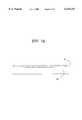
- FIG. 1is a schematic illustration of affinity-based platforms of the invention.
- FIG. 1Ais comprised of a substrate 10 coated with a specific binding reagent comprising a first member of an affinity binding pair 11.
- FIG. 1Billustrates a substrate 10 coated with a reflective material 12, which is coated with a specific binding reagent comprising a first member of an affinity binding pair 11.
- FIG. 1Cis a substrate 10, where the surface of the substrate is partially coated with a specific binding reagent comprising a first member of an affinity binding pair 11, and with a patterned reflective material 12 which is in turn derivatized with a blocking agent 13.
- FIG. 1Dis a substrate 14, into which has been formed pits carrying encoded information.
- the lower surface of 14is coated with a reflective coating 15, and a protective layer 16.
- the upper surface of 14is coated with a specific binding reagent comprising a first member of an affinity binding pair 11.
- FIG. 2is an exemplar arrangement of platform components useful for enumerating particulates in a fluid, for example cell counting, and comprises a sample input port 21 connected to an overflow chamber 23 via a fluid capillary 22.
- the sample inlet port 21is fluidly connected to the binding chamber 24, which is in turn connected to a waste chamber 25.
- Air displacement channels 26facilitate filling of chambers.
- Wash buffer chambers 27 and 29, and a dye chamber 28are fluidically connected to the binding chamber 24 via fluid capillaries 22.
- FIG. 3contains illustrations of arrangements of platform components useful for enumerating particulates in a fluid, for example cell counting.
- FIG. 3Acomprises a wash buffer chamber 31, linked by a capillary 32 containing a valve 33 to a binding chamber 34.
- the binding chamberis linked to a waste chamber 35.
- FIG. 3Bincorporates a dye chamber 36 fluidically connected via a capillary 32 and valve 33 to the binding chamber 34.
- FIG. 3Cprovides an alternate arrangement of the components of FIG. 3B.
- FIG. 3Dadds a sample inlet port 37 and a sample overflow chamber 38 to the arrangement of components pictured in FIG. 3A.
- FIG. 3Eadds a sample inlet port 37 and a sample overflow chamber 38 to the arrangement of components pictured in FIG. 3B.
- FIG. 3Fadds a sample inlet port 37 and a sample overflow chamber 38 to the arrangement of components pictured in FIG. 3C.
- FIG. 3Gis similar to FIG. 3C but incorporates a binding chamber 39 containing a number of first members of the binding pairs.
- FIG. 3His similar to 3F except that the binding chamber 34 has been replaced by a number of chambers in which distinct specific binding reagents comprising first members of an affinity binding pair have been deposited.
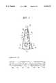
- FIG. 4contains illustrations of arrangements of platform components useful for studying the effect of a test molecule or molecules on populations of cells for enumerating particulates in a fluid, for example cell counting.
- FIG. 4Acomprises a test molecule chamber 40, linked by a capillary 32 containing a valve 33 to a cell accumulation chamber 43.
- the binding chamberis linked to a waste chamber 35.
- FIG. 4Bincorporates a dye chamber 36 fluidically connected via a capillary 32 and valve 33 to the binding chamber 34.
- FIG. 4Cprovides an alternate arrangement of the components of FIG. 4B.
- FIG. 4Dis similar to FIG. 4C, but with the incorporation of a wash buffer chamber 31 in the fluidic path between the test molecule chamber 40 and the cell accumulation chamber 43.
- FIGS. 4E and 4Fare similar to FIG. 4A, with the exception of a multiplicity of test molecule chambers 40 arrayed serially (FIG. 4E) or in parallel (FIG. 4F).
- FIG. 4Gis an arrangement in which fluid from a test molecule chamber 40 and a dilution buffer reservoir can be directed to receiving chambers 42 to provide serial dilutions of the test molecule solution.
- FIG. 4His another advantageous arrangement of the components of FIG. 4G.
- FIG. 5contains schematic illustrations of optical detection systems suitable for detecting the presence and/or optical properties of particulates bound to the platforms of the invention.
- the apparatus of FIG. 5Asuitable for transmission, light scattering or direct fluorescence measurement of a platform such as illustrated in FIG. 1A, comprises a light source 54, focusing lens system 53, assembly 51 comprising optical elements to collect, filter and focus light onto the photodetector 50.
- FIG. 5Bincorporates the detection elements of FIG. 5A, and would be suitable for chemiluminescence or bioluminescence measurements.
- FIG. 5Cis a rearrangement of the components of FIG. 5A for use where the light is reflected from platforms of the type shown in FIG. 1B.
- FIG. 5Dis an apparatus suitable for fluorescence detection on platforms 1A and 1C, where the assembly of optical elements 55 includes elements such as excitation and emission filters, a dichroic mirror and lenses.
- FIG. 5Eis an apparatus suited for use with platforms 1D.
- the elements 56comprise those necessary to read data from an optical disc such as a CD-ROM.
- FIG. 6illustrates means and methods for counting and studying individual cells using platforms of the type FIG. 1C.
- FIG. 6Aillustrates an optical apparatus or "head” derived from optical memory storage and retrieval devices for interrogating a platform 60.
- Optical componentsinclude diode laser 65, diffraction grating 64, beam splitter 63, collimating lenses 62, focusing lens and actuator 61, lens 66, side lobe detectors 67, and central detector 68. Focusing and tracking are achieved through servo control circuits 69, and actuators 61 and 71.
- the signal derived from interaction of the central beam with the cell 74e.g. fluorescence emission
- circuitry 70e.g. fluorescence emission
- FIGS. 6B and 6Cillustrate advantageous arrangements of reflective tracking features on the platform.
- track widths 72are modulated such that reflections from the two side lobes 73 produce a modulated signal.
- Amplitude or frequency modulationachieved by varying the geometry of the tracks 75, can be used to encode positional information.
- FIG. 6Cillustrates another arrangement where information is encoded by the presence or absence of reflective features in the central track.
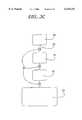
- FIG. 7illustrates an apparatus for utilizing the platforms of the invention.
- Optical detection systems 81such as those illustrated in FIG. 5, are linked to controlling circuitry 82.
- Output signalsare measured by the microcontroller 83, which processes the data and communicates results through a user interface 84 and/or stores the data in local or nonlocal memory 85 through remote data links.
- Rotatable platformsrequire the use of a motor 87 and motor control circuitry 86, and may also include optical data retrieval and storage means 88.
- samplewill be understood to encompass any fluid containing a particulate species of interest, wherein the particulate species is preferably a cell and more preferably microbial cells or somatic cells and most preferably bacterial cells and hematopoietic cells.
- the term "platform"is intended to encompass any solid support structure providing a surface or comprising a chamber that can be treated to comprise a specific binding reagent.
- the platforms of the inventionare rotatable about a central axis, providing for movement of sample and other reagents on the platform under the influence of centripetal force.
- More preferred embodiments of the platforms of the inventionare circular disks that are rotatable about a central aperture adaptably shaped to accommodate a spindle or other rotating means.
- Most preferred embodiments of the platforms of the inventionare platforms as disclosed in U.S. patent application U.S. Ser. No. 08/768,990, filed Dec. 18, 1996 and incorporated by reference, and in U.S. Provisional Application, Serial No. 60/034,327, filed Dec. 20, 1996, the disclosures of each of which are explicitly incorporated by reference herein.
- a surface or detection chambertreated to comprise a specific binding reagent.
- specific binding reagentis intended to encompass biomolecules having a specific binding affinity between pairs thereof providing a specific molecular binding interaction with a binding affinity constant of between about 10 -4 and 10 -15 M.
- pairs of specific binding reagentsinclude but are not limited to antigen and antibody, including antisera, polyclonal antibodies and most preferably monoclonal antibodies; receptor and ligands, including cell-surface receptors; integrins and adhesion proteins, including ICAM-I and ICAM-II; and carbohydrates and lectins, including phytohemagglutinin.
- specific binding reagentscomprising a first member of a specific binding pair is provided coating a surface or detection chamber of a platform designed or intended to detect the presence of a particulate, most preferably a cell expressing a cognate antigen, receptor or adhesion protein or having a carbohydrate moiety at the cell surface specific for a particular lectin.
- Said specific binding reagentis applied to the surface or detection chamber of the platform by depositing the reagent on the surface using any appropriate means, including inkjet printing, computer-positioned syringes, microetching and microlithographic methods, including photolithography, screen and airbrush printing methods, solution coating, dipping, and conventional microtitre-well techniques.
- the surface or detection chambercan be treated to provide a two-dimensional array or pattern, wherein certain areas on the surface or detection chamber are treated with said specific binding reagent and others are not in a recognizable manner.
- a surface or detection chamber of the inventionis provided having transparent portions coated with a specific binding reagent, and other portions coated with a reflective material to provide a reflecting surface in a pattern alternating with the transparent, coated portions.
- a multiplicity of specific binding reagents of distinct specificityare applied to a surface or detection chamber of the platform, or each of a multiplicity of specific binding reagents of distinct specificity are applied to different areas or regions of a surface or detection chamber of a platform of the invention, thereby providing a pattern of such distinct specific binding reagents on the platform.
- Such arrayscan be discrete arrays each comprising a different specific binding reagent or can be integrated to comprise a pattern of each of the multiplicity of distinct specific binding reagents. Exemplary patterns include alternating strips, checks, and concentric circles. Similarly, patterns of transparent, specific binding reagent-coated portions and reflective, non-binding portions arc provided by the invention.
- Exemplary patternsinclude alternating strips, checks, and concentric circles, most preferably comprising a pattern resembling or comprising a "bar code.”
- a blocking agentsuch as bovine serum albumin (BSA) to prevent non-specific binding of particulate matter on the surface of the platform.
- Preferred patterns of reflective and specific binding portions of the surfaces of the platforms of the inventionare provided analogous to and as a specific modification of technology developed in the prior art for optical information storage (e.g. CD-ROM applications).
- This technologyis based on detecting the presence or absence of signal features having dimensions on the order of 1 micron embedded in a plastic substrate.
- the detectors or "optical heads”incorporate servomechanisms designed to automatically position a focal point of light in both a direction normal to the disk and laterally in the plane of the disk to submicron accuracy.
- the focusing, tracking and data acquisition functions of the optical headare implemented using a diode laser, optical elements (diffraction gratings, lenses (including cylindrical lenses), mirrors, polarizers) and multiple detectors.
- the light emitted from the laseris split into multiple beams (typically 2 or 3) and directed on to the disk.
- the light reflected from each of the beamsis coupled back into the optical head where it is directed to a number of photodiodes (typically 6).
- the outputs from the photodiodesare combined in various ways (either added or subtracted) to obtain both the data and signals for controlling the tracking and focusing. Tracking, or following the data stream, is accomplished by moving the optical head in a radial direction, while simultaneously controlling the rate of rotation of the disk.
- the radial positioningis derived from the light reflected from the regions of the disk on either side of the data pits.
- the platform of FIG. 1Dis advantageously provided as an optical disk wherein digital information has been encoded in an standard format; however, in the platforms of the invention, the thickness of the substrate is thinned sufficiently so that the presence of particles on the surface will interfere with the reading of the encoded data using the optical detection system pictured in FIG. 5E.
- a second variationuses the optically transparent substrate is provided having parallel tracks defined by alternating transparent and reflective regions or surfaces, as is pictured in FIGS. 1C, 6B and 6C. This is done, for example, using established microlithographic methods to selectively etch vacuum-deposited gold from a glass or quartz substrate.
- a pair of reflective stripes and intervening transparent regionare provided to form a "track".
- the transparent regioncontains “data” in the form of the presence or absence of cells; this "data” is provided in the practice of the methods of the invention upon specific particulate binding to the transparent region, while the reflective regions provides a means for accomplishing the tracking and focusing.
- To confine the cells to the transparent regionit is advantageous to place the cells on the side of the substrate on which the reflective material has been deposited.
- the presence of two chemically different surfacespermits selective chemical modification of the transparent and reflective surfaces that promotes (i.e. in the transparent region) or prevents (i.e. in the reflective region) adhesion of the cells to be detected.
- An optical head similar, in principle, to that used for optical information storageis used as provided by the methods of the invention to interrogate the plane to which the cells are adhered through the transparent substrate.
- the optical headuses a light source (e.g. a diode laser operating at 650 nm) and optical elements to focus multiple beams of light on or in the plane of the platform adherent to cells or particles.
- the secondary or sub-beamsare used for tracking and focusing, while the primary or main beam illuminates the transparent region comprising the particles or cells.
- the presence or absence of a cell in the trackis detected by a number of means, for example by modulating the transmission of light through the disk to an opposing detector, or by fluorescent emission.
- the light emitted lightmay be detected by a detector (typically a photomultiplier tube (PMT)) and appropriate filter positioned either directly opposite the light source, at an oblique angle to the platform and/or the light source, or as part of an optical head which has been designed to collect light emitted back from disk (i.e. at an angle of about 0 degrees).
- a detectortypically a photomultiplier tube (PMT)
- PMTphotomultiplier tube
- the platforms of the inventionadvantageously comprise additional fluid-handling components attached to the surface or detection chamber or cell accumulation chamber on the platforms.
- These componentscan be fabricated as described below either integral to the disk or as modules attached to, placed upon, in contact with or embedded in the disk.
- Such componentsare preferably provided in combinations of related components as described in further detail herein that are in fluid communication.
- the term "in fluid communication" or “fluidly connected”is intended to define components that are operably interconnected to allow fluid flow between components.
- the platformcomprises a rotatable platform, more preferably a disk, whereby fluid movement on the disk is motivated by centripetal force upon rotation of the disk.
- the platforms of the inventionfurther comprise a sample entry port, preferably comprising metering elements to deliver a volumetric amount of sample fluid to the detection or cell accumulation chamber of the platform.
- the platforms of the inventionare also provided with an overflow reservoir for retaining excess fluid applied to the platform in excess of the amount metered into the detection or cell accumulation chamber, most preferably in fluid communication with the fluid sample input means wherein excess fluid is transferred to the overflow reservoir by capillary action.
- the metering sample portis designed to rapidly wick in fluid presented to the opening.
- the overflow chamberis connected to the entry port to take off any excess fluid not wicked into the capillary bed. The volume of the sample is thereby defined by the number and cross section of the capillaries.
- Additional chambers on the platformcontain fluids such as a wash buffer and staining solution.
- the fluid componentsare in fluid communication via narrow bore capillaries of defined cross section, which form capillary valves. Fluid in the chambers and components of the platforms of the invention will be retained until sufficient driving force overcomes the surface tension of the fluid. Differing cross sections allow fluids to be moved independently by controlling the force applied (e.g. by controlling rotation rate).
- the inventionalso comprises a device for manipulating the disks of the invention, wherein the disk is rotated within the device to provide centripetal force to effect fluid flow on the disk.
- the deviceprovides means for rotating the disk at a controlled rotational velocity, for stopping and starting disk rotation, and advantageously for changing the direction of rotation of the disk.
- electromechanical means and control meansare provided as components of the devices of the invention.
- User interface means(such as a keypad and a display) are also provided.
- fluid (including reagents, samples and other liquid components) movementis controlled by centripetal acceleration due to rotation of the platform, and by the selective activation of valves controlling the communications between the components of the systems comprising the platform.
- centripetal accelerationdue to rotation of the platform
- valvescontrolling the communications between the components of the systems comprising the platform.
- the magnitude of centripetal acceleration required for fluid to flow at a rate and under a pressure appropriate for a particular systemis determined by factors including but not limited to the effective radius of the platform, the position angle of the structures on the platform with respect to the direction of rotation and the speed of rotation of the platform.
- fluid flowis provided on the platforms of the invention by mechanical means, including but not limited to pumping using the creation of air or liquid pressure between the components of the platform to effect fluid movement, using pumping means sufficient to achieve fluid movement.
- mechanical meansincluding but not limited to pumping using the creation of air or liquid pressure between the components of the platform to effect fluid movement, using pumping means sufficient to achieve fluid movement.
- These meansmight include syringe pumps or HPLC pumps.
- fluid movementis motivated by rapid manual displacement of the platform followed by a sharp stop of such displacement; this might be actuated by a spring mechanism or simply a "flick of the wrist".
- Advantageous components of the platforms of the inventioninclude fluid sample input means, including volumetric metering means, channels for fluid flow between components, reagent reservoirs, mixing chambers, optical reading chambers, and most preferably incubation surfaces or detection chambers comprising a specific binding reagent deposited thereupon, and cell accumulation chambers comprising non-specific cell adhesion compounds, filtering means that retain cells in the chamber or treated surfaces that permit the cells to attach thereto.
- fluid sample input meansincluding volumetric metering means, channels for fluid flow between components, reagent reservoirs, mixing chambers, optical reading chambers, and most preferably incubation surfaces or detection chambers comprising a specific binding reagent deposited thereupon, and cell accumulation chambers comprising non-specific cell adhesion compounds, filtering means that retain cells in the chamber or treated surfaces that permit the cells to attach thereto.
- Sampleis applied to the detection or cell accumulation chamber of the platforms of the invention either directly or more preferably by transfer of a metered amount of a portion of the sample from a fluid sample input means to the chamber,for example, by the selective opening of valves controlling access to the chamber from the fluid sample input means.
- Said valvesinclude but are not limited to microvalves as described in more detail below including mechanical, electrical and thermal valve mechanisms, as well as capillary microvalves wherein fluid flow is controlled by the relationship between capillary forces and centripetal forces acting on the fluid.
- Reagent reservoirs, wash buffer reservoirs, other fluidic components and the contents thereofarc connected to one another and to the detection and cell accumulation chamber through channels, preferably microchannels as defined herein, controlled by such valves.
- delivery of fluids through such channelsis achieved by the coincident rotation of the platform for a time and at a rotational velocity sufficient to motivate fluid movement between the desired components, and opening of the appropriate valves.
- the amount of a fluid, or a reagent comprising a fluid, delivered to the detection or cell accumulation chamberis thus controlled by the speed of rotation and the time during which the valve to the reagent reservoirs is open.
- the apparatus of the inventionalso provides detection systems for detecting, monitoring, quantitating or analyzing particulates specifically retained on the surface of the platform, in a detection chamber comprising a specific binding reagent or in a cell accumulation chamber as described herein.
- Detection systems useful in the manufacture and use of the platforms of the inventioninclude, but are not limited to, fluorescent, chemiluminescent, colorimetric, or scattering measurements.
- FIG. 5illustrates optical systems for effecting these measurements.
- the apparatus of FIG. 5Asuitable for transmission, light scattering or direct fluorescence measurement of a platform such as illustrated in FIG. 1A, comprises a light source 54, focusing lens system 53, assembly 51 comprising optical elements to collect, filter and focus light onto the photodetector 50.
- FIG. 5Bincorporates the detection elements of FIG. 5A, and would be suitable for chemiluminescence or bioluminescence measurements.
- FIG. 5Cis a rearrangement of the components of FIG. 5A for use where the light is reflected from platforms of the type shown in FIG. 1B.
- FIG. 5Dis an apparatus suitable for fluorescence detection on platforms shown in FIGS. 1A and 1C, where the assembly of optical elements 55 includes elements such as excitation and emission filters, a dichroic mirror and lenses.
- FIG. 5Eis an apparatus suited for use with platforms shown in FIGS. 1D.
- the elements 56comprise those necessary to read data from an optical disc such as a CD-ROM. electrochemical and radioactivity detecting means.
- the detection systemcan be integral to the platform and can comprise a simple visual detection means such as the development of a visible color.
- the detection systemcan comprise a component of a device manipulating the platform, preferably comprising an optical detecting means.
- devicescomprising a light source for illuminating the platform and a magnifying means to facilitate visual inspection (direct or computer-aided imaging) of the platform.
- Non-optical detection systemssuch as electrochemical and radioactivity detecting means may also be used.
- Embodiments wherein components of the detecting meanscomprise both the platform and the device are also encompassed by the invention.
- cellsmay be mixed with a buffer containing antibody-linked labels such as gold nanoparticles (-100 nm in size or smaller than a cell) or enzyme-linked antibodies.
- the cellsare then flowed through the porous structure, binding via a different binding molecule.
- gold sol particlesif a sufficient volume is passed through the filter such that the total number of cells captured is large, a diffuse reflectance measurement can accurately quantitate the amount of gold trapped in the filter.
- an appropriate substratemay be flowed into the porous structure and the evolved dye spectrophotometrically measured.
- a second application of such a systemwould be as a cell concentrator.
- Samplemay be flowed into the porous structure and appropriate particles bind to the material.
- a very large volume of samplemay flow thorough the structure, such that eventually the number of trapped particles is many orders of magnitude orders of magnitude greater in concentration that in the fluid.
- direct assaysmay be performed on the particles trapped in the structure.
- Another possibilityis to flow a buffer through the structure which dissociates the affinity bond between particle and structure. The highly-concentrated particles may then be transported to another reservoir for further processing.
- the detection or cell accumulation chamber of the platform of the inventionis constructed so that the height (depth) of the chamber is smaller than the other dimensions of the chamber.
- the height (depth) of the chamberranges from about 25 ⁇ m to 1 mm.
- the chamberhas a volume of from about 5 ⁇ L to about 1000 ⁇ L, more preferably from about 50 ⁇ L to about 500 ⁇ L.
- Platforms of the inventionsuch as disks and the components comprising such platforms are advantageously provided having a variety of composition and surface coatings appropriate for a particular application.
- Platform compositionwill be a function of structural requirements, manufacturing processes, and reagent compatibility/chemical resistance properties.
- platformsare provided that are made from inorganic crystalline or amorphous materials, e.g. silicon, silica, quartz, inert metals, or from organic materials such as plastics, for example, poly(methyl methacrylate) (PMMA), acetonitrile-butadiene-styrene (ABS), polycarbonate, polyethylene, polystyrene, polyolefins, polypropylene and metallocene.
- PMMApoly(methyl methacrylate)
- ABSacetonitrile-butadiene-styrene
- PCCpolycarbonate
- polyethylenepolystyrene
- polyolefinspolypropylene and metallocene
- platformsmade of composites or combinations of these materials, for example, platforms manufactures of a plastic material having embedded therein an optically transparent glass surface comprising for example the detection chamber of the platform.
- the surface properties of these materialsmay be modified for specific applications. For example, appropriate surface-modification can either encourage or suppress cell and/or protein absorption. Surface modification can be achieved by silanization, ion implantation and chemical treatment with inert-gas plasmas (i.e., gases through which electrical currents are passed to create ionization). In preferred embodiments, a particular portion of the surface of the platform, most preferably comprising a chamber in the platform, is treated with a specific binding reagent.
- the specific binding reagentis a protein (e.g., an antibody, a receptor or adhesion protein), an antigen or a receptor ligand (e.g., a small molecule such as a peptide), or a lectin (such as phytohemagglutinin) or a carbohydrate that is recognized by a lectin.
- the surfaceis treated with such specific binding reagents to form an insoluble and difficult-to-dissociate bond between the reagent and the surface, to minimize loss of the reagent during subsequent treatment steps (such as washing).
- the surfaceis treated with the reagent to saturate the surface with the reagent over a defined portion of the surface.
- the treatment of the surface with a specific binding reagentconstitutes a pattern of deposition on the surface than can be recognized, and most preferably, digitized to provide a two-dimensional map of the surface for orienting a detecting means.
- Surfaces treated with a multiplicity of specific binding reagents, most preferably in a recognizable pattern of deposition,arc within the scope of the invention disclosed herein.
- surfacescomprising detection and cell accumulation detectors that are porous, i.e. comprising a three-dimensional surface in which specific binding reagents or particulates can be bound.
- the specific binding reagentis deposited on the surface in optically transparent portions thereof in combination with deposition in alternative and adjacent regions of the surface with a reflective material treated to prevent particulate binding thereupon.
- Reflective material of the appropriate feature sizeis most advantageously prepared using methods and means developed for the manufacture of microelectronic circuits.
- lift-offa negative image of the desired features is produced in a photoresist material using microlithography.
- a reflective layercomprising one or more metal layers is deposited by evaporation, after which the photoresist and overlying metal is removed (typically by dissolution of the photoresist layer) leaving the desired patterns on the surface of the substrate.
- the portion of the surface comprising the specific binding reagentis also reflective but is prepared so that this surface can be distinguished from the adjacent reflective surfaces.
- the differential patterns of optical transmission and reflectionare useful for orienting a light source of a detecting means, particularly a monochromatic and most preferably a coherent or laser light source, over the appropriate portion of the surface of the platform for detecting the particulates retained thereupon.
- the surfacecomprises a pattern of reflective coatings that are used to orient, digitize and quantitate the particulates, most preferably cells, contained within a defined area of the platform comprising the detection or cell accumulation chamber.
- the surfacecan be treated with omega-substituted alkanethiol compounds of general formula HS--(CH 2 ) n -R (where n is an integer from 1 to about 50, and R is an alkyl, alkenyl, alkynyl, aryl or alkaryl group, or substituted derivatives thereof), to form self-assembled monolayers (SAM).
- the surface of the platformis also advantageously treated with a non-specific blocking agent or agents to prevent non-specific binding of particulates, particularly cells, to the surface of the platform.
- a non-specific blocking agent or agentsto prevent non-specific binding of particulates, particularly cells, to the surface of the platform.
- the nature and extent to which such treatments are necessarydepends strongly on the nature of the surface. For example, a strong correlation has been established between water contact angle and cell adsorption, with hydrophilic surfaces showing significantly less cell adsorption than hydrophobic surfaces (see Ikada, 1994, Biomaterials 15: 725). Silicon, silica, and quartz present an inherently high-energy, hydrophilic surface. Alteration of surface properties is attained through hydroxylation (achieved, for example, by NaOH treatment at high temperatures) or silanization.
- Substituted silanes and siloxanesare particularly appropriate for increasing the hydrophilicity of an otherwise hydrophobic surface.
- These compoundsconsist of one or several reactive head-groups which bond (chemically or through hydrogen-bonding) to a substrate, for example, a core region of alkane (--CH 2 O--).
- a substratefor example, a core region of alkane (--CH 2 O--).
- These compoundsalso provide a route for more sophisticated alteration of surface properties (such as derivation with functional groups to obtain the surface properties of interest).
- a wide variety of such functionalitiescan be introduced at a surface, including vinyl, phenyl, methylene and methoxy groups, as well as surfaces providing mixed functionalities.
- these functional groupsnot only change gross properties like liquid contact angle, but provide sites for preferential adsorption of molecules, either per se or as a result of further conjugation of specific binding reagents such as peptide ligands, antibodies and the like. More preferably, the surface is treated after deposition of the specific binding reagents with a non-specific blocking agent, including but not limited to bovine serum albumin and casein.
- a non-specific blocking agentincluding but not limited to bovine serum albumin and casein.
- Plastic-based platforms and diskscan also be readily treated to achieve the required surface properties.
- Inert-gas or reactive-gas plasmasare used to alter surface energies through the formation of surface complexes, for example, hydroxyl-rich surfaces for increased hydrophilicity, or perfluorinated surfaces for increased hydrophobicity.
- surface graft polymerizationis a technique used to graft polymers or oligomers with the desired surface properties to a substrate polymer chosen for its bulk processability and manufacturing properties, such as a plastic.
- the platforms of the inventionare preferably provided with a multiplicity of components, either fabricated directly onto the platform, or placed on the platform as prefabricated modules.
- certain devices and elementscan be located external to the platform, optimally positioned on a device of the invention in relation to the platform, or placed in contact with the platform either while rotating or when at rest.
- Components optimally comprising the platforms of the invention or a controlling device in combination therewithinclude detection chambers, reservoirs, valving mechanisms, detectors, sensors, temperature control elements, filters, mixing elements, and control systems.
- the inventionprovides a detection chamber or cell accumulation chamber, or a surface or specialized section of the platform, upon which specific components of a fluid sample, preferably cells and most preferably microbial cells, especially bacterial cells, and mammalian cells, especially hematopoietic cells, can be retained.
- cellsare retained in the chamber by interaction with specific binding reagents, including but not limited to ligands, lectins, peptides, proteins, antibodies or fragments thereof derivatized to be retained within the surface of the platform.
- cellsare retained in an accumulation chamber by non-specific binding to adhesion molecules, or physical retention using filtering or other means as described below, or by allowing the cells to attach to a substrate that has been specifically surface-modified to facilitate or promote such attachment.
- Particulates captured by such specific bindingcan be eluted from the surface of the platform and transferred to a collection reservoir by treatment with appropriately-chosen ionic strength buffers, using conventional methods developed for immunological or chromatographic techniques. More preferably, particulates, particularly cells and most particularly microbial cells, can be specifically retained in the detection or cell accumulation chamber, surface or specialized section of the platform and detected using detecting means as described herein.
- cellular particulates retained in the detection or cell accumulation chamber, or on any surface or specialized section of the platformcan be counted, localized on the surface, or characterized using the detecting means of the apparatus of the invention.
- cells retained in detection or cell accumulation chambersare monitored for viability, metabolism, the effect of viral infection or drugs on viability or metabolism, or used for toxicity screening assays.
- cellsare retained in a detection or cell accumulation chamber on a platform of the invention and incubated in the presence of a test compound for a time and under conditions whereby the test compound may have an effect on cell metabolism, physiology or viability.
- the cellsare then detected, and most preferably quantitatively detected, to determine the effect of the test compound on the cell.
- cytotoxicity testingcan be performed on a cell, most preferably a mammalian cell, by incubating the test compound with cells retained in the cell accumulation chamber on the platform, followed by a determination of cell viability using visual, microscopic or spectrophotometric detection of the exclusion from the treated cells of a detectably colored "vital" stain, used according to the understanding in the art.
- Such determinationscan be performed in the presence or absence on the test compound, most preferably in multiplex fashion on the same platform, and a comparison made between the observed cell viability in the presence and absence of the test compound.
- arrays of specific binding reagentsincluding but not limited to ligands, lectins, peptides, proteins, antibodies or fragments thereof can be deposited in a detection chamber or on a surface or specialized section of a platform, preferably forming a patterned array that can be detected visually or using the detecting means disclosed herein. Also provided are such arrays that are digitized based on the signal per unit area detected from the array, so that the position of a signal with a detection chamber, or on a surface or specialized section of the platform, indicating a particulate can be identified and specifically tracked during metabolic, viability, toxicity or other assays. The capacity to collect time-integrated information in such assays is also advantageously provided.
- the platformmay contain reservoirs and chambers for containing fluids, such as the washing buffer used to detach nonspecifically bound particulates from the surface of the detection chamber, or a solution of a compound to which cells in the cell accumulation chamber are going to be exposed.
- the chambersmay be prefilled with liquid components and sealed using valving mechanisms, may be filled with dried reagents which are resolubilized by the addition of a fluid such as water, or may be filled at the time of use with prepared liquid reagents.
- the platforms of the inventionalso are provided comprising reservoirs and chambers for containing fluids, such as the washing buffer used to detach nonspecifically bound particulates from the surface of the detection chamber, or a solution of a compound to which cells in the cell accumulation chamber are to be exposed.
- the chambersmay be prefilled with liquid components and sealed using valving mechanisms, may be filled with dried reagents which are resolubilized by the addition of a fluid such as water, or may be filled at the time of use with prepared liquid reagents.
- the platforms of the inventionalso are provided comprising fluid waste receptacles and overflow receptacles and reservoirs for holding excess fluid comprising unmetered sample, wash buffers and sample fluid after displaced from the detection and cell accumulation chambers of the platforms of the invention, or any other source of excess fluid produced in the practice and use of the platforms of the invention. It will be recognized that in some embodiments, such excess fluid will be simply removed from the platforms either by user intervention, or by the placement of an outlet in the platform that permits excess fluid to drain therefrom.
- microchannelscomprising the surface of the platforms of the invention.
- Microchannel sizesare optimally determined by specific applications and by the amount of delivery rates required for each particular embodiment of the platforms and methods of the invention.
- Microchannel sizescan range from 0.1 ⁇ m to a value close to the 1 mm thickness of the platform.
- Microchannel shapescan be trapezoid, circular or other geometric shapes as required.
- Microchannelspreferably are embedded in a platform having a thickness of about 0.1 to 100 mm, wherein the cross-sectional dimension of the microchannels across the thickness dimension of the platform is less than 500 ⁇ m and from 1 to 90 percent of said cross-sectional dimension of the platform.
- Valving mechanismsare provided to control of fluid movement and transfer on the platform.
- the nature of the valves useful in the platforms of the inventionare essentially identical to the valves and microvalves disclosed in co-owned and co-pending U.S. Ser. No. 08/768,990, filed Dec. 18, 1996, explicitly incorporated by reference herein. These valve include mechanical, thermal and capillary valves.
- microvalvesexamples include a piezo activator comprising a glass plate sandwiched between two silicon wafers, as described by Nakagawa et al. (1990, Proc. IEEE Workshop of Micro Electro Mechanical Systems, Napa Valley, Calif. pp. 89); a pneumatically-actuated microvalve, as described by Veider et al. (1995, Eurosensors IX, pp. 284-286, Swiss, Sweden, June 25-29); a micromachined gas valve (that is commercially available; Redwood Microsystems, Menlo Park, Calif.; ICSensors, Milpitas, Calif.); a pressure-balanced microvalve, as disclosed by Huff et al.
- This capillary pressurevaries inversely with the sizes of the two components, or combinations thereof, the surface tension of the fluid, and the contact angle of the fluid on the material of the components.
- the details of the cross-sectional shapeare not important, but the dependence on cross-sectional dimension results in microchannels of dimension less than 500 ⁇ m exhibit significant capillary pressure.
- "valves"are fashioned that require the application of a particular pressure on the fluid to induce fluid flow. This pressure is applied in the disks of the invention by rotation of the disk (which has been shown above to vary with the square of the rotational frequency, with the radial position and with the extent of the fluid in the radial direction).
- capillary valvesare formed to release fluid flow in a rotation-dependent manner, using rotation rates of from 100 rpm to several thousand rpm. This arrangement allows complex, multistep fluid processes to be carried out using a pre-determined, monotonic increase in rotational rate.
- Control of the valves of the platforms provided by the inventionis achieved either using on-platform controller elements, device-specific controllers, or a combination thereof.
- Optical detecting meansare also provided as components of the apparatus of the invention.
- the photodetectors of the inventionare optimally provided to detect optical absorbance/transmittance, fluorescence, light-scattering or other optical signals, which are processed and translated into data on the position, number and viability of cells on the platform.
- Embodiments of such platforms and devices comprising scanning arraysare also provided.
- Also advantageously provided by the inventionare detecting means for tracking and focusing the light source on the surface of the platform, and actuating means for positioning the light source on the surface of the platform.
- Detection systems for use on the platforms of the inventioninclude spectroscopic, particularly monochromatic and stroboscopic, and electrochemical detectors (see, for example, Owicki et al., 1992, Biosensors & Biolelectronics 7: 255). Spectroscopic methods using these detectors encompass spectroscopy, particularly ultraviolet and visible light absorbance, chemiluminescence, and fluorescence spectroscopy.
- the detection systems utilizing the detectors of the inventionare preferably be external and adjacent to the platform.
- the detection systems of the inventioncomprise a light source and a photodetector; in certain embodiments (such as chemiluminescence), only the photodetector components are required.
- the orientation of these components of the detecting means of the apparatus of the inventionwill be understood to depend on the nature of the detection or cell accumulation chamber and the construction thereof.
- platforms wherein the detection or cell accumulation chamber comprises an optically transparent surfacewill advantageously be used with a device having the light source positioned on one side of the platform and the photodetector positioned on the other side of the platform.
- the photodetectoris arranged directly across from the light source (i.e. at an angle of about 180 degrees, as in FIG. 5A) or more advantageously obliquely across the platform from the light source (i.e. at an angle of between 90 and 180 degrees).
- the apparatusmay also advantageously include a mirror or other means for deflecting the transmitted light to the photodetecting means; however, it will be recognized that such mirrors are not required in embodiments provided for fluorescence detection.
- the photodetectoris advantageously positioned on the same side of the platform as the light source (FIG. 5C).
- the photodetectoris provided as an integrated component of an assembly comprising the light source (FIGS. 5D and 5E), wherein the reflected light is detected along the same axis as the incident light (i.e. at about 0 degrees).
- the photodetectors of the inventionare optimally provided to detect optical absorbance/transmittance, fluorescence, light-scattering or other optical signals, which are processed and translated into data on the position, number and viability of cells on the platform.
- Embodiments of such platforms and devices comprising scanning arraysarc also provided.
- Also advantageously provided by the inventionare detecting means for tracking and focusing the light source on the surface of the platform, and actuating means for positioning the light source on the surface of the platform.
- the apparatus 56 in FIG. 5Erepresents an optical "head" of a CD (see E. W. Williams, 1994, The CD-ROM and Optical Disc Recording Systems, Oxford University Press, New York) and is used with the platform of FIG. 1D, the substrate of which contains data encoded in an industry standard format, in the form of pits stamped into the plastic matrix and coated with a reflective coating (e.g., aluminum).
- the platformis constructed so that the aluminized pits are the focal point of the CD laser.
- Platform fluidics handling componentsare built within and upon this plastic layer. Particulates bound through the first member of the binding pair 11 to the surface of the thinner substrate 14 scatter light and interfere with the reading of the data.
- the thickness and index of refraction of optical storage mediaare such that the effect of light scattering by particles on the surface of the disk is reduced.
- the thickness and composition of the substrate, fluid layer and coverare chosen such that particulates bound to the surface interfere with the integrity of the data, and the errors generated in reading the data provide a measure of the number of particulates bound.
- the optical detection apparatus pictured in FIG. 5D and in more detail in FIG. 6Ais to be used with the platform of FIG. 1C which contains reflective features such as those pictured in FIGS. 6B and 6C.
- Optical elements similar to those used in a conventional CD headare used to generate and focus a main beam and side beams on the surface of the platform.
- the reflection of the side beams off of the reflective featuresare used to track the central beam along the transparent regions of the platform, so that particulates bound there may be detected.
- optical data retrieval systemsuse the reflection of the central beam in a servo-controlled focusing system. This approach differs in that control of the focusing actuator is based on intensities of the reflections from the side beams off of the reflective features.
- the interaction of a particle with the central beammay result in fluorescence, absorption, or scattering, which may be detected by the detector 50 within the "head” or by another detector advantageously placed (not shown).
- These embodimentsthus provide cell sorting capability, cell tracking and cell viability information, whereby the status of the cells at each point can be detected and distinguished from each other cell. This capacity enables the platforms and devices of the invention to provide cell-specific data and tracking information.
- Preferred spectroscopic methodsinclude fluorescence, whereby fluorescence detector systems developed for macroscopic uses are adapted for use with the platforms of this invention.
- an excitation sourcesuch as a laser is focused on an optically-transparent section of the disk.
- Light from any analytically-useful portion of the electromagnetic spectrumcan be coupled with a platform material that is specifically transparent to light of a particular wavelength, permitting spectral properties of the light to be determined by the dye or other reagent used to illuminate a particulate retained in an detection chamber or cell incubation chamber or surface or specialized section of the platform interrogated by illumination with light.
- the selection of light at a particular wavelengthcan be paired with a material having geometries and refractive index properties resulting in total internal reflection of the illuminating light. This enables either detection of material on the surface of the disk through evanescent light propagation, or multiple reflections through the sample itself, which increases the path length considerably.
- the outer surface of a waveguide(a fiber optic, a prism, etc.) is coated with the first member of a binding molecule pair. Particles which express the second member of the pair on their surface(s) are then introduced to the fluid which is in contact with the optical waveguide. A sufficient time is allow to pass so that the particles may bind through the binding pair to the outer surface of the waveguide. (Gentle agitation of the fluid may speed up this process.) The waveguide is then washed with buffer. Light is transmitted through the waveguide from a source to a photodetector. Light within the waveguide engages in multiple internal reflection as it passes along the waveguide.
- light of both the fluorescence excitation wavelength and the emitted wavelengthare guided through one face of the device.
- An angle of from about 90° to about 180°is used to separate the excitation and collection optical trains, more preferably an angle of about 135° to position the detector obliquely from both the edge of the platform and the direction of the light from the light source. It is also possible to use other angles, including 0 degrees, whereby the excitation and emitted light travels collinearly. As long as the source light can be distinguished from the fluorescence signal, any optical geometry can be used.
- Optical windows suitable for spectroscopic measurement and transparent to the wavelengths usedare included at appropriate positions (i.e., in detection chamber or cell accumulation chambers or surfaces or other specialized sections of the platform). The use of this type of fluorescence has been disclosed by Haab et al. (1995, Anal. Chem. 67: 3253-3260).
- Configurations appropriate for evanescent wave systemscan provide for fluorescence to be coupled back into a waveguide on the platform, thereby increasing the efficiency of detection.
- the optical component preceding the detectorcan include a dispersive element to permit spectral resolution. Fluorescence excitation can also be increased through multiple reflections from surfaces in the device whenever noise does not scale with path length in the same way as with signal.
- Absorbance measurementscan be used to detect a dye or stain, such as a vital stain, or other analyte that changes the intensity of transmitted light by specifically absorbing energy (direct absorbance) or by changing the absorbance of another component in the system (indirect absorbance).
- Absorbance measurementsare preferably used in conjunction with enzyme-linked detection of the presence of a particulate within a detection chamber of cell accumulation chamber or a surface or specialized section of the platform.
- cellular particulatesare detected by vital staining or other cell-specific staining (such as the use of dyes specific for certain cell types).
- Optical path geometryis designed to ensure that the absorbance detector is focused on a light path receiving the maximum amount of transmitted light from the illuminated sample.
- Both the light source and the detectorare advantageously positioned external and adjacent to the platform; in rotatable embodiments of the platforms of the invention, the light source and the detector can be moved in synchrony with the platform, or more preferably, the light source illuminates the platform stroboscopically so that absorbance/transmittance is sampled in synchrony with rotation of the platform and cyclical positioning of the detection or cell accumulation chamber with the solitary position of the light source/detector pair.
- the detection chamber or cell accumulation chamber, or a surface or other specialized section of the platformcan constitute a cuvette that is illuminated wherein transmitted light detected in a single pass or in multiple passes, particularly when used with a stroboscopic light signal that illuminates the detection chamber at a frequency equal to the frequency of rotation or multiples thereof.
- the detection chambercan be a planar waveguide, wherein the analyte interacts on the face of the waveguide and light absorbance is the result of attenuated total internal reflection (i.e., the analyte reduces the intensity source light if the analyte is sequestered at the surface of the chamber, using, for example, specific binding to a compound embedded or attached to the chamber surface; see Dessy, 1989, Anal.
- absorbance/transmittance detector arraysare advantageously used with platforms of the invention comprising an optically transparent surface (permitting a direct light path through the surface of the platform an positioning of the light source and detector on opposite sides of the platform), alternative embodiments wherein the detector is positioned obliquely to the light source, or other embodiments wherein the surface of the platform comprises a reflective surface and the detector and the light source are positioned on the same side of the platform are also envisioned and encompassed in this description of the invention.
- Both the light source and the detectorare advantageously positioned external and adjacent to the platform; in rotatable embodiments of the platforms of the invention, the light source and the detector can be moved in synchrony with the platform, or more preferably, the detector output is sampled periodically to so that the absorbance/transmittance is measured.
- Indirect absorbancecan be used with the same optical design.
- the analytedoes not absorb the source light; instead, a drop in absorbance of a secondary material is measured as the analyte displaces it in the sample chamber. Increased transmittance therefore corresponds to analyte concentration.
- This detection schemais advantageously used in embodiments of the invention wherein the effect of a test compound on a cell is determined, wherein the detectable analyte is displaced by a metabolite or other molecule produced by the cell in response to the presence of the test compound.
- Light scattering and turbiditycan also be measured on the platform. Optics are configured as described for absorbance measurements. In this analysis, the intensity of the transmitted light is related to the concentration of the light-scattered particles in a sample.
- Monochromatic light from a light sourceadvantageously a laser light source, is directed across the cross-sectional area of a detection chamber or cell accumulation chamber on the platform. Light scattered by particles in a sample, such as cells, is collected at several angles over the illuminated portion of the chamber (see Rosenzweig et al., 1994, Anal. Chem. 66: 1771-1776).
- Data reductionis optimally programmed directly into the device based on standards such as appropriately-sized beads to relate the signal into interpretable results. Using a calibrated set of such beads, fine discrimination between particles of different sizes can be obtained.
- the photodetectoris advantageously positioned on the same side of the platform as the light source.
- the photodetectoris provided as an integrated component of the light source, wherein the reflected light is detected along the same axis as the incident light (i.e. at about 0°).
- the light source supplied with the devices of the inventionwill also be supplied with focusing means and optical elements to focally illuminate the platform.
- the photodetectors supplied with the devices of the inventionwill be supplied with light collecting means, including mirrors and other optical elements to direct the transmitted, reflected or fluorescent light from the detection or cell accumulation chamber to the photodetector.
- Electric potential measurementcan also be used for detecting the presence of a particulate, especially a cell and preferably a microbial cell, on a surface or in a chamber comprising a specific binding reagent.
- microelectrodesare fabricated of a noble metal, preferably gold or platinum, using conventional techniques and are imbued with a biological specificity by impregnating or coating the electrodes with a specific binding reagent. Electropolymerization of organic films, such as polypyrrole or polyalanine, comprising said specific binding reagent, or electrochemical reduction of thiol-terminated specific binding reagents can be used to fashion such specific electrodes.
- Detection of the presence or absence of cellular and other particulatesis achieved by measuring the electrical impedance between one electrode and a solution contact, which can advantageously be positioned in contact with the fluid in the detection chamber comprising the electrodes. Measurements are made using alternating voltages at frequencies from about 10 Hz to 1 MHz. Impedance readings are analyzed by a reading device comprising a microprocessor to determine detection of the presence or absence of cells and other particulates.
- Temperature control elementsare provided to control the temperature of the platform during incubation of a fluid containing a particulate, most advantageously used to facilitate specific binding between the particulate and the specific binding reagent.
- a preferred temperatureis room or ambient temperature, although temperatures above ambient (e.g., 37° C.) and below ambient (e.g., 20° C.) are also preferred.
- Temperature controlis also important for embodiments of the invention used to detect particulates that are cells; in these embodiments, the temperature is advantageously kept below 42° C. and above 4° C. to protect the integrity and viability of the cells.
- the inventiontherefor provides heating elements, including heat lamps, direct laser heaters, Peltier heat pumps, resistive heaters, ultrasonication heaters and microwave excitation heaters, and cooling elements, including Peltier devices and heat sinks, radiative heat fins and other components to facilitate radiative heat loss.
- Thermal devicesare preferably arrayed to control the temperature of the platform over the area of the detection or cell accumulation chamber or surface, although arrangements whereby the platform temperature as a whole is controlled are also preferred.
- heating and cooling elementscomprise the device of the invention, although certain elements, such as radiative heat transfer "fins" and other such components may comprise the platforms of the invention.
- the temperature of any particular area on the platformis monitored by resistive temperature devices (RTD), thermistors, liquid crystal birefringence sensors or by infrared interrogation using IR-specific detectors, and can be regulated by feedback control systems.
- RTDresistive temperature devices
- thermistorsthermoelectric sensors
- liquid crystal birefringence sensorsliquid crystal birefringence sensors
- infrared interrogation using IR-specific detectorscan be regulated by feedback control systems.
- Filters, sieving structures and other means for selectively retaining or facilitating passage of particulate matter, including cells, cell aggregates, protein aggregates, or other particulate matter comprising fluids applied to a platform of the inventionare provided.
- Such filtering meansinclude microsieving structures that are fabricated directly into a fluid handling structure on a platform (e.g., U.S. Pat. No. 5,304,487; International Application, Publication No. WO93/22053; Wilding et al., 1994, Automat. Analyt. Tech. 40: 43-47) or fabricated separately and assembled into the fluid handling structures.
- the sieving structuresare provided with a range of size exclusion orifices and are optionally arranged sequentially so as to fractionate a sample based upon the sizes of the constituent parts of the sample.
- such sieving structuresare provided to permit the introduction of specific particulates into the detection chamber or surface (such as a cell, preferably a microbial cell) while excluding protein aggregates or other non-specific particle-like constituents of a fluid which could interfere with detection of the particular particulates of interest.
- reagentssuch as beads and particularly beads coated with a compound such as an antibody having an affinity for a contaminant or other substance that would interfere with the assay to be performed.
- filtering meanshaving a pore size sufficiently chosen to prevent the cells in the chamber from being lost upon evacuation of the chamber of the fluid contents thereof, or during fluid replacement, e.g., by a wash buffer.
- Static mixerscan be incorporated into fluid handling structures of the platform by applying a textured surface to channels or chambers composing the mixer. Two or more channels can be joined at a position on the platform and their components mixed together by hydrodynamic activity imparted upon them by the textured surface of the mixing channel or chamber and, for example, by the action of centripetal force imparted by a rotating platform. Fluids can be mixed for the purposes of preparing serial dilutions of test compounds for subsequent transfer to the cell accumulation chamber, as is illustrated schematically in FIGS. 4G and 4H. Mixing can also be accomplished by rapidly changing the direction of rotation and by physically agitating the platform by systems external thereto.
- Also provided as components of the apparatus of the inventionare devices for manipulating the platforms of the invention and to provide components for control of the operation of the platform, to provide detection means and data acquisition and storage means.
- the deviceis a disk player/reader that controls the function of the disk.
- This devicecomprises mechanisms, spindles and motors that enable the disk to be loaded and spun, whereby fluid movement is centripetally-motivated as a consequence (i.e., the centripetal acceleration of the rotating disk causes the fluid to move through the microchannels and in the other fluidic components of the platform).
- the deviceprovides means for a user to operate the disk and access and analyze data, preferably using a keypad and computer display.
- controllersthat include microprocessors and I/O devices provide the controlling elements of the platforms of the invention.
- Such elementscan be fabricated directly onto a platform, but are more preferably and most advantageously placed off the platform as a component of the device provided in combination with the platform to comprise the apparatus of the invention.
- the controllerscan be used to control the rotation drive motor (both speed, duration and direction), system temperature, optics, data acquisition, analysis and storage, and to monitor the state of systems integral to the platform. Examples of advantageous controlling means are provided in U.S. Ser. No. 08/768,990, filed Dec. 18, 1996, incorporated by reference herein.
- rotational controllersare those using rotation sensors adjacent to the motor itself for determining rotation rate, and motor controller chips (e.g., Motorola MC33035) for driving direction and speed of such motors.
- motor controller chipse.g., Motorola MC33035
- Such sensors and chipsare generally used in a closed-loop configuration, using the sensor data to control rotation of the disk to a rotational set-point.
- the rotational data from these sensorscan be converted from a digital train of pulses into an analog voltage using frequency-to-voltage conversion chips (e.g., Texas Instruments Model LM2917). In this case, the analog signal then provides feedback to control an analog voltage set-point corresponding to the desired rotation rate.
- Controllersmay also use the data encoded in the disk's data-carrying surface in a manner similar to that used in commercially-available compact disk (CD) players.
- the digital data read by the laseris used to control rotation rate through a phase-locked loop.
- the rotation rate information inherent in the frequency of data bits readmay be converted to an analog voltage, as described above.
- the controllerscan also include communication components that allow access to external databases and modems for remote data transfer.
- controllerscan be integrated into optical read systems in order to retrieve information contained on the disk, and to write information generated by the analytic systems on the disk to optical data storage sections integral to the disk. In these embodiments it will be understood that both read and write functions are performed on the surface of the disk opposite to the surface comprising the components disclosed herein.
- Informationi.e., both instructions and data, collectively termed "informatics" pertaining to the control of any particular analytic system on the disk can be stored on the disk itself or externally, most advantageously by the microprocessor and/or memory of the device of the invention, or in a computer connected to the device.
- the informationis used by the controller to control the timing and open/closed state of microvalves on the platform, preferably a disk, to determine optimal disk rotational velocity, to control heating and cooling elements on the disk or comprising the device, to monitor detection systems, to integrate data generated by the disk and to implement logic structures based on the data collected.
- Such informatics controllersare also advantageously provided in devices used with embodiments of the platforms of the invention not related to rotatable platforms.
- a device for manipulating the platform of the inventionand that advantageously accesses and writes information or initiate processes on the platform.
- Theseinclude the mechanical drive and circuitry for rotation monitoring and control, overall system control, data read/write devices, external detectors and actuators for use with the platform, dedicated data and assay processors for processing encoded data and assay data, a central processor unit, a user interface, and means for communicating to the platform, the user, and other devices.
- Mechanical drive and associated circuitsinclude devices to control and monitor precisely the rotation rate and angular position of the platform, and devices to select and mount multiple-disks from a cassette, turntable, or other multiple-disk storage unit.
- System control unitsprovide overall device control, either pre-programmed or accessible to the user-interface.
- Data read/write devicesare provided for reading encoded information from the platform.
- the devicecan also include external actuators comprising optical magneto-optic, magnetic and electrical components to actuate microvalves and initiate processes on the platform, as well as external detectors and sensors or components of detectors and sensors that operate in concert with other components, including analytic and diagnostic devices.
- Components of the devices comprising the apparatus of the inventioninclude the mechanical drive and circuitry for rotation monitoring and control, overall system control, data read/write devices, external detectors and actuators for use with the disk, dedicated data and assay processors for processing encoded data and assay data, a central processor unit, a user interface, and means for communicating to the disk, the user, and other devices.
- Mechanical drive and associated circuitsinclude devices to control and monitor precisely the rotation rate and angular position of the disk, and devices to select and mount multiple-disks from a cassette, turntable, or other multiple-disk storage unit.
- System control unitsprovide overall device control, either pre-programmed or accessible to the user-interface.
- Disk data read/write devicesare provided for reading encoded information from a disk or other medium.
- write-to-disk capabilitiesare included, permitting a section of the disk to contain analytical data generated from assays performed on the disk. This option is not advantageous in uses of the disk where the disks are contaminated with biological or other hazards, absent means (such as sterilization) for neutralizing the hazard.
- the devicecan also include external actuators comprising optical magneto-optic, magnetic and electrical components to actuate microvalves and initiate processes on the disk, as well as external detectors and sensors or components of detectors and sensors that operate in concert with other components on the disk, including analytic and diagnostic devices.
- Disk data processorsarc also advantageously incorporated into the devices of the invention which enable processing and manipulation of encoded disk data.
- These componentsinclude software used by the device CPU, programmable circuits (such as FPGAs, PLAs) and dedicated chipsets (such as ASICs).
- assay processorsfor processing data arising from events and assays performed on the disk and detected by external detectors or communicated from on-disk components.
- the devicealso advantageously comprises a central processing unit or computer which will allow processing of disk data and assay results data-analysis (through pre-programming); additionally, conventional computer capabilities (word-processing, graphics production, etc.) can be provided.
- a user interfaceincluding keypads, light-pens, monitors, indicators, flat-panel displays, interface through communications options to host-devices or peripheral devices, and printers, plotters, and graphics devices are provided as components of the platform and devices of the invention.
- Communication and telecommunicationsare provided through standard hard-wired interfaces (such as RS-232, IEEE-488M SCSI bus), infra-red and optical communications, short-or long-range telecommunications ("cellular" telecommunications radio-frequency), and internal or external modem for manual or automated telephone communications.
- Disk informationcomprises both software written to the disk to facilitate operation of the assays constructed thereupon, and assay data generated during use of the by the user.
- Disk informationincludes material written to the disk (as optically encoded data) and information inherent to the disk (e.g., the current status of a valve, which can be accessed through magnetic pickup or through the reflective properties of the coating material at the valve-position)
- Data written to the diskmay include but is not limited to the audio/video/test and machine format information (e.g., binary, binhex, assembler language).
- This dataincludes system control data used for initiation of control programs to spin the disk, or perform assays, information on disk configuration, disk identity, uses, analysis protocols and programming, protocols descriptions, diagnostic programs and test results, point-of-use information, analysis results data, and background information.
- Acquired data informationcan be stored as analog or digital and can be raw data, processed data or a combination of both.
- Disk configuration and compatibility datainclude data regarding the type of disk (configuration of on-disk devices, valves, and reagent, reaction and detection chambers) used to determine the applicability of desired testing protocols; this data provides a functional identity of the type of disk and capabilities of the disk. It can be also form part of an interactive feedback system for checking platform components prior to initiation of an assay on the disk. Disk identify and serial numbers are provided encoded on each disk to enable exact identification of a disk by fabrication date, disk type and uses, which data are encoded by the manufacturer, and user information, which is written to the disk by the user.
- disk dataAlso included in disk data is a history of procedures performed with the disk by the user. Also included in the disk data is a history of procedures performed with the disk, typically written for both machine recognition (i.e., how many and which assays remain unused or ready for use), as well as information written by the user.
- the inventionalso provides platforms comprising a multiplicity of detection or cell accumulation chambers and arrays of these components in fluid communication with sample input means, reservoirs, waste receptacles and other components of the invention.
- Platforms of the inventionare provided having, for example, multiple embodiments of the detection or cell accumulation chambers in fluid communication with one or a multiplicity of sample entry ports, or one or multiple embodiments of a waste receptacles in fluid communication with the detection or cell accumulation chambers of the invention. Also provided are multiplex embodiments of staining reservoirs provided in the platforms of the invention, so that multiplex staining of particulates, preferably cells, retained on the platforms of the invention, can be achieved by the sequential or simultaneous application to the detection or cell accumulation chambers of the platforms of the invention.
- the present inventionoffers a great variety of advantageous applications and embodiments of the apparatus and methods of the invention. Certain features will be common to most embodiments, however. These features include sample collection; sample application to platform, including systems adequacy tests at the time of sample application; a variety of specific assays for detecting particulates comprising a fluid sample; detection and quantitation of said particulates; data collection, processing and analysis; data transmission and storage to memory; data output to the user (including printing and screen display); and viability, metabolic, and toxicity assays performed on the platform.
- Fluid samplesare collected using means appropriate for the particular sample.
- Bloodfor example, is collected in vacuum tubes in a hospital or laboratory setting, or using a lancet for home or consumer use.
- Urinecan be collected into a sterile container and applied to the platform using conventional liquid-transfer technology.
- Salivais preferably applied to the disk diluted with a small volume of a solution of distilled water, mild detergent and sugar flavoring. This solution can be provided as a mouthwash/gargle for detecting antigens, biological secretions and microorganisms.
- Amniotic fluid and cerebrospinal fluidare, of necessity, collected using accepted medical techniques by qualified personnel.
- Cultured cellsare collected using established means for in vitro passage of cells. Milk is collected simply by observing appropriate levels of care to avoid contamination.
- Fluid samplesare optimally loaded onto the platform at a position proximal to the center of rotation in rotatable embodiments of the invention, to provide the most extensive path across the surface of the platform, and to maximize the number, length or arrangement of fluid-handling components available to interact with the sample.
- Multiple samplescan be applied to the platform comprising an array of multiple sample inlet ports.
- Devicessuch as those disclosed in FIGS. 13A through 13C of co-owned and co-pending U.S. Ser. No. 08/768,990, filed Dec. 18, 1996 are advantageously used for applying the sample.
- An embodiment of a deviceis a portable unit no larger than a portable audio CD player consisting of disk-drive, controllers and selectors for programmable or pre-programmed angular acceleration/deceleration profiles for a limited number of procedures.
- a deviceis advantageous for on-site testing applications. For example, a fluidic sample to be tested is introduced to the disk, which is inserted into the player and the appropriate program chosen. Analysis results are stored on the disk, to be later read-out by a larger player/reader unit, and/or displayed immediately to the user. Results can also be stored as the inherent state of an indicator (positive/negative status of litmus paper in different cuvettes, for example), with no other data collection or analysis performed by the device.
- This datawould be accessed by a larger player/reader or by other means outside the field-work environment.
- Information about the location, time, and other conditions of sample collectionare entered through the user interface.
- Such embodimentsare useful for particulate testing in the field, e.g., testing milk samples in a barnyard setting.
- Another embodimentis a stand-alone device with active communications capabilities and greater functionality.
- An exemplary application for such a deviceis as a home blood-assay unit. This device is used by an individual placing a drop of blood on the disk, inserting the disk, and initiating the assay, preferably simply by pressing a single button. One or more cytometric procedures are then performed. Assay data is transferred to software which performs the requisite analysis, either on-disk or within the device.
- the devicecan also be permanently or temporarily attached to the home-telephone line and automatically transmit either raw or reduced data to a computer at the central location which is used to analyze the data transmitted, compare the data with accepted standards and/or previous data from the same patient, make a permanent record as part of a patient's device a confirmation of receipt of the data, perhaps the data analysis, and advice or suggested/recommended course of action (such as contacting the physician).
- a desk-top peripheral/host application stationconstitutes a device as described above with the ability to accept instructions from and respond to a host computer over one of many advantageous data-protocols.
- the systemis capable of acting as host or can transmit data to peripherals or other networked devices and workstations. Remote accessing of pre-programmed functions, function re-programming, and real-time control capabilities are also provided.
- Yet another embodiment of this applicationis a centralized or bedside player/reader device with associated software located as a nurses' station in a hospital. As tests are performed on disks, the information is relayed to a physician by telephone, facsimile or pager via short-range transceiver. Patient identity can be entered at the time of sample collection by the use of bar codes and light pens attached to the device, providing the advantage of positive patient/sample identification.
- the devicecan also be provided having the above-capabilities and functionalities and in addition having an interface with an integrated computer having high-resolution graphics, image-processing and other features.
- the computerprovides control of the device for performing the functions described above for the peripheral system, while physical integration greatly increases data-transmission rates. Additionally, the integrated system is provided with extensive analysis software and background data-bases and information. Disk-storage cassettes of carousels are also an advantageous feature of such system. An integrated system of this type is useful in a large, analytical laboratory setting.
- a self-contained, preferably battery-powered, systemis useful for applications in isolated environments. Examples include devices used in remote or hostile setting, such as air, water and soil testing devices used in the Arctic for environmental purposes, or for use on the battlefield for toxic chemical detection.
- the inventionis advantageously used for detecting particulates, preferably cells, more preferably microbial cells and most preferably bacterial cells in biological fluids.
- a specific use for the platforms of the inventionis detection of microbial, particularly bacterial contamination, of milk.
- a platform as disclosed herein or in co-owned and co-pending U.S. Ser. No. 08/768,990, incorporated by referenceis prepared by having a surface adsorbably coated with monoclonal antibody specific to E. coli, with the remaining sites being blocked with bovine serum albumin (BSA).
- BSAbovine serum albumin
- a milk sample or a plurality of milk samplesare introduced onto the disk in a sample port, or more preferably an array of sample ports positioned proximal to the central rotating axis of the platform.
- Alternative embodimentscomprise such arrays in chambers whereby fluid movement is motivated by alternative means, such as pumping mechanisms; however, centripetal force motivation is preferred.
- Control of fluid movementis preferably controlled by valve mechanisms as disclosed herein.
- the samplesare introduced into a detection chamber or surface or a multiplicity thereof comprising the E. coli monoclonal antibody and incubated for 10 to 60 min at a temperature of 20° C., ambient temperature, 37° C. or any appropriate temperature for binding the antibody to bacterial cells.
- the milk fluidis then removed from the detection chamber on the platform preferably by rotating the platform and opening the necessary valves controlling egress from the chamber.
- the detection chamberis operatively connected to at least one effluent or waste reservoir for retaining fluid purged from the detection chamber.
- Non-specific binding of cells and other particulatesis removed from the detection chamber by washing the chamber once or repeatedly with a buffer solution, preferably containing a salt, competitor or other agent that facilitates removal of non-specifically-bound cells. Effluent from these washing steps is voided to the waste chamber through motivation of the fluid (preferably by centripetal force) through the channel connecting the detection chamber to the waste reservoir.
- Detection of specifically-bound particlesis achieved in a variety of ways as follows.
- Visual inspection of the reaction chambercan be used to resolve cells, by observation of the chamber by an operator, or alternatively an automated or computer-aided vision system.
- Optical methodsincluding absorbance, fluorescence, chemiluminescence, bioluminescence, and light scattering, can be automated and performed using a device adapted for this purpose, preferably a device comprising means for rotating the platform to provide fluid movement on the platform.
- the optical arraycomprises a laser light source and detection means related to conventional CD and CD-ROM technology.
- the lasertracks the area of each of the detection chambers on the platform, and records absorbance, transmittance, light scattering and fluorescence.
- Means for recording the output detected by the photodiode, an avalanche photodiode, a photocell or a photomultiplier tube comprising the detectorare advantageously provided as components of the device.
- stains and dyesare preferably added to the cells or fluids to enhance detection of the cells.
- stains and dyesinclude vital stains, cell-specific stains, fluorescent stains such as rhodamine, specific antibodies linked to fluorescent dyes, or antibodies linked to enzymes (such as horse radish peroxidase (HRP) or alkaline phosphate (AP)) capable of catalyzing the conversion of a substrate to a specifically-detectable product, or other embodiments thereof.
- the stains and dyesare applied to the cells prior to specific binding in the detection chamber, or such stains and dyes are applied to the cells in the wash solution of thereafter once the cells have been specifically-bound to the detection chamber.
- the platformadvantageously comprises reagent reservoirs containing such stains and dyes connected to the sample port, the detection chamber, or reservoirs containing washing buffers, for the appropriate introduction to the fluid or surface comprising the cells of interest.
- Each of the steps of the bacterial cell detection method disclosed hereinare optimized for amount, volume and time of incubation. Specific embodiments are optimized for the platforms, devices and detection systems used, and for the particulates to be detected.
- the inventionprovides means for both detecting certain bacterial cells and determining whether such cells are Gram positive or Gram negative, using the appropriate application of specific staining reagents. Cell number can be quantitated either directly, by cell counting, or in relation to the amount of a specific dye or stain retained (or in the case of enzyme-linked immunoassay, produced) in the detection chamber.
- the inventionalso provides such cells attached to a specific surface for toxicity monitoring, such as metabolic monitoring to determine the efficacy of bioactive drugs or other treatments. Ordered arrays of such surface are provided in certain embodiments to facilitate a complete determination of the purity and sterility of certain biological samples, and for cell cytometric and cytometry applications. Exemplary arrangements of platform components which can be used to carry out this type of screening assay are presented in FIG. 4, wherein the detection chamber is substituted with a cell accumulation chamber as described herein.
- a platform as described herein and in co-owned and co-pending U.S. Ser. No. 08/768,990, filed Dec. 18, 1996, incorporated by reference,is prepared by having a surface advantageously comprising a detection chamber adsorbably coated with monoclonal antibody specific to E. coli., the remaining sites on the surface comprising the detection chamber being blocked with bovine serum albumin (BSA).
- BSAbovine serum albumin
- a milk sampleis introduced onto the platform via a sample inlet port and brought into contact with the detection chamber comprising the surface coated with the antibody. The milk is incubated in this chamber for 30 min.
- the platformis then rotated to remove unwanted materials from the detection chamber and into a fluid waste receptacle with which the detection chamber is in fluid communication.
- An amount of a buffer appropriate for washing the chamberis then added to the surface of the chamber from a wash buffer reservoir with which the detection chamber is in fluid communication and containing a washing buffer, said buffer being motivated by centrifugal force and opening of a microvalve.
- the washing buffercomprises an E. coli-specific monoclonal antibody covalently attached to an enzyme (such as peroxidase). This incubation is allowed to proceed for 5 min.
- the platformis again rotated with the opening of the appropriate microvalves to remove the washing solution from the chamber and to add a solution containing an enzymatic substrate (tetramethylbenzidine and hydrogen peroxide), maintained heretofore in a reagent reservoir with which the detection chamber is in fluid communication, preferably by a microvalve-controlled microchannel.
- an enzymatic substratetetramethylbenzidine and hydrogen peroxide
- the amount of E. coli bound in the reaction chamberis quantitated with regard to the amount of detected enzymatic activity, which is determined spectrophotometrically by the appearance of a light-absorbing product or the disappearance of a light-absorbing substrate.
- an assay for a mammalian cell in a biological fluidis the detection of somatic cell in a sample of cow's milk.
- the assay systemis illustrated in FIG. 2 and consists of a sample entry port 21, wash buffer chambers 27 and 29 (containing a wash buffer solution of 25 mM potassium hydrogen (KH) phthalate, pH 5/0.001% Triton X-100), a dye chamber 28 (containing a staining solution of 0.002% ethidium bromide in KH-phthalate buffer), and a binding/detection chamber 24, which incorporates a binding surface which has been modified with a specific binding reagent comprising antibodies specific for bovine leukocyte cell surface antigens.
- KHpotassium hydrogen
- Triton X-100Triton X-100
- a dye chamber 28containing a staining solution of 0.002% ethidium bromide in KH-phthalate buffer
- a binding/detection chamber 24which incorporates a binding surface which has been modified with
- the platformalso comprises an overflow reservoir 23 and waste receptacle 25.
- the milk sampleis introduced, optionally after pretreatment, for example, to remove fat globules or other non-specific particulates, into the sample entry port on the platform.
- Platforms of the inventioncan be provided with wash buffer and stain or can be added to the platform immediately before use.
- the platformis rotated at a gradually increasing rate to move the excess fluid from the sample chamber into the overflow chamber.
- the speedis increased further to drive the sample into the binding/detection chamber, where it contacts the surface coated with the specific binding reagent.
- the milkis then incubated in the chamber for 30 minutes.
- a valve connecting the wash buffer reservoir to the binding/detection chamberis opened and fluid flow achieved, e.g., by increasing the rotation rate or by actuating a thermal valve, so that the wash buffer flushes the milk sample out of the chamber and into the waste receptacle.
- a valve connecting the dye chamber containing the staining solution to the binding/detection chamberis opened so that the staining solution fills the binding chamber and replaces the wash buffer.
- the number of cells bound to the chamberare observed visually using source light at wavelengths between 510 and 560 nm derived by filtering the light of a mercury arc lamp, and a long pass filter to detect the emitted fluorescence.
- the milk sampleis pretreated by adding buffer and dye (described in greater detail in S. Williams (ed.), 1984, Official Methods of Analysis of the AOAC, 14 th ed., Assoc. of Official Analytical Chemists, Arlington, Va.) off-platform, then the sample is introduced onto the platform.
- the platformis rotated to transfer the sample into the binding chamber where the cells settle and bind to the specific binding reagent.
- a washing stepis performed by increasing the rotation rate of the platform to transfer a washing fluid into the binding chamber, followed by cell staining and detection as described above.
- test moleculesare bacterial or mammalian cells, and originate from either primary cultures (i.e. freshly harvested, particularly hematopoietic cells) or established cell lines. Viability, metabolic activation (as indicated by changes in membrane potential, intracellular pH and/or intracellular free calcium concentration) and other cellular responses are assessed using the detection means of the invention described here.
- Test moleculesinclude drugs such as antibiotics, compounds being assessed for cellular toxicity, or molecules being screened to activate or block a particular receptor on a cell.
- the platforms or disks of the inventionare used generally according to the invention as follows.
- Cellsare cultured or isolated, stained with a suitable dye, exposed to the test molecule, and the cells then analyzed by monitoring or measuring the optical properties of the cells associated with cell viability, metabolic activation or other cellular response to the test compound.
- the sequence in which these operations are performedis dependent on the test compound; in certain embodiments these operations are performed sequentially while in others the operations are performed simultaneously.
- a preferred method of toxicology testing useful with the platforms of the inventionis the neutral red uptake test (A. M. Goldberg and J. M. Frazier, 1989, "Alternatives to Animals in Toxicity Testing", Sci. Amer. 261: 24-30).
- a cytological dyespecifically, a vital stain
- a potential toxina potential toxin
- drug discoverycells are stained with fluorescent indicators that respond to changes in membrane potential, intracellular pH or ion concentrations, and then challenged with the test molecule. The cytological response is measured using an optical detection system which detects fluorescence.
- test molecules that activate a particular receptor on a cell of interestare contacted with cells and the binding constant of successful candidate test molecules determined by studying the dependence of test molecule concentration on cell activation.
- treatmentconsists of administering drugs which increase insulin production by stimulating the beta cells of the pancreas.
- One class of drug administered in this treatmentis sulfonylureas, which are believed to act by blocking ATP-dependent potassium ion channels.
- sulfonylurea treatmentthe cells are depolarized, leading to opening of voltage dependent calcium ion channels and increased insulin secretion.
- FIG. 2A platform arrangement useful in high throughput screening for insulin-stimulating drugs is illustrated in FIG. 2.
- the platformconsists of a cell accumulation chamber 24 in which cells are cultured; a test compound buffer chamber 29 for introducing a predetermined concentration of the test compound to the cell accumulation chamber; a dye chamber containing a cytological stain solution 27; a wash buffer chamber 28; and a waste receptacle chamber 25.
- Rat insulinoma cells(strain RINm5F) are resuspended in culture medium (e.g. RPMI 1640 supplemented with serum, antibiotics and glucose) and introduced onto the accumulation chamber of the device. The cells are then cultured at 37° C. for a sufficient period of time to allow the cells to attach to the platform surface.
- culture mediume.g. RPMI 1640 supplemented with serum, antibiotics and glucose
- the staining and test molecule chambersare loaded.
- the cellsare stained with a solution of a calcium-sensitive fluorescent dye (e.g Fluo-3) prepared in a buffered medium (e.g. HEPES-NaOH) supplemented with glucose at a dye concentration of about 1 to 10 micromolar.
- a solution of a test moleculee.g. 1 mM tolbutamide
- the assayis initiated by rotating the platform at a gradually increasing rate with opening of a valve connecting the stain to the cell accumulation chamber, and increasing the rotation rate or actuating a thermal valve, so that the stain flushes the cell culture medium out of the chamber.
- the staining solutionis allowed to remain in the chamber for a sufficient period of time to effect staining of the cells (from minutes to an hour). Following this, the valve connecting the test molecule to the binding chamber is opened (by increasing rotation rate or actuating a thermal valve). The solution of the test molecule solution replaces the stain solution and fills the cell accumulation chamber.
- the effect of the test compound on the cellsis assayed by determining intracellular calcium concentration by detecting fluorescence of the cells using fluorescence optics, such as a fluorescence microscope using a filtered (450-490 nm) mercury lamp as light source, and a photomultiplier tube and filter (passing wavelengths greater than 520 nm); typically, intracellular calcium concentration is initiated monitored continuously over a time course of the assay.
- fluorescence opticssuch as a fluorescence microscope using a filtered (450-490 nm) mercury lamp as light source, and a photomultiplier tube and filter (passing wavelengths greater than 520 nm); typically, intracellular calcium concentration is initiated monitored continuously over a time course of the assay.
Landscapes
- Health & Medical Sciences (AREA)
- Life Sciences & Earth Sciences (AREA)
- Chemical & Material Sciences (AREA)
- Engineering & Computer Science (AREA)
- Immunology (AREA)
- Molecular Biology (AREA)
- General Health & Medical Sciences (AREA)
- Hematology (AREA)
- Biomedical Technology (AREA)
- Biochemistry (AREA)
- Urology & Nephrology (AREA)
- Analytical Chemistry (AREA)
- Medicinal Chemistry (AREA)
- General Physics & Mathematics (AREA)
- Physics & Mathematics (AREA)
- Pathology (AREA)
- Organic Chemistry (AREA)
- Biotechnology (AREA)
- Microbiology (AREA)
- Genetics & Genomics (AREA)
- Zoology (AREA)
- Chemical Kinetics & Catalysis (AREA)
- Food Science & Technology (AREA)
- Cell Biology (AREA)
- Proteomics, Peptides & Aminoacids (AREA)
- Wood Science & Technology (AREA)
- Bioinformatics & Cheminformatics (AREA)
- Tropical Medicine & Parasitology (AREA)
- Virology (AREA)
- Dispersion Chemistry (AREA)
- Clinical Laboratory Science (AREA)
- Toxicology (AREA)
- Gastroenterology & Hepatology (AREA)
- Biophysics (AREA)
- General Engineering & Computer Science (AREA)
- Apparatus Associated With Microorganisms And Enzymes (AREA)
- Measuring Or Testing Involving Enzymes Or Micro-Organisms (AREA)
- Investigating Or Analysing Materials By The Use Of Chemical Reactions (AREA)
Abstract
Description
Claims (40)
Priority Applications (5)
| Application Number | Priority Date | Filing Date | Title |
|---|---|---|---|
| US08/995,056US6143247A (en) | 1996-12-20 | 1997-12-19 | Affinity binding-based system for detecting particulates in a fluid |
| US09/614,834US6319468B1 (en) | 1995-06-27 | 2000-07-12 | Affinity binding-based system for detecting particulates in a fluid |
| US09/989,259US6656430B2 (en) | 1996-12-20 | 2001-11-20 | Affinity binding-based system for detecting particulates in a fluid |
| US10/726,337US6953550B2 (en) | 1996-12-20 | 2003-11-30 | Affinity binding-based system for detecting particulates in a fluid |
| US11/247,080US20060194264A1 (en) | 1996-12-20 | 2005-10-11 | Affinity based system for detecting particulates in a fluid |
Applications Claiming Priority (2)
| Application Number | Priority Date | Filing Date | Title |
|---|---|---|---|
| US3432796P | 1996-12-20 | 1996-12-20 | |
| US08/995,056US6143247A (en) | 1996-12-20 | 1997-12-19 | Affinity binding-based system for detecting particulates in a fluid |
Related Parent Applications (1)
| Application Number | Title | Priority Date | Filing Date |
|---|---|---|---|
| US08/768,990Continuation-In-PartUS6319469B1 (en) | 1995-06-27 | 1996-12-18 | Devices and methods for using centripetal acceleration to drive fluid movement in a microfluidics system |
Related Child Applications (2)
| Application Number | Title | Priority Date | Filing Date |
|---|---|---|---|
| US09/614,834DivisionUS6319468B1 (en) | 1995-06-27 | 2000-07-12 | Affinity binding-based system for detecting particulates in a fluid |
| US09/614,834ContinuationUS6319468B1 (en) | 1995-06-27 | 2000-07-12 | Affinity binding-based system for detecting particulates in a fluid |
Publications (1)
| Publication Number | Publication Date |
|---|---|
| US6143247Atrue US6143247A (en) | 2000-11-07 |
Family
ID=21875733
Family Applications (5)
| Application Number | Title | Priority Date | Filing Date |
|---|---|---|---|
| US08/995,056Expired - Fee RelatedUS6143247A (en) | 1995-06-27 | 1997-12-19 | Affinity binding-based system for detecting particulates in a fluid |
| US09/614,834Expired - Fee RelatedUS6319468B1 (en) | 1995-06-27 | 2000-07-12 | Affinity binding-based system for detecting particulates in a fluid |
| US09/989,259Expired - Fee RelatedUS6656430B2 (en) | 1996-12-20 | 2001-11-20 | Affinity binding-based system for detecting particulates in a fluid |
| US10/726,337Expired - Fee RelatedUS6953550B2 (en) | 1996-12-20 | 2003-11-30 | Affinity binding-based system for detecting particulates in a fluid |
| US11/247,080AbandonedUS20060194264A1 (en) | 1996-12-20 | 2005-10-11 | Affinity based system for detecting particulates in a fluid |
Family Applications After (4)
| Application Number | Title | Priority Date | Filing Date |
|---|---|---|---|
| US09/614,834Expired - Fee RelatedUS6319468B1 (en) | 1995-06-27 | 2000-07-12 | Affinity binding-based system for detecting particulates in a fluid |
| US09/989,259Expired - Fee RelatedUS6656430B2 (en) | 1996-12-20 | 2001-11-20 | Affinity binding-based system for detecting particulates in a fluid |
| US10/726,337Expired - Fee RelatedUS6953550B2 (en) | 1996-12-20 | 2003-11-30 | Affinity binding-based system for detecting particulates in a fluid |
| US11/247,080AbandonedUS20060194264A1 (en) | 1996-12-20 | 2005-10-11 | Affinity based system for detecting particulates in a fluid |
Country Status (3)
| Country | Link |
|---|---|
| US (5) | US6143247A (en) |
| AU (1) | AU5895898A (en) |
| WO (1) | WO1998028623A1 (en) |
Cited By (227)
| Publication number | Priority date | Publication date | Assignee | Title |
|---|---|---|---|---|
| US20010037072A1 (en)* | 1998-08-24 | 2001-11-01 | Virtanen Jorma A. | Cassette and applicator for biological and chemical sample collection |
| US20010048895A1 (en)* | 1997-02-28 | 2001-12-06 | Jorma Virtanen | Laboratory in a disk |
| US20010053535A1 (en)* | 2000-04-17 | 2001-12-20 | Purdue Research Foundation | Biosensor and related method |
| US20020012616A1 (en)* | 2000-07-03 | 2002-01-31 | Xiaochuan Zhou | Fluidic methods and devices for parallel chemical reactions |
| US20020098528A1 (en)* | 2000-11-17 | 2002-07-25 | Gordon John F. | Methods and apparatus for blood typing with optical bio-disc |
| WO2002073170A1 (en)* | 2001-03-14 | 2002-09-19 | Universities Space Research Association | Directional accelaration vector-driven displacement of fluids (davd-dof) |
| WO2002075312A1 (en)* | 2001-03-19 | 2002-09-26 | Gyros Ab | Characterization of reaction variables |
| US20020164780A1 (en)* | 2001-03-02 | 2002-11-07 | Nan-Kuang Yao | Micro organism cultivation device |
| US20020168652A1 (en)* | 2000-12-22 | 2002-11-14 | Werner Martina Elisabeth | Surface assembly for immobilizing DNA capture probes and bead-based assay including optical bio-discs and methods relating thereto |
| US20030022370A1 (en)* | 2001-07-27 | 2003-01-30 | Rocco Casagrande | Magnetic immobilization of cells |
| US20030022269A1 (en)* | 2000-11-08 | 2003-01-30 | Gregory Kirk | Method of monitoring cell motility and chemotaxis |
| US20030032071A1 (en)* | 2001-07-27 | 2003-02-13 | Evelyn Wang | Cell isolation and screening device and method of using same |
| US20030032002A1 (en)* | 2001-07-27 | 2003-02-13 | Evelyn Wang | Cell isolation and screening device and method of using same |
| US20030032048A1 (en)* | 2000-11-08 | 2003-02-13 | Enoch Kim | Device for arraying biomolecules and for monitoring cell motility in real-time |
| US20030036188A1 (en)* | 2000-11-08 | 2003-02-20 | Enoch Kim | Device for monitoring cell motility in real-time |
| US20030040033A1 (en)* | 2000-11-08 | 2003-02-27 | Enoch Kim | Assays for monitoring cell motility in real-time |
| WO2003018198A1 (en)* | 2001-08-28 | 2003-03-06 | Gyros Ab | Retaining microfluidic microcavity and other microfluidic structures |
| US20030044322A1 (en)* | 2001-08-28 | 2003-03-06 | Gyros Ab | Retaining microfluidic microcavity and other microfluidic structures |
| US20030064872A1 (en)* | 2001-07-20 | 2003-04-03 | Worthington Mark Oscar | Optical analysis disc and related drive assembly for performing interactive centrifugation |
| US20030066957A1 (en)* | 2001-03-19 | 2003-04-10 | Per Andersson | Microfluidic system (mdi) |
| US20030068638A1 (en)* | 2001-08-03 | 2003-04-10 | William Cork | Nanoparticle imaging system and method |
| US20030082075A1 (en)* | 2001-09-17 | 2003-05-01 | Tomas Agren | Detector arrangement with rotary drive in an instrument for processing microscale liquid sample volumes |
| US20030096434A1 (en)* | 2001-07-12 | 2003-05-22 | Krutzik Siegfried Richard | Multi-purpose optical analysis optical bio-disc for conducting assays and various reporting agents for use therewith |
| US20030094502A1 (en)* | 2001-10-21 | 2003-05-22 | Per Andersson | Method and instrumentation for micro dispensation of droplets |
| US20030096324A1 (en)* | 2001-09-12 | 2003-05-22 | Mikhail Matveev | Methods for differential cell counts including related apparatus and software for performing same |
| US6582662B1 (en)* | 1999-06-18 | 2003-06-24 | Tecan Trading Ag | Devices and methods for the performance of miniaturized homogeneous assays |
| US20030118486A1 (en)* | 2000-07-03 | 2003-06-26 | Xeotron Corporation | Fluidic methods and devices for parallel chemical reactions |
| US20030120062A1 (en)* | 2001-12-20 | 2003-06-26 | 3M Innovative Properties Company | Methods and devices for removal of organic molecules from biological mixtures using a hydrophilic solid support in a hydrophobic matrix |
| US20030129665A1 (en)* | 2001-08-30 | 2003-07-10 | Selvan Gowri Pyapali | Methods for qualitative and quantitative analysis of cells and related optical bio-disc systems |
| US20030138779A1 (en)* | 2001-12-20 | 2003-07-24 | 3M Innovative Properties Company | Methods and devices for removal of organic molecules from biological mixtures using anion exchange |
| US20030153023A1 (en)* | 1999-05-13 | 2003-08-14 | Starzl Timothy W. | Enumeration method of analyte detection |
| US20030157587A1 (en)* | 2000-04-17 | 2003-08-21 | Rafael Gomez | Biosensor and related method |
| WO2003018753A3 (en)* | 2001-08-23 | 2003-10-09 | Aclara Biosciences Inc | Multiple-site sample-handling apparatus and method |
| US6632656B1 (en)* | 1998-04-27 | 2003-10-14 | Gyros Ab | Microfabricated apparatus for cell based assays |
| US20030198576A1 (en)* | 2002-02-22 | 2003-10-23 | Nanostream, Inc. | Ratiometric dilution devices and methods |
| US6653625B2 (en) | 2001-03-19 | 2003-11-25 | Gyros Ab | Microfluidic system (MS) |
| US20030219713A1 (en)* | 2001-11-20 | 2003-11-27 | Valencia Ramoncito Magpantay | Optical bio-discs and fluidic circuits for analysis of cells and methods relating thereto |
| US6656430B2 (en)* | 1996-12-20 | 2003-12-02 | Tecan Trading Ag | Affinity binding-based system for detecting particulates in a fluid |
| US20030224457A1 (en)* | 2000-11-17 | 2003-12-04 | Hurt Susan Newcomb | Methods and apparatus for blood typing with optical bio-discs |
| WO2003085379A3 (en)* | 2002-04-01 | 2003-12-18 | Fluidigm Corp | Microfluidic particle-analysis systems |
| WO2002083852A3 (en)* | 2001-04-10 | 2004-02-19 | Bioprocessors Corp | Microfermentor device and cell based screening method |
| US20040042930A1 (en)* | 2002-08-30 | 2004-03-04 | Clemens Charles E. | Reaction chamber with capillary lock for fluid positioning and retention |
| US6717136B2 (en) | 2001-03-19 | 2004-04-06 | Gyros Ab | Microfludic system (EDI) |
| US20040077075A1 (en)* | 2002-05-01 | 2004-04-22 | Massachusetts Institute Of Technology | Microfermentors for rapid screening and analysis of biochemical processes |
| US20040099310A1 (en)* | 2001-01-05 | 2004-05-27 | Per Andersson | Microfluidic device |
| US6750053B1 (en)* | 1999-11-15 | 2004-06-15 | I-Stat Corporation | Apparatus and method for assaying coagulation in fluid samples |
| US20040120856A1 (en)* | 2001-03-19 | 2004-06-24 | Per Andersson | Structural units that define fluidic functions |
| US20040121417A1 (en)* | 2001-12-27 | 2004-06-24 | Akihito Kamei | Immunoassay method and immunoassay reagent kit to be used therein |
| US20040146917A1 (en)* | 2001-08-03 | 2004-07-29 | Nanosphere, Inc. | Nanoparticle imaging system and method |
| US20040197899A1 (en)* | 2000-04-17 | 2004-10-07 | Purdue Research Foundation | Biosensor and related method |
| US20040202579A1 (en)* | 1998-05-08 | 2004-10-14 | Anders Larsson | Microfluidic device |
| US20040209258A1 (en)* | 2003-04-17 | 2004-10-21 | 3M Innovative Properties Company | Methods and devices for removal of organic molecules from biological mixtures using an anion exchange material that includes a polyoxyalkylene |
| US20040224321A1 (en)* | 2002-10-31 | 2004-11-11 | Swinburne University Of Technology | Micro/nano-structures fabricated by laser ablation for micro-array applications |
| WO2004044179A3 (en)* | 2002-11-08 | 2004-11-25 | Univ Arizona | System and method for remote interrogation motion detection |
| US20040259238A1 (en)* | 2003-04-30 | 2004-12-23 | Rashid Bashir | Apparatus and method for detecting live cells with an integrated filter and growth detection device |
| US20050003459A1 (en)* | 2002-01-30 | 2005-01-06 | Krutzik Siegfried Richard | Multi-purpose optical analysis disc for conducting assays and related methods for attaching capture agents |
| US20050014129A1 (en)* | 2001-08-06 | 2005-01-20 | David Cliffel | Device and methods for detecting the response of a plurality of cells to at least one analyte of interest |
| WO2004083820A3 (en)* | 2003-03-19 | 2005-01-27 | Univ Boston | Resonant cavity biosensor |
| US20050032126A1 (en)* | 2003-03-03 | 2005-02-10 | Coombs James H. | Methods and apparatus for use in detection and quantitation of various cell types and use of optical bio-disc for performing same |
| US20050048599A1 (en)* | 2003-07-12 | 2005-03-03 | Goldberg David A. | Sensitive and rapid determination of antimicrobial susceptibility |
| US20050059169A1 (en)* | 2003-03-14 | 2005-03-17 | Hideshi Hattori | Substrate for bio-microarray and bio-microarray |
| WO2005009581A3 (en)* | 2003-07-15 | 2005-03-24 | Nagaoka Kk | Methods and apparatus for blood separation and analysis using membranes on an optical bio-disc |
| US20050089903A1 (en)* | 1999-04-27 | 2005-04-28 | Caren Michael P. | Method of performing array-based hybridization assays using thermal inkjet deposition of sample fluids |
| WO2005001429A3 (en)* | 2003-06-27 | 2005-05-06 | Nagaoka Kk | Fluidic circuits, methods and apparatus for use of whole blood samples in colorimetric assays |
| US20050100484A1 (en)* | 2002-09-02 | 2005-05-12 | Frank Arndt | Microreactor |
| US6893851B2 (en) | 2000-11-08 | 2005-05-17 | Surface Logix, Inc. | Method for arraying biomolecules and for monitoring cell motility in real-time |
| US20050106065A1 (en)* | 2003-11-18 | 2005-05-19 | Sharp Kabushiki Kaisha | Electricity supplying device, electricity supplying apparatus, sample detection device, and sample detection apparatus |
| US20050129583A1 (en)* | 2003-12-12 | 2005-06-16 | 3M Innovative Properties Company | Sample mixing on a microfluidic device |
| US20050126312A1 (en)* | 2003-12-12 | 2005-06-16 | 3M Innovative Properties Company | Variable valve apparatus and methods |
| US20050142570A1 (en)* | 2003-12-24 | 2005-06-30 | 3M Innovative Properties Company | Methods for nucleic acid isolation and kits using a microfluidic device and sedimenting reagent |
| US20050142571A1 (en)* | 2003-12-24 | 2005-06-30 | 3M Innovative Properties Company | Methods for nucleic acid isolation and kits using solid phase material |
| US20050220681A1 (en)* | 2004-03-19 | 2005-10-06 | State of Oregon acting by and through the State Board of Higher Education on behalf of | Microchemical nanofactories |
| US20050255474A1 (en)* | 2002-08-30 | 2005-11-17 | Goro Fujita | Substrate for bioassay, substrate information reader, and substrate information reading method |
| US6967101B1 (en)* | 1999-03-24 | 2005-11-22 | Gyros Ab | Surface and its manufacture and uses |
| US20050266448A1 (en)* | 2004-04-09 | 2005-12-01 | Naoto Hagiwara | Method of nucleic acid analysis, nucleic acid analysis device and disk for nucleic acid analysis |
| EP1423699A4 (en)* | 2001-09-07 | 2006-01-18 | Burstein Technologies Inc | Nuclear morphology based identification and quantitation of white blood cell types using optical bio-disc systems |
| US20060024815A1 (en)* | 2004-07-30 | 2006-02-02 | Allman Steve L | High sensitivity array-based detection system |
| US7033819B2 (en) | 2000-11-08 | 2006-04-25 | Surface Logix, Inc. | System for monitoring cell motility in real-time |
| US20060128030A1 (en)* | 2003-06-10 | 2006-06-15 | Takayoshi Mamine | Bioassay substrate and bioassay device and method |
| US20060159592A1 (en)* | 1998-05-08 | 2006-07-20 | Gyros Patent Ab | Microfluidic device |
| US20060166184A1 (en)* | 2002-08-23 | 2006-07-27 | Jsr Corporation | Apparatus for cell reaction examination and method of cell reaction examination |
| US20060163490A1 (en)* | 2005-01-21 | 2006-07-27 | Advanced Ion Beam Technology Inc. | Ion implantation cooling system |
| US20060171288A1 (en)* | 2005-01-04 | 2006-08-03 | Children's Hospital Oakland Research Institute | Optical disk assay device, system and method |
| US20060199260A1 (en)* | 2002-05-01 | 2006-09-07 | Zhiyu Zhang | Microbioreactor for continuous cell culture |
| US20060223195A1 (en)* | 2004-11-16 | 2006-10-05 | Meyer Grant D | Stress based removal of nonspecific binding from surfaces |
| DE102005016508A1 (en)* | 2005-04-09 | 2006-10-12 | Boehringer Ingelheim Microparts Gmbh | Apparatus for assaying a liquid sample comprises reaction chambers containing immobilized reagents, each connected to an assay chamber so that liquid can be transferred by centrifugal force |
| US20060228259A1 (en)* | 2005-04-12 | 2006-10-12 | Chromodex Inc. | Joint-diagnostic spectroscopic and biosensor meter |
| DE102005042601A1 (en)* | 2005-04-09 | 2006-10-12 | Boehringer Ingelheim Microparts Gmbh | Enzyme-linked immunosorbent assay (ELISA) process and assembly has a grid array of micro-dimension liquid holders and passages |
| DE102005016509A1 (en)* | 2005-04-09 | 2006-10-12 | Boehringer Ingelheim Microparts Gmbh | Apparatus for assaying a liquid sample comprises reaction chambers containing immobilized reagents, each connected to an assay chamber so that liquid can be transferred by centrifugal force |
| US7125711B2 (en)* | 2002-12-19 | 2006-10-24 | Bayer Healthcare Llc | Method and apparatus for splitting of specimens into multiple channels of a microfluidic device |
| US20060254962A1 (en)* | 2005-05-13 | 2006-11-16 | James Samsoondar | Diagnostic whole blood and plasma apparatus |
| US20070111198A1 (en)* | 2005-11-03 | 2007-05-17 | Santore Maria M | Nanopatterned surfaces and related methods for selective adhesion, sensing and separation |
| US20070154970A1 (en)* | 1998-01-05 | 2007-07-05 | Biosite, Inc. | Methods for monitoring the status of assays and immunoassays |
| US7244393B2 (en) | 2001-12-21 | 2007-07-17 | Kimberly-Clark Worldwide, Inc. | Diagnostic device and system |
| US7252758B2 (en)* | 2002-06-14 | 2007-08-07 | Medtronic, Inc. | Centrifuge system utilizing disposable components and automated processing of blood to collect platelet rich plasma |
| US20070184576A1 (en)* | 2005-11-29 | 2007-08-09 | Oregon State University | Solution deposition of inorganic materials and electronic devices made comprising the inorganic materials |
| US20070189927A1 (en)* | 2005-04-09 | 2007-08-16 | Boehringer Ingelheim Microparts Gmbh | Device and process for testing a sample liquid |
| US20070207551A1 (en)* | 1998-11-05 | 2007-09-06 | Chemometec A/S. | Method for the assessment of particles and a system and device for use in the method |
| WO2007064313A3 (en)* | 2004-11-24 | 2007-09-20 | Invitrogen Corp | Biological confirmation and detection system |
| US20070232995A1 (en)* | 2005-08-26 | 2007-10-04 | Chromedx Inc. | Hollow needle assembly |
| US7323345B1 (en)* | 1998-10-30 | 2008-01-29 | Norada Holding Ab | Liquid microvolume handling system |
| US7326563B2 (en) | 2000-11-08 | 2008-02-05 | Surface Logix, Inc. | Device and method for monitoring leukocyte migration |
| US7338639B2 (en) | 1997-12-22 | 2008-03-04 | Roche Diagnostics Operations, Inc. | System and method for analyte measurement |
| US7349158B2 (en) | 2002-09-12 | 2008-03-25 | Cyvera Corporation | Diffraction grating-based encoded micro-particles for multiplexed experiments |
| US20080108122A1 (en)* | 2006-09-01 | 2008-05-08 | State of Oregon acting by and through the State Board of Higher Education on behalf of Oregon | Microchemical nanofactories |
| US7375890B2 (en) | 2002-09-12 | 2008-05-20 | Cyvera Corporation | Method of manufacturing of a diffraction grating-based optical identification element |
| US7374906B2 (en) | 2000-11-08 | 2008-05-20 | Surface Logix, Inc. | Biological assays using gradients formed in microfluidic systems |
| US7390667B2 (en) | 1997-12-22 | 2008-06-24 | Roche Diagnostics Operations, Inc. | System and method for analyte measurement using AC phase angle measurements |
| US20080153153A1 (en)* | 2006-12-26 | 2008-06-26 | Kei Takenaka | Microorganism testing device |
| US7399643B2 (en) | 2002-09-12 | 2008-07-15 | Cyvera Corporation | Method and apparatus for aligning microbeads in order to interrogate the same |
| US7399449B1 (en)* | 2002-05-14 | 2008-07-15 | Sandia Corporation | Microfabricated diffusion source |
| US7405831B2 (en) | 2005-02-01 | 2008-07-29 | Purdue Research Foundation | Laser scanning interferometric surface metrology |
| US20080180658A1 (en)* | 2005-04-12 | 2008-07-31 | Chromedx Inc. | Spectroscopic sample holder |
| US7407811B2 (en) | 1997-12-22 | 2008-08-05 | Roche Diagnostics Operations, Inc. | System and method for analyte measurement using AC excitation |
| US20080186477A1 (en)* | 2007-01-19 | 2008-08-07 | Xuefeng Wang | System with extended range of molecular sensing through integrated multi-modal data acquisition |
| US20080241858A1 (en)* | 2003-07-12 | 2008-10-02 | Metzger Steven W | Rapid microbial detection and antimicrobial susceptibiility testing |
| US7433123B2 (en) | 2004-02-19 | 2008-10-07 | Illumina, Inc. | Optical identification element having non-waveguide photosensitive substrate with diffraction grating therein |
| US7441703B2 (en) | 2002-08-20 | 2008-10-28 | Illumina, Inc. | Optical reader for diffraction grating-based encoded optical identification elements |
| US7452457B2 (en) | 2003-06-20 | 2008-11-18 | Roche Diagnostics Operations, Inc. | System and method for analyte measurement using dose sufficiency electrodes |
| US20080293154A1 (en)* | 2007-02-26 | 2008-11-27 | Fujitsu Limited | Apparatus and method for detecting target |
| US7485454B1 (en) | 2000-03-10 | 2009-02-03 | Bioprocessors Corp. | Microreactor |
| US7488601B2 (en) | 2003-06-20 | 2009-02-10 | Roche Diagnostic Operations, Inc. | System and method for determining an abused sensor during analyte measurement |
| US7508608B2 (en) | 2004-11-17 | 2009-03-24 | Illumina, Inc. | Lithographically fabricated holographic optical identification element |
| US7522282B2 (en) | 2006-11-30 | 2009-04-21 | Purdue Research Foundation | Molecular interferometric imaging process and apparatus |
| US20090104643A1 (en)* | 2005-09-15 | 2009-04-23 | Bartholomeusz Daniel A | Bioluminescence-based sensor with centrifugal separation and enhanced light collection |
| US20090124016A1 (en)* | 2005-11-03 | 2009-05-14 | Maria Monica Santore | Nanopatterned surfaces and related methods for selective adhesion, sensing and separation |
| US7556723B2 (en) | 2004-06-18 | 2009-07-07 | Roche Diagnostics Operations, Inc. | Electrode design for biosensor |
| US7569186B2 (en) | 2001-12-28 | 2009-08-04 | 3M Innovative Properties Company | Systems for using sample processing devices |
| US7569126B2 (en) | 2004-06-18 | 2009-08-04 | Roche Diagnostics Operations, Inc. | System and method for quality assurance of a biosensor test strip |
| US20090225319A1 (en)* | 2008-03-04 | 2009-09-10 | California Institute Of Technology | Methods of using optofluidic microscope devices |
| US7597793B2 (en) | 2003-06-20 | 2009-10-06 | Roche Operations Ltd. | System and method for analyte measurement employing maximum dosing time delay |
| US7602952B2 (en) | 2004-11-16 | 2009-10-13 | Illumina, Inc. | Scanner having spatial light modulator |
| US7604721B2 (en) | 2003-06-20 | 2009-10-20 | Roche Diagnostics Operations, Inc. | System and method for coding information on a biosensor test strip |
| US7604173B2 (en) | 2004-11-16 | 2009-10-20 | Illumina, Inc. | Holographically encoded elements for microarray and other tagging labeling applications, and method and apparatus for making and reading the same |
| US7619819B2 (en) | 2002-08-20 | 2009-11-17 | Illumina, Inc. | Method and apparatus for drug product tracking using encoded optical identification elements |
| US7623624B2 (en) | 2005-11-22 | 2009-11-24 | Illumina, Inc. | Method and apparatus for labeling using optical identification elements characterized by X-ray diffraction |
| US20090296083A1 (en)* | 2006-03-14 | 2009-12-03 | Saaski Elric W | Optical assay apparatus and methods |
| US20090305238A1 (en)* | 2006-01-23 | 2009-12-10 | Applera Corporation | Microarray Microcard |
| US7645421B2 (en) | 2003-06-20 | 2010-01-12 | Roche Diagnostics Operations, Inc. | System and method for coding information on a biosensor test strip |
| US7645373B2 (en) | 2003-06-20 | 2010-01-12 | Roche Diagnostic Operations, Inc. | System and method for coding information on a biosensor test strip |
| US20100009379A1 (en)* | 2008-06-25 | 2010-01-14 | Santore Maria M | Nanoparticle-Textured Surfaces and Related Methods for Selective Adhesion, Sensing and Separation |
| US7659983B2 (en) | 2003-01-22 | 2010-02-09 | Electronics And Telecommunications Resarch Institute | Hybrid random bead/chip based microarray |
| US7713703B1 (en)* | 2000-11-13 | 2010-05-11 | Biosite, Inc. | Methods for monitoring the status of assays and immunoassays |
| US7718439B2 (en) | 2003-06-20 | 2010-05-18 | Roche Diagnostics Operations, Inc. | System and method for coding information on a biosensor test strip |
| US7745788B2 (en) | 2005-09-23 | 2010-06-29 | Massachusetts Institute Of Technology | Optical trapping with a semiconductor |
| US7754474B2 (en) | 2005-07-05 | 2010-07-13 | 3M Innovative Properties Company | Sample processing device compression systems and methods |
| US20100184228A1 (en)* | 2007-07-27 | 2010-07-22 | Panasonic Corporation | Device for analysis and analyzing apparatus and method using the device |
| US7767937B2 (en) | 2005-07-05 | 2010-08-03 | 3M Innovative Properties Company | Modular sample processing kits and modules |
| US20100195873A1 (en)* | 2009-01-21 | 2010-08-05 | Xiquan Cui | Quantitative differential interference contrast (dic) devices for computed depth sectioning |
| US7787126B2 (en) | 2007-03-26 | 2010-08-31 | Purdue Research Foundation | Method and apparatus for conjugate quadrature interferometric detection of an immunoassay |
| US20100245803A1 (en)* | 2005-04-12 | 2010-09-30 | Chromedx Inc. | Blood sample holder for spectroscopic analysis |
| US20100261286A1 (en)* | 2005-07-14 | 2010-10-14 | Young Hoon Kim | Microfluidic devices and methods of preparing and using the same |
| US20100279321A1 (en)* | 2005-08-11 | 2010-11-04 | University Of Washington | Methods and apparatus for the isolation and enrichment of circulating tumor cells |
| US7830575B2 (en) | 2006-04-10 | 2010-11-09 | Illumina, Inc. | Optical scanner with improved scan time |
| US20100309457A1 (en)* | 2009-06-03 | 2010-12-09 | Xiquan Cui | Wavefront Imaging Sensor |
| US7872804B2 (en) | 2002-08-20 | 2011-01-18 | Illumina, Inc. | Encoded particle having a grating with variations in the refractive index |
| US7900836B2 (en) | 2002-08-20 | 2011-03-08 | Illumina, Inc. | Optical reader system for substrates having an optically readable code |
| US7901630B2 (en) | 2002-08-20 | 2011-03-08 | Illumina, Inc. | Diffraction grating-based encoded microparticle assay stick |
| US7910356B2 (en) | 2005-02-01 | 2011-03-22 | Purdue Research Foundation | Multiplexed biological analyzer planar array apparatus and methods |
| US7923260B2 (en) | 2002-08-20 | 2011-04-12 | Illumina, Inc. | Method of reading encoded particles |
| US7939249B2 (en) | 2003-12-24 | 2011-05-10 | 3M Innovative Properties Company | Methods for nucleic acid isolation and kits using a microfluidic device and concentration step |
| USD638550S1 (en) | 2009-11-13 | 2011-05-24 | 3M Innovative Properties Company | Sample processing disk cover |
| USD638951S1 (en) | 2009-11-13 | 2011-05-31 | 3M Innovative Properties Company | Sample processing disk cover |
| US20110170105A1 (en)* | 2008-03-04 | 2011-07-14 | Xiquan Cui | Techniques for Improving Optofluidic Microscope Devices |
| US20110181884A1 (en)* | 2008-03-04 | 2011-07-28 | California Institute Of Technology | Optofluidic microscope device with photosensor array |
| US8021614B2 (en) | 2005-04-05 | 2011-09-20 | The General Hospital Corporation | Devices and methods for enrichment and alteration of cells and other particles |
| US20110226972A1 (en)* | 2009-09-21 | 2011-09-22 | California Institute Of Technology | Reflective Focusing and Transmissive Projection Device |
| US20110234757A1 (en)* | 2010-03-23 | 2011-09-29 | California Institute Of Technology | Super resolution optofluidic microscopes for 2d and 3d imaging |
| US20110236924A1 (en)* | 2008-12-19 | 2011-09-29 | Halverson Kurt J | System and method for processing samples |
| US8058077B2 (en) | 2003-06-20 | 2011-11-15 | Roche Diagnostics Operations, Inc. | Method for coding information on a biosensor test strip |
| US8057758B2 (en) | 2003-12-12 | 2011-11-15 | 3M Innovative Properties Company | Variable valve apparatus and methods |
| US8071384B2 (en) | 1997-12-22 | 2011-12-06 | Roche Diagnostics Operations, Inc. | Control and calibration solutions and methods for their use |
| US8081792B2 (en) | 2003-08-20 | 2011-12-20 | Illumina, Inc. | Fourier scattering methods for encoding microbeads and methods and apparatus for reading the same |
| US20120003675A1 (en)* | 2010-03-17 | 2012-01-05 | Retterer Scott T | Method for Preparing Small Volume Reaction Containers |
| US8092759B2 (en) | 2005-07-05 | 2012-01-10 | 3M Innovative Properties Company | Compliant microfluidic sample processing device |
| US20120055809A1 (en)* | 2002-12-26 | 2012-03-08 | Meso Scale Technologies, Llc | Assay cartridges and methods of using the same |
| US8137554B2 (en) | 2004-10-06 | 2012-03-20 | State Of Oregon Acting By And Through The State Board Of Higher Education On Behalf Of Oregon State University | Microfluidic devices, particularly filtration devices comprising polymeric membranes, and method for their manufacture and use |
| US8148164B2 (en) | 2003-06-20 | 2012-04-03 | Roche Diagnostics Operations, Inc. | System and method for determining the concentration of an analyte in a sample fluid |
| US8206565B2 (en) | 2003-06-20 | 2012-06-26 | Roche Diagnostics Operation, Inc. | System and method for coding information on a biosensor test strip |
| US8236599B2 (en) | 2009-04-09 | 2012-08-07 | State of Oregon acting by and through the State Board of Higher Education | Solution-based process for making inorganic materials |
| USD667561S1 (en) | 2009-11-13 | 2012-09-18 | 3M Innovative Properties Company | Sample processing disk cover |
| US8298831B2 (en) | 2005-02-01 | 2012-10-30 | Purdue Research Foundation | Differentially encoded biological analyzer planar array apparatus and methods |
| US8304230B2 (en) | 2002-09-27 | 2012-11-06 | The General Hospital Corporation | Microfluidic device for cell separation and uses thereof |
| US8404100B2 (en) | 2005-09-30 | 2013-03-26 | Bayer Healthcare Llc | Gated voltammetry |
| US8425757B2 (en) | 2005-07-20 | 2013-04-23 | Bayer Healthcare Llc | Gated amperometry |
| US20130122513A1 (en)* | 2010-06-10 | 2013-05-16 | Hemocue Ab | Detection of magnetically labeled biological components |
| US8470605B2 (en) | 2002-09-12 | 2013-06-25 | Illumina, Inc. | Optical reader for reading encoded microparticles |
| US8501009B2 (en) | 2010-06-07 | 2013-08-06 | State Of Oregon Acting By And Through The State Board Of Higher Education On Behalf Of Oregon State University | Fluid purification system |
| US8525091B2 (en) | 2008-05-05 | 2013-09-03 | California Institute Of Technology | Wavefront imaging devices comprising a film with one or more structured two dimensional apertures and their applications in microscopy and photography |
| US8535945B2 (en) | 2008-12-19 | 2013-09-17 | 3M Innovative Properties Company | System and method for concentrating samples |
| US8536545B2 (en) | 2010-09-09 | 2013-09-17 | California Institute Of Technology | Delayed emission detection devices and methods |
| US8580161B2 (en) | 2010-05-04 | 2013-11-12 | State Of Oregon Acting By And Through The State Board Of Higher Education On Behalf Of Oregon State University | Fluidic devices comprising photocontrollable units |
| US20140120521A1 (en)* | 2012-10-31 | 2014-05-01 | Ulsan National Institute Of Science And Technology | Microfluidic device, microfluidic system and method for controlling microfluidic test device |
| US8801922B2 (en) | 2009-06-24 | 2014-08-12 | State Of Oregon Acting By And Through The State Board Of Higher Education On Behalf Of Oregon State University | Dialysis system |
| US8834792B2 (en) | 2009-11-13 | 2014-09-16 | 3M Innovative Properties Company | Systems for processing sample processing devices |
| US8931331B2 (en) | 2011-05-18 | 2015-01-13 | 3M Innovative Properties Company | Systems and methods for volumetric metering on a sample processing device |
| US20150160212A1 (en)* | 2012-06-13 | 2015-06-11 | Asahi Kasei Kabushiki Kaisha | Method for detecting specific substance in milk |
| US9067205B2 (en) | 2011-05-18 | 2015-06-30 | 3M Innovative Properties Company | Systems and methods for valving on a sample processing device |
| US9157839B2 (en) | 2005-08-11 | 2015-10-13 | University Of Washington | Separation and concentration of biological cells and biological particles using a one-dimensional channel |
| US9168523B2 (en) | 2011-05-18 | 2015-10-27 | 3M Innovative Properties Company | Systems and methods for detecting the presence of a selected volume of material in a sample processing device |
| US9328969B2 (en) | 2011-10-07 | 2016-05-03 | Outset Medical, Inc. | Heat exchange fluid purification for dialysis system |
| US9402945B2 (en) | 2014-04-29 | 2016-08-02 | Outset Medical, Inc. | Dialysis system and methods |
| US9410917B2 (en) | 2004-02-06 | 2016-08-09 | Ascensia Diabetes Care Holdings Ag | Method of using a biosensor |
| US9434937B2 (en) | 2011-03-07 | 2016-09-06 | Accelerate Diagnostics, Inc. | Rapid cell purification systems |
| US9470612B2 (en) | 2011-06-30 | 2016-10-18 | 3M Innovative Properties Company | Systems and methods for detecting an analyte of interest in a sample using filters and microstructured surfaces |
| US9488563B2 (en) | 2011-06-30 | 2016-11-08 | 3M Innovative Properties Company | Systems and methods for detecting an analyte of interest in a sample using microstructured surfaces |
| US9545469B2 (en) | 2009-12-05 | 2017-01-17 | Outset Medical, Inc. | Dialysis system with ultrafiltration control |
| FR3041098A1 (en)* | 2015-09-16 | 2017-03-17 | Centre Nat D'etudes Spatiales (Cnes) | DEVICE FOR EVALUATING A PARTICULAR SURFACE CONCENTRATION IN A CONTROLLED ATMOSPHERE ENVIRONMENT SUCH AS A CLEAN ROOM AND ASSOCIATED METHOD |
| US9657327B2 (en) | 2003-07-12 | 2017-05-23 | Accelerate Diagnostics, Inc. | Rapid microbial detection and antimicrobial susceptibility testing |
| US9677109B2 (en) | 2013-03-15 | 2017-06-13 | Accelerate Diagnostics, Inc. | Rapid determination of microbial growth and antimicrobial susceptibility |
| US9739714B2 (en) | 2012-10-29 | 2017-08-22 | Mbio Diagnostics, Inc. | Particle identification system, cartridge and associated methods |
| US20170285019A1 (en)* | 2014-10-02 | 2017-10-05 | Sony Corporation | Target substance measurement kit, target substance measurement system, immunochromatography measurement kit, and immunochromatography measurement system |
| US9933385B2 (en) | 2007-12-10 | 2018-04-03 | Ascensia Diabetes Care Holdings Ag | Method of using an electrochemical test sensor |
| US10023895B2 (en) | 2015-03-30 | 2018-07-17 | Accelerate Diagnostics, Inc. | Instrument and system for rapid microogranism identification and antimicrobial agent susceptibility testing |
| US10035104B2 (en) | 2007-06-20 | 2018-07-31 | University Of Washington | Biochip for high-throughput screening of circulating tumor cells |
| US10114020B2 (en) | 2010-10-11 | 2018-10-30 | Mbio Diagnostics, Inc. | System and device for analyzing a fluidic sample |
| US10254204B2 (en) | 2011-03-07 | 2019-04-09 | Accelerate Diagnostics, Inc. | Membrane-assisted purification |
| US10253355B2 (en) | 2015-03-30 | 2019-04-09 | Accelerate Diagnostics, Inc. | Instrument and system for rapid microorganism identification and antimicrobial agent susceptibility testing |
| US10501715B1 (en)* | 2015-09-11 | 2019-12-10 | Mark H. Widick | System for the formation of fibrin foam |
| US11060958B2 (en)* | 2016-02-27 | 2021-07-13 | Hewlett-Packard Development Company, L.P. | Sample preparation system |
| US11226282B2 (en)* | 2016-06-15 | 2022-01-18 | Sorin Group Italia S.R.L. | Methods and devices for monitoring blood |
| US11459604B2 (en) | 2018-07-12 | 2022-10-04 | Luminex Corporation | Systems and methods for performing variable sample preparation and analysis processes |
| US11534537B2 (en) | 2016-08-19 | 2022-12-27 | Outset Medical, Inc. | Peritoneal dialysis system and methods |
| US11629323B1 (en)* | 2015-09-11 | 2023-04-18 | Fibrin, Llc | System for the formation of fibrin foam |
| US12201762B2 (en) | 2018-08-23 | 2025-01-21 | Outset Medical, Inc. | Dialysis system and methods |
| EP4502611A4 (en)* | 2022-03-30 | 2025-08-06 | Fujifilm Corp | Test container and nucleic acid test method |
| US12390565B2 (en) | 2019-04-30 | 2025-08-19 | Outset Medical, Inc. | Dialysis systems and methods |
Families Citing this family (108)
| Publication number | Priority date | Publication date | Assignee | Title |
|---|---|---|---|---|
| CN1230216A (en) | 1996-07-08 | 1999-09-29 | 伯斯坦恩实验室股份有限公司 | Breakable signal element apparatus and method |
| US6632399B1 (en)* | 1998-05-22 | 2003-10-14 | Tecan Trading Ag | Devices and methods for using centripetal acceleration to drive fluid movement in a microfluidics system for performing biological fluid assays |
| US7160687B1 (en) | 1997-05-29 | 2007-01-09 | Cellomics, Inc. | Miniaturized cell array methods and apparatus for cell-based screening |
| DE19815109A1 (en) | 1998-04-03 | 1999-10-07 | Bodenseewerk Perkin Elmer Co | Device for detecting a fluorescent dye |
| AU6255299A (en)* | 1998-09-18 | 2000-04-10 | Symyx Technologies, Inc. | Formation of combinatorial arrays of materials using solution-based methodologies |
| SE512994C2 (en)* | 1998-09-24 | 2000-06-12 | Biosensor Applic Sweden Ab | Biosensor cell device and its use |
| FR2784189B3 (en)* | 1998-10-05 | 2000-11-03 | Commissariat Energie Atomique | BIOCHIP AND DEVICE FOR READING A BIOCHIP COMPRISING A PLURALITY OF MOLECULAR RECOGNITION AREAS |
| FR2784188B1 (en)* | 1998-10-05 | 2000-12-22 | Commissariat Energie Atomique | BIOCHIP AND DEVICE FOR READING A BIOCHIP COMPRISING A PLURALITY OF MOLECULAR RECOGNITION AREAS |
| US6498497B1 (en)* | 1998-10-14 | 2002-12-24 | Caliper Technologies Corp. | Microfluidic controller and detector system with self-calibration |
| ATE256501T1 (en) | 1998-10-16 | 2004-01-15 | Commissariat Energie Atomique | TEST CARRIER FOR CHEMICAL AND/OR BIOCHEMICAL ANALYSIS |
| KR20010092427A (en)* | 1998-10-30 | 2001-10-24 | 번스타인 리차드 | Trackable optical discs with concurrently readable analyte material |
| CN100444206C (en)* | 1999-05-03 | 2008-12-17 | 瑞士电信流动电话公司 | Method for distributing value codes |
| US6706519B1 (en) | 1999-06-22 | 2004-03-16 | Tecan Trading Ag | Devices and methods for the performance of miniaturized in vitro amplification assays |
| DE19932958C2 (en)* | 1999-07-14 | 2003-08-07 | Walter Schubert | Device for binding molecules, groups of molecules, parts of molecules and / or cells |
| AU6366200A (en)* | 1999-07-27 | 2001-02-13 | Cellomics, Inc. | Miniaturized cell array methods and apparatus for cell-based screening |
| EP1196774A2 (en)* | 1999-07-27 | 2002-04-17 | Cellomics, Inc. | Miniaturized cell array methods and apparatus for cell-based screening |
| US7088650B1 (en) | 1999-08-23 | 2006-08-08 | Worthington Mark O | Methods and apparatus for optical disc data acquisition using physical synchronization markers |
| US6888951B1 (en) | 1999-08-23 | 2005-05-03 | Nagaoka & Co., Ltd. | Methods and apparatus for analyzing operational and analyte data acquired from optical disc |
| CA2385618A1 (en)* | 1999-10-08 | 2001-04-19 | Caliper Technologies Corporation | Use of nernstein voltage sensitive dyes in measuring transmembrane voltage |
| CA2344946A1 (en)* | 2000-05-10 | 2001-11-10 | Symyx Technologies, Inc. | Polymer libraries on a substrate, method of forming polymer libraries on a substrate and characterization methods with same |
| WO2001087486A2 (en) | 2000-05-15 | 2001-11-22 | Tecan Trading Ag | Microfluidics devices and methods for performing cell based assays |
| IES20000617A2 (en)* | 2000-08-02 | 2002-02-20 | Millipore Ireland Bv | Optical scattering monitor |
| EP1319949A4 (en)* | 2000-09-20 | 2005-06-01 | Yuko Seino | Reaction detecting method, immunoreaction detecting method and device |
| AU2002241602A1 (en)* | 2000-11-16 | 2002-06-11 | Burstein Technologies, Inc. | Methods and apparatus for detecting and quantifying lymphocytes with optical biodiscs |
| DE10113712A1 (en)* | 2001-03-16 | 2002-09-26 | Lifebits Ag | Data carrier, to take chemical and especially bio-chemical substances for the identification of reactions, as a gene chip, has CD structure of pits formed by defined points of non-mirrored surfaces to take the substances |
| DK1412487T3 (en)* | 2001-07-30 | 2010-08-30 | Meso Scale Technologies Llc | Assay electrodes having immobilized lipid / protein layers and methods for preparing and using these |
| DE10153954A1 (en)* | 2001-11-06 | 2003-05-22 | Preventor U Tbc Gmbh | Layer with a selectively perfluorinated surface |
| US7312071B2 (en)* | 2001-12-06 | 2007-12-25 | Arbor Vita Corporation | Effective monitoring system for anthrax smallpox, or other pathogens |
| US7507579B2 (en)* | 2002-05-01 | 2009-03-24 | Massachusetts Institute Of Technology | Apparatus and methods for simultaneous operation of miniaturized reactors |
| DE10233303A1 (en)* | 2002-07-22 | 2004-02-12 | Micronas Holding Gmbh | Sensor surface carrying immobilized probes, useful for detecting biomolecules, has regions that cause non-specific binding inactivated by immobilization of blocking agent |
| CN100380034C (en)* | 2002-12-04 | 2008-04-09 | 斯宾克斯公司 | Apparatus and method for programmable microscale manipulation of fluids |
| WO2004058406A2 (en)* | 2002-12-24 | 2004-07-15 | Tecan Trading Ag | Microfluidics devices and methods for diluting samples and reagents |
| AU2003292935A1 (en)* | 2002-12-31 | 2004-07-22 | Telaura Inc. | System and method for real-time detection and remote monitoring of pathogens |
| US7060225B2 (en)* | 2003-03-20 | 2006-06-13 | Northeastern Ohio Universities College Of Medicine | Self-contained assay device for rapid detection of biohazardous agents |
| US20040248306A1 (en)* | 2003-06-09 | 2004-12-09 | Hernandez Juan J. | Microfluidic water analytical device |
| WO2005043123A2 (en)* | 2003-10-31 | 2005-05-12 | Queststar Medical, Inc. | System and apparatus for body fluid analysis using surface-textured optical materials |
| GB0329220D0 (en)* | 2003-12-17 | 2004-01-21 | Inverness Medical Switzerland | System |
| DE10361073A1 (en) | 2003-12-22 | 2005-07-21 | Innovatis Ag | Method and device for taking microscopic images |
| US8592219B2 (en)* | 2005-01-17 | 2013-11-26 | Gyros Patent Ab | Protecting agent |
| CN102357352B (en) | 2004-01-26 | 2015-08-12 | 哈佛大学 | Fluid delivery system and method |
| US8030057B2 (en) | 2004-01-26 | 2011-10-04 | President And Fellows Of Harvard College | Fluid delivery system and method |
| US7276720B2 (en) | 2004-07-19 | 2007-10-02 | Helicos Biosciences Corporation | Apparatus and methods for analyzing samples |
| US7595170B2 (en)* | 2004-08-05 | 2009-09-29 | Modrovich Ivan E | Apparatus and method for measuring concentrations of molecules through a barrier |
| GB0425324D0 (en)* | 2004-11-17 | 2004-12-22 | Univ Edinburgh | Assay method |
| KR100667305B1 (en)* | 2005-01-03 | 2007-01-12 | 삼성전자주식회사 | Biomolecule binding detection device and method |
| EP1849004A1 (en)* | 2005-01-17 | 2007-10-31 | Gyros Patent Ab | A versatile flow path |
| US8921102B2 (en) | 2005-07-29 | 2014-12-30 | Gpb Scientific, Llc | Devices and methods for enrichment and alteration of circulating tumor cells and other particles |
| FR2891625B1 (en)* | 2005-10-03 | 2007-12-21 | Francois Melet | COMPACT DRY BIOCHEMISTRY ANALYZER FOR ANALYSIS OF BLOOD SAMPLES. |
| DE102005048260A1 (en)* | 2005-10-07 | 2007-04-12 | Albert-Ludwigs-Universität Freiburg | A method and apparatus for handling a liquid sample using rotation with a time-varying rotary vector |
| DE102005048233A1 (en)* | 2005-10-07 | 2007-04-12 | Albert-Ludwigs-Universität Freiburg | Apparatus and method for handling a liquid sample using a siphon structure |
| US7727473B2 (en)* | 2005-10-19 | 2010-06-01 | Progentech Limited | Cassette for sample preparation |
| US7754148B2 (en) | 2006-12-27 | 2010-07-13 | Progentech Limited | Instrument for cassette for sample preparation |
| US20070113908A1 (en)* | 2005-11-18 | 2007-05-24 | The Ohio State University And Bioloc, Inc. | Valve for microfluidic chips |
| JPWO2007105764A1 (en)* | 2006-03-16 | 2009-07-30 | パナソニック株式会社 | Sample liquid analysis disc |
| WO2008012724A2 (en)* | 2006-07-20 | 2008-01-31 | Koninklijke Philips Electronics N.V. | Optical tracking and position determination for detection methods and systems |
| US7752930B2 (en)* | 2006-10-13 | 2010-07-13 | Venturedyne, Ltd. | Microbial gaseous-fluid sampler and method of operating the same |
| US7384798B2 (en)* | 2006-10-31 | 2008-06-10 | Hewlett-Packard Development Company, L.P. | Method of detecting analytes in a microfluidic sample and a system for performing the same |
| US8570518B2 (en)* | 2007-01-31 | 2013-10-29 | Alverix, Inc. | Methods and materials for detection of target species |
| EP1977829A1 (en)* | 2007-03-29 | 2008-10-08 | Roche Diagnostics GmbH | Device for performing multiple analyses in parallel |
| ES2687620T3 (en) | 2007-05-04 | 2018-10-26 | Opko Diagnostics, Llc | Device and method for analysis in microfluidic systems |
| DE102007030347A1 (en)* | 2007-06-29 | 2009-01-02 | Ducrée, Jens, Dr. | Integrated rotor |
| EP2220494B1 (en) | 2007-11-26 | 2011-11-09 | The Secretary Of State For Innovation, Universities And Skills Of Her Majesty's Britannic Government | Electrochemical detection of a metal labelled analyte |
| GB0812679D0 (en) | 2008-07-10 | 2008-08-20 | Sec Dep For Innovation Universities | Sample carrier for effecting chemical assays |
| US8613214B2 (en)* | 2008-01-09 | 2013-12-24 | Orono Spectral Solutions, Inc. | Apparatus and method for determining analyte content in a fluid |
| US8393198B2 (en)* | 2008-01-09 | 2013-03-12 | OronoSpectral Solutions, Inc. | Apparatus and method for determining analyte content in a fluid |
| US20090185188A1 (en)* | 2008-01-22 | 2009-07-23 | Cummins Filtration Ip, Inc. | Pass-fail tool for testing particulate contamination level in a fluid |
| US7854893B2 (en)* | 2008-03-28 | 2010-12-21 | Panasonic Corporation | Analysis device and an analysis apparatus using the analysis device |
| WO2009131677A1 (en)* | 2008-04-25 | 2009-10-29 | Claros Diagnostics, Inc. | Flow control in microfluidic systems |
| US7947492B2 (en)* | 2008-08-20 | 2011-05-24 | Northeastern Ohio Universities College Of Medicine | Device improving the detection of a ligand |
| JP5461556B2 (en)* | 2008-08-21 | 2014-04-02 | シーメンス・ヘルスケア・ダイアグノスティックス・インコーポレーテッド | Multi-layer slide for urine residue analysis |
| US9201068B2 (en)* | 2008-09-03 | 2015-12-01 | Clarkson University | Bioelectronic tongue for food allergy detection |
| US7557497B1 (en) | 2008-09-22 | 2009-07-07 | Tech Patent Licensing, Llc | Asymmetric mechanical shunt switch for use in a socket of a string of lights |
| EP2376226B1 (en) | 2008-12-18 | 2018-09-12 | Opko Diagnostics, LLC | Improved reagent storage in microfluidic systems and related articles and methods |
| EP3278877B1 (en) | 2009-02-02 | 2020-06-03 | Opko Diagnostics, LLC | Structures for controlling light interaction with microfluidic devices |
| US20110001963A1 (en)* | 2009-07-02 | 2011-01-06 | Durack Gary P | System and method for the measurement of multiple emissions from multiple parallel flow channels in a flow cytometry system |
| CN102472709B (en)* | 2009-07-06 | 2015-07-15 | 索尼公司 | Microfluidic device having onboard tissue or cell sample handling capability |
| WO2011005781A1 (en)* | 2009-07-06 | 2011-01-13 | Sony Corporation | Microfluidic device |
| CN102482631A (en)* | 2009-07-07 | 2012-05-30 | 索尼公司 | Microfluidic devices adapted for selective extraction of samples after centrifugation and methods of use thereof |
| US8891084B2 (en)* | 2009-07-07 | 2014-11-18 | Sony Corporation | Microfluidic device |
| US20110008817A1 (en)* | 2009-07-08 | 2011-01-13 | Durack Gary P | Microfluidic device having a flow channel within a gain medium |
| MX2012005951A (en) | 2009-11-24 | 2012-10-01 | Opko Diagnostics Llc | Fluid mixing and delivery in microfluidic systems. |
| KR102004319B1 (en) | 2010-02-23 | 2019-07-26 | 루미넥스 코포레이션 | Apparatus and methods for integrated sample preparation, reaction and detection |
| CA2795211C (en) | 2010-04-16 | 2018-11-06 | Opko Diagnostics, Llc | Feedback control in microfluidic systems |
| USD645971S1 (en) | 2010-05-11 | 2011-09-27 | Claros Diagnostics, Inc. | Sample cassette |
| CN103025886A (en) | 2010-05-25 | 2013-04-03 | 阿尔利克斯公司 | Holographic wave microscopy apparatus and method for determining the mobility of particle and/or cell dispersions |
| EP2409765A1 (en)* | 2010-07-21 | 2012-01-25 | F. Hoffmann-La Roche AG | Microfluidic system and process for mixing liquid fluids |
| JP2013543733A (en)* | 2010-11-12 | 2013-12-09 | アッヴィ・インコーポレイテッド | High-throughput optical method and system for determining the effects of test substances on living cells |
| KR20120080765A (en)* | 2011-01-10 | 2012-07-18 | 삼성전자주식회사 | Microfluidic device and analyte detection method using the same |
| EP2705130B1 (en) | 2011-05-04 | 2016-07-06 | Luminex Corporation | Apparatus and methods for integrated sample preparation, reaction and detection |
| US9304518B2 (en)* | 2011-08-24 | 2016-04-05 | Bio-Rad Laboratories, Inc. | Modular automated chromatography system |
| US9182886B2 (en) | 2011-11-14 | 2015-11-10 | Bio-Rad Laboratories Inc. | Chromatography configuration interface |
| FR2987129B1 (en) | 2012-02-21 | 2014-03-14 | Commissariat Energie Atomique | SENSORS OF NOSE OR ELECTRONIC LANGUAGE |
| KR20130099703A (en)* | 2012-02-29 | 2013-09-06 | 삼성전자주식회사 | Optical head and sequencing apparatus including the same |
| EA030682B1 (en) | 2012-03-05 | 2018-09-28 | Ой Арктик Партнерс Аб | Methods and apparatuses for predicting risk of prostate cancer and prostate gland volume |
| WO2013172976A1 (en)* | 2012-05-14 | 2013-11-21 | Optofluidics, Inc. | Methods of using near field optical forces |
| GB201217390D0 (en) | 2012-09-28 | 2012-11-14 | Agplus Diagnostics Ltd | Test device and sample carrier |
| ES2621844T3 (en)* | 2012-12-04 | 2017-07-05 | Scinopharm Taiwan Ltd. | Procedure for the selection of a set of molecules |
| US9588027B2 (en) | 2013-03-13 | 2017-03-07 | UPKO Diagnostics, LLC | Mixing of fluids in fluidic systems |
| US9993820B2 (en) | 2013-03-15 | 2018-06-12 | Abbott Laboratories | Automated reagent manager of a diagnostic analyzer system |
| AU2015360401B2 (en) | 2014-12-12 | 2021-05-06 | Opko Diagnostics, Llc | Fluidic systems comprising an incubation channel, including fluidic systems formed by molding |
| USD804682S1 (en) | 2015-08-10 | 2017-12-05 | Opko Diagnostics, Llc | Multi-layered sample cassette |
| EP3173149A1 (en)* | 2015-11-26 | 2017-05-31 | Roche Diagnostics GmbH | Determining a quantity of an analyte in a blood sample |
| EP3387447A4 (en) | 2015-12-11 | 2019-08-28 | Opko Diagnostics, LLC | FLUID SYSTEMS INVOLVING THE INCUBATION OF SAMPLES AND / OR REAGENTS |
| JP6731311B2 (en)* | 2016-08-09 | 2020-07-29 | 株式会社日立ハイテク | Automatic analyzer |
| CA3067372A1 (en) | 2017-06-13 | 2018-12-20 | Veterinary Diagnostics Institute, Inc. | System and procedure for stabilizing, storing and recovering blood samples |
| EP3793739A4 (en)* | 2018-05-14 | 2022-03-02 | MicrobeDx, Inc. | Portable microfludic system for biological and analytical testing |
| JP7454264B2 (en) | 2018-09-05 | 2024-03-22 | ヒーロー サイエンティフィック リミテッド | particle inspection |
| CA3165264A1 (en)* | 2020-01-21 | 2021-07-29 | Reed Kelso | Devices and methods for transfection and for generation of clonal populations of cells |
Citations (56)
| Publication number | Priority date | Publication date | Assignee | Title |
|---|---|---|---|---|
| FR1292407A (en)* | 1961-03-24 | 1962-05-04 | Rotary electric distributor | |
| US3679367A (en)* | 1970-09-14 | 1972-07-25 | Technicon Instr | Apparatus for determining the pack volume of particulates in liquid mixtures |
| US3713062A (en)* | 1970-12-29 | 1973-01-23 | Texas Instruments Inc | Snap disc thermal sequencer |
| GB1307628A (en)* | 1970-08-21 | 1973-02-21 | Ver Volkseigener Betriebe Elek | Slip ring assembly for electrical machines |
| US3952116A (en)* | 1974-09-17 | 1976-04-20 | Texas Instruments Incorporated | Process for forming electrical resistance heaters |
| US4030834A (en)* | 1976-04-08 | 1977-06-21 | The United States Of America As Represented By The United States Energy Research And Development Administration | Dynamic multistation photometer |
| US4154793A (en)* | 1977-08-18 | 1979-05-15 | Jean Guigan | Device for conditioning a sample of liquid for analyzing |
| US4258740A (en)* | 1977-05-16 | 1981-03-31 | Packard Instrument Company, Inc. | Fluid flow control device |
| US4381291A (en)* | 1979-02-23 | 1983-04-26 | Ab Fortia | Measurement of free ligands |
| US4515889A (en)* | 1980-11-25 | 1985-05-07 | Boehringer Mannheim Gmbh | Method for carrying out analytical determinations |
| US4676952A (en)* | 1983-04-25 | 1987-06-30 | Boehringer Mannheim Gmbh | Photometric analysis apparatus for a liquid |
| US4722853A (en)* | 1985-08-12 | 1988-02-02 | Raychem Corporation | Method of printing a polymer thick film ink |
| US4729862A (en)* | 1981-04-24 | 1988-03-08 | Allied-Signal Inc. | Nylon composition for use in rotational molding |
| US4745072A (en)* | 1983-06-23 | 1988-05-17 | Ekins Roger Philip | Immunoassay and immunometric assay of free ligand concentrations in biological fluids |
| EP0305210A2 (en)* | 1987-08-27 | 1989-03-01 | Biotrack, Inc. | Apparatus and method for dilution and mixing of liquid samples |
| EP0322657A2 (en)* | 1987-12-28 | 1989-07-05 | Miles Inc. | Dynamic braking centrifuge |
| US4872821A (en)* | 1987-03-23 | 1989-10-10 | Gold Medal Products Co. | Cotton candy machine |
| US4940527A (en)* | 1987-06-01 | 1990-07-10 | Abbott Laboratories | Two-part test cartridge for centrifuge |
| EP0408207A2 (en)* | 1989-07-13 | 1991-01-16 | Illinois Tool Works Inc. | Positive temperature coefficient heater |
| EP0417305A1 (en)* | 1989-03-07 | 1991-03-20 | Idemitsu Petrochemical Co. Ltd. | Analyzer of liquid sample and analyzing method of liquid sample using said analyzer |
| US5006749A (en)* | 1989-10-03 | 1991-04-09 | Regents Of The University Of California | Method and apparatus for using ultrasonic energy for moving microminiature elements |
| US5061381A (en)* | 1990-06-04 | 1991-10-29 | Abaxis, Inc. | Apparatus and method for separating cells from biological fluids |
| US5122284A (en)* | 1990-06-04 | 1992-06-16 | Abaxis, Inc. | Apparatus and method for optically analyzing biological fluids |
| US5160702A (en)* | 1989-01-17 | 1992-11-03 | Molecular Devices Corporation | Analyzer with improved rotor structure |
| US5171533A (en)* | 1989-07-31 | 1992-12-15 | Fine Richard A | Biological assay cassette and method for making same |
| US5171695A (en)* | 1986-08-06 | 1992-12-15 | Multilyte Limited | Determination of analyte concentration using two labelling markers |
| US5173193A (en)* | 1991-04-01 | 1992-12-22 | Schembri Carol T | Centrifugal rotor having flow partition |
| US5173262A (en)* | 1987-07-17 | 1992-12-22 | Martin Marietta Energy Systems, Inc. | Rotor assembly and method for automatically processing liquids |
| US5186844A (en)* | 1991-04-01 | 1993-02-16 | Abaxis, Inc. | Apparatus and method for continuous centrifugal blood cell separation |
| US5242803A (en)* | 1987-07-17 | 1993-09-07 | Martin Marietta Energy Systems, Inc. | Rotor assembly and assay method |
| US5242606A (en)* | 1990-06-04 | 1993-09-07 | Abaxis, Incorporated | Sample metering port for analytical rotor having overflow chamber |
| US5252294A (en)* | 1988-06-01 | 1993-10-12 | Messerschmitt-Bolkow-Blohm Gmbh | Micromechanical structure |
| WO1993022058A1 (en)* | 1992-05-01 | 1993-11-11 | Trustees Of The University Of Pennsylvania | Polynucleotide amplification analysis using a microfabricated device |
| US5275016A (en)* | 1992-04-24 | 1994-01-04 | Abaxis, Inc. | Cryogenic apparatus |
| US5304487A (en)* | 1992-05-01 | 1994-04-19 | Trustees Of The University Of Pennsylvania | Fluid handling in mesoscale analytical devices |
| US5304348A (en)* | 1992-02-11 | 1994-04-19 | Abaxis, Inc. | Reagent container for analytical rotor |
| EP0616218A1 (en)* | 1993-03-16 | 1994-09-21 | Hitachi, Ltd. | Micro-reactor device and minute sample analysis system using the same |
| WO1994026414A1 (en)* | 1993-05-17 | 1994-11-24 | Syntex (U.S.A.) Inc. | Reaction container for specific binding assays and method for its use |
| US5368704A (en)* | 1993-08-06 | 1994-11-29 | Teknekron Corporation | Micro-electrochemical valves and method |
| US5403415A (en)* | 1993-11-17 | 1995-04-04 | Abaxis, Inc. | Method and device for ultrasonic welding |
| US5409665A (en)* | 1993-09-01 | 1995-04-25 | Abaxis, Inc. | Simultaneous cuvette filling with means to isolate cuvettes |
| US5413732A (en)* | 1991-08-19 | 1995-05-09 | Abaxis, Inc. | Reagent compositions for analytical testing |
| US5426032A (en)* | 1986-08-13 | 1995-06-20 | Lifescan, Inc. | No-wipe whole blood glucose test strip |
| US5432009A (en)* | 1993-06-17 | 1995-07-11 | Sekisui Kagaku Kogyo Kabushiki Kaisha | Film for first-aid sticking plaster |
| DE4410224A1 (en)* | 1994-03-24 | 1995-09-28 | Meinhard Prof Dr Knoll | Miniature analysis system |
| US5472603A (en)* | 1992-04-02 | 1995-12-05 | Abaxis, Inc. | Analytical rotor with dye mixing chamber |
| WO1995033986A1 (en)* | 1994-06-06 | 1995-12-14 | Abaxis, Inc. | Modified siphons for improved metering precision |
| US5478750A (en)* | 1993-03-31 | 1995-12-26 | Abaxis, Inc. | Methods for photometric analysis |
| US5496520A (en)* | 1982-01-08 | 1996-03-05 | Kelton; Arden A. | Rotary fluid manipulator |
| US5590052A (en)* | 1994-04-14 | 1996-12-31 | Abaxis, Inc. | Error checking in blood analyzer |
| US5591643A (en)* | 1993-09-01 | 1997-01-07 | Abaxis, Inc. | Simplified inlet channels |
| WO1997008556A1 (en)* | 1995-08-31 | 1997-03-06 | First Medical, Inc. | Analytical rotor and method for detecting analytes in liquid samples |
| US5622819A (en)* | 1995-03-28 | 1997-04-22 | Kinetic Biosystems, Inc. | Centrifugal fermentation process |
| US5627041A (en)* | 1994-09-02 | 1997-05-06 | Biometric Imaging, Inc. | Disposable cartridge for an assay of a biological sample |
| WO1997021090A1 (en)* | 1995-12-05 | 1997-06-12 | Gamera Bioscience | Devices and methods for using centripetal acceleration to drive fluid movement in a microfluidics system with on-board informatics |
| US5639428A (en)* | 1994-07-19 | 1997-06-17 | Becton Dickinson And Company | Method and apparatus for fully automated nucleic acid amplification, nucleic acid assay and immunoassay |
Family Cites Families (39)
| Publication number | Priority date | Publication date | Assignee | Title |
|---|---|---|---|---|
| US3615222A (en)* | 1968-09-04 | 1971-10-26 | New England Nuclear Corp | Method and apparatus for measuring the amount of a component in a biological fluid |
| US3743482A (en)* | 1970-12-30 | 1973-07-03 | Nuclear Med Lab | Method and apparatus for determining thyroid function |
| US5009997A (en)* | 1980-06-30 | 1991-04-23 | Shah Vipin D | Two site cross-reaction immunometric sandwich assay method |
| JPS5870434A (en)* | 1981-10-22 | 1983-04-26 | Toshiba Corp | Optical head |
| US5310674A (en)* | 1982-05-10 | 1994-05-10 | Bar-Ilan University | Apertured cell carrier |
| DE3425008A1 (en)* | 1984-07-06 | 1986-02-06 | Boehringer Mannheim Gmbh, 6800 Mannheim | METHOD AND DEVICE FOR CARRYING OUT ANALYTICAL PROVISIONS |
| US5338689A (en)* | 1987-08-24 | 1994-08-16 | Stiftung Fur Diagnostische Forschung | Method and card for detecting antigens and/or antibodies |
| JPH087215B2 (en)* | 1987-08-24 | 1996-01-29 | シュティフツング・フュア・ディアグノスティッシュ・フォルシュンク | Method for detecting antigen and / or antibody and test kit for detection |
| SE462015B (en)* | 1987-09-15 | 1990-04-30 | Omega Medicinteknik Ab | SETTING AND DEVICE CLEANING BLOOD CELLS |
| US5278048A (en)* | 1988-10-21 | 1994-01-11 | Molecular Devices Corporation | Methods for detecting the effect of cell affecting agents on living cells |
| DE68922390T2 (en)* | 1988-10-21 | 1995-10-05 | Molecular Devices Corp | METHOD AND APPARATUS FOR MEASURING THE EFFECTS OF CELL EFFECTIVE AGENTS ON LIVING CELLS. |
| US5110553A (en)* | 1988-12-02 | 1992-05-05 | Aluminum Company Of America | Automatic sample preparation for analysis of sample particles |
| US5229297A (en)* | 1989-02-03 | 1993-07-20 | Eastman Kodak Company | Containment cuvette for PCR and method of use |
| GB8911462D0 (en)* | 1989-05-18 | 1989-07-05 | Ares Serono Res & Dev Ltd | Devices for use in chemical test procedures |
| US5137031A (en)* | 1989-09-18 | 1992-08-11 | La Mina Ltd. | Urine testing apparatus with urinary sediment device |
| US5091318A (en)* | 1990-04-13 | 1992-02-25 | Abbott Laboratories | Binding of allergens to a solid phase |
| JPH0728758B2 (en)* | 1990-07-11 | 1995-04-05 | 出光興産株式会社 | Method and kit for rapid measurement of viable bacterial count |
| EP0553288B1 (en) | 1990-10-18 | 1997-06-18 | Cellpro Incorporated | An apparatus and method for separating particles using a pliable vessel |
| US5311062A (en) | 1991-04-04 | 1994-05-10 | Otto Farkas | Transient-free synchronous electrical power machine |
| US5267630A (en) | 1991-04-23 | 1993-12-07 | Mazda Motor Corporation | Front body structure of automotive vehicle |
| US5451504A (en)* | 1991-07-29 | 1995-09-19 | Serex, Inc. | Method and device for detecting the presence of analyte in a sample |
| US5256376A (en)* | 1991-09-12 | 1993-10-26 | Medical Laboratory Automation, Inc. | Agglutination detection apparatus |
| US5637469A (en)* | 1992-05-01 | 1997-06-10 | Trustees Of The University Of Pennsylvania | Methods and apparatus for the detection of an analyte utilizing mesoscale flow systems |
| US5296375A (en)* | 1992-05-01 | 1994-03-22 | Trustees Of The University Of Pennsylvania | Mesoscale sperm handling devices |
| US5342790A (en)* | 1992-10-30 | 1994-08-30 | Becton Dickinson And Company | Apparatus for indirect fluorescent assay of blood samples |
| US5500187A (en)* | 1992-12-08 | 1996-03-19 | Westinghouse Electric Corporation | Disposable optical agglutination assay device and method for use |
| DE4300698A1 (en) | 1993-01-13 | 1994-07-14 | Raimund Schuetze | Device and method for handling, processing and observing small particles, in particular biological particles |
| KR100309197B1 (en) | 1993-01-29 | 2001-12-28 | 아사베 히로시 | Water-based Metal Inks for Ballpoint Pens |
| US5547849A (en)* | 1993-02-17 | 1996-08-20 | Biometric Imaging, Inc. | Apparatus and method for volumetric capillary cytometry |
| US5556764A (en)* | 1993-02-17 | 1996-09-17 | Biometric Imaging, Inc. | Method and apparatus for cell counting and cell classification |
| US5503985A (en)* | 1993-02-18 | 1996-04-02 | Cathey; Cheryl A. | Disposable device for diagnostic assays |
| US5399486A (en)* | 1993-02-18 | 1995-03-21 | Biocircuits Corporation | Disposable unit in diagnostic assays |
| US5483055A (en)* | 1994-01-18 | 1996-01-09 | Thompson; Timothy V. | Method and apparatus for performing an automatic focus operation for a microscope |
| IT1264931B1 (en) | 1993-07-14 | 1996-10-17 | Arcangelo Ventura | DEVICE FOR CONTROL OF THE QUALITY OF PURIFIED WATER PARTICULARLY FOR BIOLOGICAL PURIFICATION PLANTS AND SIMILAR |
| US5491067A (en)* | 1993-07-15 | 1996-02-13 | Ortho Diagnostic Systems Inc. | Agglutination reaction and separation vessel |
| US5656154A (en)* | 1995-06-07 | 1997-08-12 | Organ, Inc. | Method and apparatus for separating a fluid into components and for washing a material |
| US20010055812A1 (en)* | 1995-12-05 | 2001-12-27 | Alec Mian | Devices and method for using centripetal acceleration to drive fluid movement in a microfluidics system with on-board informatics |
| AU5895898A (en)* | 1996-12-20 | 1998-07-17 | Gamera Bioscience Corporation | An affinity binding-based system for detecting particulates in a fluid |
| US5919711A (en)* | 1997-08-07 | 1999-07-06 | Careside, Inc. | Analytical cartridge |
- 1997
- 1997-12-19AUAU58958/98Apatent/AU5895898A/ennot_activeAbandoned
- 1997-12-19USUS08/995,056patent/US6143247A/ennot_activeExpired - Fee Related
- 1997-12-19WOPCT/US1997/022264patent/WO1998028623A1/enactiveApplication Filing
- 2000
- 2000-07-12USUS09/614,834patent/US6319468B1/ennot_activeExpired - Fee Related
- 2001
- 2001-11-20USUS09/989,259patent/US6656430B2/ennot_activeExpired - Fee Related
- 2003
- 2003-11-30USUS10/726,337patent/US6953550B2/ennot_activeExpired - Fee Related
- 2005
- 2005-10-11USUS11/247,080patent/US20060194264A1/ennot_activeAbandoned
Patent Citations (64)
| Publication number | Priority date | Publication date | Assignee | Title |
|---|---|---|---|---|
| FR1292407A (en)* | 1961-03-24 | 1962-05-04 | Rotary electric distributor | |
| GB1307628A (en)* | 1970-08-21 | 1973-02-21 | Ver Volkseigener Betriebe Elek | Slip ring assembly for electrical machines |
| US3679367A (en)* | 1970-09-14 | 1972-07-25 | Technicon Instr | Apparatus for determining the pack volume of particulates in liquid mixtures |
| US3713062A (en)* | 1970-12-29 | 1973-01-23 | Texas Instruments Inc | Snap disc thermal sequencer |
| US3952116A (en)* | 1974-09-17 | 1976-04-20 | Texas Instruments Incorporated | Process for forming electrical resistance heaters |
| US4030834A (en)* | 1976-04-08 | 1977-06-21 | The United States Of America As Represented By The United States Energy Research And Development Administration | Dynamic multistation photometer |
| US4258740A (en)* | 1977-05-16 | 1981-03-31 | Packard Instrument Company, Inc. | Fluid flow control device |
| US4154793A (en)* | 1977-08-18 | 1979-05-15 | Jean Guigan | Device for conditioning a sample of liquid for analyzing |
| US4381291A (en)* | 1979-02-23 | 1983-04-26 | Ab Fortia | Measurement of free ligands |
| US4515889A (en)* | 1980-11-25 | 1985-05-07 | Boehringer Mannheim Gmbh | Method for carrying out analytical determinations |
| US4729862A (en)* | 1981-04-24 | 1988-03-08 | Allied-Signal Inc. | Nylon composition for use in rotational molding |
| US5496520A (en)* | 1982-01-08 | 1996-03-05 | Kelton; Arden A. | Rotary fluid manipulator |
| US4676952A (en)* | 1983-04-25 | 1987-06-30 | Boehringer Mannheim Gmbh | Photometric analysis apparatus for a liquid |
| US4745072A (en)* | 1983-06-23 | 1988-05-17 | Ekins Roger Philip | Immunoassay and immunometric assay of free ligand concentrations in biological fluids |
| US4722853A (en)* | 1985-08-12 | 1988-02-02 | Raychem Corporation | Method of printing a polymer thick film ink |
| US5171695A (en)* | 1986-08-06 | 1992-12-15 | Multilyte Limited | Determination of analyte concentration using two labelling markers |
| US5426032A (en)* | 1986-08-13 | 1995-06-20 | Lifescan, Inc. | No-wipe whole blood glucose test strip |
| US4872821A (en)* | 1987-03-23 | 1989-10-10 | Gold Medal Products Co. | Cotton candy machine |
| US4940527A (en)* | 1987-06-01 | 1990-07-10 | Abbott Laboratories | Two-part test cartridge for centrifuge |
| US5242803A (en)* | 1987-07-17 | 1993-09-07 | Martin Marietta Energy Systems, Inc. | Rotor assembly and assay method |
| US5173262A (en)* | 1987-07-17 | 1992-12-22 | Martin Marietta Energy Systems, Inc. | Rotor assembly and method for automatically processing liquids |
| EP0305210A2 (en)* | 1987-08-27 | 1989-03-01 | Biotrack, Inc. | Apparatus and method for dilution and mixing of liquid samples |
| EP0322657A2 (en)* | 1987-12-28 | 1989-07-05 | Miles Inc. | Dynamic braking centrifuge |
| US5252294A (en)* | 1988-06-01 | 1993-10-12 | Messerschmitt-Bolkow-Blohm Gmbh | Micromechanical structure |
| US5160702A (en)* | 1989-01-17 | 1992-11-03 | Molecular Devices Corporation | Analyzer with improved rotor structure |
| EP0417305A1 (en)* | 1989-03-07 | 1991-03-20 | Idemitsu Petrochemical Co. Ltd. | Analyzer of liquid sample and analyzing method of liquid sample using said analyzer |
| EP0408207A2 (en)* | 1989-07-13 | 1991-01-16 | Illinois Tool Works Inc. | Positive temperature coefficient heater |
| US5171533A (en)* | 1989-07-31 | 1992-12-15 | Fine Richard A | Biological assay cassette and method for making same |
| US5006749A (en)* | 1989-10-03 | 1991-04-09 | Regents Of The University Of California | Method and apparatus for using ultrasonic energy for moving microminiature elements |
| US5061381A (en)* | 1990-06-04 | 1991-10-29 | Abaxis, Inc. | Apparatus and method for separating cells from biological fluids |
| US5242606A (en)* | 1990-06-04 | 1993-09-07 | Abaxis, Incorporated | Sample metering port for analytical rotor having overflow chamber |
| US5122284A (en)* | 1990-06-04 | 1992-06-16 | Abaxis, Inc. | Apparatus and method for optically analyzing biological fluids |
| US5173193A (en)* | 1991-04-01 | 1992-12-22 | Schembri Carol T | Centrifugal rotor having flow partition |
| US5186844A (en)* | 1991-04-01 | 1993-02-16 | Abaxis, Inc. | Apparatus and method for continuous centrifugal blood cell separation |
| US5624597A (en)* | 1991-08-19 | 1997-04-29 | Abaxis, Inc. | Reagent compositions for analytical testing |
| US5413732A (en)* | 1991-08-19 | 1995-05-09 | Abaxis, Inc. | Reagent compositions for analytical testing |
| US5304348A (en)* | 1992-02-11 | 1994-04-19 | Abaxis, Inc. | Reagent container for analytical rotor |
| US5457053A (en)* | 1992-02-11 | 1995-10-10 | Abaxis, Inc. | Reagent container for analytical rotor |
| US5693233A (en)* | 1992-04-02 | 1997-12-02 | Abaxis | Methods of transporting fluids within an analytical rotor |
| US5472603A (en)* | 1992-04-02 | 1995-12-05 | Abaxis, Inc. | Analytical rotor with dye mixing chamber |
| EP0637367B1 (en)* | 1992-04-24 | 1997-12-10 | Abaxis, Inc. | Cryogenic apparatus |
| US5275016A (en)* | 1992-04-24 | 1994-01-04 | Abaxis, Inc. | Cryogenic apparatus |
| WO1993022058A1 (en)* | 1992-05-01 | 1993-11-11 | Trustees Of The University Of Pennsylvania | Polynucleotide amplification analysis using a microfabricated device |
| US5304487A (en)* | 1992-05-01 | 1994-04-19 | Trustees Of The University Of Pennsylvania | Fluid handling in mesoscale analytical devices |
| WO1993022053A1 (en)* | 1992-05-01 | 1993-11-11 | Trustees Of The University Of Pennsylvania | Microfabricated detection structures |
| EP0616218A1 (en)* | 1993-03-16 | 1994-09-21 | Hitachi, Ltd. | Micro-reactor device and minute sample analysis system using the same |
| US5478750A (en)* | 1993-03-31 | 1995-12-26 | Abaxis, Inc. | Methods for photometric analysis |
| WO1994026414A1 (en)* | 1993-05-17 | 1994-11-24 | Syntex (U.S.A.) Inc. | Reaction container for specific binding assays and method for its use |
| US5432009A (en)* | 1993-06-17 | 1995-07-11 | Sekisui Kagaku Kogyo Kabushiki Kaisha | Film for first-aid sticking plaster |
| US5368704A (en)* | 1993-08-06 | 1994-11-29 | Teknekron Corporation | Micro-electrochemical valves and method |
| US5591643A (en)* | 1993-09-01 | 1997-01-07 | Abaxis, Inc. | Simplified inlet channels |
| US5409665A (en)* | 1993-09-01 | 1995-04-25 | Abaxis, Inc. | Simultaneous cuvette filling with means to isolate cuvettes |
| US5518930A (en)* | 1993-09-01 | 1996-05-21 | Abaxis, Inc. | Simultaneous cuvette filling with means to isolate cuvettes |
| US5403415A (en)* | 1993-11-17 | 1995-04-04 | Abaxis, Inc. | Method and device for ultrasonic welding |
| US5599411A (en)* | 1993-11-17 | 1997-02-04 | Abaxis, Inc. | Method and device for ultrasonic welding |
| DE4410224A1 (en)* | 1994-03-24 | 1995-09-28 | Meinhard Prof Dr Knoll | Miniature analysis system |
| US5590052A (en)* | 1994-04-14 | 1996-12-31 | Abaxis, Inc. | Error checking in blood analyzer |
| WO1995033986A1 (en)* | 1994-06-06 | 1995-12-14 | Abaxis, Inc. | Modified siphons for improved metering precision |
| US5639428A (en)* | 1994-07-19 | 1997-06-17 | Becton Dickinson And Company | Method and apparatus for fully automated nucleic acid amplification, nucleic acid assay and immunoassay |
| US5627041A (en)* | 1994-09-02 | 1997-05-06 | Biometric Imaging, Inc. | Disposable cartridge for an assay of a biological sample |
| US5622819A (en)* | 1995-03-28 | 1997-04-22 | Kinetic Biosystems, Inc. | Centrifugal fermentation process |
| US5821116A (en)* | 1995-03-28 | 1998-10-13 | Kinetic Biosystems, Inc. | Centrifugal fermentation process |
| WO1997008556A1 (en)* | 1995-08-31 | 1997-03-06 | First Medical, Inc. | Analytical rotor and method for detecting analytes in liquid samples |
| WO1997021090A1 (en)* | 1995-12-05 | 1997-06-12 | Gamera Bioscience | Devices and methods for using centripetal acceleration to drive fluid movement in a microfluidics system with on-board informatics |
Non-Patent Citations (80)
| Title |
|---|
| Anderson, (1968), Anal. Biochem. , 28: 545 562.* |
| Anderson, (1968), Anal. Biochem., 28: 545-562. |
| Aoki et al., (1990), Anal. Chem., 62: 2206 2210.* |
| Aoki et al., (1990), Anal. Chem., 62: 2206-2210. |
| Arkles, (1977) Chemtech, 7: 125.* |
| Arquint et al., (Sep. 1994), Clinical Chemistry, vol. 40, No. 9, pp. 1805 1809.* |
| Arquint et al., (Sep. 1994), Clinical Chemistry, vol. 40, No. 9, pp. 1805-1809. |
| Ballantine et al., (Jun. 1989), Anal. Chem., 6/11: 704 715.* |
| Ballantine et al., (Jun. 1989), Anal. Chem., 6/11: 704-715. |
| Bertrand et al., (1982), Clinica Chimica Acta, 119: 275 284.* |
| Bertrand et al., (1982), Clinica Chimica Acta, 119: 275-284. |
| Blackburn et al., (1991), Clin. Chem., 37: 1534 1539.* |
| Blackburn et al., (1991), Clin. Chem., 37: 1534-1539. |
| Bor Fuh et al., (1995), Biotechnol. Prog., 11: 14 20.* |
| Bor Fuh et al., (1995), Biotechnol. Prog., 11: 14-20. |
| Burtis et al., (1974), Clin. Chem., 20/8: 932 941.* |
| Burtis et al., (1974), Clin. Chem., 20/8: 932-941. |
| Burtis et al., (1975), Clin. Chem., 21/9: 1225 1233.* |
| Burtis et al., (1975), Clin. Chem., 21/9: 1225-1233. |
| Cho et al., (1982), Clin. Chem., 28/9: 1961 1965.* |
| Cho et al., (1982), Clin. Chem., 28/9: 1961-1965. |
| Collison et al., (Apr. 1990), Anal. Chem., 62/7: 425 437.* |
| Collison et al., (Apr. 1990), Anal. Chem., 62/7: 425-437. |
| Columbus et al., (1987), Clin. Chem., 33/9: 1531 1537.* |
| Columbus et al., (1987), Clin. Chem., 33/9: 1531-1537. |
| Dessy, (Oct. 1989), Anal. Chem., 61 19: 1079 1094.* |
| Dessy, (Oct. 1989), Anal. Chem., 61-19: 1079-1094. |
| Ekins et all., (1992), Ann. Biol. Clin., 50: 337 353.* |
| Ekins et all., (1992), Ann. Biol. Clin., 50: 337-353. |
| Enclopedia of Polymer Science & Technology, 2 nd Edition, (1989), vol. 3, pp. 421 430.* |
| Enclopedia of Polymer Science & Technology, 2 nd Edition, (1989), vol. 3, pp. 675 689.* |
| Enclopedia of Polymer Science & Technology, 2nd Edition, (1989), vol. 3, pp. 421-430. |
| Enclopedia of Polymer Science & Technology, 2nd Edition, (1989), vol. 3, pp. 675-689. |
| Esashi et al., (Jul. 1992), Proc. Micro. Electro Mechanical Systems, 11: 43 48.* |
| Esashi et al., (Jul. 1992), Proc. Micro. Electro Mechanical Systems, 11: 43-48. |
| Foucault, (1991), Anal. Chem., 63: PAGE .* |
| Foucault, (1991), Anal. Chem., 63: PAGE ???????. |
| Fritsche et al., (1975), Clin Biochem., 8: 240 246.* |
| Fritsche et al., (1975), Clin Biochem., 8: 240-246. |
| Glass et al., (Jun. 1987), Appl. Optics, 26/11: 2181 2187.* |
| Glass et al., (Jun. 1987), Appl. Optics, 26/11: 2181-2187. |
| Haab et al., (1995), Anal. Chem., 67: 3253 3260.* |
| Haab et al., (1995), Anal. Chem., 67: 3253-3260. |
| Hadjiioannou et al., (1976), Clin. Chem., 22/6: 802 805.* |
| Hadjiioannou et al., (1976), Clin. Chem., 22/6: 802-805. |
| Heineman, (1993), App. Biochem. Biotech., 41: 87 97.* |
| Heineman, (1993), App. Biochem. Biotech., 41: 87-97. |
| Huff et al., (1994), 7 th International Conference of Solid State Sensors and Actuators, pp. 98 101.* |
| Huff et al., (1994), 7th International Conference of Solid-State Sensors and Actuators, pp. 98-101. |
| Ikada, (1994), Biomaterials, 15/10: 725 736.* |
| Ikada, (1994), Biomaterials, 15/10: 725-736. |
| Lamture et al., (1994), Nucleic Acids Res., 22/11: 2121 2125.* |
| Lamture et al., (1994), Nucleic Acids Res., 22/11: 2121-2125. |
| Lee et al., (1978), Clin. Chem., 24/8: 1361 1365.* |
| Lee et al., (1978), Clin. Chem., 24/8: 1361-1365. |
| Linliu et al., (1994), Rev. Sci. Instrum., 65/12: 3823 3828.* |
| Linliu et al., (1994), Rev. Sci. Instrum., 65/12: 3823-3828. |
| Matsue et al., (1990), Rev. Polarogr., 36: 67.* |
| Nakagawa et al., (Apr. 1990), Proc. IEEE Workshop of Micro Electro Mechanical Systems, pp. 89.* |
| Patent Abstracts of Japan, (Jan. 1992), vol. 40, No. 9, pp. 1805 1809.* |
| Patent Abstracts of Japan, (Jan. 1992), vol. 40, No. 9, pp. 1805-1809. |
| Poole et al., (Jan. 1994), Anal. Chem., 66/1: 27A 37A.* |
| Poole et al., (Jan. 1994), Anal. Chem., 66/1: 27A-37A. |
| Reijenga et al., (1983), J. Chromatography, 260: 241 254.* |
| Reijenga et al., (1983), J. Chromatography, 260: 241-254. |
| Renoe et al., (1974), Clin. Chem., 20/8: 955 960.* |
| Renoe et al., (1974), Clin. Chem., 20/8: 955-960. |
| Rosenzweig et al., (1994), Anal. Chem., 66: 1771 1776.* |
| Rosenzweig et al., (1994), Anal. Chem., 66: 1771-1776. |
| Schembri et al., (1992), Clin. Chem., 38/9: 1665 1670.* |
| Schembri et al., (1992), Clin. Chem., 38/9: 1665-1670. |
| Schembri et al., (Sep. 1992), Clinical Chemistry, vol. 38, No. 9, pp. 1995 1670.* |
| Schembri et al., (Sep. 1992), Clinical Chemistry, vol. 38, No. 9, pp. 1995-1670. |
| Shoji & Esashi, (1992), Sensors and Actuators, B8: 205.* |
| Veider et al., (1995), Eurosensors IX, pp. 284 286.* |
| Veider et al., (1995), Eurosensors IX, pp. 284-286. |
| Wilding et al., (1994), Automat. Analyt. Tech., 40: 43 47.* |
| Wilding et al., (1994), Automat. Analyt. Tech., 40: 43-47. |
| Wilding et al., (1994), Clin. Chem., 40/1: 43 47.* |
| Wilding et al., (1994), Clin. Chem., 40/1: 43-47. |
Cited By (425)
| Publication number | Priority date | Publication date | Assignee | Title |
|---|---|---|---|---|
| US6953550B2 (en)* | 1996-12-20 | 2005-10-11 | Tecan Trading Ag | Affinity binding-based system for detecting particulates in a fluid |
| US20040142494A1 (en)* | 1996-12-20 | 2004-07-22 | Sheppard Norman F. | Affinity binding-based system for detecting particulates in a fluid |
| US6656430B2 (en)* | 1996-12-20 | 2003-12-02 | Tecan Trading Ag | Affinity binding-based system for detecting particulates in a fluid |
| US20060194264A1 (en)* | 1996-12-20 | 2006-08-31 | Sheppard Norman F Jr | Affinity based system for detecting particulates in a fluid |
| US20010048895A1 (en)* | 1997-02-28 | 2001-12-06 | Jorma Virtanen | Laboratory in a disk |
| US7390667B2 (en) | 1997-12-22 | 2008-06-24 | Roche Diagnostics Operations, Inc. | System and method for analyte measurement using AC phase angle measurements |
| US7338639B2 (en) | 1997-12-22 | 2008-03-04 | Roche Diagnostics Operations, Inc. | System and method for analyte measurement |
| US7407811B2 (en) | 1997-12-22 | 2008-08-05 | Roche Diagnostics Operations, Inc. | System and method for analyte measurement using AC excitation |
| US8071384B2 (en) | 1997-12-22 | 2011-12-06 | Roche Diagnostics Operations, Inc. | Control and calibration solutions and methods for their use |
| US7494816B2 (en) | 1997-12-22 | 2009-02-24 | Roche Diagnostic Operations, Inc. | System and method for determining a temperature during analyte measurement |
| US20070154970A1 (en)* | 1998-01-05 | 2007-07-05 | Biosite, Inc. | Methods for monitoring the status of assays and immunoassays |
| US8030062B2 (en) | 1998-04-27 | 2011-10-04 | Gyros Patent Ab | Microfabricated apparatus for cell based assays |
| US6632656B1 (en)* | 1998-04-27 | 2003-10-14 | Gyros Ab | Microfabricated apparatus for cell based assays |
| US7935522B2 (en) | 1998-04-27 | 2011-05-03 | Gyros Patent Ab | Microfabricated apparatus for cell based assays |
| US20040058408A1 (en)* | 1998-04-27 | 2004-03-25 | Gyros Ab | Microfabricated apparatus for cell based assays |
| US20060159592A1 (en)* | 1998-05-08 | 2006-07-20 | Gyros Patent Ab | Microfluidic device |
| US8722421B2 (en) | 1998-05-08 | 2014-05-13 | Gyros Patent Ab | Microfluidic device |
| US20040202579A1 (en)* | 1998-05-08 | 2004-10-14 | Anders Larsson | Microfluidic device |
| US20010037072A1 (en)* | 1998-08-24 | 2001-11-01 | Virtanen Jorma A. | Cassette and applicator for biological and chemical sample collection |
| US7323345B1 (en)* | 1998-10-30 | 2008-01-29 | Norada Holding Ab | Liquid microvolume handling system |
| US8906697B2 (en)* | 1998-11-05 | 2014-12-09 | Chemometec A/S | Method for the assessment of particles and a system and device for use in the method |
| US20070207551A1 (en)* | 1998-11-05 | 2007-09-06 | Chemometec A/S. | Method for the assessment of particles and a system and device for use in the method |
| US6967101B1 (en)* | 1999-03-24 | 2005-11-22 | Gyros Ab | Surface and its manufacture and uses |
| US20050089903A1 (en)* | 1999-04-27 | 2005-04-28 | Caren Michael P. | Method of performing array-based hybridization assays using thermal inkjet deposition of sample fluids |
| US20030153023A1 (en)* | 1999-05-13 | 2003-08-14 | Starzl Timothy W. | Enumeration method of analyte detection |
| US6582662B1 (en)* | 1999-06-18 | 2003-06-24 | Tecan Trading Ag | Devices and methods for the performance of miniaturized homogeneous assays |
| US7977106B2 (en) | 1999-11-15 | 2011-07-12 | Abbott Point Of Care Inc. | Method for assaying coagulation in fluid samples |
| US7923256B2 (en)* | 1999-11-15 | 2011-04-12 | Abbott Point Of Care Inc. | Method for assaying coagulation in fluid samples |
| US20100240067A1 (en)* | 1999-11-15 | 2010-09-23 | Abbott Point Of Care Inc. | Apparatus and method for assaying coagulation in fluid samples |
| US20040175296A1 (en)* | 1999-11-15 | 2004-09-09 | Opalsky Cindra A. Widrig | Apparatus and method for assaying coagulation in fluid samples |
| US7736901B2 (en)* | 1999-11-15 | 2010-06-15 | Abbot Point Of Care Inc. | Apparatus and method for assaying coagulation in fluid samples |
| US20100240136A1 (en)* | 1999-11-15 | 2010-09-23 | Abbott Point Of Care Inc. | Apparatus and method for assaying coagulation in fluid samples |
| US6750053B1 (en)* | 1999-11-15 | 2004-06-15 | I-Stat Corporation | Apparatus and method for assaying coagulation in fluid samples |
| US7485454B1 (en) | 2000-03-10 | 2009-02-03 | Bioprocessors Corp. | Microreactor |
| US20030157587A1 (en)* | 2000-04-17 | 2003-08-21 | Rafael Gomez | Biosensor and related method |
| US7435579B2 (en) | 2000-04-17 | 2008-10-14 | Purdue Research Foundation | Biosensor and related method |
| US20040197899A1 (en)* | 2000-04-17 | 2004-10-07 | Purdue Research Foundation | Biosensor and related method |
| US6716620B2 (en)* | 2000-04-17 | 2004-04-06 | Purdue Research Foundation | Biosensor and related method |
| US7306924B2 (en) | 2000-04-17 | 2007-12-11 | Purdue Research Foundation | Biosensor and related method |
| US20010053535A1 (en)* | 2000-04-17 | 2001-12-20 | Purdue Research Foundation | Biosensor and related method |
| US9926521B2 (en) | 2000-06-27 | 2018-03-27 | Fluidigm Corporation | Microfluidic particle-analysis systems |
| US20070281357A1 (en)* | 2000-07-03 | 2007-12-06 | Invitrogen Corporation | Fluidic methods for devices for parallel chemical reactions |
| US20030118486A1 (en)* | 2000-07-03 | 2003-06-26 | Xeotron Corporation | Fluidic methods and devices for parallel chemical reactions |
| US20060188413A1 (en)* | 2000-07-03 | 2006-08-24 | Xiaochuan Zhou | Fluidic methods and devices for parallel chemical reactions |
| US20020012616A1 (en)* | 2000-07-03 | 2002-01-31 | Xiaochuan Zhou | Fluidic methods and devices for parallel chemical reactions |
| US20030091476A1 (en)* | 2000-07-03 | 2003-05-15 | Xiaochuan Zhou | Fluidic methods and devices for parallel chemical reactions |
| US7326563B2 (en) | 2000-11-08 | 2008-02-05 | Surface Logix, Inc. | Device and method for monitoring leukocyte migration |
| US6864065B2 (en) | 2000-11-08 | 2005-03-08 | Surface Logix, Inc. | Assays for monitoring cell motility in real-time |
| US6893851B2 (en) | 2000-11-08 | 2005-05-17 | Surface Logix, Inc. | Method for arraying biomolecules and for monitoring cell motility in real-time |
| US20030022269A1 (en)* | 2000-11-08 | 2003-01-30 | Gregory Kirk | Method of monitoring cell motility and chemotaxis |
| US20030032048A1 (en)* | 2000-11-08 | 2003-02-13 | Enoch Kim | Device for arraying biomolecules and for monitoring cell motility in real-time |
| US7033819B2 (en) | 2000-11-08 | 2006-04-25 | Surface Logix, Inc. | System for monitoring cell motility in real-time |
| US20030036188A1 (en)* | 2000-11-08 | 2003-02-20 | Enoch Kim | Device for monitoring cell motility in real-time |
| US6844184B2 (en) | 2000-11-08 | 2005-01-18 | Surface Logix, Inc. | Device for arraying biomolecules and for monitoring cell motility in real-time |
| US7374906B2 (en) | 2000-11-08 | 2008-05-20 | Surface Logix, Inc. | Biological assays using gradients formed in microfluidic systems |
| US7033821B2 (en) | 2000-11-08 | 2006-04-25 | Surface Logix, Inc. | Device for monitoring cell motility in real-time |
| US20030040033A1 (en)* | 2000-11-08 | 2003-02-27 | Enoch Kim | Assays for monitoring cell motility in real-time |
| US7713703B1 (en)* | 2000-11-13 | 2010-05-11 | Biosite, Inc. | Methods for monitoring the status of assays and immunoassays |
| US20020098528A1 (en)* | 2000-11-17 | 2002-07-25 | Gordon John F. | Methods and apparatus for blood typing with optical bio-disc |
| US20030224457A1 (en)* | 2000-11-17 | 2003-12-04 | Hurt Susan Newcomb | Methods and apparatus for blood typing with optical bio-discs |
| US7026131B2 (en)* | 2000-11-17 | 2006-04-11 | Nagaoka & Co., Ltd. | Methods and apparatus for blood typing with optical bio-discs |
| US7087203B2 (en) | 2000-11-17 | 2006-08-08 | Nagaoka & Co., Ltd. | Methods and apparatus for blood typing with optical bio-disc |
| US20020168652A1 (en)* | 2000-12-22 | 2002-11-14 | Werner Martina Elisabeth | Surface assembly for immobilizing DNA capture probes and bead-based assay including optical bio-discs and methods relating thereto |
| US20040099310A1 (en)* | 2001-01-05 | 2004-05-27 | Per Andersson | Microfluidic device |
| US20020164780A1 (en)* | 2001-03-02 | 2002-11-07 | Nan-Kuang Yao | Micro organism cultivation device |
| WO2002073170A1 (en)* | 2001-03-14 | 2002-09-19 | Universities Space Research Association | Directional accelaration vector-driven displacement of fluids (davd-dof) |
| US6743632B2 (en)* | 2001-03-14 | 2004-06-01 | Universities Space Research Association | Directional acceleration vector-driven displacement of fluids (DAVD-DOF) |
| JP2009063590A (en)* | 2001-03-19 | 2009-03-26 | Gyros Patent Ab | Characterization of reaction variable element |
| US6653625B2 (en) | 2001-03-19 | 2003-11-25 | Gyros Ab | Microfluidic system (MS) |
| US7148476B2 (en) | 2001-03-19 | 2006-12-12 | Gyros Patent Ab | Microfluidic system |
| US7429354B2 (en) | 2001-03-19 | 2008-09-30 | Gyros Patent Ab | Structural units that define fluidic functions |
| US7759067B2 (en) | 2001-03-19 | 2010-07-20 | Gyros Patent Ab | Method for determining the amount of an analyte with a disc-shaped microfluidic device |
| US6812456B2 (en) | 2001-03-19 | 2004-11-02 | Gyros Ab | Microfluidic system (EDI) |
| US20030066957A1 (en)* | 2001-03-19 | 2003-04-10 | Per Andersson | Microfluidic system (mdi) |
| US20040120856A1 (en)* | 2001-03-19 | 2004-06-24 | Per Andersson | Structural units that define fluidic functions |
| US10620194B2 (en) | 2001-03-19 | 2020-04-14 | Gyros Patent Ab | Characterization of reaction variables |
| WO2002075312A1 (en)* | 2001-03-19 | 2002-09-26 | Gyros Ab | Characterization of reaction variables |
| US6717136B2 (en) | 2001-03-19 | 2004-04-06 | Gyros Ab | Microfludic system (EDI) |
| US20040096867A1 (en)* | 2001-03-19 | 2004-05-20 | Per Andersson | Characterization of reaction variables |
| US20050279925A1 (en)* | 2001-03-19 | 2005-12-22 | Per Andersson | Microfluidic system |
| WO2002083852A3 (en)* | 2001-04-10 | 2004-02-19 | Bioprocessors Corp | Microfermentor device and cell based screening method |
| US20030096434A1 (en)* | 2001-07-12 | 2003-05-22 | Krutzik Siegfried Richard | Multi-purpose optical analysis optical bio-disc for conducting assays and various reporting agents for use therewith |
| US20070077599A1 (en)* | 2001-07-12 | 2007-04-05 | Krutzik Siegfried R | Multi-purpose optical analysis optical bio-disc for conducting assays and various reporting agents for use therewith |
| US7141416B2 (en)* | 2001-07-12 | 2006-11-28 | Burstein Technologies, Inc. | Multi-purpose optical analysis optical bio-disc for conducting assays and various reporting agents for use therewith |
| US20030064872A1 (en)* | 2001-07-20 | 2003-04-03 | Worthington Mark Oscar | Optical analysis disc and related drive assembly for performing interactive centrifugation |
| US7285412B2 (en) | 2001-07-27 | 2007-10-23 | Surface Logix Inc. | Device for magnetic immobilization of cells |
| US7169578B2 (en) | 2001-07-27 | 2007-01-30 | Surface Logix, Inc. | Cell isolation and screening device and method of using same |
| US20030022370A1 (en)* | 2001-07-27 | 2003-01-30 | Rocco Casagrande | Magnetic immobilization of cells |
| US20030032071A1 (en)* | 2001-07-27 | 2003-02-13 | Evelyn Wang | Cell isolation and screening device and method of using same |
| US20030032002A1 (en)* | 2001-07-27 | 2003-02-13 | Evelyn Wang | Cell isolation and screening device and method of using same |
| US7169577B2 (en) | 2001-07-27 | 2007-01-30 | Surface Logix, Inc. | Cell isolation and screening device and method of using same |
| US20070148665A1 (en)* | 2001-08-03 | 2007-06-28 | Nanosphere Inc. | Method for automatically detecting spots on a substrate |
| US7110585B2 (en) | 2001-08-03 | 2006-09-19 | Nanosphere, Inc. | Nanoparticle imaging system and method |
| US20070041624A1 (en)* | 2001-08-03 | 2007-02-22 | Nanosphere Inc. | Method for Detecting the Presence of a Target Analyte in a Test Spot |
| US20030068638A1 (en)* | 2001-08-03 | 2003-04-10 | William Cork | Nanoparticle imaging system and method |
| US20040146917A1 (en)* | 2001-08-03 | 2004-07-29 | Nanosphere, Inc. | Nanoparticle imaging system and method |
| US7773790B2 (en) | 2001-08-03 | 2010-08-10 | Nanosphere, Inc. | Method for detecting the presence of a target analyte in a test spot |
| WO2003053535A3 (en)* | 2001-08-03 | 2003-09-12 | Nanosphere Inc | Nanoparticle imaging system and method |
| US20050014129A1 (en)* | 2001-08-06 | 2005-01-20 | David Cliffel | Device and methods for detecting the response of a plurality of cells to at least one analyte of interest |
| US7713733B2 (en)* | 2001-08-06 | 2010-05-11 | Vanderbilt University | Device and methods for detecting the response of a plurality of cells to at least one analyte of interest |
| WO2003018753A3 (en)* | 2001-08-23 | 2003-10-09 | Aclara Biosciences Inc | Multiple-site sample-handling apparatus and method |
| US20050153434A1 (en)* | 2001-08-28 | 2005-07-14 | Gyros Ab | Retaining microfluidic microcavity and other microfluidic structures |
| US6919058B2 (en) | 2001-08-28 | 2005-07-19 | Gyros Ab | Retaining microfluidic microcavity and other microfluidic structures |
| US7275858B2 (en) | 2001-08-28 | 2007-10-02 | Gyros Patent Ab | Retaining microfluidic microcavity and other microfluidic structures |
| WO2003018198A1 (en)* | 2001-08-28 | 2003-03-06 | Gyros Ab | Retaining microfluidic microcavity and other microfluidic structures |
| US20030044322A1 (en)* | 2001-08-28 | 2003-03-06 | Gyros Ab | Retaining microfluidic microcavity and other microfluidic structures |
| US7300199B2 (en) | 2001-08-28 | 2007-11-27 | Gyros Ab | Retaining microfluidic microcavity and other microfluidic structures |
| US7459129B2 (en) | 2001-08-28 | 2008-12-02 | Gyros Patent Ab | Retaining microfluidic microcavity and other microfluidic structures |
| US20050153432A1 (en)* | 2001-08-28 | 2005-07-14 | Gyros Ab | Retaining microfluidic microcavity and other microfluidic structures |
| US8268262B2 (en) | 2001-08-28 | 2012-09-18 | Gyros Patent Ab | Retaining microfluidic microcavity and other microfluidic structures |
| US20050153433A1 (en)* | 2001-08-28 | 2005-07-14 | Gyros Ab | Retaining microfluidic microcavity and other microfluidic structures |
| US20030129665A1 (en)* | 2001-08-30 | 2003-07-10 | Selvan Gowri Pyapali | Methods for qualitative and quantitative analysis of cells and related optical bio-disc systems |
| WO2003021223A3 (en)* | 2001-08-30 | 2004-09-23 | Burstein Technologies Inc | Methods for qualitative and quantitative analysis of cells and related optical bio-disc systems |
| EP1423699A4 (en)* | 2001-09-07 | 2006-01-18 | Burstein Technologies Inc | Nuclear morphology based identification and quantitation of white blood cell types using optical bio-disc systems |
| US20030096324A1 (en)* | 2001-09-12 | 2003-05-22 | Mikhail Matveev | Methods for differential cell counts including related apparatus and software for performing same |
| US7261858B2 (en)* | 2001-09-17 | 2007-08-28 | Gyros Ab | Detector arrangement with rotary drive in an instrument for processing microscale liquid sample volumes |
| US20030082075A1 (en)* | 2001-09-17 | 2003-05-01 | Tomas Agren | Detector arrangement with rotary drive in an instrument for processing microscale liquid sample volumes |
| US6878555B2 (en)* | 2001-10-21 | 2005-04-12 | Gyros Ab | Method and instrumentation for micro dispensation of droplets |
| US20030094502A1 (en)* | 2001-10-21 | 2003-05-22 | Per Andersson | Method and instrumentation for micro dispensation of droplets |
| EP1448992A4 (en)* | 2001-11-20 | 2005-11-02 | Burstein Technologies Inc | Optical bio-discs and fluidic circuits for analysis of cells and methods relating thereto |
| US20030219713A1 (en)* | 2001-11-20 | 2003-11-27 | Valencia Ramoncito Magpantay | Optical bio-discs and fluidic circuits for analysis of cells and methods relating thereto |
| US7157049B2 (en)* | 2001-11-20 | 2007-01-02 | Nagaoka & Co., Ltd. | Optical bio-discs and fluidic circuits for analysis of cells and methods relating thereto |
| US7871827B2 (en) | 2001-12-20 | 2011-01-18 | 3M Innovative Properties Company | Methods and devices for removal of organic molecules from biological mixtures using anion exchange |
| US20080103297A1 (en)* | 2001-12-20 | 2008-05-01 | 3M Innovative Properties Company | Methods and devices for removal of organic molecules from biological mixtures using a hydrophilic solid support in a hydrophobic matrix |
| US7347976B2 (en)* | 2001-12-20 | 2008-03-25 | 3M Innovative Properties Company | Methods and devices for removal of organic molecules from biological mixtures using a hydrophilic solid support in a hydrophobic matrix |
| US20060013732A1 (en)* | 2001-12-20 | 2006-01-19 | 3M Innovative Properties Company | Methods and devices for removal of organic molecules from biological mixtures using anion exchange |
| US20030120062A1 (en)* | 2001-12-20 | 2003-06-26 | 3M Innovative Properties Company | Methods and devices for removal of organic molecules from biological mixtures using a hydrophilic solid support in a hydrophobic matrix |
| US20070160504A1 (en)* | 2001-12-20 | 2007-07-12 | 3M Innovative Properties Company | Methods and devices for removal of organic molecules from biological mixtures using anion exchange |
| US20030138779A1 (en)* | 2001-12-20 | 2003-07-24 | 3M Innovative Properties Company | Methods and devices for removal of organic molecules from biological mixtures using anion exchange |
| US7192560B2 (en)* | 2001-12-20 | 2007-03-20 | 3M Innovative Properties Company | Methods and devices for removal of organic molecules from biological mixtures using anion exchange |
| US7244393B2 (en) | 2001-12-21 | 2007-07-17 | Kimberly-Clark Worldwide, Inc. | Diagnostic device and system |
| US20040121417A1 (en)* | 2001-12-27 | 2004-06-24 | Akihito Kamei | Immunoassay method and immunoassay reagent kit to be used therein |
| US7056682B2 (en)* | 2001-12-27 | 2006-06-06 | Matsushita Electric Industrial Co., Ltd. | Immunoassay method and immunoassay reagent kit to be used therein |
| US7569186B2 (en) | 2001-12-28 | 2009-08-04 | 3M Innovative Properties Company | Systems for using sample processing devices |
| US8003051B2 (en) | 2001-12-28 | 2011-08-23 | 3M Innovative Properties Company | Thermal structure for sample processing systems |
| US20050003459A1 (en)* | 2002-01-30 | 2005-01-06 | Krutzik Siegfried Richard | Multi-purpose optical analysis disc for conducting assays and related methods for attaching capture agents |
| US20030198576A1 (en)* | 2002-02-22 | 2003-10-23 | Nanostream, Inc. | Ratiometric dilution devices and methods |
| WO2003085379A3 (en)* | 2002-04-01 | 2003-12-18 | Fluidigm Corp | Microfluidic particle-analysis systems |
| AU2003224817B2 (en)* | 2002-04-01 | 2008-11-06 | Fluidigm Corporation | Microfluidic particle-analysis systems |
| JP2015171379A (en)* | 2002-04-01 | 2015-10-01 | フルイディグム コーポレイション | Microfluidic particle analysis system |
| JP2010042020A (en)* | 2002-04-01 | 2010-02-25 | Fluidigm Corp | Microfluidic particle-analysis system |
| US20060199260A1 (en)* | 2002-05-01 | 2006-09-07 | Zhiyu Zhang | Microbioreactor for continuous cell culture |
| US20040077075A1 (en)* | 2002-05-01 | 2004-04-22 | Massachusetts Institute Of Technology | Microfermentors for rapid screening and analysis of biochemical processes |
| US7399449B1 (en)* | 2002-05-14 | 2008-07-15 | Sandia Corporation | Microfabricated diffusion source |
| US7306555B2 (en) | 2002-06-14 | 2007-12-11 | Medtronic, Inc. | Centrifuge system utilizing disposable components and automated processing of blood to collect platelet rich plasma |
| US7252758B2 (en)* | 2002-06-14 | 2007-08-07 | Medtronic, Inc. | Centrifuge system utilizing disposable components and automated processing of blood to collect platelet rich plasma |
| US7867159B2 (en) | 2002-06-14 | 2011-01-11 | Arteriocyte Medical Systems, Inc. | Centrifuge system utilizing disposable components and automated processing of blood to collect platelet rich plasma |
| US8333325B2 (en) | 2002-08-20 | 2012-12-18 | Illumina, Inc. | Optical reader system for substrates having an optically readable code |
| US8614852B2 (en) | 2002-08-20 | 2013-12-24 | Illumina, Inc. | Elongated microparticles having an optically detectable code configured to at least one of reflect or filter light |
| US7441703B2 (en) | 2002-08-20 | 2008-10-28 | Illumina, Inc. | Optical reader for diffraction grating-based encoded optical identification elements |
| US7923260B2 (en) | 2002-08-20 | 2011-04-12 | Illumina, Inc. | Method of reading encoded particles |
| US7901630B2 (en) | 2002-08-20 | 2011-03-08 | Illumina, Inc. | Diffraction grating-based encoded microparticle assay stick |
| US8498052B2 (en) | 2002-08-20 | 2013-07-30 | Illumina, Inc. | Composition including an item and an encoded optical substrate and a method for identifying an item |
| US7872804B2 (en) | 2002-08-20 | 2011-01-18 | Illumina, Inc. | Encoded particle having a grating with variations in the refractive index |
| US7619819B2 (en) | 2002-08-20 | 2009-11-17 | Illumina, Inc. | Method and apparatus for drug product tracking using encoded optical identification elements |
| US7900836B2 (en) | 2002-08-20 | 2011-03-08 | Illumina, Inc. | Optical reader system for substrates having an optically readable code |
| US20060166184A1 (en)* | 2002-08-23 | 2006-07-27 | Jsr Corporation | Apparatus for cell reaction examination and method of cell reaction examination |
| US20040042930A1 (en)* | 2002-08-30 | 2004-03-04 | Clemens Charles E. | Reaction chamber with capillary lock for fluid positioning and retention |
| US20050255474A1 (en)* | 2002-08-30 | 2005-11-17 | Goro Fujita | Substrate for bioassay, substrate information reader, and substrate information reading method |
| US20050100484A1 (en)* | 2002-09-02 | 2005-05-12 | Frank Arndt | Microreactor |
| US7898735B2 (en) | 2002-09-12 | 2011-03-01 | Illumina, Inc. | Methods and systems for writing an optical code within or on a fiber substrate |
| US7349158B2 (en) | 2002-09-12 | 2008-03-25 | Cyvera Corporation | Diffraction grating-based encoded micro-particles for multiplexed experiments |
| US8470605B2 (en) | 2002-09-12 | 2013-06-25 | Illumina, Inc. | Optical reader for reading encoded microparticles |
| US7375890B2 (en) | 2002-09-12 | 2008-05-20 | Cyvera Corporation | Method of manufacturing of a diffraction grating-based optical identification element |
| US7399643B2 (en) | 2002-09-12 | 2008-07-15 | Cyvera Corporation | Method and apparatus for aligning microbeads in order to interrogate the same |
| US8372579B2 (en) | 2002-09-27 | 2013-02-12 | The General Hospital Corporation | Microfluidic device for cell separation and uses thereof |
| US11052392B2 (en) | 2002-09-27 | 2021-07-06 | The General Hospital Corporation | Microfluidic device for cell separation and uses thereof |
| US8895298B2 (en) | 2002-09-27 | 2014-11-25 | The General Hospital Corporation | Microfluidic device for cell separation and uses thereof |
| US8986966B2 (en) | 2002-09-27 | 2015-03-24 | The General Hospital Corporation | Microfluidic device for cell separation and uses thereof |
| US8304230B2 (en) | 2002-09-27 | 2012-11-06 | The General Hospital Corporation | Microfluidic device for cell separation and uses thereof |
| US10081014B2 (en) | 2002-09-27 | 2018-09-25 | The General Hospital Corporation | Microfluidic device for cell separation and uses thereof |
| US20040224321A1 (en)* | 2002-10-31 | 2004-11-11 | Swinburne University Of Technology | Micro/nano-structures fabricated by laser ablation for micro-array applications |
| WO2004044179A3 (en)* | 2002-11-08 | 2004-11-25 | Univ Arizona | System and method for remote interrogation motion detection |
| US7125711B2 (en)* | 2002-12-19 | 2006-10-24 | Bayer Healthcare Llc | Method and apparatus for splitting of specimens into multiple channels of a microfluidic device |
| US20120055809A1 (en)* | 2002-12-26 | 2012-03-08 | Meso Scale Technologies, Llc | Assay cartridges and methods of using the same |
| US9921166B2 (en) | 2002-12-26 | 2018-03-20 | Meso Scale Technologies, Llc. | Assay cartridges and methods of using the same |
| US8852922B2 (en)* | 2002-12-26 | 2014-10-07 | Meso Scale Technologies, Llc | Assay cartridges and methods of using the same |
| US12196749B2 (en) | 2002-12-26 | 2025-01-14 | Meso Scale Technologies, Llc. | Assay cartridges and methods of using the same |
| US9268983B2 (en) | 2003-01-22 | 2016-02-23 | Illumina, Inc. | Optical system and method for reading encoded microbeads |
| US7843567B2 (en) | 2003-01-22 | 2010-11-30 | Illumina, Inc. | Methods of identifying an analyte and nucleic acid analysis |
| US8049893B2 (en) | 2003-01-22 | 2011-11-01 | Illumina, Inc. | Methods of identifying analytes and using encoded particles |
| US7659983B2 (en) | 2003-01-22 | 2010-02-09 | Electronics And Telecommunications Resarch Institute | Hybrid random bead/chip based microarray |
| US20050032126A1 (en)* | 2003-03-03 | 2005-02-10 | Coombs James H. | Methods and apparatus for use in detection and quantitation of various cell types and use of optical bio-disc for performing same |
| US8669098B2 (en) | 2003-03-14 | 2014-03-11 | Dai Nippon Printing Co., Ltd. | Bio-microarray and a substrate for use therewith |
| US20110021380A1 (en)* | 2003-03-14 | 2011-01-27 | Dai Nippon Printing Co., Ltd. | Bio-microarray and a substrate for use therewith |
| US20050059169A1 (en)* | 2003-03-14 | 2005-03-17 | Hideshi Hattori | Substrate for bio-microarray and bio-microarray |
| US7695680B2 (en) | 2003-03-19 | 2010-04-13 | The Trustees Of Boston University | Resonant cavity biosensor |
| US20060182659A1 (en)* | 2003-03-19 | 2006-08-17 | Unlu M S | Resonant cavity biosensor |
| WO2004083820A3 (en)* | 2003-03-19 | 2005-01-27 | Univ Boston | Resonant cavity biosensor |
| US7981600B2 (en) | 2003-04-17 | 2011-07-19 | 3M Innovative Properties Company | Methods and devices for removal of organic molecules from biological mixtures using an anion exchange material that includes a polyoxyalkylene |
| US20040209258A1 (en)* | 2003-04-17 | 2004-10-21 | 3M Innovative Properties Company | Methods and devices for removal of organic molecules from biological mixtures using an anion exchange material that includes a polyoxyalkylene |
| US7553633B2 (en) | 2003-04-30 | 2009-06-30 | Biovitesse, Inc. | Apparatus and method for detecting live cells with an integrated filter and growth detection device |
| US20040259238A1 (en)* | 2003-04-30 | 2004-12-23 | Rashid Bashir | Apparatus and method for detecting live cells with an integrated filter and growth detection device |
| US7413891B2 (en)* | 2003-04-30 | 2008-08-19 | Biovitesse, Inc. | Apparatus and method for detecting live cells with an integrated filter and growth detection device |
| US20080286829A1 (en)* | 2003-04-30 | 2008-11-20 | Biovitesse, Inc. | Apparatus and method for detecting live cells with an integrated filter and growth detection device |
| US7816100B2 (en) | 2003-04-30 | 2010-10-19 | Biovitesse, Inc. | Apparatus and method for detecting live cells with an integrated filter and growth detection device |
| WO2004098741A3 (en)* | 2003-04-30 | 2005-06-30 | Biovitesse | Apparatus and method for detecting live cells with an integrated filter and growth detection device |
| US20090263856A1 (en)* | 2003-04-30 | 2009-10-22 | Biovitesse, Inc. | Apparatus and Method for Detecting Live Cells with an Integrated Filter and Growth Detection Device |
| US20060128030A1 (en)* | 2003-06-10 | 2006-06-15 | Takayoshi Mamine | Bioassay substrate and bioassay device and method |
| US7718129B2 (en)* | 2003-06-10 | 2010-05-18 | Sony Corporation | Bioassay substrate and bioassay device and method |
| US7488601B2 (en) | 2003-06-20 | 2009-02-10 | Roche Diagnostic Operations, Inc. | System and method for determining an abused sensor during analyte measurement |
| US8586373B2 (en) | 2003-06-20 | 2013-11-19 | Roche Diagnostics Operations, Inc. | System and method for determining the concentration of an analyte in a sample fluid |
| US7977112B2 (en) | 2003-06-20 | 2011-07-12 | Roche Diagnostics Operations, Inc. | System and method for determining an abused sensor during analyte measurement |
| US8298828B2 (en) | 2003-06-20 | 2012-10-30 | Roche Diagnostics Operations, Inc. | System and method for determining the concentration of an analyte in a sample fluid |
| US7718439B2 (en) | 2003-06-20 | 2010-05-18 | Roche Diagnostics Operations, Inc. | System and method for coding information on a biosensor test strip |
| US8663442B2 (en) | 2003-06-20 | 2014-03-04 | Roche Diagnostics Operations, Inc. | System and method for analyte measurement using dose sufficiency electrodes |
| US7645421B2 (en) | 2003-06-20 | 2010-01-12 | Roche Diagnostics Operations, Inc. | System and method for coding information on a biosensor test strip |
| US7645373B2 (en) | 2003-06-20 | 2010-01-12 | Roche Diagnostic Operations, Inc. | System and method for coding information on a biosensor test strip |
| US7597793B2 (en) | 2003-06-20 | 2009-10-06 | Roche Operations Ltd. | System and method for analyte measurement employing maximum dosing time delay |
| US7604721B2 (en) | 2003-06-20 | 2009-10-20 | Roche Diagnostics Operations, Inc. | System and method for coding information on a biosensor test strip |
| US8148164B2 (en) | 2003-06-20 | 2012-04-03 | Roche Diagnostics Operations, Inc. | System and method for determining the concentration of an analyte in a sample fluid |
| US8083993B2 (en) | 2003-06-20 | 2011-12-27 | Riche Diagnostics Operations, Inc. | System and method for coding information on a biosensor test strip |
| US8206565B2 (en) | 2003-06-20 | 2012-06-26 | Roche Diagnostics Operation, Inc. | System and method for coding information on a biosensor test strip |
| US8293538B2 (en) | 2003-06-20 | 2012-10-23 | Roche Diagnostics Operations, Inc. | System and method for coding information on a biosensor test strip |
| US7452457B2 (en) | 2003-06-20 | 2008-11-18 | Roche Diagnostics Operations, Inc. | System and method for analyte measurement using dose sufficiency electrodes |
| US8058077B2 (en) | 2003-06-20 | 2011-11-15 | Roche Diagnostics Operations, Inc. | Method for coding information on a biosensor test strip |
| US8507289B1 (en) | 2003-06-20 | 2013-08-13 | Roche Diagnostics Operations, Inc. | System and method for coding information on a biosensor test strip |
| US8859293B2 (en) | 2003-06-20 | 2014-10-14 | Roche Diagnostics Operations, Inc. | Method for determining whether a disposable, dry regent, electrochemical test strip is unsuitable for use |
| WO2005001429A3 (en)* | 2003-06-27 | 2005-05-06 | Nagaoka Kk | Fluidic circuits, methods and apparatus for use of whole blood samples in colorimetric assays |
| US20070166721A1 (en)* | 2003-06-27 | 2007-07-19 | Phan Brigitte C | Fluidic circuits, methods and apparatus for use of whole blood samples in colorimetric assays |
| US20080241858A1 (en)* | 2003-07-12 | 2008-10-02 | Metzger Steven W | Rapid microbial detection and antimicrobial susceptibiility testing |
| US20050048599A1 (en)* | 2003-07-12 | 2005-03-03 | Goldberg David A. | Sensitive and rapid determination of antimicrobial susceptibility |
| US7687239B2 (en) | 2003-07-12 | 2010-03-30 | Accelrs Technology Corporation | Sensitive and rapid determination of antimicrobial susceptibility |
| US8460887B2 (en) | 2003-07-12 | 2013-06-11 | Accelerate Diagnostics, Inc. | Sensitive and rapid determination of antimicrobial susceptibility |
| US11054420B2 (en) | 2003-07-12 | 2021-07-06 | Accelerate Diagnostics, Inc. | Sensitive and rapid determination of antimicrobial susceptibility |
| US8071319B2 (en) | 2003-07-12 | 2011-12-06 | Accelr8 Technology Corporation | Rapid microbial detection and antimicrobial susceptibiility testing |
| US9657327B2 (en) | 2003-07-12 | 2017-05-23 | Accelerate Diagnostics, Inc. | Rapid microbial detection and antimicrobial susceptibility testing |
| US9841422B2 (en) | 2003-07-12 | 2017-12-12 | Accelerate Diagnostics, Inc. | Sensitive and rapid determination of antimicrobial susceptibility |
| US8895255B1 (en) | 2003-07-12 | 2014-11-25 | Accelerate Diagnostics, Inc. | Sensitive and rapid determination of antimicrobial susceptibility |
| US20050130294A1 (en)* | 2003-07-15 | 2005-06-16 | Randall Brandon L. | Methods and apparatus for blood separation and analysis using membranes on an optical bio-disc |
| US7077996B2 (en) | 2003-07-15 | 2006-07-18 | Randall Brandon L | Methods and apparatus for blood separation and analysis using membranes on an optical bio-disc |
| WO2005009581A3 (en)* | 2003-07-15 | 2005-03-24 | Nagaoka Kk | Methods and apparatus for blood separation and analysis using membranes on an optical bio-disc |
| US8565475B2 (en) | 2003-08-20 | 2013-10-22 | Illumina, Inc. | Optical system and method for reading encoded microbeads |
| US8081792B2 (en) | 2003-08-20 | 2011-12-20 | Illumina, Inc. | Fourier scattering methods for encoding microbeads and methods and apparatus for reading the same |
| US20050106065A1 (en)* | 2003-11-18 | 2005-05-19 | Sharp Kabushiki Kaisha | Electricity supplying device, electricity supplying apparatus, sample detection device, and sample detection apparatus |
| US7582261B2 (en) | 2003-11-18 | 2009-09-01 | Sharp Kabuhsiki Kaisha | Electricity supplying device, electricity supplying apparatus, sample detection device, and sample detection apparatus |
| US7322254B2 (en) | 2003-12-12 | 2008-01-29 | 3M Innovative Properties Company | Variable valve apparatus and methods |
| US8057757B2 (en) | 2003-12-12 | 2011-11-15 | 3M Innovative Properties Company | Sample mixing on a microfluidic device |
| US20110027904A1 (en)* | 2003-12-12 | 2011-02-03 | 3M Innovative Properties Company | Sample mixing on a microfluidic device |
| US20050126312A1 (en)* | 2003-12-12 | 2005-06-16 | 3M Innovative Properties Company | Variable valve apparatus and methods |
| US8057758B2 (en) | 2003-12-12 | 2011-11-15 | 3M Innovative Properties Company | Variable valve apparatus and methods |
| US20050129583A1 (en)* | 2003-12-12 | 2005-06-16 | 3M Innovative Properties Company | Sample mixing on a microfluidic device |
| US7837947B2 (en) | 2003-12-12 | 2010-11-23 | 3M Innovative Properties Company | Sample mixing on a microfluidic device |
| US20050142571A1 (en)* | 2003-12-24 | 2005-06-30 | 3M Innovative Properties Company | Methods for nucleic acid isolation and kits using solid phase material |
| US7939249B2 (en) | 2003-12-24 | 2011-05-10 | 3M Innovative Properties Company | Methods for nucleic acid isolation and kits using a microfluidic device and concentration step |
| US20050142570A1 (en)* | 2003-12-24 | 2005-06-30 | 3M Innovative Properties Company | Methods for nucleic acid isolation and kits using a microfluidic device and sedimenting reagent |
| US9410917B2 (en) | 2004-02-06 | 2016-08-09 | Ascensia Diabetes Care Holdings Ag | Method of using a biosensor |
| US10067082B2 (en) | 2004-02-06 | 2018-09-04 | Ascensia Diabetes Care Holdings Ag | Biosensor for determining an analyte concentration |
| US7433123B2 (en) | 2004-02-19 | 2008-10-07 | Illumina, Inc. | Optical identification element having non-waveguide photosensitive substrate with diffraction grating therein |
| US7791802B2 (en) | 2004-02-19 | 2010-09-07 | Illumina, Inc. | Optical identification element having a non-waveguide substrate |
| US7507380B2 (en)* | 2004-03-19 | 2009-03-24 | State Of Oregon Acting By And Through The State Board Of Higher Education On Behalf Of Oregon State University | Microchemical nanofactories |
| US20050220681A1 (en)* | 2004-03-19 | 2005-10-06 | State of Oregon acting by and through the State Board of Higher Education on behalf of | Microchemical nanofactories |
| US20080206848A1 (en)* | 2004-04-09 | 2008-08-28 | Naoto Hagiwara | Nucleic acid analysis device by optical detection using disk |
| US20050266448A1 (en)* | 2004-04-09 | 2005-12-01 | Naoto Hagiwara | Method of nucleic acid analysis, nucleic acid analysis device and disk for nucleic acid analysis |
| US20070048777A1 (en)* | 2004-04-09 | 2007-03-01 | Naoto Hagiwara | Method of nucleic acid analysis by optical detection using disk |
| US9410915B2 (en) | 2004-06-18 | 2016-08-09 | Roche Operations Ltd. | System and method for quality assurance of a biosensor test strip |
| US8092668B2 (en) | 2004-06-18 | 2012-01-10 | Roche Diagnostics Operations, Inc. | System and method for quality assurance of a biosensor test strip |
| US7569126B2 (en) | 2004-06-18 | 2009-08-04 | Roche Diagnostics Operations, Inc. | System and method for quality assurance of a biosensor test strip |
| US7556723B2 (en) | 2004-06-18 | 2009-07-07 | Roche Diagnostics Operations, Inc. | Electrode design for biosensor |
| US20060024815A1 (en)* | 2004-07-30 | 2006-02-02 | Allman Steve L | High sensitivity array-based detection system |
| US8273245B2 (en) | 2004-10-06 | 2012-09-25 | State Of Oregon Acting By And Through The State Board Of Higher Education On Behalf Of Oregon State University | Microfluidic devices, particularly filtration devices comprising polymeric membranes, and methods for their manufacture and use |
| US8137554B2 (en) | 2004-10-06 | 2012-03-20 | State Of Oregon Acting By And Through The State Board Of Higher Education On Behalf Of Oregon State University | Microfluidic devices, particularly filtration devices comprising polymeric membranes, and method for their manufacture and use |
| US7604173B2 (en) | 2004-11-16 | 2009-10-20 | Illumina, Inc. | Holographically encoded elements for microarray and other tagging labeling applications, and method and apparatus for making and reading the same |
| US7602952B2 (en) | 2004-11-16 | 2009-10-13 | Illumina, Inc. | Scanner having spatial light modulator |
| US20060223195A1 (en)* | 2004-11-16 | 2006-10-05 | Meyer Grant D | Stress based removal of nonspecific binding from surfaces |
| US7796333B2 (en) | 2004-11-17 | 2010-09-14 | Illumina, Inc. | Encoded microparticles and a method for fabricating |
| US7508608B2 (en) | 2004-11-17 | 2009-03-24 | Illumina, Inc. | Lithographically fabricated holographic optical identification element |
| WO2007064313A3 (en)* | 2004-11-24 | 2007-09-20 | Invitrogen Corp | Biological confirmation and detection system |
| US20060171288A1 (en)* | 2005-01-04 | 2006-08-03 | Children's Hospital Oakland Research Institute | Optical disk assay device, system and method |
| US20060163490A1 (en)* | 2005-01-21 | 2006-07-27 | Advanced Ion Beam Technology Inc. | Ion implantation cooling system |
| US7663092B2 (en) | 2005-02-01 | 2010-02-16 | Purdue Research Foundation | Method and apparatus for phase contrast quadrature interferometric detection of an immunoassay |
| US7405831B2 (en) | 2005-02-01 | 2008-07-29 | Purdue Research Foundation | Laser scanning interferometric surface metrology |
| US8298831B2 (en) | 2005-02-01 | 2012-10-30 | Purdue Research Foundation | Differentially encoded biological analyzer planar array apparatus and methods |
| US7910356B2 (en) | 2005-02-01 | 2011-03-22 | Purdue Research Foundation | Multiplexed biological analyzer planar array apparatus and methods |
| US9956562B2 (en) | 2005-04-05 | 2018-05-01 | The General Hospital Corporation | Devices and method for enrichment and alteration of cells and other particles |
| US12409457B2 (en) | 2005-04-05 | 2025-09-09 | The General Hospital Corporation | Devices and method for enrichment and alteration of cells and other particles |
| US10786817B2 (en) | 2005-04-05 | 2020-09-29 | The General Hospital Corporation | Devices and method for enrichment and alteration of cells and other particles |
| US8021614B2 (en) | 2005-04-05 | 2011-09-20 | The General Hospital Corporation | Devices and methods for enrichment and alteration of cells and other particles |
| US9174222B2 (en) | 2005-04-05 | 2015-11-03 | The General Hospital Corporation | Devices and method for enrichment and alteration of cells and other particles |
| US8585971B2 (en) | 2005-04-05 | 2013-11-19 | The General Hospital Corporation | Devices and method for enrichment and alteration of cells and other particles |
| DE102005042601A1 (en)* | 2005-04-09 | 2006-10-12 | Boehringer Ingelheim Microparts Gmbh | Enzyme-linked immunosorbent assay (ELISA) process and assembly has a grid array of micro-dimension liquid holders and passages |
| US20070189927A1 (en)* | 2005-04-09 | 2007-08-16 | Boehringer Ingelheim Microparts Gmbh | Device and process for testing a sample liquid |
| US7731907B2 (en) | 2005-04-09 | 2010-06-08 | Boehringer Ingelheim Microparts Gmbh | Device and process for testing a sample liquid |
| DE102005016509A1 (en)* | 2005-04-09 | 2006-10-12 | Boehringer Ingelheim Microparts Gmbh | Apparatus for assaying a liquid sample comprises reaction chambers containing immobilized reagents, each connected to an assay chamber so that liquid can be transferred by centrifugal force |
| DE102005016508A1 (en)* | 2005-04-09 | 2006-10-12 | Boehringer Ingelheim Microparts Gmbh | Apparatus for assaying a liquid sample comprises reaction chambers containing immobilized reagents, each connected to an assay chamber so that liquid can be transferred by centrifugal force |
| US20060228259A1 (en)* | 2005-04-12 | 2006-10-12 | Chromodex Inc. | Joint-diagnostic spectroscopic and biosensor meter |
| US8206650B2 (en) | 2005-04-12 | 2012-06-26 | Chromedx Inc. | Joint-diagnostic spectroscopic and biosensor meter |
| US7740804B2 (en) | 2005-04-12 | 2010-06-22 | Chromedx Inc. | Spectroscopic sample holder |
| US20080180658A1 (en)* | 2005-04-12 | 2008-07-31 | Chromedx Inc. | Spectroscopic sample holder |
| US20100245803A1 (en)* | 2005-04-12 | 2010-09-30 | Chromedx Inc. | Blood sample holder for spectroscopic analysis |
| US7816124B2 (en)* | 2005-05-13 | 2010-10-19 | Chromedx Inc. | Diagnostic whole blood and plasma apparatus |
| US8101404B2 (en) | 2005-05-13 | 2012-01-24 | Chromedx Inc. | Plasma extraction apparatus |
| US20070284298A1 (en)* | 2005-05-13 | 2007-12-13 | Chromedx Inc. | Plasma extraction apparatus |
| US20060254962A1 (en)* | 2005-05-13 | 2006-11-16 | James Samsoondar | Diagnostic whole blood and plasma apparatus |
| US20110079547A1 (en)* | 2005-05-13 | 2011-04-07 | Chromedx Inc. | Plasma extraction apparatus |
| US7767937B2 (en) | 2005-07-05 | 2010-08-03 | 3M Innovative Properties Company | Modular sample processing kits and modules |
| US8092759B2 (en) | 2005-07-05 | 2012-01-10 | 3M Innovative Properties Company | Compliant microfluidic sample processing device |
| US7754474B2 (en) | 2005-07-05 | 2010-07-13 | 3M Innovative Properties Company | Sample processing device compression systems and methods |
| US8080409B2 (en) | 2005-07-05 | 2011-12-20 | 3M Innovative Properties Company | Sample processing device compression systems and methods |
| US20100261286A1 (en)* | 2005-07-14 | 2010-10-14 | Young Hoon Kim | Microfluidic devices and methods of preparing and using the same |
| US8425757B2 (en) | 2005-07-20 | 2013-04-23 | Bayer Healthcare Llc | Gated amperometry |
| US8877035B2 (en) | 2005-07-20 | 2014-11-04 | Bayer Healthcare Llc | Gated amperometry methods |
| US9733165B2 (en) | 2005-08-11 | 2017-08-15 | University Of Washington | Methods and apparatus for the isolation and enrichment of circulating tumor cells |
| US20100279321A1 (en)* | 2005-08-11 | 2010-11-04 | University Of Washington | Methods and apparatus for the isolation and enrichment of circulating tumor cells |
| US9157839B2 (en) | 2005-08-11 | 2015-10-13 | University Of Washington | Separation and concentration of biological cells and biological particles using a one-dimensional channel |
| US8669044B2 (en)* | 2005-08-11 | 2014-03-11 | University Of Washington | Methods and apparatus for the isolation and enrichment of circulating tumor cells |
| US20070232995A1 (en)* | 2005-08-26 | 2007-10-04 | Chromedx Inc. | Hollow needle assembly |
| US20090104643A1 (en)* | 2005-09-15 | 2009-04-23 | Bartholomeusz Daniel A | Bioluminescence-based sensor with centrifugal separation and enhanced light collection |
| US7745788B2 (en) | 2005-09-23 | 2010-06-29 | Massachusetts Institute Of Technology | Optical trapping with a semiconductor |
| US11435312B2 (en) | 2005-09-30 | 2022-09-06 | Ascensia Diabetes Care Holdings Ag | Devices using gated voltammetry methods |
| US10670553B2 (en) | 2005-09-30 | 2020-06-02 | Ascensia Diabetes Care Holdings Ag | Devices using gated voltammetry methods |
| US9110013B2 (en) | 2005-09-30 | 2015-08-18 | Bayer Healthcare Llc | Gated voltammetry methods |
| US8404100B2 (en) | 2005-09-30 | 2013-03-26 | Bayer Healthcare Llc | Gated voltammetry |
| US8647489B2 (en) | 2005-09-30 | 2014-02-11 | Bayer Healthcare Llc | Gated voltammetry devices |
| US9835582B2 (en) | 2005-09-30 | 2017-12-05 | Ascensia Diabetes Care Holdings Ag | Devices using gated voltammetry methods |
| US8117902B2 (en)* | 2005-11-03 | 2012-02-21 | University Of Massachusetts | Nanopatterned surfaces and related methods for selective adhesion, sensing and separation |
| US20070111198A1 (en)* | 2005-11-03 | 2007-05-17 | Santore Maria M | Nanopatterned surfaces and related methods for selective adhesion, sensing and separation |
| US20090124016A1 (en)* | 2005-11-03 | 2009-05-14 | Maria Monica Santore | Nanopatterned surfaces and related methods for selective adhesion, sensing and separation |
| US7752931B2 (en)* | 2005-11-03 | 2010-07-13 | University Of Massachusetts | Nanopatterned surfaces and related methods for selective adhesion, sensing and separation |
| US7623624B2 (en) | 2005-11-22 | 2009-11-24 | Illumina, Inc. | Method and apparatus for labeling using optical identification elements characterized by X-ray diffraction |
| US8679587B2 (en) | 2005-11-29 | 2014-03-25 | State of Oregon acting by and through the State Board of Higher Education action on Behalf of Oregon State University | Solution deposition of inorganic materials and electronic devices made comprising the inorganic materials |
| US20070184576A1 (en)* | 2005-11-29 | 2007-08-09 | Oregon State University | Solution deposition of inorganic materials and electronic devices made comprising the inorganic materials |
| US20090305238A1 (en)* | 2006-01-23 | 2009-12-10 | Applera Corporation | Microarray Microcard |
| US7651869B2 (en) | 2006-03-14 | 2010-01-26 | Research International, Inc. | Optical assay apparatus and methods |
| US20090296083A1 (en)* | 2006-03-14 | 2009-12-03 | Saaski Elric W | Optical assay apparatus and methods |
| US7830575B2 (en) | 2006-04-10 | 2010-11-09 | Illumina, Inc. | Optical scanner with improved scan time |
| US20080108122A1 (en)* | 2006-09-01 | 2008-05-08 | State of Oregon acting by and through the State Board of Higher Education on behalf of Oregon | Microchemical nanofactories |
| US7522282B2 (en) | 2006-11-30 | 2009-04-21 | Purdue Research Foundation | Molecular interferometric imaging process and apparatus |
| US20080153153A1 (en)* | 2006-12-26 | 2008-06-26 | Kei Takenaka | Microorganism testing device |
| US8409850B2 (en)* | 2006-12-26 | 2013-04-02 | Hitachi Engineering & Services Co., Ltd. | Microorganism testing device |
| US20080186477A1 (en)* | 2007-01-19 | 2008-08-07 | Xuefeng Wang | System with extended range of molecular sensing through integrated multi-modal data acquisition |
| US7659968B2 (en) | 2007-01-19 | 2010-02-09 | Purdue Research Foundation | System with extended range of molecular sensing through integrated multi-modal data acquisition |
| US8072585B2 (en) | 2007-01-19 | 2011-12-06 | Purdue Research Foundation | System with extended range of molecular sensing through integrated multi-modal data acquisition |
| US20080293154A1 (en)* | 2007-02-26 | 2008-11-27 | Fujitsu Limited | Apparatus and method for detecting target |
| US7787126B2 (en) | 2007-03-26 | 2010-08-31 | Purdue Research Foundation | Method and apparatus for conjugate quadrature interferometric detection of an immunoassay |
| US10035104B2 (en) | 2007-06-20 | 2018-07-31 | University Of Washington | Biochip for high-throughput screening of circulating tumor cells |
| US7897398B2 (en)* | 2007-07-27 | 2011-03-01 | Panasonic Corporation | Centrifugal analysis device with improved mixing and method using the device |
| US20100184228A1 (en)* | 2007-07-27 | 2010-07-22 | Panasonic Corporation | Device for analysis and analyzing apparatus and method using the device |
| US9933385B2 (en) | 2007-12-10 | 2018-04-03 | Ascensia Diabetes Care Holdings Ag | Method of using an electrochemical test sensor |
| US10690614B2 (en) | 2007-12-10 | 2020-06-23 | Ascensia Diabetes Care Holdings Ag | Method of using an electrochemical test sensor |
| US8325349B2 (en) | 2008-03-04 | 2012-12-04 | California Institute Of Technology | Focal plane adjustment by back propagation in optofluidic microscope devices |
| US20110170105A1 (en)* | 2008-03-04 | 2011-07-14 | Xiquan Cui | Techniques for Improving Optofluidic Microscope Devices |
| US20110181884A1 (en)* | 2008-03-04 | 2011-07-28 | California Institute Of Technology | Optofluidic microscope device with photosensor array |
| US20090225319A1 (en)* | 2008-03-04 | 2009-09-10 | California Institute Of Technology | Methods of using optofluidic microscope devices |
| US8314933B2 (en) | 2008-03-04 | 2012-11-20 | California Institute Of Technology | Optofluidic microscope device with photosensor array |
| US8525091B2 (en) | 2008-05-05 | 2013-09-03 | California Institute Of Technology | Wavefront imaging devices comprising a film with one or more structured two dimensional apertures and their applications in microscopy and photography |
| US8382858B2 (en)* | 2008-06-25 | 2013-02-26 | University Of Massachusetts | Nanoparticle-textured surfaces and related methods for selective adhesion, sensing and separation |
| US20100009379A1 (en)* | 2008-06-25 | 2010-01-14 | Santore Maria M | Nanoparticle-Textured Surfaces and Related Methods for Selective Adhesion, Sensing and Separation |
| US8652640B2 (en) | 2008-06-25 | 2014-02-18 | The University Of Massachusetts | Nanoparticle-textured surfaces and related methods for selective adhesion, sensing and separation |
| US9388448B2 (en) | 2008-12-19 | 2016-07-12 | 3M Innovative Properties Company | System and method for processing samples |
| US8535945B2 (en) | 2008-12-19 | 2013-09-17 | 3M Innovative Properties Company | System and method for concentrating samples |
| US20110236924A1 (en)* | 2008-12-19 | 2011-09-29 | Halverson Kurt J | System and method for processing samples |
| US8647508B2 (en) | 2008-12-19 | 2014-02-11 | 3M Innovative Properties Company | System and method for processing samples |
| US20100195873A1 (en)* | 2009-01-21 | 2010-08-05 | Xiquan Cui | Quantitative differential interference contrast (dic) devices for computed depth sectioning |
| US8660312B2 (en) | 2009-01-21 | 2014-02-25 | California Institute Of Technology | Quantitative differential interference contrast (DIC) devices for computed depth sectioning |
| US8236599B2 (en) | 2009-04-09 | 2012-08-07 | State of Oregon acting by and through the State Board of Higher Education | Solution-based process for making inorganic materials |
| US8416400B2 (en) | 2009-06-03 | 2013-04-09 | California Institute Of Technology | Wavefront imaging sensor |
| US20100309457A1 (en)* | 2009-06-03 | 2010-12-09 | Xiquan Cui | Wavefront Imaging Sensor |
| US8801922B2 (en) | 2009-06-24 | 2014-08-12 | State Of Oregon Acting By And Through The State Board Of Higher Education On Behalf Of Oregon State University | Dialysis system |
| US8633432B2 (en) | 2009-09-21 | 2014-01-21 | California Institute Of Technology | Reflective focusing and transmissive projection device |
| US20110226972A1 (en)* | 2009-09-21 | 2011-09-22 | California Institute Of Technology | Reflective Focusing and Transmissive Projection Device |
| USD638550S1 (en) | 2009-11-13 | 2011-05-24 | 3M Innovative Properties Company | Sample processing disk cover |
| USD638951S1 (en) | 2009-11-13 | 2011-05-31 | 3M Innovative Properties Company | Sample processing disk cover |
| USD667561S1 (en) | 2009-11-13 | 2012-09-18 | 3M Innovative Properties Company | Sample processing disk cover |
| US8834792B2 (en) | 2009-11-13 | 2014-09-16 | 3M Innovative Properties Company | Systems for processing sample processing devices |
| US9545469B2 (en) | 2009-12-05 | 2017-01-17 | Outset Medical, Inc. | Dialysis system with ultrafiltration control |
| US20120003675A1 (en)* | 2010-03-17 | 2012-01-05 | Retterer Scott T | Method for Preparing Small Volume Reaction Containers |
| US9630178B2 (en)* | 2010-03-17 | 2017-04-25 | Ut-Battelle, Llc | Method for preparing small volume reaction containers |
| US9743020B2 (en) | 2010-03-23 | 2017-08-22 | California Institute Of Technology | Super resolution optofluidic microscopes for 2D and 3D imaging |
| US20110234757A1 (en)* | 2010-03-23 | 2011-09-29 | California Institute Of Technology | Super resolution optofluidic microscopes for 2d and 3d imaging |
| US8580161B2 (en) | 2010-05-04 | 2013-11-12 | State Of Oregon Acting By And Through The State Board Of Higher Education On Behalf Of Oregon State University | Fluidic devices comprising photocontrollable units |
| US10105476B2 (en) | 2010-06-07 | 2018-10-23 | Oregon State University | Fluid purification system |
| US9138687B2 (en) | 2010-06-07 | 2015-09-22 | Oregon State University | Fluid purification system |
| US8501009B2 (en) | 2010-06-07 | 2013-08-06 | State Of Oregon Acting By And Through The State Board Of Higher Education On Behalf Of Oregon State University | Fluid purification system |
| US11724013B2 (en) | 2010-06-07 | 2023-08-15 | Outset Medical, Inc. | Fluid purification system |
| US10668201B2 (en) | 2010-06-07 | 2020-06-02 | Oregon State University | Dialysis system |
| US9895480B2 (en) | 2010-06-07 | 2018-02-20 | Oregon State University | Dialysis system |
| US8524086B2 (en) | 2010-06-07 | 2013-09-03 | State Of Oregon Acting By And Through The State Board Of Higher Education On Behalf Of Oregon State University | Fluid purification system |
| US20130122513A1 (en)* | 2010-06-10 | 2013-05-16 | Hemocue Ab | Detection of magnetically labeled biological components |
| US8536545B2 (en) | 2010-09-09 | 2013-09-17 | California Institute Of Technology | Delayed emission detection devices and methods |
| US10114020B2 (en) | 2010-10-11 | 2018-10-30 | Mbio Diagnostics, Inc. | System and device for analyzing a fluidic sample |
| US10202597B2 (en) | 2011-03-07 | 2019-02-12 | Accelerate Diagnostics, Inc. | Rapid cell purification systems |
| US9714420B2 (en) | 2011-03-07 | 2017-07-25 | Accelerate Diagnostics, Inc. | Rapid cell purification systems |
| US10254204B2 (en) | 2011-03-07 | 2019-04-09 | Accelerate Diagnostics, Inc. | Membrane-assisted purification |
| US9434937B2 (en) | 2011-03-07 | 2016-09-06 | Accelerate Diagnostics, Inc. | Rapid cell purification systems |
| US9725762B2 (en) | 2011-05-18 | 2017-08-08 | Diasorin S.P.A. | Systems and methods for detecting the presence of a selected volume of material in a sample processing device |
| US8931331B2 (en) | 2011-05-18 | 2015-01-13 | 3M Innovative Properties Company | Systems and methods for volumetric metering on a sample processing device |
| US9168523B2 (en) | 2011-05-18 | 2015-10-27 | 3M Innovative Properties Company | Systems and methods for detecting the presence of a selected volume of material in a sample processing device |
| US9067205B2 (en) | 2011-05-18 | 2015-06-30 | 3M Innovative Properties Company | Systems and methods for valving on a sample processing device |
| US9470612B2 (en) | 2011-06-30 | 2016-10-18 | 3M Innovative Properties Company | Systems and methods for detecting an analyte of interest in a sample using filters and microstructured surfaces |
| US9909969B2 (en) | 2011-06-30 | 2018-03-06 | 3M Innovative Properties Company | Systems and methods for detecting an analyte of interest in a sample using microstructured surfaces |
| US9488563B2 (en) | 2011-06-30 | 2016-11-08 | 3M Innovative Properties Company | Systems and methods for detecting an analyte of interest in a sample using microstructured surfaces |
| US9328969B2 (en) | 2011-10-07 | 2016-05-03 | Outset Medical, Inc. | Heat exchange fluid purification for dialysis system |
| US10545146B2 (en) | 2012-06-13 | 2020-01-28 | Asahi Kasei Kabushiki Kaisha | Method for detecting specific substance in milk |
| US10048262B2 (en)* | 2012-06-13 | 2018-08-14 | Asahi Kasei Kabushiki Kaisha | Method for detecting specific substance in milk |
| US20150160212A1 (en)* | 2012-06-13 | 2015-06-11 | Asahi Kasei Kabushiki Kaisha | Method for detecting specific substance in milk |
| US9739714B2 (en) | 2012-10-29 | 2017-08-22 | Mbio Diagnostics, Inc. | Particle identification system, cartridge and associated methods |
| US9453839B2 (en)* | 2012-10-31 | 2016-09-27 | Samsung Electronics Co., Ltd. | Microfluidic device, microfluidic system and method for controlling microfluidic test device |
| US20140120521A1 (en)* | 2012-10-31 | 2014-05-01 | Ulsan National Institute Of Science And Technology | Microfluidic device, microfluidic system and method for controlling microfluidic test device |
| US9677109B2 (en) | 2013-03-15 | 2017-06-13 | Accelerate Diagnostics, Inc. | Rapid determination of microbial growth and antimicrobial susceptibility |
| US11603550B2 (en) | 2013-03-15 | 2023-03-14 | Accelerate Diagnostics, Inc. | Rapid determination of microbial growth and antimicrobial susceptibility |
| US9579440B2 (en) | 2014-04-29 | 2017-02-28 | Outset Medical, Inc. | Dialysis system and methods |
| US9504777B2 (en) | 2014-04-29 | 2016-11-29 | Outset Medical, Inc. | Dialysis system and methods |
| US11305040B2 (en) | 2014-04-29 | 2022-04-19 | Outset Medical, Inc. | Dialysis system and methods |
| US9402945B2 (en) | 2014-04-29 | 2016-08-02 | Outset Medical, Inc. | Dialysis system and methods |
| US20170285019A1 (en)* | 2014-10-02 | 2017-10-05 | Sony Corporation | Target substance measurement kit, target substance measurement system, immunochromatography measurement kit, and immunochromatography measurement system |
| US10253355B2 (en) | 2015-03-30 | 2019-04-09 | Accelerate Diagnostics, Inc. | Instrument and system for rapid microorganism identification and antimicrobial agent susceptibility testing |
| US10669566B2 (en) | 2015-03-30 | 2020-06-02 | Accelerate Giagnostics, Inc. | Instrument and system for rapid microorganism identification and antimicrobial agent susceptibility testing |
| US10023895B2 (en) | 2015-03-30 | 2018-07-17 | Accelerate Diagnostics, Inc. | Instrument and system for rapid microogranism identification and antimicrobial agent susceptibility testing |
| US10619180B2 (en) | 2015-03-30 | 2020-04-14 | Accelerate Diagnostics, Inc. | Instrument and system for rapid microorganism identification and antimicrobial agent susceptibility testing |
| US10273521B2 (en) | 2015-03-30 | 2019-04-30 | Accelerate Diagnostics, Inc. | Instrument and system for rapid microorganism identification and antimicrobial agent susceptibility testing |
| US10501715B1 (en)* | 2015-09-11 | 2019-12-10 | Mark H. Widick | System for the formation of fibrin foam |
| US20240110145A1 (en)* | 2015-09-11 | 2024-04-04 | Fibrin, Llc | System for the formation of fibrin foam |
| US11629323B1 (en)* | 2015-09-11 | 2023-04-18 | Fibrin, Llc | System for the formation of fibrin foam |
| FR3041098A1 (en)* | 2015-09-16 | 2017-03-17 | Centre Nat D'etudes Spatiales (Cnes) | DEVICE FOR EVALUATING A PARTICULAR SURFACE CONCENTRATION IN A CONTROLLED ATMOSPHERE ENVIRONMENT SUCH AS A CLEAN ROOM AND ASSOCIATED METHOD |
| WO2017046349A1 (en)* | 2015-09-16 | 2017-03-23 | Centre National D'etudes Spatiales | Device for evaluating a per-unit-area particulate concentration in an environment with a controlled atmosphere, such as a cleanroom, and associated method |
| US11060958B2 (en)* | 2016-02-27 | 2021-07-13 | Hewlett-Packard Development Company, L.P. | Sample preparation system |
| US11226282B2 (en)* | 2016-06-15 | 2022-01-18 | Sorin Group Italia S.R.L. | Methods and devices for monitoring blood |
| US11994460B2 (en) | 2016-06-15 | 2024-05-28 | Sorin Group Italia S.R.L. | Methods and devices for monitoring blood |
| US11534537B2 (en) | 2016-08-19 | 2022-12-27 | Outset Medical, Inc. | Peritoneal dialysis system and methods |
| US11951241B2 (en) | 2016-08-19 | 2024-04-09 | Outset Medical, Inc. | Peritoneal dialysis system and methods |
| US11459604B2 (en) | 2018-07-12 | 2022-10-04 | Luminex Corporation | Systems and methods for performing variable sample preparation and analysis processes |
| US12201762B2 (en) | 2018-08-23 | 2025-01-21 | Outset Medical, Inc. | Dialysis system and methods |
| US12390565B2 (en) | 2019-04-30 | 2025-08-19 | Outset Medical, Inc. | Dialysis systems and methods |
| EP4502611A4 (en)* | 2022-03-30 | 2025-08-06 | Fujifilm Corp | Test container and nucleic acid test method |
Also Published As
| Publication number | Publication date |
|---|---|
| US20040142494A1 (en) | 2004-07-22 |
| AU5895898A (en) | 1998-07-17 |
| US20020076804A1 (en) | 2002-06-20 |
| US6319468B1 (en) | 2001-11-20 |
| US6953550B2 (en) | 2005-10-11 |
| US6656430B2 (en) | 2003-12-02 |
| WO1998028623A1 (en) | 1998-07-02 |
| US20060194264A1 (en) | 2006-08-31 |
Similar Documents
| Publication | Publication Date | Title |
|---|---|---|
| US6143247A (en) | Affinity binding-based system for detecting particulates in a fluid | |
| US5658723A (en) | Immunoassay system using forced convection currents | |
| US6656428B1 (en) | Automated point of care detection system including complete sample processing capabilities | |
| KR101608749B1 (en) | Thin film layered centrifuge device and analysis method using the same | |
| JP5161218B2 (en) | Thin film chemical analyzer and analysis method using the same | |
| US6030581A (en) | Laboratory in a disk | |
| US7087203B2 (en) | Methods and apparatus for blood typing with optical bio-disc | |
| AU702403B2 (en) | Devices and methods for using centripetal acceleration to drive fluid movement in a microfluidics system with on-board informatics | |
| JP4351539B2 (en) | Method and apparatus for accurately moving and manipulating fluid by centrifugal force and / or capillary force | |
| EP0869351B1 (en) | Measuring chip for optical analyzer | |
| US20020076354A1 (en) | Apparatus and methods for separating components of particulate suspension | |
| US8169600B2 (en) | Surface mapping by optical manipulation of particles in relation to a functionalized surface | |
| CZ306299A3 (en) | Optical disk | |
| HK1135768A (en) | Surface mapping by optical manipulation of particles in relation to a functionalized surface |
Legal Events
| Date | Code | Title | Description |
|---|---|---|---|
| AS | Assignment | Owner name:GAMERA BIOSCIENCE CORPORATION, MASSACHUSETTS Free format text:ASSIGNMENT OF ASSIGNORS INTEREST;ASSIGNORS:SHEPPARD, NORMAN F., JR.;MIAN, ALEC;KELLOGG, GREGORY;AND OTHERS;REEL/FRAME:009143/0040;SIGNING DATES FROM 19980302 TO 19980303 | |
| AS | Assignment | Owner name:SILICON VALLEY BANK, CALIFORNIA Free format text:SECURITY INTEREST;ASSIGNOR:GAMERA BIOSCIENCE CORPORATION;REEL/FRAME:010103/0615 Effective date:19990528 | |
| FEPP | Fee payment procedure | Free format text:PAT HOLDER NO LONGER CLAIMS SMALL ENTITY STATUS, ENTITY STATUS SET TO UNDISCOUNTED (ORIGINAL EVENT CODE: STOL); ENTITY STATUS OF PATENT OWNER: LARGE ENTITY | |
| FEPP | Fee payment procedure | Free format text:PAYOR NUMBER ASSIGNED (ORIGINAL EVENT CODE: ASPN); ENTITY STATUS OF PATENT OWNER: LARGE ENTITY | |
| FPAY | Fee payment | Year of fee payment:4 | |
| AS | Assignment | Owner name:GAMERA BIOSCIENCE CORPORATION, MASSACHUSETTS Free format text:RELEASE;ASSIGNOR:SILICON VALLEY BANK;REEL/FRAME:016050/0411 Effective date:20041028 | |
| AS | Assignment | Owner name:GAMERA BIOSCIENCE CORPORATION, MASSACHUSETTS Free format text:RELEASE;ASSIGNOR:SILICON VALLEY BANK;REEL/FRAME:015972/0802 Effective date:20041028 | |
| AS | Assignment | Owner name:TECAN TRADING AG, SWITZERLAND Free format text:ASSIGNMENT OF ASSIGNORS INTEREST;ASSIGNOR:TECAN BOSTON, INC.;REEL/FRAME:018279/0173 Effective date:20001202 Owner name:TECAN BOSTON, INC., MASSACHUSETTS Free format text:ASSIGNMENT OF ASSIGNORS INTEREST;ASSIGNOR:GAMERA BIOSCIENCE CORPORATION;REEL/FRAME:018279/0152 Effective date:20000720 | |
| FEPP | Fee payment procedure | Free format text:PAYER NUMBER DE-ASSIGNED (ORIGINAL EVENT CODE: RMPN); ENTITY STATUS OF PATENT OWNER: LARGE ENTITY Free format text:PAYOR NUMBER ASSIGNED (ORIGINAL EVENT CODE: ASPN); ENTITY STATUS OF PATENT OWNER: LARGE ENTITY | |
| REMI | Maintenance fee reminder mailed | ||
| LAPS | Lapse for failure to pay maintenance fees | ||
| STCH | Information on status: patent discontinuation | Free format text:PATENT EXPIRED DUE TO NONPAYMENT OF MAINTENANCE FEES UNDER 37 CFR 1.362 | |
| FP | Lapsed due to failure to pay maintenance fee | Effective date:20081107 |