US6132765A - Drug delivery via therapeutic hydrogels - Google Patents
Drug delivery via therapeutic hydrogelsDownload PDFInfo
- Publication number
- US6132765A US6132765AUS08/843,342US84334297AUS6132765AUS 6132765 AUS6132765 AUS 6132765AUS 84334297 AUS84334297 AUS 84334297AUS 6132765 AUS6132765 AUS 6132765A
- Authority
- US
- United States
- Prior art keywords
- gelatin
- medical device
- catheter
- matrix material
- external surface
- Prior art date
- Legal status (The legal status is an assumption and is not a legal conclusion. Google has not performed a legal analysis and makes no representation as to the accuracy of the status listed.)
- Expired - Fee Related
Links
- 239000000017hydrogelSubstances0.000titleclaimsabstractdescription86
- 238000012377drug deliveryMethods0.000titleabstractdescription5
- 230000001225therapeutic effectEffects0.000titledescription11
- 239000003814drugSubstances0.000claimsabstractdescription43
- 229940124597therapeutic agentDrugs0.000claimsabstractdescription19
- 239000003242anti bacterial agentSubstances0.000claimsabstractdescription16
- 229940088710antibiotic agentDrugs0.000claimsabstractdescription10
- 229920000159gelatinPolymers0.000claimsdescription105
- 239000008273gelatinSubstances0.000claimsdescription105
- MYSWGUAQZAJSOK-UHFFFAOYSA-NciprofloxacinChemical compoundC12=CC(N3CCNCC3)=C(F)C=C2C(=O)C(C(=O)O)=CN1C1CC1MYSWGUAQZAJSOK-UHFFFAOYSA-N0.000claimsdescription90
- 239000002502liposomeSubstances0.000claimsdescription71
- 108010010803GelatinProteins0.000claimsdescription61
- 235000019322gelatineNutrition0.000claimsdescription61
- 235000011852gelatine dessertsNutrition0.000claimsdescription61
- 229960003405ciprofloxacinDrugs0.000claimsdescription45
- 239000011159matrix materialSubstances0.000claimsdescription37
- 230000003115biocidal effectEffects0.000claimsdescription31
- -1polydimethylsiloxanePolymers0.000claimsdescription19
- 229920001223polyethylene glycolPolymers0.000claimsdescription17
- 239000000463materialSubstances0.000claimsdescription16
- 239000007788liquidSubstances0.000claimsdescription13
- 125000005647linker groupChemical group0.000claimsdescription12
- 229920002379silicone rubberPolymers0.000claimsdescription11
- 239000004945silicone rubberSubstances0.000claimsdescription11
- 239000002202Polyethylene glycolSubstances0.000claimsdescription10
- KILNVBDSWZSGLL-KXQOOQHDSA-N1,2-dihexadecanoyl-sn-glycero-3-phosphocholineChemical compoundCCCCCCCCCCCCCCCC(=O)OC[C@H](COP([O-])(=O)OCC[N+](C)(C)C)OC(=O)CCCCCCCCCCCCCCCKILNVBDSWZSGLL-KXQOOQHDSA-N0.000claimsdescription8
- 230000001588bifunctional effectEffects0.000claimsdescription8
- 150000001412aminesChemical group0.000claimsdescription6
- 239000005556hormoneSubstances0.000claimsdescription6
- 229940088597hormoneDrugs0.000claimsdescription6
- 239000003102growth factorSubstances0.000claimsdescription5
- 239000004698PolyethyleneSubstances0.000claimsdescription4
- 229940125715antihistaminic agentDrugs0.000claimsdescription4
- 239000000739antihistaminic agentSubstances0.000claimsdescription4
- IVRMZWNICZWHMI-UHFFFAOYSA-Nazide groupChemical group[N-]=[N+]=[N-]IVRMZWNICZWHMI-UHFFFAOYSA-N0.000claimsdescription4
- 239000004205dimethyl polysiloxaneSubstances0.000claimsdescription4
- 229920000435poly(dimethylsiloxane)Polymers0.000claimsdescription4
- 229920000573polyethylenePolymers0.000claimsdescription4
- 150000003431steroidsChemical class0.000claimsdescription4
- 102000007644Colony-Stimulating FactorsHuman genes0.000claimsdescription3
- 108010071942Colony-Stimulating FactorsProteins0.000claimsdescription3
- 102000015696InterleukinsHuman genes0.000claimsdescription3
- 108010063738InterleukinsProteins0.000claimsdescription3
- 125000002915carbonyl groupChemical group[*:2]C([*:1])=O0.000claimsdescription3
- 229940047120colony stimulating factorsDrugs0.000claimsdescription3
- 229940047122interleukinsDrugs0.000claimsdescription3
- 125000003435aroyl groupChemical group0.000claimsdescription2
- 150000001540azidesChemical class0.000claimsdescription2
- 125000000852azido groupChemical group*N=[N+]=[N-]0.000claimsdescription2
- 125000000325methylidene groupChemical group[H]C([H])=*0.000claims2
- 239000003306quinoline derived antiinfective agentSubstances0.000claims2
- QKDHBVNJCZBTMR-LLVKDONJSA-N(R)-temafloxacinChemical compoundC1CN[C@H](C)CN1C(C(=C1)F)=CC2=C1C(=O)C(C(O)=O)=CN2C1=CC=C(F)C=C1FQKDHBVNJCZBTMR-LLVKDONJSA-N0.000claims1
- GSDSWSVVBLHKDQ-UHFFFAOYSA-N9-fluoro-3-methyl-10-(4-methylpiperazin-1-yl)-7-oxo-2,3-dihydro-7H-[1,4]oxazino[2,3,4-ij]quinoline-6-carboxylic acidChemical compoundFC1=CC(C(C(C(O)=O)=C2)=O)=C3N2C(C)COC3=C1N1CCN(C)CC1GSDSWSVVBLHKDQ-UHFFFAOYSA-N0.000claims1
- RUXPNBWPIRDVTH-UHFFFAOYSA-NAmifloxacinChemical compoundC1=C2N(NC)C=C(C(O)=O)C(=O)C2=CC(F)=C1N1CCN(C)CC1RUXPNBWPIRDVTH-UHFFFAOYSA-N0.000claims1
- 229950009484amifloxacinDrugs0.000claims1
- 229940121363anti-inflammatory agentDrugs0.000claims1
- 239000002260anti-inflammatory agentSubstances0.000claims1
- 230000003110anti-inflammatory effectEffects0.000claims1
- 150000001491aromatic compoundsChemical class0.000claims1
- 229960002549enoxacinDrugs0.000claims1
- IDYZIJYBMGIQMJ-UHFFFAOYSA-NenoxacinChemical compoundN1=C2N(CC)C=C(C(O)=O)C(=O)C2=CC(F)=C1N1CCNCC1IDYZIJYBMGIQMJ-UHFFFAOYSA-N0.000claims1
- 229960003306fleroxacinDrugs0.000claims1
- XBJBPGROQZJDOJ-UHFFFAOYSA-NfleroxacinChemical compoundC1CN(C)CCN1C1=C(F)C=C2C(=O)C(C(O)=O)=CN(CCF)C2=C1FXBJBPGROQZJDOJ-UHFFFAOYSA-N0.000claims1
- 229940124307fluoroquinoloneDrugs0.000claims1
- 229960002422lomefloxacinDrugs0.000claims1
- ZEKZLJVOYLTDKK-UHFFFAOYSA-NlomefloxacinChemical compoundFC1=C2N(CC)C=C(C(O)=O)C(=O)C2=CC(F)=C1N1CCNC(C)C1ZEKZLJVOYLTDKK-UHFFFAOYSA-N0.000claims1
- 229960001180norfloxacinDrugs0.000claims1
- OGJPXUAPXNRGGI-UHFFFAOYSA-NnorfloxacinChemical compoundC1=C2N(CC)C=C(C(O)=O)C(=O)C2=CC(F)=C1N1CCNCC1OGJPXUAPXNRGGI-UHFFFAOYSA-N0.000claims1
- 229960001699ofloxacinDrugs0.000claims1
- 229960004236pefloxacinDrugs0.000claims1
- FHFYDNQZQSQIAI-UHFFFAOYSA-NpefloxacinChemical compoundC1=C2N(CC)C=C(C(O)=O)C(=O)C2=CC(F)=C1N1CCN(C)CC1FHFYDNQZQSQIAI-UHFFFAOYSA-N0.000claims1
- 229960003889rosoxacinDrugs0.000claims1
- XBPZXDSZHPDXQU-UHFFFAOYSA-NrosoxacinChemical compoundC1=C2N(CC)C=C(C(O)=O)C(=O)C2=CC=C1C1=CC=NC=C1XBPZXDSZHPDXQU-UHFFFAOYSA-N0.000claims1
- 229960004576temafloxacinDrugs0.000claims1
- 239000000758substrateSubstances0.000abstractdescription26
- 208000015181infectious diseaseDiseases0.000abstractdescription22
- 238000011282treatmentMethods0.000abstractdescription11
- 239000007943implantSubstances0.000abstractdescription7
- 230000001404mediated effectEffects0.000abstractdescription4
- 230000002265preventionEffects0.000abstractdescription2
- 239000007787solidSubstances0.000abstract1
- 238000000034methodMethods0.000description36
- 239000000243solutionSubstances0.000description30
- 239000000499gelSubstances0.000description27
- 229920001296polysiloxanePolymers0.000description24
- 229940079593drugDrugs0.000description23
- 102000004169proteins and genesHuman genes0.000description17
- 108090000623proteins and genesProteins0.000description17
- IOJFHZXQSLNAQJ-UHFFFAOYSA-N4-azido-2,3,5,6-tetrafluorobenzoic acidChemical compoundOC(=O)C1=C(F)C(F)=C(N=[N+]=[N-])C(F)=C1FIOJFHZXQSLNAQJ-UHFFFAOYSA-N0.000description13
- 239000011248coating agentSubstances0.000description12
- 238000000576coating methodMethods0.000description12
- 241000894006BacteriaSpecies0.000description11
- 239000000203mixtureSubstances0.000description11
- 238000003780insertionMethods0.000description10
- 238000006467substitution reactionMethods0.000description10
- 239000000725suspensionSubstances0.000description10
- 238000002474experimental methodMethods0.000description9
- 230000037431insertionEffects0.000description9
- 208000027418Wounds and injuryDiseases0.000description8
- 239000013543active substanceSubstances0.000description8
- 238000006243chemical reactionMethods0.000description8
- 229930006000SucroseNatural products0.000description7
- CZMRCDWAGMRECN-UGDNZRGBSA-NSucroseChemical compoundO[C@H]1[C@H](O)[C@@H](CO)O[C@@]1(CO)O[C@@H]1[C@H](O)[C@@H](O)[C@H](O)[C@@H](CO)O1CZMRCDWAGMRECN-UGDNZRGBSA-N0.000description7
- 230000001580bacterial effectEffects0.000description7
- 239000000872bufferSubstances0.000description7
- 238000011068loading methodMethods0.000description7
- 239000005720sucroseSubstances0.000description7
- XLYOFNOQVPJJNP-UHFFFAOYSA-NwaterSubstancesOXLYOFNOQVPJJNP-UHFFFAOYSA-N0.000description7
- QTBSBXVTEAMEQO-UHFFFAOYSA-NAcetic acidChemical compoundCC(O)=OQTBSBXVTEAMEQO-UHFFFAOYSA-N0.000description6
- 208000032840Catheter-Related InfectionsDiseases0.000description6
- 206010069802Device related sepsisDiseases0.000description6
- OKKJLVBELUTLKV-UHFFFAOYSA-NMethanolChemical compoundOCOKKJLVBELUTLKV-UHFFFAOYSA-N0.000description6
- HVYWMOMLDIMFJA-DPAQBDIFSA-NcholesterolChemical compoundC1C=C2C[C@@H](O)CC[C@]2(C)[C@@H]2[C@@H]1[C@@H]1CC[C@H]([C@H](C)CCCC(C)C)[C@@]1(C)CC2HVYWMOMLDIMFJA-DPAQBDIFSA-N0.000description6
- 230000000694effectsEffects0.000description6
- 230000007774longtermEffects0.000description6
- 239000012528membraneSubstances0.000description6
- 230000004048modificationEffects0.000description6
- 238000012986modificationMethods0.000description6
- 206010064687Device related infectionDiseases0.000description5
- 238000000502dialysisMethods0.000description5
- 238000011534incubationMethods0.000description5
- 239000010410layerSubstances0.000description5
- 150000002632lipidsChemical class0.000description5
- 239000002609mediumSubstances0.000description5
- 239000004005microsphereSubstances0.000description5
- 239000002077nanosphereSubstances0.000description5
- 239000002953phosphate buffered salineSubstances0.000description5
- 239000000047productSubstances0.000description5
- 239000000126substanceSubstances0.000description5
- 201000004538BacteriuriaDiseases0.000description4
- 102000008186CollagenHuman genes0.000description4
- 108010035532CollagenProteins0.000description4
- 206010040047SepsisDiseases0.000description4
- 239000007983Tris bufferSubstances0.000description4
- 238000003556assayMethods0.000description4
- 239000012620biological materialSubstances0.000description4
- 229920001436collagenPolymers0.000description4
- 150000001875compoundsChemical class0.000description4
- 238000004132cross linkingMethods0.000description4
- 238000005538encapsulationMethods0.000description4
- 239000012530fluidSubstances0.000description4
- 238000009472formulationMethods0.000description4
- 238000001727in vivoMethods0.000description4
- 230000003993interactionEffects0.000description4
- 238000007726management methodMethods0.000description4
- 235000015097nutrientsNutrition0.000description4
- 238000002360preparation methodMethods0.000description4
- 230000008569processEffects0.000description4
- 230000000717retained effectEffects0.000description4
- LENZDBCJOHFCAS-UHFFFAOYSA-NtrisChemical compoundOCC(N)(CO)COLENZDBCJOHFCAS-UHFFFAOYSA-N0.000description4
- 239000003981vehicleSubstances0.000description4
- BTJIUGUIPKRLHP-UHFFFAOYSA-N4-nitrophenolChemical compoundOC1=CC=C([N+]([O-])=O)C=C1BTJIUGUIPKRLHP-UHFFFAOYSA-N0.000description3
- BTBUEUYNUDRHOZ-UHFFFAOYSA-NBorateChemical compound[O-]B([O-])[O-]BTBUEUYNUDRHOZ-UHFFFAOYSA-N0.000description3
- QOSSAOTZNIDXMA-UHFFFAOYSA-NDicylcohexylcarbodiimideChemical compoundC1CCCCC1N=C=NC1CCCCC1QOSSAOTZNIDXMA-UHFFFAOYSA-N0.000description3
- HEMHJVSKTPXQMS-UHFFFAOYSA-MSodium hydroxideChemical compound[OH-].[Na+]HEMHJVSKTPXQMS-UHFFFAOYSA-M0.000description3
- 125000003277amino groupChemical group0.000description3
- BFNBIHQBYMNNAN-UHFFFAOYSA-Nammonium sulfateChemical compoundN.N.OS(O)(=O)=OBFNBIHQBYMNNAN-UHFFFAOYSA-N0.000description3
- 229910052921ammonium sulfateInorganic materials0.000description3
- 235000011130ammonium sulphateNutrition0.000description3
- 239000011324beadSubstances0.000description3
- 230000032770biofilm formationEffects0.000description3
- 230000015572biosynthetic processEffects0.000description3
- 125000003178carboxy groupChemical group[H]OC(*)=O0.000description3
- 210000004027cellAnatomy0.000description3
- 235000012000cholesterolNutrition0.000description3
- 238000009826distributionMethods0.000description3
- 238000001035dryingMethods0.000description3
- 125000000524functional groupChemical group0.000description3
- 239000011521glassSubstances0.000description3
- 238000011065in-situ storageMethods0.000description3
- 230000000670limiting effectEffects0.000description3
- 244000005700microbiomeSpecies0.000description3
- 238000002156mixingMethods0.000description3
- 150000003904phospholipidsChemical class0.000description3
- 239000012460protein solutionSubstances0.000description3
- 239000002904solventSubstances0.000description3
- 238000013268sustained releaseMethods0.000description3
- 239000012730sustained-release formSubstances0.000description3
- SBHRWOBHKASWGU-UHFFFAOYSA-Mtridodecyl(methyl)azanium;chlorideChemical compound[Cl-].CCCCCCCCCCCC[N+](C)(CCCCCCCCCCCC)CCCCCCCCCCCCSBHRWOBHKASWGU-UHFFFAOYSA-M0.000description3
- 210000003708urethraAnatomy0.000description3
- IJGRMHOSHXDMSA-UHFFFAOYSA-NAtomic nitrogenChemical compoundN#NIJGRMHOSHXDMSA-UHFFFAOYSA-N0.000description2
- GHXZTYHSJHQHIJ-UHFFFAOYSA-NChlorhexidineChemical compoundC=1C=C(Cl)C=CC=1NC(N)=NC(N)=NCCCCCCN=C(N)N=C(N)NC1=CC=C(Cl)C=C1GHXZTYHSJHQHIJ-UHFFFAOYSA-N0.000description2
- HEDRZPFGACZZDS-UHFFFAOYSA-NChloroformChemical compoundClC(Cl)ClHEDRZPFGACZZDS-UHFFFAOYSA-N0.000description2
- 102000001554HemoglobinsHuman genes0.000description2
- 108010054147HemoglobinsProteins0.000description2
- HTTJABKRGRZYRN-UHFFFAOYSA-NHeparinChemical compoundOC1C(NC(=O)C)C(O)OC(COS(O)(=O)=O)C1OC1C(OS(O)(=O)=O)C(O)C(OC2C(C(OS(O)(=O)=O)C(OC3C(C(O)C(O)C(O3)C(O)=O)OS(O)(=O)=O)C(CO)O2)NS(O)(=O)=O)C(C(O)=O)O1HTTJABKRGRZYRN-UHFFFAOYSA-N0.000description2
- NQTADLQHYWFPDB-UHFFFAOYSA-NN-HydroxysuccinimideChemical compoundON1C(=O)CCC1=ONQTADLQHYWFPDB-UHFFFAOYSA-N0.000description2
- 206010028980NeoplasmDiseases0.000description2
- BQCADISMDOOEFD-UHFFFAOYSA-NSilverChemical compound[Ag]BQCADISMDOOEFD-UHFFFAOYSA-N0.000description2
- FAPWRFPIFSIZLT-UHFFFAOYSA-MSodium chlorideChemical compound[Na+].[Cl-]FAPWRFPIFSIZLT-UHFFFAOYSA-M0.000description2
- 238000002835absorbanceMethods0.000description2
- 239000002671adjuvantSubstances0.000description2
- 230000015556catabolic processEffects0.000description2
- 230000021164cell adhesionEffects0.000description2
- 239000003153chemical reaction reagentSubstances0.000description2
- 239000003795chemical substances by applicationSubstances0.000description2
- 229960003260chlorhexidineDrugs0.000description2
- 239000000470constituentSubstances0.000description2
- 230000008878couplingEffects0.000description2
- 238000010168coupling processMethods0.000description2
- 238000005859coupling reactionMethods0.000description2
- 239000011243crosslinked materialSubstances0.000description2
- 238000006731degradation reactionMethods0.000description2
- 238000011161developmentMethods0.000description2
- 230000029087digestionEffects0.000description2
- 239000003937drug carrierSubstances0.000description2
- 230000008030eliminationEffects0.000description2
- 238000003379elimination reactionMethods0.000description2
- 230000005284excitationEffects0.000description2
- 238000001125extrusionMethods0.000description2
- ZFKJVJIDPQDDFY-UHFFFAOYSA-NfluorescamineChemical compoundC12=CC=CC=C2C(=O)OC1(C1=O)OC=C1C1=CC=CC=C1ZFKJVJIDPQDDFY-UHFFFAOYSA-N0.000description2
- 229960002897heparinDrugs0.000description2
- 229920000669heparinPolymers0.000description2
- 239000002054inoculumSubstances0.000description2
- 239000004816latexSubstances0.000description2
- 229920000126latexPolymers0.000description2
- 238000004519manufacturing processMethods0.000description2
- 230000000399orthopedic effectEffects0.000description2
- 239000001814pectinSubstances0.000description2
- 235000010987pectinNutrition0.000description2
- 229920001277pectinPolymers0.000description2
- 239000008188pelletSubstances0.000description2
- 229920002635polyurethanePolymers0.000description2
- 239000004814polyurethaneSubstances0.000description2
- 230000009257reactivityEffects0.000description2
- 229910052709silverInorganic materials0.000description2
- 239000004332silverSubstances0.000description2
- 238000000527sonicationMethods0.000description2
- 235000000346sugarNutrition0.000description2
- 239000006228supernatantSubstances0.000description2
- 239000004094surface-active agentSubstances0.000description2
- 238000002560therapeutic procedureMethods0.000description2
- 235000021476total parenteral nutritionNutrition0.000description2
- 230000002792vascularEffects0.000description2
- HDTRYLNUVZCQOY-UHFFFAOYSA-Nα-D-glucopyranosyl-α-D-glucopyranosideNatural productsOC1C(O)C(O)C(CO)OC1OC1C(O)C(O)C(O)C(CO)O1HDTRYLNUVZCQOY-UHFFFAOYSA-N0.000description1
- SLKDGVPOSSLUAI-PGUFJCEWSA-N1,2-dihexadecanoyl-sn-glycero-3-phosphoethanolamine zwitterionChemical compoundCCCCCCCCCCCCCCCC(=O)OC[C@H](COP(O)(=O)OCCN)OC(=O)CCCCCCCCCCCCCCCSLKDGVPOSSLUAI-PGUFJCEWSA-N0.000description1
- RYHBNJHYFVUHQT-UHFFFAOYSA-N1,4-DioxaneChemical compoundC1COCCO1RYHBNJHYFVUHQT-UHFFFAOYSA-N0.000description1
- JKMHFZQWWAIEOD-UHFFFAOYSA-N2-[4-(2-hydroxyethyl)piperazin-1-yl]ethanesulfonic acidChemical compoundOCC[NH+]1CCN(CCS([O-])(=O)=O)CC1JKMHFZQWWAIEOD-UHFFFAOYSA-N0.000description1
- 208000031729BacteremiaDiseases0.000description1
- 208000034309Bacterial disease carrierDiseases0.000description1
- 208000035143Bacterial infectionDiseases0.000description1
- 238000009631Broth cultureMethods0.000description1
- OKTJSMMVPCPJKN-UHFFFAOYSA-NCarbonChemical compound[C]OKTJSMMVPCPJKN-UHFFFAOYSA-N0.000description1
- 206010052267Catheter site inflammationDiseases0.000description1
- 208000028399Critical IllnessDiseases0.000description1
- 229920004934Dacron®Polymers0.000description1
- 102000004190EnzymesHuman genes0.000description1
- 108090000790EnzymesProteins0.000description1
- CEAZRRDELHUEMR-URQXQFDESA-NGentamicinChemical compoundO1[C@H](C(C)NC)CC[C@@H](N)[C@H]1O[C@H]1[C@H](O)[C@@H](O[C@@H]2[C@@H]([C@@H](NC)[C@@](C)(O)CO2)O)[C@H](N)C[C@@H]1NCEAZRRDELHUEMR-URQXQFDESA-N0.000description1
- 229930182566GentamicinNatural products0.000description1
- XYZZKVRWGOWVGO-UHFFFAOYSA-NGlycerol-phosphateChemical compoundOP(O)(O)=O.OCC(O)COXYZZKVRWGOWVGO-UHFFFAOYSA-N0.000description1
- LCWXJXMHJVIJFK-UHFFFAOYSA-NHydroxylysineNatural productsNCC(O)CC(N)CC(O)=OLCWXJXMHJVIJFK-UHFFFAOYSA-N0.000description1
- 206010061218InflammationDiseases0.000description1
- OUYCCCASQSFEME-QMMMGPOBSA-NL-tyrosineChemical compoundOC(=O)[C@@H](N)CC1=CC=C(O)C=C1OUYCCCASQSFEME-QMMMGPOBSA-N0.000description1
- 239000000232Lipid BilayerSubstances0.000description1
- KDXKERNSBIXSRK-UHFFFAOYSA-NLysineNatural productsNCCCCC(N)C(O)=OKDXKERNSBIXSRK-UHFFFAOYSA-N0.000description1
- 239000004472LysineSubstances0.000description1
- 241001529936MurinaeSpecies0.000description1
- 206010029148NephrolithiasisDiseases0.000description1
- 229910019142PO4Inorganic materials0.000description1
- 229930012538PaclitaxelNatural products0.000description1
- 108091005804PeptidasesProteins0.000description1
- 229920003171Poly (ethylene oxide)Polymers0.000description1
- 239000004743PolypropyleneSubstances0.000description1
- 239000004365ProteaseSubstances0.000description1
- 241000589517Pseudomonas aeruginosaSpecies0.000description1
- 206010037597Pyelonephritis acuteDiseases0.000description1
- 206010037601Pyelonephritis chronicDiseases0.000description1
- 206010037660PyrexiaDiseases0.000description1
- 102100037486Reverse transcriptase/ribonuclease HHuman genes0.000description1
- XUIMIQQOPSSXEZ-UHFFFAOYSA-NSiliconChemical compound[Si]XUIMIQQOPSSXEZ-UHFFFAOYSA-N0.000description1
- 108090000190ThrombinProteins0.000description1
- HDTRYLNUVZCQOY-WSWWMNSNSA-NTrehaloseNatural productsO[C@@H]1[C@@H](O)[C@@H](O)[C@@H](CO)O[C@@H]1O[C@@H]1[C@H](O)[C@@H](O)[C@@H](O)[C@@H](CO)O1HDTRYLNUVZCQOY-WSWWMNSNSA-N0.000description1
- 206010048038Wound infectionDiseases0.000description1
- 230000001154acute effectEffects0.000description1
- 201000001555acute pyelonephritisDiseases0.000description1
- HDTRYLNUVZCQOY-LIZSDCNHSA-Nalpha,alpha-trehaloseChemical compoundO[C@@H]1[C@@H](O)[C@H](O)[C@@H](CO)O[C@@H]1O[C@@H]1[C@H](O)[C@@H](O)[C@H](O)[C@@H](CO)O1HDTRYLNUVZCQOY-LIZSDCNHSA-N0.000description1
- 150000001408amidesChemical class0.000description1
- 150000001413amino acidsChemical class0.000description1
- 238000004458analytical methodMethods0.000description1
- 230000002924anti-infective effectEffects0.000description1
- 239000003146anticoagulant agentSubstances0.000description1
- 229940127219anticoagulant drugDrugs0.000description1
- 229960005475antiinfective agentDrugs0.000description1
- 239000004599antimicrobialSubstances0.000description1
- 125000005264aryl amine groupChemical group0.000description1
- 125000003118aryl groupChemical group0.000description1
- 208000022362bacterial infectious diseaseDiseases0.000description1
- 230000009286beneficial effectEffects0.000description1
- WPYMKLBDIGXBTP-UHFFFAOYSA-Nbenzoic acid groupChemical groupC(C1=CC=CC=C1)(=O)OWPYMKLBDIGXBTP-UHFFFAOYSA-N0.000description1
- 239000004621biodegradable polymerSubstances0.000description1
- 229920002988biodegradable polymerPolymers0.000description1
- ACBQROXDOHKANW-UHFFFAOYSA-Nbis(4-nitrophenyl) carbonateChemical compoundC1=CC([N+](=O)[O-])=CC=C1OC(=O)OC1=CC=C([N+]([O-])=O)C=C1ACBQROXDOHKANW-UHFFFAOYSA-N0.000description1
- 239000007975buffered salineSubstances0.000description1
- 201000011510cancerDiseases0.000description1
- 150000001718carbodiimidesChemical class0.000description1
- 229910052799carbonInorganic materials0.000description1
- 125000005587carbonate groupChemical group0.000description1
- 125000002843carboxylic acid groupChemical group0.000description1
- 239000003093cationic surfactantSubstances0.000description1
- 238000005119centrifugationMethods0.000description1
- 230000008859changeEffects0.000description1
- 238000012512characterization methodMethods0.000description1
- 125000003636chemical groupChemical group0.000description1
- 201000006368chronic pyelonephritisDiseases0.000description1
- 229940088516ciproDrugs0.000description1
- 239000008199coating compositionSubstances0.000description1
- 239000000515collagen spongeSubstances0.000description1
- 230000000052comparative effectEffects0.000description1
- 238000013329compoundingMethods0.000description1
- 238000012937correctionMethods0.000description1
- 239000007822coupling agentSubstances0.000description1
- 239000013078crystalSubstances0.000description1
- 238000005520cutting processMethods0.000description1
- 230000034994deathEffects0.000description1
- 231100000517deathToxicity0.000description1
- 230000003247decreasing effectEffects0.000description1
- 239000007857degradation productSubstances0.000description1
- 230000018044dehydrationEffects0.000description1
- 238000006297dehydration reactionMethods0.000description1
- 239000008367deionised waterSubstances0.000description1
- 229910021641deionized waterInorganic materials0.000description1
- YSMODUONRAFBET-UHFFFAOYSA-Ndelta-DL-hydroxylysineNatural productsNCC(O)CCC(N)C(O)=OYSMODUONRAFBET-UHFFFAOYSA-N0.000description1
- 230000008021depositionEffects0.000description1
- 230000001687destabilizationEffects0.000description1
- 239000003599detergentSubstances0.000description1
- VILAVOFMIJHSJA-UHFFFAOYSA-Ndicarbon monoxideChemical compound[C]=C=OVILAVOFMIJHSJA-UHFFFAOYSA-N0.000description1
- 238000009792diffusion processMethods0.000description1
- 239000012153distilled waterSubstances0.000description1
- 239000000890drug combinationSubstances0.000description1
- 238000005516engineering processMethods0.000description1
- 229940088598enzymeDrugs0.000description1
- YSMODUONRAFBET-UHNVWZDZSA-Nerythro-5-hydroxy-L-lysineChemical compoundNC[C@H](O)CC[C@H](N)C(O)=OYSMODUONRAFBET-UHNVWZDZSA-N0.000description1
- 230000005281excited stateEffects0.000description1
- 238000001914filtrationMethods0.000description1
- 238000003682fluorination reactionMethods0.000description1
- 239000012634fragmentSubstances0.000description1
- 238000004108freeze dryingMethods0.000description1
- 229960002518gentamicinDrugs0.000description1
- 230000005484gravityEffects0.000description1
- 230000036541healthEffects0.000description1
- 125000000487histidyl groupChemical group[H]N([H])C(C(=O)O*)C([H])([H])C1=C([H])N([H])C([H])=N10.000description1
- 230000036571hydrationEffects0.000description1
- 238000006703hydration reactionMethods0.000description1
- 239000001257hydrogenSubstances0.000description1
- 229910052739hydrogenInorganic materials0.000description1
- 229920001480hydrophilic copolymerPolymers0.000description1
- QJHBJHUKURJDLG-UHFFFAOYSA-Nhydroxy-L-lysineNatural productsNCCCCC(NO)C(O)=OQJHBJHUKURJDLG-UHFFFAOYSA-N0.000description1
- 230000006872improvementEffects0.000description1
- 238000000338in vitroMethods0.000description1
- 238000010348incorporationMethods0.000description1
- 230000004054inflammatory processEffects0.000description1
- 230000010354integrationEffects0.000description1
- 239000000543intermediateSubstances0.000description1
- 230000003834intracellular effectEffects0.000description1
- 238000007912intraperitoneal administrationMethods0.000description1
- 238000001990intravenous administrationMethods0.000description1
- 108091005979iodinated proteinsProteins0.000description1
- 238000002372labellingMethods0.000description1
- 239000003446ligandSubstances0.000description1
- 210000002540macrophageAnatomy0.000description1
- 125000002496methyl groupChemical group[H]C([H])([H])*0.000description1
- SNVLJLYUUXKWOJ-UHFFFAOYSA-NmethylidenecarbeneChemical compoundC=[C]SNVLJLYUUXKWOJ-UHFFFAOYSA-N0.000description1
- 230000000813microbial effectEffects0.000description1
- 244000000010microbial pathogenSpecies0.000description1
- 239000003068molecular probeSubstances0.000description1
- 208000022155mycobacterium avium complex diseaseDiseases0.000description1
- 210000000440neutrophilAnatomy0.000description1
- 238000011587new zealand white rabbitMethods0.000description1
- 229910052757nitrogenInorganic materials0.000description1
- 239000006916nutrient agarSubstances0.000description1
- 230000003287optical effectEffects0.000description1
- 210000000056organAnatomy0.000description1
- 229960001592paclitaxelDrugs0.000description1
- 230000000242pagocytic effectEffects0.000description1
- 206010034674peritonitisDiseases0.000description1
- CTRLRINCMYICJO-UHFFFAOYSA-Nphenyl azideChemical compound[N-]=[N+]=NC1=CC=CC=C1CTRLRINCMYICJO-UHFFFAOYSA-N0.000description1
- 125000001997phenyl groupChemical group[H]C1=C([H])C([H])=C(*)C([H])=C1[H]0.000description1
- 238000005222photoaffinity labelingMethods0.000description1
- 238000006303photolysis reactionMethods0.000description1
- 230000015843photosynthesis, light reactionEffects0.000description1
- 239000004033plasticSubstances0.000description1
- 229920003023plasticPolymers0.000description1
- 239000002798polar solventSubstances0.000description1
- 229920000139polyethylene terephthalatePolymers0.000description1
- 239000005020polyethylene terephthalateSubstances0.000description1
- 229920000642polymerPolymers0.000description1
- 229920001155polypropylenePolymers0.000description1
- 239000011148porous materialSubstances0.000description1
- 239000002244precipitateSubstances0.000description1
- 125000002924primary amino groupChemical group[H]N([H])*0.000description1
- 230000002035prolonged effectEffects0.000description1
- 208000011354prosthesis-related infectious diseaseDiseases0.000description1
- 230000001681protective effectEffects0.000description1
- 238000000746purificationMethods0.000description1
- 230000002285radioactive effectEffects0.000description1
- 238000011084recoveryMethods0.000description1
- 230000009467reductionEffects0.000description1
- 230000002829reductive effectEffects0.000description1
- 238000010992refluxMethods0.000description1
- PYWVYCXTNDRMGF-UHFFFAOYSA-Nrhodamine BChemical compound[Cl-].C=12C=CC(=[N+](CC)CC)C=C2OC2=CC(N(CC)CC)=CC=C2C=1C1=CC=CC=C1C(O)=OPYWVYCXTNDRMGF-UHFFFAOYSA-N0.000description1
- 238000006049ring expansion reactionMethods0.000description1
- 239000000523sampleSubstances0.000description1
- 229920006395saturated elastomerPolymers0.000description1
- 208000013223septicemiaDiseases0.000description1
- 230000009919sequestrationEffects0.000description1
- 238000007086side reactionMethods0.000description1
- 229910052710siliconInorganic materials0.000description1
- 239000010703siliconSubstances0.000description1
- 229940100890silver compoundDrugs0.000description1
- 150000003379silver compoundsChemical class0.000description1
- 229960003600silver sulfadiazineDrugs0.000description1
- UEJSSZHHYBHCEL-UHFFFAOYSA-Nsilver(1+) sulfadiazinateChemical compound[Ag+].C1=CC(N)=CC=C1S(=O)(=O)[N-]C1=NC=CC=N1UEJSSZHHYBHCEL-UHFFFAOYSA-N0.000description1
- 239000011780sodium chlorideSubstances0.000description1
- 238000003756stirringMethods0.000description1
- 238000007920subcutaneous administrationMethods0.000description1
- 125000001424substituent groupChemical group0.000description1
- 150000008163sugarsChemical class0.000description1
- SEEPANYCNGTZFQ-UHFFFAOYSA-NsulfadiazineChemical compoundC1=CC(N)=CC=C1S(=O)(=O)NC1=NC=CC=N1SEEPANYCNGTZFQ-UHFFFAOYSA-N0.000description1
- 229960004306sulfadiazineDrugs0.000description1
- 239000002344surface layerSubstances0.000description1
- 238000011477surgical interventionMethods0.000description1
- 238000001356surgical procedureMethods0.000description1
- 230000002459sustained effectEffects0.000description1
- 238000003786synthesis reactionMethods0.000description1
- RCINICONZNJXQF-MZXODVADSA-NtaxolChemical compoundO([C@@H]1[C@@]2(C[C@@H](C(C)=C(C2(C)C)[C@H](C([C@]2(C)[C@@H](O)C[C@H]3OC[C@]3([C@H]21)OC(C)=O)=O)OC(=O)C)OC(=O)[C@H](O)[C@@H](NC(=O)C=1C=CC=CC=1)C=1C=CC=CC=1)O)C(=O)C1=CC=CC=C1RCINICONZNJXQF-MZXODVADSA-N0.000description1
- 238000012360testing methodMethods0.000description1
- 238000011287therapeutic doseMethods0.000description1
- 229940126585therapeutic drugDrugs0.000description1
- 229960004072thrombinDrugs0.000description1
- 231100000331toxicToxicity0.000description1
- 230000002588toxic effectEffects0.000description1
- 231100000041toxicology testingToxicity0.000description1
- 238000012546transferMethods0.000description1
- 230000001052transient effectEffects0.000description1
- OUYCCCASQSFEME-UHFFFAOYSA-NtyrosineNatural productsOC(=O)C(N)CC1=CC=C(O)C=C1OUYCCCASQSFEME-UHFFFAOYSA-N0.000description1
- 239000002691unilamellar liposomeSubstances0.000description1
- 230000002485urinary effectEffects0.000description1
- 208000019206urinary tract infectionDiseases0.000description1
- OGWKCGZFUXNPDA-XQKSVPLYSA-NvincristineChemical compoundC([N@]1C[C@@H](C[C@]2(C(=O)OC)C=3C(=CC4=C([C@]56[C@H]([C@@]([C@H](OC(C)=O)[C@]7(CC)C=CCN([C@H]67)CC5)(O)C(=O)OC)N4C=O)C=3)OC)C[C@@](C1)(O)CC)CC1=C2NC2=CC=CC=C12OGWKCGZFUXNPDA-XQKSVPLYSA-N0.000description1
- 229960004528vincristineDrugs0.000description1
- OGWKCGZFUXNPDA-UHFFFAOYSA-NvincristineNatural productsC1C(CC)(O)CC(CC2(C(=O)OC)C=3C(=CC4=C(C56C(C(C(OC(C)=O)C7(CC)C=CCN(C67)CC5)(O)C(=O)OC)N4C=O)C=3)OC)CN1CCC1=C2NC2=CC=CC=C12OGWKCGZFUXNPDA-UHFFFAOYSA-N0.000description1
- 238000003260vortexingMethods0.000description1
Images


Classifications
- A—HUMAN NECESSITIES
- A61—MEDICAL OR VETERINARY SCIENCE; HYGIENE
- A61L—METHODS OR APPARATUS FOR STERILISING MATERIALS OR OBJECTS IN GENERAL; DISINFECTION, STERILISATION OR DEODORISATION OF AIR; CHEMICAL ASPECTS OF BANDAGES, DRESSINGS, ABSORBENT PADS OR SURGICAL ARTICLES; MATERIALS FOR BANDAGES, DRESSINGS, ABSORBENT PADS OR SURGICAL ARTICLES
- A61L27/00—Materials for grafts or prostheses or for coating grafts or prostheses
- A61L27/50—Materials characterised by their function or physical properties, e.g. injectable or lubricating compositions, shape-memory materials, surface modified materials
- A61L27/52—Hydrogels or hydrocolloids
- A—HUMAN NECESSITIES
- A61—MEDICAL OR VETERINARY SCIENCE; HYGIENE
- A61L—METHODS OR APPARATUS FOR STERILISING MATERIALS OR OBJECTS IN GENERAL; DISINFECTION, STERILISATION OR DEODORISATION OF AIR; CHEMICAL ASPECTS OF BANDAGES, DRESSINGS, ABSORBENT PADS OR SURGICAL ARTICLES; MATERIALS FOR BANDAGES, DRESSINGS, ABSORBENT PADS OR SURGICAL ARTICLES
- A61L27/00—Materials for grafts or prostheses or for coating grafts or prostheses
- A61L27/28—Materials for coating prostheses
- A61L27/34—Macromolecular materials
- A—HUMAN NECESSITIES
- A61—MEDICAL OR VETERINARY SCIENCE; HYGIENE
- A61L—METHODS OR APPARATUS FOR STERILISING MATERIALS OR OBJECTS IN GENERAL; DISINFECTION, STERILISATION OR DEODORISATION OF AIR; CHEMICAL ASPECTS OF BANDAGES, DRESSINGS, ABSORBENT PADS OR SURGICAL ARTICLES; MATERIALS FOR BANDAGES, DRESSINGS, ABSORBENT PADS OR SURGICAL ARTICLES
- A61L27/00—Materials for grafts or prostheses or for coating grafts or prostheses
- A61L27/50—Materials characterised by their function or physical properties, e.g. injectable or lubricating compositions, shape-memory materials, surface modified materials
- A61L27/54—Biologically active materials, e.g. therapeutic substances
- A—HUMAN NECESSITIES
- A61—MEDICAL OR VETERINARY SCIENCE; HYGIENE
- A61L—METHODS OR APPARATUS FOR STERILISING MATERIALS OR OBJECTS IN GENERAL; DISINFECTION, STERILISATION OR DEODORISATION OF AIR; CHEMICAL ASPECTS OF BANDAGES, DRESSINGS, ABSORBENT PADS OR SURGICAL ARTICLES; MATERIALS FOR BANDAGES, DRESSINGS, ABSORBENT PADS OR SURGICAL ARTICLES
- A61L29/00—Materials for catheters, medical tubing, cannulae, or endoscopes or for coating catheters
- A61L29/08—Materials for coatings
- A61L29/085—Macromolecular materials
- A—HUMAN NECESSITIES
- A61—MEDICAL OR VETERINARY SCIENCE; HYGIENE
- A61L—METHODS OR APPARATUS FOR STERILISING MATERIALS OR OBJECTS IN GENERAL; DISINFECTION, STERILISATION OR DEODORISATION OF AIR; CHEMICAL ASPECTS OF BANDAGES, DRESSINGS, ABSORBENT PADS OR SURGICAL ARTICLES; MATERIALS FOR BANDAGES, DRESSINGS, ABSORBENT PADS OR SURGICAL ARTICLES
- A61L29/00—Materials for catheters, medical tubing, cannulae, or endoscopes or for coating catheters
- A61L29/14—Materials characterised by their function or physical properties, e.g. lubricating compositions
- A61L29/145—Hydrogels or hydrocolloids
- A—HUMAN NECESSITIES
- A61—MEDICAL OR VETERINARY SCIENCE; HYGIENE
- A61L—METHODS OR APPARATUS FOR STERILISING MATERIALS OR OBJECTS IN GENERAL; DISINFECTION, STERILISATION OR DEODORISATION OF AIR; CHEMICAL ASPECTS OF BANDAGES, DRESSINGS, ABSORBENT PADS OR SURGICAL ARTICLES; MATERIALS FOR BANDAGES, DRESSINGS, ABSORBENT PADS OR SURGICAL ARTICLES
- A61L29/00—Materials for catheters, medical tubing, cannulae, or endoscopes or for coating catheters
- A61L29/14—Materials characterised by their function or physical properties, e.g. lubricating compositions
- A61L29/16—Biologically active materials, e.g. therapeutic substances
- A—HUMAN NECESSITIES
- A61—MEDICAL OR VETERINARY SCIENCE; HYGIENE
- A61P—SPECIFIC THERAPEUTIC ACTIVITY OF CHEMICAL COMPOUNDS OR MEDICINAL PREPARATIONS
- A61P31/00—Antiinfectives, i.e. antibiotics, antiseptics, chemotherapeutics
- A—HUMAN NECESSITIES
- A61—MEDICAL OR VETERINARY SCIENCE; HYGIENE
- A61K—PREPARATIONS FOR MEDICAL, DENTAL OR TOILETRY PURPOSES
- A61K9/00—Medicinal preparations characterised by special physical form
- A61K9/10—Dispersions; Emulsions
- A61K9/127—Synthetic bilayered vehicles, e.g. liposomes or liposomes with cholesterol as the only non-phosphatidyl surfactant
- A—HUMAN NECESSITIES
- A61—MEDICAL OR VETERINARY SCIENCE; HYGIENE
- A61L—METHODS OR APPARATUS FOR STERILISING MATERIALS OR OBJECTS IN GENERAL; DISINFECTION, STERILISATION OR DEODORISATION OF AIR; CHEMICAL ASPECTS OF BANDAGES, DRESSINGS, ABSORBENT PADS OR SURGICAL ARTICLES; MATERIALS FOR BANDAGES, DRESSINGS, ABSORBENT PADS OR SURGICAL ARTICLES
- A61L2300/00—Biologically active materials used in bandages, wound dressings, absorbent pads or medical devices
- A61L2300/40—Biologically active materials used in bandages, wound dressings, absorbent pads or medical devices characterised by a specific therapeutic activity or mode of action
- A61L2300/404—Biocides, antimicrobial agents, antiseptic agents
- A61L2300/406—Antibiotics
- A—HUMAN NECESSITIES
- A61—MEDICAL OR VETERINARY SCIENCE; HYGIENE
- A61L—METHODS OR APPARATUS FOR STERILISING MATERIALS OR OBJECTS IN GENERAL; DISINFECTION, STERILISATION OR DEODORISATION OF AIR; CHEMICAL ASPECTS OF BANDAGES, DRESSINGS, ABSORBENT PADS OR SURGICAL ARTICLES; MATERIALS FOR BANDAGES, DRESSINGS, ABSORBENT PADS OR SURGICAL ARTICLES
- A61L2300/00—Biologically active materials used in bandages, wound dressings, absorbent pads or medical devices
- A61L2300/40—Biologically active materials used in bandages, wound dressings, absorbent pads or medical devices characterised by a specific therapeutic activity or mode of action
- A61L2300/412—Tissue-regenerating or healing or proliferative agents
- A61L2300/414—Growth factors
- A—HUMAN NECESSITIES
- A61—MEDICAL OR VETERINARY SCIENCE; HYGIENE
- A61L—METHODS OR APPARATUS FOR STERILISING MATERIALS OR OBJECTS IN GENERAL; DISINFECTION, STERILISATION OR DEODORISATION OF AIR; CHEMICAL ASPECTS OF BANDAGES, DRESSINGS, ABSORBENT PADS OR SURGICAL ARTICLES; MATERIALS FOR BANDAGES, DRESSINGS, ABSORBENT PADS OR SURGICAL ARTICLES
- A61L2300/00—Biologically active materials used in bandages, wound dressings, absorbent pads or medical devices
- A61L2300/40—Biologically active materials used in bandages, wound dressings, absorbent pads or medical devices characterised by a specific therapeutic activity or mode of action
- A61L2300/42—Anti-thrombotic agents, anticoagulants, anti-platelet agents
- A—HUMAN NECESSITIES
- A61—MEDICAL OR VETERINARY SCIENCE; HYGIENE
- A61L—METHODS OR APPARATUS FOR STERILISING MATERIALS OR OBJECTS IN GENERAL; DISINFECTION, STERILISATION OR DEODORISATION OF AIR; CHEMICAL ASPECTS OF BANDAGES, DRESSINGS, ABSORBENT PADS OR SURGICAL ARTICLES; MATERIALS FOR BANDAGES, DRESSINGS, ABSORBENT PADS OR SURGICAL ARTICLES
- A61L2300/00—Biologically active materials used in bandages, wound dressings, absorbent pads or medical devices
- A61L2300/40—Biologically active materials used in bandages, wound dressings, absorbent pads or medical devices characterised by a specific therapeutic activity or mode of action
- A61L2300/43—Hormones, e.g. dexamethasone
- A—HUMAN NECESSITIES
- A61—MEDICAL OR VETERINARY SCIENCE; HYGIENE
- A61L—METHODS OR APPARATUS FOR STERILISING MATERIALS OR OBJECTS IN GENERAL; DISINFECTION, STERILISATION OR DEODORISATION OF AIR; CHEMICAL ASPECTS OF BANDAGES, DRESSINGS, ABSORBENT PADS OR SURGICAL ARTICLES; MATERIALS FOR BANDAGES, DRESSINGS, ABSORBENT PADS OR SURGICAL ARTICLES
- A61L2300/00—Biologically active materials used in bandages, wound dressings, absorbent pads or medical devices
- A61L2300/40—Biologically active materials used in bandages, wound dressings, absorbent pads or medical devices characterised by a specific therapeutic activity or mode of action
- A61L2300/432—Inhibitors, antagonists
- A61L2300/436—Inhibitors, antagonists of receptors
- A—HUMAN NECESSITIES
- A61—MEDICAL OR VETERINARY SCIENCE; HYGIENE
- A61L—METHODS OR APPARATUS FOR STERILISING MATERIALS OR OBJECTS IN GENERAL; DISINFECTION, STERILISATION OR DEODORISATION OF AIR; CHEMICAL ASPECTS OF BANDAGES, DRESSINGS, ABSORBENT PADS OR SURGICAL ARTICLES; MATERIALS FOR BANDAGES, DRESSINGS, ABSORBENT PADS OR SURGICAL ARTICLES
- A61L2300/00—Biologically active materials used in bandages, wound dressings, absorbent pads or medical devices
- A61L2300/60—Biologically active materials used in bandages, wound dressings, absorbent pads or medical devices characterised by a special physical form
- A61L2300/62—Encapsulated active agents, e.g. emulsified droplets
- A61L2300/626—Liposomes, micelles, vesicles
Definitions
- the present inventionis directed to an effective drug delivery vehicle involving the containment of a therapeutic agent within a hydrogel, which hydrogel is then bound to a substrate.
- the substrates of the present inventioninclude any in-dwelling medical device or implant, wound dressings, wound closures, and the like.
- the present inventionfurther provides means for compounding such hydrogels and affixing such hydrogels to a substrate.
- Nosocomial bacteriuriais the most common infection contracted in long-term care facilities and is usually associated with catheterization. The condition is virtually universal in patients after thirty days of catheterization. Complications will include fever, acute and chronic pyelonephritis, bacteremia and renal stones. The extra-lumenal surface of the catheter may become colonized with bacteria and act as a conduit for bacterial entry into the bladder. The best preventative measure is to limit the use of long-term in-dwelling catheters; this is often not possible. J. W. Ward, "Management of patients in long-term care facilities with catheter-associated bacteriuria" Infect.Urol. 9, 147-152 (1996). However, all patients will develop bacteriuria if catheterized for a long enough period.
- Catheter-related septicemiaoccurs in approximately 400,000 of the estimated five million Americans who are catheterized each year. Treatment for a single event of catheter-related septicemia in a critically ill patient adds approximately 6.5 days to a stay in an intensive care unit and will cost about $29,000. I. R. Raad and R. O. Darouchie, "Catheter-related septicemia: risk reduction.” Infect Med 13:807-812, 815-816, 823 (1996). Indeed, catheter-related septicemia represents the most common life-threatening complication associated with intravascular catheters. There is a strong relationship between catheter-site inflammation and the recovery of bacteria from the surface of the device. In situ, the catheter surface becomes colonized with opportunistic microbial pathogens, and these colonies become the source of infections.
- a common source for catheter colonization and catheter-related sepsisis the skin insertion site. Indeed, the skin surface is the most common source of short-term catheter colonization and subsequent infection. Catheter-related infections remain a significant problem in healthcare facilities. It is generally accepted that no method has yet emerged for the adequate and satisfactory management of catheter-related infection.
- the adhesion of microorganisms to the catheter surfaceis related to the interaction of the host, the microorganisms and the catheter material.
- the host tissuereacts to the catheter material as a foreign body and deposits a thrombin coat over the material, which becomes colonized with microbes, often within 24 hours; this coating of protein and microorganisms is called a biofilm.
- microbesfind a suitable niche for continued growth as well as for protection from antibiotics, phagocytic neutrophils, macrophages and antibodies.
- Ciresi et al. 1996(Am Surg 62:641-646) compared the incidence of catheter-related infection and catheter-related sepsis between a standard catheter and the recently released ArrowgardTM catheter in a clinical trial with one-hundred-ninety-one patients receiving total parenteral nutrition.
- the ArrowgardTM cathetercontains a combination of silver sulfadiazine and chlorhexidine, that is thought to render the catheter surface resistant to bacterial colonization and subsequent sepsis.
- the authorsconcluded that the coating of the central venous catheters with sulfadiazine and chlorhexidine does not reduce the rate of catheter-related infection or catheter-sepsis when compared with a standard central venous catheter in patients receiving total parenteral nutrition.
- Hasaniya et al. 1996found that the use of an attachable subcutaneous silver-impregnated cuff failed to decrease the incidence of central venous catheter-related infection and sepsis.
- U.S. Pat. No. 4,442,133there is disclosed a process for vascular prostheses with a cationic surfactant, e.g. tridodecylmethyl-ammonium chloride (TDMAC), to increase sites for antibiotic bonding.
- a cationic surfactante.g. tridodecylmethyl-ammonium chloride (TDMAC)
- TDMACtridodecylmethyl-ammonium chloride
- U.S. Pat. No. 4,749,585provides a method for coating a prosthesis with an ionically charged surfactant and an antibiotic compound encapsulated within phospholipid vesicles, wherein said vesicles have a surface charge opposite to that of said surfactant.
- the drawback of this systemis that the amount of liposomes coated on to the surface is generally low, not allowing for a therapeutic dose of drug to be retained on the device for periods of time necessary to suppress or alleviate the infection.
- Second, upon insertion of a device, such as a catheter so treatedit is expected that the surface coating of ionically bound liposomes will be sheared off from the area where the liposomes were intended to reside.
- Wachol-Drewek et al. 1996, Biomaterials 17:1733-1738disclose the use of collagen implants of various structures and a gelatin sponge which were placed in antibiotic solutions and allowed to absorb the compounds. They concluded: "If an implant that has a protective effect against wound infections over a period of 24-48 h is required, the materials described here are suitable. However, where treatment in infected areas should ensure antibiotic cover for 5-10 d[days] neither collagen materials immersed in antibiotics nor collagen sponges containing gentamicin are suitable.”
- Photoreactive surface modification of fabricated devicesis described in Matsuda & Inoue 1990 (Trans Am Soc Artif Intern Organs, Poster Session 1, Biomaterials, pp. M161-M164).
- Nakayama & Matsuda 1992(ASAIO Journal 38:M421-424) describe the incorporation of heparin, useful as a thromboresistant molecule, within a hydrophilic co-polymer of poly(N,N-dimethylacrylamide)-poly(2-cinnamoylethyl methacrylate) linked to a polyethylene terephthalate surface using a photochemical process; poly(m-azidostyrene) was initially applied to the polyethylene terephalate surface to provide a reactive interface.
- the type of drug incorporated into the hydrogel formulationis not restricted to any single antibiotic, or combination of one or more of these.
- the hydrogel compositionmight comprise a variety of active agents including antibiotics, hormones, growth factors and other factors that are beneficial for the condition under management, in accordance with sound medical judgement.
- the present inventionavails the use of antibiotic-loaded liposomes sequestered within a biocompatible hydrogel retained on the surface of the biomedical device, e.g. catheter.
- Liposomes, microspheres, nanospheres, biodegradable polymers, and other systemsare excellent drug delivery vehicles; and the methods of preparation and drug loading procedures for liposomes and the others are well-known in the art.
- Liposomescan store both apolar and polar compounds via interactions with the biocompatible and biodegradable lipid bilayer, or compartmentation within the aqueous core, respectively.
- a method for producing a biofilm-resistant surfacemight involve the binding of antibiotic-containing liposomes directly to the surface. Theoretical calculations however, indicate that if a surface was saturated with drug-carrying liposomes, only about 150 ng of the antibiotic ciprofloxacin could be localized per square centimeter of surface. Nanogram quantities of ciprofloxacin are unlikely to provide protection from microbes over substantial periods of time, e.g. several days or more. We have devised a means to effectively exploit the space above the catheter's surface to significantly increase the surface area concentration of bound liposomal antibiotic. Specific formulation of the liposome bilayer allows for drug release over a period ranging from days to weeks. See, e.g., R. Nicholov, V.
- the method of the present inventionprovides for co-valently attaching liposomes to a substrate such as a catheter, or other liquid-flow conduit, or other device, such as a wound dressing.
- the methodexploits the surface area of the device as well as the volume occupied by the hydrogel matrix bonded to the surface.
- the volume of gel matrixcan accommodate large quantities of drug-loaded liposomes, microspheres, nanospheres, or other drug carrier and consequently, relatively high doses of a therapeutic drug can be deposited at specific sites.
- the hydrogel matrixis biocompatible and biodegradable (i.e.
- the containment of the liposomes within the gel matrixalso creates an opportunity to control drug diffusion rates, thereby affording long-term drug efflux.
- the present inventionincludes a method for loading efficacious quantities of a liposomal therapeutic agent on a medical device by mixing said liposomal therapeutic agent with a hydrogel, and covalently binding said hydrogel to a preformed polymeric surface of a medical device.
- pre-formed polymeric surfaceis meant that the polymeric material used in fabricating the medical device is formed or manufactured in advance of the covalent attachment of the hydrogel.
- covalent attachment of the hydrogel to the polymeric materialcan be effected through the use of a bifunctional linker molecule, preferably one comprising an azide functional group.
- the pre-formed polymeric surfaceis a silicone rubber.
- One such embodimentis a silicone catheter loaded with a co-valently bonded polyethylene glycol-gelatin matrix containing a liposomal antibiotic-carrier coating to control catheter-related infections, such as bacteriuria and septicemia.
- Medical devices where the coating can be usedinclude catheters, wound closures, surgical dressings, temporary orthopedic implants and others.
- the liposomal hydrogel of the present inventionincludes a variety of hydrogel drug combinations. Generally, the selection or pairing of the hydrogel and drug is determined only by the desired application and relevant indication. That is, any active agent that can be compounded into liposomes, microspheres, nanospheres, or other suitable encapsulation vehicle can be confined within the hydrogel matrices of the present invention to create the therapeutic hydrogels of the present invention. Those hydrogels can then be affixed to a substrate such as the surface of a catheter or other in-dwelling liquid conduit, or the substrate or matrix of a wound closure or wound dressing material.
- One embodiment of the present inventioninvolves the deposition and co-valent attachment of a polyethylene glycol-gelatin matrix layer to the surface of in-dwelling biomedical implants (e.g. catheters, stents, intravenous tubes, dialysis tubes, orthopedic implants, surgical sponges and wound dressings, etc.) and the sequestration or covalent attachment of liposomes to the constituents of the matrix.
- the liposomescontain a therapeutic.
- the matrixthus constitutes a vehicle for the containment of high concentrations of therapeutic agent such as one or more antibiotics, hormones, steroids, growth factors, antihistamines, colony stimulating factors, interleukins, and the like, and/or combinations thereof.
- the therapeutic hydrogels of the present inventioncan be used in the management of tissue and biomaterial associated infection.
- the matrixcan be a hydrogel (e.g., gelatin, pectin, etc.), a protein (e.g. collagen, hemoglobin, etc.), or other adjuvant.
- the matrixwill have some structural integrity as by cross-linking or similar structural support to impart resistance to shear forces resulting from insertion of the device.
- the present inventionprovides a medical device having a polymeric substrate; a matrix material covalently bound to said substrate; and a liposomal therapeutic agent confined within said matrix material.
- the matrix materialcan be a hydrogel, a protein, or other suitable adjuvant.
- the matrix materialwill preferably be a cross-linked material.
- One exampleis gelatin cross-linked with polyethylene glycol as by reacting gelatin with bis-(amine)-PEG.
- Matrix materialcan be covalently bound to a substrate by a variety of means.
- a proteinsuch as gelatin can be derivatized with a bifunctional linker molecule such as 4-azido-2,3,5,6-tetrafluorobenzoic acid. That is, the carbonyl carbon of the benzoic acid group can be made to react with a free amine of a protein to form an amide; the azido functionality can be made to react with a methylene carbon of the silicone rubber. In this manner, the matrix material is covalently bonded to the substrate.
- a bifunctional linker moleculesuch as 4-azido-2,3,5,6-tetrafluorobenzoic acid. That is, the carbonyl carbon of the benzoic acid group can be made to react with a free amine of a protein to form an amide; the azido functionality can be made to react with a methylene carbon of the silicone rubber. In this manner, the matrix material is covalently bonded to the substrate.
- the therapeutic hydrogels of the present inventionserve as support material for a variety of liposomal therapeutics.
- Any therapeutic agent suitable for encapsulation in a liposome, microsphere, nanosphere or the likecan be utilized in the present invention.
- therapeutic agents useful in the present inventioninclude antibiotics, antihistamines, hormones, steroids, therapeutic proteins, and the like.
- the desired concentration of active agent within a hydrogel loaded on a substratewill vary depending upon the characteristics of the chosen active agent. For example, as between an antibiotic and a therapeutic protein, the required concentration of antibiotic, which are generally active in the microgram range, will likely be higher than the concentration of a therapeutic protein, many of which are active in the nanogram range. Other standard dosing criteria will also be considered in selecting the concentration ranges of active agent loaded onto the substrate in accordance with standard practice in the art.
- a preferred embodiment of the present inventionis a gelatin hydrogel cross-linked with polyethylene glycol (PEG); and dispersed within the hydrogel is a liposomal antibiotic such as ciprofloxacin.
- PEGpolyethylene glycol
- Ciprofloxacinhas been shown to exhibit good activity against a broad spectrum of bacteria, particularly those associated with urinary tract infections.
- Such embodimentsprovide dramatically improved in-dwelling medical devices.
- Medical devices of the present inventioncan be loaded with as much as 1000 ⁇ g/cm 2 ciprofloxacin.
- Preferred embodimentshave about 10-300 ⁇ g/cm 2 ; and still more preferred embodiments have about 25-200 ⁇ g/cm 2 .
- the present inventionavails long-term, slow release of an anti-infective active agent from an in-dwelling medical device; and dramatically reduces the frequency with which such in-dwelling medical devices must be removed and replaced.
- the PEG-gelatin-liposome mixturecan be effectively applied to the surface of a silicone Foley catheter that has been pre-treated with phenylazido-modified gelatin. Methods for immobilization of photoreactive gelatin on the catheter's surface are presented herein. Use of silicone devices is not a limiting feature, as any such polymeric device can be treated to harbor a hydrogel in which liposomes, or other drug carriers are sequestered.
- the present inventionprovides a method for associating substantial quantities of antibiotic-releasing liposomes with a silicone Foley catheter through their inclusion in a surface-coating of PEG-gelatin hydrogel covalently linked to the silicone surface, and the antibiotic was released to the surrounding area over a period of greater than five days.
- Modifications of the techniqueshould allow it to be applied to other medical devices as well, such as, intraperitoneal catheters, joint and vascular prostheses, and reconstructive implants.
- An attractive feature of this systemis the possibility of sustained release of compounds having a range of chemical properties, such as antibiotics, enzymes, growth factors, human hormones, anticoagulants, etc.
- the surface characteristics of the PEG-gelatin hydrogelwill improve biocompatibility of the device as hydrogel-coated catheters tend to minimize the inflammation associated with the presence of any foreign object in the body.
- J. N. Nacey and B. Delahunt"Toxicity study of first and second generation hydrogel-coated latex urinary catheters," Br. J. Urol, 67:314-316 (1991).
- the inclusion of gelatin in our hydrogel systemwill lead to its eventual degradation in vivo leaving a co-valently-bonded surface layer of AFB-gelatin that should be relatively resistant to further protease digestion.
- T. Okada and Y. Ikada"In vitro and in vivo digestion of collagen covalently immobilized onto the silicone surface," J. Biomed. Mater. Res., 26:1569-1581 (1992). It is possible that the remaining layers of gelatin will facilitate better integration of the catheter with the surrounding tissue.
- the liposomal matrix materials of the present inventioncan be used to prevent or treat patients at risk of or suffering from biofilm mediated infection or other forms of infection associated with in-dwelling medical devices, wound closures, and the like.
- the methodcomprises inserting into a patient a medical device of the present invention, said medical device comprising a substrate, as for example, a silicone rubber substrate, and covalently bound to said substrate is a hydrogel within which is dispersed a liposomal therapeutic material such as an antibiotic.
- the methodcomprises replacing infected medical devices with the medical devices of the present invention.
- hydrogel or gelany material forming, to various degrees, a jelly-like product when suspended in a solvent, typically water or polar solvents.
- a solventtypically water or polar solvents.
- These gelscan be proteins such as collagen or hemoglobin, or more conventional hydrogels such as gelatin, pectin, and fractions and derivatives thereof.
- liposomal therapeutic agentsany physical structure surrounding or encapsulating a therapeutic agent such as a drug.
- liposomal therapeutic agentswill include various drugs or biologically active agents such as antibiotics, antihistamines, hormones, steroids, growth factors, colony stimulating factors, interleukins, and the like confined or encapsulated within a structure such as a liposome, whether of unilamellar or bilayer structure, or microspheres or nanospheres or the like.
- a bifunctional linker moleculeis any molecule possessed of at least two functional groups that can chemically react with and form covalent bonds with other functional groups or chemical substituents such as the free amines of proteins and the like.
- the bifunctional linkerwill have an aryl amine functionality, as in an aroyl azide group, and a carbonyl functionality, as in a carboxylic acid group.
- FIG. 1illustrates a reaction scheme for binding AFB to gelatin, the attachment of AFB-gelatin to a silicone catheter surface, and the cross linking of gelatin by NP-PEG.
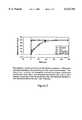
- FIG. 2is a graphical representation of the release of ciprofloxacin from catheter sections coated with PEG-gelatin hydrogels over time.
- FIG. 3illustrates a comparison of the adherence of viable bacteria to catheter sections coated with (a) PEG-gelatin hydrogel, (h) catheter sections coated with PEG gelatin hydrogel containing liposomal ciprofloxacin (lipogel), and (c) untreated sections.
- FIG. 4illustrates the reaction scheme whereby the cross linked PEG-gelatin matrix is formed by the formation of amide bonds between bis(amine)-PEG and the free carboxyl groups of gelatin.
- FIG. 5schematically illustrates the PEG-gelatin hydrogel with antibiotic containing liposomes.
- NHS-AFBwas prepared as described in J. F. W. Keana and S. X. Cai, "New reagents for photoaffinity labeling and photolysis of functionalized perfluorophenyl azides," J. Org Chem., 55:3640-3647 (1990) using the coupling agent DCC.
- AFB-gelatin gelatin of varying degrees of substitutionwas synthesized by the addition of NHS-AFB in methanol to a solution of gelatin (0.5-1.0%) in 50 mM Borate buffer (pH 8.6). The mixture was incubated overnight at room temperature with stirring.
- the degree to which gelatin's amino groups reacted with NHS-AFBwas determined. In brief, 20 ⁇ g of gelatin or AFB-gelatin in 1.5 mL of 50 mM Na 2 PO 4 buffer (pH 8.0) was used. While mixing the protein solution using a vortex agitator, 0.5 mL of fluorescamine in dioxane (1.1 mM) was added and mixing continued for 15 seconds.
- Gelatinwas iodinated using lodo Beads (Pierce, Rockford, Ill.) according to the supplier's directions.
- 100 ⁇ g of gelatin500 ⁇ L of 0.2 mg/mL gelatin in Hepes buffered saline, pH 7.4 (HBS)
- HBSHepes buffered saline
- Na 125 I(1 mCi from Amersham Canada, Oakville, ON
- the reaction vialwas washed with three 0.5 mL aliquots (200 ⁇ g/mL) of unlabeled gelatin.
- the protein solution(approx. 400 ⁇ g in 2.1 mL of HBS) was dialyzed in 200 mL of buffer until the dialysate was minimally radioactive (approx. 48 hrs with 5 changes of medium).
- the specific activity of the iodinated gelatinwas determined by a technique that exploits the insolubility of the complex formed between gelatin and the dye Sirius Red in acetic acid
- Four 50 ⁇ L aliquotswere removed from the iodinated protein solution and added to 1.5 mL polypropylene centrifuge tubes, followed by the addition of 50 ⁇ L of HBS and 1 mL of Sirius Red (50 ⁇ M) in 0.5 M acetic acid.
- the tubeswere incubated at room temperature for 30 minutes and subsequently centrifuged at 12,000 ⁇ g for 30 minutes. The supernatant was removed and a portion (0.5 mL) was used for protein quantitation via the decrease in absorbance (540 nm) of the dye remaining in solution.
- the protein/dye pelletwas resuspended with three 150 ⁇ L washes of 0.2 N NaOH containing 2 mg/mL gelatin.
- the radioactivity of the eluatewas measured in a liquid scintillation counter. Control experiments indicated that the presence of Sirius Red in the scintillation fluid did not interfere with the determination of 125 I radioactivity.
- Residual adsorbed proteinwas measured by cutting the centrifuge tubes into quarters and placing them in scintillation vials for counting. The specific activity was calculated to be 0.12 ⁇ 0.01 ⁇ Ci/ ⁇ g. This level of labeling is consistent with the paucity of tyrosine and histidine residues in gelatin.
- Radioiodinated gelatinwas modified with AFB as described above, however, the coupling solution and dialysis medium consisted of HBS (pH 8.0 and 7.4, respectively). The ratio of NHS-AFB to gelatin in the coupling solution was 1:4 (w/w). Following dialysis, the volume of the AFB-( 125 I)gelatin solution was made up to 5 mL and the protein concentration was determined to be 3.9 ⁇ 0.6 ng/ ⁇ L. Aliquots (10 ⁇ L each) of radioiodinated AFB-gelatin were applied to the side of silicone rectangles corresponding to the outer surface of the original catheter. All sections (12 in total) were dried under vacuum for 90 minutes.
- Liposomeswere composed of DPPC/Cholesterol/PEG-DSPE/Rhodamine-DPPE in a 1:1:0.05:0.001 ratio.
- the formulation to be usedis not limiting, and any number of lipid-to- other-constituents ratios may be used to effectively achieve the embodiments of this invention.
- the lipidswere dissolved in 4 mL of chloroform and the solvent was removed in vacuo. The resulting lipid film was placed under vacuum for two hours and subsequently hydrated with 1 mL of 250 mM ammonium sulfate (pH 2.5) at 45° C. Liposomes were then frozen in liquid nitrogen and thawed in a 45° C.
- PEG-gelatin solutionsconsisted of 10% gelatin, 6% NP-PEG and 10% sucrose at pH 4.0. If liposomes were required, they were added from a pure liposome suspension. The concentration of liposomes in PEG-gelatin solutions was 15 mM with respect to DPPC. All solutions were heated at 45° C. for 15 min. to dissolve gelatin.
- the PEG-gelatin matrixwas also crosslinked by the formation of amide bonds between bis-(amine)-PEG and the free carboxyl groups of gelatin.
- the silicone catheter surfaceis immersed in a solution of aqueous soluble carbodiimide (2 mg/mL) and incubated at room temperature for 30 min.
- the reaction of the activated carboxyl groups with PEG and gelatin amino moietiesis initiated by submersing the silicone material in borate buffer (200 mM, pH 8.5). Incubation in the alkaline buffer proceeds for 2 hr.
- the silicone surfaceis placed in 10% sucrose solution for 6 hr, with three changes of medium, to remove non-crosslinked material.
- This treatmentresults in a crosslinked PEG-gelatin gel that retains its integrity and remains affixed to the catheter for at least seven days when placed in a 37° C. solution of 10% sucrose.
- the crosslinking chemistryis outlined in FIG. 4.
- Residual p-nitrophenolwas leached from the gels by incubation at room temperature in 10% sucrose (pH 4.0) for 12 hrs, with four changes of medium. The absence of p-nitrophenol was confirmed by negligible absorbance of the dialysate at 410 nm.
- Liposomes in suspension and those entrapped within PEG-gelatin gelswere loaded with ciprofloxacin (Bayer, Leverkusen, Germany) according to the remote-loading technique described in Y. K. Oh, D. E. Nix, and R. M. Straubinger, "Formulation and efficacy of liposome-encapsulated antibiotics for therapy of intracellular Mycobacterium avium infection," Antimicrob. Agents Chemother., 39:2104-2111 (1995).
- Catheter pieceswere placed in 10% sucrose solution (pH 7.5) containing 2 mM ciprofloxacin, while for liposomes in suspension, an appropriate amount of drug was added to make the suspension 2 mM in ciprofloxacin.
- Dehydrated hydrogelswere prepared by drying coated catheter sections in an oven at 35° C. for 2.5 hr. The dried gels were then rehydrated in Tris buffer (10 mM Tris, 110 mM NaCl, pH 7.4) or in concentrated ciprofloxacin-HCl solution (25 mg/mL) as required. The temperature during the rehydration process was maintained at 45° C.
- the quantity of therapeutic agent loaded on the substratecan be increased or decreased over greater ranges than those shown in Table I. Greater concentrations of therapeutic agent can be loaded by increasing the amount of drug encapsulated and mixed into the hydrogel. For example, we expect that concentrations up to about 1,000 ⁇ g (1.0 mg) per cm 2 or more of an antibiotic active agent can be loaded on substrates with the methods of the present invention; and that concentrations of up to about 10,000 ⁇ g/cm 3 or more can be loaded on substrates. A preferred concentration range of antibiotic loaded on such substrates is about 10-1,000 ⁇ g/cm 2 . A preferred range for ciprofloxacin is about 10-200 ⁇ g/cm 2 .
- quantities of therapeutic agentcan be increased by increasing the quantity of gel immobilized on the surface of the substrate.
- hydrogel layersof about 0.5-10 mm thick can be loaded on substrates to effect the desired drug delivery and therapeutic results; preferred layers are in the range of about 1-5 mm; and especially preferred layers are about 2-4 mm.
- the release experimentwas initiated by placing each catheter section or dialysis membrane (containing liposome suspension 2.7 mM in DPPC) into separate liquid scintillation vials filled with 15 mL of Tris buffer. At selected time intervals 3 mL was removed from each vial for ciprofloxacin quantitation via a fluorescence-based assay using an excitation wavelength of 324 nm, an emission wavelength of 450 nm, and 5 nm slit widths. The amount of ciprofloxacin present was determined by comparisons to a standard curve. The remaining solution in the vials was emptied and replaced with 15 mL of buffer. The samples were incubated at 37° C. throughout the experiment.
- PBSglycerol-phosphate buffered saline
- Catheter sectionswere aseptically placed in 100 mL of sterile nutrient broth (Difco, Detroit, Mich.) contained within a 250 mL glass beaker. Twelve catheter sections from each coating formulation were added to individual beakers. The P. aeruginosa culture was washed 3 times in a pH 7.1 PBS solution, then inoculated to each of the beakers. The inoculum size was sufficient to yield 1.5 ⁇ 0.5 ⁇ 10 7 cfu/mL in the 100 mL volume. The inoculated catheter suspensions were then placed in an incubator maintained at 37° C. and agitated at a rate of 100 rpm.
- sterile nutrient brothDifco, Detroit, Mich.
- the catheter sectionswere removed from the bacterial suspensions and individually rinsed with a 10 mL volume of sterile PBS delivered via a gravity feed from a 10 mL pipet.
- the rinsed sectionswere placed in 20 mL plastic test tubes containing 5 mL volumes of sterile PBS and 3 mm diameter glass beads.
- the catheter sectionswere vortexed for 1 minute at high speed. The sonication and vortexing procedure was repeated three times. Aliquots were then removed from each of the suspensions and plated to nutrient agar. The plates were incubated at 37° C. for 48 h.
- the modification of the silicone catheter surface in this exampleused the photoreactive molecule 4-azido-2,3,5,6-tetrafluorobenzoic acid (AFB). It can be linked to the amino groups of gelatin via N-hydroxysuccinimide (NHS) chemistry. Based on the amino acid composition of ox hide gelatin, (J. E. Eastoe and A. A. Leach, "Chemical constitution of gelatin,” in Science and Technology of Gelatin, A. G. Ward and A. Courts (eds.), Academic Press, New York, 1977, pp. 73-107) the typical gelatin molecule (MW 75,000) contains approximately 25 ⁇ -amino groups derived from lysine and hydroxylysine.
- AZA4-azido-2,3,5,6-tetrafluorobenzoic acid
- AFBfluorinated aryl azide
- Ciprofloxacin release rateswere determined for the following samples: liposomes-only, PEG-gelatin hydrogel alone, a liposomal PEG-gelatin hydrogel, and a drug-containing liposomal hydrogel that was air dried and then rehydrated with pH 7.4 Tris buffer. All the liposomes used in this study contained DPPC and cholesterol. PEG-lipid was also included to avoid gelatin-induced destabilization of the bilayer and to increase immobilization of the liposomes within the hydrogel matrix via stearic interactions. The results of the experiment are summarized in FIG. 2. The quantity of ciprofloxacin released at a given time point is expressed as a percentage of the total amount released throughout the experiment. There are two notable trends. The hydrogel-only, and rehydrated liposomal hydrogel treatments were not successful in retaining ciprofloxacin for a sustained period of time; almost all of the drug initially incorporated was released within the first two hours.
- the dried liposomal hydrogeli.e., dried prior to being loaded with antibiotic, was found to maintain its sustained release properties after rehydration and is an important consideration for the clinical application of the system.
- An effective drying and rehydration processuses the dried liposomal hydrogel rehydrated in a solution containing 25 mg of ciprofloxacin.
- a dried hydrogel containing no liposomeswas hydrated in a 25 mg/mL ciprofloxacin solution.
- the total average amount of antibiotic entrapped within these hydrogelsis listed in Table 2, and for comparative purposes the total entrapped drug is also included.
- hydrogels rehydrated in concentrated ciprofloxacin solutionretained very large quantities of antibiotic (approx. 1.4 mg/1 cm catheter section). Almost all (>99%) of the hydrogel-associated ciprofloxacin was released after the first four hours of incubation, as expected from an analysis of the prior art.
- Ciprofloxacinwas incorporated into dried liposomal hydrogels during the rehydration step since our data indicated that pre-loaded liposomes embedded in a hydrogel were destabilized by dehydration. In effect, antibiotic was encapsulated within liposomes as they reformed during the rehydration of the PEG-gelatin film. Our calculations indicate that the encapsulation efficiency of ciprofloxacin in liposomes generated in situ was 7% relative to the amount of ciprofloxacin in pre-formed liposomes. The variation can be accounted for by the different loading techniques used. In general, compounds are more efficiently concentrated within liposomes when using a remote-loading technique exploiting pH and ammonium sulfate gradients than when a lipid film hydration method is employed.
- the optimal efflux profile in terms of prolonged release of substantial antibiotic quantitieswas obtained from liposomal hydrogel samples that were not dehydrated.
- the hydrogel systemwas shown to be capable of releasing substantial quantities of drug for up to 7 days. It is possible to improve the amount and duration of release by increasing the concentration of liposomes within the hydrogel; this aspect is not limiting. For example, the concentration can be at least doubled without affecting hydrogel stability. Increasing the liposome concentration allows the air dried liposomal hydrogel system to become a viable alternative as this compensates for the decrease in drug encapsulation efficiency associated with the in situ generation of liposomes. Alternatively, a dried liposomal hydrogel with suitable sustained release properties as presented here may be obtained by the development of a lyophilization protocol.
- a practical aim of this inventionis toward a catheter, or any polymeric biomedical device coating capable of resisting colonization by bacteria and subsequent infection in vivo and during application.
- untreated, PEG-gelatin coated, and ciprofloxacin-containing liposomal hydrogel catheter sectionswere challenged with a clinical strain of P. aeruginosa known to form biofilms on silicone catheters.
- the hydrogel coating containing antibiotic liposomeswas effective in preventing cells from adhering and remaining viable.
- the number of viable bacteria in the broth containing these sectionswas approximately 6.7 ⁇ 10 2 cfu/mL at the end of the experiment.
- DPPCdipalmitoylphosphatidylcholine
- PEG-DSPEPEG-distearoylphosphatidylethanolamine
- Rhodamine dipalmitoylphosphatidylethanolaminerhodamine-DPPE
- 4-azido-2,3,5,6-tetrafluorobenzoic acidwere purchased from Molecular Probes (Eugene, Oreg.).
- Porcine gelatin-aMW 50,000-100,000
- NP-PEGpolyoxyethylene bis(p-nitrophenyl carbonate)
- cholesterolwere obtained from Sigma (St. Louis, Mo.).
- Fluorescamine, 1,3-dicyclohexylcarbodiimide (DCC), N-hydroxysuccinimide (NHS), and Sirius Redwere purchased from Aldrich (Milwaukee, Wis.). All reagents and solvents were of analytical grade and were used without further purification. Deionized water (Milli-Q, Millipore, Bedford, Mass.) filtered through a 0.22 ⁇ m membrane was used in all experiments. Ciprofloxacin (Bayer, Germany) was analyzed in a Perkin Elmer LS-50 fluorimeter. Sirius Red and p-nitrophenol were quantitated using a Hewlett-Packard 8450 spectrophotometer.
- Silicone Foley catheters(Sherwood Medical, St. Louis, Mo.) were prepared for use by sectioning into cylinders (3 mm diameter and 10 mm length). The open ends of the sections were sealed with silicone rubber (RTV 108, GE, Pickering, ON). Occasionally, cylindrical sections were further subdivided into rectangular pieces (5 mm ⁇ 3 mm). Silicone sections were cleaned prior to each experiment by refluxing in methanol for six hours.
- Two pediatric silicone Foley catheterswere coated with a PEG-gelatin-liposome composition of the present invention as described herein, under aseptic conditions.
- the catheterswere inserted into the urethra of two male New Zealand white rabbits. After ten minutes the catheters were removed; and the catheters and excised urethra were examined. No disruption of the gel was observed on the catheter, and no gel fragments were detected in the urethra.
Landscapes
- Health & Medical Sciences (AREA)
- Life Sciences & Earth Sciences (AREA)
- Chemical & Material Sciences (AREA)
- Animal Behavior & Ethology (AREA)
- General Health & Medical Sciences (AREA)
- Public Health (AREA)
- Veterinary Medicine (AREA)
- Epidemiology (AREA)
- Medicinal Chemistry (AREA)
- Oral & Maxillofacial Surgery (AREA)
- Transplantation (AREA)
- Dermatology (AREA)
- Dispersion Chemistry (AREA)
- Molecular Biology (AREA)
- Biomedical Technology (AREA)
- Engineering & Computer Science (AREA)
- Communicable Diseases (AREA)
- Organic Chemistry (AREA)
- Pharmacology & Pharmacy (AREA)
- Nuclear Medicine, Radiotherapy & Molecular Imaging (AREA)
- General Chemical & Material Sciences (AREA)
- Chemical Kinetics & Catalysis (AREA)
- Oncology (AREA)
- Materials For Medical Uses (AREA)
- Medicinal Preparation (AREA)
- Pharmaceuticals Containing Other Organic And Inorganic Compounds (AREA)
Abstract
Description
TABLE 1 ______________________________________ Ciprofloxacin Loading into Liposomes and PEG-Gelatin Gel Sample Total Ciprofloxacin Entrapped ______________________________________ PEG-Gelatin Gel.sup.a 42 ± 12 μg/cm.sup.2 PEG-Gelatin-liposome Gel.sup.a 185 ± 16 μg/cm.sup.2 PEG-Gelatin-Liposome Gel.sup.b 3083 ± 267 μg/cm.sup.3 Dry PEG-Gelatin-Liposome Gel.sup.a 173 ± 6 μg/cm.sup.2 Dry PEG-Gelatin Gel.sup.a,c 1253 ± 80 μg/cm.sup.2 Dry PEG-Gelatin-Liposomes Gel.sup.a,c 1298 ± μg/cm.sup.2 Liposomes-only 0.52 ± 0.04 μmol cipro/μmol lipid ______________________________________ .sup.a Based on the application of 60 μL of PEG(6%)Gelatin (10%)gel to a 1 cm segment of silicon catheter with a diameter of 0.3 cm. Liposomecontaining gels were 15 mM in dipalmitoylphosphatidylcholine, n = 4. .sup.b Since 1 cm.sup.3 = 1 mL, 1000 μL of gel would occupy 1 cm.sup.3 and this quantity of PEGGelatin-Liposome gel would sequester 185 ± 16 μg * (1000 μL/60 μL) = 3083 ± 267 μg of ciprofloxacin. .sup.c These samples were dried before being rehydrated in a concentrated ciprofloxacin solution (25 mg/mL).
TABLE 2 ______________________________________ The degree of substitution of gelatin with AFB as a function of the initial ratio of ε-amino groups to NHS-AFB. ε-NH.sub.2 /NHS-AFB Degree of Substitution (%) ______________________________________ 9 99 ± 4 2 93 ± 4 1 71 ± 5 0.75 55 ± 2 ______________________________________
TABLE 3 ______________________________________ Release of ciprofloxacin from liposomes alone, constantly hydrated liposomal PEG-Gelatin hydrogel (LipoGel), dried liposomal PEG-Gelatin gel rehydrated 25 mg/mL ciprofloxacin solution (DryLipoGel (25 mg)), and dried PEG-Gelatin gel rehydrated in 25 mg/mL ciprofloxacin solution (DryGel (25 mg)). CIPROFLOXACIN RELEASED (μg/15 mL) TIME DryLipoGel DryGel (Hours) Liposomes LipoGel (25 mg) (25 mg) ______________________________________ 2.0 6.1 ± 2.1 39.5 ± 9.2 1367 ± 52 1329 ± 86 4.0 3.0 ± 0.4 7.3 ± 2.1 22.5 ± 4.8 22.0 ± 1.9 7.5 3.3 ± 0.5 7.2 ± 1.0 3.8 ± 0.9 1.2 ± 0.3 20.5 10.1 ± 0.6 21.6 ± 2.1 3.5 ± 0.3 0.34 ± 0.11 53.5 23.2 ± 4.0 67.7 ± 5.7 2.5 ± 0.2 0.14 ± 0.02 93.5 14.8 ± 4.1 47.7 ± 2.4 1.4 ± 0.2 0.13 ± 0.04 163.0 5.6 ± 0.9 7.4 ± 0.5 1.1 ± 0.1 0.15 ± 0.09 ______________________________________
Claims (18)
Priority Applications (12)
| Application Number | Priority Date | Filing Date | Title |
|---|---|---|---|
| US08/843,342US6132765A (en) | 1996-04-12 | 1997-04-15 | Drug delivery via therapeutic hydrogels |
| JP54333498AJP2001523124A (en) | 1997-04-15 | 1998-04-15 | Delivery of drugs by therapeutic hydrogel |
| EP98916701AEP0984798B1 (en) | 1997-04-15 | 1998-04-15 | Drug delivery via therapeutic hydrogels |
| PCT/CA1998/000351WO1998046287A2 (en) | 1997-04-15 | 1998-04-15 | Drug delivery via therapeutic hydrogels |
| AU70198/98AAU736584B2 (en) | 1997-04-15 | 1998-04-15 | Drug delivery via therapeutic hydrogels |
| CA002634700ACA2634700A1 (en) | 1997-04-15 | 1998-04-15 | Drug delivery via therapeutic hydrogels |
| AT98916701TATE305803T1 (en) | 1997-04-15 | 1998-04-15 | RELEASE OF ACTIVE INGREDIENTS USING THERAPEUTIC HYDROGELS |
| DE69831802TDE69831802T2 (en) | 1997-04-15 | 1998-04-15 | ACTIVE INGREDIENT RELIEF THROUGH THERAPEUTIC HYDROGEL |
| CA002286644ACA2286644C (en) | 1997-04-15 | 1998-04-15 | Drug delivery via therapeutic hydrogels |
| US09/412,584US6228393B1 (en) | 1996-04-12 | 1999-10-05 | Drug delivery via therapeutic hydrogels |
| US09/818,649US6475516B2 (en) | 1996-04-12 | 2001-03-28 | Drug delivery via therapeutic hydrogels |
| US09/849,481US20020051812A1 (en) | 1996-04-12 | 2001-05-07 | Drug delivery via therapeutic hydrogels |
Applications Claiming Priority (2)
| Application Number | Priority Date | Filing Date | Title |
|---|---|---|---|
| US63132696A | 1996-04-12 | 1996-04-12 | |
| US08/843,342US6132765A (en) | 1996-04-12 | 1997-04-15 | Drug delivery via therapeutic hydrogels |
Related Parent Applications (1)
| Application Number | Title | Priority Date | Filing Date |
|---|---|---|---|
| US63132696AContinuation-In-Part | 1996-04-12 | 1996-04-12 |
Related Child Applications (1)
| Application Number | Title | Priority Date | Filing Date |
|---|---|---|---|
| US09/412,584ContinuationUS6228393B1 (en) | 1996-04-12 | 1999-10-05 | Drug delivery via therapeutic hydrogels |
Publications (1)
| Publication Number | Publication Date |
|---|---|
| US6132765Atrue US6132765A (en) | 2000-10-17 |
Family
ID=25289698
Family Applications (4)
| Application Number | Title | Priority Date | Filing Date |
|---|---|---|---|
| US08/843,342Expired - Fee RelatedUS6132765A (en) | 1996-04-12 | 1997-04-15 | Drug delivery via therapeutic hydrogels |
| US09/412,584Expired - Fee RelatedUS6228393B1 (en) | 1996-04-12 | 1999-10-05 | Drug delivery via therapeutic hydrogels |
| US09/818,649Expired - Fee RelatedUS6475516B2 (en) | 1996-04-12 | 2001-03-28 | Drug delivery via therapeutic hydrogels |
| US09/849,481AbandonedUS20020051812A1 (en) | 1996-04-12 | 2001-05-07 | Drug delivery via therapeutic hydrogels |
Family Applications After (3)
| Application Number | Title | Priority Date | Filing Date |
|---|---|---|---|
| US09/412,584Expired - Fee RelatedUS6228393B1 (en) | 1996-04-12 | 1999-10-05 | Drug delivery via therapeutic hydrogels |
| US09/818,649Expired - Fee RelatedUS6475516B2 (en) | 1996-04-12 | 2001-03-28 | Drug delivery via therapeutic hydrogels |
| US09/849,481AbandonedUS20020051812A1 (en) | 1996-04-12 | 2001-05-07 | Drug delivery via therapeutic hydrogels |
Country Status (8)
| Country | Link |
|---|---|
| US (4) | US6132765A (en) |
| EP (1) | EP0984798B1 (en) |
| JP (1) | JP2001523124A (en) |
| AT (1) | ATE305803T1 (en) |
| AU (1) | AU736584B2 (en) |
| CA (2) | CA2286644C (en) |
| DE (1) | DE69831802T2 (en) |
| WO (1) | WO1998046287A2 (en) |
Cited By (82)
| Publication number | Priority date | Publication date | Assignee | Title |
|---|---|---|---|---|
| WO2002062406A1 (en)* | 2001-02-07 | 2002-08-15 | Efmt Entwicklungs- Und Forschungszentrum Für Mikrotherapie Gmbh | Dynamic implant coatings |
| US20020128222A1 (en)* | 1999-06-22 | 2002-09-12 | Research Development Foundation | Methods to enhance wound healing and enhanced wound coverage material |
| WO2002074355A1 (en)* | 2001-03-15 | 2002-09-26 | Dot Gmbh | Calcium phosphate materials containing active ingredients |
| WO2002057424A3 (en)* | 2001-01-17 | 2002-10-03 | Zycos Inc | Nucleic acid delivery formulations |
| US20020151958A1 (en)* | 2000-03-03 | 2002-10-17 | Chuter Timothy A.M. | Large vessel stents and occluders |
| US20030083389A1 (en)* | 2001-04-23 | 2003-05-01 | Kao Weiyun John | Bifunctional-modified hydrogels |
| US6689803B2 (en) | 1996-12-02 | 2004-02-10 | Angiotech Pharmaceuticals, Inc. | Compositions and methods for treating surgical adhesions |
| US20040147466A1 (en)* | 2002-01-17 | 2004-07-29 | Barman Shikha P. | Nucleic acid delivery formulations |
| US20050026257A1 (en)* | 2001-07-30 | 2005-02-03 | Lutz Gissmann | Production and applications for polyvalent vaccines against diseases caused by papilloma viruses |
| US20050112151A1 (en)* | 2003-11-20 | 2005-05-26 | Horng Liou L. | Skin adherent hydrogels |
| US20050129769A1 (en)* | 2002-06-03 | 2005-06-16 | Barry Stephen E. | Polymeric articles for carrying therapeutic agents |
| US20050175665A1 (en)* | 2003-11-20 | 2005-08-11 | Angiotech International Ag | Polymer compositions and methods for their use |
| US20050182450A1 (en)* | 2003-11-20 | 2005-08-18 | Angiotech International Ag | Electrical devices and anti-scarring agents |
| US20050276858A1 (en)* | 2001-04-23 | 2005-12-15 | Kao Weiyuan J | Bifunctional-modified hydrogels |
| US20060067983A1 (en)* | 2004-09-28 | 2006-03-30 | Atrium Medical Corporation | Stand-alone film and methods for making the same |
| US20070009580A1 (en)* | 2004-06-30 | 2007-01-11 | Dicosmo Frank | Non-adhesive hydrogels |
| US20070031380A1 (en)* | 2005-08-08 | 2007-02-08 | Hackett Perry B | Integration-site directed vector systems |
| US7250253B1 (en)* | 1999-01-25 | 2007-07-31 | Micronas Gmbh | Immobilization of molecules on surfaces via polymer brushes |
| US20070270447A1 (en)* | 2002-05-24 | 2007-11-22 | Angiotech International Ag | Compositions and methods for coating medical implants |
| US20080191103A1 (en)* | 2005-03-01 | 2008-08-14 | Lorne Thurgar | Clip |
| US20080255677A1 (en)* | 2007-04-10 | 2008-10-16 | Libera Matthew R | Surfaces differentially adhesive to bacteria and eukaryotic cells |
| US7445628B2 (en) | 1995-06-07 | 2008-11-04 | Cook Incorporated | Method of treating a patient with a coated implantable medical device |
| US7488757B2 (en) | 2003-03-24 | 2009-02-10 | Becton, Dickinson And Company | Invisible antimicrobial glove and hand antiseptic |
| US20100105140A1 (en)* | 2008-07-16 | 2010-04-29 | Fahrenkrug Scott C | Plaice dna transposon system |
| US20100111840A1 (en)* | 2001-03-08 | 2010-05-06 | Mark David Bednarski | Stabilized therapeutic and imaging agents |
| US20110097402A1 (en)* | 2004-09-30 | 2011-04-28 | Covalon Technologies Inc. | Non-adhesive elastic gelatin matrices |
| WO2011076924A1 (en) | 2009-12-23 | 2011-06-30 | Dsm Ip Assets B.V. | Method to activate silicone rubber surfaces |
| US20110212152A1 (en)* | 2001-02-28 | 2011-09-01 | Ditizio Valerio | Modified anti-microbial surfaces, devices and methods |
| US8124127B2 (en)* | 2005-10-15 | 2012-02-28 | Atrium Medical Corporation | Hydrophobic cross-linked gels for bioabsorbable drug carrier coatings |
| US8263102B2 (en) | 2004-09-28 | 2012-09-11 | Atrium Medical Corporation | Drug delivery coating for use with a stent |
| US8312836B2 (en) | 2004-09-28 | 2012-11-20 | Atrium Medical Corporation | Method and apparatus for application of a fresh coating on a medical device |
| US8313760B2 (en) | 2002-05-24 | 2012-11-20 | Angiotech International Ag | Compositions and methods for coating medical implants |
| US8367099B2 (en) | 2004-09-28 | 2013-02-05 | Atrium Medical Corporation | Perforated fatty acid films |
| EP2370112A4 (en)* | 2008-11-24 | 2013-04-10 | Schubert Holdings Llc | MATRIX COMPOSITION INCORPORATED IN IMPLANTABLE LIPOSOMES, USES AND PARTICLES OF POLYCAPROLACTONE AS SCAFFOLDING FOR TISSUE REGENERATION |
| EP2437844A4 (en)* | 2009-06-02 | 2013-10-16 | Concept Medical Inc | CORONARY ARBITRATION REJUVENATION BY IMPROVING BLOOD FLOW USING INSERTED NANOBILE (NANOPARTICLE ENCAPSULATED) CONTAINING THERAPEUTIC AGENTS BY NON-IMPLANTABLE DEVICES FOR TISSUES AND THUS PROVIDING RELEASE IN TISSUES TO EXAMINE THE NECESSARY CELL CYCLE |
| US8574627B2 (en) | 2006-11-06 | 2013-11-05 | Atrium Medical Corporation | Coated surgical mesh |
| US8795727B2 (en) | 2009-11-09 | 2014-08-05 | Spotlight Technology Partners Llc | Fragmented hydrogels |
| US9000040B2 (en) | 2004-09-28 | 2015-04-07 | Atrium Medical Corporation | Cross-linked fatty acid-based biomaterials |
| US9012506B2 (en) | 2004-09-28 | 2015-04-21 | Atrium Medical Corporation | Cross-linked fatty acid-based biomaterials |
| US9242026B2 (en) | 2008-06-27 | 2016-01-26 | Sofradim Production | Biosynthetic implant for soft tissue repair |
| US9278161B2 (en) | 2005-09-28 | 2016-03-08 | Atrium Medical Corporation | Tissue-separating fatty acid adhesion barrier |
| WO2016051413A1 (en)* | 2014-10-01 | 2016-04-07 | Yeda Research And Development Co. Ltd. | Liposomes-containing antifouling compositions and uses thereof |
| US9308068B2 (en) | 2007-12-03 | 2016-04-12 | Sofradim Production | Implant for parastomal hernia |
| US9427423B2 (en) | 2009-03-10 | 2016-08-30 | Atrium Medical Corporation | Fatty-acid based particles |
| US9445883B2 (en) | 2011-12-29 | 2016-09-20 | Sofradim Production | Barbed prosthetic knit and hernia repair mesh made therefrom as well as process for making said prosthetic knit |
| US9492596B2 (en) | 2006-11-06 | 2016-11-15 | Atrium Medical Corporation | Barrier layer with underlying medical device and one or more reinforcing support structures |
| US9499927B2 (en) | 2012-09-25 | 2016-11-22 | Sofradim Production | Method for producing a prosthesis for reinforcing the abdominal wall |
| US9526603B2 (en) | 2011-09-30 | 2016-12-27 | Covidien Lp | Reversible stiffening of light weight mesh |
| US9554887B2 (en) | 2011-03-16 | 2017-01-31 | Sofradim Production | Prosthesis comprising a three-dimensional and openworked knit |
| US9622843B2 (en) | 2011-07-13 | 2017-04-18 | Sofradim Production | Umbilical hernia prosthesis |
| US9700650B2 (en) | 2009-11-09 | 2017-07-11 | Spotlight Technology Partners Llc | Polysaccharide based hydrogels |
| WO2017139357A1 (en)* | 2016-02-08 | 2017-08-17 | Orbusneich Medical, Inc. | Drug eluting balloon |
| US9750837B2 (en) | 2012-09-25 | 2017-09-05 | Sofradim Production | Haemostatic patch and method of preparation |
| US9757774B2 (en) | 2015-01-15 | 2017-09-12 | MyExposome, Inc. | Passive sampling devices |
| US9801982B2 (en) | 2004-09-28 | 2017-10-31 | Atrium Medical Corporation | Implantable barrier device |
| US9839505B2 (en) | 2012-09-25 | 2017-12-12 | Sofradim Production | Prosthesis comprising a mesh and a strengthening means |
| US9867880B2 (en) | 2012-06-13 | 2018-01-16 | Atrium Medical Corporation | Cured oil-hydrogel biomaterial compositions for controlled drug delivery |
| US9877820B2 (en) | 2014-09-29 | 2018-01-30 | Sofradim Production | Textile-based prosthesis for treatment of inguinal hernia |
| US9932695B2 (en) | 2014-12-05 | 2018-04-03 | Sofradim Production | Prosthetic porous knit |
| US9931198B2 (en) | 2015-04-24 | 2018-04-03 | Sofradim Production | Prosthesis for supporting a breast structure |
| US9980802B2 (en) | 2011-07-13 | 2018-05-29 | Sofradim Production | Umbilical hernia prosthesis |
| US10080639B2 (en) | 2011-12-29 | 2018-09-25 | Sofradim Production | Prosthesis for inguinal hernia |
| US10159555B2 (en) | 2012-09-28 | 2018-12-25 | Sofradim Production | Packaging for a hernia repair device |
| US10184032B2 (en) | 2015-02-17 | 2019-01-22 | Sofradim Production | Method for preparing a chitosan-based matrix comprising a fiber reinforcement member |
| US10213283B2 (en) | 2013-06-07 | 2019-02-26 | Sofradim Production | Textile-based prosthesis for laparoscopic surgery |
| US10314935B2 (en) | 2009-01-07 | 2019-06-11 | Entrotech Life Sciences, Inc. | Chlorhexidine-containing antimicrobial laminates |
| US10322213B2 (en) | 2010-07-16 | 2019-06-18 | Atrium Medical Corporation | Compositions and methods for altering the rate of hydrolysis of cured oil-based materials |
| US10327882B2 (en) | 2014-09-29 | 2019-06-25 | Sofradim Production | Whale concept—folding mesh for TIPP procedure for inguinal hernia |
| US10363690B2 (en) | 2012-08-02 | 2019-07-30 | Sofradim Production | Method for preparing a chitosan-based porous layer |
| US10405960B2 (en) | 2013-06-07 | 2019-09-10 | Sofradim Production | Textile-based prothesis for laparoscopic surgery |
| US10646321B2 (en) | 2016-01-25 | 2020-05-12 | Sofradim Production | Prosthesis for hernia repair |
| US10675137B2 (en) | 2017-05-02 | 2020-06-09 | Sofradim Production | Prosthesis for inguinal hernia repair |
| US10682215B2 (en) | 2016-10-21 | 2020-06-16 | Sofradim Production | Method for forming a mesh having a barbed suture attached thereto and the mesh thus obtained |
| US10737075B2 (en) | 2016-02-08 | 2020-08-11 | Orbusneich Medical Pte. Ltd. | Drug eluting balloon |
| US10743976B2 (en) | 2015-06-19 | 2020-08-18 | Sofradim Production | Synthetic prosthesis comprising a knit and a non porous film and method for forming same |
| US10864304B2 (en) | 2009-08-11 | 2020-12-15 | Atrium Medical Corporation | Anti-infective antimicrobial-containing biomaterials |
| US10865505B2 (en) | 2009-09-04 | 2020-12-15 | Sofradim Production | Gripping fabric coated with a bioresorbable impenetrable layer |
| US11039615B2 (en) | 2014-04-18 | 2021-06-22 | Entrotech Life Sciences, Inc. | Methods of processing chlorhexidine-containing polymerizable compositions and antimicrobial articles formed thereby |
| CN113318278A (en)* | 2015-05-14 | 2021-08-31 | 用于组织工程和细胞为基础的技术和疗法促进协会 | Ureteral stent, method and application thereof |
| US11471257B2 (en) | 2018-11-16 | 2022-10-18 | Sofradim Production | Implants suitable for soft tissue repair |
| US11529391B2 (en)* | 2017-01-09 | 2022-12-20 | The Board Of Trustees Of The Leland Stanford Junior University | Reversing deficient hedgehog signaling restores deficient skeletal regeneration |
| US12064330B2 (en) | 2020-04-28 | 2024-08-20 | Covidien Lp | Implantable prothesis for minimally invasive hernia repair |
Families Citing this family (154)
| Publication number | Priority date | Publication date | Assignee | Title |
|---|---|---|---|---|
| WO1999049908A1 (en) | 1998-03-31 | 1999-10-07 | University Of Cincinnati | Temperature controlled solute delivery system |
| US8177762B2 (en) | 1998-12-07 | 2012-05-15 | C. R. Bard, Inc. | Septum including at least one identifiable feature, access ports including same, and related methods |
| US7892532B2 (en)* | 1999-04-30 | 2011-02-22 | Warsaw Orthopedic, In Emory University | Intracellular delivery of osteoinductive proteins and peptides |
| AU6679400A (en)* | 1999-08-27 | 2001-03-26 | Department Of National Defence | Hydrogel wound dressing containing liposome-encapsulated therapeutic agent |
| US6738661B1 (en)* | 1999-10-22 | 2004-05-18 | Biosynergetics, Inc. | Apparatus and methods for the controllable modification of compound concentration in a tube |
| US6613082B2 (en)* | 2000-03-13 | 2003-09-02 | Jun Yang | Stent having cover with drug delivery capability |
| US6800298B1 (en)* | 2000-05-11 | 2004-10-05 | Clemson University | Biological lubricant composition and method of applying lubricant composition |
| DE10025803A1 (en)* | 2000-05-24 | 2001-12-20 | Jms Co Ltd | Polymer surface with biologically active properties and process for their production |
| US20040018228A1 (en)* | 2000-11-06 | 2004-01-29 | Afmedica, Inc. | Compositions and methods for reducing scar tissue formation |
| US20060089721A1 (en)* | 2001-01-17 | 2006-04-27 | Muhanna Nabil L | Intervertebral disc prosthesis and methods of implantation |
| WO2002065947A2 (en)* | 2001-02-16 | 2002-08-29 | Abbott Laboratories Vascular Enterprises Limited | Implants with fk506 for prophylaxis and treatment of restonoses |
| WO2002070022A2 (en)* | 2001-02-28 | 2002-09-12 | Uroteq Inc. | Method of making anti-microbial polymeric surfaces |
| EP1277484B1 (en)* | 2001-07-20 | 2004-09-29 | Peter Hildebrandt | Urological implant |
| US20030064965A1 (en)* | 2001-10-02 | 2003-04-03 | Jacob Richter | Method of delivering drugs to a tissue using drug-coated medical devices |
| EP1434571B1 (en)* | 2001-10-05 | 2005-05-11 | SurModics, Inc. | Particle immobilized coatings and uses thereof |
| US20030100830A1 (en)* | 2001-11-27 | 2003-05-29 | Sheng-Ping Zhong | Implantable or insertable medical devices visible under magnetic resonance imaging |
| US7488313B2 (en)* | 2001-11-29 | 2009-02-10 | Boston Scientific Scimed, Inc. | Mechanical apparatus and method for dilating and delivering a therapeutic agent to a site of treatment |
| RU2314831C2 (en)* | 2002-01-22 | 2008-01-20 | Фармация Энд Апджон Компани | Infection-proof medical devices |
| AR038926A1 (en)* | 2002-03-13 | 2005-02-02 | Novartis Ag | MATERIALS WITH MULTIPLE VESICLE LAYER CONTENT |
| US7519418B2 (en)* | 2002-04-30 | 2009-04-14 | Boston Scientific Scimed, Inc. | Mechanical apparatus and method for dilating and delivering a therapeutic agent to a site of treatment |
| US20040034336A1 (en)* | 2002-08-08 | 2004-02-19 | Neal Scott | Charged liposomes/micelles with encapsulted medical compounds |
| WO2004037318A2 (en)* | 2002-10-22 | 2004-05-06 | The Biomerix Corporation | Method and system for intravesicular delivery of therapeutic agents |
| US7718189B2 (en) | 2002-10-29 | 2010-05-18 | Transave, Inc. | Sustained release of antiinfectives |
| BR0315868A (en)* | 2002-10-29 | 2005-09-27 | Transave Inc | Method for treating or ameliorating pulmonary infection in a patient with cystic fibrosis; and method for treating or ameliorating pulmonary infection in an animal |
| AU2003291311A1 (en)* | 2002-11-07 | 2004-06-03 | Carbon Medical Technologies, Inc. | Biocompatible medical device coatings |
| WO2005023331A2 (en)* | 2003-09-04 | 2005-03-17 | The United States Of America As Represented By The Department Of Veterans Affairs | Hydrogel nanocompsites for ophthalmic applications |
| US6992127B2 (en)* | 2002-11-25 | 2006-01-31 | Ast Products, Inc. | Polymeric coatings containing a pH buffer agent |
| US7597903B2 (en)* | 2002-12-02 | 2009-10-06 | Shenkar College Of Engineering And Design | Method and composition for producing catheters with antibacterial property |
| US8048407B2 (en)* | 2003-04-24 | 2011-11-01 | Brent Vernon | In situ gelling self-reactive materials for embolization |
| US20040254545A1 (en)* | 2003-06-16 | 2004-12-16 | Rider Dean Loller | Method and apparatus for extending feeding tube longevity |
| JP4320572B2 (en)* | 2003-07-11 | 2009-08-26 | ソニー株式会社 | Signal processing apparatus and method, recording medium, and program |
| US20050074453A1 (en)* | 2003-10-02 | 2005-04-07 | Ferree Bret A. | Methods of preventing adhesions following laminectomies and other surgical procedures |
| CA2544779A1 (en)* | 2003-11-03 | 2005-05-12 | Medtronic, Inc. | Hydrogel providing cell-specific ingrowth |
| WO2005051396A2 (en)* | 2003-11-25 | 2005-06-09 | Deltanoid Pharmaceuticals, Inc. | Methods for reducing body fat using vitamin d compounds |
| US7221982B2 (en)* | 2004-07-12 | 2007-05-22 | Cardiac Pacemakers, Inc. | Apparatus and method of coating implantable leads |
| WO2012050591A1 (en) | 2010-10-15 | 2012-04-19 | Rutgers, The State University Of New Jersey | Hydrogel formulation for dermal and ocular delivery |
| US20090104254A1 (en)* | 2004-12-22 | 2009-04-23 | Rutgers, The State University Of New Jersey | Controlled Release Hydrogels |
| WO2006079015A2 (en)* | 2005-01-21 | 2006-07-27 | Vertex Pharmaceuticals Incorporated | Quorum sensing modulators |
| US9474888B2 (en) | 2005-03-04 | 2016-10-25 | C. R. Bard, Inc. | Implantable access port including a sandwiched radiopaque insert |
| JP5484674B2 (en) | 2005-03-04 | 2014-05-07 | シー・アール・バード・インコーポレーテッド | Access port and identification method |
| US7947022B2 (en) | 2005-03-04 | 2011-05-24 | C. R. Bard, Inc. | Access port identification systems and methods |
| US8029482B2 (en) | 2005-03-04 | 2011-10-04 | C. R. Bard, Inc. | Systems and methods for radiographically identifying an access port |
| CN101180037A (en)* | 2005-03-21 | 2008-05-14 | 莱夫克尔创新私人有限公司 | A novel inter and intra multilamellar vesicular composition |
| US10307581B2 (en) | 2005-04-27 | 2019-06-04 | C. R. Bard, Inc. | Reinforced septum for an implantable medical device |
| EP3884989B1 (en) | 2005-04-27 | 2022-07-13 | C. R. Bard, Inc. | Vascular access port |
| EP1874393B1 (en) | 2005-04-27 | 2017-09-06 | C.R.Bard, Inc. | Infusion apparatuses |
| US20080213461A1 (en)* | 2005-06-17 | 2008-09-04 | Georgia Tech Research Corporation | Coated Microstructures and Methods of Manufacture Thereof |
| CN101257891A (en)* | 2005-09-01 | 2008-09-03 | 比奥里波克斯公司 | Antihistamine- and corticosteroid- containing liposome composition and its use for the manufacture of a medicament for treating rhinitis and related disorders |
| EP1962805B1 (en) | 2005-12-08 | 2016-07-06 | Insmed Incorporated | Lipid-based compositions of antiinfectives for treating pulmonary infections |
| US8663674B2 (en)* | 2006-01-13 | 2014-03-04 | Surmodics, Inc. | Microparticle containing matrices for drug delivery |
| US20070184085A1 (en)* | 2006-02-03 | 2007-08-09 | Boston Scientific Scimed, Inc. | Ultrasound activated medical device |
| ITMI20060556A1 (en)* | 2006-03-24 | 2007-09-25 | Univ Pavia | INTERACTIVE MEDICATIONS FOR THE CARE OF DERMATOLOGICAL DISEASES |
| DE102006015271A1 (en)* | 2006-04-01 | 2007-10-11 | Lohmann & Rauscher Gmbh & Co. Kg | Biguanide-containing liposomes |
| EP2037977A2 (en)* | 2006-06-28 | 2009-03-25 | SurModics, Inc. | Active agent eluting matrices with particulates |
| US8747738B2 (en)* | 2006-08-15 | 2014-06-10 | Abbott Cardiovascular Systems Inc. | Sterilization methods for medical devices |
| US9265912B2 (en) | 2006-11-08 | 2016-02-23 | C. R. Bard, Inc. | Indicia informative of characteristics of insertable medical devices |
| US9642986B2 (en) | 2006-11-08 | 2017-05-09 | C. R. Bard, Inc. | Resource information key for an insertable medical device |
| US9737640B2 (en) | 2006-11-20 | 2017-08-22 | Lutonix, Inc. | Drug releasing coatings for medical devices |
| US8414526B2 (en) | 2006-11-20 | 2013-04-09 | Lutonix, Inc. | Medical device rapid drug releasing coatings comprising oils, fatty acids, and/or lipids |
| US20080276935A1 (en) | 2006-11-20 | 2008-11-13 | Lixiao Wang | Treatment of asthma and chronic obstructive pulmonary disease with anti-proliferate and anti-inflammatory drugs |
| US8998846B2 (en) | 2006-11-20 | 2015-04-07 | Lutonix, Inc. | Drug releasing coatings for balloon catheters |
| US8414525B2 (en) | 2006-11-20 | 2013-04-09 | Lutonix, Inc. | Drug releasing coatings for medical devices |
| US8414910B2 (en) | 2006-11-20 | 2013-04-09 | Lutonix, Inc. | Drug releasing coatings for medical devices |
| US20080175887A1 (en) | 2006-11-20 | 2008-07-24 | Lixiao Wang | Treatment of Asthma and Chronic Obstructive Pulmonary Disease With Anti-proliferate and Anti-inflammatory Drugs |
| US8425459B2 (en) | 2006-11-20 | 2013-04-23 | Lutonix, Inc. | Medical device rapid drug releasing coatings comprising a therapeutic agent and a contrast agent |
| US9700704B2 (en) | 2006-11-20 | 2017-07-11 | Lutonix, Inc. | Drug releasing coatings for balloon catheters |
| KR101428122B1 (en) | 2006-12-15 | 2014-08-07 | 라이프본드 엘티디. | Gelatin-transglutaminase hemostatic dressings and sealants |
| US20080181928A1 (en)* | 2006-12-22 | 2008-07-31 | Miv Therapeutics, Inc. | Coatings for implantable medical devices for liposome delivery |
| EP1980277A1 (en)* | 2007-03-17 | 2008-10-15 | Peter Hildebrandt | Gastroenterological medical product, in particular stent for the bile duct or pancreatic duct |
| US20100196455A1 (en) | 2007-05-04 | 2010-08-05 | Transave, Inc. | Compositions of Multicationic Drugs for Reducing Interactions with Polyanionic Biomolecules and Methods of Use Thereof |
| US9119783B2 (en) | 2007-05-07 | 2015-09-01 | Insmed Incorporated | Method of treating pulmonary disorders with liposomal amikacin formulations |
| US9114081B2 (en) | 2007-05-07 | 2015-08-25 | Insmed Incorporated | Methods of treating pulmonary disorders with liposomal amikacin formulations |
| DE102007039871A1 (en)* | 2007-08-21 | 2009-02-26 | Friedrich-Baur-Gmbh | Soft tissue implant with antibacterial effect |
| ES2364686T3 (en)* | 2007-08-27 | 2011-09-12 | Theravance, Inc. | ALQUIL-8-AZABICICLO COMPOUNDS [3.2.1] OCTANO DISSTITUTED AS ANTAGONISTS OF THE OPIOID RECEIVER MU. |
| JP2011500150A (en)* | 2007-10-10 | 2011-01-06 | エムアイヴィ テラピューティクス, インコーポレイテッド | Lipid coating for medical devices for implants |
| US9579496B2 (en) | 2007-11-07 | 2017-02-28 | C. R. Bard, Inc. | Radiopaque and septum-based indicators for a multi-lumen implantable port |
| EP2265293B1 (en)* | 2008-04-18 | 2015-11-04 | SurModics, Inc. | Coating systems for the controlled delivery of hydrophilic bioactive agents |
| WO2009131672A1 (en)* | 2008-04-22 | 2009-10-29 | University Of Massachusetts | Stabilized liposome compositions and related methods of use |
| US9943614B2 (en) | 2008-06-17 | 2018-04-17 | Brigham Young University | Cationic steroid antimicrobial diagnostic, detection, screening and imaging methods |
| AU2009261559B2 (en)* | 2008-06-18 | 2014-01-16 | Lifebond Ltd | Improved cross-linked compositions |
| US20110086014A1 (en)* | 2008-06-18 | 2011-04-14 | Ishay Attar | Method for enzymatic cross-linking of a protein |
| WO2009153748A2 (en)* | 2008-06-18 | 2009-12-23 | Lifebond Ltd | Methods and devices for use with sealants |
| WO2010024898A2 (en) | 2008-08-29 | 2010-03-04 | Lutonix, Inc. | Methods and apparatuses for coating balloon catheters |
| US20100070013A1 (en)* | 2008-09-18 | 2010-03-18 | Medtronic Vascular, Inc. | Medical Device With Microsphere Drug Delivery System |
| ES2906416T3 (en) | 2008-10-31 | 2022-04-18 | Bard Inc C R | Systems and methods to identify an access road |
| US11890443B2 (en) | 2008-11-13 | 2024-02-06 | C. R. Bard, Inc. | Implantable medical devices including septum-based indicators |
| US8932271B2 (en) | 2008-11-13 | 2015-01-13 | C. R. Bard, Inc. | Implantable medical devices including septum-based indicators |
| US8709473B1 (en)* | 2009-01-28 | 2014-04-29 | Abbott Cardiovascular Systems Inc. | Method of targeting hydrophobic drugs to vascular lesions |
| US20100239630A1 (en)* | 2009-03-20 | 2010-09-23 | Wilson Kurt Whitekettle | Phosphonium salts delivery systems |
| US20100239626A1 (en)* | 2009-03-20 | 2010-09-23 | Wilson Kurt Whitekettle | Propanediol delivery systems |
| US20100239651A1 (en)* | 2009-03-20 | 2010-09-23 | Wilson Kurt Whitekettle | Nitrilopropionamide delivery systems |
| US20100239650A1 (en)* | 2009-03-20 | 2010-09-23 | Wilson Kurt Whitekettle | Isothiazolin biodelivery systems |
| US20100239627A1 (en)* | 2009-03-20 | 2010-09-23 | Wilson Kurt Whitekettle | Quarternary ammonium salts delivery systems |
| US20100247600A1 (en)* | 2009-03-24 | 2010-09-30 | Warsaw Orthopedic, Inc. | Therapeutic drug eluting implant cover and method of making the same |
| US20100249783A1 (en)* | 2009-03-24 | 2010-09-30 | Warsaw Orthopedic, Inc. | Drug-eluting implant cover |
| US9078712B2 (en)* | 2009-04-15 | 2015-07-14 | Warsaw Orthopedic, Inc. | Preformed drug-eluting device to be affixed to an anterior spinal plate |
| US9414864B2 (en) | 2009-04-15 | 2016-08-16 | Warsaw Orthopedic, Inc. | Anterior spinal plate with preformed drug-eluting device affixed thereto |
| ES2695907T3 (en) | 2009-11-17 | 2019-01-11 | Bard Inc C R | Overmolded access port that includes anchoring and identification features |
| CA2782863A1 (en) | 2009-12-22 | 2011-06-30 | Lifebond Ltd | Modification of enzymatic crosslinkers for controlling properties of crosslinked matrices |
| US20120028335A1 (en) | 2010-07-28 | 2012-02-02 | Life Technologies Corporation | Anti-viral azide-containing compounds |
| CN103237546B (en) | 2010-07-28 | 2016-06-08 | 生命科技公司 | The antiviral compound that contains azide |
| CA2807012A1 (en) | 2010-08-05 | 2012-02-09 | Lifebond Ltd. | Dry composition wound dressings and adhesives |
| US8753608B2 (en)* | 2010-08-24 | 2014-06-17 | Canon Kabushiki Kaisha | Complex and contrast agent for photoimaging using the same |
| USD676955S1 (en) | 2010-12-30 | 2013-02-26 | C. R. Bard, Inc. | Implantable access port |
| USD682416S1 (en) | 2010-12-30 | 2013-05-14 | C. R. Bard, Inc. | Implantable access port |
| WO2012155048A1 (en)* | 2011-05-11 | 2012-11-15 | Wisconsin Alumni Research Foundation | Liposome-encapsulated hydrogels for use in a drug delivery system |
| CA2842460C (en) | 2011-07-20 | 2019-08-20 | Brigham Young University | Hydrophobic ceragenin compounds and devices incorporating same |
| US8945217B2 (en) | 2011-08-25 | 2015-02-03 | Brigham Young University | Medical devices incorporating ceragenin-containing composites |
| US9603859B2 (en) | 2011-09-13 | 2017-03-28 | Brigham Young University | Methods and products for increasing the rate of healing of tissue wounds |
| US9694019B2 (en) | 2011-09-13 | 2017-07-04 | Brigham Young University | Compositions and methods for treating bone diseases and broken bones |
| CA2852989C (en) | 2011-09-13 | 2021-06-29 | Brigham Young University | Compositions for treating bone diseases and broken bones |
| JP6038155B2 (en) | 2011-09-13 | 2016-12-07 | ブリガム・ヤング・ユニバーシティBrigham Young University | Products for healing tissue wounds |
| JP6111013B2 (en)* | 2011-10-30 | 2017-04-05 | 株式会社セルシード | Tendon cell sheet and manufacturing method thereof |
| BR112014015144B1 (en) | 2011-12-21 | 2018-03-27 | Brigham Young University | AQUOSO COMPOSITION FOR ORAL CARE, USE OF A CATIONIC STEROID COMPOUND AND NON-THERAPEUTIC METHOD FOR REDUCING VOLAFLE SULFUR COMPOUNDS |
| CN104245925A (en) | 2012-01-26 | 2014-12-24 | 生命科技公司 | Method for increasing viral infectivity |
| US9533063B1 (en) | 2012-03-01 | 2017-01-03 | Brigham Young University | Aerosols incorporating ceragenin compounds and methods of use thereof |
| EP2846634A2 (en) | 2012-05-02 | 2015-03-18 | Brigham Young University | Ceragenin particulate materials and methods for making same |
| MX369828B (en) | 2012-05-21 | 2019-11-22 | Insmed Inc | SYSTEMS TO TREAT PULMONARY INFECTIONS. |
| AU2013331136B2 (en) | 2012-10-17 | 2016-05-12 | Brigham Young University | Treatment and prevention of mastitis |
| EP3581186A1 (en) | 2012-11-29 | 2019-12-18 | Insmed Incorporated | Stabilized vancomycin formulations |
| CN105451742B (en) | 2013-01-07 | 2021-04-06 | 布莱阿姆青年大学 | Methods for reducing cell proliferation and treating certain diseases |
| CN110420211A (en) | 2013-03-15 | 2019-11-08 | 布莱阿姆青年大学 | The method for treating inflammation, autoimmune disease and pain |
| US10568893B2 (en) | 2013-03-15 | 2020-02-25 | Brigham Young University | Methods for treating inflammation, autoimmune disorders and pain |
| US11524015B2 (en) | 2013-03-15 | 2022-12-13 | Brigham Young University | Methods for treating inflammation, autoimmune disorders and pain |
| US9387215B2 (en) | 2013-04-22 | 2016-07-12 | Brigham Young University | Animal feed including cationic cholesterol additive and related methods |
| US11690855B2 (en) | 2013-10-17 | 2023-07-04 | Brigham Young University | Methods for treating lung infections and inflammation |
| US20150203527A1 (en) | 2014-01-23 | 2015-07-23 | Brigham Young University | Cationic steroidal antimicrobials |
| US9364588B2 (en) | 2014-02-04 | 2016-06-14 | Abbott Cardiovascular Systems Inc. | Drug delivery scaffold or stent with a novolimus and lactide based coating such that novolimus has a minimum amount of bonding to the coating |
| CA2844321C (en) | 2014-02-27 | 2021-03-16 | Brigham Young University | Cationic steroidal antimicrobial compounds |
| US10220045B2 (en) | 2014-03-13 | 2019-03-05 | Brigham Young University | Compositions and methods for forming stabilized compositions with reduced CSA agglomeration |
| US9867836B2 (en) | 2014-03-13 | 2018-01-16 | Brigham Young University | Lavage and/or infusion using CSA compounds for increasing fertility in a mammal |
| US9931350B2 (en) | 2014-03-14 | 2018-04-03 | Brigham Young University | Anti-infective and osteogenic compositions and methods of use |
| US9686966B2 (en) | 2014-04-30 | 2017-06-27 | Brigham Young University | Methods and apparatus for cleaning or disinfecting a water delivery system |
| ME03536B (en) | 2014-05-15 | 2020-04-20 | Insmed Inc | Methods for treating pulmonary non-tuberculous mycobacterial infections |
| US10441595B2 (en) | 2014-06-26 | 2019-10-15 | Brigham Young University | Methods for treating fungal infections |
| US10238665B2 (en) | 2014-06-26 | 2019-03-26 | Brigham Young University | Methods for treating fungal infections |
| US10227376B2 (en) | 2014-08-22 | 2019-03-12 | Brigham Young University | Radiolabeled cationic steroid antimicrobials and diagnostic methods |
| US10155788B2 (en) | 2014-10-07 | 2018-12-18 | Brigham Young University | Cationic steroidal antimicrobial prodrug compositions and uses thereof |
| US10370403B2 (en) | 2015-04-22 | 2019-08-06 | Brigham Young University | Methods for the synthesis of ceragenins |
| WO2016172553A1 (en) | 2015-04-22 | 2016-10-27 | Savage Paul B | Methods for the synthesis of ceragenins |
| US10126298B2 (en) | 2015-05-04 | 2018-11-13 | Arman Nabatian | Hydrogels containing embedded substrates for targeted binding of molecules |
| US9434759B1 (en) | 2015-05-18 | 2016-09-06 | Brigham Young University | Cationic steroidal antimicrobial compounds and methods of manufacturing such compounds |
| EP3322365A4 (en)* | 2015-05-22 | 2019-04-17 | Orthobond, Inc. | Methods, compositions and techniques for polydimethylsiloxane surface modifications |
| CA2995751A1 (en) | 2015-08-25 | 2017-03-02 | Histide Ag | Compounds for inducing tissue formation and uses thereof |
| JP7056932B2 (en) | 2015-08-25 | 2022-04-19 | ヒスタイド アクツィエンゲゼルシャフト | Compounds for inducing histogenesis and their use |
| CN109715192B (en) | 2015-09-17 | 2024-02-20 | 伊斯迪德股份公司 | Drug conjugates for converting neoplastic cells to non-neoplastic cells and uses thereof |
| RU2617062C1 (en)* | 2015-10-22 | 2017-04-19 | Акционерное общество "Медсил" | Plastic endoprosthesis for stenting pancreatic ducts |
| US10226550B2 (en) | 2016-03-11 | 2019-03-12 | Brigham Young University | Cationic steroidal antimicrobial compositions for the treatment of dermal tissue |
| RU2624535C1 (en)* | 2016-10-14 | 2017-07-04 | Акционерное общество "Медсил" | Plastic endoprosthesis for stenting pancreatic ducts |
| US10959433B2 (en) | 2017-03-21 | 2021-03-30 | Brigham Young University | Use of cationic steroidal antimicrobials for sporicidal activity |
| CN111432830A (en)* | 2017-09-29 | 2020-07-17 | 巴德香农有限公司 | Compositions and methods for controlled drug release from tissue |
| EP3773505A4 (en) | 2018-03-30 | 2021-12-22 | Insmed Incorporated | PROCESS FOR THE CONTINUOUS MANUFACTURING OF LIPOSOMAL MEDICINAL PRODUCTS |
| US11998654B2 (en) | 2018-07-12 | 2024-06-04 | Bard Shannon Limited | Securing implants and medical devices |
| US12186328B2 (en) | 2019-05-23 | 2025-01-07 | Brigham Young University | Use of CSA compounds to stimulate stem cells and hair growth |
Citations (6)
| Publication number | Priority date | Publication date | Assignee | Title |
|---|---|---|---|---|
| US4442133A (en)* | 1982-02-22 | 1984-04-10 | Greco Ralph S | Antibiotic bonding of vascular prostheses and other implants |
| US4749585A (en)* | 1986-04-11 | 1988-06-07 | University Of Medicine And Dentistry Of New Jersey | Antibiotic bonded prosthesis and process for producing same |
| WO1991009616A1 (en)* | 1989-12-22 | 1991-07-11 | Yale University | Quinolone antibiotics encapsulated in lipid vesicles |
| US5575815A (en)* | 1988-08-24 | 1996-11-19 | Endoluminal Therapeutics, Inc. | Local polymeric gel therapy |
| CA2181390A1 (en)* | 1995-07-18 | 1997-01-19 | Pankaj Modi | Phospholipid formulations |
| US5741516A (en)* | 1994-06-20 | 1998-04-21 | Inex Pharmaceuticals Corporation | Sphingosomes for enhanced drug delivery |
Family Cites Families (6)
| Publication number | Priority date | Publication date | Assignee | Title |
|---|---|---|---|---|
| FR2668063A1 (en)* | 1990-10-17 | 1992-04-24 | Fabre Pierre Cosmetique | LIPOSOMES OF THERMAL WATER STABILIZED IN A DNA GEL. |
| DE69029969T2 (en)* | 1990-12-04 | 1997-06-05 | Zenoderm Ltd | Artificial skin |
| CA2097163C (en)* | 1992-06-01 | 2002-07-30 | Marianna Foldvari | Topical patch for liposomal drug delivery system |
| US5863556A (en)* | 1993-08-20 | 1999-01-26 | Euro-Celtique, S.A. | Preparations for the external application of antiseptic agents and/or agents promoting the healing of wounds |
| US5433745A (en)* | 1993-10-13 | 1995-07-18 | Allergan, Inc. | Corneal implants and methods for producing same |
| AU1999995A (en)* | 1994-04-08 | 1995-11-10 | Atrix Laboratories, Inc. | An adjunctive polymer system for use with medical device |
- 1997
- 1997-04-15USUS08/843,342patent/US6132765A/ennot_activeExpired - Fee Related
- 1998
- 1998-04-15DEDE69831802Tpatent/DE69831802T2/ennot_activeExpired - Lifetime
- 1998-04-15CACA002286644Apatent/CA2286644C/ennot_activeExpired - Fee Related
- 1998-04-15ATAT98916701Tpatent/ATE305803T1/ennot_activeIP Right Cessation
- 1998-04-15WOPCT/CA1998/000351patent/WO1998046287A2/enactiveIP Right Grant
- 1998-04-15AUAU70198/98Apatent/AU736584B2/ennot_activeCeased
- 1998-04-15EPEP98916701Apatent/EP0984798B1/ennot_activeExpired - Lifetime
- 1998-04-15JPJP54333498Apatent/JP2001523124A/enactivePending
- 1998-04-15CACA002634700Apatent/CA2634700A1/ennot_activeAbandoned
- 1999
- 1999-10-05USUS09/412,584patent/US6228393B1/ennot_activeExpired - Fee Related
- 2001
- 2001-03-28USUS09/818,649patent/US6475516B2/ennot_activeExpired - Fee Related
- 2001-05-07USUS09/849,481patent/US20020051812A1/ennot_activeAbandoned
Patent Citations (6)
| Publication number | Priority date | Publication date | Assignee | Title |
|---|---|---|---|---|
| US4442133A (en)* | 1982-02-22 | 1984-04-10 | Greco Ralph S | Antibiotic bonding of vascular prostheses and other implants |
| US4749585A (en)* | 1986-04-11 | 1988-06-07 | University Of Medicine And Dentistry Of New Jersey | Antibiotic bonded prosthesis and process for producing same |
| US5575815A (en)* | 1988-08-24 | 1996-11-19 | Endoluminal Therapeutics, Inc. | Local polymeric gel therapy |
| WO1991009616A1 (en)* | 1989-12-22 | 1991-07-11 | Yale University | Quinolone antibiotics encapsulated in lipid vesicles |
| US5741516A (en)* | 1994-06-20 | 1998-04-21 | Inex Pharmaceuticals Corporation | Sphingosomes for enhanced drug delivery |
| CA2181390A1 (en)* | 1995-07-18 | 1997-01-19 | Pankaj Modi | Phospholipid formulations |
Non-Patent Citations (72)
| Title |
|---|
| A. K. Shrock and G.B. Schuster, "Photochemistry of phenyl azide: chemical properties of the transient intermediates," J. Am. Chem. Soc., 106, 5228-5234 (1984). |
| A. K. Shrock and G.B. Schuster, Photochemistry of phenyl azide: chemical properties of the transient intermediates, J. Am. Chem. Soc., 106, 5228 5234 (1984).* |
| A. Oloffs et al., "Biocompatibility of Silver-Coated Polyurethane Catheters and Silver-Coated Dacron® Material," Biomaterials (1994) 15:753-758, No. 10. |
| A. Oloffs et al., Biocompatibility of Silver Coated Polyurethane Catheters and Silver Coated Dacron Material, Biomaterials (1994) 15:753 758, No. 10.* |
| A. Weiner et al., "Liposome-Collagen Gel Matrix: A Novel Sustained Drug Delivery System," Journal of Pharmaceutical Sciences (1985) 74:922-925. |
| A. Weiner et al., Liposome Collagen Gel Matrix: A Novel Sustained Drug Delivery System, Journal of Pharmaceutical Sciences (1985) 74:922 925.* |
| D. Ciresi et al., "Failure of Antiseptic Bonding to Prevent Central Venous catheter-Related Infection and Sepsis," The American Surgeon (1996) 62:641-646, No. 8. |
| D. Ciresi et al., Failure of Antiseptic Bonding to Prevent Central Venous catheter Related Infection and Sepsis, The American Surgeon (1996) 62:641 646, No. 8.* |
| D. Stickler et al., "Bacterial Biofilm Growth on Ciprofloxacin treated urethral Catheters," Cells and Materials (1994) 4:387-398, No. 4. |
| D. Stickler et al., Bacterial Biofilm Growth on Ciprofloxacin treated urethral Catheters, Cells and Materials (1994) 4:387 398, No. 4.* |
| E. Leyva, M. J. T. Young, and M. S. Platz, "High yields of formal CH insertion products in the reactions of polyfluorinated aromatic nitrenes," J. Am. Chem. Soc., 108, 8307-8309 (1986). |
| E. Leyva, M. J. T. Young, and M. S. Platz, High yields of formal CH insertion products in the reactions of polyfluorinated aromatic nitrenes, J. Am. Chem. Soc., 108, 8307 8309 (1986).* |
| E. Trafny et al., "Anti-Pseudomonal Activity of Collagen Sponge with Liposomal Polymyxin B," Pharmacological Research (1996) 33:63-65, No. 1. |
| E. Trafny et al., Anti Pseudomonal Activity of Collagen Sponge with Liposomal Polymyxin B, Pharmacological Research (1996) 33:63 65, No. 1.* |
| H. Sigrist et al. "Surface Immobilization of Biomolecules by Light," Optical Engineering (1995) 34:2339-2347, No. 8. |
| H. Sigrist et al. Surface Immobilization of Biomolecules by Light, Optical Engineering (1995) 34:2339 2347, No. 8.* |
| I.R. Raad and R.O. Darouchie, "Catheter-related septicemia: risk reduction." Infect Med 13, 807-812, 815-816, 823 (1996). |
| I.R. Raad and R.O. Darouchie, Catheter related septicemia: risk reduction. Infect Med 13, 807 812, 815 816, 823 (1996).* |
| J. E. Eastoe and A. A. Leach, "Chemical constitution of gelatin," Science and Technology of Gelatin, A. G. Ward and A. Courts (eds.), Academic Press, New York, 1977, pp. 73-107. |
| J. E. Eastoe and A. A. Leach, Chemical constitution of gelatin, Science and Technology of Gelatin, A. G. Ward and A. Courts (eds.), Academic Press, New York, 1977, pp. 73 107.* |
| J. F. W. Keana and S. X. Cai, "New reagents for photoaffinity labeling and photo-lysis of functionalized perfluorophenyl azides," J. Org. Chem., 55, 3640-3647 (1990). |
| J. F. W. Keana and S. X. Cai, New reagents for photoaffinity labeling and photo lysis of functionalized perfluorophenyl azides, J. Org. Chem., 55, 3640 3647 (1990).* |
| J. N. Nacey and B. Delahunt, "Toxicity study of first and second generation hydrogel-coated latex urinary catheters," Br. J. Urol., 67, 314-316 (1991). |
| J. N. Nacey and B. Delahunt, Toxicity study of first and second generation hydrogel coated latex urinary catheters, Br. J. Urol., 67, 314 316 (1991).* |
| J. Schierholz et al., "In-vitro Efficacy of an Antibiotic Releasing Silicone Ventricle Catheter to Prevent Shunt Infection," Biomaterials (1994) 15:996-1000, No. 12. |
| J. Schierholz et al., In vitro Efficacy of an Antibiotic Releasing Silicone Ventricle Catheter to Prevent Shunt Infection, Biomaterials (1994) 15:996 1000, No. 12.* |
| J.W. Warren, "Management of patients in long-term care facilities with catheter-associated bacteriuria" Infect. Urol. 9, 147-152, (1996). |
| J.W. Warren, Management of patients in long term care facilities with catheter associated bacteriuria Infect. Urol. 9, 147 152, (1996).* |
| K. Matsuda et al., "Evaluation of a Bilayer Artificial Skin Capable of Sustained Release of an Antibiotic," Biomaterials (1992) 13:119-122, No. 2. |
| K. Matsuda et al., Evaluation of a Bilayer Artificial Skin Capable of Sustained Release of an Antibiotic, Biomaterials (1992) 13:119 122, No. 2.* |
| L. D. Mayer, M. J. Hope, P. R. Cullis, and A. S. Janoff, "Solute distributions and trapping efficiencies observed in freeze-thawed multimellar vesicles," Biochim. Biophys. Acta, 817, 193-196 (1986). |
| L. D. Mayer, M. J. Hope, P. R. Cullis, and A. S. Janoff, Solute distributions and trapping efficiencies observed in freeze thawed multimellar vesicles, Biochim. Biophys. Acta, 817, 193 196 (1986).* |
| L. M. Crowe, J. H. Crowe, A. Rudolph, C. Womersley, and L. Appel, "Preservation of freeze-dried liposomes by trehalose," Arch. Biochem. Biophys., 242, 240-247 (1985). |
| L. M. Crowe, J. H. Crowe, A. Rudolph, C. Womersley, and L. Appel, Preservation of freeze dried liposomes by trehalose, Arch. Biochem. Biophys., 242, 240 247 (1985).* |
| L. Mikulec et al., "Use of p-nitrophenyl Chloroformate Chemistry to Immobilize Protein on Orthopedic Biomaterials," Journal of Biomedical Materials Research (1996) 32:203-208. |
| L. Mikulec et al., Use of p nitrophenyl Chloroformate Chemistry to Immobilize Protein on Orthopedic Biomaterials, Journal of Biomedical Materials Research (1996) 32:203 208.* |
| M. J. Hope, M. B. Bally, G. Webb, and P. R. Cullis, "Production of large unilamellar vesicles by a rapid extrusion procedure. Characterization of size distribution, trapped volume and ability to maintain a membrane potential," Biochim. Biophys. Acta, 812, 55-65 (1985). |
| M. J. Hope, M. B. Bally, G. Webb, and P. R. Cullis, Production of large unilamellar vesicles by a rapid extrusion procedure. Characterization of size distribution, trapped volume and ability to maintain a membrane potential, Biochim. Biophys. Acta, 812, 55 65 (1985).* |
| M. S. Webb, T. O. Harasym, D. Masin, M. B. Bally, and L. D. Mayer, "Sphingomyelin-cholesterol liposomes significantly enhance the pharmokinetic and therapeutic properties of vincrstine in murine and human tumour models," Br. J. Cancer, 72, 896-904 (1995). |
| M. S. Webb, T. O. Harasym, D. Masin, M. B. Bally, and L. D. Mayer, Sphingomyelin cholesterol liposomes significantly enhance the pharmokinetic and therapeutic properties of vincrstine in murine and human tumour models, Br. J. Cancer, 72, 896 904 (1995).* |
| M.J. Swanson and G.W. Opperman, "Photochemical surface modification of polymers for improved adhesion," J. Adhesion Sci. Technol. 9:385-391 (1995). |
| M.J. Swanson and G.W. Opperman, Photochemical surface modification of polymers for improved adhesion, J. Adhesion Sci. Technol. 9:385 391 (1995).* |
| M.W. Mittelman et al., "Liposomal-Hydrogel Delivery of Ciprofloxacinat Catheter Surfaces", 37th Interscience Conference on Antimicrobial Agents and Chemotherapy, Toronto, Canada, Sep. 28-Oct. 1, 1997, Abstracts 37 (0) p. 325. |
| M.W. Mittelman et al., Liposomal Hydrogel Delivery of Ciprofloxacinat Catheter Surfaces , 37 th Interscience Conference on Antimicrobial Agents and Chemotherapy, Toronto, Canada, Sep. 28 Oct. 1, 1997, Abstracts 37 (0) p. 325.* |
| N. Hasaniya et al., "Efficacy of Subcutaneous Silver-Impregnated Cuffs in Preventing Central Venous Catheter Infections," Chest (1996) 109:1030-32. |
| N. Hasaniya et al., Efficacy of Subcutaneous Silver Impregnated Cuffs in Preventing Central Venous Catheter Infections, Chest (1996) 109:1030 32.* |
| R. Nicholov, V. DiTizio, and F. DiCosmo, "Interaction of paclitaxel with phospholipid bilayers," J. Lipo. Res., 5, 503-522 (1995). |
| R. Nicholov, V. DiTizio, and F. DiCosmo, Interaction of paclitaxel with phospholipid bilayers, J. Lipo. Res., 5, 503 522 (1995).* |
| T. Matsuda et al., "Novel Photoreactive Surface Modification Technology for Fabricated Devices," Trans Am Soc Artif Intern Organs (1990) M161-164. |
| T. Matsuda et al., Novel Photoreactive Surface Modification Technology for Fabricated Devices, Trans Am Soc Artif Intern Organs (1990) M161 164.* |
| T. Okada and Y. Ikada, "In vitro and in vivo digestion of collagen covalently immobilized onto the silicone surface," J. Biomed. Mater. Res., 26, 1569-1581 (1992). |
| T. Okada and Y. Ikada, "Tissue reactions to subcutaneously implanted, surface-modified silicones," J. Biomed. Mater. Res., 27, 1509-1518 (1993). |
| T. Okada and Y. Ikada, In vitro and in vivo digestion of collagen covalently immobilized onto the silicone surface, J. Biomed. Mater. Res., 26, 1569 1581 (1992).* |
| T. Okada and Y. Ikada, Tissue reactions to subcutaneously implanted, surface modified silicones, J. Biomed. Mater. Res., 27, 1509 1518 (1993).* |
| T. Okada et al., "Surface Modification of Silicone for Tissue Adhesion," Biomaterials and Clinical Applications, (1987) 465-470. |
| T. Okada et al., Surface Modification of Silicone for Tissue Adhesion, Biomaterials and Clinical Applications, (1987) 465 470.* |
| T. Sugawara et al., "Synthesis of Phenylazido-derivatized Substances and Photochemical Surface Modification to Immobilize Functional Groups," Journal of Biommedical Materials Research, (1996) 32:157-164. |
| T. Sugawara et al., Synthesis of Phenylazido derivatized Substances and Photochemical Surface Modification to Immobilize Functional Groups, Journal of Biommedical Materials Research, (1996) 32:157 164.* |
| W. Q. Sun, A. C. Leopold, L. M. Crowe, J. H. Crowe, "Stability of dry liposomes in sugar glasses," Biophys. J., 70, 1769-1776 (1996). |
| W. Q. Sun, A. C. Leopold, L. M. Crowe, J. H. Crowe, Stability of dry liposomes in sugar glasses, Biophys. J., 70, 1769 1776 (1996).* |
| Weiner J. Pharm. Sciences. 74 #9 Sep. 1985 p. 922. |
| Weiner J. Pharm. Sciences. 74 9 Sep. 1985 p. 922.* |
| Y. Aldenhoff et al., "Studies on a New Strategy for Surface Modification of Polymeric Biomaterials," Journal of Biomedical Materials Research, (1995) 29:917-928. |
| Y. Aldenhoff et al., Studies on a New Strategy for Surface Modification of Polymeric Biomaterials, Journal of Biomedical Materials Research, (1995) 29:917 928.* |
| Y. K. Oh, D. E. Nix, and R. M. Straubinger, Formulation and efficacy of liposome encapsulated antibiotics for therapy of intracellular Mycobacterium avium infection, Antimicrob. Agents Chemother., 39, 2104 2111 (1995).* |
| Y. Kinoshita, T. Kuzuhara, M. Kirigakubo, M. Kobayashi, K. Shimura, and Y. Ikada, "Soft tissue reaction to collagen-immobilized porous polyethylene: subcutaneous implantation in rats for 20 wk," Biomaterials, 14, 241-247 (1993). |
| Y. Kinoshita, T. Kuzuhara, M. Kirigakubo, M. Kobayashi, K. Shimura, and Y. Ikada, Soft tissue reaction to collagen immobilized porous polyethylene: subcutaneous implantation in rats for 20 wk, Biomaterials, 14, 241 247 (1993).* |
| Y. Nakayama et al., "Surface Fixation of Hydrogels Heparin and Glucose Oxidase Hydrogelated Surfaces," ASAIO Journal (1992) M421-M424. |
| Y. Nakayama et al., Surface Fixation of Hydrogels Heparin and Glucose Oxidase Hydrogelated Surfaces, ASAIO Journal (1992) M421 M424.* |
| Y.-K. Oh, D. E. Nix, and R. M. Straubinger, "Formulation and efficacy of liposome-encapsulated antibiotics for therapy of intracellular Mycobacterium avium infection," Antimicrob. Agents Chemother., 39, 2104-2111 (1995). |
| Z. Wachol Drewek et al., Comparative Investigation of Drug Delivery of Collagen Implants Saturated in Antibiotic Solutions and a Sponge Containing Gentamicin, Biomaterials (1996) 17:1733 1738, No. 17.* |
| Z. Wachol-Drewek et al., "Comparative Investigation of Drug Delivery of Collagen Implants Saturated in Antibiotic Solutions and a Sponge Containing Gentamicin," Biomaterials (1996) 17:1733-1738, No. 17. |
Cited By (169)
| Publication number | Priority date | Publication date | Assignee | Title |
|---|---|---|---|---|
| US7445628B2 (en) | 1995-06-07 | 2008-11-04 | Cook Incorporated | Method of treating a patient with a coated implantable medical device |
| US6689803B2 (en) | 1996-12-02 | 2004-02-10 | Angiotech Pharmaceuticals, Inc. | Compositions and methods for treating surgical adhesions |
| US7250253B1 (en)* | 1999-01-25 | 2007-07-31 | Micronas Gmbh | Immobilization of molecules on surfaces via polymer brushes |
| US20020128222A1 (en)* | 1999-06-22 | 2002-09-12 | Research Development Foundation | Methods to enhance wound healing and enhanced wound coverage material |
| US6576618B1 (en)* | 1999-06-22 | 2003-06-10 | Research Development Foundation | Methods to enhance wound healing and enhanced wound coverage material |
| US20020151958A1 (en)* | 2000-03-03 | 2002-10-17 | Chuter Timothy A.M. | Large vessel stents and occluders |
| WO2002057424A3 (en)* | 2001-01-17 | 2002-10-03 | Zycos Inc | Nucleic acid delivery formulations |
| WO2002062406A1 (en)* | 2001-02-07 | 2002-08-15 | Efmt Entwicklungs- Und Forschungszentrum Für Mikrotherapie Gmbh | Dynamic implant coatings |
| US20110212152A1 (en)* | 2001-02-28 | 2011-09-01 | Ditizio Valerio | Modified anti-microbial surfaces, devices and methods |
| US20100111840A1 (en)* | 2001-03-08 | 2010-05-06 | Mark David Bednarski | Stabilized therapeutic and imaging agents |
| WO2002074355A1 (en)* | 2001-03-15 | 2002-09-26 | Dot Gmbh | Calcium phosphate materials containing active ingredients |
| US7615593B2 (en) | 2001-04-23 | 2009-11-10 | Wisconsin Alumni Research Foundation | Bifunctional-modified hydrogels |
| US8025901B2 (en) | 2001-04-23 | 2011-09-27 | Wisconsin Alumni Research Foundation | Bifunctional-modified hydrogels |
| US20060100369A1 (en)* | 2001-04-23 | 2006-05-11 | Kao Weiyuan J | Bifunctional-modified hydrogels |
| WO2002085419A3 (en)* | 2001-04-23 | 2004-03-18 | Wisconsin Alumni Res Found | Bifunctional-modified hydrogels |
| US20050276858A1 (en)* | 2001-04-23 | 2005-12-15 | Kao Weiyuan J | Bifunctional-modified hydrogels |
| US20030083389A1 (en)* | 2001-04-23 | 2003-05-01 | Kao Weiyun John | Bifunctional-modified hydrogels |
| US20050026257A1 (en)* | 2001-07-30 | 2005-02-03 | Lutz Gissmann | Production and applications for polyvalent vaccines against diseases caused by papilloma viruses |
| US7354714B2 (en) | 2001-07-30 | 2008-04-08 | Deutsches Krebsforschungszentrum Stiftung des öffentlichen Rechts | Production and applications for polyvalent vaccines against diseases caused by papilloma viruses |
| US20040147466A1 (en)* | 2002-01-17 | 2004-07-29 | Barman Shikha P. | Nucleic acid delivery formulations |
| US8313760B2 (en) | 2002-05-24 | 2012-11-20 | Angiotech International Ag | Compositions and methods for coating medical implants |
| US20080089952A1 (en)* | 2002-05-24 | 2008-04-17 | Angiotech International Ag | Compositions and methods for coating medical implants |
| US20070270447A1 (en)* | 2002-05-24 | 2007-11-22 | Angiotech International Ag | Compositions and methods for coating medical implants |
| US8372420B2 (en) | 2002-05-24 | 2013-02-12 | Angiotech International Ag | Compositions and methods for coating medical implants |
| US8425927B2 (en) | 2002-05-24 | 2013-04-23 | Angiotech International Ag | Compositions and methods for coating medical implants |
| US20050129769A1 (en)* | 2002-06-03 | 2005-06-16 | Barry Stephen E. | Polymeric articles for carrying therapeutic agents |
| US7488757B2 (en) | 2003-03-24 | 2009-02-10 | Becton, Dickinson And Company | Invisible antimicrobial glove and hand antiseptic |
| US20050112151A1 (en)* | 2003-11-20 | 2005-05-26 | Horng Liou L. | Skin adherent hydrogels |
| US20050182468A1 (en)* | 2003-11-20 | 2005-08-18 | Angiotech International Ag | Electrical devices and anti-scarring agents |
| US20050196421A1 (en)* | 2003-11-20 | 2005-09-08 | Angiotech International Ag | Polymer compositions and methods for their use |
| US20060282123A1 (en)* | 2003-11-20 | 2006-12-14 | Angiotech International Ag | Electrical devices and anti-scarring agents |
| US20100268288A1 (en)* | 2003-11-20 | 2010-10-21 | Angiotech International Ag | Electrical devices and anti-scarring agents |
| US20050192647A1 (en)* | 2003-11-20 | 2005-09-01 | Angiotech International Ag | Electrical devices and anti-scarring agents |
| US20050183731A1 (en)* | 2003-11-20 | 2005-08-25 | Angiotech International Ag | Polymer compositions and methods for their use |
| US20050186244A1 (en)* | 2003-11-20 | 2005-08-25 | Angiotech International Ag | Polymer compositions and methods for their use |
| US20050187140A1 (en)* | 2003-11-20 | 2005-08-25 | Angiotech International Ag | Polymer compositions and methods for their use |
| US20050208095A1 (en)* | 2003-11-20 | 2005-09-22 | Angiotech International Ag | Polymer compositions and methods for their use |
| US20050182467A1 (en)* | 2003-11-20 | 2005-08-18 | Angiotech International Ag | Electrical devices and anti-scarring agents |
| US20050182450A1 (en)* | 2003-11-20 | 2005-08-18 | Angiotech International Ag | Electrical devices and anti-scarring agents |
| US20050178396A1 (en)* | 2003-11-20 | 2005-08-18 | Angiotech International Ag | Polymer compositions and methods for their use |
| US20050182463A1 (en)* | 2003-11-20 | 2005-08-18 | Angiotech International Ag | Polymer compositions and methods for their use |
| US20050178395A1 (en)* | 2003-11-20 | 2005-08-18 | Angiotech International Ag | Polymer compositions and methods for their use |
| US20050175703A1 (en)* | 2003-11-20 | 2005-08-11 | Angiotech International Ag | Polymer compositions and methods for their use |
| US20050175665A1 (en)* | 2003-11-20 | 2005-08-11 | Angiotech International Ag | Polymer compositions and methods for their use |
| US20070009580A1 (en)* | 2004-06-30 | 2007-01-11 | Dicosmo Frank | Non-adhesive hydrogels |
| US10814043B2 (en) | 2004-09-28 | 2020-10-27 | Atrium Medical Corporation | Cross-linked fatty acid-based biomaterials |
| US10016465B2 (en) | 2004-09-28 | 2018-07-10 | Atrium Medical Corporation | Cured gel and method of making |
| US9801982B2 (en) | 2004-09-28 | 2017-10-31 | Atrium Medical Corporation | Implantable barrier device |
| US9801913B2 (en) | 2004-09-28 | 2017-10-31 | Atrium Medical Corporation | Barrier layer |
| US9682175B2 (en) | 2004-09-28 | 2017-06-20 | Atrium Medical Corporation | Coating material and medical device system including same |
| US10869902B2 (en) | 2004-09-28 | 2020-12-22 | Atrium Medical Corporation | Cured gel and method of making |
| US9827352B2 (en) | 2004-09-28 | 2017-11-28 | Atrium Medical Corporation | Cross-linked fatty acid-based biomaterials |
| US8263102B2 (en) | 2004-09-28 | 2012-09-11 | Atrium Medical Corporation | Drug delivery coating for use with a stent |
| US8312836B2 (en) | 2004-09-28 | 2012-11-20 | Atrium Medical Corporation | Method and apparatus for application of a fresh coating on a medical device |
| US10792312B2 (en) | 2004-09-28 | 2020-10-06 | Atrium Medical Corporation | Barrier layer |
| US11793912B2 (en) | 2004-09-28 | 2023-10-24 | Atrium Medical Corporation | Cross-linked fatty acid-based biomaterials |
| US8367099B2 (en) | 2004-09-28 | 2013-02-05 | Atrium Medical Corporation | Perforated fatty acid films |
| US20060067983A1 (en)* | 2004-09-28 | 2006-03-30 | Atrium Medical Corporation | Stand-alone film and methods for making the same |
| US10772995B2 (en) | 2004-09-28 | 2020-09-15 | Atrium Medical Corporation | Cross-linked fatty acid-based biomaterials |
| US9012506B2 (en) | 2004-09-28 | 2015-04-21 | Atrium Medical Corporation | Cross-linked fatty acid-based biomaterials |
| US9000040B2 (en) | 2004-09-28 | 2015-04-07 | Atrium Medical Corporation | Cross-linked fatty acid-based biomaterials |
| US8858978B2 (en) | 2004-09-28 | 2014-10-14 | Atrium Medical Corporation | Heat cured gel and method of making |
| US8795703B2 (en) | 2004-09-28 | 2014-08-05 | Atrium Medical Corporation | Stand-alone film and methods for making the same |
| US20110097402A1 (en)* | 2004-09-30 | 2011-04-28 | Covalon Technologies Inc. | Non-adhesive elastic gelatin matrices |
| US8354123B2 (en) | 2004-09-30 | 2013-01-15 | Covalon Technologies Inc. | Non-adhesive elastic gelatin matrices |
| US20080191103A1 (en)* | 2005-03-01 | 2008-08-14 | Lorne Thurgar | Clip |
| US7919583B2 (en) | 2005-08-08 | 2011-04-05 | Discovery Genomics, Inc. | Integration-site directed vector systems |
| US20070031380A1 (en)* | 2005-08-08 | 2007-02-08 | Hackett Perry B | Integration-site directed vector systems |
| US11083823B2 (en) | 2005-09-28 | 2021-08-10 | Atrium Medical Corporation | Tissue-separating fatty acid adhesion barrier |
| US9278161B2 (en) | 2005-09-28 | 2016-03-08 | Atrium Medical Corporation | Tissue-separating fatty acid adhesion barrier |
| US8501229B2 (en) | 2005-10-15 | 2013-08-06 | Atrium Medical Corporation | Hydrophobic cross-linked gels for bioabsorbable drug carrier coatings |
| US8124127B2 (en)* | 2005-10-15 | 2012-02-28 | Atrium Medical Corporation | Hydrophobic cross-linked gels for bioabsorbable drug carrier coatings |
| US9592324B2 (en) | 2006-11-06 | 2017-03-14 | Atrium Medical Corporation | Tissue separating device with reinforced support for anchoring mechanisms |
| US8574627B2 (en) | 2006-11-06 | 2013-11-05 | Atrium Medical Corporation | Coated surgical mesh |
| US9492596B2 (en) | 2006-11-06 | 2016-11-15 | Atrium Medical Corporation | Barrier layer with underlying medical device and one or more reinforcing support structures |
| US20080255677A1 (en)* | 2007-04-10 | 2008-10-16 | Libera Matthew R | Surfaces differentially adhesive to bacteria and eukaryotic cells |
| US8093039B2 (en) | 2007-04-10 | 2012-01-10 | The Trustees Of The Stevens Institute Of Technology | Surfaces differentially adhesive to eukaryotic cells and non-eukaryotic cells |
| US10368971B2 (en) | 2007-12-03 | 2019-08-06 | Sofradim Production | Implant for parastomal hernia |
| US9308068B2 (en) | 2007-12-03 | 2016-04-12 | Sofradim Production | Implant for parastomal hernia |
| US9242026B2 (en) | 2008-06-27 | 2016-01-26 | Sofradim Production | Biosynthetic implant for soft tissue repair |
| US10070948B2 (en) | 2008-06-27 | 2018-09-11 | Sofradim Production | Biosynthetic implant for soft tissue repair |
| US20100105140A1 (en)* | 2008-07-16 | 2010-04-29 | Fahrenkrug Scott C | Plaice dna transposon system |
| US9889232B2 (en) | 2008-11-24 | 2018-02-13 | Bonus Cellora Ltd. | Implantable liposome embedded matrix composition, uses thereof, and polycaprolactone particles as scaffolds for tissue regeneration |
| EP2370112A4 (en)* | 2008-11-24 | 2013-04-10 | Schubert Holdings Llc | MATRIX COMPOSITION INCORPORATED IN IMPLANTABLE LIPOSOMES, USES AND PARTICLES OF POLYCAPROLACTONE AS SCAFFOLDING FOR TISSUE REGENERATION |
| US10314935B2 (en) | 2009-01-07 | 2019-06-11 | Entrotech Life Sciences, Inc. | Chlorhexidine-containing antimicrobial laminates |
| US9427423B2 (en) | 2009-03-10 | 2016-08-30 | Atrium Medical Corporation | Fatty-acid based particles |
| US10285964B2 (en) | 2009-03-10 | 2019-05-14 | Atrium Medical Corporation | Fatty-acid based particles |
| US11166929B2 (en) | 2009-03-10 | 2021-11-09 | Atrium Medical Corporation | Fatty-acid based particles |
| EP2437844A4 (en)* | 2009-06-02 | 2013-10-16 | Concept Medical Inc | CORONARY ARBITRATION REJUVENATION BY IMPROVING BLOOD FLOW USING INSERTED NANOBILE (NANOPARTICLE ENCAPSULATED) CONTAINING THERAPEUTIC AGENTS BY NON-IMPLANTABLE DEVICES FOR TISSUES AND THUS PROVIDING RELEASE IN TISSUES TO EXAMINE THE NECESSARY CELL CYCLE |
| US10864304B2 (en) | 2009-08-11 | 2020-12-15 | Atrium Medical Corporation | Anti-infective antimicrobial-containing biomaterials |
| US10865505B2 (en) | 2009-09-04 | 2020-12-15 | Sofradim Production | Gripping fabric coated with a bioresorbable impenetrable layer |
| US11970798B2 (en) | 2009-09-04 | 2024-04-30 | Sofradim Production | Gripping fabric coated with a bioresorbable impenetrable layer |
| US9592299B2 (en) | 2009-11-09 | 2017-03-14 | Spotlight Technology Partners Llc | Hydrogel compositions |
| US9289449B2 (en) | 2009-11-09 | 2016-03-22 | Spotlight Technology Partners Llc | Hydrogel compositions |
| US8795727B2 (en) | 2009-11-09 | 2014-08-05 | Spotlight Technology Partners Llc | Fragmented hydrogels |
| US9861701B2 (en) | 2009-11-09 | 2018-01-09 | Spotlight Technology Partners Llc | Hydrogel compositions |
| US9700650B2 (en) | 2009-11-09 | 2017-07-11 | Spotlight Technology Partners Llc | Polysaccharide based hydrogels |
| US10159742B2 (en) | 2009-11-09 | 2018-12-25 | Spotlight Technology Partners Llc | Hydrogel compositions |
| WO2011076924A1 (en) | 2009-12-23 | 2011-06-30 | Dsm Ip Assets B.V. | Method to activate silicone rubber surfaces |
| US11097035B2 (en) | 2010-07-16 | 2021-08-24 | Atrium Medical Corporation | Compositions and methods for altering the rate of hydrolysis of cured oil-based materials |
| US10322213B2 (en) | 2010-07-16 | 2019-06-18 | Atrium Medical Corporation | Compositions and methods for altering the rate of hydrolysis of cured oil-based materials |
| US9554887B2 (en) | 2011-03-16 | 2017-01-31 | Sofradim Production | Prosthesis comprising a three-dimensional and openworked knit |
| US12258689B2 (en) | 2011-03-16 | 2025-03-25 | Sofradim Production | Prosthesis comprising a three-dimensional and openworked knit |
| US11612472B2 (en) | 2011-03-16 | 2023-03-28 | Sofradim Production | Prosthesis comprising a three-dimensional and openworked knit |
| US10472750B2 (en) | 2011-03-16 | 2019-11-12 | Sofradim Production | Prosthesis comprising a three-dimensional and openworked knit |
| US11039912B2 (en) | 2011-07-13 | 2021-06-22 | Sofradim Production | Umbilical hernia prosthesis |
| US9980802B2 (en) | 2011-07-13 | 2018-05-29 | Sofradim Production | Umbilical hernia prosthesis |
| US11903807B2 (en) | 2011-07-13 | 2024-02-20 | Sofradim Production | Umbilical hernia prosthesis |
| US10709538B2 (en) | 2011-07-13 | 2020-07-14 | Sofradim Production | Umbilical hernia prosthesis |
| US9622843B2 (en) | 2011-07-13 | 2017-04-18 | Sofradim Production | Umbilical hernia prosthesis |
| US12329626B2 (en) | 2011-07-13 | 2025-06-17 | Sofradim Production | Umbilical hernia prosthesis |
| US9526603B2 (en) | 2011-09-30 | 2016-12-27 | Covidien Lp | Reversible stiffening of light weight mesh |
| US11925543B2 (en) | 2011-12-29 | 2024-03-12 | Sofradim Production | Barbed prosthetic knit and hernia repair mesh made therefrom as well as process for making said prosthetic knit |
| US10342652B2 (en) | 2011-12-29 | 2019-07-09 | Sofradim Production | Barbed prosthetic knit and hernia repair mesh made therefrom as well as process for making said prosthetic knit |
| US9445883B2 (en) | 2011-12-29 | 2016-09-20 | Sofradim Production | Barbed prosthetic knit and hernia repair mesh made therefrom as well as process for making said prosthetic knit |
| US12419733B2 (en) | 2011-12-29 | 2025-09-23 | Sofradim Production | Prosthesis for inguinal hernia |
| US11471256B2 (en) | 2011-12-29 | 2022-10-18 | Sofradim Production | Prosthesis for inguinal hernia |
| US11266489B2 (en) | 2011-12-29 | 2022-03-08 | Sofradim Production | Barbed prosthetic knit and hernia repair mesh made therefrom as well as process for making said prosthetic knit |
| US10080639B2 (en) | 2011-12-29 | 2018-09-25 | Sofradim Production | Prosthesis for inguinal hernia |
| US9867880B2 (en) | 2012-06-13 | 2018-01-16 | Atrium Medical Corporation | Cured oil-hydrogel biomaterial compositions for controlled drug delivery |
| US10888617B2 (en) | 2012-06-13 | 2021-01-12 | Atrium Medical Corporation | Cured oil-hydrogel biomaterial compositions for controlled drug delivery |
| US10363690B2 (en) | 2012-08-02 | 2019-07-30 | Sofradim Production | Method for preparing a chitosan-based porous layer |
| US9499927B2 (en) | 2012-09-25 | 2016-11-22 | Sofradim Production | Method for producing a prosthesis for reinforcing the abdominal wall |
| US9839505B2 (en) | 2012-09-25 | 2017-12-12 | Sofradim Production | Prosthesis comprising a mesh and a strengthening means |
| US9750837B2 (en) | 2012-09-25 | 2017-09-05 | Sofradim Production | Haemostatic patch and method of preparation |
| US10159555B2 (en) | 2012-09-28 | 2018-12-25 | Sofradim Production | Packaging for a hernia repair device |
| US11622845B2 (en) | 2013-06-07 | 2023-04-11 | Sofradim Production | Textile-based prothesis for laparoscopic surgery |
| US10405960B2 (en) | 2013-06-07 | 2019-09-10 | Sofradim Production | Textile-based prothesis for laparoscopic surgery |
| US12059338B2 (en) | 2013-06-07 | 2024-08-13 | Sofradim Production | Textile-based prothesis for laparoscopic surgery |
| US10213283B2 (en) | 2013-06-07 | 2019-02-26 | Sofradim Production | Textile-based prosthesis for laparoscopic surgery |
| US11304790B2 (en) | 2013-06-07 | 2022-04-19 | Sofradim Production | Textile-based prothesis for laparoscopic surgery |
| US11039615B2 (en) | 2014-04-18 | 2021-06-22 | Entrotech Life Sciences, Inc. | Methods of processing chlorhexidine-containing polymerizable compositions and antimicrobial articles formed thereby |
| US11291536B2 (en) | 2014-09-29 | 2022-04-05 | Sofradim Production | Whale concept-folding mesh for TIPP procedure for inguinal hernia |
| US10327882B2 (en) | 2014-09-29 | 2019-06-25 | Sofradim Production | Whale concept—folding mesh for TIPP procedure for inguinal hernia |
| US11589974B2 (en) | 2014-09-29 | 2023-02-28 | Sofradim Production | Textile-based prosthesis for treatment of inguinal hernia |
| US9877820B2 (en) | 2014-09-29 | 2018-01-30 | Sofradim Production | Textile-based prosthesis for treatment of inguinal hernia |
| US10653508B2 (en) | 2014-09-29 | 2020-05-19 | Sofradim Production | Textile-based prosthesis for treatment of inguinal hernia |
| WO2016051413A1 (en)* | 2014-10-01 | 2016-04-07 | Yeda Research And Development Co. Ltd. | Liposomes-containing antifouling compositions and uses thereof |
| US12091788B2 (en) | 2014-12-05 | 2024-09-17 | Sofradim Production | Prosthetic porous knit |
| US11359313B2 (en) | 2014-12-05 | 2022-06-14 | Sofradim Production | Prosthetic porous knit |
| US10745835B2 (en) | 2014-12-05 | 2020-08-18 | Sofradim Production | Prosthetic porous knit |
| US12392064B2 (en) | 2014-12-05 | 2025-08-19 | Sofradim Production | Prosthetic porous knit |
| US11713526B2 (en) | 2014-12-05 | 2023-08-01 | Sofradim Production | Prosthetic porous knit |
| US9932695B2 (en) | 2014-12-05 | 2018-04-03 | Sofradim Production | Prosthetic porous knit |
| US9849489B2 (en)* | 2015-01-15 | 2017-12-26 | MyExposome, Inc. | Passive sampling devices |
| US9757774B2 (en) | 2015-01-15 | 2017-09-12 | MyExposome, Inc. | Passive sampling devices |
| US10815345B2 (en) | 2015-02-17 | 2020-10-27 | Sofradim Production | Method for preparing a chitosan-based matrix comprising a fiber reinforcement member |
| US10184032B2 (en) | 2015-02-17 | 2019-01-22 | Sofradim Production | Method for preparing a chitosan-based matrix comprising a fiber reinforcement member |
| US12161547B2 (en) | 2015-04-24 | 2024-12-10 | Sofradim Production | Prosthesis for supporting a breast structure |
| US11439498B2 (en) | 2015-04-24 | 2022-09-13 | Sofradim Production | Prosthesis for supporting a breast structure |
| US9931198B2 (en) | 2015-04-24 | 2018-04-03 | Sofradim Production | Prosthesis for supporting a breast structure |
| US10660741B2 (en) | 2015-04-24 | 2020-05-26 | Sofradim Production | Prosthesis for supporting a breast structure |
| CN113318278A (en)* | 2015-05-14 | 2021-08-31 | 用于组织工程和细胞为基础的技术和疗法促进协会 | Ureteral stent, method and application thereof |
| US10743976B2 (en) | 2015-06-19 | 2020-08-18 | Sofradim Production | Synthetic prosthesis comprising a knit and a non porous film and method for forming same |
| US11826242B2 (en) | 2015-06-19 | 2023-11-28 | Sofradim Production | Synthetic prosthesis comprising a knit and a non porous film and method for forming same |
| US12257137B2 (en) | 2015-06-19 | 2025-03-25 | Sofradim Production | Synthetic prosthesis comprising a knit and a non porous film and method for forming same |
| US11389282B2 (en) | 2016-01-25 | 2022-07-19 | Sofradim Production | Prosthesis for hernia repair |
| US10646321B2 (en) | 2016-01-25 | 2020-05-12 | Sofradim Production | Prosthesis for hernia repair |
| US10737075B2 (en) | 2016-02-08 | 2020-08-11 | Orbusneich Medical Pte. Ltd. | Drug eluting balloon |
| US11559671B2 (en) | 2016-02-08 | 2023-01-24 | Orbusneich Medical Pte. Ltd. | Drug eluting balloon |
| WO2017139357A1 (en)* | 2016-02-08 | 2017-08-17 | Orbusneich Medical, Inc. | Drug eluting balloon |
| US10792477B2 (en) | 2016-02-08 | 2020-10-06 | Orbusneich Medical Pte. Ltd. | Drug eluting balloon |
| US10682215B2 (en) | 2016-10-21 | 2020-06-16 | Sofradim Production | Method for forming a mesh having a barbed suture attached thereto and the mesh thus obtained |
| US11696819B2 (en) | 2016-10-21 | 2023-07-11 | Sofradim Production | Method for forming a mesh having a barbed suture attached thereto and the mesh thus obtained |
| US11529391B2 (en)* | 2017-01-09 | 2022-12-20 | The Board Of Trustees Of The Leland Stanford Junior University | Reversing deficient hedgehog signaling restores deficient skeletal regeneration |
| US10675137B2 (en) | 2017-05-02 | 2020-06-09 | Sofradim Production | Prosthesis for inguinal hernia repair |
| US11672636B2 (en) | 2017-05-02 | 2023-06-13 | Sofradim Production | Prosthesis for inguinal hernia repair |
| US11471257B2 (en) | 2018-11-16 | 2022-10-18 | Sofradim Production | Implants suitable for soft tissue repair |
| US12064330B2 (en) | 2020-04-28 | 2024-08-20 | Covidien Lp | Implantable prothesis for minimally invasive hernia repair |
Also Published As
| Publication number | Publication date |
|---|---|
| CA2286644C (en) | 2008-10-07 |
| ATE305803T1 (en) | 2005-10-15 |
| DE69831802D1 (en) | 2005-11-10 |
| EP0984798B1 (en) | 2005-10-05 |
| US6228393B1 (en) | 2001-05-08 |
| CA2634700A1 (en) | 1998-10-22 |
| US20020009485A1 (en) | 2002-01-24 |
| CA2286644A1 (en) | 1998-10-22 |
| US20020051812A1 (en) | 2002-05-02 |
| AU736584B2 (en) | 2001-08-02 |
| JP2001523124A (en) | 2001-11-20 |
| WO1998046287A2 (en) | 1998-10-22 |
| AU7019898A (en) | 1998-11-11 |
| US6475516B2 (en) | 2002-11-05 |
| EP0984798A2 (en) | 2000-03-15 |
| DE69831802T2 (en) | 2006-07-06 |
| WO1998046287A3 (en) | 1999-02-11 |
Similar Documents
| Publication | Publication Date | Title |
|---|---|---|
| US6132765A (en) | Drug delivery via therapeutic hydrogels | |
| DiTizio et al. | A liposomal hydrogel for the prevention of bacterial adhesion to catheters | |
| ES2258482T3 (en) | ELECTROPOLIMERIZABLE MONOMERS AND POLYMERIC COATINGS OF IMPLANTABLE DEVICES. | |
| US5013306A (en) | Anti-infective and antithrombogenic medical articles and method for their preparation | |
| US5165952A (en) | Anti-infective and antithrombogenic medical articles and method for their preparation | |
| US7056533B2 (en) | Medicament incorporation matrix | |
| EP0379269B1 (en) | Anti-infection and antithrombogenic medical articles and method for their preparation | |
| US5821343A (en) | Oxidative method for attachment of biomolecules to surfaces of medical devices | |
| US4999210A (en) | Anti-infective and antithrombogenic medical articles and method for their preparation | |
| US6261271B1 (en) | Anti-infective and antithrombogenic medical articles and method for their preparation | |
| JP2003519650A (en) | Polymer blends as biodegradable matrices for producing biocomposites | |
| JP2005505321A (en) | Constructs and techniques for local treatment of restenosis | |
| WO1999033499A2 (en) | Attachment of biomolecules to medical device surfaces | |
| JP2000505434A (en) | Bioresponsive, pharmacologically active polymers and articles made therefrom | |
| WO2008152115A2 (en) | Drug-releasing polyelectrolyte coating, method for forming a drug-releasing polyelectrolyte coating, and implantable device | |
| US6961610B2 (en) | Branched polyethylene oxide terminated biomedical polymers and their use in biomedical devices | |
| Ghosh et al. | Recent Development in Polyurethanes for Biomedical Applications | |
| EP3411088A1 (en) | Implantable medical products, a process for the preparation thereof, and use thereof | |
| Tizio | Liposomal hydrogel surface coatings for localized delivery of therapeutic compounds |
Legal Events
| Date | Code | Title | Description |
|---|---|---|---|
| AS | Assignment | Owner name:UNIVERSITY OF TORONTO, THE, CANADA Free format text:ASSIGNMENT OF ASSIGNORS INTEREST;ASSIGNORS:DICOSMO, FRANK;DITIZIO, VALERIO;REEL/FRAME:008610/0611 Effective date:19970604 | |
| AS | Assignment | Owner name:DICOSMO, FRANK, CANADA Free format text:ASSIGNMENT OF ASSIGNORS INTEREST;ASSIGNOR:TORONTO, UNIVERSITY OF, GOVERNING COUNCIL OF THE, THE;REEL/FRAME:010316/0454 Effective date:19990923 Owner name:DITIZIO, VALERIO, CANADA Free format text:ASSIGNMENT OF ASSIGNORS INTEREST;ASSIGNOR:TORONTO, UNIVERSITY OF, GOVERNING COUNCIL OF THE, THE;REEL/FRAME:010316/0454 Effective date:19990923 Owner name:BIODEL SYSTEMS, INC., CANADA Free format text:ASSIGNMENT OF ASSIGNORS INTEREST;ASSIGNORS:DICOSMO, FRANK;DITIZIO, VALERIO;REEL/FRAME:010316/0466 Effective date:19990902 Owner name:UROTEQ INC., CANADA Free format text:ASSIGNMENT OF ASSIGNORS INTEREST;ASSIGNOR:BIODEL SYSTEMS, INC.;REEL/FRAME:010316/0482 Effective date:19990902 | |
| FEPP | Fee payment procedure | Free format text:PAYOR NUMBER ASSIGNED (ORIGINAL EVENT CODE: ASPN); ENTITY STATUS OF PATENT OWNER: SMALL ENTITY | |
| FEPP | Fee payment procedure | Free format text:PAT HOLDER CLAIMS SMALL ENTITY STATUS, ENTITY STATUS SET TO SMALL (ORIGINAL EVENT CODE: LTOS); ENTITY STATUS OF PATENT OWNER: SMALL ENTITY | |
| REFU | Refund | Free format text:REFUND - PAYMENT OF MAINTENANCE FEE, 4TH YEAR, LARGE ENTITY (ORIGINAL EVENT CODE: R1551); ENTITY STATUS OF PATENT OWNER: SMALL ENTITY | |
| FPAY | Fee payment | Year of fee payment:4 | |
| FPAY | Fee payment | Year of fee payment:8 | |
| AS | Assignment | Owner name:COVALON TECHNOLOGIES INC.,CANADA Free format text:CHANGE OF NAME;ASSIGNOR:UROTEQ INC.;REEL/FRAME:023957/0779 Effective date:20031201 | |
| REMI | Maintenance fee reminder mailed | ||
| LAPS | Lapse for failure to pay maintenance fees | ||
| STCH | Information on status: patent discontinuation | Free format text:PATENT EXPIRED DUE TO NONPAYMENT OF MAINTENANCE FEES UNDER 37 CFR 1.362 | |
| FP | Lapsed due to failure to pay maintenance fee | Effective date:20121017 |